Implication of TAp73 in the p53-independent pathway of Puma induction and Puma-dependent apoptosis...
21
Implication of TAp73 in the p53-independent pathway of Puma induction and Puma-dependent apoptosis in primary cortical neurons Michael Fricker 1 , Sofia Papadia 2 , Giles E. Hardingham 2 , and Aviva M. Tolkovsky 1,*,† 1 Department of Biochemistry, University of Cambridge, Cambridge CB2 1QW, UK 2 Centre for Integrative Physiology, University of Edinburgh, Edinburgh EH8 9XD, UK. Abstract Puma is a BH3-only protein member of the Bcl-2 family that controls apoptosis by regulating the release of pro-apoptotic factors from mitochondria. Previously, we reported that sodium arsenite (NaAsO 2 ) induces Puma-dependent apoptosis in cortical neurons in a p53-independent manner. The following evidence shows that p53-independent Puma activation by NaAsO 2 is mediated by the p53-related protein TAp73: 1) NaAsO 2 causes TAp73α accumulation and increases p53- independent expression of p73 target genes; 2) two p53 response elements in the Puma promoter are required for NaAsO 2 -mediated activation of a Puma reporter construct; 3) expression of the inhibitory ΔNp73α and ΔNp73β isoforms decreases NaAsO 2 -mediated induction of Puma and other p53-family target genes in a p53-null background; 4) ΔNp73α and ΔNp73β expression protects the neurons from NaAsO 2 -dependent apoptosis. Interestingly, although ER stressors also induce p53-independent, Puma-dependent apoptosis, they do not increase TAp73 expression while NaAsO 2 does not induce ER stress. In contrast, DNA damaging agents, okadaic acid, and H 2 O 2 all induce apoptosis in a strictly Puma- and p53-dependent manner. Hence, the pivotal position of Puma as mediator of apoptosis in cortical neurons is due to the availability of at least three independent signalling pathways that ensure its activation. Keywords apoptosis; ER stress; p73; p53-independent; Puma; sodium arsenite INTRODUCTION Apoptosis has been implicated as an important mechanism of neuronal cell death following acute trauma to the CNS such as stroke, and may also contribute towards the neuropathogenesis of long term neurodegenerative disorders (Akhtar et al. 2004). Clarifying the mechanisms of neuronal apoptosis is therefore essential. In neurons, the pro-apoptotic protein Bax regulates the pivotal control point: its homo-oligomerisation on the mitochondrial surface during the early stages of apoptosis leads to mitochondrial outer membrane permeabilisation (MOMP) and irreversible release of pro-apoptotic factors such as cytochrome c and AIF into the cytosol, consequently causing caspase-dependent or - independent cell death (Kroemer and Martin 2005). Hence, deletion of Bax is sufficient to * Corresponding author. Cambridge Centre for Brain Repair, ED Adrian Building, Forvie Site, Robinson Way, Cambridge CB2 0PY, UK. Tel: +44(0)1223 331187, Fax: +44(0)1223 331174, [email protected].. † Present address: Cambridge Centre for Brain Repair, ED Adrian Building, Forvie Site, Robinson Way, Cambridge CB2 0PY, UK. We declare that there are no conflicts of interest. Europe PMC Funders Group Author Manuscript J Neurochem. Author manuscript; available in PMC 2011 February 01. Published in final edited form as: J Neurochem. 2010 August ; 114(3): 772–783. doi:10.1111/j.1471-4159.2010.06804.x. Europe PMC Funders Author Manuscripts Europe PMC Funders Author Manuscripts
Transcript of Implication of TAp73 in the p53-independent pathway of Puma induction and Puma-dependent apoptosis...

Implication of TAp73 in the p53-independent pathway of Pumainduction and Puma-dependent apoptosis in primary corticalneurons
Michael Fricker1, Sofia Papadia2, Giles E. Hardingham2, and Aviva M. Tolkovsky1,*,†
1Department of Biochemistry, University of Cambridge, Cambridge CB2 1QW, UK2Centre for Integrative Physiology, University of Edinburgh, Edinburgh EH8 9XD, UK.
AbstractPuma is a BH3-only protein member of the Bcl-2 family that controls apoptosis by regulating therelease of pro-apoptotic factors from mitochondria. Previously, we reported that sodium arsenite(NaAsO2) induces Puma-dependent apoptosis in cortical neurons in a p53-independent manner.The following evidence shows that p53-independent Puma activation by NaAsO2 is mediated bythe p53-related protein TAp73: 1) NaAsO2 causes TAp73α accumulation and increases p53-independent expression of p73 target genes; 2) two p53 response elements in the Puma promoterare required for NaAsO2-mediated activation of a Puma reporter construct; 3) expression of theinhibitory ΔNp73α and ΔNp73β isoforms decreases NaAsO2-mediated induction of Puma andother p53-family target genes in a p53-null background; 4) ΔNp73α and ΔNp73β expressionprotects the neurons from NaAsO2-dependent apoptosis. Interestingly, although ER stressors alsoinduce p53-independent, Puma-dependent apoptosis, they do not increase TAp73 expression whileNaAsO2 does not induce ER stress. In contrast, DNA damaging agents, okadaic acid, and H2O2 allinduce apoptosis in a strictly Puma- and p53-dependent manner. Hence, the pivotal position ofPuma as mediator of apoptosis in cortical neurons is due to the availability of at least threeindependent signalling pathways that ensure its activation.
Keywordsapoptosis; ER stress; p73; p53-independent; Puma; sodium arsenite
INTRODUCTIONApoptosis has been implicated as an important mechanism of neuronal cell death followingacute trauma to the CNS such as stroke, and may also contribute towards theneuropathogenesis of long term neurodegenerative disorders (Akhtar et al. 2004). Clarifyingthe mechanisms of neuronal apoptosis is therefore essential. In neurons, the pro-apoptoticprotein Bax regulates the pivotal control point: its homo-oligomerisation on themitochondrial surface during the early stages of apoptosis leads to mitochondrial outermembrane permeabilisation (MOMP) and irreversible release of pro-apoptotic factors suchas cytochrome c and AIF into the cytosol, consequently causing caspase-dependent or -independent cell death (Kroemer and Martin 2005). Hence, deletion of Bax is sufficient to
* Corresponding author. Cambridge Centre for Brain Repair, ED Adrian Building, Forvie Site, Robinson Way, Cambridge CB2 0PY,UK. Tel: +44(0)1223 331187, Fax: +44(0)1223 331174, [email protected]..†Present address: Cambridge Centre for Brain Repair, ED Adrian Building, Forvie Site, Robinson Way, Cambridge CB2 0PY, UK.
We declare that there are no conflicts of interest.
Europe PMC Funders GroupAuthor ManuscriptJ Neurochem. Author manuscript; available in PMC 2011 February 01.
Published in final edited form as:J Neurochem. 2010 August ; 114(3): 772–783. doi:10.1111/j.1471-4159.2010.06804.x.
Europe PM
C Funders A
uthor Manuscripts
Europe PM
C Funders A
uthor Manuscripts

protect neurons in vivo and in vitro from a plethora of toxic stimuli (Deckwerth et al. 1996;Miller et al. 1997; Johnson et al. 1998; Xiang et al. 1998; Smith and Deshmukh 2007).
Anti-apoptotic members of the Bcl-2 family normally keep Bax quiescent. It is the clan ofpro-apoptotic BH3-only proteins that regulate Bax release and activation. Individual BH3-only proteins members are selectively activated by various apoptotic signalling pathwaysthrough transcriptional upregulation or post-translational modifications (Puthalakath andStrasser 2002). These, in turn, bind to specific anti-apoptotic Bcl-2 family subsets (e.g. Bad,Noxa), or to all anti-apoptotic family members indiscriminately (e.g. Bim, Puma), leading todisinhibition of Bax (Willis et al., 2007) or direct Bax activation (Kim et al. 2006; Willis etal. 2007; Chipuk and Green 2009; Kim et al. 2009). Although crucial, the present knowledgeregarding the role and regulation of individual BH3-only proteins in CNS neurons afterdamage or in disease is limited.
We previously used the environmental toxin sodium arsenite (NaAsO2) to investigate theregulation of BH3-only proteins by pro-apoptotic signalling modules in primary corticalneurons (Wong et al., 2005). Of the three BH3-only proteins (Bim, Noxa, Puma)upregulated by NaAsO2 prior to death commitment, only Puma deletion was necessary andsufficient to prevent NaAsO2-induced death. Puma is amongst the few BH3-only proteinswhose expression is predominantly regulated by transcriptional activation (Yu and Zhang2008). Puma (also named Bbc3) was originally identified as a p53 target gene (Han et al.2001; Nakano and Vousden 2001; Yu et al. 2001), but has since been described as a targetfor other transcription factors including FOXO3a (You and Mak 2005; You et al. 2006), p73(Melino et al. 2004) and E2F (Hershko and Ginsberg 2004), as well as being induced by ERstress (Reimertz et al. 2003). We reported that Puma's upregulation in cortical neuronsoccurred in a JNK- and p53-independent manner. In the present study we investigate theunderlying mechanisms involved.
We demonstrate that Puma plays a dominant role in mediating apoptosis in cortical neuronsin response to a wide range of apoptotic stimuli, which we can sort into p53-dependent andp53-independent groups. The p53 family contains the homologues p63 and p73. Usinggenetic manipulation, we suggest that p73 signaling, but not ER stress, underlies the p53-independent transcriptional induction of Puma, and Puma-dependent apoptosis, followingNaAsO2 treatment.
MATERIALS AND METHODSA detailed description of Materials and Methods can be found in SupplementaryInformation.
Preparation of cortical neurons from newborn micePrimary cortical neurons were prepared from littermate newborn mice less than 24 hours oldessentially as described in Wong et al. (2005). Animal use followed Home Office guidelinesand approval by Cambridge University's ethical committee. Puma-null founder mice werekindly provided by Dr Andreas Strasser and Dr Andreas Villunger (Walter and Eliza HallInstitute, Melbourne, Australia) (Villunger et al. 2003), and p53-null founder mice werekindly provided by Dr Alan Clarke (University of Cardiff, Cardiff, United Kingdom). Allmouse strains were extensively crossed into the CD1 background.
Quantitation of apoptosisApoptosis was quantified by staining with 5 μg/ml PI and Hoechst 33342 to distinguishbetween apoptosis and necrosis as described in Wong et al. (2005). For details of criteriarefer to Supplementary Information.
Fricker et al. Page 2
J Neurochem. Author manuscript; available in PMC 2011 February 01.
Europe PM
C Funders A
uthor Manuscripts
Europe PM
C Funders A
uthor Manuscripts

Semi-quantitative and quantitative RT-PCRRNA was extracted from cortical neurons, reverse transcribed, and analysed as describedpreviously (Wong et al., 2005). In preliminary experiments, samples were collected afterdifferent cycles to ensure that bands analysed were at mid log amplification phase. Bandintensities were imaged and quantified with ImageJ software (National Institutes of Health).The intensity values obtained were normalized to the values obtained for glyceraldehyde-3-phosphate dehydrogenase (GAPDH). qPCR data were obtained using SYBR Green PCRmix (Invitrogen Molecular Probes) as detailed in Supplementary Information, which alsocontains primer list and primer design criteria.
ImmunoblottingCultures were rinsed twice with cold PBS before addition of ice-cold lysis buffer (50 mMTris-HCl pH 7.5, 1% NP-40, 120 mM NaCl, 1 mM EDTA, 25 mM NaF, 40 mM β-glycerolphosphate, 1 mM NaVO3, 1X Complete Protease Inhibitor Cocktail (Roche AppliedScience). Protein quantification, separation and immunoblotting were performed essentiallyas described in Wong et al. (2005) (for details see Supplementary Information).
Viral InfectionAdenoviruses coexpressing ΔNp73α or ΔNp73β and enhanced green fluorescent protein(EGFP) were kindly provided by Dr. Freda Miller (Hospital for sick children, Toronto,Canada). Cortical neurons were infected in suspension for 30 min at 37°C before seedingonto poly-L-lysine coated glass coverslips. Following treatments, cells were fixed (3%paraformaldehyde for 20 minutes at room temperature), nuclei were stained (5 μg/mlHoechst 33342), and coverslips mounted in Fluoromount-G (SouthernBiotech, Birmingham,AL). Cells were analysed by confocal microscopy (Olympus IX70, connected toUltraVIEW™ LCI confocal imaging system, Perkin Elmer).
Luciferase reporter assaysTransfection of DNA into primary cortical neurons was achieved using Lipofectamine™2000 (Invitrogen).. Firefly luciferase promoter constructs contained a 493 bp fragment of thePuma promoter spanning the −336/+157 bases relative to the transcriptional start site(including the two previously-identified p53 response elements) or a 193 bp fragmentspanning −36/+157 (lacking both p53 response elements). Plasmids were generouslyprovided by Dr Bert Vogelstein (John Hopkins, Baltimore, MD) (Yu et al. 2003). Pumapromoter constructs and the pTK-RL (renilla luciferase) control construct (Papadia et al.2005) were transfected at a ratio of 5:1. Luciferase assays were performed using the DualGlo assay kit (Promega) and analysed according to manufacturers instructions. Fireflyluciferase-based reporter gene activity was normalized to the renilla control.
StatisticsMultiple comparisons were made using one-way or two-way (experiment and condition)ANOVA followed by Tukey's post hoc test, single. Single comparisons were made usingtwo-tailed Student's t test.
RESULTSPuma mRNA is induced in a p53-independent manner following treatment with NaAsO2
To investigate the p53-independent mechanisms of Puma regulation following NaAsO2treatment, quantitative RT-PCR was used to evaluate the impact of p53 deletion on PumamRNA induction, using Noxa mRNA as a marker for p53-dependent transcriptionalactivation (Wong et al. 2005). NaAsO2-induced increase in Puma mRNA was unaffected by
Fricker et al. Page 3
J Neurochem. Author manuscript; available in PMC 2011 February 01.
Europe PM
C Funders A
uthor Manuscripts
Europe PM
C Funders A
uthor Manuscripts

p53 deletion, displaying a 3.6 ± 0.7 fold increase in wild type neurons compared to a 3.7 ±1.3 fold increase in p53 knockout neurons (Fig. 1A). In contrast, NaAsO2-inducedupregulation of Noxa mRNA was significantly decreased by around 50% in p53 knockoutneurons compared to wild type controls (from 4.5 ± 0.9 fold to 2.2 ± 0.8 fold), althoughsome induction of Noxa mRNA was also evident in the absence of p53 (Fig. 1B). Inclusionof the caspase inhibitor Boc-Asp(O-methyl)-FMK (BAF), which prevented caspaseactivation and apoptosis (Wong et al. 2005) (Fig. 1D), had no effect on Puma mRNAinduction in either p53 wild type or knockout neurons (Fig. 1A), excluding the possibilitythat the death of neurons expressing high amounts of Puma mRNA might alter overall PumamRNA levels measured. The p53-independent mechanism of Puma mRNA induction wasreflected in the observed increase of Puma protein following NaAsO2 treatment, which wasno different between p53 wild type and p53 knockout neurons (Fig. 1C). Thus, using qPCR,these data confirm that NaAsO2-induced p53 activity plays an important role in thetranscriptional activation of Noxa following NaAsO2-treatment, but is dispensable forupregulation of Puma mRNA.
NaAsO2 induces p73 expression and activation of p73-responsive genesThe observation that Puma is induced in a p53-independent manner by NaAsO2 suggestedthat the p53 homologues (p73 and p63) might become active following NaAsO2-treatment.We focused on p73 expression since there are reliable antibodies to this protein. Aprominent band of the approximate molecular mass of TAp73α (73 kDa), which was thesame size as exogenously expressed TAp73α (Fig. 2A), was increased about 2-foldfollowing NaAsO2-treatment for 20 hours in p53 knockout neurons (Fig. 2A,B), indicatingthat TAp73α is induced by NaAsO2. In contrast, the two ER stress-inducing drugsthapsigargin and tunicamycin, which were previously reported to induce Puma in humancells in the absence of p53 (Reimertz et al. 2003), did not induce TAp73α.
Exogenous expression of TAp73α was previously shown to induce Puma expression inSaos-2 human osteosarcoma cells, which lack p53 (Melino et al. 2004). To test whether theincreased endogenous TAp73α might be transcriptionally-active in cortical neurons, mRNAexpression of a panel of known p73 target genes (Scotin (Terrinoni et al. 2004), Tp53INP1(Tomasini et al. 2005), p21 (Kaghad et al. 1997), mdm2 (Zeng et al. 1999)) was examined inp53+/− and p53−/− neurons following NaAsO2-treatment. Expression of all the genes wasincreased significantly by NaAsO2 treatment similarly in p53 heterozygous and knockoutneurons (Fig. 2C), suggesting that the observed increase in TAp73α could contribute to p53-independent transcriptional induction of target genes following NaAsO2-treatment.
To further validate this conclusion we examined the expression of Puma promoter/luciferasereporter constructs (depicted in Fig. 2D). p53 and p73 transcriptionally regulate a largenumber of common genes by binding to p53 response elements (p53RE) within promoters.The Puma promoter contains two p53RE, one of which was shown to be required for p53-dependent activation of Puma promoter constructs (Nakano and Vousden 2001; Yu et al.2001; Yu et al. 2003). Thus, deletion of the two p53RE present within the Puma promotershould prevent p73 as well as p53-mediated activation of a luciferase construct. As shown inFig. 2E, NaAsO2 caused a significant 2-fold increase in luciferase activity from the Pumapromoter construct spanning positions −336 to +157, which includes the two p53RE. Incontrast, the construct spanning positions −36 to +157, which lacks the p53RE, was notactivated by NaAsO2-treatment. Given the previous observation that induction of PumamRNA was unaffected by p53-deletion, the requirement for the −336 to +157 region in thepromoter for reporter activation strengthens the notion that p73 may be involved in p53-independent induction of Puma mRNA following NaAsO2-treatment. It is noteworthy thatthe basal promoter activity was also reduced by deletion of the region encompassing thep53RE, in keeping with the reduced steady state levels of Puma mRNA and protein
Fricker et al. Page 4
J Neurochem. Author manuscript; available in PMC 2011 February 01.
Europe PM
C Funders A
uthor Manuscripts
Europe PM
C Funders A
uthor Manuscripts

observed previously (Wong et al. 2005), indicating that p53 family members play a role incontrolling basal levels of Puma transcription in untreated neurons.
Expression of exogenous ΔNp73α or ΔNp73β reduces NaAsO2-induced upregulation ofPuma and p53-family target genes in p53 knockout neurons
One way to test whether TAp73 isoforms are active and involved in NaAsO2-induced p53-independent Puma upregulation is to inhibit the transactivating functions of these proteins.N-terminally deleted p73 (ΔNp73) isoforms bind to TAp73 via the conservedoligomerisation domain; however, as they lack an intact transactivation domain, theyprevent TAp73 isoforms from promoting transcription of target genes, therefore acting asspecific inhibitors of p53-family-mediated gene expression (Grob et al. 2001). Adenovirusesencoding either ΔNp73α or ΔNp73β co-expressing GFP were used to overexpress theseproteins in cortical neurons, using a GFP-expressing adenovirus as control. Adenoviruseswere titrated to achieve similar numbers of GFP-positive infected neurons for allexperiments. Following infection of neuronal cultures with adenoviruses encoding eitherΔNp73α, ΔNp73β or GFP alone, NaAsO2 was added for 16 h after which RNA washarvested from untreated and treated p53-null neurons for analysis of Puma and p73 targetgenes examined previously (Figure 2). The effect of ΔNp73α or ΔNp73β expression onPuma mRNA upregulation following NaAsO2-treatment was tested using qPCR (seesupplementary figure S1 for example of raw data). In Ad-GFP infected control neurons,NaAsO2 treatment induced a 4.6 ± 1.4 fold increase in Puma mRNA levels (Fig. 3A),similar to the 3.7 ± 1.3 fold induction observed in uninfected neurons (Fig. 1A). Importantly,ΔNp73α or ΔNp73β expression caused a striking 7-8-fold reduction in the upregulation ofPuma mRNA following NaAsO2 treatment, yielding only 1.6 ± 1.0 and 1.8 ± 0.3 foldincreases respectively (Fig. 3A). This significant (p<0.02) decrease strongly indicates thatTAp73 mediates the p53-independent upregulation of Puma mRNA following NaAsO2treatment of primary cortical neurons.
We then assayed mRNA expression of TP53INP, Noxa, p21. All three transcripts wereupregulated following NaAsO2-treatment in Ad-GFP infected, p53 knockout neurons,showing that adenovirus-infected neurons responded to NaAsO2 similarly to uninfectedneurons (Fig. 3B). In contrast, expression of either ΔNp73α or ΔNp73β reduced NaAsO2-induced upregulation of the four genes. A quantitative analysis by two-way ANOVA showsthat Noxa, p21 and Tp53INP1 mRNAs were significantly lowered to levels equivalent ofthose in untreated control samples (Fig. 3B,C). It should be noted that in the case of Noxaand p21, infection with the ΔNp73 adenoviruses caused an apparent increase in the basalamount of mRNA for these genes (Fig. 3B,C). However, the expression of ΔNp73α andΔNp73β prevented further induction of Noxa and p21 mRNA following NaAsO2-treatment.Although there was a significant increase in mdm2 mRNA expression as well, the inhibitionproved to be not significant. The inhibitory effect of ΔNp73α and ΔNp73β on expression ofPuma and at least three putative p73 target genes reinforces the possibility that p73 isactivated by NaAsO2 in cortical neurons.
Exogenous ΔNp73α or ΔNp73β protect p53 knockout neurons from NaAsO2-inducedapoptosis
Given that ΔNp73α or ΔNp73β expression was sufficient to prevent Puma mRNAupregulation, we tested whether they also prevent NaAsO2-induced apoptosis in p53knockout neurons. To test this possibility, cortical neurons prepared from p53 knockoutmice were plated on coverslips to allow detailed quantification of apoptosis in adenovirus-infected cells using GFP as a marker for infection. Examples of neurons expressing ΔNp73βare shown in Fig. 4A. In Ad-GFP-infected, GFP-positive neurons, NaAsO2-treatment for 24hours increased apoptotic cell death significantly from 14.9% ± 6.5% to 37.1% ± 3.0% (Fig.
Fricker et al. Page 5
J Neurochem. Author manuscript; available in PMC 2011 February 01.
Europe PM
C Funders A
uthor Manuscripts
Europe PM
C Funders A
uthor Manuscripts

4B). In contrast, expression of either ΔNp73α or ΔNp73β produced a marked reduction of~80% in the amount of death following NaAsO2-treatment compared to GFP alone;apoptosis in ΔNp73α-expressing neurons increased from 9.7% ± 4.4% in untreated neuronsto 16.2% ± 3.2% in the presence of NaAsO2, and apoptosis in ΔNp73β-expressing neuronsincreased from 13% ± 3.2% to 16.1% ± 4.5% (Fig. 4B). The marked protective effect ofΔNp73α and ΔNp73β against NaAsO2-induced apoptosis in p53 knockout neurons isconsistent with the previous finding that both proteins reduce Puma transcriptionalupregulation and that Puma is the dominant effector of NaAsO2-induced apoptosis incortical neurons.
p53-dependent and p53–independent apoptotic insults converge on Puma as a keyregulator of apoptosis in cortical neurons
We previously demonstrated that although Puma, Noxa and Bim were significantlyupregulated upstream of caspase-dependent apoptosis triggered by NaAsO2, only Puma wasessential for Bax activation and apoptosis (Wong et al. 2005). To test whether p53-independent regulation of Puma and its apparent dominance over other BH3-only membersin causing apoptosis extended to other types of apoptotic insults, apoptosis was quantified incortical neurons in response to DNA damaging agents (araC, etoposide, cisplatin,camptothecin), ER stressors (thapsigargin, tunicamycin) previously implicated in p53-independent, Puma-dependent neuronal apoptosis (Reimertz et al. 2003), a proteasomalinhibitor (MG132), the pro-oxidant H2O2, and the phosphatase inhibitor okadaic acid. AraC,etoposide, cisplatin and okadaic acid had little effect on DIV7 neurons after 24 h so we usedDIV1/2 neurons instead. Figure 5A shows that neurons expressing Puma displayedsignificant induction of apoptosis in response to each of the stimuli, whereas Puma-nullneurons displayed statistically significant reductions in apoptosis in all cases. Notably,>90% protection against apoptosis in Puma-null neurons was achieved in response to DNAdamaging agents and ER stressors whereas the reduction for the other insults was about80%. Hence, we confirm that Puma plays a central role in apoptosis induction in corticalneurons, being essential for mediating the responses to a wide variety of insults.
When the same experiment was conducted in p53 knockout neurons, we found that the DNAdamaging agents required p53 for induction of apoptosis in both DIV1/2 and DIV7 neurons,as reported previously (Culmsee and Mattson 2005) (Fig. 5B, inhibition >90%). In addition,p53-deletion prevented okadaic acid and H2O2-induced apoptosis by >75%. By contrast,similar to NaAsO2, the ER stress agents tunicamycin or thapsigargin induced apoptosis in ap53-independent manner. Phase contrast micrographs of wild type and Puma-null neuronsresponding to NaAsO2 and p53-null cortical neurons responding to thapsigargin can be seenin Supplementary figure S2. The lack of dependence on p53 during Puma-dependent, p53-independent apoptosis was not due to compensation by increased Bax expression, Bax beingessential for cortical neuron apoptosis downstream of Puma (Wong et al. 2005), as there wasno detectable change in Bax expression (data not shown). Thus, the apoptotic insultsdescribed above can be broadly separated into p53-dependent and p53-independentcategories based on their requirement for Puma and p53 to induce apoptosis.
To examine whether p53-deletion affected Puma protein expression according to the aboveclassification, Puma expression was examined in p53+/− and p53−/− neurons followingtreatment with a subset of DNA-damaging agents, ER stressors and NaAsO2 byimmunoblotting (Fig. 5C). All the apoptotic stimuli tested resulted in increased Pumaprotein in p53 heterozygous neurons (left panel). In contrast, no increase in Puma proteinwas observed in p53-null neurons following treatment with the p53-dependent stimuli AraCand etoposide. However, treatment of neurons with the p53-independent stimuli tunicamycinand thapsigargin, as well as NaAsO2, resulted in a similarly robust increase of Puma proteinregardless of the genetic status of p53 (Fig. 5C, right panel). Thus, Puma-dependent
Fricker et al. Page 6
J Neurochem. Author manuscript; available in PMC 2011 February 01.
Europe PM
C Funders A
uthor Manuscripts
Europe PM
C Funders A
uthor Manuscripts

apoptotic insults cause an increase in Puma protein expression, and the dependence on p53for apoptosis correlates with the requirement for p53 to cause an increase in Puma protein.
NaAsO2-induced Puma upregulation is not mediated by ER stressSince both NaAsO2 and ER stressors caused similar degrees of Puma-dependent, p53-independent death and p53-independent induction of Puma protein (Fig. 5), we tested thepossibility that NaAsO2-induced Puma was mediated by an ER stress response. The grpchaperones are typically induced by ER stressors (Schroder and Kaufman 2005), leading toincreases in their mRNA and protein. Treatment of p53 knockout neurons with tunicamycinincreased grp78/BiP and grp94 protein expression by 2-4 fold, while thapsigargin promoteda similar increase in grp78/BiP mRNA (Fig. 6A,B), consistent with an ER stress induction.In contrast, no increase in grp78 or grp94 protein, or grp78 mRNA was observed followingNaAsO2-treatment, suggesting that NaAsO2 does not trigger a robust ER stress response.We looked for additional evidence for the unfolded protein response (UPR), including xbp1mRNA cleavage and pEIF2a phosphorylation following arsenite treatment and did not findevidence for these activities. However, NaAsO2 did cause a small increase in thetranscription factor CHOP/gadd153 protein in p53 knockout neurons, although this increasewas much lower than that caused by tunicamycin (Fig. 6A). CHOP/gadd153 is a C/EBPfamily transcription factor that is involved in ER stress-induced responses and is regulatedindirectly by p53 (Zhan et al. 1996; Seth et al. 1999). Hence, induction of CHOP/gadd153by NaAsO2 in the p53-null neurons is consistent with the possibility that it is activatedweakly (Terrinoni et al. 2004; Ramadan et al. 2005) by the p53 homologue TAp73.Importantly, neither thapsigargin nor tunicamycin induced TAp73α (Fig. 2). Thus, while theER stress response induces Puma in a p53-independent manner, this is unlikely to bemediated by TAp73 and ER stress is unlikely to mediate the p53-independent induction ofPuma by NaAsO2.
DISCUSSIONWe previously demonstrated that NaAsO2 triggers activation of two major pro-apoptoticsignaling pathways in cortical neurons, mediated by JNK and p53, resulting in the inductionof the BH3-only proteins Bim, Noxa and Puma (Wong et al. 2005). Intriguingly, deletion ofthe BH3-only proteins Bim and Noxa did not protect against NaAsO2-induced apoptosis, butdeletion of Puma was sufficient to render the neurons resistant to NaAsO2. Similar resultshave since been reported for sympathetic neurons following exposure to genotoxic stress(Wyttenbach and Tolkovsky 2006), in cortical neurons exposed to oxidative stressors(Steckley et al. 2007), and in response to ethanol treatment of pregnant mice (Ghosh et al.2009). Puma is also necessary and sufficient for apoptosis induced by reduced synapticactivity (Leveille et al. 2010). Although p53-independent mechanisms leading to Pumainduction were evident, they remain unexplored. In the present study, we asked twoquestions: 1) What is the mechanism of p53-independent Puma activation and is this used byother p53-independent inducers of Puma? 2) Given that Puma is dominant in mediatingNaAsO2-induced apoptosis, would it be dominant for other types of insults?
To address the first question, we tested whether a p53 homologue may mediate Pumaactivation, focusing on the transcription factor TAp73. Puma protein upregulation isdependent on Puma gene transcription in neurons (Wong et al. 2005) and Puma mRNAexpression is crucial for Puma activity in most cells (Puthalakath and Strasser 2002). Severaltranscription factors have been implicated in the transcriptional regulation of Puma inaddition to p53 (Hershko and Ginsberg 2004; Melino et al. 2004; Futami et al. 2005; Wu etal. 2005; You and Mak 2005) but very little is known about the transcriptional regulation ofPuma in neurons. TAp73 has previously been implicated in the induction of Pumaexpression in p53-null cell lines, although this was deduced following overexpression of
Fricker et al. Page 7
J Neurochem. Author manuscript; available in PMC 2011 February 01.
Europe PM
C Funders A
uthor Manuscripts
Europe PM
C Funders A
uthor Manuscripts

transactivating p73 isoforms whereas the role of endogenous p73 protein in Puma inductionwas not assessed (Melino et al. 2004). We present several lines of evidence that support thehypothesis that the increase in TAp73α contributes to the p53-independent induction ofPuma expression following NaAsO2-treatment. A) Expression of several Tap73 target geneswas increased following NaAsO2-treatment of cortical neurons, and this occurred equally inthe presence and absence of p53. B) NaAsO2-induced activation of a Puma promoterconstruct required the presence of a 300 bp sequence containing the p53 response elements.C) Expression of inhibitory ΔNp73α or ΔNp73β isoforms prevented NaAsO2-inducedupregulation of p53-family target genes, including Puma, in p53 knockout neurons. D)ΔNp73α or ΔNp73β overexpression prevented NaAsO2-induced death of p53 knockoutneurons. These data implicate TAp73 as an important regulator of p53-independent Pumainduction following NaAsO2-treatment of cortical neurons. However, given the inhibitoryeffect of ΔNp73 isoforms on both TAp73 and TAp63 proteins, it is possible that p63 mayalso play a role in this process.
The importance of p73 and p63 in causing p53-dependent and –independent apoptosis inneurons has been highlighted in recent publications. In the presence of p53, CNS neurons inp63/p73 double knockout mice were completely protected against γ-irradiation-induceddeath (Flores et al. 2002), a process previously shown to be p53-dependent (Herzog et al.1998; Chong et al. 2000; Pozniak et al. 2000; Flores et al. 2002; Pozniak et al. 2002; Lee etal. 2004; Walsh et al. 2004). Furthermore, whilst expression of exogenous TAp63 inducedapoptosis in NGF-deprived sympathetic neurons regardless of p53 genotype, exogenousexpression of p53 required p63 in order to induce apoptosis (Jacobs et al. 2005). Thus indeveloping neurons, it appears that p63 and p73 play an important role in mediating p53-dependent apoptosis. The results from our study strengthen the notion that TAp73 plays acrucial role in determining the viability of postmitotic neurons following exposure toapoptotic stimuli even in the absence of p53 expression.
In our study, expression of ΔNp73α and ΔNp73β reduced NaAsO2-induced apoptosis inp53-null neurons. Although p73 contributes towards γ-irradiation-induced death of CNSneurons in vivo (Flores et al. 2002), studies have demonstrated that p73 displays a largelyanti-apoptotic role in the developing and mature PNS and CNS (Pozniak et al. 2000;Pozniak et al. 2002; Lee et al. 2004; Walsh et al. 2004). This effect is likely due to thepredominant expression of anti-apoptotic ΔNp73 isoforms. Conversely, p73 has beenimplicated in non-apoptotic roles, for example in oligodendrocyte precursor celldifferentiation. There, ΔNp73 inhibited PDGF-withdrawal-induced differentiation, thoughits effects on apoptosis were not reported. However, overexpression of TAp73 induced somespontaneous differentiation but also marked apoptosis, as shown here (Billon et al. 2004).The anti-apoptotic effect of ΔNp73 is only partially mediated by inhibition of p53 (Lee et al.2004), suggesting that the concerted inhibition of all p53-family members is crucial duringdevelopmental and postnatal periods. Given that Puma activity accounts for most of the p53-induced apoptosis in vivo (Jeffers et al. 2003; Villunger et al. 2003), it will be of interest toexamine whether Puma deletion is sufficient to rescue the neurodegenerative phenotypeobserved in p73 knockout mice. It will also be important to assess whether levels of specificp63 protein isoforms are increased or decreased following NaAsO2-treatment, as we reportfor p73.
To our knowledge we provide the first demonstration of the accumulation of endogenousTAp73α in primary neurons. The control of TAp73 and ΔNp73 isoform expression isthought to occur largely through post-translational mechanisms (Oberst et al. 2005),including phosphorylation, acetylation, neddylation and sumoylation. Complex signalingnetworks control these many modifications differentially. In addition, both TAp73 andΔNp73 isoforms are subject to complex control by ubiquitylation (Bernassola et al. 2004;
Fricker et al. Page 8
J Neurochem. Author manuscript; available in PMC 2011 February 01.
Europe PM
C Funders A
uthor Manuscripts
Europe PM
C Funders A
uthor Manuscripts

Aqeilan et al. 2005; Rossi et al. 2005; Levy et al. 2007; Oberst et al. 2007). Activation ofany of these signaling pathways could be involved in NaAsO2-mediated stabilization ofTAp73α. Although the specific mechanisms of TAp73 stabilization require furtherinvestigation, the finding that NaAsO2 induces measurable TAp73α protein in primarycortical neurons provides a useful model for studying the mechanisms of regulation ofTAp73 protein stability in primary neurons.
To address the question concerning the dominance of Puma in mediating apoptosis incortical neurons, we exposed cortical neurons to a wide variety of toxic stimuli, many ofwhich are considered neuropathologically relevant, including DNA damaging agents(Culmsee and Mattson 2005), pro-oxidant H2O2 (Ying et al. 2000), okadaic acid (Yoon et al.2006), ER stressors (Lindholm et al. 2006), and proteasomal inhibition (Suh et al. 2005). Wedemonstrate that Puma acts as an essential integration point for each of these initiatinginsults, confirming its potential significance in preventing apoptotic death in the CNS afterdrug-induced injury or disease. Interestingly, two major pathways led to Puma induction(summarised in Fig. 8): p53-dependent insults included DNA damage and H2O2, asexpected (Miller et al. 2000) while okadaic acid was previously suggested to operatethrough Bim based on Bim upregulation (Yoon et al. 2006); we found no differences inokadaic acid-induced apoptosis between Bim-wildtype and Bim-null neurons(Supplementary figure S3). In contrast, the ER stressors acted more like NaAsO2 in inducingPuma independently of p53. We therefore examined whether NaAsO2 might be inducingapoptosis by causing ER stress.
In comparison to ER stressors thapsigargin and tunicamycin, arsenite caused a weakinduction of the p73 target gene CHOP and failed to induce two key grp-family genesassociated with ER stress via the unfolded protein response. Thus, arsenite treatment did notpromote a robust ER stress response, although we cannot rule out the possibility that p73-mediated induction of pro-apoptotic genes Scotin and/or CHOP could contribute towardsapoptosis in this model. In several non-neuronal cell types, Bim mediates ER stress-induced,p53-independent apoptosis through transcriptional activation by heteromeric CHOP-C/EBPα (Puthalakath et al. 2007). However, Puma was still upregulated in CHOP-nullthymocytes, consistent with the possibility that p53-family members may be involved.Indeed, we found no reduction in apoptosis induced by thapsigargin in Bim-null corticalneurons (Supplementary figure S3). We further dispelled the converse possibility that ERstressors work through the NaAsO2 signalling mechanism by the observation that TAp73αwas not induced by the ER stressors but was induced by NaAsO2. Clearly, the possibilitythat p73 (or p63) contribute to ER stress-initiated mechanism of Puma induction is notcompletely ruled out by our study. It is also possible that Puma itself can harness the pro-apoptotic properties of the ER independently of stress, for example by inducing calciumrelease (Shibue et al. 2006). Thus, how ER stress and proteasome inhibition upregulatePuma in cortical neurons remains to be investigated.
In summary, our research implicates the p53 homologue TAp73α in mediating NaAsO2-induced apoptosis. We further demonstrate that the BH3-only protein Puma serves as anintegration point for several apoptotic signals originating from a variety of stimuli, all ofwhich are elevated during, and postulated to contribute toward, neurodegenerative processesin vivo. Consistent with this idea, a recent study shows that early-onset motoneuron death isdelayed (but not prevented) in Puma-null, SOD1(G93A) mice (Kieran et al. 2007).However, Puma deletion did not reduce the infarct volume due to transient focal cerebralischemia (Kuroki et al. 2009). Whether these differences indicate when apoptosis is, or isnot, the main mechanism of death in models of neurodegeneration will be worth exploringfurther.
Fricker et al. Page 9
J Neurochem. Author manuscript; available in PMC 2011 February 01.
Europe PM
C Funders A
uthor Manuscripts
Europe PM
C Funders A
uthor Manuscripts

Supplementary MaterialRefer to Web version on PubMed Central for supplementary material.
AcknowledgmentsWe are grateful to Drs Andreas Strasser and Andreas Villunger for providing the founding members of our Pumamouse colony, Dr Alan Clarke for providing the founding members of our p53 mouse colony, Dr Bert Vogelsteinfor his generous provision of the Puma promoter reporter plasmids, and Dr Freda Miller for her generous provisionof the ΔNp73α/β adenoviral vectors, and Dr Gerry Melino for providing the anti-p73 antibody. We thank HelenBye and Rachael James for technical support, and Dr Christoph Goemans for critical reading of the manuscript. Ourwork was funded by a PhD studentship from the BBSRC (MF), a Royal Society University Research fellowship(GEH), and grants from the Wellcome Trust (AMT, SP).
Abbreviations
BAF Boc-Asp(omethyl)-fluoromethylketone
p53RE p53 response elements
CHOP C/EBP homologous protein
Grp glucose regulated protein
PI propidium iodide
Puma p53 upregulated modulator of apoptosis
ReferencesAkhtar RS, Ness JM, Roth KA. Bcl-2 family regulation of neuronal development and
neurodegeneration. Biochimica et biophysica acta. 2004; 1644:189–203. [PubMed: 14996503]
Aqeilan RI, Donati V, Palamarchuk A, Trapasso F, Kaou M, Pekarsky Y, Sudol M, Croce CM. WWdomain-containing proteins, WWOX and YAP, compete for interaction with ErbB-4 and modulateits transcriptional function. Cancer research. 2005; 65:6764–6772. [PubMed: 16061658]
Bernassola F, Salomoni P, Oberst A, Di Como CJ, Pagano M, Melino G, Pandolfi PP. Ubiquitin-dependent degradation of p73 is inhibited by PML. The Journal of experimental medicine. 2004;199:1545–1557. [PubMed: 15184504]
Billon N, Terrinoni A, Jolicoeur C, McCarthy A, Richardson WD, Melino G, Raff M. Roles for p53and p73 during oligodendrocyte development. Development (Cambridge, England). 2004;131:1211–1220.
Chipuk JE, Green DR. PUMA cooperates with direct activator proteins to promote mitochondrial outermembrane permeabilization and apoptosis. Cell cycle (Georgetown, Tex. 2009; 8:2692–2696.
Chong MJ, Murray MR, Gosink EC, Russell HR, Srinivasan A, Kapsetaki M, Korsmeyer SJ,McKinnon PJ. Atm and Bax cooperate in ionizing radiation-induced apoptosis in the centralnervous system. Proceedings of the National Academy of Sciences of the United States of America.2000; 97:889–894. [PubMed: 10639175]
Culmsee C, Mattson MP. p53 in neuronal apoptosis. Biochemical and biophysical researchcommunications. 2005; 331:761–777. [PubMed: 15865932]
Deckwerth TL, Elliott JL, Knudson CM, Johnson EM Jr. Snider WD, Korsmeyer SJ. BAX is requiredfor neuronal death after trophic factor deprivation and during development. Neuron. 1996; 17:401–411. [PubMed: 8816704]
Flores ER, Tsai KY, Crowley D, Sengupta S, Yang A, McKeon F, Jacks T. p63 and p73 are requiredfor p53-dependent apoptosis in response to DNA damage. Nature. 2002; 416:560–564. [PubMed:11932750]
Futami T, Miyagishi M, Taira K. Identification of a network involved in thapsigargin-inducedapoptosis using a library of small interfering RNA expression vectors. The Journal of biologicalchemistry. 2005; 280:826–831. [PubMed: 15485892]
Fricker et al. Page 10
J Neurochem. Author manuscript; available in PMC 2011 February 01.
Europe PM
C Funders A
uthor Manuscripts
Europe PM
C Funders A
uthor Manuscripts

Ghosh AP, Walls KC, Klocke BJ, Toms R, Strasser A, Roth KA. The proapoptotic BH3-only, Bcl-2family member, Puma is critical for acute ethanol-induced neuronal apoptosis. Journal ofneuropathology and experimental neurology. 2009; 68:747–756. [PubMed: 19535997]
Grob TJ, Novak U, Maisse C, Barcaroli D, Luthi AU, Pirnia F, Hugli B, Graber HU, De Laurenzi V,Fey MF, Melino G, Tobler A. Human delta Np73 regulates a dominant negative feedback loop forTAp73 and p53. Cell death and differentiation. 2001; 8:1213–1223. [PubMed: 11753569]
Han J, Flemington C, Houghton AB, Gu Z, Zambetti GP, Lutz RJ, Zhu L, Chittenden T. Expression ofbbc3, a pro-apoptotic BH3-only gene, is regulated by diverse cell death and survival signals.Proceedings of the National Academy of Sciences of the United States of America. 2001;98:11318–11323. [PubMed: 11572983]
Hershko T, Ginsberg D. Up-regulation of Bcl-2 homology 3 (BH3)-only proteins by E2F1 mediatesapoptosis. The Journal of biological chemistry. 2004; 279:8627–8634. [PubMed: 14684737]
Herzog KH, Chong MJ, Kapsetaki M, Morgan JI, McKinnon PJ. Requirement for Atm in ionizingradiation-induced cell death in the developing central nervous system. Science (New York, N.Y.1998; 280:1089–1091.
Jacobs WB, Govoni G, Ho D, Atwal JK, Barnabe-Heider F, Keyes WM, Mills AA, Miller FD, KaplanDR. p63 is an essential proapoptotic protein during neural development. Neuron. 2005; 48:743–756. [PubMed: 16337913]
Jeffers JR, Parganas E, Lee Y, Yang C, Wang J, Brennan J, MacLean KH, Han J, Chittenden T, IhleJN, McKinnon PJ, Cleveland JL, Zambetti GP. Puma is an essential mediator of p53-dependentand -independent apoptotic pathways. Cancer cell. 2003; 4:321–328. [PubMed: 14585359]
Johnson MD, Xiang H, London S, Kinoshita Y, Knudson M, Mayberg M, Korsmeyer SJ, MorrisonRS. Evidence for involvement of Bax and p53, but not caspases, in radiation-induced cell death ofcultured postnatal hippocampal neurons. Journal of neuroscience research. 1998; 54:721–733.[PubMed: 9856857]
Kaghad M, Bonnet H, Yang A, Creancier L, Biscan JC, Valent A, Minty A, Chalon P, Lelias JM,Dumont X, Ferrara P, McKeon F, Caput D. Monoallelically expressed gene related to p53 at 1p36,a region frequently deleted in neuroblastoma and other human cancers. Cell. 1997; 90:809–819.[PubMed: 9288759]
Kieran D, Woods I, Villunger A, Strasser A, Prehn JH. Deletion of the BH3-only protein pumaprotects motoneurons from ER stress-induced apoptosis and delays motoneuron loss in ALS mice.Proceedings of the National Academy of Sciences of the United States of America. 2007;104:20606–20611. [PubMed: 18077368]
Kim H, Rafiuddin-Shah M, Tu HC, Jeffers JR, Zambetti GP, Hsieh JJ, Cheng EH. Hierarchicalregulation of mitochondrion-dependent apoptosis by BCL-2 subfamilies. Nature cell biology.2006; 8:1348–1358.
Kim H, Tu HC, Ren D, Takeuchi O, Jeffers JR, Zambetti GP, Hsieh JJ, Cheng EH. Stepwise activationof BAX and BAK by tBID, BIM, and PUMA initiates mitochondrial apoptosis. Molecular cell.2009; 36:487–499. [PubMed: 19917256]
Kroemer G, Martin SJ. Caspase-independent cell death. Nature medicine. 2005; 11:725–730.
Kuroki K, Virard I, Concannon CG, Engel T, Woods I, Taki W, Plesnila N, Henshall DC, Prehn JH.Effects of transient focal cerebral ischemia in mice deficient in puma. Neuroscience letters. 2009;451:237–240. [PubMed: 19159665]
Lee AF, Ho DK, Zanassi P, Walsh GS, Kaplan DR, Miller FD. Evidence that DeltaNp73 promotesneuronal survival by p53-dependent and p53-independent mechanisms. J Neurosci. 2004;24:9174–9184. [PubMed: 15483136]
Leveille F, Papadia S, Fricker M, Bell KF, Soriano FX, Martel MA, Puddifoot C, Habel M, Wyllie DJ,Ikonomidou C, Tolkovsky AM, Hardingham GE. Suppression of the intrinsic apoptosis pathwayby synaptic activity. J Neurosci. 2010; 30:2623–2635. [PubMed: 20164347]
Levy D, Adamovich Y, Reuven N, Shaul Y. The Yes-associated protein 1 stabilizes p73 by preventingItch-mediated ubiquitination of p73. Cell death and differentiation. 2007; 14:743–751. [PubMed:17110958]
Lindholm D, Wootz H, Korhonen L. ER stress and neurodegenerative diseases. Cell death anddifferentiation. 2006; 13:385–392. [PubMed: 16397584]
Fricker et al. Page 11
J Neurochem. Author manuscript; available in PMC 2011 February 01.
Europe PM
C Funders A
uthor Manuscripts
Europe PM
C Funders A
uthor Manuscripts

Melino G, Bernassola F, Ranalli M, Yee K, Zong WX, Corazzari M, Knight RA, Green DR,Thompson C, Vousden KH. p73 Induces apoptosis via PUMA transactivation and Baxmitochondrial translocation. The Journal of biological chemistry. 2004; 279:8076–8083. [PubMed:14634023]
Miller FD, Pozniak CD, Walsh GS. Neuronal life and death: an essential role for the p53 family. Celldeath and differentiation. 2000; 7:880–888. [PubMed: 11279533]
Miller TM, Moulder KL, Knudson CM, Creedon DJ, Deshmukh M, Korsmeyer SJ, Johnson EM Jr.Bax deletion further orders the cell death pathway in cerebellar granule cells and suggests acaspase-independent pathway to cell death. The Journal of cell biology. 1997; 139:205–217.[PubMed: 9314540]
Nakano K, Vousden KH. PUMA, a novel proapoptotic gene, is induced by p53. Molecular cell. 2001;7:683–694. [PubMed: 11463392]
Oberst A, Rossi M, Salomoni P, Pandolfi PP, Oren M, Melino G, Bernassola F. Regulation of the p73protein stability and degradation. Biochemical and biophysical research communications. 2005;331:707–712. [PubMed: 15865926]
Oberst A, Malatesta M, Aqeilan RI, Rossi M, Salomoni P, Murillas R, Sharma P, Kuehn MR, Oren M,Croce CM, Bernassola F, Melino G. The Nedd4-binding partner 1 (N4BP1) protein is an inhibitorof the E3 ligase Itch. Proceedings of the National Academy of Sciences of the United States ofAmerica. 2007; 104:11280–11285. [PubMed: 17592138]
Papadia S, Stevenson P, Hardingham NR, Bading H, Hardingham GE. Nuclear Ca2+ and the cAMPresponse element-binding protein family mediate a late phase of activity-dependentneuroprotection. J Neurosci. 2005; 25:4279–4287. [PubMed: 15858054]
Pozniak CD, Radinovic S, Yang A, McKeon F, Kaplan DR, Miller FD. An anti-apoptotic role for thep53 family member, p73, during developmental neuron death. Science (New York, N.Y. 2000;289:304–306.
Pozniak CD, Barnabe-Heider F, Rymar VV, Lee AF, Sadikot AF, Miller FD. p73 is required forsurvival and maintenance of CNS neurons. J Neurosci. 2002; 22:9800–9809. [PubMed: 12427836]
Puthalakath H, Strasser A. Keeping killers on a tight leash: transcriptional and post-translationalcontrol of the pro-apoptotic activity of BH3-only proteins. Cell death and differentiation. 2002;9:505–512. [PubMed: 11973609]
Puthalakath H, O'Reilly LA, Gunn P, Lee L, Kelly PN, Huntington ND, Hughes PD, Michalak EM,McKimm-Breschkin J, Motoyama N, Gotoh T, Akira S, Bouillet P, Strasser A. ER stress triggersapoptosis by activating BH3-only protein Bim. Cell. 2007; 129:1337–1349. [PubMed: 17604722]
Ramadan S, Terrinoni A, Catani MV, Sayan AE, Knight RA, Mueller M, Krammer PH, Melino G,Candi E. p73 induces apoptosis by different mechanisms. Biochemical and biophysical researchcommunications. 2005; 331:713–717. [PubMed: 15865927]
Reimertz C, Kogel D, Rami A, Chittenden T, Prehn JH. Gene expression during ER stress-inducedapoptosis in neurons: induction of the BH3-only protein Bbc3/PUMA and activation of themitochondrial apoptosis pathway. The Journal of cell biology. 2003; 162:587–597. [PubMed:12913114]
Rossi M, De Laurenzi V, Munarriz E, Green DR, Liu YC, Vousden KH, Cesareni G, Melino G. Theubiquitin-protein ligase Itch regulates p73 stability. The EMBO journal. 2005; 24:836–848.[PubMed: 15678106]
Schroder M, Kaufman RJ. The mammalian unfolded protein response. Annual review of biochemistry.2005; 74:739–789.
Seth A, Giunta S, Franceschil C, Kola I, Venanzoni MC. Regulation of the human stress response geneGADD153 expression: role of ETS1 and FLI-1 gene products. Cell death and differentiation.1999; 6:902–907. [PubMed: 10510472]
Shibue T, Suzuki S, Okamoto H, Yoshida H, Ohba Y, Takaoka A, Taniguchi T. Differentialcontribution of Puma and Noxa in dual regulation of p53-mediated apoptotic pathways. TheEMBO journal. 2006; 25:4952–4962. [PubMed: 17024184]
Smith MI, Deshmukh M. Endoplasmic reticulum stress-induced apoptosis requires bax forcommitment and Apaf-1 for execution in primary neurons. Cell death and differentiation. 2007;14:1011–1019. [PubMed: 17218955]
Fricker et al. Page 12
J Neurochem. Author manuscript; available in PMC 2011 February 01.
Europe PM
C Funders A
uthor Manuscripts
Europe PM
C Funders A
uthor Manuscripts

Steckley D, Karajgikar M, Dale LB, Fuerth B, Swan P, Drummond-Main C, Poulter MO, FergusonSS, Strasser A, Cregan SP. Puma is a dominant regulator of oxidative stress induced Baxactivation and neuronal apoptosis. J Neurosci. 2007; 27:12989–12999. [PubMed: 18032672]
Suh J, Lee YA, Gwag BJ. Induction and attenuation of neuronal apoptosis by proteasome inhibitors inmurine cortical cell cultures. Journal of neurochemistry. 2005; 95:684–694. [PubMed: 16144541]
Terrinoni A, Ranalli M, Cadot B, Leta A, Bagetta G, Vousden KH, Melino G. p73-alpha is capable ofinducing scotin and ER stress. Oncogene. 2004; 23:3721–3725. [PubMed: 15116103]
Tomasini R, Seux M, Nowak J, Bontemps C, Carrier A, Dagorn JC, Pebusque MJ, Iovanna JL, DusettiNJ. TP53INP1 is a novel p73 target gene that induces cell cycle arrest and cell death bymodulating p73 transcriptional activity. Oncogene. 2005; 24:8093–8104. [PubMed: 16044147]
Villunger A, Michalak EM, Coultas L, Mullauer F, Bock G, Ausserlechner MJ, Adams JM, Strasser A.p53- and drug-induced apoptotic responses mediated by BH3-only proteins puma and noxa.Science (New York, N.Y. 2003; 302:1036–1038.
Walsh GS, Orike N, Kaplan DR, Miller FD. The invulnerability of adult neurons: a critical role forp73. J Neurosci. 2004; 24:9638–9647. [PubMed: 15509751]
Willis SN, Fletcher JI, Kaufmann T, van Delft MF, Chen L, Czabotar PE, Ierino H, Lee EF, FairlieWD, Bouillet P, Strasser A, Kluck RM, Adams JM, Huang DC. Apoptosis initiated when BH3ligands engage multiple Bcl-2 homologs, not Bax or Bak. Science (New York, N.Y. 2007;315:856–859.
Wong HK, Fricker M, Wyttenbach A, Villunger A, Michalak EM, Strasser A, Tolkovsky AM.Mutually exclusive subsets of BH3-only proteins are activated by the p53 and c-Jun N-terminalkinase/c-Jun signaling pathways during cortical neuron apoptosis induced by arsenite. Molecularand cellular biology. 2005; 25:8732–8747. [PubMed: 16166651]
Wu WS, Heinrichs S, Xu D, Garrison SP, Zambetti GP, Adams JM, Look AT. Slug antagonizes p53-mediated apoptosis of hematopoietic progenitors by repressing puma. Cell. 2005; 123:641–653.[PubMed: 16286009]
Wyttenbach A, Tolkovsky AM. The BH3-only protein Puma is both necessary and sufficient forneuronal apoptosis induced by DNA damage in sympathetic neurons. Journal of neurochemistry.2006; 96:1213–1226. [PubMed: 16478523]
Xiang H, Kinoshita Y, Knudson CM, Korsmeyer SJ, Schwartzkroin PA, Morrison RS. Baxinvolvement in p53-mediated neuronal cell death. J Neurosci. 1998; 18:1363–1373. [PubMed:9454845]
Ying W, Anderson CM, Chen Y, Stein BA, Fahlman CS, Copin JC, Chan PH, Swanson RA. Differingeffects of copper,zinc superoxide dismutase overexpression on neurotoxicity elicited by nitricoxide, reactive oxygen species, and excitotoxins. J Cereb Blood Flow Metab. 2000; 20:359–368.[PubMed: 10698074]
Yoon S, Choi J, Yoon J, Huh JW, Kim D. Okadaic acid induces JNK activation, bim overexpressionand mitochondrial dysfunction in cultured rat cortical neurons. Neuroscience letters. 2006;394:190–195. [PubMed: 16260088]
You H, Mak TW. Crosstalk between p53 and FOXO transcription factors. Cell cycle (Georgetown,Tex. 2005; 4:37–38.
You H, Pellegrini M, Tsuchihara K, Yamamoto K, Hacker G, Erlacher M, Villunger A, Mak TW.FOXO3a-dependent regulation of Puma in response to cytokine/growth factor withdrawal. TheJournal of experimental medicine. 2006; 203:1657–1663. [PubMed: 16801400]
Yu J, Zhang L. PUMA, a potent killer with or without p53. Oncogene. 2008; 27(Suppl 1):S71–83.[PubMed: 19641508]
Yu J, Zhang L, Hwang PM, Kinzler KW, Vogelstein B. PUMA induces the rapid apoptosis ofcolorectal cancer cells. Molecular cell. 2001; 7:673–682. [PubMed: 11463391]
Yu J, Wang Z, Kinzler KW, Vogelstein B, Zhang L. PUMA mediates the apoptotic response to p53 incolorectal cancer cells. Proceedings of the National Academy of Sciences of the United States ofAmerica. 2003; 100:1931–1936. [PubMed: 12574499]
Zeng X, Chen L, Jost CA, Maya R, Keller D, Wang X, Kaelin WG Jr. Oren M, Chen J, Lu H. MDM2suppresses p73 function without promoting p73 degradation. Molecular and cellular biology.1999; 19:3257–3266. [PubMed: 10207051]
Fricker et al. Page 13
J Neurochem. Author manuscript; available in PMC 2011 February 01.
Europe PM
C Funders A
uthor Manuscripts
Europe PM
C Funders A
uthor Manuscripts

Zhan Q, Fan S, Smith ML, Bae I, Yu K, Alamo I Jr. O'Connor PM, Fornace AJ Jr. Abrogation of p53function affects gadd gene responses to DNA base-damaging agents and starvation. DNA and cellbiology. 1996; 15:805–815. [PubMed: 8892753]
Fricker et al. Page 14
J Neurochem. Author manuscript; available in PMC 2011 February 01.
Europe PM
C Funders A
uthor Manuscripts
Europe PM
C Funders A
uthor Manuscripts

Figure 1. Puma mRNA and protein expression induced by NaAsO2 is p53-independentPuma (A) and Noxa (B) gene expression in p53+/+ or p53−/− neurons treated with 6 μMNaAsO2 for 24 hours in the absence or presence of 100 μM BAF was analysed by q-PCR.Threshold values for Puma and Noxa mRNA differed by 4-6 cycles compared to untreatedcontrol. Fold induction of Puma and Noxa relative to untreated control values was calculatedafter normalizing to GAPDH mRNA expression. Mean ± SD, n=4, ** p<0.01 (Tukey'sposthoc test). (C) p53+/+ or p53−/− neurons treated as in (A) were lysed and proteins blottedwith antibodies against Puma and total ERK as a loading control. (D) Quantification of foldincrease in intensity of Puma protein relative to total ERK. Mean ± range from 2independent experiments. D) Micrographs showing nuclear morphology of p53+/+ DIV7cortical neurons that were untreated (left) or treated with 6 μM NaAsO2 in the absence(centre) or presence (right) of 100 μM BAF for 24 h. Thin arrow indicates PI-positivenecrotic nuclei, double arrows indicate condensed and fragmented PI-negative apoptoticnuclei. Blue stain, Hoechst 33342. Scale bar = 10 μm.
Fricker et al. Page 15
J Neurochem. Author manuscript; available in PMC 2011 February 01.
Europe PM
C Funders A
uthor Manuscripts
Europe PM
C Funders A
uthor Manuscripts

Figure 2. NaAsO2 increases TAp73α expression, and regulates p53-family target gene expressionin p53-null neurons(A) p53−/− neurons were treated for 24 h with 6 μM NaAsO2 and analysed for TAp73expression using an antibody against total p73, using tERK as a loading control; (B) showsthe relative increase in TAp73α expression after normalization to intensity of tERK bands.Mean ± SD from 3 independent experiments. One sample t-test: p<0.03. (C) p53+/− orp53−/− neurons were treated with 6 μM NaAsO2 for 20 h and analysed for expression ofp73 target genes by RT-PCR. Gel is one of three experiments yielding similar results. (D,E)Puma−/− neurons were transfected with constructs containing either the −336/+157 regionof the human Puma promoter (containing two p53RE) or the −35/+157 Puma promotersequence (lacking both p53RE) fused to firefly luciferase. Both sets of neurons werecotransfected with a renilla luciferase construct. After 24 h, neurons were treated with 6 μMNaAsO2 for 16 hours prior to quantification of firefly and renilla luciferase signals using aluminometer. The firefly luciferase signal was normalized against the renilla luciferasesignal to control for varying transfection efficiencies and any decrease in signal due to celldeath. Mean ± SD, n=3, *** = p<0.001 (Tukeys posthoc).
Fricker et al. Page 16
J Neurochem. Author manuscript; available in PMC 2011 February 01.
Europe PM
C Funders A
uthor Manuscripts
Europe PM
C Funders A
uthor Manuscripts

Figure 3. Overexpression of ΔNp73α and ΔNp73β prevents NaAsO2-induced elevation of Pumaand p73 target gene mRNAsA) p53−/− neurons were infected with adenoviral expression constructs encoding GFPalone, GFP and ΔNp73α, or GFP and ΔNp73β for 24 h and then treated for a further 24 hwith 6 μM NaAsO2. Q-PCR analysis was used to monitor Puma and GAPDH mRNA levels.Fold induction of GAPDH-normalized Puma mRNA relative to untreated controls for eachvirus is displayed. Mean ± SD, n=3. Two-way ANOVA (p=0.037), * = p<0.05 (Tukeys posthoc test). (B) Semi-quantitative RT-PCR analysis of Noxa, TP53INP1, p21, and GAPDH(loading control) gene expression in p53-null neurons that were infected with the sameviruses described in (A) for 24 h then treated with 6 μM NaAsO2 for 20 h. (C)Quantification of relative band intensity compared to untreated controls. Mean ± SD, n=4.Two-way ANOVA (Noxa p=0.006; p21 p=0.011; p53INP p=0.02) * = p<0.05, ** = p<0.01(Tukeys post hoc test).
Fricker et al. Page 17
J Neurochem. Author manuscript; available in PMC 2011 February 01.
Europe PM
C Funders A
uthor Manuscripts
Europe PM
C Funders A
uthor Manuscripts

Figure 4. Expression of ΔNp73α or ΔNp73β protects p53−/− cortical neurons from NaAsO2-induced apoptosis(A) p53−/− neurons grown on coverslips were infected with adenoviral expressionconstructs encoding GFP alone, GFP and ΔNp73α or GFP and ΔNp73β for 24 h and thentreated for 24 h with 6 μM NaAsO2 prior to fixation, Hoechst staining and imaging using afluorescence microscope. Apoptotic GFP-positive cells are indicated with white arrowswhile arrowheads indicate non-apoptotic neurons. Scale bar = 10 μm. (B) Quantification ofapoptosis of GFP-positive neurons. Mean ± SD, n=3. Two way ANOVA (p<0.001), *** =p<0.001 (Tukeys post hoc test). NS, no significant difference.
Fricker et al. Page 18
J Neurochem. Author manuscript; available in PMC 2011 February 01.
Europe PM
C Funders A
uthor Manuscripts
Europe PM
C Funders A
uthor Manuscripts

Figure 5. The p53-dependence of Puma-mediated apoptosis of cortical neurons(A) Puma +/+, +/− or −/− neurons, or (B) p53+/− or p53−/− neurons were treated with oneof the following drugs: 20 μM AraC, 10 μM etoposide, 30 μM cisplatin, 10 μMcamptothecin, 10 nM okadaic acid, 100 μM H2O2, 1 μg/ml tunicamycin, 1 μMthapsigargin, 100 μM MG132, or 6 μM NaAsO2 for 24 hours. Neurons were then stainedwith Hoechst 33342 and PI for quantification of apoptosis. Neurons were DIV7 except forthose treated with AraC, etoposide, cisplatin, or okadaic acid, which were DIV1/2. Hoechst-positive, PI-negative condensed and fragmented nuclei were scored as apoptotic. In allcases, apoptosis was reduced to basal levels by inclusion of 100 μM BAF as shown in Fig.1. Results show mean ± SD (wt n=3, het n=3-4, ko n=3-4), *** p<0.001, ** p<0.01(unpaired t test). (C) DIV1 p53+/− (left) or p53−/− (right) cortical neurons were treated for20 hours with the indicated apoptotic stimulus in the presence of 100 μM BAF, lysed andblotted using anti-Puma and anti-total ERK1/2 as a loading control. Note upregulation inp53−/− neurons in response to NaAsO2, thapsigargin, or tunicamycin, but none in responseto etoposide, or AraC. Results show one out of 2 independent experiments.
Fricker et al. Page 19
J Neurochem. Author manuscript; available in PMC 2011 February 01.
Europe PM
C Funders A
uthor Manuscripts
Europe PM
C Funders A
uthor Manuscripts
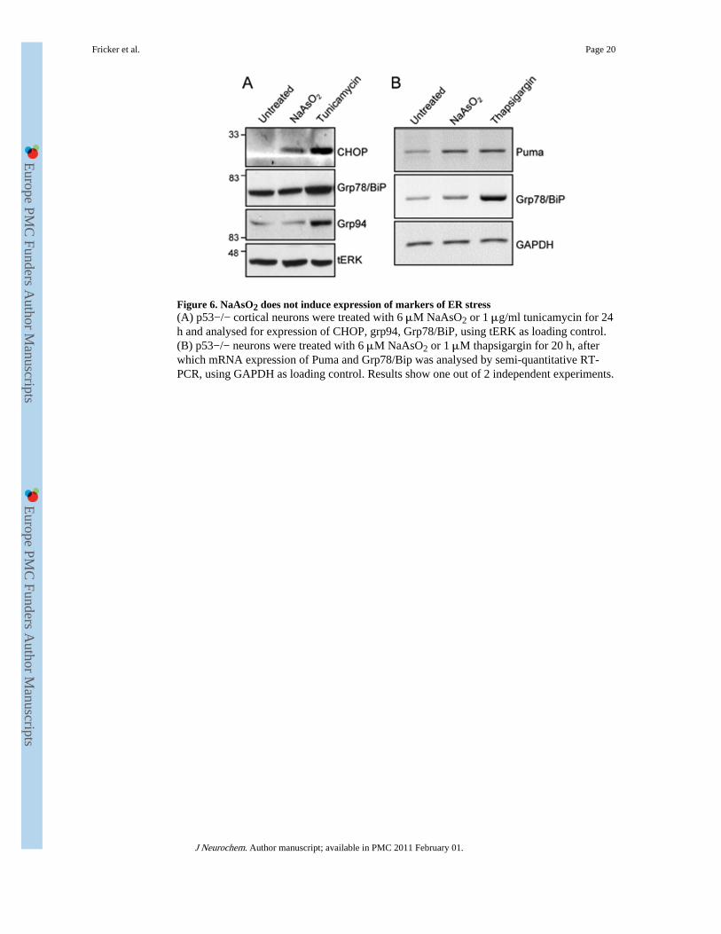
Figure 6. NaAsO2 does not induce expression of markers of ER stress(A) p53−/− cortical neurons were treated with 6 μM NaAsO2 or 1 μg/ml tunicamycin for 24h and analysed for expression of CHOP, grp94, Grp78/BiP, using tERK as loading control.(B) p53−/− neurons were treated with 6 μM NaAsO2 or 1 μM thapsigargin for 20 h, afterwhich mRNA expression of Puma and Grp78/Bip was analysed by semi-quantitative RT-PCR, using GAPDH as loading control. Results show one out of 2 independent experiments.
Fricker et al. Page 20
J Neurochem. Author manuscript; available in PMC 2011 February 01.
Europe PM
C Funders A
uthor Manuscripts
Europe PM
C Funders A
uthor Manuscripts
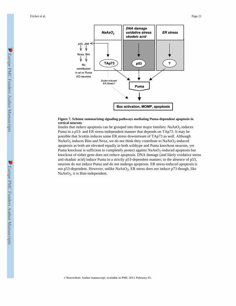
Figure 7. Scheme summarizing signaling pathways mediating Puma-dependent apoptosis incortical neuronsInsults that induce apoptosis can be grouped into three major families: NaAsO2 inducesPuma in a p53- and ER stress-independent manner that depends on TAp73. It may bepossible that Scottin induces some ER stress downstream of TAp73 as well. AlthoughNaAsO2 induces Bim and Noxa, we do not think they contribute to NaAsO2-inducedapoptosis as both are elevated equally in both wildtype and Puma knockout neurons, yetPuma knockout is sufficient to completely protect against NaAsO2-induced apoptosis butknockout of either gene does not reduce apoptosis. DNA damage (and likely oxidative stressand okadaic acid) induce Puma in a strictly p53-dependent manner; in the absence of p53,neurons do not induce Puma and do not undergo apoptosis. ER stress-induced apoptosis isnot p53-dependent. However, unlike NaAsO2, ER stress does not induce p73 though, likeNaAsO2, it is Bim-independent.
Fricker et al. Page 21
J Neurochem. Author manuscript; available in PMC 2011 February 01.
Europe PM
C Funders A
uthor Manuscripts
Europe PM
C Funders A
uthor Manuscripts