Impact of Virgin Olive Oil and Phenol-Enriched Virgin Olive Oils on the HDL Proteome in...
19
RESEARCH ARTICLE Impact of Virgin Olive Oil and Phenol- Enriched Virgin Olive Oils on the HDL Proteome in Hypercholesterolemic Subjects: A Double Blind, Randomized, Controlled, Cross-Over Clinical Trial (VOHF Study) Anna Pedret 1 , Úrsula Catalán 1 , Sara Fernández-Castillejo 1 , Marta Farràs 2,3 , Rosa-M Valls 1 , Laura Rubió 4,1 , Núria Canela 5 *, Gerard Aragonés 5 , Marta Romeu 6 , Olga Castañer 2 , Rafael de la Torre 7,8 , Maria-Isabel Covas 2 , Montse Fitó 2 , Maria-José Motilva 4 , Rosa Solà 1 * 1 Research Unit on Lipids and Atherosclerosis, CTNS, CIBERDEM, Hospital Universitari Sant Joan, Servei de Medicina Interna, IISPV, Universitat Rovira i Virgili, Reus, Spain, 2 Cardiovascular Risk and Nutrition Research Group (CARIN, Regicor Study Group), CIBER de Fisiopatología de la Obesidad y la Nutrición (CIBEROBN), IMIM (Hospital del Mar Medical Research Institute), Barcelona, Spain, 3 Ph.D. Program in Biochemistry, Molecular Biology and Biomedicine, Department of Biochemistry and Molecular Biology, Universitat Autònoma de Barcelona (UAB), Barcelona, Spain, 4 Food Technology Department, UTPV- XaRTA, University of Lleida-AGROTECNIO Research Centre, Lleida, Spain, 5 Centre for Omic Sciences (COS), Universitat Rovira i Virgili, Reus, Spain, 6 Pharmacology Unit, Facultat de Medicina i Ciències de la Salut, Universitat Rovira i Virgili, Reus, Spain, 7 Human Pharmacology and Clinical Neurosciences Research Group, CIBER de Fisiopatología de la Obesidad y la Nutrición (CIBEROBN), IMIM (Hospital del Mar Medical Research Institute), Barcelona, Spain, 8 Universitat Pompeu Fabra (CEXS-UPF), Barcelona, Spain * [email protected] (RS); [email protected] (NC) Abstract The effects of olive oil phenolic compounds (PCs) on HDL proteome, with respect to new aspects of cardioprotective properties, are still unknown. The aim of this study was to as- sess the impact on the HDL protein cargo of the intake of virgin olive oil (VOO) and two func- tional VOOs, enriched with their own PCs (FVOO) or complemented with thyme PCs (FVOOT), in hypercholesterolemic subjects. Eligible volunteers were recruited from the IMIM-Hospital del Mar Medical Research Institute (Spain) from April 2012 to September 2012. Thirty-three hypercholesterolemic participants (total cholesterol >200mg/dL; 19 men and 14 women; aged 35 to 80 years) were randomized in the double-blind, controlled, cross-over VOHF clinical trial. The subjects received for 3 weeks 25 mL/day of: VOO, FVOO, or FVOOT. Using a quantitative proteomics approach, 127 HDL-associated proteins were identified. Among these, 15 were commonly differently expressed after the three VOO interventions compared to baseline, with specific changes observed for each intervention. The 15 common proteins were mainly involved in the following pathways: LXR/RXR activa- tion, acute phase response, and atherosclerosis. The three VOOs were well tolerated by all participants. Consumption of VOO, or phenol-enriched VOOs, has an impact on the HDL proteome in a cardioprotective mode by up-regulating proteins related to cholesterol ho- meostasis, protection against oxidation and blood coagulation while down-regulating PLOS ONE | DOI:10.1371/journal.pone.0129160 June 10, 2015 1 / 19 a11111 OPEN ACCESS Citation: Pedret A, Catalán Ú, Fernández-Castillejo S, Farràs M, Valls R-M, Rubió L, et al. (2015) Impact of Virgin Olive Oil and Phenol-Enriched Virgin Olive Oils on the HDL Proteome in Hypercholesterolemic Subjects: A Double Blind, Randomized, Controlled, Cross-Over Clinical Trial (VOHF Study). PLoS ONE 10(6): e0129160. doi:10.1371/journal.pone.0129160 Academic Editor: Giuseppe Danilo Norata, University of Milan, ITALY Received: December 18, 2014 Accepted: May 3, 2015 Published: June 10, 2015 Copyright: © 2015 Pedret et al. This is an open access article distributed under the terms of the Creative Commons Attribution License, which permits unrestricted use, distribution, and reproduction in any medium, provided the original author and source are credited. Data Availability Statement: All relevant data are within the paper and its Supporting Information files. Funding: This study was supported by: the Ministerio de Economia y Competitividad (AGL2012-40144- C03-02; AGL2012-40144-C03-01 and AGL2012- 40144-C03-03 projects and, AGL2009-13517-C03-01 and AGL2009-13517-C03-03 project), CIBEROBN, FPI fellowship (BES-2010-040766), ISCIII and Departament de Salut joint contract (CP06/00100). AP is a post-graduate research student supported by
-
Upload
independent -
Category
Documents
-
view
0 -
download
0
Transcript of Impact of Virgin Olive Oil and Phenol-Enriched Virgin Olive Oils on the HDL Proteome in...

RESEARCH ARTICLE
Impact of Virgin Olive Oil and Phenol-Enriched Virgin Olive Oils on the HDLProteome in Hypercholesterolemic Subjects:A Double Blind, Randomized, Controlled,Cross-Over Clinical Trial (VOHF Study)Anna Pedret1, Úrsula Catalán1, Sara Fernández-Castillejo1, Marta Farràs2,3, Rosa-M Valls1,Laura Rubió4,1, Núria Canela5*, Gerard Aragonés5, Marta Romeu6, Olga Castañer2,Rafael de la Torre7,8, Maria-Isabel Covas2, Montse Fitó2, Maria-José Motilva4, Rosa Solà1*
1 Research Unit on Lipids and Atherosclerosis, CTNS, CIBERDEM, Hospital Universitari Sant Joan, Serveide Medicina Interna, IISPV, Universitat Rovira i Virgili, Reus, Spain, 2 Cardiovascular Risk and NutritionResearch Group (CARIN, Regicor Study Group), CIBER de Fisiopatología de la Obesidad y la Nutrición(CIBEROBN), IMIM (Hospital del Mar Medical Research Institute), Barcelona, Spain, 3 Ph.D. Program inBiochemistry, Molecular Biology and Biomedicine, Department of Biochemistry andMolecular Biology,Universitat Autònoma de Barcelona (UAB), Barcelona, Spain, 4 Food Technology Department, UTPV-XaRTA, University of Lleida-AGROTECNIO Research Centre, Lleida, Spain, 5 Centre for Omic Sciences(COS), Universitat Rovira i Virgili, Reus, Spain, 6 Pharmacology Unit, Facultat de Medicina i Ciències de laSalut, Universitat Rovira i Virgili, Reus, Spain, 7 Human Pharmacology and Clinical Neurosciences ResearchGroup, CIBER de Fisiopatología de la Obesidad y la Nutrición (CIBEROBN), IMIM (Hospital del Mar MedicalResearch Institute), Barcelona, Spain, 8 Universitat Pompeu Fabra (CEXS-UPF), Barcelona, Spain
* [email protected] (RS); [email protected] (NC)
AbstractThe effects of olive oil phenolic compounds (PCs) on HDL proteome, with respect to new
aspects of cardioprotective properties, are still unknown. The aim of this study was to as-
sess the impact on the HDL protein cargo of the intake of virgin olive oil (VOO) and two func-
tional VOOs, enriched with their own PCs (FVOO) or complemented with thyme PCs
(FVOOT), in hypercholesterolemic subjects. Eligible volunteers were recruited from the
IMIM-Hospital del Mar Medical Research Institute (Spain) from April 2012 to September
2012. Thirty-three hypercholesterolemic participants (total cholesterol >200mg/dL; 19 men
and 14 women; aged 35 to 80 years) were randomized in the double-blind, controlled,
cross-over VOHF clinical trial. The subjects received for 3 weeks 25 mL/day of: VOO,
FVOO, or FVOOT. Using a quantitative proteomics approach, 127 HDL-associated proteins
were identified. Among these, 15 were commonly differently expressed after the three VOO
interventions compared to baseline, with specific changes observed for each intervention.
The 15 common proteins were mainly involved in the following pathways: LXR/RXR activa-
tion, acute phase response, and atherosclerosis. The three VOOs were well tolerated by all
participants. Consumption of VOO, or phenol-enriched VOOs, has an impact on the HDL
proteome in a cardioprotective mode by up-regulating proteins related to cholesterol ho-
meostasis, protection against oxidation and blood coagulation while down-regulating
PLOSONE | DOI:10.1371/journal.pone.0129160 June 10, 2015 1 / 19
a11111
OPEN ACCESS
Citation: Pedret A, Catalán Ú, Fernández-CastillejoS, Farràs M, Valls R-M, Rubió L, et al. (2015) Impactof Virgin Olive Oil and Phenol-Enriched Virgin OliveOils on the HDL Proteome in HypercholesterolemicSubjects: A Double Blind, Randomized, Controlled,Cross-Over Clinical Trial (VOHF Study). PLoS ONE10(6): e0129160. doi:10.1371/journal.pone.0129160
Academic Editor: Giuseppe Danilo Norata,University of Milan, ITALY
Received: December 18, 2014
Accepted: May 3, 2015
Published: June 10, 2015
Copyright: © 2015 Pedret et al. This is an openaccess article distributed under the terms of theCreative Commons Attribution License, which permitsunrestricted use, distribution, and reproduction in anymedium, provided the original author and source arecredited.
Data Availability Statement: All relevant data arewithin the paper and its Supporting Information files.
Funding: This study was supported by: the Ministeriode Economia y Competitividad (AGL2012-40144-C03-02; AGL2012-40144-C03-01 and AGL2012-40144-C03-03 projects and, AGL2009-13517-C03-01and AGL2009-13517-C03-03 project), CIBEROBN,FPI fellowship (BES-2010-040766), ISCIII andDepartament de Salut joint contract (CP06/00100).AP is a post-graduate research student supported by

proteins implicated in acute-phase response, lipid transport, and immune response. The
common observed protein expression modifications after the three VOOs indicate a major
matrix effect.
Trial Registration
International Standard Randomized Controlled Trials ISRCTN77500181.
IntroductionOlive oil (OO) is a food item typical of the Mediterranean diet, and several studies have revealedthat it has a unique phenolic profile with specific biological properties. Results from the Europe-an EUROLIVE study [1] showed that an increase in plasma HDL cholesterol, and a decrease ofLDL oxidation, took place in a direct relationship with the phenolic compound (PC) content ofthe OO administered. These findings provided evidence to recommend the use of PC-rich OO,i.e. virgin olive oil (VOO), in order to achieve beneficial effects improving lipid profile and con-ferring protection from oxidative damage. The difference of PC content between OO and VOOis due to the elaboration process (www.internationaloliveoil.org). To date, studies have been fo-cused on hydroxytyrosol because it has been reported as being the most biologically active PC inOO [2]. In fact, the European Food Safety Authority considers that the health claim may beused only for OO which contains at least 5mg of hydroxytyrosol and its derivatives per 20g ofOO [2]. As the phenolic concentration in most of the VOOs available on the market is too lowto provide this daily amount of hydroxytyrosol, the enrichment of VOO with its own PCs couldbe a possible approach to assure the consumption without increasing caloric intake [3,4]. Fur-thermore, the enrichment of VOO with its own PCs could lead to a bitter organoleptic tastecaused by the presence of secoiridoids (hydroxytyrosol and its derivatives; oleruopein and lig-stroside aglycones) [5]. Moreover, in terms of biological effects, synergistic effects with strongerimprovements have been reported when different PCs are combined in comparison to singletreatments [6]. For these reasons, the enrichment of a VOO by complementing its own phenolswith PCs of aromatic herbs has been done in order to improve its nutritional profile and organ-oleptic characteristics. Thyme could enhance perfectly these features of a phenol-enriched VOObecause it is one of the richest sources of flavonoids, such as naringenin, eriodictyol and api-genin and phenolic acids such as rosmarinic acid, ferulic acid and caffeic acid [7,8].
Approximately one hundred different proteins, not considered to be apolipoproteins(APO), have been identified as being associated with the HDL particle [9] and linked to its car-dioprotective properties. Analysis of the HDL protein cargo is, however, still in its early stages,and data concerning the HDL proteome as a biomarker for disease or functionality are limited.Moreover, studies with respect to the impact of dietary interventions on the HDL proteome arescarce [10], and the effects of OO PCs on the complex structure of the HDL protein cargo havenot yet been evaluated.
In this context, we hypothesized that sustained consumption of PCs from VOO, or func-tional phenol-enriched VOO, could modify the HDL proteome, which in turn could be relatedto their cardioprotective benefits. The objective of the current study was to assess the impacton the HDL protein cargo of a dietary intervention supplemented with a VOO or two differentfunctional VOOs enriched with their own PCs (hydroxytyrosol and its derivatives; oleuropeinand ligstroside aglycones) or complemented with thyme PCs (flavonoids, monoterpens, andphenolic acids), in hypercholesterolemic subjects from the VOHF study.
Impact of Olive Oil Phenols on HDL Proteome
PLOSONE | DOI:10.1371/journal.pone.0129160 June 10, 2015 2 / 19
the Universitat Rovira i Virgili and the CentreTecnològic de Nutrició i Salut (CTNS).
Competing Interests: The authors have declaredthat no competing interests exist.

Materials and Methods
Subjects and study designThe VOHF study aimed at assessing whether functional VOOs, enriched with their own PCsor with them plus complementary phenols from thyme, could have a nutraceutical effect onHDL lipoprotein functionality. The present clinical trial was conducted in accordance with theHelsinki Declaration and the Good Clinical Practice for Trials on Medical Products in the Eu-ropean Community. All participants provided written informed consent, and the local institu-tional ethics committees approved the protocol (Comité Ético de Investigación Clínica delInstituto Municipal de Asistencia Sanitaria; CEIC-IMAS 2009/3347/I) registered with the In-ternational Standard Randomized Controlled Trial register (www.internationaloliveoil.org;ISRCTN77500181). This trial was conducted according to extended CONSORT 2010guidelines.
The VOHF study was a double-blind, randomized, controlled, crossover clinical trial with33 hypercholesterolemic volunteers (total cholesterol>200 mg/dL; 19 men and 14 women),aged 35 to 80 year. Exclusion criteria included the following: BMI>35 Kg/m2, smokers, ath-letes with high physical activity (>3000 Kcal/day), diabetes, multiple allergies, intestinal dis-eases, or any other disease or condition that could worsen compliance. The study wasconducted at IMIM- Hospital del Mar Medical Research Institute (Spain) from April 2012 toSeptember 2012. The participants’ flow chart is described in Fig 1.
Subjects were randomly allocated to one of 3 sequences of administration of 25 mL/day ofraw: a) VOO; 80 mg of PCs/kg oil, b) functional VOO enriched with its own PCs (FVOO; 500mg of PCs/kg oil), and c) functional VOO enriched with its own PCs plus complementary phe-nols from thyme (FVOOT; 500 mg of PCs/kg; 50% of OO PCs and 50% of thyme PCs) (Se-quence 1: FVOO, FVOOT and VOO, Sequence 2: FVOOT, VOO and FVOO, Sequence 3:VOO, FVOO and FVOOT) (Fig 2). The random allocation sequence was generated by a statis-tician, participant enrolment was carried out by a researcher, and participants’ assignment tointerventions according to the random sequence was done by a physician. Due to the fact thatall participants received each one of the three VOOs, restrictions such as blocking were unnec-essary. Intervention periods were of 3 weeks and VOOs were consumed daily distributedamong meals. There was a 2-week washout period prior to VOO interventions during which acommon OO was consumed. A 3-day dietary record was administered to the participants atbaseline and before and after each intervention-period. A nutritionist personally advised par-ticipants to replace all types of habitually consumed raw fats with the olive oils, and to limittheir PC-rich foods consumption during the clinical trial. A set of portable containers with thecorresponding 25 mL of VOO for each day of consumption were delivered to the participantsat the beginning of each VOO administration period. The participants were instructed to re-turn the containers to the center after the corresponding VOO consumption period in order toregister the amount consumed. Moreover, the metabolites hydroxytyrosol sulfate, hydroxytyr-osol acetate sulfate, thymol sulfate and hydroxyphenylpropionic acid sulfate were measured in24-h urine in order to monitor the compliance after the intake of two phenol-enriched VOO[11]. Furthermore, concentration of the different phenolic metabolites was determined in theHDL fractions of the all participants. The concentrations of the metabolites present in theHDL pools used for proteomics analyses (as described below) are shown in S1 Table.
Preparation and characterization of VOOsThe procedure to obtain the phenolic extracts and the enriched oils has been previously de-scribed [7]. Briefly, VOO with a low phenolic content was used as a control condition in the
Impact of Olive Oil Phenols on HDL Proteome
PLOSONE | DOI:10.1371/journal.pone.0129160 June 10, 2015 3 / 19

intervention and as an enrichment matrix for the preparation of the two phenol-enriched oliveoils. FVOO was enriched with its own PCs by adding a phenol extract obtained from freeze-dried olive cake collected from a commercial olive mill in the olive-growing area of Les Garri-gues (Lleida, Catalonia, Spain). FVOOT was enriched with its own PC (50%) and comple-mented with thyme PC (50%) using a phenol extract made up of a mixture of olive cake andcommercially available dried thyme (Thymus zyguis). The phenolic extracts used for enrich-ment were obtained in the laboratory using an accelerated solvent extractor (ASE 100 Dionex,Sunnyvale, CA). S2 Table shows the PCs, the fat soluble micronutrients, and the fatty acid dailyintake with 25mL of VOO, FVOO, and FVOOT. The phenolic profile of the VOO was ana-lyzed by high-performance liquid chromatography coupled to tandem mass spectrometry(HPLC/MS/MS) using the method previously described [7]. Tocopherols and fatty acids inVOO were analyzed following the procedure described by Morelló et al. (2004) [12] and the ca-rotenoid content was analyzed as described before by Criado et al. (2008) [13].
Fig 1. Participants Flow-chart based on post-interventions of HDL proteomics variable. Sequence 1: FVOO, FVOOT and VOO; Sequence 2: FVOOT,VOO and FVOO; Sequence 3: VOO, FVOO and FVOOT.
doi:10.1371/journal.pone.0129160.g001
Impact of Olive Oil Phenols on HDL Proteome
PLOSONE | DOI:10.1371/journal.pone.0129160 June 10, 2015 4 / 19

Sample size and power analysisThe sample size of 30 individuals allows at least 80% power to detect a statistically significantdifference among three groups of 3 mg/dL of HDL-C and a standard deviation of 1.9, using anANOVA test and assuming a dropout rate of 15% and a Type I error of 0.05.
Collection of blood samplesFasting blood samples were taken from 33 participants before the first washout period (firstvisit-baseline) and after each VOO intervention period (visits 3, 5, and 7), through a catheterin an antecubital vein (Fig 2). Blood was collected in Vacutainer tubes with K2EDTA antico-agulant. Blood samples were centrifuged at 1 500 xg for 15 minutes and 2.8 mL of plasmawere finally recovered. Protease Inhibitor Cocktail (PIC; Sigma-Aldrich, Tres Cantos, Spain)was added to plasma at a concentration 1/100 (1 μL of PIC for 100 μL of plasma). Allsamples were stored at -80°C until processing. A total of 123 plasma samples were used forproteomic analyses (33 participants per 4 visits minus 9 missing). See proteomic study flowchart (Fig 3).
Human plasma HDL isolationThe HDL fraction (d = 1.036–1.21 g/mL) from each sample was isolated from 2.5 mL of plas-ma by sequential density ultracentrifugation in two steps using sodium bromide (NaBr;Sigma-Aldrich, Tres Cantos, Spain), as previously described by Havel RJ, et al. [14]. Sequentialultracentrifugation continues to be the procedure most commonly used for isolation of HDLin proteomic studies. To date, nearly all proteomic studies of HDL have utilized density gradi-ent ultracentrifugation based on methods for the isolation of HDL from human plasma[15,16]. Briefly, the VLDL, IDL, and LDL, were removed by ultracentrifugation at 140 000 xgfor 21 h at 10°C after adjustment of the plasma density to 1.063mg/mL with NaBr. The HDLfraction was obtained by adjusting the infranatant density to 1.21 mg/mL with NaBr prior to
Fig 2. Experimental protocol for the intervention study. VOO: Virgin Olive Oil; FVOO: Functional VirginOlive Oil enriched with its own PC; FVOOT: Functional Virgin Olive Oil enriched with its own PC pluscomplementary phenols from thyme. Blood collection for proteomic analysis: Visit 1, baseline; Visit 3, post-first intervention; Visit 5, post-second intervention; Visit 7, post-third intervention.
doi:10.1371/journal.pone.0129160.g002
Impact of Olive Oil Phenols on HDL Proteome
PLOSONE | DOI:10.1371/journal.pone.0129160 June 10, 2015 5 / 19

Fig 3. Proteomic study flow chart. S1: Sequence 1; S2: Sequence 2; S3: Sequence 3; HDL: P.: Pool; SCX:Strong Cation Exchange. Visits with n lower than 11 was due to loss of participants for the following reasons:dropout, did not participate in all interventions, did all the interventions but not all the tests, or sample lossduring laboratory processing.
doi:10.1371/journal.pone.0129160.g003
Impact of Olive Oil Phenols on HDL Proteome
PLOSONE | DOI:10.1371/journal.pone.0129160 June 10, 2015 6 / 19

ultracentrifugation at 140 000 xg for 40 h at 10°C. 2 mL of HDL fraction were recovered andlater stored at -80°C.
HDL biochemical characterizationApo A-I, Apo A-II and Apo B-100 were determined in each volunteer HDL fraction usingimmuno-turbidimetry assays (Horiba, Montpellier, France) in an autoanalyzer Cobas-MiraPlus (Roche Diagnostic System, Madrid, Spain). Total Cholesterol, Free Cholesterol, Phospho-lipids and Triglycerides were determined by enzymatic colorimetric techniques (Spinreact, Gi-rona, Spain) also in the autoanalyzer Cobas-Mira Plus. Total protein was quantified in HDLfractions by the Bradford method (BioRad, Hercules, CA, USA). HDL fraction purity was as-sessed through the measurement of Apo B-100, almost all samples exhibited concentrationsbelow the limit of detection (2 mg/dL).
Proteomic sample preparation and quantitative analysisFor the proteomics studies (detailed in Supporting Information Data) the 123 isolated HDLsamples were reduced to ten pools: 1 from first visit- baseline, 3 after third visit, 3 after fifthvisit, and 3 after seventh visit (Fig 3). The pools were then dialyzed and delipidated by metha-nol/diethyl ether extraction, followed by a trichloroacetic acid/acetone protein precipitation be-fore an in-solution tryptic digestion. Samples were subjected to an iTRAQ labeling forquantitative analysis, the resulting labeled peptides were separated according to their isoelectricpoint on an OffGel fractionator. Finally, two aliquots of the sample were analyzed by two dif-ferent MS techniques, nano LC-MALDI MS/MS and nano LC-ORBITRAP-ESI MS/MS, toprovide reliability and robustness (Fig 3). The files generated with the two mass spectrometerswere combined in order to identify the HDL-associated proteins. The final identified proteinswere required to present more than one peptide-spectrum match (PSM), or to have a mini-mum confidence score> 30% and coverage > 10%, in order to ensure accuracy in the assign-ment of protein identifications. In addition, intracellular and cell surface proteins wereremoved as being possible contaminants. In order to identify the differentially expressed pro-teins after interventions with respect to first visit- baseline, only samples analyzed by nanoLC-ORBITRAP-ESI MS/MS were used due to the precision of this method. Relative expressionlevels were calculated for each protein as a ratio by Proteome Discoverer; biological replicatesfrom the three iTRAQ sequences were then combined in an Excel spreadsheet and the mean ofthe ratios of all proteins calculated. Proteins with a differential expression of at least 0.8-foldchange, or 1.3-fold change relative to baseline, were considered differentially expressed. In ad-dition, a cut-off inferior to 0.5, or superior to 1.5, was applied to establish the most relevantprotein expression changes observed after each intervention, defined as stronger effects. Adetailed description of the methods and analysis is provided in the online S1 SupportingInformation.
Clustering and pathway analysisVarious bioinformatics tool were employed for the biological interpretation of the results. Pro-teins are referred to by their gene encode symbol.
Hierarchical clustering of protein expression data to investigate overall similarities of theproteome samples and identify the main biological functions involved was performed in web-tool STRING 9.1 (http://string-db-org). Ingenuity Pathway analysis (IPA; Ingenuity SystemInc., Redwook, CA, USA, www.ingenuity.com) was used to analyze canonical pathways andprotein networks involving the differentially expressed proteins for biological interpretation(Fig 3). Significance levels were assessed with Fisher’s exact tests (p<0.01). The differentially
Impact of Olive Oil Phenols on HDL Proteome
PLOSONE | DOI:10.1371/journal.pone.0129160 June 10, 2015 7 / 19
expressed proteins were overlaid with IPA-curated canonical pathways to explore possible met-abolic and cell signaling pathways that were over- or under-represented by the experimentallydetermined genes. Specifically, we analyzed the proteins that overlapped the three VOO inter-ventions in order to investigate potential common OO PCs mechanisms. In addition, possibleconnections between mapped genes were evaluated and graphical networks were algorithmi-cally generated. Nodes representing genes and gene products were linked by biological relation-ships. Networks were ranked by a score that defines the probability of a collection of nodesbeing equal to or greater than the number in a network achieved by chance alone.
Immunodetection assaysApo A-I and Apo A-II, in the HDL pools, were determined using immunoturbidimetry assays(Horiba, Montpellier, France) in an autoanalyzer Cobas-Mira Plus (Roche Diagnostic System,Madrid, Spain). The quantification of haptoglobin and clusterin was performed using twocommercial ELISA assays following manufacter’s instructions; the AssayMax HaptoglobinELISA kit (AssayPro, MO, USA) and the Human Clusterin ELISA Kit (RayBiotech, GA,USA), respectively.
Statistical analysis of biochemical dataThe Kolmogorov-Smirnov test was used to verify the distributions of the variables. A pairedStudent’s t-test was employed for the comparison of paired and normally distributed variables.Wilcoxon signed-rank test was used for the comparison of paired and non-normally distribut-ed variables. The level of statistical significance was set at p< 0.05. The possible interaction be-tween the treatments and the treatment sequence (carryover effect) was assessed testing theperiod by treatment interaction-effect under a linear mixed model. All statistical analyses wereperformed with Statistical Package for the Social Sciences (SPSS) for Windows (20.0 version;IBM corp., Armonk, NY, USA).
Results
Participants’ characteristicsTable 1 summarizes the baseline characteristics of the study participants, significant differenceswere not observed among groups. No changes were observed in the main nutrients and medi-cation intake throughout the study. The three VOOs were well tolerated by all participants andno adverse events were reported.
Biochemical characterization of HDL fractionsBiochemical characterization of HDL and also Apo A-I and HDL-c plasma levels segregatedaccording to VOO intervention sequence are described in detail in S3 Table. In the HDL frac-tion and compared to baseline, significant increases in Apo A-I (p = 0.027; p = 0.021; p = 0.006,for VOO, FVOO, and FVOOT respectively), Apo A-II (p = 0.029; p = 0.019; p = 0.004, forVOO, FVOO, and FVOOT respectively), total protein (p = 0.073; p = 0.045; p = 0.037, forVOO, FVOO, and FVOOT respectively), total cholesterol (p = 0.033; p = 0.037; p = 0.005, forVOO, FVOO, and FVOOT respectively), phospholipids (p = 0.146; p = 0.021; p = 0.036, forVOO, FVOO, and FVOOT respectively) and total mass (p = 0.050; p = 0.014; p = 0.016, forVOO, FVOO, and FVOOT respectively) were observed after the three intervention periods.However, as a consequence of the increment of the different parameters of the HDL particlecomposition, the total mass of the HDL particle also increases and as a result, the compositionof HDL expressed as percentage of total mass remains stable and non-significant changes were
Impact of Olive Oil Phenols on HDL Proteome
PLOSONE | DOI:10.1371/journal.pone.0129160 June 10, 2015 8 / 19

observed. Compared to baseline, significant increases were also observed in plasma Apo A-Iand HDL-c after the three VOO intervention periods. However, no significant differences be-tween the three VOO interventions, either in basal levels or in post interventions levels, wereobserved suggesting that in the present study, the enrichment of VOO with PC might not affectthe ability of the VOO to increase different biochemical parameters and that the common ma-trix of the three VOO produced the key changes. We did not observe a carryover effect duringthe study.
HDL proteomic resultsIdentification of HDL-associated proteins. A total of 155 HDL-associated proteins were
initially identified by applying shotgun proteomics using Maldi and Orbitrap MS/MS to HDLfractions isolated by ultracentrifugation. Data were subsequently screened to ensure proteinidentification accuracy and 28 possible false-positives removed. The final list of HDL-associat-ed proteins identified both at baseline and after interventions, was refined to 127 proteins (S4Table). The majority (80/127) were consistent with well-established HDL-associated proteinsdetermined in at least 3 different MS studies [9] and 32 had been previously identified by atleast 1 MS study [9,17]. A total of 15 not previously referenced proteins were identified both atbaseline and after interventions, although they do not show any modulation during interven-tions. These novel proteins are the following: Beta-Ala-His dipeptidase, BPI fold-containingfamily B member 1, Carbonic anhydrase 6, CD209 antigen, CD44 antigen, CD5 antigen-like, Igheavy chain V-I region HG3, Ig lambda-1 chain C regions, Ig lambda-2 chain C regions, Iglambda-7 chain C region, Indian hedgehog protein, Integrin alpha-2, Integrin beta-1, Multi-merin-2, and Thymidine phosphorylase. Six well-established HDL-associated proteins werenot detected: Apo O, Ceruloplasmin, Complement factor B, Inter alpha trypsin inhibitor 2,Plasma Kallikerein and Plasminogen [9].
HDL quantitative analyses. Compared to baseline values (first visit), the HDL proteincargoes of the differentially expressed proteins differed after the interventions according to theVOO received. The complete list of the proteins differentially expressed after each VOO inter-vention, and their principal biological functions, are shown in S5 Table. The proteins were
Table 1. Characteristics of the study participants at baseline.
Variable Sequence 1 (n = 11) Sequence 2 (n = 11) Sequence 3 (n = 11)
Gender (male/female) 5/6 7/4 7/4
Age, years 54.91 ± 12.57 55.27 ± 11.88 55.45 ± 7.84
Body weight, kg 74.75 ± 16.80 74.60 ± 18.49 84.45 ± 17.74
BMI, kg/m2 25.63 ± 3.68 26.31 ±5.25 27.85 ± 4.71
SBP, mm Hg 125.09 ± 18.70 128.27 ± 16.69 130.45 ± 17.93
DBP, mm Hg 68.09 ± 13.53 72.27 ± 9.31 71.91 ± 13.43
Glucose, mg/dL 88.55 ± 11.63 93.00 ± 13.33 90.91 ± 10.53
Total cholesterol, mg/dL 228.36 ± 42.70 231.91 ± 32.70 218.82 ± 31.21
LDL cholesterol, mg/dL 150.38 ± 32.33 152.08 ± 28.46 142.26 ± 25.72
HDL cholesterol, mg/dL 52.78 ±11.75 52.96 ±12.82 53.39 ± 9.55
Tryglicerides, mg/dL 94.00 (75.00; 149.00) 119.00 (95.00; 168.00) 117.00 (81.00; 126.00)
Values expressed as mean ± standard deviation (SD) or median (25th to 75th percentile). Sequence 1 = FVOO, FVOOT and VOO; Sequence 2 = FVOOT,
VOO and FVOO; Sequence 3 = VOO, FVOO and FVOOT. No significant differences between groups were observed. To compare means or medians
among groups, ANOVA or Kruskal-Wallis test were performed, respectively; whereas χ2 and exact F-test, as appropriate, were computed to compare
proportions. Abbreviations: SBP, systolic blood pressure; DBP, diastolic blood pressure.
doi:10.1371/journal.pone.0129160.t001
Impact of Olive Oil Phenols on HDL Proteome
PLOSONE | DOI:10.1371/journal.pone.0129160 June 10, 2015 9 / 19

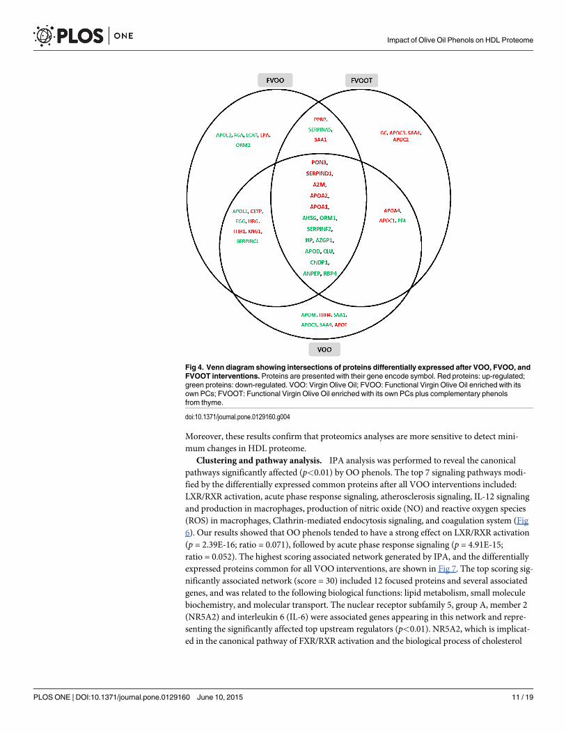
associated with a broad range of biological functions, principally cholesterol homeostasis, lipidtransport, acute-phase response, blood coagulation, immune response, protection against oxi-dation, and proteolysis. The proteins differently modulated after VOO, FVOO, and FVOOTinterventions and associated with these main functions are represented in Fig 4. The overlap-ping among interventions indicates that 15 proteins were commonly up- or down-regulatedafter the three VOO interventions. These proteins were: Serum paraxonase/lactonase 3(PON3), Apo A-II, Apo A-I, Apo D, Retinol binding protein 4 (RBP4), Heparin cofactor 2(SERPIND1), zinc-alpha-2-glycoprotein (AZGP1), alpha-2-antiplasmin (SERPINF2), alpha-2-HS-glycoprotein (AHSG), clusterin (CLU), alpha-2-macroglobulin (A2M), haptoglobin(HP), alpha-1-acid glycoprotein (ORM1), Beta-Ala-His dipeptidase (CNDP1), and Aminopep-tidase N (ANPEP). These 15 proteins that were observed to have the greatest expression modi-fications were identified with both MS techniques, nano LC-MALDI MS/MS and nanoLC-ORBITRAP-ESI MS/MS. A protein-protein interaction network was generated for the 15common, differentially expressed proteins using database and web-tool STRING 9.1 (S1 Fig).The common up-regulated proteins were related to cholesterol homeostasis, blood coagulationand protection against oxidation; the common down-regulated proteins were implicated inacute-phase response, lipid transport, immune response, and proteolysis. All proteins exceptfor ANPEP and CNDP1, which are involved in proteolysis, appeared in the center of the func-tional network intersection indicating their key role in protein interactions. The information ofthese relevant proteins commonly regulated by the three VOO interventions was summarizedin Table 2.
In addition to these 15 commonly modulated proteins, other specific protein expressionchanges were observed after each OO intervention. The stronger effects for each VOO were re-lated to the following biological functions linked to atherosclerosis protection: transport, ho-meostasis cholesterol, antioxidant protection, blood coagulation, innate immune response,acute phase, and cellular adhesion. A stronger protein expression change was observed afterVOO intervention in the following 6 proteins: Apo C-I, Apo A-I, Cholesteryl ester transfer pro-tein (CETP), PON3, histidine-rich glycoprotein (HRG), and Ig heavy chain V-I region HG3,which were up-regulated while Ig lambda chain V-III region LOI was down-regulated. A stron-ger effect after FVOO was observed on 2 protein expression changes, PON3 and platelet basicprotein (PPBP) which were up-regulated. Finally, a stronger up-expression change was noticedafter FVOOT in the following 7 proteins: Apo A-I, Apo A-IV, Apo C-II, Apo C-III, serum am-yloid A protein 4 (SAA4), afamin (AFM) and integrin beta-3 (ITGB3).
Confirmation of the proteomic results by immunodetection methods. A panel of fourproteins identified by MS analysis with changes after the three interventions, two up-regulated(Apo AI and Apo AII) and two down-regulated (Clusterin and Haptoglobin), were selected toconfirm the changes in the protein expression that were observed in the proteomic analysis.These four proteins were quantified in the same 10 HDL pools used for proteomic analysis.Apo A-I and Apo A-II increases observed by proteomics were confirmed by immunoturbidi-metry. The fold-changes observed in Apo A-I and Apo A-II measured by immunodetectionwere approximately 1.2 after the three VOO interventions compared to the basal value (Fig 5).Moreover, the results obtained for clusterin and haptoglobin quantifications measured byELISA were also consistent with the decreases observed by proteomics with fold-changes of theorder of 0.9 (Fig 5). The fold-changes observed using immunodetection methods were in thesame way than those observed in proteomics analysis, even though they did not reach the samefold-change values. We observed an increase tendency for Apo AI and Apo AII proteins and adecrease tendency for Clusterin and Haptoglobin, in agreement with the MS findings, althoughthese tendencies did not achieve statistical significance probably due to the low sample size.
Impact of Olive Oil Phenols on HDL Proteome
PLOSONE | DOI:10.1371/journal.pone.0129160 June 10, 2015 10 / 19
Moreover, these results confirm that proteomics analyses are more sensitive to detect mini-mum changes in HDL proteome.
Clustering and pathway analysis. IPA analysis was performed to reveal the canonicalpathways significantly affected (p<0.01) by OO phenols. The top 7 signaling pathways modi-fied by the differentially expressed common proteins after all VOO interventions included:LXR/RXR activation, acute phase response signaling, atherosclerosis signaling, IL-12 signalingand production in macrophages, production of nitric oxide (NO) and reactive oxygen species(ROS) in macrophages, Clathrin-mediated endocytosis signaling, and coagulation system (Fig6). Our results showed that OO phenols tended to have a strong effect on LXR/RXR activation(p = 2.39E-16; ratio = 0.071), followed by acute phase response signaling (p = 4.91E-15;ratio = 0.052). The highest scoring associated network generated by IPA, and the differentiallyexpressed proteins common for all VOO interventions, are shown in Fig 7. The top scoring sig-nificantly associated network (score = 30) included 12 focused proteins and several associatedgenes, and was related to the following biological functions: lipid metabolism, small moleculebiochemistry, and molecular transport. The nuclear receptor subfamily 5, group A, member 2(NR5A2) and interleukin 6 (IL-6) were associated genes appearing in this network and repre-senting the significantly affected top upstream regulators (p<0.01). NR5A2, which is implicat-ed in the canonical pathway of FXR/RXR activation and the biological process of cholesterol
Fig 4. Venn diagram showing intersections of proteins differentially expressed after VOO, FVOO, andFVOOT interventions. Proteins are presented with their gene encode symbol. Red proteins: up-regulated;green proteins: down-regulated. VOO: Virgin Olive Oil; FVOO: Functional Virgin Olive Oil enriched with itsown PCs; FVOOT: Functional Virgin Olive Oil enriched with its own PCs plus complementary phenolsfrom thyme.
doi:10.1371/journal.pone.0129160.g004
Impact of Olive Oil Phenols on HDL Proteome
PLOSONE | DOI:10.1371/journal.pone.0129160 June 10, 2015 11 / 19

Table 2. Relevant proteins commonly differentially regulated by the three VOO interventions.
Gene Symbol Protein Name Principal Biological Function VOO FoldChange
FVOO FoldChange
FVOOT FoldChange
UP-REGULATED
APOA1 Apolipoprotein A-I Cholesterol homeostasis 1.69 1.37 1.60
PON3 Serum paraoxonase/lactonase 3
Antioxidant protection 1.56 1.55 1.39
APOA2 Apolipoprotein A-II Cholesterol homeostasis 1.45 1.40 1.34
SERPIND Heparin cofactor 2 Blood coagulation 1.27 1.27 1.32
A2M Alpha-2-macroglobulin Blood coagulation 1.25 1.33 1.39
DOWN-REGULATED
HP Haptoglobin Acute-phase response 0.56 0.80 0.65
CLU Clusterin Complement pathway and innateimmune response
0.61 0.78 0.64
APOD Apolipoprotein D Lipid transport 0.61 0.71 0.68
AZGP1 Zinc-alpha-2-glycoprotein Immune response 0.69 0.75 0.63
ORM1 Alpha-1-acid glycoprotein 1 Acute-phase response 0.75 0.53 0.58
SERPINF2 Alpha-2-antiplasmin Acute-phase response 0.75 0.72 0.77
RBP4 Retinol-binding protein 4 Transport 0.78 0.78 0.77
ANPEP Aminopeptidase N Proteolysis 0.80 0.78 0.68
CNDP1 Beta-Ala-His dipeptidase Proteolysis 0.82 0.69 0.72
AHSG Alpha-2-HS-glycoprotein Acute-phase response 0.84 0.80 0.76
Fold change > 1.3 denotes proteins up-regulated while fold change < 0.8 denotes decrease protein expression after the VOO interventions relative to the
control-baseline. Gene symbol and principal biological function information were from UniProt database (http://www.uniprot.org/).
doi:10.1371/journal.pone.0129160.t002
Fig 5. Expression levels of Apo A-I, Apo A-II, Clusterin and Haptoglobin. Expression levels of Apo A-Iand A-II were measured by immunturbidimetry assays and clusterin and haptoglobin by ELISA assays in 10HDL pools.
doi:10.1371/journal.pone.0129160.g005
Impact of Olive Oil Phenols on HDL Proteome
PLOSONE | DOI:10.1371/journal.pone.0129160 June 10, 2015 12 / 19

homeostasis, was predicted from our results to be activated. Likewise, IL-6, which is involvedin the acute phase response canonical pathway, was expected to be inhibited.
DiscussionThe present study revealed that, in hypercholesterolemic subjects, a supplementary consump-tion of 25 mL/day of raw VOO, FVOO, or FVOOT has an impact on the HDL proteome bychanging the expression of a number of proteins with biological functions related to the cardio-protective function of the HDL particle. The greatest expression modifications were observedin 15 of the 127 proteins identified in the HDL fractions which were commonly up- or down-regulated after the three VOO interventions and might lead to a CVD protective HDL profile.A finding that encouraged us to highlight the effect of the fatty acid composition and the roleof the PCs (lignans) of the common matrix of the three VOO tested on HDL remodeling.Moreover, IPA analysis revealed that several signaling pathways related to CVD were affectedby OO PCs consumption. These data emphasize the key role of OO PCs in modifying pathwaysand conferring cardioprotective properties. PCs of VOO could be capable of transcriptionalgene regulation inducing the down or up-regulation of proteins involved in functions relatedto cardiovascular risk [18,19]. Farràs et al.[20] observed how PCs from OO could exert an invivo nutrigenomic effect on genes related to cholesterol efflux in humans. Moreover, PCs canmodulate enzyme activities which may represent relevant antioxidant mechanisms by whichdietary olive phenolic could have beneficial impact on cardiovascular health [21]. Phenol me-tabolites from olive have been observed to be incorporated to HDL particles where they couldexert a local antioxidant protection and contribute to a functional enhancement of the HDLparticle [22]. In concordance with these results, we observed that thyme phenols were also in-corporated to HDL after FVOOT intervention and that olive phenol metabolites were higherafter FVOO intervention compared to baseline and also to VOO. Furthermore, it has been re-cently reported that hydroxytyrosol contribute to the protection of endothelium reticulum(ER) stress which could cause unfolded or misfolded proteins and is considered as a newemerging risk factor for CVD. This newly described effect of hydroxytyrosol on ER stress couldrepresent an alternative pathway through which olive oil phenols can modulate cell signalingand could impact HDL proteome and functionality [23].
Fig 6. The top 7 signaling pathways that were significantly affected by the 15 common proteinsdifferentially expressed after all VOO interventions. Significance levels were assessed with Fisher’sexact test (p<0.01). The pathways were ranked by p value. Blue bars indicate the negative log value (p-value). The ratio was calculated as the number of molecules in a given pathway that meet cutoff criteriadivided by total number of molecules that make up that pathway.
doi:10.1371/journal.pone.0129160.g006
Impact of Olive Oil Phenols on HDL Proteome
PLOSONE | DOI:10.1371/journal.pone.0129160 June 10, 2015 13 / 19

To our knowledge, this is the first HDL proteomic study in humans that assesses the effectsof PCs on the HDL protein cargo.
Common effects of VOO interventionsTwo well-known proteins related to cholesterol homeostasis, Apo A-I and Apo A-II, were sig-nificantly augmented after the three VOO interventions. Both proteins participate in reversecholesterol transport and present anti-inflammatory and antioxidant properties [24,25]. Theincrease in Apo A-I concurred with the results observed by Solà et al. [26] in high CVD risksubjects after following a Mediterranean diet rich in VOO. PC could increase the expressionand secretion of Apo-AI by regulatory mechanisms involved in cholesterol biosynthesis andmetabolism [27]. The increment of Apo-AI observed could lead to a possible improvement ofHDL functionality by enhancing HDL cholesterol efflux via ATP-binding cassette transporter
Fig 7. Top scored associated network generated by IPA describing common differentially expressedproteins after all VOO interventions. The top scoring significantly associated network was assessed withFisher’s exact test (p<0.01). Proteins were presented with their gene encode symbol. The proteins indicatedin red and green are those whose expression levels were significantly up- or down-regulated, respectively.Proteins indicated in white are those available in the IPA database, but not detected as differentiallyexpressed in the present study. The shapes of the symbols denote the molecular class of proteins. Solid linesindicate direct molecular interactions, whereas dashed lines indicate indirect molecular interactions.
doi:10.1371/journal.pone.0129160.g007
Impact of Olive Oil Phenols on HDL Proteome
PLOSONE | DOI:10.1371/journal.pone.0129160 June 10, 2015 14 / 19

A1 and HDL antioxidative properties, because Apo A-I is the major HDL component involvedin these activities [28].
Two proteins related to blood coagulation, SERPIND1 and A2M, were up-regulated afterthe three VOO interventions thus increasing the cardioprotective activity of the HDL particle.
PON3, which is an antioxidant enzyme associated to HDL and with lactonase activity [29],was up-regulated after all VOO interventions. This effect could be indicative of an improvementin the oxidative status of the HDL particle promoted by the interventions. The effect of oliveand thyme PCs on the main activities of these antioxidant enzymes should be further studied.
AHSG, ORM1, SERPINF2, and HP are acute-phase response proteins which were down-regulated after all VOO interventions. Acute-phase proteins are increased during inflammationand could be used as inflammatory biomarkers related to CVD [30,31]. Our results are in con-cordance with those of Santos-González et al. [32] who observed a decrease in acute-phase re-sponse proteins after a VOO diet in rats.
Plasma Apo D and RBP4, which have been reported to be up-regulated during CVD devel-opment [33,34], decreased after the three VOO interventions.
CLU, or Apo J, has been suggested to play a compensatory protective role by acting as an in-flammatory modulator in myocardial infarction [35]. In the same way, AZGP1, has been ob-served to be up-regulated to counteract metabolic stress situations in humans [36]. Bothproteins were down-regulated after all VOO interventions.
Stronger effects of each VOO interventionIn addition to the commonly modulated proteins, specific strong effects on the HDL proteincargo were also observed after each intervention. For instance, after the intervention withVOO, Apo C-I, Apo A-I, CETP, and PON3 were markedly up-regulated. Among these changesthe up-regulation of CETP is noteworthy. This protein facilitates the transport of cholesterylester from HDL to Apo B-100 containing lipoproteins, and its plasma activity has been sug-gested to be inhibited after a PC intervention [37]. To our knowledge, data concerning the ef-fects of PCs on the expression and activity of CETP directly determined in HDL samplesinstead of plasma samples are limited. Changes in some immunoglobulins after VOO interven-tion were also reported. Immunoglobulins, which are secreted proteins related to innate im-mune response, could be plasmatic contaminants [17]. Nevertheless, the possibility that theHDL particle could act as a transporter of these proteins cannot be discarded and their roleshould be further studied.
From all the marked expression changes observed after the FVOO intervention, we wouldlike to emphasize the up-regulation of the PON3.
We also highlight the strong up-regulation of Apo A-I, Apo A-IV, Apo C-II, and Apo C-IIIamong the changes observed after the FVOOT intervention. Apo A-IV is mainly related tocholesterol homeostasis and oxidative protection, and has been associated with the cardiopro-tective properties of the HDL particle [38]. Although Apo C-III protein is present in HDL, it isalso a major protein in VLDL. Chang et al. [39] showed positive associations between HDL-Apo C-III and the presence of CVD, suggesting that high levels of Apo C-III in the HDL parti-cle may represent a class of dysfunctional HDL. Such results are, however, still controversial[39]. A redistribution of APOs between proatherogenic lipoproteins such as LDL to cardiopro-tective HDL could have been possible during the FVOOT intervention.
Limitations and strengthsThe pooling sample approach employed for the proteomic analysis, which was used to focalizeprimarily on the whole protein response and also to optimize the associated research expenses,
Impact of Olive Oil Phenols on HDL Proteome
PLOSONE | DOI:10.1371/journal.pone.0129160 June 10, 2015 15 / 19

constitutes the limitation of our study. Because of the methodological process used, the resultsof this work represent an exploratory analysis about the effect of a dietary intervention supple-mented with a VOO or two different phenol enriched VOOs on the HDL proteome. However,the analysis is not sufficiently robust to draw conclusions about which of the three VOO testedhas had the stronger effect on the HDL protein cargo. Future studies which analyze individualsamples instead of pooling samples should be done to better assess the difference in the impactproduced by the three types of VOO tested.
Moreover, HDL isolation is a key methodological point for proteomic analysis of HDL. Cur-rently, the gold standard method for the HDL isolation is still under discussion and future at-tempts will be targeted towards improving isolation procedures [40]. Whilst sequentialultracentrifugation may alter HDL functionality, composition, and lead to a loss of lipid-poorApo A-I it continues to be the most commonly used procedure in these studies. To date, nearlyall proteomic studies of HDL have utilized density gradient ultracentrifugation based on meth-ods for the isolation of HDL from human plasma [15]. However, the possibility exists thatnon-HDL plasmatic contaminants could have been present in the isolated HDL fraction.
One of the strengths of this study is its randomized and crossover design, which permittedthe participants to ingest all VOO types. Moreover, the identification of the HDL-associatedproteins was done by with two different MS techniques, Orbitrap and Maldi, which provide re-liability and robustness to our identification results. Furthermore, it is important to note thenovelty of the subject studied. The dietary modifications used in this study were very specificconsidering that only the concentration and the source of PCs of VOO administrated changedbetween interventions. The observed changes in the HDL proteome are smaller than thosefound when comparing a healthy population with patients with acute coronary syndrome [16]or with hemodialysis treatment [41]. However, our results are of great importance in the clini-cal practice meaning that small dietary changes can cause a remodeling of the HDL proteincargo improving the functionality of this particle.
ConclusionsThe results of our study illustrate the potential of HDL proteomics to lead to new biomarkersfor CVD prevention. These HDL proteomics findings help to understand the improvementof the HDL functionality by measuring the effectiveness of nutritional interventions in asimilar manner to pharmacological treatments as proposed by Birner-Gruenberger [40]. Inconclusion, consumption of VOO, or phenol-enriched VOOs, has an impact on the HDLproteome in a cardioprotective mode that could enhance HDL functionality by up-regulatingproteins related to cholesterol homeostasis, protection against oxidation, and blood coagula-tion while down-regulating proteins involved in acute-phase response, lipid transport,and immune response. The common protein expression modifications reported after thethree VOOs indicate an important effect of the fatty acid and PC composition present inthe common matrix of these VOO on the HDL remodeling. Further studies are needed, how-ever, in order to assess the specific effects of each VOO incorporating different phenoliccontents.
Supporting InformationS1 Checklist. CONSORT 2010 checklist.(DOC)
S1 Protocol. TRIAL Protocol.(DOC)
Impact of Olive Oil Phenols on HDL Proteome
PLOSONE | DOI:10.1371/journal.pone.0129160 June 10, 2015 16 / 19

S1 Fig. Protein-protein interaction network for the 15 commonly differentially expressedproteins after the three VOO interventions (http:www.string-db.org). Proteins were pre-sented with their gene encode symbol. Nodes represent genes encoding interacting proteinsand the lines between them represent known and predicted interactions.(TIFF)
S1 File. Supplemental Methods.(DOCX)
S1 Table. Concentration of the different phenolic metabolites present in the HDL pools.(DOCX)
S2 Table. Virgin olive oils composition. Phenolic compounds, fat soluble micronutrients andfatty acids daily intake through 25 mL of VOO, FVOO and FVOOT.(DOCX)
S3 Table. Biochemical characterization of HDL segregated according to VOO intervention.(DOCX)
S4 Table. HDL-associated proteins identified by MALDI and ORBITRAPMS techniques.(DOCX)
S5 Table. Proteins differentially expressed after each VOO intervention.(DOCX)
AcknowledgmentsWe thank Ester Dionís for her excellent technical assistance and Borges Mediterranean Groupfor providing the common olive oil used in the study. This work has been done in the contextof Universitat Autònoma de Barcelona (UAB) PhD Program in Biochemistry, Molecular Biolo-gy and Biomedicine.
Author ContributionsConceived and designed the experiments: M-IC M. Fitó M-JM RdlT RS AP ÚC SF-C NC. Per-formed the experiments: AP ÚC SF-C M. Farràs R-MV LR NC GAMR OC. Analyzed the data:AP NC. Contributed reagents/materials/analysis tools: AP ÚC SF-C NC RS. Wrote the paper:AP ÚC NCM. Fitó RS. Revised the manuscript and provided critically important intellectualcontent: M-JMM-IC RdlT M. Farràs SF-C GA LR OC.
References1. Covas M-I, Nyyssönen K, Poulsen HE, Kaikkonen J, Zunft H-JF, Kiesewetter H, et al. (2006) The effect
of polyphenols in olive oil on heart disease risk factors: a randomized trial. Ann Intern Med 145: 333–341. Available: PMID: 16954359
2. Marrugat J, Covas M-I, Fitó M, Schröder H, Miró-Casas E, Gimeno E, et al. (2004) Effects of differingphenolic content in dietary olive oils on lipids and LDL oxidation—a randomized controlled trial. Eur JNutr 43: 140–147. Available: PMID: 15168036
3. Rubió L, Valls R-M, Macià A, Pedret A, Giralt M, Romero M-P, et al. (2012) Impact of olive oil phenolicconcentration on human plasmatic phenolic metabolites. Food Chem 135: 2922–2929. doi: 10.1016/j.foodchem.2012.07.085 PMID: 22980891
4. Suárez M, Valls RM, Romero M-P, Macià A, Fernández S, Giralt M, et al. (2011) Bioavailability of phe-nols from a phenol-enriched olive oil. Br J Nutr 106: 1691–1701. doi: 10.1017/S0007114511002200PMID: 21736768
Impact of Olive Oil Phenols on HDL Proteome
PLOSONE | DOI:10.1371/journal.pone.0129160 June 10, 2015 17 / 19

5. Servili M, Selvaggini R, Esposto S, Taticchi A, Montedoro G, Morozzi G (2004) Health and sensoryproperties of virgin olive oil hydrophilic phenols: agronomic and technological aspects of productionthat affect their occurrence in the oil. J Chromatogr A 1054: 113–127. PMID: 15553137
6. Herrmann F, Wink M (2011) Synergistic interactions of saponins and monoterpenes in HeLa cells,Cos7 cells and in erythrocytes. Phytomedicine 18: 1191–1196. doi: 10.1016/j.phymed.2011.08.070PMID: 21968386
7. Rubió L, Motilva M-J, Macià A, Ramo T, Romero M-P (2012) Development of a phenol-enriched oliveoil with both its own phenolic compounds and complementary phenols from thyme. J Agric Food Chem60: 3105–3112. doi: 10.1021/jf204902w PMID: 22380740
8. Boros B, Jakabová S, Dörnyei A, Horváth G, Pluhár Z, Kilár F, et al. (2010) Determination of polypheno-lic compounds by liquid chromatography-mass spectrometry in Thymus species. J Chromatogr A1217: 7972–7980. doi: 10.1016/j.chroma.2010.07.042 PMID: 20692666
9. Shah AS, Tan L, Long JL, Davidson WS (2013) Proteomic diversity of high density lipoproteins: ouremerging understanding of its importance in lipid transport and beyond. J Lipid Res 54: 2575–2585.doi: 10.1194/jlr.R035725 PMID: 23434634
10. Burillo E, Mateo-Gallego R, Cenarro A, Fiddyment S, Bea AM, Jorge I, et al. (2012) Beneficial effects ofomega-3 fatty acids in the proteome of high-density lipoprotein proteome. Lipids Health Dis 11: 116.doi: 10.1186/1476-511X-11-116 PMID: 22978374
11. Rubió L, Farràs M, de La Torre R, Macià A, Romero M-P, Valls R-M, et al. (2014) Metabolite profiling ofolive oil and thyme phenols after a sustained intake of two phenol-enriched olive oils by humans: Identi-fication of compliance markers. Food Res Int 65: 59–68.
12. Morelló J-R, Motilva M-J, Tovar M-J, Romero M-P (2004) Changes in commercial virgin olive oil (cvArbequina) during storage, with special emphasis on the phenolic fraction. Food Chem 85: 357–364.
13. Criado M-N, Romero M-P, Casanovas M, Motilva M-J (2008) Pigment profile and colour of monovarietalvirgin olive oils from Arbequina cultivar obtained during two consecutive crop seasons. Food Chem110: 873–880.
14. Havel RJ, Eder HA, Bragdon JH (1955) The distribution and chemical composition of ultracentrifugallyseparated lipoproteins in human serum. J Clin Invest 34: 1345–1353. PMID: 13252080
15. Lepedda AJ, Nieddu G, Zinellu E, De Muro P, Piredda F, Guarino A, et al. (2013) Proteomic analysis ofplasma-purified VLDL, LDL, and HDL fractions from atherosclerotic patients undergoing carotid endar-terectomy: identification of serum amyloid A as a potential marker. Oxid Med Cell Longev 2013:385214. doi: 10.1155/2013/385214 PMID: 24454983
16. Tan Y, Liu TR, Hu SW, Tian D, Li C, Zhong JK, et al. (2014) Acute coronary syndrome remodels the pro-tein cargo and functions of high-density lipoprotein subfractions. PLoS One 9: e94264. doi: 10.1371/journal.pone.0094264 PMID: 24736723
17. Alwaili K, Bailey D, Awan Z, Bailey SD, Ruel I, Hafiane A, et al. (2012) The HDL proteome in acute coro-nary syndromes shifts to an inflammatory profile. Biochim Biophys Acta 1821: 405–415. doi: 10.1016/j.bbalip.2011.07.013 PMID: 21840418
18. Castañer O, Corella D, Covas M-I, Sorlí J V, Subirana I, Flores-Mateo G, et al. (2013) In vivo transcrip-tomic profile after a Mediterranean diet in high-cardiovascular risk patients: a randomized controlledtrial. Am J Clin Nutr 98: 845–853. doi: 10.3945/ajcn.113.060582 PMID: 23902780
19. Konstantinidou V, Covas M-I, Sola R, Fitó M (2013) Up-to date knowledge on the in vivo transcriptomiceffect of the Mediterranean diet in humans. Mol Nutr Food Res 57: 772–783. doi: 10.1002/mnfr.201200613 PMID: 23417868
20. Farràs M, Valls RM, Fernández-Castillejo S, Giralt M, Solà R, Subirana I, et al. (2013) Olive oil polyphe-nols enhance the expression of cholesterol efflux related genes in vivo in humans. A randomized con-trolled trial. J Nutr Biochem 24: 1334–1339. doi: 10.1016/j.jnutbio.2012.10.008 PMID: 23333095
21. Rodríguez-Gutiérrez G, Duthie GG, Wood S, Morrice P, Nicol F, Reid F, et al. (2012) Alperujo extract,hydroxytyrosol, and 3,4-dihydroxyphenylglycol are bioavailable and have antioxidant properties in vita-min E-deficient rats—a proteomics and network analysis approach. Mol Nutr Food Res 56: 1137–1147. doi: 10.1002/mnfr.201100808 PMID: 22648667
22. Hernáez Á, Fernández-Castillejo S, Farràs M, Catalán Ú, Subirana I, Montes R, et al. (2014) Olive oilpolyphenols enhance high-density lipoprotein function in humans: a randomized controlled trial. Arter-ioscler Thromb Vasc Biol 34: 2115–2119. doi: 10.1161/ATVBAHA.114.303374 PMID: 25060792
23. Giordano E, Davalos A, Nicod N, Visioli F (2014) Hydroxytyrosol attenuates tunicamycin-induced endo-plasmic reticulum stress in human hepatocarcinoma cells. Mol Nutr Food Res 58: 954–962. doi: 10.1002/mnfr.201300465 PMID: 24347345
24. AnnemaW, von Eckardstein A (2013) High-density lipoproteins. Multifunctional but vulnerable protec-tions from atherosclerosis. Circ J 77: 2432–2448. PMID: 24067275
Impact of Olive Oil Phenols on HDL Proteome
PLOSONE | DOI:10.1371/journal.pone.0129160 June 10, 2015 18 / 19

25. Maïga SF, Kalopissis A-D, Chabert M (2014) Apolipoprotein A-II is a key regulatory factor of HDL me-tabolism as appears from studies with transgenic animals and clinical outcomes. Biochimie 96: 56–66.doi: 10.1016/j.biochi.2013.08.027 PMID: 24012775
26. Solá R, Fitó M, Estruch R, Salas-Salvadó J, Corella D, de La Torre R, et al. (2011) Effect of a traditionalMediterranean diet on apolipoproteins B, A-I, and their ratio: a randomized, controlled trial. Atheroscle-rosis 218: 174–180. doi: 10.1016/j.atherosclerosis.2011.04.026 PMID: 21640348
27. Martínez-López S, Sarriá B, Sierra-Cinos JL, Goya L, Mateos R, Bravo L (2014) Realistic intake of a fla-vanol-rich soluble cocoa product increases HDL-cholesterol without inducing anthropometric changesin healthy and moderately hypercholesterolemic subjects. Food Funct 5: 364–374. doi: 10.1039/c3fo60352k PMID: 24394704
28. Rached F, Santos RD, Camont L, MinameMH, LhommeM, Dauteuille C, et al. (2014) Defective func-tionality of HDL particles in familial apoA-I deficiency: relevance of alterations in HDL lipidome and pro-teome. J Lipid Res 55: 2509–2520. doi: 10.1194/jlr.M051631 PMID: 25341944
29. Reddy ST, Wadleigh DJ, Grijalva V, Ng C, Hama S, Gangopadhyay A, et al. (2001) Human paraoxo-nase-3 is an HDL-associated enzyme with biological activity similar to paraoxonase-1 protein but is notregulated by oxidized lipids. Arterioscler Thromb Vasc Biol 21: 542–547. PMID: 11304470
30. Ix JH, Shlipak MG, Brandenburg VM, Ali S, Ketteler M, Whooley MA (2006) Association betweenhuman fetuin-A and the metabolic syndrome: data from the Heart and Soul Study. Circulation 113:1760–1767. PMID: 16567568
31. Faraj M, Messier L, Bastard JP, Tardif A, Godbout A, Prud'homme D, et al. (2006) Apolipoprotein B: apredictor of inflammatory status in postmenopausal overweight and obese women. Diabetologia 49:1637–1646. PMID: 16752182
32. Santos-González M, López-Miranda J, Pérez-Jiménez F, Navas P, Villalba JM (2012) Dietary oil modi-fies the plasma proteome during aging in the rat. Age (Dordr) 34: 341–358. doi: 10.1007/s11357-011-9239-z PMID: 21472381
33. Wei Y-J, Huang Y-X, Zhang X-L, Li J, Huang J, Zhang H, et al. (2008) Apolipoprotein D as a novel mark-er in human end-stage heart failure: a preliminary study. Biomarkers 13: 535–548. PMID: 18979643
34. Ingelsson E, Sundström J, Melhus H, Michaëlsson K, Berne C, Vasan R, et al. (2009) Circulating reti-nol-binding protein 4, cardiovascular risk factors and prevalent cardiovascular disease in elderly. Ath-erosclerosis 206: 239–244. doi: 10.1016/j.atherosclerosis.2009.02.029 PMID: 19339013
35. Van Dijk A, Vermond RA, Krijnen PAJ, Juffermans LJM, Hahn NE, Makker SP, et al. (2010) Intravenousclusterin administration reduces myocardial infarct size in rats. Eur J Clin Invest 40: 893–902. doi: 10.1111/j.1365-2362.2010.02345.x PMID: 20854280
36. Hassan MI, Waheed A, Yadav S, Singh TP, Ahmad F (2008) Zinc alpha 2-glycoprotein: a multidisciplin-ary protein. Mol Cancer Res 6: 892–906. doi: 10.1158/1541-7786.MCR-07-2195 PMID: 18567794
37. Lam CK, Zhang Z, Yu H, Tsang S-Y, Huang Y, Huang Y, et al. (2008) Apple polyphenols inhibit plasmaCETP activity and reduce the ratio of non-HDL to HDL cholesterol. Mol Nutr Food Res 52: 950–958.doi: 10.1002/mnfr.200700319 PMID: 18496813
38. Kronenberg F, Stühlinger M, Trenkwalder E, Geethanjali FS, Pachinger O, von Eckardstein A, et al.(2000) Low apolipoprotein A-IV plasma concentrations in men with coronary artery disease. J Am CollCardiol 36: 751–757. PMID: 10987595
39. Chang P-Y, Lee C-M, Hsu H-C, Lin H-J, Chien K-L, Chen M-F, et al. (2012) Identification of the HDL-ApoCIII to VLDL-ApoCIII ratio as a predictor of coronary artery disease in the general population: theChin-Shan Community Cardiovascular Cohort (CCCC) study in Taiwan. Lipids Health Dis 11: 162. doi:10.1186/1476-511X-11-162 PMID: 23173569
40. Birner-Gruenberger R, Schittmayer M, Holzer M, Marsche G (2014) Understanding high-density lipo-protein function in disease: Recent advances in proteomics unravel the complexity of its compositionand biology. Prog Lipid Res 56C: 36–46.
41. Mangé A, Goux A, Badiou S, Patrier L, Canaud B, Maudelonde T, et al. (2012) HDL proteome in hemo-dialysis patients: a quantitative nanoflow liquid chromatography-tandemmass spectrometry approach.PLoS One 7: e34107. doi: 10.1371/journal.pone.0034107 PMID: 22470525
Impact of Olive Oil Phenols on HDL Proteome
PLOSONE | DOI:10.1371/journal.pone.0129160 June 10, 2015 19 / 19