Impact of age and diet on semen quality and male fertility - Neliti
21
How to Cite: Seriki, S. A., Mfem, C. C., Oyama, S. E., & Otoikhila, O. C. (2022). Impact of age and diet on semen quality and male fertility. International Journal of Health Sciences, 6(S3), 1121– 1141. https://doi.org/10.53730/ijhs.v6nS2.5104 International Journal of Health Sciences ISSN 2550-6978 E-ISSN 2550-696X © 2022. Corresponding author: Seriki, S. A.; Email: [email protected] Manuscript submitted: 27 Nov 2021, Manuscript revised: 09 Feb 2022, Accepted for publication: 18 March 2022 1121 Impact of age and diet on semen quality and male fertility Seriki S. A Department of Human Physiology, Edo State University, Uzairue, Nigeria Mfem C. C Department of Human Physiology, University of Calabar, Nigeria Oyama S. E Department of Family Medicine, University of Alberta, Canada Otoikhila O. C Department of Human Physiology, Edo State University, Uzairue, Nigeria Abstract---Semen analysis is comprised of a set of descriptive measurements of spermatozoa and seminal fluid parameters that help to estimate semen quality and quantity. It provides useful information concerning sperm production, sperm morphology, sperm motility and viability. The analysis also involves determination of patency of the male genital tract, secretions of the accessory organs, as well as ejaculation and emission. The purpose for semen analysis is to determine the semen constituents (both macroscopically and microscopically), determine the semen analytic parameters of men from relatively the same region, and distinguish these acquired parameters of every male subject in this project with respect to those with the possibility of infertility. (not definitive measure of fertility), and use the study to improve the knowledge of individuals interested in or affected by any form of sperm abnormalities. This is to reduce the need for more complicated interventions for the female partner, and rule out significant medical problems that may be contributing to a poor semen analysis. Keywords---fertility, semen analysis, macroscopic characteristics.
-
Upload
khangminh22 -
Category
Documents
-
view
1 -
download
0
Transcript of Impact of age and diet on semen quality and male fertility - Neliti
How to Cite:
Seriki, S. A., Mfem, C. C., Oyama, S. E., & Otoikhila, O. C. (2022). Impact of age and diet on semen quality and male fertility. International Journal of Health Sciences, 6(S3), 1121–1141. https://doi.org/10.53730/ijhs.v6nS2.5104
International Journal of Health Sciences ISSN 2550-6978 E-ISSN 2550-696X © 2022. Corresponding author: Seriki, S. A.; Email: [email protected]
Manuscript submitted: 27 Nov 2021, Manuscript revised: 09 Feb 2022, Accepted for publication: 18 March 2022
1121
Impact of age and diet on semen quality and male fertility
Seriki S. A
Department of Human Physiology, Edo State University, Uzairue, Nigeria
Mfem C. C
Department of Human Physiology, University of Calabar, Nigeria
Oyama S. E
Department of Family Medicine, University of Alberta, Canada
Otoikhila O. C Department of Human Physiology, Edo State University, Uzairue, Nigeria
Abstract---Semen analysis is comprised of a set of descriptive measurements of spermatozoa and seminal fluid parameters that help to estimate semen quality and quantity. It provides useful information concerning sperm production, sperm morphology, sperm motility and
viability. The analysis also involves determination of patency of the male genital tract, secretions of the accessory organs, as well as ejaculation and emission. The purpose for semen analysis is to determine the semen constituents (both macroscopically and microscopically), determine the semen analytic parameters of men from relatively the same region, and distinguish these acquired parameters of every male subject in this project with respect to those with the possibility of infertility. (not definitive measure of fertility), and use the study to improve the knowledge of individuals interested in or affected by any form of sperm abnormalities. This is to reduce the need for more complicated interventions for the female partner, and rule out significant medical problems that may be contributing to a poor semen analysis.
Keywords---fertility, semen analysis, macroscopic characteristics.

1122
Introduction
Male fertility
Normal male fertility requires uninterrupted spermatogenesis, uninterrupted transport of the mature sperm cells, and antegrade ejaculation which depends on the proper functioning of the hypothalamo-hypophyseal-testis axis, a complex system of the male organs and hormones [1]. Infertility in males
Male infertility refers to the inability of a male to impregnate a fertile female after at least one year of unprotected intercourse with her [1]. The sperm is unable to
fertilize the mature egg of the female. Historically, infertility has been considered a women's disease. It is only within the last fifty years that the importance of the male factor contribution to infertility has been recognized. The mistaken notion that infertility is associated with impotence or decreased masculinity may contribute to this fear [2]. In men, hormone disorders, illness, reproductive anatomy trauma/obstruction, and sexual dysfunction can temporarily or permanently affect sperm and prevent conception. Some disorders become more difficult to treat the longer they persist without treatment [3]. So, male infertility does not constitute a defined clinical syndrome, but rather a collection of different conditions exhibiting a variety of etiologies and varying prognoses [4]. The appearance of sperm can be used to evaluate a man's fertility. All acquired semen specimen from male subjects will most likely have some sperm with a normal appearance and some with an abnormal appearance. This could be used to
determine the fertility status of the man depending on the percentage of normal and abnormal semen characteristics (both macroscopically and microscopically) [4]. Different criteria have been used to distinguish normal from abnormal sperm and different "cutoff" levels have been used for what is considered "normal" sperm count [5]. In this study, the routine assessment of semen in relation to their macroscopic and microscopic characteristics was examined to determine their macroscopic and microscopic characteristics using basic routine procedures. Although the assay for the work reveals useful information for the initial evaluation of the fertile and infertile male, it is not a test of fertility. It provides no insights into the functional potential of the spermatozoon to fertilize an ovum or to undergo the subsequent maturation processes required to achieve fertilization. It is important to emphasize that while the results may correlate with “fertility,” the assay is not a direct measure of fertility [6,7,8]. Semen analysis is an imperfect tool but remains the cornerstone of the investigation of male infertility [9]. It must be performed to a consistently high standard in order to evaluate descriptive parameters of the ejaculate [10]. To this day, controversy persists as to what constitutes the “normal” spermatozoa in semen, as normal and “pathologic” forms coexist in semen [11]. The exact
prevalence of infertility in developing countries such as Nigeria is unknown due to a lack of registration and well-performed studies [12]. Also, a very common
1123
phenomenon is the variety of reference ranges of these sperm parameters which could pose as a problem especially when determining these parameters. Prevalence of male infertility
The prevalence of infertility has been noted to be highly variable in Sub-Sahara Africa ranging from 20 – 46%. In contrast, an average rate of 10-15% has been consistently quoted in developed countries. The World Health Organization (WHO) sponsored multi-centre collaborative investigation of infertility study indicated that Africa might have a pattern of infertility quite different from that of other regions while primary infertility is commoner in other regions, secondary infertility is predominant in Africa. This has been attributed to high incidence of sexually transmitted disease, complications of unsafe abortion and puerperal
sepsis [10]. Factors attributed to male infertility
Sperm-factors
Most cases of male Infertility is due to an abnormal sperm count or low sperm motility or both, sperm morphology. Low sperm count or motility dramatically reduces the chances of the sperm reaching the egg [13].
Sperm Count The normal quantity of sperms in semen ejaculated during coitus varies between 35 and 200 million/millilitre.
Oligospermia refers to the decreased number of spermatozoa in semen. A sperm count of less than 20 million/ml is considered low sperm count. There are some other sperm abnormalities related to low sperm count such as;
Aspermia refers to complete lack of semen
Hypospermia refers to reduced seminal volume
Azoospermia refers to complete lack of sperm cells in the semen [13].
Sperm Motility If movement is slow, not in a straight line, or both, the sperm have difficulty invading the cervical mucus or penetrating the hard outer shell of the egg. If 60% or more of sperm have normal motility, the sperm is at least average in quality. If less than 40% of sperm are able to move in a straight line, the condition is considered abnormal. Sperm that move sluggishly may have genetic or other defects that render them incapable of fertilizing the egg. Poor sperm motility may be associated with DNA fragmentation and may increase the risk for passing on genetic diseases.
Asthenospermia refers to poor sperm motility [13].
Sperm Morphology Occasionally a man has a normal sperm count but is still infertile. When this occurs, some of the sperms are found to be abnormal physically, having two heads, abnormally shaped heads or abnormal tails as shown below. Teratospermia: Increase in sperm with abnormal morphology [14].

1124
Fig 1. Abnormal sperm morphology
The male infertility can be caused by a number of other factors including nutritional deficiency, hormonal deficiency, environmental factors, testicular factors and genetic defects. Dietary factors
Alcohol: Male Infertility is often caused by excessive alcohol consumption (which can differ in amount from person to person). Alcohol can decrease the percentage of healthy sperm and also sperm count.
Contaminated diets: It has been proven that those who eat fruits and vegetables that are exposed to pesticides and chemical fertilizers can reduce
sperm count by up to 50%.
Obesity: Diets that contain high calories leads to obesity when taken in high quantities and obesity is said to be associated with male infertility. Obesity has an overall negative impact on fertility, affecting both women and men. Not only are obese individuals more likely to experience infertility, they are less likely to benefit from fertility treatment [15].
Hormonal factors
Hypogonadism is the general name for a severe deficiency in gonadotropin-releasing hormone (GnRH) in the, the primary hormone that signals the process leading to the release of testosterone and other important reproductive hormones. Low levels of testosterone from any cause may result in defective sperm production [16]. In males with hyperprolactinemia, the prolactin tends to inhibit the production of GnRH. Besides inhibiting LH secretion and testosterone production, elevated prolactin levels may have a direct effect on the central nervous system. In individuals with elevated prolactin levels who are given testosterone, libido and sexual function do not return to normal as long as the prolactin levels are elevated [17]. Environmental factors
Occupational or other long-term exposure to certain types of toxins and chemicals (such as herbicides and pesticides) may reduce sperm count by either affecting
1125
testicular function or altering hormone systems. Oestrogen-like and hormone-disrupting chemicals such as bisphenol A, phthalates, and organochlorines are particular potential concerns. Chronic exposure to heavy metals such as lead, cadmium, or arsenic may affect sperm quality. At this time, there is no strong evidence supporting a serious harmful effect on fertility in men who have normal limited exposure to these chemicals. Reproductive organs are highly susceptible to free radical or oxidative damage from environmental toxicants (pesticides, insecticides, lead, radiation, and heavy metals) [16]. Testicular factors
Testicular factors which contribute directly or indirectly to male infertility includes;
Crytorchidism- Cryptorchidism is a condition in which the testicles fail to descend from the abdomen into the scrotum and is usually seen in newborn infants. Cryptorchidism is associated with mild to severe impairment of sperm production.
Varicocele- A varicocele develops when the one way valves in these spermatic veins are damaged causing an abnormal back flow of blood from the abdomen into the scrotum creating a hostile environment for sperm development [18].
Testicular Cancer- Birth rates among cancer survivors are only 40 - 85% of normal rates. Certain cancers, particularly testicular cancer, impair sperm production, often severely. Cancer treatments such as chemotherapy and radiation can damage sperm quality and quantity, causing infertility. The
closer radiation treatments are to reproductive organs, the higher the risk for infertility [19].
After cancer treatment, many men need artificial reproductive techniques to achieve fatherhood; usually in vitro fertilization (IVF) or intracytoplasmic sperm injection (ICSI) is indicated for successful treatment. About 15% of men will use their cryopreserved semen because of persistent azoospermia after cancer treatment [20]. Genetic factors
Certain inherited disorders can impair fertility. Examples include:
Cystic fibrosis can cause missing or obstructed vas deferens (the tubes that carry sperm).
Polycystic kidney disease, a relatively common genetic disorder that causes large cysts to form on the kidneys and other organs during adulthood, may cause infertility as the first symptom if cysts develop in the reproductive tract.
Klinefelter syndrome is marked by two X and one Y chromosomes (the norm is one X and one Y), which leads to the destruction of the lining of the seminiferous tubules in the testicles during puberty, although most other male physical attributes are unimpaired.

1126
Kartagener syndrome, a rare disorder that is associated with a reversed position of the major organs, also causes impaired sperm motility [21].
Sexually transmitted diseases
Repeated Chlamydia trachomatis or gonorrhea infections are most often
associated with male infertility. Such infections can cause scarring and block sperm passage. Human papilloma viruses, the cause of genital warts, may also impair sperm function [22]. Age
Age-related sperm changes in men are not abrupt, but are a gradual process.
Aging can adversely affect sperm counts and sperm motility (the sperm's ability to swim quickly and move in a straight line). The genetic quality of sperm declines as a man ages [22]. Infections in the urinary tract or genitals Infections that may affect fertility include prostatitis (inflammation in the prostate gland), orchitis (in the testicle), semino-vesculitis (in the glands that produce semen), or urethritis (in the urethra), perhaps by altering sperm motility. Even after successful antibiotic treatment, infections in the testes may leave scar tissue that blocks the epididymis [22]. Psychological aspects of male infertility
The stress of the non-fulfillment of a wish for a child has been associated with emotional squeal such as anger, depression, anxiety, marital problems and feelings of worthlessness among both couples involved. In general, among infertile couples, women show higher levels of distress than the male partners. Since physiological factors play an important role in the pathogenesis of infertility, exploration of this is also an important task to manage this devastating problem, which has cultural and social impact [23]. Semen analysis
Semen-macroscopic examination
Semen samples can show substantial variation so a minimum of 2 properly
collected and transported samples should be examined at a temperature of 37°C. The examination of semen should include;
Colour
Volume
Viscosity
Colour
Pathologically, seminal discoloration may be due to fresh blood, drugs (pyridium), jaundice, or contamination of semen with urine (e.g, bladder neck dysfunction).
1127
Physiologic yellowish tinge in samples with prolonged abstinence is due to carotene pigment, and sperm oxidation causes odour [24].
Volume The volume of the ejaculate is contributed mainly by the seminal vesicles and prostate gland, with a small amount from the bulbourethral glands and epididymis. Precise measurement of volume is essential in any evaluation of semen, because it allows the total number of spermatozoa and non-sperm cells in the ejaculate to be calculated. The normal volume of ejaculate after 2-7 days of sexual abstinence is about 2-6 mL.
Aspermia: No sperm seen in ejaculate after orgasm.
Hypospermia: <0.5 mL of semen.
Hyperspermia: >6 mL of semen ejaculated (prolonged abstinence or excessive secretion from the accessory sex glands) [22].
Viscosity
Viscosity measures the seminal fluid's resistance to flow. High viscosity may interfere with determination of sperm motility, concentration, and antibody coating of spermatozoa. Normally, semen coagulates upon ejaculation and usually liquefies after 30 minutes. Semen that remains a coagulum is termed non-liquefied, whereas that which pours in thick strands instead of drops is termed hyperviscous. The clinical significance of abnormalities in liquefaction remains controversial [24].
Microscopic examination
The microscopic semen examination includes;
Sperm concentration
Sperm motility (mobility)
Sperm morphology (shape)
Sperm concentration
This is a measurement of how many million sperm there are in each millilitre of fluid. Normal range is between 35-200 million per milliliter
Azoospermia refers to the absence of sperm in the seminal plasma.
Oligozoospermia (also often called oligospermia) refers to seminal plasma concentration less than 20 million per millilitre.
1128
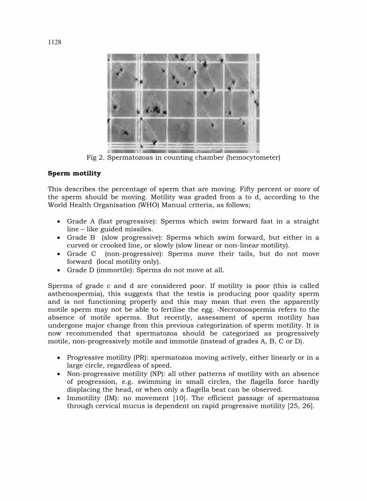
Fig 2. Spermatozoas in counting chamber (hemocytometer)
Sperm motility
This describes the percentage of sperm that are moving. Fifty percent or more of the sperm should be moving. Motility was graded from a to d, according to the World Health Organisation (WHO) Manual criteria, as follows;
Grade A (fast progressive): Sperms which swim forward fast in a straight line – like guided missiles.
Grade B (slow progressive): Sperms which swim forward, but either in a curved or crooked line, or slowly (slow linear or non-linear motility).
Grade C (non-progressive): Sperms move their tails, but do not move forward (local motility only).
Grade D (immortile): Sperms do not move at all. Sperms of grade c and d are considered poor. If motility is poor (this is called asthenospermia), this suggests that the testis is producing poor quality sperm and is not functioning properly and this may mean that even the apparently motile sperm may not be able to fertilise the egg. -Necrozoospermia refers to the absence of motile sperms. But recently, assessment of sperm motility has undergone major change from this previous categorization of sperm motility. It is now recommended that spermatozoa should be categorized as progressively motile, non-progressively motile and immotile (instead of grades A, B, C or D).
Progressive motility (PR): spermatozoa moving actively, either linearly or in a large circle, regardless of speed.
Non-progressive motility (NP): all other patterns of motility with an absence of progression, e.g. swimming in small circles, the flagella force hardly displacing the head, or when only a flagella beat can be observed.
Immotility (IM): no movement [10]. The efficient passage of spermatozoa through cervical mucus is dependent on rapid progressive motility [25, 26].

1129
Fig 3. Sperm motility
Sperm morphology This describes the shape of the sperm. The sperm is examined under a microscope and must meet specific sets of criteria for several sperm characteristics in order to be considered normal. Ideally, a good sperm should have a regular oval head, with a connecting mid-piece and a long straight tail. If too many sperms are abnormally shaped this is called teratozoospermia. The majority of sperm have abnormalities such as round heads; pin heads; very large heads; double heads; absent tails), this may mean the sperm are functionally abnormal and will not be able to fertilise the egg. Only sperm which are "perfect" are considered to be normal. A normal sample should have at least 15% normal forms (which means even upto 85% abnormal forms is considered to be acceptable) [27].
Materials and Method
Materials
Plastic gloves, Sterile container, Phase contrast microscope, Microscope slides, Cover slips, Haemocytometer counting chamber, Graded pipette (in mls), Ward coat, Incubator. Sample collection technique
Semen samples were collected from 20 unidentified male subjects suitable for this study and were between the ages of 18-55 years at a Medical Centre in Abuja,
Nigeria where all the required equipment was provided with permission from the authority of medical laboratory of the centre. Semen collection
Before producing semen samples all the participating subjects were required to abstain from sexual intercourse within the interval of 2-7days according to WHO manual recommendation.
The semen sample analysis of an ejaculate was obtained from all the 20 subjects into the sterile container provided them.
1130
The acquired semen was kept at an ambient temperature between 20 °C to 37°C contained in the clean and air tight sterile container to avoid large changes in temperature that may affect the spermatozoa.
The sterile container was labelled with the subject name and hospital
number to avoid any form of error (such as a mix-up). After labelling each sterilized container they were stored in the incubator
(37°C) to allow for liquefaction. Semen macroscopic examination
The first order of evaluation was done with the naked eyes (macroscopically) by simply inspecting the samples carefully;
The colour of each semen sample was examined with unaided eyes in terms of appearance.
The graded pipette used for determination of semen volume in ml was used to assess semen volume as well as its viscosity when the semen was allowed to drop from the pipette by gravity.
Microscopic examination
During assessment of these semen samples, the samples were kept at a room temperature of 37°C and duration of pre-analytical process did not exceed 30minutes-1 hour (liquefaction occurs at this period). The use of a suitable microscope (phase contrast) was used to effectively analyse the spermatozoa in the provided semen samples to determine; sperm count, sperm motility percentage and their various characteristic morphology. According to WHO manual recommendation, an initial microscopic examination of the sample involves scanning the preparation at a total magnification of ×100. This reveals:
mucus strand formation;
sperm aggregation or agglutination;
the presence of cells other than spermatozoa, e.g. epithelial cells, “round cells” (leukocytes and immature germ cells) and isolated sperm heads or tails.
Sperm motility
Sperm motility within semen was assessed immediately after liquefaction of the sample (between 30 minutes -1 hour), following ejaculation to limit possibility of dehydration or changes in temperature which could affect results.
Wet preparation was done on a clean glass slide and then covered with a coverslip allowing its weight to spread the sample
Once the sample stopped drifting, the sample was examined with phase contract optics at ×200 or ×400 magnification.
Sperm motility was then assessed by observing percentage of different motile category, starting from PR, NP then IM. Assess only intact

1131
spermatozoa (defined as having a head and a tail, since only intact spermatozoa are counted for sperm concentration [10].
Sperm concentration
Sperm concentration refers to the number of spermatozoa per unit volume of semen (ml) and is a function of the number of spermatozoa emitted and the volume of fluid diluting them [10].
The semen sample was diluted (1:20) with fixatives after thorough mixing.
Haemocytometer counting chambers was used with the special thick coverslips after diluted semen sample was added.
After being mounted on a haemocytometer, counting began from the centre grid of the haemocytometer.
The concentration in spermatozoa per ml was calculated.
Fig 4. The middle of the three lines defines the square’s boundary (black line, left panel). All spermatozoa within the central square are counted, as well as those
with their heads between the two inner lines (white circles), but not those whose heads lie between the outer two lines (black circles). A spermatozoon with most of
its head lying on the central line is counted only if that line is the lower or left-hand line of the square (white circles, middle panel) but not if it is the upper or
right hand line of the square (black circles, right panel). Sperm morphology
Smear of semen was prepared on a slide and allowed to air-dry.
Fig 5. Photo showing semen smear preparation (Adapted from C Brazil)
The slide was then fixed with ethanol for about 15 minutes and subsequently stained and then covered with a coverslip.

1132
The side was then examined with a contrast-phase optics of ×1000 magnification with oil immersion.
These procedures carried out were done with a ward coat and hands gloves as precautionary measures for these analysis. Statistical analysis
Basic descriptive statistics (mean, standard deviation, SEM) were calculated for
Results and Analysis
Table 1
Collective parameter of all 20 semen samples
SAMPLES AGE (years)
VOLUME (millilitre)
COLOUR COUNT (million/ml)
MOTILITY (%)
VISCOSITY MORPHOLOGY
1 19 4.5 Creamy and translucent
15.5 40%- PR 30%- NP 30%- IM
Gelatinous 10%- big and rounded heads
2 27 1.5 Creamy 45 50%- PR 20%- NP 30%- IM
Normal 5%- big and Rounded heads
3 32 2.5 Creamy and opaque
42 65%- PR 20%- NP 15%- IM
Gelatinous Normal
4 40 3.0 Creamy and opaque
21 50%- PR 10%- NP 40%- IM
Normal 20%- big heads, swollen and curved necks
5 18 <0.5 Creamy and opaque
68 70%- PR 20%- NP 10%- IM
Gelatinous Normal
6 50 3.5 Creamy and opaque
15 20%- PR 10%- NP 70%- IM
Gelatinous 20%- 2 heads and curved necks
7 45 4.5 Creamy and opaque
48 70%- PR 20%- NP 10%- IM
Normal 20%- mostly big heads
8 31 2.1 Creamy and opaque
16 65%- PR 10%- NP 25%- IM
Normal 20%- mostly two heads
9 45 4.5 Creamy 1 35%- PR 35%- NP 30%- IM
Gelatinous 30%- mostly big heads
10 26 3.6 Creamy 18 30%- PR 30%- NP 40%- IM
Normal 10%- mostly rounded heads
1133
11 46 4.5 Creamy and opaque
<1 All appeared IM
Normal 20%- mostly pin heads
12 42 2.5 Creamy and translucent
5 All appeared IM
Normal 10%- mostly rounded heads
13 46 1.0 Deep cream and opaque
15 40%- PR 5%- NP 55%-IM
Gelatinous 15%- mostly big heads
14 40 8.0 Cream and opaque
24 90%- PR 5%- NP 5%- IM
Normal Normal
15 37 0.5 Cream and opaque
61 80%-PR 10%- NP 10%- IM
Normal Normal
16 51 2.2 Creamy 78 80%-PR 10%- NP 10%- IM
Normal Normal
17 29 2.5 Creamy 3 10%-PR 90%- IM
Gelatinous 20%- big heads
18 34 3.0 Creamy and translucent
25 All appeared IM
Normal 5%- two tails
19 34 5.0 Creamy and
translucent
35 60%- PR 20%- NP
20%- IM
Gelatinous 5%- mostly big and rounded
heads
20 52 0.5 Creamy and opaque
75 80%- PR 10%- NP 10%- IM
Normal Normal
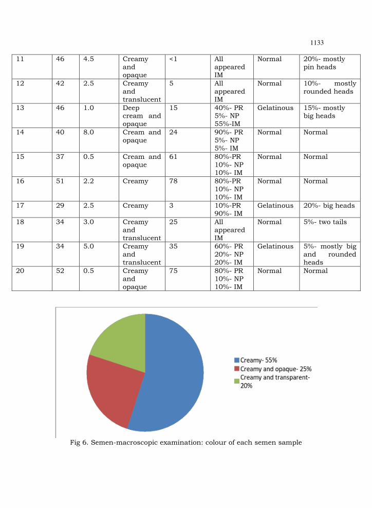
Fig 6. Semen-macroscopic examination: colour of each semen sample
1134
Figure 6 above indicates the absence of seminal discoloration, the colour differences are relatively normal with no significant abnormalities.
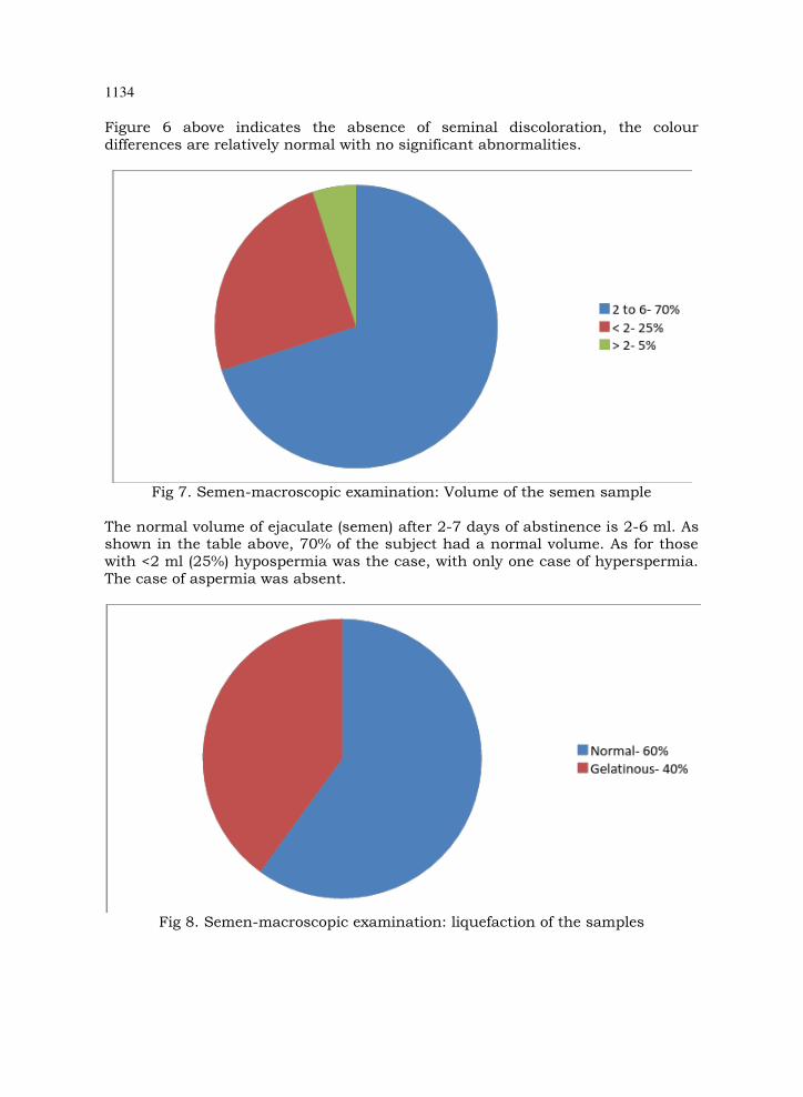
Fig 7. Semen-macroscopic examination: Volume of the semen sample
The normal volume of ejaculate (semen) after 2-7 days of abstinence is 2-6 ml. As shown in the table above, 70% of the subject had a normal volume. As for those with <2 ml (25%) hypospermia was the case, with only one case of hyperspermia.
The case of aspermia was absent.
Fig 8. Semen-macroscopic examination: liquefaction of the samples
1135
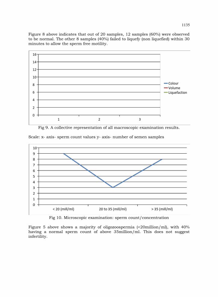
Figure 8 above indicates that out of 20 samples, 12 samples (60%) were observed to be normal. The other 8 samples (40%) failed to liquefy (non liquefied) within 30 minutes to allow the sperm free motility.
Fig 9. A collective representation of all macroscopic examination results.
Scale: x- axis- sperm count values y- axis- number of semen samples
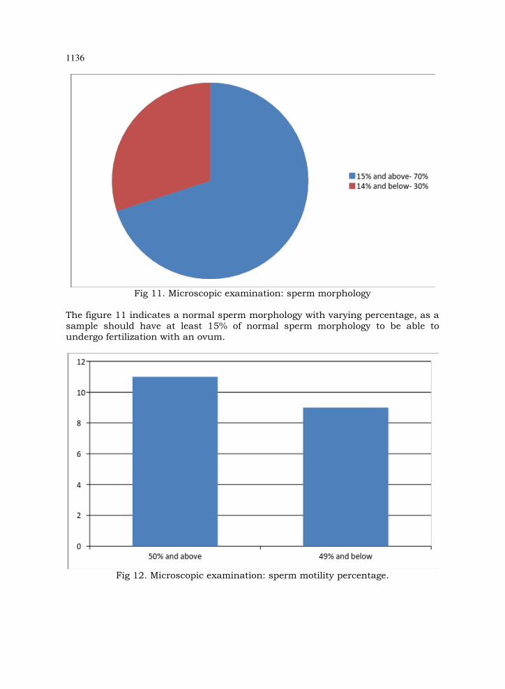
Fig 10. Microscopic examination: sperm count/concentration
Figure 5 above shows a majority of oligozoospermia (<20million/ml), with 40% having a normal sperm count of above 35million/ml. This does not suggest infertility.
1136
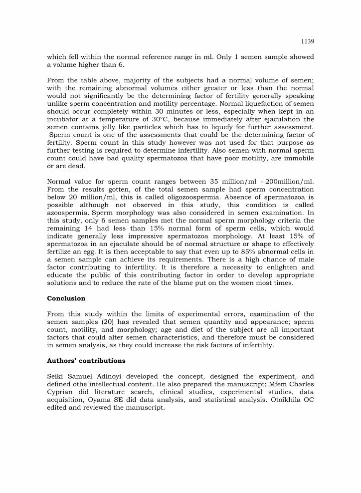
Fig 11. Microscopic examination: sperm morphology
The figure 11 indicates a normal sperm morphology with varying percentage, as a sample should have at least 15% of normal sperm morphology to be able to undergo fertilization with an ovum.
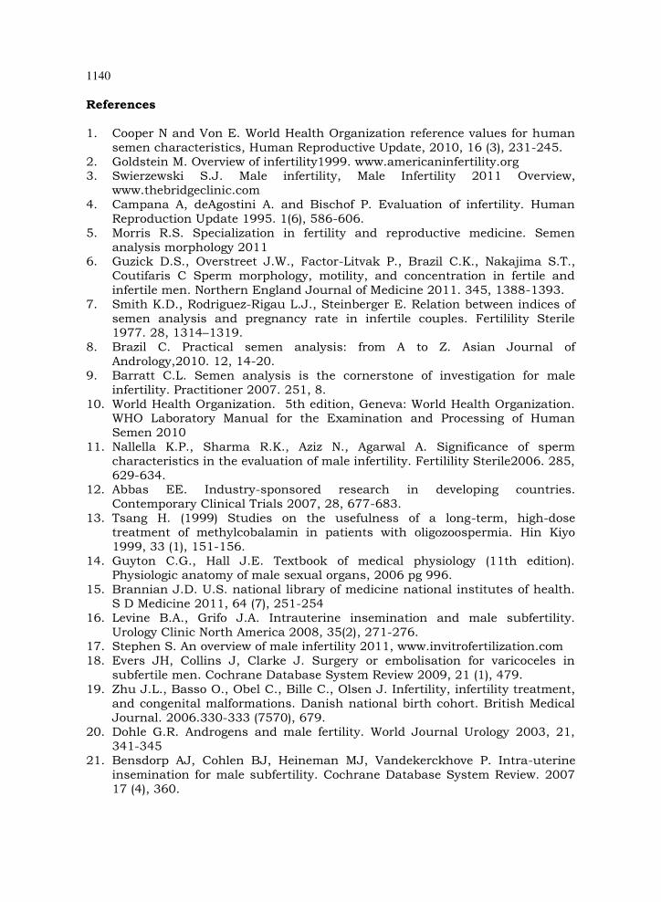
Fig 12. Microscopic examination: sperm motility percentage.

1137
50% or more sperm cells should be moving (rapid progressive motility) for efficient fertilization of the ova. From the figure above, 55% of total sample met this criterium with the remaining 45% falling short.
Fig 13. Age of anonymous subjects
Figure 7 shows the age group not below 18 and not above 55. Persons from 18 to 55 years were actively involved in this analysis. Statistics
Table 2 Unpaired t test results
SPERM CONCENTRATION
SPERM MOTILITY %
SEMEN VOLUME
SUBJECT’S AGE
GROUP 1
N 10 10 10 10
MEAN 49.700 71.50 2.820 40.60
SD 20.243 13.34 2.422 8.36
SEM 6.401 4.22 0.766 2.64
GROUP 2
N 10 10 10 10
MEAN 11.450 24.00 3.170 36.80
SD 8.301 21.83 1.173 10.40
SEM 2.625 6.90 0.371 3.29
t-test P value and statistical significance Review of data According to the t-test calculation;

1138
Group 1 represents group of subject suggested to have high possibility of fertility compared to
Group 2 represents group of subject suggested to have a lesser possibility of fertility.
The two-tailed unpaired t-test was carried between the 2 groups; group 1 and group 2 with respect to their acquired parameters, their p value and statistical significance are stated below;
Compared sperm concentrations between the 2 groups; two-tailed p value was < 0.0001. By conventional criteria, this difference is considered to be extremely statistically significant.
Standard error of difference (SED) = 6.919
Compared sperm motility percentage between the 2 groups; two-tailed p value was < 0.0001. By conventional criteria, this difference is considered to be extremely statistically significant. Standard error of difference (SED) = 8.091
Compared semen volume between the 2 groups; two-tailed p value= 0.6858. By conventional criteria, this difference is considered to not be statistically significant. Standard error of difference (SED) = 0.851
Compared sperm concentrations between the 2 groups; two-tailed p value= 0.3796. By conventional criteria, this difference is considered to not be statistically significant. Standard error of difference (SED) = 4.219
N.B. : Degree of freedom (df) = 18
Discussion
Certain factors could have as well contributed to the outcome of semen/sperm quality and quantity, such factors have been categorized as follows;
Spermatozoa factors
Hormonal factors
Dietary factors
Testicular factors
Sexually transmitted infections
Age group, etc.
Even certain lifestyle/habits: Both macroscopic and microscopic assessments were taken into consideration in this discussion. From this study; All semen samples showed a relatively normal colour with a little variation in their appearances with the absence seminal discolorations. Milky/creamy appearance was the most common characteristic observed in these semen samples with a few with whitish colorations. In addition, the absence of yellowish tinge in these samples may imply that none of the subjects were had prolonged abstinence. The normal volume of an ejaculate acquired should be between 2-6 ml after 2-7 days of abstinence. The volume assessment of each semen sample included 14 samples
1139
which fell within the normal reference range in ml. Only 1 semen sample showed a volume higher than 6. From the table above, majority of the subjects had a normal volume of semen; with the remaining abnormal volumes either greater or less than the normal would not significantly be the determining factor of fertility generally speaking unlike sperm concentration and motility percentage. Normal liquefaction of semen should occur completely within 30 minutes or less, especially when kept in an incubator at a temperature of 30°C, because immediately after ejaculation the semen contains jelly like particles which has to liquefy for further assessment. Sperm count is one of the assessments that could be the determining factor of fertility. Sperm count in this study however was not used for that purpose as further testing is required to determine infertility. Also semen with normal sperm
count could have bad quality spermatozoa that have poor motility, are immobile or are dead. Normal value for sperm count ranges between 35 million/ml - 200million/ml. From the results gotten, of the total semen sample had sperm concentration below 20 million/ml, this is called oligozoospermia. Absence of spermatozoa is possible although not observed in this study, this condition is called azoospermia. Sperm morphology was also considered in semen examination. In this study, only 6 semen samples met the normal sperm morphology criteria the remaining 14 had less than 15% normal form of sperm cells, which would indicate generally less impressive spermatozoa morphology. At least 15% of spermatozoa in an ejaculate should be of normal structure or shape to effectively fertilize an egg. It is then acceptable to say that even up to 85% abnormal cells in a semen sample can achieve its requirements. There is a high chance of male factor contributing to infertility. It is therefore a necessity to enlighten and educate the public of this contributing factor in order to develop appropriate solutions and to reduce the rate of the blame put on the women most times.
Conclusion
From this study within the limits of experimental errors, examination of the semen samples (20) has revealed that semen quantity and appearance; sperm count, motility, and morphology; age and diet of the subject are all important factors that could alter semen characteristics, and therefore must be considered in semen analysis, as they could increase the risk factors of infertility.
Authors’ contributions
Seiki Samuel Adinoyi developed the concept, designed the experiment, and defined othe intellectual content. He also prepared the manuscript; Mfem Charles Cyprian did literature search, clinical studies, experimental studies, data acquisition, Oyama SE did data analysis, and statistical analysis. Otoikhila OC edited and reviewed the manuscript.
1140
References
1. Cooper N and Von E. World Health Organization reference values for human semen characteristics, Human Reproductive Update, 2010, 16 (3), 231-245.
2. Goldstein M. Overview of infertility1999. www.americaninfertility.org 3. Swierzewski S.J. Male infertility, Male Infertility 2011 Overview,
www.thebridgeclinic.com 4. Campana A, deAgostini A. and Bischof P. Evaluation of infertility. Human
Reproduction Update 1995. 1(6), 586-606. 5. Morris R.S. Specialization in fertility and reproductive medicine. Semen
analysis morphology 2011 6. Guzick D.S., Overstreet J.W., Factor-Litvak P., Brazil C.K., Nakajima S.T.,
Coutifaris C Sperm morphology, motility, and concentration in fertile and
infertile men. Northern England Journal of Medicine 2011. 345, 1388-1393. 7. Smith K.D., Rodriguez-Rigau L.J., Steinberger E. Relation between indices of
semen analysis and pregnancy rate in infertile couples. Fertilility Sterile 1977. 28, 1314–1319.
8. Brazil C. Practical semen analysis: from A to Z. Asian Journal of Andrology,2010. 12, 14-20.
9. Barratt C.L. Semen analysis is the cornerstone of investigation for male infertility. Practitioner 2007. 251, 8.
10. World Health Organization. 5th edition, Geneva: World Health Organization. WHO Laboratory Manual for the Examination and Processing of Human Semen 2010
11. Nallella K.P., Sharma R.K., Aziz N., Agarwal A. Significance of sperm characteristics in the evaluation of male infertility. Fertilility Sterile2006. 285,
629-634. 12. Abbas EE. Industry-sponsored research in developing countries.
Contemporary Clinical Trials 2007, 28, 677-683. 13. Tsang H. (1999) Studies on the usefulness of a long-term, high-dose
treatment of methylcobalamin in patients with oligozoospermia. Hin Kiyo 1999, 33 (1), 151-156.
14. Guyton C.G., Hall J.E. Textbook of medical physiology (11th edition). Physiologic anatomy of male sexual organs, 2006 pg 996.
15. Brannian J.D. U.S. national library of medicine national institutes of health. S D Medicine 2011, 64 (7), 251-254
16. Levine B.A., Grifo J.A. Intrauterine insemination and male subfertility. Urology Clinic North America 2008, 35(2), 271-276.
17. Stephen S. An overview of male infertility 2011, www.invitrofertilization.com
18. Evers JH, Collins J, Clarke J. Surgery or embolisation for varicoceles in subfertile men. Cochrane Database System Review 2009, 21 (1), 479.
19. Zhu J.L., Basso O., Obel C., Bille C., Olsen J. Infertility, infertility treatment, and congenital malformations. Danish national birth cohort. British Medical Journal. 2006.330-333 (7570), 679.
20. Dohle G.R. Androgens and male fertility. World Journal Urology 2003, 21, 341-345
21. Bensdorp AJ, Cohlen BJ, Heineman MJ, Vandekerckhove P. Intra-uterine insemination for male subfertility. Cochrane Database System Review. 2007 17 (4), 360.
1141
22. Walsh T.J., Croughan M.S., Schembri M., Chan J.M., Turek P.J. Increased risk of testicular germ cell cancer among infertile men. Arch International Medicine 2009, 169 (4), 351-356.
23. Prasanta K.D., Swanali S. Physiological aspects of infertility. British Journal for Medical Practitioner 2010, 3(3), a336
24. Keel B.A. The semen analysis. In: Keel B., Webster B., editors. CRC Handbook of the laboratory diagnosis and treatment of infertility. Boca Raton: CRC Press 1990. Pg. 27-69.
25. Björndahl L. The usefulness and significance of assessing rapidly progressive spermatozoa. Asian Journal Andrology 2010. 12, 33-35.
26. Lindholmer C. The importance of seminal plasma for human sperm motility. Biological Reproduction. 1974. 10, 533-542.
27. Seriki Samuel A, Adebayo O Francis Atsukwei Denen, Odetola Anthony
(2015). Effects of Prolonged Fasting on Sperm Count. Journal of Molecular Pathophysiology 2015.Vol 4, Issue 3; pg 99-102