Immobilization of Allantoinase for the Development of ... - MDPI
13
Sensors 2020, 20, 196; doi:10.3390/s20010196 www.mdpi.com/journal/sensors Article Immobilization of Allantoinase for the Development of an Optical Biosensor of Oxidative Stress States Marialaura Marchetti 1 , Luca Ronda 1,2, *, Riccardo Percudani 3 and Stefano Bettati 1,2 1 Centro Interdipartimentale Biopharmanet-TEC, Università di Parma, Parco Area delle Scienze 27/A, 43124 Parma, Italy; [email protected] (M.M.); [email protected] (S.B.) 2 Dipartimento di Medicina e Chirurgia, Università di Parma, Via Gramsci 14, 43126 Parma, Italy 3 Dipartimento di Scienze Chimiche, della Vita e della Sostenibilità Ambientale, Università di Parma, Via Parco Area delle Scienze 11/A, 43124 Parma, Italy; [email protected] * Correspondence: [email protected]; Tel.: +39-0521-905502 Received: 4 December 2019; Accepted: 27 December 2019; Published: 29 December 2019 Abstract: Allantoin, the natural end product of purine catabolism in mammals, is non-enzymatically produced from the scavenging of reactive oxygen species through the degradation of uric acid. Levels of allantoin in biological fluids are sensitively influenced by the presence of free radicals, making this molecule a candidate marker of acute oxidative stress in clinical analyses. With this aim, we exploited allantoinase—the enzyme responsible for allantoin hydrolization in plants and lower organisms—for the development of a biosensor exploiting a fast enzymatic-chemical assay for allantoin quantification. Recombinant allantoinase was entrapped in a wet nanoporous silica gel matrix and its structural properties, function, and stability were characterized through fluorescence spectroscopy and circular dichroism measurements, and compared to the soluble enzyme. Physical immobilization in silica gel minimally influences the structure and the catalytic efficiency of entrapped allantoinase, which can be reused several times and stored for several months with good activity retention. These results, together with the relative ease of the sol-gel preparation and handling, make the encapsulated allantoinase a good candidate for the development of an allantoin biosensor. Keywords: silica gel; biosensor; allantoin; allantoinase 1. Introduction Oxidative stress is defined as an imbalance between the presence of reactive species and the antioxidant body response. Recently, much evidence proved the correlation of oxidative stress with several pathological conditions (cardiovascular diseases, diabetes, arthritis, neonatal hypoxia, etc.) [1–5], highlighting the necessity to find specific biomarkers for its evaluation. This increasing exigency combines with the preferential use of non-invasive methods for sample collection, electing saliva and urine as best alternatives in clinical biochemistry. In this context, the attention is progressively focusing on allantoin, a stable and polar small molecule generated in humans from the non-enzymatic oxidation of uric acid, as a potential biomarker for routine clinical analyses. Indeed, uric acid is the end product of purine metabolism in humans and one of the main constituents of the total antioxidant capacity (TAC) in body fluids [6–9], representing the most active and abundant scavenger of hydrophilic reactive oxygen and nitrogen species and contributing to 35–65% of the plasma TAC [10–12]. Since allantoin spontaneously forms upon non-enzymatic urate degradation, allantoin levels increase promptly responding to oxidative stress conditions and can be measured in different biological fluids, e.g., plasma, synovial fluid, saliva and urine [5,13,14]. The small size and the high polarity of allantoin, however, make its quantification challenging, thus limiting its
-
Upload
khangminh22 -
Category
Documents
-
view
1 -
download
0
Transcript of Immobilization of Allantoinase for the Development of ... - MDPI

Sensors 2020, 20, 196; doi:10.3390/s20010196 www.mdpi.com/journal/sensors
Article
Immobilization of Allantoinase for the Development
of an Optical Biosensor of Oxidative Stress States
Marialaura Marchetti 1, Luca Ronda 1,2,*, Riccardo Percudani 3 and Stefano Bettati 1,2
1 Centro Interdipartimentale Biopharmanet-TEC, Università di Parma, Parco Area delle Scienze 27/A, 43124
Parma, Italy; [email protected] (M.M.); [email protected] (S.B.) 2 Dipartimento di Medicina e Chirurgia, Università di Parma, Via Gramsci 14, 43126 Parma, Italy 3 Dipartimento di Scienze Chimiche, della Vita e della Sostenibilità Ambientale, Università di Parma, Via
Parco Area delle Scienze 11/A, 43124 Parma, Italy; [email protected]
* Correspondence: [email protected]; Tel.: +39-0521-905502
Received: 4 December 2019; Accepted: 27 December 2019; Published: 29 December 2019
Abstract: Allantoin, the natural end product of purine catabolism in mammals, is non-enzymatically
produced from the scavenging of reactive oxygen species through the degradation of uric acid.
Levels of allantoin in biological fluids are sensitively influenced by the presence of free radicals,
making this molecule a candidate marker of acute oxidative stress in clinical analyses. With this aim,
we exploited allantoinase—the enzyme responsible for allantoin hydrolization in plants and lower
organisms—for the development of a biosensor exploiting a fast enzymatic-chemical assay for
allantoin quantification. Recombinant allantoinase was entrapped in a wet nanoporous silica gel
matrix and its structural properties, function, and stability were characterized through fluorescence
spectroscopy and circular dichroism measurements, and compared to the soluble enzyme. Physical
immobilization in silica gel minimally influences the structure and the catalytic efficiency of
entrapped allantoinase, which can be reused several times and stored for several months with good
activity retention. These results, together with the relative ease of the sol-gel preparation and
handling, make the encapsulated allantoinase a good candidate for the development of an allantoin
biosensor.
Keywords: silica gel; biosensor; allantoin; allantoinase
1. Introduction
Oxidative stress is defined as an imbalance between the presence of reactive species and the
antioxidant body response. Recently, much evidence proved the correlation of oxidative stress with
several pathological conditions (cardiovascular diseases, diabetes, arthritis, neonatal hypoxia, etc.)
[1–5], highlighting the necessity to find specific biomarkers for its evaluation. This increasing
exigency combines with the preferential use of non-invasive methods for sample collection, electing
saliva and urine as best alternatives in clinical biochemistry. In this context, the attention is
progressively focusing on allantoin, a stable and polar small molecule generated in humans from the
non-enzymatic oxidation of uric acid, as a potential biomarker for routine clinical analyses. Indeed,
uric acid is the end product of purine metabolism in humans and one of the main constituents of the
total antioxidant capacity (TAC) in body fluids [6–9], representing the most active and abundant
scavenger of hydrophilic reactive oxygen and nitrogen species and contributing to 35–65% of the
plasma TAC [10–12]. Since allantoin spontaneously forms upon non-enzymatic urate degradation,
allantoin levels increase promptly responding to oxidative stress conditions and can be measured in
different biological fluids, e.g., plasma, synovial fluid, saliva and urine [5,13,14]. The small size and
the high polarity of allantoin, however, make its quantification challenging, thus limiting its
Sensors 2020, 20, 196 2 of 13
exploitation as a biomarker. The general methods used for allantoin measurements require sensitive
and specific instrumentations and exploit critical or time-consuming techniques like chemical
derivatization, gas or liquid chromatography associated with mass spectrometry (GC/LC-MS),
capillary electrophoresis and enzyme cycling assays [3,4,8,15–21]. To overcome these limits, we
proposed a novel assay based on the enzymatic production of allantoate from allantoin, and its
subsequent chemical conversion into a fluorescent compound, detectable with a bench fluorescence
plate reader [22]. The enzymatic step is performed by the recombinant enzyme allantoinase from
Pseudomonas fluorescens (puuE, E.C. 3.5.2.5) [23], able to stereoselectively convert (S)-allantoin into
allantoate, without any influence of the reverse reaction. The assay specificity is granted by (i) the
known reaction specificity of the enzyme, and (ii) the removal of the contribution of interferent
molecules through the subtraction of the baseline, which is generated by performing the same
chemical step of the complete assay, while skipping the enzymatic reaction, so that the detected signal
is only due to the allantoate generated from allantoinase reaction with allantoin. In this work, we
propose the immobilization of puuE to explore the possibility to develop a reusable biosensor. The
relevance of a biosensor resides in the selectivity and sensitivity in target quantification and
reusability, determining its applicability as a tool for routine clinical practice. With this purpose, we
entrapped puuE in a wet nanoporous silica gel matrix, a strategy largely exploited in enzyme
immobilization because of its several advantages, among which the easy and flexible chemistry, with
mild chemical and physical entrapment conditions compatible with protein stability, the optical
transparency allowing signal detection with conventional spectroscopic techniques, the ease of
separation of the active material from the reaction solution [24–31].
During the reaction of polymerization, the protein molecules enclosed in the sol-gel drive the
shaping of the matrix pores. The resulting nanoporous structure prevents protein leaking while
allowing diffusion of reactants and products but—together with altered solvent microviscosity
within the pores—poses steric and thermodynamic constraints in the conformational equilibrium of
the enzyme. While enzyme encapsulation in silica gels does not affect substrate specificity, it can
potentially result in the immobilization of poorly or differently active and stable conformations [32–
40]. We therefore assayed allantoinase structural features and stability after encapsulation and the
influence of the silica gel matrix on enzymatic activity, in order to verify the approach suitability for
the setup of a biosensor. The presented results demonstrate the applicability of this strategy to a
chemical/enzymatic assay for allantoin quantification [22], in which respect it represents a major
improvement in terms of reusability and storability.
2. Materials and Methods
2.1. Enzyme Expression and Purification
Recombinant puuE from P. fluorescens was expressed in Escherichia coli host, as previously
described [23]. Cells were lysed in 50 mM sodium phosphate, 300 mM NaCl, 10% glycerol (v/v), 200
µM phenylmethylsulfonyl fluoride, 200 µM benzamidine and 1.5 µM pepstatin A, pH 7.6, and
sonicated by 5-s bursts alternated to 1-min pauses. Surnatant was filtered with 0.2 µM filter units and
concentrated by Amicon Ultra-15 centrifugal filter devices (Merck-Millipore, Darmstadt, Germany)
with 30 kDa-cutoff. puuE was purified to homogeneity by gel filtration chromatography on a
BioSepra Ultrogel AcA 44 column (PALL Life Sciences, Port Washington, NY, USA) in 100 mM
potassium phosphate, 150 mM NaCl, pH 7.6. Fractions containing the enzyme were aliquoted, flash-
frozen in liquid nitrogen and stored at −80 °C until further use.
2.2. Enzymatic Activity
The hydrolase activity was followed recording single-wavelength kinetics at 210 nm at 37 °C
with a J715 spectropolarimeter (Jasco, Tokyo, Japan) equipped with a Peltier thermostatic cell.
Reactions were carried out in a 1 mm pathlength quartz cuvette, in 20 mM potassium phosphate, pH
7.4, in the presence of 70 nM enzyme (monomer concentration). The experimental buffer is optimized
to avoid instrumental interferences.
Sensors 2020, 20, 196 3 of 13
2.3. Enzyme Encapsulation
puuE was encapsulated in a silica matrix by the sol-gel method [24,25]. Tetramethylorthosilicate
(TMOS), water and a 40 mM HCl solution were mixed and sonicated for 20 min (divided in 5-min on
and 1-min off cycles), then an equal volume of 10 mM phosphate, pH 6.0, was added and the solution
was fluxed with humidified nitrogen for 40 min. The obtained sol was mixed in a 1:1 ratio with puuE
in 100 mM phosphate buffer, 150 mM NaCl, pH 7.6, and layered either on the bottom of 2 mL glass
vials, or on the non-optic wall of quartz cuvettes, or on quartz slides. The increase of pH promotes
polymerization and consequent solidification of the mixture, which occurred in about two minutes
at 4 °C. Upon polymerization, vials were maintained in 100 mM phosphate, 150 mM NaCl, pH 7.0 at
4 °C and used after at least 24 h of silica matrix aging.
2.4. Secondary and Tertiary Structure Measurements
Circular dichroism (CD) spectra were collected with a Jasco J715 spectropolarimeter between
200 and 260 nm in a 1 cm pathlength quartz cuvette, thermostated at 20 °C, in 20 mM potassium
phosphate, pH 7.4. Measurements of puuE in solution were recorded at 0.35 µM monomer
concentration. Spectra on puuE in silica gel were collected by encapsulating 29 µg of recombinant
enzyme on the 2 cm2-surface of a quartz slide, adhered to one of the optic faces of the cuvette.
Fluorescence emission spectra upon excitation of tryptophan residues at 298 nm were collected by a
Fluoromax-3 fluorometer (HORIBA Jobin Yvon, Kyoto, Japan) between 310 nm and 550 nm at 20 °C,
with slits set to optimize the signal-to-noise ratio. Measurements of puuE in solution were recorded
at 0.2 µM monomer concentration, in 20 mM potassium phosphate, pH 7.4. Front-face emission
spectra were recorded on puuE encapsulated onto the quartz slides (vide supra) in a 1 cm pathlength
quartz cuvette.
2.5. Stability Measurements
puuE stability after encapsulation was assessed both over time and after several cycles of reuse
of the protein-doped silica gel. Before use, the silica matrix was rinsed once with storage buffer,
crumbled, and washed with the same buffer. After washing, the buffer was discarded and 0.75 mL
of 0.2 mM racemic allantoin in 20 mM potassium phosphate, pH 7.4 were added to the encapsulated
enzyme (8 µg, 0.3 µM final monomer concentration). The solution was then incubated under agitation
for 10 min on a thermostated Peltier plate set at 37 °C. For the reuse, after each incubation the gel was
rinsed twice with 0.75 mL of storage buffer. The enzymatic activity was checked collecting CD spectra
between 200 nm and 340 nm on the final solution, at 20 °C in a 2 mm quartz cuvette. The experiment
was carried out in triplicate for each time point. For comparison, a solution of 23.4 µM puuE was
stored over time in storage buffer, at 4 °C. The enzymatic activity was tested at 0.3 µM (final monomer
concentration) in reaction buffer, in 2 mL glass vials. After 10 min under agitation at 37 °C, reaction
solutions were stopped trough diafiltration in Amicon Ultra 0.5 mL ultracentrifuge devices with 30
kDa-cutoff (Merck-Millipore, Darmstadt, Germany). CD spectra were recorded on the flow-through
solution.
2.6. Fluorescent Quantification of Allantoin by Immobilized Allantoinase
A calibration curve was built adding increasing concentrations of racemic allantoin to 100 mM
potassium phosphate buffer, pH 7.4 (final range 0.5–50 µM). A reaction in the absence of allantoin
was carried out for blank subtraction. The assay was performed as previously described [22]. Briefly,
recombinant puuE was encapsulated on the internal part of PCR strip caps following the protocol
previously described in this section, using 300 ng of enzyme for each cap (final volume 2 µL). Upon
polymerization, caps were maintained overnight in storage buffer at 4 °C. 30 µL of solutions
containing increasing concentrations of allantoin were added into 0.2 mL PRC tubes, closed with the
allantoinase functionalized caps, and put upside down at 37 °C for 15 min [22]. After the enzymatic
conversion of allantoin to allantoate, caps were substituted with normal ones, 120 µL of 6 M HCl and
3.5 µL of 5% resorcinol (w/v) were added and the samples were incubated in a thermocycler at 100

Sensors 2020, 20, 196 4 of 13
°C for five minutes. After cooling down the reactions in ice for two minutes, 2.5 µL of each sample
were added to 239 µL of 100 mM diethanolamine, pH 9.6, on a polystyrene 96-well black microplate
(Greiner BioOne, Kremsmünster, Austria), after the addition of 9 µL of a 1.5 M ascorbate solution.
The multiwell plate was read by a Spark 10M (Tecan, Männedorf, Switzerland) plate reader recording
signal emission at 535 nm upon excitation at 490 nm to detect the formation of the fluorescent
compound 2,2′,4,4′-tetrahydroxy-diphenylacetic acid, with an absorption peak centered at 490 nm
and an emission peak centered at 530 nm. Reactions were carried out in triplicate and the revelation
on the plate was made in duplicate.
3. Results and Discussion
3.1. Effect of Silica Gel Encapsulation on Allantoinase Secondary and Tertiary Structure
To determine whether the encapsulation process affected puuE structure and activity, we
characterized the spectroscopic and functional properties of enzyme after entrapment in comparison
to the free allantoinase. To this goal, we exploited far-UV circular dichroism spectra and tryptophan
fluorescence emission, which are sensitive probes of protein secondary and tertiary structure,
respectively.
As determined by structural studies, native puuE is a homotetramer and each monomer is
folded in a deformed (β/α) barrel structure, generating an independent active site [23]. The α-helices
constitute around 40% of the structure, whereas around 10% of the sequence folds as β-strands.
Recombinant allantoinase was mixed in a 1:1 ratio with the sol (see Section 2.3) and, before
polymerization, deposited as a thin layer on a quartz slide. The resulting puuE-doped gel was used
for the structural characterization by circular dichroism (CD) and static fluorescence spectroscopy
(Figure 1), after at least 24 h of aging.
Figure 1. Structural characterization of entrapped puuE (solid lines) in comparison to the free enzyme
(dashed lines) in 20 mM potassium phosphate, pH 7.4. (A) Normalized far-UV circular dichroism
spectra; (B) normalized tryptophan emission spectra upon excitation at 298 nm.
The CD spectrum of free allantoinase (Figure 1A, dashed line) presents a strong positive band
at wavelengths below 200 nm and two negative peaks centered at 211 and 223 nm, typical of an α/β
folding, in accordance with the crystallographic data. For comparison, the embedded enzyme (Figure
1A, solid line) does not reveal significant alterations in the secondary structure, with only minor
differences in the region between 205–215 nm that could be attributed to very slight changes in helical
content or scattering effects due to the gel matrix.
Fluorescence emission of tryptophan residues is sensitive to the microenvironment and,
therefore, protein structural changes can be monitored as shifts in their maximum emission
wavelength exploiting their solvatochromic nature. In general, the selective excitation of this amino
acid in proteins provides an emission maximum ranging from about 310 nm to about 355 nm, based
on solvent polarity and the level of solvent exposure [41,42]. Twenty-eight tryptophan residues are

Sensors 2020, 20, 196 5 of 13
present in a biological unit of puuE (Figure 2). To assess the effect of encapsulation on puuE tertiary
structure, we selectively excited at 298 nm the embedded allantoinase on a quartz slide (Figure 1B,
solid line) and, for comparison, the enzyme in solution (Figure 1B, dashed line). The two spectra are
completely superimposable, with the maximum emission peak centered at 348 nm, indicating an
overall conservation of the native folding.
Figure 2. Representation of puuE tetrameric assembly (Protein Data Bank 3cl6). Monomers are shown
as solid ribbons in different colors, while tryptophan residues are evidenced in purple ball and stick.
3.2. Analysis of Catalytic Parameters and Influence of Silica Gel Matrix on puuE Activity
The activity of an enzyme is defined through the determination of two main parameters: the
turnover number (kcat), representing the number of substrate molecules processed in a time unit, and
the Michaelis constant (KM), roughly representing the enzyme apparent affinity for its substrate. From
these two values it is possible to obtain the catalytic efficiency (also known as specificity constant,
kcat/KM), which is best used as comparative parameter between enzymatic activities in different
conditions [34,43–45]. More specifically, catalytic efficiency represents the efficiency of the encounter
complex to form and give the reaction product. This value for allantoinase is around 7.5 × 10⁴ M−1·s−1
and falls in the median range of reported enzymatic activities belonging to the primary metabolism
of nucleotides [46].
The catalytic parameters of the free puuE were calculated by fitting the enzymatic initial rates in
the presence of increasing allantoin concentrations to the Michaelis–Menten dependence equation
(Figure 3, Table 1). CD kinetics were collected following the decreasing signal at 210 nm,
corresponding to (S)-allantoin consumption. Racemization rate of the residual (R)-allantoin was
considered negligible in the time windows required for the kinetics measurements [47,48]. Moreover,
the reverse stereoselective condensation of allantoate to form (S)-allantoin does not affect the kinetic
signal, because of the lesser puuE efficiency in catalyzing this reverse reaction [23]. Conversely, the
direct measurement of encapsulated puuE activity was limited by allantoin molar ellipticity
coefficient and the experimental setup. In fact, measurements were carried out encapsulating puuE
onto the non-optical face of a 1 cm pathlength quartz cuvette, and the solution transmittance limited
to 40 µM racemic allantoin the maximum concentration feasible in these assay conditions. For these
reasons, the catalytic efficiency of allantoinase entrapped in the silica gel matrix was obtained
indirectly by the application of the equation:
[S] = [S0]e−kt (1)
With
K = (kcat/KM)[E] (2)
to 4.37 µM puuE in 50 µL of sol-gel matrix, in the presence of 750 µL of 40 µM racemic allantoin. This
procedure is applicable when the concentration of substrate is sensitively lower than the KM value
[49]. The accuracy of this indirect determination was tested extrapolating the catalytic efficiency of
the free enzyme by Equation (1) to the lower substrate concentrations of the Michaelis–Menten

Sensors 2020, 20, 196 6 of 13
dependence. The kcat/KM value calculated in this way was in good agreement with the one obtained
from the ratio of the experimentally calculated parameters.
Figure 3. Michaelis–Menten dependence of puuE activity in the presence of increasing concentrations
of racemic allantoin. Single-wavelength kinetics of (S)-allantoin consumption were recorded at 210
nm in 20 mM potassium phosphate, pH 7.4, at 37 °C. Each point corresponds to a separate (S)-allantoin
consumption kinetics; error bars represent standard errors of experimental replicates.
Table 1. Catalytic parameters and catalytic efficiency of free and encapsulated puuE on (S)-allantoin
in 20 mM potassium phosphate, pH 7.4, at 37 °C. For the free enzyme, kcat and KM values were
obtained by fitting kinetic data to the Michaelis–Menten equation and the catalytic efficiency was
calculated as the ratio between the turnover number and Michaelis constant. Catalytic efficiency for
entrapped puuE was obtained on single kinetics in the presence of 40 µM racemic allantoin, applying
Equation (1).
puuE kcat (s−1) KM (mM) kcat/KM (M−1 s−1)
Free 101.57 ± 14.26 1.35 ± 0.43 7.52 ± 2.62 × 10⁴
Silica gel — — 1.27 ± 0.12 × 10⁴
The apparent catalytic efficiency of the embedded allantoinase is lower than that calculated for
the free enzyme, but within the same order of magnitude (Table 1). This difference can be ascribed to
steric effects consequent to the immobilization, limiting functionally relevant enzyme
dynamics/conformational equilibria, or to an excessive sol-gel thickness making catalysis at least
partially rate-limited by substrate diffusion.
Therefore, although the overall efficiency of the encapsulated enzyme is satisfying and makes
puuE suitable for biosensing applications, it is significantly different from the free enzyme. Hence,
we wanted to further investigate the impact of sol-gel matrix thickness on diffusional limit and,
consequently, on allantoinase activity. With this aim, we immobilized equal amounts of puuE
layering different volumes on one of the non-optical faces of a 1 × 1 cm quartz cuvette. The thickness
of the layers was calculated considering a 3.3 cm2 coated area and the different sol-gel volumes
employed, assuming a parallelepiped as a shape.
Matrix diffusion limit can be determined from the catalytic parameters of the enzyme and the
concentration of puuE in the sol-gel, applying the following equation [34,50]:
dc = {[(KM + [S0]) × D’]/(kcat × [E])}1/2 (3)
where dc is the matrix critical thickness over which rates are diffusion-controlled, KM and kcat are the
catalytic parameters of puuE in solution (Table 1), [E] is the enzyme concentration and [S0] the
substrate concentration. D′ is the diffusion coefficient of the substrate inside the gel calculated as:
D’/D = 1 − (a2/r) (4)
Sensors 2020, 20, 196 7 of 13
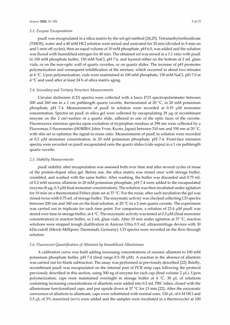
where a is the average molecular radius (5.42 Å for allantoin), D (6 × 10⁻⁶ cm2/s) is the diffusion
coefficient in water for compounds with a molecular weight around 200 Da, and r (40–50 Å) is the
average pore radius of the gel.
As can be noticed from values reported in Table 2 and in Figure 4A, the matrix critical
thicknesses calculated for the different encapsulation volumes (open circles) are systematically lower
than the real dimensions of the layers (closed circles). As expected, the catalytic efficiency is inversely
proportional to the immobilization volume and, consequently, thickness. The 25% catalytic efficiency
(with respect to the soluble enzyme) of the 25 µL enzyme represents a lower limit for the encapsulated
enzyme, indicating that the effect of entrapment and matrix physical properties on enzyme function
is not dramatic.
Table 2. Thickness and matrix critical thickness (dc) of different sol-gel volumes containing 7.75 µg of
puuE. Matrix thickness was calculated from encapsulation volume assuming 3.3 cm2 as the base area
of a parallelepiped; dc values were calculated applying Equations (3) and (4) and using catalytic
parameters of free puuE listed in Table 1.
Volume (µL) [puuE] in Gel (µM) Thickness (µM) dc (µM) kcat/KM (M−1 s−1)
25 8.75 75.8 30 1.74 ± 0.49 × 10⁴
50 4.37 151.5 43 1.27 ± 0.12 × 10⁴
100 2.19 303.0 60 1.04 ± 0.04 × 10⁴
Figure 4. Silica matrix parameters and puuE activity after entrapment in different sol-gel volumes.
(A) Comparison between sol-gel thickness (closed circles) and matrix critical thickness (open circles)
immobilizing 7.75 µg of puuE in increasing matrix volumes. (B) Catalytic efficiency of puuE
encapsulated in increasing matrix volumes with respect to the free enzyme. For each condition of
encapsulation, the experiments were carried out in triplicate and data were collected from single-
wavelength CD kinetics at 210 nm in the presence of 0.2 mM racemic allantoin 20 in mM potassium
phosphate, pH 7.4, at 37 °C.
3.3. Application of the Encapsulated Enzyme as a Biosensor
The effective suitability of immobilized puuE in biosensing applications was tested through its
application in the enzymatic step of an allantoin quantification assay recently developed by our
group [22]. Thin layers of silica gel containing 300 ng of recombinant puuE were layered on the
internal part of the caps of 0.2 mL vials. Increasing amounts of racemic allantoin, ranging from 0.5 to
50 µM in 100 mM potassium phosphate, pH 7.4, were converted into allantoate by incubating samples
upside down in 0.2 mL vials sealed by the functionalized caps. After a fixed reaction time, caps were
removed and stored for reuse [22]. The fluorescence values obtained from the plate reading were
subtracted of the blank (a reaction performed in buffer in the absence of allantoin) and normalized,
and their dependence on allantoin concentration was fitted to a linear equation (Figure 5). The
analysis provided a linear response in the explored range, confirming that encapsulated allantoinase
can represent a valuable option in the proposed routine assay for allantoin quantification.

Sensors 2020, 20, 196 8 of 13
Figure 5. Normalized fluorescence emission at 530 nm (λex = 490 nm) detected by microplate reader
at increasing concentrations of allantoin in 100 mM phosphate buffer, pH 7.4, enzymatically processed
by encapsulated puuE. Linear regression gave a R2 of 0.99.
3.4. Reusability of the Biosensor and Stability over Time
Generally, one of the pursued advantages in using an immobilized enzyme is its increased
stability and, as a consequence, its disposition to long-term storage and reusability. We tested this
feature in our allantoin biosensor, employing the same encapsulated puuE batch for several cycles of
reuse. The reactions were performed on aliquots of the same batch containing 0.2 mM racemic
allantoin in 20 mM potassium phosphate, pH 7.4, and the consumption of (S)-allantoin was checked
by recording the CD signal of the solutions. The end-point values after ten minutes of reaction were
converted into activity percentage with respect to the first cycle (Figure 6). The reported graph shows
how, after eight cycles of reuse, allantoinase retains more than 50% of the initial activity, indicating
that this strategy is applicable to the development of a recyclable enzymatic device for allantoin
quantification.
During the setting of the experimental conditions, we found that the presence of NaCl in the sol-
gel storage and washing solution is not essential for allantoinase stability. In fact, as shown in Figure
7, after four cycles of reuse the percentage of activity is almost the same either if the puuE-doped
silica gel is rinsed with a potassium phosphate buffer containing 150 mM NaCl or if the salt is absent.
As previously observed for other encapsulated proteins [51–54], the role of counter-ions in shielding
the excess negative charges exposed on the surface of gel pores at near physiological pH is important
to avoid partitioning of polar solutes and aspecific protein-pore interactions, a possible source of
functional heterogeneity for entrapped molecules. In this case, electrostatic interactions between the
pores and charged groups on protein surface seem not to limit functionally relevant dynamics.
Figure 6. Repeated uses of immobilized puuE. Reactions were carried out at 37 °C in 20 mM potassium
phosphate, pH 7.4, in the presence of 0.2 mM racemic allantoin, and stopped after a fixed time of 10
min. Reported percentage values refer to (S)-allantoin consumption as monitored at 210 nm through
CD spectra. The mean value of three reactions at cycle 1 represents the reference for the normalization
of subsequent cycles. Data are represented as mean ± s.d. and lines are for eye-guidance only.

Sensors 2020, 20, 196 9 of 13
Figure 7. Effect of NaCl on biosensor reuse. Before each cycle, the puuE-doped gel was rinsed in 100
mM potassium phosphate buffer, pH 7.0, in the presence (open circles) or in the absence (closed
circles) of 150 mM NaCl. Data were collected from single-wavelength CD kinetics at 210 nm in the
presence of 0.2 mM racemic allantoin in 20 mM potassium phosphate, pH 7.4, at 37 °C. Activity is
expressed as percentage with respect to the first cycle of use. Data are represented as mean ± s.d. of
three replicates and lines are for eye-guidance only.
The long-term storage stability of encapsulated puuE was assessed as an additional central
condition for its application as a biosensor. The enzymatic activity of single-use batches of entrapped
allantoinase was monitored for several months and the results were compared with the free enzyme
stored in solution at 4 °C. Allantoinase demonstrated to be a remarkably stable enzyme per se, and
its stability was retained even after encapsulation, keeping an almost unaltered activity over three
months of storage (Figure 8).
Figure 8. Comparison between free (closed circles) and encapsulated (open circles) puuE catalytic
activity over storage time. Reactions were carried out at 37 °C in 20 mM potassium phosphate, pH
7.4, in the presence of 0.2 mM racemic allantoin, and stopped after a fixed time of 10 min. The mean
value of three reactions at day 0 (free puuE) and day 1 (encapsulated puuE) represent the reference
for the normalization of subsequent time points. The activity of immobilized puuE was recorded from
day 1 because of the time required for silica gel matrix aging, and for each time point a new aliquot
of puuE encapsulated on day 0 was used. Data are represented as mean ± s.d. of three replicates and
lines are for eye-guidance only.
4. Conclusions
Allantoin is a promising biomarker for the evaluation of oxidative stress states. Currently
available analyses for its quantification are time-consuming and mostly performed by GC/LC-MS
techniques, requiring specific and expensive instrumentation. In this context, we developed a fast
enzymatic-chemical assay aimed at simplifying allantoin quantification by fluorescence revelation.
To further improve our assay, we decided to immobilize allantoinase, the enzyme responsible for the
initial allantoin processing, conceiving a reusable biosensor. puuE from P. fluorescens was successfully
entrapped in a nanoporous silica gel matrix, preserving its secondary and tertiary structure. The

Sensors 2020, 20, 196 10 of 13
catalytic efficiency was also conserved, with gel thickness not dramatically affecting enzymatic
activity with respect to the allantoinase free in solution. Our data show puuE to be a highly stable
enzyme, retaining an almost unvaried activity for several months. This feature is preserved after
encapsulation. The entrapment in a solid matrix allows the reusability of allantoinase for several
reactions, with the retention of more than 50% of activity after eight cycles of reuse. These results
support the development of an allantoinase-based biosensor for allantoin quantification in biological
samples.
Author Contributions: conceptualization, S.B.; methodology, S.B., L.R., R.P. and M.M.; validation, L.R. and
M.M.; formal analysis, L.R. and M.M.; investigation, M.M.; data curation, M.M.; writing—original draft
preparation, M.M.; writing—review and editing, S.B., L.R. and R.P.; visualization, M.M.; supervision, S.B. and
L.R.; project administration, S.B.; funding acquisition, S.B. All authors have read and agreed to the published
version of the manuscript.
Funding: This research received no external funding.
Acknowledgments: We are grateful to Barbara Campanini (Università di Parma) for critical reading of the
manuscript.
Conflicts of Interest: The authors declare no conflict of interest.
References
1. Plank, M.S.; Calderon, T.C.; Asmerom, Y.; Boskovic, D.S.; Anglees, D.M. Biochemical measurement of
neonatal hypoxia. JoVE 2011, 54, e2948, doi:10.3791/2948.
2. Caussé, E.; Fournier, P.; Roncalli, J.; Salvayre, R.; Galinier, M. Serum allantoin and aminothiols as
biomarkers of chronic heart failure. Acta Cardiol. 2017, 72, 397–403, doi:10.1080/00015385.2017.1335104.
3. Chung, W.-Y.; Benzie, I.F.F. Plasma allantoin measurement by isocratic liquid chromatography with
tandem mass spectrometry: Method evaluation and application in oxidative stress biomonitoring. Clin.
Chim. Acta 2013, 424, 237–244, doi:10.1016/j.cca.2013.06.015.
4. Kand’ár, R.; Žáková, P.; Mužáková, V. Monitoring of antioxidant properties of uric acid in humans for a
consideration measuring of levels of allantoin in plasma by liquid chromatography. Clin. Chim. Acta 2006,
365, 249–256, doi:10.1016/j.cca.2005.09.002.
5. Yardim-Akaydin, S.; Sepici, A.; Özkan, Y.; Torun, M.; Şimşek, B.; Sepici, V. Oxidation of uric acid in
rheumatoid arthritis: Is allantoin a marker of oxidative stress? Free Radic. Res. 2004, 38, 623–628,
doi:10.1080/10715760410001694044.
6. Kaur, H.; Halliwell, B. Action of biologically relevant oxidizing species upon uric acid. Identification of uric
acid oxidation products. Chem. Biol. Interact. 1990, 73, 235–247, doi:10.1016/0009-2797(90)90006-9.
7. Peluso, I.; Raguzzini, A. Salivary and urinary total antioxidant capacity as biomarkers of oxidative stress
in humans. Pathol. Res. Int. 2016, 2016, 5480267, doi:10.1155/2016/5480267.
8. Turner, R.; Stamp, L.K.; Kettle, A.J. Detection of allantoin in clinical samples using hydrophilic liquid
chromatography with stable isotope dilution negative ion tandem mass spectrometry. J. Chromatogr. B 2012,
891–892, 85–89, doi:10.1016/j.jchromb.2012.02.009.
9. Soukup, M.; Biesiada, I.; Henderson, A.; Idowu, B.; Rodeback, D.; Ridpath, L.; Bridges, E.G.; Nazar, A.M.;
Bridges, K.G. Salivary uric acid as a noninvasive biomarker of metabolic syndrome. Diabetol. Metab. Syndr.
2012, 4, 14, doi:10.1186/1758-5996-4-14.
10. Ames, B.N.; Cathcart, R.; Schwiers, E.; Hochstein, P. Uric acid provides an antioxidant defense in humans
against oxidant- and radical-caused aging and cancer: A hypothesis. Proc. Natl. Acad. Sci. USA 1981, 78,
6858–6862, doi:10.1073/pnas.78.11.6858.
11. Kanďár, R. The ratio of oxidized and reduced forms of selected antioxidants as a possible marker of
oxidative stress in humans. Biomed. Chromatogr. 2016, 30, 13–28, doi:10.1002/bmc.3529.
12. Wayner, D.D.M.; Burton, G.W.; Ingold, K.U.; Barclay, L.R.C.; Locke, S.J. The relative contributions of
vitamin E, urate, ascorbate and proteins to the total peroxyl radical-trapping antioxidant activity of human
blood plasma. Biochim. Biophys. Acta (BBA) Gen. Subj. 1987, 924, 408–419, doi:10.1016/0304-4165(87)90155-3.
13. Il’yasova, D.; Scarbrough, P.; Spasojevic, I. Urinary biomarkers of oxidative status. Clin. Chim. Acta 2012,
413, 1446–1453, doi:10.1016/j.cca.2012.06.012.
Sensors 2020, 20, 196 11 of 13
14. Seidel, A.; Parker, H.; Turner, R.; Dickerhof, N.; Khalilova, I.S.; Wilbanks, S.M.; Kettle, A.J.; Jameson, G.N.L.
Uric acid and thiocyanate as competing substrates of lactoperoxidase. J. Biol. Chem. 2014, 289, 21937–21949,
doi:10.1074/jbc.M113.544957.
15. Benzie, I.F.F.; Chung, W.-Y.; Tomlinson, B. Simultaneous measurement of allantoin and urate in plasma:
Analytical evaluation and potential clinical application in oxidant:antioxidant balance studies. Clin. Chem.
1999, 45, 901–904.
16. Grootveld, M.; Halliwell, B. Measurement of allantoin and uric acid in human body fluids. A potential
index of free-radical reactions in vivo? Biochem. J. 1987, 243, 803–808, doi:10.1042/bj2430803.
17. Muratsubaki, H.; Enomoto, K.; Soejima, A.; Satake, K. An enzyme cycling method for measurement of
allantoin in human serum. Anal. Biochem. 2008, 378, 65–70, doi:10.1016/j.ab.2008.04.013.
18. Pavitt, D.V.; de Fonseka, S.; Al-Khalaf, N.; Cam, J.M.; Reaveley, D.A. Assay of serum allantoin in humans
by gas chromatography–mass spectrometry. Clin. Chim. Acta 2002, 318, 63–70, doi:10.1016/s0009-
8981(01)00805-1.
19. Martinez-Moral, M.-P.; Kannan, K. Allantoin as a marker of oxidative stress: Inter- and intraindividual
variability in urinary concentrations in healthy individuals. Environ. Sci. Technol. Lett. 2019, 6, 283–288,
doi:10.1021/acs.estlett.9b00142.
20. Gruber, J.; Tang, S.Y.; Jenner, A.M.; Mudway, I.; Blomberg, A.; Behndig, A.; Kasiman, K.; Lee, C.-Y.J.; Seet,
R.C.S.; Zhang, W.; et al. Allantoin in human plasma, serum, and nasal-lining fluids as a biomarker of
oxidative stress: Avoiding artifacts and establishing real in vivo concentrations. Antioxid. Redox Signal. 2009,
11, 1767–1776.
21. Tolun, A.A.; Zhang, H.; Il’yasova, D.; Sztáray, J.; Young, S.P.; Millington, D.S. Allantoin in human urine
quantified by ultra-performance liquid chromatography–tandem mass spectrometry. Anal. Biochem. 2010,
402, 191–193, doi:10.1016/j.ab.2010.03.033.
22. Marchetti, M.; Ronda, L.; Faggiano, S.; Liuzzi, A.; Percudani, R.; Bettati, S. Fluorescence quantification of
allantoin in biological samples by cap-immobilized allantoinase/resorcinol assay. Sens. Actuators B Chem.
2018, 255, 2820–2828, doi:10.1016/j.snb.2017.09.099.
23. Ramazzina, I.; Cendron, L.; Folli, C.; Berni, R.; Monteverdi, D.; Zanotti, G.; Percudani, R. Logical
identification of an allantoinase analog (puuE) recruited from polysaccharide deacetylases. J. Biol. Chem.
2008, 283, 23295–23304, doi:10.1074/jbc.M801195200.
24. Bruno, S.; Ronda, L.; Abbruzzetti, S.; Viappiani, C.; Bettati, S.; Maji, S.; Mozzarelli, A. Protein encapsulation,
conformations, and nanobiotools. In Encyclopedia of Nanoscience and Nanotechnology; Nalwa, E.H.S., Ed.;
American Scientific Publishers: Valencia, CA, USA, 2011; Volume 21, pp. 481–517.
25. Ronda, L.; Bruno, S.; Campanini, B.; Mozzarelli, A.; Abbruzzetti, S.; Viappiani, C.; Cupane, A.; Levantino,
M.; Bettati, S. Immobilization of proteins in silica gel: Biochemical and biophysical properties. Curr. Org.
Chem. 2015, 19, 1653–1668, doi:10.2174/1385272819666150601211349.
26. Shtelzer, S.; Braun, S. An optical biosensor-based upon glucose oxidase immobilized in sol-gel silicate
matrix. Biotechnol. Appl. Biochem. 1994, 19, 293–305, doi:10.1111/j.1470-8744.1994.tb00299.x.
27. Ellerby, L.M.; Nishida, C.R.; Nishida, F.; Yamanaka, S.A.; Dunn, B.; Valentine, J.S.; Zink, J.I. Encapsulation
of proteins in transparent porous silicate glasses prepared by the sol-gel method. Science 1992, 255, 1113,
doi:10.1126/science.1312257.
28. Chiriac, A.P.; Neamtu, I.; Nita, L.E.; Nistor, M.T. Sol gel method performed for biomedical products
implementation. Mini Rev. Med. Chem. 2010, 10, 990–1013, doi:10.2174/1389557511009010990.
29. Bruno, S.; Ronda, L.; Bettati, S.; Mozzarelli, A. Trapping hemoglobin in rigid matrices: Fine tuning of
oxygen binding properties by modulation of encapsulation protocols. Artif. Cells Blood Substit. Biotechnol.
2007, 35, 69–79, doi:10.1080/10731190600974541.
30. Ronda, L.; Bettati, S.; Bruno, S. Immobilization of proteins in ormosil gels: Functional properties and
applications. Curr. Org. Chem. 2015, 19, 1677–1683, doi:10.2174/1385272819666150429232239.
31. Ronda, L.; Pioselli, B.; Bruno, S.; Micalella, C.; Bettati, S.; Mozzarelli, A. Biocatalysis in a confined
environment. Lessons from enzymes immobilized in wet, nanoporous silica gels. Chim. Oggi/Chem. Today
2007, 25, 10–15.
32. Henry, E.R.; Mozzarelli, A.; Viappiani, C.; Abbruzzetti, S.; Bettati, S.; Ronda, L.; Bruno, S.; Eaton, W.A.
Experiments on hemoglobin in single crystals and silica gels distinguish among allosteric models. Biophys.
J. 2015, 109, 1264–1272, doi:10.1016/j.bpj.2015.04.037.

Sensors 2020, 20, 196 12 of 13
33. Pioselli, B.; Bettati, S.; Demidkina, T.V.; Zakomirdina, L.N.; Phillips, R.S.; Mozzarelli, A. Tyrosine phenol-
lyase and tryptophan indole-lyase encapsulated in wet nanoporous silica gels: Selective stabilization of
tertiary conformations. Protein Sci. 2004, 13, 913–924, doi:10.1110/ps.03492904.
34. Pioselli, B.; Bettati, S.; Mozzarelli, A. Confinement and crowding effects on tryptophan synthase α2β2
complex. FEBS Lett. 2005, 579, 2197–2202, doi:10.1016/j.febslet.2005.03.011.
35. Ronda, L.; Bruno, S.; Viappiani, C.; Abbruzzetti, S.; Mozzarelli, A.; Lowe, K.C.; Bettati, S. Circular dichroism
spectroscopy of tertiary and quaternary conformations of human hemoglobin entrapped in wet silica gels.
Protein Sci. 2006, 15, 1961–1967, doi:10.1110/ps.062272306.
36. Samuni, U.; Dantsker, D.; Juszczak, L.J.; Bettati, S.; Ronda, L.; Mozzarelli, A.; Friedman, J.M. Spectroscopic
and functional characterization of T state hemoglobin conformations encapsulated in silica gels.
Biochemistry 2004, 43, 13674–13682, doi:10.1021/bi048531d.
37. Viappiani, C.; Abbruzzetti, S.; Ronda, L.; Bettati, S.; Henry, E.R.; Mozzarelli, A.; Eaton, W.A. Experimental
basis for a new allosteric model for multisubunit proteins. Proc. Natl. Acad. Sci. USA 2014, 111, 12758–12763,
doi:10.1073/pnas.1413566111.
38. Viappiani, C.; Bettati, S.; Bruno, S.; Ronda, L.; Abbruzzetti, S.; Mozzarelli, A.; Eaton, W.A. New insights
into allosteric mechanisms from trapping unstable protein conformations in silica gels. Proc. Natl. Acad. Sci.
USA 2004, 101, 14414–14419, doi:10.1073/pnas.0405987101.
39. Ronda, L.; Abbruzzetti, S.; Bruno, S.; Bettati, S.; Mozzarelli, A.; Viappiani, C. Ligand-induced tertiary
relaxations during the T-to-R quaternary transition in hemoglobin. J. Phys. Chem. B 2008, 112, 12790–12794,
doi:10.1021/jp803040j.
40. Ronda, L.; Faggiano, S.; Bettati, S.; Hellmann, N.; Decker, H.; Weidenbach, T.; Mozzarelli, A. Hemocyanin
from E. californicum encapsulated in silica gels: Oxygen binding and conformational states. Gene 2007, 398,
202–207, doi:10.1016/j.gene.2007.02.036.
41. Lakowicz, J.R. Protein Fluorescence. In Principles of Fluorescence Spectroscopy; Lakowicz, J.R., Ed.; Springer
US: Boston, MA, USA, 2006; pp. 529–575.
42. Vivian, J.T.; Callis, P.R. Mechanisms of tryptophan fluorescence shifts in proteins. Biophys. J. 2001, 80, 2093–
2109, doi:10.1016/S0006-3495(01)76183-8.
43. Alptekin, Ö.; Tükel, S.S.; Yıldırım, D.; Alagöz, D. Immobilization of catalase onto Eupergit C and its
characterization. J. Mol. Catal. B Enzym. 2010, 64, 177–183, doi:10.1016/j.molcatb.2009.09.010.
44. Lu, Q.; Kim, Y.; Bassim, N.; Raman, N.; Collins, G.E. Catalytic activity and thermal stability of horseradish
peroxidase encapsulated in self-assembled organic nanotubes. Analyst 2016, 141, 2191–2198,
doi:10.1039/c5an02655e.
45. Nothling, M.D.; Ganesan, A.; Condic-Jurkic, K.; Pressly, E.; Davalos, A.; Gotrik, M.R.; Xiao, Z.; Khoshdel,
E.; Hawker, C.J.; O’Mara,M.L.; et al. Simple design of an enzyme-inspired supported catalyst based on a
catalytic triad. Chem 2017, 2, 732–745, doi:10.1016/j.chempr.2017.04.004.
46. Bar-Even, A.; Noor, E.; Savir, Y.; Liebermeister, W.; Davidi, D.; Tawfik, D.S.; Milo, R. The moderately
efficient enzyme: Evolutionary and physicochemical trends shaping enzyme parameters. Biochemistry 2011,
50, 4402–4410, doi:10.1021/bi2002289.
47. Cendron, L.; Ramazzina, I.; Puggioni, V.; Maccacaro, E.; Liuzzi, A.; Secchi, A.; Zanotti, G.; Percudani, R.
The structure and function of a microbial allantoin racemase reveal the origin and conservation of a
catalytic mechanism. Biochemistry 2016, 55, 6421–6432, doi:10.1021/acs.biochem.6b00881.
48. Kahn, K.; Tipton, P.A. Kinetics and mechanism of allantoin racemization. Bioorganic Chem. 2000, 28, 62–72,
doi:10.1006/bioo.2000.1162.
49. Copeland, R.A. Kinetics of single-substrate enzyme reactions. In Enzymes, 2nd ed.; John Wiley & Sons:
Hoboken, NJ, USA, 2002; pp. 109–145.
50. Renkin, E.M. Filtration, diffusion, and molecular sieving through porous cellulose membranes. J. Gen.
Physiol. 1954, 38, 225–243, doi:10.1085/jgp.38.2.225.
51. Campanini, B.; Bologna, S.; Cannone, F.; Chirico, G.; Mozzarelli, A.; Bettati, S. Unfolding of Green
Fluorescent Protein mut2 in wet nanoporous silica gels. Protein Sci. 2005, 14, 1125–1133,
doi:10.1110/ps.041190805.
52. Campanini, B.; Pioselli, B.; Raboni, S.; Felici, P.; Giordano, I.; D’Alfonso, L.; Collini, M.; Chirico, G.; Bettati,
S. Role of histidine 148 in stability and dynamics of a highly fluorescent GFP variant. Biochim. Biophys. Acta
(BBA) Proteins Proteom. 2013, 1834, 770–779, doi:10.1016/j.bbapap.2013.01.014.

Sensors 2020, 20, 196 13 of 13
53. Chirico, G.; Cannone, F.; Beretta, S.; Diaspro, A.; Campanini, B.; Bettati, S.; Ruotolo, R.; Mozzarelli, A.
Dynamics of green fluorescent protein mutant2 in solution, on spin-coated glasses, and encapsulated in
wet silica gels. Protein Sci. 2002, 11, 1152–1161, doi:10.1110/ps.4490102.
54. Shen, C.; Kostić, N.M. Kinetics of photoinduced electron-transfer reactions within sol-gel silica glass doped
with zinc cytochrome c. Study of electrostatic effects in confined liquids. J. Am. Chem. Soc. 1997, 119, 1304–
1312, doi:10.1021/ja961867t.
© 2019 by the authors. Licensee MDPI, Basel, Switzerland. This article is an open access
article distributed under the terms and conditions of the Creative Commons Attribution
(CC BY) license (http://creativecommons.org/licenses/by/4.0/).