Speciation of Actinides in Granite Subjected to Tracer Studies

Imaging element distribution andspeciation in plant cellsFang-Jie Zhao1,2, Katie L. Moore3, Enzo Lombi4, and Yong-Guan Zhu5,6
1 National Key Laboratory of Crop Genetics and Germplasm Enhancement and Key Laboratory of Plant Nutrition and Fertilization in
Low-Middle Reaches of the Yangtze River, Ministry of Agriculture, College of Resources and Environmental Sciences, Nanjing
Agricultural University, Nanjing 210095, China2 Rothamsted Research, Harpenden, Hertfordshire AL5 2JQ, UK3 Department of Materials, University of Oxford, Oxford OX1 3PH, UK4 Centre for Environmental Risk Assessment and Remediation, University of South Australia, Building X, Mawson Lakes Campus,
Mawson Lakes, South Australia SA-5095, Australia5 Key Laboratory of Urban Environment and Health, Institute of Urban Environment, Chinese Academy of Sciences, Xiamen, China6 State Key Laboratory of Urban and Regional Ecology, Research Center for Eco-environmental Sciences, Chinese Academy of
Sciences, Beijing 100085, China
Review
To maintain cellular homeostasis, concentrations, che-mical speciation, and localization of mineral nutrientsand toxic trace elements need to be regulated. Imagingthe cellular and subcellular localization of elements andmeasuring their in situ chemical speciation are challen-ging tasks that can be undertaken using synchrotron-based techniques, such as X-ray fluorescence and X-rayabsorption spectrometry, and mass spectrometry-basedtechniques, such as secondary ion mass spectrometryand laser-ablation inductively coupled plasma massspectrometry. We review the advantages and limitationsof these techniques, and discuss examples of their appli-cations, which have revealed highly heterogeneous dis-tribution patterns of elements in different cell types,often varying in chemical speciation. Combining thesetechniques with molecular genetic approaches can unra-vel functions of genes involved in element homeostasis.
Spatial and chemical information of mineral elementhomeostasisPlants take up a range of mineral elements from the soil,some of which are essential for growth, whereas others arenon-essential [1]. Deficiencies of essential elements are amajor limiting factor for crop production in many areasworldwide, whereas excessive accumulation of both essen-tial and non-essential elements can lead to phytotoxicity[1]. Accumulation of some elements, such as cadmium (Cd)[2] and arsenic (As) [3], in the edible parts of crops may posea significant risk to human health well before phytotoxicityoccurs. By contrast, there is a need to increase essentialmicronutrients, such as iron (Fe) and zinc (Zn), in plant-
1360-1385/$ – see front matter
� 2013 Elsevier Ltd. All rights reserved. http://dx.doi.org/10.1016/j.tplants.2013.12.001
Corresponding authors: Zhao, F.-J. ([email protected]); Zhu, Y.-G.([email protected]).Keywords: chemical speciation; plant cells; synchrotron-based techniques.
based foods to alleviate their deficiencies in humans [4].Plant nutrition research aims to understand how mineralsare acquired, transported, distributed, stored, and used inplants. This knowledge is important not only for sustain-able agricultural production but also for ensuring thenutritional quality and safety of agricultural products [5].
Analyses of total elemental concentrations can now beperformed using high-throughput platforms to reveal theionomic profile (see Glossary) of plant tissues [6]. Althoughthe total concentrations of minerals can provide informationabout the capacity for uptake and translocation, it is wellrecognized that minerals are distributed heterogeneouslyacross different cell types [7]. Not only may the total con-centrations vary at the tissue, cellular, and subcellularscales but also the chemical speciation of minerals mayvary. This spatial information is crucial for understandingthe homeostasis of minerals, particularly how different celltypes and, fundamentally, different genes function in con-trolling the distribution, complexation, and storage ofminerals, and how these processes vary among diverse plantspecies in the ecophysiological context.
A range of techniques are available for mapping elementdistribution at various spatial scales. Traditional methods,such as energy-dispersive X-ray microanalysis (EDX) andproton (particle)-induced X-ray emission (PIXE), have beenuseful in mapping the cellular distributions of macronu-trients or metals and metalloids that accumulate to highconcentrations in hyperaccumulating plants (e.g., [8,9]).Visualizing the spatial distribution of micronutrients ortoxic trace elements in non-hyperaccumulating plant spe-cies is much more challenging because of their low con-centrations. Obtaining reliable in situ information aboutthe chemical speciation of mineral elements presents aneven greater challenge. These tasks have been greatlyfacilitated by the novel uses of imaging or analytical tech-niques that offer greater sensitivity, spatial resolution, orcapability for chemical speciation, such as synchrotron-based X-ray absorption or fluorescence and mass spectro-metry-based techniques. Here, we review the advantagesand limitations of these techniques and discuss examples
Trends in Plant Science, March 2014, Vol. 19, No. 3 183
Glossary
Absorption edge: a sharp discontinuity in the graph of the absorption
coefficient of a substance plotted against the wavelength of X-rays being
absorbed. It represents the minimum energy necessary to free electrons from
particular shells of the atoms of interest.
Beamline: the instrumentation that generates and transports synchrotron
radiation to an experimental end station where the appropriate radiation is
selected, focused, and directed on a sample mounted on a stage. It also includes
appropriate detection systems for the signals generated from the sample.
Chemical speciation: the distribution of an element among defined chemical
species in a system. Chemical species refer to a specific form of an element
defined as electronic or oxidation state, and/or complex or molecular structure.
Complexation: formation of a coordination entity consisting of a metal or
metalloid center and its ligands.
Ecophysiological: the interrelationship between the physiology of an organism
and its environment.
Edge positions: the energy of the incident X-ray where the absorption edge is
observed.
Electronegativity: the tendency of an atom or a functional group to attract
electrons towards itself.
Energy-dispersive X-ray microanalysis (EDX): a microanalytical technique that
uses the characteristic spectrum of X-rays emitted by the specimen after excitation
by high-energy electrons to obtain information about its elemental composition.
Fast fluorescence detector technologies: devices that are able to detect the
fluorescence signal originating from a sample efficiently.
Fourier transformation: a mathematical transformation employed to transform
signals between time or spatial domain and frequency domain.
Freeze substitution: the process of replacing ice in a frozen sample with
alcohol or another solvent at sub-zero temperatures.
High-pressure freezing: the rapid freezing of a sample under the application of
pressure, which allows samples 200–300 mm in size to be frozen with minimal
formation of damaging ice crystals.
Hyperaccumulating plants (or hyperaccumulators): refer to plant species that
are able to accumulate and tolerate large concentrations of metals or
metalloids in their aboveground parts. The concentration thresholds used to
define hyperaccumulation vary among metals or metalloids, but usually are
more than two orders of magnitude higher than those attained by normal non-
hyperaccumulating plant species growing on uncontaminated soils.
Image stacks: a dataset where maps of the same areas are collected at different
incident energies (e.g., across the absorption edge of an element).
Incident beam: a wave or particle beam which intercepts a sample.
Ionomic profile: the feature of the mineral nutrient and trace element
composition of an organism.
Ka line: refers to the emission line when an electron transitions from a 2p
orbital of the second or ‘L’ shell to the innermost ‘K’ shell.
K-edge: the absorption edge of the K shell electrons.
Laser ablation system: instrumentation for removing material from a solid
surface by irradiating it with a laser beam.
Laser microdissection (LMD) instrument: instrumentation for isolating specific
cells of interest from microscopic regions of tissue or organisms using laser
beam.
Lateral resolution: the ability of a system to distinguish two points in the
direction perpendicular to the direction of an incident beam.
Metalloids: a chemical element that has properties bordering those of metals
and non-metals (e.g., arsenic and selenium).
Non-hyperaccumulating plants: plant species that are not hyperaccumulating
plants (see above).
Proton (particle)-induced X-ray emission (PIXE): emission of X-rays specific to
an element in a specimen when it is irradiated with an ion (proton or a particle)
beam, used as a technique to obtain information about the elemental
composition of a sample.
Rastering: scanning of a sample in a defined pattern.
Secondary ion mass spectrometry (SIMS): emission of ions from the surface of
a sample after bombardment with a high-energy primary ion beam. Secondary
ions from the sample are analyzed in a mass spectrometer to build up a
chemical map of the sample surface. Isotopes as well as molecular species can
be detected.
Spatial resolution: the ability of a system to distinguish the position of two
points in a 3D space.
Synchrotron: a particular type of particle accelerator where electrons are
accelerated to almost the speed of light to produce electromagnetic radiation
(i.e., synchrotron radiation).
Synchrotron-based X-ray absorption: a technique based on the absorption
profile of a sample as function of the energy of an incident X-ray beam
generated at a synchrotron.
Synchrotron X-ray fluorescence (S-XRF): a mapping technique based on the
detection of the fluorescence emitted by a sample while it is rastered through
an X-ray beam generated at a synchrotron.
Tomographic techniques: techniques that allow the reconstruction of virtual
sections within a specimen.
Review Trends in Plant Science March 2014, Vol. 19, No. 3
184
of their uses in imaging the distributions of minerals,particularly micronutrients or trace elements, and forprobing their chemical speciation in plant cells.
Synchrotron-based techniquesSynchrotron facilities provide high-intensity photonsources that are >10 orders of magnitude brighter thanthose generated by conventional X-ray tubes [10,11].Owing to this characteristic, synchrotron-based techniquesare highly sensitive and can be used to detect a wide rangeof elements with a high spatial or lateral resolution. Thesensitivity increases with the atomic number, meaningthat the techniques are ideally suited to investigationson trace elements and heavy metals or metalloids. Further-more, these techniques require minimal sample prepara-tion and can be used to probe hydrated plant samples andto investigate element speciation in situ.
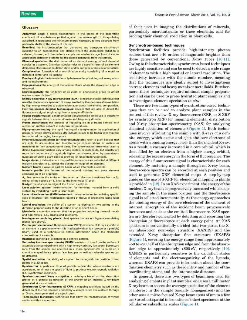
There are two main types of synchrotron-based techni-ques that can be used to analyze plant samples in thecontext of this review: X-ray fluorescence (XRF, or S-XRFfor synchrotron XRF) for imaging elemental distributionand X-ray absorption spectrometry (XAS) for analyzingchemical speciation of elements (Figure 1). Both techni-ques involve irradiating the sample with X-rays of a defi-nite energy, which excite and eject the core electrons ofatoms with a binding energy lower than the incident X-ray.As a result, a vacancy is created in a core orbital, which isthen filled by an electron from a higher energy orbital,releasing the excess energy in the form of fluorescence. Theenergy of this fluorescence signal is characteristic for eachelement. By rastering a sample through an X-ray beam,fluorescence spectra can be recorded at each position andused to generate XRF elemental maps. A step-by-stepguide on the use of S-XRF for elemental mapping in plantsis provided in [12]. In an XAS experiment, the energy of theincident X-ray beam is progressively increased while keep-ing the sample in the same position and the fluorescencesignal is collected incrementally. As the energy approachesthe binding energy of the core electrons of the element ofinterest, absorption of the incident beam progressivelyincreases and so does the emitted fluorescence. XAS spec-tra are therefore generated by detecting and recording theabsorption or fluorescence at each energy point. An XASspectrum is conventionally divided into two parts, the X-ray absorption near-edge structure (XANES) and theextended X-ray absorption fine structure (EXAFS)(Figure 1), covering the energy range from approximately–50 to +200 eV of the absorption edge and from the absorp-tion edge to approximately +800 eV, respectively [10].XANES is particularly sensitive to the oxidation statesof elements and the electronegativity of the ligands,whereas EXAFS can provide information about the coor-dination chemistry such as the identity and number of thecoordinating atoms and the interatomic distance.
In general, there are two types of beamlines used foranalyzing elements in plant samples: one uses a millimeterX-ray beam to assess the average speciation of the elementof interest in the sample (usually homogenized) and theother uses a micro-focused X-ray beam (tens of nm to a fewmm) to collect spatial information of intact specimens at thecellular or subcellular scales (Figure 1).

Purpose of thesynchrotronexperiment
Chemical specia�on in bulk �ssues:bulk XANES or EXAFS with mmbeam size
Elemental distribu�on:S-XRF with microfocused beam
Chemical specia�on in micro-spots:XANES or EXAFS withmicrofocused beam
2D scanning of intact sample:images represent a planar
compression of the volumeprobed by the beam
Thin sec�ons Tomography ofhydrated samplesHigh pressure freezing,
freeze subs�tu�on
Cryo thin sec�ons ofhydrated �ssue, requirescryo chamber
Nor
mal
ized
inte
nsity
Energy (KeV)
XANESEXAFS
Thin sec�ons ofnaturally dehydratedsamples
Rice grain (longitudinal sec�on)
Wheat grain (sec�oned in half) Zn
Cowpea root in apolyimide capillary
100 μm 'Rice node
100 μm
500 μm
R=As B=Fe G=Zn
TRENDS in Plant Science
Figure 1. Synchrotron-based techniques for analyzing elemental distribution and speciation in plant tissues. The images of cowpea root, wheat grain, and rice grain are
reproduced, with permission, from [37,75,84]. The rice node image is based on unpublished data from the authors.
Review Trends in Plant Science March 2014, Vol. 19, No. 3
The penetrating nature of X-rays means that 2D ele-mental maps are a planar compression of the volumeprobed by the beam. Therefore, elemental maps shouldbe interpreted keeping in mind the morphology of thesample in relation to the variation in thickness and theoverlaying tissues. One way to overcome this effect is to usethin sections of uniform thickness (e.g., [13,14]). Conver-sely, the penetrating nature of the X-rays enables the use oftomographic techniques to map elements in virtual 2Dsections or in 3D through mathematical reconstructions[15].
The intensity of the X-ray beam means that long expo-sure may cause damage to the specimen. This has thepotential to cause significant artifacts in both elementdistribution and, perhaps more problematically, chemicalspeciation. For example, photo-reduction of redox-sensi-tive elements (e.g., As and Se) may occur during synchro-tron beam exposure, causing speciation artifacts [16–18]. Itis therefore essential to preserve the sample using cryo-genic conditions or preferably by reducing the radiationdose. This can be achieved by using fast detector technol-ogies that drastically reduce the exposure duration andradiation doses [19].
Mass spectrometry-based techniquesSecondary ion mass spectrometry
Secondary ion mass spectrometry (SIMS) is a surface-sensitive technique that uses an energetic primary ionbeam to remove particles from the top few atomic layersof a sample surface. The ions emitted during this bombard-
ment, known as secondary ions, are analyzed in a massspectrometer to give information about the elemental ormolecular distribution within the sample. The NanoSIMSis a specific type of SIMS instrument that has beendesigned for high lateral resolution (down to 50 nm) ima-ging while still maintaining high mass resolution (theability to distinguish between species with nominallythe same mass) and high sensitivity (mg/kg range). It ispossible to detect most elements in the periodic table, fromhydrogen to uranium, as well as their different isotopes.These characteristics offer many advantages for the ana-lysis of elemental distributions in plant cells at the cellularand, crucially, subcellular scale, and enables the use ofstable isotope labeling to track element uptake and com-partmentalization [20]. The main limitations of this instru-ment are that it is difficult to analyze some elements, suchas Zn, Cd, and Mn, because of poor secondary ion yield, thedifficulty of quantifying actual concentrations, and theinability to obtain chemical state information [21]. Thefield of view is limited and mapping large areas is rela-tively slow, although it has become more rapid with theadvances in automation and beam control available onnewer instruments. Although SIMS is a destructive tech-nique, which can be a disadvantage for some samples, italso enables depth profiling through a sample to build up a3D volume.
SIMS must be conducted under an ultra-high vacuum,which imposes limitations on the form of the sample. Freshtissue cannot be analyzed because of the high water con-tent, instead it must be rapidly cryo-fixed and analyzed
185

Review Trends in Plant Science March 2014, Vol. 19, No. 3
either frozen-hydrated by cryo-SIMS [20] or, if the instru-ment does not have a cryo-stage, as is the case for theNanoSIMS, subsequently fixed at low temperature, dehy-drated with acetone, and embedded in resin [22]. Samplepreparation is the most crucial step in successfully analyz-ing elemental distributions in plant cells with SIMS ana-lysis [23]. High-pressure freezing followed by freezesubstitution has been found to be the best method topreserve cellular and subcellular structures as well aselemental distributions of plant cells for NanoSIMS ana-lysis [14,22,24].
Laser-ablation inductively coupled plasma mass
spectrometry
Laser-ablation inductively coupled plasma mass spectro-metry (LA-ICP-MS) can be used to investigate elementdistribution in solid sample materials by using a focusedlaser beam in an argon atmosphere under normal pres-sure. Sample material is ablated by the laser beam and thelaser-induced aerosol is transported with a carrier gas (Heor Ar) into the inductively coupled plasma ion source whereionization takes place. The charged ions are detected bytheir mass-to-charge ratio, m/z, in the mass spectrometer.The spatial resolution of the common laser ablation sys-tems generally ranges from 50 to 300 mm [25]. A highresolution can be achieved using a laser beam with asmaller spot size, although this may lead to insufficientenergy focused on the sample and, hence, limited ablationof the material. A laser microdissection (LMD) instrumentcan substitute a laser ablation system to substantiallyenhance the spatial resolution [26].
Imaging the cellular and subcellular distribution ofelementsThe techniques described above have been used to imageelement distribution at the tissue and cellular scales ofplant samples. Many of the earlier studies focused onhyperaccumulator plants that are able to accumulate largeconcentrations of metals or metalloids. These studiesrevealed, for example, Cd and Zn accumulation in thetrichomes of Arabidopsis halleri [27,28], Zn in the epider-mal cells of the leaves of pennycress (Thlaspi praecox) [9],Ni in the trichomes [29] and the epidermal vacuoles [22] ofAlyssum species, Se in the trichomes of milkvetch (Astra-galus bisulcatus) [30], As in the vacuoles of Chinese brakefern (Pteris vittata) fronds [31], and Tl in the veins ofcandytuft (Iberis intermedia) leaves [32].
More recently, there has been a shift in attention tostudying essential or toxic trace elements in non-hyperac-cumulator plants: some examples are described below.Fluorescence tomography images obtained at different ener-gies corresponding to different oxidation states of As show astrong accumulation of arsenate [As(V)] in the iron plaqueformed on the root surfaces of aquatic plants such as cattail(Typha latifolia) [33] and rice (Oryza sativa) [34] (Figure 2A),suggesting that iron plaque provides a barrier to the entry ofAs(V) into these plants but is less effective in blocking theuptake of arsenite [As(III)], probably owing to a weakeradsorption of As(III) by the plaque [35]. Inside rice roots,As(III) is preferentially stored in the vacuoles of the peri-cycle and endodermis cells and is strongly colocalized with S
186
as revealed by NanoSIMS analysis [24] (Figure 2B). Thiscolocalization is consistent with a vacuolar sequestration ofAs(III)–thiol (e.g., phytochelatins) complexes. Using S-XRFwith a fast detector, the As distribution has been imaged infresh hydrated roots of cowpea (Vigna unguiculata) follow-ing short-term exposure to either As(III) or As(V), showing astrong accumulation of As in the region that is likely to beendodermis and/or pericycle cells [36] (Figure 2C). The sameapproach has been applied to mapping the in situ distribu-tion of Zn, Cu, Ni, Mn, and Se in hydrated tissues [17,37–39].A recent study combining S-XRF and NanoSIMS hasrevealed that As is localized in the vacuoles of the companioncells within the phloem of various types of vascular bundlesof the rice node, internode, and leaf sheath [14](Figure 2D,E). A strong colocalization of As with S is againevident. Thus, the companion cells possibly provide the mostimportant storage capacity of As in rice aboveground tis-sues, implying that the biosynthesis of phytochelatins andthe vacuolar transport of As(III)–phytochelatin complexesare likely to be much enhanced in this cell type. Thisdistribution pattern also supports the notion that inorganicAs is transported to the grain mainly via the phloem [40,41],although with a restricted mobility, presumably because ofthe vacuolar sequestration. In rice grain, informationobtained largely through fluorescence tomography indicatesthat inorganic As [mainly As(III)] accumulates preferen-tially in the ovular vascular traces (OVTs) (Figure 2F),which are the conducting tissues transporting nutrientsand water to the grain [42–44]. By contrast, methylatedAs species such as dimethylarsinic acid (DMA) are muchmore mobile during phloem transport and can permeate intothe endosperm of rice more easily than inorganic As[40,44,45].
Mapping the cellular distribution of Cd in non-hyper-accumulator plants is difficult because of its low tissueconcentrations and the high energy of its Ka line (which isnot always accessible at microspectroscopy beamlines). Inattempts to overcome these difficulties, researchers haveoften used artificially high concentrations of Cd that causesevere phytotoxicity and therefore diminish the environ-mental and physiological relevance of the results obtained(see [46]). Cd has been shown to accumulate in the vascularbundles of roots and leaf trichomes of Arabidopsis thalianaafter plants were exposed to a high concentration of Cd[47]. Using S-XRF, Cd has also been shown to accumulatein the xylem of the enlarged vascular bundles (EVBs) andin the parenchyma cell bridge surrounding the EVBs in thenodes of rice grown in contaminated soil [48]. By contrast,Zn accumulates preferentially in the parenchyma cellbridge bordering the EVBs and the diffuse vascular bun-dles (DVBs) [48]; NanoSIMS analysis revealed that Zn waslocalized to the vacuoles of these cells [14]. Fe and Mn arelocalized to the fundamental parenchyma cells in the nodesof rice, where there is a strong colocalization of Fe and Pinside the vacuoles, possibly in insoluble forms [14](Figure 2D,E), which may explain the antagonistic effectof P on Fe nutrition [49]. The differences among Cd, Zn, Fe,and Mn distribution in the rice node are striking, eventhough Cd is known to share some common transporterswith the other metals [2]. A possible explanation lies in thedifferent chemical speciation of these metals.

(A)
8
0 Fe
---- As(III) ---- ---- As(V) ----
1 mm
CC
ST
Fe
PINAs
Min Max
Endosperm EmbryoBran
OVT
Col-0 vit1-1
FeMn MnZn Zn
As(V)0
1
28Si-
75As-
75As-32S-(D)
(F) (G)
2
1(E)
(B) (C)
TRENDS in Plant Science
Figure 2. Mapping elemental distribution at the cellular and subcellular scales. (A) Synchrotron X-ray fluorescence (S-XRF) 2D tomography of a rice root growing in an As-
contaminated soil, showing the accumulation of Fe and As(V) on the surface iron plaque [34]. (B) NanoSIMS imaging of Si, S, and As in a rice root thin section showing
colocalization of As and S in the vacuoles of pericycle and endodermal cells, and Si in the apoplast of endodermal cells [24]. Abbreviations: En, endodermis; Pc, pericycle;
SE, secondary electron image; Xy, xylem vessel. (C) S-XRF imaging with a fast detector of fresh hydrated roots of cowpea exposed As(V) or As(III) for 24 h [36]. (D) S-XRF
imaging of a rice node thin section showing distinct distribution patterns of As (red), Zn (green), and Fe (blue). Abbreviations: EVB, enlarged vascular bundles; DVB, diffuse
vascular bundles; FP, fundamental parenchyma; LVB, large vascular bundles [14]. (E) NanoSIMS imaging of the DVB and FP regions marked as areas 1 and 2 in (D), showing
strong colocalization of As and S in the vacuoles of the companion cells of the phloem in the DVBs and strong colocalization of Fe and P in the vacuoles of FP cells [14].
Abbreviations: CC, companion cells; ST, sieve tubes. (F) S-XRF 3D tomography of a rice grain showing the accumulation of As in the ovular vascular trace (OVT) [42]. (G) S-
XRF 3D tomography of Fe, Mn, and Zn in the seeds of wild type Arabidopsis (Col-0) and the vit1-1 mutant [50]. Reproduced, with permission, from [14,24,34,36,42,50].
Review Trends in Plant Science March 2014, Vol. 19, No. 3
187

Review Trends in Plant Science March 2014, Vol. 19, No. 3
Mature seeds are ideal for S-XRF or NanoSIMS ana-lysis because they are largely and naturally dehydratedand therefore require minimal sample preparation.Fluorescence tomography has been used to characterizeelemental distribution in the seeds of wild type A. thali-ana and a mutant defective in the vacuolar iron trans-porter 1 (VIT1) [50]. Whereas bulk analyses revealed nosignificant differences in the total Fe concentrations, themutant seed exhibited an Fe distribution pattern differ-ent to that in the wild type. Iron in wild type seeds isstrongly localized to the provascular strands of the hypo-cotyl, radicle, and cotyledons, probably inside thevacuoles (Figure 2G). By contrast, Fe is not detected inthese cells in the vit1 mutant seeds and is instead locatedmore diffusely in the hypocotyl and radicle, and in theepidermal cells of the cotyledons. This investigation hasunraveled the function of VIT1 in vacuolar Fe storage,which is crucial for seedling development after germina-tion. Fluorescence tomography and high-resolution S-XRF has been used to investigate the elemental distribu-tion of wild type Arabidopsis seeds and embryos, and ofmutants of endomembrane cation exchangers (CAXs) andoverexpression lines [13]. Higher concentrations of Cawere found in the seed coat and in the embryo of the caxmutants compared with that found in the wild type; themutants also had reduced partitioning of Ca into orga-nelles and higher levels of Ca in the cytosol. High resolu-tion (0.15 mm) S-XRF analysis of thin sections ofembedded embryos showed strikingly different patternsof elemental distribution. In wild type seeds, Mn isstrongly localized to a sub-epidermal layer of cells ofthe cotyledons, Fe to the organelles (probably thevacuoles) of the endodermal cells of the radicles andthe vasculature of the cotyledons, and Zn and Ca areuniformly distributed to all of the cells of the embryo [13].CAX mutation also alters the distribution of Mn. Theseexamples demonstrate the power of combining state-of-the art imaging techniques with molecular geneticmethods to understand the gene functions and cellularhomeostasis of elements.
SIMS or NanoSIMS has been used to image a widerange of elements in cereal grains at the cellular andsubcellular scales, showing strong localization of miner-als with phytate granules in the aleurone cells [51,52], ofSe in the protein matrix surrounding the starch granules,of As in the sub-aleurone cells, and in hotspots in the OVTregion of the rice grain [45]. Stable isotope 70Zn labelingand LA-ICP-MS has shown that 70Zn strongly accumu-lates in the crease vascular tissue of wheat (Triticumaestivum) grain, suggesting that the translocation fromthe maternal to filial tissues may be a bottleneck for Znaccumulation in the endosperm [53]. For Fe and Znbiofortification in cereals, it is important to increasethe concentrations of Fe and Zn in the endosperm becausethis is the part that is mostly consumed by humans.Overexpression of nicotianamine synthase and ferritingenes can increase Fe concentration in the rice endo-sperm [54]. In LA-ICP-MS analysis, Fe in the endospermof the transgenic rice appears to accumulate in spots,probably as a consequence of spatially restricted ferritinaccumulation [54].
188
Tracing the movement of elementsOne of the advantages of mass spectrometry-based tech-niques such as SIMS and LA-ICP-MS is the ability todistinguish and quantify different isotopes, thus enablingstable isotopes to be used as tracers. NanoSIMS has beenused to reveal the uptake and competition between rhizo-sphere microorganisms and plant roots for 15N labeledammonium [55,56]. Pulsing cut stems of bean (Phaseolusvulgaris) plants with 26Mg, 41K, 44Ca, and H2
18O and usingcryo-SIMS to image their distribution at different timesrevealed a rapid exchange of cations between xylem ves-sels and the adjacent xylem parenchyma cells, and a slowexchange with cambium and phloem; the three cationsalso exhibited different exchange kinetics [57,58]. In thesestudies, samples were shock-frozen in melting propane,fractured, and analyzed under cryo-conditions, thuspreserving the cellular structure and in situ elementdistribution.
Probing chemical speciation of elementsXAS analyses can be performed with bulk samples (usuallyfreeze-dried or, preferably, powdered frozen-hydratedsamples) to gain overall information about speciation, orabout specific spots of a sample at the micrometer or sub-micrometer scale using micro-focused XANES and EXAFS.However, owing to the complexity of biological samples, thespeciation information obtained is often more accuratewith regard to the oxidation state and the type of bindingligands than the exact identities of the ligand molecules.For example, XANES can distinguish As(V), As(III),As(III)–glutathione, and As(III)–phytochelatin, but notthe numerous variants of As(III)–phytochelatin or otherAs(III)–thiol complexes that have been detected in planttissues based on ex situ extraction and ICP-MS and elec-trospray mass spectrometry analysis [59]. There arenumerous examples of using XAS to investigate the che-mical speciation of elements in plant tissues; some exam-ples are described below.
Most plant species appear to be able to reduce As(V) toAs(III) rapidly, resulting in the dominance of trivalent Asinside plant cells [60]. Arsenic is found to be present mainlyas As(III) coordinated with thiol compounds in non-hyper-accumulators (e.g., [61–63]), but predominantly as freearsenite or arsenate in As hyperaccumulating ferns (e.g.,[31,64,65]). A recent study employing the fluorescence-XANES imaging technique showed dramatic changes inthe As speciation from the outer to the inner layers of cellsof rice and wheat roots that were exposed to As(V) in thegrowth medium [66]. As(V) was found only in the rhizo-dermis, whereas the As(III)–thiol complexes dominated inthe cortex and the stele [66]. Confocal XANES is anothertechnique that can be used to target chemical speciesanalysis to specific spatial regions of cells in intact planttissues [59]. Using this technique, it has been shown thatAs preferentially accumulated in the epidermal cells of theaquatic plant hornwort (Ceratophyllum demersum) withAs(III)–phytochelatins, As(III)–glutathione, and uncom-plexed As(III) and As(V) as the main chemical species ofAs [59]. A study has been performed that compared Asspeciation in rice grain by ex situ extraction followedby high-performance liquid chromatography–ICP-MS

(A)
As(III) DMAAs(V)
R110013
R110335
R110331
R110015
11870 11880Energy (eV)
11890
Zn-O
dis
tanc
e (Å
)
Zn content (μmol g-1 DW)
2.08
0 1 2 3R+ΔR(Å)
4 5
F2-10F2-9
F2-7
F2-5
F2-2
F2-1
F1-2
F1-1
ZnPhos
Zn-cell wall
Zn malate sol.
A. Iyrata
A. halleri
2.07
2.06
2.05
2.04
2.03
2.02
2.010 20 40 60 80 100
NIES 10a
As(III)GSH
(B)
(C)
TRENDS in Plant Science
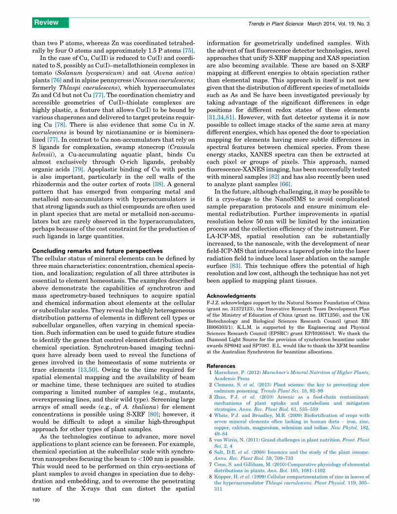
Figure 3. Probing chemical speciation of elements in plant tissues. (A) Synchrotron X-ray absorption near edge structure (XANES) arsenic K-edge spectra of arsenic
standards and five rice grain samples [67]. Abbreviations: As(III), arsenite; As(III)–GSH, arsenite–glutathione complex; As(V), arsenate; DMA, dimethylarsinic acid. (B) Zinc K-
edge extended X-ray absorption fine structure (EXAFS) spectra of three Zn standards, Arabidopsis halleri, Arabidopsis lyrata, and their F1 or F2 progeny leaves after Fourier
transformation [74]. (C) Average first-shell zinc (Zn)–O distance determined by shell simulations based on the EXAFS spectra in (B) as a function of total Zn concentration
[74]. Abbreviation: DW, dry weight. Reproduced, with permission, from [67,74].
Review Trends in Plant Science March 2014, Vol. 19, No. 3
analysis and in situ bulk XANES [67] (Figure 3A). Thesamples had low As concentrations ranging from 0.20 to0.34 mg/g. This study provided evidence of good agreementbetween the two techniques and demonstrated the highsensitivity of XANES offered by modern synchrotron beam-lines. In rice grains, As is present as As(III), As(III)–thiolcomplexes, As(V), and DMA [43,67].
Cadmium is coordinated mainly with S (probably phy-tochelatins or glutathione) in the roots of A. thaliana andmustard (Brassica juncea) [47,68], but with O or N ligandsin the trichomes [47]. In the rice node, Cd is coordinatedmainly with S in the xylem of the enlarged vascularbundles, but with both S and O ligands in the phloem[48]. Similar to As hyperaccumulators, Cd hyperaccumu-lators do not appear to use thiol complexation as the mainmechanism to detoxify Cd given that Cd is found to becoordinated mainly to O or N ligands [69–72].
Depending on plant species, type of tissues and growthconditions, common ligands for binding Zn in plant cellsinclude organic acids (O ligands), histidine, phosphate, orphytate [28,38,69,73]. For instance, EXAFS has been used
to investigate the speciation of Zn in parent plants and inthe F1 and F2 progenies from crosses between the Znhyperaccumulator Arabidopsis halleri and its non-hyper-accumulator relative Arabidopsis lyrata [74]. Figure 3Bshows the Zn EXAFS spectra after Fourier transformationof Zn standards and plant samples. Shell simulations werethen used to determine the structural parameters for thefirst and second Zn coordination shells, which were foundto be well fitted with oxygen and carbon as nearest andnext nearest atomic neighbors, respectively, as would beexpected in Zn–organic acid complexes. The results indi-cated a correlation between total Zn in plants and theproportion of Zn in octahedral coordination (Figure 3C).This is indicative of a dominant complexation by organicacids, where Zn is sixfold coordinated, in Zn-accumulatingplants. In plants that accumulated less Zn, EXAFS ana-lysis revealed a larger proportion of Zn being fourfoldcoordinated, probably with the cell wall and phosphate(Figure 3C). In aleurone cells of wheat grain, EXAFSshowed that Fe and Zn were complexed by phytates; Fewas coordinated octahedrally by six O atoms and fewer
189
Review Trends in Plant Science March 2014, Vol. 19, No. 3
than two P atoms, whereas Zn was coordinated tetrahed-rally by four O atoms and approximately 1.5 P atoms [75].
In the case of Cu, Cu(II) is reduced to Cu(I) and coordi-nated to S, possibly as Cu(I)–metallothionein complexes intomato (Solanum lycopersicum) and oat (Avena sativa)plants [76] and in alpine pennycress (Noccaea caerulescens;formerly Thlaspi caerulescens), which hyperaccumulatesZn and Cd but not Cu [77]. The coordination chemistry andaccessible geometries of Cu(I)–thiolate complexes arehighly plastic, a feature that allows Cu(I) to be bound byvarious chaperones and delivered to target proteins requir-ing Cu [78]. There is also evidence that some Cu in N.caerulescens is bound by nicotianamine or is biominera-lized [77]. In contrast to Cu non-accumulators that rely onS ligands for complexation, swamp stonecrop (Crassulahelmsii), a Cu-accumulating aquatic plant, binds Cualmost exclusively through O-rich ligands, probablyorganic acids [79]. Apoplastic binding of Cu with pectinis also important, particularly in the cell walls of therhizodermis and the outer cortex of roots [38]. A generalpattern that has emerged from comparing metal andmetalloid non-accumulators with hyperaccumulators isthat strong ligands such as thiol compounds are often usedin plant species that are metal or metalloid non-accumu-lators but are rarely observed in the hyperaccumulators,perhaps because of the cost constraint for the production ofsuch ligands in large quantities.
Concluding remarks and future perspectivesThe cellular status of mineral elements can be defined bythree main characteristics: concentration, chemical specia-tion, and localization; regulation of all three attributes isessential to element homeostasis. The examples describedabove demonstrate the capabilities of synchrotron andmass spectrometry-based techniques to acquire spatialand chemical information about elements at the cellularor subcellular scales. They reveal the highly heterogeneousdistribution patterns of elements in different cell types orsubcellular organelles, often varying in chemical specia-tion. Such information can be used to guide future studiesto identify the genes that control element distribution andchemical speciation. Synchrotron-based imaging techni-ques have already been used to reveal the functions ofgenes involved in the homeostasis of some nutrients ortrace elements [13,50]. Owing to the time required forspatial elemental mapping and the availability of beamor machine time, these techniques are suited to studiescomparing a limited number of samples (e.g., mutants,overexpressing lines, and their wild type). Screening largearrays of small seeds (e.g., of A. thaliana) for elementconcentrations is possible using S-XRF [80]; however, itwould be difficult to adopt a similar high-throughputapproach for other types of plant samples.
As the technologies continue to advance, more novelapplications to plant science can be foreseen. For example,chemical speciation at the subcellular scale with synchro-tron nanoprobes focusing the beam to <100 nm is possible.This would need to be performed on thin cryo-sections ofplant samples to avoid changes in speciation due to dehy-dration and embedding, and to overcome the penetratingnature of the X-rays that can distort the spatial
190
information for geometrically undefined samples. Withthe advent of fast fluorescence detector technologies, novelapproaches that unify S-XRF mapping and XAS speciationare also becoming available. These are based on S-XRFmapping at different energies to obtain speciation ratherthan elemental maps. This approach in itself is not newgiven that the distribution of different species of metalloidssuch as As and Se have been investigated previously bytaking advantage of the significant differences in edgepositions for different redox states of these elements[31,34,81]. However, with fast detector systems it is nowpossible to collect image stacks of the same area at manydifferent energies, which has opened the door to speciationmapping for elements having more subtle differences inspectral features between chemical species. From theseenergy stacks, XANES spectra can then be extracted ateach pixel or groups of pixels. This approach, namedfluorescence-XANES imaging, has been successfully testedwith mineral samples [82] and has also recently been usedto analyze plant samples [66].
In the future, although challenging, it may be possible tofit a cryo-stage to the NanoSIMS to avoid complicatedsample preparation protocols and ensure minimum ele-mental redistribution. Further improvements in spatialresolution below 50 nm will be limited by the ionizationprocess and the collection efficiency of the instrument. ForLA-ICP-MS, spatial resolution can be substantiallyincreased, to the nanoscale, with the development of nearfield-ICP-MS that introduces a tapered probe into the laserradiation field to induce local laser ablation on the samplesurface [83]. This technique offers the potential of highresolution and low cost, although the technique has not yetbeen applied to mapping plant tissues.
AcknowledgmentsF-J.Z. acknowledges support by the Natural Science Foundation of China(grant no. 31372123), the Innovative Research Team Development Planof the Ministry of Education of China (grant no. IRT1256), and the UKBiotechnology and Biological Sciences Research Council (grant BB/H006303/1). K.L.M. is supported by the Engineering and PhysicalSciences Research Council (EPSRC) grant EP/I026584/1. We thank theDiamond Light Source for the provision of synchrotron beamtime underawards SP6042 and SP7087. E.L. would like to thank the XFM beamlineat the Australian Synchrotron for beamtime allocations.
References1 Marschner, P. (2012) Marschner’s Mineral Nutrition of Higher Plants,
Academic Press2 Clemens, S. et al. (2013) Plant science: the key to preventing slow
cadmium poisoning. Trends Plant Sci. 18, 92–993 Zhao, F.J. et al. (2010) Arsenic as a food-chain contaminant:
mechanisms of plant uptake and metabolism and mitigationstrategies. Annu. Rev. Plant Biol. 61, 535–559
4 White, P.J. and Broadley, M.R. (2009) Biofortification of crops withseven mineral elements often lacking in human diets – iron, zinc,copper, calcium, magnesium, selenium and iodine. New Phytol. 182,49–84
5 von Wiren, N. (2011) Grand challenges in plant nutrition. Front. PlantSci. 2, 4
6 Salt, D.E. et al. (2008) Ionomics and the study of the plant ionome.Annu. Rev. Plant Biol. 59, 709–733
7 Conn, S. and Gilliham, M. (2010) Comparative physiology of elementaldistributions in plants. Ann. Bot. 105, 1081–1102
8 Ku pper, H. et al. (1999) Cellular compartmentation of zinc in leaves ofthe hyperaccumulator Thlaspi caerulescens. Plant Physiol. 119, 305–311

Review Trends in Plant Science March 2014, Vol. 19, No. 3
9 Vogel-Mikus, K. et al. (2008) Comparison of essential and non-essentialelement distribution in leaves of the Cd/Zn hyperaccumulator Thlaspipraecox as revealed by micro-PIXE. Plant Cell Environ. 31, 1484–1496
10 Sarret, G. et al. (2013) Use of synchrotron-based techniques to elucidatemetal uptake and metabolism in plants. Adv. Agron. 119, 1–82
11 Lombi, E. and Susini, J. (2009) Synchrotron-based techniques for plantand soil science: opportunities, challenges and future perspectives.Plant Soil 320, 1–35
12 Donner, E. et al. (2013) Mapping element distributions in plant tissuesusing synchrotron X-ray fluorescence techniques. In Plant MineralNutrients: Methods and Protocols. Methods in Molecular Biology(Vol. 953) (Maathuis, F.J.M., ed.), pp. 143–159, Springer
13 Punshon, T. et al. (2012) The role of CAX1 and CAX3 in elementaldistribution and abundance in Arabidopsis seed. Plant Physiol. 158,352–362
14 Moore, K.L. et al. (2014) Combined NanoSIMS and synchrotron X-rayfluorescence reveals distinct cellular and subcellular distributionpatterns of trace elements in rice tissues. New Phytol. 201, 104–115
15 de Jonge, M.D. and Vogt, S. (2010) Hard X-ray fluorescencetomography – an emerging tool for structural visualization. Curr.Opin. Struct. Biol. 20, 606–614
16 Lombi, E. et al. (2011) In situ analysis of metal(loid)s in plants: state ofthe art and artefacts. Environ. Exp. Bot. 72, 3–17
17 Wang, P. et al. (2013) In situ speciation and distribution of toxicselenium in hydrated roots of cowpea. Plant Physiol. 163, 407–418
18 George, G.N. et al. (2012) X-ray-induced photo-chemistry and X-rayabsorption spectroscopy of biological samples. J. Synchr. Radiat. 19,875–886
19 Lombi, E. et al. (2011) Trends in hard X-ray fluorescence mapping:environmental applications in the age of fast detectors. Anal. Bioanal.Chem. 400, 1637–1644
20 Metzner, R. et al. (2008) Imaging nutrient distributions in plant tissueusing time-of-flight secondary ion mass spectrometry and scanningelectron microscopy. Plant Physiol. 147, 1774–1787
21 Moore, K.L. et al. (2012) Elemental imaging at the nanoscale:NanoSIMS and complementary techniques for element localisationin plants. Anal. Bioanal. Chem. 402, 3263–3273
22 Smart, K.E. et al. (2010) High-resolution elemental localization invacuolate plant cells by nanoscale secondary ion mass spectrometry.Plant J. 63, 870–879
23 Chandra, S. et al. (2000) Subcellular imaging by dynamic SIMS ionmicroscopy. Anal. Chem. 72, 104a–114a
24 Moore, K.L. et al. (2011) NanoSIMS analysis reveals contrastingpatterns of arsenic and silicon localization in rice roots. PlantPhysiol. 156, 913–924
25 Wu, B. and Becker, J.S. (2012) Imaging techniques for elements andelement species in plant science. Metallomics 4, 403–416
26 Becker, J.S. et al. (2010) Scaling down the bioimaging of metals by lasermicrodissection inductively coupled plasma mass spectrometry (LMD-ICP-MS). Int. J. Mass Spectrom. 294, 1–6
27 Fukuda, N. et al. (2008) Micro X-ray fluorescence imaging and micro X-ray absorption spectroscopy of cadmium hyper-accumulating plant,Arabidopsis halleri ssp gemmifera, using high-energy synchrotronradiation. J. Anal. Atom. Spectrom. 23, 1068–1075
28 Sarret, G. et al. (2002) Forms of zinc accumulated in thehyperaccumulator Arabidopsis halleri. Plant Physiol. 130, 1815–1826
29 McNear, D.H. et al. (2005) Application of quantitative fluorescence andabsorption-edge computed microtomography to image metal compart-mentalization in Alyssum murale. Environ. Sci. Technol. 39, 2210–2218
30 Freeman, J.L. et al. (2006) Spatial imaging, speciation, andquantification of selenium in the hyperaccumulator plants Astragalusbisulcatus and Stanleya pinnata. Plant Physiol. 142, 124–134
31 Pickering, I.J. et al. (2006) Localizing the biochemical transformationsof arsenate in a hyperaccumulating fern. Environ. Sci. Technol. 40,5010–5014
32 Scheckel, K.G. et al. (2007) Synchrotron X-ray absorption-edge computedmicrotomography imaging of thallium compartmentalization in Iberisintermedia. Plant Soil 290, 51–60
33 Blute, N.K. et al. (2004) Arsenic sequestration by ferric iron plaque oncattail roots. Environ. Sci. Technol. 38, 6074–6077
34 Seyfferth, A.L. et al. (2010) Arsenic localization, speciation, and co-occurrence with iron on rice (Oryza sativa L.) roots having variable Fecoatings. Environ. Sci. Technol. 44, 8108–8113
35 Chen, Z. et al. (2005) Direct evidence showing the effect of root surfaceiron plaque on arsenite and arsenate uptake into rice (Oryza sativa)roots. New Phytol. 165, 91–97
36 Kopittke, P.M. et al. (2012) Examination of the distribution of arsenicin hydrated and fresh cowpea roots using two- and three-dimensionaltechniques. Plant Physiol. 159, 1149–1158
37 Lombi, E. et al. (2011) Fast X-ray fluorescence microtomography ofhydrated biological samples. PLoS ONE 6, e20626
38 Kopittke, P.M. et al. (2011) In situ distribution and speciation of toxiccopper, nickel, and zinc in hydrated roots of cowpea. Plant Physiol. 156,663–673
39 Kopittke, P.M. et al. (2013) Distribution and speciation of Mn inhydrated roots of cowpea at levels inhibiting root growth. Physiol.Plant. 147, 453–464
40 Carey, A.M. et al. (2010) Grain unloading of arsenic species in rice.Plant Physiol. 152, 309–319
41 Zhao, F.J. et al. (2012) Arsenic translocation in rice investigated usingradioactive 73As tracer. Plant Soil 350, 413–420
42 Carey, A.M. et al. (2011) Phloem transport of arsenic species from flagleaf to grain during grain filling. New Phytol. 192, 87–98
43 Lombi, E. et al. (2009) Speciation and distribution of arsenic andlocalization of nutrients in rice grains. New Phytol. 184, 193–201
44 Zheng, M.Z. et al. (2013) Differential toxicity and accumulation ofinorganic and methylated arsenic in rice. Plant Soil 365, 227–238
45 Moore, K.L. et al. (2010) NanoSIMS analysis of arsenic and selenium incereal grain. New Phytol. 185, 434–445
46 Van Belleghem, F. et al. (2007) Subcellular localization of cadmium inroots and leaves of Arabidopsis thaliana. New Phytol. 173, 495–508
47 Isaure, M.P. et al. (2006) Localization and chemical forms of cadmiumin plant samples by combining analytical electron microscopy and X-ray spectromicroscopy. Spectrochim. Acta B: Atom. Spectrom. 61,1242–1252
48 Yamaguchi, N. et al. (2012) Role of the node in controlling traffic ofcadmium, zinc, and manganese in rice. J. Exp. Bot. 63, 2729–2737
49 Zheng, L.Q. et al. (2009) Physiological and transcriptome analysis ofiron and phosphorus interaction in rice seedlings. Plant Physiol. 151,262–274
50 Kim, S.A. et al. (2006) Localization of iron in Arabidopsis seed requiresthe vacuolar membrane transporter VIT1. Science 314, 1295–1298
51 Heard, P.J. et al. (2002) Determination of the elemental composition ofmature wheat grain using a modified secondary ion mass spectrometer(SIMS). Plant J. 30, 237–245
52 Moore, K.L. et al. (2012) Localisation of iron in wheat grain using highresolution secondary ion mass spectrometry. J. Cereal Sci. 55, 183–187
53 Wang, Y.X. et al. (2011) Stable isotope labelling and zinc distribution ingrains studied by laser ablation ICP-MS in an ear culture systemreveals zinc transport barriers during grain filling in wheat. NewPhytol. 189, 428–437
54 Wirth, J. et al. (2009) Rice endosperm iron biofortification by targetedand synergistic action of nicotianamine synthase and ferritin. PlantBiotechnol. J. 7, 631–644
55 Clode, P.L. et al. (2009) In situ mapping of nutrient uptake in therhizosphere using nanoscale secondary ion mass spectrometry. PlantPhysiol. 151, 1751–1757
56 Jones, D.L. et al. (2013) Competition between plant and bacterial cellsat the microscale regulates the dynamics of nitrogen acquisition inwheat (Triticum aestivum). New Phytol. 200, 796–807
57 Metzner, R. et al. (2010) Tracing cationic nutrients from xylem intostem tissue of French bean by stable isotope tracers and cryo-secondaryion mass spectrometry. Plant Physiol. 152, 1030–1043
58 Metzner, R. et al. (2010) Contrasting dynamics of water and mineralnutrients in stems shown by stable isotope tracers and cryo-SIMS.Plant Cell Environ. 33, 1393–1407
59 Mishra, S. et al. (2013) Speciation and distribution of arsenic in thenonhyperaccumulator macrophyte Ceratophyllum demersum. PlantPhysiol. 163, 1396–1408
60 Zhao, F.J. et al. (2009) Arsenic uptake and metabolism in plants. NewPhytol. 181, 777–794
61 Pickering, I.J. et al. (2000) Reduction and coordination of arsenic inIndian mustard. Plant Physiol. 122, 1171–1177
62 Bluemlein, K. et al. (2008) Can we trust mass spectrometry fordetermination of arsenic peptides in plants: comparison of LC-ICP-
191
Review Trends in Plant Science March 2014, Vol. 19, No. 3
MS and LC-ES-MS/ICP-MS with XANES/EXAFS in analysis ofThunbergia alata. Anal. Bioanal. Chem. 390, 1739–1751
63 Castillo-Michel, H. et al. (2011) Localization and speciation of arsenicin soil and desert plant Parkinsonia florida using mu XRF and muXANES. Environ. Sci. Technol. 45, 7848–7854
64 Webb, S.M. et al. (2003) XAS speciation of arsenic in a hyper-accumulating fern. Environ. Sci. Technol. 37, 754–760
65 Lombi, E. et al. (2002) Arsenic distribution and speciation in the frondsof the hyperaccumulator Pteris vittata. New Phytol. 156, 195–203
66 Kopittke, P.M. et al. (2013) Laterally resolved speciation of arsenic inroots of wheat and rice using fluorescence-XANES imaging. NewPhytol. http://dx.doi.org/10.1111/nph.12595
67 Maher, W. et al. (2013) Measurement of inorganic arsenic species inrice after nitric acid extraction by HPLC-ICPMS: verification usingXANES. Environ. Sci. Technol. 47, 5821–5827
68 Salt, D.E. et al. (1995) Mechanisms of cadmium mobility andaccumulation in Indian mustard. Plant Physiol. 109, 1427–1433
69 Ku pper, H. et al. (2004) Tissue- and age-dependent differences in thecomplexation of cadmium and zinc in the cadmium/zinchyperaccumulator Thlaspi caerulescens (Ganges ecotype) revealed byX-ray absorption spectroscopy. Plant Physiol. 134, 748–757
70 Huguet, S. et al. (2012) Cd speciation and localization in thehyperaccumulator Arabidopsis halleri. Environ. Exp. Bot. 82, 54–65
71 Tian, S.K. et al. (2011) Cellular sequestration of cadmium in thehyperaccumulator plant species Sedum alfredii. Plant Physiol. 157,1914–1925
72 Vogel-Mikus, K. et al. (2010) Complexation of cadmium in seeds andvegetative tissues of the cadmium hyperaccumulator Thlaspi praecoxas studied by X-ray absorption spectroscopy. Plant Soil 331, 439–451
73 Salt, D.E. et al. (1999) Zinc ligands in the metal hyperaccumulatorThlaspi caerulescens as determined using X-ray absorptionspectroscopy. Environ. Sci. Technol. 33, 713–717
74 Sarret, G. et al. (2009) Zinc distribution and speciation in Arabidopsishalleri � Arabidopsis lyrata progenies presenting various zincaccumulation capacities. New Phytol. 184, 581–595
Plant Science Con
GRC: Plant MoleDecision-Making Pathways, Networ
20–25 July
Holdernes
http://www.grc.org/programs.aspx?y
2014 Wisconsin Plant Pr21–25 July
Madison,
http://www.biotech
EMBO Workshop: Intercellular Communica24–29 Augu
Bischoffsheim
http://events.embo.org/coming-so
25th International Conference on Ar28 July–1 Aug
Vancouver,
http://arabidopsiscon
192
75 Neal, A.L. et al. (2013) Iron and zinc complexation in wild-type andferritin-expressing wheat grain: implications for mineral transportinto developing grain. J. Biol. Inorg. Chem. 18, 557–570
76 Ryan, B.M. et al. (2013) Copper speciation and isotopic fractionation inplants: uptake and translocation mechanisms. New Phytol. 199, 367–378
77 Mijovilovich, A. et al. (2009) Complexation and toxicity of copper inhigher plants. II. Different mechanisms for copper versus cadmiumdetoxification in the copper-sensitive cadmium/zinchyperaccumulator Thlaspi caerulescens (Ganges ecotype). PlantPhysiol. 151, 715–731
78 Pushie, M.J. et al. (2012) The fictile coordination chemistry of cuprous-thiolate sites in copper chaperones. Biochim. Biophys. Acta Bioenerg.1817, 938–947
79 Ku pper, H. et al. (2009) Complexation and toxicity of copper in higherplants. I. Characterization of copper accumulation, speciation, andtoxicity in Crassula helmsii as a new copper accumulator. PlantPhysiol. 151, 702–714
80 Young, L.W. et al. (2006) A high-throughput determination of metalconcentrations in whole intact Arabidopsis thaliana seeds usingsynchrotron-based X-ray fluorescence spectroscopy. J. Synchr.Radiat. 13, 304–313
81 Pickering, I.J. et al. (2000) Quantitative, chemically specific imaging ofselenium transformation in plants. Proc. Natl. Acad. Sci. U.S.A. 97,10717–10722
82 Etschmann, B.E. et al. (2010) Reduced As components in highlyoxidized environments: evidence from full spectral XANESimaging using the Maia massively parallel detector. Am. Mineral.95, 884–887
83 Wu, B. and Becker, J.S. (2011) Imaging of elements and molecules inbiological tissues and cells in the low-micrometer and nanometerrange. Int. J. Mass Spectrom. 307, 112–122
84 Johnson, A.A.T. et al. (2011) Constitutive overexpression of the OsNASgene family reveals single-gene strategies for effective iron- and zinc-biofortification of rice endosperm. PLoS ONE 6, e24476
ferences in 2014
cular Biologyks, and Models in Plant Biology, 2014
s, USA
ear=2014&program=plantmolec
oteomics Workshop, 2014
USA
.wisc.edu/ppw
tion in Plant Development and Diseasest, 2014
, France
on/index.php?EventID=w14-11
abidopsis Research (ICAR 2014)ust, 2014
Canada
ference2014.org/
Copyright © 2022 FDOKUMEN