Image Registration Driven by Combined Probabilistic and Geometric Descriptors
8
Image Registration Driven by Combined Probabilistic and Geometric Descriptors ? Linh Ha 1 , Marcel Prastawa 1 , Guido Gerig 1 , John H. Gilmore 2 , Cl´ audio T. Silva 1 , Sarang Joshi 1 1 Scientific Computing and Imaging Institute, University of Utah 2 Department of Psychiatry, University of North Carolina Abstract. Deformable image registration in the presence of consider- able contrast differences and large-scale size and shape changes repre- sents a significant challenge for image registration. A representative driv- ing application is the study of early brain development in neuroimaging, which requires co-registration of images of the same subject across time or building 4-D population atlases. Growth during the first few years of development involves significant changes in size and shape of anatomi- cal structures but also rapid changes in tissue properties due to myeli- nation and structuring that are reflected in the multi-modal Magnetic Resonance (MR) contrast measurements. We propose a new registration method that generates a mapping between brain anatomies represented as a multi-compartment model of tissue class posterior images and ge- ometries. We transform intensity patterns into combined probabilistic and geometric descriptors that drive the matching in a diffeomorphic framework, where distances between geometries are represented using currents which does not require geometric correspondence. We show pre- liminary results on the registrations of neonatal brain MRIs to two-year old infant MRIs using class posteriors and surface boundaries of struc- tures undergoing major changes. Quantitative validation demonstrates that our proposed method generates registrations that better preserve the consistency of anatomical structures over time. 1 Introduction Image registration is a basic task in defining a standard space for analyzing populations that change over time, which is essential to determine development in normal growth and neurological disorders. The growth process can involve large-scale size and shape changes, as well as changes in tissue properties and appearance. These factors pose significant challenges in image registration, as image intensities need to be interpreted differently at different stages. A strong example is the human brain at early development stages. A driving research question is to determine the process of white matter myeli- nation, which manifests as two distinct white matter appearance patterns pri- marily during the first year of development. Other clinical research questions are ? Supported by NIH grants 5R01-EB007688, P41-RR023953, Conte Center MH064065, and NSF grant CNS-0751152.
-
Upload
independent -
Category
Documents
-
view
3 -
download
0
Transcript of Image Registration Driven by Combined Probabilistic and Geometric Descriptors
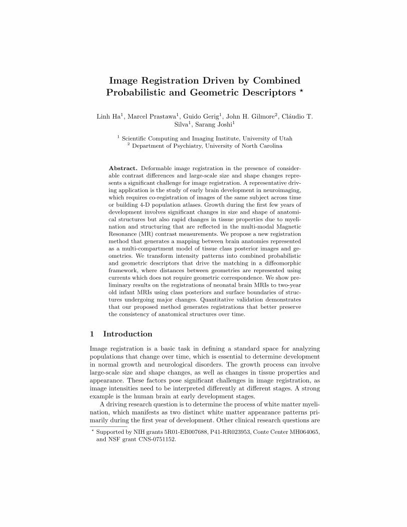
Image Registration Driven by CombinedProbabilistic and Geometric Descriptors ?
Linh Ha1, Marcel Prastawa1, Guido Gerig1, John H. Gilmore2, Claudio T.Silva1, Sarang Joshi1
1 Scientific Computing and Imaging Institute, University of Utah2 Department of Psychiatry, University of North Carolina
Abstract. Deformable image registration in the presence of consider-able contrast differences and large-scale size and shape changes repre-sents a significant challenge for image registration. A representative driv-ing application is the study of early brain development in neuroimaging,which requires co-registration of images of the same subject across timeor building 4-D population atlases. Growth during the first few years ofdevelopment involves significant changes in size and shape of anatomi-cal structures but also rapid changes in tissue properties due to myeli-nation and structuring that are reflected in the multi-modal MagneticResonance (MR) contrast measurements. We propose a new registrationmethod that generates a mapping between brain anatomies representedas a multi-compartment model of tissue class posterior images and ge-ometries. We transform intensity patterns into combined probabilisticand geometric descriptors that drive the matching in a diffeomorphicframework, where distances between geometries are represented usingcurrents which does not require geometric correspondence. We show pre-liminary results on the registrations of neonatal brain MRIs to two-yearold infant MRIs using class posteriors and surface boundaries of struc-tures undergoing major changes. Quantitative validation demonstratesthat our proposed method generates registrations that better preservethe consistency of anatomical structures over time.
1 Introduction
Image registration is a basic task in defining a standard space for analyzingpopulations that change over time, which is essential to determine developmentin normal growth and neurological disorders. The growth process can involvelarge-scale size and shape changes, as well as changes in tissue properties andappearance. These factors pose significant challenges in image registration, asimage intensities need to be interpreted differently at different stages. A strongexample is the human brain at early development stages.
A driving research question is to determine the process of white matter myeli-nation, which manifests as two distinct white matter appearance patterns pri-marily during the first year of development. Other clinical research questions are? Supported by NIH grants 5R01-EB007688, P41-RR023953, Conte Center MH064065,
and NSF grant CNS-0751152.

related to finding a link between cognitive development and the rapid, locallyvarying growth of specific anatomical structures. To approach these questions, arobust registration method is necessary for mapping longitudinal brain MRI toa reference space so that we can perform reliable analysis of the tissue propertychanges reflected in the MR measurements. Knickmeyer et al.[1] showed thatthe total brain volume grows by 100% the first year and 15% the second year,whereas the cerebellum shows 220% volume growth for the first and another15% for the second year, indicating the very different growth rates of differentanatomical structures. Through regression on shape representations, Datar etal.[2] illustrated that the rapid volume changes are also paralleled by signifi-cant shape changes which describe the dynamic pattern of localized, nonlineargrowth. These challenges require a method that does not rely on raw intensitymeasurements, while also being capable of estimating large structural deforma-tions. Xue et al.[3] addressed these issues by proposing a registration scheme forneonatal brains by registering inflated cortical surfaces extracted from the MRI.
We propose a new registration framework for longitudinal brain MRI thatmakes use of the underlying anatomies, which are represented by both class pos-teriors and boundary surfaces. This framework is able to match internal anatom-ical regions and simultaneously preserving a consistent mapping for the bound-aries of relevant anatomical objects. We show results of registering neonatal brainMRI to 2-year old brain MRI of the same subjects obtained in a longitudinal neu-roimaging study. Our method consistently provides transformations that betterpreserve time-varying structures than obtained by intensity-only registration.
2 Registration between Anatomies
Fig. 1. Overview of the proposed registration method that can handle large defor-mations and different contrast properties, applied to mapping brain MRI of neonatesto 2-year olds. We segment the brain MRIs and then extract equivalent anatomicaldescriptors by merging the two different white matter types present in neonates. Theprobabilistic and geometric anatomical descriptors are then used to compute the trans-formation h that minimizes the distance between the class posterior images, as well asthe distance between surfaces represented as currents.

We propose a registration method that makes use of the underlying anatomyin the MR images. Fig. 1 shows an overview of the registration process. We beginby extracting anatomical descriptors from the images, followed by computing atransformation that minimizes the distance between the anatomical descriptors.
2.1 Anatomical Descriptors
We represent brain anatomy as a multi-compartment model of tissue classes andmanifolds. We associate each position x with a vector of tissue probability den-sities. In a given anatomy, we capture the underlying structures by estimating,for each image, the class posterior mass functions associated with each of theclasses. Given Ω as the underlying coordinate system of the brain anatomies,each anatomy Ai=1,··· ,N is represented as
Ai = pi,c=1(x), · · · , pi,c=Nc(x),Mi,j=1(2), · · · ,Mi,j=Ns
(2) ⊂ Ω (1)
where Nc is the number of probability images, Ns is the number of surfaces, pc(x)is the class posterior for tissue c at location x, and Mj(2) are 2-dimensionalsubmanifolds of Ω (surfaces).
The classification of brain MR images with mature white matter structuresinto class posteriors are well studied. We extract the posteriors from 2-year oldbrain MR images using the segmentation method proposed by van Leemput etal.[4]. The method generates posterior probabilities for white matter (wm), graymatter (gm), and cerebrospinal fluid csf. These probabilities can then be usedto generate surfaces from the maximum a posteriori tissue label maps.
The classification of neonatal brain MR images is challenging as the whitematter structure undergoes myelination, where the fibers are being covered inmyelin sheathes. Several have proposed methods that make use of prior informa-tion from an atlas or template that takes into account the special white matterappearance due to myelination [5] [6]. We use the method described by Prastawaet al.[6] for extracting the tissue class posteriors of neonatal brain MRI whichincludes for myelinated wm, non-myelinated wm, gm, and csf. These can thenbe used to create an equivalent anatomy to the 2-year old brain by combiningthe two white matter class probabilities and surfaces.
For the results in this paper, we use the probabilities pwm(x), pgm(x), pcsf (x)and we use the surfaces of white matter, gray matter, and cerebellum. The cere-bellum surfaces are generated from semi-automated segmentations that are ob-tained by affinely registering a template image followed by a supervised level setsegmentation. The cerebellum has a significant role in motor function and it isexplicitly modeled as it undergoes the most rapid volume change during the firstyear of development and thus presents a localized large-scale deformation.
2.2 Registration Formulation
Given two anatomies A1 and A2, the registration problem can be formulated asan estimation problem for the transformation h that minimizes
h = arg minh
E(h · A1,A2)2 +D(h, e)2 (2)
where h · A1 is the transformed anatomy, E(·, ·) is a metric between anatomiesand D(·, e) is a metric on a group of transformations that penalizes deviationsfrom the identity transformation e.
We define distance between anatomies E by defining a norm on an anatomy asa combination of the L2 norm on the class posteriors and a Reproducing KernelHilbert space norm on the manifolds defined as “currents” through Glaunes[7]. The currents norm does not require geometric correspondence and thus canbe used to register manifolds with different resolutions. For an oriented surfaceM(2) in R3 the norm [M(2)] is the vector valued Borel measure correspondingto the collection of unit-normal vectors toM(2), distributed with density equalto the element of surface area ds and can be written as η(x)ds(x), where η(x) isthe unit normal and ds(x) is the surface measure at point x. When M(2) is adiscrete triangular mesh with Nf faces, a good approximation of the norm canbe computed by replacing [M(2)] by a sum of vector-valued Dirac masses:
‖[M(2)]‖2k =Nf∑f=1
Nf∑f ′=1
〈η(f), η(f ′)〉 k(c(f), c(f ′)), (3)
where k(·, ·) is a shift-invariant kernel (e.g., Gaussian or Cauchy), Nf is thenumber of faces of the triangulation, and for any face f , c(f) is its center andη(f) its normal vector with the length capturing the area of each triangle.
Having defined the norm on probability images and surfaces, the dissimilarity
metric between anatomies∥∥∥[A1]− [A2]
∥∥∥2
kis given by:
Nc∑c=1
∫Ω
|p1,c(x)− p2,c(x)|2dx+Ns∑j=1
‖[M1,j(2) ∪ (−M2,j(2))]‖2k (4)
where the distance between two surface currents ‖[M1,j(2)−M2,j(2)]‖k =‖[M1(2) ∪ (−M2(2))]‖k is computed as the norm of the union between surfaceM1(2) and surface M2(2) with negative measures.
We use the large deformation framework [8] that generates dense defor-mation maps in Rd by integrating time-dependent velocity fields. The flowequation is given by ∂hv(t,x)
∂t = v(t, hv(t, x)), with h(0, x) = x, and we defineh(x) := hv(1, x), which is a one-to-one map in Rd (diffeomorphism). We definean energy functional that ensures the regularity of the transformations on thevelocity fields: ‖v(t, ·)‖2V =
∫Rd 〈Lv(t, x), Lv(t, x)〉 dx, where L is a differential
operator acting on vector fields. This energy also defines a distance in the groupof diffeomorphisms:
D2(h, e) = infv,pv(1,·)=h
∫ 1
0
‖Lv(t)‖2V dt. (5)
The registration optimizations in this paper are performed using a greedy ap-proach by iteratively performing gradient descent on velocity fields and updatingthe transformations via an Euler integration of the O.D.E. At each iteration of

the algorithm the velocity field is calculated by solving the p.d.e Lv = F (h)
where F (h) is the variation of∥∥∥[h · A1] − [A2]
∥∥∥2
kwith respect to h. This varia-
tion is a combination of the variation of the L2 norm on the class posteriors andof the currents norm; computed using the gradient ∂‖[M(2)]‖2k
∂xr:
∑f |xr∈f
[∂η(f)∂xr
] Nf∑f ′=1
k(c(f ′), c(f))η(f ′) +23
Nf∑f ′=1
∂k(c(f), c(f ′))∂c(f)
η(f ′)tη(f) (6)
given that points xr, xs, xt form the triangular face f and its center c(f) =xr+xs+xt
3 and its area-weighted normal η(f) = 12 (xs − xr)⊗ (xt − xr).
2.3 Efficient Norm Computation using Particle MeshApproximation on GPU
The major challenge of implementing the currents norm (Eq. 3) for realisticbrain surfaces is the high computational cost to compute the dissimilarity met-ric of all pairs of surface elements, which is O(N2
f ) where Nf is the numberof faces (can be up to millions). For computational tractability, Durrleman etal. [9] used a sparse representation of the surface based on matching pursuitalgorithm. An efficient framework based on the standard fast Gauss transform[10] requires the construction and maintenance of the kd-tree structure on thefly, which is slow on the current CPU model. Our method, however, exploits theParticle-Mesh approximation to reduce the complexity to M logM where M isthe volume size of the embedded grid. The grid size is chosen as the size of theinput images to limit the approximation error to the same order of matchingterm for the class posteriors. This approximation have been extensively studiedin the cosmological N-body simulation literature (see Hockney and Eastwood[11] for details). Particle mesh framework shares the same computational gridwith the class posteriors, moving the computation to the grid level and enablingan efficient parallel computation.
Even with the particle mesh approximation of the norm computation, thetotal complexity of the method is still very high. On a high-end workstationwith 8-CPU cores, a highly optimized multi-threaded implementation in C++takes several hours for one matching pair, hence can not be used for parame-ter exploration and real-time analysis. Fortunately, this computational cost canbe amortized using the massive parallel power and bandwidth provided by thegraphics processing unit (GPU). Based on the GPU framework by Ha et al.[12],we developed an implementation that runs entirely on the GPU. The main bene-fit of a GPU implementation is the ability to exploit parallel efficiency of regulargrid presentation.
Computing the currents u and gradient between a surface with 160535 trian-gular faces and another with 127043 faces takes approximately 504 seconds onan AMD Phenom II X4 955 CPU, while it takes 0.33 seconds on an NVIDIAGTX 260 GPU. The speed gain is in order of three magnitudes over the equiva-lent CPU implementation using particle mesh, while the computing time for the

(a) (b) (c) (d)
Fig. 2. Registration results of neonates mapped to two-year olds. From left to right: (a)neonatal T1 image after affine registration, (b) reference T1 image at 2-years, followedby (c) neonatal T1 after deformable mutual information registration using B-splines,and (d) after combined probabilistic and geometric registration. From top to bottom:subject 0012, 0102, 0106.
exact norm on CPU is difficult to measure since it takes significantly longer. Theproposed algorithm typically converges in 1000 iterations, so on average it takesless than eight minutes to register two anatomies. This allows us to perform pa-rameter exploration and real-time analysis on a single desktop with commodityGPU hardware. The efficiency of the GPU method also provides an opportunityto apply the algorithm for high quality atlas formation using our framework ona GPU cluster, which gives us the ability to perform statistical tests that arepreviously impossible due to excessive time requirements.
3 Results
We have applied the registration method for mapping neonatal MRI scans totwo-year MRI scans of the same subjects in ten datasets. The datasets are takenfrom an ongoing longitudinal neuroimaging study with scans acquired at ap-proximately two weeks, one year, and two years of age. Due to rapid early braindevelopment, each longitudinal MR scan shows significant changes in brain sizeand in tissue properties. For comparison, we also applied the standard inten-sity based deformable registration using mutual information (MI) metric andB-spline transformation proposed by Rueckert et al.[13] which has been appliedfor registering 1-year old and 2-year old infants. The T1 weighted images beforeand after registration using the different approaches for the first three subjectsare shown in Fig. 2.
A quantitative study of the performance of the registration method is per-formed by measuring the overlap between the transformed segmentation mapsof neonates to the segmentation maps of two-year olds. Since we consider the

Subject 0012 0102 0106 0121 0130 0146 0156 0174 0177 0180
White matterMI 0.329 0.155 0.281 0.384 0.379 0.230 0.257 0.300 0.350 0.301
P+G 0.497 0.397 0.442 0.453 0.482 0.414 0.461 0.440 0.478 0.442
CerebellumMI 0.755 0.212 0.588 0.515 0.732 0.820 0.713 0.569 0.631 0.777
P+G 0.881 0.821 0.875 0.878 0.858 0.899 0.907 0.885 0.896 0.892
Fig. 3. Overlap measures comparing the registered segmentation maps against thereference segmentation maps for the white matter and cerebellum structure, obtainedthrough deformable mutual information registration (MI) and our proposed method(P+G).
Subject 0012 0102 0106 0121 0130 0146 0156 0174 0177 0180
MI on CPU 92 63 103 92 101 112 106 99 91 96
P+G on GPU 9 8 8 8 8 7 9 8 7 7
Fig. 4. Time elapsed, in minutes, for registration using deformable mutual informationon CPU (MI) and our proposed approach (P+G) on GPU with 1000 iterations ofgradient descent.
segmentation maps at two years of age to be the standard, we use the followingoverlap metric:
Overlap(h · S0, S2) =|h · S0 ∩ S2||S2|
(7)
where h · S0 is the transformed neonate segmentation map, S2 is the referencetwo-year segmentation map, and | · | indicates the volume of a binary map.Please note that this metric gives considerably lower values than the standardDice coefficient. Fig. 3 shows the quantitative analysis for the white matter andcerebellum segmentation maps. Registration using both probabilistic and geo-metric descriptors provides consistently better results and are generally morestable for the structures of interest. In particular, our method better preservesthe shape of the cerebellum, which has weak intensity boundaries in regionswhere it touches the cerebrum and thus cannot be registered properly usingonly image based information. Another significant challenge is that the cere-bellar growth is distinctly different from the growth of neighboring structures.Registrations using our approach on the GPU takes 8 minutes on average, whileregistration on the CPU using mutual information metric and B-spline transfor-mation takes 100 minutes on average. Detailed time measures are listed in Fig. 4.We have also performed validation using only the probabilistic descriptor, whichgenerates results that are less accurate compared to our method (particularlyfor the cerebellum) while more accurate than image registration using MI.
4 Conclusions
We have proposed a registration framework that makes use of the probabilisticand geometric structures of anatomies embedded in the images. This allows us to

enforce matching of important anatomical features represented as regional classposteriors and tissue boundaries. Our framework allows us to register imageswith different contrast properties by using equivalent anatomical representations,and we have demonstrated results for registering brain MRIs with different whitematter appearances at early stages of growth. The overlap validation measuresin Fig. 3 show that geometric constraints, particularly for the cerebellum, iscrucial for registering structures undergoing significant growth changes. In thefuture, we plan to apply this framework in early neurodevelopmental studiesfor analyzing the effects of neurological disorders such as autism and Fragile Xsyndrome. The proposed registration framework is generic and independent ofthe application domain, it can thus be applied to any registration where oneencounters large-scale deformation and different appearance patterns.
References
1. Knickmeyer, R.C., Gouttard, S., Kang, C., Evans, D., Wilber, K., Smith, J.K.,Hamer, R.M., Lin, W., Gerig, G., Gilmore, J.H.: A structural MRI study of humanbrain development from birth to 2 years. J. Neurosci. 28 (Nov 2008) 12176–12182
2. Datar, M., Cates, J., Fletcher, P., Gouttard, S., Gerig, G., Whitaker, R.: Particlebased shape regression of open surfaces with applications to developmental neu-roimaging. In: MICCAI. Number 5762 in LNCS, Springer Verlag (2009) 167–174
3. Xue, H., Srinivasan, L., Jiang, S., Rutherford, M.A., Edwards, A.D., Rueckert,D., Hajnal, J.V.: Longitudinal cortical registration for developing neonates. In:MICCAI. (2007) 127–135
4. Van Leemput, K., Maes, F., Vandermeulen, D., Suetens, P.: Automated model-based tissue classification of MR images of the brain. IEEE Trans. Medical Imaging18 (October 1999) 897–908
5. Warfield, S.K., Kaus, M., Jolesz, F.A., Kikinis, R.: Adaptive, template moderated,spatially varying statistical classification. MedIA 4(1) (Mar 2000) 43–55
6. Prastawa, M., Gilmore, J.H., Lin, W., Gerig, G.: Automatic segmentation of mrimages of the developing newborn brain. MedIA 9(5) (2005) 457–466
7. Glaunes, J., Trouve, A., Younes, L.: Diffeomorphic matching of distributions: anew approach for unlabelled point-sets and sub-manifolds matching. In: CVPR.(2004)
8. Miller, M, I., Younes, L.: Group action, diffeomorphism and matching: a generalframework. Int. J. Comp. Vis 41 (2001) 61–84
9. Durrleman, S., Pennec, X., Trouve, A., Ayache, N.: Sparse approximations ofcurrents for statistics on curves and surfaces. In: MICCAI 2008. (2008)
10. Greengard, L., Strain, J.: The fast gauss transform. SIAM J. Sci. Stat. Comput.12(1) (1991) 79–94
11. Hockney, R.W., Eastwood, J.W.: Computer Simulation Using Particles. Taylorand Francis (1989)
12. Ha, L., Kruger, J., Fletcher, P.T., Joshi, S., Silva, C.T.: Fast parallel unbiaseddiffeomorphic atlas construction on multi-graphics processing units. In: EGPGV.(2009)
13. Rueckert, D., Sonoda, L., Hayes, C., Hill, D., Leach, M., Hawkes, D.: Nonrigidregistration using free-form deformations. IEEE Trans Med Imag 18(8) (1999)712–721