Endotoxin Exposure and Inflammation Markers Among Agricultural Workers in Colorado and Nebraska
Upload
independentCategory
view
0download
0
Please cite this article as: Prause O, Bossios A, Silverpil E, Ivanov S, Bozinovski S, Vlahos R, Sjöstrand M, Anderson GP, Lindén A, Immunology L, Groups P. IL-17-producing T Lymphocytes in Lung Tissue and in the Bronchoalveolar Space after Exposure to Endotoxin from Escherichia coli in vivo � Effects of Anti-Inflammatory Pharmacotherapy, Pulmonary Pharmacology & Therapeutics (2008), doi: 10.1016/j.pupt.2008.12.005
This is a PDF file of an unedited manuscript that has been accepted for publication. As a service to our customers we are providing this early version of the manuscript. The manuscript will undergo copyediting, typesetting, and review of the resulting proof before it is published in its final form. Please note that during the production process errors may be discovered which could affect the content, and all legal disclaimers that apply to the journal pertain.
Accepted Manuscript
Title: IL-17-producing T Lymphocytes in Lung Tissue and in the Bronchoalveolar Space after Exposure to Endotoxin from Escherichia coli in vivo � Effects of Anti-Inflammatory Pharmacotherapy
Authors: Olof Prause, Apostolos Bossios, Elin Silverpil, Stefan Ivanov, Steven Bozinovski, Ross Vlahos, Margareta Sjöstrand, Gary P. Anderson, Anders Lindén, Lung Immunology, Pharmacology Groups
PII: S1094-5539(08)00133-8DOI: 10.1016/j.pupt.2008.12.005Reference: YPUPT 890
To appear in: Pulmonary Pharmacology & Therapeutics
Received Date: 20 December 2007Revised Date: 20 October 2008Accepted Date: 4 December 2008
peer
-005
3044
0, v
ersi
on 1
- 29
Oct
201
0Author manuscript, published in "Pulmonary Pharmacology & Therapeutics 22, 3 (2009) 199"
DOI : 10.1016/j.pupt.2008.12.005

TPIRCSUNAM DETPECCA
ARTICLE IN PRESS1
IL-17-producing T Lymphocytes in Lung Tissue and in the
Bronchoalveolar Space after Exposure to Endotoxin from Escherichia coli
in vivo – Effects of Anti-Inflammatory Pharmacotherapy
Olof Prause1*, Apostolos Bossios1*, Elin Silverpil1, Stefan Ivanov1, Steven Bozinovski2, Ross
Vlahos2, Margareta Sjöstrand1, Gary P. Anderson2, Anders Lindén1
*) These two authors contributed equally to this study.
1Lung Immunology & Pharmacology Groups,
1) Department of Internal Medicine/Respiratory Medicine and Allergology,
Institute of Medicine, Sahlgrenska Academy at the University of Gothenburg,
Gothenburg, Sweden. 2) Lung Disease Research Group,
Cooperative Research Centre for Chronic Inflammatory Diseases,
Departments of Pharmacology and Medicine, the University of Melbourne,
Parkville, Victoria, Australia.
Short title.
Pharmacology of IL-17-producing T lymphocytes in lungs
Corresponding author.
Professor Anders Lindén, M.D., Ph.D.
Department of Internal Medicine/Respiratory Medicine & Allergology
Bruna Stråket 11B
Sahlgrenska University Hospital
S- 413 45 Gothenburg, Sweden
e-mail. [email protected]
peer
-005
3044
0, v
ersi
on 1
- 29
Oct
201
0

TPIRCSUNAM DETPECCA
ARTICLE IN PRESSPrause et al, 2008
1
Abstract
Interleukin (IL)-17 may play a critical role for the innate immune response in mammals.
However, little is known about its production in T-lymphocytes in comparison with other
cells, in lung tissue and in the bronchoalveolar space in vivo. Even less is known about the
effects of anti-inflammatory pharmacotherapy on this IL-17 production. In this study on
mice we show that one single, intranasal exposure to endotoxin from Escherichia Coli
increases extracellular IL-17 protein in bronchoalveolar (BAL) samples during 3 days,
and is accompanied by a local increase in neutrophils and other inflammatory cells. This
endotoxin exposure also elevates IL-17 mRNA in lung tissue samples. Moreover, after
endotoxin exposure, the absolute number of CD3-positive cells containing intracellular
IL-17 protein is increased as well; from a moderate cell number in lung tissue samples
and from virtually none in BAL samples; with the number in lung tissue exceeding that
observed in BAL samples. Notably, we also demonstrate that among the cells that contain
intracellular IL-17 protein after endotoxin exposure, the percentage of CD3-positive cells
is similar to that of CD3-negative cells in lung tissue. In contrast, CD3-negative cells
dominate among IL-17-containing cells in BAL samples. A high systemic dose of a
glucocorticoid receptor agonist attenuates the endotoxin-induced increase in extracellular
IL-17 protein in BAL samples, IL-17 mRNA in lung tissue samples, and in IL-17-
containing CD3-positive cells in BAL and lung tissue samples. This is also true for the
endotoxin-induced accumulation of neutrophils and other inflammatory BAL cells in vivo.
A systemic dose of a calcineurin-phosphatase inhibitor exerts a less complete and more
selective effect on the endotoxin-induced increase in extracellular IL-17 protein and on
neutrophils in BAL samples. In vitro, endotoxin also increases extracellular IL-17 protein
in a co-culture of CD3-positive spleen cells and adherent mononuclear BAL cells; an
increase that was inhibited by a glucocorticoid as well as by a calcineurin-phosphatase
peer
-005
3044
0, v
ersi
on 1
- 29
Oct
201
0

TPIRCSUNAM DETPECCA
ARTICLE IN PRESSPrause et al, 2008
2
inhibitor In conclusion, endotoxin-induced IL-17 production and release from T
lymphocytes originates from cells that reside in lung tissue and from cells that have been
recruited to the bronchoalveolar space. In both compartments, there is also a substantial
number of cells other than T-lymphocytes that produce IL-17 after endotoxin exposure.
The sustained IL-17 production from T lymphocytes and the associated neutrophil
accumulation may be inhibited non-selectively through glucocorticoid receptor
stimulation and more selectively through calcineurin phosphatase inhibition.
peer
-005
3044
0, v
ersi
on 1
- 29
Oct
201
0

TPIRCSUNAM DETPECCA
ARTICLE IN PRESSPrause et al, 2008
3
Abbreviations: CI, calcium ionophore; COPD, chronic obstructive pulmonary disease;
CsA, cyclosporine A; BAL, bronchoalveolar lavage; ELISA, enzyme-linked
immunosorbent assay; FCS, foetal calf serum; HBSS, Hanks balanced salt solution; IL,
interleukin; i.n., intranasally; i.p., intraperitoneally; LPS, lipopolysaccharide; PBS,
phosphate buffered saline; PMA, phorbol 12-myristate 13-acetate
peer
-005
3044
0, v
ersi
on 1
- 29
Oct
201
0
TPIRCSUNAM DETPECCA
ARTICLE IN PRESSPrause et al, 2008
4
Introduction
The homodimeric cytokine interleukin (IL)-17 (A) is believed to play a critical role for the
innate component of host defence against bacteria in mammalian lungs, through its ability
to indirectly mobilize neutrophils (1-4). Based mainly upon studies on blood cells, it has
recently been suggested that there is a specific type of lymphocytes, a CD4-positive
subset of T helper lymphocytes, named “Th-17”, that is producing IL-17 in mammals and
that this subset is phenotypically and functionally different from the Th-1 and Th-2
subsets (5-8). However, it is also known that there are other subsets of T lymphocytes
capable of producing IL-17 in response to bacterial stimuli in the lungs, presumably in
addition to the “Th-17” subset. These cells include CD4-negative invariant NKT, γδ and
cytotoxic T lymphocytes (8-11).
There may be a pathogenic context for IL-17 per se in human lungs; the local
concentration of IL-17 is increased in patients with inflammatory lung diseases such as
severe asthma and cystic fibrosis as well as in healthy volunteers exposed to organic dust,
in association with a local accumulation of neutrophils (12-15). There is also solid
experimental evidence from mice that the production and release of IL-17 protein from T
lymphocytes is important for endotoxin-induced accumulation of neutrophils in the
bronchoalveolar space in vivo within a certain time frame and that this release of IL-17
protein requires co-stimulation, by antigen presenting cells such as airway macrophages
(1,16,17). However, very little is known about IL-17-producing T lymphocytes in lung
tissue in relation to the bronchoalveolar space as well as about the relative contribution of
T-lymphocytes and other cellular sources of IL-17 in these tissue compartments.
Moreover, given the potentially critical role in pulmonary host defence that has been
attributed to IL-17-producing T lymphocytes recently, surprisingly little is known about
peer
-005
3044
0, v
ersi
on 1
- 29
Oct
201
0

TPIRCSUNAM DETPECCA
ARTICLE IN PRESSPrause et al, 2008
5
the suitable pharmacological strategies for regulating the accumulation and activity of
these lymphocytes in lungs in vivo (4).
In the current study, we utilized a mouse model of gram-negative bacterial lung infection
that is relevant for humans (17,18), to characterize the induced production of IL-17
protein in the collective population of T lymphocytes in lung tissue and in the
bronchoalveolar space in vivo. In this models, we determined the sensitivity of endotoxin-
induced IL-17 production and the associated neutrophil accumulation to anti-
inflammatory pharmacotherapy in vivo; more specifically, to glucocorticoid receptor
stimulation and calcineurin phosphatase inhibition, respectively. In addition, we assessed
the relative contribution of T lymphocytes to the endotoxin-induced IL-17 production by
comparing the percentage of CD3-positive and CD3-negative cells among cells containing
intracellular IL-17 protein in vivo. Finally, we determined the direct effect of the two
principles of anti-inflammatory pharmacotherapy on endotoxin-induced release of IL-17
protein in isolated T lymphocytes co-cultured with macrophage-like cells from the
bronchoalveolar space in vitro.
peer
-005
3044
0, v
ersi
on 1
- 29
Oct
201
0

TPIRCSUNAM DETPECCA
ARTICLE IN PRESSPrause et al, 2008
6
Methods
Animal model
Pathogen-free mice (C57/Bl6, male, 6-8 weeks old, from B&K Universal AB, Stockholm,
Sweden) were kept at the animal facilities of the University of Gothenburg under
conditions which were approved from the Animal Ethics Committee of Göteborg (Diary
number 298/01). The animals received standard laboratory food and water ad libidum.
Time course of endotoxin-induced effect on the concentration of extracellular IL-17
protein and neutrophils in the bronchoalveolar space in vivo
We first characterized the time course of the endotoxin-induced effect on the
concentration of extracellular IL-17 protein and neutrophils in bronchoalveolar lavage
(BAL) samples. To do this, mice were anaesthetized transiently using isofluoran
(Apoteksbolaget, Gothenburg, Sweden) and stimulated with endotoxin from a
pathogenically-relevant, gram-negative species (LPS: 10 μg, E. coli serotype 026:B6 from
Sigma-Aldrich, St. Louis, Missouri, USA, in 50 µl of PBS) in a sub-maximally effective
dose intranasally (i.n.) as described previously (17,18). The animals recovered well from
each period of anaesthesia and did not display any clinical signs of long term side effects.
At 1, 2 and 3 days, respectively, animals were anesthetized using a mixture of ketamine
(670 mg/kg) and xylazin (130 mg/kg) (both from Apoteksbolaget, Gothenburg, Sweden)
intraperitoneally (i.p.) and then euthanized by bleeding of the right ventricle of the heart.
After establishing a tracheotomy, mouse airways were washed with PBS (two times 250
µl) to obtain BAL samples. All the recovered BAL samples from one mouse were pooled
and kept on ice until centrifugation (at 1000 rpm). After centrifugation, the cell-free BAL
fluid was frozen (-80°C) for later analysis of IL-17 protein. The BAL cells were
peer
-005
3044
0, v
ersi
on 1
- 29
Oct
201
0

TPIRCSUNAM DETPECCA
ARTICLE IN PRESSPrause et al, 2008
7
resuspended in PBS containing 0.03% of bovine serum albumin (BSA) and total cell
number was determined.
Differential cell counts were performed on cytospin slides prepared from BAL samples
(Cytospin 3; Shandon Life Sciences, Astmor, UK) using May-Grünwald-Giemsa staining.
All slides were evaluated in a light microscope (Zeiss Axioplan 2; Carl Zeiss AG, Jena,
Germany) at x 100 magnification. We counted 400 cells per sample.
Effect of pharmacotherapy on the concentration of extracellular IL-17 protein and
inflammatory cells in the bronchoalveolar space in vivo
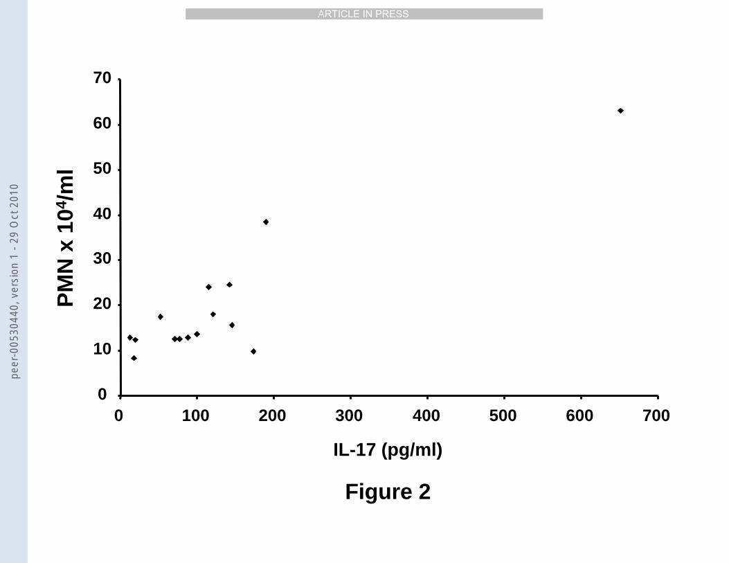
Once the time course experiments were conducted and we had confirmed a correlation
between the endotoxin-induced increase in the concentration of IL-17 protein and the
number of neutrophils in BAL samples (Figure 2), we conducted the corresponding
pharmacology experiments. For these pharmacology experiments, we used exactly the
same protocol as the time course experiments in which the BAL samples were harvested 2
days after endotoxin exposure (described above) but with one exception; one hour before
and 24 hours after intranasal administration of endotoxin (10 µg in 50 µl PBS), the mice
were treated with the glucocorticoid receptor agonist dexamethasone (20 µg/mouse and
200 µg/mouse, respectively, constituting doses of 0.77 – 1.18 mg/kg and 7.69 - 11.76
mg/kg, respectively) (19) or the inhibitor of the endogenous calcium/calmodulin-
dependent phosphatase calcineurin, cyclosporine A (calcineurin phosphatase inhibition:
500 µg/mouse, constituting a dose of 19.23 – 29.41 mg/kg) (20) or vehicle (PBS)
intraperitoneally.
Effect of pharmacotherapy on CD3-positive cells and their intracellular IL-17 protein in
lung tissue and in the bronchoalveolar space in vivo
peer
-005
3044
0, v
ersi
on 1
- 29
Oct
201
0

TPIRCSUNAM DETPECCA
ARTICLE IN PRESSPrause et al, 2008
8
To further address the pharmacology of IL-17-producing T lymphocytes, we conducted
flow cytometry analysis of intracellular expression of IL-17 in CD3-positive cells from
lung tissue and BAL suspension, respectively. This flow cytometry was performed as
described earlier (8). Briefly, BAL was conducted (4 x 250 μL of PBS with protein
secretion blockage buffer 0.5% Golgi Stop; from BD Bioscience, Erembodegem,
Belgium) and the BAL sample was processed as described above to determine cell
numbers. The lungs were then perfused, excised and placed in buffer solution (HBSS with
0.5% Golgi Stop protein secretion blockage). The lungs were mechanically disrupted in a
100-μm nylon mesh cell strainer following by filtration (40 μm) to remove tissue
fragments. Then both BAL and lung cells were incubated with a blocking solution (PBS
with 2% mouse sera; from Dako, Denmark; and 0.5% Golgi Stop) during 15 minutes in
order to block any unspecific binding. The cells were thereafter incubated with a PerCP-
conjugated anti-CD3 antibody (clone 145-2C11; from BD Bioscience) or its isotype-
matched control antibody (30 min. at 4°C), followed by two washings with wash buffer
(PBS with 1% FBS). Cells were then fixed in paraformaldehyde (4%) at room
temperature (10 min.), followed by two more washings (1% FCS/PBS). After re-
suspension in 2 ml of SAP Buffer (0.1% saponin and 0.05% NaN3, w/v in HBSS, from
Sigma-Aldrich), the cells were incubated during 45 min. with a PE-conjugated rat anti-
mouse IL-17 monoclonal antibody (clone TC11-18H10; from BD Bioscience) and its
isotype control, at room temperature, followed by two washes with SAP buffer. Finally,
the cells were washed (1% FCS/PBS) re-suspended in the same buffer, and analyzed
using a FACScan flow cytometer (from BD Bioscience). Ten thousand cells were
computed in a list mode and analyzed using the CellQuest Software (from BD
Bioscience).
Effect of pharmacotherapy on IL-17 mRNA in lung tissue in vivo
peer
-005
3044
0, v
ersi
on 1
- 29
Oct
201
0

TPIRCSUNAM DETPECCA
ARTICLE IN PRESSPrause et al, 2008
9
These pharmacology experiments focused on the impact of glucocorticoid receptor
stimulation, since this pharmacological intervention displayed the most pronounced effect
on extracellular IL-17 protein and neutrophils in BAL samples in vivo. Mice were pre-
treated with dexamethasone (20 µg/mouse and 200 µg/mouse, respectively) one hour
before intranasal exposure to endotoxin (10 µg in 50 µl PBS) (18). At 12 hours, BAL was
performed and lungs were perfused with PBS via the right ventricle of the heart. The
lungs were removed, snap frozen in liquid nitrogen and kept at -80°C until further
processing.
Total RNA was isolated from 15 mg of frozen lung tissue ground to a fine powder under
liquid nitrogen using an RNAeasy kit (Qiagen GmbH, Hilden, Germany) according to the
manufacturer's instructions. The purified total RNA prep was used as a template to
generate first-strand cDNA synthesis using Super Script III (Invitrogen, Carlsbader, CA,
USA) as described previously (8). The reaction mix containing 1 µg of RNA, 250 ng of
random hexamers (Promega, Madison, WI, USA), and 10 mM dNTP mix was diluted to
12 ml in sterile water, heated to 65°C for 5 min and chilled on ice for 1 min. First-strand
synthesis was then performed in a 20 µl total reaction volume by adding 50 mM Tris-HCl
(pH 8.3), 75mM KCl, 3 mM MgCl2, 10 mM DTT, 40 U RNaseout and 200 U Superscript
III reverse transcriptase enzyme at 50°C for 50 min. The reaction was inactivated by
heating at 70°C for 15 min. cDNA was diluted 5-fold in sterile water prior to
amplification. Quantitative real-time PCR was performed as described previously (ABI
PRISM 7900 HT Sequence Detection System) (21) using validated Assays on Demand
Taqman primers for IL-17 from Applied Biosystems-Nordic, Stockholm, Sweden.
Briefly, the gene expression for IL-17 was quantified by multiplexing single reactions,
where our gene of interest was standardized to control (18S rRNA). An individual sample
peer
-005
3044
0, v
ersi
on 1
- 29
Oct
201
0

TPIRCSUNAM DETPECCA
ARTICLE IN PRESSPrause et al, 2008
10
from the control group was then arbitrarily assigned as a calibrator against which all other
samples are expressed as a fold difference.
Effect of pharmacotherapy on the endotoxin-induced increase in extracellular IL-17
protein in vitro
Negatively selected CD3-positive cells from spleens (lymphocyte-like cells as judged by
light microscopy) and adherent mononuclear BAL cells (macrophage-like cells as judged
by light microscopy) were isolated and cultured as described previously (17).
Briefly, naïve C57/Bl6 mice were sacrificed, BAL was conducted (4 x 1.0 ml of PBS) and
the spleen was surgically removed in each mouse. Adherent mononuclear cells in the
BAL sample (macrophage-like cells as judged by light microscopy) were washed with
PBS and seeded out (5 x 10⁴ per well) on a 96-well culture plate (model 3072; from BD
Bioscience) in cell culture medium (RPMI 1640; from Sigma-Aldrich). Cells were then
incubated during 3 hours at 37°C and washed two times to remove non-adherent, non-
macrophage cells.
For the enrichment of CD3-positive cells, mouse spleens were minced, red blood cells
lysed using a hypotonic solution and remaining cells were then filtrated through a 40 µm
cell strainer to obtain a single cell solution. Cells were washed (PBS with 0.5% BSA) and
CD3-negative cells were magnetically labelled using a biotin-conjugated antibody
cocktail (Pan T-cell isolation kit, Miltenyi Biotech, Bergisch Gladbach, Germany)
followed by incubation with streptavidin-coupled microbeads (Miltenyi Biotech). The
cells were then washed and passed through a column (Miltenyi Biotech) in a magnetic
field. Labelled CD3-negative cells attached to the column while unlabelled CD3-positive
cells were eluated and seeded (0.5 x 10⁶ cells/ml) in complete media (RPMI 1640 with
FCS 10%, L-glutamine 2 mM, sodium pyruvate 1% and penicillin-streptomycin 100 U/ml
peer
-005
3044
0, v
ersi
on 1
- 29
Oct
201
0

TPIRCSUNAM DETPECCA
ARTICLE IN PRESSPrause et al, 2008
11
and 100 µg/ml; all from Sigma-Aldrich) together with the adherent mononuclear BAL
cells. The mean purity of the CD3-positive cells after magnetic separation was 88% (n=4).
After enrichment, the co-culture of cells were pre-treated with either the calcineurin
phosphatase inhibitor, cyclosporine A (10-6M) (20) or the glucocorticoid receptor agonist,
hydrocortisone (10-6M) (22) or vehicle only. Ethanol was used as solvent for these
chemicals; the final concentration of ethanol did not exceed 0.1% and was used in the
same concentration in the negative and positive control as well. Hydrocortisone was
chosen as glucocorticoid receptor agonist, because it shows a better water-solubility than
dexamethasone. Thirty minutes after pre-treatment, cells were stimulated with endotoxin
(LPS 100 ng/ml), positive control (calcium ionophore A 23487 [CI] 1 µg/ml) plus phorbol
12-myristate 13-acetate [PMA] 2 ng/ml)) or negative control (RPMI 1640 only) and
incubated (at 37°C with 5% CO2) during 20 hours, after which the conditioned cell
medium was harvested. The cell medium was then centrifuged to remove cells and it was
subsequently frozen (-80°C) for later analysis of IL-17 protein.
Measurement of cytokines
The concentration of free, soluble mouse IL-17 protein in cell-free BAL samples or
conditioned media from cell cultures was determined using a commercially available
enzyme-linked immunosorbent assay (ELISA) kit (from R&D Systems). A concentration
below the lowest detectable value of the ELISA standard curve (5.4 pg/ml) was assigned
the mean value of the lowest concentration of the ELISA standard curve and zero (i.e. 2.7
pg/ml).
Statistical analysis
A correlation analysis of certain data was conducted utilizing Spearman rank correlation
test. The Mann-Whitney U-test (preceded by the Kruskal Wallis test for multiple
peer
-005
3044
0, v
ersi
on 1
- 29
Oct
201
0

TPIRCSUNAM DETPECCA
ARTICLE IN PRESSPrause et al, 2008
12
comparisons) was utilised for comparison of data. Data are presented as mean ± standard
error of the mean (SEM). P-values ≤ 0.05 were considered significant. n refers to the
number of independent experiments for each treatment group.
peer
-005
3044
0, v
ersi
on 1
- 29
Oct
201
0

TPIRCSUNAM DETPECCA
ARTICLE IN PRESSPrause et al, 2008
13
Results
Time course of endotoxin-induced effect on the concentration of extracellular IL-17
protein and neutrophils in the bronchoalveolar space in vivo
Intranasal stimulation with endotoxin (10 µg) substantially increased the concentration of
free, soluble IL-17 protein in the extracellular compartment of BAL samples (Figure 1)
and it also increased the number of neutrophils and other inflammatory cells in the BAL
sample (Table 1). The IL-17 concentration peaked at day 1 and gradually decreased over
day 2 and 3 in a time-dependent manner (Figure 1). There was no detectable IL-17 protein
in mice exposed to the vehicle of endotoxin at either time point (Figure 1). At the time
point 2 days after stimulation, we found a statistically significant correlation between the
concentration of IL-17 and the number of neutrophils in BAL samples (rho: 0.63, p<0.05,
n=11; Figure 2). This correlation proved statistically significant, even when the most
extreme “outlier observation” was excluded (Figure 2 & data not shown).
Table 1.
Negative Control LPS Cell type 1 day 2 days 3 days 1 day 2 days 3 days
Macrophages 2.16 ± 0.35
2.83 ± 0.27
2.65 ± 0.93
2.89 ± 0.38
3.63 ± 0.47
5.52 ± 1.02
Lymphocytes 0.01 ± 0.01
0.01 ± 0.01
0.00 ± 0.00
0.20 ± 0.04
0.59 ± 0.09
1.22 ± 0.24
Neutrophils 0.03 ± 0.02
0.00 ± 0.00
0.01 ± 0.01
18.27 ± 1.53
12.82 ± 0.82
5.07 ± 1.09
Legend to table 1. Effect of local exposure to endotoxin (LPS; 10 µg i.n.) on the number
of macrophages, lymphocytes and neutrophils (x 104/ml) in BAL samples from mice at 1,
2 and 3 days, compared to negative control (PBS). Lymphocyte and neutrophil numbers
peer
-005
3044
0, v
ersi
on 1
- 29
Oct
201
0

TPIRCSUNAM DETPECCA
ARTICLE IN PRESSPrause et al, 2008
14
were increased after treatment with endotoxin (p<0.05); macrophage numbers showed a
tendency towards an increase, most pronounced at 3 days. Data presented as mean ±
SEM (n=4-11).
Effect of pharmacotherapy on the concentration of extracellular IL-17 protein and
inflammatory cells in the bronchoalveolar space in vivo
Pre-treatment with the high dose (200 µg) of dexamethasone attenuated the endotoxin-
induced increase in free, soluble IL-17 protein in the extracellular compartment of BAL
samples (Figure 3). In contrast, pre-treatment with the moderate dose (20 µg) of
dexamethasone only partially reduced the endotoxin-induced increase in concentration of
IL-17 protein in BAL samples, even though this inhibitory effect was statistically
significant as well (Figure 3).
Pre-treatment with cyclosporine A incompletely reduced the endotoxin-induced increase
concentration of IL-17 protein in BAL samples, even though this effect was statistically
significant (Figure 3). As opposed to the general inhibitory effect on the endotoxin-
induced increase in all BAL cells that was exerted by the high dose of the glucocorticoid,
cyclosporine A exerted an incomplete but clear decreasing effect that was selective and
statistically significant for BAL neutrophils (Table 2).
Table 2.
PBS i.n.
LPS i.n.
Cell type
PBS i.p. PBS i.p. Dex 20 µg
i.p. Dex 200 µg
i.p. CsA i.p.
Macrophages 3.57 ± 0.59 4.64 ±
0.34 4.36 ±
0.38 2.45
± 0.25 4.33 ± 0.51
Lymphocytes 0.03 ± 1.72 ± 1.60 ± 0.28 ± 1.49 ±
peer
-005
3044
0, v
ersi
on 1
- 29
Oct
201
0

TPIRCSUNAM DETPECCA
ARTICLE IN PRESSPrause et al, 2008
15
0.00 0.20 0.24 0.26 0.30
Neutrophils 0.04 ± 0.03 17.68 ±
1.65 19.72 ±
3.58 0.97 ± 0.52
10.31 ± 2.01
Legend to table 2. Effect of treatment with dexamethasone (Dex, 20 µg/mouse and 200
µg/mouse, respectively) and cyclosporine A (CsA, 500 µg/mouse), respectively, on cell
numbers in BAL samples harvested after endotoxin exposure in mice. The drugs were
injected intraperitoneally (i.p.) one hour before and one day after intranasal stimulation
with 10 µg endotoxin and numbers of macrophages, lymphocytes and neutrophils (mean ±
sem cell number x 104/ml) were determined two days after endotoxin exposure. Data
presented as mean ± SEM (n=6-20).
Treatment with the high dose of dexamethasone (200 µg/mouse) substantially decreased
the endotoxin-induced increase in macrophages, lymphocytes and neutrophils in BAL
samples and these effects were statistically significant. In contrast, treatment with
cyclosporine A (CsA) selectively and in a statistically significant manner decreased the
numbers of BAL neutrophils only. The low dose of dexamethasone (20 µg/mouse) caused
no substantial effect on either cell number in BAL samples.
Effect of pharmacotherapy on CD3-positive cells and their intracellular IL-17 protein in
lung tissue and in the bronchoalveolar space in vivo
Intranasal endotoxin exposure increased the total number of inflammatory cells in lung
tissue samples, compared with the negative control (47.2 ± 22 vs. 22.3 ± 5.1 x 10⁶,
p<0.05) and this increase was fully attenuated in a statistically significant manner by the
high (200 μg: 23.6± 3.6 x 10⁶ p<0.05 vs. LPS) but not the moderate (20 μg) dose of
dexamethasone or cyclosporine A (data not shown). There was no substantial effect of
either treatment on the total number of CD3-positive cells in lung tissue samples (Figure
peer
-005
3044
0, v
ersi
on 1
- 29
Oct
201
0

TPIRCSUNAM DETPECCA
ARTICLE IN PRESSPrause et al, 2008
16
4A). Interestingly, endotoxin exposure also increased the number of IL-17-containing
CD3-positive cells in this compartment, from a reproducibly low to a substantially higher
level, and the high dose of dexamethasone (200 μg) attenuated this increase in a
statistically-significant manner (mean data in Figure 4B and data from one representative
FACS analysis in Figure 4E). The moderate dose of dexamethasone and the one dose of
cyclosporine A exerted only weak inhibitory effects; effects that did not prove statistically
significant (data not shown).
Similar to the case for lung tissue, intranasal endotoxin exposure increased the total
number of BAL cells compared to control (29.2 ± 22 vs. 363.3 ± 9.3 x 10³ cells, p<0.05).
The high dose of dexamethasone (200 μg:114.5 ± 15.2 x 10³ cells) as well as the one dose
of cyclosporine A (243.3 ± 41.6 x 10³ cells) partially inhibited (p<0.05 compared with
LPS for both) this increase in BAL cells. The moderate dose of dexamethasone did not
produce any corresponding, statistically significant effect (data not shown). Noteworthy
and in contrast to the case in lung tissue, intranasal endotoxin exposure increased both the
total number of CD3-positive cells and the number of IL-17-expressing CD3-positive
cells in BAL samples, from virtually no to a substantially higher number in both cases and
these effects proved statistically significant (Fig 4 C & D). The high dose (200 μg) of
dexamethasone totally attenuated these responses to endotoxin, again in a statistically-
significant manner (Figure 4 C & D). The moderate dose of dexamethasone and the one
dose of cyclosporine A did not produce any corresponding, statistically significant effect.
Percentage of CD3-positive and -negative cells among cells containing intracellular IL-
17 protein after endotoxin exposure, in lung tissue and in the bronchoalveolar space in
vivo.
peer
-005
3044
0, v
ersi
on 1
- 29
Oct
201
0

TPIRCSUNAM DETPECCA
ARTICLE IN PRESSPrause et al, 2008
17
In lung tissue, the percentage of CD3-positive cells (57.5 ± 7.6 %) was similar to that of
CD3-negative cells (43.3 ± 8.0 %) among all IL-17-containing cells after endotoxin
exposure (p=0.66, n=3). However, in this compartment, the relative mean fluorescence
intensity (rMFI, equals signal for monoclonal antibody/signal for isotype control
antibody) was higher for CD3-positive (3.0 ± 0.2) compared with CD3-negative (2.4 ±
0.2) IL-17-containing cells and this difference was statistically significant (p<0.05, n=3).
In BAL cells, however, the percentage of CD3-positive cells (24.2 ± 5.9 %) was smaller
than that of CD3-negative cells (75.8 ± 5.9 %) among all IL-17-containing, cells after
endotoxin exposure (p<0.05, n=3). In analogy, in this compartment, the rMFI was
substantially smaller for CD3-positive (3.9 ± 0.2) compared with CD3-negative (18.1 ±
0.4) IL-17-containing cells and this difference was statistically significant (p<0.05, n=3).
Effect of pharmacotherapy on IL-17 mRNA in lung tissue in vivo
Pre-treatment with the high dose (200 µg) of dexamethasone totally attenuated the
endotoxin-induced increase in mRNA for IL-17 assessed in lung tissue samples harvested
12 hours after exposure and this impact was statistically significant (Figure 5). In contrast,
the moderate dose (20 µg) of dexamethasone did not fully attenuate the corresponding
increase in mRNA, even though the effect was statistically significant compared with the
negative control (p<0.05, n=3-4) (Figure 5).
Effect of pharmacotherapy on the endotoxin-induced increase in extracellular IL-17
protein in vitro
Endotoxin stimulation caused a 7.9 ± 1.8 fold increase of the concentration of free,
soluble IL-17 protein that was detected in the conditioned medium from the co-cultured
CD3-positive spleen cells and adherent mononuclear BAL cells, compared to the negative
control group (21.8 ± 4.5 pg/mL) (Figure 6). Treatment with hydrocortisone almost
peer
-005
3044
0, v
ersi
on 1
- 29
Oct
201
0

TPIRCSUNAM DETPECCA
ARTICLE IN PRESSPrause et al, 2008
18
completely attenuated the endotoxin-induced increase (% of positive control) in IL-17
protein (down to 7 ± 3 %), whereas cyclosporine A only partially reduced it (36 ± 10%)
(p<0.05, n=4).
peer
-005
3044
0, v
ersi
on 1
- 29
Oct
201
0

TPIRCSUNAM DETPECCA
ARTICLE IN PRESSPrause et al, 2008
19
Discussion
We found that local exposure to endotoxin from the human lung pathogen Escherichia Coli
(18) causes a sustained increase in extracellular IL-17 protein in BAL samples that lasts at
least 3 days. The time course of the concentration of extracellular IL-17 is paralleled by a
corresponding time course for neutrophil accumulation in BAL samples and there is a strong
correlation between these two parameters 2 days after endotoxin exposure. We also found
that, within the same time frame after endotoxin exposure, mRNA for IL-17 in lung tissue is
increased, just like the number of CD3-positive cells containing intracellular IL-17 protein
among BAL cells and in lung tissue. The endotoxin-induced in crease in IL-17-containing
CD3-positive cells is from a moderate number in lung tissue samples and from virtually none
in BAL samples; with the number in lung tissue clearly exceeding that observed in BAL
samples. In addition, we observed that among the cells that contain intracellular IL-17 protein
after endotoxin exposure, the percentage of CD3-positive cells is similar to that of CD3-
negative cells in lung tissue whereas the percentage of CD3-negative cells exceeds that of
CD3-positive cells among IL-17-contaning cells in BAL samples. Interestingly, the rMFI, the
estimated strength of this signal for intracellular IL-17 protein, was strongest for CD3-positive
cells in the lungs and for CD3-negative cells in BAL samples. Moreover, endotoxin exposure
causes a marked increase of extracellular IL-17 protein in co-culture of CD3-positive spleen
cells and adherent mononuclear BAL cells.
Most previous studies on IL-17 and neutrophils in the lungs have focused on T-lymphocytes
as the cellular origin of IL-17 and studies demonstrating an attenuating effect of anti-IL-17-
antibodies or the absence of the IL-17 receptor or gene have supported this concept (1-4).
However, there is one previous report suggesting that mouse airway neutrophils can produce
and release IL-17 protein during certain circumstances (16). In view of these previous
findings, the interpretation of our currently demonstrated correlation between IL-17 protein
peer
-005
3044
0, v
ersi
on 1
- 29
Oct
201
0

TPIRCSUNAM DETPECCA
ARTICLE IN PRESSPrause et al, 2008
20
and the number of BAL neutrophils 2 days after endotoxin exposure is not entirely trivial: We
cannot rule out that neutrophils contribute to IL-17 production in the bronchoalveolar space,
given that we found that there may be more CD3-negative than CD3-positive IL-17-
containing cells in BAL samples after endotoxin exposure, and that these CD3-negative cells
display a stronger signal for IL-17 than the CD3-positive cells do. In contrast, in the lung
tissue, we found approximately as many CD3-positive as CD3-negative IL-17-containing
cells after endotoxin exposure, whereas the CD3-positive cells accounted for the strongest IL-
17 signal in this compartment. We interpret these findings as the collective population of T-
lymphocytes constituting one substantial source of IL-17 protein in the lungs and that there
are other cells as well, possibly neutrophils, that can contribute to this IL-17 production as
part of the innate response to a gram-negative bacterial pathogen (1-4,16, 17). The relative
contribution of each respective cell population to IL-17 production may be somewhat
different for the two compartments that we investigated in the current study; the
bronchoalveolar space and the lung tissue. The possibility that neutrophils within the
bronchoalveolar space constitute a substantial source of IL-17 protein after exposure to gram-
negative bacteria, in addition to T lymphocytes, is indeed intriguing. Even though it is beyond
the scope of our current study, this possibility warrants further investigation in future studies
(1-4,16, 17).
Because we studied all CD3-positive cells, and not just the CD4-positive subpopulation, our
results reflect the response by the collective population of IL-17 producing T lymphocytes in
mouse lungs, including CD4-positive Th17 lymphocytes as well as CD4-negative invariant
NK T, γδ and cytotoxic T lymphocytes (8-11). We believe that this is functionally most
interesting when evaluating the biology of IL-17, in particular since there is currently no
convincing functional data published on IL-17-producing cells other that T lymphocytes in
humans (4). Moreover, when specifically targeting all the T lymphocytes in BAL samples
with flow cytometry, we found that endotoxin exposure increases the total number of CD3-
peer
-005
3044
0, v
ersi
on 1
- 29
Oct
201
0

TPIRCSUNAM DETPECCA
ARTICLE IN PRESSPrause et al, 2008
21
positive cells in this compartment, from virtually none to a substantial number, as well as the
number of IL-17-containing CD3-positive cells. These findings thus forward the possibility of
both recruitment and activation of IL-17-producing T lymphocytes in the bronchoalveolar
cells playing a role in the early innate immune response in mammalian lungs.
Even though the bronchoalveolar space represents the primary target site after local endotoxin
exposure, the IL-17 protein that is released by endotoxin could hypothetically originate from
the surrounding tissue. We therefore analysed the total number and the number of IL-17-
containing CD3-positive cells in lung tissue as well. Notably, endotoxin exposure did not
exert any substantial effect on the total number of CD3-positive cells in the lung tissue
samples, in contrast to what was the case in the BAL samples. However, endotoxin exposure
did increase the number of IL-17-containing CD3-positive cells in lung tissue samples, just
like in the BAL samples, and after endotoxin exposure, the absolute number of IL-17-
containing CD3-positive cells in the lung tissue samples by far exceeded the corresponding
number in the BAL samples. Moreover, in lung tissue samples from naïve control mice that
were not exposed to endotoxin, we repeatedly detected a population of IL-17-containing CD3-
positive cells, whereas this was not the case for the BAL samples. We speculate that these
findings illustrate that a small population of IL-17-producing T lymphocytes always reside in
the mammalian lung tissue, ready to immediately respond to emerging gram-negative
bacterial pathogens, thereby constituting a functionally critical component of local host
defence (21,24,25). Mechanistically, this possibility may explain why neutralisation of
endogenous IL-17 protein can be so powerful in attenuating endotoxin-induced neutrophil
accumulation in the bronchoalveolar space, as previously demonstrated, even though the
bronchoalveolar space may contain relatively few IL-17-producing T lymphocytes (1,2,4,16).
When assessing whether targeting glucocorticoid receptors constitute a feasible approach for
pharmacologically regulating the accumulation and activity of IL-17-producing T
peer
-005
3044
0, v
ersi
on 1
- 29
Oct
201
0

TPIRCSUNAM DETPECCA
ARTICLE IN PRESSPrause et al, 2008
22
lymphocytes in lungs in vivo, we did obtain evidence that this is the case but only if a high
dose of the agonist is utilized. Thus, we found that a high, but not a moderate, systemic dose
of the glucocorticoid receptor agonist dexamethasone effectively reduces the endotoxin-
induced increase in extracellular IL-17 protein in BAL samples, IL-17 mRNA in lung tissue
samples, total CD3-positive cells as well as IL-17-containing CD3-positive cells in BAL and
lung tissue samples. The same is true for the endotoxin-induced, IL-17 associated increase in
neutrophils and other inflammatory cells in BAL samples. In addition, we observed a
complete attenuating effect exerted by the glucocorticoid receptor agonist hydrocortisone on
the endotoxin-induced release of extracellular IL-17 protein in a co-culture of spleen CD3-
positive cells co-cultured with adherent mononuclear BAL cells in vitro.
Collectively, our observations on the impact of dexamethasone in vivo and hydrocortisone in
vitro are compatible with a high systemic dose of the glucocorticoid directly targeting the
endotoxin-induced activity in IL-17-producing T lymphocytes in the lungs, as well as the
recruitment in to the bronchoalveolar space of all inflammatory cells including IL-17-
producing T lymphocytes. Given the fact that previous in vivo studies have shown that
dexamethasone inhibits delayed type hypersensitivity, which depends upon the activity of T
lymphocytes, as well as olive oil-induced inflammation, which depends upon neutrophil
activity, it is possible that systemic glucocorticoid receptor stimulation inhibits endotoxin-
induced neutrophil-accumulation at many different levels simultaneously, including inhibition
of T lymphocytes of the memory helper subset (26,27). In line with an effect “down-stream”
of IL-17, we have previously shown that glucocorticoid receptor stimulation results in
inhibition of the C-X-C chemokine release that occurs in response to IL-17 in the lungs, in
human bronchial epithelial cells and in mice in vivo (28,29). However, it remains unclear to
what extent glucocorticoid receptor stimulation can more specifically inhibit the specific
signalling pathway “up-stream” of the IL-17-producing T lymphocytes, including event
involving T lymphocytes of the memory helper subset, even though it is known that endotoxin
peer
-005
3044
0, v
ersi
on 1
- 29
Oct
201
0

TPIRCSUNAM DETPECCA
ARTICLE IN PRESSPrause et al, 2008
23
acts via toll-like receptor (TLR)-4 on antigen-presenting cells and via the subsequent
production and release of IL-23, a specific activator of IL-17-producing T lymphocytes in the
lungs (1,2,3,4,8,23,24).
We believe that our current findings may have clinical implications, since it is traditionally
known that the innate immune response is relatively glucocorticoid-insensitive (25). This is
because IL-17-producing T lymphocytes may hypothetically constitute a link between a
“glucocorticoid-insensitive” innate immunity and a “glucocorticoid-sensitive” adaptive
immunity. Our findings that systemic glucocorticoid receptor stimulation bears the potential
to completely attenuate the endotoxin-induced production and release of IL-17 protein in the
lungs in vivo, as well as the associated neutrophil accumulation, is potentially problematic
and promising at the same time. On one hand, our findings implicate that the early,
neutrophilic component of host defence in the lungs may actually be impaired by systemic
stimulation of glucocorticoid receptors, if a really high dose of the glucocorticoid receptor
agonist is utilised. On the other hand, a very high systemic dose of the particular agonist may
be required, and useful, to down-regulate an excess activation of the neutrophilic component
of host defence in mammalian lungs.
In contrast to the case for glucocorticoid receptor stimulation, we found that calcineurin
phosphatase inhibition by cyclosporine A exerts a more selective and incomplete effect on the
endotoxin-induced increase in extracellular IL-17 protein and on neutrophils in BAL samples,
in the one dose given. In line with this incomplete inhibition, the utilised calcineurin
phosphatase inhibitor caused a weak effect on IL-17-containing CD3-positive cells in lung
tissue and in BAL samples; an effect that did prove to be statistically significant. Finally, the
incomplete but statistically significant inhibitory effect of the calcineurin phosphatase
inhibitor on IL-17 release was confirmed in the co-culture of endotoxin-stimulated CD3-
positive spleen cells and adherent mononuclear BAL cells.
peer
-005
3044
0, v
ersi
on 1
- 29
Oct
201
0

TPIRCSUNAM DETPECCA
ARTICLE IN PRESSPrause et al, 2008
24
Even though the current study is the first one conducted in a lung context, two previous
studies on isolated cells have demonstrated that the calcineurin phosphatase inhibitor
cyclosporine A does inhibit induced IL-17 production in human CD4-positive cells (30,31) In
these in vitro studies on cells harvested from blood, the IL-17 production was induced by
either IL-15 or by calcium ionophore plus PMA. Moreover, in another previous in vitro study
on mouse lymphocytes from spleens and lymph nodes, it was demonstrated that induced
production of IL-17 is sensitive to cyclosporine A as well; in this particular in vitro study, IL-
17 production was induced by anti-CD3 antibodies, yet another artificial stimulus (32). To
these previous observations, we now add our in vivo and in vitro data suggesting that
cyclosporine A exerts a substantial but incomplete inhibitory effect on the innate immune
response to a gram-negative bacterial stimulus in the lungs by directly acting on and
inhibiting the IL-17 release from T lymphocytes recruited to the bronchoalveolar space and
residing in lung tissue, as well as the associated accumulation of neutrophils. This means that
targeting endogenous calcineurin phosphatase may constitute a pharmacotherapeutic strategy
by which an excess activation of the early neutrophil component of host defence in the lungs
can be down-regulated, in a relatively selective manner without totally attenuating this critical
response (2,4,16,17).
In conclusion, this experimental study on a mouse model indicates that a component from a
bacterial pathogen that can cause pneumonia in humans (18) per se does induce sustained IL-
17 production and release from T lymphocytes that reside in lung tissue and are recruited to
the bronchoalveolar space in vivo. In addition, there is a population of cells other than T
lymphocytes that is likely to contribute to IL-17 production in lung tissue and in the
bronchoalveolar space. The endotoxin-induced IL-17 production, and the associated
neutrophil accumulation in vivo, can be inhibited by anti-inflammatory pharmacotherapy.
Non-selective inhibition can be achieved through systemic stimulation of glucocorticoid
receptors, if a high dose of the agonist is utilised. More selective, but less complete inhibition
peer
-005
3044
0, v
ersi
on 1
- 29
Oct
201
0

TPIRCSUNAM DETPECCA
ARTICLE IN PRESSPrause et al, 2008
25
can be achieved through systemic inhibition of endogenous calcineurin phosphatase. Due to
their IL-17-inhibiting capacity in the lungs in vivo and in isolated T lymphocytes in vitro, both
these approaches deserve a more in-depth clinical evaluation in neutrophilic lung disorders
such as acute lung allograft rejection, ARDS, severe asthma, COPD and cystic fibrosis;
disorders where moderate doses of glucocorticoid receptor agonists seem insufficient as anti-
inflammatory treatment (2,4,33-37).
peer
-005
3044
0, v
ersi
on 1
- 29
Oct
201
0

TPIRCSUNAM DETPECCA
ARTICLE IN PRESSPrause et al, 2008
26
Acknowledgements
Authors O.P. and A.B. contributed equally to this work. The study was financially supported
by the University of Gothenburg as well as the Heart-Lung Foundation and the Science
Council (K2005-74X-09048-16A and K2008-57X-09048-19-3) in Sweden. A.B. received
financial support from a grant from “EMPIRIKION foundation”, Athens, Greece. No support,
either direct or indirect, was obtained from the tobacco industry.
peer
-005
3044
0, v
ersi
on 1
- 29
Oct
201
0

TPIRCSUNAM DETPECCA
ARTICLE IN PRESSPrause et al, 2008
27
Legends
Figure 1. Concentration of free, soluble interleukin (IL)-17 protein in the extracellular
compartment of mouse BAL samples at 1, 2 and 3 days, respectively, after exposure to
endotoxin (10 µg intranasally, i.n., white columns) compared to negative control (PBS,
black columns) (p<0.05, n=3-9). The concentration of IL-17 correlated negatively to
time after endotoxin exposure (rho: -0.7; p<0.01, n=10-11). The detection limit for IL-
17 was 5.4 pg/ml. Data presented as mean ± SEM. (n=4-11).
Figure 2. Concentration of free, soluble IL-17 protein in the extracellular compartment
versus number of neutrophils in mouse BAL samples 2 days after exposure to endotoxin
(10 µg i.n.) Data presented as mean ± SEM (rho: 0.63; p<0.05, n=15).
Figure 3. Effect of pre-treatment (i.p.) with the glucocorticoid receptor agonist
dexamethasone (Dex, 20 µg and 200 µg, respectively) and the calcineurin-phosphatase
inhibitor cyclosporine A (CsA, 500 µg) on the concentration of free, soluble IL-17
protein in the extracellular compartment of mouse BAL samples harvested 2 days after
exposure to endotoxin (10 µg). In all samples from negative controls, the concentration
of IL-17 protein was below the detection limit. Data presented as mean ± SEM
(*=p<0.05; n=6-20).
Figure 4. Effect of pre-treatment (i.p.) with dexamethasone (Dex, 200 or 200 µg) and
the calcineurin phosphatase inhibitor, cyclosporine A (CsA, 500 µg) on the total number
of CD3-positive cells and on IL-17-containing CD3-positive cells in mouse lung tissue
(A,B) and BAL samples (C,D) harvested two days after exposure to endotoxin (10 µg)
compared to negative and positive control. Data presented as mean ± SEM (*= p≤0.05;
peer
-005
3044
0, v
ersi
on 1
- 29
Oct
201
0

TPIRCSUNAM DETPECCA
ARTICLE IN PRESSPrause et al, 2008
28
n=3). E) Representative lungs FACS plots from animals treated with PBS, endotoxin or
endotoxin and a high dose of dexamethasone (200 µg/mouse). Lungs were removed
from animals, disrupted mechanically and stained for CD3/IL-17 PE or with isotype
control antibodies. In all plots, gating was done in the CD3-positive cell population. The
numbers in the boxes represent the percentage of gated CD3-positive cells. The left
column represent CD3-positive cells stained with isotype control (IgG1 PE) and right
column represent CD3-positive cells stained with IL-17 PE.
Figure 5. Relative expression (%18S rRNA) of mRNA for IL-17 in mouse lung tissue
samples after intranasal exposure to endotoxin only (10 µg, white column) as well as
with intraperitoneal (i.p.) pre-treatment with dexamethasone (striped column: 20 µg of
dexamethasone; black column: 200 µg of dexamethasone). Data presented as mean ±
SEM (*= p≤0.05; n=4).
Figure 6. Effect of pre-treatment with the glucocorticoid receptor agonist
hydrocortisone (HC, 10-6M) and cyclosporine A (CsA, 10-6M) on the concentration of
free, soluble IL-17 protein in conditioned medium from the extracellular compartment
of a co-culture of CD3-positive spleen cells and adherent mononuclear BAL cells from
mice, after stimulation with endotoxin (LPS, 100 ng/ml) in vitro. Data presented as
mean ± SEM (n=4).
peer
-005
3044
0, v
ersi
on 1
- 29
Oct
201
0

TPIRCSUNAM DETPECCA
ARTICLE IN PRESSPrause et al, 2008
29
References
1 Kolls, J. K. and Linden, A. 2004. Interleukin-17 family members and inflammation.
Immunity 21:467-76.
2 Linden, A., Laan, M., and Anderson, G. P. 2005. Neutrophils, interleukin-17A and
lung disease. Eur Respir J 25:159-72.
3 Linden, A. 2007. A role for the cytoplasmic adaptor protein Act1 in mediating IL-17
signaling. Sci STKE 2007:re4.
4 Ivanov S, Lindén A. Th-17 cells in the lungs? Expert Review of Respiratory Medicine.
2007; vol 1 (2): p 279-293.
5 Harrington, L. E., Hatton, R. D., Mangan, P. R., Turner, H., Murphy, T. L., Murphy,
K. M., and Weaver, C. T. 2005. Interleukin 17-producing CD4+ effector T cells
develop via a lineage distinct from the T helper type 1 and 2 lineages. Nat Immunol
6:1123-32.
6 Nakae, S., Iwakura, Y., Suto, H., and Galli, S. J. 2007. Phenotypic differences
between Th1 and Th17 cells and negative regulation of Th1 cell differentiation by IL-
17. J Leukoc Biol.
7 Nishihara, M., Ogura, H., Ueda, N., Tsuruoka, M., Kitabayashi, C., Tsuji, F., Aono,
H., Ishihara, K., Huseby, E., Betz, U. A., Murakami, M., and Hirano, T. 2007. IL-6-
gp130-STAT3 in T cells directs the development of IL-17+ Th with a minimum effect
on that of Treg in the steady state. Int Immunol 19:695-702.
8 Ivanov, S., Bozinovski, S., Bossios, A., Valadi, H., Vlahos, R., Malmhall, C.,
Sjostrand, M., Kolls, J. K., Anderson, G. P., and Linden, A. 2007. Functional
relevance of the IL-23-IL-17 axis in lungs in vivo. Am J Respir Cell Mol Biol 36:442-
51.
9 Michel, M. L., Keller, A. C., Paget, C., Fujio, M., Trottein, F., Savage, P. B., Wong,
C. H., Schneider, E., Dy, M., and Leite-de-Moraes, M. C. 2007. Identification of an
peer
-005
3044
0, v
ersi
on 1
- 29
Oct
201
0

TPIRCSUNAM DETPECCA
ARTICLE IN PRESSPrause et al, 2008
30
IL-17-producing NK1.1(neg) iNKT cell population involved in airway neutrophilia. J
Exp Med 204:995-1001.
10 Shibata, K., Yamada, H., Hara, H., Kishihara, K., and Yoshikai, Y. 2007. Resident
Vdelta1+ gammadelta T cells control early infiltration of neutrophils after Escherichia
coli infection via IL-17 production. J Immunol 178:4466-72.
11 Lockhart, E., Green, A. M., and Flynn, J. L. 2006. IL-17 production is dominated by
gammadelta T cells rather than CD4 T cells during Mycobacterium tuberculosis
infection. J Immunol 177:4662-9.
12 Laan, M., Palmberg, L., Larsson, K., and Linden, A. 2002. Free, soluble interleukin-
17 protein during severe inflammation in human airways. Eur Respir J 19:534-7.
13 Molet, S., Hamid, Q., Davoine, F., Nutku, E., Taha, R., Page, N., Olivenstein, R.,
Elias, J., and Chakir, J. 2001. IL-17 is increased in asthmatic airways and induces
human bronchial fibroblasts to produce cytokines. J Allergy Clin Immunol 108:430-8.
14 McAllister, F., Henry, A., Kreindler, J. L., Dubin, P. J., Ulrich, L., Steele, C., Finder,
J. D., Pilewski, J. M., Carreno, B. M., Goldman, S. J., Pirhonen, J., and Kolls, J. K.
2005. Role of IL-17A, IL-17F, and the IL-17 receptor in regulating growth-related
oncogene-alpha and granulocyte colony-stimulating factor in bronchial epithelium:
implications for airway inflammation in cystic fibrosis. J Immunol 175:404-12.
15 Ivanov, S., Palmberg, L., Venge, P., Larsson, K., and Linden, A. 2005. Interleukin-
17A mRNA and protein expression within cells from the human bronchoalveolar
space after exposure to organic dust. Respir Res 6:44.
16 Ferretti, S., Bonneau, O., Dubois, G. R., Jones, C. E., and Trifilieff, A. 2003. IL-17,
Produced by lymphocytes and neutrophils, is necessary for lipopolysaccharide-
induced airway neutrophilia: IL-15 as a possible trigger. J Immunol 170:2106-12.
peer
-005
3044
0, v
ersi
on 1
- 29
Oct
201
0

TPIRCSUNAM DETPECCA
ARTICLE IN PRESSPrause et al, 2008
31
17 Miyamoto, M., Prause, O., Sjostrand, M., Laan, M., Lotvall, J., and Linden, A. 2003.
Endogenous IL-17 as a mediator of neutrophil recruitment caused by endotoxin
exposure in mouse airways. J Immunol 170:5335-42.
18 Pitout JDD, Laupland KB. Extended spectrum β-lactamase-producing
Enterobacteriaceae: an emerging public health concern. Lancet 2008; 8: 159-166.
19 Yi, E. S., Remick, D. G., Lim, Y., Tang, W., Nadzienko, C. E., Bedoya, A., Yin, S.,
and Ulich, T. R. 1996. The intratracheal administration of endotoxin: X.
Dexamethasone downregulates neutrophil emigration and cytokine expression in vivo.
Inflammation 20:165-75.
20 Ewart, S. L., Gavett, S. H., Margolick, J., and Wills-Karp, M. 1996. Cyclosporin A
attenuates genetic airway hyperresponsiveness in mice but not through inhibition of
CD4+ or CD8+ T cells. Am J Respir Cell Mol Biol 14:627-34.
21 Bozinovski, S., Jones, J., Beavitt, S. J., Cook, A. D., Hamilton, J. A., and Anderson,
G. P. 2004. Innate immune responses to LPS in mouse lung are suppressed and
reversed by neutralization of GM-CSF via repression of TLR-4. Am J Physiol Lung
Cell Mol Physiol 286:L877-85.
22 van der Velden, V. H. 1998. Glucocorticoids: mechanisms of action and anti-
inflammatory potential in asthma. Mediators Inflamm 7:229-37.
23 Happel, K. I., Dubin, P. J., Zheng, M., Ghilardi, N., Lockhart, C., Quinton, L. J.,
Odden, A. R., Shellito, J. E., Bagby, G. J., Nelson, S., and Kolls, J. K. 2005. Divergent
roles of IL-23 and IL-12 in host defense against Klebsiella pneumoniae. J Exp Med
202:761-9.
24 Dubin, P. J. and Kolls, J. K. 2007. IL-23 mediates inflammatory responses to mucoid
Pseudomonas aeruginosa lung infection in mice. Am J Physiol Lung Cell Mol Physiol
292:L519-28.
peer
-005
3044
0, v
ersi
on 1
- 29
Oct
201
0

TPIRCSUNAM DETPECCA
ARTICLE IN PRESSPrause et al, 2008
32
25 Schleimer, R. P. 2004. Glucocorticoids suppress inflammation but spare innate
immune responses in airway epithelium. Proc Am Thorac Soc 1:222-30.
26 Carlsten H, Verdrengh M, Taube M. Additive effects of suboptimal doses of estrogen
and cortisone on the suppression of T lymphocyte dependent inflammatory responses
in mice. Inflam Res 1996; 45: 26-30.
27 Taube M, Carlsten H. Action of dexamethasone in the suppression of delayed-type
hypersensitivity in reconstituted SCID mice. Inflam Res 2000; 49: 548-552.
28 Prause, O., Laan, M., Lotvall, J., and Linden, A. 2003. Pharmacological modulation of
interleukin-17-induced GCP-2-, GRO-alpha- and interleukin-8 release in human
bronchial epithelial cells. Eur J Pharmacol 462:193-8.
29 Laan M, Cui ZH, Hoshino H, Lötvall J, Sjöstrand M, Gruenert DC, Skoogh B-E,
Lindén A. Neutrophil recruitment by human IL-17 via C-X-C chemokine release in
the airways. J Immunol 1999; 162 (4): 2347-2352.
30 Cho, M. L., Ju, J. H., Kim, K. W., Moon, Y. M., Lee, S. Y., Min, S. Y., Cho, Y. G.,
Kim, H. S., Park, K. S., Yoon, C. H., Lee, S. H., Park, S. H., and Kim, H. Y. 2007.
Cyclosporine A inhibits IL-15-induced IL-17 production in CD4(+) T cells via down-
regulation of PI3K/Akt and NF-kappaB. Immunol Lett 108:88-96.
31 Ziolkowska M, Koc A, Luszczykiewicz G, Ksiezopolska-Pietrzak K, Klimczak E,
Chwalinska-Sadowska H , Maslinski W. High levels of IL-17 in rheumatoid arthritis
patients: IL-15 triggers in vitro IL-17 production via cyclosporin A-sensitive
mechanism. J Immunol. 2000;164(5):2832-2838.
32 Liu, X. K., Clements, J. L., and Gaffen, S. L. 2005. Signaling through the murine T
cell receptor induces IL-17 production in the absence of costimulation, IL-23 or
dendritic cells. Mol Cells 20:339-47.
33 Culpitt, S. V., Maziak, W., Loukidis, S., Nightingale, J. A., Matthews, J. L., and
Barnes, P. J. 1999. Effect of high dose inhaled steroid on cells, cytokines, and
peer
-005
3044
0, v
ersi
on 1
- 29
Oct
201
0

TPIRCSUNAM DETPECCA
ARTICLE IN PRESSPrause et al, 2008
33
proteases in induced sputum in chronic obstructive pulmonary disease. Am J Respir
Crit Care Med 160:1635-9.
34 Hattotuwa, K. L., Gizycki, M. J., Ansari, T. W., Jeffery, P. K., and Barnes, N. C.
2002. The effects of inhaled fluticasone on airway inflammation in chronic obstructive
pulmonary disease: a double-blind, placebo-controlled biopsy study. Am J Respir Crit
Care Med 165:1592-6.
35 Keatings, V. M., Jatakanon, A., Worsdell, Y. M., and Barnes, P. J. 1997. Effects of
inhaled and oral glucocorticoids on inflammatory indices in asthma and COPD. Am J
Respir Crit Care Med 155:542-8.
36 Morell, F., Orriols, R., de Gracia, J., Curull, V., and Pujol, A. 1992. Controlled trial of
intravenous corticosteroids in severe acute asthma. Thorax 47:588-91.
37 Stein, L. M. and Cole, R. P. 1990. Early administration of corticosteroids in
emergency room treatment of acute asthma. Ann Intern Med 112:822-7.
peer
-005
3044
0, v
ersi
on 1
- 29
Oct
201
0

TPIRCSUNAM DETPECCA
ARTICLE IN PRESS
0
20
40
60
80
100
120
140
1 day 2 days 3 days
IL-1
7 (p
g/m
l)
Figure 1
*
*
*
peer
-005
3044
0, v
ersi
on 1
- 29
Oct
201
0
TPIRCSUNAM DETPECCA
ARTICLE IN PRESS
0
10
20
30
40
50
60
70
0 100 200 300 400 500 600 700
IL-17 (pg/ml)
PMN
x 1
04/m
l
Figure 2
peer
-005
3044
0, v
ersi
on 1
- 29
Oct
201
0

TPIRCSUNAM DETPECCA
ARTICLE IN PRESS
0
5
10
15
20
25
30
35
40
45
LPS Dex 20 Dex 200 CsA
IL-1
7 (p
g/m
l)
Figure 3
PBS
*
*
*
peer
-005
3044
0, v
ersi
on 1
- 29
Oct
201
0

TPIRCSUNAM DETPECCA
ARTICLE IN PRESS
Lung
CD
3+ C
ells
BA
L C
D3+
Cel
lsA
C D
Bx 103
x 103 x 103
x 103
CsADex 20LPSPBS Dex 200
CsADex 200LPSPBS Dex 20 CsADex 200LPSPBS Dex 20
CsADex 20LPSPBS Dex 200
8000
6000
4000
2000
0
50
40
30
20
10
0
300
200
100
0
1.5
1
0.5
0
* **
**
*
BA
L C
D3+
/IL-
17+
Cel
lsLu
ng C
D3
+/IL
-17+
Cel
ls
Figure 4
peer
-005
3044
0, v
ersi
on 1
- 29
Oct
201
0

TPIRCSUNAM DETPECCA
ARTICLE IN PRESS
IgG
1 P
E
CD3 PercPFigure 4e
IL-1
7 PE
PBS
LPS
LPS + Dex 200
E
peer
-005
3044
0, v
ersi
on 1
- 29
Oct
201
0

TPIRCSUNAM DETPECCA
ARTICLE IN PRESS
Figure 5
0
20
40
60
80
100
120
140
160
PBS LPS Dex 20 Dex 200
IL-1
7 m
RN
A (r
el. e
xpre
ssio
n)
**
peer
-005
3044
0, v
ersi
on 1
- 29
Oct
201
0

TPIRCSUNAM DETPECCA
ARTICLE IN PRESS
0
50
100
150
200
250
PBS LPS HC CsA
IL-1
7 [p
g/m
l]
Figure 6
*
*
peer
-005
3044
0, v
ersi
on 1
- 29
Oct
201
0
Copyright © 2022 FDOKUMEN