IKK Causes Chromatin Modification on Pro-Inflammatory Genes by Cigarette Smoke in Mouse Lung
10
IKKa Causes Chromatin Modification on Pro-Inflammatory Genes by Cigarette Smoke in Mouse Lung Se-Ran Yang 1 , Samantha Valvo 1 , Hongwei Yao 1 , Aruna Kode 1 , Saravanan Rajendrasozhan 1 , Indika Edirisinghe 1 , Samuel Caito 1 , David Adenuga 1 , Ryan Henry 1 , George Fromm 2 , Sanjay Maggirwar 3 , Jian-Dong Li 3 , Michael Bulger 2 , and Irfan Rahman 1 1 Department of Environmental Medicine, 2 Pediatrics, and 3 Microbiology and Immunology, University of Rochester Medical Center, Rochester, New York Cigarette smoke (CS) induces abnormal and sustained lung inflam- mation; however, the molecular mechanism underlying sustained inflammation is not known. It is well known that activation of IkB kinase b (IKKb) leads to transient translocation of active NF-kB (RelA/ p65-p50) in the nucleus and transcription of pro-inflammatory genes, whereas the role of IKKa in perpetuation of sustained inflammatory response is not known. We hypothesized that CS activates IKKa and causes histone acetylation on the promoters of pro-inflammatory genes, leading to sustained transcription of pro-inflammatory medi- ators in mouse lung in vivo and in human monocyte/macrophage cell line (MonoMac6) in vitro. CS exposure to C57BL/6J mice resulted in activation of IKKa, leading to phosphorylation of ser10 and acetyla- tion of lys9 on histone H3 on the promoters of IL-6 and MIP-2 genes in mouse lung. The increased level of IKKa was associated with increased acetylation of lys310 RelA/p65 on pro-inflammatory gene promoters. The role of IKKa in CS-induced chromatin modification was confirmed by gain and loss of IKKa in MonoMac6 cells. Overexpression of IKKa was associated with augmentation of CS-induced pro-inflammatory effects, and phosphorylation of ser10 and acetylation of lys9 on histone H3, whereas transfection of IKKa dominant-negative mutants reduced CS-induced chromatin modification and pro-inflammatory cytokine release. Moreover, phosphorylation of ser276 and acetyla- tion of lys310 of RelA/p65 was augmented in response to CS extract in MonoMac6 cells transfected with IKKa. Taken together, these data suggest that IKKa plays a key role in CS-induced pro-inflammatory gene transcription through phospho-acetylation of both RelA/p65 and histone H3. Keywords: histone acetylation; macrophages; NF-kB; inflammation; chronic obstructive pulmonary disease Cigarette smoke (CS) is a major etiologic factor in the pathogenesis of chronic obstructive pulmonary disease (COPD), which is characterized by chronic inflammatory re- sponse in the lungs with a progressive and irreversible airflow limitation (1). We and others have shown that CS exposure resulted in lung inflammation with an increased inflammatory cell influx such as macrophages, neutrophils, CD8 1 lympho- cytes, and increased release of pro-inflammatory mediators (2– 8). Histone acetylation/deacetylation is an epigenetic event that plays an important role in perpetuation of CS-induced inflam- matory response in the lung (3, 4, 6, 7). Acetylation of lysine residues on histones H3 and H4 has been suggested to be directly related to the regulation of pro-inflammatory gene transcription (9–11). Recently, it has been shown that increased acetylation of histones occurs in lungs of smokers (6), in bronchial biopsy/peripheral lungs obtained from patients with COPD (3), and in rat lung (7) leading to heightened inflamma- tory response. However, the molecular signaling mechanism underlying CS-mediated chromatin modifications (e.g., in- creased acetylation of histone proteins and nuclear factor– kappa B [NF-kB]–dependent gene transcription) is not known. Pro-inflammatory genes are regulated by the transcription factor NF-kB, which is activated in inflammatory cells, partic- ularly in alveolar macrophages and in lungs of patients with COPD (2, 3, 5, 6, 12). It is well known that NF-kB is activated via phosphorylation and degradation of inhibitor-kappa B (IkB) by IkB kinases (IKKs), which in turn lead to nuclear trans- location of NF-kB and subsequent transcription of NF-kB– dependent genes. Several studies have suggested that degrada- tion of IkBa and nuclear translocation of NF-kB are not sufficient to promote a maximal NF-kB transcriptional activity; rather, the NF-kB complex must undergo additional post- translational modifications involving site-specific phosphoryla- tion and acetylation (13–17). Previously, we have reported that CS extract (CSE)-mediated NF-kB activation was associated with increased phosphorylation and acetylation of RelA/p65, implying that acetylation of RelA/p65 might be required for increased transcription of pro-inflammatory cytokine genes in response to CS exposure in macrophages (2, 13). IKK is a heterotrimer which contains two functionally distinct kinases, IKKa and IKKb, and a regulatory subunit, IKKg (NEMO). IKKb, in conjunction with NEMO/IKKg, is directly involved in phosphorylation and degradation of in- hibitory IkBa in response to a variety of inflammatory stimuli, including CS (2, 7, 13, 18, 19). In contrast, IKKa is not essential for IkBa phosphorylation and degradation, and nuclear trans- location of RelA/p65 (20–22). However, it has been recently reported that transcription of NF-kB–dependent genes is de- creased in IKKa -/- cells, suggesting that IKKa might have an accessory role in the NF-kB activation pathway (23). Further- more, the pro-inflammatory cytokine-induced NF-kB–dependent transcription and promoter activation was markedly decreased CLINICAL RELEVANCE IKKa plays a key role in cigarette smoke–induced pro- inflammatory gene transcription by epigenetic/chromatin modifications, which explains the abnormal and sustained lung inflammatory response that occurs in smokers and in patients with chronic obstructive pulmonary disease. (Received in original form October 21, 2007 and in final form January 23, 2008) This work was supported by National Institutes of Health R01-HL085613 to I.R., National Institute of Environmental Health Sciences Center (NIEHS) Grant ES- 01247, and NIEHS Toxicology Training grant # ES07026. Correspondence and requests for reprints should be addressed to Dr. Irfan Rahman, Department of Environmental Medicine, Lung Biology and Disease Program, University of Rochester Medical Center, Box 850, 601 Elmwood Ave., Rochester, NY 14642. E-mail: [email protected] This article has an online supplement, which is accessible from this issue’s table of contents at www.atsjournals.org Am J Respir Cell Mol Biol Vol 38. pp 689–698, 2008 Originally Published in Press as DOI: 10.1165/rcmb.2007-0379OC on January 31, 2008 Internet address: www.atsjournals.org
Transcript of IKK Causes Chromatin Modification on Pro-Inflammatory Genes by Cigarette Smoke in Mouse Lung

IKKa Causes Chromatin Modification onPro-Inflammatory Genes by Cigarette Smokein Mouse Lung
Se-Ran Yang1, Samantha Valvo1, Hongwei Yao1, Aruna Kode1, Saravanan Rajendrasozhan1, Indika Edirisinghe1,Samuel Caito1, David Adenuga1, Ryan Henry1, George Fromm2, Sanjay Maggirwar3, Jian-Dong Li3,Michael Bulger2, and Irfan Rahman1
1Department of Environmental Medicine, 2Pediatrics, and 3Microbiology and Immunology, University of Rochester Medical Center, Rochester,
New York
Cigarette smoke (CS) induces abnormal and sustained lung inflam-mation; however, the molecular mechanism underlying sustainedinflammation is not known. It is well known that activation of IkBkinase b (IKKb) leads to transient translocation of active NF-kB (RelA/p65-p50) in the nucleus and transcription of pro-inflammatory genes,whereas the role of IKKa in perpetuation of sustained inflammatoryresponse is not known. We hypothesized that CS activates IKKa andcauses histone acetylation on the promoters of pro-inflammatorygenes, leading to sustained transcription of pro-inflammatory medi-ators in mouse lung in vivo and in human monocyte/macrophage cellline (MonoMac6) in vitro. CS exposure to C57BL/6J mice resulted inactivation of IKKa, leading to phosphorylation of ser10 and acetyla-tion of lys9 on histone H3 on the promoters of IL-6 and MIP-2 genes inmouse lung.The increased levelof IKKa wasassociatedwith increasedacetylation of lys310 RelA/p65 on pro-inflammatory gene promoters.The roleof IKKa inCS-inducedchromatinmodification wasconfirmedby gain and loss of IKKa in MonoMac6 cells. Overexpression of IKKa
was associated with augmentation of CS-induced pro-inflammatoryeffects, and phosphorylation of ser10 and acetylation of lys9 onhistoneH3, whereas transfection of IKKa dominant-negativemutantsreduced CS-induced chromatin modification and pro-inflammatorycytokine release. Moreover, phosphorylation of ser276 and acetyla-tion of lys310 of RelA/p65 was augmented in response to CS extract inMonoMac6 cells transfected with IKKa. Taken together, these datasuggest that IKKa plays a key role in CS-induced pro-inflammatorygene transcription through phospho-acetylation of both RelA/p65and histone H3.
Keywords: histone acetylation; macrophages; NF-kB; inflammation;
chronic obstructive pulmonary disease
Cigarette smoke (CS) is a major etiologic factor in thepathogenesis of chronic obstructive pulmonary disease(COPD), which is characterized by chronic inflammatory re-sponse in the lungs with a progressive and irreversible airflowlimitation (1). We and others have shown that CS exposureresulted in lung inflammation with an increased inflammatorycell influx such as macrophages, neutrophils, CD81 lympho-cytes, and increased release of pro-inflammatory mediators (2–8). Histone acetylation/deacetylation is an epigenetic event that
plays an important role in perpetuation of CS-induced inflam-matory response in the lung (3, 4, 6, 7). Acetylation of lysineresidues on histones H3 and H4 has been suggested to bedirectly related to the regulation of pro-inflammatory genetranscription (9–11). Recently, it has been shown that increasedacetylation of histones occurs in lungs of smokers (6), inbronchial biopsy/peripheral lungs obtained from patients withCOPD (3), and in rat lung (7) leading to heightened inflamma-tory response. However, the molecular signaling mechanismunderlying CS-mediated chromatin modifications (e.g., in-creased acetylation of histone proteins and nuclear factor–kappa B [NF-kB]–dependent gene transcription) is not known.
Pro-inflammatory genes are regulated by the transcriptionfactor NF-kB, which is activated in inflammatory cells, partic-ularly in alveolar macrophages and in lungs of patients withCOPD (2, 3, 5, 6, 12). It is well known that NF-kB is activatedvia phosphorylation and degradation of inhibitor-kappa B (IkB)by IkB kinases (IKKs), which in turn lead to nuclear trans-location of NF-kB and subsequent transcription of NF-kB–dependent genes. Several studies have suggested that degrada-tion of IkBa and nuclear translocation of NF-kB are notsufficient to promote a maximal NF-kB transcriptional activity;rather, the NF-kB complex must undergo additional post-translational modifications involving site-specific phosphoryla-tion and acetylation (13–17). Previously, we have reported thatCS extract (CSE)-mediated NF-kB activation was associatedwith increased phosphorylation and acetylation of RelA/p65,implying that acetylation of RelA/p65 might be required forincreased transcription of pro-inflammatory cytokine genes inresponse to CS exposure in macrophages (2, 13).
IKK is a heterotrimer which contains two functionallydistinct kinases, IKKa and IKKb, and a regulatory subunit,IKKg (NEMO). IKKb, in conjunction with NEMO/IKKg, isdirectly involved in phosphorylation and degradation of in-hibitory IkBa in response to a variety of inflammatory stimuli,including CS (2, 7, 13, 18, 19). In contrast, IKKa is not essentialfor IkBa phosphorylation and degradation, and nuclear trans-location of RelA/p65 (20–22). However, it has been recentlyreported that transcription of NF-kB–dependent genes is de-creased in IKKa-/- cells, suggesting that IKKa might have anaccessory role in the NF-kB activation pathway (23). Further-more, the pro-inflammatory cytokine-induced NF-kB–dependenttranscription and promoter activation was markedly decreased
CLINICAL RELEVANCE
IKKa plays a key role in cigarette smoke–induced pro-inflammatory gene transcription by epigenetic/chromatinmodifications, which explains the abnormal and sustainedlung inflammatory response that occurs in smokers and inpatients with chronic obstructive pulmonary disease.
(Received in original form October 21, 2007 and in final form January 23, 2008)
This work was supported by National Institutes of Health R01-HL085613 to I.R.,
National Institute of Environmental Health Sciences Center (NIEHS) Grant ES-
01247, and NIEHS Toxicology Training grant # ES07026.
Correspondence and requests for reprints should be addressed to Dr. Irfan
Rahman, Department of Environmental Medicine, Lung Biology and Disease
Program, University of Rochester Medical Center, Box 850, 601 Elmwood Ave.,
Rochester, NY 14642. E-mail: [email protected]
This article has an online supplement, which is accessible from this issue’s table of
contents at www.atsjournals.org
Am J Respir Cell Mol Biol Vol 38. pp 689–698, 2008
Originally Published in Press as DOI: 10.1165/rcmb.2007-0379OC on January 31, 2008
Internet address: www.atsjournals.org

in IKKa-/- fibroblasts even though IkBa degradation and NF-kBin vitro DNA binding activity were normal in these cells inresponse to tumor necrosis factor–alpha (TNF-a) or interleu-kin-1 (IL-1) (24). These studies implied that IKKa is requiredfor full activation of NF-kB–dependent gene expression (10,25). Further evidence for the involvement of IKKa in pro-inflammatory gene transcription comes from our own observa-tion that IKKb inhibitors were not effective in controlling CS-induced pro-inflammatory cytokine release from macrophagesin vitro (2). Interestingly, it has been shown that IKKa is anessential regulator of NF-kB–dependent gene expressionthrough promoter-associated histone H3 phosphorylation (H3)in response to TNF-a (10, 25–27). The phosphorylation ofhistone H3 in turn facilitates its interaction with CREB-bindingprotein (CBP) through histone acetyltransferase activity, lead-ing to acetylation of histone H3 on lysines 9 and 14 (28). In lightof the regulatory role of IKKa in chromatin modifications on pro-inflammatory gene promoters, we hypothesized that CS exposureleads to increased activation of IKKa and that the activated IKKa
induces phosphorylation/acetylation of histones (H3 and H4) onpromoters of NF-kB–driven pro-inflammatory genes, leading tosustained transcription of pro-inflammatory genes. To test thishypothesis, we investigated the role of IKKa in CS-inducedhistone acetylation on promoters of key pro-inflammatory genesin mouse lung. Furthermore, the role of IKKa in regulation of CS-mediated chromatin remodeling and pro-inflammatory cytokinerelease was confirmed using molecular approaches in a humanmonocyte-macrophage cell line (MonoMac6).
MATERIALS AND METHODS
Reagents
Unless otherwise stated, all biochemical reagents used in this studywere purchased from Sigma Aldrich, Inc. (St. Louis, MO). Antibodiesused in this study include the following: b-actin (CP-01; Oncogene, SanDiego, CA), histone H3, acetylated (Ac)/phosphorylated (P) histoneH3 (lys9/ser10), histone H4, Ac histone H4 (Lys12), IKKb, andphospho-RelA/p65 (ser276) (antibody catalog nos., 9715, 9711, 2592,2591, 2370, and 3037, respectively; Cell Signaling Technology Inc.,Danvers, MA), IKKa and RelA/p65 (SC-7182 and SC-372, respec-tively; Santa Cruz Biotechnology, Inc., Santa Cruz, CA), anti-acety-lated histone H3 and histone H4, and IKKa (06-599, 06-598 and 05-536;Upstate, Charlottesville, VA), IKKa pS176/180 (ab17943; Abcam, Inc.,Cambridge, MA). The anti-acetylated RelA/p65 antibody specific forAcLys310 or AcK310 (16) was provided by Dr. Leonard Buckbinder atPfizer Global R&D (Groton, CT).
Animals
Adult male C57BL/6J mice (8–10 wk, 37 6 1.5 g; Jackson Laboratory,Bar Harbor, ME) were housed in the Inhalation Core Facility of theUniversity of Rochester. The Animal Research Committee of theUniversity of Rochester approved all animal experimental proceduresdescribed in this study.
CS Exposure
Mice (six to eight per group) were used for acute (3 d) and sub-chronic(8 wk) CS exposure. The mice were placed in individual compartmentsof a wire cage that was placed inside an aerated plastic box connectedto the smoke source. The CS was generated from 2R4F researchcigarettes (total particulate matter [TPM] concentration 11.7 mg/cigarette, tar 9.7 mg/cigarette, nicotine 0.85 mg/cigarette; Universityof Kentucky, Lexington, KY). CS exposure was performed accordingto the Federal Trade Commission protocol (1 puff/min of 2-s durationand 35 ml volume) in an automatic Baumgartner-Jaeger CSM2082i CSmachine (CH Technologies, Westwood, NJ). Mainstream CS wasdiluted with filtered air and directed into the exposure chamber. Thesmoke exposure (TPM per cubic meter of air, mg/m3) was monitored inreal time with a MicroDust Pro-aerosol monitor (Casella CEL, Bed-ford, UK) and verified daily by gravimetric sampling. The smoke
concentration was set at a nominal value of approximately 300 mg/m3
TPM by adjusting the flow rate of the dilution air (13, 29–31). Shamcontrol animals were exposed only to filtered air in the same mannerfor the same duration. Mice received two 1-hour exposures (1 h apart)per day for 3 days and 8 weeks (5 d/wk), and were killed 2 and 24 hoursafter the final exposure. Concentration of carbon monoxide in the CS-filled chamber was approximately 350 ppm. The dosimetry of carbonmonoxide in CS was estimated by measuring the blood carboxyhemo-globin levels. Mice tolerated CS without the evidence of toxicity(carboxyhemoglobin, COHb levels z17% and no body weight loss).
Tissue Harvest and Bronchoalveolar Lavage
Mice were injected with 100 mg/kg (body weight) of pentobarbiturate(Abbott laboratories, Abbott Park, IL) intraperitoneally and killed byexsanguinations. The heart and lung were removed en bloc, and thelungs were lavaged three times with 0.5 ml of 0.9% sodium chloride.The lavage fluid was centrifuged, and the cell-free supernatants werefrozen at 2808C for later analysis. The bronchoalveolar lavage (BAL)fluid cell pellet was resuspended in saline, and the total cell number wasdetermined with a hemocytometer. Differential cell count (500 cells/slide) was performed on cytospin-prepared slides (Thermo Shandon,Pittsburgh, PA) stained with Diff-Quik (Dade Bering, Newark, DE).
The detailed procedures for hematoxylin and eosin (H&E) staining,macrophage immunohistochemistry, and analysis of pro-inflammatorymediators in BAL fluid are provided in the online supplement.
Immunohistochemical Localization of IKKa
The levels of IKKa were measured in the fixed lung sections (4 mmthick) by immunohistochemical staining using IKKa rabbit polyclonalantibody (1:100 dilution) with avidin-biotin-peroxidase complex (ABC)method followed by hematoxylin counterstaining. Appearance of darkbrown color represents the presence of IKKa in the lung tissue. Thenumbers of IKKa-positive cells in the lung sections (five randommicroscopic fields per lung section in three different sections) werecounted manually in a blinded manner under a magnification of 3200,and the numbers were averaged.
Cell Culture
The human monocyte-macrophage cell line (mature monocytes-macrophages) MonoMac6, which was established from peripheral bloodof a patient with monoblastic leukemia (32, 33), were grown in RPMI1640 medium supplemented with 10% FBS, 2 mM L-glutamine, 100 mg/ml penicillin, 100 U/ml streptomycin, 1% nonessential amino acids,1 mM sodium pyruvate, 1 mg/ml human holo-transferrin, and 1 mMoxaloacetic acid. These cells do not require PMA to differentiate intothe macrophages, thus avoiding any stress to the cells. The cells werecultured at 378C in a humidified atmosphere containing 7.5% CO2.
Preparation of Aqueous CSE
Research-grade cigarettes (1R3F) were obtained from the KentuckyTobacco Research and Development Center at the University ofKentucky (Lexington, KY). Tar and nicotine contents of 1R3F were15 mg/cigarette and 1.16 mg/cigarette, respectively. CSE (10%) wasprepared by bubbling smoke from one cigarette into 10 ml of culturemedium supplemented with 1% FBS at a rate of one cigarette per2 minutes as described previously (34), with modifications (2, 13, 35,36). The pH of the CSE was adjusted to 7.4 and was sterile filteredthrough a 0.45-mm filter (25-mm Acrodisc; Pall, Ann Arbor, MI). TheCSE preparation was standardized by monitoring the absorbance atl320 (optical density of 0.74 6 0.05). The absorption spectrum observedat l320 showed very little variation between different preparations ofCSE. Freshly prepared CSE was diluted with culture medium contain-ing 1% FBS immediately before use for each experiment. Controlmedium was prepared by bubbling air through 10 ml of culture mediumsupplemented with 1% FBS, adjusting pH to 7.4, and sterile filtered asdescribed for CSE preparation.
Treatments
MonoMac6 cells were seeded at a density of 1 3 106 cells/well (totalfinal volume 5 2 ml), grown to approximately 80 to 90% confluence in6-well plates containing RPMI 1640 medium with 10% FBS, washed
690 AMERICAN JOURNAL OF RESPIRATORY CELL AND MOLECULAR BIOLOGY VOL 38 2008

in Ca21- and Mg21-free PBS, and then exposed to various treatmentsin media containing 1% serum. All treatments were performed induplicate. The cells were treated with CSE (0.5%, 1.0%, and 2.5%) for1 hour at 378C with 7.5% CO2. At the end of treatment, the cells werewashed with cold, sterile Ca21- and Mg21-free PBS and were lysed inRIPA buffer and stored at 2808C. Culture media from these cells werecollected and stored at 2808C until analyzed for IL-8 release.
Transfection
The plasmids for overexpression and dominant-negative IKKa wereobtained as described previously (37). Transient transfection wasperformed with 1 mg of plasmids in the presence of Lipofectamine2000 transfection reagent (product no. 11668-027; Invitrogen, Carlsbad,CA) in MonoMac6 cells. Transfection efficiency in case of bothplasmids transfection was more than 80%. One day after transfection,MonoMac6 cells were treated with CSE (0.5%, 1.0%, and 2.5%).Supernatant of transfected cells was assayed for IL-8 release andcytospin slides were prepared for immunocytochemistry. Whole celllysate was used in Western blotting analysis.
Analysis of Pro-Inflammatory Mediators in BAL Fluid, Lung
Homogenates, and Cell Culture Media
Mice BAL fluid (150 ml) was analyzed for the cytokine levels using thesensitive mice Multi-Analyte Profile (version 1.6) screening by Luminex(Rules Based Medicine, co-marketed with Charles River Laboratories,Austin, TX). The assays permit simultaneous cytometric quantificationof multiple chemokines/cytokines with minimal sample volume.
The levels of keratinocyte chemoattractant (KC), IL-6, monocytechemotactic protein (MCP)-1, and granulocyte macrophage colony-stimulating factor (GM-CSF) in lung homogenates (100 ml) weremeasured by the Luminex 100 using the beadlyte mouse multi-cytokinebeadmaster kit (Upstate) according to the manufacturer’s instructions.The assays use microspheres as the solid support for immunoassays. Thelevels are expressed as pg/mg protein.
In MonoMac6 cells, the culture medium was collected after treat-ment and centrifuged at 2,500 rpm for 5 minutes to pellet the cells. Thesupernatant was then removed and stored at 280 8C for later analysis.IL-8 level in the supernatant was determined by enzyme-linked immu-nosorbent assay from the respective human IL-8 Cytoset (catalog no.CHC1303; BioSource International, Inc., Camarillo, CA) according tothe manufacturer’s instructions.
Nuclear Protein Isolation and Acid Extraction of
Histone Proteins
One hundred milligrams of lung tissue was mechanically homogenizedin 0.5 ml buffer A (10 mM HEPES [pH 7.8], 10 mM KCl, 2 mM MgCl2,1 mM DTT, 0.1 M EDTA, 0.2 mM NaF, 0.2 mM Na orthovandate, 1%[vol/vol] NP-40, 0.4 mM phenylmethylsulfonyl fluoride, and 1 mg/mlleupeptin) on ice. The homogenate was centrifuged at 2,000 rpm ina benchtop centrifuge for 30 seconds at 48C to remove cellular debris.The supernatant was then transferred to a 1.7-ml ice-cold microtubeand further centrifuged for 30 seconds at 13,000 rpm at 48C. Thesupernatant was collected as a cytoplasmic extract. The pellet wasresuspended in 200 ml of buffer C (50 mM HEPES [pH 7.8], 50 mMKCl, 300 mM NaCl, 0.1 M EDTA, 1 mM DTT, 10% [vol/vol] glycerol,0.2 mM NaF, 0.2 mM Na orthovandate, and 0.6 mM phenylmethylsul-fonyl fluoride) and placed on the rotator in the cold room for 30minutes. After centrifugation at 13,000 rpm in a micro Eppendorf tubefor 5 minutes, the supernatant was collected as the nuclear extract andkept frozen at 2808C for Western blotting. For extraction of histoneprotein, pellets from the nuclear extraction were resuspended in 150 mldeionized water, 0.2 N HCl, and 0.36 N H2SO4. The histone proteinswere precipitated from the supernatant, agitated overnight at 48C, andthen centrifuged at 13,000 rpm for 10 minutes, and the supernatantdecanted into a fresh tube. Ice-cold acetone precipitation samples wereincubated overnight at 2808C, centrifuged, and the air-dried pelletswere resuspended in 50 ml deionized water.
Western Blotting
Lung tissue homogenate samples (cytoplasmic and nuclear proteins)were separated on a 7.5%-12% SDS-PAGE. MonoMac6 cells were
harvested (24 h after transfection), lysed, and the nuclear fraction wasseparated with 10% Igepal CA-630 lysis buffer supplemented witha protease inhibitor cocktail (leupeptin, aprotinin, pepstatin, andPMSF). Equal amount of protein was subjected to electrophoresis on7.5%-12% PAGE gels, electroblotted onto nitrocellulose membranes(Amersham Bioscience, Piscataway, NJ), and then incubated overnightwith primary antibodies at 48C. The next day, membranes were washedand incubated for 1 hour at room temperature with the appropriatesecondary antibody linked to horseradish peroxidase (Dako, SantaBarbara, CA); bound complexes were detected with the use of theenhanced chemiluminescence method (Jackson Immunology Research,West Grove, PA).
Immunocytochemisty
MonoMac6 cells (1 3 104 cells/slide) were used to prepare cytospinslides (1,500 rpm for 5 min), and fixed in 4% paraformaldehyde for 10minutes. The cells were then permeabilized for 10 minutes in 0.3%Triton X-100 in PBS, and blocked for 1 hour using 10% normal goatserum in TBS. Samples were incubated with antibodies specific foracetylated (Ac)/phosphorylated (P) histone H3 (lys9/ser10) and IKKa
using 1% BSA in TBS in a humidified chamber overnight. The primaryantibodies were detected with FITC-labeled anti-mouse (MP Biomed-icals, Solon, OH), or Alexa Flour 594 goat anti-rabbit secondaryantibody (catalog no. A-11037; Invitrogen). Nuclei were stained with1 mg/ml Hoechst 33342 for 1 minute. Samples without primary anti-bodies were used as negative controls. The coverslips were mountedonto the slides (Product no. H-1000; Vector Laboratories, Burlingame,CA) and viewed under a fluorescence microscope.
Chromatin Immunoprecipitation
One hundred milligrams of lung tissue was homogenized in 1 mg/mlBSA with protease inhibitors (one tablet) in PBS, and cross-linked with1% formaldehyde for 10 minutes, rinsed three times with PBS, andthen 0.5 ml 2.5 M glycine was added. After a brief centrifugation, cellpellets were resuspended in SDS-lysis buffer (50 mM Tris-HCl, 1%SDS, 5 mM EDTA, 5 mM Na-butyrate, protease inhibitors). Sonicationof nuclear pellet containing chromatin was performed four times for30 seconds and one time for 15 seconds at a maximum speed usingMisonix-3000 Sonicator (Misonix Inc, Farmingdale, NY). Supernatantswere collected and diluted (1:10 dilution) with buffer (1% TritonX-100, 2 mM EDTA, 150 mM NaCl, 20 mM Tris-HCl pH 8.0, 5 mMNaButyrate, protease inhibitor) followed by preclearing the extractwith 60 ml of protein A agarose/salmon sperm DNA (Cat no. 16-157;Upstate) for 3 hours at 48C (38). Immunoprecipitation was carried outovernight at 48C with 1 mg of specific antibodies as mentioned above.After immunoprecipitation, 40 ml of protein A agarose/salmon spermDNA was added and incubated for 2 hours, followed by brief centrifu-gation. Precipitates were washed with Paro buffer I (0.1% SDS, 1%Triton X-100, 2 mM EDTA, 20 mM Tris-HCl pH 8.1, 150 mM NaCl),Paro buffer II (0.1% SDS, 1% Triton X-100, 2 mM EDTA, 20 mMTris-HCl pH 8.1, 500 mM NaCl), and Paro buffer III (0.25 M LiCl, 1%Igepal CA-630, 1% deoxycholate, 1 mM EDTA, 10 mM Tris-HCl pH8.1) for 5 minutes at 48C. Precipitates were then washed again withTris-buffer twice for 5 minutes each. The antigen–antibody complexeswere extracted two times with 50 ml elution buffer (0.6 mg/ml proteinaseK, 1% SDS, 0.1 M NaHCO3). The eluted samples were heated at 658Covernight to reverse formaldehyde cross-linking. The recovered DNAwas purified with a QIAquick PCR purification kit (Product no. 28106;Qiagen, Valencia, CA) (38). Samples of input DNA were also preparedin the same way as described above. PCR amplification was performedusing a PTC-200 DNA engine (M. J. Research, Waltham, MA) underthe following conditions: 948C for 180 seconds; 30 to 38 cycles at 948C for
TABLE 1. PRIMER SEQUENCES USED IN CHROMATINIMMUNOPRECIPITATION ASSAY
Gene Primer Sequence
MIP-2 Sense 59-CAA CAG TGT ACT TAC GCA GAC G-39
Antisense 59-CTA GCT GCC TGC CTC ATT CTA C-9
IL-6 Sense 59-GAC ATG CTC AAG TGC TGA GTC AC-39
Antisense 59-AGA TTG CAC AAT GTG ACG TCG-39
Yang, Valvo, Yao, et al.: Role of IKKa in Lung Inflammation 691
45 seconds, 608C for 60 seconds, and 728C for 60 seconds; and final
elongation at 728C for 10 minutes. PCR for the input reaction was
performed using 100 ng of genomic DNA. Mouse primer sequences are
given in Table 1, and PCR products were analyzed on a 1.5 to 2.0%
agarose gel.
Protein Assay
Protein level was measured with a BCA kit (Pierce, Rockford, IL).Protein standards were obtained by diluting a stock solution of BSA.Linear regression was used to determine the actual protein concentra-tion of the samples.
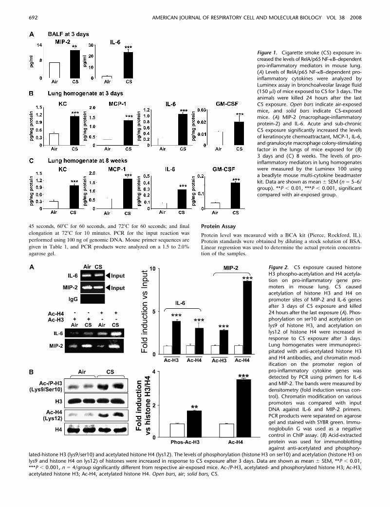
Figure 1. Cigarette smoke (CS) exposure in-
creased the levels of RelA/p65 NF-kB–dependentpro-inflammatory mediators in mouse lung.
(A) Levels of RelA/p65 NF-kB–dependent pro-
inflammatory cytokines were analyzed by
Luminex assay in bronchoalveolar lavage fluid(150 ml) of mice exposed to CS for 3 days. The
animals were killed 24 hours after the last
CS exposure. Open bars indicate air-exposed
mice, and solid bars indicate CS-exposedmice. (A) MIP-2 (macrophage-inflammatory
protein-2) and IL-6. Acute and sub-chronic
CS exposure significantly increased the levels
of keratinocyte chemoattractant, MCP-1, IL-6,and granulocyte macrophage colony-stimulating
factor in the lungs of mice exposed for (B)
3 days and (C ) 8 weeks. The levels of pro-inflammatory mediators in lung homogenates
were measured by the Luminex 100 using
a beadlyte mouse multi-cytokine beadmaster
kit. Data are shown as mean 6 SEM (n 5 5–6/group). **P , 0.01, ***P , 0.001, significant
compared with air-exposed group.
Figure 2. CS exposure caused histone
H3 phospho-acetylation and H4 acetyla-
tion on pro-inflammatory gene pro-moters in mouse lung. CS caused
acetylation of histone H3 and H4 on
promoter sites of MIP-2 and IL-6 genesafter 3 days of CS exposure and killed
24 hours after the last exposure (A). Phos-
phorylation on ser10 and acetylation on
lys9 of histone H3, and acetylation onlys12 of histone H4 were increased in
response to CS exposure after 3 days.
Lung homogenates were immunopreci-
pitated with anti-acetylated histone H3and H4 antibodies, and chromatin mod-
ification on the promoter region of
pro-inflammatory cytokine genes wasdetected by PCR using primers for IL-6
and MIP-2. The bands were measured by
densitometry (fold induction versus con-
trol). Chromatin modification on variouspromoters was compared with input
DNA against IL-6 and MIP-2 primers.
PCR products were separated on agarose
gel and stained with SYBR green. Immu-noglobulin G was used as a negative
control in ChIP assay. (B) Acid-extracted
protein was used for immunoblotting
against anti-acetylated and phosphory-
lated-histone H3 (lys9/ser10) and acetylated histone H4 (lys12). The levels of phosphorylation (histone H3 on ser10) and acetylation (histone H3 onlys9 and histone H4 on lys12) of histones were increased in response to CS exposure after 3 days. Data are shown as mean 6 SEM, **P , 0.01,
***P , 0.001, n 5 4/group significantly different from respective air-exposed mice. Ac-/P-H3, acetylated- and phosphorylated histone H3; Ac-H3,
acetylated histone H3; Ac-H4, acetylated histone H4. Open bars, air; solid bars, CS.
692 AMERICAN JOURNAL OF RESPIRATORY CELL AND MOLECULAR BIOLOGY VOL 38 2008

Statistical Analysis
Results are shown as means 6 SEM. Statistical analysis of significancewas calculated by one-way ANOVA followed by Fisher’s PLSD posthoc test for multigroup comparisons (StatView 5.0; SAS Institute,Cary, NC). Statistical significance is as indicated in figure legends.
RESULTS
CS Increased the Levels of NF-kB–Dependent Pro-Inflammatory
Mediators in Mouse Lung
In order to determine whether CS induces inflammatory lungresponse, C57BL/6J mice were exposed to CS for 3 days and8 weeks. Inflammatory response in mouse lung tissue was eval-uated by differential cell counts, H&E staining, and Mac-3staining. CS exposure resulted in increased accumulation of in-flammatory cells in mouse lung tissue (Figures E1 and E2 in theonline supplement). We next determined the NF-kB–dependentpro-inflammatory cytokines in response to CS in mouse lungby Luminex-based multiplex assay in BAL fluid of miceexposed to CS for 3 days or 8 weeks after the mice were killedat 24 hours after the last exposure. As shown in Figure 1, thelevels of MIP-2 and IL-6 were significantly increased in BALfluid of mice exposed to CS for 3 days, and the levels of KC,MCP-1, IL-6, and GM-CSF were significantly increased inmouse lung homogenate at both 3 days and 8 weeks of CSexposure. The levels of NF-kB-dependent pro-inflammatorymediators, such as IL-2, IL-10, IP-10, LIF, MCP-1, MCP-3,MCP-5, MIP-3b, and MMP-9, were significantly increased at3 days in CS-exposed mice (Figure E3). Similar data wereobtained for these cytokines at 8 weeks of CS exposure inmouse lung (data not shown). These data confirmed the pro-inflammatory effect of CS in mouse lung.
Chromatin Modification Occurred on Pro-Inflammatory Gene
Promoters in Response to CS in Mouse Lung
Chromatin immunoprecipitation (ChIP) assay is the mostpowerful tool for identifying proteins/nuclear factors associatedwith specific promoter regions. We studied the CS-inducedacetylation of histones H3 and H4 on pro-inflammatory pro-moters (IL-6 and MIP-2) by the ChIP assay in lungs of mice.The levels of acetylated histone H3 and H4 on pro-inflamma-tory promoter sites of MIP-2 and IL-6 were increased (Figure2A). However, the regions that were non-coding for IL-6 andMIP-2 showed no change in gene expression, attesting thespecificity of the ChIP assays (data not shown).
It has been reported that phosphorylation of histone H3 onser10 is known to be an early step in chromatin remodeling.Moreover, histone H4 lys12 residue is mainly known as thetypical acetylation site in chromatin remodeling (39). We there-fore next determined the levels of histone H3 on ser10/lys9 andH4 on lys12 using acid-extracted histone proteins by Westernblot analysis in lungs of mice exposed to CS. Phosphorylation onser10 and acetylation on lys9 of histone H3 was significantlyincreased in response to CS exposure (Figure 2B). We alsoobserved increased acetylation on lys12 of histone H4 as well ashistone H3 acetylation (Figure 2B). These results suggested thatthe pro-inflammatory effect of CS is associated with chromatinmodifications on various pro-inflammatory gene promoters.
CS Increased the Levels of IKKa in Alveolar Macrophages and
Alveolar/Airway Epithelial Cells in Mouse Lung
We determined the localization of IKKa and its activation inlungs of mice exposed to CS. To assess the level and distributionof IKKa in lung sections, immunostaining for IKKa wasperformed in mid-sagittal sections. Immunostaining of IKKa
Figure 3. CS exposure increased the
expression of IKKa in alveolar macro-phages and alveolar/airway epithelial
cells in mouse lung. Immunohistochem-
istry data revealed that IKKa is not only
localized, but also increased, in alveolarmacrophages, alveolar type II cells, and
airway epithelial cells in response to CS
both after 3 days and 8 weeks. (A)Representative photographs (3200)
from immunostaining for IKKa in lung
tissues from air- and CS-exposed mice.
The insets in boxes are magnified alveolarmacrophages and epithelial cells show-
ing increased IKKa staining in response
to CS compared with air-exposure. Ap-
pearance of dark brown color representsthe presence of IKKa, which was in-
creased in CS exposed mouse lung. E,
epithelial cells; M, macrophage; Alv,alveoli; Aw, airway. (B) Immunostaining
scores for IKKa per cell type in alveolar
and airway regions of the lung. The
assessment of immunostaining intensitywas performed semi-quantitatively and
in a blinded fashion. Black bars, intense
staining; grey bars, moderate/weak stain-
ing; open bars, no staining. Results aremean of four experiments 6 SEM. **P ,
0.01 and ***P , 0.001, significant com-
pared with air-exposed mice.
Yang, Valvo, Yao, et al.: Role of IKKa in Lung Inflammation 693
showed a significantly increased levels of IKKa in macrophagesand airway/alveolar epithelial cells in lung tissue of CS-exposedmice compared with air-exposed mice (Figures 3A and 3B). Thelevels of IKKb (an isoform of IKKa) were not changed inresponse to CS exposure. These data suggest that CS exposureincreased the level of IKKa, particularly in macrophages andepithelial cells of CS-exposed mouse lungs.
CS Caused the Recruitment of IKKa and RelA/p65 on
Pro-Inflammatory Gene Promoters in Mouse Lung
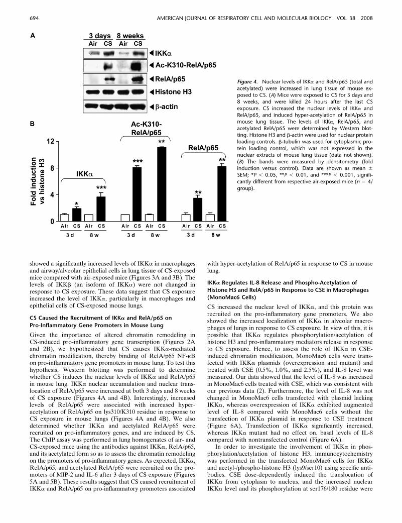
Given the importance of altered chromatin remodeling inCS-induced pro-inflammatory gene transcription (Figures 2Aand 2B), we hypothesized that CS causes IKKa-mediatedchromatin modification, thereby binding of RelA/p65 NF-kBon pro-inflammatory gene promoters in mouse lung. To test thishypothesis, Western blotting was performed to determinewhether CS induces the nuclear levels of IKKa and RelA/p65in mouse lung. IKKa nuclear accumulation and nuclear trans-location of RelA/p65 were increased at both 3 days and 8 weeksof CS exposure (Figures 4A and 4B). Interestingly, increasedlevels of RelA/p65 were associated with increased hyper-acetylation of RelA/p65 on lys310/K310 residue in response toCS exposure in mouse lungs (Figures 4A and 4B). We alsodetermined whether IKKa and acetylated RelA/p65 wererecruited on pro-inflammatory genes, and are induced by CS.The ChIP assay was performed in lung homogenates of air- andCS-exposed mice using the antibodies against IKKa, RelA/p65,and its acetylated form so as to assess the chromatin remodelingon the promoters of pro-inflammatory genes. As expected, IKKa,RelA/p65, and acetylated RelA/p65 were recruited on the pro-moters of MIP-2 and IL-6 after 3 days of CS exposure (Figures5A and 5B). These results suggest that CS caused recruitment ofIKKa and RelA/p65 on pro-inflammatory promoters associated
with hyper-acetylation of RelA/p65 in response to CS in mouselung.
IKKa Regulates IL-8 Release and Phospho-Acetylation of
Histone H3 and RelA/p65 in Response to CSE in Macrophages
(MonoMac6 Cells)
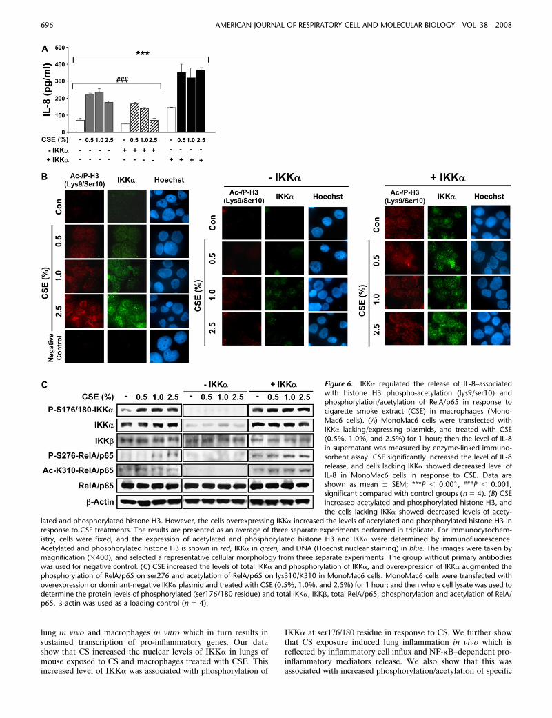
CS increased the nuclear level of IKKa, and this protein wasrecruited on the pro-inflammatory gene promoters. We alsoshowed the increased localization of IKKa in alveolar macro-phages of lungs in response to CS exposure. In view of this, it ispossible that IKKa regulates phosphorylation/acetylation ofhistone H3 and pro-inflammatory mediators release in responseto CS exposure. Hence, to assess the role of IKKa in CSE-induced chromatin modification, MonoMac6 cells were trans-fected with IKKa plasmids (overexpression and mutant) andtreated with CSE (0.5%, 1.0%, and 2.5%), and IL-8 level wasmeasured. Our data showed that the level of IL-8 was increasedin MonoMac6 cells treated with CSE, which was consistent withour previous data (2). Furthermore, the level of IL-8 was notchanged in MonoMac6 cells transfected with plasmid lackingIKKa, whereas overexpression of IKKa exhibited augmentedlevel of IL-8 compared with MonoMac6 cells without thetransfection of IKKa plasmid in response to CSE treatment(Figure 6A). Transfection of IKKa significantly increased,whereas IKKa mutant had no effect on, basal levels of IL-8compared with nontransfected control (Figure 6A).
In order to investigate the involvement of IKKa in phos-phorylation/acetylation of histone H3, immunocytochemistrywas performed in the transfected MonoMac6 cells for IKKa
and acetyl-/phospho-histone H3 (lys9/ser10) using specific anti-bodies. CSE dose-dependently induced the translocation ofIKKa from cytoplasm to nucleus, and the increased nuclearIKKa level and its phosphorylation at ser176/180 residue were
Figure 4. Nuclear levels of IKKa and RelA/p65 (total and
acetylated) were increased in lung tissue of mouse ex-posed to CS. (A) Mice were exposed to CS for 3 days and
8 weeks, and were killed 24 hours after the last CS
exposure. CS increased the nuclear levels of IKKa and
RelA/p65, and induced hyper-acetylation of RelA/p65 inmouse lung tissue. The levels of IKKa, RelA/p65, and
acetylated RelA/p65 were determined by Western blot-
ting. Histone H3 and b-actin were used for nuclear proteinloading controls. b-tubulin was used for cytoplasmic pro-
tein loading control, which was not expressed in the
nuclear extracts of mouse lung tissue (data not shown).
(B) The bands were measured by densitometry (foldinduction versus control). Data are shown as mean 6
SEM; *P , 0.05, **P , 0.01, and ***P , 0.001, signifi-
cantly different from respective air-exposed mice (n 5 4/
group).
694 AMERICAN JOURNAL OF RESPIRATORY CELL AND MOLECULAR BIOLOGY VOL 38 2008

associated with increased acetylation and phosphorylation ofhistone H3 (lys9/ser10) in the nucleus (Figures 6B and 6C).Furthermore, overexpression of IKKa led to increased acetyla-tion and phosphorylation of histone H3 (lys9/ser10) in responseto CSE treatments in MonoMac6 cells, whereas transfection ofMonoMac6 cells with plasmid lacking IKKa led to disappear-ance of histone H3 acetylation and IKKa levels (Figure 6B).We assessed the effects of overexpression and knock-down ofIKKa on basal protein level of IKKa in transiently transfectedMonoMac6 cells. We found that the level of IKKa was sub-stantially increased in IKKa transfected cells, whereas the levelof IKKa was abolished in dominant-negative IKKa-transfectedcells (Figure 6C).
We next performed Western blotting to determine themodification of RelA/p65 in response to CSE in MonoMac6cells transfected with overexpression or dominant-negativeIKKa plasmid. We found that the levels of phosphorylatedRelA/p65 (ser276) and acetylated RelA/p65 (lys310/K310) wereincreased in response to CSE, and that phosphorylation ofRelA/p65 on ser276 and acetylation of RelA/p65 on lys310/K310 were further increased in IKKa-transfected cells (Figure6C). These data suggest that IKKa regulates IL-8 release
associated with phospho-acetylation of both histone H3 andRelA/p65 on specific lysine/serine residues in response to CSEin MonoMac6 cells.
DISCUSSION
It is currently thought that abnormal inflammatory responsesto smoking in patients with COPD are due to chronic pro-inflammatory effects of CS. We and others have recently shownthat CS induces pro-inflammatory genes by chromatin remodel-ing in the lung (6, 7). However, the molecular mechanismwhereby CS alters chromatin remodeling on promoters of pro-inflammatory genes was not studied. It has been shown thatIKKa induces NF-kB–dependent gene expression by causingphosphorylation of histone H3 on the pro-inflammatory genepromoters in response to TNF-a treatment (10, 25–27). It is alsoknown that IKKa, but not IKKb, constitutively shuttles betweenthe cytoplasm and the nucleus, suggesting a nuclear function ofIKKa (26). In light of the above-mentioned observations, wehypothesized that CS triggers the activation of IKKa, leading toacetylation of histones (H3 and H4) and RelA/p65 subunit ofNF-kB on the promoters of pro-inflammatory genes in mouse
Figure 5. CS exposure led to recruitment of IKKa andRelA/p65 (total and acetylated) on promoters of IL-6 and
MIP-2 genes. Mice were exposed to CS for 3 days
and killed 24 hours after the last CS exposure. (A) CS
exposure caused recruitment of IKKa, RelA/p65 (totaland acetylated) on MIP-2 and IL-6 gene promoters. The
nuclear extracts were immunoprecipitated with specific
antibodies, and binding to the promoter of pro-inflamma-
tory mediator genes was detected by PCR-primers for IL-6or MIP-2 (n 5 3/group). Binding to the promoters is
compared with PCR of the input DNA. (B) The bands were
measured by densitometry (fold induction versus control).Data are shown as mean 6 SEM; **P , 0.01, and ***P ,
0.001, significantly different from respective air-exposed
mice. Open bars, air; solid bars, CS.
Yang, Valvo, Yao, et al.: Role of IKKa in Lung Inflammation 695
lung in vivo and macrophages in vitro which in turn results insustained transcription of pro-inflammatory genes. Our datashow that CS increased the nuclear levels of IKKa in lungs ofmouse exposed to CS and macrophages treated with CSE. Thisincreased level of IKKa was associated with phosphorylation of
IKKa at ser176/180 residue in response to CS. We further showthat CS exposure induced lung inflammation in vivo which isreflected by inflammatory cell influx and NF-kB–dependent pro-inflammatory mediators release. We also show that this wasassociated with increased phosphorylation/acetylation of specific
Figure 6. IKKa regulated the release of IL-8–associatedwith histone H3 phospho-acetylation (lys9/ser10) and
phosphorylation/acetylation of RelA/p65 in response to
cigarette smoke extract (CSE) in macrophages (Mono-
Mac6 cells). (A) MonoMac6 cells were transfected withIKKa lacking/expressing plasmids, and treated with CSE
(0.5%, 1.0%, and 2.5%) for 1 hour; then the level of IL-8
in supernatant was measured by enzyme-linked immuno-
sorbent assay. CSE significantly increased the level of IL-8release, and cells lacking IKKa showed decreased level of
IL-8 in MonoMac6 cells in response to CSE. Data are
shown as mean 6 SEM; ***P , 0.001, ###P , 0.001,significant compared with control groups (n 5 4). (B) CSE
increased acetylated and phosphorylated histone H3, and
the cells lacking IKKa showed decreased levels of acety-lated and phosphorylated histone H3. However, the cells overexpressing IKKa increased the levels of acetylated and phosphorylated histone H3 inresponse to CSE treatments. The results are presented as an average of three separate experiments performed in triplicate. For immunocytochem-
istry, cells were fixed, and the expression of acetylated and phosphorylated histone H3 and IKKa were determined by immunofluorescence.
Acetylated and phosphorylated histone H3 is shown in red, IKKa in green, and DNA (Hoechst nuclear staining) in blue. The images were taken by
magnification (3400), and selected a representative cellular morphology from three separate experiments. The group without primary antibodieswas used for negative control. (C) CSE increased the levels of total IKKa and phosphorylation of IKKa, and overexpression of IKKa augmented the
phosphorylation of RelA/p65 on ser276 and acetylation of RelA/p65 on lys310/K310 in MonoMac6 cells. MonoMac6 cells were transfected with
overexpression or dominant-negative IKKa plasmid and treated with CSE (0.5%, 1.0%, and 2.5%) for 1 hour; and then whole cell lysate was used to
determine the protein levels of phosphorylated (ser176/180 residue) and total IKKa, IKKb, total RelA/p65, phosphorylation and acetylation of RelA/p65. b-actin was used as a loading control (n 5 4).
696 AMERICAN JOURNAL OF RESPIRATORY CELL AND MOLECULAR BIOLOGY VOL 38 2008

histone H3 (lys9/ser10) and histone H4 (lys12) on pro-inflammatorygene promoters using the ChIP assay in CS exposed mouselung. Furthermore, these modifications of histones, particularlyH3 (lys9/ser10), had direct interaction with IKKa on pro-inflammatory gene promoters in macrophages (MonoMac6cells) in response to CS. These findings are in agreement withprevious studies showing that IKKa directly phosphorylateshistone H3 (independent of IKKb) in mouse embryonic fibro-blasts in response to TNF-a treatment (25).
Recently, a role of NF-kB–inducing kinase (NIK) has beenshown in histone H3 phosphorylation by IKKa, and thephosphorylation of H3 in turn facilitates its interaction withCBP, leading to acetylation of histone H3 on lysines 9 and 14(26). Our data showing increased nuclear IKKa and modifica-tions of histone H3 in response to CS may involve NIK as anintermediator of IKKa-induced histone modification. Indeed,our preliminary data support this notion that NIK is activated inresponse to CS in mouse lung (S.-R. Yang and coworkers,unpublished observations). In addition, our data showed thatrecruitment of IKKa and acetylation of RelA/p65 on pro-inflammatory gene promoters in response to CS in mouse lungis associated with acetylation of specific histone proteins onthese promoters. CBP is a master co-activator and known to berecruited on promoters of NF-kB–dependent pro-inflammatorygenes, thereby leading to acetylation of histone proteins.Furthermore, CBP was recruited onto the pro-inflammatorygene promoters in response to CS exposure in mouse lung (S.-R.Yang and coworkers, unpublished observations), suggesting thatonce IKKa is activated, it forms a complex with CBP and RelA/p65 which may then lead to acetylation of histone H3 and RelA/p65. Indeed, increased acetylation of RelA/p65 at lys310/K310residue was observed on the pro-inflammatory gene promotersas detected by the ChIP assay. Therefore, increased levels ofacetylated RelA/p65 would interact with IKKa and transcrip-tional co-activators on the promoters of pro-inflammatory genes,and hence will lead to sustained gene transcription of pro-inflammatory mediators in response to CS exposure.
Macrophages are the main orchestrators and amplifiers ofthe chronic inflammatory responses seen in smokers andpatients with COPD, and have the ability to cause lung damagewhen they are recruited and activated by CS (40, 41). We havepreviously shown that CS induced the release of pro-inflammatorymediators by activating NF-kB, and with the reduction ofHDAC levels/activity in monocyte-macrophage cell line(MonoMac6 cells) (2). We also show the increased levels ofnuclear IKKa in lung interstitial macrophages in response to CSexposure. Using an approach of gain and loss of IKKa inMonoMac6 cells, we demonstrated that the IKKa is activatedby CS and is involved in phosphorylation of ser10 and acetyla-tion of lys9 on histone H3, and phosphorylation of ser276 andacetylation of lys310/K310 on RelA/p65. It is known that IKKb
regulates activation of RelA/p65 by IkB degradation in re-sponse to diverse stimuli (14, 15, 42). However, recent studiesshowed that RelA/p65 can also be phosphorylated on ser536residue by IKKa and on ser276 residue by MSK1 (mitogen- andstress-activated protein kinase), leading to acetylation of RelA/p65 (43–45). Our finding is consistent with the phenomenon thatphosphorylation of RelA/p65 at ser276 promotes the acetyla-tion of RelA/p65 at lys310/K310, and this acetylation is requiredfor the full transcriptional activity of RelA/p65 (16). Recentstudies showed that IKKa can interact and/or phosphorylateCBP, which enhances the interaction between CBP and RelA/p65 on pro-inflammatory gene promoters in response to TNF-a(10, 46). In the present study, we showed that IKKa and RelA/p65 were recruited on the promoters of MIP-2 and IL-6 genes inresponse to CS in mouse lung. In addition, knockdown of IKKa
decreased the phosphorylation/acetylation of histone H3 andIL-8 release in MonoMac6 cells in response to CSE treatment,suggesting that IKKa might directly interact with acetylatedhistones by co-activating CBP. However, this effect is notglobal, since acetylation of RelA/p65 on lys310 is not observedon IL-6 promoter, suggesting that acetylation of RelA/p65 onpromoters of pro-inflammatory genes are specific in response toCS. These findings are corroborated by a recent finding showinga promoter-specific NF-kB recruitment and histone acetylationin LPS-mediated pro-inflammatory and anti-microbial genes (11).
MSK1 is a major kinase that phosphorylates CBP in mousefibroblast in response to cellular stress (47). This leads tophosphorylation of histone H3 at ser10 (44, 45). However, therole of MSK1 in IKKa-mediated chromatin modification andNF-kB activation in response to CS is not known. We foundthat the level of MSK1 was increased in MonoMac6 cells inresponse to CSE treatment, but the level was decreased inIKKa-knockdown MonoMac6 cells (S.-R. Yang and coworkers,unpublished observations), suggesting that MSK1 might play animportant role in IKKa-mediated chromatin modifications inresponse to CS on pro-inflammatory gene promoters. However,further studies are required to confirm this contention usingknockdown or overexpression of MSK1. Overall, our data sug-gest that CS-induced IKKa is directly associated with histonemodifications (phosphorylation and acetylation of histone H3)on pro-inflammatory gene promoters, and activation of RelA/p65 led to recruitment of acetylated RelA/p65 on pro-inflammatorygene promoters culminating in sustained inflammatory cytokinerelease.
In conclusion, we have demonstrated a novel role of IKKa inCS-mediated chromatin modifications and acetylation of RelA/p65 in mouse lung in vivo and in macrophages in vitro. In-creased activation of IKKa and acetylation of histones andRelA/p65 were associated with sustained pro-inflammatorygene transcription. These findings not only provide new insightinto the molecular mechanisms that underlie COPD pathogen-esis, but also validate the relevance of chromatin remodeling inCS-induced abnormal and sustained lung inflammation.
Conflict of Interest Statement: None of the authors has a financial relationshipwith a commercial entity that has an interest in the subject of this manuscript.
References
1. Burchfiel CM, Marcus EB, Curb JD, Maclean CJ, Vollmer WM,Johnson LR, Fong KO, Rodriguez BL, Masaki KH, Buist AS. Effectsof smoking and smoking cessation on longitudinal decline in pulmo-nary function. Am J Respir Crit Care Med 1995;151:1778–1785.
2. Yang SR, Chida AS, Bauter MR, Shafiq N, Seweryniak K, MaggirwarSB, Kilty I, Rahman I. Cigarette smoke induces proinflammatorycytokine release by activation of NF-kappaB and posttranslationalmodifications of histone deacetylase in macrophages. Am J PhysiolLung Cell Mol Physiol 2006;291:L46–L57.
3. Ito K, Ito M, Elliott WM, Cosio B, Caramori G, Kon OM, Barczyk A,Hayashi S, Adcock IM, Hogg JC, et al. Decreased histone deacetylaseactivity in chronic obstructive pulmonary disease. N Engl J Med 2005;352:1967–1976.
4. Rahman I, Adcock IM. Oxidative stress and redox regulation of lunginflammation in COPD. Eur Respir J 2006;28:219–242.
5. Di Stefano A, Caramori G, Oates T, Capelli A, Lusuardi M, Gnemmi I,Ioli F, Chung KF, Donner CF, Barnes PJ, et al. Increased expressionof nuclear factor-kappaB in bronchial biopsies from smokers andpatients with COPD. Eur Respir J 2002;20:556–563.
6. Szulakowski P, Crowther AJ, Jimenez LA, Donaldson K, Mayer R,Leonard TB, Macnee W, Drost EM. The effect of smoking on thetranscriptional regulation of lung inflammation in patients withchronic obstructive pulmonary disease. Am J Respir Crit Care Med2006;174:41–50.
7. Marwick JA, Kirkham PA, Stevenson CS, Danahay H, Giddings J,Butler K, Donaldson K, Macnee W, Rahman I. Cigarette smoke
Yang, Valvo, Yao, et al.: Role of IKKa in Lung Inflammation 697

alters chromatin remodeling and induces proinflammatory genes inrat lungs. Am J Respir Cell Mol Biol 2004;31:633–642.
8. Morrison D, Rahman I, Lannan S, MacNee W. Epithelial permeability,inflammation, and oxidant stress in the air spaces of smokers. Am JRespir Crit Care Med 1999;159:473–479.
9. Clayton AL, Hazzalin CA, Mahadevan LC. Enhanced histone acetyla-tion and transcription: a dynamic perspective. Mol Cell 2006;23:289–296.
10. Yamamoto Y, Verma UN, Prajapati S, Kwak YT, Gaynor RB. HistoneH3 phosphorylation by IKK-alpha is critical for cytokine-inducedgene expression. Nature 2003;423:655–659.
11. Foster SL, Hargreaves DC, Medzhitov R. Gene-specific control ofinflammation by TLR-induced chromatin modifications. Nature2007;447:972–978.
12. Jeffery P. Remodeling and inflammation of bronchi in asthma andchronic obstructive pulmonary disease. Proc Am Thorac Soc 2004;1:176–183.
13. Yang SR, Wright J, Bauter M, Seweryniak K, Kode A, Rahman I.Sirtuin regulates cigarette smoke-induced proinflammatory mediatorrelease via RelA/p65 NF-kappaB in macrophages in vitro and in ratlungs in vivo: implications for chronic inflammation and aging. Am JPhysiol Lung Cell Mol Physiol 2007;292:L567–L576.
14. Chen LF, Mu Y, Greene WC. Acetylation of RelA at discrete sitesregulates distinct nuclear functions of NF-kappaB. EMBO J 2002;21:6539–6548.
15. Chen L, Fischle W, Verdin E, Greene WC. Duration of nuclear NF-kappaB action regulated by reversible acetylation. Science 2001;293:1653–1657.
16. Chen LF, Williams SA, Mu Y, Nakano H, Duerr JM, Buckbinder L,Greene WC. NF-kappaB RelA phosphorylation regulates RelAacetylation. Mol Cell Biol 2005;25:7966–7975.
17. Vermeulen L, De Wilde G, Notebaert S, Vanden Berghe W, HaegemanG. Regulation of the transcriptional activity of the nuclear factor-kappaB p65 subunit. Biochem Pharmacol 2002;64:963–970.
18. Rahman I, MacNee W. Role of transcription factors in inflammatorylung diseases. Thorax 1998;53:601–612.
19. Schmitz ML, Bacher S, Kracht M. I kappa B-independent control of NF-kappa B activity by modulatory phosphorylations. Trends BiochemSci 2001;26:186–190.
20. Hu Y, Baud V, Delhase M, Zhang P, Deerinck T, Ellisman M, JohnsonR, Karin M. Abnormal morphogenesis but intact IKK activation inmice lacking the IKKalpha subunit of IkappaB kinase. Science 1999;284:316–320.
21. Matsushima A, Kaisho T, Rennert PD, Nakano H, Kurosawa K, UchidaD, Takeda K, Akira S, Matsumoto M. Essential role of nuclear factor(NF)-kappaB-inducing kinase and inhibitor of kappaB (IkappaB)kinase alpha in NF-kappaB activation through lymphotoxin betareceptor, but not through tumor necrosis factor receptor I. J ExpMed 2001;193:631–636.
22. Takeda K, Takeuchi O, Tsujimura T, Itami S, Adachi O, Kawai T, SanjoH, Yoshikawa K, Terada N, Akira S. Limb and skin abnormalities inmice lacking IKKalpha. Science 1999;284:313–316.
23. Li Q, Lu Q, Hwang JY, Buscher D, Lee KF, Izpisua-Belmonte JC,Verma IM. IKK1-deficient mice exhibit abnormal development ofskin and skeleton. Genes Dev 1999;13:1322–1328.
24. Sizemore N, Lerner N, Dombrowski N, Sakurai H, Stark GR. Distinctroles of the Ikappa B kinase alpha and beta subunits in liberatingnuclear factor kappa B (NF-kappa B) from Ikappa B and inphosphorylating the p65 subunit of NF-kappa B. J Biol Chem 2002;277:3863–3869.
25. Anest V, Hanson JL, Cogswell PC, Steinbrecher KA, Strahl BD,Baldwin AS. A nucleosomal function for IkappaB kinase-alpha inNF-kappaB-dependent gene expression. Nature 2003;423:659–663.
26. Park GY, Wang X, Hu N, Pedchenko TV, Blackwell TS, Christman JW.NIK is involved in nucleosomal regulation by enhancing histone H3phosphorylation by IKKalpha. J Biol Chem 2006;281:18684–18690.
27. Gloire G, Horion J, El Mjiyad N, Bex F, Chariot A, Dejardin E, Piette J.Promoter-dependent effect of IKKalpha on NF-kappa B/p65 DNAbinding. J Biol Chem 2007;282:21308–21318.
28. Vandel L, Trouche D. Physical association between the histone acetyltransferase CBP and a histone methyl transferase. EMBO Rep 2001;2:21–26.
29. Foronjy RF, Mirochnitchenko O, Propokenko O, Lemaitre V, Jia Y,
Inouye M, Okada Y, D’Armiento JM. Superoxide dismutase expres-sion attenuates cigarette smoke- or elastase-generated emphysema inmice. Am J Respir Crit Care Med 2006;173:623–631.
30. Thatcher TH, McHugh NA, Egan RW, Chapman RW, Hey JA, Turner
CK, Redonnet MR, Seweryniak KE, Sime PJ, Phipps RP. Role ofCXCR2 in cigarette smoke-induced lung inflammation. Am J PhysiolLung Cell Mol Physiol 2005;289:L322–L328.
31. Thatcher TH, Maggirwar SB, Baglole CJ, Lakatos HF, Gasiewicz TA,
Phipps RP, Sime PJ. Aryl hydrocarbon receptor-deficient micedevelop heightened inflammatory responses to cigarette smoke andendotoxin associated with rapid loss of the nuclear factor-kappaBcomponent RelB. Am J Pathol 2007;170:855–864.
32. Moesby L, Jensen S, Hansen EW, Christensen JD. A comparative study of
Mono Mac 6 cells, isolated mononuclear cells and Limulus amoebocytelysate assay in pyrogen testing. Int J Pharm 1999;191:141–149.
33. Ziegler-Heitbrock HW, Thiel E, Futterer A, Herzog V, Wirtz A,
Riethmuller G. Establishment of a human cell line (MonoMac6)with characteristics of mature monocytes. Int J Cancer 1988;41:456–461.
34. Carp H, Janoff A. Possible mechanisms of emphysema in smokers. In
vitro suppression of serum elastase-inhibitory capacity by freshcigarette smoke and its prevention by antioxidants. Am Rev RespirDis 1978;118:617–621.
35. Moodie FM, Marwick JA, Anderson CS, Szulakowski P, Biswas SK,
Bauter MR, Kilty I, Rahman I. Oxidative stress and cigarette smokealter chromatin remodeling but differentially regulate NF-kappaBactivation and proinflammatory cytokine release in alveolar epithelialcells. FASEB J 2004;18:1897–1899.
36. Kode A, Yang SR, Rahman I. Differential effects of cigarette smoke on
oxidative stress and proinflammatory cytokine release in primaryhuman airway epithelial cells and in a variety of transformed alveolarepithelial cells. Respir Res 2006;7:132–152.
37. Kweon SM, Wang B, Rixter D, Lim JH, Koga T, Ishinaga H, Chen LF,
Jono H, Xu H, Li JD. Synergistic activation of NF-kappaB bynontypeable H. influenzae and S. pneumoniae is mediated by CK2,IKKbeta-IkappaBalpha, and p38 MAPK. Biochem Biophys ResCommun 2006;351:368–375.
38. Bulger M, Schubeler D, Bender MA, Hamilton J, Farrell CM, Hardison
RC, Groudine M. A complex chromatin landscape revealed bypatterns of nuclease sensitivity and histone modification within themouse beta-globin locus. Mol Cell Biol 2003;23:5234–5244.
39. Strahl BD, Allis CD. The language of covalent histone modifications.
Nature 2000;403:41–45.40. Barnes PJ. Alveolar macrophages as orchestrators of COPD. COPD
2004;1:59–70.41. Shapiro SD. The macrophage in chronic obstructive pulmonary disease.
Am J Respir Crit Care Med 1999;160:S29–S32.42. Natoli G, Saccani S, Bosisio D, Marazzi I. Interactions of NF-kappaB
with chromatin: the art of being at the right place at the right time.Nat Immunol 2005;6:439–445.
43. Vermeulen L, De Wilde G, Van Damme P, Vanden Berghe W, Haegeman
G. Transcriptional activation of the NF-kappaB p65 subunit bymitogen- and stress-activated protein kinase-1 (MSK1). EMBO J 2003;22:1313–1324.
44. Soloaga A, Thomson S, Wiggin GR, Rampersaud N, Dyson MH,
Hazzalin CA, Mahadevan LC, Arthur JS. MSK2 and MSK1 mediatethe mitogen- and stress-induced phosphorylation of histone H3 andHMG-14. EMBO J 2003;22:2788–2797.
45. Duncan EA, Anest V, Cogswell P, Baldwin AS. The kinases MSK1 and
MSK2 are required for epidermal growth factor-induced, but nottumor necrosis factor-induced, histone H3 Ser10 phosphorylation.J Biol Chem 2006;281:12521–12525.
46. Lawrence T, Bebien M, Liu GY, Nizet V, Karin M. IKKalpha limits
macrophage NF-kappaB activation and contributes to the resolutionof inflammation. Nature 2005;434:1138–1143.
47. Deak M, Clifton AD, Lucocq LM, Alessi DR. Mitogen- and stress-
activated protein kinase-1 (MSK1) is directly activated by MAPK andSAPK2/p38, and may mediate activation of CREB. EMBO J 1998;17:4426–4441.
698 AMERICAN JOURNAL OF RESPIRATORY CELL AND MOLECULAR BIOLOGY VOL 38 2008