
IJST- vol(8)- No.(3)- Sept. 2013 drtaghreed
97
Transcript of IJST- vol(8)- No.(3)- Sept. 2013 drtaghreed


International Journal for Sciences and Technology
Volume 8. No. 3/ September 2013 / ISSN: 2305-9346
A Refereed Scientific Journal Since 2006
��� ��� �� �� � � �2006
Issued By:
The International Centre for Advancement of Sciences and Technology
IJST contact Information:
P.O. Box 2793 Amman 11953 Jordan
Tel. +96265602285
E-mails: [email protected] / [email protected]
URL: www.ijst-jo.com

EDITORIAL BOARD - 2013 Al- Shammari , Abdul- Jabbar N. (Editor-in- Chief)
Professor of Microbiology / Faculty of Pharmacy / Royal University for Medical Sciences (RUMS) / P.O. Box 2793. Amman 11953 Jordan [email protected]
Abbas, Jamal A. Professor of Plant Ecophysiology / College of Agriculture / Kufa University / Iraq [email protected]
Abdul- Ghani, Zaki G. Professor of Microbiology / Faculty of Pharmaceutical Sciences / Amman Private University / Jordan [email protected]
Abdul- Hameed, Hayder M. PhD in Environmental Engineering / Environmental Engineering Dept./ Faculty of Engineering/ Baghdad University/ Iraq [email protected]
Abdullah, Ahmed R. PhD in Cancer Immunology and Genetics /Biotechnology Research Centre / Al- Nahrain University / Baghdad / Iraq [email protected] Al – Banna , Anton S. A Professor in Microbiology and Virology/ Faculty of Veterinary Medicine/ Baghdad University / Iraq [email protected]
Al- Dabbagh, Riadh H. Professor of Engineering Hydrology/ UAE [email protected]
Al- Daraji, Hazim J. Professor of Avian Reproduction and Physiology / Animal Resources Dept./ College of Agriculture / Baghdad University / Iraq [email protected]
Al- Douri, Atheer A. R PhD in Microbiology/Faculty of Veterinary Medicine/ Baghdad University/ Iraq [email protected]
Al- Jashami, Najim A. Professor of Nuclear Material Sciences / Dept. of Physics / College of Sciences / Kufa University / Iraq [email protected]
Al- Mashaykhi, Akram Othman PhD in IT / Amman Arab University for Graduate Studies / Jordan [email protected] Al- Murrani, Waleed K. Professor of Genetics and Biostatistics / University of Plymouth/ UK [email protected]
Al- Noor, Taghreed H. PhD in Chemistry / Dept. of Chemistry / College of Education – Ibn Al- Haitham / Baghdad University / Iraq [email protected]
Al- Saqur, Ihsan M. Professor of Parasitology/ Faculty of Sciences / Baghdad University/ Iraq [email protected]
Al- Shamaony, Loai Professor of Biochemistry / Faculty of Pharmacy / Misr University for Sciences and Technology / Egypt [email protected] Al- Shebani, Abdullah S. PhD in Dairy Sciences and Technology / Food Sciences Dept./ College of Agriculture / Kufa University / Iraq [email protected]
Alwachi, Sabah N. Professor of Physiology / Biology Dept./ College of Sciences/ Baghdad University/ Iraq [email protected]
Khamas, Wael Professor of Anatomy and Histology / College of Vaterinary Medicine / Western University of Health Sciences / Ponoma -California/ USA [email protected]
Mohammed, Ramadhan H. PhD in Geology / College of Sciences / Duhok University / Iraq [email protected]
Editorial Board Secretary Pharmacist. Nansi Elian Amman- Jordan [email protected]
d
Highlight

International Journal for Sciences and Technology Vol. 8, No.3, Sٍeptember 2013 1
FORWARD
IJST is coming for you all today in its recent issue of volume eight for year 2013, as our deep
belief in continuing the steps we began since eight years ago.
Year after year, IJST proves its strength and faithful belief in developing our scientific
communities among Arab World, especially in Iraq by giving an opportunity to all
researchers to present their fruitful achievements in main vital fields to let all world knows
that we are still the first leaders in civilized scientific life, despite all the unfortunate
situations or constraints.
It is my pleasure to welcome you and present you a new issue of our Journal, Volume 8, No. 3
(2013), the third issue of this year, with diversity of researches and elite experts of the
Editorial Board and Advisory Group. The current issue comes to you while the Islamic World
is celebrating Eid Al- Adha , which gives me an opportunity to send you all my deep wishes
and faithful prays to Allah for peaceful times.
The members of Editorial Board, the ICAST and TSTC teamwork and I hope you will find this
collection of research articles useful and informative.
IJST had the honor to welcome a new editorial board member , Dr. Taghreed Al-Noor, with
all our deep appreciation and thanks for her valuable efforts in supporting the journal.
The journal is one of the scientific contributions offered by the International Centre for
Advancement of Sciences and Technology in cooperation with Treasure Est. for Scientific
Training and Consultations to the science and technology community (Arab region with
specific focus on Iraq and International).
Finally, on behalf of the International centre, I would like to express my gratitude and
appreciation to the efforts of the Editorial Board, Advisory group with their valuable efforts
in evaluating papers and special thanking to the Editorial Board Secretary for her faithful
efforts in managing the scientific, design, technical and administrative aspects of the Journal
and for preparing this issue for final printing and publishing.
Editor-in-Chief
IJST
Abdul Jabbar Al- Shammari
d
Highlight

International Journal for Sciences and Technology Vol. 8, No.3, Sٍeptember 2013 2
The Referees for this Issue * The referees and advisory group below are listed according to alphabetical order, with deep
appreciation for all. Prof. Abdul- Jabbar N. Al- Shammari Faculty of Pharmacy, Royal University for Medical Sciences (RUMS). Jordan Dr. Abdullah Sh. M. Al- Shebani Dept. of food sciences, Faculty of Agriculture, Kufa University. Iraq
Prof. Ahmed M. Abdul-Lettif College of Sciences, University of Karbala. Iraq
Prof. Bashar R. Al- Shreidah National Centre for Agricultural Researches . Jordan
Dr. Dawood S. Al- Azzawi College of Pediatrics, Diyala University. Iraq
Dr. Harith F. Al- Mathkhouri College of Sciences, Baghdad University. Iraq
Prof. Jamal A. Abbas Faculty of Agriculture, Kufa University. Iraq
Dr. Khalid Al- Azzawi Faculty of Pharmacy, Al- Isra University. Jordan
Prof. Mahmoud M. Othman Matar College of Medicine, Al- Najah National University. Palestine
Dr. Mohammed A.M. Al- Hajaj College of Sciences . Basra University. Iraq
Dr. Ramadhan H. Mohammed College of Sciences , Duhok University . Iraq
Dr. Taghreed H. Al- Noor College of Education for Pure Sciences, Ibn Al- Haitham , Baghdad University . Iraq
Prof. Taha Al- Samaraei Crown Research Institutes, Palmerston North. New Zealand
Prof. Zaki G. Abdul- Ghani Faculty of Pharmaceutical Sciences, Amman Private University. Jordan

International Journal for Sciences and Technology Vol. 8, No.3, Sٍeptember 2013 3
TABLE OF CONTENTS * Articles in this issue are listed below according to field specialties order, starting by English section and followed by
Arabic section.
(I) ENGLISH SECTION:
CHEMISTRY
Synthesis and characterization of some new compounds derivatives from para-amino benzoic acid ………………………………………………………………………… 6-11 Ismaeel Y. Majeed, Duhaa AL-Saady & Shaimaa A. Saoud
ENVIRONMENTAL SCIENCES
Evaluation of some limiting factors affecting the hygienic standards of water chlorination (Tap and Trough’s Water) of Animal farm /Veterinary Medicine College - Baghdad University …………………………………………..……………………………........... 12-17
Dergham H.Y.Al-Zwean
Heavy metal contamination in canned fish marketed in Baghdad ..................…………………18-22 Salim S. Al- Timimi, Sundus H. Ahmed, Salwa Khudadat, & Qais Assi
GENOMIC
Mitochondrial Genome Variation within Iraqi Population ……………………….…………...23-27 Basheer I. Azzawi, Abdulrahman A. Oleiwi, Majida J. Darweesh, Said Jaradat, & Ali Al-Zaag
MEDICAL SCIENCES Cardiac complications in the Kurdish Infants of Diabetic Mothers in Duhok ……………….28-31 Akrem M. Atrushi Demographic study on rheumatoid arthritis (RA) in Babylon-Iraq …………………………32-39 Amina N. Al-Thwani & Ibtisam A. I. El-Baghdady
Molecular Subtypes of Breast Carcinoma Using Immunehistochemical Stains in Central Public Health Labs……………………………………………………………………40-50. Anwar I. S. Al- Assaf, Rana Z. Naji, & Ali H.M. Al- Khafagi
Post-Injection Sciatic Nerve Injury in Duhok………………………………………………….51-55 Nadir A. Garjees
International Journal for Sciences and Technology Vol. 8, No.3, Sٍeptember 2013 4
)II (قسم الدراسات والبحوث العربية – ARABIC SECTION
يا الغذاء ــتكنولوج
63-57......................................لبان باستخدام التقانات الحديثةاألعادة استخدام المياه العادمة لصناعات إ
حمد، إلهام مجيد خورشيد،أسامة جورج أمين، أزهار قيس كيطان، ابتهال عبد الجبار عبد القادر، أحمد سالم لمياء سلمان رشيد، سندس فؤاد أحمد، أحمد خلف حردان، صالح عبد المحسن رحيم، هشام حميد علوان
مدراسة العالقة بين تفضيل المستهلك والتحليل المختبري ألنواع شائعة من معجون الطماط 68-64 ...............................................................................................في مدينة بغداد
علياء سعد الحافظمريم مال اهللا غزال،
الزراعيةعلـــــوم ال
Salvia officinalis في نمو نبات الميرمية الري بالماء الممغنط والتسميد التداخل بينتأثير Thujone( ..................................................................69-78الثيوجين ( وتركيز المادة الفعالة حسين محمود شكري
في بعض مؤشرات النمو الخضري لفسائل نخيل التمرHumic acidتأثير الحامض العضوي
L. Phoenix dactylifera 83- 79.......................................................................... المروية بمياه اآلبار جمال أحمد عباس، رعد طه بالكت، علي حيدر عريان
الوراثة الجينية
Klebsiella pneumoniae و Escherichia coli بكتريا عزالت من لكل الوراثية العالقة دراسة القطع اطوال تباينات نيةتق باستخدام الهضمية والقناة البولية والحصى المرارة حصى من حاالت من المعزولة
DNA) AFLP(.........................................................84-94 الحمض النووي من المتضخمة عقيل حسين علي، الجبوريعلي صالح ، فرحانمثنى بديع ، اياد محمد علي العبيدي

International Journal for Sciences and Technology Vol. 8, No.3, Sٍeptember 2013 5
ENGLISH SECTION

International Journal for Sciences and Technology Vol. 8, No.3, Sٍeptember 2013 6
Synthesis and characterization of some new compounds derivatives from para-
amino benzoic acid
Ismaeel Y. Majeed, Duhaa AL-Saady & Shaimaa A. Saoud
Dept. of Chemistry/ College of Education – Ibn Al- Haitham / Baghdad University/ Republic of Iraq
ABSTRACT
The newly compounds containing 1,3-oxazepine and 1,3-thiazepinedioxide derivatives were synthesized by reaction of compounds containing imine group with maleic or phthalic anhydride and orthosulphobenzoic acid cyclic anhydride respectively in dry benzene to give compounds [1-5]. The other compound (oxo-Thiazolidine) [6] was synthesized by reaction of imine group with thioglycolic acid . Tetrazole derivative was synthesized by reaction of imine group with sodium azide to give compound[7]. Finally compound [1] was converted to ester compound by reaction with ethylchloroacetate in the presence of potassium carbonate and dry acetone, then, the compound [8] was reacted with hydrazine hydrate to give compound (9). The hydrazide derivative [9] was cyclized by KOH and CS2 to give Oxadiazole derivative [10]. All these synthesized compounds have been characterized by melting points, elemental analysis, FTIR and HMNR spectroscopy.
Keywords: 1,3-Oxazepine, 1,3-Thiazepinedioxide, Thiozolidine-one, tetrazole, Oxadiazole.
الملخص باللغة العربية
ثايازبين ثنائي االوكسجين من خالل تفاعل المركبات الحاوية على مجموعة االيمين -1,3اوكسازبين و-1,3 تم تحضير المركبات
. )5-1(باستعمال البنزين الجاف كمذيب لتعطي المركبات) االورثوسلفوبنزويك انهدريد,القثالك,الماليك(مع االنهدريدات الحلقية اما مشتق ,الذي حضر بمفاعلة مركب الشف مع حامض الثاياكاليكولك ) 6( الذي حضر هو مشتق الثايازولديناآلخرركب الم
مع خالت االثيل )1(اخيرا تم مفاعلة المركب).7(التيترازول فقد حضر من تفاعل مشتق قاعدة الشف مع ازيد الصوديوم لينتج مركب إجراءوهو مشتق الهيدرازيد الذي تم ) 9(اعلته مع الهيدرازين المائي الذي اعطى مركب الذي تم مف) 8(كلوريد لينتج مشتق االستر
تم . ) 10( مشتق حلقة االوكسادايازولأنتج حيث الكربونالغلق الحلقي له من خالل استعمال هيدروكسيد البوتاسيوم وثنائي كبريتيد .الفيزيائيةو الطيفية الطرق بعض باستخدام الناتجة المركبات جميع تشخيص

International Journal for Sciences and Technology Vol. 8, No.3, Sٍeptember 2013 7
INTRODUCTION
Heterocyclic compounds (oxazepine, thiazepine, tetrazole, and thiazolidine) four-five-seven membered rings have occupied an important place in the field of pharmaceuticals and industries(1). Oxazepine derivative (Oxazepam) is used as an antibiotic, enzyme inhibitor, pharmacological interest (2), and has a biological activity(3). It is used in relief of the psychoneuroses characterized by anxiety and tension(4). Seven-membered ring analogous benzothiazepine derivatives exhibit diverse biological activities as vasodilatory (5), tranquilizer (6), antidepressant (7),CNS stimulant (8), antihypertensive (9), calcium channel blocker(10), antifungal (11) anti cancer(12), and inhibitors of enzymes(13). Thiazepine is one of hetrocyclic compound contains two heteroatoms (nitrogen and sulphur) in seven-membered ring. Oxo-Thiazolidine ring system derives special important from the fact that it plays an important role in medicinal chemistry. Substituted thiazolidine derivatives represent important key intermediates for synthesis of pharmacologically active drug thiazolidinone has wide range biological activaties such as antifungal (14), antiproliferative(15), anti-inflammatory(16), herbicidal (17), and antiviral properties(18).
MATERIALS AND METHODS
Materials Chemicals, solvents were of analytical grade (supplied by either Merck or Fluka,Aldrich) received. All melting points were recorded using open capillary tube and uncorrected. Fouriertransform Infrared spectra were recorded in KBr disc on Schimadzu 8300 spectrophotometer. 1H NMR spectra were measured in DMSO solutions on a Bruker Av spectrophotometer (300 MHz) using TMS as an internal reference (chemicals) shifts in d ppm). All synthesized compounds were elemental analysis C, H, N and S on a European Elemental analyzer. Thin layer chromatography was performed on silica gel as a stationary phase, ethyl acetate as eluent. Synthesis procedure The synthetic route of compounds is presented in scheme (1).
COOH
NH2
+
OH
OCH3
CHO
EtOHHOOC N
OH
OCH3
[1]
HOOC NOH
OCH3
[2]
O
OO
HOOC NOH
OCH3
O
OO
[3] HOOC
N
O
O
O
O
N
O
O
COOH
OH
OCH3
HOOC
[4]
HOOC NOH
OCH3
O2S
O
O
[5]
HOOC NOH
OCH3
S
O
[6]
HOOC NOCH2COOEt
OCH3
[8]
NaN3
HOOCN
OH
OCH3
HN N
N
[7]
HOOC NOCH2CONHNH2
OCH3
[9]
HOOC NOCH2
OCH3
O
NN
SH
Maleicanhydride
Phthalic anhydridePyromela
ticanhydrid
e
Thioglycolicacid
ClCH2 COOEt
NH2NH2.H2O
CS2/KOH
O-sulfobenzoicacid cyclic anhydride
Scheme (1): The synthesis route of compounds (1-10)
Synthesis of 4-(4-hydroxy-3-methoxybenzylideneamino) benzoic acid[1] A solution of the p-amino benzoic acid (0.01 mol) in absolute ethanol (30 ml) was slowly added to a solution of the aldehyde (0.01 mol) in absolute ethanol (20 ml). The stirred reaction mixture was refluxed for 4hrs. After cooling, aprecipitate was formed which collected by fillration then washed with cold ethanol and recrystallized from ethanol, Yield (90%),m.p.=(160-162)°C. Synthesis of Oxazepine [2,3,4] A mixture of Schiff base (0.01 mol) with (0.01 mol) of maleic, phthalic, (0.02mol)of Schiff base with (0.01mol) of pyromelitic anhydride were dissolved in (10 ml) of dry benzene and refluxed in a water bath for 4 hrs. Then solvent was removed and the resulting crystalline solid was recrystallized from dioxan to give the titled compounds (2) Yield (83%),m.p=(179)°C.

International Journal for Sciences and Technology Vol. 8, No.3, Sٍeptember 2013 8
(Z)-4-(2-(4-hydroxy-3-methoxyphenyl)-4,7-dioxo-1,3-oxazepin-3(2H,4H,7H)-yl)benzoic acid FT-IR(KBr) ν cm-1: 3460 and 3363(OH) 3080-3075 (C-H Aromatic), 2895-2899(C-H Aliphatic), 1695(C=O),1670(C=O),1660 (C=O),1566(C=C);1HNMR(DMSO-d6)δ ppm 9.98(s,1H,OH,carboxylic),9.01(s,1H,OH),8.4(s,1H,in oxazepine ring)(19),7.2-7.09(m,7H,ArH),3.8(s,3H,OCH3): Anal. Calcd. for C19H15NO7: C, 61.79; H, 4.09; N, 3.79.found: C, 61.88; H, 4.17; N, 3.87. Synthesis of 4-(3-(4-hydroxy-3-methoxyphenyl)-1,5 dioxobenzo[e][1,3]oxazepin-4(1H,3H,5H)-yl)benzoic acid Yield :(86%) m.p=(159)°C FT-IR(KBr) ν cm-1: 3502 and 3361(OH) 3070-3080 (C-H Aromatic), 2886-2889(C-H Aliphatic), 1717(C=O),1683(C=O),1665 (C=O),1594(C=C);1HNMR(DMSO-d6)δppm 9.88(s,1H,OH,carboxylic),9.71(s,1H,OH),8.35(s,1H,in oxazepine ring),7.3-7.12(m,11H,ArH),3.91(s,3H,OCH3): Anal. Calcd. for C23H17NO7: C, 65.87; H, 4.09; N, 3.34.found: C, 66.07; H, 4.13; N, 3.41. Synthesis of bis [4-(3-(4-hydroxy-3-methoxyphenyl)-1,5-dioxobenzo[e][1,3]oxazepin-4(1H,3H,5H)-yl)benzoic acid] Yield (85%)m.p=(300)°C. FT-IR(KBr) ν cm-1: 3499 and 3370(OH) 3091-3085 (C-H Aromatic), 2897-2887(C-H Aliphatic), 1714(C=O),1676(C=O),1669 (C=O),1577(C=C);1HNMR(DMSO-d6)δppm 9.80(s,1H,2OH,carboxylic),9.65(s,1H,2OH),8.31(s,1H,in oxazepine ring),7.13-7.11(m,16H,ArH),3.84(s,3H,2OCH3): Anal. Calcd. for C40H28N2O14: C, 63.16; H, 3.71; N, 3.68.found: C, 63.89; H, 3.65; N, 3.70. Synthesis of Thiazepine dioxide derivative The orthosulfobenzoic acid cyclic anhydride (0.01 mol) was dissolved in a small amount of dry benzene for 30 min, schiff base (0.01 mol) in (10 ml) of dry benzene was added with a few drops of tetra hydro furan (THF) the mixture was refluxed in a water bath for (5-6) hrs. until the colour was converted. The mixture was cooled at room temperature.the prcipitate was collected by filtration and washed with cold dry benzene, recrystallized from benzene (5) m.p= (230)°C,Yield 81%.
FT-IR(KBr) ν cm-1: 3499 and 3361(OH) 3090-3086 (C-H Aromatic), 2875-2874(C-H Aliphatic), 1701(C=O), 1653 (C=O),1589(C=C),1140 and 1367(SO2);
1HNMR(DMSO-d6)δppm 9.95(s,1H,OH,carboxylic),9.67(s,1H,OH),8.26(s,1H,in thiazepine ring),7.6-7.31(m,11H,ArH),3.88(s,3H,OCH3): Anal. Calcd. for C22H17NO8S: C, 58.02; H, 3.76; N, 3.08;S, 7.04; N, 3.34.found: C, 59.00; H, 3.79; N, 3.11; S, 7.11. Synthesis of 4-(2-(4-hydroxy-3-methoxyphenyl)-4-oxothiazolidin-3-yl) benzoic acid The compound (1) (0.01 mol) and thioglycolic acid (0.01 mol) in ethanol (50 ml) in the presence of ZnCl2 were allowed to react at room temperature. The reaction mixture was first stirred on a magnetic stirrer for about 2.30 hrs. followed by reflux on a steam bath for about 5.0 hrs. the product was filtered and cooled at room temperature, m.p= (239)°C,Yield 75%. FT-IR(KBr) ν cm-1: 3498 and 3369(OH) 3087-3069 (C-H Aromatic), 2872 (C-H Aliphatic), 1701(C=O), 1657 (C=O),1593(C=C);
1HNMR(DMSO-d6)δppm 9.81(s,1H,OH,carboxylic),9.76(s,1H,OH), ,7.45-7.22(m,7H,ArH), 5.26(s,2H,in thiazolidine ring)3.79(s,3H,OCH3): Anal. Calcd. for C17H15NO5S: C, 59.12; H, 4.38; N, 4.06; S, 9.28.found: C, 59.32; H, 4.45; N, 4.13; S, 9.30. Synthesis of 4-(5-(4-hydroxy-3-methoxyphenyl)-2,5-dihydro-1H-tetrazol-1-yl)benzoic acid Sodium azide (0.01 mol) in (10 ml) of dry benzene was added to a stirring solution of Schiff base (0.01 mol) in (10 ml) of dry benzene. The mixture was refluxed for (7-8 ) hrs. then cooled at d at room temperature. The prcipitate was filtered and washed with coldwater, recrystallized from benzene-petrolum spirit (40-60), m.p= (164)°C,Yield 80%. FT-IR(KBr) ν cm-1: 3456 and 3398(OH) 3091 (C-H Aromatic), 2889 (C-H Aliphatic), 1701(C=O), 1589(C=C); 1HNMR(DMSO-d6)δppm 9.81(s,1H,OH,carboxylic),9.76(s,1H,OH), ,7.45-7.22(m,7H,ArH), 5.06(s,2H,in tetrazole ring)3.83(s,3H,OCH3),3.03(s,1H,NH)weak: Anal. Calcd. for C15H14N4O4: C, 57.32; H, 4.49; N, 17.83;.found: C, 57.93; H, 4.53; N, 17.90.

International Journal for Sciences and Technology Vol. 8, No.3, Sٍeptember 2013 9
Synthesis of (Z)-4-(4-(2-ethoxy-2-oxoethoxy)-3-methoxybenzylideneamino)benzoic acid The previous compound (1) (0.01 mol) was stirred with an equivalent amount of potassium carbonate in (30 ml) of acetone and then ethylchloro acetate was added and refluxed for 3hrs. after completion of the reaction (T.L.C). The mixture was filtered and the solvent was removed under reduced pressure to give compound (8), m.p= (95)°C,Yield 74%. FT-IR(KBr) ν cm-1: 3456 (OH,carboxylic) 3099 (C-H Aromatic), 2898 (C-H Aliphatic), 1730(C=O),1701(C=O), 1587(C=C): Anal. Calcd. for C19H19NO6: C, 63.86; H, 5.36; N, 3.92;.found: C, 63.98; H, 5.30; N, 3.97 Synthesis of (Z)-4-(4-(2-hydrazinyl-2-oxoethoxy)-3-methoxybenzylideneamino)benzoic acid Hydrazide compound (9) was synthesizeby addition of the hydrazine hydrate (80%) (0.01 mol) to compound (8) (0.01 mol) with stirring, then the absolute ethanol (10 ml) was added and refluxed until the precipitate formed 3 hrs. after cooling, the precipitate was filtered off and recrystallized from ethanol, m.p=(252)°C,Yield 60%. FT-IR (KBr) ν cm-1: 3449 (OH,carboxylic) 3083 (C-H Aromatic), 2819 (C-H Aliphatic), 1701(C=O),1625(C=O), 1587(C=C): Anal. Calcd. for C17H17N3O5: C, 59.47; H, 4.99; N, 12.24:.found, C, 59.98; H, 509; N, 12.43. Synthesis of (Z)-4-(4-((5-mercapto-1,3,4-oxadiazol-2-yl)methoxy)-3-methoxybenzylideneamino)benzoic acid This compound was synthesized by reaction of (0.01 mol) CS2 with hydrazide compound (9) (0.01 mol) in the presence of (0.01 mol) KOH in ethanol absolute (20 ml). The mixture was stirring at 0.5 hr. then refluxed for 4hrs., after cooling, the precipitate was filtered off and recrystallized from ethanol, m.p=(297)°C,Yield 59%.%. FT-IR(KBr) ν cm-
1: 3456 (OH) 3080 (C-H Aromatic), 2890 (C-H Aliphatic),2600(SH,w), 1701(C=O),1635(C=N), 1576(C=C),1047(C=S); 1HNMR(DMSO-d6)δppm 9.81(s,1H,OH,carboxylic), ,7.45-7.22(m,7H,ArH), 3.83(s,3H,OCH3): Anal. Calcd. for C18H15N3O5S: C, 56.10; H, 3.92; N, 10.90; S, 8.32.found: C, 56.45; H, 3.89; N, 11.09; S, 8.30
RESULTS AND DISCUSSION
The reaction of aldehyde with primary amines is one of the most common reactions to synthesize Schiff base. Compound (1) was prepared from the condensation of p- aminobenzoic acid with 4-hydroxy -3-methoxy benzaldehyde in ethanol with few drops of glacial acetic acid(GAA).schiff base were identified melting point, (FT.IR) spectroscopy. The FT.IR spectra for Schiff base showed the appearance absorption band at 1625 cm-1 due to stretching vibration of the C=N for azomethine band besides the disappearance of the absorption band in the region (3290-3100) cm-1 due to symmetric and asymmetric stretching vibration of the (-NH2) group. Seven memberd rings were synthesized by cycloaddition of Schiff base. cycloaddition reaction is classified as a 5+2, implying 5-atom component plus 2-atom component leading to 7-membered cyclic ring. The suggested mechanism for the formation of seven member ring derivatives as shown in scheme (2). The maleic anhydride may be replaced by phthalic or o-sulfobenzoic acid cyclic anhydrides but the same mechanism.
Scheme(2): mechanism for synthesis of seven membered ring (2-5)
The thiazolidinone (6) was obtained by refluxing equimolar amounts from imine (1) and thioglycolic acid in dry benzene. The suggested mechanism for the formation of thiazolidinone derivative (6) as following scheme(3).

International Journal for Sciences and Technology Vol. 8, No.3, Sٍeptember 2013 10
NHOOC
O
HO
+ SH-CH2COOHP.T
NHHOOC
O
HO
S
O
HO
HNHOOC
O
HO
S
HO O
P.T
NHOOC
O
HO
S
HO OH
-H2O
NHOOC
O
HO
S
O
Scheme (3): mechanism for the Synthesis of thiazolidinone ring (6)
Tetrazole is a five membered ring was prepared from the reaction of Schiff base [1] with sodium azide give compound[6]. The mechanism of the reaction systematically investigated as [3+2] cycloadditions which christened as 1,3-dipolar cycloadditions. The common features of this type of reactions is best accommodated by a transition state geometry between imine group with 1,3-dipolar of azide compound in scheme (4).
Scheme (4): approximate transition state geometry for azide addition to Schiff base
Substitution of oxadiazoles had attracted considerable interest owing their wide spectrum biological activity.Oxadiazole is a five membered ring containing oxygen and two atoms of nitrogen.compound [10] was prepared by the condensation of compound [1] with ethylchloroacetate with potassium carbonate in dry acetone to converted the(OH) phenolic group to ester compound[9],compound [9] was reacted with carbon disulfide with KOH give oxadiazole derivative [10] .
REFERENCES
1. Eicher T. and Hauptmann S. (2003). The Chemistry of Heterocycles: Structure, Reactions, Syntheses and Applications. Wiley-VCH, 2nd ed.: 371. 2. Griera R; Lcopart CC; Amat M; Bosch J; Castillo JC; and Huguet J. (2005). A synthetic route to a novel type of conformationally constrained N-aryloxazolidinones. BioOrgan. Med. Chem. Lett. (15): 2515-2517. 3. Bajaj K; Archana A; and Kumar A. (2004). Synthesis and pharmacological evaluation of newer substituted benzoxazepine derivatives as potent anticonvulsant agents. Eur. J. Med. Chem. 39: 369. 4. Wilson CO; and givold O. (1966). Text book of organic medicinal and pharmaceutical chemistry. 5th ed. London Cott compane. 5. Kugita H; Takeo S; and Matsushima M. (1972). 1,5-Benzothiazepine derivatives. Jap. Pat. Chem. Abstr. 77:5554. 6. Kugita H; Inoue H; and Ikezaki M. (1971). 1,5-Benzothiazepine derivatives. Jap. Pat. Chem. Abstr. 75: 63848. 7. Geyer HM; Watzman N; and Buckley JP. (1970). Effects of a tranquilizer and two antidepressants on learned and unlearned behaviors. J. Pharmacol. Sci. 59: 964-968. 8. Kawashima Co. Ltd. (1985). Benzothiazepine derivative. Jap. Pat. Chem. Abstr. 103: 105014.

International Journal for Sciences and Technology Vol. 8, No.3, Sٍeptember 2013 11
9. Inoue H; Konda M; Hashiyama T; Otsuka H; Takahashi K; Gaino M; Date T; Aoe K; Takeda M; Murata S; Narita H; and Nagao T. (1991). Synthesis of halogen-substituted 1,5-benzothiazepine derivatives and their vasodilating and hypotensive activities. J. Med. Chem. 34: 675-687. 10. Kugita H; Inoue H; Ikezaki M; Konda M; and Takeo S. (1971). Synthesis of 1,5-benzothiazepine derivatives III. Chem. Pharm. Bull. 19: 595-602. 11. Dandia A; singh R; and Khaturia S. (2007). Efficient microwave enhanced solvent-free synthesis of potent antifungal agents: Fluorinated benzothiazepine fused β-lactam derivatives. J. Fluorine Chem.128: 524–529. 12. Sharma AK; Singh G; Yadav AK; and Prakash L. (1997). Improved Method for the Synthesis of New 1,5-Benzothiazepine Derivatives as Analogues of Anticancer Drugs. Molec. 2(9):129-134. 13. Ansari FL; Umbreen S; Hussain L; Makhmoor T; Nawaz SA; Lodhi MA; Khan SN; Shaheen F; Choudhary MI; and Rahman A. (2005). Syntheses and Biological Activities of Chalcone and 1,5-Benzothiazepine Derivatives: Promising New Free-Radical Scavengers, and Esterase, Urease, and α-Glucosidase Inhibitors. J. Chem. Biodiversity. 2(4):487–496. 14. Xua X; Qianb X; and Zhong L.(2005). Synthesis and fungicidal activity of fluorine-containing phenylimino-thiazolidines derivatives. J. Fluorine Chem. 126:297-300. 15. Li W; Lu Y; Wang Z; James T; Dalton M DD. (2007). Synthesis and antiproliferative activity of thiazolidine analogs for melanom. Bioorg. Med. Chem. Lett. 17:4113-4117. 16. Vazzana I; Terranovaa E; Mattiolib F; and Sparatorea F. (2004). Aromatic Schiff bases and 2,3-disubstituted-1,3-thiazolidin-4-one derivatives as antiinflammatory agents. Arkivok 5:364-374. 17. Li G; Qian X; Cui J; Huang Q; Cui D; Zhang R; and Liu F. (2006). Synthesis and herbicidal activities of fluorine-containing 3-pyridylmethyl-2-phenyliminothiazolidine derivatives. J. Fluorine Chem. 127:182-186. 18. Ami E; Nakahara K; Sato A; Nguyen JT; Hidaka K; Hamada Y; Nakatani S; Kimura T; Hayashia Y; and Kisoa Y. (2007). Synthesis and antiviral property of allophenylnorstatine-based HIV protease inhibitors incorporating D-cysteine derivatives as P2/P3 moieties. Bioorg. Med. Chem. Lett. 17:4213-4217. 19. Silverstein M R. (2005). Spectrometric Identification of Organic compounds. Wiley 6th ed : 149.

International Journal for Sciences and Technology Vol. 8, No.3, Sٍeptember 2013 12
Evaluation of some limiting factors affecting the hygienic standards of water
chlorination (Tap and Trough’s Water) of Animal farm /Veterinary Medicine
College -Baghdad University
Drgham H.Y.Al-Zwean Dept. of veterinary Public Health/ College of Veterinary Medicine / Baghdad University /Republic of
Iraq
E-mail: [email protected]
ABSTRACT
This study was conducted to find out and evaluate of some limiting factors affecting the hygienic standard of water chlorination (tap and trough’s water) of Animal Farm –Veterinary Medicine College / Baghdad University. One hundred and eighty water samples were collected at morning and evening times from the three farms (sheep, calves and cattle) lasted from 1st.Feb up to 1st. April 2013 with a range of sixty samples from each farm in order to estimate the concentration of free chlorine, pH, temperature and most probable number of Coliform (MPN). The results showed the free chlorine concentration of tap water and temperature were significantly (P<0.05) higher than trough’s water, while trough’s water pH and MPN significantly (P<0.05) and (p<0.01) respectively r e c o r d e d higher than tap water. Also the results revealed that there was significant (P<0.05) and (P<0.01) n e g a t i v e a n d positive correlation coefficients among parameters. However the water sampling time (morning and evening) had significant impact in the result and correlation coefficient respect .Tap and trough’s water MPN higher than the recommended hygienic standard of international healthy and environmental organization and this due to contaminated trough’s water with fecal Coliform bacteria those originated from domestic animals waste and human sewage , in addition for tap water is due to the present of biofilms of coliform on pipes fixtures ,leaks and fractures. Therefore ,it is recommended to clean and continuous washing the troughs at least at weekly intervals with drainage trough’s water periodically ,and comply with time of trough supply with water before watering of animals avoiding water stagnant and algae growth and comply with hygienic standard of trough’s construction in order to obtain clean ,health and safety water for animal consumption. In addition, it should notice for tap water pipes supply avoiding any leakage through any fixtures or fractions.
Key words: Hygienic, Water, Tap, Troughs, Veterinary.
الملخص باللغة العربية
مياه األسالة (قل الحيواني العوامل المؤثرة في المستوى الصحي لمياه شرب حيوانات الح دراسة بعض العوامل إلى البحثهدفاألغنام، العجول، ( عينة ماء صباحا ومساء من ثالثة حقول 180 جامعة بغداد، أذ جمعت –لكلية الطب البيطري ) واألحواض
عينة لكل حقل لغرض قياس كل من تركيز الكلور الحر، األس 60وبواقع ) 2013, نيسان1-شباط 1(للفترة من ) األبقار . الحرارة والعد الجرثومي األكثر احتماال لبكتريا القولونالهيدروجيني، درجة
وسجلت . على مياه األحواض في قيم تركيز الكلور الحر ودرجة الحرارة) P<0.05(أظهرت النتائج تفوق مياه األسالة معنويا في قيم العد الجرثومي، ) P<0.01(على مياه األسالة في قيم اآلس الهيدروجيني وبمستوى ) P<0.05(مياه األحواض تفوقا معنويا
بين المعامالت، وكان لوقت ) P<0.01(و ) P<0.05(كما أظهرت النتائج وجود معامالت ارتباط معنوية موجبة وسالبة وبمستوى على أثراً مهماً في النتائج ومعامالت االرتباط، وكانت قيم العد الجرثومي لمياه األحواض واألسالة أ) صباحاً ومساءاً(جمع العينات
من المستوى الصحي الموصى به من قبل المنظمات البيئية والصحية الدولية وهذا عائد الى التلوث الحاصل لمياه األحواض ببكتريا القولون البرازية وصدرها الفضالت البشرية والحيوانية، ولمياه األسالة عائد الى وجود الغشاء الحيوي لبكتريا القولون
اه أو التسريبات والتكسرات ألنابيب المياه، وعليه يوصى بضرورة التنظيف والغسل المستمر لألحواض المبطنة ألنابيب نقل الميعلى األقل مرة واحدة أسبوعياً مع تغيير مياه األحواض دورياً وااللتزام بمواعيد ملئ األحواض قبل ورود الحيوانات اليها للشرب
مع ضرورة التقيد بالمقاييس الصحية ألنشاء األحواض وكذلك يجب مالحظة تجنبا لركود المياه ونمو الطحالب واألشنات فيها أنابيب المياه المجهزة تجنبا لتسرب المياه بفعل التخسفات الحاصلة في شبكات المياه من أجل توفير مياه آمنة وصحية نظيفة وذات
.محتوى ميكروبي منخفض لشرب الحيوانات

International Journal for Sciences and Technology Vol. 8, No.3, Sٍeptember 2013 13
INTRODUCTION
Animal life requires a steady supply of water to fulfill its vital function, it is accepted world that water is one of the most elements in both human and animals’ life (1). Water is the most abundant ingredient of the animal body in all phases of growth and development. It is the medium in which all chemical reactions in the body take place, It also acts as lubricant to transport feed, aid in excretion , regulate body temperature and a buffering agent to regulate pH of body fluids(2). Quality of water is broadly defined as its fitness for consumption by livestock to maintain satisfactory production , water with a pH value below or above hygienic standards can cause acidosis or alkalosis , these affected animals usually go off feed , get infections easier, and have fertility problems(3). The amount of water that stock drink depends on the temperature of the water. Generally, animals prefer water at or below body temperature (4). Troughs used in watering systems should be drained and cleaned regularly. Algae grow in troughs naturally respond to sunlight and fertilizers producing unpleasant odors in the water which can repeal stock (5). Water is unsafe for human and animal consumption when it contains pathogenic, or disease – causing microorganisms. Pathogenic microorganisms (and their associated disease) may include bacteria such as Salmonella typhi , Vibriocholerae, Shigella, Viruses such as poliovirus or Hepatitis A and protozoa such as Giardia lamblia and Cryptosporidium parvum that infects the upper portion of the small intestine of humans and many other species of mammals(6). Escherichia Coli is a single species of bacteria that is a subset of total and fecal coliform is normally found in human and animal intestine, and is the most reliable indicator of fecal contamination in water. Its presence in drinking water represents a health concern because they are usually associated with sewage or animal wastes (7). Therefore, the objectives of this study are the evaluation of some limiting factor that’s affecting the hygienic standards of water, investigation of the correlations among these factors and to evaluate the effect of these factors in animal health.
MATERIALS AND METHODS
Water Sampling The experiment was carried out in three animal farms of Veterinary college – Baghdad University lasted from 1st Feb up to 1st April 2013, One hundred and eighty water samples were collected from the three farms (Sheep, Calves, Cattle) for each tap and troughs water with a range of sixty samples for each farm divided into thirty samples for tap and thirty for troughs these also divided (fifteen samples at morning and fifteen at evening) All samples aseptically collected to the sterilized screw capped bottles and transported to the laboratory as early as possible. Free Chlorine Measurement Free chlorine concentration in the water samples measured by pocket colorimeter ll Analysis for free chlorine and total chlorine.(DPD Free Chlorine Reagent- PERMACHEM REAGENTS (USA)(2013) (8). pH and Temperature Measurement
pH and temperature of the water samples measured by pH 315/set (pH meter).
Bacteriological examination of water sample:
1. Neutralization of chlorine in the water: Sodium thiosulphate should be added to the water samples contain chlorine before sterilization as a (0.1 ml of 3% sodium thiosulphate per 100 ml of water) should neutralize of residual chlorine(9).
2. The most probable number (MPN): The (MPN) method is most commonly used to estimate the number of bacteria in a water sample (10-12).
2-1: eleven tubes (1×50 ml,5×10 ml, 5×1 ml) method with water expected to be of good quality one (50 ml) volume and five (10 ml) volumes of water sample are similarly added to their own volumes of double- strength MacConkey broth. (1 ml) volumes of water sample are pipetted into five tubes containing (5 ml)of single-strength MacConkey broth,also all the tubes contain

International Journal for Sciences and Technology Vol. 8, No.3, Sٍeptember 2013 14
Durham tube for the detection of gas production. The inoculated tubes are incubated at 37C° and examined for the presence of growth accompanied by gas production at 24 and 48 hours, tubes showing acid and bubble of gas in the Durham tube .Growth and gas production are both necessary for a positive result. The MPN is calculated and results are expressed as presumptive coliform MPN/100 ml (10,11). 2-2: Confirmed test (Indol test): each of positive tubes should then be subcultured to a tube of peptone water (each tube contain 10 ml of peptone water) both for incubation in water bath at 44.5 ± 0.2 C° for 24 hours in order to give a rapid indication of the presence of Thermotolerant fecal coliform such as E.Coli produce Indole from tryptophan at 44.5 ± 0.2 C° after incubation of peptone water culture, (0.2-0.3) ml of Kovac’s reagent is added and the tube gently shaken if the test is positive a deep red color appears in the upper layer almost immediately(12). 2-3: Completed test: The presumptive positive tube may similarly be plated out or streak on Eosine Methylene Blue Agar (EMB),for isolated colonies, then the plates should be incubated at 37C° for 24-48 hours after that colonies type observed on EMB agar ,gram-negative lactose-fermenting bacteria will show a relatively dark color ,when observed directly against the light usually a green seen by reflected light .This sheen is due to the precipitation of methylene blue in the medium , a result of the very high amount of acid produced from fermentation .Those which from this type of colony are methyl red positive organisms including E.coli and those strains of Citrobacter which ferment lactose rapidly(12). Statistical Analysis Data were analyzed using Complete Randomized Design (CRD). Least Significant Difference (LSD) among different treatment were used. Correlation coefficient among different traits were conducted (13- 15).
RESULTS AND DISCUSSION
Water free chlorine concentration Table (1) showed that the free chlorine concentration in tap water at morning time which supplied all different animals troughs was significantly (P<0.05) recorded higher than the tap water supplied at evening , also the free chlorine concentration in tap water at evening of the sheep farm water troughs was significantly (P<0.05) recorded higher than those of cattle ,calves farm water troughs, but in general the free chlorine concentration in water trough’s of all farms animals were significantly (P<0.05) recorded lower than the tap water which supplied also at morning and evening . These results were accordance with that of (8,16) who noticed that free chlorine in water is not stable and it’s concentration will decrease rapidly due to exposure to sunlight , strong light or agitation will accelerate the reduction of chlorine.
pH concentration The pH concentration of cattle and calves farms troughs water at evening was significantly (P<0.05) recorded higher than sheep farm troughs water at evening table (1) ,also the pH concentration recorded of cattle and calves troughs water was significantly (P<0.05) higher in evening than in morning while the tap water which supplied different animals farm troughs. Its pH concentration was significantly lower than all different animals farm troughs, also the tap water of sheep farm recorded significantly lower at morning compared with tap water of cattle farm. These results are in agreement with pervious study result by (17, 18) that the best compromise of activity and stability is achieved by maintaining a water pH between (6.5-8.0) and the disinfecting efficiency of chlorine is dependent on the amount hypochlorous acid (HOCl) present .Because of the many possible forms of chlorine ,HOCl is the really good disinfecting .At allow pH there will be more HOCl. As the pH rises the acid will
dissociate into H+
and hypochlorite ion (OCl-
). (6,7) recommended to maximize the HOCl and minimize all the other forms of chlorine that the most germicidal chlorine
species is HOCl because HOCl+
destroys

International Journal for Sciences and Technology Vol. 8, No.3, Sٍeptember 2013 15
pathogens approximately (40-100) times
effectively than OCl-.
Temperature The degrees temperature of tap water which supplied sheep farm troughs at morning and evening was significantly (P<0.05) recorded higher more than the tap water that supplied cattle and calves water troughs at the same time. While the troughs water temperature of the sheep farm troughs recorded significantly (P<0.05) higher more than those of cattle and calves troughs water (table 1). However the calves farm troughs water temperature significantly (P<0.05) recorded higher than those cattle farm troughs water. Generally the temperature of tap water that supplied the all farms significantly (P<0.05) recorded higher than those cattle farm troughs water. Generally the temperature of tap water that supplied the all farms significantly (P<0.05) recorded higher compared with all animals farm troughs water, this could be attributed to (19), that chlorine activity slightly increase with temperature but some chlorine gas is lost to the atmosphere, as warmer temperature increase the rate of volatilization .The aesthetic objective for temperature of less than or equal (15 C°) in water to be used for drinking water is adopted from (20). Most probable number of coliform (MPN) Table (1) denoted that the troughs water MPN/100ml at evening significantly (P<0.05) recorded higher than those at morning for all farm of animals troughs , while the calves farm troughs water significantly (P<0.05) recorded more than the sheep farm troughs water at evening in the mean time , the (MPN) of the tap water which supplied to all animals farm troughs significantly recorded lower than the troughs water either at morning or evening time. The higher significant recorded of MPN in tap water is not agreement with the recommendation of (2,6,7) that the standard for coliform bacteria in drinking water is less than (1) coliform colony per 100 ml of sample (<1/100 ml). Also(21) refer to that the present of E.coli or (Thermotolerant coliforms) in drinking water (Tap chlorinated water ) is indicative of (fecal contamination) the present of biofilms on pipes fixture and contact with soil as a result of leaks
fractures or repair works . While the higher significant recorded of MPN /100ml sample of troughs water due to the entering of these bacteria in trough’s water directly from domestic animals waste and human sewage (22). Correlation Coefficients among treatments of the study Tap water: Tables (2,3 and 4) indicated that there was significant negative correlation coefficient (P<0.01)(-0.755) among free chlorine concentration and the (MPN) at morning time in the tap water of cattle farm , while there was significant negative correlation coefficient (P<0.05) (-0.571) among free chlorine concentration and MPN of cattle tap water at evening time. In addition, there was significant negative correlation coefficient among free chlorine concentration and temperature in tap water that supplied sheep farm at morning (P<0.05) (-0.558) .while tap water free chlorine concentration recorded significant negative correlation coefficient (P<0.01)(-0.782) with tap water pH of sheep farm, also in cattle farm (P<0.05)(-0.628) and calves farm (P<0.01)(-0.880) at evening time. Also there was significant positive correlation coefficients among the pH and MPN of tap water that supplied sheep farm troughs only at morning time (P<0.01)(-0.664) and cattle farm at evening time (P<0.05)(-0.532). The temperature recorded negative correlation coefficient at morning time (-0.550) with MPN and positive at evening time (P<0.01)(-0.866) for water that supplied calves troughs and positive for water supplied sheep troughs only at morning time (P<0.05)(-0.602). Troughs water: There was significantly negative correlation coefficient among free chlorine concentration and MPN of troughs water of sheep(P<0.01) (-0.921) and calves farm (P<0.01)(-0.773) at morning time .Also for cattle (P<0.01)(-0.981) and calves farm (P<0.01)(-0.763) at evening time . The free chlorine concentration and pH recorded significantly negative correlation coefficient either in morning or evening time of cattle (P<0.05)(-0.519)( -0.554) and sheep troughs (P<0.05)(-0.221)(-0.100), while the positive correlation coefficient among temperature and MPN (P<0.05). (0.634) at evening time only for sheep farm troughs.

International Journal for Sciences and Technology Vol. 8, No.3, Sٍeptember 2013 16
Table (1): means of Parametric Scales ± Standard error of Cattle, Calves and Sheep
Different letters indicated significant differences among treatment at level (P<0.05) Cl (free chlorine), pH (power of H ion), Temp. (temperature), MPN (most probable numer)
Table (2): Correlations coefficient among treatments of Cattle farm
* significant at 5% level ** significant at 1% level
Table (3): Correlations coefficient among treatments of Calves farm
* significant at 5% level ** significant at 1% level
Table (4): Correlations coefficient among treatments of Sheep farm
* significant at 5% level ** significant at 1% level
Cattle farm Calves farm Sheep farm
Tap water Troughs water Tap water Troughs water Tap water Troughs water
Parametric scales
LSD value
Morn. Even. Morn. Even. Morn. Even. Morn. Even. Morn Even. Morn. Even.
Cl 0.16 1.78 ±
0.08a
0.88 ±
0.05c
0.04 ±
0.006d
0.02 ±
0.006d
1.89 ±
0.05a
0.87 ±
0.05c
0.04 ±
0.008d
0.01 ± 0.003
d
1.86 ± 0.07a
1.15 ± 0.01b
0.03 ±
0.005d
0.02 ± 0.005
d
pH 0.18 7.38 ±
0.04c
7.35 ±
0.03 cd
7.92 ±
0.04b
8.41 ±
0.05a
7.33 ±
0.02cd
7.31 ±
0.04cd
7.83 ±
0.10b
8.28 ± 0.09a
7.18 ±
0.01d
7.20 ± 0.01 cd
7.92 ±
0.04b
7.86 ± 0.05b
Temp. 0.16 17.85 ±
0.09d
16.61 ±
0.05 hi
16.82 ±
0.07g
16.50 ±
0.05i
17.94 ±
0.08cd
16.67 ±
0.07gh
17.29 ±
0.08c
16.99 ± 0.02f
18.05 ± 0.16 bc
19.12 ± 0.10a
17.89 ± 0.14 cd
18.21 ± 0.20b
MPN/100ml 2.7 0.20 ±
0.11d
1.13 ±
0.32d
23.47 ±
1.59c
33.67 ±
1.58ab
0.13 ±
0.09d
0.60 ±
0.21d
24.47 ±
1.87c
35.00 ± 1.29a
0.13 ±
0.09d
0.20 ± 0.11d
25.00 ±
1.78c
31.00 ± 1.84b
Tap water (morning)
Tap water (evening)
Troughs water (morning)
Troughs water (evening)
Parametric scales
MPN Temp. pH MPN Temp. pH MPN Temp pH MPN Temp. pH
Cl -0.755 **
0.400 0.216 -0.571 *
-0.316 -.628 *
-0.297 -0.207 -0.519 *
-0.981 **
0.122 -0.554 *
pH -0.230 0.164 0.532 *
-0.110 0.544 *
0.250 0.541 *
0.458
Temp. -0.167 0.164 0.304 0.165 -0.164
Tap water (morning)
Tap water (evening)
Troughs water (morning)
Troughs water (evening)
Parametric scales
MPN Temp. pH MPN Temp. pH MPN Temp pH MPN Temp. pH
Cl -0.779 **
0.554 *
0.557 *
-0.867 **
-0.109 -0.880 **
-0.773 **
0.106 -0.810 **
-0.763 **
-0.250 -0.651 **
pH -0.462 0.920 **
0.328 0.252 0.769 **
-0.266 0.812 **
0.133
Temp. -0.550 0.886 **
-0.161 0.067
Tap water (morning)
Tap water (evening)
Troughs water (morning)
Troughs water (evening)
Parametric scales
MPN Temp. pH MPN Temp. pH MPN Temp pH MPN Temp. pH
Cl -0.890 **
-0.588 *
-0.782 **
-0.458
0.084 0.493 -0.921 **
0.042 0.221 -0.426
-0.382 -0.100
pH 0.664 **
0.461 -0.287 0.562 0.047
0.371 -0.215
-0.021
Temp. 0.602 *
-0.375
0.194 0.634 *

International Journal for Sciences and Technology Vol. 8, No.3, Sٍeptember 2013 17
The significant correlation coefficient either, positive or negative refer to present reversely correlation among parameters (Free chlorine, pH, Temp, MPN) of (tap , troughs water) vice versa, that is any lower record in the parameters (pH, Temp) attendant with higher record of (Free chlorine) , consequently lower record in (MPN)(11,12,23,24).
REFERENCES
1. Michael OS.(2012). Importance of water in animal life. Ehow Education, University of Florida. 2. UNESCO.(2009).Water in a changing world :The United Nation World Water Development Report 3,UNESCO Publishing, Paris /Earthscan, London. 3. Johanna P.(2012).Water and its importance to animals .Rangeland Management Specialist Natural Resource Conservation Service. 4. Mekonen MM; and Hoekstra AY.(2010).The green, blue and grey water footprint of farm animals and animal products; Value of water. Research report series (1) 48,December. UNESCO-IHE, Institute for Water Education ,University of Twente. 5. State of New South Wales (NSW) Department of Primary Industries.(2011).Water requirements for sheep and cattle. Prime Facts 326 Replaces AGfact AO.5.4. 6. Environmental Protection Agency (EPA).(2009).Edition of the drinking water standards and health advisories. 7. WHO.(2010).Guidelines for drinking water quality : Incorporation first addendum, 4th ed. Geneva, WHO Press. 8. HACH LANGE GmbH.(2011).Willstatte retrasse11:40549 Dusseldrof: Germany. 9. Salman LH.(2008).Evaluation of some limiting factors affecting water chlorination at Baghdad/Al-Rasafa District. Master Thesis, Veterinary Public Health, College of Veterinary Medicine/Baghdad University. 10. American Public Health Association.(2010).Standard methods for the examination of water and waste water,24th ed., Washington/USA. 11. Environmental Protection Agency (EPA). (2007). National primary drinking water standards. EPA 816-F-01-007,March ,Washington DC. 12. WHO. (2006). Guideline for Drinking water quality.3rd.
Ed. Addendum:
Microbiological Agent in drinking water. Geneva.
13. Steel RG; and Torries JH.(1997).Principles and procedures of statistics. Abiometrcal approach, 3rd ed. McGraw.Hill Book Co.NewYork.USA. 14. SAS.(2010).Statistical analysis system. User’s Guide statistical version, 9th ed.SAS.InsT.Inc.Cary.N.C.USA
15. SPSS.(2013). Statistical package for the social sciences, Version 21(Win/Mac/Linux),User’s guide SPSS Inc.,Chicago ІІІ,USA. 16. (WHO).(2011).Water safety plans. Geneva, World Health Organization. Undergoing Finalization. 17. Suslow T.(2005).Post harvest chlorination :Basic properties and key points for effective sanitation. Oakland: University of California. Division of Agriculture and Natural Resources. 18. Tavakoli A; Yazdani R; Shahmansouri A; and Isfahani BN.(2007). Chlorine residual efficiency in inactivating bacteria from secondary contamination in Isfahan. East. Mediterran. Health J.11:3. 19. Ndiongue S; Huck PM; and Salwson RM.(2013).Effect of the temperature and biodegradable organic matter on control of biofilms by free chlorine in a model drinking water. Department of Civil Engineering,
University of Waterloo, Ontario, Canada. 20. Saiful I; Housne AB;NilufarY; and Yeasmin.N. (2010). Bacteriological safety Assessment of Municipal tap water and quality of bottle water in Dhaka City:Health hazard analysis. J.Med.Microbiol. (10):9-13. 21. Health Canada (2006).Guidelines for Canadian drinking water quality: Technical guideline Document-Escherichia Coli. Water quality and Health Bureau, Health Environments and Consumer Safety Branch, Health Canada, Ottawa, Ontario. 22.ISO.(2011).Water quality-Detection and enumeration of Escherichia Coli and Coliform bacteria in surface and waste water-part 31:Miniaturized method (Most Probable Number)by inoculation in liquid medium .Geneva. International Organization for Standardization (ISO/FDIS 9308-3). 23. Kim AA; and Michael DP.(2011). Drinking water and recreational water quality: microbiological criteria .University of Idaho, College of Agriculture. 24. Sarbatly RH; and Krishnaiah D.(2007).Free chlorine residual content with in the drinking water distribution system. Int. J. Phys. Sci. 2(8):196-201.
International Journal for Sciences and Technology Vol. 8, No.3, Sٍeptember 2013 18
Heavy metal contamination in canned fish marketed in Baghdad Salim S. Al- Timimi (1), Sundus H. Ahmed (2), Salwa Khudadat (3), & Qais Assi (2) (1) Market and consumer protection research centre/ Baghdad University. (2) Directorate of
agricultural research/ Ministry of Sciences and Technology / Baghdad. (3) Al- Mustansiryah
University /Republic of Iraq
E –mail: [email protected]
ABSTRACT
The presence of some heavy metals in food is considered hazardous for human to security and public health. They can be accumulated in marine habitats such as fish. The aim of this study was to determine chromouim (Cr) lead (Pb), cadmium (Cd), nickel (Ni) and iron (Fe) concentration levels in fifteenth brands of canned fish in Baghdad markets. Measurement of heavy metal contents of canned fish was carried out by using flame atomic absorption. The metal contents, expressed in ppm wet weight for The metal contents in canned fish, expressed in ppm. varied from (0.005 - 0.65) for chromium, (0.001 - 0.019) for cadmium, (0.039 - 0.0045) for nickel, and (0.001-0.019) for lead respectively. There is therefore serious health risk associated with the frequent consumption of canned fishes.
Key words: canned fish, chromouim lead, cadmium, nickel and iron, heavy metal.
الملخص باللغة العربية
يشكل وجود بعض المعادن الثقيلة بتراكيز معينة في األغذية خطورة تهدد الصحة البشرية وأمن الغذاء البشري، ومن هذه المعادن
. الثقيلة ما يتراكم في أنسجة الكائنات البحرية التي يتناولها اإلنسان مثل األسماككيز بعض المعادن الثقيلة في األسماك المعلبة الموجودة في أسواق بغداد في جمهورية هدفت الدراسة الحالية إلى الكشف عن ترا
الكروم، الكادميوم، النيكل، الحديد والرصاص، وتم جمع خمسة عشر عينة من : العراق، شملت الدراسة المعادن الثقيلة التاليةياس محتويات هذه المعادن في العينات باستخدام تقنية معلبات األسماك المعروضة للبيع واالستهالك في أسواق بغداد، إذ تم ق
). ppm(االمتصاص الذري ذي اللهب، وقد تم تحديد مستويات تراكيز المعادن باستخدام وحدات قياس الوزن الرطب 0.019-0.001(، الكادميوم )ppm 0.65-0.005(الكروم : أظهرت النتائج اختالفا في تراكيز المعادن المقاسة وكانت كالتالي
ppm( النيكل ،)0.039 -0.0045ppm( والرصاص ،)0.019 -0.001ppm( ،والحديد) 1.5أكثر من ppm (في بعض العينات . وقد توصلت الدراسة من خالل هذه النتائج إلى أن بعض األصناف المعروضة لالستهالك من األسماك المعلبة تشكل خطرا كبيرا
. استهالك تلك المنتجاتعلى الصحة العامة خاصة في حال تكرار

International Journal for Sciences and Technology Vol. 8, No.3, Sٍeptember 2013 19
INTRODUCTION
Heavy metals are considered as one of the most important sources of pollution of the aquatic environment because of their toxicity and accumulation by marine organisms. , which can be delivered to humans by ingestion. Examples of these toxic heavy metals are: Chromouim(Cr), lead (Pb), cadmium(Cd), nickel(Ni) and iron(Fe), is through the ingestion of contaminated fish, especially larger predators. While cadmium and lead can be tolerated at extremely low concentrations, they are extremely toxic to humans(1). Heavy metal toxicity can result in damaged or reduced mental and central nervous function, lower energy levels, and damage to blood composition, lungs, kidneys, liver, and other vital organs (2). Long-term exposure may result in slow and progressive physical, muscular, and neurological degenerative processes such as muscular dystrophy and multiple sclerosis (3). Metal contaminations in food, especially in marine products, have been broadly investigated (4). The present study was carried out in view of the paucity of information about heavy metals in canned products. It is hoped that the results of this study will help in generating data needed for the assessment of toxic metal intake from this food source. The objective of the study was to determine the concentration of chromouim, lead, cadmium, nickel and iron in canned fish products purchased from supermarkets located in Iraq/ Baghdad. MATERIALS AND METHODS
Heavy metal detection Reagents: Standard stock solutions of chromouim, cadmium, lead, nickel, and iron were prepared from Titrasol (1000 mg/l) (Merck, Germany) and were diluted to the desired concentrations for the corresponding metals. The working solutions were freshly prepared by diluting an appropriate aliquot of the stock solutions using 10% HNO3 for diluting lead and cadmium solutions. The solution was boiled for about 5 min, cooled, and nitrogen bubbled through it to expel any impurities. Sample preparation,
digestion and chemical analysis followed according to (5) for fifteen canned fish. Table (1) gives general description for the study samples.
Table (1): General description of study samples
Sample
no. Type of fish Origin Contents
A1 Tuna Thailand Tuna, sun
flower oil, salt
A2 Sardine Thailand Sardine,
pepper, plant oil, salt
A3 Sardine Thailand Sardine, Soya oil, salt
A4 Tuna Thailand Tuna, salt
A5 Tuna Thailand Tuna, Soya oil, salt
A6 Sardine Thailand Sardine, oil, salt
A7 Sardine Thailand Sardine, oil, salt
A8 Tuna Thailand Tuna, salt
A9 Tuna Thailand Tuna, sun flower oil, salt
A10 Sardine Thailand Sardine, pepper,
vegetable oil, salt
A11 Sardine Thailand Sardine, pepper,
vegetable oil, salt
A12 Sardine Thailand Sardine, pepper,
vegetable oil, salt
A13 Sardine Thailand Sardine, Salsa
A14 Sardine Thailand sardine, vegetable oil
A15 Smoked Sardine
Thailand Sardine,
vegetable oil, salt
RESULTS AND DISCUSSION
Table (1) showed the canned fish samples for different fifteen brands existed in Baghdad markets. Results showed that fish could be considered as good indicators for heavy metals contamination in aquatic systems because they occupy different tropic levels with different sizes and ages. In addition, fish is widely consumed in many parts of the world by human and its very danger to human health if it is polluted. Fifteen cans of fish were analyzed for chromium heavy

International Journal for Sciences and Technology Vol. 8, No.3, Sٍeptember 2013 20
metal as shown in figure (1). The study found that the concentration of Chromium in all our samples were between ( 0.005- 0.065) ppm., A4 was the highest in chromium concentration followed by A9, A12, A8, A7, A11, A14, A15, A2, A10, A6, and A1. All the samples contained higher concentrations of this metal than the standard limits 0.008 ppm. (6) Except A1, which contains 0.005 ppm.
Fig (1): Chromium concentration in canned fish. A1 Tuna, A2 Sardine, A3 Sardine, A4 Tuna, A5 Tuna, A6 Sardine, A7 Sardine, A8 Tuna,A9 Tuna, A10 Sardine, A11
Sardine, A12 Sardine, A13 Sardine, A14 , A15 Smoked Sardine
Cadmium is poorly excreted by the human body and although only 5–10% of the ingested cadmium is absorbed, it does accumulate in the body over time with renal damage being caused by long-term exposure (7). One sign of Cadmium damage is proteinuria (the appearance of increased levels of unaltered proteins in the urine. The major source of cadmium contamination in fish may be from supply of phosphate fertilizers, atmospheric deposition and sewage sludge. All samples were within the accepted cadmium concentration level except A13, A15, A7, A12, A10, A1 which were higher than the recommended standard level 0.005ppm (7). Figure (2) .
Fig (2); Cadimum concentration in canned fish. A1, Tuna, A2 Sardine, A3 Sardine, A4 Tuna, A5 Tuna, A6 Sardine, A7 Sardine, A8 Tuna,A9 Tuna, A10 Sardine, A11
Sardine, A12 Sardine, A13 Sardine, A14 , A15 Smoked Sardine
Lead is a widely distributed environmental poison. For instance, solder used in the manufacture of cans is a source of contamination of food by Pb. Clinical manifestations of Pb toxicity include symptoms referable to the Central nervous system, the peripheral nervous system, the hematopoietic system, the renal system and the gastrointestinal system (8). The maximum lead level permitted for canned fish is 0.3 ppm by (9). Sample A9 (0.45ppm) recorded the highest concentration of Pb but all samples had their concentration falling within the specified limits. Figure (3). As well as the results indicated that the concentration varied from 0.0394 to 0.0045 ppm for nickel in fish samples the limits set by US- EPA for nickel was 1.0 ppm in fish (10). Figure (4).
Fig (3): Lead concentration in canned fish. A1, Tuna, A2 Sardine, A3 Sardine, A4 Tuna, A5 Tuna, A6 Sardine,
A7 Sardine, A8 Tuna,A9 Tuna, A10 Sardine, A11 Sardine, A12 Sardine, A13 Sardine, A14 , A15 Smoked Sardine

International Journal for Sciences and Technology Vol. 8, No.3, Sٍeptember 2013 21
Fig (4): Nickel concentration in canned fish.
A1, Tuna, A2 Sardine, A3 Sardine, A4 Tuna, A5 Tuna, A6 Sardine, A7 Sardine, A8 Tuna,A9 Tuna,
A10 Sardine, A11 Sardine, A12 Sardine, A13 Sardine, A14 , A15 Smoked Sardine
The concentration of iron, in canned fish reported by FDA (2000) was1ppm. The results of the current study showed that all canned fish samples were over the permitted value except A1 0.886, A3 0.6247, A50.4449. Figure (5).
Fig (5): Iron concentration in canned fish. A1, Tuna, A2 Sardine, A3 Sardine, A4 Tuna, A5
Tuna, A6 Sardine, A7 Sardine, A8 Tuna,A9 Tuna, A10 Sardine, A11 Sardine, A12 Sardine,
A13 Sardine, A14 , A15 Smoked Sardine
The results had shown that A1,A3 had unacceptable heavy metal concentrations of chromium, cadmium, Iron and lead while, A2, A6,A7,A8, A10, A11, A12, A1, 15 had unacceptable heavy metal concentrations of chromium. A9 had unacceptable lead concentration. The study suggested that all these samples were unacceptable as shown in the figures. The levels of toxic elements in fish are related to age, sex, season and place (11). It is also reported that cooking reduces the amount of some metals (11). CONCLUSION
The metal contents in canned fish expressed in ppm. varied from 0.005 - 0. 65) for chromium, (0.001 - 0.019) for cadmium, from 0.039 - 0.0045 for nickel, 0.001-0.019)for lead. The results from this study suggested that there were differences existed in the element concentrations across fifteen different canned fish brands. In addition, analytical data obtained from this study showed that the metal concentrations for the varieties of canned fishes were generally out of WHO/FAO. FDA and US. EPA recommended limits for fish for all samples. There is therefore serious health risk associated with the consumption of canned fishes analyzed, high-risk groups (pregnant mothers and children) followed by adolescents and adults must, based on the results obtained, reduce the consumption of canned fish as frequent consumption may result in bioaccumulation of the metals and increased health risks. REFERENCES
1. Krincs-Etherton PM; Hecker KD; Bonanome A; Coval SM; Binkoski AE; Hilpert KF; Griel AE; and Etherton TD. (2002). Bioactive compounds in foods: their role in the prevention of cardiovascular disease and cancer. Am. J. Med. 113:71–88. 2. Kaye P; Young H; and O’Sullivan I. (2002), Metal fume fever: a case report and review of the literature. Emerg. Med. J. 19(3): 268 – 269. 3. International Occupational Safety and Health Information Centre (1999). Basics of chemical safety. International Labour Organization Conference, Geneva.

International Journal for Sciences and Technology Vol. 8, No.3, Sٍeptember 2013 22
4. Catsiki VA; and Strogyloudi E. (1999). Survey of metal levels in common fish species from Greek waters. Sci. Total Environ. 237/238:387–400. 5. Frick F; and Robbins W.(1979). Trace elements Analysis of Food and Beverages by AAS. Analyt. Atom. Spectro. 2:185-286. 6. US EPA (1987). Quality Criteria for Water. EPA Publication 440/5-86-001. US Government Printing Office, Washington D.C. 7. WHO. (1993). Evaluation of certain food additives and contaminants’, WHO Technical Report Series Number 837, World Health Organization, Geneva. 8. Satarug S; and Moore MR. (2004). Adverse health effects of chronic exposure to low-level cadmium in foodstuffs and cigarette. Environ. Health Perspect. 12(10):1099. 9. Waqar A. (2004). Levels of selected heavy metals in Tuna fish. Arab. J. Sci. Eng. 31(1A): 19-23. 10. Atta MB; El-Sebaie LA; Noaman MA; and Kassab HE. (1997). The effect of cooking on the content of heavy metals in fish. Food Chem. 58:1–4. 11. Kagi JH; and Schaffer A. (1998). Biochemistry of metallothionein. Biochem. 27: 8509-8515.
International Journal for Sciences and Technology Vol. 8, No.3, Sٍeptember 2013 23
Mitochondrial Genome Variation within Iraqi Population
Basheer I. Azzawi (1), Abdulrahman A. Oleiwi (2), Majida J. Darweesh (2), Said Jaradat (3), & Ali Al-Zaag (1)
(1)Forensic DNA Centre / Al- Nahrain University- Baghdad/ Iraq. (2) Institute of Genetic Engineering
and Biotechnology/ University of Baghdad / Iraq. (3) Princess Haya Biotechnology Center /Jordan
University of Science and Technology/ Irbid/ Jordan.
ABSTRACT
The sequence variation within the mitochondrial genomes of 153 individuals from Iraq was characterized. Hypervariable regions I and II of the D-loop control region were examined by direct sequencing. The most notable findings from the analysis of the composition of haplogroups were: (i) a high frequency of haplogroups U, M, K,H,J and W (15.3%, 8.1%,12%,16.9%,8% and 8.87% respectively), (ii) a moderate frequency of haplogroups HV,T,B, L3 and I, (6.45%, 3.22%,3.22%, 4% and 1.61% respectively) , (iii) a low frequency of haplogroups L4,L6, S and A (0.8% each) and (iiii) a lack of V, P, Y, X, O, Z, Q, G, E and C haplogroups.
Key words: mitochondrial DNA, haplotypes, sequencing, Iraqi population
الملخص باللغة العربية
فرد عراقي عن طريق االختبار المباشر 153أظهرت نتائج التنميط لتغايرات تتابعات الدنا المايتوكونديري لعينة مكونة من في منطقة عروة ) Hypervariable regions I and II( والثانية األولىلتتابعات سالسل الدنا ضمن مناطق التغيير الفائق
و % 8.1و % 15.3 ( W و J و H و K و M و U: عن وجود سيادة لمجاميع الهابلوكروب التالية) D-loop (االنفصال T وHV: ؛ في حين ظهرت بمستوى تردد متوسط مجاميع الهابلوكروب التالية) على التوالي% 8.87و % 8و % 16.9و % 12 L4: وب التاليةر؛ بينما ظهرت مجاميع الهابلوك)ي على التوال %1.61 و %4 و%3.22و%3.22 وI) 6.45% و L3 و Bو وO وX وY وP وV : بمعدل تردد واطئ ؛ وكان هناك غياب تام لكل من المجاميع التالية ) لكل منها% A)0.8 و S و L6و Zو Qو GوE.

International Journal for Sciences and Technology Vol. 8, No.3, Sٍeptember 2013 24
INTRODUCTION
The earliest presence of modern humans outside of Africa has been documented in the Levant about 95–125 Kya before present, during a period of unusually warm climatic conditions (1). No subsequent modern human evidence emerges in the Levant again until 50 thousand years ago, also during a relatively warm period (2). As a link for ancient human migration between East Africa and South Asia, the Arabian Peninsula has recently attracted attention with the publication of several mitochondrial DNA (mtDNA) studies for this region (3). However, previous studies show that the bulk of the Arab mtDNA lineages have northern Neolithic or more recent Asian or African origins (4). Although a newly defined subclade L6 in Yemenis, with no close matches in the extant African populations, could suggest an ancient migration from Africa to Yemen (4), the lack of N and/or M autochthonous lineages leaves this “southern route” without genetic support. It could be that unfavorable climatic conditions forced a fast migration through Arabia without leaving a permanent track, but it is also possible that sample sizes to date have been insufficient to detect ancient residual lineages in the present day Arab populations(5). Because mtDNA is transmitted as non-recombining units through maternal inheritance, it provides an exceptional record of mutations over time. Therefore, in spite of the fact that mtDNA constitutes only a small fraction (less than 0.0001%) of total cellular DNA, it has been extensively used in elucidating the evolutionary history of anatomically modern humans through the reconstruction of prehistoric human dispersals (5). Recently, a sizeable number of mtDNA databases have been generated in many countries; however, most of the mtDNA sequence data published to date have been focused on the hypervariable region I (HVI), whereas studies on the hypervariable region II (HVII) segment remain relatively neglected (6). The analysis of genetic polymorphism in humans is crucial for disease mapping, human anthrobiology and forensic applications. The aim of this study was to analyze the genetic variability of the mtDNA D-loop region in the Iraqi population, this time by including data for HVII as well as HVI.
MATERIALS AND METHODS
Total genomic DNA was extracted from 153 blood samples using Promega DNA (Madison, WI, USA) extraction kit. The DNA was quantified using Nanodrop (Thermo 2000, USA). The HVI segments of the mtDNA D-loop region were amplified using L15996 5'-CAGTCTTGTAAACCGGAGATG-3' and H16401 5'-TGATTTCACGGAGGATGGTG-3' primers whereas the HVII segments were amplified by using L029 5'-CTCACGGGAGCTCTCCATGC-3' and H408 5'-CTGTTAAAAGTGCATACCGCCA-3' primers (mitochondrial universal primers). The PCR amplification was performed using a Verity ABI Thermal Cycler (Applied BioSystem, USA), the HVI and HVII amplification protocol involved an initial denaturation step of 12 minutes at 95°C, followed by 35 cycles of 95°C for 1 minute (denaturation), 56°C for one minute (annealing), and 72°C for one minute (extension) with a final elongation for 10 minutes at 72°C to ensure complete extension of all PCR products (7). HVI amplification resulted in a 350 base pair (bp) PCR product, whereas HVII resulted in a 400 bp fragment; the two amplified products were subjected to sequencing using the ABI 3130XL Genetic Analyzer (Foster City, CA, USA). The resulting sequences were extracted using Chromas software (Technelysium Pty. Ltd., News land, Australia) and then analyzed by alignment to the revised standard reference sequence (Cambridge Reference Sequence-rCRS) by used same software. The differences haplogroups noted for all samples were reported.
RESULTS AND DISCUSSION
The sequencing results indicate that specific variations fall into known haplotypes (Table 1), based on mitochondrial haplogroups software, where every haplogroup represents known specific mutations (8). The results of the haplogrouping indicate the prevalence of HV, H, V, J, T, K, U, I, X, and W types (72%). This frequency is comparable to the Iranian haplogroup and Middle Eastern population, whereas it substantially differs from that observed in the Arabian Peninsula (Saudi Arabia). This overall frequency is less than Middle Eastern population values (75.1–80.4%), European population values (>90%), and more than Arabian Peninsula population values (60.4%) (9).

International Journal for Sciences and Technology Vol. 8, No.3, Sٍeptember 2013 25
Table (1): Haplogroups and their defining polymorphisms (Error! Bookmark not defined.,Error! Bookmark not defined.).
Variations Haplogroup
Control regions polymorphisms(HVI and HVII)
L3 16223T
L4 16362C
L6 185C L0 263G
S 249d
A 16233T, 16290T, 16319A, 235G
HV 16298C T 16126C, 16294T
B 16217C, 16189C, 16159C
I 16223T, 199C, 204C, 250C
K 16224C, 16311C H 73A and lack of CRS differences
J 16069T, 16126C, 295T
W 16223T, 189G, 195C, 204C, 207A
U 16270T M 16223T, 16298C
Considering a single haplogroup, the incidence of haplogroup H (16.9%) is much lower than in Europeans (33–50%) but higher than in Central Asians (14%). Alternatively, the incidence of haplogroup U (15.3%) is in the range of those observed in Trans-Caucasian and European populations. The other Western Eurasian haplogroups, HV, J, K and W are all well represented (6.4% ,8.06%, 12% and 8.87% respectively), however, T and I, are rather uncommon and generally do not exceed a frequency of 3% (Table 2). In a related study (12) the incidence of haplogroups were different; HV, J, K and T were higher within the Iraqi population, while the incidence of haplogroups W and M are less; with the absence of L3 and I. consequently these variations leave the Iraqis in weak heritages of the South-West Asian and African along with a dominant autochthonous of Middle Eastern ancestry. As suggested by the presence or absence of particular haplogroups, gene flow to Iraq could have multiple origins: Europe, Africa, and Central Asia. A lack of haplogroup V is observed in Iraqi mtDNA, confirming gene flow from Europe as proposed by Torroni et al (10).The mtDNA M (8.1%) and B (3.22%) are most likely due to gene flow from Central Asia, whereas presence of haplogroups L (6.4%) signals mtDNA of African origin. Haplogroup L mtDNA is the mtDNA macro-haplogroup that is at the root of the human mitochondrial phylogenetic tree. As a result of this, haplogroup L represents the African mitochondrial descent for all humanity. Its origin is connected with
Mitochondrial Eve, and thus with the African origin of modern humans. Its major subclades currently include L0, L1, L2, L3, L4, L5 and L6, with L3 dominating all of non-African humanity (11). Iraq clusters with the other Middle Eastern populations, and together with Iran, occupies a central position among Arabia, Caucasus, and Europe (12). The component (J, T, pre-HV, M, and U in Table 2) separates Arabia from the others, mainly from Europe and Caucasus, being the Arabia characterized by a substantial presence of African haplogroups (10), whereas Europe and Caucasus lack the African components and harbor a high frequency of haplogroup H (13-15). The second component, which links the Iraqi and Iranian populations to each other and distinguishes them from the other Middle Eastern populations, is mainly due to the distribution of haplogroup HV and, in part to that of haplogroup U (10). The main determinant of Middle Eastern mtDNA is the presence of HV supercluster members, haplogroups H, HV, and pre-HV. In the European populations haplogroup H reaches its highest frequencies, whereas HV and pre-HV mtDNAs (when present) have a very low incidence. Haplogroup H is a descendant of haplogroup HV. The Cambridge Reference Sequence (CRS), the human mitochondrial sequence to which all other sequences are compared, belongs to haplogroup H. It is interesting to note the high frequency of haplogroups pre-HV and HV in the Arabians (18.8%) and in the Arabian Bedouins (20.6%) in which the H haplogroup shows one of its lowest values (16). These data support the hypothesis that haplogroup H originated in the Middle East where, although less frequent than in Europe, it displays the greatest heterogeneity (17). Haplogroup U has in Iraq a frequency comparable to haplogroup H(18). According to these authors, the Iraqi U5 mtDNA belongs to the highly derived European subcluster U5a1a, and is due to recent gene flow from Europe. The finding of the U6, which is typical of Berbers and North Africans (19) may be considered the result of contact with these populations. On the other hand it could also indicate a genetic connection between Southwestern Asia (Iran, Arabia, Anatolia, the two Caucasus regions, and India) and Eastern Africa (Nile Valley) where it is also found (15). We conclude, based on the HV1 and HVII types, that the Iraqi population is similar to West Asian groups. It is more similar to European groups than Caucasus groups, and most distant from Central Asian groups (Table 2).

International Journal for Sciences and Technology Vol. 8, No.3, Sٍeptember 2013 26
Table (2): mtDNA haplogroups frequency in different population
Haplogroup frequency % Population/ Regions
Sample size L1 L3 X
Pre-HV
HV V T B I K H J W U M others
Iraq 153 - 4 - - 6.4 - 3.22 3.22 1.61 12 16.9 8.06 8.87 15.3 8.1 11.52
Irana 451 Nr 2.2 2.9 5.5 5.5 - 8.4 Nr 2 7.5 17.1 13.5 2 21.5 Nr 8.6
Arabiaa 389 Nr 10.5 1.8 3.6 3.6 - 4.6 Nr 0.8 3.6 12.9 20.8 1.8 10.5 Nr 6.2
Syriab 69 2.9 - - 4.3 4.3 2.9 10.1 - - 4.3 24.6 10.1 2.9 15.9 1.4 11.6 Palestinianb 117 0.9 - 3.4 1.7 1.7 - 12.8 - - 6.8 30.8 9.4 2.6 7.6 1.7 15.4
Georgianc 139 - - 10.1 7.2 7.2 0.7 12.9 - 2.2 10.1 17.3 3.6 1.4 21.6 2.9 9.3
Americanc 192 - - 2.1 7.3 7.3 - 11.5 0.5 1.6 7.9 30.9 8.9 1 22.5 - 5.3 Anatoliaa&c 388 Nr 0.3 4.4 3.6 3.6 - 11.9 - 2.3 5.9 25 10.9 3.9 19.3 4.4 4.5
Italiand 99 - - 3 2 2 5.1 9.1 - 4 8.1 33.3 7.1 2 22.2 - 4.1
Slave 324 - - 0.6 Nr Nr 3.1 12.3 - 2.8 3.7 41.4 10.5 0.9 19.4 0.9 4.4
Finno-Ugrice 149 - - 2 Nr Nr 2 6 - 1.4 3.3 45.6 12.1 2.7 22.8 0.7 1.4
Germanf 200 - - 0.5 Nr Nr 2.5 8.5 - 2.5 6.5 50 7.5 1 13.5 - 7.5 C-Asiang 205 - - - Nr Nr - 3.5 6.8 1 0.5 14 2.5 1 8 38.5 24.2
Indiana 1300 - - 0.2 0.6 0.6 - 1.1 nr 0.6 0.2 2.4 0.8 1.5 12 nr 13.8 (20)a, (9)b,(15) c, (10) d, (21) e, (22) f, (23) g, Nr = not reported
Acknowledgment
This study was supported by a Fund from
Sandia National Laboratories (SPO 930915,
6). We are so thankful. We also thank Amanda
C. Sozer of the SNA International for varios
scientific advice.
REFERENCES
1. Valladas H; Reyss J; Joron J; Valladas G; Bar-Yosef O; and Vandermeersch B.(1988). Thermolumininescence datin of Mousterian Proto-CroMagnon remains from Israel and the origin of modern man. Nature. 331:614-616. 2. Mercier N; Valladas H; Bar-Yosef O; Vandermeersch B; Stringer C; and Joron J-L (1993). Thermoluminescence date for the Mousterian burial site of Es Skhul, Mt. Carmel. J. Archaeol. Sci. 20:169-174. 3. Rowold DJ; Luis JR; Terreros MC; and Herrera RJ. (2007). Mitochondrial DNA geneflow indicates preferred usage of the Levant Corridor over the Horn of Africa passageway. J. Hum. Genet. 52:436-447. 4. Abu-Amero KK; Gonzلlez AM; Larruga JM; Bosley TM; and Cabrera VM. (2007). Eurasian and African mitochondrial DNA influences in the Saudi Arabian population. BMC Evolu. Biol. 7:32. 5. Pakendorf B, and Stoneking M. (2005). Mitochondrial DNA and human evolution. Annu. Rev. Genomics Hum Genet 6:165–183.
6. Pereira L; Richards M; Goios A; Alonso A; Albarran C; Garcia O; Behar DM; Golge M; Hatina J; Al-Gazali L; Bradley DG; Macaulay V A. (2005). High-resolution mtDNA evidence for the late-glacial resettlement of Europe from an Iberian refugium. Genome Res. 15:19–24. 7. Alfonso-Sa´nchez MA; Martı´nez-Bouzas C; Castro A; Pen˜ a JA; Ferna´ndez- Ferna´ndez I; Herrera RJ; and de Pancorbo MM. (2006). Sequence polymorphisms of the mtDNA control region in a human isolate: The Georgians from Swanetia. J. Hum. Genet.51:429–439. 8. Butler JM. (2005). Forensic DNA Typing. Biology, Technology and Genetic of STR markers. 2nd Edition. Elsevier Academic Press. 9. Richards M; and Macaulay V.(2000). Genetic data and the colonization of Europe: gene a logies and founders.In: Archaeogenetics:DNA and thepopulation prehistory of Europe (edsC.Renfrew&K.Boyle),Cambridge:Mc Donald Institute for Archaeological Research. pp.139-151. 10. Torroni A; Bandelt HJ; Macaulay V; Richards M; Cruciani F; Rengo C; Martinez-Cabrera V; Villems R; Kivisild T; Metspalu E; Parik J. et al. (2003). A signal from human mtDNA of postglacial recolonization in Europe. Am. J. Hum. Genet. 69:844–852. 11. Gonder M; Katherine HM; Reed FA; de Sousa A; and Tishkoff SA.(2006). Whole-mtDNA Genome Sequence Analysis of Ancient African Lineages. Molec. Biol. Evol. 24(3):757-768. 12. Al-Zahery N; Pala M; Battaglia V; Grugni V; Hamod MA; Kashani BH; Olivieri A; Torroni A; Santachiara-Benerecetti AS. and

International Journal for Sciences and Technology Vol. 8, No.3, Sٍeptember 2013 27
Semino O. (2011). In search of the genetic footprints of Sumerians: a survey of Y-chromosome and mtDNA variation in the Marsh Arabs of Iraq. BMC Evolu. Biol. 11:288. 13. Torroni A; Petrozzi M; Durbano L; Sellitto D; Zeviani M; Carrara F; Carducci C; Leuzzi V; Carelli V; Barboni P; DeNegri A; and Scozzari R.(1997). Haplotype and phylogenetic analyses suggest that one European-specific mtDNA background plays a role in the expression of Leber hereditary optic neuropathy by increasing the penetrance of the primary mutations 11778 and 14484. Am. J. Hum. Genet. 60:1107–1121. 14. Passarino G; Cavalleri GL; Lin AA; Cavalli-Sforza LL; Brresen-Dale AL; Underhill PA. (2002). Different genetic components in the Norwegian population revealed by the analysis of mtDNA and Y chromosome polymorphisms. Eur. J. Hum. Genet. 10, 521–529. 15. Tambets K; Kivisild T; Metspalu E; Parik J; Kaldma K; Laos S; Tolk HV; Geolge M; Demirtas H; Geberhiwot T; Papiha SS; de Stefano GF; Villems R. (2000). The topology of the maternal lineages of the Anatolian and Trans-Caucasus populations and the peopling of Europe: some preliminary considerations.In: Renfrew, C., Boyle, K. (Eds.), Archaeogenetics: DNA and the Population Prehistory of Europe. Cambridge, pp. 219–235. 16. Di Rienzo A; and Wilson AC. (1991). Branching pattern in the evolutionary tree for human mitochondrial DNA. Proc. Natl. Acad. Sci. USA 88:1597–1601. 17. Torroni A; Bandelt HJ; D_Urbano L; Lahermo P; Moral P; Sellitto D; Rengo C; Forster P; Savontaus ML; Bonne-Tamir B; and Scozzari R.(1998). mtDNA analysis reveals a major late Paleolithic population expansion from southwestern to northeastern Europe. Am. J. Hum. Genet. 62: 1137–1152. 18. Richards M; Macaulay V; Torroni A; and Bandelt HJ. (2002). In search of geographical patterns in European mitochondrial DNA. Am. J. Hum. Genet. 71:1168–1174. 19. Macaulay V; Richards M; Hickey E; Vega E; Cruciani F; Guida V; Scozzari R; Bonne-Tamir B; Sykes B; and Torroni A.(1999). The emerging tree of West Eurasian mtDNAs: a synthesis of control-region sequences and RFLPs. Am. J. Hum. Genet. 64: 232–249. 20. Kivisild T; Rootsi S; Metspalu M; Metspalu E; Parik J; Katrin K; Usanga E; Mastana S; Papiha SS; and Villems R. (2003). Genetics of the language and farming spread in India. In: Renfrew C; Boyle K. (Eds.). McDonald Institute Monographs series. Cambridge. UK. Cambridge. 21. Kivisild T; Kaldma K; Metspalu M; Parik J; Papiha S; and Villems R.(1999). The place
of the Indian mitochondrial DNA variations inthe global network of maternal lineages and the peopling of the old world. In: Papiha S; Deka R; Chakraborty R. (Eds.). Genomic Diversity: Applications in Human Population Genetics. Kluwer Academic/Plenum publishers. New York. 22. Lutz S; Weisser HJ; Heizmann J; and Pollak S. (1998). Location and frequency of polymorphic positions in the mtDNA control region of individuals from Germany. Int. J. Legal Med. 111:67–77. 23. Comas D; Calafell F; Mateu E; Perez-Lezaun A; Bosch E; Martinez-Arias R; Clarimon J; Facchini F; Fiori G; Luiselli D; Pettener D; and Bertranpetit J. (1998). Trading genes along the Silk Road: mtDNA sequences and the origin of Central Asian populations. Am. J. Hum. Genet. 63:1824–1838.

International Journal for Sciences and Technology Vol. 8, No.3, Sٍeptember 2013 28
Cardiac complications in the Kurdish Infants of Diabetic Mothers in Duhok
Akrem M. Atrushi
Pediatric Dept. / Faculty of Medical Sciences/ School of Medicine / University of Duhok Republic of
Iraq
E –mail: [email protected]
ABSTRACT
Cardiac functional abnormalities are present in up to 30% of IDMs and include intraventricular septal hypertrophy and cardiomyopathy; 10% may have frank heart failure. The intraventricular septal pathology is related directly to the degree of maternal—and thus fetal—hyperglycemia and concomitant fetal hyperinsulinemia, and it is caused by glycogen loading of the septum. It is frequently seen in association with cardiomyopathy. The study aims to assess the incidence of the cardiac complications and their types in the Kurdish infants of diabetic mothers in Duhok, North of Iraq. Infants from 108 consecutive births to mothers with overt diabetes mellitus or gestational diabetes that took place in Duhok, North of Iraq were studied prospectively. All infants were weighed and were classified as low birth weight(<2500 grams), Large for gestational age (>4000 grams) and normal (2500-3999 grams). Echocardiographic studies were performed for all the patients.. Chest X ray in erect posture was taken for all the patients. Heart failure was diagnosed in those with cadiomegaly, clinical symptoms and signs and reduced ejection fraction on echocardiographic study. Mean maternal was 36.59 years. Diabetes was permanent in 51.8% of cases and gestational in the rest. Insulin therapy was used in 50% of mothers during pregnancy to control diabetes. The gestational age in 10 cases (9.25%) showed prematurity. Low Apgar score at birth was encountered in 7.4% of cases. A cardiac murmur was heard in 11.1% of cases. Cardiomegaly in 11.1% of cases. The mean birth weight was 3931 +/- 233 grams. The most common cardiac complication was heart failure in 66.6% of cases followed by septal hypertrophy in 41.6%, then Atrial septal defect in 16.6% and patent ductus arteriosus in 16.6%. One tetralogy of Fallot with atrial septal defect and patent ductus arteriosus and one pulmonary atresia with ventricular septal defect and aortic stenosis (8.3%) each. The study concluded that maternal diabetes is a significant risk factor for CHDs. A high incidence of CHDs was observed in this study population compared to othe international studies. Early diagnosis and prompt management are important in this group of patients, as is the development of fetal diagnosis programs to detect pregnancies at risk of severe CHD.
الملخص باللغة العربية
العـضل واعـتالل الحاجزالبطيني تضخم تشمل و السكر بداء مصابات المهات المولودين الرضع من% 30عند توجد الوظيفية القلبية التشوهات
االم عنـد االنـسولين وبالنتيجـة السكر مستوئ بارتفاع مباشرة يرتبط الحاجزالبطيني اعتالل. الحاالت من% 10 في يحدث لبالق عجز. القلبي .القلبي العضل اعتالل مع يتزامن الكاليكوجين ترسب حيث والجنين السكر بداء المصابات الكورديات للالمهات لودينالمو الرضع االطفال عند وانواعها القلبية المضاعفات حدوث نسبة تحديد الدراسة هذه من الهدف
.العراق شمال دهوك في قياس تم. العراق شمال دهوك في بالحمل المرتبط و الدائمي السكر بداء مصابات امهات من والدات 108لعدد المولودين الرضع الدراسة شملت
الـوزن وطبيعيي) غم 4000 من اكثر (الوزن وثقيلي) غم 2500 من اقل (الوزن قليلي الى صنفوا و المشمولين الرضع لجميع الوالدية االوزان بعجز مصاب المريض اعتبر. المرضى لجميع الوقوف وضع من للصدر سينية اشعة واخذت بالصدى القلب تصوير اجري). غم2500-3999(
متوسـط ، وقد أظهرت نتائج الدراسة أن لصدىبا التصوير اثناء للقلب الجزئي الضخ ضعف مع السريرية واالعراض القلب تضخم بوجود القلب اسـتخدم .الحاالت باقي في بالحمل ومرتبط الحاالت من% 51.8 في الدائمي النوع من االمهات لدى السكر داء ، وكان سنة 36.59 االمهات عمر
كـان الوالدة عند ابكار مقياس. التالحا من% 9.5 في خديجة كانت الوالدات. الحمل اثناء السكر على للسيطرة الحاالت من% 50في االنسولين -\+3931 للرضـع الـوالدي الوزن متوسط. بالدراسة المشمولين الرضع من% 11.1 عند سمعت القلبية النفحة. الحاالت من% 7.4 في واطئا وجدت بينما% 16.6 نةاالذي الفتحة ثم% 41.6 البطيني الحاجز تضخم يليه% 66.6 القلب عجز هي شيوعا االكثر القلبية المضاعفات. غم 233 .منها لكل الحاالت من% 8.3 عند البطنية الفتحة و الرئوي للشريان الشديد الضمور و والرئوي االبهر الشريانين بين الواصلة القناة من كل

International Journal for Sciences and Technology Vol. 8, No.3, Sٍeptember 2013 29
INTRODUCTION
Historically, infants born to mothers with diabetes have been at significantly greater risk for spontaneous abortion, stillbirth, congenital malformations, and perinatal morbidity and mortality (1). Cardiac functional abnormalities are present in up to 30% of IDMs and include intraventricular septal hypertrophy and cardiomyopathy; 10% may have frank heart failure (2). The intraventricular septal pathology is related directly to the degree of maternal—and thus fetal—hyperglycemia and concomitant fetal hyperinsulinemia, and it is caused by glycogen loading of the septum. It is fre- quently seen in association with cardiomyopathy, because septal hypertrophy can result in hypertrophic subaortic stenosis physiology and can compromise cardiac contractility. Although intraventricular hypertrophy and cardiomyopathy are frequently seen together, they are not always comorbid, which suggests that therebe other causes (eg, iron deficiency) for the cardiomyopathy. During the newborn period, the IDM with septal hypertrophy presents with symptoms of left ventricular outflow obstruction. Dehydration and hyperviscosity caused by polycythemia exacerbate the symptoms. Because the degree of impairment varies considerably, treatment needs vary from close monitoring to full cardiovascular and ventilatory support for congestive heart failure. Inotropes and hypovolemia exacerbate the condition, whereas beta-blockers relieve the outflow obstruction. Ultimately, the lesion is transient and resolves over a period of weeks to months (3). Hypertrophic cardiomyopathy observed in the infant of the diabetic mother is characterised by thickening of the interventricular septum, and to a lesser extent the ventricular free walls. It is observed in infants of diabetic mothers whether or not there is reasonable metabolic control (4). Maternal diabetes is known to have a teratogenic effect on the cardiovascular system with a reported risk of malformation in published studies of 1.7–4.0% (5,6). Most studies on the relationship of maternal diabetes to cardiovascular malformations (CVM) have been prospective investigations of pregnancy outcome and therefore could not identify associations with rare cardiac lesions. The results of a retrospective study shed new light on the risks of specific cardiac defects in diabetic pregnancies (7). Infants born to diabetic mothers have an increased risk of developing congestive cardiac failure.' Myocardial dysfunction (tachypnoea and tachycardia) may be caused by primary cardiac disease or secondary to hypoxia, polycythaemia, or metabolic disturbances (8).
MATERIALS AND METHODS
During the period between April 2011 and June 2013, infants from 108 consecutive births to mothers with overt diabetes mellitus or gestational diabetes that took place at the Azadi Teaching Hospital in Duhok, North of Iraq were studied prospectively. All the women were proved to have diabetes mellitus through fasting and postprandial blood glucose concentrations. Insulin was given to some diabetic mothers while the others were managed by dietary control alone. All deliveries were attended by a pediatrician well versed in techniques of resuscitation. Apgar scoring at birth was recorded in all. All babies who had uncomplicated deliveries were nursed with their mothers in the maternity ward. All infants were weighed and were classified as low birth weight(<2500 grams), Large for gestational age (>4000 grams) and normal (2500-3999 grams) Echocardiographic studies (HP Sonos 4500/5500 equipped with an 8- to 12-MHZ/pediatric probe) were performed for all the patients.. Chest X ray in erect posture was taken for all the patients and cardiomegaly was considered to be present when cardiothoracic ratio exceeded 60%. Heart failure was diagnosed in those with cadiomegaly, clinical symptoms and signs and reduced ejection fraction on echocardiographic study.
RESULTS
Maternal history Maternal age ranged from 22 to 41 years (36.59 years). Diabetes was permanent in 56 cases (51.8%) and gestational in 52 cases (48.2%). Polyhydramnios was present in 24 cases (22.2%). Hypertesion was present in 6 cases (5.5%) while pre-eclampsia was in 30 cases (27.7%). Sixteen mothers (14.8%) had urinary tract infections during pregnancy. Insulin therapy was used in 54 (50%) of mothers during pregnancy to control diabetes. Patient characteristics The gestational age ranged from 28 to 42 weeks (mean 38.4 weeks) with 10 cases (9.25%) being premature and 98 (90.75%) full term. Low Apgar score at birth was encountered in 8 cases (7.4%). A cardiac murmur was heard in 12 cases ( 11.1%). Chest X ray revealed cardiomegaly in 12 cases (11.1%). The mean birth weight was 3931 +/- 233 grams. Table (1) shows details of their weight distribution.

International Journal for Sciences and Technology Vol. 8, No.3, Sٍeptember 2013 30
Table (1): Birth weight of the infants of diabetic mothers
Birth weight Number Percentage % <2500 grams 16 14.9%
2501-3999 grams
48 44.4%
> 4000 grams 44 40.7%
Total 108 100
Echocardiographic findings Cardiac complications were present in 12 patients (11.1%). Echocardiographic study was done in the first month of life. The most common finding was heart failure that was present in 8 cases (66.6%) followed by septal hypertrophy in 5 cases(41.6%) then Atrial septal defect in 2 cases (16.6%) and patent ductus arteriosus in 2 cases (16.6%). Two complex congenital heart diseases were present; one tetralogy of Fallot with atrial septal defect and patent ductus arteriosus and the other pulmonary atresia with ventricular septal defect and aortic stenosis (8.3%) each.
DISCUSSION
The incidence of CHD in this cohort is notably high. It accounts for 110 per 1000 live births. Alabdulgader reported an incidence of CHD of 10.7 per 1000 live births in Saudi Arabia (8). The large population-based case–control study design of the BWIS showed that overall risk of structural heart disease given overt maternal diabetes mellitus was 3.2 times higher than the risk if maternal diabetes was not present (9,10). Mills et al. reported an incidence of 2.8 and 3.2% in controlled and noncontrolled diabetes mellitus patients, respectively (11). The fact that poor diabetic control and hyperglycemia are associated with an increased risk of congenital malformation in offspring is well documented (15, 21, 25, 26). Our high incidence is due mainly to detection of minor forms of congenital heart lesions by echocardiography, which could not be detected during routine physical exam immediately after birth and during infancy. Other genetic and environmental factors may play a role. The high consanguinity rate in the Kurdish community is associated with certain CHDs such as ASD and VSD (6). Gutgesel et al. first reported the occurrence of HCMP in infants of diabetic mothers (13). The clinical outcome appears to relate directly to the degree of dynamic obstruction to ventricular outflow or to the degree of diastolic dysfunction. This hypertrophy most commonly involves the interventricular septum but may also involve the free walls (13,27). Other researchers suggested that either increased fetal endogenous catecholamine production or hormonal interactions of growth
hormones and insulin produced this effect (8,17). A high level of fetal insulin (hyperinsulinemia) may also explain the developmentof HCMP. Weber and co-workers have shown that fetuses of diabetic mothers who were thought to be in good control, compared with normal control, had evidence of cardiac hypertrophy by measuring IVS and left/right ventricular free walls by late gestation (28). In the BWIS, the risk of cardiomyopathy among offspring of mothers with IDDM was 18 times the risk among offspring of mothers not affected. In this study 41.6% of neonates had HCMP, especially the IVS. Although the incidence of HCMP is high, no significant left ventricular outflow tract obstruction occurred. Our population showed an increased incidence but a milder clinical course. Reller and coworkers reported that early treatment and control of diabetes mellitus in pregnancy improve the clinical outcome but do not prevent the development of the disease (23). Various forms of severe CHD have been reported in IDM with a particular predisposition to D-transposition of great arteries, hypoplastic left heart syndrome, tetralogy of Fallot, and pulmonary/tricuspid atresia (15, 26, 28-30). CONCLUSION
The findings in this study are consistent with previous observations. The most common CHD seen in this study after septal hypertrophy are atrial septal defect and patent ductus arteriosus. Heart failure was diagnosed in 66.6% of cases with cardiac complications. Maternal diabetes is a significant risk factor for CHDs. A high incidence of CHDs was observed in this study population compared to other local and international studies. Early diagnosis and prompt management are important in this group of patients, as is the development of fetal diagnosis programs to detect pregnancies at risk of severe CHD. REFERENCES
1. Kicklighter SD.(2001). Infant of diabetic mother.E-medicine. www.emedicine.com/ped/topic485.htm 2. Georgieff MK.(1995). Therapy of infants of diabetic mothers. In: Burg FD; Ingelfinger JR; Wald ER; and Polin RA. Current pediatric therapy. 15th ed. Philadelphia: WB Saunders. p. 793– 803. 3. Nold JL; and Georgieff MK.(2004). Infants of diabetic mothers. Pediatr. Clin. N. Am. 51: 619–637. 4. Hornberger LK.(2006). Maternal diabetes and the fetal heart. Heart. 92(8):1019–1021.

International Journal for Sciences and Technology Vol. 8, No.3, Sٍeptember 2013 31
5. Ferencz C; Rubin JD; and McCarter RJ. (1990). Maternal diabetes and cardiovascular malformations: predominance of double outlet right ventricle and truncus arteriosus. Teratol. 41:319–326. 6. Becerra JE; Khoury MJ; and Cordero JF.(1990). Diabetes mellitus during pregnancy and the risks for specific birth defects: a population-based case-control study. Pediat. 85:1–9. 7. Serrato M; Yeh TS; and Harris V. (1979). A prospective study on cardiopulmonary findings in infants of diabetic mothers. Riviste Italiana di Pediatrica.5:59-63. 8. Becker SM; Al Halees Z; and Paterson R. (2001). Consanguinity and congenital heart disease in Saudi Arabia. Am. J. Med.Genet. 99:8–14. 9. Chung CS; and Myrianthopos NC. (1975). Effects of maternal diabetes on congenital malformations. Birth. Defects. 11:23–58. 10. Criley JM; Lennon PL; and Abbasi AS. (1976). In: Levine H (ed). Clinical cardiovascular physiology. Grune and Stratton. New York, pp 771–827. 11. Ferencz C. (1990). A case-control study of cardiovascular malformation in live born infants: the morphogenic relevance of epidemiological findings.. In: Clark EB, Takao A (Eds.), Developmental cardiology: Morphogenesis and function. Future, Mount Kisco, NY, pp 523–539. 12. Fernecz C; Rubin JD; McCarter RJ; and Clark EB. (1990). Maternal diabetes and cardiovascular malformations: Predominance of double-outlet right ventricle and truncus arteriosus. Teratol. 41:319–326. 13. Gladman G; McCrindle B; Boutin C; and Smallhorn J. (1997). Fetal echocardiographic screening of diabetic pregnancies for congenital heart disease. Am. J. Perinat. 14:259–262. 14. Goldman JA; Dicker D; and Feldberg D. (1986). Pregnancy outcome in patients with insulin-dependant diabetes mellitus with preconception diabetic control: A comparative study. Am. J. Obstet. Gynecol. 155:293–297. 15. Gutgesell HP; Mullins CE; and Gillette PC. (1976). Transient hypertrophic subaortic stenosis in infants of diabetic mothers. J. Pediat. 89:120–125. 16. Heinonen OP. (1976). Risk factors for congenital heart disease: A prospective study.. In: Klell S (Eds.), Birth defects, risks and consequences.. Academic Press. New York. pp 221–264. 17. Kitzmiller JL; Cloherty JP; and Younger MD. (1978). Diabetic pregnancy and perinatal morbidity. Am. J. Obstet. Gynecol. 131:560. 18. Maron BJ; Clark CE; and Epstein SE. (1977). Asymmetric septal hypertrophy: Echocardiographic identification of the pathognomonic anatomic abnormality of IHSS. Circulation. 47:233.
19. Maron BJ; Edward JE; and Henry WL. (1974). Asymmetric septal hypertrophy in infancy. Circulation 50:809. 20. Menutti MT. (1985). Tetratology and genetic counseling in the diabetic pregnancy. Clin. Obstet. Gynecol. 28:486–495. 21. Miller E; Hare JW; and Cloherty JP. (1981). Elevated maternal hemoglobin A1c in early pregnancy and major congenital anomalies in infants of diabetic mothers. N. Engl. J. Med. 304:1331–1334. 22. Mills J; Knopp R; and Simpson J. (1988). Lack of relation of increased malformation rate in infants of diabetic mothers to glycemic control during organgenesis. N. Engl. J. Med. 318:671–676. 23. Mills JL; Baker L; and Goldman AS. (1979). Malformations in infants of diabetic mothers occur before the seventh gestational week: Implications for treatment. Diabet. 28:292–293. 24. Mills JL. (1982). Malformations in infants of diabetic mothers. Teratol. 25:385–394. 25. Reller MD; Tsang RC; and Meyer RA. (1985). Relationship of prospective diabetes control in pregnancy to neonatal cardiorespiratory function. J. Pediat. 106:86–90. 26. Rowland TW; Hubbell JP; and Nadas AS. (1973). Congenital heart disease in infants of diabetic mothers. J. Pediat. 815–820. 27. Shields LE; Gan EA; and Murphy HF. (1993). The prognostic value of hemoglobin A1c in predicting fetal heart disease in diabetic pregnancies. Obstet. Gynecol. 81:954–957. 28. Soler NG; Soler SM; and Malins JM. (1978). Neonatal morbidity among infants of diabetic mothers. Diabet. Care. 1:390. 29. Way GL; Wolfe RR; and Eshagpour E. (1979). The natural history of Hypertrophic cardiomyopathy in infants of diabetic mothers. J. Pediat. 95:1020–1025. 30. Weber HS; Copel JA; and Reece EA. (1991). Cardiac growth in fetuses of diabetic mothers with good metabolic control. J. Pediat. 118:103–110.

International Journal for Sciences and Technology Vol. 8, No.3, Sٍeptember 2013 32
Demographic study on rheumatoid arthritis (RA) in Babylon-Iraq
Amina N. Al-Thwani & Ibtisam A. I. El-Baghdady
Institute of Genetic Engineering and Biotechnology for Post Graduations Studies /Baghdad / Republic
of Iraq
ABSTRACT
Rheumatoid arthritis (RA) is an autoimmune disorder of unknown etiology despite therapy, may result in progressive joint destruction, deformity that lead to disability, and even premature death in some cases.The demographic study to the disease revealed that RA has more incidence in females than males with a ratio 4.2: 1(female: male) with a higher incidence at age range (30-50) than young and elderly people. Also showed that RA has no association with the blood group according to tested specimen and increase in incidence in the subjects having family history with RA and subjects having diabetes mellitus type 2.
الملخص باللغة العربية
منشأ اسبابها مجهولة، وبالرغم من العالج قد تتسبب في المراحـل المتقدمـة بـاتالف التهاب المفاصل الرثواني متالزمة مناعية ال اشارت الدراسة الديموغرافيـة للمـرض الـى ان . المفاصل وتشوهها مما يؤدي الى االعاقة وحتى الموت المبكرفي بعض االحيان
-50) والى انه اكثر انتشار في الفئات العمرية بـين )ذكر: انثى (1: 4.2المرض اكثر انتشارا عند النساء مماهو عند الرجال بنسبة كما اظهرت الدراسة عدم وجود عالقة بين اصناف الدم واالصابة بمـرض التهـاب . منه في االعمار الصغيرة وكبار السن (30
عائلي باالصابة المفاصل الرثواني ضمن العينة المدروسة ووجود زيادة في نسبة االصابة بالمرض عند االشخاص الذين لهم تاريخ .و االشخاص المصابين بداء السكري النمط الثاني

International Journal for Sciences and Technology Vol. 8, No.3, Sٍeptember 2013 33
INTRODUCTION
Rheumatoid arthritis (RA) is a complex, systemic, chronic inflammatory with autoimmune features which is associated with significant disability and early mortality; it affects approximately 1% of the population worldwide and is therefore one of the most common autoimmune diseases (AID) (1). Rheumatoid arthritis characterized by inflammation of joints, progressive destruction of the synovial joints and subsequent cartilage damage leading to pain, long term disability, and reduced quality of life in many patients despite therapy (2). Rheumatoid arthritis is not a rare disease in Iraq it has been previously demonstrated its presence in Iraqi population with more frequency in women than men (3,4). The etiology of Rheumatoid arthritis remains largely unknown, although it is established as a multifactorial disease resulting from a complex interplay between genetic and environmental factors (5). Low concordance rates between monozygotic twins emphasize the importance of environmental factors in RA susceptibility otherwise monozygotic twins would have a 100% concordance (6). Environmental factors can be classified as regional environmental factors, including geography, climate, endemic microbes, and socio-cultural environmental factors, such as lifestyle, smoking and dietary habits (7). Long-term smoking remains the only validated environmental factor that contributes to an increased risk of developing seropositive RA, other studies have implicated other potential environmental risk factors including hormones, pollution, diet, infectious agents and Trauma, including surgery, may precipitate the initial symptom ms and is often identified by patients in retrospect as a potential trigger (8,9). There are different regimens used to treat RA including traditional non biological treatments and the recently used biological treatment (10). For the mentioned above reasons this study was planned to the association of RA with age, gender, risk factors and treatment in a sample of Iraqi RA patients. MATERIALS AND METHODS
The study was carried out from October 2012 till May 2013 on 108 subjects divided into two groups. The first group consist of 75 Iraqi rheumatic arthritis patient selected from RA patients attended the consulting clinic of
Mergan Hospital in Babylon, who have previously diagnosed with RA for at least three months. The second group composed from 33 subjects of apparently healthy individuals from different Iraqi populations were selected randomly with no RA symptoms as joint pain, swallowed joints , morning stiffness that were stated classification of RA and they had no family history of RA. Serum samples from all healthy subjects were used to detect the presence of rheumatoid factor through RF latex kit (Sinreact, Spain), and blood from both of the healthy subjects and patients were used to detect ABO blood groups. Questionnaires were applied to both groups in order to obtain data concern: gender, age, presence of RA risk factors (smoking, presence of other autoimmune diseases, presence of family history of RA, and presence of other genetic diseases), and type of treatment. RESULTS AND DISCUSSION
The Distribution According to Gender
The demographic study revealed that there is a significant difference in distribution of RA patients according to gender, because 19.17% of the samples were males while 80.281% were females, as shown in table (1). The overall ratio of male: female in this study was 1:4.2. Other studies in Iraq reported similar results (3) who reported that the percentage of disease incidence in male and female at a ratio 1:5.2, while (11) reported high disease incidence in female in compares to male with ratio was 5.27:1. Higher incidences of RA in female were reported by (12), (13) and (14). The ratio was 5.8:1, 11:1 and 6.1:1 respectively. In contrast to these results, Naji et
al. (15) and Al Rubaye et al. (16) revealed higher incidence of RA in male than females in Baghdad, the ratio was 1.5:1 and 1.6:1 in male: female respectively. The high incidence of RA among women than men have been previously reported in many geographic areas of Iraq like Baghdad (4,13,14), Babylon (17), Mosul (18), and Basrah (19). The difference between the ratios which stated in most of researches, may be attributed to the small number of cases involved in these studies, while the higher frequency of the disease among females rather than males which stated in most articles, that attributed to

International Journal for Sciences and Technology Vol. 8, No.3, Sٍeptember 2013 34
the hormonal differences between them and in turn, their effects on the immune responses, those make women tend to mount more T helper-1 responses which are proinflammatory, hence may enhance the development of autoimmunity (20). Many studies have been done to detect the possible association between the gender differences (hormonal effect) and disease activity, characteristics and even response to different treatments and morbidity due to the complication of the disease, which found that the disease progression is more aggressive in women than men and women during pregnancy shows remarkable improvement and also the disease increase the mobility due to extra-articular RA complication in men (21,22). The Distribution According to Age
Table (1) Reveals that the distribution of RA according to the age was higher in patients between (31-50) (60%), followed by group of patients between (51-70) (29.33%). About 80% of people with RA are diagnosed between the ages of 35-50. RA appears to run in families, although certain factors in the environment may also influence the development of the disease (23). The current results come in accordance with the results of other studies, like the study of Manhal (24) who showed that (26%) patients were between 16-30 yrs age group, (54%) patients were between (31-50) yrs age group, and (20%) patients were between (51-70) yrs age group, and the results of Abdullah (25) who found that most of patients (57 %) were located within the range (41-60) yrs., while Merza (26) showed that (17%) were in age lower 35 years, (41%) were in age (35-55) yrs and, (42%) were in age above 55 yrs, which differed somehow from the current study results.
Table (1): Distribution of RA patients according to age and gender
Male Female Total Patients
Age (Years)
No. % No.
% No.
%
10-30 1 7.14 7 11.47
8 10.66
31-50 9 64.28
36 59.01
45 60
51-70 4 28.57
18 29.50
22 29.33
Total 14 100 61 100 75 100
Relation between RA and Blood group
ABO blood groupings were determined for all the samples of patients and control group. The distribution of blood group phenotypes in total subjects was presented in figure (1). The most frequent blood group phenotype in RA patients, was “A” with a percentage 38.35% followed by the blood group phenotypes, “B” with a 32.87% then phenotype “O” which had reached to 24.65% from the total count. Lately, the “AB” phenotype with the lowest percentage among the others, 4.10%, there was no significant association between blood groups and RA because the control shared a similar picture of the patient blood group.
AB
OAB
0
20
40
Fig. (1): The distribution of RA patients by blood
group phenotype The current results are somehow similar to the results of Stoia et al. (27) who found that the distribution of blood group in Romanian RA patients in 1967 was “A”,42.22%, “B” 20.54%,“O” 30.10% and “AB” 7.13% and disagree with the findings of Papiha et al. (28), who found that in United Kingdom RA patients “O” blood group represent the highest
No. of P
ateints

International Journal for Sciences and Technology Vol. 8, No.3, Sٍeptember 2013 35
percentage 51.11%, “A” blood group 39.11%,“B” blood group 7.54% and “AB” blood group 2.66%. However, there were no previous studies found an association between ABO blood groups and incidence of RA, but there were several previous studies in Iraq detected the presence of association between ABO blood group and some genetic disease like the study of Al-Shamma et al. (29) who studied the association of ABO blood group and bronchial asthma in Najaf Governorate and found that there is an increase in the incidence of bronchial asthma in subjects having O blood group, and the study of Hassab et al. (30) who found an increase in the incidence A blood group in beta thalassimia patients in Baghdad, also the study of Al-Ali (31) increase of O blood group among diabetic and hypertension patients in Basrah.
The Distribution according to the risk factors The distribution of RA patients was investigated according to the risk factors, such as family history with RA, other autoimmune diseases like asthma, smoking and presence of diabetes mellitus (DM) type 2 because they increase incidence of RA. The ratio of patients with family history of RA and autoimmune disease (asthma) was 37.33% and 9.33% respectively. There were no previous studies about the association of family history of autoimmune disease and RA in Iraq. Anyhow, Virella (32) mentioned that the incidence of familial rheumatoid arthritis is low, and only 15% of the identical twins are concordant for the disease. However, 70–90% of Caucasians with rheumatoid arthritis express the HLA DR4 antigen, which is found in no more than 15–25% of the normal population. Individuals expressing this antigen are 6 to 12 times more at risk for having RA, but HLA-DR 1 was also found to increase susceptibility to RA and wide fluctuations in the frequency of these markers are seen between different patient populations (32). In Iraq Abdullah (25) reported the presence of highly significant frequencies of HLA-DR4 and DR53 antigens in RA patients. The idea that there are genes predispose to multiple autoimmune diseases has been around for many years. Indeed, both the HLA region and the PTPN22 gene were known to be associated with multiple autoimmune diseases, overlap has also been identified for multiple
loci across a number of autoimmune diseases. All of the confirmed RA loci identified to date have also been associated with juvenile idiopathic arthritis (33). Considerable overlap exists between loci identified for type 1 diabetes mellitus, celiac disease, Graves’ disease, and RA, suggesting that multiple risk factors influence the breakdown of self-tolerance and the onset of autoimmunity, It is likely that RA specific loci exist, but of those tested so far, none has been associated exclusively with RA, with the possible exception of the shared epitope (34). The percent of smoker among the studied group was very small (6.66%) that why it is cannot be considered important factor according to our data this small percentage of smoker in the studied subjects due to the fact that men subject, the main smokers, were low number and because of the ethical and social traditions of Iraqi people which restrict smoking among women who were high number among patients. Smoking is interesting environmental risk factor for RA, there are different compounds in smoke may have different actions, it was demonstrated that smoking was a risk factor mainly in RF positive but not in RF negative RA, and that the disease risk conferred by smoking was greatly enhanced in individuals carrying some allelic forms of the major histocompatibility antigens, more precisely the shared epitope of HLA-DRB1: HLA-DRB1 SE (35). Källberg et al. (36) suggested that smoking triggers a reaction in immuno-tolerance which can promote the onset of RA in people who have certain types of antibodies called anti-citrulline antibodies. Citrulline is an amino acid which affects proteins in the body and these proteins, modified with citrulline, are linked to the development of RA. In a person with the correct genetic predisposition, chemicals from smoking activate an enzyme called peptidylarginine deiminase type 2, and the activation of this enzyme causes a biochemical change in the body’s own proteins. This change is called citrullination. The citrullination stimulate neoepitopes to form, which the immune system falsely identifies as foreign. These genetically susceptible individuals then form anti-citrullinated protein/peptide antibodies (ACPAs) to fight the altered proteins, which lead to the eventual onset of rheumatoid arthritis, although ACPAs can be present years before the first clinical symptoms of

International Journal for Sciences and Technology Vol. 8, No.3, Sٍeptember 2013 36
rheumatoid arthritis appear. Essentially, if a person inherits two copies of the genes that most strongly predispose to rheumatoid arthritis, and is a smoker, and is positive for ACPAs, then their risk of developing rheumatoid arthritis is 21 times greater than that of a non-smoker who doesn’t have the genes (35,37). According to the results in table (2), we found increase incidence of RA among Diabetes Mellitus (DM) type 2 patients (29.33%). Similar association was found in a study done in the Specialized Center for Endocrinology and Diabetes , Al- Risafa Baghdad, 2011 in which also found an increase of the incidence of RF factor among DM patients comparing with healthy control, and explain it by the presence of pro-inflammatory cytokines such as TNF- α which has been closely linked to obesity and insulin resistance it also play an important role in chronic inflammatory and autoimmune diseases like rheumatoid arthritis (38). Raised levels of systemic inflammation have also been shown to predispose to developing both insulin resistance and type 2 diabetes mellitus (DM), In the case of the latter, Pradhan and colleagues reported that the development of type 2 DM in women was predicted by elevated levels of C reactive protein (CRP) and interleukin, both markers of systemic inflammation (39). The potential role of insulin resistance as a cardiovascular risk factor in patients with inflammatory arthritis has been examined by Svenson et al. (40) who reported impaired glucose handling in a sample of RA patients compared to controls. These investigators also found evidence of an inverse relationship between insulin sensitivity and acute phase markers in RA. Also significantly higher levels of insulin resistance in patients with inflammatory arthritis compared with controls, and an association between high CRP concentrations and insulin resistance were conferred by Doran (39).
Table (2): The distribution of patients according to the risk factors
Male Female Total Risk
factor No. % No. % No. % History with RA
1 7.14 27 44.26 28 37.33
Asthma 1 7.14 6 9.83 7 9.33
Smoking 3 21.42 2 3.27 5 6.66 DM* 4 28.57 18 29.50 22 29.33
Total 14 61 75 *DM: Diabetes Mellitus
Rheumatoid arthritis treatment There are different drug regimens currently used for rheumatoid arthritis involve glucocorticoid like presilone, disease modifying drugs like azathopurine and methotrexate, and biological treatment like infliximab and enabrel. The treatment usually involve one of the mentioned regimes or may involve combination of different drugs such as biological and disease modifying anti-arthritis drugs (DMARD) combination of prednisolone and MTX combination. The subjects involved in our study were taken two regimens MTX in 42 cases (57.53%) and presilone in 31 (42.46%) cases and few cases (four) taken combination of both regimens we couldnot find any patient taken biological treatments or any untraditional alternative medicine treatments like aqupanture or hypnosis. The reason for this patients distribution may be that the subjects involved in the study were attending to the consulting clinic for joints of Merjan hospital in which the physician describe only these drugs. Also, the biological treatment like TNF inhibitor or TNF blocker have limited usage in Iraq and limited to certain hospitals due their high economic cost and strict storage as constant temperature and because of the high risk of infections in their users due to their action that lead to decrease the immunity in the patients especially in immunocompramise patient and elderly. These facts have been demonstrated by many studies such as Borg (41), Lai and Chen (42), Fleischmann (43) and Furst (44). The facts that MTX is the drug of choice for the treatment of RA in low doses which is known to be a potent anti-inflammatory and immunosuppressant agent that acts by decreasing cell proliferation, increasing

International Journal for Sciences and Technology Vol. 8, No.3, Sٍeptember 2013 37
adenosine release, and inhibiting enzymes of folate metabolism. Morgan et al. (45) Cronstein (46) Halilova et al. (47), found that in patients with polymorphism in G80A in the reduced Folate Carrier which decrease its effect due to reduce uptake in B- and CD4 T-cells (48,49), and low percent of patients with increase toxicity who have C677T and A1298C Methylentetrahydrofolate gene (MTHFR) polymorphism and need monitoring to prevent toxicity as reported by (50) and (49), and Methotrexale side effects can be minimized by concomitant use of folic acid as supplement for MTX patients as mentioned by (50) and (51). REFERENCES
1. Yüksel-Konuk B; and Baltac V. (2009). Current Advances in the Genetic Basis of Rheumatoid Arthritis. Turk. J. Rheumatol. 24: 218–221. 2. March L; Rada J; Haesler E; and Jasper A. (2009). Early rheumatoid arthritis: a literature review of recent evidence. The Royal Australian College of General Practitioners. 3. Al-Thuwani NA; Abdullah NH; Nadir IM; and Al-Badri K.(2012). Diagnostic value of anti-CCP antibodies compared with Rheumatoid factor in Rheumatic Arthritis Patients . J. Univ. Anbar pure sci.; 7 (1):22-29. 4. Al-Taaee QR. (2012). Immunological and Physiologycal Study for Sample of Rheumatiod Arithritis Patients. MSC. thesis , collage of science, University of Al-Anbar. 5. Allister K; Eyre S; and Orozco G. (2011). Genetics of rheumatoid arthritis: GWAS and beyond. Open Access Rheumatology. Res. Rev. 3:31–46. 6. Alamanos Y; and Drosos AA. (2005). Epidemiology of adult rheumatoid arthritis. Autoimmun. Rev. 4:130–136. 7. Perricone C; Ceccarelli F; and ValesiniS. (2011). An overview on the genetic of rheumatoid arthritis: A never-ending story. Aut. Rev. 10: 599–608. 8. Kilimozhi C; Parthasarathy V; and Upendar K. (2010). Arthritis: A Review of Clinical Features, Differential Diagnosis and Treatments. Int. J. Pharm. Technol. 2: 1 – 40. 9. Tobón GJ; Youinou P; and Saraux A.(2010). The environment, geo-epidemiology, and autoimmune disease: Rheumatoid arthritis. J Autoimmun. 35:10–14. 10. Pisetsky DS; and Ward MM. (2012). Advances in the treatment of inflammatory
arthritis. Best Pract. Res. Clin. Rheumatol. 26: 251–261. 11. Gorial F. (2012). Validity and reliability of CDAI in comparison to DAS28 in Iraqi patients with active rheumatoid arthritis. Fac. Med. Baghdad. 54:231–233. 12. Tawfeeq H; and Ali J.(2012). Assessment of Liver Enzymes Activity in Patients with Rheumatoid Arthritis in Nineveh province. Tikrit J. Pharmaceut. Sci. 8:138–144. 13. Ali A; and Jasim M. (2011). Performance of Anticyclic Citrullinated Peptide Antibodies versus Rheumatoid Factor in diagnosis of Rheumatoid Arthritis. Diyala J. Med. 1:81–90. 14. Abbas N; AL- Obeidy E; AL- Naim S; and Kadim A. (2006). Correlation between some immunological parameters and clinical presentation in RA patients. J. Fac. Med. Baghdad. 48: 293–296. 15. Naji Z; Mohammed Sh; and Muhammad M.(2012). Uric Acid as a Natural Scavenger of Peroxynitrite in a Sample of Iraqi Patients with Rheumatoid Arthritis. Iraqi. J. Pharm. Sci. 21:51–55. 16. Al-Rubaye M; Abdul-Qader S; Abdul–Majeed N; and Kamil L. (2012). The Role of Anti- Cathapsin G among Patients with R A. Fac. Med. Baghdad. 54:68–72. 17. Umran H.(2010). Hepatic Toxicity in Patients with Rheumatoid Arthritis and Those with Psoriasis taking Methotrexate Therapy. Med. J. Babylon. 7: 130–139. 18. AL-Jammas M; and AL-Khan H. (2008). Activity of the Enzymes: Alkaline Phosphatase, 5-Nucleotidase and Acetylcholine Esterase in the Blood and Synovial Fluid for Some Rheumatoid Arthritis Patients. Uloom AlRafidain J. 19:26–36. 19. Al-Ali H.S. (2008). Association of ABO and Rh Blood Groups with Diabetes Mellitus and Hypertension in Basrah. Basrah J. Sci. 26: 29–37. 20. Kindt T; Goldspy R; and Osborne B. (2007). HLA. In: Kuby Immunology. 6th edition. WH Freeman and company. New York. pp:401–21. 21. Sokka T; Sergio TS; Cutolo M; Kautiainen H; Makinen H; Gogus F; Skakic V; Pincus T; and the Quest-RA Group. (2009).Women, men, and rheumatoid arthritis: analyses of disease activity, disease characteristics, and treatments in the QUEST-RA Study. Arthritis. Res. Ther. 11(1): R7. 22. Vollenhoven RF. (2009). Sex differences in rheumatoid arthritis: more than meets the eye. BMC Med. 7(12): 1 – 4.

International Journal for Sciences and Technology Vol. 8, No.3, Sٍeptember 2013 38
23. Carrasco R; and Barton A. (2010). Biomarkers of outcome in rheumatoid arthritis. Pharmacol. Rep. 2: 26–28. 24. Manhal F. (2009). Cytokine Profile in Patients with Rheumatoid Arthritis. Fac. Med. Baghdad. 51:433–436. 25. Abdullah NH. (2011). Evaluation of Some Immunogenetic Markers of Rheumatoid Arthritis in Samples of Iraqi Patients. PhD thesis / the Institute of genetic and biotechnology. University of Baghdad. 26. Merza R. (2009). Hand Deformities in Patients with Rheumatoid Arthritis. MMJ. 8:27–32. 27. Stoia I.; Ramneantu R; and Poitas M. (1967). Blood Groups ABO and Rh (D) Factor in the Rheumatic Diseases. Annals Rheum. Dis. 26:332–333. 28. Papiha S; Lanchbury J; and Pal B. (1986). Genetic structure of the population with rheumatoid arthritis in north east England: a genetic approach to define different subtypes. Annals Rheum. Dis. 45: 881–891. 29. Al-Shamma YMH; Al-Zubaidy A M; and Al-Turjomand SMA. (2008). The Association of Bronchial Asthma to (ABO) Blood Groups in Najaf Governorate. Kufa Med. J. 11: 234–245. 30. Hassab ZS; Abdullah NF; and Saleh YA.(2010). Polymorphism of ABO and MNS Blood Group Systems in Beta-Thalassemia. Iraqi J. Cancer Med. Genet. 3(2): 65– 69. 31. Virella G. (1998). Introduction to Medical Immunology. 4th ed.. MARCEL DEKKER, INC. p.298. 32. Hinks A; Worthington J; and Thomson W. (2006). The association of PTPN22 with rheumatoid arthritis and juvenile idiopathic arthritis. Rheumatol. 45:365–368. 33. Barton A; and Worthington J.(2009). Genetic Susceptibility to Rheumatoid Arthritis: An Emerging Picture. Arthritis Rheuma. 61: 1441–1446. 34. Klareskoga L; Padyukova L; and Alfredsson L. (2007). Smoking as a trigger for inflammatory rheumatic diseases. Opin. Rheumatol. 19:49–54. 35. Källberg H; Ding B; Padyukov L; Bengtsson C; Rönnelid J; Klareskog L; Alfredsson L; and EIRA Study Group (2011). Smoking is a major preventable risk factor for rheumatoid arthritis: estimations of risks after various exposures to cigarette smoke. Annals Rheum. Dis. 70: 508–511.
36. Klareskoga L; Stolt LK; Ka¨llberg H; Bengtsson C; Grunewald J; Ro¨nnelid J; Harris H; Ulfgren A; Rantapa¨a¨-Dahlqvist S; Eklund A; Padyukov L; and Alfredsson L.(2006). A New Model for an Etiology of Rheumatoid ArthritisSmoking May Trigger HLA–DR (Shared Epitope)–Restricted Immune Reactions to Autoantigens Modified by Citrullination. Arthritis Rheuma. 54: 38–46. 37. Salih R; Al-Gharawi A; and Al-Lehibi K. (2012). The Correlation between Hyperglycemia and Rheumatoid Factor in Type 2 Diabetic Patients in Al- Risafa Area, Baghdad. Iraqi J Pharm. Sci. 21:105–111. 38. Doran M. (2007). Rheumatoid arthritis and diabetes mellitus: evidence for an association. J. Rheumatol. 34:460–462. 39. Svenson KL; Pollare T; Lithell H; and Hallgren R. (1988). Impaired glucose handling in active rheumatoid arthritis: relationship to peripheral insulin resistance. Metabol. 37:125–130. 40. Borg AA. (2006). Biologic treatments in rheumatoid arthritis and juvenile idiopathic arthritis. Malta Med. J. 18: 20-24. 41. Lai HM; and Chen CJ. (2008). Biologics in the treatment of rheumatoid arthritis: recent advances. Formosan J. Rheumatol. 22:12–24. 42. Fleischmann RM. (2009). Safety of Biologic Therapy in Rheumatoid Arthritis and Other Autoimmune Diseases: Focus on Rituximab. Arthritis Rheum. 38:265–280. 43. Furst ED. (2010). The Risk of Infections with Biologic Therapies for Rheumatoid Arthritis. Semin. Arthritis. Rheum. 39:327–346. 44. Morgan S; Oster RA; Lee J; Alarcon GS; and Baggott JE. (2004). The effect of folic acid and folinic acid supplements on purine metabolism in methotrexate-treated rheumatoid arthritis. Arthritis Rheum. 50: 3104– 3111. 45. Cronstein B. (2005). Low-Dose Methotrexate: A Mainstay in the Treatment of Rheumatoid Arthritis. Pharmacol. Rev. 57:163–172. 46. Halilova KI; Brown E; Morgan SL; Bridges SL; Hwang MH; Arnett DK; and Danila MI. (2012). Markers of Treatment Response to Methotrexate in Rheumatoid Arthritis: Where Do We Stand? Int. J. Rheumatol. 978396; Epub. 47. Baslund B; Gregers J; and Nielsen C. (2008). Reduced folate carrier polymorphism determines methotrexate uptake by B and CD4 T cells. Rheumatol. 47:451–453.

International Journal for Sciences and Technology Vol. 8, No.3, Sٍeptember 2013 39
48. Bilginer Y; Utine E; Kasapçopur Ö; Gülhan B; Demirkaya E; Topaloğlu R; Düzova A; Özaltın F; Alikaşifoğlu M; and Özen S. (2011). Influence of Reduced Folate Carrier and Aminoimidazole Carboxamide Ribonucleotide Transformylase gene polymorphisms on the efficacy of methotrexate in juvenile idiopathic arthritis. Pediat. Rheumatol. 9 (Suppl. 1): 120. 49. Aggarwal P; Naik S; Mishra KP; Aggarwal A; and Misra R. (2006). Correlation between methotrexate efficacy and toxicity with C677T polymorphism of the methylenetetrahydrofolate gene in rheumatoid arthritis patients on folate supplementation. Indian J. Med. Res. 124: 521–526. 50. Tasbas Ö; Borman P; Karabulut HG; Tükün A; and Yorgancıoglu R. (2011). The Frequency of A1298C and C677T Polymorphisms of the Methylentetrahydrofolate Gene in Turkish Patients with Rheumatoid Arthritis: Relationship with Methotrexate Toxicity. Open Rheumatol. J. 5:30–35. 51. Weisman MH; Furst DE; Park GS; Kremer JM; Smith KM; Wallace DJ; Caldwell JR; and Dervieux T. (2006). Risk Genotypes in Folate-Dependent Enzymes and Their Association With Methotrexate-Related Side Effects in Rheumatoid Arthritis. Arthritis Rheuma. 54: 607–612.

International Journal for Sciences and Technology Vol. 8, No.3, Sٍeptember 2013 40
Molecular subtypes of Breast Carcinoma using Immunehistochemical stains in
Central Public Health Labs
Anwar I. S. Al- Assaf (1), Rana Z. Naji (2), & Ali H.M. Al- Khafagi (2)
(1) Dept. of Biology / College of Education / Baghdad University. (2) Dept. of Histopathology /
Central Public Health Labs. / Baghdad /Republic of Iraq
ABSTRACT
Breast cancer is a heterogeneous group of disease. Steroid hormone receptors(estrogen ER and progesterone receptors PR) & epidermal growth factor receptor( HER2/neu) are used to study the gene molecular profile of breast cancer in addition to ki67which is the most important proliferation marker. The objective of this study is to evaluate the role of four biomarkers (ER,PR,Her2/neu & KI67) in molecular classification of breast cancer patients. The study involved 79 cases with breast carcinoma taken from of teaching labs/medical city and histopathology department of central public health labs both located in Baghdad/Iraq. Sent for immunehistochemical assessment of hormonal status and HER2/neu at the central public health labs /Baghdad/ Iraq, cases were reviewed by single pathologist for assessment of the diagnosis, tumors were typed according to the WHO classification of breast tumors and grading was done according to the Nottingham modification of BLOOM-RICHARDSON grading system. The age group of patients ranged from 30-73 years with an average of 48.4 ,clinical and pathological features were studied , the most common molecular subtype of breast carcinoma was triple negative subtype (38% ) followed by luminal B (27.8%) then luminal A 24.1% finally HER2 subtype 10.1% , Ki67 is a well established cell proliferation biomarker in cancer, so adding this marker to standard biomarkers is useful to identify the non identified type of breast cancer using these markers alone.
الملخص باللغة العربية
. مختلفة في الوفيات بين النساء في مراحل عمريةاألوليعتبر سرطان الثدي ورم متنوع ويعد السبب تم , حالة من حاالت سرطان الثدي الموضعي و االنتشاري لمريضات عراقيات لتسعة وسبعين البحث هو دراسة استرجاعية
من شهر ءجمعها من المختبرات التعليمية لمدينة الطب و شعبة النسيج المرضي في مختبر الصحة العامة المركزي في بغداد ابتدا السرطانية الخاصة األورام تم صبغ الحاالت السرطانية بمعلمات ، و 2012 /)أغسطس(أب شهرى حت2012/)فبراير (شباط
ري اث صبغة العامل التكإضافةوتم ، HER2/neu البشرة و البروجسترون و صبغة عامل نمو األستروجينبالمستقبالت الهرمونية KI67 ، جزيئيا اعتمادا على العامل التكاثري سرطان الثدي وتصنيفها أنواعالغرض من البحث هو دراسة KI67 وبمساعده
. المحددة لهذه الدراسة السرطانية األورامصبغات معلمات الصفات دراسة كذلك تمت ، سنة 48.4 وبلغ متوسط العمر ة سن73-30 للمراحل العمرية الحاالت المرضية تمت دراسة
السرطان شيوعا هو سرطان الثدي الثالثي السالب أنواع أكثر أند وج. المرضية لجميع الحاالت السرطانية -السريريةTRIPLE NEGATIVE نوع القنوي الثدي سرطان يعقبه% 38 بنسبه B نوع القنوي الثدي سرطان ثم% 27.8 بنسبه A
له KI67 تكاثريال العامل إن% . 10.1 بنسبه Her2/neu البشري النمو لعامل الموجب الثدي سرطان وأخيرا% 24.1 بنسبه األورام معلمات صبغات على اعتمادا تصنيفها يمكن ال والتي منه الجزيئية األنواع وإظهار الثدي سرطان تصنيف في دور
.فقط Her2/neu البشرة نمو عامل وصبغه والبروجسترون األستروجين الهرمونية بالمستقبالت الخاصة السرطانية

International Journal for Sciences and Technology Vol. 8, No.3, Sٍeptember 2013 41
INTRODUCTION
Breast carcinoma has become the most common malignancy in female population affecting one in eight women and it’s one of the leading causes of death among females (1,2). In Iraq, it’s considered number cancer among the top ten cancers (15.3% according to the published data of Iraqi cancer board registry 2006). Breast carcinoma is a heterogeneous disease; several prognostic factors are known to predict its biological behavior and clinical outcome of breast including proliferation index, tumor size, histological grade, age, steroid hormones receptors and her 2 status. Among them, the proliferation index is the most important parameter in predicting aggressiveness and prognosis in breast carcinoma (3). The importance of several molecular markers in breast cancer has been of considerable interest during recent years, not only as prognostic makers, but also as predictors of response to therapy. Especially the steroid hormones (estrogen receptors, progesterone receptors, Her2 / neu and KI67) have gained increasing interest (1). The immunehistochemical assessment of cell proliferation is done by assessment of nuclear staining of KI67; it’s the most widely used method for comparing the proliferation between different molecular and histopathological subtypes. It’s expressed in all phases of the cell cycle other than G0. In spite of consistent data on KI67 as a prognostic marker in early breast carcinoma (3). Breast carcinoma has at least four major subtype’s classified immunehistochemically and by gene expression profile into: - Luminal A (figure 1): characterized by having positive hormonal receptor (ER &/or PR), negative HER2 status, low KI67 index. - Luminal B (figure 2): two subtypes (ER &/or PR positive, HER2 negative, high KI67 index) or (ER &/ or PR positive, HRE2 positive,high KI67 index). - HER2 type (figure 3): characterized by having hormonal receptors negative (ER & PR negative), HER2 positive. - Triple negative (figure 4): characterized by having hormonal receptors (ER&PR) and HER2 negative (4).
Fig. (1): Luminal A subtype of Infiltrative ductal
carcinoma non other wise specified. A - Strong expression of ER x20HP
B – Strong – moderate expression of PR x20HP C - HER2/neu negative expression x20HP
D – weak expression of ki67
Fig. (2): Luminal B subtype of Infiltrative ductal carcinoma non otherwise specified.
A - negative expression of HER2/neu score 1 X10 HP B - strong expression of KI67 X40HP. C - Weak expression of PR X20 HP.
D - Strong expression of ER X40

International Journal for Sciences and Technology Vol. 8, No.3, Sٍeptember 2013 42
Fig. (3): Her 2 subtype Infiltrative ductal carcinoma non other wise specified.
A - negative expression of ER X10 HP B - HER2/neu score 3 positive expression X20HP.
C - Negative expression PR X10 HP
Fig. (4): triple negative Infiltrative ductal carcinoma non other wise specified.
A – negative ER expression X10HP. B – negative PR expression X10HP.
C - NEGATIVE HER2/neu expression X20HP
The only predictive markers with an associated targeted therapy are the estrogen, HER2/neu. For fifteen percent of patients with breast cancers that have HER2/neu over expressing or amplifying tumors are treated with trastuzumab that’s an antibody targeting HER2. For two thirds of breast carcinoma that are positive for estrogen & / or progesterone hormone receptors endocrine therapy with tamoxifen or aromatase inhibitors is indicated. Recent studies have shown that high level of KI67 expression can be associated with worse outcome and can predict the long-term outcome yet KI67 estimation has not been implicated in the routine work of immunehistochemical assessment because of the lack of clarity of its measurement (5).
PATIENTS AND METHODS
Case Selection and Slide Review A retrospective study of breast carcinoma 79 cases, [15 diagnosed at teaching labs (histopathology unite)/medical city hospital and 64 diagnosed at histopathology department of central public health laboratories] both labs located in Baghdad/ iraq,starting from February 2012-August 2012. Sent for immunehistochemical assessment of hormonal status and HER2/neu to the central public health labs, cases were reviewed by single pathologist for assessment of the diagnosis tumors were typed according to the WHO classification of breast tumors and grading was done according to the Nottingham combined histological system used for tumor grading as follows:
- Grade I tumors with score 3, 4 or 5
- Grade II tumors with score 6, 7
- Grade III tumors with score 8, 9 (6).
No family history or previous history of adjuvant chemo or radiotherapy or hormonal therapy was available. Sections containing adequate tissue were selected to perform an immunehistochemical stain. Protocol For immunehistochemical staining, sections were cut at 4 Mm thickness taken from formalin fixed paraffin embeded blocks, mounted onto sialanized slides and left to dry overnight inside the oven at 50C and at 70C for one hour next morning. Sections were deparaffinized with two changes of xyelen and rehydrated by alcohol (95% ethanol alcohol two changes, 85% ethanol alcohol one change, 70% ethanol alcohol one change), epitope retrieval achieved by heat using bench tope water bath at 95-99C�, slides were placed in coplin jar containing enough preheated retrieval solution PH.9, PH.6, S1700 (all manufactured by dako/Denmark) for 20 minutes in water bath. After finishing the epitope retrieval, cooling of the slides for another 20 minutes (7). The staining procedure done using Dako REAL™ EnVision™ Detection System, Peroxidase/DAB+, Rabbit/Mouse (dako cytomation) Code K5007 staining kit for ER, PR, KI 67 and

International Journal for Sciences and Technology Vol. 8, No.3, Sٍeptember 2013 43
HercepTest TM, Code K5207 kit for HER-2 (dako cytomation). Using DAKO autostainer plus (automated staining) the device was programmed to do the following steps: The endogenous peroxidase activity quenched by peroxidase block for 10 minutes. washed with wash buffer exchange for 5 minutes then slides were incubated with primary antibody 250 Ml for 30 minutes at room temperature in a moisturizing chamber , and then washed in wash buffer exchange for 5 minutes. The linkage was done by visualization reagent made of dextran back bone with numerous HRP molecules and secondary antibody linked to it, Slides were washed with wash buffer exchange for 5 minutes, DAB mixture was added (one drop of diaminobenzidene chromogen and 1 ml of chromogen substrate) for 10 minutes on tissue sections to have the characteristic brown colour then sections counter stained with mayer's hematoxylin at room temperature for 2 minutes. Tissue control was internal (7,8).
Semi quantitative histochemical scores were used to assess both estrogen and progesterone by ALLRED'S scoring system (9). It includes assessment of both intensity of staining and proportion of staining as follows:
- Score zero - negative.
- Score one - weak stain.
- Score two - moderate stain.
- Score three - strong stain.
Proportion Score (PS) estimates the proportion of positive tumor cells and range from zero to five as follows:
- Score one - 1% positive tumour cells.
- Score two - 10% positive tumour cells.
- Score three - 1/3 positive tumour cells.
- Score four - 2/3 positive tumour cells.
- Score five - 100% positive tumour cells.
The combination of proportion score and intensity score to get a total score of 3-8. Scores from 3 to 8 are considered to be
positive scores, those with total score less than 3 are considered to be negative cases (9). Scoring of HER2/neu was done according to dako scoring system by assessing 10% of malignant epithelial cells (9):
- Score 0 - negative. Faint or no stain in less than 10 % of malignant cells.
- Score +1- negative. Faint /incomplete membrane staining in more than 10% of malignant cells. - Score +2 - weakly positive. A weak to moderate complete membrane staining in more than 10% of malignant cells. - Score +3 - strongly positive. A strong complete membrane staining in more than 10% of malignant cells.
For ki67 semi quantitative assessment is based on distinct nuclear brown color staining considered to be positive, at least 1000 malignant cells were counted within the tissue section (10 high power fields x 40) the cutoff value of the staining is considered as follows (3): Nill- negative. Low- < or equal 10% immune positivity. High- > 10% immune positivity (3). All the scoring of the four biomarkers was done virtually using light microscope type (Olympus). All clinicopathological parameters (including; the age, tumor size, lymph node and grade) were analyzed and correlated with the biomarkers. Molecular profiling (sub typing) was done according to susan komen.org sub typing of breast tumors dividing it into four subtypes as follows; - Luminal A (ER+ and/or PR+, HER2-, low
Ki67).
- Luminal B (ER+ and/or PR+, HER2+ (or HER2- with high Ki67).
- Triple negative / basal type (ER-, PR-, HER2-, cytokeratin 5/6 + and/or HER1+).
- HER2 type (ER-, PR-, and HER2+).
This classification has been adapted from (10-12). Statistics All statistical analysis in the study was performed using SPSS program version 18.0 (Statistical Package for Social Science, Inc., Chicago, IL., USA). Descriptive analysis was used to show the mean and standard deviation of the variables. The point of statistical

International Journal for Sciences and Technology Vol. 8, No.3, Sٍeptember 2013 44
significance was noted when probability was p<0.001.
RESULTS
All cases were reviewed by and confirmed to be breast tumors. The age of the patients ranged from 30-73 years old with a median of 46 years and average age of 48.4 years with a standard deviation of 9.6. The clinicopathological criteria of all cases were estimated in table (1) as follows: Table (1): Clinical and Pathological variables of
the 79 cases included in the study
Variables No. of cases
% p. value
< 50 42 54.4% Age group
≥ 50 37 45.6%
0.34 N.S.*
T1 4 5.1%
T2 54 68.4%
T3 16 20.3% Tumor size
T4 5 6.3%
< 0.001 sig
N0 24 30.4%
N1 32 38.0%
N2 18 22.8% Nodal status
N3 5 6.3%
0.0024 Sig
G.I 3 3.8%
GII 40 50.6% Tumor grade
GIII 36 45.6%
0.0015 sig
Positive 16 20.3% ER Negative 63 79.7%
< 0.001 sig
Positive 38 48.1% PR Negative 41 51.9%
0.75 N.S.
Positive 15 19.0% HER -2 NEU Negative 64 81.0%
< 0.001 sig
Positive 47 49.4% Ki67 Negative 32 50.6%
0.91 N.S.
Pure IDC
56 69.6%
IDC & DCIS
15 19.0%
Mixed type
(IDC+ILC) 3 3.8%
ILC 4 5.1%
Tumor type
Pure CIS 1 2.5%
< 0.001 sig
*N.S. ; Not significant, sig; significant
The molecular subtypes were estimated in all cases using chi square test and the p value was highly significant = 0.007, showing significant difference between the four groups. Tables (2,3,4).
Table (2): Prevalence of tumor intrinsic subtype according to the molecular profiling
Molecular subtypes Number of cases %
TRIPLE NEGATIVE 29 38.0%
LUMINAL B 22 27.8%
LUMINAL A 19 24.1%
HER 2 TYPE 9 10.1%
Total 79 100%
P. value = 0.007 sig
Table (3): Comparison of KI67 expression in different subtypes of breast carcinoma
value Total Triple
negative HER2
neu Luminal
B Luminal
A Tumor
type
0.0004 sig*
39 16 6 13 4
IDC/Ki67 (no. of
positive cases of Ki67 in
IDC only)
0.54 Ns
8 1 3 3 1
IDC/CIS Ki67
(no. of positive cases of Ki67 in
IDC\CIS only)
0.35 Ns
47 17 9 16 5 Total
*Significant difference is present within IDC type only while neither
significant neither within IDC/CIS nor overall. The assessment done using ANOVA test.

International Journal for Sciences and Technology Vol. 8, No.3, Sٍeptember 2013 45
Table (4): Antibodies used for IHC characterization of tumor tissue section stain
Source Dilution clone Antibody
type Dako 1:35 ID5 ER
= 1:50 PgR PR = Ready
to use Her 2/neu Her 2 neu
= 1:75 MIB -1 Ki67
DISCUSSION
CA breast is a molecularly heterogeneous disease that appears to include at least four major subtypes of the tumor. In this study, we developed on easily applied immunohistochemistry (IHC) surrogate for gene expression profile defined subtypes of breast carcinoma, we demonstrated that biological sub typing by use of this surrogate panel of four biomarkers (ER,PR,HER2&KI67). Currently the choice of adjuvant systemic therapy is based on patient’s age, tumor size, histological grade, lymph node (L.N) involvement, hormonal and her2status. Since they are considered as an important prognostic (13). The only predictive markers with an associated targeted therapy are ER & HER2. 15% of patient with CA. breast who have combination of herceptin a monoclonal antibody targeting her2 and adjuvant chemotherapy, for the two-thirds of breast carcinoma that are positive for ER &/or PR, endocrine therapy with tamoxifen or aromatase inhibitors is generally indicated. Breast carcinoma expressing KI67 (nuclear marker of cell proliferation) are associated with worse prognosis, ki67 is not included in routine clinical decision making because of lack of clarity regarding how ki67 measurements should influence clinical decision (5). In this study, 54.4% of patients were under 50 years old in regards to 45.6% of patients over 50 years old, this is explained by the fact that large percentage of ca. breast patients presented at premenopausal age (14) and seen in many developed countries (15). Unlike patients of ca. breast diagnosed the highest overall incidence rate seen in old women were 80% of patients diagnosed > or equal 50 years old (16), while the age of breast carcinoma in Iraqi women living in KIRKUK province were 61% diagnosed under age of 50 years and 38 % years diagnosed over 50 years
old (17). In this study, the age ranged from 30-73 years with an average of 48.4 which was close to that of Runak et. al. who estimated that the average age of the Kurdish patients was 47.8 (18). Tumor size is considered the most significant prognostic factors in breast carcinoma and there is increased incidence of auxiliary lymph node involvement with increased tumor size (19). As for tumor size in this study the largest percentage of breast tumors were diagnosed as T2 (no=54 cases, 68.4%) which is more than found by (21) who estimated that breast cancer at T2 is 37.6% and tumors present as T1 38 .3% unlike our study, which revealed the percentage of tumors with T1 as (5.1%, no=4 cases ) the reason for this low percentage is probably due to late diagnosis and absence of health awareness. While tumors present as T3 were 20.3% which is less than that found by (21), who estimated that breast tumors present as T3 41.6% of mastectomy specimens of 120 patients, as for T4 in our study it was estimated to be 6.3 %, which was nearly compatible with (20) who estimated 6.6% T4 tumor size in his study. The p value of the tumor size assessment in our study was highly significant (p<0.001). In regards to the nodal status the largest proportion of our cases presented with N1 nodal status (38%, no=32 cases) followed by (30.4% ,24 cases ) present with N0 status then N2 status in (22.8% ,no=18 cases ) finally N3 nodal status represent the least proportion presented as (3.8% ,no=3 cases) the overall significance value of nodal status is p=0.002.(significance). Zubair A., et al. (21) estimated that the largest proportion of cases present as N1 status with27% followed by NO status of 25.2% then N2 status 24% and finally N3 status of 23.3% which means that a large percentage of our patients have positively involved axaillary L.N at first time when they seek a medical advice and majority of axaillary L.N are those of N1 status the positivity of axaillary L.N is one of the most important prognostic parameters in breast cancer and there is a sharp difference in survival in LN negative and LN positive patients of breast cancer (22). Various studies have analyzed the importance of histologic grade (based on modified bloom & Richardson grading system ) as a prognostic factor in breast carcinoma ,it has been estimated that patients with a high grade tumors have a high frequency of L.N metastasis and more of such patients die

International Journal for Sciences and Technology Vol. 8, No.3, Sٍeptember 2013 46
compared to those of low grade tumors (23,24). In our study, the largest percentage of cases present with grade II (no=40 cases,50.6%) approaching the percentage registered by Iraqi national cancer program , which showed that majority of cases present as grade II 56.6% followed by grade III were 45.6% of our patients presented at this grade which is slightly higher than that of percentage registered by Iraqi national cancer program was 39.9% of cases present as high grade, in our study only 3.8% of cases present as grade I which is slightly less than of (21), who estimated 4.1%, so the majority of our patients present with G II & GIII with tumor size T2&T3 with largest proportion present with N1 axiallary L.N status. The assessment of hormonal status has been routinely used in diagnosis of breast carcinoma for their important predictive value to response to adjuvant therapy (chemo and radio therapy), the use of such markers in clinical practice have individualized the treatment, the assessment of hormonal status in our study revealed 20.3% of all tumors positive for ER which is far less than generally known about breast tumors to be 75% positive for ER (25). The p value is highly significant <0.001. Probably due to presentation late as poorly differentiated tumors were 45.6% of patients in this study present as grade III tumors. While PR was 48.1% positive, which is less than what reported by (26). The p value was non-significant 0.75. Who estimated that 65-67.7% of breast carcinoma patients positive for PR; this indicates that the detection of ER immunohistochemically dose not necessary reflects its functionally competence and doesn't reflect the function of the ER dependent genes. Approximately 15-20% of breast carcinoma patient will develop genomic alterations involving HER2/NEU 2, gene locus, which result in amplification of region on chromosome 17 containing this proto-oncogene, this gene amplification is the early event in development of tumor for a subset of breast carcinoma drives HER2/neu gene & protein expression & results in a marked increase in the number of HER2 receptor molecule at tumor cell membrane for HER2 proto-oncogene assessment in our study revealed 19% positive of breast carcinoma which goes with (19). P value is highly significant <0.001.
Uncontrolled proliferation is a hallmark of malignancy and may be assessed by a variety of methods, including counting mitotic figures in stained tissue sections, incorporation of labeled nucleotides into DNA, and flow cytometric evaluation of the fraction of the cells in S phase (27-29). The most widely practiced measurement involves the immunohistochemical (IHC) assessment of Ki67 antigen (also known as antigen identified by monoclonal antibody Ki-67 [MKI67]), a nuclear marker expressed in all phases of the cell cycle other than the G0 phase (30). In spite of consistent data on Ki67 as a prognostic marker in early breast cancer, its role in breast cancer management remains uncertain (31). As shown by (32), 17 of the 18 studies that included more than 200 patients showed statistically significant association between Ki67 and prognosis providing compelling evidence for a biological relationship, but the cutoffs to distinguish "Ki67 high" from "Ki67 low" varied from 1% to 28.6%, thereby severely limiting its clinical utility. Assessment of KI67 biomarker in our study showed positive expression in 49.4% which is less than that recorded by (33), who estimated that 54% of tumor examined was positive for ki67(33). The p value is non significant 0.91. Considering the tumor type, our study shows pure infiltrative ductal carcinoma as the most prevalent type (69.6%, n=56), which is compatible with (34), who estimated that infiltrative ductal carcinoma (IDC) represents 50-75% of all types of invasive breast cancer for IDC containing component of ductal carcinoma in situ (DCIS). The study showed that 19% of cases, no=15 cases) proved to be IDC & DCIS which lies within the range of (35),who estimated that 8-36% of all breast cancer are IDC with DCIS component. Mixed type carcinoma in our study was 3.8% unlike those found by Susan Komen .org (10-12), who estimated that mixed type carcinoma of 14% . The rest of breast carcinoma in our study was infiltrative lobular carcinoma of 5.1% which goes with results obtained by (36), who did a large size study on breast carcinoma included 975 cases and found that ILC represent 5-10% of all cases. The least type of breast carcinoma in our study was pure carcinoma in situ, which represent 2.5%, which is found to be much lower than that of (35), who estimated that 30-50% of all types of breast tumors are pure CIS (35). The

International Journal for Sciences and Technology Vol. 8, No.3, Sٍeptember 2013 47
p value of significance of the types of breast tumors in this study was highly significant 0.001. This low percentage of pure CIS probably related to the small size of the studied cases or because the fact that Iraqi women are diagnosed at advanced stage III or IV according to the Iraqi national cancer research program. The recent studies have added a new insight to the classification of breast carcinoma these studies have resulted in the identification of several breast cancer subgroups that vary in their gene expression signatures and clinical course the molecular distinct breast cancer subgroups identified to date include luminal A, luminal B, HER 2 type and triple negative subtypes (10-12). In our study, we compared the expression of KI67 nuclear antigen biomarker in cases of pure infiltrative ductal carcinoma classified according to the Susan komen .org. (10-12) and (14). The total number of IDC was (39 cases, 69.6%) the p value of this correlation is found to be highly significant 0.0004 I, e there is a strong correlation between KI67in cases of IDC and molecular sub typing, while cases of IDC/DCIS (total no=8 cases, 23.3%) which were positive for KI67 expression showed no significant correlation with molecular sub typing of breast cancer (p value = 0.5) although the speculations seems to confirm that KI67 biomarker shows a lower expression in IDC/CIS than pure IDC alone yet our study showed no significant correlation between the expression of ki67 in pure IDC and ki67 expression of IDC/CIS which is supported by (37). As estimated earlier breast carcinoma is a heterogeneous disease morphologically and biologically (38). Although the conventional histological classification system is independent for accurate histological diagnosis of breast cancer, it doesn’t always provide sufficient information to evaluate the biological characteristics of individual tumors and it’s not useful for treatment selection, this indicates that a more reliable classification system which guides clinical decision making such as determining the type of optimal therapy for cancer patient is indicated (39,40). Susan komen org. (10-12) had proposed a biological classification depending on ER,PR,HER2/NEU and KI67 expression adapted from (10,11&12). It categorizes both carcinoma in situ and invasive carcinoma into the following subtypes:
- Luminal A (ER+ and/or PR+, HER2-, low Ki67).
- Luminal B(ER+ and/or PR+, HER2+ (or HER2- with high Ki67).
- Triple negative / basal type(ER-, PR-, HER2-, cytokeratin 5/6 + and/or HER1+).
- HER2 type (ER-, PR-, and HER2+).
These are the most common profiles for each subtype. However, not all tumors within each subtype will have all these features. In our study, the most prevalent molecular subtype of breast cancer was TN with 38% among all tumors (no=29 cases) it encompasses a breast tumor subtype that’s clinically negative for biomarker expression of ER,PR and over expression of HER2/NEU which is far more than that’s known indifferent studies such as those reported at san Antonio breast cancer symposium 2006 estimating that 47% of breast cancer in African American women are of triple negative subtype compared to 22% in white women. These disparities in incidence among different racial groups lead us to question whether genes or mutations predispose women, particularly premenopausal African American women, to triple negative Breast Cancer. Studies had shown that breast cancers in women with germ-line BRCA1 mutations are more likely to be triple-negative and high-grade. Gene expression studies have confirmed this phenomenon and that BRCA1-associated breast cancer appears to cluster in the basal-like subtype. This explains that genetic background determines the breast cancer subtype and that the risk factors vary by tumor subtype (41,42). The second most common molecular subtype is LUMINAL B (no=22 cases, 27.8%) which is much less than that of Hinde Fatemi et al. (43), who estimated that luminal B subtype in north African women from morocco had approximately 42% of breast cancer as luminal B subtype. The percentage in our study was slightly more than that of (44), who found that 26% of breast cancer in patients over 80 years old are of luminal B subtype. Moreover, more than that of estimates of cancer genome Atlas net work 2012, who estimated luminal B subtype as 24%. So it’s not like the distribution recorded by the other IHC studies taking in consideration that majority of cases were selected from the same teaching labs the

International Journal for Sciences and Technology Vol. 8, No.3, Sٍeptember 2013 48
possible effects of selection bias should be regarded. As for luminal A was 24.1% in our study, unlike other studies that showed luminal A as prevailed subtype as in (45),who estimated that 55.4% of the breast cancer is luminal A & 48.6% luminal A as estimated by (46). Unlike Yanagawa et al.,(47), who estimated that Luminal A represents 30.6%, so our study is less than that of Yanagawa et al.(47). This variation probably was due to genomic polymorphism of luminal A subtype as suggested by (47), luminal A and B subtypes should be further divided into two subgroups according to the diploid and aneuploid status (diploid CIN - and aneuploid CIN + based on genomic status). The least common subtype of breast carcinoma was HER2/NEU subtype (no=9 cases, 10.1%), which is compatible with that of Susan komen org. (10-12), who estimated HER2/NEU subtype as 10-15% of all cases of breast carcinoma. CONCLUSION AND
RECOMMENDATIONS
Ki67 is a well established cell proliferation biomarker in cancer. Recent studies showed significant association between expression of ki67 and risk of relapse and death of breast cancer, although gene profiling is the most sensitive method yet adding KI67 to the standard biomarker panel ER, PR, HER2/NEU is important to identify the type of breast tumor that cannot be identified by such markers alone. REFERENCES
1. Gerber B; and Muller H.(2003). Nutrition and life style factors on the risk of developing breast cancer. Breast Cancer Res. Treat. 79: 265.
2. Gerber B; and Mylonas J.(2003). Reduction of the risk of the breast cancer. Zentralbl. Gynaekol. 125:6-16.
3. Deepa M; and Swarna SH.(2012). Retrospective study of immunehistochemical detection of ki67 and correlation with ER, PR& HER2 NEU status in invasive breast cancer. J. clin. Oncol. 30:128-134.
4. Genoly P; Masumi Y; and Kenzo K.(2012). Luminal A and luminal B subtypes of breast carcinoma consist a mixture of tumors with different genotypes. Biomed. central. 5: 376.
5. Maggie C; Stephen K; and David V.(2009). KI67 index, HER2 status and prognosis of patient with luminal B breast cancer. J. Nat. cancer inst. 10:736.
6. Rosai J.(2004). ROSAI and ACKERMAN'S surgical pathology. 9th ed. chapter 20. p.1763.
7. Kelly M. (2009). Education guide -Immunohistochemical staining method. 5th ed. Chapter 9. p.57.WWW.DAKO.COM(staining protocols). 8. Herceptest for dako autostainer. www.dako.com. 9. AL-Nafussi AI. (2005). Tumor diagnosis. 2nd ed. chapter 4. p.131. 10.Tamimi RM; and Baer HJ.(2008). Comparison of molecular phenotypes of ductal carcinoma insitu and invasive breast carcinoma. Breast cancer Res. 67: 10. 11. Carley LA. (2012). Molecular intrinsic subtypes of breast carcinoma in: update. Hayes DF. Dizon DS (eds) Waltham. 12. Carley LA; and Perou CM.(2006). Breast carcinoma subtypes and survival in Carolina breast cancer study. JAMA. 21: 2492. 13. Yildiz M; and Sezgin A.(2013). Bcl2 & ki67 expression in young women with breast cancer. J. Clin. Analyt. Med. 4(50): 386. 14. Khalid D; and Renato MC. (2012). KI67 labeling index in primary invasive breast cancer from Sudanese patients a pilot study. Pathol. J. 232171. Epub. 15. Hermann B; and Timo H. (2004). Are patients diagnosed with breast cancer before 50 years ever cured?. J. clin. Oncol. (22) 3:16-21. 16. Cancer research in UK.
www.breastcancerincidencestatistics.com. 17. Nahrain JA. (2009). breast cancer in Kirkuk- Iraq: A review of 170 breast cancer females. Basrah J. surg . 15(1):86-88. 18. Runnak M; and Mohammed H.(2012). A population based study of Kurdish breast cancer in northern Iraq; hormone receptor and her2 status: A comparison with Arabic women and united state seer data by. BMC women's health.12:16.

International Journal for Sciences and Technology Vol. 8, No.3, Sٍeptember 2013 49
19. Russo J; and Fredrick J. (1987). Predictors of recurrence and survival of patients with breast cancer. Am. J. clin. Pathol. 88:123. 20. Narode SA. (2012). Tumor size predicts long-term survival among women with lymph node positive breast cancer. J. Med. Oncol. 19: 5. 21. Ahmed Z; and Kurshid A. (2009). Breast cancer grading, tumor size, axaillary lymph node status and staging and Nottingham prognostic index scoring on mastectomy specimens. Indian J. Pathol. Microbiol.52 (4) : 477-481. 22. Hutter RVP. (1980). The influence of pathologic factors on breast cancer management. Cancer. 46: 961. 23. Henson DE. (1988). Histolologic grading of breast carcinoma, significance of grading on recurrence and mortality. Pathol. Lab. Med. 112:109. 24. Hopton DS; and Thorogood. J.(1989). Histologic grading of breast cancer significance of grading on recurrence and mortality: Europ. J. Surg. Oncol. 15:25. 25. Shapocka DO; and Zaletok SP.(2012). Relationship between NF-K & PR, her2/neu, ki67 and p53 expression in human breast carcinoma. Exper. Oncol. 12: 25. 26. Christopher I; and Kathleen E.(2003). Incidence of invasive breast cancer, her2 receptor status from 1992-1998. J. Clin. Oncol. 12(1): 28-34. 27. Tubiana M; Pejovic MH; Chavaudra N; Contesso G; and Malaise EP. (1984). The long-term prognostic significance of the thymidine labelling index in breast cancer. Int.
J. Cancer. 33(4):441–445. 28. Dressler LG; Seamer L; Owens MA; Clark GM; and McGuire WL. (1987). Evaluation of a modeling system for S-phase estimation in breast cancer by flow cytometry. Cancer Res. 47(20):5294–5302. 29. Tovey SM; Witton CJ; Bartlett JM; Stanton PD; Reeves JR; and Cooke TG. (2004). Outcome and human epidermal growth factor receptor (HER) 1-4 status in invasive breast carcinomas with proliferation indices evaluated by bromodeoxyuridine labelling. Breast Cancer Res. 6(3):R246–
R251. 30. Gerdes J; Lemke H; Baisch H; Wacker HH; Schwab U; and Stein H. (1984). Cell cycle analysis of a cell proliferation-associated human nuclear antigen defined by the monoclonal antibody Ki-67. J. Immunol. 133(4):1710–1715.
31. Yerushalmi R; Woods R; Ravdin PM; Hayes MM; and Gelmon KA.(2010). Ki67 in breast cancer: prognostic and predictive potential. Lancet Oncol. 11 (2):174–183.
32. Urruticoechea A; Smith IE; and Dowsett M.(2005). Proliferation marker Ki-67 in early breast cancer. J. Clin. Oncol. 23(28):7212–722.
33. Bouzubar N; and Waker K.(1989). Ki67 immunostaining in primary breast cancer: pathological and clinical associations. British J. Cancer. 59 :943.
34. Dillon DAG.(2010). Pathology of invasive breast cancer chapter 28. Diseases of the breast. 4th ed. Lippincott Williams & Wilkins.
35. Tan PH.(2001). Pathology of ductal carcinoma insitu of breast. Annals. Acad. Med. 30: 671.
36. Sastre G; and Jouve M.(1996). ILC clinicopathological analysis of 975 cases with reference to data on conservative therapy and secondary cancer. Patients. 77:113.
37. Ioannis M; and Josef M.(2005). Expression of steroid receptors ER, PR, KI67& p53 in invasive ductal carcinoma associated with ductal carcinoma insitu versus invasive breast carcinoma alone. Anticancer Res. J. 25:1719.
38. Reis–Filho JS.(2005). Molecular evaluation of breast cancer. J. Pathol. 205:248.
39. Oakman C; and Bessis S.(2009). Recent advances in systemic therapy, new diagnostics and biological predictors of outcome in early breast cancer. Breast cancer Res. 11: 205.
40. Rose AN; and Siegel RM.(2010). Emerging therapeutic targets in breast cancer. Future. Oncol. J.6 (1):55.
41. Ismail-Khan MD; Marilyn M; and Bui MD.(2010). A Review of Triple- Negative Breast Cancer. Cancer cont. J. 17: 3.
42. Dezheug H; and Francis I. (2009). Population differences in breast cancer. J. clin. Oncol. 24: 27.

International Journal for Sciences and Technology Vol. 8, No.3, Sٍeptember 2013 50
43. El- Fatemi H; and Sanae C L.(2012). Luminal B tumors are the most frequent molecular subtype in breast cancer of North African women: an immunohistochemical profile study from Morocco. Diagnos. Pathol.
7:170. 44. Durbecq V; and Ameye L. (2008). A significant proportion of elderly patients develop hormone-dependant “luminal-B” tumors associated with aggressive characteristics Critic. Rev. Oncol./Hematol. 67(1): 80-92. 45. Ihemelandu A; and Leffalo D.(2007). Molecular breast cancer subtype in premenopausal and postmenopausal African American women age specific prevalence and survival. J. surg. Res. 143(1):109- 118. 46.Ying-zheng Y. (2011). Distinct distribution and prognostic significance of molecular subtypes of breast cancer in Chiness women population based study. BMC cancer 11: 292. 47. Yanagawa M. (2012). Luminal A & luminal B (her2 negative) subtype of breast carcinoma consist a mixture of different genotype. BMC. Res. Notes. 25(5):376.
International Journal for Sciences and Technology Vol. 8, No.3, Sٍeptember 2013 51
Post-Injection Sciatic Nerve Injury in Duhok
Nadir A. Garjees Pediatric Dept. / Faculty of Medical Sciences/ School of Medicine / University of Duhok /Republic of
Iraq
E –mail: [email protected]
ABSTRACT
In developing countries, sciatic nerve injury following gluteal intramuscular injection is a persistent problem. The objective of this study was to determine the pattern of the cases of injection induced sciatic nerve injury among children in Duhok Governorate in Kurdistan. Forty eight patients, age ranged from 6 months to 12 years with post-injection sciatic nerve injury attended the Heevi Pediatric teaching Hospital in Duhok city from January 2010 to January 2012, formed the study population. All patients were evaluated for the site of injection, person who injected the drug, severity of neurological deficit. All patients were followed for 2 years (mean follow up 6 months). A total of 48 patients with sciatic nerve injury after deep intramuscular injection were studied, 33 (68.75%) cases were male and 15 (31.25%) were female. The side affected was the left in 28 (58.33%) of the patients and right in 20 patients (41.66%). The results of nerve conduction study was normal in 18 patients. Twenty one patients had partial sciatic nerve injury. Nine patients had severe nerve injury. Following a program of electrical nerve stimulation and physiotherapy 26(54.16%) patients were completely improved, 13(27.06%) patients were partially improved while 9(18%) patients did not improve and developed foot drop. This alarming situation of unsafe injection practice needs an urgent check by preventing unauthorized personnel from injecting medicines, the site of injection should be the anterolateral aspect of the thigh instead of gluteal region. Key words: Sciatic Nerve, Children, Post-injection injury
الملخص باللغة العربية
في البلدان النامية مشكلة تحتاج إلى حل، وقد هدفت الدراسة الحالية إلى إصابة العصب اإلنسي نتيجة الحقن في عضلة اإلليةما زالت مشكلة نون من هذه االصابات في نمط حاالت إصابة العصب اإلنسي نتيجة الحقن في عضلة اإللية وكذلك نتائج العالج التحفظي للمرضى الذين يعاتحديد
.مدينة دهوك سنة تعرضوا إلصابة العصب اإلنسي نتيجة الحقن في عضلة 12شهر وأ 6 بين أعمارهم تتراوح من األطفال مريضا48 عينة الدراسةشملت 2010 عام )يناير (نون الثانيطفال في محافظة دهوك في شمال العراق للفترة بين كاجريت الدراسة في مستشفى هيفي التعليمي لألأ ، حيثاإللية
ثم تم ،تم تقييم موقع الحقن للعضلة اإللية و الشخص الذي قام بحقن الدواء و شدة العجز العصبي الناتج و م 2011 عام )يناير (م و كانون الثانيكانوا %) 31,25 (15كورا وان كانوا ذ%) 68,75( مريضا من أصل عينة الدراسة 33ن أ النتائج، وقد أظهرت متابعة كل المرضى لمدة سنتين
.في الجهة اليمنى %) 41,66(20حالة و %) 58,33 (28في ) الساق االيسر(الحقن كان في الجهة اليسرى وأن . إناثا حاالت كانت 9 بينما في ،صابة جزئيةإجة ي حالة كانت النت21حالة وفي 18نتائج دراسة التوصيل الكهربائي للعصب سليمة في كما كانت
من المرضى اكتسبوا %) 54,16 (26 ، فإن بروغرام من التحفيز الكهربائي للعصب والمعالجة الطبيعية للمرضى إجراءبعدو. صابة شديدة اال مما نتج عنه اإلصابة من المرضى لم يحصل اي درجة من الشفاء 9بينما في ، من المرضى اكتسبوا الشفاء الجزئي) (%1327.06الشفاء التام و
الحاالت في بالعضلة إال الحقن استخدام الدراسة بعدم هذه تنصح ، لذالإلصابة عرضة ااالكثر هم األطفالومن هذه النتائج يتبين بأن . دم بسقوط الق .الكفوء الشخص قبل ومن الضرورية

International Journal for Sciences and Technology Vol. 8, No.3, Sٍeptember 2013 52
INTRODUCTION
Sciatic neuropathy is the second commonest neuropathy in the lower limbs after common peroneal nerve injury. The vulnerability of the nerve to damage is attributed to its long anatomic course from the lambosacral plexus, through the sciatic notch down to its bifurcation just above the popliteal fossa (1,2). Damage to the sciatic nerve by injection has been reported from several parts of the world; these injuries are related mainly to a faulty injection technique and lack of trained manpower capable of administering paranteral drugs in the developing countries (3,4).
The degree of nerve fiber injury depend on the site of injection and upon the specific agent injected. The pathological alteration in the nerve is evident as early as 30 minutes following injection injury in the form of wide spread axonal and myelin degeneration (5).The mechanism of injury includes direct needle trauma ,secondary constriction by scar and direct nerve fiber damage to both axon and Shwan cell ,with breakdown in the blood nerve fiber by neurotoxic chemicals in the injected agent (6). A review of literature on relevant injection procedure found that injury to sciatic nerve is associated with the use of dorsogluteal site for injection , because the sciatic nerve commonly courses this site (7). The World Health Organization (WHO) estimates that out of the 12 billion injections administered worldwide annually, 50% are unsafe and 75% are unnecessary(8). The most frequent presentation of sciatic nerve injury includes radicular pain and parasthesia with almost immediate onset of variable motor and sensory deficit (9).
Sequels can range from minor
transient sensory disturbance to severe sensory disturbance and paralysis with poor recovery (10).
Children are more prone to sciatic injury
than adults, because of their thin fat pad and less muscle bulk of buttock. Beside the thickness of fat and muscle bulk, the injection direction and the nature of the injected substance also has been implicated (11).
This study was conducted to analyze the clinical features and outcomes of conservative treatment of patients with sciatic nerve injury following gluteal intramuscular injection.
PATIENTS AND METHODS
The study was carried out at Heevi Pediatric Teaching Hospital in Duhok city; during the period from January 2010 to January 2012. All patients with history of lower limb weakness after intragluteal injection were referred to our hospital from other health centers in the city, because we had a focal unit for such cases. Our inclusion criteria (neurological symptoms and/or signs in the lower limb following an intragluteal injection (like numbness, limping, radicular pain and variable motor and sensory deficit) .All patients had history of recent gluteal intramuscular injection and they did not have any weakness in the lower limbs or associated sensory disturbance before the injection. The total number of the patients with post injection sciatic nerve injuries enrolled was 48; aged 6 months to 12 years (mean age was 3.2 years). Thirty three (68.75%) were males and 19 (39.6%) were females. All patients were thoroughly evaluated for the indication for injection, site of injection and person who injected the drug. Nerve conduction study was done for all patients. Two stool specimens were collected 24-48 hour apart from each patient and evaluated for polio virus in the National Laboratories in Baghdad. All patients were followed up for 6 months to 2 years. Also the patients were examined for any sensory or motor deficits. Passive stretching exercise and electrical stimulation used to prevent contractures and denervation atrophy. The statistical analysis used in this study included Chi-square and Fisher's exact tests for testing association. A P-value less than 0.05 was considered statistically significant. RESULTS
Out of the 48 patients of post injection sciatic nerve injury fulfilling the inclusion Criterias studied, 33 (68.75%) were males and 19(39.6%) were females, so the ratio of male to female was 1.73:100. The relationship between age groups and frequency of post injection sciatic nerve injury were illustrated in Table (1). Age group 1-5 years had the highest number of cases 28(58.34%).

International Journal for Sciences and Technology Vol. 8, No.3, Sٍeptember 2013 53
Table (1): Age and sex distribution of the study samples
Sex No. & %
of Patients < 1 year 1-5 years > 5years
Male 33 (68.75 %)
7 (14.58%)
20 (41.66%)
6 (12.5%)
Female 15 (31.25%)
3 (6.25%)
8 (16.66%)
4 (8.3%)
Total 48 (100%)
10 (20.83%)
28 (58.32%)
10 (20.8%)
Fisher's exact P = 0.909
Sex distribution according to side affected is shown in Table (2). From this table it appears that the side affected was the left in 28 patients (58.34%) while 20 patients (41.66%) had their right side affected. Females had a higher percentage of left sided affection while in males the right side was slightly more affected than the left. Injections were administered by the nurses in 42 patients (87.5%) and by unqualified persons in 6 patients (12.5%). Injections were not administered at the recommended sites in all of the affected patients. The nerve conduction study was normal in 18 patients (37.5 %) and in 21 patients (43.75%) there was partial sciatic nerve injury, while in 9 patients (18.75%) there was severe sciatic nerve injury. Table (2): Sex distribution of affected lower limb Sex / affected
side No. of patients Percentage %
Male Right side 17 35.41
Left side 16 33.33
Female Right side 2 4.16
Left side 13 27.08
Total 48 100 Chi-square = 2.501, P = 0.114
After two years of follow up and electrical nerve stimulation with physiotherapy 26 patients (54.16%) were completely improved, while 13 patients (27.08%) were partially improved and only 9 patients (18.%) did not improve and developed foot drop as shown in Table (3). All cases were negative for poliomyelitis.
Table (3): Distribution and outcomes of nerve lesions
Nerve Conduction
Study
No. and % of
patients
Complete recovery
Partial recovery
No recovery
(drop foot) Normal 18
(37.5 %) 15
(31.25 %) 3
(6.25%) 0
Complete nerve injury
9 (18.75%)
1 (2.08%)
2 (4.16%)
6 (10.42%)
Partial nerve injury
21 (43.75%)
10 (20.83%)
8 (16.66%)
3 (6.25%)
Total 48 (100%)
26 (54.15%)
13 (27.1%)
9 (18.75%)
Fisher's exact P = 0.001
DISCUSSION
Intramuscular administration of drugs is presently a common practice in developing countries, especially among pediatric age group (12).
Some health workers are known to
have strong preference for injections even when oral and other alternative routes for drug administration are equally good and safer, on the other hand parents of ill children consult health workers, demanding to be given injections, which they belief, would work better and faster than drugs given orally (13). Male to female ratio in this study was 1.73:1.00 which was similar to other studies like a study done by (14), and a study by (15), but it is higher than another study done by Ezeukwu AO, where the ratio was (1.13:1) (16).
Children within age group one to five
years were the ones most affected by sciatic nerve injury in this study. These findings are similar to those obtained by (17). The predominance of this age group in those affected can be explained by the fact that this age group is often affected with respiratory tract infections, the commonest condition for which the injections were administered (18). All the cases reviewed were unilateral with higher incidence affecting the left lower limb than the right. This observation was similar to that of previous studies such as Mansoor (19), Hamzat and Omotade (20), while Ezeukwu AO (13) found that the right lower limb was more affected (52.1% of the patients) than the left lower limb, but in this study males had no side predominance while females had the left side predominantly affected. In our study, it was observed that in 87.5% of cases the injections were administered by nurses and in 12.5% of patients the injections were administered by unqualified persons. This

International Journal for Sciences and Technology Vol. 8, No.3, Sٍeptember 2013 54
result is consistent with those of other studies (13 and 21). The outcome of conservative treatment (electrical nerve stimulation and physiotherapy) in this study was 54.16% of patients completely improved, while 27.08% showed partial improvement and only 18. % did not improve at all and developed drop foot. This is in agreement with the results of two studies which showed partial or no recovery in 72% and 82% of the cases respectively (22).
We found that when the sciatic nerve was severely injured, the recovery is difficult and even is insufficient. This is also considered with most of the previous reports (21,22).
CONCLUSION
Sciatic nerve injury from an intramuscular injection in the upper outer quadrant of the buttock is an avoidable but persistent global problem. This alarming situation of unsafe injection practice needs an urgent check by preventing unauthorized personnel from injecting medicines, the site of injection should be the anterolateral aspect of the thigh instead of gluteal region. REFERENCES
1. Kline DG; Kim D; Midha R; Harsh C; and Tiel R.(1998). Management and results of sciatic nerve injuries: A 24-years experience. J. Neurosurg. 89: 13-23. 2. Sharma S; and Kale R. (2003). Post injection Paralysis in Chhatisgraph region. Indian pediatr. 40:508-510. 3. Simonsen L; Kane A; Lioyd J; Zaffran M; and Kane M. (1999). Unsafe injection in developing world and transmission of blood borne pathogens. Bull World Health Organ. 77: 789-800. 4. Kotwal A; Priya R; Thakur R; Gupta V; Kotwal J; and Seth T. (2004). Injection Practices in Metropolis of North India. Perceptions, determinants and issues of safety. Indian J. Med. Sci. 58: 334-344. 5. Raglow GJ; Luby SP; and Nabi N.(2001). Therapeutic injection in Pakistan from the patient's perspective. Trop. Med. Int. Health. 6: 69-75. 6. Villarejo FJ; and Pascual A M.(1993). Injection injury of the sciatic nerve 370 cases. Child. Nerve Syst. 9: 229-232. 7. Ndiaye A; Sakho Y; Fall F; Dia A; and Sow M L.(2004). Sciatic nerve in gleuteal portion:
Application of sciatic nerve post injection lesion. Morphol. 88(282): 135- 138. 8. Halsey NA. (2003). Commentary: poliomyelitis and unnecessary injections. Int. J. Epidemiol. 32: 278-279. 9. Gentili F; Hudson AR; and Midha R.(1997). Peripheral nerve injuries: Types, causes, and grading. In Wilkins RH, Rengachary SS (eds): Neurosurgery, 3rd ed. McGraw- Hill, New York. pp. 3105-3114. 10. Napiontek M; and Ruszkowski K.(1993). Paralytic drop foot and gluteal fibrosis after intramuscular injections. J. Bone Joint. Surg. Br. 75: 83-5. 11. Ahuja B. (2003). Post injection sciatic nerve injury. Indian Pediatr. 21: 797-801. 12. Adetunji O; Olusola E; Joseph A; Dare O; Ademola O; and Segun O. (2006). Injection-Induced Sciatic Nerve Injuries among Children Seen At A Nigerian Physiotherapy Unit. Int. J. Third World Med. 3: 12-15. 13. Oyedeji OA; Elimile PO; Adebami OJ; Ogunlusi JD; Oyedeji GA; and Oluwafemi S.(2006). Injection-induced sciatic nerve injuries among children seen at a Nigerian Physiotherapy unit. Int. J. Third World Med. 3(2): 21-24. 14. Orubuloye IO; and Ajakaiye DO. (2002). Health Seeking Behaviour in Nigeria. New World Press, Ibadan. 80-133. 15. Ezeukwu AO. (2007). Injection- induced sciatic nerve injury among children managed in Nigeria. Physiotherapy child: a five-year review. J. Med. Rehablit. 1: 22-24. 16. Naddumba EK; and Ndoboli P.(1999). Sciatic nerve palsy associated with intramuscular quinine injection in children. East and Central Afri. J. Surg. 2:17-20. 17. Lagunju IA; and Okafor OO. (2009). An Analysis of Disorders seen at the Paediatric Neurology Clinic, University College Hospital, Ibadan, Nigeria. West Afr. J. Med.
28(1): 326–3304. 18. Mansoor F. (2004). Case definition of traumatic injection neuropathy. Pakistan J. Med. Res. 43 (3): 143-149. 19. Hamzat TK; and Omotade TT. (2006). Acute flaccid paralysis: A five-year review of cases managed by physiotherapy at the University College Hospital, Ibadan. Afri. J. Health Sci. 13: 28-32. 20. Alonge IAO; and Akinwola MO. (2010). Post-injection sciatic neuropathy: A five-year review of cases managed. AJPARS. 3(1): 13-16.

International Journal for Sciences and Technology Vol. 8, No.3, Sٍeptember 2013 55
21. Maqbool W; Saleem S; and Asrar A.(2009). Clinical, electrophysiological and prognostic study of post injection sciatic nerve injury: An avoidable cause of loss of limb in the peripheral medical service. Annals Indian Acad. Neurol. 12: 116-119. 22. Pandian JD; Bose S; Daniel V; Singh Y; and Abraham AP.(2006). Nerve injuries following intramuscular injections: a clinical and neurophysiological study from Northwest India. J. Peripher. Nerv. Syst. 11: 165-171.

International Journal for Sciences and Technology Vol. 8, No.3, Sٍeptember 2013 56
قسم الدراسات العربية
ARABIC SECTION

International Journal for Sciences and Technology Vol. 8, No.3, Sٍeptember 2013 57
لبان باستخدام التقانات الحديثةاألعادة استخدام المياه العادمة لصناعات إ
، إلهام مجيد )2(، أحمد سالم حمد )1(، ابتهال عبد الجبار عبد القادر )1(، أزهار قيس كيطان )1(أسامة جورج أمين
، )1(، صالح عبد المحسن رحيم )1(، أحمد خلف حردان )2(، سندس فؤاد أحمد )2(،لمياء سلمان رشيد ) 2(رشيدخو )1(هشام حميد علوان
جمهورية العراق/ مصنع ألبان ابو غريب ) 2(بغداد / وزارة العلوم والتكنولوجيا )1(
WXوZ[\]^_ا abZc^ا :[email protected]
الملخص باللغة العربية
غريب و ذلك بتـصميم وتنفيـذ منظومـة أبو ألبانتم في هذا البحث تطبيق و تجربة التقانات الحديثة لمعالجة المياه العادمة لمعمل
L/hr 24سـعة MBR (Membrane Bio Reactor) و هي من التكنولوجيات الجديدة في هذا المجال ةالبيولوجي األغشيةمفاعل
اإلزالـة يوم وكانت معـدل نـسبة 133 من ألكثر تجربة استمرت وإجراء, الليف المجوف Hollow fiberباستخدام غشاء نوع
و ) لتـر / ملغـم 100لتر و هي اقل من المحددات البيئية العراقية / ملغم75% (96.8 بلغت COD الكيمياوي األوكسجينللمتطلب
16 بالنسبة للنترات بلغت قيمته في الماء المعـالج أما NTU 3 الماء المعالج المنتج تعكربلغت قيمة كما %59.1للفسفور بلغت
). لتر/ ملغم50(لتر و هي اقل من المحددات القياسية العراقية للمياه المعالجة المطروحة /ملغم
يتغير بشكل كبير مما يدل على عدم وجود انـسداد فرق الضغط عبر الغشاء المستخدم في المنظومة لم أن إلى نتائج البحث أشارت .مؤثر على عمل المنظومة
مفاعل غشائي بيولوجي, وكسجين الكيمياوياألمتطلب , فسفور , ليف مجوف , نترات : المفتاحيةالكلمات
ABSTRACT
In this research, the design and implementation of MBR system was done. The capacity of the unit is 24L/hr and using a Hollow Fiber membrane, An experiment for 133 days was implemented in which the % removal of COD (Chemical Oxygen Demand) was 96.8% (75 mg/lit. which was less than Iraqi limits standards 100 mg/lit.) and for phosphor 59.1% ,the value of NTU 3 was obtained for treated water (product water)and for nitrates the value 16 mg/liter which was less than Iraqi standard limits for treated wastewater(50 mg/liter) The results of the research showed that the pressure difference across the used membrane in the system did not changed significantly, and that refers that there was no effective blockage on the system work.

International Journal for Sciences and Technology Vol. 8, No.3, Sٍeptember 2013 58
المقدمة
membranes technology األغـشية انتشر استخدام تقنية بشكل واسع , و الصناعي الصحيفي معالجة مياه الصرف
وتـشير الدراسـات األخيرةعبر العالم في السنوات العشر والتقارير الدولية واالقتـصادية إلـى إن قيمـة المـشاريع
MBR األغشيةلمحطات معالجة الصرف الصحي باستخدام وتـصاعدت 1995 مليون دوالر عام 10عبر العالم بلغت
مليون دوالر 360الى و 2005 مليون دوالر عام 217إلى سبب انتشارها للنوعية فائقـة الجـودة ويرجع. 2010عام
للمياه المعالجة النهائية الناتجة عن محطة المعالجة وبالتـالي قدرتها التامة على تلبية اشد المتطلبات والمعـايير الـالزم توفرها في نوعية ومواصـفات الميـاه المعالجـة العـادة
, خدم الصحة والبيئـة المحيطـة استخدامها بشكل امن بما ي باالضافة الى ان استخدام االغشية ضمن نظـام المعالجـة البيولوجية يؤدي الى تخفيض المـساحة الالزمـة لمحطـة المعالجة الى حد كبير مقارنة مع طرق المعالجـة التقليديـة كنظام الحمأة المنشطة والتهوية المطولة وفوق هذا كله فهي
وبالقابلية التامة للتأقلم مـع أي تغييـر تتميز بالكلفة المعتدلة بـسبب . بالمواصفات الالزمة للمياه المعالجة في المستقبل
الميزات اعاله فقد اعتبـرت تقنيـة االغـشية البايولوجيـة انطالقة مهمة في مجال معالجة ميـاه الـصرف الـصحي والصناعي واعادة االستخدام المستدام للمياه المعالجة علـى
كان عليه سابقا وخصوصا للمنـاطق التـي نحو اوسع مما ).1(تعاني شحا في المصادر المائية
ذات Package units (MBR) توفير منظومات صغيرة إن لمعالجة مياه الصرف الصحي والصناعي توفر فرصة كفاءة
لالمركزية معالجة مياه الصرف الصناعي وإعادة استخدامها الغـسيل ومـاء ,موقعيا ألغراض مختلفة مثل ري الحدائق
ولتجنب نقل المياه لمسافات طويلة كالذي يحـصل . الحرائقحيث يتم يوميـا ) معمل البان ابو غريب (في معامل االلبان
انتاج كميات كبيرة من المخلفات السائلة وتتم معالجتها امـا تجميع المخلفات في احـواض (بأستخدام برك مفتوحة للجو
أو بأستخدام الطريقة ) ومن ثم تضخ الى البرك وتترك لتجف ,Conventional Activated Sludge Process)التقليدية
CASP)) تجميع المياه العادمة في احواض ومن ثم تـضخ
عـن 4kmالى محطة المعالجة التقليدية التي تبعد مـسافة ) .المعمل
ان مخلفات مصانع األلبان ومنتجاتها عديدة ومتنوعة واهـم : )2(تلك المخلفات السائلة
. األلبان الناتجـة عـن الغليـان او اللـبن المـسكوب - 1
منتجات األلبان الناتجة عن المعدات واآلالت في حالـة - 2ــال ــن األعطــــ ــا وال يمكــــ . إعادتهــــ
األجبـان وهـو ذا محتـوى إنتاج الشرش الناتج عن - 3عضوي عالي لما يحتويه من بروتينات ودهون وسـكريات
.وامالح جة عن مخلفات الغـسل لمعـدات التـصنيع المياه النات - 4
.لاللبان في تلك المصانع
:تعريف بالتقنية Activatedوهي تقنية تجمع بين عملية المعالجة البيولوجية
sludge ــيح ــشية والترش ــي ultra filtrationباألغ فف المخلفات السائلة إمرارمنظومات المعالجة التقليدية يتم
Aeration tank التهويـة أحـواض خالل األلبانلمعامل
والتي يتم فيها المعالجة البيولوجية للـتخلص مـن المـواد العضوية الذائبة عن طريق التهوية النجاز الهضم البيولوجي
غـاز إلىيتم في هذه العملية تحويل المواد العضوية الذائبة الجو ومواد صـلبة إلى والذي يطرح الكربون وكسيدأثاني
تم فصل المواد الصلبة عن المياه المعالجـة حية وغير حية ي بترسيبها بفعل Settling tank الترسيببأحواض بإمرارها
.الجاذبية وطرحها الى الخارج MBR في تقنية المعالجة البيولوجية ذات األغشية النفاذةأما
Suspended Growth Activated فهي تتضمن منظومة
Sludge نوع حاوية على أغشية نفاذة دقيقةUltra Filter لفصل المواد الصلبة عن السائلة كبديل عن أحواض
مما تسمح للعمل الترسيب المستخدمة في التقنية التقليدية مما أعلى (Mixed liquor suspended solid, MLSS)ب
( sludge age)هو في المعالجة التقليدية وبعمر حمأة اكبر
MLSSكيز ان زيادة تر. والذي يؤدي الى نمو بكتيرييؤدي الى صغر حجم المعالجة الهوائية والمنظومة بشكل
.)4، 3 ( احتواء المنظومة داخل بنايةإمكانيةعام مما يعطي بين تقنية المعالجة ويمكن تلخيص المقارنة
بالجدول رقم CASP والتقنية التقليدية MBRالبيولوجية
)1 .(
تقليدية مع التقنية الMBRمقارنة تقنية ):1(جدول رقم
CASP
MBR CASP
تشغل حيز كبير 30%تشغل حيز اقل بحدود
اكثر ) sludge(تنتج حمأة 25%تنتج حمأة أقل بحدود
مكـان بنـاء بتنتج روائح أقل متر من 50منظومة على بعد أقرب بناية مأهولة
تنتج روائح أكثر
ثباتية عالية في مواصفات الماء المنتج
BOD5 < 5 mg/L , TSS
< 1mg /L Turbidity <
1 N.T.U..
عدم ثباتية المواصفات للميـاه المنتجة
:خيارات المعالجة الحيوية الهوائية
البيولوجيـة األغـشية ربـط أنظمةهناك نوعان من
MBRهما :
وفيها تكـون : ذات الربط الخارجي MBR أنظمة -1
). 1(شكل. البيولوجية خارج حوض المعالجةاألغشية
األغشيةوفيه تكون : ذات الربط الداخلي MBR أنظمة -2
).2(شكل. البيولوجية داخل حوض المعالجة

International Journal for Sciences and Technology Vol. 8, No.3, Sٍeptember 2013 59
ذات الربط الخارجيMBRنظمةأ :)1(شكل
ذات الربط الداخلي MBRنظمةأ :)2( شكل
ذات MBRنظمـة أ مقارنة مـابين )2(ويبين الجدول رقم
ذات الـربط MBRنظمة أ وcross flowالربط الخارجي
).submerged )5 ،6الداخلي
غشية البيولوجية األنظمة ربط أمقارنة بين ): 2(جدول رقم
MBR
ذات الربط الداخليMBRأنظمة ذات الربط الخارجيMBRنظمة أ
مستوى جريان عالي up to 200 l/m2.h
مستوى جريان واطيء 10-20 l/m2.h
تكاليف الغشاء البديلة عالية لة منخفضةتكاليف االغشية البدي
سهولة التنظيف (cleaning In Place-CIP) unit
الحاجة الى غسل عكسي
استهالك طاقة قليل استهالك طاقة عالي
:مادة صنع األغشية
تصنع األغشية من مواد عديدة منهـا المـواد البوليمريـة المعدنيـة السليلوزية أو السيراميك أو تصنع من األكاسـيد
. وبعضها يتكون من تشكيل طبقة رقيقة من البولي اميد ــواص ــة الخ ــة متباين ــة ببني ــشية التقليدي ــز األغ تتمي
)Anisotropic Structure (في األتجاه " وتملك فقط تناسقاالمتعامد مع سطح الغشاء كما أن االغشية البوليمرية تصنع
ـ من لتحقيق مسامية سطحية عالية او مـسامية عاليـة ضالمقطع العرضي لالغشية وفق توزيع معين للثقوب الـذي
حجز المواد الغيـر وبما يحقق 0.4µm-0.01يتراوح بين
مرغوب بها وهي النوعية التي تهمنا فـي ميـاه الـصرف الصناعي ومن أهم المواد البوليمريـة المـستخدمة بـصنع
): 8، 7، 4: (األغشية هي
1. Polyvinylidene Difluoride ( PVDF). 2. Polyethlene( PE). 3. Polyproplene (PP).
: أشكال األغشية
هناك ستة أشكال رئيسية لألغشية والتي لكل منها خصائصه المميزة واستخداماته المتنوعة وعموما إما أن تكون أنبوبية أسطوانية أو مسطحة على شكل رقائق وهذه األنـواع هـي
)4 ،8:(
1- Plate –and Frame/ Flat Sheet(FS) 2- Hollow fiber (HF). 3- Multi tubular (MT) . 4- Capillary Tube (CT). 5- Pleated Filter Cartridge (FC) . 6- Spiral – Wound ( SW )
المواد وطرق العمل
:المواد
, تم أستخدام مياه صرف صناعي من معمل البان ابو غريب اصفات المياه مـع المحـددات يوضح مو )3(الجدول رقم
: الوطنية المطلوبة ألطالق هذه المياه الى البيئة
يبين مواصفات مياه معمل البان ابو غريب ):3( رقمجدول مع المحددات الوطنية
المحددات الوطنية
إلىللمياه المصرفة المصادر المائية
مياه صرف صناعي أبو ألبانمعمل (
)غريب نوع المواصفة
<40 mg/l 2000 mg/l Biochemical
oxygen demand, BOD
5
<100 mg/l 1200-3500
mg/l ) لtuv2350(
Chemical oxygen
demand, COD
60 mg/l 250 mg/l Total
suspended solid, TSS
50 mg/l nitrate 125 mg/l Total nitrogen,
TN
3ppm phosphate
11 mg/l Total
phosphorous, TP
---- 500NTU Turbidity 6 - 9.5 6 - 8 pH
:المنظومة MBR tank من خـزان التهويـة MBRتتكون منظومة
) متر 1×1×0.5 (وأبعادالمصنع من مادة البالستك الشفاف PP hollow نوعالبيولوجيمثبت في داخل الخزان الغشاء
fiber 4( كما في الجدول رقم )10(بالمواصفات التالية .(

International Journal for Sciences and Technology Vol. 8, No.3, Sٍeptember 2013 60
مواصفات الغشاء المستخدم في التجربة):4(قم رجدول
Membrane module Parameters
hollow fiber Type
Polyproplene (PP) Material
0.1 Nominal pore size (µm)
40 Wall thickness (µm)
400 Outside diameter (µm)
8 Membrane area m2
790*580 Size( length*width) (mm)
مثقـب أنبوب بناشر للهواء وهو األسفلجهز الخزان من ي
(1/2" PVC)هواء خـالي مـن ( عن طريق ضاغط الهواءبعـد . لضمان التهوية المستمرة داخـل الخـزان ) الدهون
يتم ) أسبوعينخالل ( المطلوب البيولوجي النمو إلىالوصول سحب الماء المنتج من الغشاء عن طريق مـضخة الـسحب
إلـى وضخه L/min 0.4معدل جريان ب diaphragmنوع
مع االستمرار بإضافة المياه effluent tankخزان التجميع وسحب الماء المنتج يتم مراقبة فرق الضغط لأللبانالعادمة في هذه ) ضغط جوي ( بار 0.95 – 0.9 وهو عادة ,للغشاء
).3 (التجربة كما في الشكل
مخطط يوضح سير العملية):3(شكل
:تحاليل الالزمة للتجربة ال لأللبـان عدة تحاليل للمخلفات السائلة إجراءتتطلب التجربة
واألجهـزة يبين نـوع التحاليـل )5(رقم وفيما يلي جدول :المستخدمة
المستخدمة في التجربةاألجهزةاالختبارات ونوع :) 5(جدول رقم
Analytical Instrument & method
Wastewater quality
parameter The titration method according to standard method NO.5220B
COD
Turbid meter (TURBO550,WTW,Germany)
Turbidity
Colorimetric method, spectrometer (UV754,CANY,China)
Nitrite nitrogen
The drying method according to standard method NO.2540D
Mixed liquor suspended solid
MLSS
pH 110 (Senso Direct, Lovibond, Type 330
pH
:طريقة العمل معمل / لأللبان لتر من المخلفات السائلة 350إضافةتم
خزان التهوية الذي يضم حوض إلى غريب أبو ألبان ). 4 ( كما موضح بالشكلاألغشيةالمعالجة البيولوجية مع
البيولوجي خزان التهوية مع الغشاء ): 4(شكل
ئلة في الخزان لمدة أسبوعين تقريبـا تم ترك المخلفات السا مع التهوية المستمرة عن طريق ناشر الهواء لحين تغير لون ( المياه إلى اللون البني الذي يدل على أن هناك نموا بكتيريا
وباإلمكان تسريع العمليـة بإضـافة حمـأة ) بكتريا هوائية يتم سحب المياه المعالجة بالضغط السالب ) لتر 20(منشطة
diaphragm pumpلغشاء عن طريق مضخة السحب من ا influent (wastewater(وأخذ النماذج من المـاء الـداخل
مـع , إلجراء التحاليـل الالزمـة effluentوالماء المنتج خـزان إلـى لأللبـان المخلفات السائلة بإضافةاالستمرار
و بنفس معدل سـحب ) يوم 133استمرت التجربة (التهوية

International Journal for Sciences and Technology Vol. 8, No.3, Sٍeptember 2013 61
و ضمان اسـتمرار العمليـة ) ساعة/ لتر 24(الماء المنتج البيولوجية و إن هذه العملية تزيد من كثافة المخلفات السائلة
وبالتــالي زيــادة (MLSS)و الــصلبة داخــل الخــزانActivated sludge , بإمكان األغشية أن تعمـل علـى
MLSS ـ /ملغم20000 بمديات عالية تصل إلى . )11(ر لت .8-6بحدود ) دروجينياألس الهي (pHوكانت قيمة
النتائج والمناقشة
علـى أسابيع الماء المعالج بعد مرور ستة )5(يبين الشكل البنـي إلىالتجربة تبين تغير لون الماء في حوض المعالجة
133 استمر العمل حيثالمنتج على الماء إيجابامما ينعكس .يوم تقريبا
سابيع من التجربةالماء المعالج بعد مرور ستة أ ):5 (شكل
لتر إلى / ملغم2350 من CODوقد لوحظ هبوط في قيمة األسبوع األول (لتر في بداية فترة عمل األغشية / ملغم425
ليصل في CODو يستمر النزول في قيمة ا ) و الثانيو يالحظ ) نسبة إزالة% 96.8(لتر/ ملغم75نهاية التجربة
ذلك بسبب اختالف للماء المعالج وCODالتذبذب في قيمةِ تراكيز المياه العادمة المضافة حسب عمل معمل األلبان
إلى1200 للمياه المضافة CODالذي تراوحت قيمة ). 6شكل (لتر/ ملغم3500
0
100
200
300
400
500
0 50 100 150
Time ( da ys)
COD
األوكسجين ومقدار المتطلب باأليام العالقة بين زمن التشغيل :)6(شكل
CODالكيمياوي
NTUضا القيمة المرتفعة لتعكر الماء لوحظ أي)Nephelometric Turbidity Unit( 36 و البالغة
و NTU 3 يوم إلى 133للمياه المعالجة الناتجة لتصل بعد هذا يعود إلى تكون الكتلة الحيوية في مفاعل األغشية حيث كلما يزداد تركيزها يزداد تكون المادة اللزجة البيولوجية و
معالجة المياه المشبعة بالملوثات العضوية و تحسن من خاصة ذات الحجم الصغير بالتصاقها على المادة اللزجة
MBRمنها مع أن قياس التعكر في داخل خزان المفاعل 500 و الماء المعالج الداخل للمنظومة NTU 1000بلغ
NTU و الذي يمثل معظمه مواد عضوية بروتينية أو أو أي مواد أخرى من منتجات حليب أو شرش األجبان
.)7شكل (األلبان
0
10
20
30
40
0 50 100 150
Time (days)
NT
U. NTU
باأليام الماء المنتج وزمن التشغيل تعكر العالقة بين :)7(شكل
يبين عالقة خطية للتركيز تزداد مع مرور زمن ) 8(الشكل
لتر وفي هذه التقنية / ملغم3220التشغيل بلغت أقصاها لتر حسب نوعية / ملغم20000إلى باإلمكان أن تزداد
األغشية المستخدمة و التي بدورها تكون عامل ايجابي في حيث تتحسن )6( في الشكل تبيننوعية المياه المنتجة كما
نوعية المياه المنتجة مع مرور زمن التشغيل بسبب ازدياد ) السائلة والصلبة العالقة المواد(تركيز الكتلة الحيوية
MLSS.
0
1000
2000
3000
4000
0 50 100 150
Time (days)
TSS
و تركيز الموادباأليام العالقة بين زمن التشغيل :)8(شكل
MLSSالسائلة والصلبة العالقة
ان تركيز النترات في المياه الملوثة الداخل الى منظومة لتر و مع مرور / ملغم 125 البيولوجيالمفاعل الغشائي
المعالجة الوقت يحصل هبوط في تركيز النترات في المياهلتر و هي اقل من / ملغم16 – 15ليصل في نهاية المطاف ). 9( شكل ).لتر/ ملغم50(المحددات البيئية العراقية

International Journal for Sciences and Technology Vol. 8, No.3, Sٍeptember 2013 62
تركيز النترات في المياه المعالجة مع زمن التشغيل): 9(شكل
3.7 قيمة إلىوصل ) 10( الفسفور المزال في الشكلإن
لتر و هي قريبة من / ملغم 8.5 أصللتر من /ملغمالمحددات المطلوبة للمياه المعالجة حسب المواصفة العراقية
العملية بمقدار الحمل الواصل تأثر وقد اتضح) لتر/ ملغم3(
و مقارنة نسبة ) 11( خزان المعالجة و لكن من الشكل إلى عملية المعالجة في تقدم مع زمن إن للفسفور تبين اإلزالة
يوم 90 بعد إزالةنسبة % 59.1وصلت الى التشغيل حيث
في بداية زمن إزالةنسبة % 37.4تشغيل مقارنة مع الحالة إلى عند الوصول اإلزالةالتشغيل وتزداد نسبة
الفسفور تتطلب هضم هوائي إزالة عملية أنالمستقرة كما ). 9 (و الهوائي
الغشاء مع الخزان المحتوى أداء البحث لدراسة إجراء تم كما األمثل توسيع للخزان لغرض المعالجة إضافةفيه دون
. )2(في الشكل
0
2
4
6
8
10
12
0 20 40 60 80 100
Time (days)
ph
osp
ho
rou
s co
nc.
mg
/lit
ph in
ph out
تركيز الفسفور في المياه المعالجة الناتجة و الداخلة: )10(شكل
باأليام للفسفور مع الزمن اإلزالة نسبة ):11(شكل
النتائج
الليفـي البيولـوجي لية نجاح الغشاء تشير نتائج البحث العم البيولوجيـة ضـمن العمليـة hollow fiberالمجوف نوع
MBR في معالجة المياه الصناعية نوع مخلفـات االلبـان و المواد العـضوية وتقليـل نـسبة تعكر الماء وذلك بتقليل
النترات والفسفور على الرغم من ان قيمة النترات في الماء لتر مقارنة مع المواصفة / ملغم 16قليال المنتج تعتبر مرتفعة
باإلمكـان لتـر ولكـن / ملغم 10العراقية لمياه الري وهي .النزول بهذه القيمة باستمرار التجربة
ــتخدام ــشية إن اس ــة األغ ــوض المعالج ــمن ح ض لسحب المياه المعالجة بالـضغط الـسالب MBRالبيولوجية
حجزه لىإ باإلضافةيلغي الحاجة ألحواض الترسيب الثانوية الشوائب والكائنات الدقيقة ضمن الحوض البيولـوجي ممـا
إلعـادة يعطي مياه معالجة ذات جودة عالية ومناسبة جـدا تخفيض المساحة الالزمـة لمحطـة إلى باإلضافةاالستخدام
. ةالمعالجة الى حد كبير مقارنة مع طرق المعالجة التقليدي
0
10
20
30
40
50
60
70
0 20 40 60 80 100 120 140
Time ( days )
Nit
rate
m
g/li
t.
Nitrate
30
35
40
45
50
55
60
65
70
0 25 50 75 100
Time (days)
% r
em
ova
l p
ho
sph
oro
us
%removal ph

International Journal for Sciences and Technology Vol. 8, No.3, Sٍeptember 2013 63
المعمـل نظـرا ضمن إقامتها بإمكانيةتتميز هذه المحطات الكبيرة لمجمعات الـصرف مما يوفر التكاليف أدائهالثباتية
تمثل المركزية معالجة مياه الصرف الصناعي ، إذ المركزية MBR استخدامها من أهم السمات التي تجعل لتقنية وإعادة
. المياه في العراقإدارة إستراتيجية في ا مهمادور
المصــادر
1. Mani AR. (2011). Membranes bioreactor sewage treatment for sustainable effluents reused MBR WWTPs. Ministry of Sciences and Technology , Baghdad. Iraq.
تقييم األثر البيئـي لـصناعة ). 2010. (عزيزي، محمد . 2منـشورات . األلبان واألجبان وتـصميم محطـة معالجـة
. لبيئيةالمجموعة الهندسية لألبحاث اwww.env-gro.com 3. Judd S. (2006). The MBR Book: Principles and Applications of Membrane Bioreactors for Water and Wastewater Treatment. 2nd ed. Elsevier Ltd. P. 306-330. 4.Wesch P. (2006). spiral wound immersed membranes future .ultra filtration technology. AWWOA Annual Seminar. USA. March. P. 2-6. 5. Wang Y; Huang X; Yuan Q; and Cheng Y. (2002). Anoxic/aerobic membrane bioreactor for treatment of industrial wastewater containing high strength of carbon and nitrogen. xiandai huagong (Japanese Journal). 22(8):47-49. 6. O'Conror K; and Dophen M. (2010). Pilot testing membrane bioreactor reactant plant for reuse applications. EPA. USA. P. 46. 7. John R; and Morrison MT. (1996). Use of membrane bioreactor for biodegradation of MTBE in contaminated water. Water Res.30(7): 2385–2392. 8. Chu LB; and Li SP. (2006). Filtration capability and operational characteristics of dynamic membrane bioreactor for Municipall wastewater treatment. Sep. Purif. Technol. 51:98-112. 9. Fitzke B. (2009). Membrane BioReactors (MBR) For Dairy Effluent Treatment IDF World Dairy Summit. (Berlin). 10. water and wastewater treatment solutions – Ecoloogix environmental systems. P. 125. 11. Patsios SI; and Karabelas AJ. (2007). Advanced wastewater treatment processes: membrane bioreactors for water reuse “Modern Technologies for Water and Wastewater Treatment. Workshop, May 16. INTERREG III A / CARDS, Greece. F.Y.R.O.M.

International Journal for Sciences and Technology Vol. 8, No.3, Sٍeptember 2013 64
في مدينة بغداد مسة العالقة بين تفضيل المستهلك والتحليل المختبري ألنواع شائعة من معجون الطماطدرا
مريم مال اهللا غزال، علياء سعد الحافظ
جمهورية العراق/ جامعة بغداد / كلية التربية للبنات / قسم االقتصاد المنزلي
[email protected]: البريد االليكتروني
ص باللغة العربية الملخ، من مناشئ مختلفة ، ونظرا لكثرة استهالك العائالت ممن المالحظ في األسواق العراقية وجود أنواع كثيرة من معجون الطماط
. م ، هدفت هذه الدراسة الى توفير بيانات علمية تساعد المستهلك في اختيار األنواع األفضل من معجون الطماطالمنتجالعراقية لهذا لمعرفة اتجاهاتها نحو نوع المعجون المفضل لديهم , عائلة في مناطق مختلفة من بغداد1000عت استمارات استبانة على وز
لمعرفة مدى مطابقتها , بغداد وتم تحليلها كيميائيا ومايكروبيولوجياأسواق وجمعت عينات معجون مختلفة من .ومواصفاته KDDزير وتوسان و: أكثر األنواع استهالكا هي: أنالدراسةبينت نتائج . ى التقييم الحسيباالضافة ال, للمواصفة القياسية العراقية
وكانت هذه األنواع هي األفضل في . على التوالي) 9(و ) 9,5(و ) 9,8(وحصلت هذه األنواع على أعلى تقييم حسي هو . وليلىلوسن وطراوت وحسني : مالنماذج األخرى وهي معجون طماطأما . ومطابقة للمواصفة القياسية العراقية, أيضاالكيماويالتحليل
غير مطابقة للتحليل اإلعالميةالدهن والبروتين والمواد المالئة التي دونت على بطاقتها : وحدائق شتورة والواحة ، فقد كانت نسب موح بها في المواصفة أعلى من النسب المس) القصدير والنحاس والرصاص (وقد احتوت على نسب من ملوثات . الكيميائي .معجون طراوت) 5,8(لمعجون حسني و) 5,5( وهي , وحصلت على أدنى درجة في التقييم الحسي, العراقية
ABSTRACT
It can be noticed that among the Iraqi markets there are different brands of tomato pastes, with different origins. According the high consumption among families, the current study aimed to provide some scientific information that could help consumers in choosing the best brands of tomato pastes. A questionnaire was distributed to 1000 families in different areas of Baghdad to clarify their attitudes toward certain brand which is preferable and its characteristics. Moreover, samples of tomato pastes brands sold at Baghdad markets were collected and analyzed chemically and microbiologically to determine their extent to meet the Iraqi standard, in addition to applying the sense evaluation. Results showed that the most consumable brnads were: Zeer Tosan, KDD, and Laila, where these brands had reflected the highest values of sense evaluation: (9.8), (9.5) and (9) respectively. These brands also were the best regarding the chemical analysis, as well as the most compatible to Iraqi standard. The other brands such as: Losen, Trawt, Husni, Shtoora gardens and Al- Waha were not matched the information registered on their packages such as: percentages of fat, protein, and additives, which inturns were not compatible to the Iraqi standard. These tomato pastes brands contain some yields of cupper, lead and aluminum pollutants in a higher percentages than permitted in the Iraqi standard, and had the lowest values of sense evaluation, with (5.5) for Husni tomato paste and (5.8) for Trawt.

International Journal for Sciences and Technology Vol. 8, No.3, Sٍeptember 2013 65
المقدمة
فاكهة حقيقية ، ولكن في ضوء وجبات الطعام متعد الطماطنجد أن تناولها أكثر رواجا بحالتها الطازجة، بوصفها مكون من مكونات السلطة ،وهي من المواد األولية ذات األهمية والقيمة الغذائية البالغتين لنطاق واسع جدا من األغذية
صانع األغذية عليها المصنعة حيث أن الطلب كبير من قبل م ممستمر ومتزايد على نحو كبير ، وذلك لتحويل ثمار الطماط
الغذائي المعد من المنتجإلى معجون ، والذي يعرف بأنه % 30-28 تحت تفريغ ، حتى تركيز متركيز عصير الطماط
Double(مواد صلبة ، ويطلق عليه ثنائي التركيز Concentration ( طلق عليه مواد صلبة وي% 40 -35أو
ثالثي التركيز )Triple Concentration ( مواد صلبة ويطلق % 45أو
أما ملح ) Highly Concentration(عليه عالي التركيز من نسبة % 10الطعام فيضاف بنسبة ال يزيد تركيزها عن
يجب أن تكون سليمة ومكتملة اللون مالمواد الصلبة والطماطركيزها بالحرارة إلى األحمر بعد فصل البذور والقشور وت
) 2,1.(نسبة معينة من المواد الصلبة تعد في الوقت م الى معجون الطماطمإن تحويل ثمار الطماط
الحاضر فقرة صناعية مهمة في التجارة الدولية ، فضال عن عالقتها بوجبات الطعام فهي تستهلك معلبة ، او مجمدة ، أو
شرائح ، أو تستعمل مقشرة وتحفظ أما كاملة أو تقطع بهيئة والشوربات ) Sauces(كمكون من مكونات الصلصة
)Soup ( أو تجفف بشكل كامل الى مسحوق الذي يستعمل ، ) 3.(مكون جاهز للعديد من المنتجات الغذائية األخرى
موعند خطوات الصناعة ، يجب تحديد اهم مواصفات الطماط :لصناعة المعجون والتي هي
نسبة عالية من المواد الصلبة الذائبة الثمار على احتواء-1 تامة النضج وذات لون أحمر كثيف وحموضتها واطئة -2 غير مصابة بالحشرات والفطريات -3 )1(خالية من الخدوش والضرر أثناء جنيها ونقلها -4
الفرز ، , الغسل : موتتضمن خطوات تصنيع معجون الطماطثم التسخين ) Pulp(السحق ، للحصول على اللب المهروس
القشور والبذور ، وبعد ذلك التركيز إلى إلزالةوالتصفية المستوى المطلوب والتعبئة في عبوات محكمة القفل ، ثم
) 1( الخزن ونظرا لألهمية الكبيرة لهذا المنتج الغذائي والذي ال تستغني عنه العائلة يوم واحد سواء كان بشكله الطازج أو كمعجون ،
السوق وامتألكبير في أصناف المعاجين ،ونظرا للتنوع الالمحلية بالمتنوع والمتباين المصدر منه، وبغية التعرف على أفضل النماذج من حيث الصفات والتحاليل الكيميائية ومدى
وذلك ممطابقتها للمواصفة العراقية الخاصة بمعجون الطماط األفضل الختيارلوضع المستهلك أمام إضاءة صحيحة
حيث الصفات الصحية والميكروبية وبشكل واألنسب من النموذج األفضل واألمن والسليم الختياريضعه بالجانب اآلمن
، وهو ما هدف إليه بحثنا الحالي بإعطاء اإلجابة المثلى عن والشراء ذلك بمقارنة ، األنواع االستهالكالعينة التي تستحق
. منها االستهالكالشائعة
المواد وطرق العمل
االستهالك والشائعة مجمعت عينات من نماذج معجون الطماطاستبانات العراقية ، وذلك من خالل توزيع العائالتمن قبل
عائلة وبشكل عشوائي قصدي ، وبعد جمع ) 1000(على البيانات فرزت اإلجابات بشكل تنازلي للنماذج وأدرجت في
رقم الذي مثل تراكيز النماذج ، والجدول) 1(الجدول رقم تضمن المتطلبات المتعلقة بالقوام واللون والرائحة والخلو ) 2(
، والخلو من الشوائب والحشرات. من التحلل والتحمض تضمن نسبة الملح المضاف واألس ) 3(والجدول رقم
, الهيدروجيني وحامض الستريك والرماد والمواد المالئة .والمواد الملونة
لطماطم المستهلكة بشكل شائع معجون اأنواع ): 1( رقمجدول حسب التراكيز
م اس المعجون
لونا
/ KDD
حدائق حسني طراوت زير توسان ليلى لوسن العاللي شتورة
الواحة
30 28 24 التركيز
درجات التقييم الحسي لنماذج المعجون في عينة ):2( رقم جدول
*الدراسة
اسم التقييم الحسيالخلو من الطعم القوام اللون المعجون
التحمضالخلو من الشوائب
الخلو من الحشرات
المعدل العام
6,8 10 10 5 4 6 6 لونا
KDD 9 8 9 8 10 10 9 7 10 10 4 6 5 7 العاللي 6,3 10 10 4 4 5 5 لوسن 8,8 10 10 7 9 9 8 ليلى
9,5 10 10 8 9 10 10 توسان 9,8 10 10 9 10 10 10 زير
5,8 10 10 3 4 4 4 طراوتحدائق شتورة
5 6 4 4 10 10 6,5
5,5 10 10 2 3 4 4 حسني 7,3 ا10 10 6 5 6 7 الواحة
وأدنى الدرجات صفر وفق ما جاء ) 10(٭درجات التقييم الحسي من )2(في المصدر العلمي الخاص بتقييم التغذية

International Journal for Sciences and Technology Vol. 8, No.3, Sٍeptember 2013 66
في عينة م نتائج التحليل الكيميائي لنماذج معجون الطماط): 3( رقم جدول لدراسةا
طرق العمل :pHقياس األس الهيدروجيني / أوال
) Gashti) 2002قدر األس الهيدروجيني حسب طريقة جون وأضيف له من كل عينة من المع) غم 5(حيث أخذ
من ماء مقطر ومزجت جيدا ، وتم قياس األس ) مل5( )pH- meter) .4 جهاز باستعمالالهيدروجيني
قدر الرماد حسب طريقة : قياس نسبة الرماد / ثانياA.O.A.C) 2005 ( وذلك بأخذ)من كل عينة ) غم2
ذات وزن معلوم ووضعت في ملعقة في ملمعجون الطماطم 252 وبدرجة حرارة Muffle Furnace فرن الترميد
) 5.( ووزنتالمالعقساعة ثم بردت 24لمدة محلول اليود للكشف عن باستعمالفحص النشا / ثالثا
تلقيح : وتتلخص طريقة العمل باآلتي ) النشا(المواد المالئة أطباق وسط النشا بالتخطيط ، وحضنها عند درجة حرارة
بمحلول لوكال ساعة ، ثم غمر األطباق48م ولمدة 37Lugal Solution وتعد النتيجة موجبة عند ظهور نقطة ،
ليز يشفافة حول المستعمرات داللة على إنتاج أنزيم األم) 13(، ويتفق هذا المصدر مع المصدر )6(المحلل للنشا
ان التحري عن التحلل المائي إلى أشار حداثة، الذي األقل الذي يحدث األزرق للنشا يتم باستخدام كاشف اليود، فاللون
النشا يعبر عن جزء االميلوز حيث إلىعند إضافة اليود يمتص االميلوز اليود، وتنمى الجراثيم في مرق مغذي او آكار يحوي على النشا وبعد الحضن يضاف محلول النشا الى الوسط فعند تكوين لون ازرق دالله على عدم حدوث
اليود يدل افةإض بعد األزرق غياب اللون أماتحلل مائي، .على غياب النشا اي انه قد تحلل مائياً
قياس حامض الستريك وتتلخص طريقة العمل باآلتي / رابعا الطريقة يعتمد على اللون الناتج من التفاعل الذي أساس:
يعطيه حامض الستريكون مع البيريدين وحامض الخليك الخالي من الماء فيتكون مع حامض الستريك خالت
، ثم يحصل لهذا الحامض غير المائي تفاعل مع الحامض وتقاس شدة . البيريدين يصاحبه ظهور لون خاص بذلك
على Spectrophotometer باستعمالاللون المتكون ، فيؤثر البروتين الموجود في النموذج في 428موجة طولها
عملية الفحص ،لذا يجب التخلص منه بواسطة ترسيبه )7.( ثم يرشح Trichloride –acetic acid باستعمال
قدرت نسبة الدهن حسب طريقة : تقدير الدهن / خامسا A.O.A.C جهاز السوكسيليت باستعمال Soxhlet -Diethyl( المذيب العضوي أيثر ثنائي األثيل وباستخدام
Ether). (8( قدرت نسبة البروتين حسب : تقدير البروتين / سادسا -Semi ة طريقاستخدمت ، حيث Pearsonطريقة
Microkeldal في تقدير النتروجين الكلي وأستخدم العامل ) 9.( للحصول على نسبة البروتين ) 6,25(
وشملت عناصر القصدير ، النحاس : تقدير الملوثات / سابعا : ، الرصاص وتتلخص طريقة العمل باآلتي
Atomic الذري اللهبي االمتصاص جهاز استعمالAbsorption Flame Spectrophotometer وتتلخص
يؤخذ وزن معين : عملية تحضير العينة للفحص كما يلي من النموذج ويذاب في حامض النتريك المركز ، وبعد
ثم يخفف النموذج ) مل 50(ها إذابته ينقل إلى قنينة حجمالمستخدم في القياس ، ) Working Curve(ليكون ضمن
:وتجرى الحسابات اآلتية تركيز النموذج في ×رات التخفيف عدد م×حجم النموذج
/ ج تركيز النموذج النات= % وزن النموذج / الجهاز ) 10.(تركيز النموذج = % 10000
طريقة المقارنات المتعددةاختيارتم : التقييم الحسي / ثامنا Multiple Comparison Test وفي هذه الطريقة يتم
من عدد من العينات في وقت واحد ثم يطلب اختيارالمحكمين تقييم العينات من حيث صفة معينة أو مجموعة
الخلو من / الطعم / اللون القوام / من الصفات مثل الخلو من الحشرات ، وذلك / الخلو من الشوائب / التحمض
: وتشمل االستباناتعن طريق الترتيب حيث ترتب العينات تبعا لدرجة التفضيل لكل -1
.صفة مما سبق ، Scoringايير كمية أو ما يعرف بالـ معاستخدام-2
حيث تعطى درجات من عشرة لكل صفة من الصفات ) 2.( الواردة في الفقرة ثامنا
النتائج والمناقشة
من قبل استهالكانماذج معجون الطماطم األكثر / أوال كما وقد صنفت حسب التراكيز / العراقية ، مرتبة العائالت
:)1(بينها الجدول رقم وفق ما )1(الوارد في الجدول رقم لقد تم إدراج التصنيف
) 11 (الطماطمجاء بالمواصفة العراقية الخاصة بمعجون : حيث قسمت إلى األنواع اآلتية
المعجون خفيف التركيز ال تقل فيه نسبة المواد الصلبة / أ .وزنا% 24الذائبة الكلية عن
بة المواد الصلبة المعجون متوسط التركيز ال تقل فيه نس/ ب .وزنا % 28الذائبة الكلية عن
المعجون المركز ال تقل فيه نسبة المواد الصلبة الذائبة / ج .وزنا % 32الكلية عن
إلى التقييم ) 2( الفقرات التي تضمنها الجدول رقم أشارتوالحسي للنماذج السابقة والذي يعرف بالجودة الحسية
Sensory Quality بأنها انطباع الشخص والتي تعرف لغذاء ما ، وتنتج من اختيارهعن الصفات الحسية عند
محصلة حكم المستهلك على المنتج الغذائي بواسطة اللمس، الطعم , الشم ، التذوق , النظر ( أحاسيسه المختلفة
.)2(مصادر العلمية في هذا المجالوهذا ما بينته ال) ،القوام
d]ef[^ا Wgh]i]\^ا
jkا
ا^mnoiن
pH pqhr sctX
t^اubZ[ sctX ا^hqZد
sctXا^miاد sw^hi^ا
sctXxهa^ا
sctXx]zوZc^ا
yz{| 3 4 5.1 1,1 1 3 KDD 4 3 3 - - 2 }~{| 3,1 4 5,3 0,5 0,5 2 ���| 4,2 3,5 4.6 - - 2,1
y~{� 4,2 3,5 4,6 - - 2,4ن�� 2,4 - - 4,6 3 4,4 ز
1 0,3 2,1 5,6 4 2 ��اوت t� 3 2,7 5,2 3 0,2 1ا�� ��}رة
���� 3 3,8 5,1 2,3 1 1 1 1 1,4 5,2 3,6 3,4 ا|}ا��

International Journal for Sciences and Technology Vol. 8, No.3, Sٍeptember 2013 67
أن أعلى معدل للصفات نجد)2(رقم من مالحظة الجدول الحسية حصل عليها نموذج معجون زير وبلغ
) 9(KDDو) 9,5(وبالتدريج تاله نموذج توسان )9,8( ) . 8,8(ونموذج ليلى
) 5,5(نموذج معجون حسني : أما أدنى النسب فقد كانت ) 6,3(ونموذج معجون لوسن ) 5,8(ومعجون طراوت
) .6,8(ونموذج معجون لونا لتي حازت على أعلى النسب من حيث الصفات أن النماذج ا
جاءت مطابقة للمتطلبات الواردة في المواصفة العراقية والتي تشير إلى كون المنتج متجانس القوام ، خالي من التكتالت والفقاعات ، وهذا يدل على طريقة تصنيع جيدة ، فضال عن لونه أحمر جيد وله رائحة وطعم طبيعيين وخالي
. النماذج كانت خالية من األتربة والشوائبمن التحمض ، و% 3أما نسبة الملح فقد كانت للنماذج بالتركيز الخفيف
وكانت جميع النماذج ضمن % 5والنسبة بالمعجون المركز وقد أدرجت الخصائص المتعلقة بالتحليل ، تلك الحدود
التحليل ، حيث أظهر )3(الكيميائي للنماذج في الجدول رقم التي كانت مطابقة م نماذج معجون الطماطالكيميائي أن
:يأتيللمواصفة العراقية الخاصة بما : وكانت نماذج معجون ) pH) (4,2 - +0,2(قيمة -1
KDD / هي األفضل من العينات / زير / توسان / ليلى. والنماذج التي % 3,5تزيد عن نسبة حامض الستريك ال-2
/ توسان /ليلى / KDD: سجلت أفضل المستويات هي .زير
والنماذج التي حصلت على % 5تزيد عن نسبة الرماد ال-3 .توسان / زير / ليلى / KDD: أفضل النسب هي
نسبة المواد المالئة يجب أن تكون خالية والنماذج التي -4 .زير / توسان / ليلى / KDD: سجلت األفضل هي
نسبة الدهن صفر والنسب األفضل كانت لنماذج معجون -5 . زير/ توسان / ليلى / KDD: هي مماطط ، النماذج التي حصلت 2,4نسبة البروتين ال تزيد عن -6
لوسن / KDD : معلى أفضل النسب كانت لمعجون طماط . زير / توسان / ليلى /
النماذج التي حصلت على أفضل النسب لكل وبذلك فإن / KDD: نوعمالتحليالت الكيميائية كانت لمعجون طماط
.زير / توسان / لى ليأما البطاقة اإلعالمية وما تتضمنه من معلومات غذائية فقد كانت بعض النماذج كاملة والتي شملت عينات معجون
والتي كانت ) زير / توسان / ليلى / KDD (مطماط . )10 (2001مطابقة للمواصفة العراقية للعام
ل للتحلي بعض النماذج على معلومات غير مطابقةواحتوت : مالكيميائي والتي شملت نماذج معجون طماط
نسبة البروتين ببطاقة المعلومات ونسبته بالتحليل : أوال : الكيميائي كما يأتي
2 / 3,9نموذج لوسن -1 1 / 3,8نموذج طراوت -2 3/ 4,2نموذج لونا -3 1/ 3,2حدائق شتورة -4 1 / 2الواحة -5
: اآلتية النماذجاحتوتهاالمواد المالئة : ثانيا 2,3 / 4حسني -1 0,5/ 1,5لوسن -2 2,1 / 0,9 طراوت -3
:نسبة الدهن: ثالثا 1/ لونا صفر -1 1/ حسني صفر -2 0,5/ لوسن صفر -3 0,3 / 0,9 طراوت -4 0,2/ حدائق شتورة صفر -5
معلومات غير دقيقة احتوتالنماذج السابقة وبذلك يتبين أن .تضمنها المنتج الغذائي عن العناصر الغذائية التي ي
والفحص الكيميائي اآلخر المهم هو نسب العناصر الملوثة والمواد المعدلة للحموضة التي تحتويها المنتجات السابقة
). 4(الجدول رقم يوضحهاوالتي
الدراسة نسب العناصر الملوثة في نماذج عينات ): 4( رقم جدول
ملوثة والمواد المعدلة النسب المئوية للعناصر ال للحموضة
اسم المعجون
القصدير ppm*
النحاس ppm
الرصاص ppm
كربونات الصوديوم
ppm 0.12 0.11 0.43 0.31 لونا
KDD 0.41 0.51 0.13 0.073 0.21 0.16 0.53 0.57 لوسن 0.303 0.12 0.12 0.31 ليلى
0.62 0.32 0.54 0.41 توسان 0.032 0.16 0.31 0.047 زير
0.56 0.805 0.52 0.054 طراوتحدائق شتورة
0.62 0.27 0.16 0.54
0.101 1.27 0.53 0.061 حسني 0.102 2.11 0.81 0.61 الواحة
*ppm: part per million
: تشير إلى احتواء نماذج )4(السابقة في جدول رقم النتائج الواحة على نسب مرتفعة من / حسني / حدائق شتورة
عزى ذلك إلى العلب التي تحتويها العينات الملوثات ، وقد يوقد يكون ذلك له , فقد كانت بعضها متآكلة ومصابة بالصدأ
فضال عن , محتواها من تلك العناصر ارتفاعتأثير في العرض والخزن والحفظ السيئ، أو قد يكون السبب هو سوء
فضال عن . المواد األولية المستخدمة في األعداد والتحضير ر قد ينتج من أثناء عملية الشحن ، بفعل كون الضر
الرافعات الشوكية ، وتلعب طبيعة هذا المنتج الغذائي دور في حدوث التلف فيه ، فلو تضررت علبة واحدة يمكن أن تتسبب في تآكل العلب األخرى من الخارج على نحو سيء
من العلبة المتضررة ، وهو ما المنتجفتتسبب في تلف مصادر علمية ذات عالقة بالموضوع أشارت إليه بحوث و
المجهرية دور كبير في الفساد األحياء، كما تلعب ) 12( :ومنها
وهو تفاعل كيميائي يحدث بين : الهيدروجيني االنتفاخ-1 إلى مما يؤدي المنتجمعدن العلبة والحموضة الموجودة في
. العبوة انتفاخ

International Journal for Sciences and Technology Vol. 8, No.3, Sٍeptember 2013 68
أو أكثر من ويكون سببه واحد : البكتيري االنتفاخ-2 . Lactobacillus Lycopersicaبكتريا
ويتميز بنكهة غير مرغوبة ومظهر : التحمض المستتر -3 ، وهذه من معكر ، حيث يحدث تعكر في لون الطماط
. المعلبة مالدالالت على تحمض وفساد الطماطوهي تتحمل الحرارة : فساد بواسطة الخمائر المخمرة -4
، مع تكوين غازالمنتجميرة في العالية ، وتظهر نكهة الخ)12 .(
والتوصياتاالستنتاجات عدم شراء المنتجات الغذائية لمجرد كونها مناسبة الثمن -1
. النوع الجيد أوال اختياربل يجب قراءة البطاقة اإلعالمية الموجودة ومحاولة التعرف على -2
.المحتويات فيها لمنتجات الغذائية المنتجات المعلبة خصوصا وااقتناء-3
. عموما ، المحفوظة بشكل جيد والمعروضة بشكل جيدإجراء دراسة مسحية عن منتجات غذائية أخرى والعمل -4
على التحليل الكيميائي لمحتوياتها وبيان مدى مطابقتها .للمواصفة العراقية
تجنب شراء العبوات المتضررة ألنها قد تتعرض للفساد -5 الهيدروجيني أو البكتيري أو النتفاخكابأي نوع من أنواعه
,اإلصابة بالخمائر واألعفان أفضل أنواع العبوات المستعملة في التعليب هي -6
الزجاجية ، حيث كانت أفضل الخصائص للمنتجات المحفوظة بها ، تليها العبوات المعدنية المطلية بمادة ال
.تتفاعل مع المنتج الغذائي
المصــادر عمر فوزي و , عبد العزيز , ماجد بشير ,األسود .1
مبادئ الصناعات الغذائية ، ) 2000( أمجد بويا ,سوالقا دار الكتب للطباعة والنشر والتوزيع , الطبعة الثانية
.،الموصل محمد حمادي ، وأبو , عبد العال , محمد خليل ,خليل .2
) 2010(هاني علي , أبو غريبة , سماحة ، أسامة راشد مكتبة بستان , عملية في حفظ وتصنيع األغذية تطبيقات
.مصر – اإلسكندرية, المعرفة للطباعة والنشر , الصناعات الغذائية ) 1983( الجندي ، محمد ممتاز .3
.دار اآلفاق الجديدة , الجزء الثاني 4. Ghshti GZ. (2002). Estimation of microbiological and chemical variation in minced fish processing of Atlantic Pollpck (pollachius vireas ) final project . The United Nations University fisheries training program. P. 30. 5. A.O.A.C. (2005). Association of Official Analytical Chemist. Official methods of analysis . microbiological food testing .chapter (17) .USA 6.Atlas RM; Lawrence C; Parks A; and Brown E. (1995). Laboratory Manual of Experimental Microbiology . Mosby Company Inc . London.
أرينا , يوليان ، مولسكا , كافو , ستانسواف ,زمارليتسكي .7وبعض الطرق العملية لتحليل الحليب ومشتقاته .)1985(
وزارة , ترجمة الراوي ، طارق ساكن حكيم .الخضراوات .التعليم العالي والبحث العلمي ، جامعة بغداد
8. A.O.A.C (2000). Association of Official analytical Chemists . Official methods of Analysis .USA 9. Pearson D. (1972). The Chemical Analysis of National College of food Technology , University of Reading . Weighbridge . Surrey.
الحدود الميكروبية )3/ 2270(المواصفة العراقية رقم . 10 . 2006 الجزء الثالث، للعام األغذية،والتلوث الكيميائي في
التحديث األول ، ) 248(رقم المواصفة العراقية.11التعديل األول ) المكثف معصير الطماط (ممعجون الطماط
.2001عامواقع وآفاق صناعة .)1989( مجلة الصناعات الغذائية .12
االتحاد العربي . المعلبات الغذائية في الوطن العربي .بغداد . 213ص. العدد العاشر. للصناعات الغذائية
.)1982( بالنديفيل، كريستي ماري ادرر – دوناج-13 األحياءمبادئ االختبارات الكيميائية الحيوية في علوم
طابع ترجمة صائب نظمي السخن، م.المجهرية التشخيصي .107-105جامعة الموصل ، ص

International Journal for Sciences and Technology Vol. 8, No.3, Sٍeptember 2013 69
وتركيـز Salvia officinalis في نمو نبات الميرمية الري بالماء الممغنط والتسميد التداخل بينتأثير
)Thujoneالثيوجين ( المادة الفعالة
حسين محمود شكري
جمهورية العراق/ جامعة النهرين / ز بحوث التقنيات اإلحيائية مرك/ قسم التقنيات اإلحيائية البيئية
[email protected] :البريد االليكتروني
الملخص باللغة العربية
الثيـوجين (فعالـة فـي النبـات نبات الميرمية مـن المـادة ال وإنتاجأجريت تجربة لدراسة تأثير استخدام المياه الممغنطة في نمو Thujone ( فوسفات ثنائي األمونيوم بالتداخل مع سماد)DAP)(P%21 ( في البيت البالستيكي.
, 0العامل الرئيسي مغنطة مياه الري بخمسة شـدد مغناطيـسية . RCBDصممت التجربة وفق تصميم القطاعات الكاملة التعشية كمصدر للفسفور بـدون اضـافة DAPالعامل الثاني أربع مستويات من سماد , ) Gauss( كاوز 2000 و 1500, 1000 , 500 . كغم 5زرعت النباتات في أصص سعة , .هكتار \ كغم 300 و 200, 100
النباتات عند اكتمال نمو النباتات قبل التزهير و حساب الوزن الرطب و الوزن الجاف للنبات لكل سندانة والمـساحة ارتفاعتم قياس Thujone)الثيوجين (األوراقفي ) الزيت ( و قياس المادة الفعالة لألوراقية السطح
لمعامالت المياه المعالجة مغناطيـسيا والمـستويات الـسمادية 0.05 النتائج وجود اختالفات معنوية على مستوى المعنوية أظهرتأصـيص \ غم47.66, سم 54.33في معاملة السيطرة افالوزن الج, الوزن الرطب , والتداخل بينها اذ بلغت القيم الرتفاع النبات
66.43, سـم DAP 74.00هكتـار \ كغـم 300 كاوز و 2000أصيص على التوالي وكانت أعلى القيم في معاملة \ غم 27.39و .أصيص على التوالي \ غم38.17أصيص و\غم
300 كـاوز و 2000عند معاملـة % 3.41لغت عند معاملة السيطرة و ب % Thujone 1.51)الثيوجون (بلغ تركيز الزيت الفعال .DAPهكتار \غم
ABSTRACT
During spring 2012 Biological experiment was done to study magnetic water treatment (MWT) and its effect on growth and yield of sage plant, interacting with 21% Di-Ammonium Phosphate (DAP) fertilizer , in green house. The first factor was 5 levels of MWT; (0, 500, 1000, 1500 and 2000 Gauss). The second factor was 4 doses of DAP 21%P; (0, 100, 200 and 300 Kg/H). The experiment was designed according to complete random block design (CRBD), and planted in 5Kg pots. Before flowering, the heights of plants, wet and dry weights, surface areas and the active oil components in the leaves of the sage were measured. Results revealed significant differences (p= 0.05) in MWT and fertilizers levels and in between. Plants heights and wet and dry weights in the control group were (54.33cm, 47.66g/pot and 38.17g/pot respectively). Higher values were reported with 2000Gauss MWT and 300Kg/H DAP (74.00cm, 66.43g/pot and 38.17g/pot respectively). Concentration of active oil (Thujone) was 1.51% at control group, and 3.41% at 2000Gauss and 300kg/H treatment.

International Journal for Sciences and Technology Vol. 8, No.3, Sٍeptember 2013 70
المقدمة
تعد النباتات الطبية احد اهتمامات المراكز البحثية في مجال التقنيات اإلحيائية وصناعة األدوية والمستحضرات الطبيـة
أو المريمية احد هذه النباتات ويعد نبات الميرمية. األخرى وهو نبات طبي عشبي معمر له ساق يرتفـع قلـيالً عـن
سـم 100 إلى اناأحي سم ويصل 60 الى 30األرض بحدود التـصنيف يوضح ) 1 ( رقمالشفوية جدول وهي من الفصيلة
تضم الريحان والنعنـاع الذي ينتمي إلى عائلة العلمي للنبات النباتـات من أشهر وأقـدم وهي والحبق والزعتر والمرامية
وتشتهر بها بلـدان . التي تستخدم في الطب القديم والحديث . ر في األماكن الجبليةوتكث. البحر األبيض المتوسط حوض
السنة وتزهر في الربيع وأوائل الصيف ، و توجد على مدارإلى ثمر بحجم حبـة ولون الزهرة زهري ويتحول بعد ذلك
).1( الكرز ويصبح لونه أبيض في حالة الجفاف
)2 ( العلمي لنبات الميرميةالتصنيف :)1 ( رقمجدول
Kingdom Plantaeالمملكة
Magnoliophyta القسم class Magnoliopsidaالصنف
order Lamialesالرتبة
family Lamiaceaeالعائلة
genus Salviaالجنس
species S. officinalisالنوع
Binomial name االسم الثنائي Salvia officinalis L.
يستخدم شراب الميرمية الساخن كـشراب عـشبي طبـي كالشاي حيث ذكر العالم جيرارد في القرن السابع عشر أن
وقد أكد الباحث االنجليزي هذه الصفة اكرة المرمية تقوي الذ األنزيم المسئول عن مستوى حيث أثبتوا أن الميرمية تهبط
) colane esterase( تحطيم اسـتيايل كـولين الـدماغ كما تحتوي الميرميـة علـى ) . 3( والذي يسبب الزهيمر
المركبات المهمة في المرمية مواد مضادة لألكسدة ومن أهم ر الـذي يحتـوي علـى مركـب الثيـوجين الزيت الطيا
(Thujone) كمية كبيرة من استخدام ولكن يجب الحذر من ، وتحتـوي هذا المركب حيث أنه يسبب بعض التـشنجات
الميرمية على زيوت طيارة وفالفونيدات وأحماض فينوليـة ومواد عفصية والمادة الفعالة تعود إلـى مركبـات الزيـت
). 4 (الطيار صناعة طبية ومنزلية طبية ونباتـات : فوائد للميرمية عدة
والحـدائق Botanic Gardensزينة في الحدائق النباتيـة )1( المنزلية
وللتوسع والزيادة في اإلنتاج الزراعي كانـت هنـاك عـدة األصـناف , تقنيات علمية حديثة منها اسـتخدام األسـمدة
ائل وبرز حديثاً استخدام الوس . المحسنة وراثيا وطرق الري استخدام التقانة أهمهاالفيزيائية في المجـال الزراعي ومن
يعتقد ان هذا العصر سيكون عصر تطبيـق إذالمغناطيسية تشمل التقانة المغناطيـسية ري النبـات . الوسائل الفيزيائية
بماء ممغنط أو التحفيز الحيوي المغناطيسي للبـذور وذلـك ء مغناطيسياً تحدث عند معالجة الما .بنقع البذور بماء ممغنط
تغييرات في خواص الماء الفيزيائية منها الـشد الـسطحي وبالتالي سهولة عبـور األمالح إذابةواللزوجة والقدرة على
تحملـه مـن جزيئات الماء خالل غشاء الخلية وزيادة مـا ).5( الفسلجي للنبات األداءمغذيات مما ينعكس على
راعة عالميـا وعربيـا استخدمت التقنية المغناطيسية في الز وفي العراق وعالجت العديد من المـشاكل منهـا التـرب
. )7 (و )6 (واإلنتـاج المالحة والمياه المالحة وزيادة النمو للمــاء أن Amira) ( 2010 و Hozaynكمــا وجــد
الممغنط تأثيرات ايجابية في صفات النمـو فـي محـصول و الـوزن الحنطة اذ زاد ارتفاع النبات والـوزن الجـاف
الطري ومحتوى األوراق من الكلورفيل وعزيـت الزيـادة ).8( لمركبات األندول
يؤثر علـى حركـة وآخرون أن الماء الممغنطHilalوجد العناصر الغذائية في المنطقة الجذرية و يختلف من عنـصر آلخر حسب قابلية العنصر للمغنطة و كانت حركة الحديـد
مقارنة مع الحالـة العاديـة أعلى بتسع مرات في المغنطة خمـسة مـرات و أعلـى وحركة الزنك ) معاملة السيطرة (
الفسفور ثالث مرات وكان محتوى الفسفور تضاعف ثالث كما أشار .مرات في األوراق ولم يتأثر النيتروجين بالمغنطة
البحث إلى انه يمكن أن ينتقل الماء المعالج مغناطيسيا خالل بـدون أن يفقـد ) م 700-550(أنابيب بالستيكية لمـسافة
).9( الطاقة المغناطيسية لبذور الحنطـة اإلنباتوجد الجبوري وآخرون زيادة نسبة
عنــد معاملتهــا بميــاه معالجــة مغناطيــسيا بمــستويات كـاوس مقارنــة بمعاملــة 2000 و 1000,1250,1500
السيطرة كما حصلوا على زيادة ارتفاع النباتـات والـوزن لمساحة الورقيـة للمعـامالت المعاملـة الرطب والجاف وا
).10( بالمياه المعالجة مغناطيسيا مقارنة بمعاملة المحايد

International Journal for Sciences and Technology Vol. 8, No.3, Sٍeptember 2013 71
2500 استخدام مـاء ممغـنط بـشدة أنوجد فهد وآخرون كاوس يعطي زيادة معنوية في ارتفاع النبات والوزن الجاف
كما وجد عبد المنعم ). 11(والرطب لنبات الذرة الصفراء زيادة ارتفاع نباتات الذرة الصفراء عند الـري ) 2008(
).12( بمياه معالجة مغناطيسياً في ترب كلسية وجبسية على فروقات معنويـة وإحسانلم يحصل الجلبي في حين
في معامالت مغنطة مياه الري في صـفات النمـو لنبـات ).5( الحنطة
إن الهدف من الدراسة تطوير زراعة وإنتاج نبات الميرمية في ظروف وبيئة وسط العراق و للحصول على نمو جيـد للنبات وزيادة تركيز الزيـت الفعـال المـسمى الثيوجـون
(Thujone) هـذه الدراسـة أجريـت ، النبـات أوراق في باستخدام أربعة مستويات لمغنطة ميـاه الـري والتـسميد
اإلنتـاج المركب لمعرفة التداخل وتأثيره على نمو و زيادة .ب بزيادة تركيز المادة الزيتية الفعالة والمصحالخضري
:وتسعى الدراسة الحالية لألهداف التاليةلمعرفـة تــأثير اســتخدام الميــاه المعالجــة . 1
نبات الميرميـة وزيـادة وإنتاجمغناطيسيا في زيادة نمو .في النبات) الزيت(كمية المادة الفعالة
دراسة تأثير األسمدة المركبة في نمو النبات و . 2 .Thujoneزيادة تركيز الزيت الفعال الثيوجين
دراسة تأثير الماء المعالج مغناطيسيا بالتداخل . 3مع السماد المركب في نمو النبات و زيادة تركيز الزيـت
. Thujoneالفعال الثيوجين
المواد وطرق العمل
فـي 20112012-أجريت الدراسة خالل الموسم الربيعـي ت البالستيكي لزراعـة وإكثـار نبـات تربة مزيجه في البي
المرمية ومعرفة التداخل بين مغنطة مياه الـري والتوصـية بـثالث Magnetic Rodالسمادية إذ تم تهيئة ثالث مغانط
\شدد مغناطيسية صنعت في قسم التقنيات االحيائية البيئيـة . جامعة النهرين\مركز بحوث التقنيات االحيائية
Gauss كاوز500.أ Gauss كاوز 1000.ب Gauss كاوز 1500.ج Gauss كاوز 2000. د
بواسـطة ) انج 0.5بقطر (قيست الشدة المغناطيسية للمغانط
ALPHA شـركة أنتجتـه الذي Gauss meterجهاز LAB. Inc. موديل GM-2 صـناعة الواليـات المتحـدة
.األمريكية
:التجربة البيولوجية
ير الماء المعالج مغناطيسيا في إجراء تجربة لمعرفة تأث :أوال . نسبة إنبات البذور
تم الحصول على بذور نبات المرمية من األسواق المحليـة مثبت علـى ) بذور غوطة الشام (مستوردة من خارج القطر
% 98النقاوة , Salvia Officinalis االكياس االسم العلميوأجريت عليها تجربة نـسبة اإلنبـات إذ , % 79واالنبات
عت ووزعت خمسة وعشرون بذرة في أطباق زجاجيـة وض)Petri dish ( على ورق ترشيح وثـم تـم إضـافة المـاء
وتغطى بورق ترشيح وفقا للمعامالت وبـثالث مكـررات 5X3 تم وضعها في حاضنة على درجة 1/3/2012 بتاريخ
).13( درجة مئوية 20حرارة
بتةعدد البذور النا
X 100---------------=سبة اإلنبات ن عدد البذور الكلية للعينة في الطبق
تجربة أصص لزراعة نبات المرمية وفق تصميم :ثانيا). 14(العوامل المنشقة / RCBDالقطاعات الكاملة التعشية
x تسميدxماء معالج مغناطيسيا ( 5x4x3تكون المعامالت . وحدة تجريبية60) مكررات
: و تم تصميم المعامالت وكما يلي :العامل األول نوعية المياه
( M0 ).ماء حنفية ) control(معاملة سيطرة . 1 ( M1 ) . كاوز500ماء معالج مغناطيسيا بشدة . 2 ( M2 ) . كاوز1000ماء معالج مغناطيسيا بشدة . 3 ( M3 ) . كاوز1500ماء معالج مغناطيسيا بشدة . 4 ( M4 ) . كاوز2000ماء معالج مغناطيسيا بشدة . 5
:العامل الثاني Di تم إضافة سماد فوسفات ثنائي االمونيوم
Ammonium phosphate "DAP" P%21 كمصدر 300, 200 ,100 , إضافةبدون , مستويات أربعللفسفور
يوريا( هكتار \ كغم100 سماد اليوريا وأضيفهكتار \كغم46 %N ( كمصدر مكمل لعنصر النيتروجين على دفعتين
مع الزراعة والثانية بعد شهر ونصف من الزراعة األولى سماد كبريتات البوتاسيوم وأضيف لجميع المعامالت
K%41) ( كتاره\كغم , 100كمصدر لعنصر البوتاسيوم يبين مواصفات ) 2 ( رقمجدول. دفعة واحدة قبل الزراعة
.األسمدة
. : DAPمالت التسميد بسماد معا (F0) ).معاملة سيطرة( بدون إضافة . 1 DAP ( F1 )هكتار / كغم100. 2 DAP ( F2 )هكتار / كغم200. 3 DAP ( F3 ) هكتار / كغم300. 4
)15( المستخدمة األسمدة مواصفات :)2 ( رقمجدول
الرمز الكيميائي اسم السمادة المئويالنسبة
للعنصرالسوبر فوسفات ثنائي األمونيوم
DAP “Di Ammonium phosphate
(NH4)2HPO4 P%21
اليورياUrea
CO(NH2)2 N%46
كبريتات البوتاسيومPotassium
sulfate K2SO4
K2O %50 S %17

International Journal for Sciences and Technology Vol. 8, No.3, Sٍeptember 2013 72
زرعت البذور بشكل داية في ظروف البيت البالستيكي فـي بيت (للة في مادة عضوية متح ) 2011تشرين ثان ( شهر بعد نقعها في ماء معالج مغناطيسياً وفق ) pet mossموس
ساعة وريها بنفس المعامالت ومن ثـم 24المعامالت لمدة نبات واحد فـي -) سنادين ( تم نقلها وتفريدها الى أصص
سم بعد شهر من االنبـات 20 كغم بقطر 5كل سندانة سعة ـ ي في تربة مزيجة من ضفاف نهـر دجلـة مواصـفاتها ف
كذلك في ظروف البيت البالسـتيكي فـي ) 2(الجدول رقم ).2012شباط ( شهر
تم إضافة سماد الداب وكبريتات البوتاسيوم دفعة واحدة قبـل ). 16(الزراعة
تم الري وفقا الحتياج النبات للسعة الحقلية باستخدام الميزان علما تم التخلص من تأثير ايـون الكلـور . وفقا للمعامالت
للماء في محطات التصفية وذلك بترك المـاء فـي المضافومن .خزان بالستك وتعريضه للهواء للسماح للكلور بالتطاير
وللحصول على الماء . ثم معالجته مغناطيسياً وفقا للمعامالت المعالج مغناطيسيا تم إمرار الماء خالل المغانط وبتصريف ة ثابت وتركه لمدة عشرون دقيقة قبل استعماله مـع مراعـا
استعمال أنابيب وحاويات بالستيكية للحفاظ علـى مغنطـة يبين الخواص الكيميائية لمياه الري ) 3(جدول ).17(المياه
).ماء حنفية(المستخدمة للري ارتفاع النبات عن سـطح التربـة (تم قياس ارتفاع النباتات
اسبوعياً لمعرفة الفرق فـي ) لنهاية اعلى ورقة في النبات المعـامالت قبـل اختفـاء الفـروق فـي االطوال حـسب
).2012نيسان ( األرتفاعات وقبل مرحلة التزهير
عند اكتمال نمو النباتات وقبـل مرحلـة التزهيـر قطعـت وتم حـساب الـوزن الرطـب و .النباتات عند سطح التربة
) 2سـم (ومعدل مساحة الورقة الواحدة الوزن الجاف للنبات .لكل أصيص
كل وحدة أوراقتم حساب معدل مساحة الورقة الواحدة من عـشوائياً قبـل مرحلـة التزهيـر أخذت) أص ( تجريبية
.Menoltaباستخدام جهاز قياس المساحة الورقية نوع Thujone)الثيوجـون (األوراقتم قياس الزيت الفعال في
قبل مرحلة التزهير اعتمادا علـى الطريقـة الـواردة فـي Walch 18( وآخرون.(
حللت النتائج إحصائيا وفق تصميم القطاعات الكاملة المعشاة Randomized Complete Block)RCBD ( وبثالثـة
وقورنت المتوسطات للمعامالت باختبار اقل ) 14(مكررات Least Significant Difference (LSD)فرق معنوي
. باستعمال برنامج تحليل التباينقدير الصفات الفيزيائية للتربة المستعملة فـي الدراسـة تم ت
ــة : وهــي ــسجة بطريق ــة Pipette-methodالن والكثافتـم ) (19 كما ورد في Core Sampleالظاهرية بطريقة
تقدير الصفات الكيميائيـة للتربـة االيـصالية الكهربائيـة والدالة الحامضية HANNA- HI 8820باستعمال جهاز
في مستخلص pH - meterاستعمال جهاز ب(pH)للتربة كذلك تم تقدير السعة التبادلية لاليونـات الموجبـة . اإلشباع
)CEC ( باستعمال واحد عياري خالت الصوديوم وخـالت Flame)االمونيوم وقيـاس الـصوديوم بجهـاز اللهـب
Photometer) وااليونات الذائبة والجبس والكلـس كمـا ، ). 4(جدول رقم ) 20(ورد في
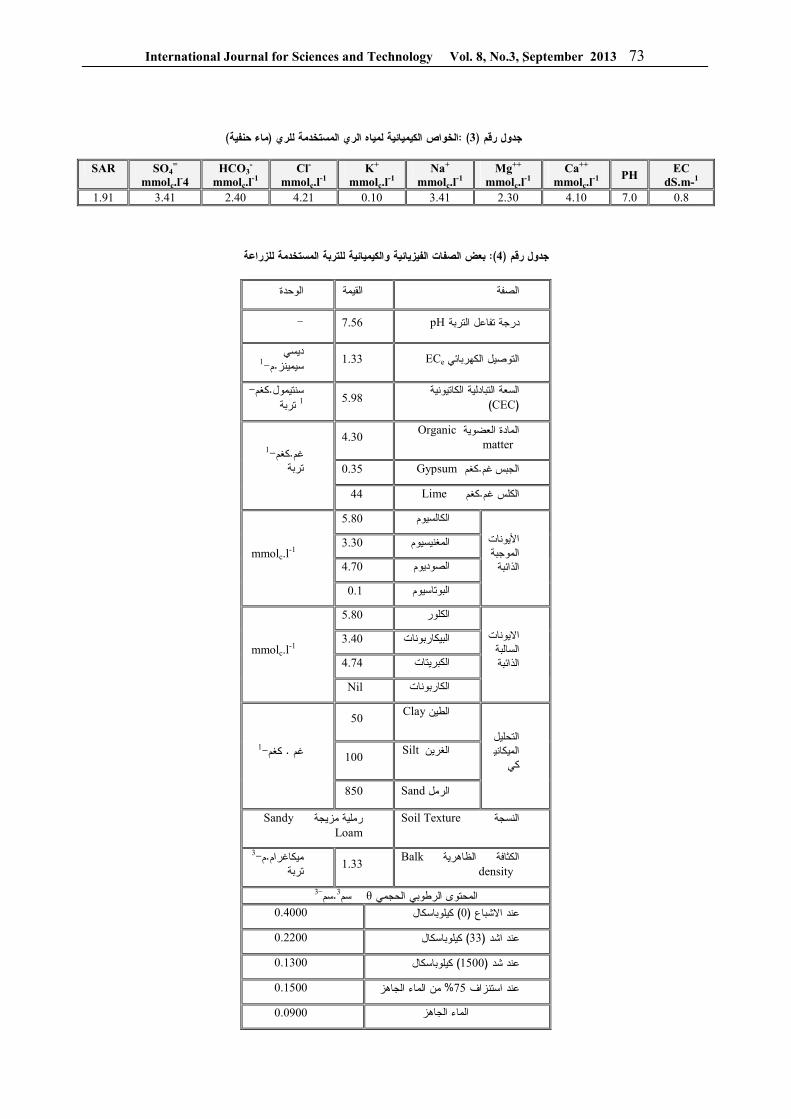
International Journal for Sciences and Technology Vol. 8, No.3, Sٍeptember 2013 73
)ماء حنفية(الخواص الكيميائية لمياه الري المستخدمة للري :) 3 ( رقمجدول
EC dS.m-1
PH Ca++
mmolc.l-1
Mg++ mmolc.l
-1 Na+
mmolc.l-1
K+ mmolc.l
-1 Cl-
mmolc.l-1
HCO3-
mmolc.l-1
SO4=
mmolc.l-4
SAR
0.8 7.0 4.10 2.30 3.41 0.10 4.21 2.40 3.41 1.91
بعض الصفات الفيزيائية والكيميائية للتربة المستخدمة للزراعة:)4 ( رقمجدول
الوحدة القيمة الصفة
- pH 7.56درجة تفاعل التربة
ECe 1.33التوصيل الكهربائيديسي 1- م.سيمينز
السعة التبادلية الكاتيونية )CEC(
5.98 - كغم.سنتيمول
تربة1
Organic المادة العضوية matter
4.30
Gypsum 0.35 كغم.غم الجبس
Lime 44 كغم.الكلس غم
1- كغم.غم تربة
5.80 الكالسيوم
3.30 المغنيسيوم
4.70 الصوديوم
األيونات الموجبة
الذائبة
0.1 البوتاسيوم
mmolc.l-1
5.80 الكلور
3.40 البيكاربونات
4.74 الكبريتات
االيونات السالبة الذائبة
Nil الكاربونات
mmolc.l-1
Clayالطين
50
Siltالغرين
100
التحليل الميكاني
كي
Sand 850الرمل
1-كغم. غم
Soil Textureالنسجة
Sandy رملية مزيجة Loam
Balkالكثافة الظاهريةdensity
1.33 3-م.رامميكاغ تربة
3-سم.3 سمθالمحتوى الرطوبي الحجمي
0.4000 كيلوباسكال) 0(عند االشباع
0.2200 كيلوباسكال) 33(عند اشد
0.1300 كيلوباسكال) 1500(عند شد
0.1500 من الماء الجاهز% 75عند استنزاف
0.0900 الماء الجاهز

International Journal for Sciences and Technology Vol. 8, No.3, Sٍeptember 2013 74
النتائج والمناقشة
جود تأثير معنوي لمغنطة المياه علـى و إلىأشارت النتائج بذور الميرمية حيـث كانـت نـسبة اإلنبـات إنباتنسبة
لمعاملة المحايد باستخدام مياه بـدون تعريـضها % 79.00فـي حـين ) Tap water ماء حنفيـة ( لشدة مغناطيسية
شـدود إلـى ازدادت نسبة اإلنبات عنـد تعـريض الميـاه كاوس 500نطة مياه عند مغ % 82.00 بلغت إذمغناطيسية
كـاوس 2000 و 1500 عنـد 83.00وبلغت ) 5جدول (بدون فرق معنوي بين الشدد المغناطيـسية علـى مـستوى
. فرق معنوي ألقل وفقا 0.05
بذور إنبات تأثير شدة مغنطة المياه على نسبة :)5 ( رقمجدول الميرمية
%اإلنبات نسبة شدة مغنطة المياه
a 79.00 ايدبدون مغنطة معاملة مح
b 82.00 كاوس500
b 83.00 كاوس1000
b 83.00 كاوس1500
b 83.00 كاوس2000
LSD 0.05=1.0
الذين حصلوا على ) 10(تتفق النتائج مع الجبوري وآخرين
مع مغنطـة الميـاه مقارنـة اإلنباتزيادة معنوية في نسبة حيـث )17 (وتتفق النتائج مـع . بالمياه في معاملة المحايد
معنوي الستخدام المياه المعالجة مغناطيسيا علـى تأثير بينت . بذور و بزوغ بادرات محاصيل الخضر إنبات
اذ ) 21( كما تتفق النتائج مع الجرواني وعبـد الـرحمن بذور المحاصـيل اسـتخدامهم ميـاه إنباتوجدا زيادة في
ممغنطة لري ثالثة محاصـيل هـي الحنطـة والطمـاطم .لياءوالفاصو
تأثير مغنطة ميـاه الـري فـي ) أ , 1( تبين النتائج شكل بالتداخل مـع معـامالت مـستويات ارتفاع نباتات الميرمية
بلغ أقل معدل ارتفاع نبات الميرمية عند معاملة إذ األسمدة، سم مقارنـة مـع 59.16) بدون مغنطة ( M0المقارنة
ياه الـري سم عند شدة مغنطة لم 75.49معدل أعلى ارتفاع 0.05 بفرق معنوي على مستوى المعنوية M4 كاوز 2000
وعدم وجـود ,LSD فرق معنوي ألقلبين المعامالت وفقا ب , 1( ويبين الـشكل M2 و M1 و M0فروق معنوية بين
مستويات التسميد بالتداخل مع معامالت مغنطة ميـاه تأثير) فـاع الري في ارتفاع نباتات الميرمية اذ بلغ معدل أقـل ارت
ارتفـاع أعلى ومعدل F0 سماديةإضافة سم بدون 59.73 هكتـار \ كغم 300 سم عند مستوى السماد 69.93للنباتات
F3 عـدم وجـود فـروق إلى اإلحصائي التحليل ، وأشار ووجود 0.05 على مستوى المعنوية F1 وF0معنوية بين
فـرق بأقل عند المقارنة F3 وF2 و F1فروق معنوية بين تأثير مغنطة ميـاه ) ج , 1(ويبين الشكل . LSD معنوي
إذالري والتسميد بدون تداخل في ارتفاع نباتات الميرميـة سم عند معاملة المقارنـة ري 54.33يبلغ معدل اقل ارتفاع
في حـين أعلـى M0F0بمياه بدون مغنطة وبدون تسميد 2000 سم عند ماء ري تحت شد مغناطيسي 74.00ارتفاع
اإلحصائي والتحليل M4F3هكتار \ كغم 300د اوس وسما ك وفقـا 0.05 معنوية على مستوى المعنوية فروقيبين وجد
. LSD ألقل فرق معنوي
d\{ )1( : W| hi}~]� dا�a[^و ا a]it[^وا �~�ii^ء اhi^ا Z]��z skراa^ت ا�qhoi^ s]qZ]i^ت اhzhcX عh�zار ) jk(

International Journal for Sciences and Technology Vol. 8, No.3, Sٍeptember 2013 75
ير مغنطة ميـاه الـري فـي تأث) أ , 2( تبين النتائج شكل الوزن الرطب للجزء الخضري لنباتات الميرمية بالتـداخل مع معامالت مستويات االسمدة اذ بلغ أقل معدل وزن رطب
) بدون مغنطـة ( M0لنبات الميرمية عند معاملة المقارنة غم 65.64 غم مقارنة مع معدل أعلى وزن رطب 56.50
ـ 2000عند شدة مغنطة لمياه الري و بفـرق M4اوز ك M2 و M1 و M0معنوي وعدم وجود فروق معنوية بين
مـستويات التـسميد تأثير) ب , 2(ويبين الشكل . M3و بالتداخل مع معامالت مغنطة مياه الري في الوزن الرطـب للجزء الخضري لنباتات الميرمية اذ بلغ معـدل أقـل وزن
لـى أع ومعدل F0 سماديةإضافة غم بدون 53.17رطب 300 غم عند مستوى الـسماد 64.64وزن رطب للنباتات
حصائي اإلويبين التحليل , وبفرق معنوي F3 هكتار \كغم بأقـل عند المقارنة F3 وF2بعدم وجود فروق معنوية بين
تـأثير مغنطـة ) ج , 2(ويبين الشكل . LSDفرق معنوي مياه الري والتسميد في الوزن الرطب للجـزء الخـضري
غـم عنـد 47.66 يبلغ معدل اقل وزن إذت الميرمية لنباتامعاملة المقارنة ري بمياه بـدون مغنطـة وبـدون تـسميد
M0F0 غم عند ماء ري تحت 66.43 في حين أعلى وزن هكتـار \ كغـم 300اوس وسـماد ك 2000شد مغناطيسي
M4F3 معنوية علـى فروق يبين وجد اإلحصائي والتحليل .ألقل فرق معنوي وفقا 0.05مستوى المعنوية
تأثير الماء الممغنط والتسميد و التداخل بينهما في : )2( شكل للجزء الخضري لنباتات الميرمية لمعامالت الدراسة الوزن الرطب
)أصيص / غم (
تأثير مغنطة ميـاه الـري فـي ) أ , 3( تبين النتائج شكل خل مع الوزن الجاف للجزء الخضري لنباتات الميرمية بالتدا
معامالت مستويات االسمدة اذ بلغ اقل معدل للوزن الجـاف ) بدون مغنطـة ( M0لنبات الميرمية عند معاملة المقارنة
غـم 36.63 غم مقارنة مع معدل أعلى وزن جاف 32.52 وبفـرق M4 كـاوز 2000عند شدة مغنطة لمياه الـري
تاثير مـستويات التـسميد ) ب , 3(ويبين الشكل , معنوي لتداخل مع معامالت مغنطة مياه الري في الوزن الجـاف با
للجزء الخضري لنباتات الميرمية اذ بلغ معـدل أقـل وزن ومعدل اعلـى F0 غم بدون اضافة سمادية30.07رطب
300 غم عند مستوى الـسماد 37.14وزن جاف للنباتات بين التحليل االحصائي على وجود فوقـات F3 هكتار \كغم
بين المعـامالت عنـد 0.05توى المعنوية معنوية على مس ) ج , 3(ويبين الـشكل ، LSDالمقارنة باقل فرق معنوي
تأثير مغنطة مياه الري والتسميد في الوزن الجاف للجـزء 27.39الخضري لنباتات الميرمية اذ يبلغ معدل اقـل وزن
غم عند معاملة المقارنة ري بمياه بـدون مغنطـة وبـدون غم عند الـري 38.17ن أعلى وزن في حي M0F0تسميد
300 غـاوس وسـماد 2000بماء تحت شد مغناطيـسي والتحليل االحصائي يبين وجد فروقات M4F3هكتار \كغم
. وفقا ألقل فرق معنوي0.05معنوية على مستوى المعنوية
تأثير الماء المعالج مغناطيسيا والتسميد و التداخل : )3( شكل
اف لنباتات الميرمية لمعامالت الدراسةبينهما في الوزن الج

International Journal for Sciences and Technology Vol. 8, No.3, Sٍeptember 2013 76
تأثير مغنطة ميـاه الـري فـي ) أ , 4( تبين النتائج شكل المساحة السطحية ألوراق نباتات الميرميـة بالتـداخل مـع معامالت مستويات االسمدة اذ بلغ أقـل معـدل للمـساحة
بدون مغنطة ( M0السطحية لألوراق عند معاملة المقارنة عند 2 سم 8.51مقارنة مع معدل أعلى مساحة 2 سم 6.23)
, وبفرق معنـوي M4 كاوز 2000شدة مغنطة لمياه الري تاثير مستويات التسميد بالتداخل مع ) ب , 4(ويبين الشكل
معامالت مغنطة مياه الري في المساحة السطحية لـألوراق F0 بدون اضافة سمادية2 سم6.53اذ بلغ معدل أقل مساحة
300 عند مستوى السماد 2 سم 7.91 مساحة ومعدل اعلى يبين التحليل االحصائي الى وجـود فـرق F3 هكتار \كغم
علـى مـستوى المعنويـة F3 و F0معنوي بين المعاملة ويبين الـشكل . LSD عند المقارنة باقل فرق معنوي 0.05
تأثير مغنطة مياه الري والتـسميد فـي المـساحة ) ج , 4(ات الميرمية اذ يبلغ معدل اقل مـساحة السطحية الورقية لنبات
عند معاملة المقارنة ري بمياه بـدون مغنطـة 2 سم 5.215 2 سـم 9.211 في حين أعلى مساحة M0F0وبدون تسميد
غـاوس وسـماد 2000عند ماء ري تحت شد مغناطيسي والتحليل االحصائي يبين وجـد M4F3هكتار \ كغم 300
وفقا ألقل فرق 0.05فروقات معنوية على مستوى المعنوية .معنوي
تأثير الماء الممغنط والتسميد و التداخل بينهما في : )4( شكل )2سم( المساحة السطحية الوراق نباتات الميرمية
تأثير مغنطة ميـاه الـري فـي ) أ , 5( تبين النتائج شكل تركيز الزيت في الجزء الخضري لنباتات الميرمية بالتداخل
تويات االسمدة اذ بلغ أقـل معـدل تركيـز مع معامالت مس الزيت في الجزء الخضري للنباتات عند معاملـة المقارنـة
M0 ) مقارنة مع معـدل أعلـى % 2.38) بدون مغنطة كـاوز 2000عند شدة مغنطة لمياه الـري % 2.71تركيز
M4 تاثير مستويات ) ب , 5( وبفرق معنوي ويبين الشكلت مغنطة مياه الري في تركيـز التسميد بالتداخل مع معامال
بـدون اضـافة % 1.81الزيت اذ بلغ معدل أقل تركيـز عنـد مـستوى % 3.29 ومعدل اعلى تركيز F0 سمادية ويبين التحليل االحصائي الـى F3 هكتار \ كغم 300السماد
بين جميع 0.05وجود فروق معنوية على مستوى المعنوية . LSDق معنـوي معامالت التسميد عند المقارنة باقل فر
تأثير مغنطة مياه الري والتسميد في ) ج , 5(ويبين الشكل عند معاملة % 1.51تركيز الزيت اذ يبلغ معدل اقل تركيز
فـي M0F0المقارنة ري بمياه بدون مغنطة وبدون تسميد عند ماء ري تحت شد مغناطيسي % 3.41حين أعلى تركيز
والتحليـل M4F3هكتار \ كغم 300 غاوس وسماد 2000االحصائي يبين وجد فروقات معنوية على مستوى المعنوية
. وفقا ألقل فرق معنوي0.05
تأثير الماء الممغنط والتسميد و التداخل بينهما في : )5( شكل ( % )تركيز الزيت في الجزء الخضري لنباتات الميرمية

International Journal for Sciences and Technology Vol. 8, No.3, Sٍeptember 2013 77
يتبين من النتائج وجود تـأثيرات معنويـة علـى مـستوى لمستويات الشدة المغناطيـسية و لمـستويات 0.05عنوية الم
االسمدة والتداخل بينهما في معدل ارتفاع نباتات الميرميـة والزيادة في الوزن الرطب والجاف والمـساحة الـسطحية
.لالوراق وتركيز الزيت الفعال للجزء الخضري للنباتاتان زيادة الوزن الرطب والجاف للجزء الخضري لنباتـات
رمية هو انعكاس الى زيادة اطوال وحجم النبات ويعزى الميسبب ذلك الى استجابة النباتات الى زيادة الشدة المغناطيسية
ان اضـافة . لمياه الري و المستويات الـسمادية المـضافة االسمدة الكيميائية وخاصة النيتروجين تسبب زيادة في النمو
عاتـه ممـا الخضري الذي يتمثل بزيادة ارتفاع النبات وتفر . يسبب زيادة الوزن الرطب والجاف للنباتات
يعزى سبب الزيادة الى التأثير األيجـابي للتـسميد وتـأثير النيتروجين في نشاط األنـسجة المرسـتيمية و دوره فـي األنقسام الخلوي ودخول النيتروجين في تركيب الكلورفيـل وفي تكوين العديد من مركبات واألنزيمات مما تـؤثر فـي
ادة نمو النبات والى تأثير الفسفور فـي عمليـة تكـوين زيوانقسام الخاليا واشتراكه في تحفيز وتطور الجـذور ممـا يسبب زيادة كفاءة النبات المتصاص المغذيات مـن التربـة
اما الزيادة الناتجة في صفات النمو الناتجة مـن الـري ) 5(لمياه فـي بالمياه الممغنطة فيعزى الى التأثير األيجابي لهذه ا
زيادة وحركة المغذيات في المنطقة الجذرية وبالتالي زيـادة ).9(دخولها الى النبات من خالل المنظومة الجذرية
في زيادة النمو عند )9 (وآخرين Hilalتتفق النتائج مع استخدام المياه الممغنطة للري و تتفق مع الجبوري وآخرون
حنطة عند اسـتخدامهم اذ وجدوا زيادة في نمو نبات ال ) 10() 12(وتتفق النتائج مع عبد المـنعم , المياه الممغنطة للري
الذي وجد زيادة في ارتفاع نباتات الذرة الصفراء عند الري كما تتفق . بمياه معالجة مغناطيسياً في ترب كلسية وجبسية
الذين وجدوا ان استخدام ماء ) 11(النتائج مع فهد وآخرون كاوس ادى الى زيادة معنوية في ارتفاع 2500ممغنط بشدة
وتتفق .النبات والوزن الجاف والرطب لنبات الذرة الصفراءالذين وجدا تاثير ذو ) Amira ) 8 وHozaynالنتائج مع
مغزى الستخدام المياه المعالجة مغناطيسيا في صفات النمـو .واالنتاج لنبات الحنطة
اذ وجد ان هنـاك ) 6( كما وتتفق النتائج مع المعاضيدي زيادة في كل من اطوال النباتات والمساحة السطحية الورقية والوزن الرطب والجاف للمجموع الخضري عند الري بمياه
كما تتفـق النتـائج مـع . ممغنطة لنباتات الزينيا والجيربرا اذ وجدا زيادة فـي تحـسين ) 21(الجرواني وعبد الرحمن
دامهم مياه ممغنطـة لـري صفات النمو واالنتاج عند استخ ال تتفـق .ثالثة محاصيل هي الحنطة والطماطم والفاصولياء
كونهم لم يحـصلوا علـى ) 5(النتائج مع الجلبي وأحسان تأثيرات معنوية للماء المعالج مغناطيسيا على صفات النمـو
.واالنتاج لنباتات الذرة الصفراء المياه الممغنطة تبين من خالل النتائج التأثير األيجابي للري ب
الى زيادة في نمـو DAP اللـ بالتداخل مع استخدام سمادنباتات الميرمية الوزن الرطب والـوزن الجـاف لنباتـات
.الميرمية والزيت الفعال في األوراق
International Journal for Sciences and Technology Vol. 8, No.3, Sٍeptember 2013 78
المصــادر النباتات واالعـشاب ) . 2000. (الشحات نصر , ابو زيد .1
.80ص.بيروت.الدار العربية للنشر والتوزيع.الطبية 2. Kintzios S E. (2000) . Sage: The Genus Salvia. CRC Press. pp. 10–11 .2 3. Guyton AC; and Hall JE. (2011). Medical physiology. 12th ed .Saunders.P.551 .3 4. Akhondzadeh S; Noroozian M; Mohammadi M; Ohadinia S; Jamshidi AH; and Khani M. (2003). Salvia officinalis extract in the treatment of patients with mild to moderate Alzheimer's disease: a double blind, randomized and placebo-controlled trial". J Clin. Pharm. Ther. 28 (1): 53–59.
). 2012. (فائق توفيق و احسان نواف دحـل , الجلبي . 5تأثير مياه الري الممغنطة ومستويات األسمدة فـي صـفات
ـ مجلـة العلـوم . النمو لحنطة الخبز .العراقيـة ة الزراعي43)1:(10 – 24.
تـأثير التقنيـة ). 2006(علي فاروق قاسم ,المعاضيدي . 6ـ . المغناطيسية في بعض نباتات الزينة . ة دكتـوراة اطروح
.جامعة بغداد. كلية الزراعة. قسم البستنة 2009. سامي كريم محمد و علي فاروق قاسـم , أمين . 7تأثير ملوحة ماء الري الممغنط في صفات النمو الخضري .
مجلة جامعة دمشق .Gerbera Jamesoniiلنبات الجربيرا .74 – 63 :)1 (25. للعلوم الزراعية
8. Hozayn M; and Abdul Qados AM. (2010) . Magnetic water application for improving wheat (Triticum aestivum L.) crop production. Agric. Biol. J. N. Am. 1(4): 677-682. 9. Hilal MH; Shata SM; Abdel Dayem AA; and Hilal MM. (2002). Application of magnetic technologies in desert agriculture : III . Effect of magnetized water on yield and uptake of certain element by citrus in relation to nutrient mobilization in soil. Egypt .J. Soil Sci. 42(1): 43-55.
, اشواق شنان عبـد , عبد الجاسم محيسن , الجبوري . 10صـالح محمـد , مجيد ضحى ميسر , خالد عباس رشيد
تـأثير مغنطـة . )2011(. حسن و ايمان نعمان اسماعيل مياه الري المالحة في انبات ونمو بـادرات حنطـة الخبـز
Triticum aestivum L. مجلة مركز بحـوث التقنيـات .38-32): 3(5. اإلحيائية
عدنان شبار فالح , قتيبة محمد حسن , علي عبد , فهد . 11التكييـف المغناطيـسي . )2005(.و طارق لفتـة رشـيد
الـذرة : لخواص المياه المالحة ألغراض ري المحاصـيل ) 1(36.مجلة العلوم الزراعية العراقية . الصفراء والحنطة
:34-29. تأثير مغنطة مياه . )2008(.سنان نزار , عبد المنعم . 12
الري في بعض الصفات الفيزيائية لعينات ثالث ترب كلسية رسـالة . Zea mays Lو الـذرة الـصفراء وجبسية ونم
– كليـة الزراعـة –قسم علوم التربة والميـاه , ماجستير .جامعة بغداد
كامل سعيد جواد و سيد عرفان ,محمد عبيد , السعدي . 13 –اساسيات انتاج المحاصـيل الحقليـة . )1984(. راشد
- دار التقنـي للطباعـة -مؤسسة المعاهد الفنية . العملي . 113- 107-106ص . داد بغ
. )1990(.مدحت و كريمة محمد وهيـب , الساهوكي . 14وزارة التعليم العالي . تطبيقات في تصميم وتحليل التجارب
دار الحكمـة للطباعـة , جامعـة بغـداد , والبحث العلمي .226 - 221ص .شرنوالاألسمدة وخـصوبة . )1987(. سعد اهللا نجم , النعيمي . 15
وزارة التعلـيم العـالي والبحـث .معة الموصل جا. التربة .167-151-122ص . جمهورية العراق.. العلمي
زراعة ري تسميد افـات . )2005( . إرشاديةنشرة . 16قـسم بحـوث النباتـات .امراض حصاد محصول الميرمية
مجلـة الخيـرات الزراعيـة . معهد بحوث البساتين .الطبية . 985نشرة رقم .
17. Hilal MH; and Hilal MM. (2000). Application of Magnetic Technologies in desert agriculture I. Seed Germination and seedling emergence of some crops in a saline calcareous soil. Egypt . J. Soil Sci. 40(3): 413-422. 18. Walch SG; Thomas K; Wolf S; and Dirk W L. (2011). Determination of the biologically active flavor substances thujone and camphor in foods and medicines containing sage (Salvia
officinalis L.). . Chem. center J. (5): 44. 19. Black CA. (1965). Method of Soil Analysis. Part(1). Physical properties. Am. Soc. Agron. Inc. Publisher, Madison, Wisconsin, USA. 20. Jackson ML. (1979). Soil Chemical Analysis. Advanced Course, 2nd printing. Madison, Wis.
عبد الرحمن بـن محمـد , محمد محمد , الجرواني . 21تـأثير . )2011(. المديني و أحمد بن عبد العزيز العمران
المحاصيل الزراعيـة إنتاجيةمغنطة مياه الري على نمو و .جامعة الملك فيصل.

International Journal for Sciences and Technology Vol. 8, No.3, Sٍeptember 2013 79
في بعض مؤشرات النمو الخضري لفسائل نخيل التمرHumic acidتأثير الحامض العضوي
L. Phoenix dactylifera المروية بمياه اآلبار
)3(، علي حيدر عريان)2 (، رعد طه بالكت)1( جمال أحمد عباس جمهورية العراق/ للنخيل الهيئة العامة) 3(الكوفة / المعهد التقني ) 2(جامعة الكوفة / كلية الزراعة ) 1(
[email protected]: البريد االليكتروني
العربية الملخص باللغة
فسيلة من نخيل التمر بعمر سنة واحدة لكل من الصنفين نيرسي ) 20( حيث تم اختيار 2013 و 2012نفذ البحث خالل العامين , كوفة / وسلطاني متماثلة تقريبا في الحجم والمجموع الخضري مزروعة في بستان النخيل بقسم االنتاج النباتي في المعهد التقني
% 12 السائلة تركيز Humic acidاضيفت مادة حامض الهيوميك , متر 23ي بالتنقيط من بئر عمق استخدمت طريقة الر بمقدار لتر واحد من المحاليل 2012 / 2 / 5 شهرا متتاليا اعتبارا من 12إلى التربة المحيطة بكل فسيلة مرة واحدة شهريا لمدة وقد استخدمت خمسة ) مللتر من حامض الهيوميك 8 أو 4 , 2, صفر (المائية المحضرة بأربعة معامالت اشتملت الكميات
وتم قياس معنوية الفروق ) . C . B . D. R(صمم البحث وفق تصميم القطاعات كاملة التعشية . لكل معاملة ) فسائل ( مكررات % .5بمستوى احتمال ) .L . S. D(بين المتوسطات بواسطة الفرق المعنوي األصغر
سبب حصول زيادة % 12 شهر من تاريخ تنفيذ البحث بأن استخدام حامض الهيوميك 13 النتائج المحسوبة بعد مرور أظهرتمعنوية في عدد األوراق الجديدة وطول الورقة لفسائل كال الصنفين كما سبب زيادة في محتوى األوراق من عنصري النتروجين
لكل % 12 مللتر من حامض الهيوميك 8أفضل النتائج هي عند استخدام المعاملة والبوتاسيوم والكلوروفيل والكربوهيدرات وكانت .فسيلة شهريا
ABSTRACT
The current study was carried out during the years 2012 and 2013. Twenty date palm offshoots with one year old were selected from each of Nersy and Sultani cultivars almost similar in the shoot and size planted in the orchard of the plant production department / technical institute at Kufa / Iraq. Drip irrigation systems were used from a well depth of 23 meter for the purpose of irrigation , liquid humic acid 12% with four treatment ( 0 , 2 , 4 , or 8 ml. ) was added to the soil around the offshoots once a month for 12 months from 5 / 2 / 2012 by one liter of the water solutions prepared for these treatment. Randomized complete block design was used with five replicates (offshoots) in each treatment. The results were calculated after 13 months from the date of conducted the study and indicated that used humic acid 12% caused significantly increase in the number of new leaves and leaf length in offshoots for both cultivars ,also caused increased in leaves contain of nitrogen, potassium , chlorophyll and carbohydrate. Best results showed when using the treatment of humic acid 12 % at 8 ml./ Offshoot monthly .

International Journal for Sciences and Technology Vol. 8, No.3, Sٍeptember 2013 80
المقدمة
المهتمون بدراسة نخلة التمر من الثابت لدى المزارعون وبأنها تتحمل ملوحة التربة والماء أكثر من العديد من أشجار الفاكهة األخرى حيث تستطيع النخلة تحمل ملوحة الماء ومقاومة ضرر التراكيز العالية من كلوريد الصوديوم لفترة طويلة دون ضرر في النمو واإلنتاج لحد تركيز ملحي
ون لكن زيادتها عن ذلك سوف جزء بالملي6000مقداره يؤثر في نموها وإنتاجها بنسب تتفاوت حسب كمية الملوحة
).2، 1(في ماء الري إن لملوحة التربة أو مياه الري تأثيرات سمية على النبات حيث تسبب إحداث زيادة في تركيز ايونات الصوديوم والكلور والبورون في أنسجة النبات يرافقها تثبيط جاهزية
عناصر المعدنية لالمتصاص من قبل النبات وبالتالي المي اإلخالل بالفعاليات الحيوية والتوازن الغذائي و اإلنزي
وفي ظل ظروف ، )4، 3(والهرموني في خاليا أنسجته شحة مياه األنهار في العراق صار االتجاه حاليا إلى استخدام
ملحية مياه اآلبار في عمليات الري والتي تعتبر ذات تراكيز ولغرض دراسة إمكانية تقليل ,عالية مقارنة بالمياه العذبة
ضرر اإلجهاد المتسبب من زيادة ملوحة ماء الري فقد نفذ هذا البحث على فسائل تخيل التمر المروية بمياه اآلبار مع
السائلة وهو ) ( Humic acidاستعمال حامض الهيوميك المحلية بشكل من األحماض العضوية المتوفرة في األسواق
حيث تسبب إضافتها للتربة ) حبيبات ( سائل أو صلب تحسين خواصها وزيادة محتواها من العناصر المعدنية الجاهزة لالمتصاص من قبل جذور النباتات وحماية النبات
وقد ،)5(وبالتالي تحسين النمو واإلنتاج من اإلجهاد الملحي ي وهما من استخدمت فسائل الصنفين النيرسي والسلطان
األصناف الشائعة في المنطقة الوسطى من العراق ثمارهما ذات قيمة غذائية واقتصادية تسوق وتؤكل في مرحلتي
.الرطب والتمر
المواد وطرق العمل
في 2013 و 2012نفذت الدراسة الحالية خالل العامين المعهد التقني في الكوفة / بستان النخيل بقسم االنتاج النباتي
شجرة نخلة تمر بعمر سنة واحدة لكل ) 20(يث اختيرت حمن الصنفين نيرسي وسلطاني متماثلة تقريبا من حيث الحجم
* 10وطبيعة النمو الخضري مزروعة بالطريقة الرباعية متر جلبت فسائلها من محطة النخيل في محافظة النجف 10
الهيئة العامة للنخيل وقد استخدمت طريقة الري / االشرف ( م و تم تثبيت اعداد االوراق ) 23(بالتنقيط من بئر بعمق
استعمل الحامض . لكل فسيلة قبيل تنفيذ البحث ) السعف اضافة % 12 بتركيز Humic acidالعضوي السائل
) مللتر 8 أو 4 , 2, صفر ( ارضية بأربعة معامالت وذلك بعد تخفيفها بمقدار لتر واحد من الماء المقطر
ا إلى التربة المحيطة بكل فسيلة مرة واحدة شهريا واضافتهكما تم . 2012 / 2 / 5 شهرا متتالية اعتبارا من 12لمدة
سم 50تحليل ماء الري وتربة البستان من عمق صفر ــ وكانت المواصفات الفيزياوية والكيميائية كما موضحة في الملحق المرفق وقد اقتصر البستان بزراعة الفسائل ولم
.سمد طيلة فترة الدراسة يصممت الدراسة وفق تصميم القطاعات كاملة التعشية بخمسة مكررات لكل معاملة حيث اعتبرت كل فسيلة وحدة تجريبية وحللت النتائج باستخدام الحاسوب اآللي حسب نظام
Gen stat وتم اختبار معنوية الفروق بين المتوسطات % 5 احتمال بواسطة الفرق المعنوي األصغر بمستوى
)6 .(
: قياسات الصفات المدروسة / 5 شهر من تاريخ بدء تنفيذ البحث وبتاريخ 13بعد مرور
تم إجراء القياسات التالية في جميع المكررات 2013 / 3جامعة /كوفة وكلية الزراعة / بمختبرات المعهد التقني
. الكوفة :القياسات الحقلية للنمو الخضري : أوال
حسب عدد األوراق الجديدة ) : السعف ( دد األوراق ـ ع1 التي ظهرت بعد األوراق الثالثة للقمة النامية ثم حسب
.المعدل لكل معاملة حسب طول كل ورقة جديدة بواسطة : ـ طول الورقة 2
وريقة شريط القياس من بداية قاعدتها إلى مكان اتصال آخر .تر لكل معاملة بالسعفة ثم حسب المعدل بالسنتم) خوصة (
أخذت عينات من وريقات : القياسات المختبرية : ثانيا لجميع ) 7(الورقة األقدم عمرا من األوراق الجديدة
:المكررات وتم حساب القياسات التالية بعد غسل : ـ محتوى الورقة من العناصر المعدنية 1
درجة مئوية حتى 70وتقطيع النماذج جففت بالفرن بدرجة غم 0,5نها ثم طحنت العينات المجففة واخذ منها استقر وز
% ) H2SO4 ) 96خلطت مع حامض الكبريتيك المركز ، ) 8( وفقا لطريقة % ) HCL3 ) 4وحامض البلكلوريك
ثم حسبت النسبة المئوية للنتروجين بواسطة جهاز أما النسبة المئوية للبوتاسيوم فقد حسبت , مايكروكلدال
.Flame photometer بواسطة جهاز اللهبتم حساب تركيز : ـ محتوى الورقة من الكلوروفيل 2
اليدوي بطريقة SPAD meterالكلوروفيل بواسطة جهاز / مايكروغرام 1 تساوي SPAD، حيث ان كل وحدة )9(
.لتر حسبت النسبة : ـ محتوى الورقة من الكربوهيدرات 3
جهاز المطياف المئوية للكربوهيدرات الكلية الذائبة بواسطة 490 على طول موجي Spectro photometerالضوئي
n.m 10( حسب طريقة.(
النتائج والمناقشـة
: النمو الخضري: أوال
8بأن استعمال حامض الهيوميك بمقدار ) 1( يوضح شكل مل سبب زيادة معنوية في إنتاج األوراق الجديـدة لفـسائل
قت الزيادة علـى جميـع صنفي النيرسي والسلطاني وقد تفو المعامالت األخرى المستعملة حيث بلغ معدل عـدد األوراق
ورقـة و 4,80الجديدة في الفسائل المعاملة بهذه الكميـة ورقة في كال الصنفين على التوالي بينما ال نالحـظ 4,60
مـل فـي زيـادة عـدد 2تأثيرا معنويا الستعمال المعاملة مقارنة بفسائل معاملة المقارنـة األوراق للفسائل المعاملة به
وقد بلغ فيها معدل عدد األوراق الجديدة اقـل مـن جميـع ورقـة و 3.80معامالت حامض الهيوميك األخرى وهـي
كما نالحظ فـي , ورقة في كال الصنفين على التوالي 3.40 مل لم يؤثر معنويا في زيادة 4الشكل بأن استعمال معاملة
مقارنة باستعمال معاملة عدد األوراق للصنف سلطاني

International Journal for Sciences and Technology Vol. 8, No.3, Sٍeptember 2013 81
ورقة في 4.60 ورقة و 4.40 مل حيث بلغ عدد األوراق 8فسائل هاتين المعاملتين بعكس الحالة في فـسائل الـصنف
.نيرسي التي اختلفت معنويا
تأثير حامض الهيوميك في معدل عدد األوراق الجديدة): 1(شكل
في معدل فهو يوضح تأثير المعامالت ) 2( أما الشكل طول الورقة حيث نالحظ تفوق معنوي في زيادة طول الورقة لفسائل صنفي النيرسي و السلطاني عند استخدام
مل من حامض الهيوميك مقارنة بجميع 8المعاملة 80,20 سم و 82,60المعامالت األخرى حيث بلغ المعدل
بينما ال نالحظ وجود تأثير معنوي , سم فيهما على التوالي مل من حامض الهيوميك في 4 مل أو 2لمعاملتين ل
تأثيرهما على زيادة طول الورقة في الفسائل المعاملة بهما مقارنة بفسائل معاملة المقارنة وقد كان اقل معدل هو عند
سم 60 , 64 سم و 80 , 65 مل إذ بلغ 2استعمال المعاملة .في كال الصنفين على التوالي
حالة عدم التوازن الغذائي المتسببتان إن التأثير االسموزي ومن تأثيرات الشد الملحي الناتج من ارتفاع ملوحة ماء الري سببا قلة امتصاص الماء والعناصر الغذائية واختزال عملية االنقسام والتنظيم الخلوي وإعاقة بناء الحوامض النووية و الهرمونات النباتية مثل االوكسينات والجبرلينات وتقليل نمو
لذلك نالحظ ضعف النمو الخضري ) . 13 -11(الجذور لفسائل معاملة المقارنة بينما سببت الحموضة الهيومية المضافة للتربة في معامالت حامض الهيوميك تحسين
الغذائية مما ادى الى زيادة قوة امتصاص الماء والعناصرنمو المجموعة الجذرية وتحسينها وزيادة تكوين الهرمونات
وقد , ) 15، 14 (اتية وبالتالي تحسين النمو الخضريالنب بأن زيادة ملوحة ماء بينت حيث ،)16(نتائج دراسة أيد ذلك
الري تسبب انخفاضا واضحا في النمو الخضري وإنتاج األوراق الجديدة لنبيتات نخيل التمر وان إضافة البوتاسيوم إلى ماء الري سبب حصول زيادة في معدالت النمو
.ري الخض
)سم(تأثير حامض الهيوميك في معدل طول الورقة ): 2(شكل
:محتوى الورقة من العناصر المعدنية: ثانيا
محتوى األوراق من ) 3( توضح النتائج في الشكل النتروجين حسب المعامالت المستعملة إذ نالحظ بأن نسبة
ت حامض العنصر ازدادت معنويا في األوراق لجميع معامالالهيوميك لكال الصنفين مقارنة بالنسبة في أوراق معاملة
مل معنويا على 8المقارنة وقد تفوق استعمال المعاملة 0.944و % 0.914 مل حيث بلغت النسبة 4 و 2معاملتي
في أوراق صنفي النيرسي والسلطاني وقد تناسبت % 0.978الزيادة طرديا مع زيادة الكمية حيث بلغت اقل نسبة
مل لكال 2في أوراق فسائل معاملة % 0.830و% .الصنفين على التوالي
تأثير حامض الهيوميك في النسبة المئوية للنيتروجين في األوراق): 3(شكل

International Journal for Sciences and Technology Vol. 8, No.3, Sٍeptember 2013 82
) 4( أما محتوى األوراق من البوتاسيوم فيالحظ في الشكل ية مل من حامض الهيوميك سبب زيادة معنو8بأن معاملة
في النسبة المئوية للبوتاسيوم في األوراق حيث تفوق على % 0.234جميع المعامالت األخرى فقد بلغت النسبة
في صنفي النيرسي والسلطاني ونالحظ بان % 0.194و مل في زيادة النسبة كان معنويا فقط في 2تأثير المعاملة
أوراق الصنف نيرسي مقارنة بالنسبة في أوراق معاملة إن ارتفاع نسبة األمالح في % .0.188رنة إذ بلغت المقا
مياه الري يؤثر على النشاطين الفسلجي والفيزيائي لعملية امتصاص الماء ويعيق امتصاص وانتقال العناصر الغذائية وبالتالي انخفاض تركيز العناصر ومنها النتروجين
، وقد سببت )18، 17، 12(والبوياسيوم في النسيج النباتي حامض الهيوميك تحسين عملية امتصاص الماء إضافة
والعناصر الغذائية لذلك ازداد محتوى األوراق من عنصري وآخرون Kodoma، كما ذكر )5(النتروجين والبوتاسيوم
بأن حامض الهيوميك يزيد من تحرر البوتاسيوم في ) 19(محلول التربة ومن ثم زيادة امتصاصه من قبل النبات
.في األنسجة النباتية فتزداد مستوياته
تأثير حامض الهيوميك في النسبة المئوية للبوتاسيوم ): 4(شكل في األوراق
:محتوى الورقة من الكلوروفيل: ثالثا
بأن محتوى الورقة من الكلوروفيل ) 5( نالحظ في الشكل قد تفوق معنويا في الـصنفين النيرسـي والـسلطاني فـي
مل مقارنة بمعاملـة 8 مل و 4 معاملتي حامض الهيوميك مل التي لم تسبب تأثيرا معنويا 2المقارنة ومعاملة استعمال
و 50,074في زيادة المحتوى وقد بلغـت أعلـى كميـة في أوراق كال الـصنفين علـى SPAD وحدة 51,634
كما نالحظ من خالل , مل 8التوالي عند استعمال المعاملة ـ ة بـين نتـائج اسـتعمال الشكل عدم وجود فـروق معنوي
مل من حامض الهيوميك في الصنف 8 مل و 4المعاملتين نيرسي بعكس الحالة في الـصنف سـلطاني حيـث تفـوق
مل في زيادة 4 مل معنويا على معاملة 8استعمال المعاملة وحـدة 48,786 و 51,634المحتوى فقد بلغـت الكميـة
SPAD في أوراق المعاملتين .
من الكلوروفيل قد بلغ في فسائل معاملـة إن محتوى الورقة فـي كـال SPAD وحدة 42,096 و 40,128المقارنة
الصنفين وذلك بسبب تعرض الفسائل لإلجهاد المائي حيـث إن مستويات الملوحة العالية تؤدي إلـى زيـادة مـستويات
وزيـادة ) ABA(الهرمون النباتي حامضي االبسيـسك للذان يسرعان في عمليـة تحلـل فعالية إنزيم الكلوروفيليز ا
الكلوروفيل باإلضافة إلى تأثير الملوحة في تقليل امتـصاص العناصر الضرورية لبناء جزيئة الكلوروفيل مثل النتروجين
، لذلك فقد سـببت إضـافة حـامض )22-20(والمغنيسيوم الهيوميك زيادة محتوى األوراق من الكلوروفيل نتيجة تأثيره
عة الجذرية و زيادة امتصاص الماء في تحسين نمو المجمو والعناصر المعدنية من التربة وتكوين االوكسينات وتحسين
). 23، 15، 14( النمو الخضري
وحدة (تأثير حامض الهيوميك في محتوى الورقة من الكلوروفيل ): 5(شكل SPAD(
:النسبة المئوية للكربوهيدرات: رابعا
تعمال حامض الهيوميك سبب بأن اس ) 6( يوضح الشكل زيادة في النسبة المئوية للكربوهيدرات في أوراق كال الصنفين تناسبت طرديا مع زيادة الكمية المستخدمة من
و % 0.146الحامض في المعامالت حيث بلغت أعلى نسبة مل في كال الصنفين 8عند استعمال المعاملة % 0.136
وية مقارنة بالنسبة في على التوالي وكانت هذه الزيادة معن الكربوهيدرات نسبة فيها بلغت التي المقارنة معاملة أوراق
في اوراق كال الصنفين وذلك نتيجة % 0.059 % 0.080تأثير ارتفاع ملوحة ماء الري والتي أثرت على نشاط إنزيم
RUBP Carboxylase الضروري لتثبيت CO2 في ، 24( الضوئي سلسلة تفاعالت الظالم في عملية التركيب
،وقد سببت المعاملة بحامض الهيوميك زيادة نسبة ) 25الكربوهيدرات في األوراق نتيجة تأثيره في تحسين النمو
عندما تم ) 26، 23(الخضري وقد أيد ذلك نتائج دراسة استخدام حامض الهيوميك في تأثيره على نمو أشجار
.الزيتون وشتالت الكازورينا
International Journal for Sciences and Technology Vol. 8, No.3, Sٍeptember 2013 83
المصــادر
1. Furr JR; andBallard AL. (1966). Growth of young date palm in relation to soil salinity and chloride content of the pinnae . Date Growers Inst. Rept. 43:4 – 8.
نخلة التمر بين البحث ). 2005. (فرج ،كريم محمد . 2 . ظبيدولة االمارات العربية المتحدة، أبو. والتطبيق
3. Marschner H. (1995). Mineral nutrition of higher plant .2nd ed. Academic press. Inc. London. P.446.
استخدام النمذجة ). 2006(التميمي ،ابتهاج حنظل . 4 Phoenixالرياضية للتنبؤ بإنتاجية نخلة التمر
dactylifera L. صنف الحالوي تحت تأثير تغاير بعض اطروحة دكتوراه ، كلية الزراعة، جامعة . اإلنتاج عوامل
.البصرة ، العراق5. Cimrin KM; Onder T; Metin T; and Burcu T. (2010). Phosphorus and humic acid application alleviate salinity stress of pepper seedling. African J. biotechnol. 9 (36):58451.
. محمد العزيز عبد خلف اهللا، ; محمود شعخا ، الراوي. 6 الزراعة كلية . الزراعية التجارب وتحليل تصميم ).2000(
.العراق الموصل، جامعة والغابات،دراسة امكانية ). 1998(العاني ، مؤيد رجب عبود . 7
تمييز جنس النخيل في مرحلة البادرات بأستخدام الهجره . هه بالجبرلينات بيالكهربائية للبروتينات والمواد الش
. العراق، جامعة بغداد ،كلية الزراعة اطروحة دكتوراه، 8. Cresser MS; and Parsons JW. (1979). Sulphuric perchloric and digestion of plant material for the determination of nitrogen, phosphorus, potassium , calcium and magnesium . Anal . Chem . Acta . 109: 431- 463. 9. Felixloh J G; and Nina B. (2000). Use of the Minolta SPAD - 502 to determine chlorophyll concentration in Ficus benjamina L . and populous deltoids Marsh leaf tissue . Hort. Sci. 35 (3): 423. 10. Dubois MK; Crilles KA; Hamilton JK; Rebers DA: and Smith F. (1956). Colorimetric method for determination of sugar and related substances . Anal . Chem. 28 :350 – 356. 11. Bose TK. (1985). Fruit of indid , Tropical and subtropical , Naya prokash . India.
عالقة ). 1990. (م عبد اهللا النعيمي ، سعد اهللا نج. 12جامعة الموصل، العراق ، ص . التربة بالماء والنبات
532 . تأثير التداخل ). 2000(الحمداني ، فوزية محسن علي . 13
بين ملوحة الري والسماد الفوسفاتي على بعض خصائص اطروحة دكتوراه ،كلية الزراعة ، . التربة وحاصل النبات .جامعة بغداد ، العراق
14. Donnel RW. (1973). The auxin - like effects of humic preparation from leonardite . Soil Sci. 16 :106 – 112. 15. Senn TL; and Kingman AR. (1973). A review of humus and humic acid . South Carolina agricultural experiment station. Clemson, Sc. Research. Series report .No.145.
16. Rasmia D; and EL-Banna AA. (2011). Role of potassium and salinity effects on growth and chemical composite ions of date palm plantlets. J.Agric.Sci,Ain- shams univ. 19(1):233-244. 17. Szalai I. (1968). Plant biology . pp:223-256 . Budapest. 18. Julian W. (2004). Text citrus subtropical fruits nutritice and fertilization . Horticulture taxas cooperative extension. P. 145. 19. Kodoma H; and Schnitzer M; and Jack K. (1983). Chlorite and biotite weathering by fulvic solution in closed and opened system . J. Soil Sci. 63:619-629.
كلية . مبادئ علم التربة ). 1980(العاني ،عبد اهللا نجم . 20 . الزراعة ،جامعة بغداد ،العراق
21. Sun C X; Hong X; Cao H; Xin T; and Yongxiao O. (2011). Growth and physiological responses to water and nutrient stress in oil palm . African J. biotechnol. 10(51):10465-10471. 22. Jain BL; and Pareek OP. (1989). Effect of drip irrigation and mulch on soil and performance of date palm under saline water. The biochemical constituents of tomato plant. Zagazig J. Agric. Res.12: 417-441. 23. Tatini M; Bertoni P; Landi A; and Traverisi ML. (1991). Effect of humic acids on growth and biomass portioning of container - grown olive plants. Acta . Hort. 294 :75 – 80. 24. Seemann RJ; and Sharky TD. (1986). Salinity and nitrogen effects on photosynthesis, ribulos 1,5 by phosphate carboxylase and metabolite pool size in Phaseolus vulgaris L . Plant physiol. 82:555-560. 25. Sinha A; Gupta SR; and Rana RS. (1986). Effect of soil salinity and soil water availity on growth and chemical composition of Sorghum
halepense L. Plant and soil .95 :411-418. تأثير التسميد الحيوي ). 2008(درويش ، منى احمد . 26
وحامض ) بوتاسيوم , فسفور , نتروجين ( والسماد المتكامل الهيوميك على النمو والتركيب الكيمياوي لشتالت الكازورينا
): 3 (13نتاجية والتنمية مجلة اال. النامية في تربة رملية . جامعة الزقازيق، جمهورية مصر العربية169-177

International Journal for Sciences and Technology Vol. 8, No.3, Sٍeptember 2013 84
Klebsiella pneumoniae و Escherichia coli بكتريـا عـزالت مـن لكل الوراثية العالقة دراسة
اطـوال نـات تباي تقنية باستخدام الهضمية والقناة البولية والحصى المرارة حصى من حاالت من المعزولة
)DNA) AFLP الحمض النووي من المتضخمة القطع
)3(عقيل حسين علي ،)3( الجبوريعلي صالح ، )2 (فرحانمثنى بديع ، )1 (اياد محمد علي العبيدي قـسم ) 3. (رجامعة األنبـا / كلية التربية للبنات / قسم علوم الحياة ) 2. (جامعة النهرين / كلية العلوم / قسم التقنيات اإلحيائية ) 1(
. جمهورية العراق/ جامعة تكريت / كلية العلوم / علوم الحياة
[email protected]: البريد االليكتروني
الملخص باللغة العربية
عزالت 10 عزلة من حصى المرارة و 12( عزلة 26 من مستشفى الرمادي التعليمي والتي شملت Escherichia coliجمعت عزالت بكتيريا عزالت من حصى 4( عزلة 14 فشملت Klebsiellapneumoniaeاما عزالت بكتيري ) عزالت من القناة الهضمية4من الحصى البولية و
وقد بينت نتائج البعد الوراثي باستخدام تقنية تباينات اطوال القطع ). عزالت من القناة الهضمية4عزالت من الحصى البولية و 6المرارة و 14 و 13 لكل من العزلتين ) (0.4466فان اقل بعد وراثي كان E. coliوقد بينت نتائج البعد الوراثي بالنسبة لبكتيريا ) AFLP(المتضاعفة
E. coliوكالهما من عزالت الحصى البولية لذلك قد تكون كل من هاتين العزلتين هما الساللتين االقرب وراثيا من جميع سالالت بكتيريا احدى 24 وهي احدى عزالت حصى المرارة والعزلة 4 لكل من العزلة (0.8798)صى البولية اما اعلى بعد وراثي فقد كان المعزولة من الح
اما بالنسبة , تعود لساللة ذات نمط وراثي يختلف عن السالالت التي تم عزلها من القناة الهضمية4عزالت القناة الهضمية لذلك تكون العزلة وهما من عزالت القناة الهضمية والتي يمكن ان 12 و 11لكل من العزلتين ) 0.18757( فقد كان اقل بعد وراثي هو K. pneumoniaeلبكتيريا
اما . الموجودة في القناة الهضميةK. pneumoniaeتشير الى امكانية ان تكون كال العزلتين هما االقرب وراثيا من بين جميع عزالت بكتيريا وهي احدى عزالت الحصى البولية والعزلة 5 بين كل من العزلة (0.95256)فقد كان K. pneumoniaeت بكتيريا اعلى بعد وراثي بين عزال
قد تكون من مصدر اخر غير القناة الهضمية بل وقد 5 وهي احدى عزالت القناة الهضمية وهذا قد يفسر بان الساللة البكتيرية للعزلة رقم 11رقم كذلك لوحظ المجاميع . منطقة الجهاز البولي يمكن ات تتعرض الى وجود بكتيريا من مصدر خارجييكون مصدرها خارجيا خصوصا ان
المعزولة من حصى المرارة والحصى البولية والقناة الهضمية ان العالقة الوراثية بين عزالت E. coliالرئيسية للشجرة الوراثية لعزالت بكتيريا الحصى البولية اكثر مما هو لعزالت القناة الهضمية وهذا واضح من خالل وجود مجموعتين رئيسيتين حصى المرارة هي االقرب وراثيا لعزالت
ارتبطت فيها عزالت حصى المرارة وراثيا مع عزالت الحصى البولية ومن هنا نجد ان كل من البكتيريا المرافقة لحصى المرارة والبكتيريا في حين كانت هنالك مجموعة رئيسية واحدة ارتبطت فيها عزالت حصى , ت من مصدر واحد المرافقة لحصى الكلى قد تكونان في بعض الحاال
اما المجاميع , المرارة وراثيا مع عزالت القناة الهضميةكما لم نالحظ وجود اي عالقة وراثية بين عزالت الحصى البولية وعزالت القناة الهضمية المعزولة من حصى المرارة والحصى البولية والقناة الهضمية نجد عموما ان K. pneumoniaeالرئيسية للشجرة الوراثية لعزالت بكتيريا
العالقة الوراثية بين عزالت حصى المرارة هي االقرب وراثيا لعزالت الحصى البولية كما لم نجد اي من عزالت حصى المرارة والقناة الهضمية نفس الوقت لم نالحظ وجود اي عالقة وراثية بين عزالت الحصى البولية وعزالت ضمن عالقة وراثية متقاربة ضمن اي مجموعة رئيسية وفي
.القناة الهضمية في اي من المجاميع الرئيسية
ABSTRACT
Bacterial isolated were collected from Al-Ramadi Teaching Hospital from total samples 65 of gallstone , 42 of urine stone and 8 samples of stoole. Twenty six isolates of Escherichia coliwere collected (12 from gallstones , 10 from urine stones and 4 from gastrointestinal canal) and 14 isolates of Klebsiellapneumoniae (4 from gallstones , 6 from urine stones and 4 from gastrointestinal canal).The result of E. coli showed lower genetic distance (0.4466) between isolates 14 and 13 of urine stone while larger genetic distance (0.8798) between isolate 4 of gallstone and isolate 24 of gastrointestinal canal. The result of K. pneumoniae showed lower genetic distance (0.18757) between isolates 11 and 12 of gastrointestinal canal , while larger genetic distance (0.95256) between isolate 5 of urine stone and isolate 11 of gastrointestinal canal , this meaning isolate 5 from another source of gastrointestinal canal . The result of E. colialso showed there was genetic relationship between isolates of gallstone and urine stone more than between isolates of gallstone and gastrointestinal canal , this meaning probability bacteria of gallstone and urine stone frome similar source in the some cases . The result of K. pneumonia also showed there was genetic relationship between isolatesof gallstone and urine stone and not found any genetic relationship between isolates of gallstone and gastrointestinal canal and also between isolates of urine stone and gastrointestinal canal .

International Journal for Sciences and Technology Vol. 8, No.3, Sٍeptember 2013 85
المقدمة
تعد تقنية تباينات اطوال القطع المتضخمة من التقنيات البالغة االهمية في التشخيص الجزيئي وهي مختصر
)Amplified Fragment Length Polymorphism ( اذئمة ودقيقة للحصول على مؤشر البصمة تكون تقنية مال
لكائن حي معين سواء نبات او Finger printingالوراثية حيوان او بكتيريا ومن خالل هذه التقنية يمكن من خاللها االستفادة من نتائجها الجراء تحليالت وراثية اخرى كرسم الخارطة الوراثية لكائن معين وهذه التقنية تعتمد ايضا على
وفي هذه التقنية يمكن PCRفاعل البلمرة التسلسلي تقنية تالحصول على العشرات من قطع لدنا لكائن معين تتراوح
قطعة من الدنا المقطع بانزيمات قاطعة 100-50حوالي باستخدام مكيفات PCRمحددة والمضاعفة باستخدام تقنية
)Adapters ( ذات عدد قليل من النيوكليوتيدات))Oligonucleotides وتسلسالت من الدنا مقطعة للكائن
الحي يتم التعرف عليها كمواقع هدف ترتبط معها البادئات . )8-1(الخاصة حسب نوع االنزيم القاطع المستخدم
في دراستنا هذه على مستوى AFLPاستخدمت تقنية باالضافة الى ان , االحياء المجهرية للمرة االولى في العراق
محتمل ان يكون االول من نوعه من الAFLPتطبيق تقنية على المستوى العالمي تم تطبيقه الجراء المقارنة الوراثية
المعزولة K. pneumoniae و E. coliلعزالت بكتيريا من حصى المرارة وحصى الجهاز البولي والقناة الهضمية اي من ثالث بيئات مختلفة من جسم االنسان حيث لم نجد
في شبكة المعلومات الدولية الى ما في الدوريات العلمية او .يشير الى ذلك
المواد وطرق العمل
:عزل وتشخيص البكتيريا من K. pneumoniae و E. coliعزلت كل من بكتيريا
عينات حصى المرارة والحصى البولية والقناة الهضمية من الى 24/5/2010مستشفى الرمادي التعليمي للفترة من
اجراء الزرع البكتيري بعد الحصول اذ تم ,17/3/2011على العينات مباشرة وذلك باستخدام الطرق التقليدية المتبعة لزرع العينات مع مراعاة الظروف المعقمة ابتداءاً من
لغرض تشخيص العزالت أجريت و.مرحلة اخذ العينات (9)االختبارات الكيموحيوية باالعتماد على مصنف بركي
لغرض تأكيد تشخيص البكتيريا كما (10)وما تم إجرائه وفق API 20 وهي API Systemتم استعمال عدة التشخيص
E . عزلة لبكتيريا 26وشملت العزالت البكتيرية E.
coli عزالت 10 عزلة من حصى المرارة و 12تمثلت بـعينات (عزالت من القناة الهضمية 4من الحصى البولية و
14 فقد شملت K. pneumoniaeاما بكتيريا , ) برازيةعزالت من حصى المرارة و 4عزلة بكتيرية تمثلت بـ
عزالت من القناة الهضمية 4عزالت من الحصى البولية و 6 ).عينات برازية(
:استخالص الدنا الجينومي لبكتيريا استخلصت عينات الدنا الجينوميللعزالت البكتيرية
E. coli و بكتيريا K. pneumoniae باستخدام عدة للبكتيريا السالبة لصبغة گرام جينوميستخالص الدنا الا
ذي الرقم (Promega)المنتجة من قبل شركة بروميكا Wizard® Genomic باسم منتج A1120التسلسلي
DNA Purification Kit .
:DNAتركيز اجري توحيد التراكيز الخاصة للدنا الجينوي الذي تم
لغرض ng /1 µl 250استخالصه من العزالت البكتيرية واجري العمل تحت ظروف مشددة AFLPاجراء اختبار
.من تعقيم المواد ومكان العمل
Amplified Fragment Length (AFLP)تفاعالت Polymorphism: ثالث مراحل رئيسية وهي (AFLP)وقد تضمن اختبار
AFLPاستخدمت عدة اختبار تقنية AFLPمرحلة تفاعالت وقد تم اجراء Gibco BRLكة المجهزة اغلبها من شر
. Gibco BRLظروف هذا االختبار حسب تعليمات شركةوالمرحلة الثانية هي اجراء الترحيل الكهربائي في هالم
باستخدام جهاز الترحيل الكهربائي % 6االكريالمايد بتركيز العمودي اما المرحلة الثالثة فهي مرحلة التصبيغ باستخدام
ة من شركة بروميكا صبغة نترات الفضة المجهز)Promega.. ( وقد تضمنت المرحلة االولى مرحلة تقطيع
Restriction digestion of genomic DNAالجينوموهنا ) EcoR І / Mse І(باستخدام انزيمي التقطيع انزيمي
يتم تقطيع الدنا لجميع البكتيريا المعزولة في المواقع و EcoRІ AATTC � Gالتعريفية لكل انزيم قاطع وهي
MseІ T � TAA , ومرحلة لحم المكيفاتLigation of adapters وقد عمل مزيج تفاعل اللحم للمكيفات ذات
:التتابعات التالية
EcoRІ adapter 5- CTCGTAGACTGCGTACC -3
5- AATTGGTACGCAGTCTAC -3
MseІ adapter 5- GACGATGAGTCCTGAG -3
5- TACTCAGGACTCAT -3
خرى ضمن المرحلة االولى هي مرحلة تفاعل المرحلة اال
وقد تم Preamplification reactionالتضخيم التمهيدي المنتجة M-00 و E-00استخدام توليفة بادئات
اذ وضع مزيج التفاعل في Alpha DNAمـــن شركة مليلتر في جهاز البلمرة الحراري 0.2انبوبة ابندورف
م 56: ثانية 30 م لمدة 94 دورة على 20وينظم الجهاز والمرحلة االخيرة من . دقيقة 1 م لمدة 72: دقيقة 1لمدة
Selectiveالمرحلة االولى هي مرحلة التضخيم االنتقائي preamplification عند هذه المرحلة شملت توليفة
البادئات مع القواعد النتروجينية المضافة المجهزة لكل من Alpha والمنتجة من شركةMseІ و EcoRІانزيمي القطع
DNA وهي توليفتي E-A/M-C و E-C/M-A وقد Gibco BRLحضر مزيج التفاعل حسب توصيات شركة
وتهيئة المزيج لوضعه في جهاز المبلمر الحراري وحسب / ثانية 30 م لمدة 94المرحلة االولى دورة واحدة البرنامج
لثانية المرحلة ا, دقيقة 1م لمدة 72/ ثانية 30م لمدة 65 دورة وفي كل 12ضمن برنامج جهاز المبلمر الحراري
م o.7 – م بحدود 65دورة يتم تقليل درجة الحرارة عن م لمدة 94 دورة ضمن 23يليها المرحلة االخيرة وتتضمن
International Journal for Sciences and Technology Vol. 8, No.3, Sٍeptember 2013 86
. دقيقة 1 م لمدة 72/ ثانية 30 م لمدة 56/ ثانية 30الدنا وبعد اجراء مرحلة التضخيم االنتقائي لجميع عينات
للبكتيريا المعزولة بواسطة جهاز البلمرة الحراري اصبحت جميع هذه العينات جاهزة للمرحلة الثانية وهي مرحلة
المحضر حسب % 6الترحيل في هالم االكريالمايد بتركيز وتم اجراء هذه المرحلة حسب توصيات شركتي (5)
Gibco BRL و Promega اما المرحلة الثالثة وهي مرحلةاذ Silver staining بواسطة صبغة الفضة الكشف
استخدمت صبغة الفضة المجهزة من شركة بروميكا )Bromega ( وقد تطلبت طريقة العمل باالضافة الى مواد
الصبغة المجهزة من الشركة تجهيز محلول حامض الخليك ) . Glacial acetic acid(الثلجي
:التحليل اإلحصائي
اليجاد العالقة الوراثية لكل من AFLPتحليل النتائج الختبار كل على حدة K. pneumonia و E. coliعزالت بكتيريا
والمعزولة من حصى المرارة والحصى البولية والقناة الهضمية اذ تحسب الحزم الناتجة من عملية التضاعف
1والواضحة على هالم االكريالمايد من خالل جعل الرقم رمز الى عدم وجود ي0يرمز إلى وجود الحزمة والرقم
الحزمة ، ويتم ترتيبها بشكل جدول متسلسل يشمل كافة .K و E. coliنتائج البادئات لعزالت بكتيريا
pneumoniae كل على حدة ومن اجل تحليل هذه البيانات منها يتم Genetic distance((واستخراج البعد الوراثي
Similarity for)إدخالها إلى الكومبيوتر ضمن برنامج Quantitative Data) ((SIMQUAL) . وتعتمد
وبعد استخراج ) 6(حسابات هذا النظام باألساس على معادلة .K و E. coliقيم األبعاد الوراثية بين عزالت بكتيريا
pneumonia كل على حدة يتم رسم مخطط التحليل الذي يجسد المجاميع الوراثية التي Dendrogramالتجمعي
ك النماذج ، وذلك عن طريق إدخال تلك القيم تنتمي إليه تل ,Sequential, Agglomerativeإلى نظام
Hierarchical, and Nested Clustering)( (7) (SAHNالذي يعتمد على حساب المعدالت الحسابية ضمن
(Unweighted Pair – group method with arithmetic averages) (UPGMA) وكل ذلك ضمن
Numerical Taxonomy and) -:حصائي البرنامج اإلMultivariate Analysis System NTSYS – PC
Version )8.(
النتائج والمناقشة
في دراستنا على مستوى االحياء AFLPاستخدمت تقنية باالضافة الى ان تطبيق , المجهرية للمرة االولى في العراق
على من المحتمل ان يكون االول من نوعهAFLPتقنية المستوى العالمي تم تطبيقه الجراء المقارنة الوراثية
المعزولة من K. pneumoniae و E. coliلعزالت بكتيرياحصى المرارة وحصى الجهاز البولي والقناة الهضمية اي من ثالث بيئات مختلفة من جسم االنسان حيث لم نجد في
ى ما الدوريات العلمية او في شبكة المعلومات الدولية ال على عدة مراحل AFLPاجريت تقنية . يشير الى ذلك
وتعتبر المرحلة االولى من المراحل المهمة وهي مرحلة اختيار االنزيمات القاطعة المناسبة للجينوم مع النوعين
ويستخدم في , K. pneumoniae و E. coliالبكتيريين ل نوعين من االنزيمات القاطعة االنزيم االوAFLPتقنية
Average((يعمل على قطع الجينوم بتكرار قطع معتدل cutting frequency مثل االنزيم القاطع EcoRI واالنزيم
االخر يعمل على قطع الدنا الجينومي بتكرار قطع عالي )Higher cutting frequency ( مثل االنزيم القاطع
MseI وبالتالي فان استخدام انزيمين قاطعين مختلفين سيقلل عدد القطع الكثيرة الناتجة من استخدام انزيم القطع من
بتكرار عالي وذلك لتهيئة القطع الناتجة لالرتباط مع الخاصة لكل من Spesific adaptersالمكيفات النوعية
وقد اشارت العديد من البحوث التي ، )9، 1(انزيمي القطع استخدام كل من انزيمي AFLPاعتمدت على مؤشرات
EcoRI و MseI لهضم الجينوم الخاص لكل من البكتيريا التي ال تكون غنية بمحتواها من الـقاعدتين النتروجينيتين
MseIوان االنزيم القاطع % G-Cالكوانين والسايتوسين% 50اقل من % G-Cيعمل بشكل مثالي عندما تكون نسبة
% G-Cولكن هذا االنزيم يعمل ايضا عندما تكون نسبة واذا ما علمنا ان نسبة ،) 10)% (50-65(تتراوح مابين
G-C % لبكتيرياE. coli ولبكتيريا )% 48-52(تتراوحK. pneumoniae 13 -11)% (50-65(تتراوح( ،
و EcoRIلذلك فان اختيارنا لكل من االنزيمين القاطعين MseI يكونان من االختيارات الجيدة فضال عن استخدام كل
ن مع الجينوم الخاص لكل من من هذين االنزيمين القاطعي في العديد من K. pneumoniae و E. coliبكتيريا
البحوث التي اعطت نتائج موفقة لتحقيق االهداف المتوخاة لذلك جاء استخدام ، )AFLP) 14-17تقنية من استخدام
االنزيمات القاطعة المصنعة ضمن عدة خاصة من شركة GIBCO BRLاستنا اذ مطابقا لما هو مطلوب ضمن در
كان عدد الحزم المتضاعفة المرحلة في هالم االكريالمايد حزمة وهو الى حد كبير مطابق 50 الى 16يتراوح مابين
اذ كان عدد ) 14(لما ظهر عليه عدد الحزم لدى الباحثين الحزم المتضاعفة المرحلة في هالم االكريالمايد عند
جة من المنتMseI و EcoRIاستخدام االنزيمات القاطعة حزمة 50 الى 20 يتراوح ما بين GIBCO BRLشركة
تم استخدام مكيفات لحم نوعية لكل من انزيمي القطع .EcoRI و MseI ويالحظ من نتائج ظهور الحزم في هالم
االكريالمايد بعد عملية التصبيغ للهالم بصبغة نترات الفضة ان عملية ربط المكيفات مع نهايتي كل قطعة من قطع دنا
لجينومي المهضومة كانت جيدة اذ تعتمد عملية تضاعف االحزم على مرحلة ربط المكيفات اذ ان المسافة لكل من المكيفين عند نهايتي كل حزمة ستمثل الموقع الهدف الذي ينتج عنه الحزمة المتضاعفة بعد ارتباط كل واحد من البادئين المناسبين ضمن الموقع الهدف مع شريطي الدنا
اذ تعمل , لين خالل مرحلة التضاعف االنتقائي المنفصالمكيفات المرتبطة على منع اعادة ارتباط قطع الدنا المهضومة بانزيمي القطع اذ ان هذه المكيفات ال تحتوي
وبالتالي يزداد احتمال -5على النهاية المفسفرة باالتجاه ارتباط هذه المكيفات مع المواقع المناسبة لها في نهايتي كل
طعة دنا ناتجة من عملية هضم الدنا وبالتالي زيادة نسبة قمرحلة ).10(القطع التي التحمت معها تلك المكيفات
التضاعف التمهيدي هي االخرى يتم فيها اختزال عدد قطع دنا الجينومي الناتجة من عملية هضم دنا الجينوميباالنزيمين
وفي نفس الوقت اعتبرت MseI و EcoRIالقاطعين ج هذه المرحلة كخزين يمكن الرجوع اليه وخصوصا نوات
اخرى من combinationsفي حالة استخدام توليفات البادئات في مرحلة التضاعف االنتقائي وقد تم استخدام
Alpha المجهزين من شركة M-00 و E-00البادئين DNA وكان الهدف من استخدام توليفة البادئين االخيرين

International Journal for Sciences and Technology Vol. 8, No.3, Sٍeptember 2013 87
االختالف والتباين في نتائجها بناء قاعدة يكون مستوىمعتمدة على ما يضاف من القواعد إلى البادئات االنتقائية في المرحلة الالحقة وهي مرحلة التضاعف االنتقائي بشكل محدد دون التعرض إلى تفسير التداخالت التي تنجم من أضافه القواعد النتروجينية في اكثر من مرحلة، باالضافة
يجاد خزين يمكن استخدامه فيما بعد إلى إن الهدف هو إل, بدراسات مستقبلية للمقارنه أو إليجاد مواقع تأشير أخرى
وقد اكدت العديد من الدراسات استخدام هذه النوعية من البادئات الخاصة لكل من مواقع القطع والمكيفات الخاصة
بل وحتى MseI و EcoRIلكل من االنزيمين القاطعين النزيمات قاطعة اخرى عند اجراء عند استعمال مكيفات
اما مرحلة ، )14، 10، 3 ( للبكتيريا AFLPاختبار تقنية التضاعف االنتقائي والتي ايضا يتم فيها اختزال عدد قطع دنا الجينومي الداخلة في هذه المرحلة وتلعب توليفة البادئات
.Eالمستخدمة وطبيعة وحجم الدنا الجينومي لكل من بكتيريا
coli و K. pneumoniae دورا هاما في تحديد عدد الحزم التي تظهر في هالم االكريالمايد وخصوصا وان كل من البكتيريتين يوجد منهما الكثير من السالالت التي يتغير فيها
% 20حجم الجينوم بين هذه السالالت الى نسبة قد تزيد عن للعديد من هذه السالالت وهذا له تاثير في عدد الحزم
إن فاعلية برنامج التفاعل التضاعفي لسلسلة ) . 15(اتجة النلكل قطعة دنا تحتوي على المكيفات المناسبة ) PCR(الدنا
والمعد لهذه المرحلة كان له التأثير ايضا في اختزال أعداد قطع الدنا التي دخلت إلى هذه المرحلة والتي يكون عددها
هرة على الهالم التي كبيراً جداً بالمقارنة مع عدد الحزم الظاتتم بعد اكمال مرحلة التضاعف االنتقائي وذلك لتاثير
في مرحلة التضاعف ) PCR(البرنامج الخاص لتفاعل الـ االنتقائي في توجيه عملية االنتقاء إلى نوعية معينة من القطع لها خصوصية تحمل التدرج الحراري الذي تضمنه
ج بهذه الكيفية كون ترتيب هذا البرنامPCRبرنامج الـ اسهم في استبعاد اكبر عدد ممكن من باقي القطع الصغيرة من خالل درجات حرارة االلتحام العالية وخصوصاً تلك
، ومن ثم ˚ م65التي استهل البرنامج العمل بها والتي بلغت تلتها دورات أخرى تنخفض فيها درجة حرارة االلتحام
0.7о استبعاد منظم ودقيق م لكل دورة تقريباً مما يتسبب فيقدر اإلمكان ألي قطعة ال تمتلك المطاولة إلى نهاية دورات
23التضاعف التنازلية، لتصبح مؤهلة للتضاعف من خالل م 56دورة تكون فيها درجة التحام البادئات ثابتة عند درجة
وهي الدرجة التي يمكن ان تكون مناسبة بالفعل لبادئي كل ذلك فان عدد القواعد النتروجينية باالضافة الى . توليفة
المضافة مع كل بادئ خاص لمكيفات االنزيمات القاطعة ايضا لها تاثير في اختزال اعداد قطع الدنا الداخلة في مرحلة التضاعف االنتقائي والذي بدوره يؤثر في عدد الحزم الظاهرة في هالم االكريالمايد اذ ان اضافة قاعدة
تتابعات البادئات وباالخص في مرحلة نتروجينية واحدة الى التضاعف االنتقائي يختزل اعداد قطع الدنا الداخلة لهذه
وقد اظهرت نتائج استخدام , ) 1(المرحلة اربعة مرات البادئات وتوليفاتM-C وE-Aتوليف البادئات لكل من
E-C و M-A نتائج ايجابية لظهور حزم الدنا في هالم عدد الحزم الظاهرة في هالم االكريالمايد اذ تراوحت
ما بين M-C و E-Aاالكريالمايد باستخدام توليفة البادئات E-Cوفي توليفة البادئات ) 1الصورة ( حزمة 22-52في ) 2الصورة ( حزمة 46-24 تراوحت ما بين M-Aو
.Kاما في بكتيريا, عزلة 26 البالغ عددها E. coliبكتيريا
pneumoniaeعزلة فقد تراوحت 14 والتي كان عددها اعداد الحزم التي ظهرت في هالم االكريالمايد باستخدام
حزمة 40-15 ما بين M-C و E-Aتوليفة البادئات
تراوحت M-A و E-Cوفي توليفة بادئات ) 3الصورة (يتبين من , ) 4الصورة ( حزمة 32-20اعداد الحزم ما بين
ان توليفة خالل نتائج ظهور الحزم في هالم االكريالمايد كانت M-A و E-C والتوليفة M-C و E-Aالبادئات
وقد E. coliموفقة في اظهار عدد حزم مناسبة لبكتيريا تكون توليفات البادئات المستخدمة في الدراسة هي االنسب
عند ) (14من بقية التوليفات االخرى اذ ذكر الباحث في مرحلة M-C و E-Aاستخدامه لتوليفة بادئات
ف االنتقائي ان عدد الحزم التي ظهرت في هالم التضاع .E حزمة لبكتيريا 20-50االكريالمايد كانت تتراوح ما بين
coli . اما في بكتيرياK. pneumoniae فقد كان عدد ويبدو ان E. coliالحزم الناتجة اقل مما هو في بكتيريا
هي انسب من توليفة البادئات M-C و E-Aتوليفة البادئاتE-Cو M-A من خالل مدى عدد الحزم الناتجة لذلك
من قبل اغلب M-C و E-Aيتم استخدام توليفة البادئات مع البكتيريا من العائلة AFLPالباحثين عند استخدام تقنية
).15، 14، 3(المعوية
E-A / M-Cالبادئين توليفة تضاعف نواتج ):1(صورة .والبادئين

International Journal for Sciences and Technology Vol. 8, No.3, Sٍeptember 2013 88
E-C / M-A البادئين توليفة تضاعف نواتج : )2(صورة
E-A / M-Cا^hcدmX ��h�z s�]^mz x]gاm�)3 :(�zرة
E-C / M-A ا^hcدmX ��h�z s�]^mz x]gاm�)4(: �zرة

International Journal for Sciences and Technology Vol. 8, No.3, Sٍeptember 2013 89
فقد كريالمايداال هالم في والظاهرة الناتجة الحزم حجم اما الدليل وهو المستخدم الحجمي الدليل حسب حجمها تراوح
تراوحت فقد بروميكا شركة من المجهز 100bp الحجمي .K وبكتيريا E. coli بكتيريا من لكل الحزم احجام
pneumoniae 100 من اقل بين ما bp 1500 حوالي الى bp باحثال ذكره ما مع تقريبا يتفق ما وهذا بقليل اكثر او K. pneumoniae لبكتيريا الناتجة الحزم ان من) (3
2000 من يقارب ما الى bp 100 من اقل بين ما يتراوحbp , اختبار باجراء المتعلقة النتائج اكدت وايضا AFLP
طبقت M-C و E-A البادئات توليفة نفس باستخدام نظريا النتائج نفس E. coli بكتيريا من مختلفة ساللة 30 على
من اقل بين ما تراوح جزيئي بحجم حزما اظهرت ندماع100 bp 2000 حوالي الى bp إن حيث , بقليل اقل او
إلنزيمي التعريفية المواقع بين البعد يعتمدعلى القطع حجم من يكون لذا المستخدمة البادئات لتتابعات والمكملة القطع
وجود الحتمالية القطع لبعض يظهرهكذاحجم أن المتوقع الدنا ومتناثرعلى متباعد بشكل لإلنزيمات القطع قعموا
االكثر التفسير هذا يكون وقد البكتيرية للعزالت الجينومي كبيرة حزم ظهور اليه يعزى الذي االخر التفسير من منطقيا الكامل الهضم من بدال جزئي هضم حدوث الى الحجم مع نظريا AFLP تقنية استخدام ولكن الجينوم لعينات من الناتجة الكبيرة الحزم هذه مثل وظهور E. coli ابكتيري البادئات وتوليفة القاطعة االنزيمات بنفس الجينوم هضم
. للجينوم الجزئي الهضم حدوث من احتمالية ابعد المستخدمة
: الوراثي البعد وايجاد AFLP تقنية اظهرت التي الدراسة هذه في التوليفات نتائج استخدمت
بين الوراثي البعد عن للكشف الحزم ظهورل موجبة نتيجة وكذلك عزلة 26 عددها كان والتي E. coli بكتيريا عزالت
والتيK. pneumoniae بكتيريا عزالت بين الوراثي البعد من معزولة البكتيريا من النوعين وكال عزلة 14 عددها كان
تم وقد الهضمية والقناة البولية والحصى المرارة حصى NTSYS,PC Version (رنامجالوراثيالب استخدام
االولى دراستنا تكون لذلك ، الوراثي البعد قيم اليجاد)1.7 على ايضا واالولى العراق في المحلي المستوى على
بين الوراثية المقارنات هذه اجراء تم اذ العالمي المستوى ثالث من البكتيريا من النوعين من لكل البكتيرية العزالت
الصفراوية المنطقة بيئة وهي النسانا جسم من بيئات البولي الجهاز منطقة وبيئة المرارة بحصى للمصابين الهضمية القناة منطقة وبيئة البولية بالحصى للمصابين ايجاد محاولة هو AFLP تقنية مؤشرات كانت وبالتالي في الموجودة حدة على كل البكتيريتين من لكل وراثية عالقة
بالنسبة الوراثي البعد نتائج بينت وقد, ] الثالثة البيئات هذه بعد اقل ان (1) بالجدول موضح كما E. coli لبكتيريا 14 و 13 العزلتين من لكل) (0.4466 كان وراثي
من كل تكون قد لذلك البولية الحصى عزالت من وكالهما جميع من وراثيا االقرب الساللتين هما العزلتين هاتين
اما البولية الحصى من المعزولة E. coli بكتيريا سالالت 4 العزلة من لكل (0.8798) كان فقد وراثي بعد اعلى احدى 24 والعزلة المرارة حصى عزالت احدى وهي
ذات لساللة تعود4 العزلة تكون لذلك الهضمية القناة عزالت القناة من عزلها تم التي السالالت عن يختلف وراثي نمط
البكتيريا مصدر االعتبار بنظر اخذنا ما واذا ، الهضمية االحتمال احتمالين لدينا فيكون المرارة حصى في الموجودة
القناة منطقة من ليس البكتيريا هذه مصدر ان هو االول آخر مصدر من اتت 4 العزلة ساللة كانت لذلك الهضمية
القناة عدا الجسم من اخر موقع من مصدرها يكون فقد المنطقة الى وصلت دمال مجرى طريق وعن الهضمية
تداخل عمليات خالل الساللة بهذه تلوث نتيجة او الصفراوية البحوث من عدد وتشير الصفراوية للمنطقة سابق جراحي
بهاتين الصفراوية المنطقة الى البكتيريا وصول امكانية عن يكون قد والذي الثاني االحتمال اما، )19، 18 (الطريقتين
فعال الهضمية القناة هي الساللة ههذ مصدر ان احتماال اكثر واحدة ساللة من اكثر وجود امكانية الى يشير هذا وبالتالي
في وخصوصا الهضمية القناة في E. coli بكتيريا من ). 21، 20 (من كل ذلك الى اشار كما المعي منطقة
بالجدول موضح فكما K. Pneumoniaeلبكتيريا بالنسبة اما من لكل) 0.18757 (هو ثيورا بعد اقل كان فقد) 2(
يمكن والتي الهضمية القناة منعزالت وهما12 و11العزلتين االقرب هما العزلتين كال تكون ان امكانية الى تشير ان
K. Pneumoniaeبكتيريا عزالت جميع من وراثيا بين وراثي بعد اعلى اما. الهضمية القناة في الموجودة بين (0.95256) كان فقدK. pneumoniaeبكتيريا عزالت
البولية الحصى عزالت احدى وهي 5 العزلة من كل قد وهذا الهضمية القناة عزالت احدى وهي 11 رقم والعزلة
من تكون قد 5 رقم للعزلة البكتيرية الساللة بان يفسر مصدرها يكون وقد بل الهضمية القناة غير اخر مصدر ات يمكن البولي الجهاز منطقة ان خصوصا خارجيا
والتي خارجي مصدر من بكتيريا وجود الى ضتتعر الى ثم ومن االحليل منطقة طريق عن الدخول تستطيع ومن االصابة منطقة استعمار على قدرتها حالة في المثانة
مسببة البولي الجهاز من العلوية االجزاء الى تنتقل قد ثم نتيجة البكتيريا هذه تدخل وقد االصابة منطقة في التهابات
القسطرة عملية خالل وخصوصا الطبية الدواتا لتلوث ان احتمال وهنالك (24 ; 23 ; 22) البولي الجهاز لمنطقة لساللة ولكنها الهضمية القناة مصدرها البكتيريا هذه تكون
.Kلبكتيريا عليها الحصول تم التي السالالت عن تختلف
pneumoniae المخزن ان (26 ; 25) البعض اشار وقد بكتيريا هي K. pneumoniae بكتيريا التلسال الرئيسي
K. pneumoniae منها والتي الهضمية القناة في الموجودة الى الدم مجرى طريق عن البكتيريا هذه تنتقل ان يمكن تجرثم مسببة البولي كالجهاز االنسان جسم من اخرى اجزاء
باالضافةSepticemia الدم انتان او (Bacteremia) الدم ان احتمال وهنالك , البولي الجهاز منطقة في التهابات الى
حاالت من K. pneumoniae بكتيريا مصدر يكون منطقة في الجروح او التنفسي الجهاز منطقة في االصابة
الجهاز منطقة في التهابات تسبب مهمة ساللة وهنالك الجلد المحفظة نمط على يحتوي الذي النمط الى تعود التنفسي
Capsule نوع من K1 الساللة وهي CC82K1 تكون التي الدم مجرى طريق عن تصل ان يمكن والتي شيوعا اكثر . (27) البولي الجهاز منطقة الى

International Journal for Sciences and Technology Vol. 8, No.3, Sٍeptember 2013 90
عزالت23-26 / البولية الحصى عزالت13-22 / المرارة حصى عزالتE. coli :12-1 بكتيريا لعزالت الوراثية االبعاد قيم): 1(جدول رقم
ةالهضمي القناة
/المرارة حصى عزالتK. pneumonia :4-1 بكتيريا لعزالت الوراثية االبعاد قيم): 2(جدول رقم الهضم القناة عزالت11-14 / البولية الحصى عزالت10-5
14 13 12 11 10 9 8 7 6 5 4 3 2 1
0.00 1
0.00 0.70515 2
0.00 0.71765 0.79843 3
0.00 0.60478 0.63898 0.61638 4
0.00 0.62470 0.70336 0.66487 0.69086 5
0.00 0.71488 0.65345 0.62321 0.89907 0.61070 6
0.00 0.85338 0.57040 0.64736 0.58158 0.68477 0.61454 7
0.00 0.67948 0.88340 0.77258 0.86328 0.63569 0.61981 0.75324 8
0.00 0.57556 0.68686 0.66477 0.69086 0.64107 0.66838 0.60249 0.70589 9
0.00 0.73780 0.79749 0.63287 0.70902 0.76251 0.56111 0.61310 0.71427 0.68065 10
0.00 0.54722 0.73254 0.61981 0.60878 0.69840 0.95256 0.66565 0.63969 0.65176 0.67848 11
0.00 0.18757 0.61665 0.88100 0.66025 0.59102 0.79493 0.88478 0.68244 0.59723 0.58399 0.66477 12
0.00 0.45234 0.47249 0.65543 0.64948 0.56550 0.50917 0.98786 0.72289 0.58401 0.68865 0.59610 0.64948 13
0.00 0.42754 0.54699 0.61476 0.71701 0.89129 0.69429 0.54090 0.80650 0.69009 0.78280 0.62508 0.67360 0.69713 14

International Journal for Sciences and Technology Vol. 8, No.3, Sٍeptember 2013 91
التحليل طريقة ضمن البكتيرية العزالت ترتيب ان
البرنامجالوراثي باستخدام Cluster analysis العنقودي)NTSYS,PC Version 1.7 (التحليل هذا ونتيجة
على الحصول سيتم الوراثية االبعاد قيم على وباالعتماد مجاميع ترتيب ان) (Dendogram بالشجرة شبيه شكل
.K وبكتيرياE. coli بكتيرية من لكل العزالت
pneumoniae فيه ترتبط الذي الوراثي المدى على يعتمد خالل من نالحظ لذلك حدة على كل النوعين لكال المجاميع
البكتيريا من النوعين لكال Phenogram) (الظاهري الشكل من قسم وجود فان وبالتالي رئيسية مجاميع عدة ظهور
الوراثي الكم مدى على يدل معينة مجموعة في العزالت المجاميع تلك تضمنت وقد المجموعة تلك لعزالت المتشابه
ما K. pneumoniae بكتيريا و E. coli بكتيريا من لكل :يلي :E. coli لبكتيريا وراثيةال التصنيفية المجاميع : اوال
من المعزولة E. coli بكتيريا من عزلة 26 الـ وزعت سبعة الى الهضمية والقناة البولية والحصى المرارة حصى المجاميع وهذه) 1 (الشكل في موضح كما رئيسية مجاميع
:هي 1 هي عزالت3 وتضمنت : االولى الرئيسية المجموعة -1 عزالت من 22 العزلةو المرارة حصى عزالت من 6 و
من قسماً فإن المجموعة لهذه بالنسبة .البولية الحصى من لكل Genetic pool) (الوراثي) الجينومي (الخزين الدناالجينومي قطع لعدد تبعاً تشابه قد 22 و 6و 1 تالعزال , المستخدمة البادئات توليفة لتتابعات المكملة العزالت لهذه
اقل6 العزلة مع الوراثي بعدها كان 22 العزلة ان ويالحظ اكثر هي 22 العزلة تكون وبالتالي 1 العزلة مع هو مما
وبالتالي 1 العزلة يليها ثم ومن 6 العزلة مع وراثي تشابه نجدها ان يمكن التي E. coli بكتيريا من سالالت فان
ضمن ايضا نجدها ان يمكن البولي الجهاز لحصى مرافقة المرارة حصى في توجد ان يمكن تيال البكتيريا هذه سالالت
هي 6 وتضمنت : الثانية الرئيسية المجموعة -2 من وجميعها 18 و 17 و 16 و 15 و 14 و 13العزالت المجموعة هذه تميزت وبذلك البولية الحصى عزالت االخرى العزالت وبين بينها كبيرا وراثيا بعدا تمتلك بكونها
ان يحي قد مما لهضميةا والقناة المرارة حصى عزالت من من حتى او الهضمية القناة من ليس العزالت هذه مصدر متقاربة المجموعة هذه عزالت ولكون الصفراوية المنطقة
و 16 والعزلتين 14 و 13 العزلتين وخصوصا بعضها مع البكتيريا هذه منه انتقلت الذي مصدر يكون فقد وبالتالي 18 قد المتكونة للحصى رافقةوم البولي الجهاز منطقة داخل الى
طبيعيا الموجودة E. coli بكتيريا سالالت من يكون التلوث الى تعرضوا المرضى يكون قد او الجلد سطح على عملية خالل الطبية االدوات لتلوث نتيجة البكتيريا بهذه
الحصول تم اتي العينات جمع تم قد وانه خصوصا القسطرة من المجموعة هذة في الستة العزالت جميع على منها
.التعليمي الرمادي مستشفى وهي واحدة عزلة وتضمنت : الثالثة الرئيسية المجموعة -3
هذه ووجود المرارة حصى عزالت احدى وهي 9 العزلة ليس مصدرها ان يفسر قد المجموعة هذه في لوحدها العزلة بسبب تلوث نتيجة مصدرها يكون قد وبالتالي الهضمية القناة
عن الصفراوية المنطقة وصولها او سابق جراحي تداخل اي خالل من البكتيريا هذه لدخول نتيجة الدم مجرى طريق .الجسم من جزء اي في موجود جرح
4 و 3 العزلتين وتضمنت : الرابعة الرئيسية المجموعة -4 ما تؤيد ان يمكن وهذه المرارة حصى عزالت احدى وهما
.الثالثة يةالرئيس المجموعة في تفسيره تم هي عزالت6 وتضمنت : الخامسة الرئيسية المجموعة -5 المرارة حصى عزالت من 12 و 11 و 7 و 5 و 2
كبير حد الى وهنا الهضمية القناة عزالت من 23 والعزلة الثالثة المجموعتين من لكل تفسيره تم ما تؤيد ان يمكن
من اثياور االقرب اللذان 23 و 7 العزلتين باستثناء والرابعة الجينوم قطع عدد اساس على المجموعة هذه عزالت بين
كون احتمالية الى ذلك يشير مما العزلتين لكال المتشابهة باعتبارها الهضمية القناة مصدرها يكون ان يمكن 7 العزلة
العزلة وهي الهضمية القناة عزالت احد الى وراثيا االقرب .E تيريابك ان من لدينا يكون قد وبالتالي 23 رقم
coliالبكتيريا من باالصل تعود المرارة حصى في الموجود .الهضمية القناة في الموجودة
وتضمنت : السادسة الرئيسية المجموعة -6 المرارة حصى عزالت من 10 و 8عزالتالعزالت5
وفي البولية الحصى عزالت من 21 و 20 و 19والعزالت وراثيا االقرب هما 20 و 8 العزلتين كانت المجموعة هذه تفسيره تم ما يعزز ما ووهذا المجموعة هذه عزالت بقية من . االولى المجموعة الى العائدتين 22 و 6 العزلتين من لكل
من كل بان السادسة الرئيسية المجموعة في ايضا ونجد هذه عزالت باقي عن وراثيا االقرب هما 21 و 19 العزلتين
المجموعة لعزالت تفسيره تم ما يؤيد وهذا المجموعة العزلة المجموعة هذه في انفردت وقد . الثانية الرئيسية
المجموعة هذه عزالت فان لذلك المجموعة هذه عزالت10 العزالت في اليه التوصل تم ما مع الوراثي تفسيرها يتفق قد
. سابقة لمجموعات عزالت ثالث وتضمنت : السابعة الرئيسية المجموعة -7
الهضمية القناة عزالت من وجميعها 26 و 25 و 24 هي الفلورا من العزالت هذه تكون ان المحتمل من وبالتالي العزلتين من كل وان الهضمية القناة في الموجودة الطبيعية
العزلة مع عليه هو مما اكثر وراثيا االقرب هما 26 و 2526. الوراثية للشجرة الرئيسية المجاميع على عامة نظرة القاء ان المرارة حصى من المعزولة E. coli بكتيريا عزالتل
العالقة ان عموما نجد الهضمية والقناة البولية والحصى وراثيا االقرب هي المرارة حصى عزالت بين الوراثية الهضمية القناة لعزالت هو مما اكثر البولية الحصى لعزالت
ارتبطت رئيسيتين مجموعتين وجود خالل من واضح وهذا الحصى عزالت مع وراثيا المرارة حصى زالتع فيها
لحصى المرافقة البكتيريا من كل ان نجد هنا ومن البولية في تكونان قد الكلى لحصى المرافقة والبكتيريا المرارة هنالك كانت حين في , واحد مصدر من الحاالت بعض
المرارة حصى عزالت فيها ارتبطت واحدة رئيسية مجموعة هنالك ان نجد وبالتالي الهضمية لقناةا عزالت مع وراثيا
من E. coli بكتيريا مصدر يكون ان بسيطة احتمالية لم الوقت نفس وفي . الهضمية القناة في الموجودة البكتيريا البولية الحصى عزالت بين وراثية عالقة اي وجود نالحظ
الرئيسية المجاميع من اي في الهضمية القناة وعزالت العزالت بان جدا كبيرة احتمالية يعطي ذاه وبالتالي السبعة
الهضمية القناة غير مصدرها يكون البولية للحصى المرافقة الجهاز تلوث خالل من اما ناتجة كبيرة بنسبة يكون وقد
طريق عن الخارج من تدخل التي البكتيريا بهذه البولي االدوات لتلوث نتيجة البكتيريا هذه مصدر يكون او االحليل

International Journal for Sciences and Technology Vol. 8, No.3, Sٍeptember 2013 92
يسبب جراحي او طبي تداخل اي فان وبالتالي بها الطبية االحتمال يكون وقد E. coli ببكتيريا المنطقة تلك تلوث الهضمية القناة في البكتيريا لهذه اخرى سالالت وجود االخر
الحصى عزالت مع متقاربة وراثية عالقة لها تكون قد .الدراسة هذه في عزلها يتم لم ولكن البولية
من المعزولة E. coli بكتيريا عزالت بين الوراثية العالقات: )1(شكل ) 22-12عزالت (البولي الجهاز وحصى )12-1عزالت (المرارة حصى
AFLP لمؤشرات وفقا) 26-23 (الهضمية القناة وعزالت
.K لبكتيريا الوراثية التصنيفية المجاميع : ثانيا
pneumoniae: .K بكتيريا من عزلة 14 ـال توزعت وقد
pneumoniaeوالحصى المرارة حصى من المعزولة موضح كما رئيسية مجاميع ستة الى الهضمية والقناة البولية
:هي المجاميع وهذه) 2 (الشكل في هي عزالت ثالث وتضم : االولى الرئيسية المجموعة -1
من 6 و 5 والعزلتين المرارة حصى عزالت من 1 العزلة هما 6 و 5 العزلتين ان نجد وهنا البولية الحصى عزالت ان يعني وهذا 1 العزلة مع هو مما قربا اكثر وراثيا
مما اكثر متشابهة قطع عدد على تحتويان 6 و 5 العزلتين هي 1 العزلة ان هنا نجد ولكننا 1 العزلة من عليه هو
المرارة حصى عزالت جميع بين من وراثيا االقرب متقاربة سالالت وجود امكانية الى يشير قد وهذا االخرى لحصى وكذلك البولية للحصى مرافقة تكون ان يمكن وراثيا
.المرارة هي عزالت ثالث وتضم : الثانية الرئيسية المجموعة -2
من 9 و 8 والعزلتين المرارة حصى عزالت من 2 العزلة مع تشابهت انها المالحظ ومن البولية الحصى عزالت
وراثيا هما 9 و 8 العزلتين ان هنا نجد اذ ولىاال المجموعة ما تؤيد النتيجة هذه فان لذلك 2 العزلة مع هو مما قربا اكثر
.االولى المجموعة في اليه التوصل تم
هي عزالت ثالث وتضم : الثالثة الرئيسية المجموعة -3 من 10 والعزلة المرارة حصى عزالت من 4 و 3عزالت وراثيا اقرب 4 العزلة كانت هناو البولية الحصى عزالت
يمكن وبالتالي 3 العزلة مع عليه هو مما اكثر 10 العزلة مع مع توجد وراثيا متقاربة ساللة من اكثر هنالك تكون ان
الوقت نفس وفي البولية والحصى المرارة حصى بعض وراثيا مختلفة كانت وان حتى ساللة من اكثر هنالك تكون . لمرارةا حصى من البعض مع توجد
فقط واحدة عزلة وتضم : الرابعة الرئيسية المجموعة -4 تكون قد وبالتالي البولية الحصى عزالت من 7 العزلة وهي عن كثيرا وراثيا تبتعد معينة ساللة الى تابعة العزلة هذه والقناة البولية والحصى المرارو حصى عزالت باقي
.خصوصا الهضمية العزلتين من كل وتضم : مسةالخا الرئيسية المجموعة -5
يدل قد وهذا الهضمية القناة عزالت من وهما 12 و 11 القناة بيئة ضمن نامية كانت البكتيريا هذه كون احتمالية على
تم والتي الدراسة هذه عينتي من لكل متماثلة الهضمية للتقارب نتيجة وذلك العزلتين هاتين على منها الحصول بعدا العزلتين لهذه يوجد الوقت نفس وفي بينهما الوراثي عزالت من لكل العزالت باقي وبين بينها عاليا وراثيا وجود يستبعد قد وبالتالي البولية والحصى المرارة حصى
المرارة حصى من لكل مرافقة العزلتين هاتين سالالت مثل .البولية والحصى
العزلتين من كل وتضم : السادسة الرئيسية المجموعة -6 هذه ان ويبدو الهضمية القناة عزالت من وهما 14 و 13
الرئيسية المجموعة في اليه التوصل تم ما تؤيد المجموعة .الخامسة
International Journal for Sciences and Technology Vol. 8, No.3, Sٍeptember 2013 93
d\�^2 (ا(: s��o^ا s]را�m^ا x]� ت_�� hbZ][\� K. pneumoniae s^و�oi^ا xq ��r ارةZi^4-��1_ت (ا (��rز وh}n^ا W^mc^ا) ة و��_ت) 10-��5_تh~�^ا
s]i�}^14-11 (ا (h�|ات وZ{�i^ AFLP الوراثية للشجرة الرئيسية المجاميع على عامة نظرة القاء ان
حصى من المعزولة K. pneumoniae بكتيريا لعزالت ان عموما نجد الهضمية والقناة البولية والحصى المرارة باالقر هي المرارة حصى عزالت بين الوراثية العالقة وجود خالل من واضح وهذا البولية الحصى لعزالت وراثيا المرارة حصى عزالت فيها ارتبطت رئيسية مجاميع ثالث من كل ان نجد هنا ومن البولية الحصى عزالت مع وراثيا
لحصى المرافقة والبكتيريا المرارة لحصى المرافقة البكتيريا واحد مصدر من الحاالت بعض في تكونان قد الكلى
من اي نجد لم اخرى جهة ومن , وراثيا متقاربة سالالتول وراثية عالقة ضمن الهضمية والقناة المرارة حصى عزالت التباعد على داللة رئيسية مجموعة اي ضمن متقاربة ان نجد وبالتالي الدراسة هذه في العزالت لسالالت الوراثي
بكتيريا مصدر يكون ال ان في جدا كبيرة احتمالية هنالكK. pneumoniae القناة من ليس المرارة حصى لبعض
هذه وصول يكون كان اخرى مصادر من بل الهضمية التهاب حاالت من اما آتية الدم مجرى طريقة عن البكتيريا
من او التنفسي الجهاز التهاب حاالت من او البولية القناة سالالت هنالك ان االخر االحتمال يكون وقد الجروح خالل في عزلها يتم لم لكن الهضمية القناة في البكتيريا هذهل اخرى
عزالت بين وراثية عالقة ظهور عدم وبالتالي الدراسة هذه المرارة حصى من المعزولة الدراسة لهذه البكتيريا هذه
اي وجود نالحظ لم الوقت نفس وفي . الهضمية والقناة القناة وعزالت البولية الحصى عزالت بين وراثية عالقة
هذا وبالتالي السبعة الرئيسية المجاميع من اي في الهضمية للحصى المرافقة العزالت بان جدا كبيرة احتمالية يعطي بنسبة يكون وقد الهضمية القناة غير مصدرها يكون البولية البكتيريا بهذه البولي الجهاز تلوث خالل من اما ناتجة كبيرة مصدر يكون او االحليل طريق عن الخارج من تدخل التي فان وبالتالي بها الطبية االدوات لتلوث نتيجة البكتيريا هذه ببكتيريا المنطقة تلك تلوث يسبب جراحي او طبي تداخل اي
K. pneumoniae وجود االخر االحتمال يكون وقد لها تكون قد الهضمية القناة في البكتيريا لهذه اخرى سالالت لم ولكن البولية صىالح عزالت مع متقاربة وراثية عالقة
.الدراسة هذه في عزلها يتم
المصــادر
1.Vos P ; Hogers R ; Bleeker M; Reijans M; Van de Lee T; Hornes M ; Frijters A; Pot J; Peleman J; Kuiper M. and Zabeau M. (1995) . AFLP: a new technique for DNA fingerprinting. Nucliec Acid Res.23 (21): 4407-4414 . 2. Tan Y; Wan C; Zhu Y L; Xiang Z; and Deng H. (2001). An Amplified Fragment Length Polymorphism map of the silkworm . Genetics. 157: 1277-1284 . 3. Zee A; Steer N; Thijssen E; Nelson J; Veen A; and Buiting A. (2003) . Use of multienzyme multiplex PCR Amplified Fragment Length Polymorphism typing in analysis of outbreaks of multiresistantKlebsiellapneumoniae in an intensive care unit . J. Clin. Micro. 41 (2): 798-802 . 4. Brugmans B; Van Der Hulst RG; Visser RGF; Lindhout P; and Van Eck HJ. (2003). A new and versatile method for the successful conversion of AFLP markers into simple single locus markers . Nuc. Acid Res. 31(10): 1093-1101. 5. Hazen SP; Leroy P; and Ward RW. (2002). AFLP in Triticumaestivum L: Pattern of genetic diversity and genome distribution . Euphytica. 125 (1): 89-102 . 6. Nei M; and Lei W. (1979). Mathematical model for studying genetic variation in terms of restriction endonuclease. P. Natl. Acad. Sci. 76: 5269-5273. 7. Sneath PHA; and Sokal RR. (1973). Numerical Taxonomy. Freeman. San Francisco. pp: 573. 8. Rohlf FJ. (1998). NTSYSpc: Numerical Taxonomy and Multivariate Analysis System Version 2.0, User Guide. Exeter Software, Applied Biostatistics, New York. pp: 1-31 . 9. Savelkoul PH; Aarts HJ ; Haas JD ; Dijkshoorn L; Duim B; Otsen M; Rademaker JL; Schouls L; and Lenstra JA. (1999). Amplified-Fragment Length Polymorphism

International Journal for Sciences and Technology Vol. 8, No.3, Sٍeptember 2013 94
analysis: the state of an art . J. Clin. Microbiol. 37 (10): 3083-3091 . 10. Janssen P; Coopman R; Huys G ; Swings J; Bleeker M; Vos P; Zabeau M; and Kersters K. (1996) . Evaluation of the DNA fingerprinting method AFLP as a new tool in bacterial taxonomy . Microbiol. 142: 1881-1893. 11. Sadhu C; Dutta S; and Gopinathan KP. (1984) . Influence of formamide on the thermal stability of DNA . J. Biosci. 6 (6): 817-821 . 12. Holt JG; Krieg NR; Sneath PHA; Staley JT; and Williams ST.(1994). Bergey’s Manual of Determinative Bacteriology. 9th ed. Williams and Wilkins, U.S.A. 13. Collee JG; Fraser AG; Marmion BP; and Simmons A. (1996). Practical Medical Microbiology, 14th ed., Vol.1, Churchill Livingstone, New York. 14. Lin J; Kuo J; and Ma J. (1996) . A PCR-based DNA fingerprinting technique: AFLP for molecular typing of bacteria . Nucleic Acid Res.24 (18): 3649-3650 . 15. Arnold C; Metherell L ; Willshaw G ; Maggs A; and Stanley J. (1999). Predictive Fluorescent Amplified-Fragment Length Polymorphism analysis of Escherichia coli: High-Resolution typing method with phylogenetic significance . J. Clin. Micro.37 (5): 1274-1279 . 16. Jonas D; Spitzmüller B; Daschner FD; Verhoef J; and Brisse S. (2004). Discrimination of Klebsiella pneumoniae and Klebsiellaoxytoca phylogenetic groups and other Klebsiella species by use of amplified fragment length polymorphism. Res. Micro.155: 17-23. 17. Keto-Timonen R. (2008). Amplified Fragment Length Polymorphism analysis in strain typing and identification of Listeria an Clostridium species. Academic Dissertation. Faculty of Veterinary Medicine . University of Helsinki .pp: 17-54 . 18. Stewart L; Smith AL; Pellegrini C A; Motson RW; and Way LW. (1987) . Pigment gallstones form as a composite of bacterial microcolonies and pigment solids . Ann. Surg. 206 (3): 242-249 . 19. Hazrah P; Oahn K; Tewari M; Pandey A; Kumar K; Mohapatra T; and Shukla H. (2004) . The frequency of live bacteria in gallstone . H. P. B. 6 (1): 28-32 . 20. Itoh K; and Freter R. (1989). Control of Escherichia coli populations by a combination of indigenous clostridia and lactobacilli in Gnotobiotic mice and continuous-flow cultures. Infec. and Imm. 57(2): 559-565.
21. Lukjancenko O; Wassenaar TM; and Ussery DW. (2010) . Comparison of 61 sequenced Escherichia coli genomes. Micro. Ecol. 60: 708-720 . 22. Kil KS; Darouiche RO; Hull RA; Mansouri MD; and Musher DM. (1997). Identification of a Klebsiella pneumoniae strain associated with nosocomial urinary tract infection. J. Clin. Microbiol. 35 (9): 2370-2374 . 23. Fang C; Lai S ; Yi W; Hsueh P; Liu K; and Chang S. (2007). Klebsiella pneumoniae genotype K1: An emerging pathogen that causes septic ocular or central nervous system complications from pyogenic liver abscess. Clin. Infec. Dis.(45): 284-293. 24. Nagano N; Cordevant C; and Nagano Y. (2008). Upper and lower urinary tract infection caused by Klebsiella pneumoniae serotype K2 and CTX-M-15 b-lactamase-producing serotype K1: a case report and characterization of serum killing resistance. J. Med. Micro. 57: 121-124. 25. Favre-Bonte S; Darfeuille-Michaud A; and Forestier C. (1995). Aggregative adherence of Klebsiella pneumonia to Human Intestine-407 Cells . Infec. and Imm. 63 (4): 1318-1328 . 26. Struve C; Forestier C; and Krogfelt KA. (2003). Application of a novel multi-screening signaturetagged mutagenesis assay for identification of Klebsiella pneumoniae genes essential in colonization and infection. Microbiol. 149: 167-176 . 27. Brisse S; Fever C; Passet V; Issenhut-Jeanjean S; Tournebize R; Diacourt L; and Grimont P. (2009). Virulent clones of Klebsiella pneumonia identification and evolutionary scenario based on genomic and phenotypic characterization . Plos One. 4: 4982-4994.