Identifying Carotenoids and Phenolic Compounds In Naranjilla ( Solanum quitoense Lam. Var. Puyo...
11
Subscriber access provided by Cirad-dist Journal of Agricultural and Food Chemistry is published by the American Chemical Society. 1155 Sixteenth Street N.W., Washington, DC 20036 Article Identifying Carotenoids and Phenolic Compounds In Naranjilla (Solanum quitoense Lam. Var. Puyo Hybrid), an Andean Fruit Anne-Laure Gancel, Pascaline Alter, Claudie Dhuique-Mayer, Jenny Ruales, and Fabrice Vaillant J. Agric. Food Chem., 2008, 56 (24), 11890-11899• DOI: 10.1021/jf801515p • Publication Date (Web): 18 November 2008 Downloaded from http://pubs.acs.org on February 23, 2009 More About This Article Additional resources and features associated with this article are available within the HTML version: • Supporting Information • Access to high resolution figures • Links to articles and content related to this article • Copyright permission to reproduce figures and/or text from this article
-
Upload
independent -
Category
Documents
-
view
1 -
download
0
Transcript of Identifying Carotenoids and Phenolic Compounds In Naranjilla ( Solanum quitoense Lam. Var. Puyo...

Subscriber access provided by Cirad-dist
Journal of Agricultural and Food Chemistry is published by the American ChemicalSociety. 1155 Sixteenth Street N.W., Washington, DC 20036
Article
Identifying Carotenoids and Phenolic Compounds In Naranjilla(Solanum quitoense Lam. Var. Puyo Hybrid), an Andean Fruit
Anne-Laure Gancel, Pascaline Alter, Claudie Dhuique-Mayer, Jenny Ruales, and Fabrice VaillantJ. Agric. Food Chem., 2008, 56 (24), 11890-11899• DOI: 10.1021/jf801515p • Publication Date (Web): 18 November 2008
Downloaded from http://pubs.acs.org on February 23, 2009
More About This Article
Additional resources and features associated with this article are available within the HTML version:
• Supporting Information• Access to high resolution figures• Links to articles and content related to this article• Copyright permission to reproduce figures and/or text from this article
Identifying Carotenoids and Phenolic Compounds InNaranjilla (Solanum quitoense Lam. Var. Puyo
Hybrid), an Andean Fruit
ANNE-LAURE GANCEL,*,† PASCALINE ALTER,† CLAUDIE DHUIQUE-MAYER,†
JENNY RUALES,‡ AND FABRICE VAILLANT†
Centre de Cooperation Internationale en Recherche Agronomique pour le Developpement,Departement PERSYST, UMR Qualisud, TA B-95/16, F-34398 Montpellier Cedex 5, France, and
Department of Food Science and Biotechnology, Escuela Politecnica Nacional,P.O. Box 17012759, Quito, Ecuador
The naranjilla or lulo (Solanum quitoense Lam.) is a little known fruit that originated in the Andes.Commonly consumed as a fresh drink, it is particularly appreciated for its aroma. Besides itsorganoleptic qualities, the naranjilla also seems to have good antioxidant properties. We thereforestudied the physicochemical characteristics of variety “Puyo hybrid”; determined its juice composition;identified its carotenoids and phenolic compounds, using HPLC-DAD and HPLC/ESI-MS, respectively,in each fruit part; and measured the antioxidant capacities of each part, using the ORAC and DPPHmethods. We found the following bioactive compounds: all-trans-�-carotene, 13-cis-�-carotene, and9-cis-�-carotene and the lutein (carotenoids); chlorogenic acids and their hexosides in the flesh andplacental tissues, and flavonol glycosides in the skin (phenolic compounds); and many dihydrocaffeoylspermidines in all three parts of the fruit. The naranjilla appeared to be a fruit with good nutritionalpotential that can provide the basis for a new fruit-drink flavor or other fruit derived-products.
KEYWORDS: Solanum quitoense var. “Puyo hybrid”; carotenoids; phenolic compounds; dihydrocaffeoyl
polyamines; antioxidant capacity; HPLC/ESI-MS
INTRODUCTION
Growing demand in both developed and developing countriesfor diverse and novel foods is creating new markets forunderutilized crops. These crops are cultivated on a very smallscale and are recognized as having traditional uses in local areassuch as highly nutritious food and/or medicinal remedies. Onesuch crop is naranjilla (Solanum quitoense Lam.), a fruit alsoknown as lulo.
The naranjilla belongs to the huge Solanaceae family (1).This 1-2.5 m high shrubby perennial is native to the Andes.The geographical distribution of S. quitoense stretches fromVenezuela to Peru. It is generally cultivated between 1000 and1900 m (2, 3). Two geographically separated varieties of S.quitoense Lam. are recognized: var. quitoense, a spineless form,found in southern Colombia and Ecuador, and var. septentrio-nale, a form with spines, that is found in central Colombia,Panama and Costa Rica (3).
In the 1980s, a local Ecuadorian farmer crossed S. quitoensewith cocona (S. sessiliflorum Dunal) (1), resulting in the hybrid
now known as the “Puyo hybrid”. It is vigorous and highlyproductive but the fruits are much smaller than those of thecommon naranjilla. The fruits of this hybrid can also bedistinguished from pure naranjilla fruits by their lighter greenpulp and lack of filled seeds. Other hybrids have been developed,such as the “Palora”. This hybrid produces fruits significantlybigger than those of the “Puyo hybrid” and also differs fromthe “Puyo hybrid” by giving orange rather than green juice.Although the “Palora hybrid” is now widely cultivated inEcuador and southern Colombia for its higher productivity,consumers prefer the color of the “Puyo hybrid” and are willingto pay higher prices for it.
The naranjilla plant produces a spherical fruit with a diameterthat may range from 3 to 8 cm (4). The skin (exocarp) is orange,giving the fruit its Spanish name naranjilla for “little orange”.Usually, the skin is covered with short, prickly, stiff hairs (or“spines”) that easily rub off. The skin is usually peeled anddiscarded in food preparation. The fruit’s internal structure issimilar to that of the tomato: the yellow-green flesh (mesocarpand endocarp) forms four compartments separated by membra-nous partitions and filled with translucent green or yellowish,very juicy, slightly acid to acid, pulp (5). The naranjilla fruit israrely eaten fresh, but is most commonly used to make flavoreddrinks, preserves and desserts. The fresh juice is also processedinto frozen concentrates and can be fermented to make
* Author to whom correspondence should be addressed. Tel: 33-(0)4-67-61-44-32. E-mail: [email protected].
† Centre de Cooperation Internationale en Recherche Agronomiquepour le Developpement.
‡ Escuela Politecnica Nacional.
11890 J. Agric. Food Chem. 2008, 56, 11890–11899
10.1021/jf801515p CCC: $40.75 2008 American Chemical SocietyPublished on Web 11/18/2008

wine (4, 5). The fruit appears to be rich in vitamins, proteinsand minerals that have considerable nutritional potential (5, 6).
For these reasons, interest in the naranjilla is growing. Studiesare being conducted on improving the plant (7), its resistanceto diseases and nematodes (8, 9), the aroma composition of itspulp, peel and leaves (10, 11), and postharvest treatment (12).Huyskens-Keil et al. (2001) (12), in their study on the effect ofsurface coating on preservation of fruit quality, refer to�-carotene as the main carotenoid in the naranjilla fruit.However, no other literature has been published on character-izing the carotenoids and phenolic compounds of naranjilla.
Our study therefore has two main objectives: to better describethe physicochemical characteristics of the naranjilla fruit suchas weight, dimensions, total soluble solids, pH, and titratableacidity; and to identify the carotenoids and phenolic compoundspresent in each part of the fruit (i.e., exocarp, mesocarp, andplacental tissues) and determine the antioxidant capacities ofeach part. We hope our study will further knowledge onnaranjilla for use in future research, especially in a world thatincreasingly recognizes the cultural, economic and food valueof indigenous biodiversity.
MATERIALS AND METHODS
Reagents. All organic solvents were of HPLC grade and purchasedfrom Carlo Erba (Val de Reuil, France) except for methyl tert-butylether (MTBE) (Sigma-Aldrich, Steinheim, Germany). Folin-Ciocalteureagent, magnesium hydroxide carbonate, sodium chloride, and formicacid were also from Carlo Erba (Val de Reuil, France). Ascorbic acid,sugars, organic acids, diphenylpicrylhydrazyl (DPPH), fluorescein,6-hydroxy-2,5,7,8-tetramethyl-2-carboxylic acid (Trolox), and 2, 6-di-tert-butyl-4-methylphenol (BHT) were from Sigma-Aldrich (Steinheim,Germany). 2-2′-azobis(2-amidinopropane) dihydrochloride (AAPH) waspurchased from Wako Chemicals (Richmond, VA). Standards used werepurchased from Extrasynthese (Genay, France): �-carotene, lutein, 13-cis-�-carotene, gallic acid. Water was bidistillated.
Plant Materials. Ripe and frozen fruits of naranjilla (Solanumquitoense var. Puyo hybrid) came from the povince of Pichincha,Ecuador (latitude 00° 05′ 50′′ N, longitude 78° 44′ 50′′ W; 2506 masl).Fruits were randomly grouped into five batches of five. Results wereexpressed as the mean of the five lots.
Fruit Preparation. Whole fruits were weighed without theirpeduncles and their diameters and heights measured. The pulp(placentas, locular tissues, and seeds), flesh (mesocarp and endocarp)and peel (exocarp) were removed separately (Figure 1) and each partwas weighted. For each batch, half of each part of each fruit was groundin liquid nitrogen and kept at -20 °C for the carotenoid analyses (5batches × 3 parts). The other halves were ground, freeze-dried andkept at -20 °C for the phenolic compound analyses (5 batches × 3parts).
Naranjilla juice was obtained as follows: 1 kg of fruits was cut into8 pieces and refined in a juice extractor. Aliquots were kept at -20 °Cfor the determination of pH and titratable acidity. The remaining juicewas freeze-dried for sugar, organic acid and, mineral analyses.
Fruit Color Measurement. The CIE L*, a*, and b* parameters weredetermined with a Minolta Chroma Meter CR-300 (Minolta Co., Osaka,Japan) using the illuminant D65 diffused illumination standardized witha white porcelain calibration plate. The CIE L*, a*, and b* parameterswere defined as follow: CIE L* ) black (0) to white (100) scale; CIEa* ) red (+) to green (-) color scale; CIE b* ) yellow (+) to blue(-) color scale. Peel and flesh colors were measured on the fruit surface(3 measures × 4 areas × 5 fruits) before and after peeling respectively.Color of placental tissues was measured after assembling and mixingthe five fruit pulps (3 measures × 5 areas) in a Petri box.
For each fruit part, the chroma value (C*) and the hue angle (H*)were calculated as follows:
C∗)(a*2+b*2)1⁄2
H∗) tan-1(b∗ ⁄ a∗)C* indicates the saturation; H* is a color indicator such as 0°
corresponds to red, 90° to yellow, 180° to green, and 270° to blue.Naranjilla Juice Composition. Titratable Acidity. Titratable acidity
was estimated according to AOAC methods (1990) (13) and expressedas grams of citric acid equivalent per 100 grams fresh weight (FW).
Sugar and Organic Acid Content. Sugar and acid organic contentswere determined by the SCA laboratory (CNRS, Vernaison, France).Briefly, 0.6 g of freeze-dried material was homogenized with 70 mLof water in an ultrasonic water bath. Analyses were performed with aDionex DX 500 (Dionex Corporation, Sunnyvale, CA).
For the sugar analyses, an ED 40 amperometric detector, equippedwith an Ag/AgCl reference electrode and a gold working electrode,was used. Sugars were separated in a CarboPac PA1 column (DionexCorporation, Sunnyvale, CA), 5 µm particle size, 25 cm length, 4 mmi.d. The chromatographic conditions were as follows: flow rate at 1mL/min; injection volume at 100 µL, and the eluent was NaOH at 200mM.
For the organic acid analyses, separation was conducted in AS11HC column (25 cm length, 4 mm i.d) (Dionex Corporation, Sunnyvale,CA) equipped with a carbonate trap (ATC-2). The electrolyzing currentwas 100 mA, and the chromatographic conditions were as follows:gradient from NaOH (5 mM) to NaOH (100 mM) at 1.5 mL/min.Detection was performed with an electrochemical detector (ED 40,conductivity cell) with Anion Self-Regenerating Suppressor (ASRSULTRA II, 4 mm; Dionex Corporation).
Mineral Content. 500 mg of freeze-dried juice was placed in a quartzcapsule, which itself was placed in a oven. The temperature wasgradually raised to 500 °C and kept at this temperature for 2 h. Aftercooling, the ash was moistened with a few drops of water and 2 mL of6 N HCl. The preparation was placed on a hotplate and evaporateduntil dried. After another 2 mL of 6 N HCl, was added, the preparationwas left for 10 min before being filtered into 50-mL flasks. The residuewas heated at 500 °C. Fluorhydric acid (1 to 2 mL) was added to theashes and then evaporated. The residue was dissolved in 1 mL of 6 NHCl and filtered into the same 50-mL flasks. The solutions wereadjusted to the gauge line, homogenized by manual agitation, andtransferred to sample cups previously rinsed with the solution andmarked with the sample number.
The elements in solution were assayed by a Varian Vista ICPspectrometer fitted with CCD detector (Varian Inc., Palo Alto, CA).This technique was used to analyze simultaneously the followingelements: P, K, Ca, Mg, Na, Fe, Mn, Cu, Zn, Al, and B.
Fibers, Proteins and Lipids. Total, soluble, and insoluble fibers wereestimated as described by Asp et al. (1983) (14). The protein contentand lipid content were determined using the AOAC methods (2000)(15).
Total Phenolic Compounds and Ascorbic Acid. Total phenoliccompounds and ascorbic acid contents were determined according toGeorge et al. (2005) (16).
Preparation of Extracts. 500 mg of placental tissues, 500 mg offlesh, and 200 mg of peel (freeze-dried material) were extracted with20 mL, 20 and 12 mL of acetone/water (7:3, v/v), respectively, stirringfor 30 min. Supernatants of each mixture were then recovered, usingfilter papers (Whatman, U.K.). The filtrates constituted the raw extracts(REx). Distilled water was added to each aliquot of REx to reduce the
Figure 1. Scheme of the transverse section of the naranjilla fruit.
Bioactive Compounds of the Naranjilla Fruit J. Agric. Food Chem., Vol. 56, No. 24, 2008 11891

proportion of acetone to 7%. Two milliliters of diluted REx was settledon an Oasis cartridge (Waters, Milford, MA). The filtrate (interferingwater-soluble components such as reducing sugars, ascorbic acid...) wasrecovered with 2 mL of distillated water. The recovered volume ofthis washing extract (WEx) was carefully measured. Aliquots of WExwere boiled at 80 °C for 2 h to remove the vitamin C (BWEx).
Folin-Ciocalteu Assay. All extracts were submitted to the Folin-Ciocalteu method, as described by George et al. (16), to estimate thecontents of total phenolic compounds and acid ascorbic.
Expression of the Results. Total polyphenols, as determined bysubtracting gallic acid equivalents (GAE) from REx from that of WEx,were expressed as milligrams of gallic acid per 100 grams of dried orfresh material. Ascorbic acid content was determined by subtractingascorbic acid equivalents from WEx from that of BWEx, and expressedas milligrams of ascorbic acid per 100 grams of dried (DW) or freshweight (FW).
Antioxidant Capacity. DPPH Assay. Scavenging free radicalpotentials were tested in methanolic solution of 1,1-diphenyl-2-picrylhydrazyl (DPPH). That is, we mixed 1950 µL of methanolicDPPH solution (60 µM final) and 50 µL of the sample in a 2.5-mLspectrophotometer tub.
Juice samples were obtained by stirring 50 mg of dried fruit materialdissolved in 2 mL of water and then centrifuging for 5 min at 14 000rev/min. Several dilutions of the samples were then tested.
Results were expressed in ED50 ) dilution (mg of freeze-driedmaterial or fresh material per mL of tub medium) required for a 50%decrease of DPPH radicals.
ORAC Assay. ORAC assays were performed with a microplatespectrofluorimeter (Infinite 200 series, Tecan France S.A.S., Lyon,France) using 96-well polypropylene plates. The excitation and emissionwavelengths were 485 ( 9 nm and 520 ( 20 nm, respectively. Amaximum of 48 wells were used per observation. Solutions wereprepared with 75 mM phosphate buffer (pH 7.4). Each well was filledwith 160 µL of a 78.75 nM fluorescein (FL) solution (63 nM final inthe well) and 20 µL of buffer (blank), or standard 0-50 µM Troloxsolutions (0-5 µM final), or a sample of an appropriate dilution.
The plates were incubated at 37 °C during 20 min, and 20 µL of a178 mM AAPH solution (17.8 mM final) was then added. (Fluoresceinand Trolox solutions were made daily, using stock solutions at 787.5µM and 500 µM, respectively, and kept in the dark at 4 °C). Afteradding the AAPH, the fluorescence decay was measured every minutefor 240 min. The long period was needed because the plates were nottemperature regulated.
Final values were calculated by using a regression equation betweenthe Trolox concentration and the net area under the FL decay curve.The area under the curve (AUC) was calculated as follows:
AUC) 0.5+ f1 ⁄ f0 + f2 ⁄ f0 + f3 ⁄ f0 + ... + fi ⁄ f0 + ... +f239 ⁄ f0 + 0.5(f240 ⁄ f0)
where f0 is the initial fluorescence reading at 0 min and fi thefluorescence reading at time i.
The net AUC was calculated as follows:
net AUC)AUCsample-AUCblank
The relative ORAC value was calculated as follows:
relative ORAC value) [(AUCsample-AUCblank) ⁄ (AUCTrolox-
AUCblank)](molarity of Trolox ⁄ molarity of the sample)
The ORAC value is expressed as micromoles of Trolox equivalentsper gram of DW or FW.
Carotenoid Analysis. Preparation of the Extracts. The carotenoidextraction method was adapted from that described by Taungbodhithamet al. (1998) (17). We first stirred 10 g of puree (flesh or placentaltissues), and 1 g of pureed peel, each for 5 min with 100 mg of MgCO3
and 35 mL of extraction solvent (×2) (ethanol/hexane, 4:3 v/v,containing 0.1% of BHT as antioxidant). The residue was separatedfrom the liquid phase using a filter funnel (porosity no. 2), and washedwith 30 mL of ethanol and 30 mL of hexane. Organic phases were
transferred to a separation funnel and successively washed with 50 mLof 10% sodium chloride and 50 mL of distilled water (×2). The aqueouslayer was removed. For unsaponified extracts, the hexanic phase wasdried under anhydrous sodium sulfate, filtered, and evaporated todryness at 40 °C in a rotary evaporator. The residue was dissolved in500 µL of dichloromethane and 500 µL of MTBE/methanol (80:20,v/v). Samples were placed in amber vials before HPLC analysis. Forsaponified extracts, the hexanic phase was evaporated to dryness witha rotary evaporator and redissolved with 20 mL of hexane, and placedin a 50 mL ambered vial to which were added 20 mL of 10% methanolicKOH. Saponification was carried out overnight in the dark at roomtemperature. The sample was shaken under nitrogen in the sealed vial.Sample was transferred to a separatory funnel to which 50 mL ofdistilled water was added to separate the layers. The hexanic layer wasrinsed until free of alkali. The methanolic KOH layer was extractedwith 3 × 15 mL of dichloromethane. The extracts were pooled andwashed to remove alkali. Aqueous traces from organic extracts wereremoved with anhydrous sodium sulfate; then extracts were filteredand evaporated to dryness under vacuum and the residue was dissolvedin 500 µL of dichloromethane and 500 µL of MTBE/methanol (80:20,v/v) for HPLC analysis.
HPLC Analysis. Unsaponified and saponified extracts of carotenoidswere analyzed by reverse-phase high-performance liquid chromatog-raphy using an Agilent 1100 system (Agilent Technologies, Massy,France) according to the previously published method of Dhuique-Mayer et al. (2005) (18). Carotenoids were separated along a C30 column(250 × 4.6 mm, 5 µm particle size) (YMC EUROP GmbH, Dinslaken,Germany). The mobile phases were water as eluent A, methanol aseluent B, and MTBE as eluent C. Flow rate was fixed at 1 mL/minand the column temperature was set at 25 °C. A gradient program wasperformed: the initial condition was 40% A/60% B; 0-5 min, 20%A/80% B; 5-10 min, 4% A/81% B/15% C; 10-60 min, 4% A/11%B/85% C; 60-71 min, 100% B; 71-72 min, back to the initialconditions for reequilibration. The injection volume was 20 µL andthe absorbance was followed at 290, 350, 450 and 470 nm using anAgilent photodiode array detector.
Carotenoids were identified using retention times, UV-visiblespectra, and coinjection with authentic standards (�-carotene, lutein,and 13-cis-�-carotene). The spectral fine structure value, % III/II, wascalculated as the percentage of the quotient between band III and bandII (λmax), taking the trough between the two bands as the baseline. TheUV-visible spectra and % III/II were compared with those reportedin the literature.
Quantitification of carotenoids was achieved using calibration curveswith all-trans-�-carotene with five concentrations (correlation coefficient) 0.998). Results were expressed as micrograms of �-caroteneequivalents per 100 grams of FW. Total carotenoids were estimated asthe sum of all compounds present in the unsaponified extracts andexpressed as milligrams of �-carotene equivalents per 100 grams ofFW or DW. The concentrations of external solution (�-carotene) weredetermined using molar extinction coefficient (εmol) in appropriatesolvent checked by spectrophotometry according to the method ofBritton et al. (1995) (19).
The vitamin A value was calculated as retinol equivalents (RE), usingthe following conversion:
RE) (µg of �-carotene ⁄ 6)+(µg of other provitamin A carotenoid ⁄ 12)
In our case only �-carotene, 13-cis-�-carotene, and 9-cis-�-carotenecontributed to vitamin A activity.
Phenolics Compounds Analysis. Preparation of Extracts. 500 mgof placental tissues, flesh or peel was extracted with 20 mL of acetone/water (7:3, v/v) during 30 min. Mixture supernatants were thenrecovered by filtration (Whatman, Maidstone, U.K.) and evaporatedunder vacuum at 40 °C until almost all the solvent was removed. Theremaining extracts were purified with Amberlite XAD-7 resin (Rhomand Hass France SA, Lauterbourg, France) and then concentrated. Theywere then diluted with water in a 10-mL flask and filtered through asyringe filter (0.45 µm). These phenolic extracts were stored at -20°C until injection.
11892 J. Agric. Food Chem., Vol. 56, No. 24, 2008 Gancel et al.

HPLC/ESI-MS Analysis. The injection volume was 10 µL and thedetection was carried out between 200 and 600 nm. Separation of thepolyphenols was conducted with a Surveyor Plus HPLC System(Thermo Electron Corporation, San Jose, CA) and an ACE C-18 column(5-µm particle size, 25 cm length, 4.6 mm i.d), (AIT, Houilles, France).The chromatographic conditions were as follows: 0.5 mL/min flow rate;20 µL injection volume, and eluents, A (water/formic acid, 99.9:0.1,v/v) and B (acetonitrile/water/formic acid, 80:19.9:0.1, v/v). Thegradient used for separation was as follows: 0 min, 3% B; 0-50 minlinear gradient from 3 to 35% B; 50-55 min linear gradient from 35to 50% B; 55-60 min linear gradient from 50 to 80% B; 60-65 minlinear gradient from 80 to 100% B; 65-70 min 100% B; 70-72 minfrom 100 to 3% B; and 72-85 min 100% B. The initial conditions aremaintained at least 5 min before a new run was conducted.
An MSn analysis was carried out, using a Finnigan LCQ ion trapmass spectrometer, fitted with an electrospray interface (ThermoFinnigan, San Jose, CA). Scan range was 95-1000. The desolvationtemperatures were 250 and 300 °C in the positive and negative ionmodes, respectively. High spray voltage was set at 5 kV. Nitrogen wasused as the dry gas at a flow rate of 75 mL/min. MS/MS and MS3
were carried out using helium as the target gas and the collision energywas set at 30-40% and 50-60%, respectively. Identifications wereachieved on the basis of the ion molecular mass, MSn and UVsvisiblespectra.
Statistical Analyses. Results were expressed as the mean of the fivelots of fruits. Standard deviations were calculated for all results.
RESULTS AND DISCUSSION
Physical Characterization of the Naranjilla Fruits. Table1 presents the main physical traits of the naranjilla fruit var.“Puyo hybrid”, together with the dry weight and color of eachpart. As previously described (4), the naranjilla is a small almostround fruit, slightly larger than high (5.4 versus 4.7 cm) andweighs ∼66 g. The fruit is composed of different parts: the peel(exocarp), the flesh (mesocarp and endocarp), and the placentaltissues (placenta, locular gel and seeds) (Figure 1). The placentaltissues, the flesh and the peel represent 57%, 38%, and 5%,respectively, of the whole fruit weight.
Each part has a different color (Table 1). The peel appearsorange when ripe with a* and b* values both being positive.The corresponding H* angle (70°) agrees with this result, as 0°corresponds to red and 90° to yellow. The flesh is yellow (b*> 0 and a* ≈ 0), and the placental tissues have a green color(b* > 0 and a* < 0). Together, they make the juice yellowishgreen to green. In Latin America, this color in juice isparticularly appreciated. For all fruit parts, chroma values C*are positive, meaning that the colors are bright.
Physicochemical Characteristization of the NaranjillaJuice. Table 2 shows juice composition, including sugars,organic acids, and mineral contents. Total soluble solids content(TSS), pH, and titratable acidity were also determined.
The naranjilla fruit had a low TSS content (7.3), similar tothat of the lemon (7.7-8.6) (20). Organic acids contributed∼40% of this value. Its pH was low at 3.24, being similar tothat of the mango (3.32) (21).
The titratable acidity of the naranjilla juice was ∼2.86 g ofcitric acid equivalents per 100 g of FW, being between theorange one (0.6-1.3%) (22) and lemon (5.84-6.52%) (20). Thisresult agrees with the total organic acid content evaluated byHPLC (2.69 g per 100 g of FW). The high content of organicacid makes the naranjilla juice particularly acid. Indeed, citricacid represented more than 30% of DW and 97% of the totalorganic acids.
The content of total, soluble, and insoluble dietary fibers innaranjilla were similar to those of the grape (23). The solubledietary fibers represented ∼22% of the total dietary fibers.Protein content (7.44 g per 100 g of DW or 0.63 g per 100 g ofFW) corresponded to the highest value estimated by Morton inthe naranjilla (1987) (5). Lipid content was 11.65 g per 100 gof DW.
Minerals represented ∼52 mg per 100 g of DW. Potassiumwas the major mineral in the naranjilla juice like in most fruitjuices. K and P contents were similar to those obtained byLeterme et al. (2006) (24).
Total Phenolics, Carotenoids, and Antioxidant Capacities.Table 3 presents the composition in antioxidant compounds(phenolic compounds and carotenoids) and the antioxidantcapacities (ORAC and DPPH values) in the placental tissues,flesh, and peel of naranjilla. All results were calculated for driedand fresh materials.
The peel had the highest content of total polyphenols. Itcontained 1.5 and 2.6 times more than the flesh and placentaltissues, respectively. When expressed as mg of GAE per 100 gof FW, the ratios were even more significant (×5 and ×10).When comparing contents of total phenolic compounds in theplacental tissues and flesh (which are the edible parts of thenaranjilla fruit) with contents in other tropical fruits such asguava, dragon fruit, lychee, mango, or papaya, the naranjillaappeared to have intermediary amounts of phenolic compounds(25).
Total carotenoid content was much higher (23.0 mg of�-carotene equivalents per 100 g of DW) in naranjilla peel thanin placental tissues (5.0) or flesh (7.4). When compared withother tropical fruits, contents for edible parts were similar tothose of the cashew apple (5.3 µg of �-carotene equivalentsper g of FW) and the orange cv. “pera” (5.4) but lower thanthose of mango (18.0-25.0) and papaya (44.0-46.4) (26).
High ORAC value and low DPPH ED50 value (dilutionrequired for a 50% decrease of DPPH radicals expressed asmilligrams of freeze-dried material or fresh material permilliliters of tub medium) correspond to an important antioxidantcapacity. Thus, the antioxidant capacity of the placental tissues,estimated with ORAC and DPPH methods, was higher than forthe flesh, which, in its turn, was higher than for the peel. Theseresults were inversely ranked when compared against thecontents of total phenolic compounds and carotenoids that areknown as good antioxidants. Other compounds such as ascorbicacid can contribute to the antioxidant capacity and explain thisranking. Hence, ascorbic acid content was assessed in each partfrom freeze-dried materials. Acid ascorbic contents of the threeparts of the naranjilla fruit were 125, 69 and 20 mg per 100 gof DW for placental tissues, flesh, and peel respectively. Thevalues of the placental tissues and flesh were lower than thatfound by Morton (1987) (5) for the edible part (31.2-83.7 mgascorbic acid per 100 g of FW). The difference may be explained
Table 1. Weight, Dimensions, and Color (L*, a*, b*, C*, H* Parameters) ofthe Placental Tissues, Flesh, and Peel of the Naranjilla Fruit
unit whole fruit placental tissues flesh peel
fruit weight g 65.7a (5.9)b 37.6 (2.4) 24.9 (3.6) 3.2 (0.2)dimensionsdiameter cm 5.4 (0.1)height cm 4.7 (0.1)dry weight % 9.2 (0.3) 10.8 (0.5) 32.4 (1.3)colorL* 59.78 (1.69) 61.23 (2.58) 47.23 (2.11)a* -5.24 (0.48) -1.22 (1.83) 16.67 (1.89)b* 46.42 (1.45) 53.76 (5.45) 46.12 (4.04)C* 46.71 (1.48) 53.77 (5.41) 49.04 (3.78)H* 96.4 (0.5) 91.3 (2.1) 70.1 (2.8)
a Mean of five lots (a lot contains 5 fruits). b Standard deviation.
Bioactive Compounds of the Naranjilla Fruit J. Agric. Food Chem., Vol. 56, No. 24, 2008 11893
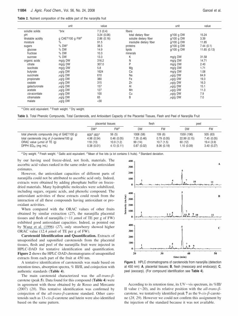
by our having used freeze-dried, not fresh, materials. Theascorbic acid values ranked in the same order as the antioxidantestimates.
However, the antioxidant capacities of different parts ofnaranjilla could not be attributed to ascorbic acid only. Indeed,extracts were obtained by adding phosphate buffer on freeze-dried materials. Many hydrophilic molecules were solubilized,including sugars, organic acids, and phenolic compound. Theantioxidant activities of these extracts could result from theinteraction of all these compounds having antioxidant or pro-oxidant activities.
When compared with the ORAC values of other fruitsobtained by similar extraction (27), the naranjilla placentaltissues and flesh of naranjilla (∼11 µmol of TE per g of FW)exhibited good antioxidant capacities. Indeed, as pointed outby Wang et al. (1996) (27), only strawberry showed higherORAC value (12.4 µmol of TE per g of FW).
Carotenoid Identification and Quantification. Extracts ofunsaponified and saponified carotenoids from the placentaltissues, flesh and peel of the naranjilla fruit were injected inHPLC-DAD for tentative identification and quantification.Figure 2 shows the HPLC-DAD chromatograms of unsaponifiedextracts from each part of the fruit at 450 nm.
A tentative identification of carotenoids was made based onretention times, absorption spectra, % III/II, and coinjection withauthentic standards (Table 4).
The main carotenoid characterized was the all-trans-�-carotene (peak 5). Data found for this compound (Table 4) werein agreement with those obtained by de Rosso and Mercante(2007) (28). This tentative identification was confirmed bycoinjection of the all-trans-�-carotene standard. Other caro-tenoids such as 13-cis-�-carotene and lutein were also identifiedbased on the same pattern.
According to its retention time, its UV-vis spectrum, its %III/II value (∼20), and its relative position with the all-trans-�-carotene, we tentatively identified peak 7 as the 9-cis-�-carote-ne (28, 29). However we could not confirm this assignment bythe injection of the standard because it was not available.
Table 2. Nutrient composition of the edible part of the naranjilla fruit
unit value unit value
soluble solids °brix 7.3 (0.4) fiberspH 3.24 (0.06) total dietary fiber g/100 g DW 15.24titratable acidity g CAEa/100 g FWb 2.86 (0.16) soluble dietary fiber g/100 g DW 3.39moisture % 91.5 insoluble dietary fiber g/100 g DW 11.85sugars % DWc 38.5 proteins g/100 g DW 7.44 (0.1)
glucose % DW 14.9 lipids g/100 g DW 11.65 (0.13)fructose % DW 10.3 mineralssucrose % DW 13.3 K mg/g DW 31.58
organic acids mg/g DW 316.2 N mg/g DW 14.71citrate mg/g DW 307.0 P mg/g DW 2.40isocitrate mg/g DW 5.8 Mg mg/g DW 1.71butyrate µg/g DW 1624 Ca mg/g DW 1.08succinate µg/g DW 610 Na µg/g DW 64.9propionate µg/g DW 380 Fe µg/g DW 19.3oxalate µg/g DW 315 Zn µg/g DW 16.3galacturonate µg/g DW 157 Al µg/g DW 15.1acetate µg/g DW 127 Mn µg/g DW 11.3cis-aconitate µg/g DW 100 Cu µg/g DW 7.9citramalate µg/g DW 65 B µg/g DW 7.0malate µg/g DW <30
a Citric acid equivalent. b Fresh weight. c Dry weight.
Table 3. Total Phenolic Compounds, Total Carotenoids, and Antioxidant Capacity of the Placental Tissues, Flesh and Peel of Naranjilla Fruit
placental tissues flesh peel
DWa FWb DW FW DW FW
total phenolic compounds (mg of GAEc/100 g) 605d (60)e 56 (5) 1008 (58) 109 (6) 1559 (196) 505 (63)total carotenoids (mg of �-carotene/100 g) 4.98 (0.54) 0.46 (0.05) 7.35 (0.46) 0.79 (0.05) 22.99 (0.15) 7.45 (0.05)ORAC value (µmol of TE /g) 118 (13) 10.9 (1.2) 99 (14) 10.7 (1.5) 60 (12) 19.4 (3.9)DPPH ED50 (mg /mL) 0.38 (0.01) 4.13 (0.11) 0.87 (0.02) 8.06 (0.19) 1.10 (0.09) 3.40 (0.27)
a Dry weight. b Fresh weight. c Gallic acid equivalent. d Mean of five lots (a lot contains 5 fruits). e Standard deviation.
Figure 2. HPLC chromatrograms of carotenoids from naranjilla (detectionat 450 nm): A, placental tissues; B, flesh (mesocarp and endocarp); C,peel (exocarp). (For compound identification see Table 4).
11894 J. Agric. Food Chem., Vol. 56, No. 24, 2008 Gancel et al.

Esters (peaks 6, 8, 9, 10, and 11) were identified comparingchromatograms of unsaponified and saponified extracts. Ac-cording to the area augmentation of the lutein peak (peak 1)after saponification (data not shown), peaks 6, 8, 9, 10, and 11(Figure 2) could be esters of lutein.
Table 5 reports the carotenoid composition of the differentparts of the naranjilla fruit. all-trans-�-carotene, the majorcarotenoid, represented ∼45% of total carotenoid contents. Ofthe three fruit parts, the peel had the highest content of�-carotene (3622 µg per 100 g of FW) followed by the flesh(380) and the placental tissues (208). These results agreed withthe measured colors of the different parts (Table 1), where thepeel appeared orange, the flesh yellow, and the placental tissuesyellowish-green.
Compared with other fruit sources of �-carotene, the edibleparts of the naranjilla fruit fell between mango (444 µg per 100 gof FW) and papaya (275 µg per 100 g of FW) according toUSDA data (30). Peaks that were characterized as estersrepresented 12% of carotenoids in the placental tissues, 7% inthe flesh, and 5% in the peel. Other minor carotenoidsrepresented between 25 and 31% of the total contents.
The different fruit parts exhibited provitamin A activity at37, 72, and 707 RE per 100 g of FW for the placental tissues,flesh, and peel, respectively. Although the peel is not consumed,it appeared to be a very good source of carotenoids with a highprovitamin A activity close to that of the carrot (835 RE per100 g of FW) (30).
Phenolic Compound Identification. Phenolic compounds ofnaranjilla were studied in placental tissues, the flesh and thepeel. Total phenolic extracts were purified with XAD-7 resinbefore being injected for the HPLC/ESI-MS assay. The HPLC-DAD chromatograms are shown in Figure 3. Table 6 showsthe tentative identification of some of the peaks.
The chromatograms of the placental tissues (Figure 3A) andthe flesh (Figure 3B) shared more peaks in common than with
that of the peel (Figure 3C). While the placental tissuesexhibited more very polar compounds (peaks 1-7), the peelpresented less polar compounds (peaks 23, 24, 26, and 28).
Caffeoylquinic Acids and DeriVatiVes. Peaks 14 and 17 werepresent in all three parts of naranjilla fruit. Both peaks had amolecular mass of 354 and similar UV-visible spectra (Table6). Compounds with such characteristics are known as chloro-genic acids. The MS/MS fragmentation in negative mode ofthe molecular ion ([M s H]- ) 353) of these peaks, carriedout with 30% energy, produced different base peaks: m/z 191for the compound 14 and m/z 179 for the compound 17.According to Clifford et al. (2003) (31) and Nandutu et al.(2007) (32), peak 14 was identified as 5-O-caffeoylquinic acidand not as 3-O-caffeoylquinic. Although both these compoundsproduce an MS/MS fragment base peak at m/z 191, they canbe distinguished on the basis of the relative intensity of thesecondary ion m/z 179. For 5-O-caffeoylquinic acid the second-ary ion at m/z 179 is very weak or undetectable, whereas, for3-O-caffeoylquinic acid, the relative intensity of the secondaryion at m/z 179 is ∼50% or more. Based on the elution orderand the absence of m/z 191, component 17 should be the 4-O-caffeoylquinic acid.
Although compounds 2, 3, and 7 produced a molecularion at m/z 515, they were not identified as dicaffeoylquinicacids. Indeed, even though our HPLC conditions weredifferent from those of Clifford et al. (2007) (33), thedicaffeoylquinic acids should have eluted after the caf-feoylquinic acids. On the contrary, peaks 2, 3, and 7 wereeluted before the caffeoylquinic acids, suggesting caf-feoylquinic acid glycosides (33). At MS/MS, both compounds3 and 7 produced m/z 353 base peak that could correspondto [caffeoylquinic acid s H+]- ion and therefore to the lossof a hexose. The MS3 fragmentation of MS/MS m/z 353 basepeak provided fragment ions at m/z 191 for compound 3 andm/z 179 for compound 7 (Table 6). Thus, as previouslydescribed for compounds 14 and 17, the m/z 353 base peaksof compounds 3 and 7 could be identified as [5-O-caf-feoylquinic s H+]- and [4-O-caffeoylquinic s H+]- ions.Otherwise, the MS/MS fragmentation (Table 6) of m/z 515of component 2 produced m/z 341 and m/z 179 (base peak),suggesting another 4-O-caffeoylquinic acid glycoside (33).These three caffeoylquinic acid glycosides were found inplacental tissues. Compound 3 was also present in flesh.
FlaVonol Glycosides. The UV spectra of peaks 9, 10, 23, 24,28, and 30 were similar to those of quercetin glycosides, whereaspeaks 12, 19, and 26 showed UV spectra similar to those ofkaempferol derivatives (Table 6). All these peaks were exclu-sively present in naranjilla peel. Such tissue-specific accumula-tion agreed with findings by Stewart et al. (2000) which showed∼98% of the flavonols (quercetin- and kaempferol-glycosides)to be located in peel tissue (34).
On the basis of their fragmentation patterns, peaks 9, 23, and24 were identified as quercetin glycosides, although peaks 10, 28,and 30 were identified as isorhamnetin glycosides. Indeed, the MS2
or MS3 fragmentation permitted discrimination between bothcompounds class derivatives of quercetin, producing a fragmention at m/z 301, although isorhamnetin glycosides produce fragmentions at m/z 315 and m/z 301. Molecular weights of quercetin andishoramnetin are, respectively, 302 and 316, with isorhamnetinhaving the same structure as quercetin with an additional methylgroup. A MS full scan of compound 24 showed an [Ms H]- ionat m/z 609 with an MS/MS fragmentation pattern matching that ofrutin (quercetin-3-rutinoside) (34).
Table 4. Tentative Identification of Carotenoids by HPLC-DAD in theNaranjilla Placental Tissues, Flesh, and Peel
UV-visspectra (nm)
no. tRa fruit part cis-peakpeak
Ipeak
IIpeak
III % III/IItentative
identification
1 22.3 pl,b fl,c ped 420 444 472 54 luteine
2 32.0 pl, fl 400 424 448 64 unknown3 32.7 fl, pe 337 416 440 464 7 unknown4 33.8 fl, pe 338 422 444 468 6 13-cis-�-carotenee
5 37.4 pl, fl, pe 428 452 478 19 all-trans-�-carotenee
6 38.4 pl, fl, pe 422 446 472 40 ester7 39.0 pl, fl, pe 338 422 446 472 20 9-cis-�-carotene8 39.9 fl, pe 422 446 472 36 ester9 41.6 fl, pe 420 444 472 44 ester10 42.3 pl, fl 418 440 470 78 ester11 48.8 pl, fl 422 446 472 57 ester
a Retention time. b Placental tissues. c Flesh. d Peel. e Identified using authenticstandards.
Table 5. Carotenoid Composition of the Naranjilla Fruit (µg of �-CaroteneEquivalents/100 g FW)
compound placental tissues flesh peel
all-trans-�-carotene 208a (8)b 380 (10) 3622 (60)lutein 29 (1) 43 (1) 370 (40)13-cis-�-carotene 55 (4) 630 (60)9-cis-�-carotene 24 (3) 54 (6) 610 (60)esters 54 (5) 52 (4) 380 (70)others 144 (15) 210 (30) 1838 (40)
a Mean of five lots (a lot contains 5 fruits). b Standard deviation.
Bioactive Compounds of the Naranjilla Fruit J. Agric. Food Chem., Vol. 56, No. 24, 2008 11895

Dihydrocaffeoyl Polyamines and DeriVatiVes. The tentativeidentification of components 13, 16, 18, 21, 25, 27, 29 and 31was based on their UV spectra, molecular masses and HPLC/ESI-MS data.
Compounds 18 and 21 produced the same molecular ion [Ms H]-/[M + H]+ at 473/474 and the same MS/MS fragmentions in negative mode m/z 351 and m/z 308 but with differentrelative intensities (Table 6), indicating that they were probablyisomers. The MS/MS fragmentation in positive mode of bothcomponents provided the same fragment ions (e.g., m/z 457,
m/z 293, m/z 236, m/z 222, and m/z 165; data not shown) asthose obtained by Parr et al. (2005) for the N,N′- bis(dihydro-caffeoyl) spermidine, a dihydrocaffeoyl polyamine (35). Ac-cording to this identification and Roshani and Duroy (2006)(36), m/z 351 and m/z 308 fragments ions could be explainedby the loss of -C7H7O2 and -dihydrocaffeoyl moieties,respectively. Both compounds could be tentatively identifiedas N1,N4- bis(dihydrocaffeoyl) spermidine or N4,N8- bis(dihy-drocaffeoyl) spermidine for peak 18 and N1,N8- bis(dihydro-caffeoyl) spermidine for peak 21 (Figure 4).
Figure 3. HPLC chromatrograms of phenolic extracts from naranjilla (UV detection at 280 nm): A, placental tissues; B, flesh (mesocarp and endocarp);C, peel (exocarp). (For compound identification see Table 6).
11896 J. Agric. Food Chem., Vol. 56, No. 24, 2008 Gancel et al.

Following the same scheme and according to Parr et al. (2005)(35), component 31 ([M s H]-/[M + H]+ at 637/638) wasidentified as tris(dihydrocaffeoyl) spermidine.
Two components with molecular ions of [M s H]-/[M +H]+ at 635/636 (peaks 13 and 16) and three others withmolecular ions of [M s H]-/[M + H]+ at 799/800 (peaks 25,27 and 29) were also found in the chromatogram (Figure 3),with retention time that was lower than that of bis(dihydrocaf-
feoyl) spermidine and tris(dihydrocaffeoyl) spermidine, respec-tively. The MS/MS fragmentation of these compounds producedbase peaks of 473 and 637, which may correspond to the lossof a hexose. Parr et al. (2005) had already mentioned thepresence of a putative N,N′,N′′-tris(dihydrocaffeoyl) spermidineglycoside ([M + H]+ at 800 with a major loss of m/z 162,retention time of ∼3 min less than for the aglycon) in the potato(35). Based on the same hypothesis, we could identify the peaks25, 27 and 29 as N1,N4,N8-tris(dihydrocaffeoyl) spermidineglycosides and peaks 13 and 16 as N,N′-bis(dihydrocaffeoyl)spermidine glycosides.
Polyamines are present in food, especially in breast milk andmeat but also in plants where they can be free or conjugatedwith compounds such as hydroxycinnamic acids (37). Thesespolyamines have been studied for their properties, as they playan important role in several activities such as cell proliferation,gene expression, and cell signaling. They are known to havebiological effects, particularly in the differentiation of immunecells and regulation of the inflammatory reactions. Larque etal. (2007) explained that diets with a high polyamine contentcould play an important role in the recovery of intestinalmicroflora and gut mucosa because of their influence on tissuerepair processes (38). They added that the administration ofspermidine might improve treatment of traumas. In contrast,polyamine-free diets may be crucial for reducing tumorousgrowth (39). Hence, the naranjilla fruit may provide a goodsource of spermidine, as all the polyamines present in this fruitare spermidine derivatives.
Unidentified Peaks. Some peaks (1, 4, 5, 6, 8, 15, 20 and 22)remained unidentified by the HPLC-DAD and HPLC/ESI-MSmethods (Table 6). These peaks were found in the placental
Table 6. Identification of Phenolic Compounds by HPLC/ESI-MS in the Naranjilla Placental Tissues, Flesh and Peel
no. tRa fruit partUV-vis
spectra (nm)[M s H]- /[M + H]+
negativeMS2
negativeMS3
tentativeidentification
1 17 pl,b flc 276, 300sh -/-2 20.7 pl 318 515/517 341 (52) 179 (100) 4-O-caffeoylquinic acid hexoside3 23.2 pl, fl 291, 316 515/517 353 (100) 191 (40) 191 (100) 5-O-caffeoylquinic acid hexoside4 23.5 pl, fl 293, 318 -/-5 25.2 pl, fl 307 -/-6 25.5 pl, fl 300, 325sh -/-7 26 pl 291, 316sh 515/517 353 (100) 341 (15) 179 (67) 173 (100) 179 (86) 4-O-caffeoylquinic acid hexoside8 27 ped 285 471/473 453 (70) 257 (65) 241 (100)9 27.7 pe 254, 266sh, 353 771/773 609 (100) 463 (18) 301 (2) 301 (100) rutin hexoside10 28.6 pe 252, 266, 346 639/- 477 (100) 315 (10) isorhamnetin dihexoside11 28.7 pl, fl 267 797/799 635 (100) 473 (12) N,N′-bis(dihydrocaffeoyl) spermidine dihexoside12 29.7 pe 266, 233sh, 347 755/757 593 (100) 429 (26) 285 (100) kaempferol rutinoside hexoside13 30.7 pl, fl 273 635/637 473 (100) 351 (100) 308 (90) N1,N4 or N4,N8-bis(dihydrocaffeoyl) spermidine hexoside14 31.4 pl, fl, pe 300sh, 324 353/355 191 (100) 179 (8) 5-O-caffeoylquinic acid15 31.8 pl 288, 335 -/-16 32 pl, fl 279 635/637 473 (100) 351 (100) 308 (98) N1,N8 -bis(dihydrocaffeoyl) spermidine hexoside17 32.7 pl, fl, pe 300sh, 326 353/355 191 (4) 179 (100) 173 (98) 4-O-caffeoylquinic acid18 33.9 pl, fl, pe 281 473/474 351 (100) 308 (98) 308 (100) N1,N4 or N4,N8-bis(dihydrocaffeoyl) spermidine19 34.2 pe 265, 324sh, 347 609/611 447 (100) 285 (10) 285 (100) kaempferol dihexoside20 35.2 pl, fl 330 599/601 563 (100)21 36.2 pl, fl, pe 265,320sh 473/474 351 (52) 309 (100) N1,N8-bis(dihydrocaffeoyl) spermidine22 39.3 pl 289, 321 -/-23 39.6 pe 255, 263sh, 353 755/757 609 (20) 591 (40) 301 (100) 301 (100) rutin pentoside24 42.6 pe 254, 265sh, 354 609/611 301 (100) rutin25 45.7 pl, fl 280, 320sh 799/800 637 (100) 515 (5) 473 (100) N1,N4,N8-tris(dihydrocaffeoyl) spermidine hexoside26 46 pe 265, 345 593/595 429 (65) 285 (100) kaempferol rutinoside27 46.1 pl, fl 281, 319sh 799/800 637 (100) 473 (100) N1,N4,N8-tris(dihydrocaffeoyl) spermidine hexoside28 46.5 pe 253, 266, 353 623/625 459 (45) 315 (100) 300 (24) 301 (100) isorhamnetin dihexoside29 47.1 pl, fl 281, 319sh 799/800 637 (100) 473 (100) N1,N4,N8-tris(dihydrocaffeoyl) spermidine hexoside30 49.4 pe 255, 267sh, 354 623/625 315 (100) 300 (15) 301 (100) isorhamnetin dihexoside31 51.7 pl, fl, pe 281 637/638 473 (100) 351 (70) 308 (100) N1,N4,N8-tris(dihydrocaffeoyl) spermidine32 53.1 pe 283, 327sh -/-
a Retention time. b Placental tissues. c Flesh (mesocarp and endocarp). d Peel (exocarp).
Figure 4. Structure of the dihydrocaffeoyl spermidines.
Bioactive Compounds of the Naranjilla Fruit J. Agric. Food Chem., Vol. 56, No. 24, 2008 11897

tissues and flesh of naranjilla. Unidentified compound 32,however, was present only in the peel.
Isolation and NMR identification are needed to unequivocallyelucidate, from a structural viewpoint, all the unidentified peaks.
Further analyses, such as acid hydrolysis, are needed todetermine the identity of sugars in the glycosylated compounds,although we suspect glucose as hexose and rhamnose as pentose.
Finally, our study is the first to attempt to characterize theunderutilized specie, Solanum quitoense var. “Puyo hybrid”,especially with regard to its carotenoids and phenolic com-pounds. The main chlorogenic acids and dihydrocaffeoylspermidine were found in all parts of the naranjilla fruit.Glycosylated chlorogenic acids and dihydrocaffeoyl spermidinesappeared to be located mainly in the placental tissues and flesh,whereas flavonol glycosides were exclusively present in the peel.Although Parr et al. (2005) (35) have already reported N1,N4,N8-tris(dihydrocaffeoyl) spermidine glycosides in potato, our studyis the first to report N,N′-bis(dihydrocaffeoyl) spermidineglycosides in a fruit.
Thus, besides its original organoleptic qualities, the naranjillaappears to be a fruit with good nutritional potential and therebydeserves to be well-known. This fruit can provide the basis fora new fruit-drink flavor or other fruit derived-products that couldbecome popular in North America, Japan, Europe, and similarareas. But further research is needed, especially on improvingstorage conditions, which remain the limiting factor to thistropical fruit’s development.
ACKNOWLEDGMENT
The authors thank the Escuela Politecnica Nacional (EPN,Ecuador) for providing fruits. We also thank P. Brat and C.Mertz for helpful advice and Pr. J. Vercauteren for permittingus to use the spectrofluorimeter in his laboratory.
LITERATURE CITED
(1) Heiser, C. B.; And erson, G., “New” Solanums. In PerspectiVeson new crops and new use; Janick, J., Ed.; ASHS Press:Alexandria, VA, 1999; pp 379-384.
(2) Dennis, F. G. J.; Herner, R. C. Naranjilla: a potential cash cropfor the small farmer in Latin America. Acta Hortic. 1985, 158,475–481.
(3) Heiser, C. B. The relationships of the naranjilla, Solanumquitoense. Biotropica 1972, 4, 77–84.
(4) National Research Council. Naranjilla (Lulo). In Lost crops ofthe Incas: Little known plants of the Andes with promise of theworld cultiVation; National Academy Press: Washington, 1989;pp 267-275.
(5) Morton, J. Naranjilla. In Fruits of warm climates; Morton, J. F.,Ed.; Miami, FL, 1987; pp 425-428.
(6) Griffiths, F. P. Processing procedure to retain Vitamin C inNaranjilla, Solanum quitoense, products. J. Rio Grande Val.Hortic. Soc. 1965, 19, 33–36.
(7) Ruiz, J. J.; Lentini, Z.; Buitrago, M. E.; Florez-Ramos, C. P.; J.,C.; Segovia, V. In vitro propagation and regeneration of Solanumquitoense (Lulo) plants and their use as elite clones by resourcefarmers. Acta Hortic. 2007, 738, 491–494.
(8) Monteros, A.; Munoz, L.; Revelo, J.; Tapia, C.; Zambrano, E.;Fiallos, J. Nematode resistance through mutation induction in alocal variety of naranjilla (Solanum quitoense Lam) in Ecuador.In Proceedings of a final Research Coordination Meetingorganized by the joint FAO/IAEA DiVision of Nuclear Techniquesin Food and Agriculture; 2003; pp 87-99.
(9) Ochoa, J. B.; Yangari, B.; Galarza, V.; Fiallos, J.; Ellis, M. A.Vascular wilt of common naranjilla (Solanum quitoense) causedby Fusarium oxysporum in Ecuador, Plant Health Prog. [Online]2001. DOI: 10.1094/PHP-2001-0918-01-HN.
(10) Osorio, C.; Duque, C.; Batista-Viera, F. Studies on aromageneration in lulo (Solanum quitoense L.): enzymatic hydrolysisof glycosides from leaves. Food Chem. 2003, 481, 333–340.
(11) Brunke, E.-J.; Mair, P.; Hammerschmidt, F.-J. Volatiles fromnaranjilla fruit (Solanum quitoense Lam.). GC/MS analysis andsensory evaluation using sensory evaluation using sniffing GC.J. Agric. Food Chem. 1989, 37, 746–748.
(12) Huyskens-Keil, S.; Prono-Widayat, H.; Schreiner, M.; Peters, P.Effect of surface coating and film packaging on the keeping qualityof solanaceous crops (Solanum muricatum Ait., Solanum quitoenseLam.). Acta Hortic. 2001, 553, 621–625.
(13) AOAC. Official Methods of Analysis of the Association of OfficialAnalytical Chemists; 15th ed.; AOAC: Arlington, VA, 1990.
(14) Asp, N. G.; Johansson, C. G.; Hallmer, H.; Siljestroem, M. Rapidenzymatic assay of insoluble and soluble dietary fiber. J. Agric.Food Chem. 1983, 31, 476–482.
(15) AOAC. Official Methods of Analysis of the Association of OfficialAnalytical Chemist; 17th ed.; AOAC: Washington, DC, 2000.
(16) George, S.; Brat, P.; Alter, P.; Amiot, M. J. Rapid determinationof polyphenols and vitamin C in plant-derived products. J. Agric.Food Chem. 2005, 53, 1370–1373.
(17) Taungbodhitham, A. K.; Jones, G. P.; Walhlqvist, M. L.; Briggs,D. R. Evaluation of method for the analysis of carotenoids in fruitsand vegetables. Food Chem. 1998, 63, 577–584.
(18) Dhuique-Mayer, C.; Caris-Veyrat, C.; Ollitraut, P.; Curk, F.;Amiot, M.-J. Varietal and interspecific influence on micronutrientcontents in citrus from the mediterranean area. J. Agric. FoodChem. 2005, 53, 2140–2145.
(19) Britton, G. UV/Visible spectroscopy. In Carotenoids: SpectroscopyVol. 1B, Britton, G., Liaaen-Jensen, S.; Pfander, H., Eds:Birkhauser: Basel, Switzerland, 1995; pp 13-62.
(20) Al-Jaleel, A.; Zekri, M.; Hammam, Y. Yield, fruit quality, andtree health of ‘Allen Eureka’ lemon on seven rootstocks in SaudiArabia. Sci. Hortic. 2005, 105, 457–465.
(21) Gonzalez-Aguilar, G. A.; Fortiz, J.; Cruz, R.; Baez, R.; Wang,C. Y. Methyl jasmonate reduces chilling injury and maintainspostharvest quality of mango fruit. J. Agric. Food Chem. 2000,48, 515–519.
(22) Shaaban, E. A.; Abd-el-Aal, S. K.; Zaied, N. S.; Rizkalla, A. A.Assessment of Genetic Variability on Some Orange AccessionsUsing RAPD-DNA Markers. Res. J. Agric. Biol. Sci 2006, 2, 564–570.
(23) Ramulu, P.; Udayasekhara Rao, P. Total, insoluble and solubledietary fiber contents of Indian fruits. J. Food Compos. Anal. 2003,16, 677–685.
(24) Leterme, P.; Buldgen, A.; Estrada, F.; Londono, A. M. Mineralcontent of tropical fruits and unconventional foods of the Andesand the rain forest of Colombia. Food Chem. 2006, 95, 644–652.
(25) Mahattanatawee, K.; Manthey, J. A.; Luzio, G.; Talcott, S. T.;Goodner, K.; Baldwin, E. A. Total antioxidant activity and fibercontent of select florida-grown tropical fruits. J. Agric. FoodChem. 2006, 54, 7355–7363.
(26) Melo, E. A.; Lima, V. L. A. G.; Maciel, M. I. S.; Caetano, A. C. S.;Leal, F. L. L. Polyphenol, ascorbic acid and total carotenoidcontents in common fruits and vegetables. Braz. J. Food Technol.2006, 236.
(27) Wang, H.; Cao, G.; Prior, R. L. Total Antioxidant Capacity ofFruits. J. Agric. Food Chem. 1996, 44, 701–705.
(28) deRosso, V. V.; Mercadante, A. Z. Identification and Quantifica-tion of Carotenoids, By HPLC-PDA-MS/MS, from AmazonianFruits. J. Agric. Food Chem. 2007, 55, 5062–5072.
(29) Pott, I.; Marx, M.; Neidhart, S.; Muhlbauer, W.; Carle, R.Quantitative Determination of beta-Carotene Stereoisomers inFresh, Dried, and Solar-Dried Mangoes (Mangifera indica L.). J.Agric. Food Chem. 2003, 51, 4527–4531.
(30) U.S. Department of Agriculture, A. R. S. USDA National NutrientDatabase for Standard Reference, Release 20. Nutrient DataLaboratory Home Page, http://www.ars.usda.gov/ba/bhnrc/ndl.
11898 J. Agric. Food Chem., Vol. 56, No. 24, 2008 Gancel et al.
(31) Clifford, M. N.; Johnston, K. L.; Knight, S.; Kuhnert, N.Hierarchical Scheme for LC-MSn Identification of ChlorogenicAcids. J. Agric. Food Chem. 2003, 51, 2900–2911.
(32) Nandutu, A. M.; Clifford, M.; Howell, N. K. Analysis of phenoliccompounds in Ugandan sweet potato varieties (NSP, SPK ANDTZ). Afr. J. Biochem. Res 2007, 1, 29–36.
(33) Clifford, M. N.; Wu, W.; Kirkpatrick, J.; Kuhnert, N. Profilingthe Chlorogenic Acids and Other Caffeic Acid Derivatives ofHerbal Chrysanthemum by LC-MSn. J. Agric. Food Chem. 2007,55, 929–936.
(34) Stewart, A. J.; Bozonnet, S.; Mullen, W.; Jenkins, G. I.; Lean,M. E. J.; Crozier, A. Occurrence of Flavonols in Tomatoes andTomato-Based Products. J. Agric. Food Chem. 2000, 48, 2663–2669.
(35) Parr, A. J.; Mellon, F. A.; Colquhoun, I. J.; Davies, H. V.Dihydrocaffeoyl Polyamines (Kukoamine and Allies) in Potato(Solanum tuberosum) Tubers Detected during Metabolite Profiling.J. Agric. Food Chem. 2005, 53, 5461–5466.
(36) Roshani, S.; Duroy, A. N. Rapid Screening of Ascorbic Acid,Glycoalkaloids, and Phenolics in Potato Using High-PerformanceLiquid Chromatography. J. Agric. Food Chem. 2006, 54, 5253–5260.
(37) Edreva, A. Polyamines in plants. Bulg. J. Plant Physiol. 1996,22, 73–101.
(38) Larque, E.; Sabater-Molina, M.; Zamora, S. Biological significanceof dietary polyamines. Nutrition 2007, 23, 87–95.
(39) Bachrach, U. Polyamines and cancer: Minireview article. AminoAcids 2004, 26, 307–309.
Received for review May 14, 2008. Revised manuscript receivedSeptember 29, 2008. Accepted October 3, 2008. This work wasfinancially supported by the European Union (PAVUC project, INCONo. 015279).
JF801515P
Bioactive Compounds of the Naranjilla Fruit J. Agric. Food Chem., Vol. 56, No. 24, 2008 11899