Identification of the C. elegans anaphase promoting complex subunit Cdc26 by phenotypic profiling...
8
BioMed Central Page 1 of 8 (page number not for citation purposes) BMC Developmental Biology Open Access Research article Identification of the C. elegans anaphase promoting complex subunit Cdc26 by phenotypic profiling and functional rescue in yeast Yan Dong 1 , Aliona Bogdanova 2 , Bianca Habermann 2 , Wolfgang Zachariae 2 and Julie Ahringer* 1 Address: 1 The Gurdon Institute and Department of Genetics, University of Cambridge, Tennis Court Road, Cambridge CB2 1QN, UK and 2 Max Planck Institute of Molecular Cell Biology and Genetics, Pfotenhauerstrasse 108, 01307 Dresden, Germany Email: Yan Dong - [email protected]; Aliona Bogdanova - [email protected]; Bianca Habermann - [email protected]; Wolfgang Zachariae - [email protected]; Julie Ahringer* - [email protected] * Corresponding author Abstract Background: RNA interference coupled with videorecording of C. elegans embryos is a powerful method for identifying genes involved in cell division processes. Here we present a functional analysis of the gene B0511.9, previously identified as a candidate cell polarity gene in an RNAi videorecording screen of chromosome I embryonic lethal genes. Results: Whereas weak RNAi inhibition of B0511.9 causes embryonic cell polarity defects, strong inhibition causes embryos to arrest in metaphase of meiosis I. The range of defects induced by RNAi of B0511.9 is strikingly similar to those displayed by mutants of anaphase-promoting complex/cyclosome (APC/C) components. Although similarity searches did not reveal any obvious homologue of B0511.9 in the non-redundant protein database, we found that the N-terminus shares a conserved sequence pattern with the N-terminus of the small budding yeast APC/C subunit Cdc26 and its orthologues from a variety of other organisms. Furthermore, we show that B0511.9 robustly complements the temperature-sensitive growth defect of a yeast cdc26∆ mutant. Conclusion: These data demonstrate that B0511.9 encodes the C. elegans APC/C subunit CDC- 26. Background A major goal in biology is to understand the function of each gene. For many organisms, complete genome sequences are now available. Combined with knockdown techniques such as RNAi or morpholino oligos, genes can be quickly assayed for in vivo function [1,2]. In C. elegans, genome-wide knockdown of gene activity through RNAi has provided important phenotypic information for thou- sands of genes [3-8]. Although a useful starting point, much of the phenotypic information lacks detail; for example, many genes are only annotated as essential for viability. Several studies have carried out additional analyses to identify more precise gene functions. In particular, RNAi videorecording screens have uncovered very detailed defects allowing genes to be grouped into more specific phenotypic classes [4,7-9]. However, within each class there still exist groups of genes with different functions. Analysing the phenotypes of individual genes in more Published: 20 March 2007 BMC Developmental Biology 2007, 7:19 doi:10.1186/1471-213X-7-19 Received: 26 September 2006 Accepted: 20 March 2007 This article is available from: http://www.biomedcentral.com/1471-213X/7/19 © 2007 Dong et al; licensee BioMed Central Ltd. This is an Open Access article distributed under the terms of the Creative Commons Attribution License (http://creativecommons.org/licenses/by/2.0 ), which permits unrestricted use, distribution, and reproduction in any medium, provided the original work is properly cited.
-
Upload
biochem-mpg -
Category
Documents
-
view
0 -
download
0
Transcript of Identification of the C. elegans anaphase promoting complex subunit Cdc26 by phenotypic profiling...

BioMed CentralBMC Developmental Biology
ss
Open AcceResearch articleIdentification of the C. elegans anaphase promoting complex subunit Cdc26 by phenotypic profiling and functional rescue in yeastYan Dong1, Aliona Bogdanova2, Bianca Habermann2, Wolfgang Zachariae2 and Julie Ahringer*1Address: 1The Gurdon Institute and Department of Genetics, University of Cambridge, Tennis Court Road, Cambridge CB2 1QN, UK and 2Max Planck Institute of Molecular Cell Biology and Genetics, Pfotenhauerstrasse 108, 01307 Dresden, Germany
Email: Yan Dong - [email protected]; Aliona Bogdanova - [email protected]; Bianca Habermann - [email protected]; Wolfgang Zachariae - [email protected]; Julie Ahringer* - [email protected]
* Corresponding author
AbstractBackground: RNA interference coupled with videorecording of C. elegans embryos is a powerfulmethod for identifying genes involved in cell division processes. Here we present a functionalanalysis of the gene B0511.9, previously identified as a candidate cell polarity gene in an RNAivideorecording screen of chromosome I embryonic lethal genes.
Results: Whereas weak RNAi inhibition of B0511.9 causes embryonic cell polarity defects, stronginhibition causes embryos to arrest in metaphase of meiosis I. The range of defects induced byRNAi of B0511.9 is strikingly similar to those displayed by mutants of anaphase-promotingcomplex/cyclosome (APC/C) components. Although similarity searches did not reveal any obvioushomologue of B0511.9 in the non-redundant protein database, we found that the N-terminusshares a conserved sequence pattern with the N-terminus of the small budding yeast APC/Csubunit Cdc26 and its orthologues from a variety of other organisms. Furthermore, we show thatB0511.9 robustly complements the temperature-sensitive growth defect of a yeast cdc26∆ mutant.
Conclusion: These data demonstrate that B0511.9 encodes the C. elegans APC/C subunit CDC-26.
BackgroundA major goal in biology is to understand the function ofeach gene. For many organisms, complete genomesequences are now available. Combined with knockdowntechniques such as RNAi or morpholino oligos, genes canbe quickly assayed for in vivo function [1,2]. In C. elegans,genome-wide knockdown of gene activity through RNAihas provided important phenotypic information for thou-sands of genes [3-8]. Although a useful starting point,much of the phenotypic information lacks detail; for
example, many genes are only annotated as essential forviability.
Several studies have carried out additional analyses toidentify more precise gene functions. In particular, RNAivideorecording screens have uncovered very detaileddefects allowing genes to be grouped into more specificphenotypic classes [4,7-9]. However, within each classthere still exist groups of genes with different functions.Analysing the phenotypes of individual genes in more
Published: 20 March 2007
BMC Developmental Biology 2007, 7:19 doi:10.1186/1471-213X-7-19
Received: 26 September 2006Accepted: 20 March 2007
This article is available from: http://www.biomedcentral.com/1471-213X/7/19
© 2007 Dong et al; licensee BioMed Central Ltd. This is an Open Access article distributed under the terms of the Creative Commons Attribution License (http://creativecommons.org/licenses/by/2.0), which permits unrestricted use, distribution, and reproduction in any medium, provided the original work is properly cited.
Page 1 of 8(page number not for citation purposes)

BMC Developmental Biology 2007, 7:19 http://www.biomedcentral.com/1471-213X/7/19
depth is important for assigning genes to specific func-tions. Through an interest in embryonic polarity, weinvestigated the function of B0511.9, previously identi-fied as having embryonic polarity defects in a large-scaleRNAi videorecording screen [9]. Through phenotypicanalyses, we show that B0511.9 shares functions withcomponents of the cell cycle regulator, the anaphase pro-moting complex/cyclosome (APC/C).
The APC/C is a complex of 12 subunits in animal cells (13in yeast) that regulates destruction of key cell cycle regula-tors at the appropriate times by targeting them for degra-dation by the 26 S proteasome through its E3 ubiquitinligase activity (reviewed in [10-12]). In C. elegans, nine ofthe 12 APC/C subunits have been identified based onsequence analysis [13-17]; Cdc26, Apc7, and Apc13 werenot identified. For seven subunits, loss of function usingmutants or RNAi causes an arrest at metaphase of meiosisI; for two (apc-5 and apc-10), weak embryonic lethalitywas seen along with germline maintenance problems con-sistent with mitotic defects (reviewed in [18]).
Results and DiscussionB0511.9 is required for the metaphase to anaphase transition of meiosis IIn large scale RNAi videorecording screens, RNAi ofB0511.9 was shown to cause different phenotypes: loss ofasymmetry in the first cell division or one cell arrest dueto failure to pass through meiosis [8,9]; To understand therole of B0511.9 in cell division, we examined the RNAiphenotype in detail.
We first carried out a time course of RNAi feeding ofB0511.9 and examined embryos laid at different timesafter RNAi was initiated in the mother (Table 1). RNAiknockdown increases in strength during the time course.There was an increase in lethality from 17.9% at 24–32hours post feeding to 100% at 56 hours post feeding orlater. The terminal arrest phenotypes of the embryoschanged over time. At 24–32 hours post feeding, mostarrested embryos contained many cells whereas embryoslaid 56 hours after initiation of RNAi feeding arrested as asingle cell (Table 1). At intermediate time points, bothtypes of terminal arrest embryos were seen (Table 1).
To investigate the arrest stage of the embryos after strongRNAi of B0511.9 we examined the pattern of DNA con-
densation of embryos inside the uterus using a GFP::his-tone reporter gene. Compared to the wild-type whereprogressively older embryos have progressively more cells(Figure 1A, B), embryos in the uteri of B0511.9(RNAi)mothers all showed the DNA condensation typical of met-aphase of meiosis I (Figure 1C, D). Staining these arrestedembryos for tubulin confirmed that embryos arrestedwith a meiosis I metaphase-like spindle (inset in Figure1D). This phenotype is similar to that reported formutants or strong RNAi of anaphase promoting complex/cyclosome (APC/C) subunits [13-15,17]. We confirmedthat the phenotype of strong B0511.9(RNAi) embryosdescribed above is identical to that seen after RNAi ofAPC/C component emb-27/Cdc16 (Figure 1E, F). Theseresults indicate that like APC/C components, B0511.9 isrequired for progression from metaphase to anaphase ofmeiosis I.
Weak depletion of B0511.9 causes embryonic polarity defectsWe next examined the phenotype of embryos laid aftershorter maternal RNAi of B0511.9, which should cause aweaker depletion. In wild-type embryos, the first cell divi-sion is asymmetric, occurring at 56% embryo length(range of 54–57%, n = 20). In contrast, we found that firstdivision in B0511.9(RNAi) embryos is much more sym-metric, occurring on average at 52% embryo length (rangeof 51–54%, n = 10). This suggests that B0511.9(RNAi)embryos have a defect in embryonic polarity.
In wild-type embryos, polarity is initiated during the firstcell cycle, leading to the asymmetric localisation of PARpolarity proteins, with a complex of PAR-3/PAR-6/PKC-3at the anterior cortex and PAR-1 and PAR-2 at the poste-rior cortex (reviewed in [19]). These proteins are requiredfor the posterior displacement of the first mitotic spindle,which leads to an asymmetric cell division. In par mutantembryos, the first division is symmetric instead of asym-metric and the remaining PAR proteins are often mislocal-ized [19-26].
To determine whether RNAi of B0511.9 affected PARpolarity, we examined the localization of PAR-2 and PAR-3 in weakly affected B0511.9(RNAi) embryos. We foundthat these PAR proteins were abnormally distributed in82% (n = 67) of such embryos, compared to 0% abnor-mal distribution in wild-type embryos (n = 30), indicating
Table 1: Time course of RNAi of B0511.9
Time post RNAi Dead embryos (n) Hatched embryos (n) Phenotype of dead embryos
24–32 hours 17.9% (93) 82.1% (425) Multicellular32–48 hours 31.1% (324) 68.9% (719) One cell arrest or multicellular48–56 hours 81.6% (288) 18.4% (65) Predominantly one cell arrest> 56 hours 100% (93) 0% One cell arrest
Page 2 of 8(page number not for citation purposes)

BMC Developmental Biology 2007, 7:19 http://www.biomedcentral.com/1471-213X/7/19
a defect in embryonic polarity. In most of B0511.9(RNAi)embryos, PAR-3 was expanded to encompass the entirecortex whereas PAR-2 was found in cytoplasmic puncta(Figure 2C–F and legend). This pattern is strikingly similarto that seen in partial loss of function mutants or weakRNAi of APC/C subunits emb-27/Cdc16, mat-1/Cdc27,mat-2/Apc1, mat-3/Cdc23, and emb-30/Apc4 [16]. The sim-ilarity in the range of phenotypes induced by RNAi ofB0511.9 and APC/C components argues that B0511.9functions with the APC/C.
B0511.9 shows homology to the APC/C subunit Cdc26There are two alternatively spliced isoforms of B0511.9inferred from the sequence of ESTs, called B0511.9a andB0511.9b, which are predicted to encode proteins of175aa and 187aa, respectively [27]. The final intron pre-dicted in Wormbase [27] is not removed in the ESTs, mak-ing the proteins shorter than originally proposed due toan earlier stop codon. Homology searches using Blastp[28] with standard settings against the non-redundantprotein database (NCBI) did not uncover any similarity of
Meiotic metaphase arrest induced by strong RNAi of B0511.9Figure 1Meiotic metaphase arrest induced by strong RNAi of B0511.9. Pairs of pictures show DIC and fluorescence images of embryos in the uterus of a mother carrying a GFP::H2B transgene marking nuclei. (A, B) wild-type embryos show progressively more nuclei as divisions proceed. (C, D) B0511.9(RNAi) embryos are all arrested at the one cell stage; staining of such embryos for beta-tubulin (red) and DNA (blue) shows arrest stage is at meiotic metaphase I (inset). (E, F) emb-27/Cdc16(RNAi) embryos arrested in metaphase of meiosis I [15], a phenotype identical to that of B0511.9(RNAi) in (C, D). Arrows in (D) and (F) point to sperm chromatin, indicating that the embryos have been fertilized.
A B
C D
E F
Page 3 of 8(page number not for citation purposes)

BMC Developmental Biology 2007, 7:19 http://www.biomedcentral.com/1471-213X/7/19
Page 4 of 8(page number not for citation purposes)
Embryonic polarity defects after weak RNAi of B0511.9Figure 2Embryonic polarity defects after weak RNAi of B0511.9. (A) wild-type 2 cell embryo after asymmetric first division; anterior AB cell is larger than the posterior P1 cell. (B) B0511.9(RNAi) embryo showing a symmetric first division. (C) PAR-3 at the anterior cortex of a wild-type one-celled embryo at anaphase. (D) PAR-3 on the entire cortex of a B0511.9(RNAi) embryo at anaphase. (E) PAR-2 at the posterior cortex of the wild-type one-celled embryo in (C). (F) PAR-2 in cytoplasmic structures in the B0511.9(RNAi) embryo shown in (D). The PAR-2 antibody shows weak cross-reaction with microtubules. PAR-2 and PAR-3 distributions were scored in wild-type and B0511.9(RNAi) embryos from prophase to the two cell stage. We distin-guished weak versus strong classes of PAR staining defects in B0511.9(RNAi) embryos: (1) weak: an enlarged domain of cortical PAR-3 with a reduced domain of cortical PAR-2. (2) strong: complete cortical PAR-3 with PAR-2 in cytoplasmic puncta. In embryos where both meiotic divisions occurred, scored by the presence of two polar bodies, 50% were in the weak class and 17% in the strong class (n = 18). In embryos with a meiotic division defect, scored by the presence of only one polar body, 23% were in the weak class and 63% were in the strong class (n = 49). The PAR distribution defects in B0511.9(RNAi) embryos hav-ing two polar bodies suggests that its polarity function is separable from its meiotic function.
A
C D
B
FE
wild-type B0511.9(RNAi)
PAR-2
PAR-3

BMC Developmental Biology 2007, 7:19 http://www.biomedcentral.com/1471-213X/7/19
B0511.9 to any known protein. However, Blastp searchesagainst the S. cerevisiae protein database revealed homol-ogy of the N-terminus of B0511.9 to the N-terminus ofCdc26.
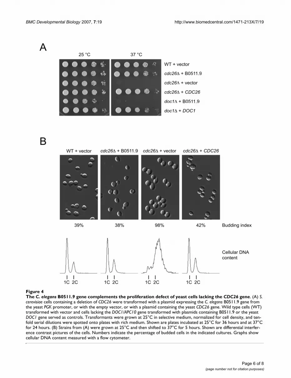
Budding yeast CDC26 encodes a small protein of 124 aathat is dispensable for proliferation at 25°C but essentialfor progression through mitosis at 37°C [29]. The proteinresides within the APC/C where it stabilizes the associa-tion of the TPR repeat proteins Cdc16 and Cdc27 withother subunits of the complex [30,31]. Proteins related toCdc26 have been found in the APC/C isolated from fis-sion yeast and human cells [32,33]. Although the B0511.9sequence is longer than those of other Cdc26 orthologues,it shares with all these proteins a conserved pattern ofcharged and large, hydrophobic residues at the very N-ter-minus (Figure 3). In contrast, the C-terminal regions ofCdc26 orthologues lack detectable sequence conserva-tion. Indeed, the first 71 amino acids of S. cerevisiae Cdc26are sufficient for proliferation albeit at a reduced rate [29].
B0511.9 complements a budding yeast cdc26 mutantTo test whether B0511.9 could functionally replaceCdc26, we expressed it in S. cerevisiae cells lacking theendogenous CDC26 gene. As shown in Figure 4A, theB0511.9 plasmid but not the empty vector restored prolif-eration of cdc26∆ mutant cells at 37°C. Complementationwas remarkably robust: cdc26∆ cells expressing B0511.9grew with wild-type kinetics and were normal with respectto cell size, budding index, and cellular DNA content (Fig-
ure 4B). Expression of B0511.9 failed to rescue the tem-perature-sensitive growth defect of cells lacking anotherAPC/C subunit, Doc1/Apc10. This result demonstratesthat B0511.9 provides Cdc26 function to the yeast cellsbut does not rescue a general defect in APC/C activity.
ConclusionOur functional and phenotypic assays indicate thatB0511.9 encodes the C. elegans APC/C subunit Cdc26 andaccordingly we have named it cdc-26. Inhibition of cdc-26activity leads to the same range of defects as seen afterinhibition of other APC/C subunits, namely embryonicpolarity defects after weak knockdown, and meiotic met-aphase I arrest following strong knockdown. Consistentwith the role of yeast Cdc26 in stabilizing the associationof Cdc16 with other subunits, a large-scale two-hybridstudy in C. elegans showed that CDC-26 binds to APC/Ccomponent EMB-27/Cdc16 [34].
Previous studies had identified C. elegans homologs of 9of the 12 known human APC/C subunits (reviewed in[18]). No sequences with significant matches to Cdc26,Apc7, or Apc13 had been found. This study illustrates thepower of RNA interference screens coupled with detailedphenotypic analyses in assigning gene function. RNAiembryo videorecording data for hundreds of genes areavailable [4,7-9]. In many cases, the defects observed inthese movies give insight into the biological processaffected. Further study of genes in different phenotypicclasses will provide a deeper understanding into the
Multiple sequence alignment of N-terminal sequences from Cdc26 orthologuesFigure 3Multiple sequence alignment of N-terminal sequences from Cdc26 orthologues. All sequences start at the initiator methionine, and numbers give the total length of the protein. Conserved residues are highlighted in yellow. Ce, Caenorhabditis elegans (NP_740913, B0511.9); Cb, Caenorhabditis briggsae (CAE67051); Hs, Homo sapiens (NP_644815); Mm, Mus musculus (NP_647452); Xl, Xenopus laevis (BP677104); Dr, Danio rerio (NP_001004005); At, Arabidopsis thaliana (AAN10198); Am, Apis mellifera (XP_001122028); An, Aspergillus nidulans (AI210365); Sp, Schizosaccharomyces pombe (O13916, Hcn1); Ag, Ashbya gos-sypii (NP_984005); Cg, Candida glabrata (CAG60767); Sc, Saccharomyces cerevisiae (NP_116694).
Ce MSMLRRPLTQLELCEDDIQWLT-DQLNKRVLPAVIVPKCEMMDIDEMEPMDQSEPPRGIT 194
Cb MSMLRRPLTQLELCEDDIQWLS-EQLAKKETGFEDEVKYEVMDVDEDEPMDQSEPTGGIS 199
Hs --MLRRKPTRLELKLDDIEEF--ENIRKDLE----------TRKKQKEDVEVVGGSDGEG 85
Mm --MLRRKPTRLELKLDDIEEF--ESIRKDLE----------ARKKQKEDVEGVGTSDGEG 85
Xl --MLRRKPTRLELKLDDIEEF--ESIKKDLE----------GRKKQREEVDLS-ATETDA 84
Dr --MLRRKPTRLELKLDDTEEF--ESVKKELE----------SRKKQRDEVDVVGVATSSE 88
At --MLRRKPTKIQLKIEDREEL--EQSRKSQP----------STTTTTAPSSSSAASSLHH 65
Am --MIRRSPTRIDLRLDDLQEY--EAMRKALE----------AKKESERPPTFNPPSWG-- 72
An --MIRRKPTAIAITSDDLTMFEEERLRK-LE----------PRNSGHDPAQNGTRVNFDP 75
Sp --MLRRNPTAIQITAEDVLAYDEEKLRQTLD----------SESTTEEALQKNEESTRLS 80
Ag --MLRREPTTIQLTSDDLAELQ-DNLEEFKL----------LQQIKSQHMDLVRSSTRVG 118
Cg --MIRREATTLTLSQTDINDLVDELEERKLQR---------IINKQRDRVLRTSTKLESG 147
Sc --MIRRAPTTLQLSHDDVTSLIDDLNEQKLKQQLN------IEKTKYFQGKNGGSLHSNT 124
Page 5 of 8(page number not for citation purposes)
BMC Developmental Biology 2007, 7:19 http://www.biomedcentral.com/1471-213X/7/19
Page 6 of 8(page number not for citation purposes)
The C. elegans B0511.9 gene complements the proliferation defect of yeast cells lacking the CDC26 geneFigure 4The C. elegans B0511.9 gene complements the proliferation defect of yeast cells lacking the CDC26 gene. (A) S. cerevisiae cells containing a deletion of CDC26 were transformed with a plasmid expressing the C. elegans B0511.9 gene from the yeast PGK promoter, or with the empty vector, or with a plasmid containing the yeast CDC26 gene. Wild type cells (WT) transformed with vector and cells lacking the DOC1/APC10 gene transformed with plasmids containing B0511.9 or the yeast DOC1 gene served as controls. Transformants were grown at 25°C in selective medium, normalized for cell density, and ten-fold serial dilutions were spotted onto plates with rich medium. Shown are plates incubated at 25°C for 36 hours and at 37°C for 24 hours. (B) Strains from (A) were grown at 25°C and then shifted to 37°C for 5 hours. Shown are differential interfer-ence contrast pictures of the cells. Numbers indicate the percentage of budded cells in the indicated cultures. Graphs show cellular DNA content measured with a flow cytometer.
WT + vector cdc26 + B0511.9 cdc26 + vector cdc26 + CDC26
39% 38% 98% 42% Budding index
WT + vector
cdc26 + B0511.9
cdc26 + vector
cdc26 + CDC26
doc1 + B0511.9
doc1 + DOC1
25 °C 37 °C
A
B
Cellular DNA
content
1C 2C 1C 2C 1C 2C 1C 2C

BMC Developmental Biology 2007, 7:19 http://www.biomedcentral.com/1471-213X/7/19
mechanisms of shared cell division processes. For exam-ple, careful analyses of these data may lead to the identifi-cation of C. elegans Apc7 and Apc13.
MethodsStrainsThe following strains were used and cultured by standardmethods [35]: wild-type Bristol N2, AZ212: unc-119(ed3)ruIs32 [unc-119(+) pie-1::gfp::H2B] [36].
RNA interferenceRNAi was carried out by feeding [37] as in [38] using RNAifeeding clones from [3,5]. Sequences of the clones areavailable in Wormbase [27] as sjj_B0511.9 for B0511.9and as sjj_F10B5.6 for emb-27. For the time course inTable 1, wild-type N2 L4 hermaphrodites were placed onRNAi feeding plates containing the same bacterial strain at20°C for 24 hours, then moved to new RNAi feedingplates for the indicated collection times. Embryos laid oneach plate were scored the next day for embryo hatchingand terminal phenotype. Fertilized embryos were easilydistinguished from unfertilized oocytes by their oblongrather than rounded shape and presence of an eggshell,visible in the dissecting microscope. For strong RNAi ofB0511.9 in Figure 1, GFP::H2B L4 hermaphrodites wereplaced on feeding plates for 30 hours at 25°C and thenscored by DIC and GFP microscopy; for RNAi of emb-27/Cdc16, RNAi feeding was for 20 hours. For weak RNAi ofB0511.9 in Figure 2 and for videorecording analyses, wild-type N2 L4s were placed on feeding plates for 40 hours at20°C, then embryos dissected and processed for antibodystaining or videorecording.
Embryo analysesVideorecordings were done as in [9]. Antibody stainingwas done as in [22]. The PAR-3 antibody was raised in ratusing GST-PAR-3 described in [21] and then affinity puri-fied against the same protein after preclearing the serumof anti-GST antibodies. The PAR-2 antibody was raised inrabbit against N-terminally His-tagged full length PAR-2and then affinity purified against the His-tagged N-termi-nus of PAR-2 (amino acids 1–100). Secondary antibodieswere from Jackson Immunoresearch.
Yeast experimentsThe cdc26∆::KanMX4 strain and the corresponding wild-type are in the BY4741 genetic background (MATa his3∆1leu2∆0 met15∆0 ura3∆0) and were obtained from theEuropean Saccharomyces cerevisiae Archive for FunctionalAnalysis (EUROSCARF). The doc1∆::KanMX4 deletionallele was introduced into the W303 background (MATaade2-1 trp1-1 can1-100 leu2-3,112 his3-11,15 ura3-1).
To express C. elegans B0511.9a in yeast, a cDNA in thedonor vector pDONR201 was obtained from [39] and
then transferred to the expression destination vectorpVV214 using GATEWAY recombinational cloning [40].The resulting plasmid pJA189 contains an URA3 marker,the yeast 2-micron origin, and expresses B0511.9a fromthe PGK promoter. Translation starts at the secondmethionine of the original B0511.9a sequence where thehomology with other Cdc26 orthologues begins (see Fig-ure 3). To construct positive controls for the complemen-tation of yeast mutants, yeast CDC26 and DOC1 werecloned into the vectors YCplac33 [41] and pRS426 [42],respectively. Standard protocols were used to transformyeast and to prepare growth media [43]. To determine thebudding index > 200 cells were counted after brief sonica-tion. To measure cellular DNA content cells were stainedwith propidium iodide and analyzed on a Becton Dickin-son FACScan flow cytometer.
BioinformaticsDatabase searches were performed at the National Centerfor Biotechnology Information with Tblastn and Blastp[28]. Multiple sequence alignments were generated withClustalX [44] and edited manually.
Authors' contributionsYD participated in the design of the study, performed theexperiments described in Figures 1 and 2 and Table 1, andhelped to draft the paper; BH did the alignment in Figure3; AB and WZ did the experiment described in Figure 4; JAconceived the study and participated in its design; JA andWZ wrote the paper. All authors read and approved thefinal manuscript.
AcknowledgementsThanks to Richard Durbin, Bruno Fievet, Gino Poulin, and Josana Rodriguez and anonymous reviewers for helpful comments on the manuscript and Ken Kemphues for reagents. Some strains used in this work were provided by the Caenorhabditis Genetics Center, which is funded by the NIH National Center for Research Resources (NCRR). YD and JA are sup-ported by the Wellcome Trust (054523). Work in the WZ lab is supported by the Max Planck Society.
References1. Ekker SC: Morphants: a new systematic vertebrate functional
genomics approach. Yeast 2000, 17(4):302-306.2. Fire A, Xu S, Montgomery MK, Kostas SA, Driver SE, Mello CC:
Potent and specific genetic interference by double-strandedRNA in Caenorhabditis elegans. Nature 1998,391(6669):806-811.
3. Fraser AG, Kamath RS, Zipperlen P, Martinez-Campos M, SohrmannM, Ahringer J: Functional genomic analysis of C. elegans chro-mosome I by systematic RNA interference. Nature 2000,408(6810):325-330.
4. Gonczy P, Echeverri C, Oegema K, Coulson A, Jones SJ, Copley RR,Duperon J, Oegema J, Brehm M, Cassin E, Hannak E, Kirkham M, Pich-ler S, Flohrs K, Goessen A, Leidel S, Alleaume AM, Martin C, Ozlu N,Bork P, Hyman AA: Functional genomic analysis of cell divisionin C. elegans using RNAi of genes on chromosome III. Nature2000, 408(6810):331-336.
5. Kamath RS, Fraser AG, Dong Y, Poulin G, Durbin R, Gotta M, Kana-pin A, Le Bot N, Moreno S, Sohrmann M, Welchman DP, ZipperlenP, Ahringer J: Systematic functional analysis of the Caenorhab-
Page 7 of 8(page number not for citation purposes)
http://www.ncbi.nlm.nih.gov/entrez/query.fcgi?cmd=Retrieve&db=PubMed&dopt=Abstract&list_uids=9486653
http://www.ncbi.nlm.nih.gov/entrez/query.fcgi?cmd=Retrieve&db=PubMed&dopt=Abstract&list_uids=9486653

BMC Developmental Biology 2007, 7:19 http://www.biomedcentral.com/1471-213X/7/19
ditis elegans genome using RNAi. Nature 2003,421(6920):231-237.
6. Maeda I, Kohara Y, Yamamoto M, Sugimoto A: Large-scale analysisof gene function in Caenorhabditis elegans by high-through-put RNAi. Curr Biol 2001, 11(3):171-176.
7. Piano F, Schetter AJ, Mangone M, Stein L, Kemphues KJ: RNAi anal-ysis of genes expressed in the ovary of Caenorhabditis ele-gans. Curr Biol 2000, 10(24):1619-1622.
8. Sonnichsen B, Koski LB, Walsh A, Marschall P, Neumann B, Brehm M,Alleaume AM, Artelt J, Bettencourt P, Cassin E, Hewitson M, Holz C,Khan M, Lazik S, Martin C, Nitzsche B, Ruer M, Stamford J, Winzi M,Heinkel R, Roder M, Finell J, Hantsch H, Jones SJ, Jones M, Piano F,Gunsalus KC, Oegema K, Gonczy P, Coulson A, Hyman AA,Echeverri CJ: Full-genome RNAi profiling of early embryogen-esis in Caenorhabditis elegans. Nature 2005,434(7032):462-469.
9. Zipperlen P, Fraser AG, Kamath RS, Martinez-Campos M, Ahringer J:Roles for 147 embryonic lethal genes on C.elegans chromo-some I identified by RNA interference and video micros-copy. Embo J 2001, 20(15):3984-3992.
10. Passmore LA: The anaphase-promoting complex (APC): thesum of its parts? Biochem Soc Trans 2004, 32(Pt 5):724-727.
11. Peters JM: The anaphase-promoting complex: proteolysis inmitosis and beyond. Mol Cell 2002, 9(5):931-943.
12. Pines J: Mitosis: a matter of getting rid of the right protein atthe right time. Trends Cell Biol 2006, 16(1):55-63.
13. Davis ES, Wille L, Chestnut BA, Sadler PL, Shakes DC, Golden A:Multiple subunits of the Caenorhabditis elegans anaphase-promoting complex are required for chromosome segrega-tion during meiosis I. Genetics 2002, 160(2):805-813.
14. Furuta T, Tuck S, Kirchner J, Koch B, Auty R, Kitagawa R, Rose AM,Greenstein D: EMB-30: an APC4 homologue required for met-aphase-to-anaphase transitions during meiosis and mitosis inCaenorhabditis elegans. Mol Biol Cell 2000, 11(4):1401-1419.
15. Golden A, Sadler PL, Wallenfang MR, Schumacher JM, Hamill DR,Bates G, Bowerman B, Seydoux G, Shakes DC: Metaphase to ana-phase (mat) transition-defective mutants in Caenorhabditiselegans. J Cell Biol 2000, 151(7):1469-1482.
16. Rappleye CA, Tagawa A, Lyczak R, Bowerman B, Aroian RV: Theanaphase-promoting complex and separin are required forembryonic anterior-posterior axis formation. Dev Cell 2002,2(2):195-206.
17. Shakes DC, Sadler PL, Schumacher JM, Abdolrasulnia M, Golden A:Developmental defects observed in hypomorphic anaphase-promoting complex mutants are linked to cell cycle abnor-malities. Development 2003, 130(8):1605-1620.
18. Yeong FM: Anaphase-promoting complex in Caenorhabditiselegans. Mol Cell Biol 2004, 24(6):2215-2225.
19. Munro EM: PAR proteins and the cytoskeleton: a marriage ofequals. Curr Opin Cell Biol 2006, 18(1):86-94.
20. Kemphues KJ, Priess JR, Morton DG, Cheng NS: Identification ofgenes required for cytoplasmic localization in early C. ele-gans embryos. Cell 1988, 52(3):311-320.
21. Etemad-Moghadam B, Guo S, Kemphues KJ: Asymmetrically dis-tributed PAR-3 protein contributes to cell polarity and spin-dle alignment in early C. elegans embryos. Cell 1995,83(5):743-752.
22. Guo S, Kemphues KJ: par-1, a gene required for establishingpolarity in C. elegans embryos, encodes a putative Ser/Thrkinase that is asymmetrically distributed. Cell 1995,81(4):611-620.
23. Hung TJ, Kemphues KJ: PAR-6 is a conserved PDZ domain-con-taining protein that colocalizes with PAR-3 in Caenorhabdi-tis elegans embryos. Development 1999, 126(1):127-135.
24. Tabuse Y, Izumi Y, Piano F, Kemphues KJ, Miwa J, Ohno S: Atypicalprotein kinase C cooperates with PAR-3 to establish embry-onic polarity in Caenorhabditis elegans. Development 1998,125(18):3607-3614.
25. Watts JL, Etemad-Moghadam B, Guo S, Boyd L, Draper BW, MelloCC, Priess JR, Kemphues KJ: par-6, a gene involved in the estab-lishment of asymmetry in early C. elegans embryos, medi-ates the asymmetric localization of PAR-3. Development 1996,122(10):3133-3140.
26. Boyd L, Guo S, Levitan D, Stinchcomb DT, Kemphues KJ: PAR-2 isasymmetrically distributed and promotes association of P
granules and PAR-1 with the cortex in C. elegans embryos.Development 1996, 122(10):3075-3084.
27. Wormbase [http://www.wormbase.org]28. Altschul SF, Madden TL, Schaffer AA, Zhang J, Zhang Z, Miller W, Lip-
man DJ: Gapped BLAST and PSI-BLAST: a new generation ofprotein database search programs. Nucleic Acids Res 1997,25(17):3389-3402.
29. Araki H, Awane K, Ogawa N, Oshima Y: The CDC26 gene of Sac-charomyces cerevisiae is required for cell growth only athigh temperature. Mol Gen Genet 1992, 231(2):329-331.
30. Schwickart M, Havlis J, Habermann B, Bogdanova A, Camasses A,Oelschlaegel T, Shevchenko A, Zachariae W: Swm1/Apc13 is anevolutionarily conserved subunit of the anaphase-promotingcomplex stabilizing the association of Cdc16 and Cdc27. MolCell Biol 2004, 24(8):3562-3576.
31. Zachariae W, Shin TH, Galova M, Obermaier B, Nasmyth K: Identi-fication of subunits of the anaphase-promoting complex ofSaccharomyces cerevisiae. Science 1996, 274(5290):1201-1204.
32. Gmachl M, Gieffers C, Podtelejnikov AV, Mann M, Peters JM: TheRING-H2 finger protein APC11 and the E2 enzyme UBC4are sufficient to ubiquitinate substrates of the anaphase-pro-moting complex. Proc Natl Acad Sci U S A 2000, 97(16):8973-8978.
33. Yamada H, Kumada K, Yanagida M: Distinct subunit functions andcell cycle regulated phosphorylation of 20S APC/cyclosomerequired for anaphase in fission yeast. J Cell Sci 1997, 110 ( Pt15):1793-1804.
34. Li S, Armstrong CM, Bertin N, Ge H, Milstein S, Boxem M, VidalainPO, Han JD, Chesneau A, Hao T, Goldberg DS, Li N, Martinez M, RualJF, Lamesch P, Xu L, Tewari M, Wong SL, Zhang LV, Berriz GF, Jaco-tot L, Vaglio P, Reboul J, Hirozane-Kishikawa T, Li Q, Gabel HW,Elewa A, Baumgartner B, Rose DJ, Yu H, Bosak S, Sequerra R, FraserA, Mango SE, Saxton WM, Strome S, Van Den Heuvel S, Piano F,Vandenhaute J, Sardet C, Gerstein M, Doucette-Stamm L, GunsalusKC, Harper JW, Cusick ME, Roth FP, Hill DE, Vidal M: A map of theinteractome network of the metazoan C. elegans. Science2004, 303(5657):540-543.
35. Brenner S: The genetics of Caenorhabditis elegans. Genetics1974, 77(1):71-94.
36. Praitis V, Casey E, Collar D, Austin J: Creation of low-copy inte-grated transgenic lines in Caenorhabditis elegans. Genetics2001, 157(3):1217-1226.
37. Timmons L, Fire A: Specific interference by ingested dsRNA.Nature 1998, 395(6705):854.
38. Ahringer (ed.) J: Reverse genetics. Wormbook [http://www.wormbook.org/]. Wormbook
39. Reboul J, Vaglio P, Rual JF, Lamesch P, Martinez M, Armstrong CM, LiS, Jacotot L, Bertin N, Janky R, Moore T, Hudson JR Jr., Hartley JL,Brasch MA, Vandenhaute J, Boulton S, Endress GA, Jenna S, Chevet E,Papasotiropoulos V, Tolias PP, Ptacek J, Snyder M, Huang R, ChanceMR, Lee H, Doucette-Stamm L, Hill DE, Vidal M: C. elegans ORFe-ome version 1.1: experimental verification of the genomeannotation and resource for proteome-scale protein expres-sion. Nat Genet 2003, 34(1):35-41.
40. Van Mullem V, Wery M, De Bolle X, Vandenhaute J: Constructionof a set of Saccharomyces cerevisiae vectors designed forrecombinational cloning. Yeast 2003, 20(8):739-746.
41. Gietz RD, Sugino A: New yeast-Escherichia coli shuttle vectorsconstructed with in vitro mutagenized yeast genes lackingsix-base pair restriction sites. Gene 1988, 74(2):527-534.
42. Christianson TW, Sikorski RS, Dante M, Shero JH, Hieter P: Multi-functional yeast high-copy-number shuttle vectors. Gene1992, 110(1):119-122.
43. Amberg DC, Burke DJ, Strathern JN: Methods in yeast genetics.Cold Spring Harbor, NY , Cold Spring Harbor Laboratory Press;2005.
44. Thompson JD, Gibson TJ, Plewniak F, Jeanmougin F, Higgins DG: TheCLUSTAL_X windows interface: flexible strategies for mul-tiple sequence alignment aided by quality analysis tools.Nucleic Acids Res 1997, 25(24):4876-4882.
Page 8 of 8(page number not for citation purposes)
http://www.ncbi.nlm.nih.gov/entrez/query.fcgi?cmd=Retrieve&db=PubMed&dopt=Abstract&list_uids=3345562
http://www.ncbi.nlm.nih.gov/entrez/query.fcgi?cmd=Retrieve&db=PubMed&dopt=Abstract&list_uids=3345562
http://www.ncbi.nlm.nih.gov/entrez/query.fcgi?cmd=Retrieve&db=PubMed&dopt=Abstract&list_uids=3345562
http://www.ncbi.nlm.nih.gov/entrez/query.fcgi?cmd=Retrieve&db=PubMed&dopt=Abstract&list_uids=8521491
http://www.ncbi.nlm.nih.gov/entrez/query.fcgi?cmd=Retrieve&db=PubMed&dopt=Abstract&list_uids=8521491
http://www.ncbi.nlm.nih.gov/entrez/query.fcgi?cmd=Retrieve&db=PubMed&dopt=Abstract&list_uids=8521491
http://www.ncbi.nlm.nih.gov/entrez/query.fcgi?cmd=Retrieve&db=PubMed&dopt=Abstract&list_uids=7758115
http://www.ncbi.nlm.nih.gov/entrez/query.fcgi?cmd=Retrieve&db=PubMed&dopt=Abstract&list_uids=7758115
http://www.ncbi.nlm.nih.gov/entrez/query.fcgi?cmd=Retrieve&db=PubMed&dopt=Abstract&list_uids=7758115
http://www.ncbi.nlm.nih.gov/entrez/query.fcgi?cmd=Retrieve&db=PubMed&dopt=Abstract&list_uids=9834192
http://www.ncbi.nlm.nih.gov/entrez/query.fcgi?cmd=Retrieve&db=PubMed&dopt=Abstract&list_uids=9834192
http://www.ncbi.nlm.nih.gov/entrez/query.fcgi?cmd=Retrieve&db=PubMed&dopt=Abstract&list_uids=9834192
http://www.ncbi.nlm.nih.gov/entrez/query.fcgi?cmd=Retrieve&db=PubMed&dopt=Abstract&list_uids=9716526
http://www.ncbi.nlm.nih.gov/entrez/query.fcgi?cmd=Retrieve&db=PubMed&dopt=Abstract&list_uids=9716526
http://www.ncbi.nlm.nih.gov/entrez/query.fcgi?cmd=Retrieve&db=PubMed&dopt=Abstract&list_uids=9716526
http://www.ncbi.nlm.nih.gov/entrez/query.fcgi?cmd=Retrieve&db=PubMed&dopt=Abstract&list_uids=8898226
http://www.ncbi.nlm.nih.gov/entrez/query.fcgi?cmd=Retrieve&db=PubMed&dopt=Abstract&list_uids=8898226
http://www.ncbi.nlm.nih.gov/entrez/query.fcgi?cmd=Retrieve&db=PubMed&dopt=Abstract&list_uids=8898226
http://www.ncbi.nlm.nih.gov/entrez/query.fcgi?cmd=Retrieve&db=PubMed&dopt=Abstract&list_uids=8898221
http://www.ncbi.nlm.nih.gov/entrez/query.fcgi?cmd=Retrieve&db=PubMed&dopt=Abstract&list_uids=8898221
http://www.ncbi.nlm.nih.gov/entrez/query.fcgi?cmd=Retrieve&db=PubMed&dopt=Abstract&list_uids=8898221
http://www.ncbi.nlm.nih.gov/entrez/query.fcgi?cmd=Retrieve&db=PubMed&dopt=Abstract&list_uids=9254694
http://www.ncbi.nlm.nih.gov/entrez/query.fcgi?cmd=Retrieve&db=PubMed&dopt=Abstract&list_uids=9254694
http://www.ncbi.nlm.nih.gov/entrez/query.fcgi?cmd=Retrieve&db=PubMed&dopt=Abstract&list_uids=1736102
http://www.ncbi.nlm.nih.gov/entrez/query.fcgi?cmd=Retrieve&db=PubMed&dopt=Abstract&list_uids=1736102
http://www.ncbi.nlm.nih.gov/entrez/query.fcgi?cmd=Retrieve&db=PubMed&dopt=Abstract&list_uids=1736102
http://www.ncbi.nlm.nih.gov/entrez/query.fcgi?cmd=Retrieve&db=PubMed&dopt=Abstract&list_uids=8895471
http://www.ncbi.nlm.nih.gov/entrez/query.fcgi?cmd=Retrieve&db=PubMed&dopt=Abstract&list_uids=8895471
http://www.ncbi.nlm.nih.gov/entrez/query.fcgi?cmd=Retrieve&db=PubMed&dopt=Abstract&list_uids=8895471
http://www.ncbi.nlm.nih.gov/entrez/query.fcgi?cmd=Retrieve&db=PubMed&dopt=Abstract&list_uids=9264466
http://www.ncbi.nlm.nih.gov/entrez/query.fcgi?cmd=Retrieve&db=PubMed&dopt=Abstract&list_uids=9264466
http://www.ncbi.nlm.nih.gov/entrez/query.fcgi?cmd=Retrieve&db=PubMed&dopt=Abstract&list_uids=9264466
http://www.ncbi.nlm.nih.gov/entrez/query.fcgi?cmd=Retrieve&db=PubMed&dopt=Abstract&list_uids=4366476
http://www.ncbi.nlm.nih.gov/entrez/query.fcgi?cmd=Retrieve&db=PubMed&dopt=Abstract&list_uids=9804418
http://www.ncbi.nlm.nih.gov/entrez/query.fcgi?cmd=Retrieve&db=PubMed&dopt=Abstract&list_uids=3073106
http://www.ncbi.nlm.nih.gov/entrez/query.fcgi?cmd=Retrieve&db=PubMed&dopt=Abstract&list_uids=3073106
http://www.ncbi.nlm.nih.gov/entrez/query.fcgi?cmd=Retrieve&db=PubMed&dopt=Abstract&list_uids=3073106
http://www.ncbi.nlm.nih.gov/entrez/query.fcgi?cmd=Retrieve&db=PubMed&dopt=Abstract&list_uids=1544568
http://www.ncbi.nlm.nih.gov/entrez/query.fcgi?cmd=Retrieve&db=PubMed&dopt=Abstract&list_uids=1544568