Identification of prolylcarboxypeptidase as the cell matrix-associated prekallikrein activator
9
CALL FOR PAPERS Proteomic and Metabolomic Approaches to Cell Physiology and Pathophysiology Identification of prolyl carboxypeptidase as an alternative enzyme for processing of renal angiotensin II using mass spectrometry Nadja Grobe, 1 Nathan M. Weir, 1 Orly Leiva, 1 Frank S. Ong, 2 Kenneth E. Bernstein, 2 Alvin H. Schmaier, 3 Mariana Morris, 1 and Khalid M. Elased 1 1 Department of Pharmacology and Toxicology, Wright State University Boonshoft School of Medicine, Dayton, Ohio; 2 Department of Biomedical Sciences, Cedars-Sinai Medical Center, Los Angeles, California; and 3 University Hospitals Case Medical Center, Case Western Reserve University, Cleveland, Ohio Submitted 31 October 2012; accepted in final form 27 January 2013 Grobe N, Weir NM, Leiva O, Ong FS, Bernstein KE, Schmaier AH, Morris M, Elased KM. Identification of prolyl carboxypeptidase as an alternative enzyme for processing of renal angiotensin II using mass spectrometry. Am J Physiol Cell Physiol 304: C945–C953, 2013. First published February 7, 2013; doi:10.1152/ajpcell.00346.2012.— Angiotensin-converting enzyme 2 (ACE2) catalyzes conversion of ANG II to ANG-(1–7). The present study uses newly established proteomic approaches and genetic mouse models to examine the contribution of alternative renal peptidases to ACE2-independent formation of ANG-(1–7). In situ and in vitro mass spectrometric characterization showed that substrate concentration and pH control renal ANG II processing. At pH 6, ANG-(1–7) formation was significantly reduced in ACE2 knockout (KO) mice. However, at pH 6, formation of ANG-(1–7) in ACE2 KO mice was similar to that in wild-type (WT) mice, suggesting alternative peptidases for renal ANG II processing. Furthermore, the dual prolyl carboxypeptidase (PCP)- prolyl endopeptidase (PEP) inhibitor Z-prolyl-prolinal reduced ANG- (1–7) formation in ACE2 KO mice, while the ACE2 inhibitor MLN- 4760 had no effect. Unlike the ACE2 KO mice, ANG-(1–7) formation from ANG II in PEP KO mice was not different from that in WT mice at any tested pH. However, at pH 5, this reaction was significantly reduced in kidneys and urine of PCP-depleted mice. In conclusion, results suggest that ACE2 metabolizes ANG II in the kidney at neutral and basic pH, while PCP catalyzes the same reaction at acidic pH. This is the first report demonstrating that renal ANG-(1–7) formation from ANG II is independent of ACE2. Elucidation of ACE2- independent ANG-(1–7) production pathways may have clinically important implications in patients with metabolic and renal disease. renin-angiotensin system; mass spectrometry; angiotensinase C; ang- iotensin-converting enzyme 2; prolyl endopeptidase THE RENIN-ANGIOTENSIN SYSTEM (RAS) is a key regulator of renal and cardiovascular function. In diabetes and renal disease, angiotensin-converting enzyme (ACE) is activated and ANG II is increased (26, 36). ANG II is produced from ANG I by ACE, while ACE2 catalyzes conversion of ANG II to ANG-(1–7). ANG-(1–7) is a heptapeptide that counterbalances the effects mediated by ANG II (14, 24, 34, 45). ACE2 also cleaves the vasoactive peptides apelin-13 and des-Arg 9 bradykinin, as well as the opioid dynorphin A-(1–13); however, the biological role of the resulting metabolites has yet to be completely revealed (9, 46). The high efficiency of ACE2 to degrade ANG II and generate ANG-(1–7) indicates an important role for ACE2 in the management of cardiovascular and renal dysfunction. Therefore, amplification of ACE2 activity has therapeutic po- tential (6, 31, 33, 55). While the central roles of ACE and ANG II have been well established since the pioneering work of Skeggs and co- workers (39), ACE2 is a relatively newly recognized partici- pant in the RAS (10, 43). A plethora of recent studies suggest a protective role for ACE2 against renal and cardiovascular disease in animal models of diabetes, hypertension, and heart failure (3, 21, 30, 54, 59, 60). ACE2 delivery, in the form of recombinant protein or virus, attenuates the progression of hypertension (13, 53, 58), cardiovascular disease (22), and diabetes-related complications such as retinopathy and ne- phropathy (6, 25, 31, 45). However, contrary to the known function of ACE2, mice deficient in ACE2 have at baseline a normal phenotype, with renal, cardiovascular, and metabolic parameters minimally affected (17, 19, 50, 57, 61). In addition, ANG II levels in kidney, heart, and plasma are similar to those in wild-type (WT) animals (18, 52, 57, 61), and ANG-(1–7) is not reduced (61). Evidence for ANG-(1–7) production in the ACE2 knock- out (KO) strain suggests that the peptide may be generated by enzymes other than ACE2 (12, 42, 61). Therefore, we propose that, in the absence of ACE2, other proteases assume part of its role and degrade ANG II to ANG-(1–7) to keep the balance between these two biologically active peptides. In addition to ACE2, ANG-(1–7) can be synthesized by prolyl endopeptidase (PEP) (51, 61), prolyl carboxypeptidase (PCP) (40), neprilysin (NEP) (51, 56), thimet oligopeptidase (35), and neurolysin (35). However, only PEP and PCP are capable of generating ANG-(1–7) from ANG II, and a contri- bution of these peptidases to renal ANG metabolism has been indicated in previous studies (16, 44). Therefore, the experi- mental focus of the present study will be on these two proteo- lytic enzymes. Here we describe the characterization of possi- ble ACE2 compensatory pathways that are involved in renal ANG II processing. Using sensitive, mass spectrometry (MS)- based enzyme assays that were developed in our laboratory for tissue sections (16) or tissue homogenates (12), we determined Address for reprint requests and other correspondence: K. M. Elased, Dept. of Pharmacology & Toxicology, Wright State Univ. Boonshoft School of Medicine, 3640 Colonel Glenn Hwy., Dayton, OH 45435 (e-mail: khalid.elased @wright.edu). Am J Physiol Cell Physiol 304: C945–C953, 2013. First published February 7, 2013; doi:10.1152/ajpcell.00346.2012. 0363-6143/13 Copyright © 2013 the American Physiological Society http://www.ajpcell.org C945
-
Upload
independent -
Category
Documents
-
view
4 -
download
0
Transcript of Identification of prolylcarboxypeptidase as the cell matrix-associated prekallikrein activator
CALL FOR PAPERS Proteomic and Metabolomic Approaches to Cell Physiology and
Pathophysiology
Identification of prolyl carboxypeptidase as an alternative enzyme forprocessing of renal angiotensin II using mass spectrometry
Nadja Grobe,1 Nathan M. Weir,1 Orly Leiva,1 Frank S. Ong,2 Kenneth E. Bernstein,2 Alvin H. Schmaier,3
Mariana Morris,1 and Khalid M. Elased1
1Department of Pharmacology and Toxicology, Wright State University Boonshoft School of Medicine, Dayton, Ohio;2Department of Biomedical Sciences, Cedars-Sinai Medical Center, Los Angeles, California; and 3University Hospitals CaseMedical Center, Case Western Reserve University, Cleveland, Ohio
Submitted 31 October 2012; accepted in final form 27 January 2013
Grobe N, Weir NM, Leiva O, Ong FS, Bernstein KE, SchmaierAH, Morris M, Elased KM. Identification of prolyl carboxypeptidase asan alternative enzyme for processing of renal angiotensin II using massspectrometry. Am J Physiol Cell Physiol 304: C945–C953, 2013. Firstpublished February 7, 2013; doi:10.1152/ajpcell.00346.2012.—Angiotensin-converting enzyme 2 (ACE2) catalyzes conversion ofANG II to ANG-(1–7). The present study uses newly establishedproteomic approaches and genetic mouse models to examine thecontribution of alternative renal peptidases to ACE2-independentformation of ANG-(1–7). In situ and in vitro mass spectrometriccharacterization showed that substrate concentration and pH controlrenal ANG II processing. At pH �6, ANG-(1–7) formation wassignificantly reduced in ACE2 knockout (KO) mice. However, at pH�6, formation of ANG-(1–7) in ACE2 KO mice was similar to that inwild-type (WT) mice, suggesting alternative peptidases for renal ANGII processing. Furthermore, the dual prolyl carboxypeptidase (PCP)-prolyl endopeptidase (PEP) inhibitor Z-prolyl-prolinal reduced ANG-(1–7) formation in ACE2 KO mice, while the ACE2 inhibitor MLN-4760 had no effect. Unlike the ACE2 KO mice, ANG-(1–7) formationfrom ANG II in PEP KO mice was not different from that in WT miceat any tested pH. However, at pH 5, this reaction was significantlyreduced in kidneys and urine of PCP-depleted mice. In conclusion,results suggest that ACE2 metabolizes ANG II in the kidney at neutraland basic pH, while PCP catalyzes the same reaction at acidic pH.This is the first report demonstrating that renal ANG-(1–7) formationfrom ANG II is independent of ACE2. Elucidation of ACE2-independent ANG-(1–7) production pathways may have clinicallyimportant implications in patients with metabolic and renal disease.
renin-angiotensin system; mass spectrometry; angiotensinase C; ang-iotensin-converting enzyme 2; prolyl endopeptidase
THE RENIN-ANGIOTENSIN SYSTEM (RAS) is a key regulator of renaland cardiovascular function. In diabetes and renal disease,angiotensin-converting enzyme (ACE) is activated and ANG IIis increased (26, 36). ANG II is produced from ANG I by ACE,while ACE2 catalyzes conversion of ANG II to ANG-(1–7).ANG-(1–7) is a heptapeptide that counterbalances the effectsmediated by ANG II (14, 24, 34, 45). ACE2 also cleaves thevasoactive peptides apelin-13 and des-Arg9 bradykinin, as well
as the opioid dynorphin A-(1–13); however, the biological roleof the resulting metabolites has yet to be completely revealed(9, 46). The high efficiency of ACE2 to degrade ANG II andgenerate ANG-(1–7) indicates an important role for ACE2 inthe management of cardiovascular and renal dysfunction.Therefore, amplification of ACE2 activity has therapeutic po-tential (6, 31, 33, 55).
While the central roles of ACE and ANG II have been wellestablished since the pioneering work of Skeggs and co-workers (39), ACE2 is a relatively newly recognized partici-pant in the RAS (10, 43). A plethora of recent studies suggesta protective role for ACE2 against renal and cardiovasculardisease in animal models of diabetes, hypertension, and heartfailure (3, 21, 30, 54, 59, 60). ACE2 delivery, in the form ofrecombinant protein or virus, attenuates the progression ofhypertension (13, 53, 58), cardiovascular disease (22), anddiabetes-related complications such as retinopathy and ne-phropathy (6, 25, 31, 45).
However, contrary to the known function of ACE2, micedeficient in ACE2 have at baseline a normal phenotype, withrenal, cardiovascular, and metabolic parameters minimallyaffected (17, 19, 50, 57, 61). In addition, ANG II levels inkidney, heart, and plasma are similar to those in wild-type(WT) animals (18, 52, 57, 61), and ANG-(1–7) is not reduced(61). Evidence for ANG-(1–7) production in the ACE2 knock-out (KO) strain suggests that the peptide may be generated byenzymes other than ACE2 (12, 42, 61). Therefore, we proposethat, in the absence of ACE2, other proteases assume part of itsrole and degrade ANG II to ANG-(1–7) to keep the balancebetween these two biologically active peptides.
In addition to ACE2, ANG-(1–7) can be synthesized byprolyl endopeptidase (PEP) (51, 61), prolyl carboxypeptidase(PCP) (40), neprilysin (NEP) (51, 56), thimet oligopeptidase(35), and neurolysin (35). However, only PEP and PCP arecapable of generating ANG-(1–7) from ANG II, and a contri-bution of these peptidases to renal ANG metabolism has beenindicated in previous studies (16, 44). Therefore, the experi-mental focus of the present study will be on these two proteo-lytic enzymes. Here we describe the characterization of possi-ble ACE2 compensatory pathways that are involved in renalANG II processing. Using sensitive, mass spectrometry (MS)-based enzyme assays that were developed in our laboratory fortissue sections (16) or tissue homogenates (12), we determined
Address for reprint requests and other correspondence: K. M. Elased, Dept. ofPharmacology & Toxicology, Wright State Univ. Boonshoft School of Medicine,3640 Colonel Glenn Hwy., Dayton, OH 45435 (e-mail: [email protected]).
Am J Physiol Cell Physiol 304: C945–C953, 2013.First published February 7, 2013; doi:10.1152/ajpcell.00346.2012.
0363-6143/13 Copyright © 2013 the American Physiological Societyhttp://www.ajpcell.org C945
if kidneys obtained from animals deficient in ACE2, PEP, orPCP generate ANG-(1–7) from ANG II.1
METHODS
Animals. Male mice were used at 16 wk of age. ACE2 KO breedingstocks were generously provided by Drs. T. Coffman and S. Gurley(Duke University, Durham, NC) (8, 57). PCPgt/gt animals were pre-pared by insertion of LacZ into intron 4 for gene interruption, asdescribed previously (2). PEP KO mouse embryos were obtained fromthe European Mouse Mutant Archive with assistance from Martin D.Fray (Mammalian Genetics Unit, Medical Research Council, Harwell,UK). The targeted allele for Prep�tm1Dgen� is located on mousechromosome 10:44787020-44878801 bp. Animals were housed at22°C under a 12:12-h light-dark cycle with ad libitum access to waterand standard mouse chow (Harlan Teklad, Madison, WI). All exper-imental protocols were approved by the institutional Animal Care andUse Committees. For tissue collection, mice were decapitated andkidneys were quickly removed and frozen in liquid nitrogen.
Urinary ANG peptide analysis. Twenty-four-hour urine sampleswere obtained as follows: 10 �l of 6 N HCl were added to the urinecollection tube before and 12 h after the urine collection was started.Urinary ANG peptides were measured in the laboratory of Dr. K.Bridget Brosnihan (Wake Forest University Medical Center, Winston-Salem, NC). A cocktail of protease inhibitors, including 0.44 mM1,20-o-phenanthroline monohydrate, 0.12 mM pepstatin, 1 �M ratrenin inhibitor, and 1 mM Na p-hydroxymercuribenzoate, was addedto the samples. Urine ANG was extracted using Sep-Pak columns andanalyzed by radioimmunoassay. ANG II was measured using anantibody obtained from Alpco Diagnostics (Windham, NH), andANG-(1–7) was measured using an antibody described elsewhere(38). The minimum detectable levels of the assays were 0.8 and 2.8fmol/ml for ANG II and ANG-(1–7), respectively. Intra- and interas-say coefficients of variation were 12% and 22% for ANG II and 8%and 20% for ANG-(1–7).
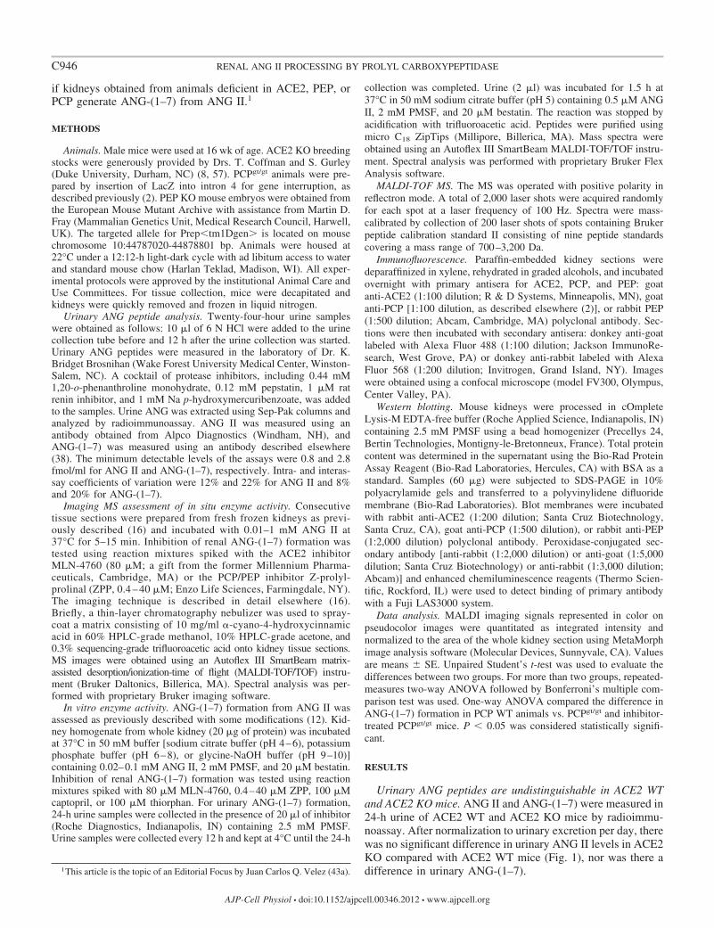
Imaging MS assessment of in situ enzyme activity. Consecutivetissue sections were prepared from fresh frozen kidneys as previ-ously described (16) and incubated with 0.01–1 mM ANG II at37°C for 5–15 min. Inhibition of renal ANG-(1–7) formation wastested using reaction mixtures spiked with the ACE2 inhibitorMLN-4760 (80 �M; a gift from the former Millennium Pharma-ceuticals, Cambridge, MA) or the PCP/PEP inhibitor Z-prolyl-prolinal (ZPP, 0.4 – 40 �M; Enzo Life Sciences, Farmingdale, NY).The imaging technique is described in detail elsewhere (16).Briefly, a thin-layer chromatography nebulizer was used to spray-coat a matrix consisting of 10 mg/ml �-cyano-4-hydroxycinnamicacid in 60% HPLC-grade methanol, 10% HPLC-grade acetone, and0.3% sequencing-grade trifluoroacetic acid onto kidney tissue sections.MS images were obtained using an Autoflex III SmartBeam matrix-assisted desorption/ionization-time of flight (MALDI-TOF/TOF) instru-ment (Bruker Daltonics, Billerica, MA). Spectral analysis was per-formed with proprietary Bruker imaging software.
In vitro enzyme activity. ANG-(1–7) formation from ANG II wasassessed as previously described with some modifications (12). Kid-ney homogenate from whole kidney (20 �g of protein) was incubatedat 37°C in 50 mM buffer [sodium citrate buffer (pH 4–6), potassiumphosphate buffer (pH 6–8), or glycine-NaOH buffer (pH 9–10)]containing 0.02–0.1 mM ANG II, 2 mM PMSF, and 20 �M bestatin.Inhibition of renal ANG-(1–7) formation was tested using reactionmixtures spiked with 80 �M MLN-4760, 0.4–40 �M ZPP, 100 �Mcaptopril, or 100 �M thiorphan. For urinary ANG-(1–7) formation,24-h urine samples were collected in the presence of 20 �l of inhibitor(Roche Diagnostics, Indianapolis, IN) containing 2.5 mM PMSF.Urine samples were collected every 12 h and kept at 4°C until the 24-h
collection was completed. Urine (2 �l) was incubated for 1.5 h at37°C in 50 mM sodium citrate buffer (pH 5) containing 0.5 �M ANGII, 2 mM PMSF, and 20 �M bestatin. The reaction was stopped byacidification with trifluoroacetic acid. Peptides were purified usingmicro C18 ZipTips (Millipore, Billerica, MA). Mass spectra wereobtained using an Autoflex III SmartBeam MALDI-TOF/TOF instru-ment. Spectral analysis was performed with proprietary Bruker FlexAnalysis software.
MALDI-TOF MS. The MS was operated with positive polarity inreflectron mode. A total of 2,000 laser shots were acquired randomlyfor each spot at a laser frequency of 100 Hz. Spectra were mass-calibrated by collection of 200 laser shots of spots containing Brukerpeptide calibration standard II consisting of nine peptide standardscovering a mass range of 700–3,200 Da.
Immunofluorescence. Paraffin-embedded kidney sections weredeparaffinized in xylene, rehydrated in graded alcohols, and incubatedovernight with primary antisera for ACE2, PCP, and PEP: goatanti-ACE2 (1:100 dilution; R & D Systems, Minneapolis, MN), goatanti-PCP [1:100 dilution, as described elsewhere (2)], or rabbit PEP(1:500 dilution; Abcam, Cambridge, MA) polyclonal antibody. Sec-tions were then incubated with secondary antisera: donkey anti-goatlabeled with Alexa Fluor 488 (1:100 dilution; Jackson ImmunoRe-search, West Grove, PA) or donkey anti-rabbit labeled with AlexaFluor 568 (1:200 dilution; Invitrogen, Grand Island, NY). Imageswere obtained using a confocal microscope (model FV300, Olympus,Center Valley, PA).
Western blotting. Mouse kidneys were processed in cOmpleteLysis-M EDTA-free buffer (Roche Applied Science, Indianapolis, IN)containing 2.5 mM PMSF using a bead homogenizer (Precellys 24,Bertin Technologies, Montigny-le-Bretonneux, France). Total proteincontent was determined in the supernatant using the Bio-Rad ProteinAssay Reagent (Bio-Rad Laboratories, Hercules, CA) with BSA as astandard. Samples (60 �g) were subjected to SDS-PAGE in 10%polyacrylamide gels and transferred to a polyvinylidene difluoridemembrane (Bio-Rad Laboratories). Blot membranes were incubatedwith rabbit anti-ACE2 (1:200 dilution; Santa Cruz Biotechnology,Santa Cruz, CA), goat anti-PCP (1:500 dilution), or rabbit anti-PEP(1:2,000 dilution) polyclonal antibody. Peroxidase-conjugated sec-ondary antibody [anti-rabbit (1:2,000 dilution) or anti-goat (1:5,000dilution; Santa Cruz Biotechnology) or anti-rabbit (1:3,000 dilution;Abcam)] and enhanced chemiluminescence reagents (Thermo Scien-tific, Rockford, IL) were used to detect binding of primary antibodywith a Fuji LAS3000 system.
Data analysis. MALDI imaging signals represented in color onpseudocolor images were quantitated as integrated intensity andnormalized to the area of the whole kidney section using MetaMorphimage analysis software (Molecular Devices, Sunnyvale, CA). Valuesare means � SE. Unpaired Student’s t-test was used to evaluate thedifferences between two groups. For more than two groups, repeated-measures two-way ANOVA followed by Bonferroni’s multiple com-parison test was used. One-way ANOVA compared the difference inANG-(1–7) formation in PCP WT animals vs. PCPgt/gt and inhibitor-treated PCPgt/gt mice. P � 0.05 was considered statistically signifi-cant.
RESULTS
Urinary ANG peptides are undistinguishable in ACE2 WTand ACE2 KO mice. ANG II and ANG-(1–7) were measured in24-h urine of ACE2 WT and ACE2 KO mice by radioimmu-noassay. After normalization to urinary excretion per day, therewas no significant difference in urinary ANG II levels in ACE2KO compared with ACE2 WT mice (Fig. 1), nor was there adifference in urinary ANG-(1–7).1This article is the topic of an Editorial Focus by Juan Carlos Q. Velez (43a).
C946 RENAL ANG II PROCESSING BY PROLYL CARBOXYPEPTIDASE
AJP-Cell Physiol • doi:10.1152/ajpcell.00346.2012 • www.ajpcell.org
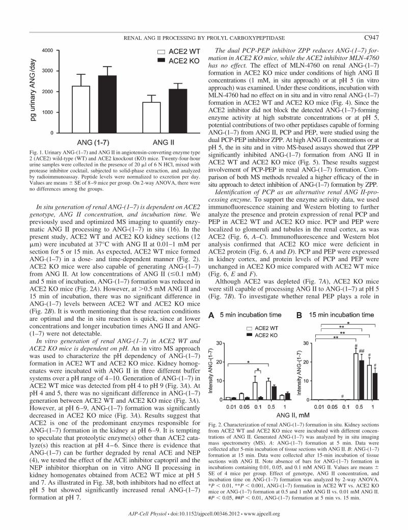
In situ generation of renal ANG-(1–7) is dependent on ACE2genotype, ANG II concentration, and incubation time. Wepreviously used and optimized MS imaging to quantify enzy-matic ANG II processing to ANG-(1–7) in situ (16). In thepresent study, ACE2 WT and ACE2 KO kidney sections (12�m) were incubated at 37°C with ANG II at 0.01–1 mM persection for 5 or 15 min. As expected, ACE2 WT mice formedANG-(1–7) in a dose- and time-dependent manner (Fig. 2).ACE2 KO mice were also capable of generating ANG-(1–7)from ANG II. At low concentrations of ANG II (�0.1 mM)and 5 min of incubation, ANG-(1–7) formation was reduced inACE2 KO mice (Fig. 2A). However, at �0.5 mM ANG II and15 min of incubation, there was no significant difference inANG-(1–7) levels between ACE2 WT and ACE2 KO mice(Fig. 2B). It is worth mentioning that these reaction conditionsare optimal and the in situ reaction is quick, since at lowerconcentrations and longer incubation times ANG II and ANG-(1–7) were not detectable.
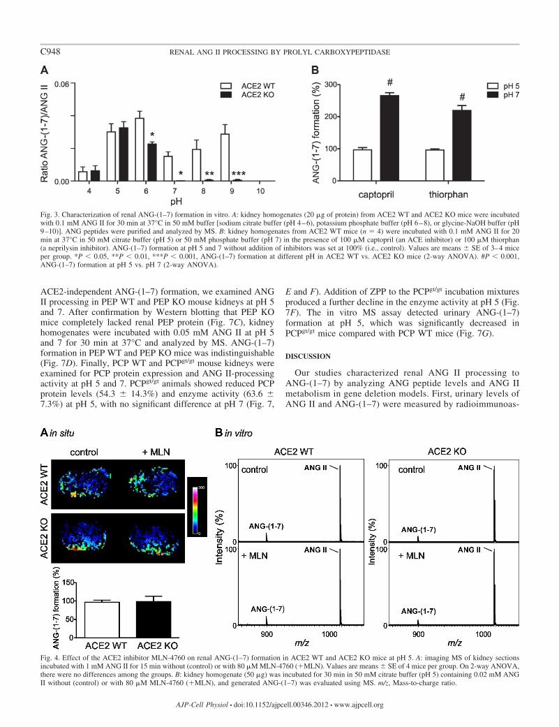
In vitro generation of renal ANG-(1–7) in ACE2 WT andACE2 KO mice is dependent on pH. An in vitro MS approachwas used to characterize the pH dependency of ANG-(1–7)formation in ACE2 WT and ACE2 KO mice. Kidney homog-enates were incubated with ANG II in three different buffersystems over a pH range of 4–10. Generation of ANG-(1–7) inACE2 WT mice was detected from pH 4 to pH 9 (Fig. 3A). AtpH 4 and 5, there was no significant difference in ANG-(1–7)generation between ACE2 WT and ACE2 KO mice (Fig. 3A).However, at pH 6–9, ANG-(1–7) formation was significantlydecreased in ACE2 KO mice (Fig. 3A). Results suggest thatACE2 is one of the predominant enzymes responsible forANG-(1–7) formation in the kidney at pH 6–9. It is temptingto speculate that proteolytic enzyme(s) other than ACE2 cata-lyze(s) this reaction at pH 4–6. Since there is evidence thatANG-(1–7) can be further degraded by renal ACE and NEP(4), we tested the effect of the ACE inhibitor captopril and theNEP inhibitor thiorphan on in vitro ANG II processing inkidney homogenates obtained from ACE2 WT mice at pH 5and 7. As illustrated in Fig. 3B, both inhibitors had no effect atpH 5 but showed significantly increased renal ANG-(1–7)formation at pH 7.
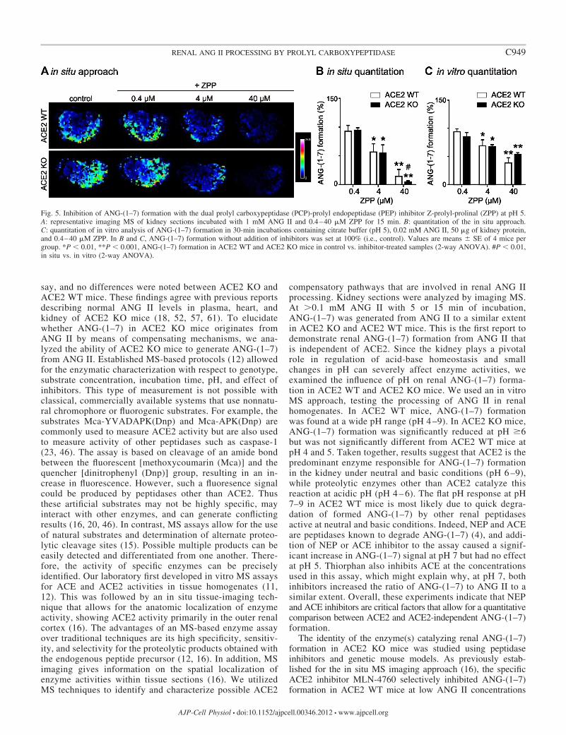
The dual PCP-PEP inhibitor ZPP reduces ANG-(1–7) for-mation in ACE2 KO mice, while the ACE2 inhibitor MLN-4760has no effect. The effect of MLN-4760 on renal ANG-(1–7)formation in ACE2 KO mice under conditions of high ANG IIconcentrations (1 mM, in situ approach) or at pH 5 (in vitroapproach) was examined. Under these conditions, incubation withMLN-4760 had no effect on in situ and in vitro renal ANG-(1–7)formation in ACE2 WT and ACE2 KO mice (Fig. 4). Since theACE2 inhibitor did not block the detected ANG-(1–7)-formingenzyme activity at high substrate concentrations or at pH 5,potential contributions of two other peptidases capable of formingANG-(1–7) from ANG II, PCP and PEP, were studied using thedual PCP-PEP inhibitor ZPP. At high ANG II concentrations or atpH 5, the in situ and in vitro MS-based assays showed that ZPPsignificantly inhibited ANG-(1–7) formation from ANG II inACE2 WT and ACE2 KO mice (Fig. 5). These results suggestinvolvement of PCP-PEP in renal ANG-(1–7) formation. Com-parison of both MS methods revealed a higher efficacy of the insitu approach to detect inhibition of ANG-(1–7) formation by ZPP.
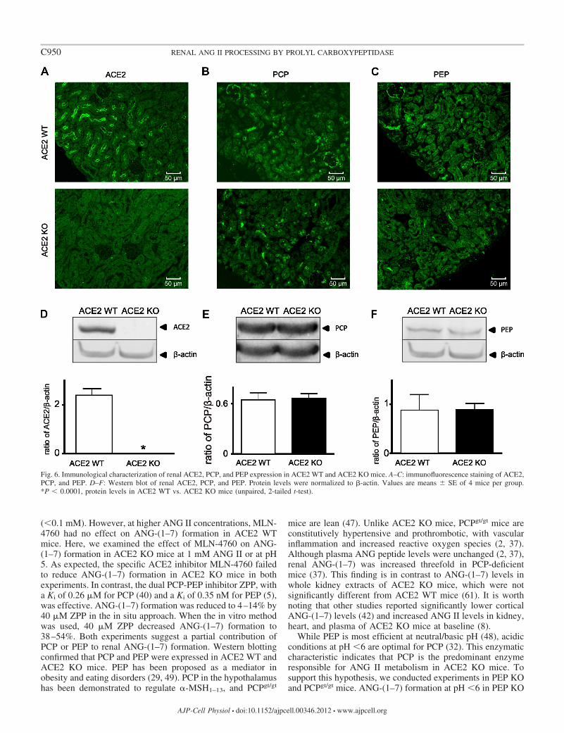
Identification of PCP as an alternative renal ANG II-pro-cessing enzyme. To support the enzyme activity data, we usedimmunofluorescence staining and Western blotting to furtheranalyze the presence and protein expression of renal PCP andPEP in ACE2 WT and ACE2 KO mice. PCP and PEP werelocalized to glomeruli and tubules in the renal cortex, as wasACE2 (Fig. 6, A–C). Immunofluorescence and Western blotanalysis confirmed that ACE2 KO mice were deficient inACE2 protein (Fig. 6, A and D). PCP and PEP were expressedin kidney cortex, and protein levels of PCP and PEP wereunchanged in ACE2 KO mice compared with ACE2 WT mice(Fig. 6, E and F).
Although ACE2 was depleted (Fig. 7A), ACE2 KO micewere still capable of processing ANG II to ANG-(1–7) at pH 5(Fig. 7B). To investigate whether renal PEP plays a role in
Fig. 2. Characterization of renal ANG-(1–7) formation in situ. Kidney sectionsfrom ACE2 WT and ACE2 KO mice were incubated with different concen-trations of ANG II. Generated ANG-(1–7) was analyzed by in situ imagingmass spectrometry (MS). A: ANG-(1–7) formation at 5 min. Data werecollected after 5-min incubation of tissue sections with ANG II. B: ANG-(1–7)formation at 15 min. Data were collected after 15-min incubation of tissuesections with ANG II. Note absence of bars for ANG-(1–7) formation inincubations containing 0.01, 0.05, and 0.1 mM ANG II. Values are means �SE of 4 mice per group. Effect of genotype, ANG II concentration, andincubation time on ANG-(1–7) formation was analyzed by 2-way ANOVA.*P � 0.01, **P � 0.001, ANG-(1–7) formation in ACE2 WT vs. ACE2 KOmice or ANG-(1–7) formation at 0.5 and 1 mM ANG II vs. 0.01 mM ANG II.#P � 0.05, ##P � 0.01, ANG-(1–7) formation at 5 min vs. 15 min.
Fig. 1. Urinary ANG-(1–7) and ANG II in angiotensin-converting enzyme type2 (ACE2) wild-type (WT) and ACE2 knockout (KO) mice. Twenty-four-hoururine samples were collected in the presence of 20 �l of 6 N HCl, mixed withprotease inhibitor cocktail, subjected to solid-phase extraction, and analyzedby radioimmunoassay. Peptide levels were normalized to excretion per day.Values are means � SE of 8–9 mice per group. On 2-way ANOVA, there wereno differences among the groups.
C947RENAL ANG II PROCESSING BY PROLYL CARBOXYPEPTIDASE
AJP-Cell Physiol • doi:10.1152/ajpcell.00346.2012 • www.ajpcell.org
ACE2-independent ANG-(1–7) formation, we examined ANGII processing in PEP WT and PEP KO mouse kidneys at pH 5and 7. After confirmation by Western blotting that PEP KOmice completely lacked renal PEP protein (Fig. 7C), kidneyhomogenates were incubated with 0.05 mM ANG II at pH 5and 7 for 30 min at 37°C and analyzed by MS. ANG-(1–7)formation in PEP WT and PEP KO mice was indistinguishable(Fig. 7D). Finally, PCP WT and PCPgt/gt mouse kidneys wereexamined for PCP protein expression and ANG II-processingactivity at pH 5 and 7. PCPgt/gt animals showed reduced PCPprotein levels (54.3 � 14.3%) and enzyme activity (63.6 �7.3%) at pH 5, with no significant difference at pH 7 (Fig. 7,
E and F). Addition of ZPP to the PCPgt/gt incubation mixturesproduced a further decline in the enzyme activity at pH 5 (Fig.7F). The in vitro MS assay detected urinary ANG-(1–7)formation at pH 5, which was significantly decreased inPCPgt/gt mice compared with PCP WT mice (Fig. 7G).
DISCUSSION
Our studies characterized renal ANG II processing toANG-(1–7) by analyzing ANG peptide levels and ANG IImetabolism in gene deletion models. First, urinary levels ofANG II and ANG-(1–7) were measured by radioimmunoas-
Fig. 3. Characterization of renal ANG-(1–7) formation in vitro. A: kidney homogenates (20 �g of protein) from ACE2 WT and ACE2 KO mice were incubatedwith 0.1 mM ANG II for 30 min at 37°C in 50 mM buffer [sodium citrate buffer (pH 4–6), potassium phosphate buffer (pH 6–8), or glycine-NaOH buffer (pH9–10)]. ANG peptides were purified and analyzed by MS. B: kidney homogenates from ACE2 WT mice (n � 4) were incubated with 0.1 mM ANG II for 20min at 37°C in 50 mM citrate buffer (pH 5) or 50 mM phosphate buffer (pH 7) in the presence of 100 �M captopril (an ACE inhibitor) or 100 �M thiorphan(a neprilysin inhibitor). ANG-(1–7) formation at pH 5 and 7 without addition of inhibitors was set at 100% (i.e., control). Values are means � SE of 3–4 miceper group. *P � 0.05, **P � 0.01, ***P � 0.001, ANG-(1–7) formation at different pH in ACE2 WT vs. ACE2 KO mice (2-way ANOVA). #P � 0.001,ANG-(1–7) formation at pH 5 vs. pH 7 (2-way ANOVA).
Fig. 4. Effect of the ACE2 inhibitor MLN-4760 on renal ANG-(1–7) formation in ACE2 WT and ACE2 KO mice at pH 5. A: imaging MS of kidney sectionsincubated with 1 mM ANG II for 15 min without (control) or with 80 �M MLN-4760 (�MLN). Values are means � SE of 4 mice per group. On 2-way ANOVA,there were no differences among the groups. B: kidney homogenate (50 �g) was incubated for 30 min in 50 mM citrate buffer (pH 5) containing 0.02 mM ANGII without (control) or with 80 �M MLN-4760 (�MLN), and generated ANG-(1–7) was evaluated using MS. m/z, Mass-to-charge ratio.
C948 RENAL ANG II PROCESSING BY PROLYL CARBOXYPEPTIDASE
AJP-Cell Physiol • doi:10.1152/ajpcell.00346.2012 • www.ajpcell.org
say, and no differences were noted between ACE2 KO andACE2 WT mice. These findings agree with previous reportsdescribing normal ANG II levels in plasma, heart, andkidney of ACE2 KO mice (18, 52, 57, 61). To elucidatewhether ANG-(1–7) in ACE2 KO mice originates fromANG II by means of compensating mechanisms, we ana-lyzed the ability of ACE2 KO mice to generate ANG-(1–7)from ANG II. Established MS-based protocols (12) allowedfor the enzymatic characterization with respect to genotype,substrate concentration, incubation time, pH, and effect ofinhibitors. This type of measurement is not possible withclassical, commercially available systems that use nonnatu-ral chromophore or fluorogenic substrates. For example, thesubstrates Mca-YVADAPK(Dnp) and Mca-APK(Dnp) arecommonly used to measure ACE2 activity but are also usedto measure activity of other peptidases such as caspase-1(23, 46). The assay is based on cleavage of an amide bondbetween the fluorescent [methoxycoumarin (Mca)] and thequencher [dinitrophenyl (Dnp)] group, resulting in an in-crease in fluorescence. However, such a fluoresence signalcould be produced by peptidases other than ACE2. Thusthese artificial substrates may not be highly specific, mayinteract with other enzymes, and can generate conflictingresults (16, 20, 46). In contrast, MS assays allow for the useof natural substrates and determination of alternate proteo-lytic cleavage sites (15). Possible multiple products can beeasily detected and differentiated from one another. There-fore, the activity of specific enzymes can be preciselyidentified. Our laboratory first developed in vitro MS assaysfor ACE and ACE2 activities in tissue homogenates (11,12). This was followed by an in situ tissue-imaging tech-nique that allows for the anatomic localization of enzymeactivity, showing ACE2 activity primarily in the outer renalcortex (16). The advantages of an MS-based enzyme assayover traditional techniques are its high specificity, sensitiv-ity, and selectivity for the proteolytic products obtained withthe endogenous peptide precursor (12, 16). In addition, MSimaging gives information on the spatial localization ofenzyme activities within tissue sections (16). We utilizedMS techniques to identify and characterize possible ACE2
compensatory pathways that are involved in renal ANG IIprocessing. Kidney sections were analyzed by imaging MS.At �0.1 mM ANG II with 5 or 15 min of incubation,ANG-(1–7) was generated from ANG II to a similar extentin ACE2 KO and ACE2 WT mice. This is the first report todemonstrate renal ANG-(1–7) formation from ANG II thatis independent of ACE2. Since the kidney plays a pivotalrole in regulation of acid-base homeostasis and smallchanges in pH can severely affect enzyme activities, weexamined the influence of pH on renal ANG-(1–7) forma-tion in ACE2 WT and ACE2 KO mice. We used an in vitroMS approach, testing the processing of ANG II in renalhomogenates. In ACE2 WT mice, ANG-(1–7) formationwas found at a wide pH range (pH 4 –9). In ACE2 KO mice,ANG-(1–7) formation was significantly reduced at pH �6but was not significantly different from ACE2 WT mice atpH 4 and 5. Taken together, results suggest that ACE2 is thepredominant enzyme responsible for ANG-(1–7) formationin the kidney under neutral and basic conditions (pH 6 –9),while proteolytic enzymes other than ACE2 catalyze thisreaction at acidic pH (pH 4 – 6). The flat pH response at pH7–9 in ACE2 WT mice is most likely due to quick degra-dation of formed ANG-(1–7) by other renal peptidasesactive at neutral and basic conditions. Indeed, NEP and ACEare peptidases known to degrade ANG-(1–7) (4), and addi-tion of NEP or ACE inhibitor to the assay caused a signif-icant increase in ANG-(1–7) signal at pH 7 but had no effectat pH 5. Thiorphan also inhibits ACE at the concentrationsused in this assay, which might explain why, at pH 7, bothinhibitors increased the ratio of ANG-(1–7) to ANG II to asimilar extent. Overall, these experiments indicate that NEPand ACE inhibitors are critical factors that allow for a quantitativecomparison between ACE2 and ACE2-independent ANG-(1–7)formation.
The identity of the enzyme(s) catalyzing renal ANG-(1–7)formation in ACE2 KO mice was studied using peptidaseinhibitors and genetic mouse models. As previously estab-lished for the in situ MS imaging approach (16), the specificACE2 inhibitor MLN-4760 selectively inhibited ANG-(1–7)formation in ACE2 WT mice at low ANG II concentrations
Fig. 5. Inhibition of ANG-(1–7) formation with the dual prolyl carboxypeptidase (PCP)-prolyl endopeptidase (PEP) inhibitor Z-prolyl-prolinal (ZPP) at pH 5.A: representative imaging MS of kidney sections incubated with 1 mM ANG II and 0.4–40 �M ZPP for 15 min. B: quantitation of the in situ approach.C: quantitation of in vitro analysis of ANG-(1–7) formation in 30-min incubations containing citrate buffer (pH 5), 0.02 mM ANG II, 50 �g of kidney protein,and 0.4–40 �M ZPP. In B and C, ANG-(1–7) formation without addition of inhibitors was set at 100% (i.e., control). Values are means � SE of 4 mice pergroup. *P � 0.01, **P � 0.001, ANG-(1–7) formation in ACE2 WT and ACE2 KO mice in control vs. inhibitor-treated samples (2-way ANOVA). #P � 0.01,in situ vs. in vitro (2-way ANOVA).
C949RENAL ANG II PROCESSING BY PROLYL CARBOXYPEPTIDASE
AJP-Cell Physiol • doi:10.1152/ajpcell.00346.2012 • www.ajpcell.org
(�0.1 mM). However, at higher ANG II concentrations, MLN-4760 had no effect on ANG-(1–7) formation in ACE2 WTmice. Here, we examined the effect of MLN-4760 on ANG-(1–7) formation in ACE2 KO mice at 1 mM ANG II or at pH5. As expected, the specific ACE2 inhibitor MLN-4760 failedto reduce ANG-(1–7) formation in ACE2 KO mice in bothexperiments. In contrast, the dual PCP-PEP inhibitor ZPP, witha Ki of 0.26 �M for PCP (40) and a Ki of 0.35 nM for PEP (5),was effective. ANG-(1–7) formation was reduced to 4–14% by40 �M ZPP in the in situ approach. When the in vitro methodwas used, 40 �M ZPP decreased ANG-(1–7) formation to38–54%. Both experiments suggest a partial contribution ofPCP or PEP to renal ANG-(1–7) formation. Western blottingconfirmed that PCP and PEP were expressed in ACE2 WT andACE2 KO mice. PEP has been proposed as a mediator inobesity and eating disorders (29, 49). PCP in the hypothalamushas been demonstrated to regulate �-MSH1–13, and PCPgt/gt
mice are lean (47). Unlike ACE2 KO mice, PCPgt/gt mice areconstitutively hypertensive and prothrombotic, with vascularinflammation and increased reactive oxygen species (2, 37).Although plasma ANG peptide levels were unchanged (2, 37),renal ANG-(1–7) was increased threefold in PCP-deficientmice (37). This finding is in contrast to ANG-(1–7) levels inwhole kidney extracts of ACE2 KO mice, which were notsignificantly different from ACE2 WT mice (61). It is worthnoting that other studies reported significantly lower corticalANG-(1–7) levels (42) and increased ANG II levels in kidney,heart, and plasma of ACE2 KO mice at baseline (8).
While PEP is most efficient at neutral/basic pH (48), acidicconditions at pH �6 are optimal for PCP (32). This enzymaticcharacteristic indicates that PCP is the predominant enzymeresponsible for ANG II metabolism in ACE2 KO mice. Tosupport this hypothesis, we conducted experiments in PEP KOand PCPgt/gt mice. ANG-(1–7) formation at pH �6 in PEP KO
Fig. 6. Immunological characterization of renal ACE2, PCP, and PEP expression in ACE2 WT and ACE2 KO mice. A–C: immunofluorescence staining of ACE2,PCP, and PEP. D–F: Western blot of renal ACE2, PCP, and PEP. Protein levels were normalized to �-actin. Values are means � SE of 4 mice per group.*P � 0.0001, protein levels in ACE2 WT vs. ACE2 KO mice (unpaired, 2-tailed t-test).
C950 RENAL ANG II PROCESSING BY PROLYL CARBOXYPEPTIDASE
AJP-Cell Physiol • doi:10.1152/ajpcell.00346.2012 • www.ajpcell.org
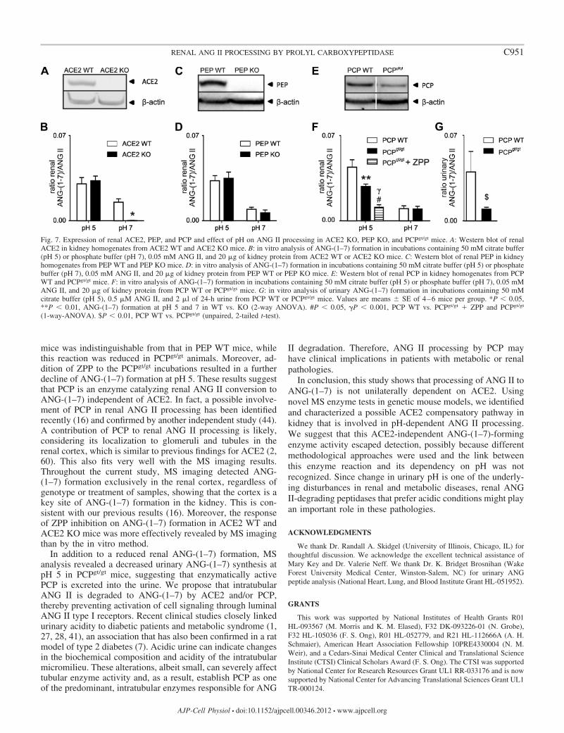
mice was indistinguishable from that in PEP WT mice, whilethis reaction was reduced in PCPgt/gt animals. Moreover, ad-dition of ZPP to the PCPgt/gt incubations resulted in a furtherdecline of ANG-(1–7) formation at pH 5. These results suggestthat PCP is an enzyme catalyzing renal ANG II conversion toANG-(1–7) independent of ACE2. In fact, a possible involve-ment of PCP in renal ANG II processing has been identifiedrecently (16) and confirmed by another independent study (44).A contribution of PCP to renal ANG II processing is likely,considering its localization to glomeruli and tubules in therenal cortex, which is similar to previous findings for ACE2 (2,60). This also fits very well with the MS imaging results.Throughout the current study, MS imaging detected ANG-(1–7) formation exclusively in the renal cortex, regardless ofgenotype or treatment of samples, showing that the cortex is akey site of ANG-(1–7) formation in the kidney. This is con-sistent with our previous results (16). Moreover, the responseof ZPP inhibition on ANG-(1–7) formation in ACE2 WT andACE2 KO mice was more effectively revealed by MS imagingthan by the in vitro method.
In addition to a reduced renal ANG-(1–7) formation, MSanalysis revealed a decreased urinary ANG-(1–7) synthesis atpH 5 in PCPgt/gt mice, suggesting that enzymatically activePCP is excreted into the urine. We propose that intratubularANG II is degraded to ANG-(1–7) by ACE2 and/or PCP,thereby preventing activation of cell signaling through luminalANG II type I receptors. Recent clinical studies closely linkedurinary acidity to diabetic patients and metabolic syndrome (1,27, 28, 41), an association that has also been confirmed in a ratmodel of type 2 diabetes (7). Acidic urine can indicate changesin the biochemical composition and acidity of the intratubularmicromilieu. These alterations, albeit small, can severely affecttubular enzyme activity and, as a result, establish PCP as oneof the predominant, intratubular enzymes responsible for ANG
II degradation. Therefore, ANG II processing by PCP mayhave clinical implications in patients with metabolic or renalpathologies.
In conclusion, this study shows that processing of ANG II toANG-(1–7) is not unilaterally dependent on ACE2. Usingnovel MS enzyme tests in genetic mouse models, we identifiedand characterized a possible ACE2 compensatory pathway inkidney that is involved in pH-dependent ANG II processing.We suggest that this ACE2-independent ANG-(1–7)-formingenzyme activity escaped detection, possibly because differentmethodological approaches were used and the link betweenthis enzyme reaction and its dependency on pH was notrecognized. Since change in urinary pH is one of the underly-ing disturbances in renal and metabolic diseases, renal ANGII-degrading peptidases that prefer acidic conditions might playan important role in these pathologies.
ACKNOWLEDGMENTS
We thank Dr. Randall A. Skidgel (University of Illinois, Chicago, IL) forthoughtful discussion. We acknowledge the excellent technical assistance ofMary Key and Dr. Valerie Neff. We thank Dr. K. Bridget Brosnihan (WakeForest University Medical Center, Winston-Salem, NC) for urinary ANGpeptide analysis (National Heart, Lung, and Blood Institute Grant HL-051952).
GRANTS
This work was supported by National Institutes of Health Grants R01HL-093567 (M. Morris and K. M. Elased), F32 DK-093226-01 (N. Grobe),F32 HL-105036 (F. S. Ong), R01 HL-052779, and R21 HL-112666A (A. H.Schmaier), American Heart Association Fellowship 10PRE4330004 (N. M.Weir), and a Cedars-Sinai Medical Center Clinical and Translational ScienceInstitute (CTSI) Clinical Scholars Award (F. S. Ong). The CTSI was supportedby National Center for Research Resources Grant UL1 RR-033176 and is nowsupported by National Center for Advancing Translational Sciences Grant UL1TR-000124.
Fig. 7. Expression of renal ACE2, PEP, and PCP and effect of pH on ANG II processing in ACE2 KO, PEP KO, and PCPgt/gt mice. A: Western blot of renalACE2 in kidney homogenates from ACE2 WT and ACE2 KO mice. B: in vitro analysis of ANG-(1–7) formation in incubations containing 50 mM citrate buffer(pH 5) or phosphate buffer (pH 7), 0.05 mM ANG II, and 20 �g of kidney protein from ACE2 WT or ACE2 KO mice. C: Western blot of renal PEP in kidneyhomogenates from PEP WT and PEP KO mice. D: in vitro analysis of ANG-(1–7) formation in incubations containing 50 mM citrate buffer (pH 5) or phosphatebuffer (pH 7), 0.05 mM ANG II, and 20 �g of kidney protein from PEP WT or PEP KO mice. E: Western blot of renal PCP in kidney homogenates from PCPWT and PCPgt/gt mice. F: in vitro analysis of ANG-(1–7) formation in incubations containing 50 mM citrate buffer (pH 5) or phosphate buffer (pH 7), 0.05 mMANG II, and 20 �g of kidney protein from PCP WT or PCPgt/gt mice. G: in vitro analysis of urinary ANG-(1–7) formation in incubations containing 50 mMcitrate buffer (pH 5), 0.5 �M ANG II, and 2 �l of 24-h urine from PCP WT or PCPgt/gt mice. Values are means � SE of 4–6 mice per group. *P � 0.05,**P � 0.01, ANG-(1–7) formation at pH 5 and 7 in WT vs. KO (2-way ANOVA). #P � 0.05, P � 0.001, PCP WT vs. PCPgt/gt � ZPP and PCPgt/gt
(1-way-ANOVA). $P � 0.01, PCP WT vs. PCPgt/gt (unpaired, 2-tailed t-test).
C951RENAL ANG II PROCESSING BY PROLYL CARBOXYPEPTIDASE
AJP-Cell Physiol • doi:10.1152/ajpcell.00346.2012 • www.ajpcell.org
DISCLAIMERS
The content is solely the responsibility of the authors and does notnecessarily represent the official views of the National Institutes of Health.
DISCLOSURES
No conflicts of interest, financial or otherwise, are declared by the authors.
AUTHOR CONTRIBUTIONS
N.G., N.M.W., M.M., and K.M.E. are responsible for conception anddesign of the research; N.G., N.M.W., O.L., and F.S.O. performed theexperiments; N.G., N.M.W., O.L., and F.S.O. analyzed the data; N.G.,N.M.W., O.L., F.S.O., K.E.B., A.H.S., M.M., and K.M.E. interpreted theresults of the experiments; N.G., N.M.W., and O.L. prepared the figures; N.G.,M.M., and K.M.E. drafted the manuscript; N.G., N.M.W., O.L., F.S.O.,K.E.B., A.H.S., M.M., and K.M.E. edited and revised the manuscript; N.G.,N.M.W., O.L., F.S.O., K.E.B., A.H.S., M.M., and K.M.E. approved the finalversion of the manuscript.
REFERENCES
1. Abate N, Chandalia M, Cabo-Chan AV, Moe OW, Sakhaee K. Themetabolic syndrome and uric acid nephrolithiasis: novel features of renalmanifestation of insulin resistance. Kidney Int 65: 386–392, 2004.
2. Adams GN, LaRusch GA, Stavrou E, Zhou Y, Nieman MT, JacobsGH, Cui Y, Lu Y, Jain MK, Mahdi F, Shariat-Madar Z, Okada Y,D’Alecy LG, Schmaier AH. Murine prolylcarboxypeptidase depletioninduces vascular dysfunction with hypertension and faster arterial throm-bosis. Blood 117: 3929–3937, 2011.
3. Alghamri MS, Weir NM, Anstadt MP, Elased KM, Gurley SB, MorrisM. Enhanced angiotensin II-induced cardiac and aortic remodeling inACE2 knockout mice. J Cardiovasc Pharmacol Ther 18: 138–151, 2013.
4. Allred AJ, Diz DI, Ferrario CM, Chappell MC. Pathways for angio-tensin-(1–7) metabolism in pulmonary and renal tissues. Am J PhysiolRenal Physiol 279: F841–F850, 2000.
5. Bakker AV, Jung S, Spencer RW, Vinick FJ, Faraci WS. Slowtight-binding inhibition of prolyl endopeptidase by benzyloxycarbonyl-prolyl-prolinal. Biochem J 271: 559–562, 1990.
6. Bindom SM, Hans CP, Xia H, Boulares AH, Lazartigues E. Angio-tensin I-converting enzyme type 2 (ACE2) gene therapy improves glyce-mic control in diabetic mice. Diabetes 59: 2540–2548, 2010.
7. Bobulescu IA, Dubree M, Zhang J, McLeroy P, Moe OW. Effect ofrenal lipid accumulation on proximal tubule Na�/H� exchange andammonium secretion. Am J Physiol Renal Physiol 294: F1315–F1322,2008.
8. Crackower MA, Sarao R, Oudit GY, Yagil C, Kozieradzki I, ScangaSE, Oliveira-dos-Santos AJ, da Costa J, Zhang L, Pei Y, Scholey J,Ferrario CM, Manoukian AS, Chappell MC, Backx PH, Yagil Y,Penninger JM. Angiotensin-converting enzyme 2 is an essential regulatorof heart function. Nature 417: 822–828, 2002.
9. Danilczyk U, Penninger JM. Angiotensin-converting enzyme II in theheart and the kidney. Circ Res 98: 463–471, 2006.
10. Donoghue M, Hsieh F, Baronas E, Godbout K, Gosselin M, StaglianoN, Donovan M, Woolf B, Robison K, Jeyaseelan R, Breitbart RE,Acton S. A novel angiotensin-converting enzyme-related carboxypepti-dase (ACE2) converts angiotensin I to angiotensin 1–9. Circ Res 87:E1–E9, 2000.
11. Elased KM, Cool DR, Morris M. Novel mass spectrometric methods forevaluation of plasma angiotensin converting enzyme 1 and renin activity.Hypertension 46: 953–959, 2005.
12. Elased KM, Cunha TS, Gurley SB, Coffman TM, Morris M. Newmass spectrometric assay for angiotensin-converting enzyme 2 activity.Hypertension 47: 1010–1017, 2006.
13. Feng Y, Xia H, Cai Y, Halabi CM, Becker LK, Santos RA, Speth RC,Sigmund CD, Lazartigues E. Brain-selective overexpression of humanangiotensin-converting enzyme type 2 attenuates neurogenic hypertension.Circ Res 106: 373–382, 2010.
14. Giani JF, Munoz MC, Pons RA, Cao G, Toblli JE, Turyn D, DominiciFP. Angiotensin-(1–7) reduces proteinuria and diminishes structural dam-age in renal tissue of stroke-prone spontaneously hypertensive rats. Am JPhysiol Renal Physiol 300: F272–F282, 2011.
15. Greis KD. Mass spectrometry for enzyme assays and inhibitor screening:an emerging application in pharmaceutical research. Mass Spectrom Rev26: 324–339, 2007.
16. Grobe N, Elased KM, Cool DR, Morris M. Mass spectrometry for themolecular imaging of angiotensin metabolism in kidney. Am J PhysiolEndocrinol Metab 302: E1016–E1024, 2012.
17. Gurley SB, Allred A, Le TH, Griffiths R, Mao L, Philip N, HaysteadTA, Donoghue M, Breitbart RE, Acton SL, Rockman HA, CoffmanTM. Altered blood pressure responses and normal cardiac phenotype inACE2-null mice. J Clin Invest 116: 2218–2225, 2006.
18. Gurley SB, Clare SE, Snow KP, Hu A, Meyer TW, Coffman TM.Impact of genetic background on nephropathy in diabetic mice. Am JPhysiol Renal Physiol 290: F214–F222, 2006.
19. Gurley SB, Coffman TM. Angiotensin-converting enzyme 2 gene tar-geting studies in mice: mixed messages. Exp Physiol 93: 538–542, 2008.
20. Guy JL, Jackson RM, Acharya KR, Sturrock ED, Hooper NM,Turner AJ. Angiotensin-converting enzyme-2 (ACE2): comparativemodeling of the active site, specificity requirements, and chloride depen-dence. Biochemistry 42: 13185–13192, 2003.
21. Gwathmey TM, Pendergrass KD, Reid SD, Rose JC, Diz DI, ChappellMC. Angiotensin-(1–7)-angiotensin-converting enzyme 2 attenuates reac-tive oxygen species formation to angiotensin II within the cell nucleus.Hypertension 55: 166–171, 2010.
22. Huentelman MJ, Grobe JL, Vazquez J, Stewart JM, Mecca AP,Katovich MJ, Ferrario CM, Raizada MK. Protection from angiotensinII-induced cardiac hypertrophy and fibrosis by systemic lentiviral deliveryof ACE2 in rats. Exp Physiol 90: 783–790, 2005.
23. Kawahara A, Enari M, Talanian RV, Wong WW, Nagata S. Fas-induced DNA fragmentation and proteolysis of nuclear proteins. GenesCells 3: 297–306, 1998.
24. Liu C, Lv XH, Li HX, Cao X, Zhang F, Wang L, Yu M, Yang JK.Angiotensin-(1–7) suppresses oxidative stress and improves glucose up-take via Mas receptor in adipocytes. Acta Diabetol 49: 291–299, 2012.
25. Liu CX, Hu Q, Wang Y, Zhang W, Ma ZY, Feng JB, Wang R, WangXP, Dong B, Gao F, Zhang MX, Zhang Y. Angiotensin-convertingenzyme (ACE) 2 overexpression ameliorates glomerular injury in a ratmodel of diabetic nephropathy: a comparison with ACE inhibition. MolMed 17: 59–69, 2011.
26. Luther JM, Brown NJ. The renin-angiotensin-aldosterone system andglucose homeostasis. Trends Pharmacol Sci 32: 734–739, 2011.
27. Maalouf NM, Cameron MA, Moe OW, Adams-Huet B, Sakhaee K.Low urine pH: a novel feature of the metabolic syndrome. Clin J Am SocNephrol 2: 883–888, 2007.
28. Maalouf NM, Cameron MA, Moe OW, Sakhaee K. Metabolic basis forlow urine pH in type 2 diabetes. Clin J Am Soc Nephrol 5: 1277–1281,2010.
29. Maes M, Monteleone P, Bencivenga R, Goossens F, Maj M, van WD,Bosmans E, Scharpe S. Lower serum activity of prolyl endopeptidase inanorexia and bulimia nervosa. Psychoneuroendocrinology 26: 17–26,2001.
30. Monteiro MB, Senador D, Zhang WF, Morris M, Elased KM. Balanceof ACE and ACE2 in diabetes: decreased renal ACE2 activity in hyper-tensive db/db diabetic mice. Hypertension 52: E91, 2008.
31. Nadarajah R, Milagres R, Dilauro M, Gutsol A, Xiao F, ZimpelmannJ, Kennedy C, Wysocki J, Batlle D, Burns KD. Podocyte-specificoverexpression of human angiotensin-converting enzyme 2 attenuatesdiabetic nephropathy in mice. Kidney Int 82: 292–303, 2012.
32. Odya CE, Marinkovic DV, Hammon KJ, Stewart TA, Erdos EG.Purification and properties of prolylcarboxypeptidase (angiotensinase C)from human kidney. J Biol Chem 253: 5927–5931, 1978.
33. Oudit GY, Liu GC, Zhong J, Basu R, Chow FL, Zhou J, Loibner H,Janzek E, Schuster M, Penninger JM, Herzenberg AM, Kassiri Z,Scholey JW. Human recombinant ACE2 reduces the progression ofdiabetic nephropathy. Diabetes 59: 529–538, 2010.
34. Patel VB, Bodiga S, Fan D, Das SK, Wang Z, Wang W, Basu R, ZhongJ, Kassiri Z, Oudit GY. Cardioprotective effects mediated by angiotensinII type 1 receptor blockade and enhancing angiotensin 1–7 in experimentalheart failure in angiotensin-converting enzyme 2-null mice. Hypertension59: 1195–1203, 2012.
35. Rioli V, Kato A, Portaro FC, Cury GK, te Kaat K, Vincent B, CheclerF, Camargo AC, Glucksman MJ, Roberts JL, Hirose S, Ferro ES.Neuropeptide specificity and inhibition of recombinant isoforms of theendopeptidase 342416 family: comparison with the related recombinantendopeptidase 342415. Biochem Biophys Res Commun 250: 5–11, 1998.
36. Rodriguez-Iturbe B, Pons H, Herrera-Acosta J, Johnson RJ. Role ofimmunocompetent cells in nonimmune renal diseases. Kidney Int 59:1626–1640, 2001.
C952 RENAL ANG II PROCESSING BY PROLYL CARBOXYPEPTIDASE
AJP-Cell Physiol • doi:10.1152/ajpcell.00346.2012 • www.ajpcell.org
37. Schadock I, Todiras M, Vilianovich L, Qadri F, Heuser A, Siems WE,Santos RA, Bader M. Contribution of central angiotensin II to thehypertensive phenotype of prolylcarboxypeptidase knockout mice. Hyper-tension 58: E36, 2011.
38. Senanayake P, Moriguchi A, Kumagai H, Ganten D, Ferrario CM,Brosnihan KB. Increased expression of angiotensin peptides in the brainof transgenic hypertensive rats. Peptides 15: 919–926, 1994.
39. Skeggs LT Jr, Kahn JR, Shumway NP. The preparation and function ofthe hypertensin-converting enzyme. J Exp Med 103: 295–299, 1956.
40. Tan F, Morris PW, Skidgel RA, Erdös EG. Sequencing and cloning ofhuman prolylcarboxypeptidase (angiotensinase C). Similarity to both ser-ine carboxypeptidase and prolylendopeptidase families. J Biol Chem 268:16631–16638, 1993.
41. Taylor EN, Curhan GC. Body size and 24-hour urine composition. AmJ Kidney Dis 48: 905–915, 2006.
42. Tikellis C, Bialkowski K, Pete J, Sheehy K, Su Q, Johnston C, CooperME, Thomas MC. ACE2 deficiency modifies renoprotection afforded byACE inhibition in experimental diabetes. Diabetes 57: 1018–1025, 2008.
43. Tipnis SR, Hooper NM, Hyde R, Karran E, Christie G, Turner AJ. Ahuman homolog of angiotensin-converting enzyme. Cloning and func-tional expression as a captopril-insensitive carboxypeptidase. J Biol Chem275: 33238–33243, 2000.
43a.Velez JC. Prolyl carboxypeptidase: a forgotten kidney angiotensinase. Focuson “Identification of prolyl carboxypeptidase as an alternative enzyme forprocessing of renal angiotensin II using mass spectrometry.” Am J PhysiolCell Physiol (April 3, 2013). doi:10.1152/ajpcell.00081.2013.
44. Velez JC, Ierardi JL, Bland AM, Morinelli TA, Arthur JM, RaymondJR, Janech MG. Enzymatic processing of angiotensin peptides by humanglomerular endothelial cells. Am J Physiol Renal Physiol 302: F1583–F1594, 2012.
45. Verma A, Shan ZY, Lei B, Yuan LH, Liu X, Nakagawa T, Grant MB,Lewin AS, Hauswirth WW, Raizada MK, Li QH. ACE2 and ANG-(1–7) confer protection against development of diabetic retinopathy. MolTher 20: 28–36, 2012.
46. Vickers C, Hales P, Kaushik V, Dick L, Gavin J, Tang J, Godbout K,Parsons T, Baronas E, Hsieh F, Acton S, Patane M, Nichols A, TumminoP. Hydrolysis of biological peptides by human angiotensin-converting en-zyme-related carboxypeptidase. J Biol Chem 277: 14838–14843, 2002.
47. Wallingford N, Perroud B, Gao Q, Coppola A, Gyengesi E, Liu ZW,Gao XB, Diament A, Haus KA, Shariat-Madar Z, Mahdi F, WardlawSL, Schmaier AH, Warden CH, Diano S. Prolylcarboxypeptidase reg-ulates food intake by inactivating �-MSH in rodents. J Clin Invest 119:2291–2303, 2009.
48. Ward PE, Bausback HH, Odya CE. Kinin and angiotensin metabolismby purified renal post-proline cleaving enzyme. Biochem Pharmacol 36:3187–3193, 1987.
49. Warden CH, Fisler JS, Espinal G, Graham J, Havel PJ, Perroud B.Maternal influence of prolyl endopeptidase on fat mass of adult progeny.Int J Obes (Lond) 33: 1013–1022, 2009.
50. Weir NM, Salem E, Grobe N, Yan ZY, Morris M, Elased KM. Renalangiotensin converting enzyme 2 protects kidney function during chronicANG II infusion in mice. Hypertension 58: E79, 2011.
51. Welches WR, Brosnihan KB, Ferrario CM. A comparison of theproperties and enzymatic activities of three angiotensin processing en-zymes: angiotensin converting enzyme, prolyl endopeptidase and nepri-lysin. Life Sci 52: 1461–1480, 1993.
52. Wong DW, Oudit GY, Reich H, Kassiri Z, Zhou J, Liu QC, Backx PH,Penninger JM, Herzenberg AM, Scholey JW. Loss of angiotensin-converting enzyme-2 (Ace2) accelerates diabetic kidney injury. Am JPathol 171: 438–451, 2007.
53. Wysocki J, Ye M, Rodriguez E, Gonzalez-Pacheco FR, Barrios C,Evora K, Schuster M, Loibner H, Brosnihan KB, Ferrario CM,Penninger JM, Batlle D. Targeting the degradation of angiotensin II withrecombinant angiotensin-converting enzyme 2: prevention of angiotensinII-dependent hypertension. Hypertension 55: 90–98, 2010.
54. Wysocki J, Ye M, Soler MJ, Gurley SB, Xiao HD, Bernstein KE,Coffman TM, Chen S, Batlle D. ACE and ACE2 activity in diabeticmice. Diabetes 55: 2132–2139, 2006.
55. Xiao L, Gao L, Lazartigues E, Zucker IH. Brain-selective overexpres-sion of angiotensin-converting enzyme 2 attenuates sympathetic nerveactivity and enhances baroreflex function in chronic heart failure. Hyper-tension 58: 1057–1065, 2011.
56. Yamamoto K, Chappell MC, Brosnihan KB, Ferrario CM. In vivometabolism of angiotensin I by neutral endopeptidase (EC 3.42411) inspontaneously hypertensive rats. Hypertension 19: 692–696, 1992.
57. Yamamoto K, Ohishi M, Katsuya T, Ito N, Ikushima M, Kaibe M,Tatara Y, Shiota A, Sugano S, Takeda S, Rakugi H, Ogihara T.Deletion of angiotensin-converting enzyme 2 accelerates pressure over-load-induced cardiac dysfunction by increasing local angiotensin II. Hy-pertension 47: 718–726, 2006.
58. Yamazato Y, Ferreira AJ, Hong KH, Sriramula S, Francis J, Ya-mazato M, Yuan L, Bradford CN, Shenoy V, Oh SP, Katovich MJ,Raizada MK. Prevention of pulmonary hypertension by angiotensin-converting enzyme 2 gene transfer. Hypertension 54: 365–371, 2009.
59. Ye M, Wysocki J, Naaz P, Salabat MR, LaPointe MS, Batlle D.Increased ACE 2 and decreased ACE protein in renal tubules fromdiabetic mice: a renoprotective combination? Hypertension 43: 1120 –1125, 2004.
60. Ye M, Wysocki J, William J, Soler MJ, Cokic I, Batlle D. Glomerularlocalization and expression of angiotensin-converting enzyme 2 and ang-iotensin-converting enzyme: implications for albuminuria in diabetes. JAm Soc Nephrol 17: 3067–3075, 2006.
61. Zhong J, Guo D, Chen CB, Wang W, Schuster M, Loibner H,Penninger JM, Scholey JW, Kassiri Z, Oudit GY. Prevention ofangiotensin II-mediated renal oxidative stress, inflammation, and fibrosisby angiotensin-converting enzyme 2. Hypertension 57: 314–322, 2011.
C953RENAL ANG II PROCESSING BY PROLYL CARBOXYPEPTIDASE
AJP-Cell Physiol • doi:10.1152/ajpcell.00346.2012 • www.ajpcell.org