ICAR sponsored short course on “Preparation of ...
185
ICAR sponsored short course on “Preparation of bioformulation...........management of biotic stress agricultural crops” 1
-
Upload
khangminh22 -
Category
Documents
-
view
3 -
download
0
Transcript of ICAR sponsored short course on “Preparation of ...
ICAR sponsored short course on “Preparation of bioformulation...........management of biotic stress agricultural crops” 1
ICAR sponsored short course on “Preparation of bioformulation...........management of biotic stress agricultural crops” 2
ICAR Sponsored Short Course
On
“Preparation of Bioformulation of Fungal and Bacterial Biocontrol Agents for Management of
Biotic Stress of Agricultural Crops”
Organized by:
Department of Plant Pathology Assam Agricultural University
Jorhat-785013, Assam
ICAR sponsored short course on “Preparation of bioformulation...........management of biotic stress agricultural crops” 3
The publication has been brought out on the occasion of a Short Course on “Preparation of Bioformulation of Fungal and Bacterial Biocontrol Agents for Management of Biotic Stress of Agricultural Crops” sponsored by ICAR, New Delhi and organized by Department of Plant Pathology, Assam Agricultural University, Jorhat-13, Assam from 1st to 10th Sept, 2017.
Editorial Board
Dr. Pranab Dutta Dr. K. C. Puzari
Dr. A. Bhattacharyya
Member
Ms. Himadri Kaushik
Cover design:
Dr. Pranab Dutta
© Department of Plant Pathology, Assam Agricultural University, Jorhat-13, Assam

ICAR sponsored short course on “Preparation of bioformulation...........management of biotic stress agricultural crops” 4
CONTENTS
Sl. No. Title Page No.
1. Past present and future of biopesticides 1-7
2. Entomopathogenic fungi for insect pest management 8-12
3. Biological control – An ecological perspective 13-20
4. Techniques for isolation of fungal biocontrol agents, endophytes and
study on their evaluation
21-22
5. Taxonomy based identification of fungal biocontrol agents 23-26
6. Bacterial species characterization by polyphasic taxonomy 27
7. Microbes in the management of banana diseases 28-35
8. Phylloplane bacteria and plant disease management 36-47
9. Preparation of liquid bioformulation of fungal biocontrol agents and its
shelf life study
48-51
10. Techniques for development of microbes based bioformulation 52-57
11. Biointensive disease management of vegetable crops 58-65
12. Isolation of biofertilizer microbes from crops rhizosphere 66-73
13. Preparation of plant extracts, efficacy test and their use for plant disease management
74-79
14. Recent advances for biological control of insect pests 80-83
15. Preparation of some biopesticide materials at farm level for pest management
84-90
16. Biocontrol of mite pests of horticultural crops 91-103
17. Post-harvest food grain management: potential green approaches for
controlling stored grain insect pest 104-109
18. Isolation and diagnosis of nematodes 110-123

ICAR sponsored short course on “Preparation of bioformulation...........management of biotic stress agricultural crops” 5
19. Role of nematode in disease complex 124-128
19. Biological control of plant parasitic nematodes 129-136
20. Pasteuria – A potential biocontrol agent for the management of nematodes
137-140
21. Molecular basis of plant pathogen interaction 141-149
22. Large scale adoption of biocontrol agents: challenges and opportunity 150-152
23. Biopesticides and IPR Issues
153-156
24. Biological management of Invasive Alien Species (IAS) 157-172
25. Isolation and Enumeration of Microorganisms by Serial Dilution Method
173-174
26. Purification of Biological Agents
175
27. Counting of Cells/Spores of Microorganisms 176
ICAR sponsored short course on “Preparation of bioformulation...........management of biotic stress agricultural crops” 6
Past Present and Future of Biopesticides
Lakshmi Kanta Hazarika Department of Entomology,
Assam Agricultural University, Jorhat- 785013, Assam
Birth rate (one child per 8 seconds), death rate (one death in every 12 seconds), migration (one per 33 seconds) contribute to population growth of a nation resulting a net gain of one person every 12 seconds. Thus global population is increasing in a geometrical progression to reach at 9 billions by 2050. Every day, 795 million people - one in nine - go hungry, one in three is malnutrition. Zero hunger and malnutrition is the goal. To achieve the goal, we need 45 to 90 billion MT food per year, 9 million kg/day tea and other beverages.
In the face of population being progressed in a geometric proportion, area of the land is diminishing, climate adversities are increasing resulting in reduction of biodiversity. People especially consumers are more for organic agriculture over the chemocentric one. Everybody wants to live in a green environment. There is also demand for doubling farmer’s income and sustainable agriculture or second green revolution is the need of the hour. Under these contrasting situations, agriculture now a days is in a cross road, and demands a paradigm shift towards use of biopesticides for pest management.
Pest menace
Insect pests have been a serious threat to agriculture since time immemorial. Nearly 10,000 species of insect pests are found to attack agricultural crops along with about 50 000 species of fungi, 1800 species of weeds and 15 000 species of nematodes worldwide (Kaul 2011), accounting an annual loss of about $ 6 to 50 billions. Over 20 million man days are lost for insect vector borne diseases. Synthetic pesticides are the most common and popular method of pest control, but their use creates adverse effects on soil health, water quality, produces quality and develops problems like insecticide resistance, pest resurgence, and outbreak of secondary pests, pesticide residues and harmful effect on non-target organisms. As an alternative, biopesticides can play major role in changing the scenario of chemocentric agriculture, make the environment green without denting the economy and health of the farmers as well as creating ecologically suitable agricultural landscape sustainable.
What is biopesticide?
Biopesticides are the natural products, which are of biological origin or derived from plants, animals, fungi, bacteria and virus to prevent, reject, eliminate or reduce the damage caused by the pests.
Biopesticides at a glance
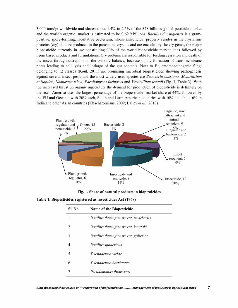
Biopesticides cannot be a panacea to all the problems but are alternatives to synthetic pesticides. Biopesticides are obtained from naturally occurring substances, microbes and plants and being a living organisms or products thereof pose less hazards. Over the past 150 years, a plethora of knowledge has been accumulated on use of biological control agents including bacterial, fungal, viral, protozoan, nematode and botanical -based biopesticides in agriculture and public health (Fig. 1). Microbials constitute the largest group of broad-spectrum biopesticides covering about 1500 naturally occurring insect-specific microorganisms (Khachatourians, 2009. Over 200 microbial biopesticides are available worldwide, out of which 53 numbers were registered in the USA (Kiewnick, 2007), but the products registered for use in Asia are variable (Thakore, 2006) (Table 1, Fig. 2). Annual availability of biopesticides in India is listed in Table 2. The total production of biopesticides is over
ICAR sponsored short course on “Preparation of bioformulat
3,000 tons/yr worldwide and shares about 1.4% to 2.5% of the $28 billand the world's organic market is estimated to be $ 62.9 billions. positive, spore-forming, facultative bacterium, whose insecticidal property resides in the crystalline proteins (cry) that are produced in the parasporal crystals and are encoded by the cry genes, the major biopesticide currently in use constituneem based products and formulations. Cry proteins are responsible for feeding cessation and death of the insect through disruption in the osmotic balance, because of the formation of transpores leading to cell lysis and leakage of the gut contents. Next to Bt, entomopathogenic fungi belonging to 12 classes (Koul, 2011) are promising microbial biopesticides showing pathogenesis against several insect pests and the most widely used specianisopilae, Nomuraea rileyi, Paecilomyces farinosus the increased thrust on organic agriculture the demand for production of biopesticide is definitely on the rise. America uses the largest percentage of the biopesticide market share at 44%, followed by the EU and Oceania with 20% each, South and Latin American countries with 10% and about 6% in India and other Asian countries (Khachatourians, 2009; Bailey
Fig. 1. Share of natural products in biopesticides
Table 1. Biopesticides registered as insecticides Act (1968)
Sl. No. Name of the Biopesticide
1 Bacillus thuringiensis
2 Bacillus thuringiensis
3 Bacillus thuringiensis
4 Bacillus sphaericus
5 Trichoderma viride
6 Trichoderma harzianum
7 Pseudomonas fluoresens
Plant growth regulator, 6
10%
Plant growth regulator and nematicide, 2
3%
Others, 1322%
ICAR sponsored short course on “Preparation of bioformulation...........management of biotic stress agricultural crops”
shares about 1.4% to 2.5% of the $28 billions global pesticide market and the world's organic market is estimated to be $ 62.9 billions. Bacillus thuringiensis
forming, facultative bacterium, whose insecticidal property resides in the crystalline proteins (cry) that are produced in the parasporal crystals and are encoded by the cry genes, the major biopesticide currently in use constituting 90% of the world biopesticide market. it is followed by neem based products and formulations. Cry proteins are responsible for feeding cessation and death of the insect through disruption in the osmotic balance, because of the formation of transpores leading to cell lysis and leakage of the gut contents. Next to Bt, entomopathogenic fungi belonging to 12 classes (Koul, 2011) are promising microbial biopesticides showing pathogenesis against several insect pests and the most widely used species are Beauveria bassiana, Metarhizium anisopilae, Nomuraea rileyi, Paecilomyces farinosus and Verticillium lecanii (Fig. 3, Table 3)the increased thrust on organic agriculture the demand for production of biopesticide is definitely on
erica uses the largest percentage of the biopesticide market share at 44%, followed by the EU and Oceania with 20% each, South and Latin American countries with 10% and about 6% in India and other Asian countries (Khachatourians, 2009; Bailey et al., 2010).
Fig. 1. Share of natural products in biopesticides
Table 1. Biopesticides registered as insecticides Act (1968)
Name of the Biopesticide
Bacillus thuringiensis var. israelensis
Bacillus thuringiensis var. kurstaki
Bacillus thuringiensis var. galleriae
Bacillus sphaericus
Trichoderma viride
Trichoderma harzianum
Pseudomonas fluoresens
Bactericide, 24%
Fungicide, insect attractant and
animal reppelent; 9
15%Fungicide and bactericide, 2
3%
Insect repellent, 5
9%
Insecticide, 1220%
Insecticide and acaricide, 8
14%
Others, 13
ion...........management of biotic stress agricultural crops” 7
ions global pesticide market Bacillus thuringiensis is a gram-
forming, facultative bacterium, whose insecticidal property resides in the crystalline proteins (cry) that are produced in the parasporal crystals and are encoded by the cry genes, the major
ting 90% of the world biopesticide market. it is followed by neem based products and formulations. Cry proteins are responsible for feeding cessation and death of the insect through disruption in the osmotic balance, because of the formation of trans-membrane pores leading to cell lysis and leakage of the gut contents. Next to Bt, entomopathogenic fungi belonging to 12 classes (Koul, 2011) are promising microbial biopesticides showing pathogenesis
Beauveria bassiana, Metarhizium (Fig. 3, Table 3). With
the increased thrust on organic agriculture the demand for production of biopesticide is definitely on erica uses the largest percentage of the biopesticide market share at 44%, followed by
the EU and Oceania with 20% each, South and Latin American countries with 10% and about 6% in
Fungicide, insect attractant and
animal reppelent; 9
15%Fungicide and bactericide, 2
3%
Insect repellent, 5
9%
Insecticide, 1220%
ICAR sponsored short course on “Preparation of bioformulation...........management of biotic stress agricultural crops” 8
8 Beauveria bassiana
9 NPV of Helicoverpa armigera
10 NPV of Spodoptera litura
11 Neem based pesticides
12 Cymbopogan
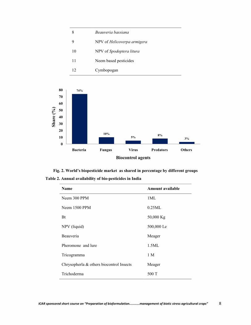
Fig. 2. World’s biopesticide market as shared in percentage by different groups
Table 2. Annual availability of bio-pesticides in India
Name Amount available
Neem 300 PPM 1ML
Neem 1500 PPM 0.25ML
Bt 50,000 Kg
NPV (liquid) 500,000 Le
Beauveria Meager
Pheromone and lure 1.5ML
Tricogramma 1 M
Chrysopherla & others biocontrol Insects Meager
Trichoderma 500 T
74%
10%5%
8%3%
0
10
20
30
40
50
60
70
80
Bacteria Fungus Virus Predators Others
Shar
e (%
)
Biocontrol agents
ICAR sponsored short course on “Preparation of bioformulat
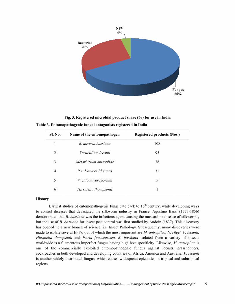
Fig. 3. Registered microbial product share (%) for use in India
Table 3. Entomopathogenic fungal antagonists registered in India
Sl. No. Name of the entomopathogen
1 Beauveria bassiana
2 Verticillium lecanii
3 Metarhizium anisopliae
4 Pacilomyces lilacinus
5 V. chloamydosporium
6 Hirsutella thompsonii
History
Earliest studies of entomopathogenic fungi date back to 18to control diseases that devastated the silkworm industry in France. Agostino Bassi (1773demonstrated that B. bassiana was the infectious agent causing the muscardine disease of silkworms, but the use of B. bassiana for insect pest control was first studied by Audoin (1837). This discovery has opened up a new branch of science, i.e. Insect Pathology. Subsequently, many dimade to isolate several EPFs, out of which the most important are Hirsutella thompsonii and Isaria fumosoroseaworldwide is a filamentous imperfect fungus one of the commercially exploited entomopathogenic fungus against locusts, grasshoppers, cockroaches in both developed and developing countries of Africa, America and Australia. is another widely distributed fungus, which causes widespread epizootics in tropical and subtropical regions
Bacterial30%
ICAR sponsored short course on “Preparation of bioformulation...........management of biotic stress agricultural crops”
Registered microbial product share (%) for use in India
fungal antagonists registered in India
Name of the entomopathogen Registered products (Nos.)
Beauveria bassiana 108
Verticillium lecanii 95
Metarhizium anisopliae 38
Pacilomyces lilacinus 31
V. chloamydosporium 5
Hirsutella thompsonii 1
Earliest studies of entomopathogenic fungi date back to 18th century, while developing ways to control diseases that devastated the silkworm industry in France. Agostino Bassi (1773
was the infectious agent causing the muscardine disease of silkworms, for insect pest control was first studied by Audoin (1837). This discovery
has opened up a new branch of science, i.e. Insect Pathology. Subsequently, many discoveries were made to isolate several EPFs, out of which the most important are M. anisopliae, N. rileyi, V. lecanii,
Isaria fumosorosea. B. bassiana isolated from a variety of insects worldwide is a filamentous imperfect fungus having high host specificity. Likewise, M. anisopliae one of the commercially exploited entomopathogenic fungus against locusts, grasshoppers, cockroaches in both developed and developing countries of Africa, America and Australia.
widely distributed fungus, which causes widespread epizootics in tropical and subtropical
Fungus66%
NPV4%
ion...........management of biotic stress agricultural crops” 9
(Nos.)
century, while developing ways to control diseases that devastated the silkworm industry in France. Agostino Bassi (1773-1856)
was the infectious agent causing the muscardine disease of silkworms, for insect pest control was first studied by Audoin (1837). This discovery
scoveries were M. anisopliae, N. rileyi, V. lecanii,
isolated from a variety of insects M. anisopliae is
one of the commercially exploited entomopathogenic fungus against locusts, grasshoppers, cockroaches in both developed and developing countries of Africa, America and Australia. V. lecanii
widely distributed fungus, which causes widespread epizootics in tropical and subtropical
Fungus66%

ICAR sponsored short course on “Preparation of bioformulat
Biopesticide for rice pest management: Rice hispa
The rice hispa Dicladispa armigerarice in southern Asia and Australasia, more particularly in Bangladesh, India and Nepal (Polaszek al., 2002). Adults scrape parenchymatous tissues off the upper surface of the leaves, making parastreaks, while larvae mine inside the leaves between the epidermal layers. In certain years, many adults (25 – 38/plant) and larvae (10 causes considerable damage to vegetative stages and Dutta 1997), between 20–30% in Nepal (Dhaliwal Bangladesh (Islam, 1989); however, it may be as high as 100% in the rice transplanted post flood in Assam (Hazarika, 2005). Comprehensive field as well as laboratory experiments revealed that bassiana (10 million spores/ml dilution) was superior to neemwith conventional insecticide (0.072% monocrotophos) in controlling the rice hispa, leading to increase in yield.
Proposed the mode of action of B. bassiana
Adhere to body surface Spore germinate Enter into haemocoel (Fig. 4) Grow profusely by utilizing haemolymph Ramification
Interaction of B. bassina with D. armigera
In B. bassiana infected hispa, the total haemocyte count (THC) drastically reduced and PL & GR are major immunocytes responding to the infection. A series of changes taking place at cellular levare mentioned below -
Disintegration and oozing out cell content Pseudopod formation Clumping of granulocyte Morphological alteration of granulocyte GRs aggregated around mycelia Group of cells covered by some dense Clumping of hemocyte Excessive spreading of cytoplasm
Biopesticides for tea pest management
Tea, Camellia sinensis (L.) O. Kuntze, is an intensively managed perennial monoculture crop cultivated on large- and small-scale plantations situated between latitudes 41on over 2.71 million ha in more than 34 countries across Asia, Africa, Latin America, and Oceania to produce 3.22 million metric tons of made tea annually. The national economy of many of these countries is largely dependent upon its productioninsect and mite pests (arthropods) are the most damaging, causing on average a 5% to 55% yield loss. This loss costs approximately U.S. $500 million to $1 billion. In some cases yield loss can be 100%. To defend the tea crop against pests, organosynthetic pesticides are commonly applied, which can result in a resurgence of primary pests or mite syndrome, secondary pest outbreak such as the resistance development and environmental contamination, i
ICAR sponsored short course on “Preparation of bioformulation...........management of biotic stress agricultural crops”
Fig. 4. Mode of pathogenesis by penetration of host cuticle (Source: Hazarika, 2005)
Biopesticide for rice pest management: Rice hispa
Dicladispa armigera (Olivier) (Coleoptera: Chrysomelidae) is a major pest of rice in southern Asia and Australasia, more particularly in Bangladesh, India and Nepal (Polaszek
, 2002). Adults scrape parenchymatous tissues off the upper surface of the leaves, making parastreaks, while larvae mine inside the leaves between the epidermal layers. In certain years, many
38/plant) and larvae (10 – 35/leaf) may attack rice plants (Hazarika et al.causes considerable damage to vegetative stages of rice resulting in yield loss of 28% in India (Nath
30% in Nepal (Dhaliwal et al.,1998) and up to 52% in deepwater rice in Bangladesh (Islam, 1989); however, it may be as high as 100% in the rice transplanted post flood in
am (Hazarika, 2005). Comprehensive field as well as laboratory experiments revealed that (10 million spores/ml dilution) was superior to neem-seed oil (1% concentration) but at par
with conventional insecticide (0.072% monocrotophos) in controlling the rice hispa, leading to increase in yield.
B. bassiana on hispa
(Fig. 4) Grow profusely by utilizing haemolymph
B. bassina with D. armigera hemocytes
infected hispa, the total haemocyte count (THC) drastically reduced and PL & GR are major immunocytes responding to the infection. A series of changes taking place at cellular level which
Disintegration and oozing out cell content
Morphological alteration of granulocyte GRs aggregated around mycelia
Group of cells covered by some dense
Excessive spreading of cytoplasm
Biopesticides for tea pest management
(L.) O. Kuntze, is an intensively managed perennial monoculture crop scale plantations situated between latitudes 41◦N and 16
on over 2.71 million ha in more than 34 countries across Asia, Africa, Latin America, and Oceania to produce 3.22 million metric tons of made tea annually. The national economy of many of these countries is largely dependent upon its production, and of several constraints that affect production, insect and mite pests (arthropods) are the most damaging, causing on average a 5% to 55% yield loss. This loss costs approximately U.S. $500 million to $1 billion. In some cases yield loss can be 100%.
o defend the tea crop against pests, organosynthetic pesticides are commonly applied, which can result in a resurgence of primary pests or mite syndrome, secondary pest outbreak such as the resistance development and environmental contamination, including undesirable residues on made tea
ion...........management of biotic stress agricultural crops” 10
Fig. 4. Mode of pathogenesis by B. bassiana after (Source: Hazarika, 2005)
(Olivier) (Coleoptera: Chrysomelidae) is a major pest of rice in southern Asia and Australasia, more particularly in Bangladesh, India and Nepal (Polaszek et
, 2002). Adults scrape parenchymatous tissues off the upper surface of the leaves, making parallel streaks, while larvae mine inside the leaves between the epidermal layers. In certain years, many
et al., 2005 a,b). It of rice resulting in yield loss of 28% in India (Nath
1998) and up to 52% in deepwater rice in Bangladesh (Islam, 1989); however, it may be as high as 100% in the rice transplanted post flood in
am (Hazarika, 2005). Comprehensive field as well as laboratory experiments revealed that B. seed oil (1% concentration) but at par
(L.) O. Kuntze, is an intensively managed perennial monoculture crop N and 16◦S. It is grown
on over 2.71 million ha in more than 34 countries across Asia, Africa, Latin America, and Oceania to produce 3.22 million metric tons of made tea annually. The national economy of many of these
, and of several constraints that affect production, insect and mite pests (arthropods) are the most damaging, causing on average a 5% to 55% yield loss. This loss costs approximately U.S. $500 million to $1 billion. In some cases yield loss can be 100%.
o defend the tea crop against pests, organosynthetic pesticides are commonly applied, which can result in a resurgence of primary pests or mite syndrome, secondary pest outbreak such as the Tortrix,
ncluding undesirable residues on made tea
ICAR sponsored short course on “Preparation of bioformulation...........management of biotic stress agricultural crops” 11
(Hazarika et al., 2009). Many research institutes around the world are involved in studying the biology and ecology of tea pests and developing suitable techniques for their suppression. The Department of Entomology, Assam Agricultural University (AAU), Jorhat had screened several plant species including Clerodendron ineme, Aegle marmelos, Phologocanthus thyrsiflorus and Linostoma decundrum for management of tea red spider mite. The bunch caterpillar, Andraca bipunctata is widely distributed pest of tea in North-East India and a A. bipunctata specific nuclear polyhedrosis virus (AbNPV) was isolated and found to be effective in suppressing the bunch caterpillar, Andreca bipunctata upto 80%. (Hazarika et al., 1995). Moreover, B. bassiana is an endophytic mycopathogen which is found to be pathogenic to Helopeltis and other insects (Hazarika et al., 2009).
Issues related to biopesticides
Region specificity Species specificity Low reliability Slow in action Short shelf life Photodegradeble
Challenges faced by the biopesticides
Competition with chemical pesticides Regulatory system is not conducive to bio-pesticides Formulation and delivery Farmers acceptance
Future research needs
Nanotechnology Nanoencapsulation. Release mechanism
Diffusion
Dissolution Biodegradation and
Osmotic pressure with specific pH Bio- and Gene-technology
Policy issues related to biopesticides
Installing subsidized loan facility to biopesticide entrepreneurs Creating awareness among farmers and general public Popularizing biopesticides through media Establishment of model BIO-VILLAGES Introduction of vocational courses on biopesticides at school-, college- and university- level
for HRD
References
Bailey, K. L.; Boyetchko, S.M. and Langle, T. (2010). Social and economic drivers shaping the future of biological control: a Canadian perspective on the factors affecting the development and use of microbial biopesticides. Biological Control 52: 221-9.
Dhaliwal, G. S.; Arora, R.; Randhwa, N. S. and Dhawan, A. K. (1998). Ecological Agriculture and sustainable development. In: Proceedings of an International Conference on

ICAR sponsored short course on “Preparation of bioformulation...........management of biotic stress agricultural crops” 12
Ecological Agriculture: Towards Sustainable Development, Vol. 1, Chandigarh, India, November 15–17, 1997.
Hazarika, L. K. (2005). Rice hispa: How much we really know about it? In: Ramamurthy, V.V., Singh, V.S., Gupta, G.P. and Paul, A.V.N. (eds), Gleanings in Entomology, Division of Entomology, Indian Agricultural Research Institute, New Delhi, pp. 1–19.
Hazarika, L. K.; Barthakur, B. K. and Singh, K. (1995). A new pathogen of tea bunch caterpillar. Two and a Bud 42: 40-41.
Hazarika, L. K., Bhuyan, M. and Hazarika, B. N. (2009). Insect Pests of Tea and Their Management. Annu. Rev. Entomol. 54: 267–84.
Hazarika, L. K.; Deka, M. and Bhuyan, M. (2005a). Oviposition behaviour of the rice hispa Dicladispa armigera (Coleoptera: Chrysomelidae). International Journal of Tropical Insect Science 25: 50–54.
Hazarika, L. K;, Puzari K. C. and Bhuyan, M. (2005b). Biopesticide technology and entrepreneurship development. In: Dolui, S.K. and Mahanta, C. (eds.), Science and technology for regional development: Case for North-East India. Tezpur University, Tezpur, IIT, Guwahati, C-MMACS, Bangalore, pp. 27-33.
Islam, Z. (1989). Crop losses due to hispa beetle damage in deep water rice (DWR). International Rice Research Notes 14: 53.
Khachatourians, G. G. (2009). Insecticides, microbials. Applied Microbiology: Agro/Food 95-109.
Kiewnick, S. (2007). Practicalities of developing and registering microbial biological control agents. CAB Reviews: Perspectives in Agriculture Veterinary Science Nutrition and Natural Resources 2 (013): 11.
Koul, P. (2011). Microbial biopesticides: opportunities and challenges. CAB Reviews: Perspectives in Agriculture, Veterinary Science, Nutrition and Natural Resources 6(056): 1-26.
Nath, R. and Dutta, B. (1997). Economic injury level of rice hispa, Dicladispa armigera (Oliv.). Journal of the Agricultural Science Society of North East India 10: 273–274.
Polaszek, A.; Rabbi, M. F.; Islam, Z. and Buckley, Y. M. (2002). Trichogramma zahiri (Hymenoptera: Trichogrammatidae) and egg parasitoid of the rice hispa Dicladispa armigera (Coleoptera: Chrysomelidae) in Bangladesh. Bulletin of Entomological Research 92: 529 – 537.
Thakore Y. (2006). The biopesticide market for global use. Industrial Biotechnology 2: 194-208.
**************
ICAR sponsored short course on “Preparation of bioformulation...........management of biotic stress agricultural crops” 13
Entomopathogenic Fungi for Insect Pest Management
Keshob Chandra Puzari Department of Plant Pathology
Assam Agricultural University, Jorhat-785013, Assam
The word entomopathogen refers to anything that is pathogenic to insects and pests. Agricultural pests include plant pathogens e.g. fungi, oomycetes, bacteria, viruses, nematodes and weeds, arthropods primarily insects and mites, molluscs like slugs and snails and a small number of vertebrates. They reduce the yield and quality of produce by feeding on crops, by transmitting diseases, or by competition with crop plants for space and other resources weeds, for example. There are estimated to be about 67,000 different pest species worldwide. They are a significant constraint on agricultural production, responsible for around 40% loss of potential global crop yields. These losses occur despite the considerable efforts made at pest control, and they suggest that improvements in pest management are significant way forward for improving yields and access to food. Many farmers and growers are now familiar with the use of predators and parasitoids for biological control of arthropod (insect and mite) pests, but it is also possible to use specific micro-organisms that kill arthropods. These include entomopathogenic fungi, nematodes, bacteria and viruses. These are all widespread in the natural environment and cause infections in many pest species. Entomopathogens contribute to the natural regulation of many populations of arthropods. Much of the research in this area concerns the causal agents of insect diseases and their exploitation for biological pest control. Many entomopathogens can be mass produced, formulated, and applied to pest populations in a manner analogous to chemical pesticides, i.e. as nonpersistent remedial treatments that are released inundatively. Entomopathogens have also been used as classical biological control agents of alien insect pests, and natural pest control by entomopathogens has been enhanced by habitat manipulation. India is endowed with a rich biodiversity of entomopathogens and exploitation of this natural resources can be integrated with Integrated Pest Management for management of insect and pests.
Entomopathogenic fungi
Louis Pasteur recognized the potentiality of fungi as a biocontrol agent long back ago. During 1880s Metarhizium species was used to control wheat chafer, Anisoplia austriaca and the sugar beet curculio, Cleonis punctiventris. The genera such as Metarhizium, Beauveria, Verticilium, Nomurea, Entomopthora, Neozygites etc are commonly encountered in nature. There are thought to be about 750 species of fungi that cause infections in insects or mites. As a group, they attack a wide range of insect and mite species, but individual species and strains of fungus are very specific. There are two main taxonomic orders of entomopathogenic fungi. The Entomophthorales occur in the phylum Zygomycota and include genera such as Pandora, Entomophthora and Conidiobolus. These fungi often cause natural epizootics in insect and mite populations. However, some of them are very difficult to mass produce in culture, which is a challenge for people wanting to develop them as biopesticides. The second major order of entomopathogenic fungi – the Hypocreales – occurs in the phylum Ascomycotina. There are many species in the Ascomycotina in which the sexual phase (teleomorph) is not known . Important anamorphic genera of entomopathogenic fungi include Beauveria, Isaria, Metarhizium and Lecanicillium. Species from all these genera are used as biopesticides of insect pests. The spores of many species of the anamorphic entomopathogenic fungi can be mass produced on a variety of culture media, and so are suitable for development as biopesticides which are applied inundatively to pest populations. They have a range of desirable characteristics including safety to people, compatibility with other natural enemies, and a lack of toxic residues. They also offer the possibility of providing persistent control by multiplying in the pest
ICAR sponsored short course on “Preparation of bioformulation...........management of biotic stress agricultural crops” 14
population. Because they have contact action, they are good for the control of sap feeding pests, like aphids and whiteflies, which cannot be infected by other types of biopesticide (such as bacteria and viruses) which are active only when ingested.
B. bassiana has a wide host range and is reported from all over the world; however, it is found to be more predominant in the tropics and sub-tropics under moist and wet conditions. Nevertheless, it was also reported from the temperate regions. The rice ecosystem, being moist and wet, serves as a favourable environment for exploitation of mycoinsecticides including B. bassiana and Pandora delphacis.
The genus Cordyceps is a cosmopolitan Ascomycete confined almost entirely to insects. About 200 species are recognized attacking a great variety of immature and adult insects. Ophiocordyceps is a closely related genus. Cordyceps is characterized by the growth of long fruiting stems, sometimes branched, arising from a sclerotium within the body of the insect. Certain genera of Fungi Imperfecta as Isaria, Botrytis and Hirsutella in part are considered as conidial stages of Cordyceps. When a Cordyceps spore under favourable conditions strikes an insect, the germ tube penetrates the integument. On reaching the haemocoele, they soon fill the cavity; absorb most of the blood and the host dies. The mycelium attacks and absorbs most of the tissue distending the insect integument to near normal size. The sclerotium thus formed may remain dormant for months or in favourable situations develops the perfect conspicuous fruiting stems.
During April-May 1977, a large number of banana leaf beetle (Nodostoma subcostatum) infesting banana in the horticultural orchard, Assam Agricultural University (AAU), Jorhat, were found to be infected by white muscardine fungus causing death of the pests (Roy and Puzari., 1979). They noticed that white frosty growth of the fungus first emerges through the suture of elytra, thoracic segment and ventral side of abdomen of the death insect which ultimately covered the whole body
Among the major insect pests of rice in North East India, rice hispa Dicladispa armigera (Olivier) (Coleoptera: Chrysomelidae) causes extensive damage to the vegetative stages of the crops. Control of rice hispa has been a major problem for farmers who depend primarily on rice as subsistence crop. Chemical control may sometimes proves to be detrimental by creating additional problems, such as environmental hazards, development of insect resistance and resurgence, killing non target organisms especially aquatic fauna and so on. Hence, a search for alternative to these agents which are compatible with a range of control tactics for D. armigera has resulted in identifying an entomopathogenic fungus B. bassiana (Hazarika and Puzari 1990). Subsequently, a field survey in rice ecosystem on rain fed rice was conducted by Puzari et. al., 1992 and recovered 240 cadavers of rice hispa, D. armigera (Olivier) (Coleoptera: Chrysomelidae). The entomogenous association formed/established by the fungi on the insects were B. bassiana, A. flavus, Fusarium hetetoporum Nees ex Fr., Penicillium cyclopium Westling, Geotrichum sp. and Mucor sp.
Cutworm or greasy surface caterpillar, Agrotis ipsilon is an important pest of potato especially in transplanted potato seeds. The seedlings mortality may be as high as 25-30 per cent within 10-20 days of germination. Naturally occurring entomopathogenic fungus was seen growing on adult cadavers of A. ipsilon The cadavers infected with the fungus were collected from the potato fields and the fungus was isolated, purified on Czapek-Dox agar medium and identified as M. anisopliae var. anisopliae (Metsch.) Sorok, var.anisopliae.
The cowpea aphid, Aphis craccivora Koch (Homoptera: Aphididae) is ubiquitous and is also responsible for transmission of cowpea aphid borne mosaic virus in persistent and non persistent

ICAR sponsored short course on “Preparation of bioformulation...........management of biotic stress agricultural crops” 15
manner resulting in substantial damage to the crop especially early in the season. Four different entomopathogenic fungi were also isolated from seven different insects’ species belonging to the order Lepidoptera, Hemiptera, Isoptera, Homoptera and Orthoptera from vegetable growing areas of Majuli the largest river island of the world. The pathogenic fungi were characterised and identified as M. anisopliae, B. bassiana, N. rileyi and Fusarium sp.
Termites are an important pest of agricultural, horticultural and plantation crops, forest trees, structural timbers and various wood and textile products. Tea (Camellia sinensis L (O) Kuntze), being a perennial crop attracts insects and mites that thrive and flourish on tea. Termites are considered as good candidate for control with the entomopathogenic organisms because they live in conducive environment-humid, minimal diurnal temperature fluctuations, crowded and with considerable social interaction. The pathogenecity of M. anisopliae (Metschnikoff) Sorokin and B. bassiana (Balls.) Vuill. were evaluated against the workers of termite Microtermes obesis. The treated insects showed changes in their normal behavior and in morphology. With the help of scanning electron microscopy the minutiae deatails of morphological changes in the cuticles and cuticular sensilla present in various locations of the body were revealed. The observations suggested that the ventral cuticle of the abdomen have been totally distorted along with the deformation in sensilla trichoidae. Fungal colonies were also clearly visible throughout the ventral portion of the body, which suggest that fungal growth can cause serious damage to the pest disturbing its major physiological activities resulting in its death.
Entomopathogenic mematode
Entomopathogenic nematode worms are just visible to the naked eye, being about 0.5 mm in length. Two families – the steinernematids and the heterorhabditids - are obligate parasites of insects and used for microbial control. Juvenile nematodes parasitize their hosts by directly penetrating the cuticle or through natural openings. They then introduce symbiotic bacteria, which multiply rapidly and cause death by septicaemia, often within 48 hours. The bacteria break down the insect body, which provides food for the nematodes. After the insect has died, the juvenile nematodes develop to adults and reproduce. A new generation of infective juveniles emerges 8 – 14 days after infection.
Entomopathogenic Baculoviruses
Over 1600 viruses have been recorded from more than 1100 species of insects and mites. Of these, three families (Baculoviridae, Polydnaviridae, Ascoviridae) are specific for insects and related arthropods. The baculoviruses are the most widely exploited virus group for biocontrol: they are very different from viruses that infect vertebrates and are considered very safe to use. The mode of pathogenesis and replication of entomopathogenic viruses varies according to the family, but infection nearly always occurs by ingestion. Virions then bind to receptors in the gut and penetrate epithelial cells. In the Baculoviruses, the infection often spreads to the haemocoel and then to essential organs and tissues, particularly fat bodies. Acute infections lead to host death in 5 – 14 days. There are two genera of Baculoviruses: nucleopolyhedroviruses (NPV) & granuloviruses (GV).
Mass production
One of the greatest obstacles for biological control by introduced agents has been lack or scarcity of methods for mass culturing and delivering the bio agents. The unique problem in developing bioformulation is that it represents a living system, which must be able to stand the process of formulation and should remain sufficiently viable for a period until it reaches the end users. Despite the limited progress, scientists are engaged in developing effective experimental system for

ICAR sponsored short course on “Preparation of bioformulation...........management of biotic stress agricultural crops” 16
growth and delivery of bio agents. After a continuous effort mass production technique of B. bassiana have been standardized. The solid state artificial media contains rice husk: saw dust: rice bran (1:1:4) + 2% dextrose + 2% chitin which has ability to yield 39.33 x 10⁷ conidia/ml water with high pathogenecity (LC50 90.16 conidia/ml), ability to penetrate through elytral punctuations and found superior performance in the field to that of the recommended insecticides .
Shelf life and virulence of entomopathogenic fungus:
Loss of viability over time is one of the critical obstacles for commercialization of a bio pesticides preparation. Several attempts have been made to determine the viability of entomopathogens in their preparations when stored at room temperature and in refrigerator. Result of the experiment showed that shelf life of B. bassiana varied on differential shelved conditions and where the temperature plays an important role. Its conidial density, virulence and viability were found decreased at their different rates in different shelf conditions, i.e., at room temperature 24±1⁰C, in refrigerated conditions (4⁰C) and in deep-freeze condition. At room temperature its virulence (90.97%) and conidial density (39.41x10⁷conidia/ml) did not differ up to 90 days of storage. After 90 days they declined significantly to 82.00% and 30.27x10⁷conidia/ml, respectively, with concomitant increase of percentage of dried conidia.
Desiccation of conidia in other way can be prevented to a considerable extend by adding certain osmoticum like mannitol, silica powder, sucrose, sodium glutamate, anti oxidizing agents like sodium ascorbate etc. Rich carbon source with a minimum compactness of the medium for space and aeration of fungal growth, the rice husk: sawdust: ricebran medium produced maximum numbers of propagules in which temperature played a major role in the extension of shelf life.
The biomass production of B. bassiana strains showed that the fungus could grow well at temperature 20-25ºC. The biomass production among the strains had significant effect in respect of temperature; the highest biomass production (283.67 mg) was recorded in AAU-09 strain followed by MTCC- 4497 (273.67mg) strain at temperature 25ºC.
Compatibility of B. bassiana with different insecticides commonly used in rice ecosystem
Microbial may not bring about desired control of the targeted pests, under such situations combination of microbial with chemical pesticides may prove to be useful for which compatibility of pesticides and entomopathogens must be tested. Integrated management of insect pests is an important way in reducing the severe impact of chemicals pesticides on ecosystem. Insecticides may have antagonistic or synergistic effect on the potentiality of B. bassiana, and may disrupt natural epizootics. Under such epizootic conditions it is expected to enhance its effectiveness through joint action of pathogen and compatible insecticide, which would reduced not only the cause of protection but also reduce the contamination of the environment. It is known that sub lethal dose of an insecticide would make the insect more susceptible to the attack of the entomopathogens. Growth inhibition of entomopathogenic fungi is a useful criterion for initial testing of its compatibility.
Varying effects of chemicals on the fungi, their actual effects at cellular and field level need to be investigated to understand if the effects are permanent or temporary. In case of temporary arrest of fungus activity, it may recover after degradation of toxicant and such chemicals can be employed in combination with the fungus under field conditions. Field studies to evaluate the compatibility of these pesticides with fungal isolates, applied either as combinations or incorporated singly with the
ICAR sponsored short course on “Preparation of bioformulation...........management of biotic stress agricultural crops” 17
isolate, should generate additional information on how it can be successfully incorporated in the integrated pest, management systems together with pesticides.
**************
ICAR sponsored short course on “Preparation of bioformulation...........management of biotic stress agricultural crops” 18
Biological Control – An Ecological Perspective
D. K. Sarmah AICRP on Mushroom, Department of Plant Pathology, Assam Agricultural University, Jorhat- 785013, Assam
Biological control of plant diseases involves the use of an organism or organisms to inhibit the pathogen and reduce disease (Chaur,1998). In variously defined biological control processes the basic idea is to evolve a strategy for reducing disease incidence or severity by direct or indirect manipulation of microorganisms (Shurtleff and Averre, 1997). A clear understanding of the mechanisms of biological control of plant diseases through the interactions between biocontrol agent and pathogen may allow us to manipulate the soil environment to create conditions conducive for successful bio-control or to improve biocontrol strategies (Chaur, 1998). Biocontrol agents are widely regarded in general as natural and therefore non-threatening products, although risk assessments must clearly be carried out on their effects on non-target organisms and plants. Moreover, knowledge concerning the behaviour of such antagonists is essential for their effective use (Monte and Liobell, 2003).
Biological disease control is an attractive alternative strategy for the control of plant diseases. Many factors have to be considered in deciding whether a biological system is feasible for the control of a particular pathogen. The availability of a suitable antagonist capable of maintaining itself on the host plant is of prime importance. The environment under which the crop is grown will play a significant part in determining whether effective population levels of an antagonist can be established in competition with the existing microflora. Environment may also govern the choice of antagonist. Bio agents themselves being non-pathogenic to plants need to be formulated in a way that favours the activity and survival of the microbe it contains. Over the past few years, the novel applications of molecular techniques have broadened our insight into the basis of biological control of plant diseases. New molecular approaches have been available for assessment of interaction between the antagonist and pathogen, ecological traits of antagonists in rhizosphere and improving the efficacy of bacterial, fungal and viral biocontrol agent. Thus, biological control will be a viable alternative strategy for the control of plant diseases given the history of fungicides, in the near future.
In spite of decades of research on the biological control of soil-borne plant diseases, there remain few commercially successful examples of biological control using introduced microbial inoculants. There are a number of reasons for the lack of development and grower adoption. Among the more important are problems in formulation and delivery, variability in performance, and problems with poor efficacy under optimum conditions for disease development. There are countless examples of biological control organisms that perform quite effectively under defined laboratory conditions but fail miserably when introduced on different crops under varying conditions in the field. Still others might perform effectively in the field, but exhibit strong year-to-year or site-to-site variability. Unpredictable performance coupled with this extreme variability represents one of the greatest obstacles to the implementation of biological disease control practices in agriculture. Our inability to predict the behaviour of microorganisms introduced for biological control purposes stems from a lack of sustained and broad research on the mechanisms regulating biological control processes in plant-associated microorganisms. The emphasis in past studies of biological control mechanisms has been on the attributes of the biocontrol organism and the role of specific microbial properties in pathogen suppression (Martin and Loper, 1999; Whipps, 2001).
ICAR sponsored short course on “Preparation of bioformulation...........management of biotic stress agricultural crops” 19
As a result, our understanding of how various microbial traits influence biological control processes is fairly well understood. However, the important role of the host plant in defining and regulating biological control processes is often overlooked. An increasing number of studies indicate the importance of the host plant in influencing microbial interactions in the spermosphere and rhizosphere (Chanway, Holl et al., 1988; Chanway,Nelson et al., 1988; Mavingui et al., 1992; Lemanceau et al., 1995; Hervas et al., 1997; Koch, 1997; Bensalim et al., 1998). The host plant also has an important role in supporting biological control interactions, which has been indicated in an increasing number of studies (Atkinson and Neal, 1975; Azad et al., 1987; Koch, 1997; Grayston et al., 1998).
The study of interactions among plant pathogens and other microorganisms is a fascinating but challenging area of scientific investigation that has potential applications for biological control of plant pathogens. In pioneering studies as early as 1920, antagonistic fungi were introduced to forest nursery soils to reduce damping off of pine seedlings (Hartley, 1921). Reduction in disease occurred in some treatments and Hartley concluded that “competition of different fungi is a factor to be considered”. This potential for biological control has continued to be a well established objective of plant pathologist for many decades. If the study of interactions of plant pathogens and other microorganisms is to be applied to the management of plant diseases, factors that contribute to the lack of available systems must be identified and effective strategies developed for the application of biological controls to disease management. There are several areas where the development of such systems can be encouraged: the ecological selection and evaluation of potential agents, the environmental enhancement of biocontrol efficacy, the genetic enhancement of efficacy, the commercial production and development of biocontrol agents and the registration of biocontrol products.
After its establishment in the soil, the BCA will interact not only with the pathogen to be controlled but also with all the biotic components of the soil. There is a fear that a successful BCA might displace the microbial balance of the soil and have some unexpected effects on the non target organisms. Therefore, there is a need to study the side effects of an introduced antagonist on the native microbial communities. In Europe, application of BCA is subjected to the Directive 91/414-ECC, which imposes this type of study. Until recently, there were no practical methods available to detect the impacts of an introduced BCA on the whole soil microbial community. With the development of molecular approaches based on extraction of total DNA from the soil, it is now possible to overcome this limitation today. Several methods are available to assess microbial community structures by molecular fingerprinting. Among them, terminal restriction fragment length polymorphism (T-RFLP) has already been used to address the impact of cultural practices on the structure of bacterial and fungal communities (Edel-Hermann et al., 2004; Pérez-Piqueres et al., 2006).
Ecological selection
A growing plant contains several ecological microhabitats that represent unique microclimatic and nutritional conditions. Terms such as rhizoplane, rhizosphere, phylloplane, spermosphere, gemmisphere, cauliplane, palynosphere and anthoplane are used to emphasize the uniqueness of these habitats and their influence on the growth and survival of pathogenic and saprophytic microorganisms. The ecological competence of biological control agents within individual habitats is a primary determinant of potential efficacy. Furthermore, the study of biological control must be placed within the context of the ecological requirements of the pathogen and the biological control
ICAR sponsored short course on “Preparation of bioformulation...........management of biotic stress agricultural crops” 20
agent. Potential agents can be selected from indigenous populations collected from the target habitat or from non indigenous population in other habitats. Classical approaches have selected from indigenous populations, with the assumption that such microorganisms are ecologically competent within that habitat. However, studies on biocontrol of insect and weed pests have suggested that there is up to a 75% greater chance of success if the parasite and the host represent a new biological association instead of an old association (Hokanen and Pimental, 1984; Waage and Greathead, 1988). These considerations are based on the principle that interactions between two organisms that have coevolved may be less disruptive than interactions that have not coevolved. Little information is available on the influence of population origin on probability of selecting effective biological controls for plant diseases but promising agents have been identified using both approaches (Cook and Baker, 1983).
Environmental enhancement of biocontrol efficacy
The influence of environment on biological control can be subdivided into physical environment and the influence of the chemical or nutritional environment on the growth and survival of agents. Manipulation of chemical and nutritional environments of plant surfaces has potential for the enhancement of biological control. Like the addition of adjuvants in case of fungicides to enhance their efficacy, the efficacy of biocontrol agents can be improved by adjuvants that modify the environmental, physical, or nutritional conditions in the target microhabitat. Many organic substrates influence the biological activity of pathogens and biocontrol agents (Cook and Baker, 1983).
Selection criteria
Programmes for screening antagonists for disease control of plant pathogens are often focused on testing antagonistic properties in vitro, in bioassays and subsequently in crops. For commercial use, however, antagonists must fulfil many more criteria. Besides the toxicological profile of an antagonist, industries will consider technologies for production and formulation and their costs, genetic stability of the antagonist, market size for the biocontrol product and the possibilities of patent protection for the application (Whitesides et al., 1994; Köhl, 2010).
Microbial antagonists occupy the same ecological niche as the target plant pathogen and interact directly with it. The mechanisms of interaction include parasitism, competition for space, water or food, or ‘chemical warfare’ using antibiotics or other secondary metabolites that harm the target pathogen. The second class involves an indirect effect in which the control agent induces a resistance response in the plant that gives it protection against virulent plant pathogens. The strain of biocontrol agent which possesses most of these criteria is supposed to be the most effective one.
Suppressive soils
Several soil-borne pathogens, such as Fusarium oxysporum (the cause of vascular wilts), Gaeumannomyces graminis (the cause of take-all of wheat), Phytophthora cinnamomi (the cause of root rots of many fruit and forest trees), Pythium spp. (a cause of damping-off), and Heterodera avenae (the oat cyst nematode), develop well and cause severe diseases in some soils, known as conducive soils, whereas they develop much less and cause much milder diseases in other soils, known as suppressive soils. The mechanisms by which soils are suppressive to different pathogens are not always clear but may involve biotic and/or abiotic factors and may vary with the pathogen. In most cases, however, it appears that they operate primarily by the presence in such soils of one or several microorganisms antagonistic to the pathogen. Such antagonists, through the antibiotics they
ICAR sponsored short course on “Preparation of bioformulation...........management of biotic stress agricultural crops” 21
produce, through lytic enzymes, through competition for food, or through direct parasitizing of the pathogen, do not allow the pathogen to reach high enough populations to cause severe disease (Agrios, 2005).
Numerous kinds of antagonistic microorganisms have been found to increase in suppressive soils; most commonly, however, pathogen and disease suppression has been shown to be caused by fungi, such as Trichoderma, Penicillium, and Sporidesmium, or by bacteria of the genera Pseudomonas, Bacillus, and Streptomyces. However, in several diseases, continuous cultivation (monoculture) of the same crop in a conducive soil, after some years of severe disease, eventually leads to reduction in disease through increased populations of microorganisms antagonistic to the pathogen. This effect, which is selective for certain pathogens and not for others, is an area of in depth investigation.
Durability
The durability of a control method for plant protection is defined as the persistence of its efficacy in space and time. Erosion of effectiveness of conventional plant protection methods has been widely studied in the past. The durability of chemical control has for instance been studied because of the frequent and recurrent apparition of resistance to fungicides in major plant pathogenic fungal populations (Brent and Hollomon, 2007). The breakdown of varietal resistance, especially that conferred by major resistance genes, has also been widely studied for plant pathogens (McDonald and Linde, 2002). In contrast, the durability of biological control has long been assumed to be higher than that of chemical control (Holt and Hochberg, 1997). However, recent results concerning pest management in agricultural systems have shown that this assumption may not always be justified.
The most striking example may be the development of resistance to the most widely used bio-insecticide in the world. Resistance to one or several toxins produced by the bacterium Bacillus thuringiensis (Bt) has been described shortly after the market approval of products based on various strains of this bacterium.
In contrast with the situation for pests, the durability of biological control of plant diseases has hardly been studied. This may be related to the limited use of biological control against plant diseases in practice until recently. A bibliographical study conducted in the framework of the European project ENDURE (European Network for Durable Exploitation of Crop Protection Strategies) established that despite the large amount of microorganisms as potential candidates for biological control (Nicot et al., 2011), there are still few biocontrol agents registered against plant diseases in the European Union (Heilig et al., 2011).
However, several studies reported the inconsistency of efficacy of various biocontrol agents when introduced under commercial field conditions-being less effective or completely ineffective even though their efficacy was very good in controlled conditions (Shtienberg and Elad, 1997; Guetsky et al., 2001; Mark et al., 2006; Nicot et al., 2011).
This variability of efficacy is generally attributed to climatic variations (temperature, humidity, radiation) encountered in field conditions, a lack of ecological competence (survival, colonization ability) of the biocontrol agent, intrinsic traits of the antagonistic microbe (variable production of required metabolites or enzymes) and/or an unstable quality of the formulated product (Elad and Stewart, 2004; Mark et al., 2006; Ruocco et al., 2011). However, reduction of efficacy in the field may also result from the diversity of sensitivity of plant pathogens to biocontrol agents, with

ICAR sponsored short course on “Preparation of bioformulation...........management of biotic stress agricultural crops” 22
the existence of less sensitive isolates in natural populations of plant pathogens. The durability of biological control against plant pathogens may be related to specific traits of the plant pathogen such as genetic diversity and ability to evolve in response to a selection pressure. This is affected by population genetic processes including mutation, population size, recombination, gene flow and selection. This point was extensively studied to achieve durable plant disease resistance in agriculture (McDonald and Linde, 2002; McDonald, 2014). Thus, McDonald and Linde (2002) have hypothesized that populations of plant pathogens with high evolutionary potential are more likely to overcome a varietal resistance.
The same assumption can be proposed for the development of resistance to biocontrol agents. The durability of biological control against plant pathogens may also be related to the selection pressure exerted by the biocontrol agent. This selection pressure clearly depends on the extent of use of biocontrol agents in practice (surfaces treated, doses of application etc.). It may also depend on the specific mode of action of biocontrol agents. Various modes of action are involved in the protective effect of biocontrol agents against plant pathogens. Although the number of studies done on this subject is important, knowledge of the precise mode of action of biocontrol agents is still partial. However, it is generally considered that there are three main ways for a biocontrol agent to control a plant pathogen (Jacobsen, 2006; Alabouvette et al., 2009) : first, by acting directly on the plant pathogen, through antibiosis, competition for nutrient or space, or parasitism; secondly by interfering with the mechanisms of pathogenesis of the plant pathogen, and thirdly by modifying the interaction of the plant pathogen with its plant host for instance, through the induction of local or systemic acquired resistance. These modes of action are not incompatible, they can instead be complementary and a single species or a single strain of a biocontrol agent may act with several of these modes of action (Janisiewicz and Korsten, 2002). A given biocontrol agent may therefore operate through several mechanisms potentially expressed successively, simultaneously or synergistically and possibly depending on the environmental conditions encountered. Nevertheless, it is not yet clear if biocontrol agents have a dominant mode of action and under what conditions they switch from a mode of action to another. Even though all biocontrol agents should create selection pressure on target populations of plant pathogens once treatments are applied in the field, some modes of action may present a clear opportunity for pathogens to evolve resistance.
Capacity of plant pathogens to adapt to biological control
Besides existing diversity in susceptibility of plant pathogens to biocontrol agents, another concern could be that resistance would develop through adaptation under selection pressure, following the generalized use of a biological control method in the field, as has already occurred for various pathogens with certain fungicides. The estimation of this potential risk can be achieved through experimental evolution studies with the production of successive generations of the pathogens under selection pressure, as commonly carried out to evaluate the durability of efficacy of antimicrobial compounds in human pathology (Cowen et al., 2002), or to assess the capacity of plant pathogens to adapt to fungicides (Brent and Hollomon, 1998). The experimental evolution studies illustrate the potential of plant pathogens to adapt to the effect of biocontrol agents. Studies also suggest that the use of chemical methods in parallel or in combination with biological control may have an impact on the durability of the efficacy of ctain biocontrol agents.
Conclusion
Ecological factors play very important roles in the performance and activity of biocontrol-active microorganisms. Biological control agents against plant pathogens, especially those in soil, operate within physically, biologically, and spatially complex systems by means of a variety of
ICAR sponsored short course on “Preparation of bioformulation...........management of biotic stress agricultural crops” 23
trophic and nontrophic interspecific interactions. The biocontrol agents themselves are also subject to the same types of interactions, which may reduce or in some cases enhance their efficacy against target plant pathogens (Knudsen and Dandurand, 2014). Characterization of these ecologically complex systems is challenging, but a number of tools are available to help unravel this complexity. New molecular approaches have been available for assessment of interaction between the antagonist and pathogen, ecological traits of antagonists in rhizosphere and improving the efficacy of bacterial, fungal and viral biocontrol agent. However, other methods in IPM for crop disease control are still necessary in various environmental conditions, because an agro-ecosystem is a variable and functioning system that includes several factors that influence disease and crop development.
References
Agrios, 2005. Plant pathology. 5th edition. Elsevier, London. pp 303-304. Alabouvette, C.; Olivain, C.; Migheli, Q. and Steinberg, C. (2009). Microbiological control of
soil-borne phytopathogenic fungi with special emphasis on wilt inducing Fusarium oxysporum. NewPhytol.184: 529–544.
Atkinson, T. G. and Neal, J. L. J. (1975).Genetic control of rhizosphere microflora of wheat. In: Biology and Control of Soil Borne-Plant Pathogens, G. W. Bruehl (Ed). The American Phytopathologcal Society, St Paul. pp 116-122
Azad, H. R.; Davis, J. R.; Schnathorst,W. C. and Kado, C. I. (1987). Influence of Verticillium wilt resistant and susceptible potato genotypes on populations of antagonistic rhizosphere and rhizoplane bacteria. App.Microbiol.Biotechnol.26: 99-104.
Bardin, M.; Ajouz, S.; Comby, M.; Lopez-Ferber, M.; Graillot, B.; Siegwart, M. and Nicot, P. C. (2015). Is the efficacy of biocontrol against plant diseases likely to be more durable than that of chemical pesticides? Front. Pant Science. 6: 566.
Brent, K. J. and Hollomon, D.W. (1998). Fungicide resistance: the assessment of risk. In: RAC Monograph No.2 (Brussels:Global Crop Protection Federation). pp 1–48.
Brent, K. J. and Hollomon, D. W. (2007). Fungicide Resistance in Crop Pathogens: How Can it be Managed. In2nd Revised Edn. Brussels: Crop Life International.
Chaur, T. (1998). General mechanisms of action of microbial biocontrol agents. Plant Pathology Bulletin 7: 155-166.
Cook, R. J. and K. F. Baker (1983). The nature and Practice of biological control of plant pathogens. The American Phytopathoogical Society. St.Paul. pp 539.
Cowen, L. E.; Anderson, J. B. and Kohn, L. M. (2002). Evolution of drug resistance in Candida albicans. Annu. Rev. Microbiol. 56: 139–165.
Edel-Hermann, V.; Dreumont, C.; Pérez-Piquerez, A. and Steinberg, C. (2004). Terminal restriction fragment length polymorphism analysis of ribosomal RNA genes to assess changes in fungal community structure in soil. FEMS Microbiol Ecol. 47: 397–404.
Elad, Y. and Stewart, A. (2004). Microbial control of Botrytis spp. In: Botrytis: Biology, Pathology and Control. Eds Y. Elad, B. Williamson, P. Tudzynski, and N. Delen (Dordrecht: Kluwer Academic Press). pp 223–241.
Grayston, S. J.; Wang, S.; Campbell, C. D. and Edwards, A.C. (1998). Selective influence of plant species in the rhizosphere. Soil Biol.Biochem. 30:369-378.
Guetsky, R.; Shtienberg, D.; Elad, Y.and Dinoor, A. 2001.Combining biocontrol agents to reduce the variability of biological control. Phytopathology 91:621–627.
Hartley, C. (1921). Damping off in forest nurseries. U.S.Dept.Agric.Bulletin 934: 1-99. Heilig, U.; Delval, P. andBlum, B.(2011). Registered biocontrol products and their use in
Europe. In: Classical and Augmentative Biological Control Against Diseases and Pests:
ICAR sponsored short course on “Preparation of bioformulation...........management of biotic stress agricultural crops” 24
Critical Status Analysis and Review of Factors Influencing Their Success. Ed. P. C. Nicot (IOBC-WPRS). pp 34–41.
Hokkanen, M. and Pemental, D. (1984). New approach for selecting biological control agents. Can.Ent. 116:1109-1121.
Holt, R. D. and Hochberg, M. E. (1997). When is biological control evolutionarily stable (or is it)? Ecology 78:1673–1683.
Janmaat, A. F. and Myers, J. (2003). Rapid evolution and the cost of resistance to Bacillus thuringiensis in greenhouse populations of cabbage loopers, Trichoplusiani. Proc. R. Soc. Biol. Sci. 270: 2263–2270.
Knudsen, G. R. and and Dandurand L. C. (2014). Ecological complexity and the success of fungal biological control agents. Advances in Agriculture pp 1-11.
Koch, E. (1997). Screening of rhizobacteria for antagonistic activityagainst Pythium ultimum on cucumber and kale. Zeitschrift furPflanzenckrankheiten und pflanzenschutz.104: 353-361.
Köhl, J. (2010). Screening of biocontrol agents for control of foliar diseases. In: Recent Developments in Management of Plant Diseases, Series: Plant Pathology in the 21st Century, Vol. 1; Eds. Gisi, Chet and Gullino. Springer, Dordrecht, The Netherlands. pp 107-119.
Li, H. and Leifert, C. (1994). Development of resistance in Botryotinia fuckeliana (deBarry) Whetzel against the biological control agent Bacillus subtilis CL27.Z. Pflanzenkr Pflanzenschutz 101: 414–418.
Mark, G. L.; Morrissey, J. P.; Higgins, P. and O’gara, F. (2006). Molecular based strategies to exploit Pseudomonas biocontrol strains for environmental biotechnology applications. FEMS Microbiol. Ecol. 56:167–177.
Martin F. N. and Loper J. E. (1999).Soil borne plant diseases caused by Pythium spp: ecology, epidemiology and prospects for biological control. Crit.Rev.Plant Sci.18: 111-181.
Mavingui, P.; Laguerre G.; Berge, O. and Heaulin, T. (1992). Genetic and phenotypic diversity of Bacillus polymyxa in soil and in wheat rhizosphere. Appl. Environ. Microbiol. 58:1894-1903.
McDonald, B. A. and Linde, C. (2002). Pathogen population genetics, evolutionary potential, and durable resistance. Annu. Rev. Phytopathol. 40: 349–379.
McDonald, B. A. (2014). Using dynamic diversity to achieve durable disease resistance in agricultural ecosystems. Trop. Plant Pathol. 39: 191–196.
McGaughey, W. H. (1985). Insect resistance to the biological insecticide Bacillus thuringiensis. Science 229: 193–194.
McGaughey, W. H. and Johnson, D. E. (1992). Indian meal moth (Lepidoptera: pyralidae) resistance to different strains and mixtures of Bacillus thuringiensis. J. Econ. Entomol. 85:1594–1600.
Nega, A. (2014). Review on concepts in biological control of plant pathogrns. Journal of Biology, Agriculture and Healthcare. 4(27): 33-54.
Pérez-Piqueres, A.; Edel-Hermann, V.; Alabouvette, C. and Steinberg, C. (2006) Response of soil microbial communities to compost amendments. Soil Biol Biochem. 38: 460–470.
Ruocco, M.; Woo, S.; Vinale, F.; Lanzuise, S. and Lorito, M. (2011). Identified difficulties and conditions for field success of biocontrol, Technical aspects: factors of efficacy. In: Classical and Augmentative Biological Control Against Diseases and Pests: Critical Status, Analysis and Review of Factors Influencing Their Success. Ed. P. C. Nicot (IOBC-WPRS).pp 45-57.
Shtienberg, D. and Elad, Y. (1997). Incorporation of weather forecasting in integrated, biological, chemical management of Botrytis cinerea. Phytopathology 87:332–340.

ICAR sponsored short course on “Preparation of bioformulation...........management of biotic stress agricultural crops” 25
Shurtleff, M. C. and Averre, C. W. (1997). Glossary of Plant Pathological Terms. APS Press, St. Paul, MN.
Tabashnik, B. E.(1994). Evolution of resistance to Bacillus thuringiensis. Annu. Rev. Entomol. 39: 47–79.
Waage, J. K. and Greathead, D. J. (1988). Biological control: challenges and opportunities. Phil. Trans. R. Soc. Lond. B. 318: 111-128.
Whipps, J. M. (2001). Microbial interactions and biocontrol in the rhizosphere. J. Exptl. Bot. 52: 487-511.
Whitesides, S. K.; Daoust, R. A. and Gouger, R. J. (1994). Commercialization of biological control agents: an industry perspective. In: Biological Control of Postharvest Diseases Theory and Practice. Eds. Wilson and Wisniewski, CRC Press, Boca Raton, Florida.pp 107-121.
**************
ICAR sponsored short course on “Preparation of bioformulation...........management of biotic stress agricultural crops” 26
Techniques for Isolation of Fungal Biocontrol Agents, Endophytes and Study on their Evaluation
Pranab Dutta and Himadri Kaushik Department of Plant Pathology
Assam Agricultural University, Jorhat-785013, Assam
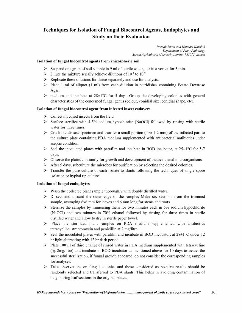
Isolation of fungal biocontrol agents from rhizospheric soil
Suspend one gram of soil sample in 9 ml of sterile water, stir in a vortex for 3 min. Dilute the mixture serially achieve dilutions of 10-1 to 10-6 Replicate these dilutions for thrice separately and use for analysis. Place 1 ml of aliquot (1 ml) from each dilution in petridishes containing Potato Dextrose
Agar. medium and incubate at 28±1°C for 5 days. Group the developing colonies with general
characteristics of the concerned fungal genus (colour, conidial size, conidial shape, etc).
Isolation of fungal biocontrol agent from infected insect cadavers
Collect mycosed insects from the field. Surface sterilize with 4-5% sodium hypochlorite (NaOCl) followed by rinsing with sterile
water for three times. Crush the disease specimen and transfer a small portion (size 1-2 mm) of the infected part to
the culture plate containing PDA medium supplemented with antibacterial antibiotics under aseptic condition.
Seal the inoculated plates with parafilm and incubate in BOD incubator, at 25±1°C for 5-7 days.
Observe the plates constantly for growth and development of the associated microorganisms. After 5 days, subculture the microbes for purification by selecting the desired colonies. Transfer the pure culture of each isolate to slants following the techniques of single spore
isolation or hyphal tip culture.
Isolation of fungal endophytes
Wash the collected plant sample thoroughly with double distilled water. Dissect and discard the outer edge of the samples Make six sections from the trimmed
sample, averaging 6x6 mm for leaves and 6 mm long for stems and roots. Sterilize the samples by immersing them for two minutes each in 5% sodium hypochlorite
(NaOCl) and two minutes in 70% ethanol followed by rinsing for three times in sterile distilled water and allow to dry in sterile paper towel.
Place the sterilized plant samples on PDA medium supplemented with antibiotics tetracycline, streptomycin and penicillin at 2 mg/litre.
Seal the inoculated plates with parafilm and incubate in BOD incubator, at 28±1°C under 12 hr light alternating with 12 hr dark period.
Plate 100 µl of third change of rinsed water in PDA medium supplemented with tetracycline (@ 2mg/litre) and incubate in BOD incubator as mentioned above for 10 days to assess the successful sterilization, if fungal growth appeared, do not consider the corresponding samples for analyses.
Take observations on fungal colonies and those considered as positive results should be randomly selected and transferred to PDA slants. This helps in avoiding contamination of neighboring leaf sections in the original plates.
ICAR sponsored short course on “Preparation of bioformulation...........management of biotic stress agricultural crops” 27
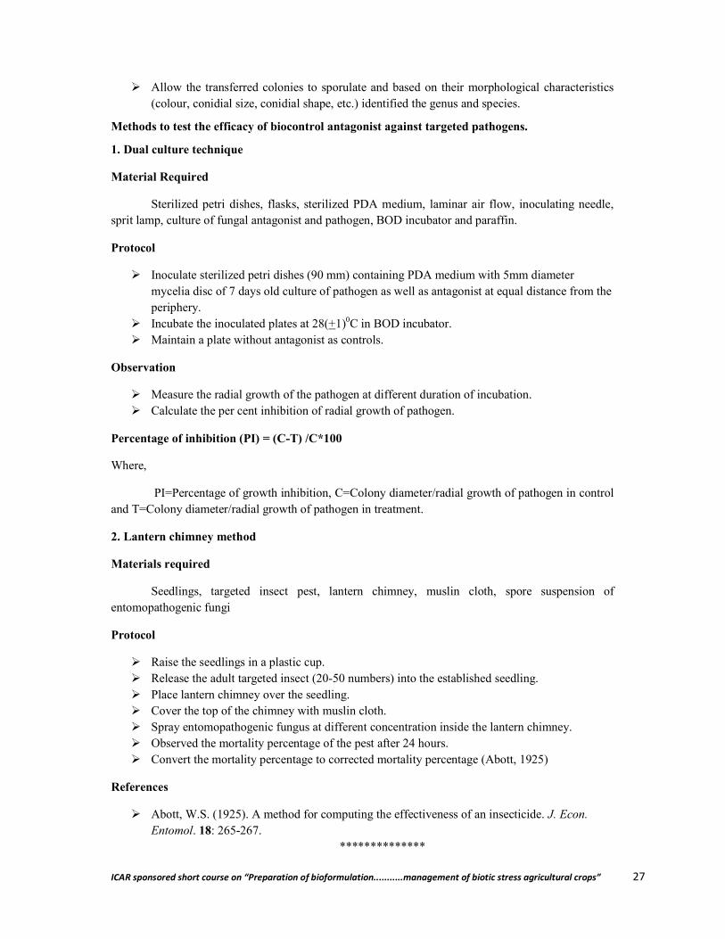
Allow the transferred colonies to sporulate and based on their morphological characteristics (colour, conidial size, conidial shape, etc.) identified the genus and species.
Methods to test the efficacy of biocontrol antagonist against targeted pathogens.
1. Dual culture technique
Material Required
Sterilized petri dishes, flasks, sterilized PDA medium, laminar air flow, inoculating needle, sprit lamp, culture of fungal antagonist and pathogen, BOD incubator and paraffin.
Protocol
Inoculate sterilized petri dishes (90 mm) containing PDA medium with 5mm diameter mycelia disc of 7 days old culture of pathogen as well as antagonist at equal distance from the periphery.
Incubate the inoculated plates at 28(+1)0C in BOD incubator. Maintain a plate without antagonist as controls.
Observation
Measure the radial growth of the pathogen at different duration of incubation. Calculate the per cent inhibition of radial growth of pathogen.
Percentage of inhibition (PI) = (C-T) /C*100
Where,
PI=Percentage of growth inhibition, C=Colony diameter/radial growth of pathogen in control and T=Colony diameter/radial growth of pathogen in treatment.
2. Lantern chimney method
Materials required
Seedlings, targeted insect pest, lantern chimney, muslin cloth, spore suspension of entomopathogenic fungi
Protocol
Raise the seedlings in a plastic cup. Release the adult targeted insect (20-50 numbers) into the established seedling. Place lantern chimney over the seedling. Cover the top of the chimney with muslin cloth. Spray entomopathogenic fungus at different concentration inside the lantern chimney. Observed the mortality percentage of the pest after 24 hours. Convert the mortality percentage to corrected mortality percentage (Abott, 1925)
References
Abott, W.S. (1925). A method for computing the effectiveness of an insecticide. J. Econ. Entomol. 18: 265-267.
**************
ICAR sponsored short course on “Preparation of bioformulation...........management of biotic stress agricultural crops” 28
Taxonomy Based Identification of Fungal Biocontrol Agents
Md. Sakendar Ali Department of Plant Pathology,
Assam Agricultural University, Jorhat- 785013, Assam
Most of the antagonist fungi belong to deuteromycetes (Fungi Imperfecti). The teleomorph and anamorph of some of them have been well established and being exploited for plant and insect management. But there is much confusion about their taxonomy and very often misapplied inadvertently. To identify fungi, their taxonomic status is of vital need to utilize those in proper perspective.
Most common fungi that have been found as prominent antagonists are Trichederma spp, Verticillium lecanii, Gliocladium virens, Paecilomyces lilacinus, Metarhizium anisopliae, Beauveria bassiana, Nomuraea, Aschersonia, Cordyceps spp, Hirsutella formicarum along with some Aspergillus spp.
Many of the entomopathgenic Deuteromycetes are associated with an ascogenous state (Cordyceps, Torrubiella, Necteria and others). The ascogenous and conidial states may occur separately, or on the same stroma. It is often difficult to prove a true connection between a perfect and imperfect stage unless cultural studies are done; one has to depend on micro and macro-morphological studies.
How to overcome the misidentification of Metarhizium and Myrothecium, Aspergillus flavus and A. parasiticus, Isaria vs Beauveria?
What are the keys to species identification of Metarhizium anisopliae, M. flavoviride, M. anisopliae var. major; Nomuraea rileyi and N. atypicola? A thorough knowledge of taxonomy and perhaps continued effort in this endeavor will definitely give a way of solution and to mitigate the problem.
Deuteromycotina (Hyphomycetes and Coelomycetes) complexes:
Conidium ontogeny and conidiogenesis provide diagnostic keys to delineate some of the complex nature of fungi, which is otherwise difficult to identify with mere conidium and fruiting morphology. The classes Hyphomycetes and Coelomycetes are well studied on the above aspects and due weight age might be given to understand these classes as complementary in the classification and identification of fungi.
Hyphomycetes are asexual reproductive structures produced directly on their substrate without any kind of enclosing tissues. For example Drechslera oryzae, Pyricularia grisea, Fusarium moniliforme, Epicoccum nigrum etc. They grow directly on the surface of individual substrate (leaves, dead leaves, stems etc) where they are exposed to the environment and can have its conidia (spores) carried away by air currents. Because of this its conidia are abundant in the air and are commonly found by people investigating the air borne fungi. Mycologists working with fungal cultures in petridishes commonly get E. nigrum as uninvited guest.
The most common function of hyphomycetes is reproduction and dispersal, although in some species the conidia may act as gametes or ‘spermatia’ that can fertilize an incipient dikaryon. If this is so they are not likely to germinate and produce hyphae as happens in the great majority of hyphomycetes. Hyphomycetes come in a staggering variety of forms. This immense diversity reflects
ICAR sponsored short course on “Preparation of bioformulation...........management of biotic stress agricultural crops” 29
the role they play in the dispersal of the fungus producing them. Each species grow in a particular habitat. When the nutrients in this habitat are exhausted, the fungus must ensure that its offspring find their way to a similar source of nutrition. Getting their conidia to this new place requires precise dispersal mechanisms. The mechanisms include air dispersal, water, and dispersal by insects and other small animals.
Insects, mites and other small invertebrate animals live among fungi and share many common difficulties. Such organisms including fungi have fairly precise nutritional and environmental requirements and may be separated from acceptable habitats by considerable distances. Reaching a precise habitat, especially one that is not large, may require more than just casting some spores to the wind. Insects, of course, can fly well and have eyes and sensitive chemical detectors to aid them get where they need to go. Fungi, lacking such abilities often solve the problem by hitching a ride on an insect. This solution to a difficult problem of dispersal is so successful that nearly all groups of fungi have made use of it. The hyphomycetes are among the most adept.
The hyphomycetes fungus Acremonium can be regarded as an excellent exploiter of insects for its dispersal. The fungus produces its conidia at the tips of the small ‘spines’ along the hyphal strand. The cells called phialides, produce a continuous succession of conidia at their ends, and these conidia collect there in glistening drops of liquid. Air currents donot dislodge the conidia nor are they likely to come in contact with water. Instead they stick to the bodies of insects and mites that are moving about in their habitat. The conidia are sticky because of the liquid they are borne in. Eventually the insect will move on to a new habitat and the fungus will be rubbed off and begin to grow there.
Based on organization pattern and habitat of fungi, one has some idea on a fungal identity. As for example, Haplotrichum conspersum and Bactridium sp. H. conspersum forms an almost continuous carpet in log, irregular in places but nevertheless all on one surface of conidia. On the other hand, the Bactridium species produced its conidia in discrete little caushions, well separated from one another. Species of Bactridium are said to produce their conidia in sporodochia, a term denoting a dense cluster of conidium-bearing hyphae or conidiophores. The conidiophores in Haplotrichum are said to be solitary because they do not form distinct clusters. Synnemata are often associated with dispersal of conidia by insects. Here the conidiophores have united to form an elongated compound structure called a synnema (plural, synnemata). Pesotum ulmi, a synnematous form associated with the infamous Dutch elm disease, is well known to transfer its spores to bark beetles, the vectors of the disease. In Graphium species the conidia are borne in a droplet of liquid at the top of large bark synnemata. In A. albicans the conidia are borne on the upper part of the synnemata but also occur along the sides.
Alexopoulos (1962) classification divided Deuteromycotina into following form orders.
a) Reproduction by means of conidia, by oidia, or by budding. b) Reproduction by means of conidia borne in pycnidia - Sphaeropsidales
(bb) Conidia, when formed, not in pycnidia c) Reproduction by means of conidia borne in acervuli - Melanconiales
(cc) Reproduction by means of conidia borne otherwise, by oidia, or by budding – Moniliales
(d) No reproductive structures known - Mycelia sterilla
ICAR sponsored short course on “Preparation of bioformulation...........management of biotic stress agricultural crops” 30
Ainsworth (1973),divided Deuteromycotina into three classes: Blastomycetes, Hyphomycetes and Coelomycetes
Identification of Hyphomycetes
Hyphomycetes are difficult to name. Identification requires knowledge of developmental features and some skill with a microscope. Two criteria have been used traditionally for identifying hyphomycetes: colour and conidial septation. Colour is usually separated into two categories, light and darked. Darkly pigmented species are said to be ‘dematiaceous ‘while light and colorless ones are called ‘moniliaceous’. Conidial septation is usually separated into one-celled, two celled, more than two celled with both tranverse and vertical septa. Although these criteria do not indicate genetic relatedness rather they do help in sorting things out.
Coelomycetes:
The thallus is mycelia, eucarpic and septate; the conidia are formed in various ways from condiogenous cells lining the cavity which is initially enclosed by single tissue (pycnidia and eustromata) or a combination of fungi and host tissue (acervuli and pseudostromata). Conidiogenous cells are formed on the inner surface of the fructification walls, sometimes restricted to the lateral or basal walls, unicellular, often grouped into complex structures (conidiophores), producing conidia asexually.
Conidia are enterothallic, holothallic, enteroblastic, phialides and annellides being most common, deciduous, hyaline or pigmented, aseptate, euseptate or various shapes and with cellular or extracellular appendages, setulae and mucilaginous ornamentations.
Microscopical observations and cultivation in vitro
It is usually possible to identify pathogen directly from the insect by mounting in lactophenol or lactic acid with some aniline blue. Immature specimens without sporulation should be placed in moist chambers. Old or over-mature specimens, on which no conidial structure can be recognized, must be isolated in pure culture. For this malt or potato dextrose agar supplemented with antibiotics are recommended. Sometimes Sabouraud or Mealworm (Samson, 1974) agars can be used for species that grow or sporulate poorly on agar media. Culture of entomopathogenic species can be maintained on malt or oatmeal agars. Metarhizium, Beauveria and Paecilomyces sporulate well on sterilized rice. When strains are used for infection or physiological experiments, the cultures should be freeze-dried since regular sub-culturing reduces physiological and biochemical properties.
Key to common and important Genera
1a. Conidia produced by phialides in chain or in slimy heads -2 1b. Conidia produced by sympodial conidiogenous cells -12 2a. Conidia in dry, long chains -3 2b. Conidia in slimy heads -8 3a. Conidiophores consisting of an unbranched stripe terminating in a vesicle bearing
conidiogenous cells and /or metulae -4 3b. Conidiophores without vesicles -5 4a. Conidiophores united in distinct synnemata; host, spider -Gibellula 4b. Conidiophores arranged along synnemata as in a hymenium -Akanthomyces 5b. Phialides differently arranged -6

ICAR sponsored short course on “Preparation of bioformulation...........management of biotic stress agricultural crops” 31
6a. Conidiophore closely packed in sporodochial structures; conidia in columns -Metarhizium
6b. Conidiophores loosely arranged in synnemata or single; conidia in long divergent chains -7
7a. Phialides very short necked; conidiophores bearing dense whorls of branches and phialides -Nomuraea
7b. Phialides with distinct necks; conidiophores are irregular or verticillately branched elements -Paecilomyces
8a. Conidia one or more septate, usually curved -Fusarium 8b. Conidia non-septate -9 9a. Conidiophores arranged in sporodochia on scale insects or white flies, conidia usually
fusiform -Aschersonia 9b. Conidiophores not arranged in sporodochia -10 10a. Phialides mostly single, with a swollen basal part abruptly tapering to a thin, long neck;
conidia single or few and covered by a slime sheath -Hirsutella 10b. Phialides in whorls on verticillately branched conidiophores -11 11a. Phialides flask shaped, conidia clavate; on mosquitoes and related Diptera -
Culicinomyces 11b. Phialides usually awl-shaped, conidia of various shapes –Verticillium 12a. Conidiogenous cells in a hymenium like layer along distinct synnemata -Hymenostilbe 12b. Conidiogenous cells in cluster or singly -13 13a. Conidiophores with unbranched stipes terminating in a vesicle bearing metulae and
conidiogenous cells -Psudogibellula 13b. Conidiophores without swollen vesicles -14 14a. Conidiogenous cells elongate and slender with inconspicuous, terminal or lateral, scars
-Sporothrix 14b. Conidiogenous cells with a swollen basal part terminating in a ziz-zag rachis -
Beauveria
References
Mehrotra, R. S. and Aneja, K. R. (1990). An Introduction to Mycology,Wiley Eastern Ltd, New Delhi, 766 pp.
Sarbhoy, A. K. (2006).Text Book of Mycology, ICAR, New Delhi ,347pp.
**************

ICAR sponsored short course on “Preparation of bioformulation...........management of biotic stress agricultural crops” 32
Bacterial Species Characterization by Polyphasic Taxonomy
Anil Kumar Singh Biotechnology Section, BSTD,
CSIR-North East Institute of Science and Technology, Jorhat-785006, Assam
Bacteria are the most diverse and abundant group of organisms on Earth. Attempts to describe bacterial diversity and abundance often yield impressive numbers. For example, there are reports that there is one billion times more individual bacteria on earth than stars in the universe, that the number of prokaryotic species exceeds that of all other species, that prokaryotic cells comprise the majority of all biomass, and that even the most hostile habitats are inhabited by bacteria.
However, the advances of various molecular techniques despite their limitations and biases, have led to an increased understanding of bacterial diversity. Today there exist numerous molecular tools are giving the precise details of bacterial diversity, for example 16S rRNA clone libraries, followed by Denaturation Gradient Gel Electrophoresis (DGGE), Fluorescence In Situ Hybridization (FISH), and Quantitative Dot Blot Hybridization. Nevertheless, methodological constraints hinder the ability to measure biodiversity, for example the widely used community fingerprinting techniques detect only app. 10% of the most common bacterial species that coexist in freshwater habitats. Furthermore, a consistent bacterial species concept, following the principles of polyphasic taxonomy, which comprises phenotypic, chemotaxonomic, genotypic, and phylogenetic information, is still under consideration.
**************

ICAR sponsored short course on “Preparation of bioformulation...........management of biotic stress agricultural crops” 33
Microbes in the management of banana diseases
Ashok Bhattacharyya AICRP on Fruits, Department of Plant Pathology,
Assam Agricultural University, Jorhat- 785013, Assam
Banana (Musa spp.) is the fourth most important global food commodity after rice, wheat and maize in terms of gross value production (Singh et al., 2011). At present, it is grown in more than 150 countries throughout tropical and subtropical regions and it is the staple food for more than 400 million people (Singh et al., 2011). India -highest producer of banana with a production of 26509.1000 metric tonnes from 776.0 thousand ha with a productivity 34.2 t/ha contributes 29.19% to the world’s production (Annon, 2015). The combined global production of Banana, the most valuable primary agricultural commodity, was reported to be about 145 million tonnes with a gross production value of US$44.10billion (FAOSTAT, 2013). Bananas are the eighth most important food crop in the world and the fourth most important among the world's least-developed countries. Locally consumed bananas are significant staple foods in Africa, Asia and tropical America, and diverse cultivars are eaten raw, cooked and brewed (Karamura et al., 2012).
Important diseases of Banana
Banana is globally important crop in terms of nutrition and food security especially for underdeveloped countries of African continent and other developed and developing countries of the world. Its production and productivity is hampered by a lot of biotic and abiotic stresses. The following are the important diseases of banana:
Fusarium wilt (Panama disease) caused by Fusarium oxysporum f.sp. cubense Eumusae (Sigatoka) disease caused by Pseudocercospora musae Crown rot caused Colletotrichum musae (in association with other organisms) Anthracnose caused Colletotrichum musae Bunchy Top disease (Banana Bunchy top virus,BBTV) Infectious Chlorosis (Cucumber mosaic virus,CMV) Rhizome rot of banana caused by Erwinia chrysanthemii
1. Panama wilt of banana- a potentially dreaded disease
Fusarium wilt is one of the most destructive diseases of banana (Ploetz and Pegg, 2000).The pathogen probably originated in Southeast Asia (Ploetz and Pegg, 1997; Ploetz, 2007; Stover, 1962; Vakili, 1965), but the disease was first recognized elsewhere. Bancroft's (1876) initial description from Australia was followed by reports from tropical America (Costa Rica and Panama in 1890) (Stover, 1962).Currently, the disease is found in virtually all areas where banana is grown. Effective, long-term management of Fusarium wilt of banana remains a challenge, due largely to the perennial host plant (Ploetz and Evans, in press; Ploetz and Pegg, 2000). Tactics for Fusarium wilt management on short cycle hosts (e.g. tomato or radish) are often ineffective over the multiple years that a banana crop is grown. The disease almost destroyed the banana export industry, built on the Gros Michel variety, in Central America during the 1950’s (Stover, 1962). In addition, the widely grown clones in the ABB ‘Bluggoe’ and AAA ‘Gros Michel and Cavendish’ sub groups are also highly susceptible to this disease worldwide. The chlamydospores of the fungus can survive for decades. The rhizome as well as soil borne nature of the pathogen clubbed with prolonged survival ability makes the chemical control a very tough proposition.
ICAR sponsored short course on “Preparation of bioformulation...........management of biotic stress agricultural crops” 34
The earliest symptoms are faint yellow streaks on the petiole of the oldest, lower most leaves. Affected leaves show progressive yellowing, break at the petiole and hang down along the pseudostem. Longitudinal splitting of pseudostem is very common. Usually discolouration appears first in the outer or oldest leaf sheath and extends in to the inner sheaths. Affected plants don not produce bunches even if produced fruits are malformed and ripened prematurely or irregularly.
Management
There are limited options for managing Fusarium wilt of banana. The perennial nature of this pathosystem and the corresponding polycyclic nature of the disease has complicated the development of long-term measures (Ploetz, 2007). Moreover, poor resistance exists in important groups of banana and technical hurdles confront those who would improve disease-susceptible cultivars. For example, extremely poor fertility in the Cavendish subgroup constrains its improvement via conventional breeding (Moran, 2013).Susceptible banana cultivars can usually be grown only if pathogen-free propagation materials are used in pathogen-free soil. Tissue-culture-derived plantlets are the most reliable source of clean material. Although they are more susceptible to Fusarium wilt than traditional banana seed pieces (Smith et al., 1998), they should be used to propagate this crop whenever possible. In subsistence agriculture or other situations in which their expense may be an issue, tissue-culture plantlets can be used to initiate disease free nurseries to produce pathogen-free conventional seed pieces (Lule et al., 2013).
Management of Panama wilt of banana, as mentioned earlier, is quite difficult due to the soil inhabitant nature of survival and also polycyclic nature of the pathogen. Managing the disease with chemicals is generally considering now a days as last option. Utilizing the microbes for the management of panama wilt of banana is an eco-friendly approach without the problems of pesticide residues and pest resurgence. The challenging part of the fungus is that it survives in soil for up to 30-40 years as chlamydospores in infested plant material or in the roots of alternative hosts (Ploetz, 1990). There have been very few field studies in which long-term biocontrol efficacy has been investigated for Fusarium wilt of banana (Ploetz, 2004).In the decade since biocontrol work on Fusarium wilt of banana was last reviewed (Ploetz, 2004), the above trends have continued, as few field studies have been reported and most publications describe only lab and greenhouse research (Fishal et al., 2010; Forsyth et al., 2006; Lian et al., 2008; Thangavelu and Jayanthi, 2009; Thangavelu and Mustaffa, 2012;Ting et al., 2010; Wang et al., 2013; Wu et al., 2013; Zacky and Ting, 2013). Field results have been reported in different modes and promising greenhouse treatments failed in the field. Belgrove et al. (2011) noted that neither the non pathogenic F. oxysporum, P. fluorescens, nor combinations reduced Fusarium wilt development significantly.
Use of Trichoderma sp.
Trichoderma spp., are free-living fungi that are common in soil and root ecosystems. They are highly interactive in root, soil and foliar environments. They produce or release a variety of compounds that induce localized or systemic resistance responses in plants. Several reports indicate that Trichoderma species can effectively suppress Fusarium wilt pathogens (Sivan and Chet, 1986; Thangavelu et al. 2004). Raghuchander et al. (1997) reported that T. viride and P. fluorescens were equally effective in reducing the wilt incidence. Similarly, soil application of T. viride NRCB1 as chaffy grain formulation significantly reduced the external (up to 78%) and internal symptoms (up to 80 %) of Fusarium wilt disease in tissue cultured as well as sucker derived plants of banana cv. Rasthali (Silk-AAB) and increased the plant growth parameters significantly as compared to the talc powder formulation under pot culture and field conditions (Thangavelu and Mustaffa, 2010). In India

ICAR sponsored short course on “Preparation of bioformulation...........management of biotic stress agricultural crops” 35
ICAR-NRCB actively involved in development of bioformulations. Trichoderma viride NRCB-1 isolated from virgin soils of waynad district of Kerala serving as effective bioformulation.
2. Eumusae (Sigatoka leaf spot/yellow sigatoka) disease – a potential leaf spot disease
Mycospaerella musicola J. L. Mulder in J. L. Mulder & R. H. Stover.
The disease is caused by Yellow sigatoka disease was first recognized in Java in 1902. It occurs throughout the world and is one of the most destructive diseases of banana. It causes losses by reducing the functional leaf surface of the plant, which results in small, unevenly ripened bananas that fail to ripen and may fall. The disease first appears as small, light yellow spots parallel to the side veins of leaves. A few days later, the spots become enlarge in size and turn brown with light gray centers. Such spots soon enlarge further, the tissue around them turns yellow and dies, and adjacent spots coalesce to form large, dead areas on the leaf. Rapid drying and defoliation of mature leaves are the characteristic feature of this disease.
Ecofriendly management of Sigatoka leaf spot disease of banana with the combination of sucker treatment with Pseudomonas fluorescens and foliar application of Pseudomonas fluorescens, vegetable oil and baking soda mixture and mineral oil showed significant difference in disease intensity when compared to the control plants that did not receive any treatment application (Thara et al., 2007).
Pseudomonas fluorescens was most significant (54.42 % inhibition) 42.55% growth inhibition Trichoderma viridewas found to be a potent mycoparasite with a maximum inhibition of about 88.51% followed by Th-3 of T. harzianum with 82.31%.mycelial inhibition. However, there was a significant difference between the fungal and bacterial bioagents on inhibition of mycelial growth, being fungal bioagents proved most promising. There is a tremendous scope to exploit Trichoderma viride (Isolate Tv-5) or Pseudomonas fluorescens (isolate Pf-1)as a component of integrated Sigatoka disease management to substitute one or two fungicidal applications under field conditions (Noorulla et al., 2013).
3. Crown rot and Anthracnose disease
Banana is one of the most important tropical crops and is affected by several fungal diseases, such as crown rot postharvest disease. Crown rot is responsible for significant losses in banana fruits. Predominantly, Colletotrichum musae and Fusarium spp. are its causative agents. Inoculum sources include mainly infected flowers but also decaying leaves, and fungal transfer can occur from banana stalks onto the crown surface during the cutting of banana bunches (knife-induced) as well as when the bunches are cleaned in contaminated water. Fungal infection starts at harvest, and the first symptoms of crown rot appear only after packaging and shipping from producing countries to consuming countries. Crown rot begins with a mycelium development on the crown surface, followed by an internal development. This internal development can, subsequently, affect the peduncle and the whole fruit, leading to softening and blackening of the fruit tissue. Postharvest fungicidal treatments are applied to control crown rot disease, though severely affected banana fruits are still found in consumer markets. Moreover, onset and spreading of the disease is unpredictable and can also induce early ripening of banana fruits during transport.
Alvinda et al., 2009 reported inhibitory effect of epiphytic bacteria Bacillus amyloliquifacieans isolated from banana fruit surface against banana fruit rot pathogen when applied
ICAR sponsored short course on “Preparation of bioformulation...........management of biotic stress agricultural crops” 36
as postharvest application, similarly Sangeetha et al., 2010 found that the timing of application of bio control agents has significant influence in controlling the crown rot disease of banana, the results obtained from their experiments as the highest reduction of crown rot disease of banana was achieved when the biocontrol agents were applied 4 and 2 h before pathogen inoculation (preventive method of application) compared with the other treatments.
4. Banana Bunchy top disease
Banana bunchy top disease (BBTD) is one of the most destructive diseases of banana in tropical Asia, Australia and the South Pacific (Dietzgen and Thomas 1991). In India, Banana bunchy top virus (BBTV) is widespread in all banana-growing states. BBTV is an isometric virus, 18 nm in diameter with a circular single stranded DNA genome consisting of at least six components (BBTV DNA 1–6) (Burns et al., 1995).
The first characteristic symptoms appear as irregular, nodular, dark green streaks in the lower portion of the leaf midrib and petiole. This type of symptom is sometimes referred to as ‘Morse code streaking’ due to the irregularity and resemblance to the series of dots and dashes (Magee 1940). As the successive leaves of an infected plant emerge, they seem to be more and more abnormal. They appear to be bunched at the top of the plant giving the typical bunchy top appearance. Severely infected banana plants usually will not fruit, and even if they produce, the banana hands and fingers are likely to be distorted and twisted.
Microbe based management
Activating the systemic resistance in plants for the management of diseases is considerably new trend in the disease management. Trichoderma viride, Pseudomonas fluorescens are most widely used biocontrol agents. Kavino et al., 2007 reported Induction of systemic resistance in banana (Musa spp.) against Banana bunchy top virus (BBTV) by combining chitin with root-colonizing Pseudomonas fluorescens strain CHA0.
5. Banana Mosaic/ Infectious chlorosis/ Heart rot: Cucumber Mosaic Virus (CMV)
This virus is a domestic quarantine pest in India. Hence movement of planting suckers from Gujarat and Maharashtra to other parts of the country is banned.
Typical mosaic like or discontinuous linear streaking in bands extending from margin to midrib running parallel to veins. Chlorosis of newly emerged leaves, occasionally rotting of central youngest leaf and leaf sheaths in severe cases (Heart rot) which progress in to pseudostem leading to death of the plants. Diseased plants do not reach maturity and may fail to produce the bunch.
6. Erwinia rhizome rot
One of the important bacterial disease of banana generally occur during younger stage of the crop. Tissue culture banana plants are highly susceptible to rhizome rot disease. Severe epidemic of rhizome rot disease of banana has been reported from tissue culture plantlets from Theni, Tiruchirapalli and Villupuram districts of Tamil Nadu.
Water soaked spots develop on outer leaf sheath on base of pseudostem near to soli line. The spots turn brown and soft rotting of the pseudostem tissue and rhizome take place. In many cases pseudostem tip over because by rotting at the ground level. In infected rhizomes, pockets of dark
ICAR sponsored short course on “Preparation of bioformulation...........management of biotic stress agricultural crops” 37
water soaked areas develop; infection may result in the production of cavities which resemble the root tunnels.
Management
Microbe based management options are considerably less adopted when compared to chemicals due to various reasons. Vasundhara et al., 2014 reported the in viro antagonistic effect of Pseudomonas fluorescens, Bacillus subtillis against the Erwinia rhizome rot of banana.
Mode of action of Biocontrol agents
Trichoderma spp.
The possible mechanisms involved in the reduction of Fusarium wilt severity due to Trichoderma spp. treatment might be the mycoparasitism, spatial and nutrient competition, antibiosis by enzymes and secondary metabolites, and induction of plant defence system. The mycoparasitism involves in coiling, disorganization of host cell contents and penetration of the host (Papavisas, 1985; University of Sydney, 2003). During the mycoparasitism, Trichoderma spp. parasitizes the hyphae of
the pathogen and produce extracellular enzymes such as proteolytic enzymes, ┚-1, 3- glucanolytic
enzymes and chitinase etc., which cause lysis of the pathogen. The toxic metabolites such as extracellular enzymes, volatiles and antibiotics like gliotoxin and viridin which are highly fungistatic substances (Weindling, 1941) are considered as elements involved in antibiosis. In addition, Trichoderma spp. could compete and sequester ions of iron (the ions are essential for the plant pathogen,) by releasing compounds known as siderophores (Srinivasan et al. 1992)
Antagonistic bacteria
Generally biocontrol agents can antagonize soil-borne pathogens through the following strategies:
(1) Competition for niches and nutrients (niche exclusion), (2) Production of secondary metabolites which are used in direct antagonism (3) Growth promotion by changing the physiology of the plant (4) Induction of resistance to disease
Benhamou et al. (1996) provided evidence that root colonization by the endophytic bacterium Pseudomonas fluorescens, involved in a sequence of events that included bacterial attachment to the plant roots, proliferation along the elongation root, and local penetration of the epidermis. M’Piga et al. (1997) also confirmed the entry of P. fluorescens into the root system and their colonization inside. Once inside the host tissue, these bacteria produce an array of antifungal metabolites like siderophores and different antibiotics like phenazine-1 carboxylic acid, and 2, 4-diacetylphloroglucinol preventing the further advancement of the fungus (Beckman et al. 1982; Mueller & Beckmann,1988) by inducing severe cell disturbances in pathogenic fungi (Dowling and O’Gara, 1994).
References
Aguilar Moran, J. F. (2013). Improvement of Cavendish Banana Cultivars through conventional breeding. Acta Hort. 986: 205-208.
Alvinda, D.G.; Kobayashi, T.; Natsuaki, K.T, Tanda, S. (2006). Inhibitory influence of inorganic salts on banana postharvest pathogens and preliminary application of Trichoderma viride. Journal of Biological Control 11: 105– 110.

ICAR sponsored short course on “Preparation of bioformulation...........management of biotic stress agricultural crops” 38
Anonymous (2015). Annual report AICRP on Fruits, AAU Centre, Jorhat. Bancroft, J. (1876). Report of the board appointed to enquire into the cause of disease
affecting livestock and plants. In: Queensland, 1876. Votes and Proceedings 1877, vol. 3, pp. 1011-1038.
Beckman, C. H.; Mueller, W. C.; Tessier, B. J. and Harrisson, N. A. (1982). Recognition and callose deposition in response to vascular infection in Fusarium wilt-resistant or susceptible tomato plants. Physiological Plant Pathology 20: 1–10.
Belgrove, A.; Steinberg, C. and Viljoen, A. (2011). Evaluation of nonpathogenic Fusarium oxysporum and Pseudomonas fluorescens for Panama disease control. Plant Dis. 95: 951-959.
Benhamou, N.; Belanger, R. R. and Paulitz, T. (1996). Ultrastructural and cytochemical aspects of the interaction between Pseudomonas fluorescens and Ri T-DNA transformed pea roots: host response to colonization by Phythium ultimum Trow, Planta 199: 105-11.
Burns, T. M.; Harding, R. M. and Dale, J. L. (1995). The genome organization of Banana bunchy top virus: Analysis of six ssDNA components. Journal of General Virology 76: 1471–1482.
Carlier, A.K. and Ting, A. S. Y. (2013). Investigating the bioactivity of cells and cell-free extracts of Streptomyces griseus towards Sigatoka disease of banana. Biol. Cont. 66: 204-208
Dietzgen, R. G. and Thomas, J. E. (1991). Properties of virus-like particles associated with banana bunchy top disease in Hawaii, Indonesia and Tonga. Australasian Plant Pathology 20: 161-165.
Dowling, D. N. and O’Gara, F. (1994). Metabolites of Pseudomonas involved in the biocontrol of plant disease. Trends in Biotechnology 3:121–141.
FAOSTAT (2013). http://faostat.fao.org/. Fishal, E. M. M.; Meon, S. and Yun, W. M. (2010). Induction of tolerance to Fusarium wilt
and defense-related mechanisms in the plantlets of susceptible Berangan banana pre-inoculated with Pseudomonas sp. (UPMP3) and Burkholderia sp. (UPMB3). Agric. Sci. China 9: 1140-1149.
Forsyth, L. M.; Smith, L. J. and Aitken, E. A. B. (2006). Identification and characterization of non-pathogenic Fusarium oxysporum capable of increasing and decreasing Fusarium wilt severity. Mycol. Res. 110: 929-935.
Karamura, D. A.; Karamura, E. and Tinzaara, W. (Eds.) (2012). Banana Cultivar Names, Synonyms and Their Usage in East Africa. Bioversity Intl, Kampala, Uganda.
Kavino, M.; Harish, S.; Kumar, N.; Saravanakumar, D. and Samiyappan, R. (2008). Induction of systemic resistance in banana (Musa spp.) against Banana bunchy top virus (BBTV) by combining chitin with root-colonizing Pseudomonas fluorescens strain CHA0. European Journal of Plant Pathology 120(4): 353-362.
Lakshmanan, P.; Selvaraj, P. and Mohan, S. (1987). Efficiency of different methods for the control of Panama disease. Trop. Pest Manage 33: 373–376.
Lian, J.; Wang, Z. and Zhou, S. (2008). Response of endophytic bacterial communities in banana tissue culture plantlets to Fusarium wilt pathogen infection. J. Gen. Appl. Microbiol. 54: 83-92.
Lule, M.; Dubois, T.; Coyne, D.; Kisitu, D.; Kamusiime, H. and Bbemba, J. (2013). Trainer's Manual. A Training Course for Banana Farmers Interested in Growing Tissue Culture Bananas. International Institute of Tropical Agriculture, Ibadan, Nigeria, 126 p.
M’Piga, P.; Belanger, R. R.; Paulitz, T.C. and Benhamou, N. (1997). Increased resistance to Fusarium oxysporum f. sp. radicis-lycopersici in tomato plants treated with the endophytic bacterium Pseudomonas fluorescens strain 63–28. Physiology Molecular Plant Pathology 50: 301–320.

ICAR sponsored short course on “Preparation of bioformulation...........management of biotic stress agricultural crops” 39
Magee, C. J. P. (1940). Transmission studies on the Banana bunchy top virus. The Journal of the Australian Institute of Agricultural Science 6: 109–110.
Mueller, W. C. and Beckman C. H. (1988). Correlated light and EM studies of callose deposits in vascular parenchyma cells of tomato plants inoculated with Fusarium oxysporum f.sp. lycopersici. Physiological and Molecular Plant Pathology 33: 201–208.
Noorulla, A. K.; Jayashree, K. and Samiyappan, R. (2013). Management of Fusarium wilt of banana using antagonistic microorganisms. Journal of Biological Control 11: 101– 105.
Papavizas, G. C. (1985). Trichoderma and Gliocladium: biology, ecology and potential for biocontrol. Annu Rev Phytopathol 23: 23–54.
Ploetz, R.C. (1992). Fusarium wilt of banana (Panama disease). In: Mukhopadhyay, A.N., Chaube, H.S., Kumar, J., Singh, U.S. (Eds.), Plant Diseases of International Importance, vol. III. Prentice Hall, Englewood Cliffs, New Jersey, USA, pp. 270-282.
Ploetz, R. C. and Pegg, K. G. (1997). Fusarium wilt of banana and Wallace's line: was the disease originally restricted to his Indo-Malayan region? Australas. Plant Pathol. 26: 239-249.
Ploetz, R. C. and Pegg, K. G. (2000). Fusarium wilt. In: Jones, D.R. (Ed.), Diseases of Banana, Abaca and Enset. CABI Publishing, Wallingford, UK, pp. 143-159
Ploetz, R. C. (2004). Biological control of Fusarium wilt: a review and an evaluation. In: Abstr. Booklet, Intl. Congr. on Musa: Harnessing Research to Improve Livelihoods, p. 141. http://www.inibap.org/pdf/penang2_en.pdf.
Ploetz, R.C. (2007). Diseases of tropical perennial crops: challenging problems in diverse environments. Plant Dis. 91: 644-663.
Ploetz, R. C. an Evans, E. A. (2015). Banana diseases and the future of the industry. Hort. Rev. (in press).
Raguchander, T.; Jayashree, K. and Samiyappan, R. (1997). Management of Fusarium wilt of banana using antagonistic microorganisms. Journal of Biological Control 11: 101– 105.
Sangeetha, Ganesan; Swaminathan, Usharani, and Arjunan, Muthukumar (2010). Biocontrol with Trichoderma species for the management of postharvest crown rot of banana. Phytopathologia mediterranea 48: 214-225.
Singh, H. P.; Uma, S. and Karihaloo, J. L. (2011). Micropropagation for Production of Quality Banana Planting Material in Asia-Pacific. Asia Pacific consortium on Agricultural Biotechnology (APCoAB), New Delhi, India, pp 92.
Sivan, A. and Chet, I. (1986). Biological control of Fusarium spp. in cotton, wheat and muskmelon by Trichoderma harzianum. J. Phytopathol. 116: 39-47.
Smith, M. K.; Whiley, A. W.; Searle, C.; Langdon, P. W.; Schaffer, B. and Pegg, K. G. (1998).Micropropagated bananas are more susceptible to fusarium wilt than plants grown from conventional material. Austral. J. Agric. Res. 49: 1133-1139.
Srinivasan, U.; Staines, H. J. and Bruce, A. (1992). Influence of media type on antagonistic modes of Trichoderma spp. against wood decay basidiomycetes, Mater. Org. 27: 301-321.
Stover, R. H. (1962). Fusarial Wilt (Panama Disease) of Bananas and Other Musa Species. CMI, Kew, Surrey, UK.
Thangavelu, R.; Palaniswami, A. and Velazhahan, R. (2004). Mass production of Trichoderma harzianum for managing Fusarium wilt of banana. Agriculture, Ecosytems and Environment 103: 259–263.
Thangavelu, R. and Jayanthi, A., 2009. A RFLP analysis of rDNA-ITS regions of nativenon-pathogenic Fusarium oxysporum isolates and their field evaluation for the suppression of Fusarium wilt disease of banana. Australas. Plant Pathol. 38:13-21.

ICAR sponsored short course on “Preparation of bioformulation...........management of biotic stress agricultural crops” 40
Thangavelu, R., and Mustaffa M. M. (2010). A Potential isolate of Trichoderma viride NRCB1and its mass production for the effective management of Fusarium wilt disease in banana. Tree and Forestry Science and Biotechnology 4 (2): 76-84.
Thangavelu, R. and Mustaffa, M. M. (2012). Current advances in the Fusarium wilt disease management in banana with emphasis on biological control. In: Cumagun, C.J. (Ed.), Plant Pathology. InTech, ISBN 978-953-51-0489-6, pp. 273-298. http:// dx.doi.org/10.5772/33775.
Thara. A. K.; Steinberg, C. and Viljoen, A. (2011). Evaluation of Trichoderma viridae for sigatoka disease control. Plant Dis. 95: 951-959.
Ting, A. S. Y.; Meon, S.; Kadir, J.; Radu, S. and Singh, G. (2010). Induction of host defence enzymes by the endophytic bacterium Serratia marcescens, in banana plantlets. Int. J. Pest Manage 56: 183-188.
Vakili, N. G. (1965). Fusarium wilt resistance in seedlings and mature plants of Musa species. Phytopathology 55: 135-140.
Vasundhara, B. A.; Thammaiah, N.; Swamy, G. S. K.; Kulkarni, M. S. Devappa and Gangadharappa, P. M. (2014). In vitro evaluation of antagonistic micro-organisms against the growth of Erwinia chrysanthemi. Internat. J. Plant Protec. 7(1): 265-266.
Wang, B. B.; Yuan, J.; Zhang, J.; Shen, Z. Z.; Zhang, M. X.; Li, R.; Ruan, Y. Z. and Shen, Q. R. (2013). Effects of novel bioorganic fertilizer produced by Bacillus amyloliquefaciens W19 on antagonism of Fusarium wilt of banana. Biol. Fert. Soils 49: 435-446.
Weindling, R. (1941). Experimental consideration of the mold toxin of Gliocladium and Trichoderma. Phytopathology 31: 991-1003.
Wu, Y. L.; Yi, G. J.; Peng, X. X.; Huang, B. Z.; Liu, E. and Zhang, J. J. (2013). Systemic acquired resistance in Cavendish banana induced by Infection with an incompatible strain of Fusarium oxysporum f. sp. cubense. J. Plant Physiol. 170: 1039-1046.
Zacky, F. A. and Ting, A. S. Y. (2013). Investigating the bioactivity of cells and cell-free extracts of Streptomyces griseus towards Fusarium oxysporum f. sp. cubense race 4. Biol. Cont. 66: 204-208.
**************
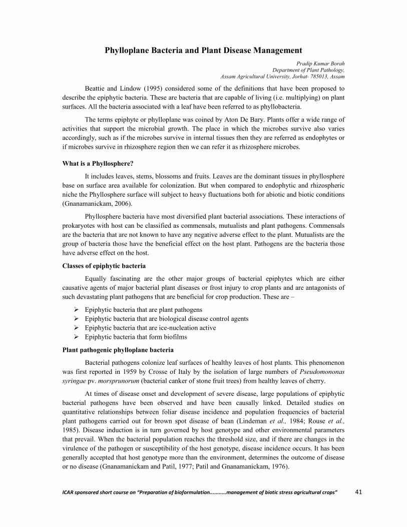
ICAR sponsored short course on “Preparation of bioformulation...........management of biotic stress agricultural crops” 41
Phylloplane Bacteria and Plant Disease Management
Pradip Kumar Borah Department of Plant Pathology,
Assam Agricultural University, Jorhat- 785013, Assam
Beattie and Lindow (1995) considered some of the definitions that have been proposed to describe the epiphytic bacteria. These are bacteria that are capable of living (i.e. multiplying) on plant surfaces. All the bacteria associated with a leaf have been referred to as phyllobacteria.
The terms epiphyte or phylloplane was coined by Aton De Bary. Plants offer a wide range of activities that support the microbial growth. The place in which the microbes survive also varies accordingly, such as if the microbes survive in internal tissues then they are referred as endophytes or if microbes survive in rhizosphere region then we can refer it as rhizosphere microbes.
What is a Phyllosphere?
It includes leaves, stems, blossoms and fruits. Leaves are the dominant tissues in phyllosphere base on surface area available for colonization. But when compared to endophytic and rhizospheric niche the Phyllosphere surface will subject to heavy fluctuations both for abiotic and biotic conditions (Gnanamanickam, 2006).
Phyllosphere bacteria have most diversified plant bacterial associations. These interactions of prokaryotes with host can be classified as commensals, mutualists and plant pathogens. Commensals are the bacteria that are not known to have any negative adverse effect to the plant. Mutualists are the group of bacteria those have the beneficial effect on the host plant. Pathogens are the bacteria those have adverse effect on the host.
Classes of epiphytic bacteria
Equally fascinating are the other major groups of bacterial epiphytes which are either causative agents of major bacterial plant diseases or frost injury to crop plants and are antagonists of such devastating plant pathogens that are beneficial for crop production. These are –
Epiphytic bacteria that are plant pathogens Epiphytic bacteria that are biological disease control agents Epiphytic bacteria that are ice-nucleation active Epiphytic bacteria that form biofilms
Plant pathogenic phylloplane bacteria
Bacterial pathogens colonize leaf surfaces of healthy leaves of host plants. This phenomenon was first reported in 1959 by Crosse of Italy by the isolation of large numbers of Pseudomononas syringae pv. morsprunorum (bacterial canker of stone fruit trees) from healthy leaves of cherry.
At times of disease onset and development of severe disease, large populations of epiphytic bacterial pathogens have been observed and have been causally linked. Detailed studies on quantitative relationships between foliar disease incidence and population frequencies of bacterial plant pathogens carried out for brown spot disease of bean (Lindeman et al., 1984; Rouse et al., 1985). Disease induction is in turn governed by host genotype and other environmental parameters that prevail. When the bacterial population reaches the threshold size, and if there are changes in the virulence of the pathogen or susceptibility of the host genotype, disease incidence occurs. It has been generally accepted that host genotype more than the environment, determines the outcome of disease or no disease (Gnanamanickam and Patil, 1977; Patil and Gnanamanickam, 1976).
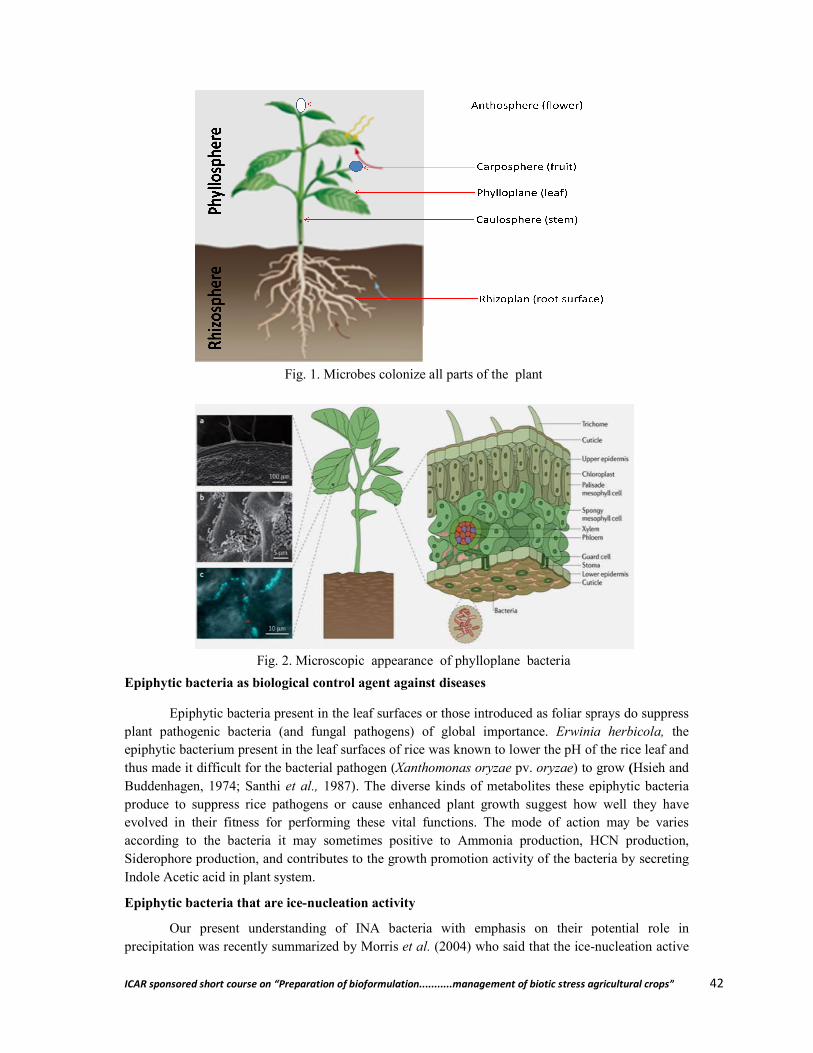
ICAR sponsored short course on “Preparation of bioformulat
Epiphytic bacteria as biological control agent against diseases
Epiphytic bacteria present in the leaf surfaces or those introduced as foliar sprays do suppress plant pathogenic bacteria (and fungal pathogens) of gloepiphytic bacterium present in the leaf surfaces of rice was known to lower the pH of the rice leaf and thus made it difficult for the bacterial pathogen (Buddenhagen, 1974; Santhi et al., produce to suppress rice pathogens or cause enhanced plant growth suggest how well they have evolved in their fitness for performing these vital fuaccording to the bacteria it may sometimes positive to Ammonia production, HCN production, Siderophore production, and contributes to the growth promotion activity of the bacteria by secreting Indole Acetic acid in plant system.
Epiphytic bacteria that are ice-nucleation activity
Our present understanding of INA bacteria with emphasis on their potential role in precipitation was recently summarized by Morris
Fig. 1. Microbes colonize all parts of the plant
Fig. 2. Microscopic
ICAR sponsored short course on “Preparation of bioformulation...........management of biotic stress agricultural crops”
Epiphytic bacteria as biological control agent against diseases
Epiphytic bacteria present in the leaf surfaces or those introduced as foliar sprays do suppress plant pathogenic bacteria (and fungal pathogens) of global importance. Erwinia herbicola,epiphytic bacterium present in the leaf surfaces of rice was known to lower the pH of the rice leaf and thus made it difficult for the bacterial pathogen (Xanthomonas oryzae pv. oryzae) to grow
1987). The diverse kinds of metabolites these epiphytic bacteria produce to suppress rice pathogens or cause enhanced plant growth suggest how well they have evolved in their fitness for performing these vital functions. The mode of action may be varies according to the bacteria it may sometimes positive to Ammonia production, HCN production, Siderophore production, and contributes to the growth promotion activity of the bacteria by secreting
nucleation activity
Our present understanding of INA bacteria with emphasis on their potential role in precipitation was recently summarized by Morris et al. (2004) who said that the ice-nucleation active
Fig. 1. Microbes colonize all parts of the plant
Fig. 2. Microscopic appearance of phylloplane bacteria
ion...........management of biotic stress agricultural crops” 42
Epiphytic bacteria present in the leaf surfaces or those introduced as foliar sprays do suppress Erwinia herbicola, the
epiphytic bacterium present in the leaf surfaces of rice was known to lower the pH of the rice leaf and ) to grow (Hsieh and
1987). The diverse kinds of metabolites these epiphytic bacteria produce to suppress rice pathogens or cause enhanced plant growth suggest how well they have
nctions. The mode of action may be varies according to the bacteria it may sometimes positive to Ammonia production, HCN production, Siderophore production, and contributes to the growth promotion activity of the bacteria by secreting
Our present understanding of INA bacteria with emphasis on their potential role in nucleation active
ICAR sponsored short course on “Preparation of bioformulation...........management of biotic stress agricultural crops” 43
(INA) strains of bacteria as certain bacteria that are commonly found on plants and have the capacity to catalyze the freezing of supercooled water at temperatures as warm as -1°C. This capacity is conferred by a protein present in the outer membrane of the bacterial cell. That they participate in a sort of biological cycle of precipitation whereby they are transported into clouds from plant canopies and incite rain thereby causing favorable conditions for their growth on plant surfaces was proposed about 20 years ago. Today, sufficient evidence and meteorological tools have emerged to re-ignite interest in bio-precipitation and in the ways in which plants play a role as cloud seeders.
Epiphytic bacteria that form biofilms
Biofilms have been defined as frequently observed assemblages made by plant- associated bacteria that have been referred to as aggregates, microcolonies, and symplasmata. Basically biofilms are assemblages of microorganism adherent to each other and are embedded in a matrix of exopolymers. There are three well known models of hydrated biofilm structures, those are as follows:
The water-channel model The mosaic biofilm model The dental plaque biofilm model
This third model of biofilms has high cell densities and arises in a high nutrient environment where they are bathed continuously in a fluid which has a limited fluid flow. While the first three saturated models have been observed in aquatic plants and plants that are raised in hydroponic systems, the unsaturated biofilms have been observed on roots of terrestrial plants by Auerbach et al. (2000). It is known that the densities of microorganisms occurring in leaf surface biofilms is much lower than those observed in water-unsaturated systems. This is an indication, perhaps for low nutrient availability.
Microbiology of phylloplane
Phylloplane is a natural habitat on the leaf surface which supports a heterogenous population comprising both pathogens and non-pathogens. The phylloplane microbes cover a wide range of organisms including yeasts, filamentous fungi, bacteria, actinomycetes, blue-green algae and even pigmy ferns.
Potter (1910) emphasized the importance of the part played by the epiphytic micro-organisms in inciting the disease. Investigations by Last (1955) and Ruinen (1956) emphasized the signifcance of the leaf surface as an ecological niche for saprophytic and pathogenic micro-organisms. Last (1955) termed this niche as phyllosphere (now recognized as phylloplane). The phylloplane microbes are of special interest from various points of view. For instance, some of them have antagonistic action against fungal parasites, degrade plant surface wax and cuticles, produce plant hormones, decompose plant material, activate plants to produce phytoalexins, act as a source of allergic air-borne spores and influence growth behaviour and root exudation of plants.
Recently, there has been a considerable interest to understand the biology of pathogens in relation to leaf surface saprophytes. In nature, intereactions are known to take place between pathogenic and saprophytic microbes as well as within the pathogens themselves. It seems that net effect of phylloplane saprophytes is to reduce the effective inoculum dose of pathogens. However, it is extremely difficult to determine the individual effect of the host, the pathogen or the non-pathogen on leaf surface. The establishment of natural equilibrium in phylloplane is actually a combined effect of all the three components sometimes directly, and at times, indirectly.
Leaves secrete/excrete certain exudates which may directly affect the surface microorganisms, some of which may be pathogenic. The host tissues are also known to exudates
ICAR sponsored short course on “Preparation of bioformulation...........management of biotic stress agricultural crops” 44
phytoncides which are inhibitory to the invading fungi and bacteria. Some of the surface microorganisms may induce the production of phytoalexins in the host and bring about changes in the host reaction to parasites. The micro-organisms themselves produce self-inhibitory and self-stimulatory substances greatly influencing their own germination. They also interact and this interaction leads to the suppression or stimulation of one of the other bringing in antagonistic or associative effects. This complex problem of the phylloplane has a profound influence on the course of events in host infection and is ultimately related to the formulation of methods of disease control.
Host vs. Phylloplane microflora
Common phylloplane bacteria associated with crop plants are Erwinia herbicola (epiphytic), Aerobacter, Corynabacterium, Flavobacterium, Lactobacillus, Pseudomonas, Klebsiella aeroginosa, Micrococcus, Sarcina, Achromobacter, Bacillus, Aeromonas, Clavibacter, Clostridium, Acetobacter, Gluconobacter, Leuconostoc, Arthrobacter, Streptococcus, Alcaligens, Streptomyces, Enterobacter etc.
Epiphytic microbes are influenced by several factors. The indeterminate growth of leaf is the first thing to affect the nature of their substrates and the microbes harboured by them.
The surface microflora is very much under the influence of the host on which it occurs. There is definite exosmosis of materials from the external surface of plants. The moisture film on plant surfaces inevitably carries both organic and inorganic substances. Presence of diffusible inhibitors from healthy leaves of a resistant variety of sugarbeet has been reported by Kovacs (1955). Fawcett (1963) demonstrated the presence of antifungal compounds in certain plants. Some antifungal compounds are slightly inhibitory in vitro to a pathogen of a host, but may be strongly inhibitory to other fungi which are unable to attack the host.
The leaf surface substrates have been reported to be comprised of micro- and macro-elements together with organic substances like sugars, pectic substances, alcohols, amino acids, organic acids, growth hormones, vitamins and phenolic substances. Leaves of certain plants secrete phenolic or terpenoid substances which inhibt the microbial growth. Some microbes are reported to be capable of releasing even growth hormone onto the leaf surface.
Certain hosts are known to contain preformed chemical compounds, phytoncides, which are inhibitory to pathogens. Some of these may be volatile and influence the microorganisms present on the leaf surface.
The foliar microbes are likely to induce production of phytoalexins which are fungistatic. Phytoalexins which are produced as a result of interaction between two metabolic systems, inhibit the growth of phytopathogenic microbes. Perhaps Bernard (1909) appears to be the first to point out that local saprophytic association may provide protection against subsequent infections of the host. Later Muller (1956) provided conclusive evidence regarding the formation of phytoalexins. He pointed out that in the course of interaction between the host and avirulent pathogens, a factor is activated in the former which exerts an antibiotic effect on the later, and greater the speed with which the factor appears in the infected host tissue, the earlier is the pathogen checked. The phytoalexin activity is not influenced by chemical constituents of the host tissues. Phytoalexin formation appears to be a general phenomenon. Cruickshank and Perrin (1961) conclude that a host gives a resistant reaction when a given infection stimulates sufficient phytoalexin to inhibt the pathogen and susceptibility may be the result of the tolerance of phytoalexin by the pathogen or its inability to stimulate phytoalexin production.
ICAR sponsored short course on “Preparation of bioformulation...........management of biotic stress agricultural crops” 45
The morphological features of the leaves also have a significant influence on the microbes. They affect the wet ability of the leaves. The irregular presence of surface moisture on the leaves results in intermittent growth of micro-organisms, particularly bacteria and filamentous fungi. Dense covering of trichomes/crystalline deposits of epicuticular wax increases the water repellency of the surface, restricting microbial growth, spread of pathogen inoculum and leaching of substances from the leaf. The micro-sites (which are present along the veins of leaves) of the leaf surfaces favour the growth of both pathogens and saprophytes (Blakeman et al., 1981).
Leaves at the seedling stage usually harbour minimum number of microbes. With the age of plant, both the quality and quantity of the microbial population changes. The probable reason of variation in the quality and quantity of the microbes on the leaf surface seems to be the types and amounts of substrates at different stages of leaf maturity.
The newly opened leaves are free of any colonization by the microbes. Thus, the earliest colonies do not face the problem of competition and also receive a good supply of nutrients. With the gradual colonization of the leaf surface by the microbes, the later colonizers have to face stiff competition. Leben (1965) has grouped the colonizers into two groups. The first representing the air-borne spores, called casual inhabitants, is generally present on the leaf surface by chance. The second one, resident inhabitants, is well adapted to the leaf surface and usually outnumbers the casuals. Hudson (1968) says that the forms that succeed in colonizing the leaves at their early stage are the common primary saprophytes, while the forms which are restricted in their host range are the ‘restricted primary saprophytes’.
Physical factors vs. Phylloplane microflora
Once microbes come in contact with the leaf, they are affected not only by the surface substrates but also by a number of other factors. Physical factors like temperature, relative humidity, light and wind velocity also contribute to the population of microbes. Several workers have investigated the microbial population in the phylloplane in relation to meteorological factors. Most of them have reported that, in addition to plant factors, the conditions to which plants are exposed play a major role in the establishment of the microbes on the leaf surface.
Sharma and Gupta (1979) working on the leaf surface microflora of brown sarson found that the climatic conditions played a significant role in the establishment of microbes in the phylloplane. Similar observations have been made by Jensen (1971) on beech leaves, Sharma (1971) on sorghum, leaves and Rajkumar and Gupta (1976) on potato leaves. Dixit and Gupta (1980) analysed the phylloplane microorganism and air spora of barley, and reported that low temperature, low wind velocity and high humidity might have adversely affected both. On the other hand, Narula and Mehrotra (1981) analysed the microbial population on the leaf surface of Colocasia esculenta and observed that temperature and relative humidity did not have significant correlation with the variation in the phylloplane microflora.
Of the several factors which influence the survival of the microbes on the leaf surface, temperature and relative humidity variations during the life of the plants affect the adaptability of the microbes on the leaf surface.
Even natural polluntants like smoke, dust and pollen grains of higher plants contribute appreciably to the leaf surface niche. Fokkema (1971) found that pollen grains of higher plants alter the microbial population of the phylloplane by enanching the growth of certain fungal forms.

ICAR sponsored short course on “Preparation of bioformulation...........management of biotic stress agricultural crops” 46
Microbes vs. microbes
The phylloplane microflora is not only under the influence of the host but also subject to its own influence. An important aspect is the production of self-inhibitory and self-stimulatory products by microorganisms present in the phylloplane. Dickinson (1953), working on stem rust of wheat, reported that the germinating rust spores, under certain conditions produced a self inhibiting volatile substance which retarded germ tube elongation.
The microorganisms of the leaf surface have an interacting influence also. The interaction with parasitic forms is of special importance. Sometimes, a fungus or a bacterium present on the leaf surface may be parasitic on the pathogen (hyperparasitism). Causes of hyperparasitism have been reported by many workers including Keener (1954) and Pon et al. (1964).
Non-pathogens on the leaf surface may influence the growth of pathogens and play an important role in reducing the incidence of a disease. Mechanism of antagonism by phylloplane organisms has been discussed by various workers like Blakeman and Brodie (1976), Fokkema (1976). Antagonism includes competition (for food, space and oxygen), antibiosis (production of antibiotics), parasitism and predation. In antibiosis, the metabolites of one or more microorganisms adversely affect other microorganisms. Many phylloplane microbes have been reported to produce antibiotics in vitro. Some of the leaf surface fungal forms inhabiting the leaf have been reported to produce antiotics, for example, Alternaria, Botrytis and Aureobasidium. Species of Trichoderma produce both non-volatile and volatile antibiotics.
There are many reports that saprophytic microorganisms enhance disease resistance in plants. Bamberg (1931) showed that a bacterium isolated from corn, when mixed with smut spores of Ustilago zeae, reduced the infection rate of corn and inhibited the germination of chlamydospores. Newhook (1957) isolated organisms antagonistic to Botrytis cinerea from lettuce and tomato. A reduction in the infection of B. cinerea from 50 to 20 per cent occurred with the increase in colonization of Cladosporium herbarum and Penicillium sp
Resident antagonists vs. biological control
Role of phylloplane bacteria in the interactions between host and pathogen
1. Erwinia herbicola : X.a. pv. malvacearum (Black arm of cotton) (Hattersley, 1929); P.s. pv. phaseolicola (halo blight of bean) (Adam & Pugsley, 1935) and E. amylovora (fire blight of apple) (Farabea & Lockwood, 1958)
2. Pseudomonas fluorescens : P.s. pv. phaseolicola (Teliz-Ortiz & Burkholder, 1960) 3. Flavobacterium sp. : X.a. pv. malvacearum (Habish, 1968) 4. Bacillus spp, Streptomyces spp
and Pseudomonas spp. : X.a. pv. cyamopsidis (Saini & Parashar, 1991)
5. E. herbicola, Flavobacterium spp,
Serratia marceseens and Penicillium oxalicum (fungi)
: X.a. pv. vignaeradiatae (Bora et al., 1993)
6. Bacillus spp. : Xa pv. vignaeradiatae (Borah et al., 2000) 7. Bacillus subtilis, Cladosporium,
Penicillium and Aspergillus : Helminthosporium oryzae (Harish et al., 2007)
8. Bacillus cereus, Novosphingobium
capsulatum : Phytophthora infestans (Late blight of tomato) (Halfeld
et al., 2008) 9. Ochrobactrum anthropi : Pestalotiopsis theae (Grey blight of tea) and
Exobasidium vexans (Blister blight of tea)

ICAR sponsored short course on “Preparation of bioformulation...........management of biotic stress agricultural crops” 47
(Sandhararanjan et al., 2012) 10. Bacillus mojavensis : Pseudomonas savastonoi (olive knot disease) (Ghanney
et al., 2016)
Fig. Antagonistic effect of Bacillus spp. against X.a. pv. vignaeradiatae (Borah et al., 2000)
Antagonism by the non-pathogens has opened the possibilities of biological control of foliar diseases. Antagonistic non-pathogens may be employed to control diseases caused by the pathogens. The control may involve application of an effective antagonist on the leaf surface. However, such an antagonist should have the ability to multiply and colonise the leaf surface.
The biological control of foliar pathogens by means of alien saprophytic microorganisms on leaf surfaces has been achieved in a number of cases. Tveit and Wood (1976) reported that Fusarium blight of oat seedlings could be controlled by the antagonistic activity of Chaetomium species. Artificial introduction of Trichoderma and Epicoccum onto the living needles of Pinus trichocarpa was reported to inhibit the development of Melampsora occidentalis. Heuvel (1971) demonstrated that the lesion formation due to Alternaria zinniae (a pathogen) on bean leaves was greatly reduced when the leaves were inoculated with Alternaria tenuissima (a non-pathogenic fungus).
Fokkema and Lorbeer (1974) reported that the infection of onion leaves by Alternaria porri was reduced by Aureobasidium pullulans. Sharma and Gupta (1979) working on the leaf blight diseases of brown sarson caused by Alternaria brassicae and A. brassicicola reported control by the application of the spore suspension of Streptomyces rochei and its diffusate.
Kapooria and Sinha (1969) found that a number of leaf saprophytes of pearl millet (Pennisetum americanum) were antagonistic to Pucinia peniseti. The most effective among them was Chaetomium globosum. Sinha (1976) obtained considerable suppression of the rust (Uromyces ciceris arietini) of gram (Cicer arietinum) by employing spores of certain leaf saprophytes, viz., Chaetomium globosum, Trichoderma koningi, Malustela area, Fusarium orthoceras and Fusarium oxysporum.
Dixit and Gupta (1980) were successful in controlling leaf blotch disese of barley caused by Alternaria alternata with the help of spores of the antagonist Streptomyces olivaceous as well as its
ICAR sponsored short course on “Preparation of bioformulation...........management of biotic stress agricultural crops” 48
diffusates. Sinha (1976) reported the control of leaf blight of wheat (Alternaria triticina) by the use of the spore suspension of Aspergillus nidulans, A. terreus and Alternaria alternata.
Capoor (1985) made qualitative and quantitative surveys of phylloplane microflora of two important cereals (wheat and triticale) and a pulse (mung) in relation to certain foliar diseases, viz., leaf spot of wheat caused by Alternaria alternata, leaf blight of triticale caused by Alternaria triticina and leaf spot of mung caused by Alternaria alternata. The resident antagonists which caused 100 per cent inhibition of spore germination of the pathogens were employed for trials as bioagents for the control of diseses. Trichoderma harzianum was used as foliar spray against Alternaria alternata on wheat, Alternaria triticina on triticale, and Aspergillus quadrilineatus against Alternaria alternata on mung. Lowest effective concentration of the culture filtrate or spore suspension was used as foliar spray under greenhouse and field conditions. The treatments effectively reduced the disease incidence. Nearly 60 per cent disease control was reported in case of all the three diseases.
Conclusion
In spite of reports of successful control of several foliar diseases mentioned by employing antagonists or their diffusates; due emphasis has not been given to the studies on phylloplane microflora especially with a view of evolving biological control for foliar pathogens.
Several methods are known for the control of foliar diseases of plants including the use of fungicides, antibiotics and resistant varieties. However, they are generally losing their relevance because of certain limitations associated with them. Use of fungicides very often poses the problem of toxicity in the treated plants and pollution hazards in the environment. The persistence of the poisonous chemicals in the plant parts and their passage in the food chain has been the limiting factors for large scale adoption of chemical control. Antibiotics have been brought in use instead of mercurials and copper fungicides with the hope to overcome the problem of toxicity. But these, too, have their own limitations. The high cost of antibiotics prohibits their use on a large scale and in several instances in vitro results do not match with field performance. Resistant varieties serve very well for a few years but fall easy prey to new races of parasites thereafter.
In view of the foregoing observations, it is advisable, for the plant pathologists, to undertake exhaustive investigations on the phylloplane microbiology or important crops known to suffer from foliar diseases so that suitable methods of biological control are developed.
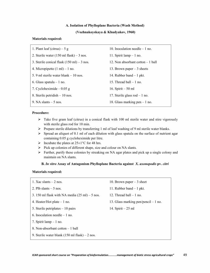
ICAR sponsored short course on “Preparation of bioformulation...........management of biotic stress agricultural crops” 49
A. Isolation of Phylloplane Bacteria (Wash Method)
(Vozhnakoyskaya & Khudyakov, 1960)
Materials required:
1. Plant leaf (citrus) – 5 g
2. Sterile water (150 ml flask) – 3 nos.
3. Sterile conical flask (150 ml) – 3 nos.
4. Micropipette (1 ml) – 1 no.
5. 9 ml sterile water blank – 10 nos.
6. Glass spatula – 1 no.
7. Cycloheximide – 0.05 g
8. Sterile petridish – 10 nos.
9. NA slants – 5 nos.
10. Inoculation needle – 1 no.
11. Spirit lamp – 1 no.
12. Non absorbant cotton – 1 ball
13. Brown paper – 3 sheets
14. Rubber band – 1 pkt.
15. Thread ball – 1 no.
16. Spirit – 50 ml
17. Sterile glass rod – 1 no.
18. Glass marking pen – 1 no.
Procedure:
Take five gram leaf (citrus) in a conical flask with 100 ml sterile water and stire vigorously with sterile glass rod for 10 min.
Prepare sterile dilutions by transferring 1 ml of leaf washing of 9 ml sterile water blanks. Spread an aliquot of 0.1 ml of each dilution with glass spatula on the surface of nutrient agar
containing 0.05 g cycloheximide per litre. Incubate the plates at 25±1°C for 48 hrs. Pick up colonies of different shape, size and colour on NA slants. Further, purify these colonies by streaking on NA agar plates and pick up a single colony and
maintain on NA slants.
B. In vitro Assay of Antagonism Phylloplane Bacteria against X. axonopodis pv. citri
Materials required:
1. Xac slants – 2 nos.
2. Plb slants – 5 nos.
3. 150 ml flask with NA media (25 ml) – 5 nos.
4. Heater/Hot plate – 1 no.
5. Sterile petriplates – 10 pairs
6. Inoculation needle – 1 no.
7. Spirit lamp – 1 no.
8. Non-absorbant cotton – 1 ball
9. Sterile water blank (150 ml flask) – 2 nos.
10. Brown paper – 3 sheet
11. Rubber band – 1 pkt.
12. Thread ball – 1 no.
13. Glass marking pen/pencil – 1 no.
14. Spirit – 25 ml
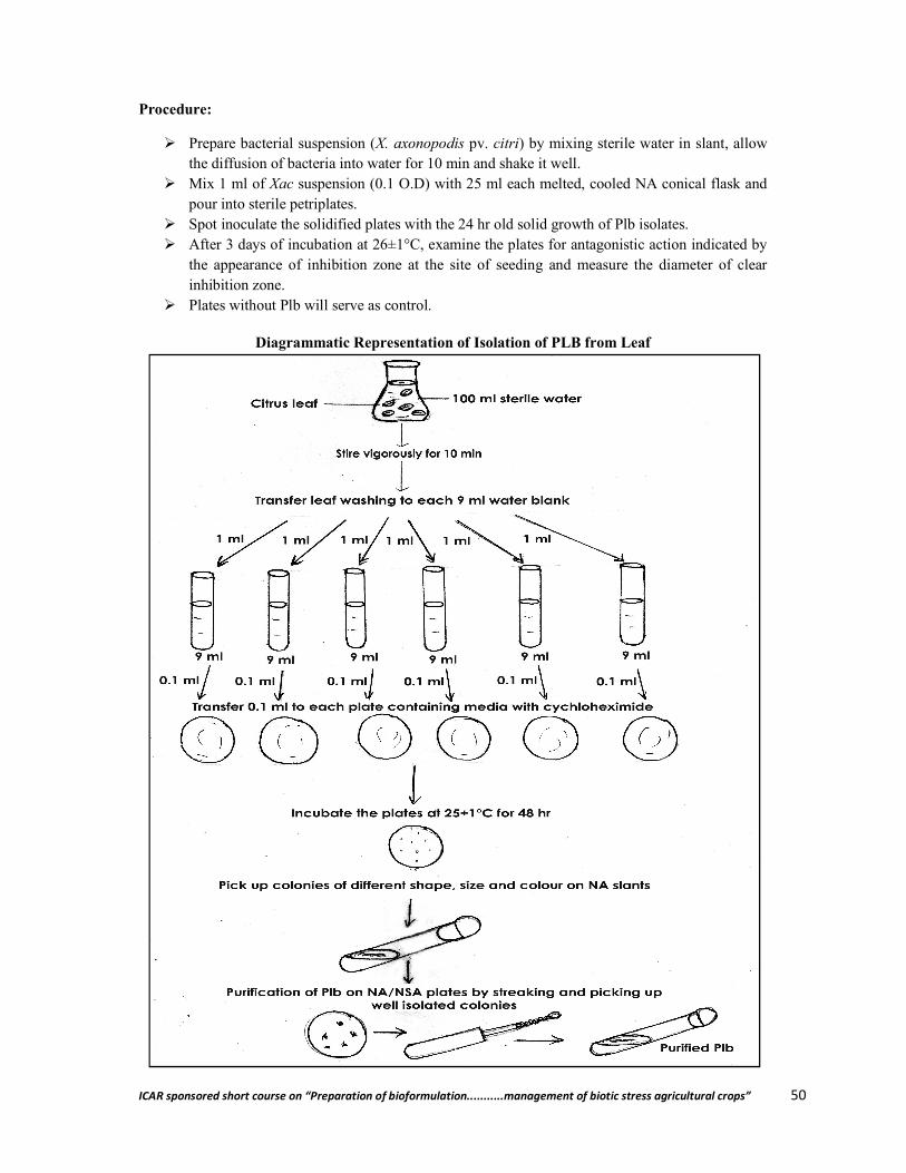
ICAR sponsored short course on “Preparation of bioformulation...........management of biotic stress agricultural crops” 50
Procedure:
Prepare bacterial suspension (X. axonopodis pv. citri) by mixing sterile water in slant, allow the diffusion of bacteria into water for 10 min and shake it well.
Mix 1 ml of Xac suspension (0.1 O.D) with 25 ml each melted, cooled NA conical flask and pour into sterile petriplates.
Spot inoculate the solidified plates with the 24 hr old solid growth of Plb isolates. After 3 days of incubation at 26±1°C, examine the plates for antagonistic action indicated by
the appearance of inhibition zone at the site of seeding and measure the diameter of clear inhibition zone.
Plates without Plb will serve as control.
Diagrammatic Representation of Isolation of PLB from Leaf
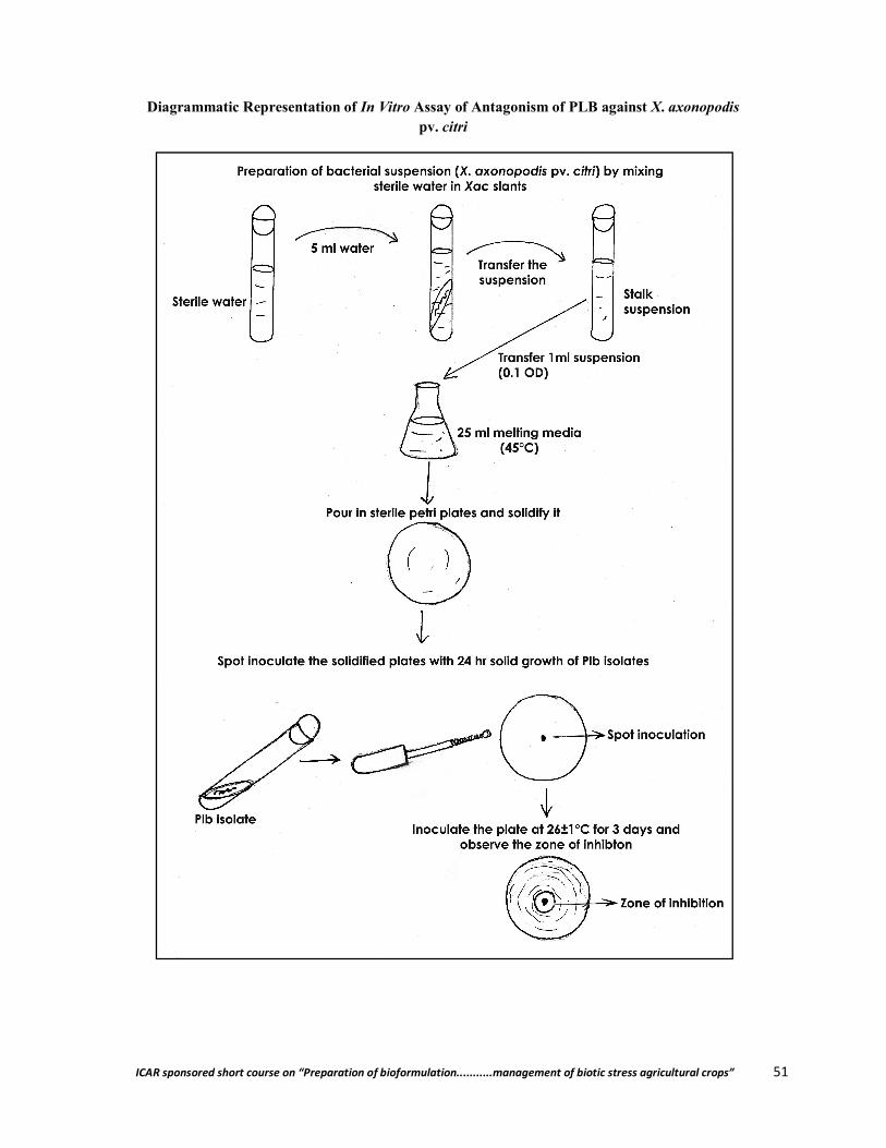
ICAR sponsored short course on “Preparation of bioformulation...........management of biotic stress agricultural crops” 51
Diagrammatic Representation of In Vitro Assay of Antagonism of PLB against X. axonopodis pv. citri
ICAR sponsored short course on “Preparation of bioformulation...........management of biotic stress agricultural crops” 52
References:
Bora, L. C.; Gangopadhya, S. and Chand, J.N. (1993). Biological control of bacterial leaf spot of mungbean (Xanthomonas campestris pv. vignaeradiatae Dye) with phylloplane antagonists. Indian J. Mycol. Pl. Pathol. 23(2): 162-168.
Borah, P. K.; Jindal, J. K. and Verma J. P. (2000). Biological management of bacterial leaf spot of mungbean caused by Xanthomonas axonopodis pv. vignaeraditae. Indian Phytopath. 53(4): 381-394.
Borah, P. K.; Jindal, J. K. and Verma, J. P. (2000). Management of Bacterial leaf spot of mung bean caused by Xanthomonas axonopodis pv. vignaeradiatae with phylloplane bacteria and chemicals. J. Mycol. Pl. Pathol. 30(1): 22-26.
Sudhir, C.; Capoor, A.; Srivastava, A. K. and Kehri H. K. (1988). Phylloplane Microbiology and control of foliar pathogen s through Resident Antagonists. Perspectives in Mycology and Plant Pathology (ed) : Malohotra Publishing House, 510-518.
Halfeld Vieira, B. A.; Romeiro, R. S.; Mounteer, A. and Mizubuti, E. S. G. (2008). Efficacy of phylloplane bacteria in controlling aerial tomato diseases under field conditions. Summa Phytopathologica 34(1): 86-87.
Harish, S.; Sarvanakumar, D.; Kamalakanan, A.; Vivekananthan, R.; Ebenezar, E. G. and Seetharaman, K. (2007). Phylloplane microorganisms as a potential biocontrol agent against Helminthosporium oryzae Breda de Hann, the incitant of rice brown spot. Archieves of Phytopathology and Plant Protection 40: 1 (Abstr.).
Nadia, Ghanney; Pletro, Locantore; Sabrine, Nahni; Ali, Ferchichi and Nicola Sante, Lacobellis (2016). Potential Biocontrol Effect of the Phylloplane Bacterium Bacillus mojavensis ABC-7 on the olive knot disease. J. Plant Pathol. Microbiol. 7: 337. doi:10.4172/2157-7471.1000#337.
Sanndhararanjan, K.; Marmuthu, S. and Manian, S. (2012). Biocontrol potential of phylloplane bacterium Ochrobacterum anthropi BMO-111 against blister blight disease of tea. J. Applied Microbiology 114: 209-218.
Voznyakoyskaya, Y. M. and Khudyakav, Y. P. (1960). Species composition of the epiphytic microflora of living plants. Microbiologiya 29: 97-103.
**************

ICAR sponsored short course on “Preparation of bioformulation...........management of biotic stress agricultural crops” 53
Preparation of Liquid Bioformulation of Fungal Biocontrol Agents and its Shelf Life Study
Pranab Dutta and Sarodee Boruah Department of Plant Pathology,
Assam Agricultural University, Jorhat- 785013, Assam
Management of plant diseases so far has been undertaken by means of chemical pesticides, irradication of diseased plants and removal of weed host. These conventional measures such as application of chemicals, breeding for resistant varieties and modification of cultural practices have limitations and chemicals have various detrimental effects. Now there is overwhelming evidence that some of the pesticides do pose a potential risk to humans and other life forms and unwanted side effects to the environment which consequently make it difficult to accomplish to the desired level. No segment of the population is completely protected against exposure to pesticides and the potentially serious health effects. The world-wide deaths and chronic diseases due to pesticide poisoning is about 1 million per year (Environews Forum, 1999).
Pesticide resistance is also an alarming problem. Recent studies indicate that there are over 500 species of insects and mites that are resistant to pesticides. That is why pesticides could not be considered the primary solution to curb the pest problem.
Keeping these points in view the urgent need of the hour is the alternative management approaches. Biological control is an important, effective, eco-friendly and economical component of Integrated Pest Management (IPM) in almost all important crops for development of sustainable cropping systems. There is also ample scope for microbial control of pests and diseases of vegetable and horticultural crops. Now a days, there is considerable interest in the exploitation of naturally occurring organisms, such as bacteria, viruses and fungi, for the control of crop pests, weeds and diseases. Amongst these, fungal biological control is an exciting and rapidly developing research area with implications for plant productivity, animal and human health and food production.
Bioformulation of fungal biocontrol agent
Many a time efforts were made by different scientists to develop suitable formulations of the fungal biocontrol agents for management of plant diseases. As because efficacy of the biocontrol agent largely depends on the formulation of that particular bioagent. Most of the biocontrol agents are formulated in solid form (contain only conidia) and in liquid form with shelf life of 6 months at 5°C and 3 months at 25-35°C. There are many examples where fungi have been formulated with various adjuvants, surfactants and oils for their better efficacy. The addition of nutrients to a spore spray of fungi could improve efectivity of biocontrol agent, compared with spores applied in water alone (Hall, 1982). Verticillium lecanii formulated with arachnid oil showed significantly better control of powdery mildew than without the addition of oil (Verhaar et al., 1999). In case of mass production of entomoapathogenic fungi like Metarhizium anisopliae, a number of naturally occurring carrier cum growth media have been evaluated (Fogal, 1986 and Quintela and McCoy, 1997). Development of potential bioformuulations has been improved by the discovery that fungal conidia formulated in oils that has shown greater infectivity than conventional water-based suspensions. Oils can substantially enhance the efficacy of entomopathogens against insects (Prior et al., 1988), hyperparasitic fungi (Hofstein and Chapple, 1998) and mycoherbicides (Amsellem et al., 1990). Burges (1988) gives a comprehensive review of the formulation of biopesticides and further details on oil formulation. Being non-evaporative, their use is readily compatible with ultra-low volume (ULV) application techniques for spraying mycoinsecticides at low relative humidities (Bateman, 1997).
ICAR sponsored short course on “Preparation of bioformulation...........management of biotic stress agricultural crops” 54
Basic concept of liquid bioformulation
There are four functions of liquid bioformulation:
To stabilize the organism during production, distribution and storage To aid easy handling and application of the product so that it is easily delivered to the target
in the most appropriate manner and form To protect the agent from harmful environmental factor at the target site, thereby increasing
persistence To enhance the activity of the organism at the target site by increasing its activity,
reproduction, contact and interaction with the target pest or disease organism
Steps involved in preparation of liquid bioformulation of fungal biocontrol agents
1. Preparation of culture media
Fungal biocontrol agents can be mass multiplied in Potato dextrose broth medium.
Preparation of Potato Dextrose Broth (PDB)
Materials required
Readymade PDB (HIMEDIA): 24.0 g, Distilled water: 1000ml, Beaker, Measuring cylinder, Conical flasks, Non- absorbent cotton, Brown paper, Rubber band etc
Procedure
Suspend 24.0 grams of PDB media in 1000 ml distilled water Heat to boiling to dissolve the medium completely Dispense about 250ml of medium into each of the four 500 ml conical flasks Plug the mouth of the flasks with non-absorbent cotton Sterilize the medium by autoclaving at 15 lbs pressure (121 ⁰C) for 15 minutes Mix well before dispensing.
2. Mass multiplication of biocontrol agents
Materials required
Pure culture of antagonistic organism, Suitable liquid medium, Inoculating needles, Alcohol, Spirit lamp, Non absorbent cotton, Brown paper, Thread etc.
Procedure
Inoculate the PDB with mycelial disc of 5 mm size cut out from 3-4 days old culture of the fungal antagonist
Concentration of the antagonist should be 1×10⁸ cfu/ml of water Incubate the inoculated flasks at 28±1⁰C for 7 days Count the spore concentration and colony forming unit (cfu) per ml of water after 7 and 14
days of inoculation
After that the liquid bioformulation need to be applied in field by following the appropriate method of application. There are different method of application of biopesticides are available. This are-
A. Soil application, B. Seed treatment and C. Foliar application
ICAR sponsored short course on “Preparation of bioformulation...........management of biotic stress agricultural crops” 55
A. Soil application: Soil application is done either before or at the time of planting to control of wide range of soil borne plant pathogens like Fusarium sp., Rhizoctonia sp. and Sclerotium sp. etc. for soil application, 5ml of bio formulation should be mixed with one litre of water and then applied in soil at the rate of 3 litres per square meter.
B. Seed treatment: Seed treatment is the most effective method of application of Biopesticides against soil as well as seed borne pathogens. For seed treatment, below mentioned procedure can be followed:
Take 5ml of bio formulation and mix it with one litre of water Add 0.02% Carboxy methyl cellulose (CMC) Mix it thoroughly Soak the seeds in bio formulation for one hour Take out seeds and dried in shade
C. Foliar application: foliar application of Biopesticides is done to restrict the air borne infection. Foliar applications can be done 30-35 days after sowing of seeds at the rate of 5ml/litre of water.
Precautionary measures to be taken at the time of application of Biopesticides
Spraying should be done in evening hours to avoid desiccation of fungal/ bacterial spores Do not use any chemical pesticide in the field where bio formulation is sprayed
Shelf life of bioformulations
Shelf life of the formulated product of a biocontrol agent plays a significant role in successful commercialization. In general, the antagonists multiplied in an organic food base have longer shelf life than the inert or inorganic food bases. The viable propagules of Trichoderma in talc formulation were reduced by 50 per cent after 120 days of storage. At PDBC, Bangalore work on increasing shelf life of talc formulations of Trichoderma using various ingredients (chitin and glycerol) in production medium and heat shock at the end of log phase of fermentation was carried out which extended the shelf of talc formulation of successfully up to one year. However, the problem how to maintain the CFU (106) and its efficacy in formulated products in viable form for one year during storage at the time of application and or throughout cropping season after application in the field still remained unsolved.
Procedure
Prepared bioformulations were need to kept at room temperature (15-35°C) and in refrigerator (4°C)
Viability of the antagonist in the bioformulation need to be determined at one month interval up 12 month
At each sampling time 1g or ml formulation should be suspended in 10 ml sterile distilled water and diluted up to desired dilutions (countable cfu/ g or ml in 90mm petri plate)
One ml suspension should be poured on to the surface of petri plates containing culture media. After 48 hrs incubation at 27±1°C colonies were noted.
References
Amsellem, Z.; Sharon, A.; Gressel, J. and Quimby, P. C. (1990). Complete abolition of high inoculums threshold of two mycoherbicides (Alternaria cassiae and A. crassa) when applied in invert emulsion. Phytopathol. 80: 925-929.
Bateman, R. P. (1997). Methods of application of microbial pesticide formulations for the control of grasshoppers and locusts. Memo. Entomol. Soc. Can. 171: 67-79.
ICAR sponsored short course on “Preparation of bioformulation...........management of biotic stress agricultural crops” 56
Burges, H. D. (1998). Formulation of mycoinsecticides. In: Formulation of Microbial Biopesticides, (Ed. H.D. Burges). Kluwer Academic Publishers Dordrecht, pp. 131-185.
Fogal, W. H.; Mittal, R. K. and Thurston, G.S. (1986). Production and quality evaluation of Beauverea bassiana for control of white spruces cone and seed insects. Information Report, Petawana National Forestry Institute, Ontario, Canada, p. 15.
Hall, R. A. (1982). Control of white fly, Trialeurodes vaporariorum and the cotton aphid, Aphis gossyapii, in glass house by Verticillium lecanii. Ann. App. Biol. 101: 1-11.
Hofstein, R. and Chapple, A. (1998). Commercial development of biofungicides. In: Biopesticides: Use and Delivery, (Eds. F.R. Hall & J.J. Menn). Humana Press, Totowa, NJ, USA. 6: 77-102.
Prior, C.; Jollands, P. and Le Patourel, G. (1988). Infectivity of oil and water formulations of Beauveria bassiana (Deuteromycotina; Hyphomycetes) to the cocoa weevil pest Pantorhytes plutus (Coleoptera: Curculionidae). J. Inverteb. Pathol. 52: 66-72.
Quintela, E. D. and McCoy, C. W. (1997). Pathogenicity enhancement of Metarhizium anisopliae and Beauverea bassiana instars of Diaprepes abbreviates (Coleoptera: Curculionidae) with sublethal doses of imidacloprid. Environ. Entomol. 26: 1173-1182.
Verhaar, M. A.; Hijwegen, T. and Zadoks, J. C. (1999). Improvement of the efficacy of Verticillium lecanii used in biocontrol of Sphaerothecea fuliginea by addition of oil formulations. Bioctrl. 44: 73-87.
**************
ICAR sponsored short course on “Preparation of bioformulation...........management of biotic stress agricultural crops” 57
Techniques for Development of Microbes based Bioformulation
L. C. Bora Department of Plant Pathology
Assaam Agricultural University, Jorhat- 785013, Assam
In the journey of ever challenging agricultural development, we have pursued the policies of intensive use of agro-chemicals. The level of consumption of agro-chemicals has been held as yardstick of agricultural development during the last 30-40 years (1960-2000). Although use of agro-chemicals along with other technologies like improved hybrids/varieties and irrigation has elevated our country to self-sufficiency in food production, however, due to use of these chemicals has also damaged our eco-system and delicate balance between various components of eco-system. Use of fertilizers and pesticides had their designated aims of increased productivity and reduced damage due to pests respectively. But, the productivity of many crops has not shown proportionate improvement in the recent 8-10 years- despite the increased use of fertilizers. Similarly, extensive use of pesticides has not reduced the losses due to pests.
Worldwide growing concern about the chemical intensive crop protection measures has promptly elevated the popularity of biological pest and disease management technologies. But, the menace of spurious biopesticides may visibly harm the confidence of farmers on biocontrol methods of insect pests and disease management. Therefore, equipping farming community with quality biopesticide and monitoring on the spurious biopesticides at any point is very much crucial for building and maintaining trust among farmers on biocontrol systems and to avoid the serious problems such as pest outbreak, low yields and environmental hazards in future. Unacceptable level of harmful microbial and heavy metal load in agricultural food products from use of deceptive and inappropriate substrate based bioformulations may carry potential health hazard to consumers. Use of such kind products non-conforming to NPOP/NSOP requirement/guidelines consequent into suspension of certification process and thus may face rejection in international market.
Biological control of disease employs natural enemies of pests or pathogens to eradicate or control their population. This involves either the introduction of exotic species, or harnessing whatever form of biological control exists naturally in the ecosystem. The induction of plant resistance using non-pathogenic or incompatible micro-organisms is also a form of biological control.
Definition and characteristics of Biological Control
Biological control is the reduction of inoculums density or disease producing activities of a pathogen or parasite in its active or dormant state, by one or more organisms, accomplished naturally or through manipulation of the environment, host or antagonist, or by mass introduction of one or more antagonists”.
Biological control of plant diseases can be broadly the use of one organism to influence the activities of a plant pathogen.
Biocontrol organisms (BCA) can be fungi, bacteria, or nematodes. BCA are natural inhabitants of the soil and the environment and are not pathogenic to
birds, mammals (including humans), and fish. BCA are not genetically modified. BCA work by competing with the pathogen for space and nutrients, by parasitism or
predation, by inducing the plant’s natural defense system, and/or by the production of antimicrobial substances (antibiotics like streptomycin).

ICAR sponsored short course on “Preparation of bioformulation...........management of biotic stress agricultural crops” 58
Mostly several mechanisms function together to make BCA more effective. These products are living organisms or dried spore preparations and must be handled
differently than conventional fungicides. BCA are sensitive to temperature extremes and must be applied immediately after
mixing with water. BCA may also require special attention to pH, exposure to chlorine or UV light,
otherwise their shelf life may be limited.
Protection by biocontrol agents
Biological control practices for direct protection of plants from pathogens involve the deployment of antagonistic microorganisms at the infection court before or after infection take place. The mechanisms employed by biocontrol organisms in weakening or destroying the plant pathogens are:
Their ability to parasitize the pathogens directly, Production of antibiotics (toxins) against the pathogens, Their ability to compete for space and nutrients and to survive in the presence of other
microorganisms, Production of enzymes that attack the cell components of the pathogens, Induction of defense responses in the plants they surround, Metabolism of plant produced stimulants of pathogen spore germination, etc.
Commercial use of Biological Control
Commercial application and grower acceptance of biological control has been slow to develop, mainly due to the variation in efficacy under the range of environmental conditions of the field. BCA are therefore generally formulated as wettable powders, dusts, granules and aqueous or oil-based liquid products, with various additives to attain all the desirable attributes.
Examples of some commercial bioformulations
Actinovate (Streptomyces lydicus) can reduce root and seed rot in peas; lower disease in spinach caused by Pythium and Fusarium (soil-borne fungi).
Compete Plus (Six species of Bacillus, Streptomyces griseoviridis, Trichoderma harzianum plus organic nutrients) can reduce Black Scurf potato (Rhizoctonia solani) and common scab of potato (Streptomyces scabies).
BioYield (plant growth promoting rhizobacteria) can reduce incidence of root rot (Pythium, Rhizoctonia) and wirestem (Rhizoctonia) in broccoli.
Compost Tea can reduce potato tubers from both Black Scurf and Common Scab. Bi-nucleate Rhizoctonia can reduce diseases caused by Rhizoctonia (Black Scurf, stem
canker of potatoes). Contans (Coniothyrium minitans) can reduce lettuce drop caused by Sclerotinia
species. Kodiak (Bacillus subtilis) can reduce Black Scurf and stem canker on potato. Muscador (Muscador albus) is a novel biocontrol organism that acts as a biofumigant
by producing gaseous compounds. It has shown good efficacy against storage insect pests of apples and potatoes. It can also reduce root, hypocotyl rot and Phytophthora fruit rot on pepper.
Plant Shield (Trichoderma harzianum) can reduce Rhizoctonia rot of bean and potato, Common Scab on potato, Botrytis on tomato, or Early Blight on tomato.
ICAR sponsored short course on “Preparation of bioformulation...........management of biotic stress agricultural crops” 59
Serenade (Bacillus subtilis) can lower the incidence of root rot caused by Rhizoctonia on both beans and radish.
SoilGard (Trichoderma virens) can reduce Black Scurf and Common Scab in potato.
Status of biocontrol research at Assam Agricultural University (AAU) for crop management
Assam Agricultural University (AAU), Jorhat has been working in the field of biological crop management for more than two decade to develop efficient, economically viable, reproducible, and environment friendly organic crop management technologies. The in-house technologies developed so far includes plant protection and soil fertility management systems. These have paved path for fail safe agriculture based livelihood of farming community segregated in different agro climatic regions of Assam and neighbouring states.
Microbial Bioformulation Protocol and Products Developed through Programme on Biopesticides:
More than ten (10) numbers of biocontrol agent based bioformulation production technologies have been standardized and mass multiplied in biopesticide units under Programme on Biopesticides. The biopesticides produced in these units include, Biofor Pf-2, Biozin-PTB, Bioveer and Biozium based on the bioagents Pseudomonas fluorescens and Trichoderma sp. effective against most of the diseases of vegetable, field and plantation crops; Metarhizium anisopliae, Beauveria bassiana, Verticillium lecanii based biopesticide respectively Biometa, Biosona, Biollium effective against rice hispa, gundhi bug, rice stem borer, leaf hopper and tea mosquito bugs, nematode, grubs; Plant Growth Promoting Microbe (PGPM) and entomopathogens viz Bacillus thuringiensis M. anisopliae, B. bassiana, V. lecanii based consortia bioformulation Biogreen 5, Biogreen L and Biotime effective against many pest and diseases of Agricultural, Horticultural and plantation crops; egg parasitoid Trichogramma sp based ‘Trichocards’ effective against lepidopteran pests. Besides, another two (2) Actinomycetes based bioformulation technologies are in pipeline. All products were duly validated before releasing to farmers by conducting multi location trials at Research Stations of AAU, Jorhat and OFTs in different districts of Assam along with Department of Agriculture, Govt of Assam.
Microbial bioagents isolated and characterized
A numbers of native microbial bioagents both bacterial and fungal have been isolated and characterized. At present the following bioagents are duly characterized and maintained at the culture collection of the Biopesticide lab- B. thuringiensis BTJ-S-1 (MTCC: 25035; GenBank: KF439054; MRC: MRC: 0277), Paecilomyces variotii Isolate-1 (GenBank: KF439053; MRC 0314), Pseudomonas aeruginosa PfD1 (MTCC: 7903), Pseudomonas fluorescens PFJ-S-1 (MTCC: 25036; MRC 0278), Trichoderma parareesei TPJ-S-1 (GenBank: KF439052; MRC 0313), Trichoderma viride TVJ-S-1 (GenBank: KF439055), Trichoderma harzianum 7477 (MTCC: 7477), Metarhizium anisopliae MAJ-S-1 (MTCC: 25040), Verticillium lecanii VLJ-S-1 and Beauveria bassiana BBJ-S-1. DNA barcodes of these bioagents have been developed and submitted into NCBI GenBank for use as future reference. Besides, specimen culture of potential bioagents has been deposited into Microbial Type Culture Collection (MTCC), Chandigarh.
Annual bio-input production capacity
The production facility of the Biopesticide laboratory, AAU annually produces about 20-25 MT biopesticidal formulations.
ICAR sponsored short course on “Preparation of bioformulation...........management of biotic stress agricultural crops” 60
Promotion of bioinputs for field application
Organized numbers training programmes to promote organic cultivation and use of bio inputs among stakeholders. About 25 trainings and 2 workshops has been conducted to enlighten the growers on the utility of developed biopesticides for production of organic tea. Till date more than 5,000 numbers of farmers were trained on organic agricultural practices using bio-inputs.
Protocol for Mass Multiplication of Bioagents and Application Techniques
1. Preparation of commercial formulation of Trichodrma harzianum
Inoculate a mycelial disc of T. harzianumin 70 ml molasses yeast medium in 250 ml conical flasks and incubate at room temperature 28±20C for 10 days.
Inoculate mycelial disc measuring 6mm of T. harzianum from 5 day old culture grown on PDA separately in each conical flask and incubate for 10 days at room temperature.
Then mycelial mat along with the medium is homogenized using a homogenizer and mix with the sterilized substrates viz., maize cob, black gram shell, coir pith, gypsum, peat soil and talc powder super white grade at 1:2 (V/W).
The substrates, viz. shelled maize cob, black gram shell should be separately crushed to make a powder.
Add carboxy methyl cellulose (CMC) @ 0.5%, dry in shade for 5 days and pack in transparent polypropylene bags and heat sealed.
Estimate population (cfu) of Trichoderma in the product just before packing and subsequently at 15 days interval for 45 days using Trichoderma selective medium.
2. Preparation of commercial formulation of Pseudomonas fluorescens
The bacterium is grown in King’s B broth and incubated in rotary shaker for 48 hrs at room temperature (28±20C).
The carrier material viz. maize cob powder, black gram shell powder, composed coir pith, gypsum, peat soil and talc powder should be taken in a poly propylene bag @ 200g/bag along with 2% CMC, seal and autoclave at 1.4 kg/cm2 for one hour on two successive days.
Inoculate 40 ml of 48 hrs grown bacterial culture and incubated at room temperature for 45 days.
Check the population of bacterium at 15 days interval adopting dilution plate method.
Protocol for application of bioformulation for plant disease management
1. Direct method
Apply to soil or compost FYM heap using conventional application equipment. Ensure that the soil is moist and that the temperature is at least 120C.
2. Seedling dip method
Seven days old seedlings are place individually in vials containing bioagent spore suspension in water the root are immersed in the solution for 5 minutes. Dipped root portion of the seedling are transplant thereafter.
ICAR sponsored short course on “Preparation of bioformulation...........management of biotic stress agricultural crops” 61
3. Seed coating method
Seeds can be soaked in spore suspension for 12 hours in a polyethylene bag. Soak the seed in 1% solution, dry under shade and use for sowing.
The coated seeds ruptures and the radical may come out. Such seeds can be kept in the bag for a further period of 12 hours when the seed coat ruptures and the radical should come out. Such seeds can be sown in the field.
4. Spray application
Disperse 1kg of Trichoderma formulation in 100 lit of water and spray on the crop as a preventive or curative treatment against diseases. One spray during vegetative stage and another before flower initiation.
Protocol for estimation of population of fungal biocontrol agent from formulated product
The density of cells, spores/conidia of microorganisms can be measured in the laboratory by several methods either by direct or indirect counts. In the direct microscopic count, a known volume of liquid is added to the slide and the number of microorganisms are counted by examining the slide with the bright field microscope. For direct microscope counts Neubauer or Petroff-Hausser counting chamber, breed smear or electronic cell counter are used. Population density may be determined by observing some property that provide indirect evidence of microbial numbers in a sample. Various methods for indirect counts are, determining cell mass or cellular constitutions, oxygen uptake, carbon dioxide production, spectrophotometric or colorimetric, member filter counter count and serial dilution agar plate method.
Protocol for evaluation of commercial formulations for microbial count
In recent years, interest among growing regulatory authorities, agricultural advisors and growers in alternative methods of plant disease control has encouraged intensive research of microbial products as agents for plant disease control. This has resulted in the commercialization of microbial product. These products need to be scientifically tested before use so that the real use of bioproducts can be made.
Dual Culture-bioassay method
Mix 15 mg of the product with 15ml of molten and cooled agar medium and pour in sterilized plate. After solidification, transfer 8mm mycelial disc of any of the test fungus e.g., Macrophomina, Rhizoctonia, Sclerotinia etc. in the center of the plate. Complete inhibition of the test fungus after 3-5 days of incubation indicates the bioefficacy.
Qualitative analysis
Take 1 gm of product and make it upto 10 ml with sterile water and shake well (1:10). Take 1 ml from 1:10 dilution and transfer 9 ml sterile water in next tube (1:100). Make serial dilutions by transferring 1 ml of the suspension to get 1:106 dilutions.
Preparation of inoculum based on isolates from commercial products
The fungus isolated from commercial formulation can be cultured for two to three weeks in flasks on a 1:1:1 mixture of wheat bran, vermiculite and Czapek-dox Broth (200ml of each). The colonized substrate can then be removed from the flasks, air dried and triturated. In the efficacy tests these preparations ca be mixed into the potting substrates at the rate of 3000 mg/1 substrate.
ICAR sponsored short course on “Preparation of bioformulation...........management of biotic stress agricultural crops” 62
DO’S and DON’T’S
cfu count of Powder formulation should never be taken from an already opened sample bottles.
Once opened packet should not be reused.
The dilution tubes should be vortexed for a minute before making next dilutions.
Vortex the dilutions for 15 sec before adding it to the plate.
Shake the sample bottles properly before opening it for cfu results.
**************
ICAR sponsored short course on “Preparation of bioformulation...........management of biotic stress agricultural crops” 63
Biointensive Disease Management of Vegetable Crops
Bubul Chandra Das Department of Plant Pathology,
Regional Agricultural Research Station, Titabar Assam Agricultural University, Assam
Vegetables are a rich source of dietary fibers, vitamins, minerals, antioxidants, phytochemicals and are important for food and nutritional security of the country. India is the second largest producer of vegetables in the world (ranks next to China) and accounts for about 15% of the world’s production of vegetables. India produces about 162.89 million metric tonnes of vegetables from an area of 9.39 million hectares with an average productivity of 17.34 Mt/Ha (Statistical Year Book India 2016). Majority of Indians are vegetarian, with a per capita consumption 135 g per day as against the recommended 300 g per day. It is still very less than recommended diet level. In near future, there is a need of around 5-6 million tons of food to feed our 1.3 billion Indian population expected by the year 2020 (Paroda, 1999).
The major limiting factor, include the extensive crop devastations due to increased pest menace. In many cases, there is 100 per cent yield loss due to viral diseases vectored by insects. The crop losses in the country due to various pests range from 10 to 30 percent. Damping off, late blight, early blight, leaf spot (Anthracnose, Septorial), powdery mildew and fruit rot are the major diseases affecting vegetable crops while thrips, white fly, aphids, jassids, mites and caterpillar are the major pests. Besides these, bacterial diseases also affect the vegetable crop. Vegetables are more prone to insect pests and diseases mainly due to their tenderness and softness as compared to other crops and virtual absence of resistance characters because of intensive hybrid cultivation. Indiscriminate use of pesticides has led to severe ecological consequences like destruction of natural enemy fauna, effect on non target organisms, residues in consumable products including packed pure and mineral water and ultimately resistance to the pesticides, to which we solely rely. Biointensive pest management (BIPM) is the recent trend in Indian farming and attracting the farmers for higher income to their produce.
Biointensive IPM
Bio-intensive IPM is defined as “A systems approach to pest management based on an understanding of pest ecology. It begins with steps to accurately diagnose the nature and source of pest problems, and then relies on a range of preventive tactics and biological controls to keep pest populations within acceptable limits. Reduced-risk pesticides are used if other tactics have not been adequately effective, as a last resort, and with care to minimize risks” (Benbrook, 1996).
Biointensive IPM incorporates ecological and economic factors into agricultural system design and decision-making and addresses public concerns about environmental quality and food safety. The benefits of implementing biointensive IPM can include reduced chemical input costs, reduced on-farm and off-farm environmental impacts and more effective and sustainable pest management. An ecology-based IPM has the potential of decreasing inputs of fuel, machinery and synthetic chemicals – all of which are energy intensive and increasingly costly in terms of financial and environmental impact. Such reductions will benefit the grower and society.
Why move to biointensive IPM?
Integrated pest management originated as a reaction to the overuse of insecticides and is now the dominant paradigm that guides development of insect pest management technologies all over the
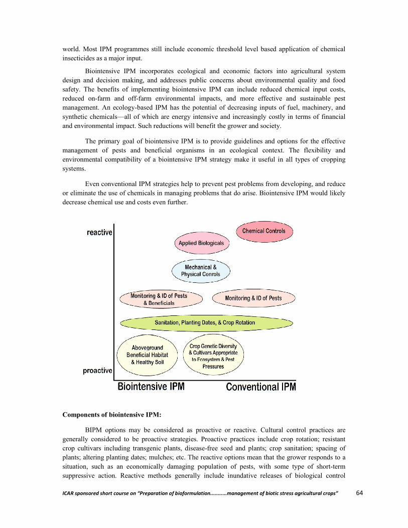
ICAR sponsored short course on “Preparation of bioformulation...........management of biotic stress agricultural crops” 64
world. Most IPM programmes still include economic threshold level based application of chemical insecticides as a major input.
Biointensive IPM incorporates ecological and economic factors into agricultural system design and decision making, and addresses public concerns about environmental quality and food safety. The benefits of implementing biointensive IPM can include reduced chemical input costs, reduced on-farm and off-farm environmental impacts, and more effective and sustainable pest management. An ecology-based IPM has the potential of decreasing inputs of fuel, machinery, and synthetic chemicals—all of which are energy intensive and increasingly costly in terms of financial and environmental impact. Such reductions will benefit the grower and society.
The primary goal of biointensive IPM is to provide guidelines and options for the effective management of pests and beneficial organisms in an ecological context. The flexibility and environmental compatibility of a biointensive IPM strategy make it useful in all types of cropping systems.
Even conventional IPM strategies help to prevent pest problems from developing, and reduce or eliminate the use of chemicals in managing problems that do arise. Biointensive IPM would likely decrease chemical use and costs even further.
Components of biointensive IPM:
BIPM options may be considered as proactive or reactive. Cultural control practices are generally considered to be proactive strategies. Proactive practices include crop rotation; resistant crop cultivars including transgenic plants, disease-free seed and plants; crop sanitation; spacing of plants; altering planting dates; mulches; etc. The reactive options mean that the grower responds to a situation, such as an economically damaging population of pests, with some type of short-term suppressive action. Reactive methods generally include inundative releases of biological control
ICAR sponsored short course on “Preparation of bioformulation...........management of biotic stress agricultural crops” 65
agents, mechanical and physical controls, botanical pesticides and chemical controls. The most recent bio-intensive integrated approaches for pest management utilizes components such as cultural methods viz., crop rotation, summer ploughing, fallowing, intercropping, pruning, mulching, spacing, planting date, trap cropping, etc and use of resistant cultivars; bio-agents viz., predators, parasitoids and bio-control agents, mycorrhizal fungi, botanicals including bio-fumigation, oil cakes, FYM, crop residues, green manuring and other organic amendments, physical methods viz., hot water treatment of planting material, soil solarization and bio-rational chemicals like pheromones.
Proactive strategies (Cultural Controls)
Healthy, biologically active soils (increasing belowground diversity)
Habitat for beneficial organisms (increasing aboveground diversity)
Appropriate plant cultivars
Cultural controls are manipulations of the agroecosystem that make the cropping system less friendly to the establishment and proliferation of pest populations. Although they are designed to have positive effects on farm ecology and pest management, negative impacts may also result, due to variations in weather or changes in crop management.
Crop rotation and disease suppression
Avoiding disease buildup is probably the most widely emphasized benefit of crop rotation in vegetable production. Crop rotations radically alter the environment both above and below ground, usually to the disadvantage of pests of the previous crop. Many diseases build up in the soil when the same crop is grown in the same field year after year. Rotation to a non-susceptible crop can help break this cycle by reducing pathogen levels. Since diseases usually attack plants related to each other, it is helpful to group vegetable rotations by family—e.g., nightshades, alliums, cole crops, cucurbits. The susceptible crop, related plants, and alternate host plants for the disease must be kept out of the field during the rotation period. Since plant pathogens persist in the soil for different lengths of time, the length of the rotation will vary with the disease being managed. To effectively plan a crop rotation, it is essential to know what crops are affected by what disease organisms. In most cases, crop rotation effectively controls those pathogens that survive in soil or on crop residue. Crop rotation will not help control diseases that are wind-blown or insect vectored from outside the area. Nor will it help control pathogens that can survive long periods in the soil without a host—Fusarium, for example.
Table 1. Rotation periods to reduce vegetable soil-borne diseases (Sullivan, 2004)
Vegetable Disease Years without susceptible crop
Asparagus Fusarium rot 8
Beans Root rots 3–4
Cabbage Clubroot 7
Cabbage Blackleg 3–4
Cabbage Black rot 2–3
Muskmelon Fusarium wilt 5
ICAR sponsored short course on “Preparation of bioformulation...........management of biotic stress agricultural crops” 66
Parsnip Root canker 2
Peas Root rots 3–4
Peas Fusarium wilt 5
Pumpkin Black rot 2
Radish Clubroot 7
Other cropping structure options:
Intercropping is the practice of growing two or more crops in the same, alternate, or paired rows in the same area. This technique is particularly appropriate in vegetable production. The advantage of intercropping is that the increased diversity helps "disguise" crops from insect pests, and if done well, may allow for more efficient utilization of limited soil and water resources.
Strip cropping is the practice of growing two or more crops in different strips across a field wide enough for independent cultivation (e.g., alternating six-row blocks of soybeans and corn or alternating strips of alfalfa and cotton or alfalfa and corn). It is commonly practiced to help reduce soil erosion in hilly areas. Like intercropping, strip cropping increases the diversity of a cropping area, which in turn may help "disguise" the crops from pests. Another advantage to this system is that one of the crops may act as a reservoir and/or food source for beneficial organisms.
Plant nutrients and disease suppression
Soil pH, calcium level, nitrogen form, and the availability of nutrients can all play major roles in disease management. Adequate crop nutrition makes plants more tolerant of or resistant to disease. Also, the nutrient status of the soil and the use of particular fertilizers and amendments can have significant impacts on the pathogen’s environment.
One of the most widely recognized associations between fertility management and a crop disease is the effect of soil pH on potato scab. Potato scab is more severe in soils with pH levels above 5.2. Below 5.2 the disease is generally suppressed.
Adequate levels of calcium can reduce clubroot in crucifer crops (broccoli, cabbage, turnips, etc.). The disease is inhibited in neutral to slightly alkaline soils (pH 6.7 to 7.2) (Campbell and Arthur 1990). A direct correlation between adequate calcium levels, and/or higher pH, and decreasing levels of Fusarium occurrence has been established for a number of crops, including tomatoes, cotton, melons, and several ornamentals (Jones et al., 1989). Calcium has also been used to control soil-borne diseases caused by Pythium, such as damping off.
Compost and disease suppression
Compost has been used effectively in the nursery industry, in high-value crops, and in potting soil mixtures for control of root rot diseases. Compost acts as a food source and shelter for the antagonists that compete with plant pathogens, for those organisms that prey on and parasitize pathogens, and for those beneficials that produce antibiotics. Root rots caused by Pythium and Phytophthora are generally suppressed by the high numbers and diversity of beneficial microbes found in the compost. Such beneficials prevent the germination of spores and infection of plants growing on the amended soil.
ICAR sponsored short course on “Preparation of bioformulation...........management of biotic stress agricultural crops” 67
Table 2. Compost Treatment and Disease Management of Vegetables (Sullivan, 2004)
Vegetable Pathogen/Disease Treatment Comments
Beans
Rhizoctonia sp. Compost added to soil at varying rates (36-72 tons/acre).
Disease reduced 80% in areas with highest compost rates, 40% where intermediate rates applied. Control plots yielded 75 bushels/acre, compost plots yielded 200 bu/acre
Cucumber Sphaerotheca sp./
Powdery mildew
Young cucumber plants grown in soil/compost mix of variable rates.
1:1 soil:compost mix decreased PM by 20% over control; 1:3 mix decreased infection by 40%
Pea
Pythium sp./
Damping off
Seed treatment; seeds soaked in dilute compost extract, dried before sowing.
Peas seed-treated with compost extract germinated significantly better than untreated seed in soil artificially inoculated with Pythium ultimum
Other cultural management options
Disease-free seed and plants are available from most commercial sources, and are certified as such. Use of disease-free seed and nursery stock is important in preventing the introduction of disease in vegetable crops.
Resistant varieties are continually being bred by researchers. Growers can also do their own plant breeding simply by collecting non-hybrid seed from healthy plants in the field. The plants from these seeds will have a good chance of being better suited to the local environment and of being more resistant to insects and diseases. Since natural systems are dynamic rather than static, breeding for resistance must be an ongoing process, especially in the case of plant disease, as the pathogens themselves continue to evolve and become resistant to control measures.
Spacing of plants heavily influences the development of plant diseases and weed problems. The distance between plants and rows, the shape of beds, and the height of plants influence air flow across the crop, which in turn determines how long the leaves remain damp from rain and morning dew. Generally speaking, better air flow will decrease the incidence of plant disease. However, increased air flow through wider spacing will also allow more sunlight to the ground, which may increase weed problems.
Mulches, living or non-living, are useful for suppression of weeds, insect pests, and some plant diseases. Mulching helps to minimize the spread of soil-borne plant pathogens by preventing their transmission through soil splash. Mulch, if heavy enough, prevents the germination of many annual weed seeds.
Biological control
Biological control is the use of living organisms—parasites, predators, or pathogens—to maintain pest populations below economically damaging levels, and may be either natural or applied.
There are a number of commercial products containing beneficial, disease-suppressive or-ganisms. These products are applied in various ways—including seed treatments, compost inoculants,
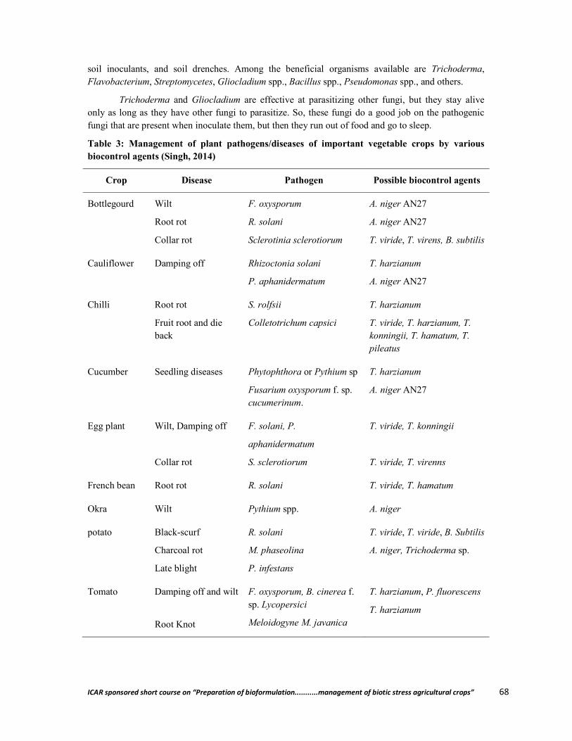
ICAR sponsored short course on “Preparation of bioformulation...........management of biotic stress agricultural crops” 68
soil inoculants, and soil drenches. Among the beneficial organisms available are Trichoderma, Flavobacterium, Streptomycetes, Gliocladium spp., Bacillus spp., Pseudomonas spp., and others.
Trichoderma and Gliocladium are effective at parasitizing other fungi, but they stay alive only as long as they have other fungi to parasitize. So, these fungi do a good job on the pathogenic fungi that are present when inoculate them, but then they run out of food and go to sleep.
Table 3: Management of plant pathogens/diseases of important vegetable crops by various biocontrol agents (Singh, 2014)
Crop Disease Pathogen Possible biocontrol agents
Bottlegourd Wilt
Root rot
Collar rot
F. oxysporum
R. solani
Sclerotinia sclerotiorum
A. niger AN27
A. niger AN27
T. viride, T. virens, B. subtilis
Cauliflower Damping off
Rhizoctonia solani
P. aphanidermatum
T. harzianum
A. niger AN27
Chilli Root rot
Fruit root and die back
S. rolfsii
Colletotrichum capsici
T. harzianum
T. viride, T. harzianum, T. konningii, T. hamatum, T. pileatus
Cucumber Seedling diseases Phytophthora or Pythium sp
Fusarium oxysporum f. sp. cucumerinum.
T. harzianum
A. niger AN27
Egg plant Wilt, Damping off
Collar rot
F. solani, P.
aphanidermatum
S. sclerotiorum
T. viride, T. konningii
T. viride, T. virenns
French bean Root rot R. solani T. viride, T. hamatum
Okra Wilt Pythium spp. A. niger
potato Black-scurf
Charcoal rot
Late blight
R. solani
M. phaseolina
P. infestans
T. viride, T. viride, B. Subtilis
A. niger, Trichoderma sp.
Tomato Damping off and wilt
Root Knot
F. oxysporum, B. cinerea f. sp. Lycopersici
Meloidogyne M. javanica
T. harzianum, P. fluorescens
T. harzianum
ICAR sponsored short course on “Preparation of bioformulation...........management of biotic stress agricultural crops” 69
Mechanical and physical controls
Methods included in this category utilize some physical component of the environment, such as temperature, humidity, or light, to the detriment of the pest. Common examples are tillage, flaming, flooding, soil solarization, and plastic mulches to kill weeds or to prevent weed seed germination. Heat or steam sterilization of soil is commonly used in greenhouse operations for control of soil-borne pests. Cold storage reduces post-harvest disease problems on produce.
Pest identification
A crucial step in any IPM program is to identify the pest. The effectiveness of both proactive and reactive pest management measures depend on correct identification. Misidentification of the pest may be worse than useless; it may actually be harmful and cost time and money. Help with positive identification of pests may be obtained from university personnel, private consultants, etc.
Monitoring
Monitoring involves systematically checking crop fields for pests and beneficials, at regular intervals and at critical times, to gather information about the crop, pests, and natural enemies. Sweep nets, sticky traps, and pheromone traps can be used to collect insects for both identification and population density information. Leaf counts are one method for recording plant growth stages. Square-foot or larger grids laid out in a field can provide a basis for comparative weed counts. Records of rainfall and temperature are sometimes used to predict the likelihood of disease infections.
Economic injury and action levels
The economic injury level (EIL) is the pest population that inflicts crop damage greater than the cost of control measures. Because growers will generally want to act before a population reaches EIL, IPM programs use the concept of an economic threshold level (ETL or ET), also known as an action threshold. The ETL is closely related to the EIL, and is the point at which suppression tactics should be applied in order to prevent pest populations from increasing to injurious levels.
Conclusion
Biointensive IPM is considered the desirable path to sustainability in agriculture. There are several ecofriendly ways available to reduce the pesticide usage in vegetable cultivation and produce optimization. There is a need to realize the potential of indigenous biocontrol agents and attention should be given to conserve them. There is also a need to strengthen research in strategic areas like genetically improved bioagents/biopesticides and pest-resistant transgenics.
This bio-intensive approach needs building the knowledge and information infrastructure by making changes in research and education priorities in order to emphasize ecology-based pest management and redesign management programs to promote bio-intensive IPM.
References
Benbrook, C. M. (1996). Pest Management at the Crossroads. Yonkers, N.Y: Consumers Union.
Campbell, R. N., and Arthur Greathead, S. (1990). Control of clubroot of crucifers by liming. p. 90–101. In: A.W. Engelhard (ed.). Management of Diseases with Macro- and Microele-ments. APS Press. American Phytopathological Society, St. Paul, MN.
Dufour, R. ( 2001). Biointensive Integrated Pest Management(IPM) Jones, J. P.; Engelhard, A. W. and Woltz., S. S. (1989). Management of fusarium wilt of
vegetables and ornamentals by macro- and microelement nutrition. p. 18–32. In: A.W. Engel-

ICAR sponsored short course on “Preparation of bioformulation...........management of biotic stress agricultural crops” 70
hard (ed.). Soilborne Plant Pathogens: Management of Diseases with Macro- and Microelements. American Phytopathological Society. 217 p.
Paroda, R. S. (1999). For a food secure future. The Hindu Survey of Indian Agriculture. Singh, H. B. ( 2014). Management of Plant Pathogens with Microorganisms. In : Proc Indian
Natn Sci Acad 80 No. 2 June 2014 Spl. Sec. pp. 443-454 Sullivan, P. (2004). Sustainable management of soil-borne plant diseases
**************
ICAR sponsored short course on “Preparation of bioformulation...........management of biotic stress agricultural crops” 71
Isolation of Biofertilizer Microbes from Crops Rhizosphere
Anjuma Gayan and Dhruba Jyoti Nath Department of Soil Sience,
Assam Agricultural University, Jorhat- 785013, Assam
Plant obtains almost everything directly from the soil to support growth, with the notable exceptions of carbon dioxide, oxygen, and light. Of all the 17 essential elements for plant growth and reproduction, fourteen of these elements are acquired primarily from the soil solution. These include six macronutrients (N, K, P, S, Mg and Ca) and eight micronutrients (B, Cl, Cu, Fe, Mn, Mo, Ni and Zn). The interaction processes between plant and microorganism within the rhizosphere region or microorganisms alone help them to acquire the essential plant nutrient, suppressing soil borne diseases and also help in detoxifying certain toxic products in soil.
Beneficial organisms are generally classified into two broad groups based on their primary effects, i.e., their most well-known beneficial effect on the plant: (i) microorganisms with direct effects on plant growth promotion [plant growth promoting microorganisms (PGPM)] and (ii) biological control agents (BCA) that indirectly assist with plant productivity through the control of plant pathogens. More specifically, PGPM have shown activities relating to biocontrol of soil borne pathogens. Conversely, BCA have demonstrated properties that directly promote plant growth. Considering the presence of diverse microbial communities, soil is regarded as living environment and often called as ‘Black Box’. The PGPM and BCA operates by a wide variety of mechanisms, including N2 fixation, enhanced solubilization and mobilization of P, potash solubilization, zinc solubilization, biological control of soil borne diseases and phytohormone production correspondingly.
Beneficial effects on plant growth by the soil microorganisms have a high relevance for agricultural ecosystems because they reduce the need for fertilizers, leading to a decrease in pollution of agricultural soils and water. In nature, there are a number of useful soil micro organisms which can help plants to absorb nutrients. Their utility can be enhanced with human intervention by selecting efficient organisms, culturing them and adding them to soils directly or through seeds commonly called as biofertilizer.
Isolation of Symbiotic and non-symbiotic nitrogen fixing bacteria
1. Isolation of Rhizobium
Rhizobium constitutes the root nodule bacteria. This is symbiotic bacterium which fixes atmospheric dinitrogen in legumes plant by forming nodules. For isolation of Rhizobia well nodulated fresh leguminous plants are uprooted from the fields and brought to the laboratory. The root system is washed gently in running tap water to remove the adhering soil particles prior to separation of nodules from roots.
Materials Required
Well nodulated leguminous plants Hydrogen peroxide (5%) Ethyl alcohol (80%) Sterile water blanks in test tubes Sterile petri plates
ICAR sponsored short course on “Preparation of bioformulation...........management of biotic stress agricultural crops” 72
Scissors, forceps and a blunt edged glass rod Yeast Extract Mannitol Congo red agar medium(YEMA-CR) BOD Incubator & Compound Microscope
Processing the Nodule
Effective pink colored nodules are selected and incised from the root system. Then the nodules are surface sterilized with hydrogen peroxide (5%) for 2-3 minutes
followed by ethyl alcohol for 5 minutes. Thereafter rinse the nodules thoroughly in at least 6 changes of sterile water to get rid of
disinfectants. Then the nodules are transferred to test tubes containing sterile distilled water. Using the blunt end of a surface sterilized glass rod, the nodules are crushed in the tubes
and made in the form of a suspension and serially diluted upto 10-6 -10-7.
Isolation procedure
YEMA-CR media contained in conical flask is sterilized in autoclave and poured in sterile Petri plates inside Laminar Airflow Cabin and the media are allowed to solidify.
Using fresh sterile pipette/micropipette pour 0.1-0.2ml of aliquot of selected dilution over the solidified media in triplicate.
With the help of a glass rod or spreader, spread the aliquot over the entire surface of media.
Incubate the Petri plates in inverted position in BOD incubator at 30±10C for 2-5 days.
Observation
Rhizobium forms white, transparent, translucent, glistening, elevated and comparatively small colonies on the medium. Moreover, Rhizobium colonies do not absorb Congo red dye added in the medium. Those colonies, which readily take up the Congo red stain, are not rhizobia but presumably Agrobacterium, a soil bacterium closely related to Rhizobium. After careful selection, a loopful of Rhizobium colony is purified by streak plate method on fresh YEMA-CR agar plates for getting single colony.
Characterization of Rhizobium
Main objective of this study is to distinguish rhizobia from other microorganisms by cell morphology, staining reactions and growth response to various media.
Gram Reaction Growth in YEMA containing bromothymol blue Growth in Peptone glucose agar Growth in Hofer’s alkaline broth
2. Isolation of Cyanobacteria
Cyanobacteria also known as blue green algae are typical photosynthetic prokaryotes falling under the domain bacteria and placed in the phylum Cyanobacteria. Unlike other bacteria which is mostly unicellular, Cyanobacteria exhibit wide heterogeneity in shapes ranging from small unicellular spherical cells to multicellular colonial and filamentous forms. Cyanobacteria are the only phototrophic organisms that perform N2 fixation with the help of specialized heterocyst cell.
ICAR sponsored short course on “Preparation of bioformulation...........management of biotic stress agricultural crops” 73
Materials Required:
Rhizosphere soil (preferably paddy soil) Sterile water blanks in test tube. Series of Conical flask (100ml size) Selective BGII liquid & solid media Sterile pipette or Microtip, inoculating Needle lluminated light & Compound Microscope
Isolation procedure
Prepare the BGII liquid media dispense 50ml each in a series of 100ml conical flask and sterilize.
Serially dilute 1g soil sample (rice rhizosphere soil) upto 10-5 – 10-6. Transfer aseptically 0.1ml from each of 10-4, 10-5 & 10-6 dilution into the liquid BGII media
contained in conical flask in quadruplicate. The conical flasks are kept under illuminated light for 15-20 days or till bluish tinge of algal
growth appear. Observation & enumeration
The algal growth is picked up with the help of inoculating needle and observed under microscope for presence or absence of heterocyst and subjected to purification in solid BGII media.
The positive growth of Cyanobacteria is enumerated using Most Probable number method.
Purification of Cyanobacteria
For purification of Cyanobacteria BGII solid media is prepared, sterilized, dispense onto sterilized petriplates and allowed to solidify. The positive bluish tinge of Cyanobacteria grown in conical flask is streaked onto solid BGII media and allowed to grow under illuminated light till isolated colonies are formed. This is repeated till Cyanobacteria get purified. The isolated colonies are again transferred into fresh BGII liquid media for preservation and mass scale production.
3. Isolation of Azotobacter
The importance of Azotobacter as potential free living N- fixing microorganisms has been convincingly established since its discovery by Beijerinck in 1901. The bacteria of genus Azotobacter are pleomorphic, ranging from rods to cocci and are aerobic in nature. Azotobacter prefers nitrogen – free medium with sugar as the carbon source. Optimum temperature for its growth ranges from 32oC to 37oC and it grows well at pH 7.0-7.5.
Materials Required
Rhizosphere soil Sterile water blanks in test tube. Sterile petri plates N-free Jensen’s or Burk’s solid media Sterile pipette or Microtip, Spreader BOD incubator & Compound Microscope

ICAR sponsored short course on “Preparation of bioformulation...........management of biotic stress agricultural crops” 74
Isolation procedure
1gm rhizosphere soil is suspended in 9ml sterile water and mix vigorously. Serial dilution of the sample and dilutions are restricted to 10-4 or 10-5. Sterilized media is poured in Petri plates and allowed for solidification. Using sterile pipettes or microtip 0.1ml of each dilution is transferred aseptically to the
petriplates containing solid media and spread uniformly with spreader. Petriplates are incubated in BOD incubator at 30±10C for 3-5days.
Observation
Azotobacter cells grow as raised, slimy colonies on agar surface and morphologically highly pleomorphic ranged from blunt rods to oval shaped. Production of extracellular slime, capsular material and yellowish brown/black coloration in aged culture are important characters.
4. Isolation of Azospirillum
It is an associative, microaerophillic gram negative spiral shaped N-fixing bacterium occurs in or on the roots and plants or free living in soil. In 1925, Beijerinck described the bacterium under the name Spirillum lipoferum. The nomenclature of the organism was revised and designated as Azospirillum (Tarrand et al., 1979)
Materials Required Roots & Rhizosphere soil Sterile water blanks in test tube Sterile petri plates Selective N-free bromothymol blue semisolid media (NFb) Sterile pipette or Microtip, Spreader, blunt rod BOD incubator & Compound Microscope
Isolation procedure
Roots are gently washed in tap water, dried and cut aseptically into smaller bits (1 cm). The small root bits are macerated with glass rod in test tube containing sterile water &
serially diluted upto 10-5-10-6 The semisolid NFb media is dispensed @ 10ml in (15x1.5cm) test tube and sterilized. Using sterile pipettes or microtip 0.1ml of each dilution is transferred aseptically to the
semisolid NFb media in test tube. Similarly few macerated root pieces from the first dilution is also transferred onto the semisolid NFb media.
The inoculated tubes are incubated at 37±1ºC for 3-5 days.
Observation
The formation of thin pellicle at 1-2 mm below the surface of NFb media as well as color change from green to blue indicates growth of Azospirillum.
Purification of Azospirillum:
Few opportunistic bacteria may also grow immediately after formation of pellicle that makes the thin pellicle thick. To purify the Azospirillum from the contaminants, solid Rojo-congo (RC) media (Rodriquez-Caceres 1982.) is used.

ICAR sponsored short course on “Preparation of bioformulation...........management of biotic stress agricultural crops” 75
Sterilized RC media is poured in sterilized petriplates and allowed to solidify. The pellicle is streaked onto solid RC media and is incubated at 35±1ºC until isolated
colonies developed. In this medium, Azospirillum is readily distinguished from other associative organisms as
Azospirillum absorbed Congo red dye and formed pink to scarlet colonies. These pink or scarlet colonies are further transferred to tube containing NFb semi solid media
to ascertain reformation of pellicle. The process is continued by repeated transfer of colonies from RC plate to NFb media until a
fine and thin pellicle formed 1-2mm below the surface of NFb media. Finally, purified cultures are preserved in slants containing NFb solid media
5. Isolation of Potash, phosphate and Zn solubilizing microbes
5.1 Isolation of Potash solubilizing microorganisms
Potassium (K) plays a fundamental role in growth and development of plant which is absorbed through the soil minerals, organic materials and synthetic fertilizers. It is invariably involved in the plant cellular osmotic pressure adjustment and transportation of compounds in plants. According to the published report in FAI, 2013 the consumption of this very important element had exceeded to 260 lakh tons for the two consecutive year (2011-2012) in India wherein the entire amount of potassic fertilizers were imported to meet the demand for agricultural productivity. Despite of its huge consumption, the widespread deficiency of K in the rhizosphere of economically important crops has become a constraint for sustainable development in India. On the other hand, the mass application of fertilizer can increase production costs, decrease the K use efficiency and cause damage to the environment necessitate to find out the substitute for the chemical K fertilizers. On this regard, the exploitation of reservoir of K in soil could be an alternative. Soil is loaded with enough reserves of K, out of which only 1-2% can be directly absorbed by the plants for their activities. While bulk of soil K (90-98%) exist in silicate minerals such as K -feldspar and mica. These minerals undergo weathering thereby slowly released the entrapped K from their lattice into soil. Studies have shown that a variety of soil microbes can cause dissolution of this K bearing minerals such as K-feldspar, mica and illite. The silicate rock get dissolved by the various kinds of organic acids released by the soil microbe and this organic acid in turn chelate silicon ion, releasing K ion into the soil. .Pertinent literature suggest that there is a wide range of rhizobacteria of genus Pseudomonas, Burkholderia, Paenibacillus sp and Acidothiobacillus ferrooxidans, Bacillus mucilaginosus, Bacillus edaphicus, B. circulans in soil which can release potassium in accessible form from K-bearing mineral. There is also a finding of releasing of polysaccharide and carboxylic acids, such as tartaric acid and citric acid by the silicate bacteria Bacillus mucilaginosus and Bacillus edaphicus to solubilize K compounds. Hence, it can be said that mitigation K deficiency in plant could be achieved through microbe assisted dissolution of K bearing mineral in soil.
Materials Required
Rhizosphere soil Series of sterile water blanks in test tube Sterile petri plates Selective Aleksandrov medium containing mica as K source Sterile pipette or Microtip, Spreader, inoculating needle BOD incubator & Compound Microscope

ICAR sponsored short course on “Preparation of bioformulation...........management of biotic stress agricultural crops” 76
Isolation Procedure
1 gm of Rhizosphere soil is aseptically transfer to 9ml sterilized water, and shaken for 30 min at approximately 200 r min−1.
Immediately after shaking, a series of tenfold dilutions of the suspension to prepared by pipetting 1 ml. aliquots into 9 ml. sterile water blank upto 10-6.
Transfer aseptically 0.1ml from each of 10-4, 10-5 & 10-6 dilution onto the solid Aleksandrov medium in triplicate. The plates are incubated at 30 ± 10C for 5-7 days.
Observation
Fast-growing colonies with clear zones that have grown in Aleksandrov plates have to be considered as putative KSB. The diameter of the solubilisation zone has to be measured and purified by repeated streaking on the same media. The solubilisation index shoud be calculated uusing Khandeparkar’s selection ratio.
Khandeparkar ′s selection ratio =Diameter of the clear zone(D in mm)
Diameter of the bacterial growth(d in mm)
The solubilisation zone is calculated by subtracting the zone of bacterial growth from the zone of clearance (D-d in mm). Colonies displaying >1.0 Khandeparkar’s selection ratio of clear is purified for further assessment of the microbes.
K-release kinetics, polysaccharide formation, changes of pH in broth culture to be assessed for the putative K solubilizing bacteria before application in crops as biofertilizer.
5.2 Isolation of P-Solubilizing Microorganisms
Phosphorus (P) is one of the major essential macronutrients limiting plant growth owing to its low bioavailability in soils. Fertilizer P tends to be fixed soon after application and becomes mostly unavailable, resulting in low recovery by crops and a considerable P- Fixation in soils. Microorganisms able to solubilize P pools in soils are considered to be vital. Bacteria & Fungi are the predominant microorganisms that solubilize mineral P in soils. .Bacillus & Pseudomonas are two major genera of P- Solubilizing Bacteria and Aspergillus & Penicillium are of Fungi.
Materials Required
Rhizosphere soil Series of sterile water blanks in test tube Sterile petri plates Selective Pikovskaya media containing TCP as phosphate source Sterile pipette or Microtip, Spreader, inoculating needle BOD incubator & Compound Microscope
Isolation Procedure
1 gm of Rhizosphere soil is aseptically transfer to 9ml sterilized water, and shaken for 30 min at approximately 200 r min−1.
Immediately after shaking, a series of tenfold dilutions of the suspension to prepared by pipetting 1 ml. aliquots into 9 ml. sterile water blank upto 10-6.
ICAR sponsored short course on “Preparation of bioformulation...........management of biotic stress agricultural crops” 77
Transfer aseptically 0.1ml from each of 10-4, 10-5 & 10-6 dilution onto the solid Pikovskaya Media in triplicate. The plates are incubated at 30 ± 10C for 5-7 days.
Observation
The clear halo zones around colonies indicative of solubilization of insoluble phosphate are to be considered. Colonies displaying >1.0 Khandeparkar’s selection ratio of clear zone is purified for further assessment of the microbes.
Assay of P- solubilizing abilities
The culture that exhibits maximum clear zones on the agar surface is to be selected for quantification of extent of P-solubilization in liquid media.
5.3 Isolation of Zn solubilizing microorganisms
Zinc (Zn) deficiency is a substantial global public health and nutritional problem affecting nearly one-third of world population. In South and Southeast Asia, over half a billion people are estimated to be at risk from inadequate Zn intake. In India, zinc is now considered the fourth most important yield-limiting nutrient after, nitrogen, phosphorus and potassium, respectively which accounts10 Mha are considered Zn deficient. Analysis of 97,464 GPS point wise soils samples across India showed that 43.0 % of the soils were potentially deficient in available Zn in which 25.5 and 65.5% recorded in Assam and Tamilnadu respectively. Of the cereals, rice is a special case and 70% of the crop is produced in flooded soils in the paddy system with half of all lowland rice soils prone to Zn deficiency. During flooding or inundation (anaerobic conditions),plant available Zn decreases as a result of the formation of insoluble compounds like ZnCO3 in calcareous and ZnS in acidic soils. Zinc solubilizing potential of PGPR like Acinetobacter sp and Burkholderia in Zn deficient soils for increasing the Zn in rice grain has been convincingly established. PGPR can improve the availability of nutrients for plants through different mechanisms, including soil acidification, chelation, exchange reactions, and organic acid biosynthesis.
Materials Required
Rhizosphere soil Series of sterile water blanks in test tube Sterile petri plates Selective Pikovskaya media containing ZnO as Zn source Sterile pipette or Microtip, Spreader, inoculating needle BOD incubator & Compound Microscope
Isolation Procedure
1 gm of Rhizosphere soil is aseptically transfer to 9ml sterilized water, and shaken for 30 min at approximately 200 r min−1.
Immediately after shaking, a series of tenfold dilutions of the suspension to prepared by pipetting 1 ml. aliquots into 9 ml. sterile water blank upto 10-6.
Transfer aseptically 0.1ml from each of 10-4, 10-5 & 10-6 dilution onto the solid Media in triplicate. The plates are incubated at 30 ± 10C for 5-7 days.

ICAR sponsored short course on “Preparation of bioformulation...........management of biotic stress agricultural crops” 78
Observation
The clear halo zones around the colonies indicate the solubilization of insoluble ZnO. Colonies displaying >1.0 Khandeparkar’s selection ratio of clear zone has to be purified for further assessment of the microbes. Assay of Zn- solubilizing abilities
The culture that exhibits maximum clear zones on the agar surface has to be selected for quantification of extent of Zn-solubilization in liquid media.
**************
ICAR sponsored short course on “Preparation of bioformulation...........management of biotic stress agricultural crops” 79
Preparation of Plant Extracts, Efficacy Test and their use for Plant Disease Management
Daisy Senapoty Department of Plant Pathology
Assam Agricultural University, Jorhat-785013, Assam
There is a worldwide swing to the use of eco- friendly methods for protecting the crops from pests and diseases. The use of potential harmful chemicals is viewed with dissatisfaction. Recently, in different parts of the world, attention has been paid towards exploitation of plants as novel chemotherapeutants in the plant protection in view of their long term effect on crop disease management, low cost and safety to ecosystem. Plants have been known for their medicinal and antimicrobial properties since ancient times. Ahmed and Grainage (1982) identified many plant products, which were found to be effective for the control of many plant diseases. Among the 5280 plant species tested, 1134, 346, 92 and 90 plant species possessed insecticidal, fungicidal, antibacterial and antiviral properties, respectively. This clearly indicates that the plant kingdom is a vast store house of chemicals that can check many plant pathogens. As many of them have more than one type of activity there is less chance for development of resistance and moreover, the plant products are safer to non- target organisms. We can combine its ability with other bioagents and fungicides as integrated means for disease management.
1. Preparation of plant extracts
Selection/collection of botanicals
Different plant parts like fresh leaves, bulbs and rhizome of selected plants are collected for the preparation of plant extracts. Plants are selected on the basis of their easy availability in a particular locality.
Methods for preparation of plant extracts
Plant extraction involves the separation of medicinally active portions of plant tissues from the inactive or inert components by using selective solvents in standard extraction procedures. Plant extracts are prepared in water or in chemical solvents like alcohol, methanol, acetone, hexane, chloroform, petroleum ether etc. However, the most commonly used solvents are water and alcohol. Generally water extract shows higher inhibitory activity than alcohol extract .The alcohol extraction is slightly more complex, but still one of the most frequently used methods for extracting botanical compounds. More toxic organic chemicals are also used and can draw out certain compounds that elude water and alcohol, but these harsh chemicals can leave residues that are challenging to remove or minimize.
a. Preparation of plant extracts in water
Cold water extract: The plant extracts are prepared following Shekhawat and Prasad (1971) with certain modifications. Fresh plant materials (eg. Leaf, rhizomes, bulbs, seeds, shoots, roots etc) of healthy plants are collected and washed thoroughly in tap water followed by sterile distilled water. Hundred grams of washed plant parts are ground in pre-chilled mortar and pestle by adding equal amount (100ml) of sterilized distilled water (1:1 w/v). After grinding, the extract is filtered through muslin cloth and finally the extracts are centrifuged at 10,000 rpm for 20 minutes in centrifuge (Remi
ICAR sponsored short course on “Preparation of bioformulation...........management of biotic stress agricultural crops” 80
C 24) at room temperature. The supernatant is taken as standard plant extract solution (100%). This stock solution can be further diluted to different concentrations with sterile distilled water.
Hot water extract: The collected plant materials are washed thoroughly in tap water followed by sterile distilled water, shade dried for 15 days and then powdered. Plant extract (15% w/v) are prepared by brewing in hot water. 15 g dry powder of each plant sample is weighed and put in a cheese cloth bag and suspended in 100 ml of boiling distilled water for 20 minutes. The extract is allowed to stand for some time and decanted off into the flask and supernatant was used to assay the antifungal activity of each plant extract.
b. Preparation of plant extracts in other solvents
The collected plant materials are washed thoroughly in tap water followed by sterile distilled water. The plant materials are allowed to air dry and afterwards powdered with the help of hands. The powders are further subjected to extraction protocols. The solvent used for the preparation of extracts are ethanol, methanol, chloroform and petroleum ether etc.
50 grams of plant material are dissolved in 250 ml of solvent (ethanol, methanol, chloroform and petroleum ether) and kept in a mechanical shaker for overnight. The obtained extracts are filtered with Whatmann No- 42 filter paper. Thus obtained filtrates are concentrated by complete evaporation of solvent at room temperature to yield the pure extract and quantify the yield. The extracts are stored in airtight bottle and kept at 4ºC until further use.
2. Efficacy test of plant extracts against pathogens
a. Mycelial growth inhibition test
The efficacy of plant extracts in relation to the growth of fungi is determined by ‘Poisoned food technique’ (Nene and Thapliyal, 2000). The principle involved in this technique is to poison the nutrient media with a fungitoxicant and then inoculate a test fungus on such a media. In this technique, either a solid or liquid medium can be used.
PDA media are prepared in flask and sterilized. To this medium required quantity of plant extract are added in order to get required concentration (say to get 20% concentration, 20ml of 100 per cent aqueous extract is added to 80ml of PDA). The plant extract is thoroughly mixed before the medium is solidified. The medium is then equally distributed into the sterilized petri plates (9 cm diameter) in five replications and allowed to solidify. Mycelial discs prepared using a cork borer (5mm diameter) from the tip of 5 days old cultures of the test pathogen. One disc of the pathogen is placed at the center of a Petri dish after solidification of the medium. The medium without plant extracts serve as control. The plates are incubated at 28±1ºC for 7 days.
The diameter of the colony is measured when the mycelium fully covered the petri plates of the control treatments. The percent inhibition of the mycelial growth is calculated by the formula of Vincent (1927).
I= C
TC × 100
Where, I = Inhibition of mycelial growth, C = Growth in control, T = Growth in treatment
ICAR sponsored short course on “Preparation of bioformulation...........management of biotic stress agricultural crops” 81
b. Spore germination inhibition test
The antifungal effect of leaf extracts on conidial germination of the pathogen is tested using different concentrations of aqueous plant extracts by spore germination method using cavity slides. Spore suspension of the fungus is prepared aseptically from 7 days old pure culture. One drop (50µl) of 20% plant extract is placed in a cavity slide and allowed to air dry. A drop (50µl) of spore suspension (5x10⁵ spores/ml) of the test fungus prepared in sterile distilled water is added to the dried plant extract and mixed thoroughly. Three replications are maintained for each treatment. The spore suspension in sterile distilled water served as control. Then the cavity slides are incubated at ambient temperature (25±2°C) in moist chambers (in large Petri dishes containing blotting papers blotted with sterile water) for 48 hrs. After the incubation period, observations are made under microscope to calculate the per cent inhibition (PI) by counting the number of spore germinated and the total number of spores in different microscopic view by using the formula given by Vincent (1947).
I= Gc
GtGc × 100
Where, I = Inhibition of spore germination, Gc = Germination in control, Gt = Germination in treatment
c. Determination of zone of inhibition:
Paper disc plate method: Sterilized petri dishes are poured with PDA medium and allowed to solidify. Sterilized filter/blotting paper disc of 10 mm diameter are dipped in required concentration of plant extract and placed in the centre of plate on the surface of the medium. Then place 5 mm disc of inoculum of the pathogen at three places in the periphery of the plate at equal distance from each other and from centre as well. The plates are incubated at 25°C for 2-6 days and inhibition zone around the paper disc are recorded.
Agar diffusion assay: In this test, agar wells are prepared with cork borer and poured with 400µl plant extracts, dried and placed on PDA plates, prior inoculated with fungal spore suspension (1x10⁶ spores/ml). Antifungal activity is assessed by measuring the diameter of growth inhibition zone after incubation at 30ºC for 48 hrs.
In case of bacterial pathogen, one ml of bacterial suspension are introduced into sterile Petri dishes using a sterile syringe and poured with 20ml of Nutrient Agar medium (NA) and allowed to solidify. After this, wells are made using sterile 5 mm cork borer. Three wells per plate are prepared and filled with the plant extracts. Three replicates are maintained for each of the different plant extracts used. Antimicrobial activity is assessed by measuring the diameter of growth inhibition zone after incubation at 37ºC for 24 hrs.
3. Use of plant extracts in plant disease management
Preparation of plant extracts: The plant extracts are prepared as mentioned above in cold water extract, but without centrifugation.
Different methods of application:
a) Seed treatment, b) Foliar Spray application, c) Soil application

ICAR sponsored short course on “Preparation of bioformulation...........management of biotic stress agricultural crops” 82
a) Seed treatment: Three ml of extracts are used to treat 10 grams of seeds. Seeds are soaked for 6 hrs and then shade dried for 2 hrs before plating or sowing in pot containing sterile soil.
Leaf extracts of Adjantum candatum and Polypodium multilineatum protect the seeds of wheat, barley, maize, sorghum and rice and the treated seeds were completely free from seed borne fungi (Kanaujia, 1974).
Garlic extract seed treatment significantly reduced the post emergence seedling mortality in Jute and significant increase in germination (Ahmed and Sultana, 1984)
Hawlader (2003) reported that seed treatment with allamanda leaf extracts effectively increased germination of brinjal seeds and tremendously decreased nursery diseases.
b) Foliar spray application: The aqueous extracts of the botanicals are sprayed at required concentration on the plants with the help of atomizer till the wetting of leaves.
Spraying with 10% garlic extract controlled Pseudoperenospora cubensis causing downy mildew in radish, cucumber and spinach. (Dikshit and Dixit, 1982).
Siddiquee et al. (2011) evaluated the efficacy of foliar spray with Allamanda leaf extract to control scab and die-back of citrus and found it effective to achieve significant reduction in severity of scab and dieback disease and increase fruit yield of lemon.
c) Soil application: Application of plant products in soil or soil drenching with plant extracts significantly reduces many soil borne diseases.
The damping off disease due to Pythium monosperum in tomato was significantly reduced by the leaf extracts of Bouganvillea glabra or Piper betle (Alice, 1984).
Drenching the soil immediately after sowing with the extracts of Tamarindus indica and Leucaena leucephala at 50% or 20% concentration gave higher percentage of germination and stand of the tomato seedlings against Pythium indicum under greenhouse condition (Ravichandar, 1987).
Some commercial formulations are also available: Godrej Achook, Neemark, Neem gold, Neem guard, Nethrin, Nimba (prepared from neem)
Management of viral diseases by plant extracts
Plants are known to contain some compounds which are inhibitory to virus. They are called Anti- Viral Principles (AVP) or Anti-Viral Factors (AVF). The presence of AVPs in extracts of several plants has been reported.
Some plants producing AVPs
Source Nature of AVPs Effective against Reference Solanum spp Glycoalkaloids TMV & Sunhemp
Rosette virus Roychaudhury and Basu (1983)
Beta vulgaris Polysaccharides TMV Ebrahim-Hesbat & Nienhaus (1972)
Chenopodium album Protein TMV Smookler (1971) Cocos nucifera Protein Tomato spotted wilt
virus Narayansamy and Ramiah(1983)
ICAR sponsored short course on “Preparation of bioformulation...........management of biotic stress agricultural crops” 83
Preparation of AVP extract
For the preparation of AVP extract, dried leaves are cut and powdered. Twenty kg of leaf powder is mixed with 50 litres of water and heated at 60° C for 1 hr. It is filtered and volume is made upto 200 litres. This gives 10 per cent extract. Five hundred litres of extract is required to cover 1 ha.
10 % AVP extract of dried coconut or sorghum leaves are very effective in controlling Groundnut ring mosaic virus (bud necrosis). Two sprays are to be given at ten and twenty days after sowing. Similarly 10 per cent leaf extracts of Prosopis juliflora and Cynodon dactylon effectively reduced the tomato spotted wilt virus in tomato. The leaf extracts are known to contain some proteinaceous substances which induce virus inhibition in the plants.
Use of leaf extracts of Vitex negundo L. as a foliar spray showed high disease suppression efficiency (60%) as an elicitor for inducing resistance in Carica papaya against Papaya ring spot virus (Kashyap et al, 2016).
References
Ahmed, N. and Grainage, M. (1982). Some promising plants for pest control under scale farming operation in developing countries. Working Paper Research Institute, pp. 2-6.
Ahmed, N. and Sultana, K. (1984). Fungitoxic effect of Garlic on seed treatment of Jute seed. Bangladesh Journal of Botany 13: 130.
Alice, D. (1984). Studies on antifungal properties of some plant extracts. M.Sc. Thesis, TNAU, Coimbatore, pp. 93.
Dikshit, A. and Dixit, S. N. (1982). Cedrus oil- A promising antifungal agent. Indian Perfumer 26: 216-227.
Ebrahim, N. F. and Nienhaus, F. (1972). Beeeinflussung von Tabak mosaic virusund- Gurken mosaic virus infection durch hemmende prinzipien in Pflanzenextrakten. Phytopathol. Z. 73: 235-250.
Hawlader, A. N. (2003). Effect of seed selection and seed treatment on the development of Phomopsis blight and fruit rot of eggplant. Unpublished [MS Thesis], Department of Plant Pathology, Bangladesh Agricultural University, Mymensingh. pp 40-68.
Kanaujia, R. S. (1974). Studies on certain aspects of seed borne fungi. IV. Seed borne fungi of some starchy seeds. Acta. Bot. Indica 2: 129-135.
Kashyap et al. (2016). Paper presented in the Indian Phytopathological Society, NE Zonal Meeting.
Narayansamy, P. and Ramia, M. (1983).Effect of leaf extract of millet crops on lesion production by groundnut ring mosaic strain of tomato spotted wilt virus. Proc.National Sem.Management of diseases of oilseed crops, Madurai, pp.11-12.
Navrekar, S. M. and Patil, B .P. (1986). A study on spore germination as influenced by some plant extracts. J. Maharastra Agril. Univ. 11(1): 103.
Nene, Y. L. and Thapliyal, P. N. (2000). Fungicides in Plant Disease Control. (5th ed.) Oxford and IBH publishing Co. Pvt. Ltd., New Delhi. pp. 325.
Ravichandar, R. (1987). Studies on antifungal activity of some plant extracts-II. M.Sc. (Ag.) Thesis, Tamil Nadu Agric. Univ., Coimbatore, pp. 90.
Roychoudhury,R. and Basu,P. K. (1983). Characterization of a plant virus inhibitor from two Solanum species. Indian J. Experimental Biology 21:212-215.
Shekhawat, P. S. and Prasad, R. (1971). Antifungal properties of plant extracts inhibition of spore germination. Indian Phytopathol. 24(4): 801-802.

ICAR sponsored short course on “Preparation of bioformulation...........management of biotic stress agricultural crops” 84
Siddiquee, T. A.; Islam, M. R.; Aminuzzaman, F. M.; Faruq, A. N. and Islam, M. M. (2011). Efficacy of Foliar Spray with Seven Fungicides and a Botanical to Control Scab (Elsinoe fawcettii) and Dieback (Colletotrichum gloeosporioides) Diseases of Lemon. A Sci. J. Krishi Found. The Agric. 9 (1&2): 99-105.
Smookler, M. M. (1971). Properties of inhibitors of plant virus infection occurring in the leaves of species of chenopdiales. Ann. Appl. Biol.69: 157-168.
Vincent, J. M. (1947). Distortion of fungal hyphae in the presence of certain inhibitors. Nature 159:850.
**************
ICAR sponsored short course on “Preparation of bioformulation...........management of biotic stress agricultural crops” 85
Recent Advances for Biological Control of Insect Pests
D. K. Saikia AICRP on Biological Control, Department of Entomology
Assaam Agricultural University, Jorhat- 785013, Assam
Biological control is the method of combating insect pests by making use of their natural enemies viz., parasite, predators and pathogens, which are purely beneficial. It is the key component of all pest management strategies. But yet the potential biocontrol agents (bioagents) have not been realized to the core. As many as 99 per cent of the injurious organisms are being kept under check by natural controlling factors (abiotic and biotic), and 60 per cent of this natural pest suppression occurs due to bioagent (Chaudhary, 1997; Puri, 1998). Man has to worry for only a meagre 1 per cent of the pest. Such natural enemies are the nature’s gift to man, which plays a vital role in keeping a large number of potential pests under check. It has been observed that many potentially injurious pests are kept below economic threshold level (ETL) due to the effective action of naturally occurring natural enemies, without deliberate intervention by man. This may be termed as natural biological control. The pest management tactic involving purposeful natural enemy deployment to suppress pest population is known as ‘biological pest control’, ‘bio control’, or ‘bio intensive pest management’. Professor H.S. Smith first coined the term “ Biological Control” in 1919 in an article “On Some Phases of Insect control by the Biological Method” , published in the journal of Economic Entomology. The use of natural enemies for suppressing phytophagous insect has been in practice since 900 AD where the Chinese citrus growers used ant, Oceophylla smaragdina to protect the citrus tree from foliage feeding caterpillars. In 1762, the Indian Mynah was introduced to Mauritius for the management of red locust. The first significant biological control was achieved at the suggestion of C.V.Riley, California in 1888, when the vadalia beetle, Rodolia cardinalis was introduced from Adelide ( Australia) for the control of Cottony cushion scale, Icerya purchasi on citrus. By now, about 120 pests in 65 countries of the world have been controlled by using biological control agents.
The entomophages that are used to bio-control programme can be grouped into two categories, viz., parasitoids (special kind of parasite) and predators. The body size of parasitoids is the same with that of host. Female parasitoids lays egg in, on or near the host body and the larvae after emergence feed on host tissue but may kill their host until the larvae develop to adult stage. The majority of the parasitoid used in biological control programme belongs to insect order Hymenoptera and Diptera. About 66 per cent of all successful bio control programme have involved parasitoid that come under insect order Hymenoptera. On the other hand, the size of predator is bigger than the host and it requires more than one host for the completion of its life cycle. Predatory insect (Lady bird beetles, Green lace wing , spiders and mite) feeds on all host stages i.e., egg, larva, pupa and adult. The insect prey on other insects belong to many insect order, the most prominent being Coleoptera, Neuroptera and Odonata.
Insect form the single largest and most important group of predators and parasites. They suppress population of known potential pests. Amongst the predacious insects, the lady bird beetle form a group of predators of aphids and other soft bodied insects in the larval as well as adult stages. The green lacewing, Chrysoperla spp. feed voraciously on aphid and other soft bodied insects. Spiders live a universal predatory life and constantly look out for insect as their food. Recent trends in agriculture towards reduced pesticides use and ecological sustainability have lead to increased interest in spiders as potential biological control agents. Spiders of several families are commonly found in agro ecosystem ( Lycosidae, Oxyopidae,Tetragnathidae, Salticidae). Spiders have been successfully used as biocontrol agents in two groups of crop ecosystem throughout the world, orchard and rice
ICAR sponsored short course on “Preparation of bioformulation...........management of biotic stress agricultural crops” 86
fields. Altogether, 94 numbers of different spider species have been identified from rice ecosystem. Similarly many mite species have acquired a parasitic life on insect pests. At present, biological control is a constitutive component of integrated pest management (IPM) approach that provides sound ecological foundation for sustainable crop production.
With the establishment of All India Coordinated Research Project on Biological Control of Crop pests and weeds in 1977 with its headquarters at Bengaluru. The major mandate of AICRP on Biological control of Crop pests is to evolve effective biological control strategies for important insect pests, plant pathogens and nematode and also to serve as nodal agency for introduction, exchange and conservation of biological control agents at national level. Spectacular success was achieved during the past five years in the management of the papaya mealy bug, sugarcane and rice borers, major lepidopteran pests of cabbage, brinjal and tomato using parasitoids and predators. Similarly, biocontrol technologies for weed management have also been developed.
There are three major thrust of biological control. These are:
Survey, importation and colonization of parasitoids and predators Mass culture of the parasitoids and predators Conservation and augmentation of parasitoid and predators.
Though a large number of natural enemies of various insect pests have been identified in India and other countries, there has been less emphasis on practical use. Importation of natural enemy is also known as classical biological control. It is the introduction of natural enemies to a new locality where they did not originate or do not occur naturally. This is usually performed by Government authorities. This is especially evident when an insect pest is accidentally introduced into a new geographic area without its associated natural enemies. These introduced pests are generally referred to as exotic pests. Examples of classical biological control are:
Control of coconut moth Levuna iridescens in the Fiji islands with the technid fly, Bessa romata imported from Malay in1925
Control of papaya mealy bug, Paracoccus marginatus In India with Acerophagus papayae imported from Mexico
Control of eucalyptus gall wasp, Leptocybe invasa by Quadrastichus mendeli and Selitrichodes kryceri, imported from Southeast Asia during 2006.
Colonization is the most critical operation in biological control. The imported bioagent species establish itself in a new environment is called as colonization. Conservation is the actions to preserve and increase natural enemies by environmental manipulation. Use of selective insecticides, avoidance of cultural practices which are harmful (burning of sugarcane trash,weeds etc. ), mixed and intercropping cultivation that favour colonization of natural enemies, provision of food like pollen and nectar for adult stages of natural enemies are the examples of conservation. It has been observed that if the natural enemies are properly conserved the need for other control measures is greatly reduced whereas augmentation in biological control refers to the supplemental release of natural enemies, boosting the naturally occurring population. Augmentative biological control can be divided into two major heads:
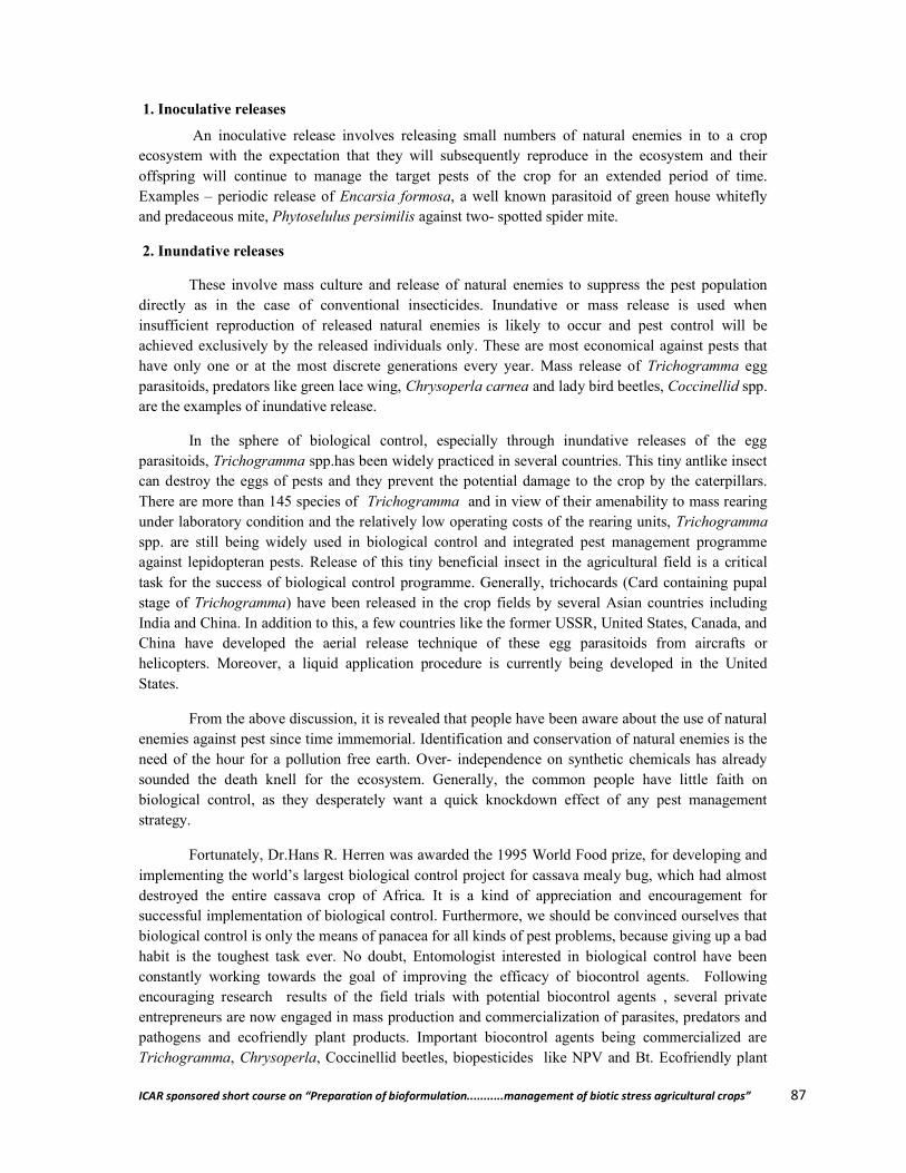
ICAR sponsored short course on “Preparation of bioformulation...........management of biotic stress agricultural crops” 87
1. Inoculative releases
An inoculative release involves releasing small numbers of natural enemies in to a crop ecosystem with the expectation that they will subsequently reproduce in the ecosystem and their offspring will continue to manage the target pests of the crop for an extended period of time. Examples – periodic release of Encarsia formosa, a well known parasitoid of green house whitefly and predaceous mite, Phytoselulus persimilis against two- spotted spider mite.
2. Inundative releases
These involve mass culture and release of natural enemies to suppress the pest population directly as in the case of conventional insecticides. Inundative or mass release is used when insufficient reproduction of released natural enemies is likely to occur and pest control will be achieved exclusively by the released individuals only. These are most economical against pests that have only one or at the most discrete generations every year. Mass release of Trichogramma egg parasitoids, predators like green lace wing, Chrysoperla carnea and lady bird beetles, Coccinellid spp. are the examples of inundative release.
In the sphere of biological control, especially through inundative releases of the egg parasitoids, Trichogramma spp.has been widely practiced in several countries. This tiny antlike insect can destroy the eggs of pests and they prevent the potential damage to the crop by the caterpillars. There are more than 145 species of Trichogramma and in view of their amenability to mass rearing under laboratory condition and the relatively low operating costs of the rearing units, Trichogramma spp. are still being widely used in biological control and integrated pest management programme against lepidopteran pests. Release of this tiny beneficial insect in the agricultural field is a critical task for the success of biological control programme. Generally, trichocards (Card containing pupal stage of Trichogramma) have been released in the crop fields by several Asian countries including India and China. In addition to this, a few countries like the former USSR, United States, Canada, and China have developed the aerial release technique of these egg parasitoids from aircrafts or helicopters. Moreover, a liquid application procedure is currently being developed in the United States.
From the above discussion, it is revealed that people have been aware about the use of natural enemies against pest since time immemorial. Identification and conservation of natural enemies is the need of the hour for a pollution free earth. Over- independence on synthetic chemicals has already sounded the death knell for the ecosystem. Generally, the common people have little faith on biological control, as they desperately want a quick knockdown effect of any pest management strategy.
Fortunately, Dr.Hans R. Herren was awarded the 1995 World Food prize, for developing and implementing the world’s largest biological control project for cassava mealy bug, which had almost destroyed the entire cassava crop of Africa. It is a kind of appreciation and encouragement for successful implementation of biological control. Furthermore, we should be convinced ourselves that biological control is only the means of panacea for all kinds of pest problems, because giving up a bad habit is the toughest task ever. No doubt, Entomologist interested in biological control have been constantly working towards the goal of improving the efficacy of biocontrol agents. Following encouraging research results of the field trials with potential biocontrol agents , several private entrepreneurs are now engaged in mass production and commercialization of parasites, predators and pathogens and ecofriendly plant products. Important biocontrol agents being commercialized are Trichogramma, Chrysoperla, Coccinellid beetles, biopesticides like NPV and Bt. Ecofriendly plant

ICAR sponsored short course on “Preparation of bioformulation...........management of biotic stress agricultural crops” 88
products of neem origin are being marketed for control of various pests particularly in vegetable ecosystem.
Biological control is an important tool in the pest management, it certainly needs to be supplemented with other methods of pest control. Emerging trends in biological control of phytophagous insects including important biological methods viz., use of resistant varieties, cultural practices, mechanical control, seed treatment and conservation of natural enemies may also be given weightage for healthy and pollution free environment. Moreover efforts to evaluate biocontrol agents need to be intensified and improved. Microbial control of insect pests has made little progress. Although several potentially valuable pathogens have been studied and are available for practical use.
References
Chaudhary, S. D. (1997). Advanced training course on Insect Pest Management. ICAR. Centre of Advanced Studies, HAU, Hissar.
Puri, S. N. (1998). Summer School on Current Approaches in Biological Control of Insect- pests and Weeds, IARI, New Delhi.
**************
ICAR sponsored short course on “Preparation of bioformulation...........management of biotic stress agricultural crops” 89
Preparation of Some Biopesticide Materials at Farm Level for Pest Management
Badal Bhattacharyya and Sudhansu Bhagawati AINP on Soil Arthropod Pests, Department of Entomology,
Assam Agricultural University, Jorhat- 785013, Assam
Agricultural crops suffer a massive loss due to ravages of insects and diseases thus causing a serious threat to our agricultural production. The insects and diseases cause about 20-30 per cent damage in agricultural produce, which in monetary terms values more than 1,00,000 crores per annum. The indiscriminate use of synthetic poisonous chemicals resulting in development of resistance problem among insects to insecticides, residue hazards of pesticides, upsetting of balance in nature and resurgence of treated populations. Considering all these drawbacks, many components of the IPM concept were developed in the late nineteenth and early twentieth centuries and biopesticides was placed as an important tool in pest management strategies which are practical, economical and eco-friendly. Biopesticides offer powerful tools to create a new generation of sustainable agriculture products. They are the most likely alternatives to some of the most problematic chemical pesticides currently in use. Biopesticides offer solutions to concerns such as pest resistance and public concern about side effects of pesticides on the surrounding environment and ultimately on human health.
Concept of biopesticides
Biopesticides have been defined by EU as "a form of pesticide based on micro-organisms or natural products". According to the US Environmental Protection Agency (USEPA), “biopesticide include naturally occurring substances that control pests (biochemical pesticides), microorganisms that control pests (microbial pesticides) and pesticidal substances produced by plants containing added genetic material (plant-incorporated protectants) or PIPs.
Homemade biopesticides
The use of homemade bio-pesticides in the farming practices is old aged practices. It is very much environment friendly and can obtain from nature directly. It is almost free of cost and there is no negative impact on human health, soil, animals, plants and environment. Bio-pesticides are derived from natural materials such as animals, plants, microorganisms and minerals. Bio-pesticides tend to be less toxic, more quickly biodegradable and more targeted to the specific pest. Now a days it is widely used due to increased environmental awareness and the pollution potential and health hazards from many conventional pesticides as well as increasing global demand for organically grown food are driving the use of homemade bio-pesticides. The preparation and use of botanicals requires some know-how, but not much material and infrastructures. It’s a common practice under many traditional agricultural systems.
Plant species with pesticidal properties
Biopesticides are natural plant products and may be grown by the planters with minimum cost and extracted by indigenous methods. Biopesticides are secondary metabolites, which include alkaloids, terpenoids, phenolics and minor secondary chemicals. It is estimated that as many as 2121 plant species have been reported to possess pest control properties (Mamun and Ahmed, 2011). Some of the insecticidally promising plants along with their bioactive principle (s) are listed in Table 1.

ICAR sponsored short course on “Preparation of bioformulation...........management of biotic stress agricultural crops” 90
Table 1: List of promising plant species with pesticidal properties
Sl. No.
Plant Scientific name Plant part used Active principle
1. Neem Azadirachta indica Seed and Leaf Azadirachtin
2. Chinaberry Melia azedarach Seed Kernel Meliantriol Melianone
3. Pongamia Pongamia pinnata Seed Karanjin
4. Poison vine Derris elliptica Roots Rotenone
5. Tobacco Nicotiana tobaccum N. rustica
Whole plant Nicotine
6. Chrysanthemum Chrysanthemum cinerariaefolium
Flowers Pyrethrum
7. Garlic Allium sativum Rhizome Diallylsulfide
8. Custard Apple/ Atlas
Annona squamosal A. reticulata
Seed, bark and roots
Anonaine
9. Black pepper Piper nigrum Seeds Piperine
10. Congress grass Parthenium hysterophorus
Parthenin Leaf extract
11. Soybean Glycine max Pods Pinitol 12. Lantana Lantana camara Leaf extract Lantadenes
13. Billygoat-weed Ageratum conyzoides Leaf Phenolic chromenes and hydroxyl ethyl chromenes
14. Jimson weed Datura stramonium Leaf and Seed Tropane alkaloids atropine, hyoscyamine and scopolamine
15. Physic nut Jatropha curcas Leaf and Seed Leaf (curcain) and Seed (curcin)
16. Lemon Citrus limon Peel Limonin and Nomilin 17. Marigold Tagetes minuta Flower 5 E- Ocimenone 18. Sweet flag Acorus calamus Root Trans-asarone 19. Basil Ocimum basilicum Leaf Labiatae
20. Common oleander
Nerium oleander Seed Oleandrin and cardiotonic
21. Hill glory bower Clerodendron infortuanatum
Leaf Trans- decalin
22. Tonka Bean Eupatorium odoratum Leaf Tannins
23. Siam weed Chromolaena odorata Leaf Glycosides, Steroids and Saponins
24. Bel Aegle marmelos Leaf Marmelosin, alloimperatorin and marmeli
25. King chili Capsicum chinense Fruit Capsaicin
General procedure of preparing plant extracts
The simple procedure for the preparation of indigenous plant extracts to be used in organic pest management is given below:
Collection of Leaves/Seeds/Rhizomes
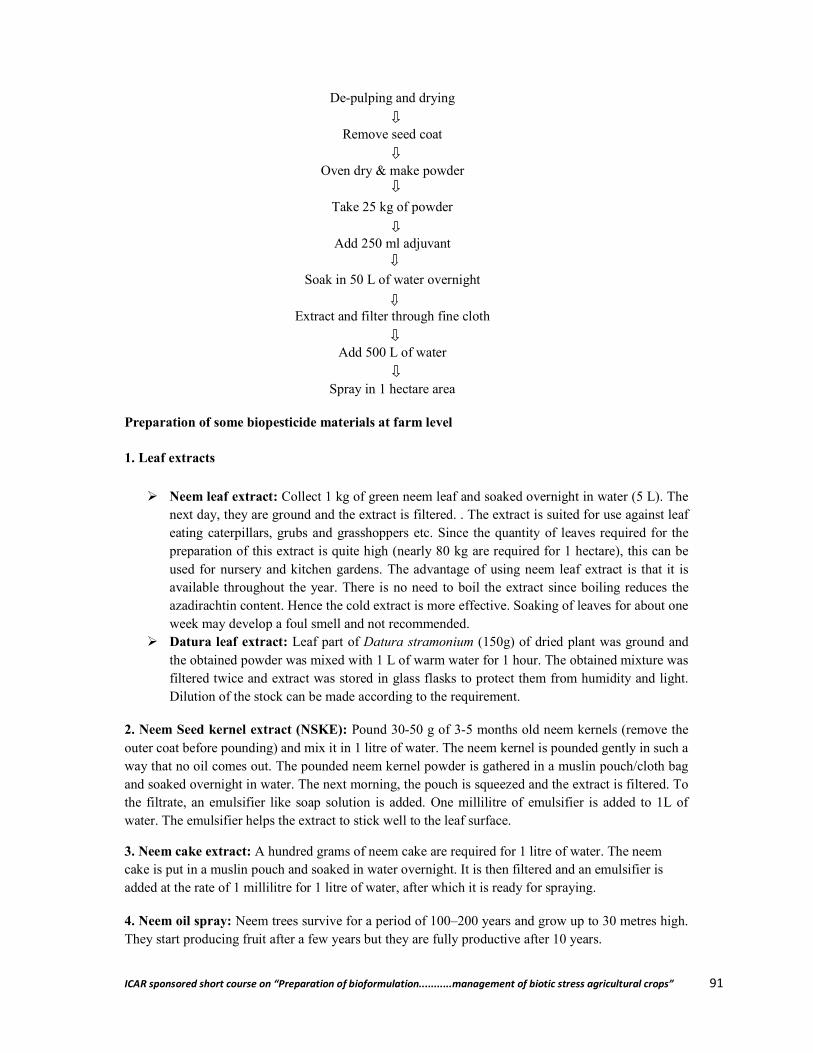
ICAR sponsored short course on “Preparation of bioformulation...........management of biotic stress agricultural crops” 91
De-pulping and drying
Remove seed coat
Oven dry & make powder
Take 25 kg of powder
Add 250 ml adjuvant
Soak in 50 L of water overnight
Extract and filter through fine cloth
Add 500 L of water
Spray in 1 hectare area
Preparation of some biopesticide materials at farm level
1. Leaf extracts
Neem leaf extract: Collect 1 kg of green neem leaf and soaked overnight in water (5 L). The next day, they are ground and the extract is filtered. . The extract is suited for use against leaf eating caterpillars, grubs and grasshoppers etc. Since the quantity of leaves required for the preparation of this extract is quite high (nearly 80 kg are required for 1 hectare), this can be used for nursery and kitchen gardens. The advantage of using neem leaf extract is that it is available throughout the year. There is no need to boil the extract since boiling reduces the azadirachtin content. Hence the cold extract is more effective. Soaking of leaves for about one week may develop a foul smell and not recommended.
Datura leaf extract: Leaf part of Datura stramonium (150g) of dried plant was ground and the obtained powder was mixed with 1 L of warm water for 1 hour. The obtained mixture was filtered twice and extract was stored in glass flasks to protect them from humidity and light. Dilution of the stock can be made according to the requirement.
2. Neem Seed kernel extract (NSKE): Pound 30-50 g of 3-5 months old neem kernels (remove the outer coat before pounding) and mix it in 1 litre of water. The neem kernel is pounded gently in such a way that no oil comes out. The pounded neem kernel powder is gathered in a muslin pouch/cloth bag and soaked overnight in water. The next morning, the pouch is squeezed and the extract is filtered. To the filtrate, an emulsifier like soap solution is added. One millilitre of emulsifier is added to 1L of water. The emulsifier helps the extract to stick well to the leaf surface.
3. Neem cake extract: A hundred grams of neem cake are required for 1 litre of water. The neem cake is put in a muslin pouch and soaked in water overnight. It is then filtered and an emulsifier is added at the rate of 1 millilitre for 1 litre of water, after which it is ready for spraying.
4. Neem oil spray: Neem trees survive for a period of 100–200 years and grow up to 30 metres high. They start producing fruit after a few years but they are fully productive after 10 years.
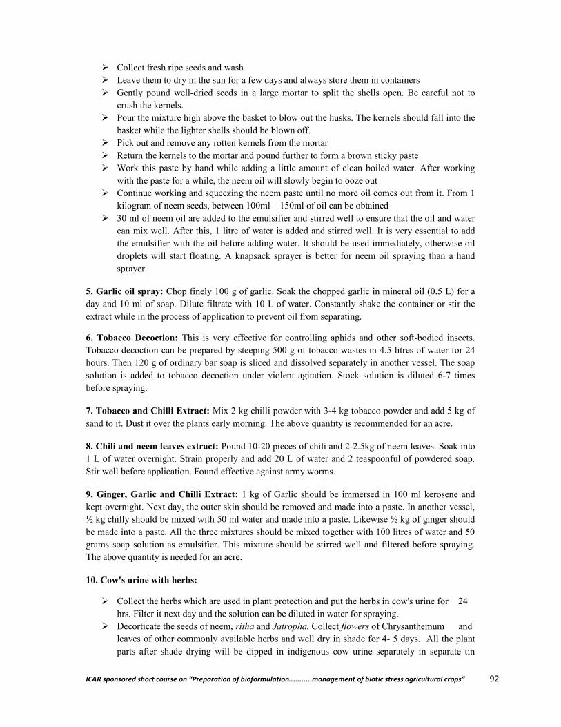
ICAR sponsored short course on “Preparation of bioformulation...........management of biotic stress agricultural crops” 92
Collect fresh ripe seeds and wash Leave them to dry in the sun for a few days and always store them in containers Gently pound well-dried seeds in a large mortar to split the shells open. Be careful not to
crush the kernels. Pour the mixture high above the basket to blow out the husks. The kernels should fall into the
basket while the lighter shells should be blown off. Pick out and remove any rotten kernels from the mortar Return the kernels to the mortar and pound further to form a brown sticky paste Work this paste by hand while adding a little amount of clean boiled water. After working
with the paste for a while, the neem oil will slowly begin to ooze out Continue working and squeezing the neem paste until no more oil comes out from it. From 1
kilogram of neem seeds, between 100ml – 150ml of oil can be obtained 30 ml of neem oil are added to the emulsifier and stirred well to ensure that the oil and water
can mix well. After this, 1 litre of water is added and stirred well. It is very essential to add the emulsifier with the oil before adding water. It should be used immediately, otherwise oil droplets will start floating. A knapsack sprayer is better for neem oil spraying than a hand sprayer.
5. Garlic oil spray: Chop finely 100 g of garlic. Soak the chopped garlic in mineral oil (0.5 L) for a day and 10 ml of soap. Dilute filtrate with 10 L of water. Constantly shake the container or stir the extract while in the process of application to prevent oil from separating.
6. Tobacco Decoction: This is very effective for controlling aphids and other soft-bodied insects. Tobacco decoction can be prepared by steeping 500 g of tobacco wastes in 4.5 litres of water for 24 hours. Then 120 g of ordinary bar soap is sliced and dissolved separately in another vessel. The soap solution is added to tobacco decoction under violent agitation. Stock solution is diluted 6-7 times before spraying.
7. Tobacco and Chilli Extract: Mix 2 kg chilli powder with 3-4 kg tobacco powder and add 5 kg of sand to it. Dust it over the plants early morning. The above quantity is recommended for an acre.
8. Chili and neem leaves extract: Pound 10-20 pieces of chili and 2-2.5kg of neem leaves. Soak into 1 L of water overnight. Strain properly and add 20 L of water and 2 teaspoonful of powdered soap. Stir well before application. Found effective against army worms.
9. Ginger, Garlic and Chilli Extract: 1 kg of Garlic should be immersed in 100 ml kerosene and kept overnight. Next day, the outer skin should be removed and made into a paste. In another vessel, ½ kg chilly should be mixed with 50 ml water and made into a paste. Likewise ½ kg of ginger should be made into a paste. All the three mixtures should be mixed together with 100 litres of water and 50 grams soap solution as emulsifier. This mixture should be stirred well and filtered before spraying. The above quantity is needed for an acre.
10. Cow's urine with herbs:
Collect the herbs which are used in plant protection and put the herbs in cow's urine for 24 hrs. Filter it next day and the solution can be diluted in water for spraying.
Decorticate the seeds of neem, ritha and Jatropha. Collect flowers of Chrysanthemum and leaves of other commonly available herbs and well dry in shade for 4- 5 days. All the plant parts after shade drying will be dipped in indigenous cow urine separately in separate tin
ICAR sponsored short course on “Preparation of bioformulation...........management of biotic stress agricultural crops” 93
containers in the ratio of 1: 9 (w/v). Keep the plant extracts for fermentation for one to two weeks. Before treatment, the extracts should be filtered through sieve of 40 mesh size. Further dilute the extracts in water @ 5ml of extract/ litre of water and apply as soil drenching.
Tobacco with other plant extracts in cow's urine: Take ½ kg garlic, ¼ kg chilli and ¼ kg of ginger. Grind all these ingredients into a paste with considerable quantity of water. Take 250 ml neem oil, 250 ml tobacco extract and 100 ml asafoetida extract. Dissolve the extracts of garlic, chilli, ginger, neem, tobacco, asafoetida in 72 hours old cow's urine (5 - 6 litres) and dilute with 50 - 60 litres of water. Before spraying, add an emulsifier at the rate of 4 ml per litre. This quantity is recommended for an acre.
11. Panchagavya as bio‐pesticide and bio‐enhancer: Panchgavya made up of five cow products; milk, curd, ghee, urine and dung, is also used as fertilizers and pesticides in agricultural operations. As per recent studies cow urine has proved to be an effective pest controller and larvicide when used alone and also in combination with different plant preparations by enhancing the efficacy of different herbal preparations. It acts as a bio-enhancer of anti-infective, anticancer agents/ nutrients from compounds, antibiotics, drugs, therapeutic, nutraceuticals, ions, and also independently as a bioactive agent. Physico-chemical properties of Panchagavya revealed that they possess almost all the major nutrients, micro nutrients and growth hormones (IAA & GA) required for crop growth. Predominance of fermentative microorganisms like yeast and lactobacillus might be due to the combined effect of low pH, milk products and addition of jaggery/sugarcane juice as substrate for their growth. The low pH of the medium was due to the production of organic acids by the fermentative microbes as evidenced by the population dynamics and organic detection in GC analysis. Lactobacillus produces various beneficial metabolites such as organic acids, hydrogen peroxide and antibiotics, which are effective against other pathogenic microorganisms besides its growth. GC-MS analysis resulted in following compounds of fatty acids, alkanes, alconol and alcohols.
Panchagavya consists of nine products viz. cow dung, cow urine, milk, curd, jaggery, ghee, banana, Tender coconut and water. When suitably mixed and used, these have miraculous effects.
Ingredients of Panchagavya
1. Cow dung - 7 kg 2. Ghee - 1 kg 3. Cow Urine - 10 L 4. Water - 10 L 5. Cow milk - 3 L 6. Cow curd - 2 L 7. Tender coconut water - 3 l L 8. Jaggery - 3 kg 9. Well ripened banana – 12 nos.
Method of Preparation
Mix cow dung & cow ghee thoroughly in morning and evening hours and keep it for 3 days Mix cow urine and water, keep it for 15 days with regular mixing both in morning and
evening hours. After 15 days, mix cow milk, cow curd, coconut, jaggery and banana and panchagavya will
be ready after 30 days.

ICAR sponsored short course on “Preparation of bioformulation...........management of biotic stress agricultural crops” 94
Recommended dosage: Spray system: 3% solution was found to be most effective compared to the higher and lower
concentrations investigated. Three litres of Panchagavya to every 100 litres of water is ideal for all crops. The power sprayers of 10 litres capacity may need 300 ml/tank. When sprayed with power sprayer, sediments are to be filtered and when sprayed with hand operated sprayers, the nozzle with higher pore size has to be used.
Flow system: The solution of Panchagavya can be mixed with irrigation water at 50 litres per hectare either through drip irrigation or flow irrigation
Seed/seedling treatment: 3% solution of Panchagavya can be used to soak the seeds or dip the seedlings before planting. Soaking for 20 minutes is sufficient. Rhizomes of turmeric, ginger and sets of sugarcane can be soaked for 30 minutes before planting.
Seed storage: 3% of Panchagavya solution can be used to dip the seeds before drying & storing them.
General precautions for spraying
Spraying should be undertaken in the morning or late in the evening. Avoid spraying during hot conditions.
For better efficacy treat the both surfaces of leaves While using a power sprayer, the quantity of water used should be halved. It is better to use low concentrations of extracts frequently. Approximately 60 litres of ready-to-use biopesticide solution (not the concentrate) is
required to cover each acre of land. However, the spray volume may have to be varied depending on the exact conditions prevailing, such as the intensity of the pest attack.
Factors affecting growth of biopesticides
Some of the factors which have restricted the growth of biopesticides are:
Low reliability because of low stability in effect Target specificity which distracts farmers Slow in action compared to synthetic insecticides Shorter shelf life Inconsistent availability of biopesticides in the market Regulatory system favorable to chemical pesticides
Conclusion
The use of biopesticides in India has tremendous scope because of the wide biodiversity, suitable climatic conditions for the application of biopesticides and low consumption rate of synthetic chemical pesticides especially in North Eastern Region. The rich traditional knowledge base available with the highly diverse indigenous communities in India may provide valuable clues for developing newer and effective biopesticide. Therefore, proper emphasis should be given to train and encourage farm graduates and unemployed youths for the mass production techniques to set up biopesticide units in rural areas for gainful employment. The stress on organic farming and on residue free commodities would certainly warrant increased adoption of biopesticides by the farmers. In this context, the all concerned stakeholders must act with a proper roadmap force for the entrepreneurship development on biopesticides so that the biopesticide industry will scale new heights in the twenty first century.
ICAR sponsored short course on “Preparation of bioformulation...........management of biotic stress agricultural crops” 95
References
Anonymous. (2015). https://en.wikipedia.org/wiki/Biopesticide Anonymous. (2015). Preparation of neem biopesticides at farm level: PART Ill: Appropriate
technology. tcdc2.undp.org/GSSDAcademy/SIE/Docs/Vol4/Neem_biopesticides.pdf Bhoomati, N.; Sivasubramanian, P. and Raguraman, S. (2006). Biological activities of cow
excreta with neem seed kernel extract against Helicoverpa armigera (Hubner). Ann. Pl. Protec. Sci. 14 (1): 11-16.
Biofertilizer products for sustainable agriculture in India. In: Business Potential for Agricultural Biotechnology, Asian Productivity Organisation. pp 96- 102.
Dhaliwal, G.S. and Arora, R. (1996). Principles of insect pest management, Kalyani publishers, New Delhi. pp 149-167.
Dutta, S. (2015). Biopesticides: an ecofriendly approach for pest control. World journal of pharmacy and pharmaceutical sciences 4 (6): 250-265.
Encouraging innovation in biopesticide development. European Commission (2008). ec.europa.eu/environment/integration/research/.../pdf/134na5_en.pdf
Kalra, A. and Khanuja, S. P. S. (2007). Research and Development priorities for biopesticide. Mamun, M. S. A. and Ahmed, M. (2011). Prospect of indigenous plant extracts in tea pest
management. Int. J. Agril. Res. Innov. & Tech. 1(1&2): 16-23. Mishra, J.; Tewari, S.; Singh, S. and Arora, A. K. (2015). Biopesticides: where we stand?
Plant Microbes Symbiosis: Applied Facets, 37. DOI 10.1007/978-81-322- 2068-8_2. Mohanty, I.; Senapati, M. S.; Jena, D. and Palai, S. (2014). Diversified uses of cow urine. Int.
J. Pharm. Pharmaceutical Sciences.6:3. Radhakrishnan, B. (2005). Indigenous preparations useful for pest and disease management.
Planters’ Chronicle. 101(4): 4-16. Reddy, D. S. (2010). Applied Entomology, New Vishal Publications, New Delhi, pp. 109-
113. Saunders, W.H.M.1982. Effects of cow urine and its major constituents on pasture properties.
NZ J Agricul Res. 25 (1):61-68. Senthil kumar, M.; Bharath, M.; Josmin Laali Nisha, L. L. and Basavaraju, H. (2015). Field
Efficacy of Panchagavya on Insect Pests Recorded during the study in Tectona grandis. International Journal of Research in Agriculture and Forestry, 2 (7): 1-8.
Singh, K.M.; Patidar, R.K.; Sen, D. and Kumawat, M. M. (2015). Integrated pest management for sustainable agriculture, New Delhi Publisher, New Delhi, pp. 1-5.
Thakore, Y. (2006). The biopesticide market for global agricultural use. Industrial Biotechnology. 26: 194- 208.
**************
ICAR sponsored short course on “Preparation of bioformulation...........management of biotic stress agricultural crops” 96
Biocontrol of Mite Pests of Horticultural Crops
Sahidur Rahman Department of Entomology
Assaam Agricultural University, Jorhat- 785013, Assam
Horticultural crops include fruits, vegetables and ornamental plants. Mites are encountered as pests regularly or as sporadic pests under all situations. Fruits (both temperate and tropical fruits) are infested by different groups of mites at all growing stages and appear under favourable conditions of climatic conditions. Spider mites, false spider mites and eriophyid mites and broad mites are commonly observed damaging different horticultural crops in different regions. Vegetable production mainly during hot and dry conditions and in protected cultivation is commonly infested by different mite species. Floricultural plants grown in open field conditions as well as under protected conditions are comparatively highly infested by mites causing significant reduction in flower and seed production. Mite pests of important fruit crops, vegetable crops and flowering plants are elaborately discussed giving nature of damage caused and their biological control are discussed in this chapter.
Mite pests
Mites are the microscopic arthropods under the class Arachnida and subclass Acari. They are extremely diverse group of arachnids, closely related to spiders and scorpions. Even if a room looks perfectly clean, it is home to tens of thousands of tiny dust mites. These creatures are among the most diverse subclasses of life, with over 50,000 known species, and an estimated total approaching one million.
Scientific classification
Kingdom: Animalia
Phylum: Arthropoda
Sub phylum: Chelicerata
Class: Arachnida
Subclass: Acari
Although mites are the most successful group of arachnids, most of them are less than 0.04 inches (1mm) in length, meaning people never see them with naked eyes. Immature mites may be even smaller. Like some other arachnids, mites are among the oldest known terrestrial creatures, with fossils going back to the Devonian period, 400 million year ago. The oldest fossil mite recorded is Protacarus crani Hirst, 1923 from red sandstone deposits of Aberdeenshire, Scotland. These creatures lived among some of the earliest land plants. They are totally ubiquitous having colonized pretty much in every known terrestrial, fresh water and marine habitat including polar and alpine extremes. They are one of the few animals found in Antarctica.
Mites are more closely related to spiders than they are to insects. The body tagma of the Acari comprise of two regions, the gnathosoma and the ideosoma. They do not have antennae like the insects do, nor segmented body nor wings. Most mites have an egg stage, a larval stage (six leg) and two nymphal stages (protonymph and deutonymph) having eight legs before becoming an adult. They

ICAR sponsored short course on “Preparation of bioformulation...........management of biotic stress agricultural crops” 97
feed on mosses, ferns, leaves stems, flowers, fruits, lichens, microbes other arthropods and each other. Many mites on agricultural crops are major economic pests like the spider mites, broad mites or the eriophyid mites where as some are useful to human beings as biocontrol agents of those pest mites and other insects like thrips and white flies. And some others are parasitic mites (e.g. scabies and mangemites).
Important mite pests
Mainly four families of mites are pests of various crops and are called phytophagous. Phytophagous mites belong to the family of tetranychidae, tenuipalpidae, Tarsonemidae and Eriophydae.
The tetranychid mites are commonly called spider mites. Like spiders many species spin webs, hence the name; soft bodied, variously coloured, colony forming, inhabiting both surfaces of leaves, 0.5-0.6mm sized. As polyphagous it feeds on variety of crops like beans, bhendi, brinjal, tea etc. Spider mites feed on the underside of the leaves and cause yellow spots, later even yellow leaves. Plant cells turn yellow, which can be seen on the upper surface of the leaf as small yellow spots. This reduces the photosynthetic area of the leaf and the plant gets out of the physiological balance. This results in decreased plant growth and production. Finally the crop may die from the infestation. Nymphs and adults produce webbing that can cause cosmetic damage to the crop. If large numbers of spider mites are present, plants may be completely covered with webs.
Biological Control
Spider mites have many natural enemies, which limit their numbers in many landscapes and gardens, especially when undisturbed by pesticide sprays. Some of the most important are the predatory mites, including the Galendromus (=Metaseiulus) occidentalis, and persimilis species. Predatory mites are about the same size as plant-feeding mites but have longer legs and are more active. The purchase and release of predatory mites can be useful in establishing populations in large plantings or orchards, but the best results are obtained by creating favorable conditions for naturally occurring predators-for instance, by avoiding dusty conditions and pesticide sprays. The major predator mites commercially available for release are the western predatory mite and Phytoseiulus. The western predatory mite is more effective under hot, dry conditions. These predators do not feed on foliage or become pests; thus if pest mites are not available when predatory mites are released, the predators starve or migrate elsewhere. If you wish to establish predators in a heavily infested orchard or garden that has few predators, use a soap spray to bring pest mites to a lower level and then release predatory mites. A good guideline is that one predator is needed for every ten spider mites to provide control. More than one application of predatory mites may be required if you want to reduce pest populations rapidly. Concentrate releases in hot spots where spider mite numbers are highest. Once established on perennials, predatory mites may reproduce and provide biological control indefinitely without further augmentation unless non-selective insecticides are applied that kills the predators.
Release rates
For tomatoes and cucumbers, 1 predator per plant plus 1-2 per infested leaf. For other greenhouse crops, tropical plants, and outdoor gardens, 2,000 per 3,000 sq. ft. For bedding plants, 1,000 per 10,000 sq. ft. For large agri-business, 5,000 - 20,000 per acre depending on infestation.
ICAR sponsored short course on “Preparation of bioformulation...........management of biotic stress agricultural crops” 98
Predatory Mites of Spider Mites in Greenhouses
Phytoseiulus persimilis is the most commonly used predatory mite in greenhouses, but many other species of predatory mites in the family Phytoseiidae feed on spider mites and can provide good control of spider mites in greenhouses.
The globose, light- to deep-red females of P. macropilis lay oval orange eggs that hatch into six-legged larvae. Both larvae and nymphs have a similar white to light orange colour. Males are identical to females in shape and colour but are smaller. These mites have a strong preference for immature spider mites over adults. Each predator consumes four to six spider mite eggs or larvae daily during its development and an average of eight eggs per day as an adult. P. macropilis has a short life cycle in comparison to many spider mite species, allowing it to build up quickly to suppress pest populations. In the absence of spider mites they will prey on their own immatures. P. macropilis occurs naturally in Florida and is available commercially.
The western predatory mite, Galendromus occidentalis, is smaller than P. persimilis and develops best at cooler temperatures, but it tolerates a wide range of relative humidity (40-80%). It has the capability of regulating spider mite populations at lower densities and for longer periods of time than P. persimilis, although it will not control spider mite populations as quickly as P. persimilis can. It can also survive long periods without prey. Several different strains are commercially available, including non-diapausing strains that allow control of spider mites through the winter, when days are short, and strains resistant to a number of organophosphate insecticides.
Neoseiulus (=Amblyseius) californicus is smaller, pale, and does not suppress spider mite populations as quickly as P. persimilis. However, it is useful for keeping low populations under control because it can survive longer periods without prey. Some other species of Neoseiulus, such as N. fallacis, feed on a variety of tetranychid mites and are commercially available, but little is known of their utility in greenhouses.
Predatory mites have been used for years to manage spider mites in European vegetable greenhouses. They have also effectively controlled spider mites on chrysanthemum, rose, and other ornamental crops under experimental conditions. However, the need to prevent cosmetic damage on floral or foliage crops may make biological control of mites difficult, especially when pesticides that kill predatory mites are used to suppress other pests and/or diseases. Your spider mite control strategy may depend on the crop you raise and conditions in your greenhouse, especially temperature and humidity.
Species selection and release rates vary considerably depending on the plant species and the environmental conditions such as temperature and humidity which influence the growth rate of both predator and prey. P. persimilis is an excellent predator of spider mites on low growing plants in humid greenhouses with moderate temperatures. There are a few crops on which P. persimilis cannot be used. For example, P. persimilis slips off the stems and leaves of carnations. It does not do well on tomato because the mites become trapped on glandular hairs on the leaf petioles and stems, and are also affected by toxic compounds in the tomato leaf. P. macropilis performs better than P. persimilis on ornamental plants, such as dieffenbachia, dracena, parlor palm, and schefflera, under warm, humid conditions. M. longipes is frequently used to control spider mites in hot, dry greenhouses on taller plants because it tolerates lower humidity than does P. persimilis. N. californicus does well on most potted plants in greenhouses with moderate temperatures and average
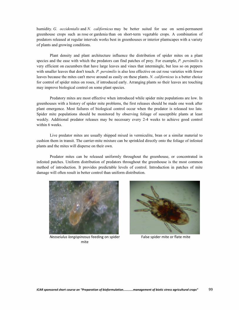
ICAR sponsored short course on “Preparation of bioformulat
humidity. G. occidentalis and N. californicusgreenhouse crops such as rose or gardeniapredators released at regular intervals works best in greenhouses orof plants and growing conditions.
Plant density and plant architecture influence the distribution of spider mites on a plant species and the ease with which the predators can find patches of prey. For example,very efficient on cucumbers that have large leaves and vines that intermingle, but less so on peppers with smaller leaves that don't touch.leaves because the mites can't move around as efor control of spider mites on roses, if introduced early. Arranging plants so their leaves are touching may improve biological control on some plant species.
Predatory mites are most effective whengreenhouses with a history of spider mite problems, the first releases should be made one week after plant emergence. Most failures of biological control occur when the predator is released too late. Spider mite populations should be monitored by observing foliage of susceptible plants at least weekly. Additional predator releases may be necessary every 2within 6 weeks.
Live predator mites are usually shipped mixed in vecushion them in transit. The carrier-plants and the mites will disperse on their own.
Predator mites can be released uniformly throughout the infested patches. Uniform distribution of predators throughout the greenhouse is the most common method of introduction. It provides predictable levels of control. Introduction in patches of mite damage will often result in better control than uniform distribution.
Neoseiulus longispinosus feeding on spider mite
ICAR sponsored short course on “Preparation of bioformulation...........management of biotic stress agricultural crops”
N. californicus may be better suited for use on semigardenia than on short-term vegetable crops. A combination of
predators released at regular intervals works best in greenhouses or interior plantscapes
Plant density and plant architecture influence the distribution of spider mites on a plant species and the ease with which the predators can find patches of prey. For example, P. persimilis
that have large leaves and vines that intermingle, but less so on peppers with smaller leaves that don't touch. P. persimilis is also less effective on cut rose varieties with fewer leaves because the mites can't move around as easily on these plants. N. californicus is a better choice for control of spider mites on roses, if introduced early. Arranging plants so their leaves are touching may improve biological control on some plant species.
Predatory mites are most effective when introduced while spider mite populations are low. In greenhouses with a history of spider mite problems, the first releases should be made one week after plant emergence. Most failures of biological control occur when the predator is released too late.
ider mite populations should be monitored by observing foliage of susceptible plants at least weekly. Additional predator releases may be necessary every 2-4 weeks to achieve good control
Live predator mites are usually shipped mixed in vermiculite, bran or a similar material to -mite mixture can be sprinkled directly onto the foliage of infested
plants and the mites will disperse on their own.
Predator mites can be released uniformly throughout the greenhouse, or concentrated in infested patches. Uniform distribution of predators throughout the greenhouse is the most common method of introduction. It provides predictable levels of control. Introduction in patches of mite
etter control than uniform distribution.
feeding on spider False spider mite or flate mite
ion...........management of biotic stress agricultural crops” 99
may be better suited for use on semi-permanent term vegetable crops. A combination of
plantscapes with a variety
Plant density and plant architecture influence the distribution of spider mites on a plant P. persimilis is
that have large leaves and vines that intermingle, but less so on peppers is also less effective on cut rose varieties with fewer
is a better choice for control of spider mites on roses, if introduced early. Arranging plants so their leaves are touching
introduced while spider mite populations are low. In greenhouses with a history of spider mite problems, the first releases should be made one week after plant emergence. Most failures of biological control occur when the predator is released too late.
ider mite populations should be monitored by observing foliage of susceptible plants at least 4 weeks to achieve good control
rmiculite, bran or a similar material to mite mixture can be sprinkled directly onto the foliage of infested
greenhouse, or concentrated in infested patches. Uniform distribution of predators throughout the greenhouse is the most common method of introduction. It provides predictable levels of control. Introduction in patches of mite
False spider mite or flate mite
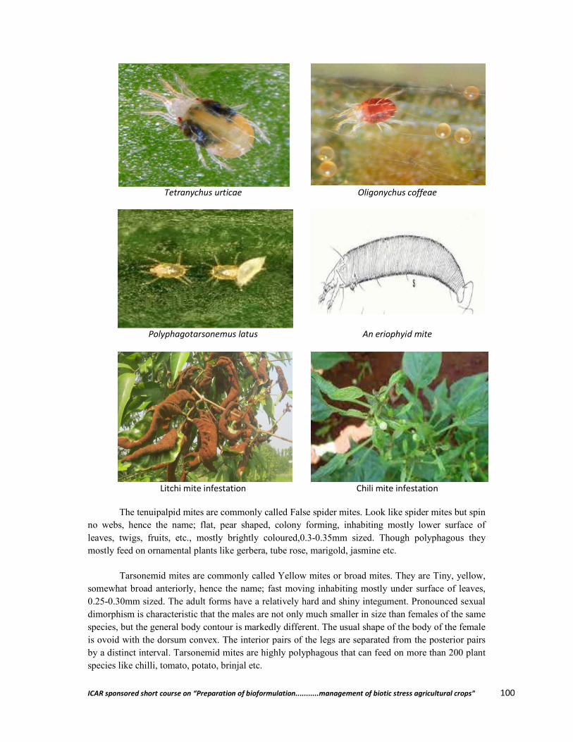
ICAR sponsored short course on “Preparation of bioformulat
Tetranychus urticae
Polyphagotarsonemus latus
Litchi mite infestation
The tenuipalpid mites are commonly called False spider mites. Look like spider mites but spin no webs, hence the name; flat, pear shaped, colony forming, inhabiting mostly lower surface of leaves, twigs, fruits, etc., mostly brightly coloured,mostly feed on ornamental plants like gerbera, tube rose, marigold, jasmine etc.
Tarsonemid mites are commonly called Yellow mites or broad mites. They are Tiny, yellow, somewhat broad anteriorly, hence the name; f0.25-0.30mm sized. The adult forms have a relatively hard and shiny integument. Pronounced sexual dimorphism is characteristic that the males are not only much smaller in size than females of the same species, but the general body contour is markedly different. The usual shape of the body of the female is ovoid with the dorsum convex. The interior pairs of the legs are separated from the posterior pairs by a distinct interval. Tarsonemid mites are highlyspecies like chilli, tomato, potato, brinjal etc.
ICAR sponsored short course on “Preparation of bioformulation...........management of biotic stress agricultural crops”
Tetranychus urticae Oligonychus coffeae
Polyphagotarsonemus latus An eriophyid mite
mite infestation Chili mite infestation
The tenuipalpid mites are commonly called False spider mites. Look like spider mites but spin no webs, hence the name; flat, pear shaped, colony forming, inhabiting mostly lower surface of leaves, twigs, fruits, etc., mostly brightly coloured,0.3-0.35mm sized. Though polyphagous they mostly feed on ornamental plants like gerbera, tube rose, marigold, jasmine etc.
Tarsonemid mites are commonly called Yellow mites or broad mites. They are Tiny, yellow, somewhat broad anteriorly, hence the name; fast moving inhabiting mostly under surface of leaves,
0.30mm sized. The adult forms have a relatively hard and shiny integument. Pronounced sexual dimorphism is characteristic that the males are not only much smaller in size than females of the same pecies, but the general body contour is markedly different. The usual shape of the body of the female
is ovoid with the dorsum convex. The interior pairs of the legs are separated from the posterior pairs by a distinct interval. Tarsonemid mites are highly polyphagous that can feed on more than 200 plant species like chilli, tomato, potato, brinjal etc.
ion...........management of biotic stress agricultural crops” 100
Oligonychus coffeae
An eriophyid mite
Chili mite infestation
The tenuipalpid mites are commonly called False spider mites. Look like spider mites but spin no webs, hence the name; flat, pear shaped, colony forming, inhabiting mostly lower surface of
0.35mm sized. Though polyphagous they
Tarsonemid mites are commonly called Yellow mites or broad mites. They are Tiny, yellow, ast moving inhabiting mostly under surface of leaves,
0.30mm sized. The adult forms have a relatively hard and shiny integument. Pronounced sexual dimorphism is characteristic that the males are not only much smaller in size than females of the same pecies, but the general body contour is markedly different. The usual shape of the body of the female
is ovoid with the dorsum convex. The interior pairs of the legs are separated from the posterior pairs polyphagous that can feed on more than 200 plant

ICAR sponsored short course on “Preparation of bioformulation...........management of biotic stress agricultural crops” 101
Eriophyid mites are known as gall mites or bud mites. Apart from leaves, they also live within buds. It has only two pairs of legs, adult tiny, elongated wormlike, 0.20 – 0.25 mm sized. This group of mites are pests of litchi, arahar, coconut, jasmine etc.
Brief history of biological control of mites in India
The history of biological control dates back to the seventeenth century and since then a great deal of success has been achieved in biological methods of pest control. In India, organized and systematic biological control research began with the establishment of the Indian station of Commonwealth Institute of Biological Control (CIBC) at Bangalore in 1957 with need based substations at Srinagar (Jammu & Kashmir), Dalhausi, Kulu and Shimla (Himachal Pradesh), Ludhiana (Punjab), Sriganganagar (Rajasthan), Lucknow (Uttar Pradesh), Dehra Dun (Uttaranchal), Bhopal (Madhya Pradesh), Parbani (Maharashtra), Surat (Gujarat), Motipur (Bihar), Bhubaneshwar (Orissa), Plassey (West Bengal), Jorhat and Gauhati (Assam), Gangtok (Sikkim), Shillong (Meghalaya), Ambajipet and Ramachandrapuram (Andhra Pradesh), Coimbatore (Tamil Nadu), Mandya (Karnataka) and Palghat (Kerala). These substations were temporary to complete a definite project (The duration of substations varied from few months to 3-5 years). The All-India Co-ordinated Research Project on Biological Control of Crop Pests and Weeds (AICRP) was established in 1977 with 10 centres under the aegis of the Indian Council of Agricultural Research (ICAR) for carrying out biological control research in different parts of the country.
Importance of predatory mites in biological control of insect and mite pests
Within recent years as a result of the widespread use of organic insecticides, especially chlorinated hydrocarbons, economic entomologist have observed a tremendous increase in the population of phytophagous mites that infest agricultural crops and orchard trees, as the population of the natural enemies has decreased. Indeed, the population is often so large that the mites almost assume the status of a major pest. In this context, the role of predatory mites assumes particular significance. There is also some evidence to indicate that the predatory mites are more susceptible to these insecticides than the phytophagous ones and so with the disappearance of these predator species in the field that keep the population of the phytophagous mites in a stabilized equilibrium, the population of phytophagous mites have increased and become very destructive to agricultural and horticultural crops.
As an alternative to insecticides and acaricides, biological control has been attempted on a variety of crops prone to attack of mites and some other pests. Biological control provides an environmentally safe, cost-effective and energy efficient means of pest control, either alone or as a component of integrated pest management. The objective of using biological control agent is to restore and or to enhance the relationship between pests and their natural enemies either by reintroduction and or by creating the same habitat conditions under which the relationship would be strengthened or would form naturally. As a control tactic, biological control is most suited pest species with a relatively high economic injury level. This is because the minimum prey density will usually be required to support a permanent predatory population.
Application of beneficial or predatory mites against phytophagous mites and some other insects is a recent addition to non-chemical pest management strategy. As an alternative to insecticides and acaricides, biological control using these predatory mites has been attempted on a variety of crops worldwide.
ICAR sponsored short course on “Preparation of bioformulation...........management of biotic stress agricultural crops” 102
The most important predatory mite species explored in this regard includes members of phytoseiidae, cunaxidae, laelapidae, tydeidae, ascidae, stigmaeidae, anystidae, erythraeidae, and gamasidae. Many predatory mite species are commercially available to control tetranychids and thrips. Of 54 commercially available predatory species for pest control used on a global level, 13 are predatory mites of the family phytoseiidae.
Major groups of predatory mites
1. Phytoseiid mites
Phytoseiid mites belong to the family Phytoseiidae of the order Mesostigmata. Phytoseiid mites are predators of spider mites and other small insects and mites on plants. Some species feed on nematodes, fungal spores pollen and exudates from plants, but yearly on plant tissues. The phytoseiid is a large family of worldwide distribution several members of this family are of great importance in the biological control of spider mites and thrips in green house production.
The family consist of three sub-families – Amblyseiinae, Phytoseiinae and Typhlodrominae. Effective biocontrol agents occus in all these three sub families. Phytoseiid mites ability to prosper on non-animal food items like pollen, honey and nectar is another factor behind their success as a biocontrol agent, besides this phytoseiid mites possess an assay of supreme adaptive features which often raise them to the level of potential predators of pest mites and also insect to certain extent, this include wide abundance, short life cycle then the prey, equivalent reproductive potential, good searching capacity, good dispersal rate, ability to survive in low prey density and adoptability to different ecological niches.
Now a days, the role of phytoseiid mites as a component of IPM programmes has gained recognition worldwide. As a result countries have started implementing biological control programmes also as a part of IPM through mass rearing, release and export of phytoseiid predators.But in India, there importance is not yet properly recognized and a little work is known to be done on the biological aspects of the same. So far the plant inhabiting predators mites under the order Mesostigmata which have been recorded in India belongs to four families viz., Ascidae, Laelapidae, Otopheidomenidae and Phytoseiidae of these the members of the family phytoseiidae are most abundant, followed by Asiidae.
2. Phytoseiulus persimilis (Athias-Henriot)
It is also known as chilean predatory mite as its 1st used in biological control was in Germany where it was accidently introduced in orchid roots from chile. This mite was 1st discovered on roses grown in greenhouses in Algeria in 1957.It is an specific predator of Tetranychus spider mites and shows reduced reproduction and survival on other insects and phytophagous mites. This predatory mite Phytoseiulus persimilis is an important predator of two spotted mites and inoculative releases have been successful to control TSM on hops in Australia (Leggett, 1987; McKinnon, 1987).
P. permilis can provide effective control against spider mites on cucumber. Tetranychus cinnalarinus also attacks cucumber and P. persimites provides effective control when released at the 1:5 predator: prey ratio.
When introduced in sweet pepper at the predators prey ratio 1: 10, in Bulgaria P. persimilis provide effective control of T. urticae and T. turkestani on pepper when released at a rate of 10,000-80,000 individuals/ha. In greenhouse strawberries in Italy P. persimilis gives effective control of T.
ICAR sponsored short course on “Preparation of bioformulation...........management of biotic stress agricultural crops” 103
urticae. In Japan P. persimilis is effective against Tetraxychus kanzawai in greenhouses grown grapes.
The role of P. persimilis in IPM has been expanded through the development of resistant strains. In the primer USSR strains resistant to malathion, pirimiphosmetyl, high temperature and high temperature plus organophosphorus compounds, respectively have been developed for use in the control of T. urticae.
P. persimilis has been used to control spider mites on a variety of ornamental plant species with success e.g. in rose, chrysanthemums, orchids. Another phytoseiidae mite is Euseius (Amblyseias) finlandicus (Oudemans). It is a potential predator of phytophagous mites worldwide. Euseius spp. occurring in temperate climates important in the control of European red mite Ponorychus ulmi (Koch) (Tetranychidae) (Greys, 1982; McMustry, 1982; Duso, 1992) and rust mites (Eriophyidae) on apple (Collyer, 1964, Van de Vries, 1975, Genini et al., 1983; Sechser et al., 1984; Schausbeiger, 1991).
For successful biological control it is important to detect the pest presence on time and to act immediately. As a spider mite population grows faster in summer and it is then it is difficult to keep pace with. After detecting the first spider mite hot spots, Phytoseiulus is released depending upon the crop and the circumstances an all over 4-6 phytoseiulus is released per m2. On and around the infested plant ± predatory mites/m2 are released.
Commercially they are packed per 1000, 2000 or 25000 adults in vermiculate in tubes and tubes are sprinkled over the infested plants. It is effective because can be used in several crops it eats all stages of spider mites. It is fast developing and provides long lasting protection.
3. Amblyseius andersoni
It is an indigenous species in southern and western Europe and is naturally present in several biotopes such as vineyards and orchards. A. andersoni is a polyphagous mite it feeds on different mites such as spider mites gall mites and russet mites. It main targets as biocontrol agent are red spidermites (Tetranychus urticae), European red mite (Pononychus ulmi) apple rust mite (Aculus schlexhtendali) and boxwood bud mite (Eriephyes canestrinii). Chell also feeds on thrips pollen, honeydew and some fungi.
The predatory mites developed per 10,000 and 25,000 pieces in 1 liter tubes. The carrier material consists of bran and vermiculite. Depending on the crop and pest level at least 20 to 100 mites/m2 applied. They are also available in breeding water proof sachets. Each sachet contains minimum 250 mites. It contains carrier bran and other mite species as food source. There is no need to open sachets, mites emerge from the prepunched holes and spread all over. It applied @ 1 sachet every 2 meters in a row of plants.It is effective because of wide range of prey and also applicable in outdoor crops and has a temperature tolerance of 6°C to 40°C.
4. Neoseiulus cucumeris (Oudermans)
It is also known as Amblyseius cucumeris. This species is known throughout the world. This species is generalize predator (Type III) feeding on small insects and small mites.The eggs are oval and translucent, larvae and nymphs are pale yellowish. The adult females are larger than the males. The dorsal shield is reticulate throughout and bears 17 pairs of setae. Most setae are shorter than the distance between setal bases in the same series. The calyx of the spermathecal apparatus is elongated

ICAR sponsored short course on “Preparation of bioformulation...........management of biotic stress agricultural crops” 104
flash-like the ventral sheidl is quadrate, broad anteriorly. The female lays an average 53 eggs during the oviposition period at a rate of 1.9 eggs/day. At 25°C the egg to adult development in completed in eight to nine days when feeding on thrips larvae and about 7 days when feeding on acarid mites.They are effective predator of some tarsonemid mites and some spider mites that do not produce webbing.
Neoseiulus cucumeris (Oudemans) and Iphiseus degenerans (Berlese). Both species are charactrised by McMurthy and Croft (1997) as Type III (generates) predators and can effectively feed on thrips and white fly larvae, spider mites and pollen (Sabelis and Van Rijn, 1997). Both species are widely used for biological control of thrips in greenhouses (Van Houten and Van Strateem, 1995; Sabelis and Van Rijn, 1997).
In former USSR, larvae of Thrips tabaci are controlled on cucumber in greenhouses when N. cucumeris is released at predatory: prey ratio of 1: 2. In Turkey, effective control is achieved by releasing predator at the rate four to five individuals per plant as soon as thrips detected on host plant. In Canada, N. cucumeris provides effective control of T. tabaci and Frankliniella occidentalis on seedless cucumber in greenhouses in British Columbia.
Neoseiulus cucumeris can keep F. occidentalis population at a low level but do not provide effective control and to overcome this problem the control release system is developed. The CRS consist of a specially formulated bran-based population of N. cucumeris in a waxed paper pack of specific porosity and gives more rapid establishment. The predator continues to breed and emerge from the pack for at least six weeks. This provides better control and is less expensive. The best control is prevention when N. cucumeris is introduced in good quality Sachets immediately after planting thrips population do not develop. In New Zealand, three releases of 10-140 N. cucumeris per plant against Thrips tabaci and T. obscuratus result in low population of thrips and non apparent damage to plants. In the USA, it provides control of Frankliniella tritici and F. occidentalis on ornamental bedding plants in greenhouses when breeding sachets containing 50 mites are introduced at the rate of 125 sachets per 200 m2 of graving area. Control of F. occidenalis on chrysanthemums using N. cucumeris has been effective in both Europe and North America.
5. Neoseiulus barkeri Hughes
Also known as Amblyseius barkeri is a widespread polyphagous species. It feeds on pollen, many small mites and also small insects such as thrips and white flies immatures. The eggs are oval and translucent about 90 m long. Immature stages are pale to yellowish, but adults are darker in colour, often pale brown. Adult females are about 400 m long. The dorsal shield bears 17 pairs of setae. There are some faint reticulate marking in dorsal shield. The ventrianal shield is subquadrate. Development of N. barkeri occur between 15-35°C and a relative humidity of >90% with Tyrophagus putrescentiae as prey the egg to adult development takes six days. Females consume more than males and maximum of 1.96 eggs per day is laid by females. Adult life span is average 161 days at 26°C.
The species provide effective control over broad mite on pepper. They are available commercially for thrips control. This spp. effects and reproduce on immatures of Bemisia tabaci and has been suggested as a bio-control agent of the white fly.
6. Neosciulus californious (McGregor)
It is also known as Amblyseius californicus, this species was originally described from California. It is also known from Central and South America and Southern Europe. It is type II phttoseiid species, namely feeding on Tetranychus spider mites and also other mites and pollen.
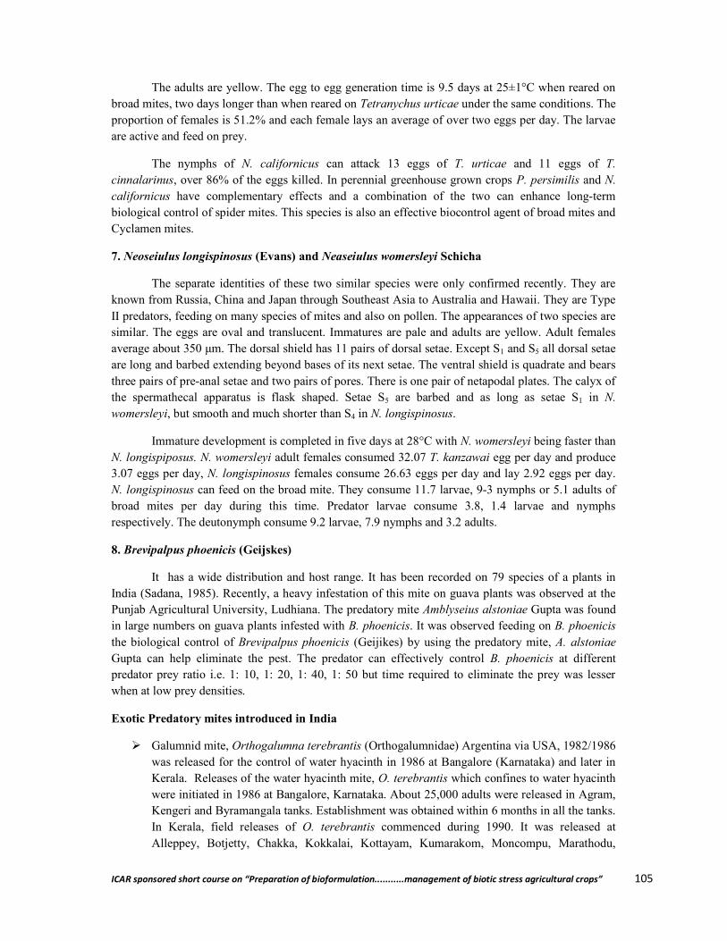
ICAR sponsored short course on “Preparation of bioformulation...........management of biotic stress agricultural crops” 105
The adults are yellow. The egg to egg generation time is 9.5 days at 25±1°C when reared on broad mites, two days longer than when reared on Tetranychus urticae under the same conditions. The proportion of females is 51.2% and each female lays an average of over two eggs per day. The larvae are active and feed on prey.
The nymphs of N. californicus can attack 13 eggs of T. urticae and 11 eggs of T. cinnalarinus, over 86% of the eggs killed. In perennial greenhouse grown crops P. persimilis and N. californicus have complementary effects and a combination of the two can enhance long-term biological control of spider mites. This species is also an effective biocontrol agent of broad mites and Cyclamen mites.
7. Neoseiulus longispinosus (Evans) and Neaseiulus womersleyi Schicha
The separate identities of these two similar species were only confirmed recently. They are known from Russia, China and Japan through Southeast Asia to Australia and Hawaii. They are Type II predators, feeding on many species of mites and also on pollen. The appearances of two species are similar. The eggs are oval and translucent. Immatures are pale and adults are yellow. Adult females average about 350 μm. The dorsal shield has 11 pairs of dorsal setae. Except S1 and S5 all dorsal setae are long and barbed extending beyond bases of its next setae. The ventral shield is quadrate and bears three pairs of pre-anal setae and two pairs of pores. There is one pair of netapodal plates. The calyx of the spermathecal apparatus is flask shaped. Setae S5 are barbed and as long as setae S1 in N. womersleyi, but smooth and much shorter than S4 in N. longispinosus.
Immature development is completed in five days at 28°C with N. womersleyi being faster than N. longispiposus. N. womersleyi adult females consumed 32.07 T. kanzawai egg per day and produce 3.07 eggs per day, N. longispinosus females consume 26.63 eggs per day and lay 2.92 eggs per day. N. longispinosus can feed on the broad mite. They consume 11.7 larvae, 9-3 nymphs or 5.1 adults of broad mites per day during this time. Predator larvae consume 3.8, 1.4 larvae and nymphs respectively. The deutonymph consume 9.2 larvae, 7.9 nymphs and 3.2 adults.
8. Brevipalpus phoenicis (Geijskes)
It has a wide distribution and host range. It has been recorded on 79 species of a plants in India (Sadana, 1985). Recently, a heavy infestation of this mite on guava plants was observed at the Punjab Agricultural University, Ludhiana. The predatory mite Amblyseius alstoniae Gupta was found in large numbers on guava plants infested with B. phoenicis. It was observed feeding on B. phoenicis the biological control of Brevipalpus phoenicis (Geijikes) by using the predatory mite, A. alstoniae Gupta can help eliminate the pest. The predator can effectively control B. phoenicis at different predator prey ratio i.e. 1: 10, 1: 20, 1: 40, 1: 50 but time required to eliminate the prey was lesser when at low prey densities.
Exotic Predatory mites introduced in India
Galumnid mite, Orthogalumna terebrantis (Orthogalumnidae) Argentina via USA, 1982/1986 was released for the control of water hyacinth in 1986 at Bangalore (Karnataka) and later in Kerala. Releases of the water hyacinth mite, O. terebrantis which confines to water hyacinth were initiated in 1986 at Bangalore, Karnataka. About 25,000 adults were released in Agram, Kengeri and Byramangala tanks. Establishment was obtained within 6 months in all the tanks. In Kerala, field releases of O. terebrantis commenced during 1990. It was released at Alleppey, Botjetty, Chakka, Kokkalai, Kottayam, Kumarakom, Moncompu, Marathodu,
ICAR sponsored short course on “Preparation of bioformulation...........management of biotic stress agricultural crops” 106
Thrissur, and Thiruvananthapuram in different spots at each of the water bodies. O. terebrantis has established all over the release sites and is spreading on its own. It has spread far and wide across the vast stretches of Kuttanad backwaters of Kerala. The mite was more efficient in water bodies where weevils, Neochetina spp. have established. O. terebrantis has established in Kerala and Karnataka and it complements the two exotic weevils in hastening the collapse of water hyacinth.
Amblyseius chilenensis (Phytoseiidae) USA, 1984 was released and recovered from spider mites, Tetranychus spp. on various crops such as beans, okra and strawberry.
Phytoseiulus persimilis (Phytoseiidae) Chile via Switzerland, 1965; UK, 1984 was released and recovered from spider mites, Tetranychus spp. on various crops such as citrus, beans, okra and strawberry.
Advantages of predatory mites
There are many advantages of using predatory mites as biological control agents to control mite and insect pests.
It is easier to augment predatory mites in the field by introducing them with the rearing medium
Predatory mites can be released in several protected and open air crops They have wide range of prey mites and insects which makes them effective biocontrol agents The application methods are easy and user friendly Long –standing protection in the field and protected cultivation can be achieved Survive on non animal products like pollen and nectar There development is faster and life cycle is short When applied it spreads well in the crops Wide Range of temperature tolerance can be seen in some species Problems like development of resistance is very rare Environmentally safe and sound
Disadvantages of predatory mites
The disadvantages regarding use of predatory mites as biological control agents are very few.
The mass production, shipping, storage and application techniques of predatory mites need skilled persons.
Initial cost is often high as mass rearing of predatory mites require specific infrastructure like polycarbonate houses.
They may also cause some kind of skin disease and respiratory problems to the workers continuously dealing with these mites.
References
Ashihara, W.; Inoue, K.; Osakabe, M. and Hamamura, T. (1992). Effectiveness of Phytoseiulus persimilis Athias-Henriot (Acarina: Phytoseiidae) as a control agent for the Kanzawa spider mite, Tetranychus kanzawai Kishida (Acarina: Tetranychidae) and occurrence of native natural enemies of the spider mite on grapevine in glasshouse. Bulletin of the Fruit Tree Research Station 22: 109-130.
ICAR sponsored short course on “Preparation of bioformulation...........management of biotic stress agricultural crops” 107
Childers, C. C. and Rock, G. C. (1981). Observations on the ocurrence and feeding habits of Blaustium putmani (Acari: Erythraeidae) in North Carolina apple orchards. International Journal of Acarology 7: 63-68.
Courcy Williams, M. de (1993). Amblyseius cucumeris (Acari: Phytoseiidae) in the control of western flower thrips (Frankliniella occidentalis) on cyclamen. Bulletin OILB/SROP 16: 48-52.
Grafton-Cardwel, E .E.; Ouyang, Y. and Striggow, R. A. (1999). Predacious mites for control of citrus thrips, Scirtothrips citri (Thysanoptera: Thripidae) in nursery citrus. Biological Control 14: 29-36.
Hessein, N. A. and Parrella, M. P. (1990). Predatory mites help control thrips on floriculture crops. California Agriculture 44 (6): 19-21.
Hessein, N. A. and Perring, T. M. (1986). Feeding habits of the Tydeidae with evidence of Homeopronematus anconai (Acari: Tydeidae) predation on Aculops lycopersici (Acari: Eriophyidae). International Journal of Acarology 12, 215-22.
Houten, Y.M. van (1996). Biological control of western flower thrips on cucumber using the predatory mites Amblyseius cucumeris and A. limonicus. Bulletin OILBSROP 19(1): 59-62.
`Houten, Y.M. van and Stratum, P. van (1993). Biological control of western flower thrips in greenhouse sweet peppers using nondiapausing predatory mites. Proceedings of the Section Experimental and Applied Entomology of The Netherlands Entomological Society 4, 229-234.
Karmakar, and Gupta, S. K. (2011). Predatory mite fauna associated with agri-horticultural crops and weeds from the Gangatic Plains of West Bengal,India. Zoosymposia 6: 62-67.
Kumari, M. and Sadana, G. L. (1995). Biological control of false spider mite, Brevepalpus phoenics by the predaceous mite, Amblyseius alstoniae (Acari: Tenuipalpidae: Phytoseiidae) J.Acarol, 13(1&2): 69-74.
Lester, P. J.; Thistlewood, H. M. A. and Harmens, R. (2000). Some effects of pre-release host-plant on the biological control of Pononychus ulmi by the predatory mite
Leeuwen, T. V.; Witters, J. and Nauen, R. (2010). The control of eriophyoid mites: state of the art and future challenges. Exp Appl Acarol 51: 205-224
Mattu, V. K.; Sharma, M. and Thakur, M. L. (2007) .Photoseiid mites on mountain fruit crops in Himachal Pradesh J.Acarol, 16(1&2): 34-35
Mc Murtry,J.A.and Croft,B.A. (1997). Life-style of phytoseiid mites and their role in biological control.Ann.Rev.Entomol. 42:291-321.
Nakao, H.; Saito, Y. and Mori, H. (1987). Biological control of the two-spotted spider mite by the resistant strain of Phytoseiulus persimilis. I. Estimation of effectiveness of the predator by experiments and simulation under chemical spraying conditions (Acari, Phytoseiidae; Tetranychidae). Japanese Journal of Applied Entomology and Zoology 31: 359-368.
Negloh, K., Hanna, R. and Schausberger, P. (2012). Interguild predation and cannibalism between the predatory mites Neoseiulus neobaraki and N. Paspalivorus, natural enemies of the coconut mite Aceria guerreronis Exp Appl Acarol 58: 235-246
Simmonds, S. P. (1972). Observations on the control of Tetranychus urticae on roses by Phytoseiulus persimilis. Plant Pathology 21: 163-165.
Sobha, T. R. and Haq, M. A (2007). Acarine predators associated with coconut mite, Aceria guerreronis (Acari:Eriophyidae) and studies on feeding potential of Typhlodromus pyri (Acari: Phytoseiidae) J.Acarol 17(1&2) : 45-47
Takahashi, F. and Chant, D. A. (1992). Adaptive strategies in the genus Phytoseiulus Evans (Acari: Phytoseiidae). I. Development times. International Journal of Acarology 18: 171-176.
ICAR sponsored short course on “Preparation of bioformulation...........management of biotic stress agricultural crops” 108
Zhang, Z.-Q. and Sanderson, J. P. (1995). Two spotted spider mites (Acari: Tetranychidae) and Phytoseiulus persimilis (Acari: Phytoseiidae) on greenhouse roses: spatial distribution and predator efficacy. Journal of Economic Entomology 88: 352-357.
**************
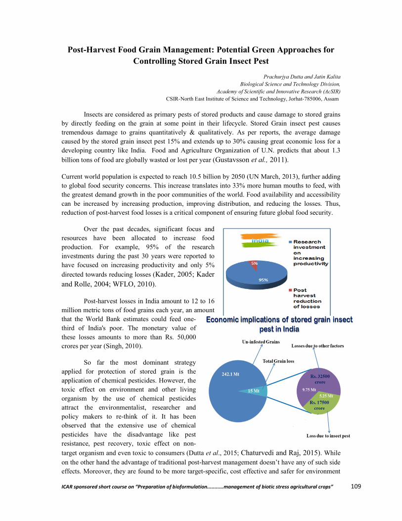
ICAR sponsored short course on “Preparation of bioformulation...........management of biotic stress agricultural crops” 109
Post-Harvest Food Grain Management: Potential Green Approaches for Controlling Stored Grain Insect Pest
Prachurjya Dutta and Jatin Kalita Biological Science and Technology Division,
Academy of Scientific and Innovative Research (AcSIR) CSIR-North East Institute of Science and Technology, Jorhat-785006, Assam
Insects are considered as primary pests of stored products and cause damage to stored grains by directly feeding on the grain at some point in their lifecycle. Stored Grain insect pest causes tremendous damage to grains quantitatively & qualitatively. As per reports, the average damage caused by the stored grain insect pest 15% and extends up to 30% causing great economic loss for a developing country like India. Food and Agriculture Organization of U.N. predicts that about 1.3
billion tons of food are globally wasted or lost per year (Gustavsson et al., 2011).
Current world population is expected to reach 10.5 billion by 2050 (UN March, 2013), further adding to global food security concerns. This increase translates into 33% more human mouths to feed, with the greatest demand growth in the poor communities of the world. Food availability and accessibility can be increased by increasing production, improving distribution, and reducing the losses. Thus, reduction of post-harvest food losses is a critical component of ensuring future global food security.
Over the past decades, significant focus and resources have been allocated to increase food production. For example, 95% of the research investments during the past 30 years were reported to have focused on increasing productivity and only 5%
directed towards reducing losses (Kader, 2005; Kader and Rolle, 2004; WFLO, 2010).
Post-harvest losses in India amount to 12 to 16 million metric tons of food grains each year, an amount that the World Bank estimates could feed one-third of India's poor. The monetary value of these losses amounts to more than Rs. 50,000 crores per year (Singh, 2010).
So far the most dominant strategy applied for protection of stored grain is the application of chemical pesticides. However, the toxic effect on environment and other living organism by the use of chemical pesticides attract the environmentalist, researcher and policy makers to re-think of it. It has been observed that the extensive use of chemical pesticides have the disadvantage like pest resistance, pest recovery, toxic effect on non-
target organism and even toxic to consumers (Dutta et al., 2015; Chaturvedi and Raj, 2015). While on the other hand the advantage of traditional post-harvest management doesn’t have any of such side effects. Moreover, they are found to be more target-specific, cost effective and safer for environment
ICAR sponsored short course on “Preparation of bioformulation...........management of biotic stress agricultural crops” 110
and consumer (Prakash and Rao, 1997). Now, at this juncture we are in need to emphasis more on biological agents, botanical pesticide, and cultural practice for sustainable agriculture. Indigenous knowledge may play a key role to get a flourishing success. Whereas many ethnic groups of people are using traditional way to protect the grains, still there is a need of institutionalization of traditional knowledge for its full validation in modern agricultural system. It is now-a-days widely accepted among agricultural scientist throughout the world that the re-assessment of indigenous technical knowledge is an indispensable part of the introduction of new agricultural technology. It is recognized that the knowledge of farmers must be taken into account before any new technology is developed
and disseminated (Chaturvedi and Raj, 2015; Sinha, 2010; Samanta and Prasad, 1995).
Insect pest of stored grains
A stored grains pest is any organism that infests or destroys the stored grain. Stored grain pest get unnoticed because they infest items that are not regularly used. Very often the infestation is come to noticed when pest emerge from storage as a result of crowding or after completely destroying a food product, and in search of new food.
Insects pest are of two types primary and secondary, primary pest attack grains which are undamaged or previously not infested by any other pest. In case of secondary pest they only attack or feed on grains which are damaged by some other pest or by poor threshing, drying and handling. Usually primary pest are more destructive then secondary pest.
According to World Food Programme (WFP) there are about 30 species of insect pests which commonly infest the stored food grains and most of them are either beetle or moth. Most commonly found primary and secondary pest of stored grains are listed in table 1.
Table 1: Primary and secondary pest of stored grains.
Green approach for stored grain pest management
Primary Pest Secondary pest Rice weevil Sitophilus oryzae, S. zeamais, S. granaries
Rust red flour beetle Tribolium castaneum
Khapra beetle Trogoderma granarium
Long headed flour beetle Latheticus oryzae
Lesser grain borer Rhyzopertha dominica
Saw toothed grain beetle Oryzaephilus surinamensis
Grain moth Sitotroga cerealella
Rice moth Corcyra cephalonica
Pulse beetle Callosobruchus chinensis, C. maculates
Indian meal moth Plodia interpunctella
Peanut bruchid Caryedon serratus
Grain feeder Attagenus unicolor
Cigarette beetle Lasioderma sericorne
Hairy spider beetle Ptinus villiger
ICAR sponsored short course on “Preparation of bioformulat
India is a tropical country and due to its particular environmental condition, suffer several losses from pest. Though insect pest causes lots of damage, using synthetic pesticide to control it is more harmful for environment and other organism. Therefore, an ecostored grain from pests without harming the environment and other living organism is required. Although lots of alternative ways are there for pest management, Indian farmers are not fully ait. Green approach means controlling stored pest by different methods like bioImproving storage structure, botanical pesticides etc.
1. Bio-control
Bio-control is the process of controlling pest by introducing naturally enemies. Sthe addition of parasitoids and predators to stored raw commodities has been allowed under law
(Anon, 1992). There are many advantages of using insect parasitoids and predators to control stored product insect pests as natural enemies leave no hmethods applied for the management of stored grains are microbial control and insect parasite.
1.1. Microbial Control
Lots of work has been done for screening of microbes which can control the stored grain pest without harming the grains and make it safe for human consumption. reported that the isolates of Beauveriacontrol agents against Sitophilus oryzaefungal mix of Beauveria bassiana weevil Sitophilus oryzae. Bacillus thuringiensisapplied to control stored pest population. It contains an insecticidal protein that kills the insect either directly or by septicemia (blood poisoning) of the insect gut.
1.2. Insect parasite and predators
From long decades Insect parasitoids and predators haveParasitoids are released either as adults from small plastic Structures or emerge from pupae stuck to cardboard strips placed in the storage rooms. According to Arbogast and Mullen 1990, release of parasite Anisopteromalus calandrae and against maize weevil, suppressed 90% of weevil population. Press and Mullen, 1992 reported that Anisopteromalus calandrae controlled the 99% of rice weevil population up to 4 month.
2. Storage condition
Farmers normally use different type of traditional storage structure for storage of grains. Out of the different storage structures, Dulicommon structure used by the people of NE India. According to Kalita et al. (2002), modification otraditional structures with scientific knowledge also provides better protection from stored grain insect pest.
3. Improved grain storage Structure
ICAR sponsored short course on “Preparation of bioformulation...........management of biotic stress agricultural crops”
India is a tropical country and due to its particular environmental condition, suffer several losses from pest. Though insect pest causes lots of damage, using synthetic pesticide to control it is
nd other organism. Therefore, an eco-friendly way to protect the stored grain from pests without harming the environment and other living organism is required. Although lots of alternative ways are there for pest management, Indian farmers are not fully ait. Green approach means controlling stored pest by different methods like bio-control agents, Improving storage structure, botanical pesticides etc.
control is the process of controlling pest by introducing naturally enemies. Sthe addition of parasitoids and predators to stored raw commodities has been allowed under law
. There are many advantages of using insect parasitoids and predators to control stored product insect pests as natural enemies leave no harmful chemical residues. The most common methods applied for the management of stored grains are microbial control and insect parasite.
Lots of work has been done for screening of microbes which can control the stored grain pest thout harming the grains and make it safe for human consumption. In the year 1998 Moino Jr
Beauveria bassiana and Metarhizium anisopliae can beoryzae and Sitophilus zeamais. Bello et al., 2001 reported that a
Beauveria bassiana and Metarhizium anisopliae can successfully control the rice Sitophilus oryzae. Bacillus thuringiensis (Bt) which available in market as “Dipel” is also
d to control stored pest population. It contains an insecticidal protein that kills the insect either directly or by septicemia (blood poisoning) of the insect gut.
1.2. Insect parasite and predators
From long decades Insect parasitoids and predators have been used to controlinsect pests. Parasitoids are released either as adults from small plastic Structures or emerge from pupae stuck to cardboard strips placed in the storage rooms. According to Arbogast and Mullen 1990, release of parasite
and Theocolax elegans against maize weevil, suppressed 90% of weevil population. Press and Mullen, 1992 reported that
controlled the 99% of rice
Farmers normally use different type of traditional storage structure for storage of grains. Out of
Duli is found to be most common structure used by the people of NE India.
(2002), modification of traditional structures with scientific knowledge also provides better protection from stored grain insect pest.
3. Improved grain storage Structure
ion...........management of biotic stress agricultural crops” 111
India is a tropical country and due to its particular environmental condition, suffer several losses from pest. Though insect pest causes lots of damage, using synthetic pesticide to control it is
friendly way to protect the stored grain from pests without harming the environment and other living organism is required. Although lots of alternative ways are there for pest management, Indian farmers are not fully aware of
control agents,
control is the process of controlling pest by introducing naturally enemies. Since 1992, the addition of parasitoids and predators to stored raw commodities has been allowed under law
. There are many advantages of using insect parasitoids and predators to control stored armful chemical residues. The most common
methods applied for the management of stored grains are microbial control and insect parasite.
Lots of work has been done for screening of microbes which can control the stored grain pest In the year 1998 Moino Jr et a.l,
be consider as , 2001 reported that a
can successfully control the rice (Bt) which available in market as “Dipel” is also
d to control stored pest population. It contains an insecticidal protein that kills the insect either
been used to controlinsect pests. Parasitoids are released either as adults from small plastic Structures or emerge from pupae stuck to
ICAR sponsored short course on “Preparation of bioformulation...........management of biotic stress agricultural crops” 112
The farmers in the country have been storing their food grains in different types of indigenous storage structures, bamboo & wooden structures, jute bags & structures made of locally available raw materials from ancient times. However, these structures afford little protection to pulse grains and become unviable very soon due to depredations by insect pests, apart from other factors. For resource poor farmers living at or near subsistence even losses of this magnitude have important implications for food security. Rectifying these losses can only be achieved by subsistence farmers if changes are made in the traditional system of storage which bear no or very little cost.
In view of the above problems, an improved storage Structure has been developed which can be constructed with locally available materials with little cost, provides protection to stored grain insect pests with reduced storage loss of pulses (1.39% in compared to as high as 15.46% in other traditional storage structures in 10 months storage duration). Moreover, the increase in moisture content of stored pulses can also be reduced to a great extent compared to other structure, inhibiting the insect pest infestation & fungal infection to retain the quality of the stored grains.
Application/Uses of the Storage Structure
The present storage Structure can be used for storage of all types of Food grains, pulses, oilseeds and highly suitable for seed preservation up to 5 quintal.
The advantages of the present storage structure are
The improved pulses grain storage Structure gives an air tight environment to the stored pulses grains which inhibit moisture absorption by the stored grains from atmosphere resulting in lower infestation of insect pests and micro-organisms.
It provides insulation against transfer of heat from outside atmosphere to inside the Structure prohibiting accumulation of temperature by the stored grain disrupting congenial environment for multiplication of insect pests resulting in less insect pests’ damage.
The storage Structure not only can be used for storing pulses grain for food purpose, but also can be utilized to store pulses grain meant for seed purpose.
The improved pulses grain storage Structure can also be used for storing other food grains. The improved pulses grain storage Structure provides easy loading and unloading of stored
grains. 4. Botanical pesticides
The use of botanical pesticides particularly for stored grain pests is being recommended globally and use of essential oils seem to be the best choice for it. Reports suggests that essential oils are readily biodegradable and less detrimental to non-target organisms as compared to synthetic pesticides (Dubey, 2008). Kalita et al, 2014 reported that Essential oils of two variants of Cinnamomum verum have significant repellent action and reduced the fecundity and decreased egg hatchability of Callosobruchus chinensis.
Development of a novel herbal fumigant formulation against stored grain insect pest Fumigation is considered as quick and effective tool for control of stored grain insect-pests. The concept of ‘Zero tolerance of insect-pests in food commodities’ has made fumigants further indispensable. However, given to the regulatory concerns and development of resistance, use of conventional fumigants such as phosphine has become very challenging. Environmental safety, efficacy and cost shall determine the value of a fumigant. Fumigation registration takes into account any adverse effect of its residues in food and the environment. Since the last 3 decades, several fumigants have been withdrawn or discontinued on the bases of above parameters. Literature indicates that intensive investigation is

ICAR sponsored short course on “Preparation of bioformulation...........management of biotic stress agricultural crops” 113
required to explore natural plant products for grain fumigation. No herbal fumigant tablets/formulations available in the market at present against stored grain insects.
Methyl bromide (MB), a cheap, broad spectrum fumigant, has to be phased out honouring ‘Montreal Protocol’. Phosphine widely used worldwide, is the only fumigant currently used in India, because of its low cost, availability and residue-free treatment. But one serious limitation of use of phosphine is development of resistance in the major stored grain insect-pests.
Hence, there is an urgent need to develop a formulation without having side health hazards. The north east region of India is having rich biodiversity and CSIR-NEIST has already identified a few plant species having bioactive components as strong as to repel stored grain insect pest of cereals & pulses effectively and translated in to a fumigant formulation. It is expected that, the herbal fumigant formulation with significant repellent properties against stored grain insect pest will have tremendous application for safe storage of grains without any risk of health hazards.
References
Anonymous, (1992). Parasitic and predaceous insects used to control insect pests: Exemption from a tolerance. Federal Register 57(78): 14644.
Arbogast, R. T. and Mullen, M. A. (1990). Interaction of maize weevil (Coleoptera: Curculionidae) and parasitoid Anisopteromalus calandrae (Hymenoptera: Pteromalidae) in a smallbulk of stored corn. J. Econ. Ent. 83: 2462-2468.
ICAR sponsored short course on “Preparation of bioformulation...........management of biotic stress agricultural crops” 114
Dal Belloa, G.; Padin, S.; LoÂpezLastra, C. and Fabrizio, M. (2001). Laboratory evaluation of chemical-biological control of the rice weevil (Sitophilus oryzae L.) in stored grains. Journal of Stored Products Research 37: 77-84.
Dubey, N. K.; Srivastava, B. and Kumar, A. (2008). Current Status of Plant Products as Botanical Pesticides in storage pest management. Journal of Biopesticides 1(2): 182 – 186.
Chaturvedi, B. K. and Raj, A. (2015). Agricultural Storage Infrastructure in India: An Overview. OJBM 17(5): 37-43.
Dutta, P.; Dey, T.; Kalita, M. and Kalita, J. (2015). Insecticidal activity of leaf extract of Cannabis indica against two major stored grain pest Callosobruchus chinensis and Sitophilus oryzae. IJTA 33(4): 3057-3061.
Gustavsson, J.; Cederberg, C.; Sonesson, U.; Van Otterdijk, R. and Meybeck, A. (2011). Global food losses and food waste. Rome: Food and Agriculture Organization of the United Nations. http://www.fao.org/fileadmin/user_ upload/ags/publications/ GFL_web.pdf
Kader, A. A. and Rolle, R. S. (2004). The Role of Post-harvest Management in Assuring the Quality and Safety Horticultural Crops. Food and Agriculture Organization. Agricultural Services Bulletin. 152: 52.
Kader, A. A. (2005). Increasing food availability by reducing postharvest losses of fresh produce. Acta Horticulture. 682: 2169-2176.
Kalita, J.; Bhattacharyya, P. R. and Handique, R. (2002). Development and testing of some modified indigenous structures of North East India for safe storage of pulse grains. Journal of Applied Zoological Research 13(2 & 3): 255 – 257.
Kalita, J.; Dutta, P.; Gogoi, P.; Bhattacharyya, P. R. and Nath, S. C. (2014). Biological activity of essential oils of two variant of Cinnamomum verum from North East India on Callosobruchus chinensis (L.). IJABPT 5(3): 190-194
MoinoJr, A.; Alves, S. B. and Pereira, R. M. (1998). Efficacy of Beauveria bassiana (Balsamo) Vuillemin isolates for control of stored-grain pests. 122(1-51): 301–305.
Prakash, A. and Rao, J. (1997). Botanical pesticides in agriculture. CRC Press Inc. 461p. Press, J. P. and Mullen, M. A. (1992). Potential of the weevil parasitoid, Anisopteromalus
calandrae (Howard) (Hymenoptera: Pteromalidae) for protecting commercially packaged wheat from infestation by the rice weevil, Sitophilus oryzae (L.) (Coleoptera: Curculionidae). J. Kans. Ent. Soc. 65: 348-351.
Samanta, R. K. and Prasad, M. V. (1995). An indigenous post-harvest technology. http://www.nuffic.nl/ciran/ikdm/3-2/articles/samanta.html.
Singh, P. K. (2010). A decentralized and holistic approach for grain management in India. Current Science 99: 1179–1180.
Sinha, B. (2010). An Apprisal for the traditional post-harvest pest management method in the Northeast Indian uplands. IJTK 9(3): 536-543.
WFPhttp://foodqualityandsafety.wfp.org/home;jsessionid=F158E74DBA0BD03731D2AD655DF94761.
World Food Logistics Organization (2010). Identification of Appropriate Postharvest Technologies for improving Market Access and Incomes for Small Horticultural Farmers in Sub-Saharan Africa and South Asia. Alexandria VA, March.
************
ICAR sponsored short course on “Preparation of bioformulation...........management of biotic stress agricultural crops” 115
Isolation and Diagnosis of Nematodes
Debanand Das and Nibedita Buragohain Department of Nematology
Assaam Agricultural University, Jorhat- 785013, Assam
What is nematode?
The word ‘Nematode’ is derived from two Greek words viz., nema (thread) and oides (resemble/form). Thus, nematodes are thread like organism. They are commonly called threadworms (due to thread like form), roundworms (due to tubular shape and circular in cross section) or eelworms (due to general shape of the body and serpentine movement). In one sentence, nematodes can be defined as ‘triploblastic, pseudocoelomic, bilaterally symmetrical, unsegmented animal’. However, this group of organism shows extensive variations in its form and thus, there is hardly any sentence regarding their morphology stands fit for all the species.
Habitat
Nematodes are ubiquitous. Few lines written by N. A. Cobb (he is regarded as Father of Nematology) clearly define its habitat:
“If all the matter in the universe except nematodes were swept away, our world would still be dimly recognizable ..... we would finds its mountains, hills, valleys, rivers, lakes and oceans represented by a thin film of nematodes”.
Nematodes occur at the bottom of lakes, rivers, at enormous depths in the ocean; some thrives in temperatures constantly below freezing point while, others in the water of hot spring, and still some can withstand complete dryness on the surface of rocks during hot summer, reviving again during the monsoon. They are found in all type soil supported by thin film of water.
Abundance
Nematodes are abundant in nature, perhaps next to the insects. It is estimated that a single acre of arable soil may contain as many as 3,00,00,00,000 nematodes, while a sandy sea beach may contain 1, 50, 00, 00, 000 nematodes per acre. A single wheat gall infested with Anguina tritici may contain 11, 000 to 1, 00, 000 specimen of that species.
Ironically, out of an estimated 5, 00, 000 species of nematodes, only about 25,000 are identified so far till date.
The major obstacle in the development of Nematology is the difficulty faced in the identification of nematode species. The first step in this direction is to acquire in-depth knowledge of morphology of nematodes and the proper interpretation of these structures as diagnostic characters for differentiating the taxa. Correct identification of the species is the key for all the successful applied or experimental works. Nematodes can be identified and characterised on the basis of morphology, ecology, serology, genetics, embryology, ethology, physiology etc., but for all the practical purposes present day nematode taxonomy is still based exclusively on the morphology.
The free living and plant parasitic nematode are very small in size, generally microscopic. Plant parasitic nematodes are usually present at the rhizosphere of their host plants. In most of the
ICAR sponsored short course on “Preparation of bioformulation...........management of biotic stress agricultural crops” 116
cases, nematodes spend at least one stage of its life cycle in soil, and therefore nematodes can be collected or extracted by processing the soil from the rhizosphere and root samples.
Isolation of nematodes
1. Sampling
The suitable time for collecting soil sample (as well as roots) is at flowering stage of the crop. Site of the sample collection depends on the objective of the study. If the nematodes associated with diseased or poorly growing crops are to be compared, paired sample (one from healthy and other from diseased plant) are collected. Field characteristics play important role in selecting sampling methods. The field characteristics are: aggregated distribution of nematodes due to host root system and the seasonal behavior of the nematode; crop type and history; areas planted to different varieties; soil moisture; soil compaction; soil type and temperature and seasonal changes.
Sampling tools
A khurpi or soil auger A knife Polythene bags Twin thread or rubber bands Paper tag Lead pencil
Sample size
Sample size may be prescribed on the basis of population densities of nematodes. Enough samples should be taken to ensure that they are representative of the situation in the field. The greater the number of sub-samples/cores combined for each field sample, the more accurate the assessment will be. However, resources and available time to collection of sample should be taken into consideration.
From an area of 0.5 to 1 hectare, we can take a minimum of 10 core sub-samples, and even as many as 50 sub samples. These sub samples are combined to make one composite sample of 1-2 kg to represent the field area sampled. Bulking of samples in this way helps to preserve them by maintaining the temperature and moisture of samples.
Sampling pattern
Nematodes are rarely distributed evenly in a field, and samples should therefore be collected from several areas within the field.
Sampling patterns can be random or systematic (Fig. 1). Random sampling does not accommodate the patchy nature of nematode distribution, and is only representative if the sampling area is small. Systematic sampling is a more structured way to remove samples as it takes into consideration the nature of the field and nematode distribution.
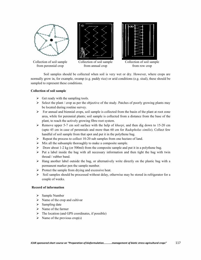
ICAR sponsored short course on “Preparation of bioformulat
Collection of soil sample from perennial crop
Collection of soil sample
Soil samples should be collected when soil is very wet or dry. However, where crops normally grow in, for example, swamp (e.g.sampled to represent these conditions.
Collection of soil sample
Get ready with the sampling tools. Select the plant / crop as per the objective of the study. Patches of poorly growing plants
be located during routine survey. For annual and biennial crops, soil sample is collected from the basin of the plant at root zone
area, while for perennial plants;plant, to reach the actively growing fibre root system.
Remove upper 5-7 cm soil surface with the help of (upto 45 cm in case of perennials and more than 60 cm for handful of soil sample from that spot and put
Repeat the process to collect 10 Mix all the subsample thoroughly to make a composite sample. Draw about 1-2 kg (or 500ml) from the composite sample and put it in a polythene bag. Put a label inside the bag with all necessary information and then tight the bag with twin
thread / rubber band. Hang another label outside the bag, or alternatively write directly on the plastic bag with a
permanent marker pen the sample number. Protect the sample from drying and excessive heat. Soil samples should be processed without delay, otherwise may be stored in refrigerator for a
couple of weeks.
Record of information
Sample Number Name of the crop and cultivar Sampling date Name of the farmer The location (and GPS coordinates, Name of the previous crop(s)
ICAR sponsored short course on “Preparation of bioformulation...........management of biotic stress agricultural crops”
Collection of soil sample from annual crop
Collection of soil sample from row crop
Soil samples should be collected when soil is very wet or dry. However, where crops normally grow in, for example, swamp (e.g. paddy rice) or arid conditions (e.g. sisal), these should be sampled to represent these conditions.
Get ready with the sampling tools. Select the plant / crop as per the objective of the study. Patches of poorly growing plants be located during routine survey. For annual and biennial crops, soil sample is collected from the basin of the plant at root zone
plants; soil sample is collected from a distance from the base of the vely growing fibre root system.
7 cm soil surface with the help of khurpi, and then dig down to 15upto 45 cm in case of perennials and more than 60 cm for Radopholus similis
handful of soil sample from that spot and put it in the polythene bag. Repeat the process to collect 10-20 sub samples from one hectare of land.
Mix all the subsample thoroughly to make a composite sample. 2 kg (or 500ml) from the composite sample and put it in a polythene bag.
label inside the bag with all necessary information and then tight the bag with twin
Hang another label outside the bag, or alternatively write directly on the plastic bag with a permanent marker pen the sample number.
mple from drying and excessive heat. Soil samples should be processed without delay, otherwise may be stored in refrigerator for a
cultivar
The location (and GPS coordinates, if possible) Name of the previous crop(s)
ion...........management of biotic stress agricultural crops” 117
Collection of soil sample
from row crop
Soil samples should be collected when soil is very wet or dry. However, where crops are paddy rice) or arid conditions (e.g. sisal), these should be
Select the plant / crop as per the objective of the study. Patches of poorly growing plants may
For annual and biennial crops, soil sample is collected from the basin of the plant at root zone soil sample is collected from a distance from the base of the
and then dig down to 15-20 cm Radopholus similis). Collect few
2 kg (or 500ml) from the composite sample and put it in a polythene bag. label inside the bag with all necessary information and then tight the bag with twin
Hang another label outside the bag, or alternatively write directly on the plastic bag with a
Soil samples should be processed without delay, otherwise may be stored in refrigerator for a
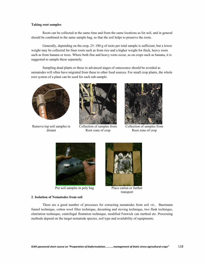
ICAR sponsored short course on “Preparation of bioformulation...........management of biotic stress agricultural crops” 118
Taking root samples
Roots can be collected at the same time and from the same locations as for soil, and in general should be combined in the same sample bag, so that the soil helps to preserve the roots.
Generally, depending on the crop, 25–100 g of roots per total sample is sufficient, but a lower weight may be collected for finer roots such as from rice and a higher weight for thick, heavy roots such as from banana or trees. Where both fine and heavy roots occur, as on crops such as banana, it is suggested to sample these separately.
Sampling dead plants or those in advanced stages of senescence should be avoided as nematodes will often have migrated from these to other food sources. For small crop plants, the whole root system of a plant can be used for each sub-sample.
Remove top soil samples in
distant Collection of samples from
Root zone of crop Collection of samples from
Root zone of crop
2. Isolation of Nematodes from soil
There are a good number of processes for extracting nematodes from soil viz., Baermann funnel technique, cotton wool filter technique, decanting and sieving technique, two flask technique, elutriation technique, centrifugal floatation technique, modified Fenwick can method etc. Processing methods depend on the target nematode species, soil type and availability of equipments.
Put soil samples in poly bag Place carton or further
transport
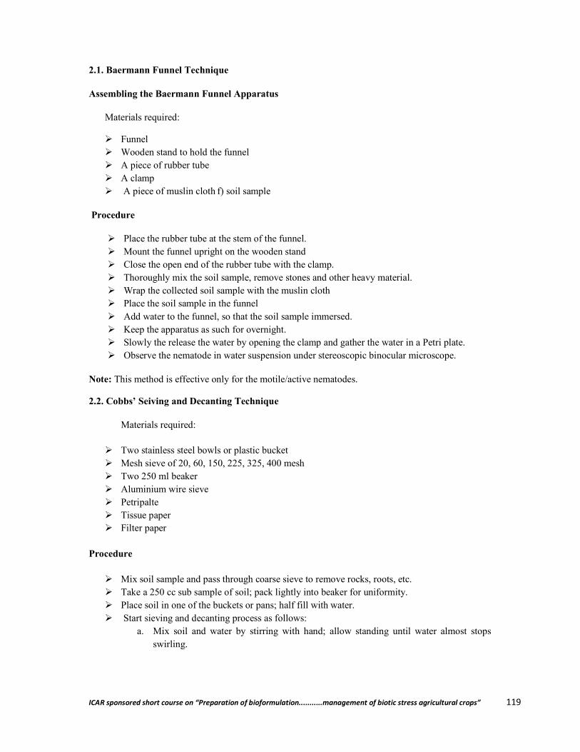
ICAR sponsored short course on “Preparation of bioformulation...........management of biotic stress agricultural crops” 119
2.1. Baermann Funnel Technique
Assembling the Baermann Funnel Apparatus
Materials required:
Funnel Wooden stand to hold the funnel A piece of rubber tube A clamp A piece of muslin cloth f) soil sample
Procedure
Place the rubber tube at the stem of the funnel. Mount the funnel upright on the wooden stand Close the open end of the rubber tube with the clamp. Thoroughly mix the soil sample, remove stones and other heavy material. Wrap the collected soil sample with the muslin cloth Place the soil sample in the funnel Add water to the funnel, so that the soil sample immersed. Keep the apparatus as such for overnight. Slowly the release the water by opening the clamp and gather the water in a Petri plate. Observe the nematode in water suspension under stereoscopic binocular microscope.
Note: This method is effective only for the motile/active nematodes.
2.2. Cobbs’ Seiving and Decanting Technique
Materials required:
Two stainless steel bowls or plastic bucket Mesh sieve of 20, 60, 150, 225, 325, 400 mesh Two 250 ml beaker Aluminium wire sieve Petripalte Tissue paper Filter paper
Procedure
Mix soil sample and pass through coarse sieve to remove rocks, roots, etc. Take a 250 cc sub sample of soil; pack lightly into beaker for uniformity. Place soil in one of the buckets or pans; half fill with water. Start sieving and decanting process as follows:
a. Mix soil and water by stirring with hand; allow standing until water almost stops swirling.
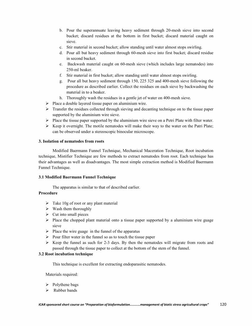
ICAR sponsored short course on “Preparation of bioformulation...........management of biotic stress agricultural crops” 120
b. Pour the superannuate leaving heavy sediment through 20-mesh sieve into second bucket; discard residues at the bottom in first bucket; discard material caught on sieve.
c. Stir material in second bucket; allow standing until water almost stops swirling. d. Pour all but heavy sediment through 60-mesh sieve into first bucket; discard residue
in second bucket. e. Backwash material caught on 60-mesh sieve (which includes large nematodes) into
250-ml beaker. f. Stir material in first bucket; allow standing until water almost stops swirling. g. Pour all but heavy sediment through 150, 225 325 and 400-mesh sieve following the
procedure as described earlier. Collect the residues on each sieve by backwashing the material in to a beaker.
h. Thoroughly wash the residues in a gentle jet of water on 400-mesh sieve. Place a double layered tissue paper on aluminium wire. Transfer the residues collected through sieving and decanting technique on to the tissue paper
supported by the aluminium wire sieve. Place the tissue paper supported by the aluminium wire sieve on a Petri Plate with filter water. Keep it overnight. The motile nematodes will make their way to the water on the Patri Plate;
can be observed under a stereoscopic binocular microscope.
3. Isolation of nematodes from roots
Modified Baermann Funnel Technique, Mechanical Maceration Technique, Root incubation technique, Mistifier Technique are few methods to extract nematodes from root. Each technique has their advantages as well as disadvantages. The most simple extraction method is Modified Baermann Funnel Technique.
3.1 Modified Baermann Funnel Technique
The apparatus is similar to that of described earlier. Procedure
Take 10g of root or any plant material Wash them thoroughly Cut into small pieces Place the chopped plant material onto a tissue paper supported by a aluminium wire guage
sieve Place the wire guage in the funnel of the apparatus Pour filter water in the funnel so as to touch the tissue paper Keep the funnel as such for 2-3 days. By then the nematodes will migrate from roots and
passed through the tissue paper to collect at the bottom of the stem of the funnel. 3.2 Root incubation technique
This technique is excellent for extracting endoparasitic nematodes.
Materials required:
Polythene bags Rubber bands
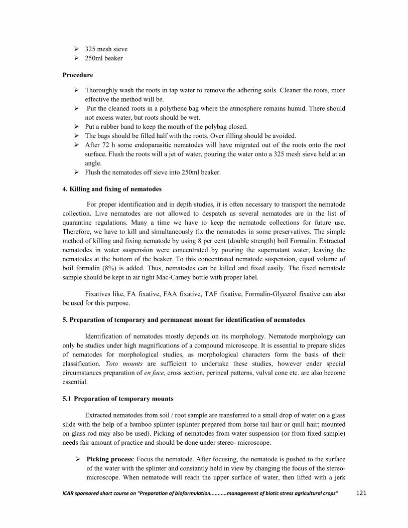
ICAR sponsored short course on “Preparation of bioformulation...........management of biotic stress agricultural crops” 121
325 mesh sieve 250ml beaker
Procedure
Thoroughly wash the roots in tap water to remove the adhering soils. Cleaner the roots, more effective the method will be.
Put the cleaned roots in a polythene bag where the atmosphere remains humid. There should not excess water, but roots should be wet.
Put a rubber band to keep the mouth of the polybag closed. The bags should be filled half with the roots. Over filling should be avoided. After 72 h some endoparasitic nematodes will have migrated out of the roots onto the root
surface. Flush the roots will a jet of water, pouring the water onto a 325 mesh sieve held at an angle.
Flush the nematodes off sieve into 250ml beaker.
4. Killing and fixing of nematodes
For proper identification and in depth studies, it is often necessary to transport the nematode collection. Live nematodes are not allowed to despatch as several nematodes are in the list of quarantine regulations. Many a time we have to keep the nematode collections for future use. Therefore, we have to kill and simultaneously fix the nematodes in some preservatives. The simple method of killing and fixing nematode by using 8 per cent (double strength) boil Formalin. Extracted nematodes in water suspension were concentrated by pouring the supernatant water, leaving the nematodes at the bottom of the beaker. To this concentrated nematode suspension, equal volume of boil formalin (8%) is added. Thus, nematodes can be killed and fixed easily. The fixed nematode sample should be kept in air tight Mac-Carney bottle with proper label.
Fixatives like, FA fixative, FAA fixative, TAF fixative, Formalin-Glycerol fixative can also be used for this purpose.
5. Preparation of temporary and permanent mount for identification of nematodes
Identification of nematodes mostly depends on its morphology. Nematode morphology can only be studies under high magnifications of a compound microscope. It is essential to prepare slides of nematodes for morphological studies, as morphological characters form the basis of their classification. Toto mounts are sufficient to undertake these studies, however ender special circumstances preparation of en face, cross section, perineal patterns, vulval cone etc. are also become essential.
5.1 Preparation of temporary mounts
Extracted nematodes from soil / root sample are transferred to a small drop of water on a glass slide with the help of a bamboo splinter (splinter prepared from horse tail hair or quill hair; mounted on glass rod may also be used). Picking of nematodes from water suspension (or from fixed sample) needs fair amount of practice and should be done under stereo- microscope.
Picking process: Focus the nematode. After focusing, the nematode is pushed to the surface of the water with the splinter and constantly held in view by changing the focus of the stereo-microscope. When nematode will reach the upper surface of water, then lifted with a jerk
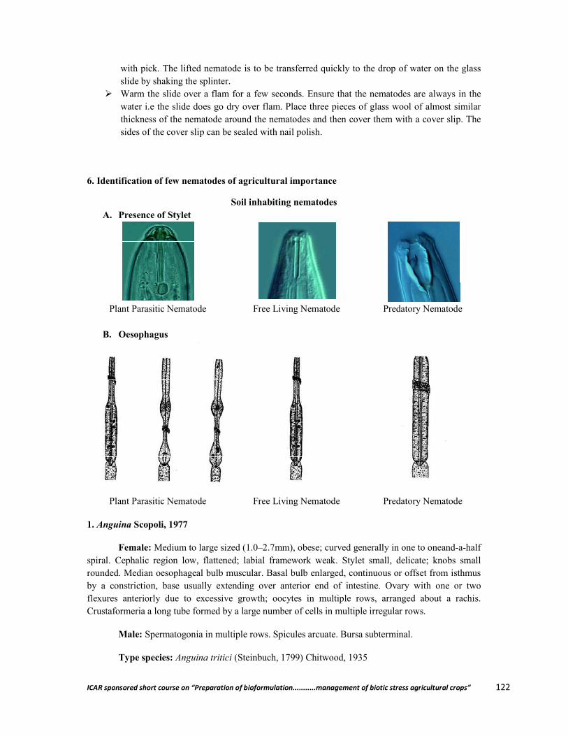
ICAR sponsored short course on “Preparation of bioformulation...........management of biotic stress agricultural crops” 122
with pick. The lifted nematode is to be transferred quickly to the drop of water on the glass slide by shaking the splinter.
Warm the slide over a flam for a few seconds. Ensure that the nematodes are always in the water i.e the slide does go dry over flam. Place three pieces of glass wool of almost similar thickness of the nematode around the nematodes and then cover them with a cover slip. The sides of the cover slip can be sealed with nail polish.
6. Identification of few nematodes of agricultural importance
Soil inhabiting nematodes A. Presence of Stylet
Plant Parasitic Nematode Free Living Nematode Predatory Nematode
B. Oesophagus
Plant Parasitic Nematode Free Living Nematode Predatory Nematode
1. Anguina Scopoli, 1977
Female: Medium to large sized (1.0–2.7mm), obese; curved generally in one to oneand-a-half spiral. Cephalic region low, flattened; labial framework weak. Stylet small, delicate; knobs small rounded. Median oesophageal bulb muscular. Basal bulb enlarged, continuous or offset from isthmus by a constriction, base usually extending over anterior end of intestine. Ovary with one or two flexures anteriorly due to excessive growth; oocytes in multiple rows, arranged about a rachis. Crustaformeria a long tube formed by a large number of cells in multiple irregular rows.
Male: Spermatogonia in multiple rows. Spicules arcuate. Bursa subterminal.
Type species: Anguina tritici (Steinbuch, 1799) Chitwood, 1935
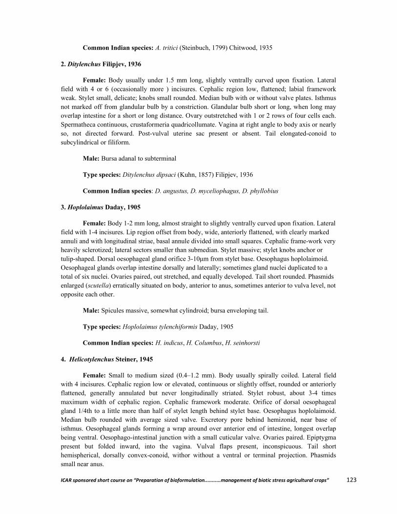
ICAR sponsored short course on “Preparation of bioformulation...........management of biotic stress agricultural crops” 123
Common Indian species: A. tritici (Steinbuch, 1799) Chitwood, 1935
2. Ditylenchus Filipjev, 1936
Female: Body usually under 1.5 mm long, slightly ventrally curved upon fixation. Lateral field with 4 or 6 (occasionally more ) incisures. Cephalic region low, flattened; labial framework weak. Stylet small, delicate; knobs small rounded. Median bulb with or without valve plates. Isthmus not marked off from glandular bulb by a constriction. Glandular bulb short or long, when long may overlap intestine for a short or long distance. Ovary outstretched with 1 or 2 rows of four cells each. Spermatheca continuous, crustaformeria quadricollumate. Vagina at right angle to body axis or nearly so, not directed forward. Post-vulval uterine sac present or absent. Tail elongated-conoid to subcylindrical or filiform.
Male: Bursa adanal to subterminal
Type species: Ditylenchus dipsaci (Kuhn, 1857) Filipjev, 1936
Common Indian species: D. angustus, D. myceliophagus, D. phyllobius
3. Hoplolaimus Daday, 1905
Female: Body 1-2 mm long, almost straight to slightly ventrally curved upon fixation. Lateral field with 1-4 incisures. Lip region offset from body, wide, anteriorly flattened, with clearly marked annuli and with longitudinal striae, basal annule divided into small squares. Cephalic frame-work very heavily sclerotized; lateral sectors smaller than submedian. Stylet massive; stylet knobs anchor or tulip-shaped. Dorsal oesophageal gland orifice 3-10μm from stylet base. Oesophagus hoplolaimoid. Oesophageal glands overlap intestine dorsally and laterally; sometimes gland nuclei duplicated to a total of six nuclei. Ovaries paired, out stretched, and equally developed. Tail short rounded. Phasmids enlarged (scutella) erratically situated on body, anterior to anus, sometimes anterior to vulva level, not opposite each other.
Male: Spicules massive, somewhat cylindroid; bursa enveloping tail.
Type species: Hoplolaimus tylenchiformis Daday, 1905
Common Indian species: H. indicus, H. Columbus, H. seinhorsti
4. Helicotylenchus Steiner, 1945
Female: Small to medium sized (0.4–1.2 mm). Body usually spirally coiled. Lateral field with 4 incisures. Cephalic region low or elevated, continuous or slightly offset, rounded or anteriorly flattened, generally annulated but never longitudinally striated. Stylet robust, about 3-4 times maximum width of cephalic region. Cephalic framework moderate. Orifice of dorsal oesophageal gland 1/4th to a little more than half of stylet length behind stylet base. Oesophagus hoplolaimoid. Median bulb rounded with average sized valve. Excretory pore behind hemizonid, near base of isthmus. Oesophageal glands forming a wrap around over anterior end of intestine, longest overlap being ventral. Oesophago-intestinal junction with a small cuticular valve. Ovaries paired. Epiptygma present but folded inward, into the vagina. Vulval flaps present, inconspicuous. Tail short hemispherical, dorsally convex-conoid, withor without a ventral or terminal projection. Phasmids small near anus.
ICAR sponsored short course on “Preparation of bioformulation...........management of biotic stress agricultural crops” 124
Male: Tail short, conical with a distinct terminal hyaline portion. Bursa enveloping entire tail tip, rarely subterminal. Gubernaculum trough or rod-shaped, fixed.
Type species: Helicotylenchus dihystera (Cobb, 1893) Sher, 1961
Common Indian species: H. dihystera, H. multicinctus, H. mucronatus, H. erythrinae
5. Rotylenchulus Linford & Oliveira, 1940
Juveniles, males and young females vermiform, arcuate to spiral upon fixation.
Mature female: Kidney shaped, with an irregular, less swollen neck, a postmedian vulva and a short pointed tail.
Immature female: Vermiform, c-shaped, Cuticle annulated. Lateral fields each with four incisures, not–areolated, Cephalic region high, continuous. Stylet 2-3 times lip region widths long. Orifice of dorsal gland usually one stylet length behind stylet base. Oesophagus hoplolaimoid. Ovaries paired with double flexures. Tail elongated-conoid, with prominent hyaline terminal portion.
Male: Stylet and oesophagus regressed, tail similar to that of young female; bursa subterminal, low not quite projecting beyond tail contour in lateral view. Spicules slender, lacking distal flanges, gubernaculum fixed. Cloacal lips pointed, not forming a tube; hypoptygma absent.
Juvenile: Tail more rounded terminally and with shorter hyaline terminal portion than that of female.
Type species: Rotylenchulus reniformis Linford & Oliveira, 1940
Common Indian species: R. reniformis 6. Pratylenchus Filipjev, 1936
Female: Body length under 0.8 mm. No sexual dimorphism in the anterior part of the body. Lateral fields each with 4-6 incisures, occasionally with oblique median markings. Cephalic region low, flattened anteriorly or rarely rounded, continuous with body contour; sclerotization massive. Stylet 20 μm or less long, with round, anteriorly flat or indented basal knobs. Median bulb oval to round, very muscular. Oesophageal glands usually less than two body widths long, extending over intestine mostly ventrally. Genital tract monoprodelphic, Spermatheca large, rounded, usually axial. Post- vulval uterine sac present, with or without rudiments of posterior ovary. Vulva in posterior region (usually at 70-80%). Tail subcylindrical to conoid, usually about 2-3 anal body widths long; terminus smooth or annulated, devoid of a process or mucro. Phasmids near middle of tail.
Male: Bursa enclosing tail terminus. Spicules with subterminal pore on dorsal side. Gubernaculum simple, trough–shaped, fixed.
Type species: Pratylenchus pratensis (de Man, 1880) Filipjev, 1936
Common Indian species: P. zeae, P. thornei, P. delattrei, P. coffeae, P. pratensis and P. vulnus

ICAR sponsored short course on “Preparation of bioformulation...........management of biotic stress agricultural crops” 125
7. Radopholus Thorne, 1949
Sexual dimorphism in the anterior region well marked, male having higher rounded and more offset cephalic region and cephalic framework, stylet and oesophagus markedly reduced.
Female: Body 0.4 -0.9mm long, straight to arcuate upon fixation. Lateral field with 3-7 incisures, not areolated. Deirids absent. Cephalic region low, continuous or slightly offset, annulated or smooth; framework strongly sclerotized. Stylet well developed, conus about as long as shaft. Oesophagus hoplolaimoid, oesophageal glands elongated, dorsal to intestine; subventral glands symmetrical or asymmetrical, much longer than the dorsal gland, nuclei of the three glands lie behind oesophago intestinal junction, just behind nerve ring. Ovaries paired, outstretched. Spermatheca round to oval. Vulva at 50-70% body length. Tail elongate-conoid to subcylindroid, usually 2-4 times anal body width long. Phasmids usually in anterior region of tail.
Male: Tail generally more tapering than that of the female; bursa subterminal. Spicules cephalated, slightly arcuate. Gubernaculum long, protrusible.
Type species: Radopholus similis (Cobb, 1893) Thorne, 1949
Common Indian species: R. similis 8. Hirschmanniella Luc & Goodey, 1964
Female: Long and slender (1-4 mm), straight to arcuate upon fixation. Lateral field with four incisures, areolated towards extremities but rarely also on most of body. Deirids absent. Cephalic region continuous, anteriorly flattened or hemispherical; framework heavily sclerotized, labial disc indistinct, annules not divided into sectors. Stylet massive, 15-46 μm long , conus tubular; basal knobs large, rounded. Orifice of dorsal oesophageal gland close to stylet base. Median oesophageal bulb round to oval, slightly offset from procorpus, with distinct valve plates. Oesophageal glands asymmetrical, larger and much longer than the dorsal gland; nuclei of three glands lying in a row, well separated from each other. Excretory pore near oesophago intestinal junction, behind hemizonid. Two branches of female reproductive organs equally developed. Spermatheca round to oval, axial. Ovaries mostly with single row of oocytes. Vulva transverse, lips not modified. Tail similar between sexes, elongate – conoid, usually with a terminal mucro; Phasmids pore –like, in the posterior third of tail.
Male: Similar to female. Spicules with subterminal pore appearing to be on dorsal side. Gubernaculum fixed, .male tail similar to female but carrying a simple, crenate, subterminal bursa lacking phasmidial pseudoribs. Cloacal lips not modified; hypoptygma absent.
Type species: Hirschmanniella spinicaudata ( Schuurmans Stekhoven, 1944) Luc & Goodey, 1964
Common Indian species: H. oryzae, H. gracilis and H. mucronata 9. Heterodera Schmidt, 1871
Mature female and cyst: Lemon shaped with a short neck and terminal cone, turns into hard-walled cyst, brown to black in colour, with a lace-like or zig-zag pattern. Vulva terminal. Anus dorsally subterminal, near vulva but not on vulval lip. Vulval fenestration present, ambifenestrate,
ICAR sponsored short course on “Preparation of bioformulation...........management of biotic stress agricultural crops” 126
bifenestrate or very rarely circumfenestrate; anal fenestration absent. Underbridge generally present. Bullae present or absent. Eggs retained in body; in some cases egg mass also present.
Male: Body twisted. Lateral fields each with four incisures, outer band often areolated. Lip region generally offset by a constriction, and with 3-6 annules; labial disc indistinct; basal lip annule may or may not be indented. Spicules robust, over 30 μm long, with blunt bifid or single tip. No cloacal tube. Tail very short and rounded.
Second stage juvenile: Body slender, straight to arcuate. Lip region generally continuous; labial disc indistinct. Stylet less than 30 μm long. Oesophageal glands filling body cavity, pointed with prominent terminal hyaline part. Phasmids punctiform.
Type species: Heterodera schachtii Schmidt, 1871
10. Gobodera Skarbilovich, 1959
Mature female and cyst: Spheroid with a short projecting neck, terminal region not forming a cone. Cyst brown, surface with a lace-like pattern Vulva terminal, slit length less than 15 μm, usually lost from old cysts; tuberculate area near vulva present. Vulval fenestrae circumfenestrate; underbridge and bullae rarely present. Anus dorsally subterminal not on dorsal lip, separated from vulva by a short distance, but both lie in a terminal vulval basin; no anal fenestra. All eggs retained in body.
Male: Body twisted; tail short, rounded, less than anal body width long. Lateral field with 4 incisures, outer bands often areolated. Lip region offset by constriction, with indistinct labial disc, and 3-7 annules. Spicules over 30 μm long, distally pointed. No cloacal tube.
Second stage juvenile: Labial disc subcircular; distinct lip sector present. Stylet less than 30 μm long. Oesophageal glands filing body cavity. Tail concial, pointed, with terminal half hyaline, Phasmids punctiform.
Type species: Globodera rostochiensis (Wollenweber, 1923) Behrens, 1975
11. Meloidogyne Goeldi, 1892
Female: Round to pear-shaped with short projecting neck, white, sedentary. No cyst stage. Vulva and anus close together, terminal; perineum with a fingerprint-like cuticular pattern, not elevated. Phasmids dot-like, slightly above and on either side of anus. Cuticle striated. Stylet slender, generally 12-15 μm long, with small knobs. Excretory pore anterior to median bulb, often closely behind base of stylet. Ovaries paired, prodelphic, convoluted. Rectal glands six, large secrete gelatinous material in which eggs are deposited; eggs not retained in body.
Male: Vermiform, up to 2 mm long, tail end twisted, develops by metamorphosis within a swollen larvae. Cuticle strongly annulated, lateral field with 4 incisures. Lip region not sharply offset with distinct labial disc and 1-3 annules; lateral sectors wider than submedians. Stylet robust, 18-25 μm long with large knobs. Oesophageal glands mostly ventral to intestine. Spicules slender, generally 25-35 μm long, gubernaculum 7-11 μm long. Testis single or paired (in case of sex reversal). Tail rounded. Phasmids dot-like, near cloacal aperture which is subterminal. Bursa absent.

ICAR sponsored short course on “Preparation of bioformulation...........management of biotic stress agricultural crops” 127
Second stage juvenile: Vermiform, straight to arcuate on death. Lip region with coarse annules (1-4), a distinct labial disc; framework lightly sclerotized, lateral sectors wider than submedians. Stylet slender, under 20 μm. Excretory pore posterior to hemizonid. Median bulb with large oval valve plates. Tail with conspicuous hyaline region, tip narrow and irregular in outline.
Type species: Meloidogyne exigua Goeldi, 1892
Common Indian Species: M. incognita, M. graminicola, M. arenaria, M. javanica The most important taxonomic character for species identification of Meloidogyne is the perineal pattern
12. Tylenchulus Cobb, 1913
Adult female: Elongate- obese, sedentary enlarging behind median bulb mostly on dorsal side, ventrally arcuate; neck elongated; post-vulval part elongate tapering. Stylet 11-15 μm long, knobs prominent. Excretory system well developed, produce gelatinous matrix. Excretory pore in front of vulva at 68-85% of body length from anterior end. Basal bulb offset from intestine. Vulva a transverse slit. Post-vulval uterine sac absent. Spermatheca present. Ovary coiled or with 1-2 flexures. Anus obscure. Tail tapering, tip rounded or with a peg.
Immature female: Slender straight to arcuate. Stylet and oesophagus as in mature female. Ovary immature with few oocytes.
Male: Short slender, straight to slightly arcuate. Stylet and oesophagus degenerated. Excretory pore at 53-60% of body length. Spicules slender, arcuate. Gubernaculum simple, fixed. Bursa absent, tail elongate-conoid, tip rounded or with a peg.
Second stage juvenile: Slender, straight to slightly arcuate. Lateral field with two incisures. Stylet 11-15 long with rounded knobs. median bulb oval or fusiform with elongate valve-plates, isthmus long, basal bulb offset from intestine. Tail elongate conoid.
Type species: Tylenchulus semipenetrans Cobb, 1913
Common Indian species: T. semipenetrans
13. Aphelenchus Bastian, 1865
Female: Straight or ventrally arcuate when fixed, 0.8- 1.4 mm long, vermiform. Labial region weakly sclerotized. Stylet weak, without basal swelling. Median oiesophageal bulb well-developed, rounded-rectangular in shape and more or less filling body diamaeter. Dorsal oesophageal gland duct opening within bulb, just anterior to valve plates. Oesophageal gland lobe originating from a short isthmus and overlapping intestine dorsally. Genital tract monoprodelphic withwith post uterine sac. Tail cylindroid without mucro.
Male: Straght or ventrally arcuate when fixed. Spicules arcuate, caudal alae covering the entire tail and provided with bursal rays.
Type species: A. avenae Bastian, 1865
ICAR sponsored short course on “Preparation of bioformulation...........management of biotic stress agricultural crops” 128
Common Indian species: A. avenae, A. radicicolus
14. Aphelenchoides Fischer, 1894
Female: Straight or ventrally arcuate when fixed, 0.4- 1.2 mm long, vermiform. Labial region weakly sclerotized. Stylet weak, usually with basal swelling. Median oiesophageal bulb well-developed, spherical to rounded-rectangularin shape and more or less filling body diamaeter. Dorsal oesophageal glanf duct opening within bulb, just anterior to valva plates. Oesophageal gland lobe originating from median bulb itself and overlapping intestine dorsally. Genital tract monoprodelphic with post uterine sac. Tail medium conoid with mucro.
Male: Walking stick-shaped due to curling of tail. Spicules Rose- thorn shaped, no bursa.
Type species: Aphelenchoides kuehnii Fischer, 1894
Common Indian species: A. andrassyi, A. asterocaudatus , A. besseyi, A. bicaudatus, A. composticola and A. swarupi
15. Xiphinema Cobb, 1913
Female: Slender, 1.3-5 mm long. Labial region continuous or set off. Amphids stirrup shaped with slit-like amphidial apertures. Stylet 60-200 um long, consisting of a needle like odontostyle with a forked base attached to an odontophore with flanges. Guiding ring double, located near the junction of odontostyle and odontophore. Oesophagus consisting of a long narrow slender part and a short basal bulb. Basal bulb provided with three oesophageal gland nuclei; dorsal one being larger than subventrals. Genital tract amphi- or monopistho- or pseudomonodelphic with reflexed ovaries. Vulva located at 25 to 65% of total body length from anterior end. Tail variable from short rounded to long filiform. Male: Diorchic. Spicules very prominent. Ventromedian supplements present. No bursa.
Type species: Xiphinema americanum Cobb, 1913
Common Indian species: X. americanum, X. basiri, X. insigne and X. opisthohysterm.
**************

ICAR sponsored short course on “Preparation of bioformulation...........management of biotic stress agricultural crops” 129
Role of nematode in disease complex
Bhabesh Bhagawati Department of Nematology
Assaam Agricultural University, Jorhat- 785013, Assam
What are nematodes?
Nematodes are small, microscopic organisms which are found in all types of habitats. Most of the plant parasitic nematodes are soil inhabitants and are not visible. That is why, they are known as the hidden enemies of farmers. The plant parasitic nematodes are 0.3-1.0 mm in length; exceptionally few nematodes may be as long as 1.0 cm. They do not have any colour and their body is un-segmented.
Shapes of nematodes
They may be of different shapes but most of the plant parasitic nematodes are vermiform in shape i.e. they are slender, cylindrical, tapering towards both ends.
Depending upon their nature of feeding the nematodes may assume round, globular, kidney shapes or may retain the vermiform shapes throughout the life.
Plant parasitic nematodes of economic importance
Common name Genera Major species
Cyst nematodes Heterodera
H. glycines, H. avenae, H. schachtii, H. trifolii, H. gottingiana, H. cajani, H. zeae
Globodera G. rostochiensis, G. pallida, G. tabacum.
Root-knot nematodes Meloidogyne M. arenaria, M. incognita, M. javanica, M. hapla, etc.
Stem and bulb nematode Ditylenchus D. angustus, D. destructor, D. dipsaci Seed gall nematodes Anguina A. tritici, A. agrostis,
Lesion nematodes Pratylenchus P. penetrans, P. brachyurus, P. coffeae, P. zeae, P. goodeyi, P. thornei, P. vulnus.
Burrowing nematodes Radopholus R. similis Root nematodes Hirschmanniella H. oryzae, H. mucronata, H. spinicauda Reniform nematodes Rotylenchulus R. reniformis Citrus nematode Tylenchulus T. semipenetrans Spiral nematodes Helicotylenchus H. multicinctus, H. mucronatus, H. dihystera, Reniform nematodes Rotylenchulus R. reniformis Ring nematodes Criconemella C. xenoplax, C. axestis, C. spharocephalum Xiphinema, Longidorus, Trichodorus Paratrichodorus
Dagger, Needle stubby root nematodes
X. americanum, X. index, L. africanus, T. chrisitie & Paratrichodorus minor
Role of phyto-nematodes in disease development:
1. Wound causing agents
Mechanical wounding caused by the nematode promotes the easy invasion of fungal and bacterial pathogens. All together pathogens cause more severe damage than either of the pathogen alone.
ICAR sponsored short course on “Preparation of bioformulation...........management of biotic stress agricultural crops” 130
Tobacco cultivars resistant to black shank disease caused by the fungus Phytophthora parasitica var. nicotianae suffered severe wilt in presence of root-knot nematode. Lesions caused by Pratylenchus penetrans on brinjal and tomato make them more susceptible to wilt caused by Verticillium dahliae.
Nematodes may assist the pathogenic bacteria to enter/damage the host plant by providing them igress points at the site of nematode penetration, as carriers, vectors or as resistance breakers. Bacterial wilt of certain carnation increases manifold in plants infected with Meloidogyne or Helicotylenchus sp. Agrobacterium tumefaciens and can cause crown galls in peach and raspberry only in presence of root-knot nematode. Sometimes the nematode juveniles carry bacterial cells on their body surface and establish them at the site of infection. Ditylenchus dipsaci is known to carry Pseudomonas fluorescens in garlic where it causes ‘Caif au lait’ bacteriosis.
2. Host modifying agents
Modification of host tissue by endoparasitic nematodes may be localized or systemic in nature. Powell and Nusbaum (1960) were the first to demonstrate that the modification in host substrate caused by nematodes provide advantage to fungal pathogens. They demonstrated that Phytopthora parasitica var. nicotianae entered the roots of resistant cultivars of tobacco if roots galls caused by Meloidogyne incognita acrita have developed. Another example is Meloidogyne javanica and Rhizotonia solani on tobacco where fungus was unable to colonise healthy roots. Root knot infected roots were however heavily galled.
The altered tissue also favors the establishment of bacterial pathogens (Johnson and Powell, 1969). They showed that RKN act as modifier of infected tissue in such a way that the infested tissue and surrounding cells become more suitable for bacterial colonization. They found that plant inoculated with nematodes 3-4 weeks prior t bacterial al inoculation develop bacterial wilt symptoms to a greater extent than plants inoculated with nematode and bacteria simultaneously.
3. Rhizosphere modifying Agents
Phytonematodes during feeding invariably cause modifications in the host substrate that is advantageous for fungal pathogens. These modifications are simpler in ectoparasitic nematodes but more extensive and complex in endoparasitic nematodes. Root-knot nematodes cause increased leakage of carbohydrates, proteins and amino acids from the giant cells and galled tissue which activate the resting spores of fungal pathogens. Prior infection of Meloidogyne incognita is essential for Rhizoctonia solani to cause root rot disease in tobacco and okra.
Root exudates exert a powerful influence on the size and competition of the bacterial population next to the roots during the entire life of the plant. They affect the life process of the plant and also the plant’s resistance to soil borne pathogens. With increasing proximity to plant roots there are increasing of microorganism, bacteria being the most responsive organisms of the mircoflora to root exudates.. Physiological changes induced within the root tissue by endopatrastic nematodes bring about qualitative changes in root exudates. Root exudates stimulate the dormant stages of the fungi in the rhizosphere.
4. Breakers of disease resistance
In several cases a cultivar resistant to some root infecting fungi become susceptible in presence of root knot nematodes. Fassuliotis and Rau (1969) postulated that two types of resistance of Fusarium
ICAR sponsored short course on “Preparation of bioformulation...........management of biotic stress agricultural crops” 131
wilt- qualitative and quantitative. Qualitative resistance is based on incompatibility between the host and the fungal pathogen. Quantitative resistance is influenced by nematode infection because of improved nutrition for the fungus resulting from nematodes attack (Pitcher, 1965). Jenkins and Coursen (1957) noticed breaking of resistance to Fusarium wilt in tomato cultivars in presence of Meloidogyne incognita and M..hapla.
Resistance breaking to bacterial pathogens implies that there is a possibility of that PPN could be able to supply something which is obviously lacking in resistance cultivar in the absence of nematode invasion, or the nematode could alter cell permeability, making a particular cultivar susceptible to bacterial colonization (Pitcher, 1978). Reddy et al., (1979) reported that when the eggplant cultivar Pusa puple cluster (highly resistance to Pseudomonas solanacearum) was inoculated with a combination of the bacterium and M. incognita, a greater number of plants wilted when the organisms were inoculated simultaneously than when one was inoculated before the other.
5. Nematode act as vector of bacterial pathogen
Bacteria can be transmitted by nematodes externally on their body surface or internally within the alimentary canal. Some nematodes have been implicated as disseminators of plant –pathogenic bacteria.
External transmission of plant pathogenic bacteria by some nematodes is significant in the development of some disease complex in plants. The nematodes act as vectors of the bacteria and carry bacterial cells over their body. Yellow ear rot ‘tundu’ disease of wheat results from joint infection of Clavibacter tritici (= Corynebacterium michiganense pv. tritici) and Anguina tritici (Gupta and Swarup, 1968). Rhodococcus fascians (=Corynebacterium fascians) and Aphelenchoides ritzemabosi or A. fragariae together cause cauliflower disease of strawberry (Cross and Pitcher, 1952).
6. Nematode act as vector of plant viruses
Nematode transmitted viruses belong to two different groups, NEPO and NETU or tobra viruses respectively. Nematode ingests viruses and become viruliferous when they feed on roots of virus infected plants. Though long been suspected to act as virus transmitting agents, it was only in 1958 when Hewitt, Raski and Goheen experimentally proved that Xiphinema index was responsible for transmitting Grapevine fan leaf virus (GFLV). Presently, more than 100 species of root ectoparasites belonging to five genera viz., Xiphinema, Longidorus, Paralongidorus, Trichodorus and Paratrichodorus are known to act as vectors of viruses in plants. Interestingly, all these genera belong to order Dorylaimida, have global distribution and moderate to wide host. The acquisition, retention, dissociation and inoculation are steps involved in transmission of virus in plant by plant parasitic nematode.
Plant parasitic nematode Transmitted virus References
Xiphinema americanum Cherry rasp leaf Tobacco ring spot Tomato ring spot
McGuire, 1973
X. brevicolle Tomato ring spot Lambert et al., 1975
X. diversicaudatum Arabis mosaic virus Harrison et al., 1971
X. index Grapevine fan leaf Alfaro et al., 1974
Longidorus elongates Paratrichodorus minor
Pepper ring spot, Tobacco rattle
Christie and Perry , 1951

ICAR sponsored short course on “Preparation of bioformulation...........management of biotic stress agricultural crops” 132
7. Nematode act as non vector of plant viruses
The general interactions between nematodes and plant viruses have effects on the multiplication of each other and thus influence the plants to a greater or lesser extent than when present alone. The first general interaction between nematodes and viruses was reported from Germany by Goffart (1956) who studied the relationship between Heterodera schachtii and yellow disease of sugarbeet. These general interactions attained considerable attention in India after swarup and Goswami (1969) reported the leaf curl virus and root knot nematode interaction in tomato. Several workers have observed more suppression of plant growth in combination than the damage caused by each of the pathogen alone. Goswami et al. (1971) stated that M.incognita and tobacco mosaic virus together causes greater reduction in vitamin C contents in tomato plants thereby hampering the plant metabolism. The combined effect of nematode and virus weekened the plants considerably causing reduced uptake, poor translocation of the inorganic elements and metabolites to the above ground parts of the tomato plants whereas virus and virus preceding nematode inoculation caused an increase in N, P, K, Ca, Mg contents of shoots (Goswami et al., 1976).
Conclusion
Apart from nematodes being able to cause disease in crops by itself, they also play certain roles in predisposing the plants to attack of other plant pathogens by bringing about modification in the rhizosphere, host substrate, breaking resistance to the other pathogens. While doing all these, they may act as incitant, aggravators and vectors of plant pathogens. Effort should be made for proper identification and understanding of bio ecology of nematodes attacking various crops.. Further, before going for management of the diseases of complex aetiology, the role of each pathogen involved has to be ascertained.
References:
Goffart, H. (1956). Uber daszasammenwirken von rubennematoden (Heterodera schachtii) and vergibsbungskrankheit (Beta virus 4). Modidelingen van de landbouwwhogeschod van de Statle Gent. 21: 351-360
Goswami, B.K.; Raichoudhury, S. P. and Khurana, S. M. P. (1971). Changes in vitamin C content of fruit in nematode resistant and susceptible tomato varieties with infection of nematode and/ tobacco mosaic virus
Goswami, B.K.; Singh, S. and Verma, V .S. (1976). Uptake and translocation of calcium and magnesium in tomato plants as influenced by infection with root knot nematode, Meloidogyne incognita and tobacco mosaic virus. Nematologica. 22: 116-117
Gupta, P. and Swarup, G (1968). On the ear-cockle and yellow-rot diseases of wheat.II Nematode bacterial association. Nematological, 18: 320-4
Harrison, B. D.; Finch, J. T.; Gibbs, A. J.; Hollings, M.; Valenta, R. J. and Finch, J. T. (1971). Sixteen groups of plant viruses. Virology. 45:356-363.
Khan, M. W. (1993). Nematode interactions. Publication Chapman and hall, London pp-55-115. McGuire, J. M. (1973). Retention of tobacoo ringspot virus by Xiphinema americanum.
Phytopathology 63:324-326. Mohilal, N. and Gambhir, R .K. (2006). Plant Nematology in India. Publication –Published by
S.Saratkumar and M.Promodini. pp 73-87. Pitcher , R. S (1978). Interaction of nematodes with other pathogen, in Plant Nematology 3rd
edn (Ed. J. F. Southey), Her Majesty’s Stationery Office, London, pp 63-77.
ICAR sponsored short course on “Preparation of bioformulation...........management of biotic stress agricultural crops” 133
Powell, N. T. and Nusbaum, C. J. (1960). The blank shank –root-knot complex in flue-cured tobacoo. Phytopathology, 50: 899-906.
Reddy, P. P.; Singh, D. B. and Ramkishun (1979). Effect of root-knot nematodes on susceptibility of Pusa Purple Cluster brinjal to bacatrial wilt. Current Science 48: 915-16.
**************
ICAR sponsored short course on “Preparation of bioformulation...........management of biotic stress agricultural crops” 134
Biological Control of Plant Parasitic Nematodes
Bhupen Choudhury AICRP on Nematology, Department of Nematology
Assaam Agricultural University, Jorhat- 785013, Assam
The antagonists of nematodes are a fascinating group of organisms, as they have diverse feeding habits and and some novel mechanisms of parasitism and predation. Many of the antagonists used in biological control experiments have been selected at random and little consideration appears to have been directed towards the target nematodes. They have no effect on eggs and endoparasitic stages which are the dominant phases of the life cycle of plant parasitic nematodes. Unlike other pests, nematodes are not easy to control by the use of biological control agents. The main reasons of this limitation are:
1. Plant parasitic nematodes- a difficult target
Plant parasitic nematodes have evolved protective structures and metabolic adaptations which allow them to survive and flourish in harsh and competitive soil environment. The body of the nematode is protected by multi-layered pertinacious cuticle which functions as a flexible skeleton and as a barrier to undesirable elements. The cuticle is freely permeable to water but differently permeable to various ions and other chemicals. It is also relatively resistant structure and is readily not destroyed by chemical or bio agents.
The eggs of nematodes are also well protected, as the embryo is surrounded by a triple layered shell which contains chitin as its major component. Once embryological development is complete, antagonists face the additional problem of penetrating not only the egg shell but also cuticle of unhatched juveniles. Some nematodes lay their eggs in gelatinous matrix or are retained within the body of females, which may act as an additional protective layer.
In addition to the structural features, the physiological capacity of many plant parasitic nematodes to survive adverse conditions may give them an advantage over some of their parasites and predators. Nematodes are the most successful anhydrobiotic animals and are less likely to be affected by dry conditions (Womersly, 1987).
The high reproductive capacity of plant parasitic nematodes is one of important features which make them such significant pests as well as difficult to control. The life cycle of many nematodes takes only few weeks and each female has capacity to produce hundreds and thousands of progenies.
2. Plant parasitic nematodes – a diverse target
Although plant parasitic nematodes share many common characteristics, they should not be considered as a single homogeneous target for biological control agents. The sedentary endoparasites, migratory endoparasites, ecto-endoparasites, ectoparasites and above ground parasites have different characteristics and these must be considered when biological control strategies for a particular nematode is being developed.
2.1 Sedentary endoparasites
The root knot nematodes (Meloidogyne spp.) and cyst nematodes (Heterodera spp. and Globodera spp.) are completely surrounded by root tissues for most of their life and thereby protected from soil borne parasites and predators. The juveniles of these nematodes have the capacity to enter the root thereby they escape the predation and parasitism. Since Meloidogyne females are protected by roots and juveniles are mobile, the eggs appear to be the stage of life cycle most vulnerable to the attack by

ICAR sponsored short course on “Preparation of bioformulation...........management of biotic stress agricultural crops” 135
antagonists. Eggs are generally located on root surface and an effective parasite or predator could be expected to eliminate many of the eggs.However, there are situations where eggs are laid within galled tissues and antagonists are unable to exert effective control as they have limited access to the eggs.
2.2 Sedentary semi endoparsites
This group of nematodes (Rotylenchulus and Tylenchulus) have juveniles and young females that migrate and feed ectoparasitically and saccate females that are partly exposed on the root surface throughout their life and eggs that are laid in a gelatinous matrix secreted by the females. The damage caused by the feeding process could be expected to increase leakage of exudates from roots and perhaps the additional nutrients stimulate general microbial activity in the vicinity of nematodes and this makes it more difficult for parasites and predators to become established.
2.3 Migratory endoparasites
The migratory endoparasites (Pratylenchus, Radopholus etc.) are likely to be most difficult to control by bio agents as they spend much of their lives in root and tend to be found in soil when host plants are stressed or harvested. Since eggs are laid in root tissues, juveniles hatch and develop to maturity without moving from the root tissues, multiplication can proceed for several genarations without nematodes being exposed to antagonists.
2.4 Ectoparasites
Although ectoparasites are the only group of nematodes that do not receive protection from the roots, their migratory nature makes them a difficult target for bio agents.
2.5 Above ground feeders
The nematodes belong to genera Anguina, Ditylenchus, Aphelenchoides etc. are important pest of many crops. They feed on avobe ground plant parts like stem, leaf, bud etc. In someways they are similar to migratory endoparasites because they are protected from antagonists by the plants. It is easy to use the bioagents in soil environment than in aerial parts of the plant.
Since Linford et al.(1938) who first laid out classical pot and field experiments on management of plant parasitic nematodes by biocontrol agents, lots of efforts have been made to control many important nematode pests by using different groups of antagonists. The antagonists or biological control agents against nematodes are belong to:
Fungi Bacteria VAM Predatory nematodes
Out of these, few potential biocontrol agents belonging to fungi and bacteria are more extensively used against nematodes. Some of the potential and extensively used fungi and bacetria will be discussed here.
1. Trichoderma spp.
Trichoderma are one of the small but potential group of beneficial fungi that are present nearly all soils and diverse habitats. They have proven commercially viable as a biological control agent. This microorganism is registered as a biopesticides in many countries including India and its different

ICAR sponsored short course on “Preparation of bioformulation...........management of biotic stress agricultural crops” 136
formulations are available in markets at different names. There are many species of Trichoderma but T.viride and T.harzianum are most commonly used against nematodes.
Mechanisms of action against phytonematodes
The following are the mechanisms by which Trichoderma spp.functions:
a) Mycoparasitism b) Antibiosis c) Competition for nutrients or space d) Tolerance to stress through enhanced root and plant development e) Induced resistance f) Solubilization and sequestration of inorganic nutrients
a. Mycoparasitism
This is complex process that involves trophic growth of the biocontrol agent towards target pathogen. Trichoderma hyphae penetrate the cell wall of the target nematodes and suck the cell contents. These fungi have the ability to secrete chitinases which dissolve the cell wall of nematodes. Nematode cell wall as well as the egg shells is provided with chitin which makes them impermeable to foreign materials. So these fungi can dissolve the chitin of the cell wall (Chet, 1987). Some species of Trichoderma can produce acetic acids which has nematicidal properties (Dijian et al.,1991). Moreover, they can penetrate the nematodes through the natural openings like mouth, vulva and anus.
b. Antibiosis
Antibiotics are low molecular weight secondery metabolitesproduced during nutrient limiting conditions. About 43 substances produced by Trichoderma spp. which have antibiotic activities have been recorded. It produes antibiotics like trichodermin, deramatin, trichoviridin, gliotoxin, gliovirin, trichotoxin etc. These antibiotics interact with phospholipid membrane of nematodes and induce membrane permeability. Many of these antibiotics are synergistic with cell wall degrading enzymes.
c. Competition
Competition for space and nutrients has long been considered as one of the classical mechanisms of biocontrol by Trichoderma spp.(Chet, 1987). They have high rhizosphere competency and can easily colonize the roots.They reduce the feeding cites for nematodes.
d. Tolerance to stress through enhanced root and plant development
Another possible mechanism gaining credence is tolerance to stress through enhanced root and plant growth. The enhanced rooting due to application of Trichoderma probably also induces tolerance to pest that it does directly control. The larger root systems of plant colonized by Trichoderma are better able to withstand the damaging effect of nematodes.
e. Induced resistance
Trichoderma spp. elicits resistance in plants. T.viride was found to induce systemic resistance against root knot nematode Meloidogyne incognita in green gram (Umamaheswari el al., 2002). Enzymes such as peroxidase, polyphenol oxidase, phenyl alanine ammonia lyase, catalase and chitinase are induced in T.viride treated plants. Induction of these defence mechanisms determine the ability of a plant to survive any pathogen attack.
ICAR sponsored short course on “Preparation of bioformulation...........management of biotic stress agricultural crops” 137
f. Solubilization and sequestration of inorganic nutrients
In soil, various plant nutrients undergo complex transitions fro unsoluble to soluble forms that influence their accessibility and absorption by roots. Enhanced nitrogen use efficiency was documented in corn by Piekielek and Fox (1992) due to application of T.harzianum as seed treatment. A number of poorly soluble nutrients like rock phosphate, zinc, manganese, iron, copper etc. are made soluble to plants due to application of T.harzianum (Altomare et al., 1999).
Trichoderma spp. are found to be effective against sedentary nematodes like Meloidogyne, Heterodera, Globodera.They can be used as seed treatment or as soil application.Seeds of different vegetables like tomato, brinjal, okra, cowpea and pulses like black gram, green gram, pea etc are treated with T.viride @ 10g/kg seed. It is also effective as soil application. T.viride or T.harzianum should be applied in soil before sowing or transplanting @ 2.5 kg/ha (Talc formulation). The spore load should be 2x10⁸ cfu. For effective use, it should be multiplied in FYM or vermicompost. The biocontrol agent should be mixed with these materials and incubated for 15 days. Before application in the field it is advised to assess the spore load (Anonymous, 2017).
Trichoderma spp. is compatible with many insectides, nematicides, fungicides etc.
2. Paecilomyces lilacinus (= Purpureocillium lilacinum)
It is an opportunistic fungi which is a very good egg parasite of nematodes. Jatala et al.,(1979) were the first to report P.lilacinum as a parasite of eggs of M.incognita on potato roots in Peru. In India, it was first isolated from M.incognita eggs during a survey in 1993-94 (Goswami and Uma Rao, 1997).
Mode of action
The fungus has been reported to parasitize eggs of many sedentary endoparasitic nematodes. The infection process starts with growth of fungal hyphae in the gelatinous matrix and eventually the eggs of nematodes are engulfed by the mycelial hyphae. The proliferated hyphal branches penetrate the eggs. In cyst nematodes, the fungus penetrate through vulva or the broken and exposed neck region.After entering the cyst, the fungus grows saprophytically on the body content surrounding the eggs during or before its parasitism of the eggs. In all cases, eggs in the early embryonic developmental stages prior to gastrulation process are more vulnerable to infection. Once the hyphae is in contact with the eggs a series of ultrastructural changes occurs in the eggs due to effects of exogenous metabolites and chitinolytic activities of the fungus. Once inside, the fungal mycelium radiated profusely in the eggs of early embryonic development and the entire embryo is replaced by the mycelial biomass. Occasionally, P.lilacinum may penetrate the egg laying female through the anus or vulva. In such cases, the infected female body cavity filled with the fungal biomass and the nematode die.
P.lilacinum is compatible with organic amendments like neem cake, castor cake and green manures. It can also be combined with Trichoderma viride, Verticilium chlamydosporium (= Pochonia chlamydosporia) against root knot nematode and best as seed treatment. It can also be applied in soil. In that case, its culture filtrate should be used to enrich the vermicompost and this should be used before sowing or transplanting (Anonymous, 2017).Its liquid and powder formulations are available in different names. Pre planting soil treatment of P.lilacinum was found effective in reducing root gall and soil population of M.incognita in tomato (Kiewnick and Sikora, 2006)
ICAR sponsored short course on “Preparation of bioformulation...........management of biotic stress agricultural crops” 138
3. Pseudomonas fluorescens
Among the different PGPR strains, P.flourescens has been proved to be an efficient biocontrol agent against plant parasitic nematodes.
Mechanisms of action by PGPR strains
a. Plant growth promotion
Mechanisms of growth promotion mays be direct, to be mediated by production of plant hormones such as auxins, cytokinins or gibberellic acid (Arshad and Berger,1991) or indirect due to control of minor pathogens such as deleterious rhizobacteria (Schippers et al.,1987).
b. Parasitism and lysis
The Pseudomonas spp.produce lytic enzymes such as chitinase and glucanase which are involved in degradation of chitin layer of nematode eggs(Loganathan et al.,2001). Moreover, some other toxic substances are also produce by PGPR for which the juveniles of Meloidogyne and Heterodera become vulnerable
c. Induced systemic resistance
The defensive capacity of the host plants increases due to the introduction of PGPR. The resulting elevated resistance due to biotic agent like Pseudomonas is referred to as ISR In PGPR treated plants, several defence related proteins and chemicals viz., peroxidase, phenylalanine ammonia lyase, polyphenol oxidase, phenols, chitinase, glucanase etc are reported (Loganathan et al.,2001).
P.flourescens can effectively be used against sedentary endoparasitic nematodes like Meloidogyne, Heterodera and Globodera. It is very effective as seed treatment. It can also be used in combination with other biocontrol agent like Trichoderma viride. It is effective as soil application also (Anonymous, 2016). The spore load should be 2x10⁸cfu.
Different liquid and powder formulations of Psedumonas flourescens are available in market now a day. Moreover, in most of the Agricultural universities these common biocontrol agents are available. It is always advisable to use the local isolates of biocontrol agents not only against nematodes but against all pest and diseases.
4. Pasteuria penetrans
This is an obligate parasitic bacterium of phytoparasitic nematodes and has attracted the attention of nematologists and bacteriologists since this has great potential as biocontrol agent against plant parasitic nematodes. It is mycelial, gram positive endospore forming bacterial parasite. The bacterial parasite produces endospore that adheres to the cuticle of plant parasitic nematodes and interface with the life cycle of the nematodes resulting in the suppression of nematode population.
Life cycle
Life cycle of P.penetrans starts with endospore, the infective stage of the bacterium that perpetuates year after year in the soil and and are released on decomposition of nematode body of adult females and males or in plant root tissues. The spores of P.penetrans attach to the cuticle of second stage juvenile and germinate within a few days after the juvenile starts feeding within the root. The germination of spores occure only after the encumbered second stage juveniles invade the plant root and
ICAR sponsored short course on “Preparation of bioformulation...........management of biotic stress agricultural crops” 139
begin to feed. The germ tube emerges through a central opening in the basal attachment layer and penetrates the cuticle of the nematodes and finally reaches the pseudocoelom. The germ tube in the pseudocoelom develops into vegetative microcolony consisting of dichotomously branched septate mycelium. The intercalary cells in the microcolony lyse and give rise to daughter colonies. The process continues resulting in a large number of daughter colonies containing fewer but large vegetative cells. Eventually, quartets of developing sporangia predominate in the nematode body cavity. These quartets separate into doublets of sporangia and finally separate into a single sporangium that will eventually contain an endospore. Endospores of P.penetrans are released into the soil when the plant roots with parasitized root knot nematode female decompose. The life cycle completes within 20 30 days at 30o C
Biocontrol potential
There are many strains of P.penetrans and the strains are host specific. P. penetrans exerts various degrees of nematode biocontrol under greenhouse and field condition. Dried root powder containing spores of P.penetrans when incorporated in the soil @ 100mg/kg soil, 99 per cent second stage juveniles of Meloidogyne javanica were infected. Kokalis (2015) effectively controlled M.incognita in tomato and cucumber and M.arenaria in snapdragon by seed treatment. It can be used as seed treatment as well as soil application with known spore load.Moreover, it is compatible with nematode like carbofuran and other biocontrol agents.
5. Arbuscular mycorrhizas
Mycorrhizal fungi are also used against plant parasitic nematodes. Mycorrhizal fungi colonize the roots of more than 90% plant species which are mutualistic. These symbiotic fungi are present in almost all terrestrial ecosystems and play a major role in plant growth and development of plants.
AM symbiosis is often associated with improved plant growth. This enhanced growth has been attributed to nutritional and non-nutritional effects of AM fungi. It has been reported to benefit plants by increasing uptake of nutrients such as P, Zn, Cu and N. The non-nutritional effects of AM fungi would be due increased tolerance to saline conditions, improved water relations, increased survival rates of transplanted seedlings, control of root diseases and increased soil aggregation by the external hyphal network (Dodd and Thompson, 1994).The AM are also known to produce wide array of plant growth promoting substances like IAA, IBA, GA. Out of all these attributes, the increased uptake of P is considered as the primary factor responsible for improved plant growth.
So AM has indirect effect on plant parasitic nematodes. Due to the enhanced plant growth, the host plants may develop resistance to the nematode pests.
6. Predatory nematodes
This is another group of biocontrol agent which has been given minimum attention. In soil ecosystem along with plant parasitic and free living nematodes, numerous predatory nematodes are also encountered. These nematodes belong to different groups. Most of them are found under the order Mononchida. Others are dorylaims, nygolaims, aphelenchs, actinolams etc. Mononchs have large and strong buccal cavity provided with teeth and denticles. They can swallow the prey nematodes. Others cannot swallow their prey. They rupture the cuticle of the prey nematodes by the needle like feeding apparatus and kill the prey nematodes.
The biocontrol potential of predatory nematodes is very limited. They are difficult to mass multiply due to their low fecundity. Moreover, the predatory nematodes hardly search for the prey

ICAR sponsored short course on “Preparation of bioformulation...........management of biotic stress agricultural crops” 140
nematodes. It is mostly chance encounter only. The predatory nematodes can not kill and swallow the large sized nematodes (Choudhury, 1996)
References
Altomare, C.; Norvell, W. A.; Bjorkmar, T. and Harman, G. E. (1999). Solubilization of phosphates and micronutrients by the plant growth promoting and biocontrol fungus Trichoderma harzianum. Appl. Environ. Microbiol.65: 2926 -2933.
Anonymous (2016). Biennial Report – AICRP on Nematodes in Cropping Systems, Jorhat centre, Assam Agricultural University.
Anonymous (2017). Annual Report – AICRP on Nematodes in Cropping Systems, Jorhat centre, Assam Agricultural University.
Arshad, M. and Frankenberger, Jr. W.T. (1991).Microbial production of plant hormones. In: The Rhizosphere and Plant Growth. Eds. Keister and Cregan, The Netherlands. pp 327 -334.
Chet, I. (1987). Trichoderma – application, mode of action and potential as a biocontrol agent of soil borne plant pathogenic fungi. In: Innovative approaches to plant disease control. Eds. Chet; John Willy And Sons, New York. pp 137-160.
Choudhury, B. N.(1996). Studied on predatory mononchs with special reference to biocontrol of plant parasitic nematodes. Ph.D.thesis submitted to Tamil Nadu Agricultural University, India.
Dijian, C.; Pijarowski, L.; Ponchet, M.; Arpin, N. and Bonvim, F. J. (1991). Acetic acid- a selective nematicidal metabolite from culture filtrates of Paecilomyces lilacinus and Trichoderma longibrachiatum. Nematologica 37: 101-112.
Dodd, J. C. and Thomson, B. D. (1994). The screening and selection of inoculant arbuscular mycorrhiza and ectomycorrhizal fungi. Plant Soil 159: 149-158.
Goswami, B. K.and Uma, Rao (1997). Studies on different isolates of Paecilomyces lilacinus collected from different agro-climatic regions in India. Indian J. Nematol.27: 238-240.
Jatala, P.; Kaltenbach, R. and Bogangel, M. (1979). Biological control of Meloidogyne incognita acrita and Globodera pallida on potapoes. J.Nematol. 11; 303.
Kiewnick, S. and Sikora, R. A. (2006). Biological controlof root knot nematode Meloidogyne incognita by Paecilomyces lilacinus strain 251. Biological Control 38:179-187.
Kokalis- Burelle, N. (2015). Pasteuria penetrans for control of Meloidogyne incognita in tomato and cucumber and M.arenaria on snapdragon. J.Nematol. 47: 207-213.
Linford, M. B.;Yap, F. and Oliveira, J. M.(1938). Soil Science 45: 127-141. Loganathan, N.; Swarnakumari, N.; Sivakumar, M.; Prakasam, V.; Mohan, l.; Ramaraj, B. and
Samiyappan, R.(2001). Biological suppression of fungal nematode complex diseases of major cruciferous vegetables. South Indian Hort. 49.
Piekielek, W. P. and Fox, R. H. (1992). Use of a chlorophyll meter to predict sidedress nitrogen requirements for mango. Agron. J. 84: 59-65.
Schippers, B.; Bakker, A. W. and Bakker, P. A. H. M. (1987). Interaction of deleterious and beneficial rhizosphere microorganisms and effect of cropping practices. ANN. Rev.Phytopathol. 25: 339-358.
Umamaheswari, R.; Anita, B.; Sivakumar, M. and Samiyappan, R. (2002).Defence enzymes induced in green gram treated with Trichoderma viride against Meloidogyne incognita. Proceedings of National Symposium on biodiversity and management of nematodes in cropping systems for sustainable agriculture. Held at Rajasthan Agricultural University, Bikaner, 11-13 November, 2002.

ICAR sponsored short course on “Preparation of bioformulation...........management of biotic stress agricultural crops” 141
Womersly, C. (1987). A re-evaluation of strategies employed by nematode enhydrobiotes in relation to their natural environment. In: Vistas on Nematology, Veech and Dickson (edt). pp 165-173.
************
ICAR sponsored short course on “Preparation of bioformulation...........management of biotic stress agricultural crops” 142
Pasteuria – A Potential Biocontrol Agent for the Management of Nematodes
Bina B. Gogoi Department of Nematology
Assaam Agricultural University, Jorhat- 785013, Assam
Among the various antagonists identified so far, for the management of nematodes in agricultural crops, Pasteuria penetrans a mycelial, endospore forming bacterium, has been the subject of intensive study. This bacterial parasite is an obligate parasite of large number of nematodes, has shown great potential as a biological control agent of root-knot, cyst and some other nematodes. Considerable progress in research and has been made during the last 20 years in understanding the significance of this bacterium as an agent capable of effectively suppressing root-knot nematodes in field soil.
Thorne (1940) was first person to describe this species as protozoan, Duboscquia penetrans. Subsequently, based on electron microscopic studies by Mankau (1975) and Imbriani and Mankau (1977), its prokaryotic nature was established and accordingly designated the organism as Bacillus penetrans. Later on due to striking morphological similarities between Pasteuria ramosa and B. penetrans, Sayre and Starr (1985) renamed it as Pasteuria penetrans.
P. penetrans at present has been reported to parasitise 208 nematode species belonging to 96 genera from 10 orders. Its distribution has been reported so far from 52 countries of 5 continents and islands of Atlantic, Pacific and Indian oceans (Sayre and Starr, 1988).It is now known that P. penetrans is not a single entity, but a complex of numerous pathotypes.
In India P. penetrans was reported for the first time in cyst forming nematodes by Sharma (1985). Subsequently, Bhattacharya and Swarup (1988) recorded its occurrence on M. incognita, Heterodera cajani, H. avenae and H. zeae. Singh and Dhawan (1990) reported that spores of P. penetrans obtained from M. incognita females did not adhere to H. cajani juveniles, and thus suggested the existence of two strains. Pasteuria penetrans was reported from Assam by Gogoi and Neog (2001), from root-knot nematode infecting vegetables from Jorhat District.
Life Cycle
The life cycle of pasteuria penetrans consist of four stages: spore germination, vegetative growth, fragmentation and sporogenesis. The electrostatic forces on the spores help attach themselves to the nematode cuticle in the soil. Such attached spores germinate by forming a germ tube and penetrate into the nematode cuticle in about 3 days after infection. The bacterial thalli then start spreading and form spherical microcolony or mother colony with dichotomously branched mycelium in about 14 days after infection. Four celled stage of bacterium, called quartet, is observed in about 18 days after infection. Later quartets fragment into 2 celled stages called doublets which develop into unicellular stage on the 24 day by further fragmentation. Mature spores of the bacterium are formed in about 30 days after infection, synchronizing with the life cycle of the host nematode.
Steps of infection/mode of action
When an infected host nematode dies, it releases spores to the soil. Nematods that are susceptible to Pasteuria become infected when they are exposed to spores in soil or water. The likelihood of infections is related to the spore density in the soil environment and can be affected by temperature. After contact with the host, Pasteuria spores are activated, and get attached to their host cuticle. Infection process occurs through direct penetration of the cuticle by the germinating spores that encumbers to the surface of the nematodes. Germ tubes penetrates the host cuticle, proliferate within the
ICAR sponsored short course on “Preparation of bioformulation...........management of biotic stress agricultural crops” 143
host, and it interferes with the reproduction of the hosts nematode and as a result kills the host. Parasitized females nematode of Meloidogyne species is filled with large mass of bacterial endospores. Endospores that do not infect animals and pass through a resistant host can still remain viable and infectious. Sporangia are liberated in the soil from the body of the nematode upon decomposition. As much as 3.6x 10 5 can be formed in a parasitized female of root-knot nematode.
Mass culturing
1. Dried root powder culture
Interest in developing Pasteuria as a biological control agent lead to the search of procedures for culturing this organism. Spores for inoculation can be produced by exposing juveniles of Meloidogyne to spore suspension in water. Spore populations can then be increased by inoculating infected juveniles on susceptible hosts. At harvest, roots with spores-bearing nematodes can be dried and grounded to fine powder, which can be used in the root soil. The dried root powder containing spores of P. Penetrans incorporated in soil @100 mg/kg of soil results in 99 % infection of second stage juveniles of Meloidoigyne within 24 hrs. The bacterium is also incorporated in the soil by mixing infected female /cyst , spore suspension or spore infected sand or through seed treatment. Gogoi (2010) recorded maximum amount of spores/g of root powder in Brinjal (163750.20).
2. Hydroponic root powder culture
Another method of large scale production of Pasteuria penetrans for root knot nematodes management has been developed in CCSH Agricultural University (2010) Infected plants are grown in moss with nematode, Meloidogyne incognita and Meloidogyne javanica attached with bacteria Pasteuria penetrans were wrapped in the roots in moss followed by incorporation of the same in pipe and watering in growth room on tissue culture rack; drying of the moss bearing the roots followed by grinding to obtain a powder containing Pasteuria penetrans.
In 2012, Syngenta acquired a company named “Pateuria Bioscience” to commercialize Pasteuria as a biological control agent. In 2013, Syngenta launched CLARIVA™ pn, which has the active ingredient of Pasteuria nishizawae to combat soybean cyst nematode. It was very effective in protecting soyabean against cyst nematode. The effectiveness of Pasteuria as a biocontrol may depend on the biotypes of the nematode host that are present since they can vary in their susceptibility to Pasteuria.
Pasteuria species and their host
The described species and their hosts include:
P. ramosa: parasite of Cladocerans, including Daphnia
P. nishizawae: parasite of cyst-forming nematodes in the genera Heterodera and Globodera
P. penetrans: parasite of root knot nematodes in the genus Meloidogyne spp.
P. thornei: parasite of root-lesion nematodes in the genus Pratylenchus
Candidate species and their hosts include:
P. usage: parasite of the sting nematode, Belonolaimus longicaudatus
P. aldrichii: parasite of bacterivorous nematodes in the genus Bursilla spp.

ICAR sponsored short course on “Preparation of bioformulation...........management of biotic stress agricultural crops” 144
Compatibility of P. penetrans with nematicides
Being an endospore forming bacteria, Pasteuria is very much resistant to agricultural chemicals such as fertilizer and pesticides. Hence can be applied along with nematicides. Mankau and Prasad (1972) tested seven nematicides at recommended field dosages to determine their compatability with P. penetrans. They found that telone, temik, furadon, nemacur and mocap had no noticleable effect on the bacterial parasite whereas nemagon was slightly toxic. Brown and Nordmeyer (1985) reported synergistic reduction in root galling in tomato due to M. javanica with the combined application of P. penetrans and nematicides.
Effect of P. penetrans in combination with other biocontrol agents
Pasteuria is very well compatible with other biocontrol agents when applied together. Dube and Smart (1987) observed that the root knot nematode was more effectively controlled and yield of host plants (tomato, tobacco, soybean, Vicia villosa and Capsicum annum was greater when P. penetrans was applied in combination with Paecilomycis lilacinus in field micrplots than when either was applied alone. Maheswari and Mani (1988) also reported the simultaneous application of both P. penetrans and P. lilacinus to be more effective in increasing dry weight of shoot of tomato cv. Pusa Ruby by 66.20%. Similar results were obtained by Shahzad et al.(1990), Zaki and Maqbool (1991) who tested the efficacy of P. penetrans with two soil fungi, P. lilacinus and Taloromyces flavus and a bacterium Bacillus subtilis on the biological control of M. incognita on okra. These organisms individually, or in combination with P. penetrans enhance the plant growth characteristic. Leij et. al., 1992 compared the effect of P. penetrans alone and in combination with egg parasite Verticillium chlamydosporia against Aldicarb @3.75 Kg a.i./ha on root knot invasion, galling, no of females and egg production in tomato. At the first harvest, Aldicarb was found to be the most effective in reducing galling. By the second harvest all treatments had similar root gall indices. The biological control agents tended to complement each other giving up to 92 per cent population control at the second harvest.
References
Bhattacharya, D. and Swarup, G. (1988). Pasteuria penetrans, a pathogen of the genus Heterodera, its effect on nematode biology and control. Indian J. Nematol. 18: 61-70.
Brown, S. M. and Nordmeyer, D. (1985). Synergistic reduction in root galling by Meloidogyne javanica with Pasteuria penetrans and nematicides. Rev. Nematol. 8: 285-286.
Dube, B., and Smart, Jr. G. C. (1987). Biological control of Meloidogyne incognita by Paecilomyces lilacinus and Pasteuria penetrans. J. Nematol. 19: 222-227.
Imbriani, J. L. and Mankau, R. (1977). Ultrastructure of the nematode pathogen Bacillus penetrans. J. Invertebr. Pathol. 30: 337-347.
Leij de, F. A. A. M.; Davis, K. G. and Kerry, B. R. (1992). The use of Verticillium chlamydosporium Goddard and Pasteuria penetrans (Thorne) Sayre and Starr alone and in combination to control M. incognita on tomato plants. Fund. Appl. Nematol. 15: 235-242.
Maheshwari, T. U. and Mani, A. (1988). Combined efficiency of Pasteuria penetrans and Paecilomyces lilacinus on the biocontrol of Meloidogyne javanica tomato. Int. Nematol. Network Newsletter. 5: 10-11.
Mankau, R and Prasad, N. (1972). Possibilities and problems in the use of a sporozoan endoparasite for biological control of plant parasitic nematodes. Nematropica. 2: 7-8.
Mankau, R. (1975). Prokaryotic affinities of Duboscquia penetrans Thorne. J. Proto ool. 22:31-34.
ICAR sponsored short course on “Preparation of bioformulation...........management of biotic stress agricultural crops” 145
Singh, B. and Dhawan, S. C. (1990). A new bacterial strain of Pasteuria penetrans, its host range and effect of temperature on spore attachment to second stage juveniles of pigeon pea cyst nematode, Heterodera cajani. Indian. J. Nematol. 20: 161-167.
Sayre, R. M. and Starr, M. P. (1985). Pasteuria penetrans (ex. Thorne, 1949) nom, rev., comb. n., sp., n., a mycelia and endospore forming bacterial parasite in plant parasitic nematodes. Proc. Helmnthol. Soc. Wash. 52: 149-165.
Sharma, R. (1985). Studies on pathology of some cyst nematodes of the genus Heterodera. Ph. D. Thesis, I.A.R.I., New Delhi. pp. 121.
Shah Zad, S.; Ehteshamul-Haque, S. and Ghaffar, A. (1990). Efficiency of Pasteuria penetrans and Paecilomyces lilacinus in the biological control of Meloidogyne javanica on mung bean. Int. Nematol. Newl. 17: 34-35.
Thorne, G. (1940). Duboscquia penetrans n sp. (Sporozoa Microsporidea, Nosematidae), a parasite of the nematode Pratylenchus pratensis (deMan) Filipjev. Proc. Helminthol. Soc. Wash. 7: 51-53.
Zaki, M. J. and Maqbool, M. A. (1991). Combined effect of Pasteuria penetrans and other biocontrol agents in the control of root-knot nematodes in okra. Pak. J. Nematol. 9: 49-52.
**************
ICAR sponsored short course on “Preparation of bioformulation...........management of biotic stress agricultural crops” 146
Molecular Basis of Plant Pathogen Interaction
Sumita Acharjee Department of Agricultural Biotechnology
Assaam Agricultural University, Jorhat- 785013, Assam
Plants are in a continuous evolutionary battle against many microbial and other pathogens. Pathogens usually access the plant interior either by penetrating the leaf and root surfaces directly or by entering through wounds and natural openings such as leaf stomata. During the invasion process, plant pathogens degrade the cell wall by synthesizing and liberating cell wall-degrading enzymes, then deliver pathogen effectors by specialized infection structures, and eventually interfere with the normal activities of the host. For a pathogen to successfully colonize a host plant and acquire nutrients, several independent and complex networks of defense have to be overcome. The first line of inducible defense in plants is mediated through surface-localized pattern recognition receptors (PRRs). PRRs perceive pathogens directly via “non-self” molecules or indirectly through the detection of danger associated “self” signals.
The direct sensing of microbes is activated by the recognition of general elicitors, Pathogen-Associated Molecular Patterns (PAMPs) or Microbe-Associated Molecular Patterns (MAMPs), which are characteristic for entire groups of microbes, such as bacterial peptidoglycan or fungal chitin. Since PAMPs are often common to both pathogentic and nonpathogenic microbes, the wider term MAMP will be used throughout this text. Attempted infections may lead to the indirect recognition of the pathogen through host derived danger signals, Danger-Associated Molecular Patterns (DAMPs), that arise from wounding or injury. These include plant peptides released from the cell wall. MAMP-binding and DAMP-binding activate the PRRs and induce multiple defense responses in the plant cells resulting in MAMP-triggered immunity (MTI). Moreover, PRR receptors are required for the discrimination of “non-self” during the establishment of beneficial interactions with symbiotic bacteria and mycorrhiza fungi. Due to the relatively low selectivity and broad range response of MTI, the immunity it confers was previously referred to as basal resistance. The patternperception systems of plants are conceptually similar to that of the innate immune system of animals. However, at a molecular level, the antigen epitopes perceived are not shared and the receptor molecules involved differ, although they often respond to common microbial elicitors. Evidence therefore suggests that plant and animal signaling systems have emerged independently through convergent evolution
To evade MTI, adapted pathogens secrete effector molecules into the plant cells that interfere with PRR signaling and suppress pattern-triggered responses. Effectors may also enforce metabolic shifts on the host plant which are beneficial for the attacker. In turn, plants express intracellular resistance (R) proteins that directly interact with the effectors or sense their presence through perturbation of endogenous effector targets. The resulting Effector-triggered immunity (ETI) is a much faster and stronger immune reaction then those triggered by MAMPs. ETI and MTI responses are often overlapping although distinct differences exist. For example, the hypersensitive response (HR), a type of localized programmed cell death, most often follows R-mediated resistance, while callose deposition and cell wall fortification are commonly associated with PRR-triggered resistance. As an evolutionary twist to the system, pathogens have developed effectors that render the R proteins useless. These effectors may in turn be sensed by another set of R proteins, reflecting an evolutionary arms race between the plant and the microbe (the "zigzag model")

ICAR sponsored short course on “Preparation of bioformulation...........management of biotic stress agricultural crops” 147
Non-host and Race-specific
Resistance in most plant pathogens is specially restricted to one or a few closely related host species. Consequently, all other plants are “non-host plants” to a certain pathogen. The phenomenon of non-host resistance (NHR) confers durable protection to plants against the vast majority of potential pathogens. Since plants only rarely develop disease, this is also the most common type of resistance. NHR can be divided into two subclasses depending on whether or not HR-associated cell death is initiated in the plant. Type I NHR is analogous to basal resistance and takes place when non-adapted pathogens are unable to overcome the responses of MTI. The underlying mechanism may be that the pathogen is incapable of delivering its set of effector molecules, or that the effectors are ineffective on the new host, resulting in failure of pathogen growth. This type of resistance does not cause any visible symptoms and is the outcome of most attempted attacks. In type II NHR, effector molecules from non-adapted pathogens trigger ETI and hypersensitive cell death. The result can be observed as macroscopic lesions in leaves. One or both types of resistance mechanisms can be triggered by the same pathogen, working independently or in parallel. On the other end of the scale of plant resistance we find race-specific resistance (also called cultivar level resistance). This type of resistance is the result of co-evolution between host and pathogen and is frequently determined through recognition of a single effector by a cognate plant R protein. Race-specific resistance is narrow and often varies considerable between plant cultivars and pathogen races. To exemplify this, the hemi-biotrophic bacterial pathogen Pseudomonas syringae pv. tomato is adapted to tomato and a non-pathogen to the model plant Arabidopsis thaliana (hereafter Arabidopsis). By removing the gene encoding the effector protein AvrRpt2 from P. syringae, Arabidopsis plants are unable to mount ETI and lose their resistance to the pathogen. Likewise, plants mutated in the RPS2 gene, encoding the plant AvrRpt2 cognate R protein, are susceptible to P. syringae. Pathogens that are able to evade recognition and cause disease in plants are said to be virulent whereas pathogens that trigger ETI and fails to colonize the plant are referred to as avirulent.
First Encounter
The Leaf Surface Plants are equipped with pre-existing physical barriers that limit damage by herbivores and pest. Bark, trichomes, thorns and other specialized organs act as impediments to many types of organisms. However, these defense layers only have limited effect against the advancement of pathogens. Biotrophic plant pathogens rarely enter host cells. Instead they proliferate in the intercellular space within the tissue, the apoplast. Once the apoplast is reached, their surface epitopes may betray them to the plant immune receptors. But even before a prospective pathogen reaches the sentinels of the MTI and ETI systems, several physical and chemical barriers have to be fought. The hydrophobic cuticle of plant leaves is rich in allelochemicals and waxes, constituting an inhospitable environment for microbes. Although many pathogens are able to live epiphytically for some time, the sooner they can enter the tissue interior to derive nutrients the better. In this context, it is not surprising that many plant surface molecules act as determinants for fungal spore germination. Another obstacle for the pathogen to overcome is the plant cell wall, a highly dynamic structure that provides mechanical support and connects the living protoplast with the plant body through the apoplastic space. If an infection is sensed and stopped at this level, no further defense actions are needed from the plant. Phytopathogens use various strategies to cross the surface of the plant host and reach the intercellular space from where they can feed. Viruses and bacteria, for example, are dependent on insect vectors, wounds or natural openings (e.g. stomata and hydathodes) for their entry. Fungal and oomycete pathogens can either enter the plant

ICAR sponsored short course on “Preparation of bioformulation...........management of biotic stress agricultural crops” 148
tissue through openings or by directly penetrating both the cuticle and cell walls. They do so by forming an appressorium, a hyphal structure that exerts an extremely high pressure on the underlying cell wall. Once breached, the fungus develops the haustorium, a feeding organ consisting of host plasma membrane invaginations. To improve their penetration success, fungi and oomycets are known to secrete hydrolytic enzymes that degrade cell wall polysaccharides of the host (e.g. endopolygalacturonases, cutinases and pectin lyases). These enzymes are believed to be essential for pathogenecity and are often encoded be several functionally redundant genes. Cell wall fragments released by such enzymes are the classic example of DAMP signals in plants; exogenous application of oligogalacturonides (OGs) and cutin monomers have well documented effect as defense elicitors. The systems by which plant perceive these danger signals are however still elusive. Another strategy for pathogens to gain access to the plant interior is to manipulate plants’ own gates to the apoplastic space, the stomata pores. Recent studies have shown that certain pathogenic bacteria and fungi play an active role in the regulation of plant stomata aperture. Several pathovars of the bacterium P. syringae secrete coronatine, a phytotoxic compound structurally similar to the plant hormone jasmonoyl-isoleucine (JA-Ile). When delivered into the plant tissue, coronatine promotes opening of stomata and provides an entry route for the bacteria. As such, the leaf surface constitutes a battleground for the chemical warfare between microbe and host.
MAMP Induced Signaling
MAMPs are highly conserved molecules that are shared among several classes of microbes. They include lipopolysaccharides and flagellin from gram-negative bacteria, peptidoglycans from gram-positive bacteria, chitin, ergosterol and β-glucans from oomycetes and fungi. As many of the MAMPs represent vital components for microbial life, they are not per se important for pathogenicity. MAMPs serve as molecular cues for surface localized pattern-recognition receptors (PRRs) that relay the signal of anattack to the plant cell interior. Principally, the PRRs identified so far can be divided into receptor-like kinases (RLKs) or receptor-like proteins (RLP), both belonging to the RLK/Pelle superfamily of protein kinases. RLKs consist of a ligand-binding extracellular region, a single membrane spanning domain, and a cytoplasmic kinase domain. RLPs differ from RLKs in that they lack the kinase domain and only have a short cytoplasmic tail. Therefore, RLPs require the interaction with accessory proteins for signal transduction. In addition to their role in translating the presence of pathogens, RLK/Pelle proteins have key roles in development, growth and perception of hormones. The extracellular region of RLKs/RLPs shows great diversity and more than 20 structurally distinct domains exist. This large versatility in amino acid sequence has been ascribed the need for plants to quickly adapt to the ever-changing structures of microbial elicitors. Most of the PRRs with known function in plant defense contain a leucine-rich repeat (LRR) or a lysine motif (LysM) ectodomain. In silico analyses of the Arabidopsis genome have identified 56 RLPs and more than 600 RLK sequences, of which 216 contain LRR domains. PAMP Induced Signaling
PAMPs are membrane-associated pattern recognition receptors (PRRs), which leads to activation of PTI. While PTI is believed to provide sufficient defense against non-pathogenic microbes, pathogens have developed the ability to secrete virulence effectors into the plant cell to suppress PTI and promote disease . Plants have evolved resistance (R) genes to recognize these effectors and activate a much stronger immune response, effector-triggered immunity (ETI), which often results in a type of programmed cell death response known as the hypersensitive response (HR) in pathogen-infected tissue (Jones and Dangl, 2006). ETI may also trigger secondary immune responses in distal, uninfected tissues and lead to so-called systemic acquired resistance (SAR). Plant hormones are small organic molecules that are required by plants in low concentrations and regulate growth, development, reproduction, and
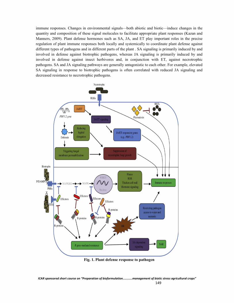
ICAR sponsored short course on “Preparation of bioformulation...........management of biotic stress agricultural crops” 149
immune responses. Changes in environmental signals—both abiotic and biotic—induce changes in the quantity and composition of these signal molecules to facilitate appropriate plant responses (Kazan and Manners, 2009). Plant defense hormones such as SA, JA, and ET play important roles in the precise regulation of plant immune responses both locally and systemically to coordinate plant defense against different types of pathogens and in different parts of the plant . SA signaling is primarily induced by and involved in defense against biotrophic pathogens, whereas JA signaling is primarily induced by and involved in defense against insect herbivores and, in conjunction with ET, against necrotrophic pathogens. SA and JA signaling pathways are generally antagonistic to each other. For example, elevated SA signaling in response to biotrophic pathogens is often correlated with reduced JA signaling and decreased resistance to necrotrophic pathogens.
Fig. 1. Plant defense response to pathogen
ICAR sponsored short course on “Preparation of bioformulation...........management of biotic stress agricultural crops” 150
Effector Stimulation and NB-LRR Signaling
To counteract and suppress plant responses evoked by PAMP triggered immunity, adapted pathogens secrete effector molecules that increase their virulence. Plants resistance (R) proteins perceive effectors by direct physical interaction or indirectly through effector modification of host target. Although many types of genetically and structurally unrelated receptors can be activated by effectors, the largest class of R proteins is the nucleotide binding-leucine rich repeat (NB-LRR) receptors. Activation of NB-LRR receptors results in a fast and strong response that has been termed effectortriggered immunity (ETI). This type of defense is also called gene-for-gene resistance, because a single plant R protein can confer resistance through recognition of a matching avirulence (Avr) protein of the pathogen. Much of our current knowledge on effectors and their host targets come from studies conducted on two major groups of Gram-negative bacteria, Pseudomonas syringae and Xanthomonas spp. Gram-negative bacteria inject a repertoire of effector molecules into the plant cell via their type III secretion system (T3SS). A typical phytopathogenic strain of P. syringae expresses around 15-30 effectors that are 8 secreted during the infection. Several studies have identified components involved in PRR signaling as effector targets. AvrPphB, a cysteine protease from P. syringae, and AvrAC, an uridylyl transferase from X. campestris pv. campestris, both target the BIK1 kinase of the FLS2/EFR/CERK1 signalosome. AvrPto is an E3 ligase that promotes degradation of the FLS2 receptor by catalyzing polyubiquitination of the kinase domain in Arabidopsis. Similarly, the MAP kinase pathways downstream of PRR activation are targeted by multiple effectors. Also later events in the plant defense reaction have been identified as effector targets; the HopZ effector was found to enhance pathogenicity by degrading an enzyme involved in isoflavonoid biosynthesis in soybean. However, not all effector molecules associates with, and interfere with protein function. TAL effectors from Xanthomonas bacterial pathogens contain domains that are characteristic for eukaryotic transcription activators. TALEs bind host DNA with high sequence specificity and induce expression of target genes, also termed disease susceptibility genes. Target genes for transcriptional reprogramming by TAL effectors include transcription factors and SWEET sugar transporters. SWEET proteins mediate glucose transport and up-regulation of the encoding genes may help the pathogen to fulfill its nutritional needs.
Structure and function of NB-LRR proteins
Plant NB-LRR receptors can be categorized accordingly to their N terminal domain: TIRNB-LRR with a Toll/interleukin 1-like receptor domain, and CC-NB-LRR with a coiled-coil domain. The multi-domain structure of NB-LRRs permits them to simultaneously act as sensors and response factors of pathogen elicitation. In the absence of pathogen produced effectors, the NB-LRR proteins are maintained in an inactive but primed state through a complex fold that is stabilized by domain-domain interactions. Small molecular perturbations may easily switch on the receptors from this stage and initiate signaling. To avoid auto-reactivity, the stability and the turnover of these receptors are kept under tight control by chaperones and ubiquitin E3 ligases. Failure to regulate NB-LRR receptor titer has been associated with autoimmune responses in plants. The LRR domain seems to have a dual function, namely, as a sensor of pathogen stimuli and as an intramolecular signal transducer. In the inactive state, the NB domain interacts with the N-terminal part of the LRR and forms a closed nucleotidebinding pocket. During activation, the NB domain is released allowing exchange of ADP for ATP, alternatively ATP hydrolysis, and enables the protein to assume an open conformation. It is still unclear which domain(s) is required for downstream signaling that leads to the execution of the defense response. Evidence suggests that different subdomains are required to accomplish this role in different NB-LRR proteins.
ICAR sponsored short course on “Preparation of bioformulation...........management of biotic stress agricultural crops” 151
The Guard Model
Direct binding between a NB-LRR receptor and pathogen-derived effector has yet only been described in a few cases. Instead, it appears that NB-LRR receptors and other 9 R proteins act as guards by monitoring the targets of pathogen effectors. NB-LRRs are activated if the integrity of the effector target, the “guardee”, is altered and downstream signaling is initiated. This Guard Model explains how several functionally unrelated effectors can be recognized by a single NB-LRR if they share a common target. The model also explains how a relatively low number of NB-LRRs proteins, 150 in Arabidopsis and 600 in rice, can confer resistance to a virtually endless repertoire of pathogen-encoded effectors. Recently, this model has been challenged and it was suggested that some effector targets act as baits or plant decoys for effector detection by R proteins. The authors proposed that gene duplication of true effector targets, or evolution of effector target mimics, could result in decoys that are strictly involved in effector perception. It seems reasonable to believe that these concepts are not mutually exclusive and that plants have evolved several ways to perceive effector action from a general set of components. A molecularly well characterized guarded effector target is the protein RIN4 of Arabidopsis. RIN4 is a negative regulator of MAMP signaling that is under the surveillance of at least two CC-NB-LRR receptors, RPM1 and RPS2. The R protein RPM1 specifically detects phosphorylation of RIN4 by the P. syringae effectors AvrRpm1 and AvrB, whereas RPS2 recognizes proteolytic cleavage of RIN4 by the AvrRps2 effector. Effector stimulated activation of either RPM1 or RPS2 result in effector-triggered immunity. Interestingly, peptide fragments of RIN4 that were produced by AvrRps2-mediated degradation could suppress MTI in plants, supporting the hypothesis that RIN4 is indeed a virulence target and not a host decoy. Emerging evidence suggests that other effector targets, as the LeEix2 receptor for the fungal elicitor EIX, can act as pathogen bait and prevent virulence.
NB-LRR signal integrators
Two proteins, NON-RACE SPECIFIC DISEASE RESISTANCE-1 (NDR1) and ENHANCED DISEASE SUSCEPTIBILITY 1 (EDS1), have been identified to integrate signaling from several CC-NB-LRRs and TIR-NB-LLRs, respectively. NDR1 resides in the plasma membrane where it interacts with the RIN4/RPM1/RPS2 complex via its cytoplasmic tail. Loss-of-function mutations in NDR1 render plants susceptible to P. syringae expressing any of the effectors AvrRpm1, AvrRpt2 or AvrB. It therefore appears that the role of NDR1 is to act as an adaptor protein that assures proper localization and assembly of immune receptors. EDS1 encodes a lipase-like protein that is required for signaling through the TIR-NB-LRR resistance proteins RPS4 in Arabidopsis and L6 in Flax (Linum usitatissimum) among others. As for NDR1, mutations in EDS1 result in reduced resistance to virulent pathogens. EDS1 is found in two distinct subcellular pools, one in the cytoplasm associated with PAD4 and one in the nucleus in complex with PAD4 and SAG101. Translocation of EDS1 between those cellular compartments seems to be important for relaying TIR-NB-LRR signaling and for the activation of ETI. Thus, protein nodes as NDR1 and EDS1 can coordinate diverse signals into a limited number of downstream targets.
What Happens After Pathogen Recognition?
The intracellular signaling events that follow MTI and PTI have been intensively studied. Many of the components of this sophisticated regulatory network have been identified on a genetic and biochemical level. In general, plant perception of specific effectors and MAMP molecules lead to the activation of similar intracellular signaling cascades. Also responses to MAMP and effector elicitation overlap considerably and ETI has been described as “an accelerated and amplified MPI response”.
ICAR sponsored short course on “Preparation of bioformulation...........management of biotic stress agricultural crops” 152
However, specific elicitor-receptor interactions may activate certain subsets of the signaling machinery and employ distinct sets of signal transducers. Moreover, even though common signaling routes are triggered by several types of elicitors, amplitude and timing of the responses are known to differ substantially.
Minutes after elicitation
One of the first detectable physiological responses of pathogen stimulation is the activation of membrane localized ion channels. Within minutes after elicitation, an influx of H+ and Ca2+ , and an efflux of K+ and Clresults in depolarization of the plasma membrane. Ca2+ originating from the apoplastic space serves as a second messenger in the cytoplasm that further activates ion transporters and other calciumdependent proteins, including calmodulins and transcriptional activators. Another important element of the early responses is the oxidative burst which constitutes the generation of reactive oxygen species (ROS) at the infection site. ROS are produced by extracellular NADPH oxidases and cell wall peroxidases and depend on transient increase in cytosolic calcium levels. ROS may act directly as microbial toxins or they may work as activators of other defense signals through protein modifications . In addition, nitric oxide (NO), a reactive gaseous radical, accumulates in the plant tissue in response to pathogens and acts in cooperation with ROS to execute defense programs. Lipids and lipid derived molecules are also associated with early defense signaling. Ca2+ and hydrogen peroxide formed from the oxidative burst can activate 11 phospholipase C (PLC) and phospholipase D (PLD) that produce the lipid second messenger phosphatidic acid (PA). PLC cleaves off the headgroup of the membrane lipids PIP and PIP2, which can then be phosphorylated by diacylglycerol kinase (DAGK) to PA. PLD catalyses the hydrolysis of the headgroup from structural lipids like PC and PE and directly forms PA. Pharmacologic inhibition or genetic silencing of PA production in plants result in reduced pathogen responsiveness. How PA relays the signal to downstream targets still remains an open question. Elicitor activation of PLCs generates, in addition to PA, inositol phosphates (InsP2 and InsP3 that can be phosphorylated to bioactive InsP5 and InsP6). Inositol phosphates further stimulate influx of calcium into the cytosol, thereby creating a feed-forward loop. Multiple post-translational modifications of proteins are identified as yet another early event of plant pathogen responses. As in animals, phosphorylation mediated through mitogen-activated protein kinases (MAPKs) is known to play a central role in biotic stress signaling in plants. The Arabidopsis genome contains 20 MAPKs that are under the regulatory control of 10 MAPK kinases (MAPKKs), which in turn are the substrate of approximately 60 MAPKK kinases. Stimulation with the flagellin fls22 epitope in Arabidopsis was shown to initiate a MAPK cascade that culminated in the activation of MPK3 and MPK6, leading to the subsequent activation of WRKY proteins (a family of transcription factors that serve as key players of plant immunity). In other studies, dozens of proteins have been found to be phosphorylated in a flg22 dependent manner. Interestingly, RbohD, one of the enzymes responsible for pathogen induced ROS formation, was identified as one of these proteins (41 and references therein). A consequence of the oxidative burst and ROS accumulation is the induction of cellular redox changes. These alterations in redox can be sensed by reactive cysteine residues of regulatory proteins. For instance, NPR1 is found as oligomeres in the cytoplasm under steady-state conditions. In response to pathogen evoked redox changes and accumulation of salicylic acid, internal disulfide bonds are reduced and NPR1 oligomers can dissociate into monomers. NPR1 monomers are then free to translocate to the nucleus and activate a subclass of TGA transcription factors. Other protein targets subjected to modifications during defense responses include histones which may be acetylated or deacetylated. Taken together, these and other studies present a direct link between transcriptional control and pathogen perception.
ICAR sponsored short course on “Preparation of bioformulation...........management of biotic stress agricultural crops” 153
Minutes and hours after elicitation
Transcriptional reprogramming The series of alarm signals that a pathogen triggers ultimately reaches the nucleus where substantial transcriptional reprogramming occurs. Large-scale expression profiling has provided us with detailed information of transcriptome regulation in plants with high spatial and temporal resolution. Pathogen infection is known to alter expression in up to 25% of the host’s genome. In general, responses to different pathogens, avirulent as well as virulent, target 12 overlapping sets of genes. This observation is in line with the notion that signals generated from several types of immune receptors converge at some level (see above). Analogous to elicitor-evoked signaling, transcriptional output is quantitative rather than qualitative and depends on the input stimulus. Reprogramming of defense-associated genes is known to take place earlier and with greater amplitude following effector recognition compared to MAMP perception. The global transcriptional switch that follows pathogen recognition establishes a transition from normal to a defense-orientated metabolism. These changes comprise the up-regulation of genes encoding components of the pattern recognition and signaling machinery, and enzymes involved in production of secondary metabolites. Down-regulated genes include those who govern cell division and other housekeeping functions.
Hormones in plant immunity
Plant hormones, phytohormones, play essential roles in all stages of plant life and reproduction. Immune responses are no exception. Precise regulation of hormones in time and space allows plants to accurately respond and react to a wide range of external stimuli. Three hormones in particular, salicylic acid (SA), jasmonic acid (JA) and ethylene (ET), are crucial for plant immunity. This phytohormone triad also has a welldocumented role in responses to abiotic stresses like wounding. In addition, recent studies have identified abscisic acid (ABA), auxin, brassinosteriods (BR), cytokinin (CK), gibberellic acid (GA) and peptide hormones as important regulators of immune responses. Overly simplified, SA is primarily induced by and confers resistance against biotrophic and hemi-biotrophic pathogens. By contrast, the JA and ethylene pathways are generally induced by and effective against necrotrophic pathogens and chewing insects. Mutants impaired in the accumulation of any of these hormones exhibit enhanced susceptibility to bacterial and fungal pathogens. Crosstalk between the SA pathway and the JA/ET pathway has been observed in many plants. Evidence for both synergistic and antagonistic interactions between SA and JA are reported. The details of JA synthesis and its role in plant defense are discussed in one of the following sections.
Systemic acquired resistance
Infection and defense activation in one part of a plant is frequently associated with the induction of resistance responses in distal organs. This phenomenon is known as systemic acquired resistance (SAR) and helps plants to withstand secondary infections. The local cell death of the hypersensitive response is usually the inducer of SAR but is not obligatory required for the production of mobile SAR signals. Systemic responses to pathogens were first reported more than half a century ago. Over the years, a range of candidate molecules has been proposed to act as the longdistance signal from the infection site to the healthy tissue. Methyl salicylate, jasmonic 13 acid, a glycerol-3-phosphate dependent factor, azaleic acid, abietic acid, dehydroabietinal, and most recently pipecolic acid are some of the substances reported to mediate SAR.

ICAR sponsored short course on “Preparation of bioformulation...........management of biotic stress agricultural crops” 154
Conclusion Over the past several years, detailed models for plantepathogen interactions have emerged involving recognition, evasion, and defense. It does, however, appear likely that the molecular basis of plant resistance will draw upon an even broader mechanistic base. Aspects such as components of the signal transduction system, antimicrobial compounds such as phytoalexins, and other unknown factors are also likely to be important components of plant resistance responses that remain to be characterized. Cloning additional resistance genes and QTLs that underlie plant resistance will reveal how they contribute to plant defenses. This knowledge will enable more efficient and effective utilization of these genes in crop improvement and protection Reference
Fan, J. and Doerner, P. (2012.). Genetic and molecular basis of nonhost disease resistance: complex, yes; silver bullet, no. Current Opinion in Plant Biology 15(4): 400-406.
Jones, J. D. G. and Dangl, J. L. (2006). The plant immune system. Nature 444(7117): 323-329. Naito, K.; Taguchi, F.; Suzuki, T.; Inagaki, Y.; Toyoda, K.; Shiraishi, T. and Ichinose, Y.
(2008). Amino acid sequence of bacterial microbe-associated molecular pattern flg22 is required for virulence. Molecular Plant-Microbe Interactions 21(9): 1165-1174.
Thordal-Christensen, H. (2003). Fresh insights into processes of nonhost resistance. Current Opinion in Plant Biology 6(4): 351-357.
Uma, B.; Rani T. S. and Podile, A. R. (2011). Warriors at the gate that never sleep: non-host resistance in plants. Journal of Plant Physiology 168(18): 2141-52.
Whalen, M. C.; Innes, R. W.; Bent, A. F. and Staskawicz, B. J. (1991). Identification of Pseudomonas syringae pathogens of Arabidopsis and a bacterial locus determining avirulence on both Arabidopsis and soybean. The Plant cell 3(1): 49-59.
Zhang, Z.; Wu, Y.; Gao, M.; Zhang, J.; Kong, Q.; Liu, Y.; Ba, H.; Zhou J. and Zhang, Y. (2012). Disruption of PAMP-Induced MAP Kinase Cascade by a Pseudomonas syringae Effector Activates Plant 41 Immunity Mediated by the NB-LRR Protein SUMM2. Cell Host & Microbe 11(3): 253-263.
**************

ICAR sponsored short course on “Preparation of bioformulation...........management of biotic stress agricultural crops” 155
Large Scale Adoption of Biocontrol Agents: Challenges and opportunity
Ranjan Daipaayan Sen CEO, Peak Chemical Industries Ltd.
Sevoke road, 2nd mile, Siliguri-734001, West Bengal
Mother Nature has provided us all the tools whether good or bad to sustain and control life on Earth. As human and to a great extent custodian of Mother Earth, we should respect the choices and use Science to the extent of not only preserving the sanctity of Nature but help towards a more Sustainable livelihood for generations to come.
Unfortunately, the ever-increasing population of ours with increasing feeding mouth is one of the biggest of the many challenges that we are facing along with Mother Earth. It has come to the responsibility of Scientist to be responsible in choosing the right alternatives and processes to balance the need and requirements towards a greener future and address the challenges properly.
Microbial formulations such as Bio Fertilizers and Bio Control Agents and their potential use commercially was initially done in the US to a large extent. The formulations were in great demand and like all new things Americans took to commercialising the industry in similar ways like all their big corporations. But the initial thrust on the mass adoption of Bio Control Agents for Plant protection needs failed to find the required success. Lot many questions were raised for their failures which itself is a case study and became the foundation of today’s technological changes and the answer towards large scale adoption of Bio Control Agents.
In my limited capacity, am sharing some of the most important points which needs to be looked into prior to commercial production of BCA anywhere in the world.
Technology bottlenecks
During the Eighties, the technology was pretty crude and the availability of high end commercial scale fermenters were not available. The problem area was with the viable cfu/gm content, and control of contamination.
During the initial years, the scientist also faced the major challenge of packaging the microbial formulations and preserving their spores in aseptic condition for field application at a later date. This could be done in laboratory scale by keeping them in an ambient temperature in sealed condition. But commercially the cost was too much if the transportation was to be done in a similar way.
Shelf Life
Simultaneously, the shelf life of the products were no more than 3 months, which made them commercially unviable. Agriculture crops are Seasonal and if the application time of these microbes missed the golden window, then the products could not be used for the next season leading to loss of the channel partners / traders who became disinterested. Any such products require a minimum shelf life of 6 months to make them commercially viable for the distributors and channel partners to handle this segment efficiently.
Knowledge & Skill
Unlike Synthetic chemicals available for Plant Protection needs, knowledge is an integral part of Bio Control Agents success. There are several factors effecting the efficacy of such products and the
ICAR sponsored short course on “Preparation of bioformulation...........management of biotic stress agricultural crops” 156
success of these to a great extent is with the applicants of this products. Factors such as Temperature, Humidity, Spraying Machine, Time of Applications, Pre and Post application management, Soil Conditions, Pest Population, etc. effect the microbial formulations to a great extent and their reasons behind success and failures.
Local and Correct Microbial Strains
We need to treat the product as living and hence the management of such product has to be done in a similar fashion. One microbial strain cannot be expected to perform in a similar way for all agro climatic and geographical conditions to which it is applied. Since, as living organisms, they are born with certain traits and adjusted to an environment, expecting them to perform in the same way as in arid conditions with an alkaline pH Soil and in high humid condition with an acidic pH will be too much to expect from a tiny bacteria or fungi. We have to have different microbial strains accustomed to certain geographical conditions and agro climatic needs and accordingly mass produce them for regional requirements. One for all and all for one does not stand true with Microbial formulations. They are more specific to Soil and Agro Climatic conditions then to crop.
Certifications & Licenses
Though this aspect of the challenge was not present during early nineties etc., but to a great extent this challenge is a man-made obstacle to the Industry with specific interest to guard. Unlike synthetic chemicals these products are natural and living organism and are more oriented towards an ecosystem than a crop. To treat a living organism with a same measuring scale as like synthetic chemicals will be doing unjust to the science itself. Unfortunately, such trade barriers are created by the scientific community itself and hence the toughest barrier among all the challenges is the Certification and License segment for this products.
Since the US was one of the early adopters of this potential beneficial microbes, the permission to manufacture these products are governed by the USDA and read under EPA Act wherein the mass scale manufacturing of these microbes are very much simplified.
Unfortunately, in India, the permission to manufacture such products is one of the steepest barriers and comes with a huge cost and time lag. The trade barrier is consciously done to safeguard the synthetic chemical industry interest and thus making us dependent on them for years to come.
One of the biggest lesson from the experience of the American microbial Industry in this segment was into challenges of large scale commercial production of microbial strains for plant nutrition and plant protection and the reasons behind their failures for mass scale adaptation by the farmers.
Today one of the biggest corporations in BCA are from the US and commands a market share of over 80% of all the Bio pesticides sold in the world and in them Bt takes the major chunk of its market share. If we are to grow in this segment, our policies and investment in technologies has to address the above discussed points.
Also in the present context, the challenges to a great extend have got reduced due to high awareness of the people towards our environment and health. Various Govt.’s are trying to promote use of green technologies and reducing the use of synthetic chemicals to a great extent. Various F&V companies, have put restriction and come up with their own certifications to assure the consumer that less or no chemicals are being used. The Central Govt. has given a major thrust towards promoting Organic farming in various parts of the country, thus increasing the scope of use of BCA in the farming
ICAR sponsored short course on “Preparation of bioformulation...........management of biotic stress agricultural crops” 157
practices. Hence the opportunities in present market is manifold but proper input substitution using microbial and organic solutions has to be kept in mind keeping the above discussed points for a suceesful journey in this segment.
A successful mass adaptation of BCA are not impossible but with the right Technology, Strain selection, Extension services, with appropriate certifications can provide the right mix of solution to successful mass scale production & adoption of these products by the farmers.
**************
ICAR sponsored short course on “Preparation of bioformulation...........management of biotic stress agricultural crops” 158
Biopesticides and IPR Issues
Gargi Sharma Department of Plant Breeding and Genetics,
Member-Secretary, IPR cell Assam Agricultural University, Jorhat- 785013, Assam
Biopesticides are biological or biologically derived agents that are used as plant protection products, usually applied in a manner similar to chemical pesticides. With the increase in concern about the environment and contamination of food with synthetic chemical pesticides the demand for biopesticides and biocompatible products to control pests, diseases and weeds is also increasing at a substantial extent. The expansion of organic farming further raises the demand of bio pesticides across the world. The global market of biopesticides is projected to reach USD 8.82 billion by 2022. To encourage the research and development of biopesticides, its Intellectual Property (IP) protection is also very important especially for a country like India which holds promising opportunities for biopesticides research and marketing. Intellectual Property when efficiently used becomes an important tool in positioning the business in the market. IP rights combined with other marketing tools are crucial for differentiation, promotion, diversification and marketing of biopesticides. In this era of knowledge based economy, protection of intellectual properties of biopesticides is important for innovators as well as marketers. There are many categories of IPs in India, viz., patent, copyright, trade mark, industrial design, trade secret, geographical indication, lay of integrated circuits etc. However, few of these are associated with biopesticides.
Biopesticides, microorganisms and patent
The concept of patenting “life forms and living matters” have always been a challenging issue and as such patent protection of biopesticides is also a complex one. In India, The Patent Act, 1970 is enacted to grant patents “not merely to enable patentees to enjoy a monopoly on a patented article” but “to secure that the inventions are worked in India on a commercial scale and to the fullest extent that is reasonably practicable without undue delay”. This cast a duty on the patentee to make efforts for commercial working of the patented invention at the earliest. The chief requirements for grant of a patent are novelty, inventiveness and industrial applications. There have always been controversies as to whether substances isolated or derived from naturally occurring living organisms are "inventions" or "discoveries”. A product of nature is not considered as a patentable subject matter because it is indistinguishable from something that occurs in nature and as such it lacks novelty. The history of the product of nature doctrine dates back to 1889, when Ex Parte Latimer, the Commissioner of Patents rejected a claim on a new article of manufacture, consisting of the cellular tissues of the Southern pine (Pinus australis). The inventor was asked to identity of the claimed substance from its natural counterpart as the claim and description did not set forth any physical characteristics by which the fibre could be distinguished from other vegetable fibres. Later on as the fibre claimed could not be distinguished from other fibres by any physical characteristic, the claim, therefore, was refused. It was concluded that a product whose physical characteristics are indistinguishable from those of its naturally occurring counterpart does not constitute patentable subject matter because it lacks novelty.
Again, Section 3 (c) of the Patent Act, 1970 states that the mere discovery of a scientific principle or the formulation of an abstract theory or discovery of any living thing or non-living substances occurring in nature is not patentable. Section 3(j) of the Act excludes from patentability “plants and animals in whole or any part thereof other than micro-organisms but including seeds, varieties and species and essentially biological processes for production or propagation of plants and
ICAR sponsored short course on “Preparation of bioformulation...........management of biotic stress agricultural crops” 159
animals”. In recent years the agreement on Trade Related Aspects of Intellectual Property Rights (TRIPS) is considered as one of the most important steps towards harmonising law in the field of intellectual properties across the world. As per the article 27(3)(b) of TRIPS Agreement member states should not grant patents for “plants and animals, other than microorganisms and essentially biological processes for the production of plants or animals other than non-biological and microbiological processes.” As a result TRIPS makes it obligatory for all its signatories to extend patents for microorganisms, non-biological, and microbiological processes. India in compliance with TRIPS amended the Patents Act in June 2002, by giving patent rights for new microorganisms. The 2002 amendment of Indian Patent Act added explanation to chemical processes which states chemical processes include biochemical, biotechnological and microbiological process. In India some other areas involving microorganisms are also patentable such as a synergistic composition containing the microorganism, which is either new or known, a process using microorganisms to produce a substance, the process of biosynthesis of a new microorganism etc. Any invention resulting from human intervention, where living things have been used for conducting experimentation will be patentable in India. It is pertinent to mention herewith that even before this amendment Calcutta High Court addressed the issue of whether a process involving microorganisms that are living as an end product can be patented or not. In the year 2002, the Calcutta High Court delivered a landmark judgment in Dimminaco AG v Controller of Patent and Designs and others. Dimminaco AG filed a patent application for the process of creating a vaccine to protect poultry from infectious bursitis. The Controller of Patents determined that the process was not an invention because the end product produced by the process contained a living organism and as such it was not patentable. Thereafter the applicant appealed the Controller’s decision to the Calcutta High Court. The Controller opined that a patent is given only for a process that results either in an article, substance, or manufacture and a vaccine with a living organism is not an article, substance or manufacture. As the meaning of manufacture was not defined in the Patents Act, therefore, the court used the normal dictionary meaning of manufacture as “the material in question after going through the process of manufacture has under-gone any change by the inventive process and it becomes a material which is different from the starting material.” The Court determined that this meaning does not exclude the process of preparing a product that contains a living substance from patentability. The court decided that “since the claim process for patent leads to a vendible product, it is certainly a substance after going through the process of manufacture.” The court ultimately concluded that “a new and useful art or process is an invention,” and because the process is new and useful it “is apparently patentable under section 5 read with section 2(j)(i)” of the Patent Act. The court determined that “where the end product is a new article, the process leading to its manufacture is an invention.” However, later on the definition of invention was amended.
Another landmark judgment was passed by the US Supreme Court on patentability of microorganisms. Genetic engineer Ananda Mohan Chakrabarty developed a bacterium Pseudomonas putida capable of breaking down crude oil, which he proposed to use in treating oil spills. There were three parts to the patent viz., method of producing the bacteria, composition of a slurry of the bacteria and the bacteria themselves. The United States Patent and Trademark Office allowed the first two claims but rejected the third one because under patent law at that time, living things were generally considered as non patentable subject matter under section 101 of title 35 U. S. C. Chakrabarty appealed before the US Supreme Court stating that the genetic engineering he did on the bacteria was a form of manufacture, therefore, it met the standard. Finally the US Supreme Court ruled in favour of Chakrabarty, holding that: “A live, human-made microorganism is patentable subject matter under 35 U. S. C. 101. Respondent’s microorganism constitutes a ‘manufacture’ or ‘composition of matter’ within the statute.” Basically ‘products of nature’ are not patentable because the discoverer isn’t really an ‘inventor’ as they

ICAR sponsored short course on “Preparation of bioformulation...........management of biotic stress agricultural crops” 160
did not do anything themselves, they just found something that pre existed. However, that argument does not apply to Chakrabarty because he did not work to create the new bacteria.
Another important aspect of the grants of patent in respect of microorganisms is the regulations concerning the requirements for the deposition of microorganisms and accessibility of that microorganism from the depositories as par the provisions of Budapest Treaty in which India is also a member. The microorganism if not being described fully and particularly and is not available to public, the said microorganism is to be deposited before the International Depositary Authority(IDA) under the Budapest Treaty within 3 months of making application in India. All details, available characteristics of the microorganisms and details of depositary institutions shall be mentioned in specification for correctly identifying the same. Furthermore access to the same is required to be made available only after date of application in India or date of priority. For the purpose of microorganisms and other biological materials Microbial Type Culture Collection and Gene Bank (MTCC) is an internationally recognized depository institution. In India, two microbial repositories viz. Microbial Type Culture Collection (MTCC) and Microbial Culture Collection (MCC) have acquired the status of IDA.
‘Trade Mark’ as a marketing tool of biopesticides
Intellectual property more specifically trade mark is of primary importance in the marketing process. Biopesticide products also face competition with the products that are often similar, identical and good substitute. To distinguish a particular product from those of its competitors and to maintain the goodwill and trust of the consumers, trade mark plays a vital role. Trade mark helps to create a brand name in the market and many times consumers are willing to pay even higher price for these branded products. However, use of trade mark for effective marketing of products needs expertise in the field of trade mark law and practice. In India, Trade Marks Act, 1999 and the rules there under are used for registration of trade mark, protection of trade marks for goods and services and to prevent fraudulent use of the mark.
Protection of biopesticide formulations as ‘Trade Secret’
Article 39 of agreement on TRIPS provides protection of undisclosed information, sometimes also referred to as trade secrets. Trade secrets can be a valuable assest to a company’s growth. A trade secret may refer to a practice, process, design, instrument or a compilation of data or information relating to the business which is not generally known to the public and which the owner reasonably attempts to keep secret and confidential. Considering the multiple objectives of biopesticide formulations, wide range of materials used in the formulations, formulation processes etc. microbial formulations are often held by many companies as trade secrets and confidential know how (Behle and Birthisel, 2014). There is no specific legislation in India for trade secret and is enforced contractually or under common law. Indian courts uphold trade secret protection on the basis of principles of equity, breach of confidence, breach of contractual obligations etc.
Conclusion
Biopesticides offer powerful tools to create a new generation of sustainable agricultural products and these have been studied, researched, promoted and marketed as replacement of synthetic pesticides in the recent years. Big business is starting in the world of biopesticides. Moreover, the advancement in the technologies has further increased the scope and prospect of biopesticides exponentially. The protection of biopesticides under the IPR regime may give boost not only to the scientific community but

ICAR sponsored short course on “Preparation of bioformulation...........management of biotic stress agricultural crops” 161
put the business of biopesticides in a safe and respectable position which will eventually encourage the scientists to become more innovative and competitive.
References
Behle, R. and Birthisel, T. (2014). Formulation of entomopathogens as Biopesticides. In: Mass Production of Beneficial Organisms. Elsevier, Amsterdam. Eds. Morales-Ramos, J. A.; Guadalupe Rojas, M. and Shapirollan, D. L.
Chandler, D.; Grant, W. P.; Greaves J.; Prince, G. And Tatchell, M. (2010). Biopesticides: Pest Management and Regulation. Oxfordshire CABI.
Diamond v. Chakrabarty, 447 U. S. 303, 100 S. Ct. 2204, 65 L. Ed. 2d 144, 1980 U. S. LEXIS 112, 206 U. S. P. Q. (BNA) 193 (U. S. June 16, 1980).
Dimminaco AG vs Controller of Patent and Designs and others (2001) AID No.1 Pandey, N. and Dharni, K. (2014). Intellectual Property rights. PHI Learning Private Limited,
New Delhi. The Patents Act, 1970 as amended by The Patents (Amendement) Act, 2005 along with the
Patents Rules, 2003 as amended by the Patent (Amendment) rules, 2006 The Trade marks Act, 1999. Thirvenugam B. C. (2005). Biotech Industry: IPR and other legal issues, Biospectrum, (13th
June) 12.
**************
ICAR sponsored short course on “Preparation of bioformulation...........management of biotic stress agricultural crops” 162
Biological Management of Invasive Alien Species (IAS)
Rajib Kumar Borah Rain Forest Research Institute, P. Box-136, Sotai, Jorhat, Assam
Biological invasion is becoming a major threat to the natural ecosystems and indigenous biodiversity all over the world. Biological invasion by invasive alien species (IAS) are considered one of the main driver in biodiversity loss and endangered species listing worldwide (OTA, 1993). This has got worst effect especially on island ecosystem (Clout and Lowe, 2000) including the isolated protected areas. It is, therefore, important to preserve the values and functions of protected areas along with the wealth and support they provide to the livelihoods of millions of people from the IAS threats. IAS is recognized as the second largest threat to biological diversity after deforestation and forest degradation (Singh 2001). Article 8(h) of Convention on Biological Diversity (CBD) recognizes the importance of this global issue and calls on contracting parties to prevent the introduction of, control or eradicate those alien species which threaten ecosystems, habitats and species.
IAS represents the greatest threat to the preservation of global biodiversity after habitat destruction, as well as imposing an increasing financial burden on agriculture (Kaiser, 1999). There has been an extensive movement of plant species around the world by humans, as a consequence of trading activities. This has resulted in exotic species forming a significant part of the agricultural weed flora, and in natural ecosystems, invasive weeds are almost exclusively alien (Groves, 1991). Oerke et al. (1994) calculated that losses due to weeds (based on eight major crops) average almost 13% of the world’s agricultural output. In natural ecosystems, it is impossible to put a price on the cost of the loss of biodiversity and the implications for society.
Weeds contribute to the destruction of global biodiversity by altering habitat structure via a number of different processes. For example, direct competition with the native flora can result in monocultures of an alien weed, such as by Psidium cattleianum Sabine (strawberry guava) in Mauritius. In addition, direct competition can be aided by alleopathic effect produced by the weed, such as Parthenium hysterophorus L. (white top) in Australia and India. More insidious effects can also be caused, such as the alteration of the hydrology of a region, that result in a fundamental change in the type of habitat that can be supported, for example, the effects of Andropogon virginicus L. (broom sedge) on tropical rainforest in Hawaii (Cronk and Fuller 2001). Practically, IAS have invaded and affected local biota in every ecosystem type and almost in every region (Matthews, 2005). United Nations conference on Environment and Development (Rio,1992) acknowledged that a major cause of biodiversity loss is IAS (Agenda 21 of chapter15). Seventh ordinary Meeting of the Conference of the Parties to the Convention on Biological Diversity (COP7) held in Kuala Lumpur, Malayasia from 9 to 20th February 2004, underlined the threats of invasive species to wetland ecosystems (resolution VII/14). The committee requests the Executive Secretary to explore methodologies for fostering awareness, promoting education and generating information on invasive alien species for abroad audience, including indigenous and local communities, the public and other stakeholders (CBD, 2012).
The Indian scenario
India has a long trade history with different countries and the pathways of the trade were land, sea and air. These pathways helped introduction of a number of alien species from different geographical regions. The movement of species is faster in the present era and hence the chances of invasion also became greater. Cataloguing of invasive species in an area required knowledge on the flora of the country of origin and area invaded, Information on the territory of origin and its history are all useful
ICAR sponsored short course on “Preparation of bioformulation...........management of biotic stress agricultural crops” 163
tools for botanists (Pysek et al., 2012). A comprehensive list of invasive species in India has been prepared by Reddy et al. (2008). The Ministry of Environment and Forests, Govt. of India has also collated information on invasive species in India (www.apfisn.net). Sankaran & Suresh (2013) have given comprehensive information on invasive plants in the forests of India. However, a thorough knowledge on the taxonomy of the invasive species, its status as an invader and its biogeographic affinity are necessary to base any study on invasion biology. There is in need to undertake intensive surveys with in the country to develop a region - specific categorization of alien plant species . The major invasive plants in India include Chromolaena odorata, Lantana camara, Parthenium hysterophorus, Mikania micrantha, Mimosa diplotricha var. diplotricha, Acacia mearnsii, Ageratum conyzoides, Agertaina adenophora, Arundo donax, Cuscuta reflexa, Imperata cylindrica, Leucaena leucocephala, Merremia peltata, Prosopis juliflora, Pteridium aquilinum and Sphagneticolaa trilobata (Sankaran & Suresh, 2013). Impact of some of these species on various ecosystems have been studied e.g., Chromolaena odorata and Lantana camara, Ageratum conyzoides, , Parthenium hysterophorus, Mikania micrantha, Prosopis juliflora (Sw.) DC.; and Ageratina adenophora (Spreng.) (Sankaran et al., 2001). All these studies indicated that invasion of these species resulted in extensive loss in agricultural and forest ecosystems in terms of economy and harm to ecosystems. However, the loss has not been quantified. Sankaran et al., (2013) assessed the risk of alien plants in the forests of Kerala State. It was a good start. But, surveillance and risk assessment are to be conducted at periodic intervals to understand the arrival of new species and the potential of these to become invasive. It is also necessary to understand the pathways of invasion and the vectors involved. Customs and import rules in India are to be strengthened and strictly implemented to prevent new invasions.
Definition
International Union for Conservation of Nature (IUCN, 2000) defines invasive alien species as an alien species, which becomes established in natural or semi-natural ecosystems or habitat, as an agent of change, and threatens native biological diversity.’ As per Conference of the Parties to the Convention on Biological Diversity-CoP6, Invasive alien species are alien species whose introduction and spread threatens ecosystems, habitats or species with economic or environmental harm’. The European Union defines “Invasive Alien Species” as those that are, ‘firstly, outside their natural distribution area, and secondly, threaten biological diversity’.
Importance of IAS
Why so much importance is tagged with invasive alien species? The answer lies in the very nature of such species. Alien species are aggressive colonists (invaders), thanks to their higher adaptability and greater ecological amplitude. Due to efficient ways of sexual reproduction and vegetative multiplication, effective dispersal; of fruits and seeds, longer seed dormancy, biotic and abiotic stress tolerance the invasive species seems to be the worst plant of the human civilization. They have extremely damaging effect on the ecosystem as a whole and often responsible for large scale habitat destruction. IAS can create extensive environmental and economic damages. Homogenization of biodiversity, reducing local diversity and distinctiveness are the other important consequences of increasing domination by a few invasive species. The harmful effects of IAS are intensified by pollution, habitat loss and other human induced disturbances. IAS can alter vital ecosystem processes such as fire, hydrology and nutrient cycling, kill, suppresses, compete with or displace native species and communities, or alter gene pools through hybridization (Chornesky and Randall, 2003). Human land use pattern and global climate change would open up new opportunities for introduced species that could devastate native flora and fauna and affect the spread of invasive species (Mooney and Hobbs, 2000). It seems highly likely that invasive species are going to have even more opportunities in the changed future

ICAR sponsored short course on “Preparation of bioformulation...........management of biotic stress agricultural crops” 164
climate than they have at present. The Indian region because of its diverse climatic and environmental conditions is highly vulnerable to biotic invasion. Moreover, a burgeoning population, high rate of trade and transport, coupled with greater movement of people favour the accidental and international entry of plant species in this region. The recent fast rate of economic growth of the country is also expected to leave its mark on loss of plant diversity and increased invasion of alien species. Three hot spots of biodiversity, although supporting rich floral and faunal diversity including endemic species, also reflect a high rate of habitat degradation where opportunists invasive species can easily established themselves. Consequently, a number of invasive species have made their abode in the region.
Impact of plant invasion
The impacts of alien species on the native ones and the spontaneity of the ecosystem are immense, unpredictable and often irreversible. The costs of invasive alien species are highly significant in economic terms. Plant invasion threaten the existence of endangered species and the integrity of ecosystems, and their ravages cost national economies tens of billions of dollars every year. Alien plant invasion, particularly in protected areas bear greater ecological impact because these are the areas practically forms an island of biodiversity and literally hold the remaining biological diversity left with us till date. Invasive alien species are a problem in a great variety of types of protected areas, with national as well as with international designations. It is reported that around 106 countries in Asia, Africa, South and Central America have protected area(s) recorded as having invasive alien species as a threat. Twenty seven World Heritage sites (15% of the total number of Natural and Mixed sites) are reported to be under threats from invasion by alien species. Invasive alien species may be released, deliberately or accidentally, within a protected area, or may move in form surroundings areas. Lack of awareness on ways to address IAS at local level may contribute to an unwanted sense of dejection in decision makers either at site level, or at system level.
Major invasive plants and their attributes
Important invasive plants that have created havoc in a number of habitats include terrestrial herbaceous weeds viz. Ageratum conyzoides, Eupatorium adenophorum, Chromolaena odorata, Lantana camara, Mimosa diplotricha, Mikania micrantha, Parthenium hysterophorus and Eichhornia crassipes, Ipomea sp. and Salvinia molesta in aquatic ecosystems (Raghubanshi et al., 2005). The weed also spreads through its seeds, which are minute and are carried by wind and water, as is Parthenium hysterophorus, Eichhornia crassipes, Ipomea sp. and Salvinia molesta are invaders of aquatic ecosystems and wetlands; these have done much harm to the biodiversity of aquatic ecosystems (Reddy, 2008).
Lantana camara is perhaps the best known example of a serious weed having been internationally introduced for ornamental value. Lantana camara is now rated as one of the worst invasive identified by the global invasive species database and is also included in the top 100 invasive species of the world. Lantana is a member of the family Verbenaceae and is a pantropical weed affecting pastures and native forests in more than 60 countries worldwide (Parsons and Cuthbertson, 2001). In India, Lantana was first introduced in the early nineteenth century as an ornamental plant but is now growing densely throughout the country. It is now spreading to form impenetrable thickets on the edges of forest and covers 4x 10⁶ ha across Australia (Van Oosterhout et al., 2004). Globally, it infests millions of serious concern in 14 major crops including tea, coffee, rice, cotton and sugercane. Disturbed areas such as roadsides, railways tracts and canals are also favourable for the species. Lantana possesses a number of attributes in its life cycle that characterize it as an invader. There is still uncertainty as to
ICAR sponsored short course on “Preparation of bioformulation...........management of biotic stress agricultural crops” 165
which attributes make some species more invasive or what makes some ecosystems more vulnerable to invasion than others (Lodge, 1993).
Another weed introduced in India as an ornamental plant is Chromolaena odorata and is also included in the list of the top 100 worst invaders (GISD, 2010). It is also one of the most obnoxious weeds in the Western Ghats, North Eastern parts of the country and impacts on coconut, rubber, coffee, and teak plantations (Singh,1998). C. odorata is an invasive shrub species belonging to the family Asteraceae native to continental America and now cosmopolitan weed, for predicting its distribution and invasion in two different climate change scenarios in the Indian subcontinent. Characters such as high reproductive capacity, efficient dispersal mechanism, high competitive ability and wide ecological amplitude make the plant a troublesome weed and a successful invader. The species is one of the world’s 100 worst invasive alien species according to the Global Invasive Species database. In India the species was first introduced in 1845 in Calcutta as an ornamental plant (Munniappan et al., 2004). It invaded North Eastern India during World War I and by 1924/25 it spreads to West Bengal and Orissa. From the eastern region it spreads to Kerala in 1942 and then invaded rapidly to all the southern states (Singh, 1998). At present the species is a serious weed with allelopathic effects in plantations, natural forests, pastures and natural reserves.
Mikania micrantha is native to Central and South America (Holm et al., 1977) and is commonly known as mile a minute. There are around 425 species of Mikania (King and Robinson, 1987). Mikania micrantha was introduced in to India during the Second World War to camouflaging airfields or as a ground cover for tea plantations. It is now a very noxious weed in plantations and forests, especially in the southern and north western parts of the country (Muniappan et al., 2004). It is an extremely fast growing, perennial vine and is regarded as one of the world’s most notorious invaders (Cronk and Fuller, 2001). Invasive Mikania micrantha (family Asteraceae) pose a serious threat to biodiversity (Wang et al., 2004) and hence it is essential to understand the biology of this weed. It is a multi branched, perennial scambling vine with a 6-ribbed stem, internodes pubescent or glabrous and 7.5-21.5 cm long.
Of species introduced accidentally to the region, Parthenium hysterophorus Linn, commonly known as white top weed, carrot weed or congress grass, belonging to Asteraceae family is native to the area around the Gulf of Mexico, including West Indies and Central and South America. The weed is now widely distributedin Australlia, Taiwan, South China and Pacific Island and become one of the most harmful weeds and the best known example (Kohli and Rani, 1994). It was introduced in India in the early50’s as contaminant of wheat consignment received from Mexico under the PL480 Scheme. In India, it was first noticed in 1955 near Pune. It grows rapidly in vacant areas, agricultural lands, pastures, urban areas and natural and manmade forests, where it forms it own monoculture stands. It is an obnoxious invasive weed from tropical America that has spread to various tropical and subtropical parts of the world. It occurs widely in different habitats varying from hot and arid, semi arid to humid and from low to middle to high altitude regions. It is an erect plant with an angular, grooved and profousely branched stem bearing dissected pale green leaves that resembles carrot leaves during initial growth. Parthenium is known to cause a number of health problems in both human and livestocks. In human it causes Allargenic Eczematous Contact Dermatitis (AECD), rhinitis, and asthama (Lonkar et al., 1974; Rao et al., 1985). Parthenium is also very toxic to livestocks and other animals, causing acute to chronic toxicity, symptoms may appear as ulceration, nausea, loss of appetite or restlessness and can even prove fatal (Narasimhan et al., 1992).
Likewise, Ageratum conyzoides is a fast spreading weed which is the most serious invader of agricultural land and is a problem in hilly tracts. The weed also spread through its seeds, which are
ICAR sponsored short course on “Preparation of bioformulation...........management of biotic stress agricultural crops” 166
minute and are carried by wind and water. It is originated in America (Okunade, 2002). The plant is now found as a weed of over 36 crops in 46 different countries (Holm et al., 1977). It has been ranked as 19th of the world’s worst weeds. In India it has been reported as existing prior to 1982 in the Flora of British India (Hooker, 1982). It was probably introduced as an ornamental plant in the 1860s (National Focal Point for APFISN, India, 2005), later attained a weekly habit and turned harmful to mankind. The plant has also been found as a major weed in the littoral and swamp forests of Assam (DOEF, 2010). In addition, the weed has also been reported in north eastern and southern India (Rao, 2000) and the forests of the Gandhamardan Hills range, Orissa (Reddy and Pattnaik, 2009).
According to Waterhouse (1994), tropical America, the region from Brazil to Paraguay and tropical north east Argentina and low lands of Central America is the native range of Mimosa diplotricha var. diplotricha and M. diplotricha var. inermis (Mimosaceae). The thorn-less variety was imported to the Asia-Pacific region as a tropical pasture legume, but its tendency to revert to the thorny type and its potential toxicity has discouraged further import. The genus Mimosa is widely distributed in all inhabited continents. More than 16 species of Mimosa species are reported from tropical America alone. Mimosa diplotricha var. inermis was first reported as an invader from Java, Indonesia in 1900 (Soerjani et al., 1987). The plant generally occurs as a thicket among grasses and along road sides, river banks and in waste lands in its native range. Mimosa diplotricha var. diplotricha, M. pigra and M. pudica are the three weedy species of Mimosa which occur in South - East Asia and Australia . In India, M. diplotricha var. diplotricha was first recorded in Kerala in 1964 . However, the earliest reference of the plant was regarding its introduction in coffee plantations in South India as a cover crop (Anonymous, 1955). Planters in Kerala also used the plant in hilly areas to prevent cattle from entering the plantations (Rajan et al., 1986). The plant is now widespread in Kerala along roadsides, railway tracks and in vacant lands, degraded areas and agricultural systems (Sankaran et al., 2012). According to Holm et al., (1977), M. diplotricha var. diplotricha is one of the 76 worst weeds of the world. It is classified as a ‘serious’ or ‘principal’ weed in Australia, Borneo, Fiji, Indonesia. Malaysia, Melanesia, New Guinea, Philippines, Taiwan and West Polynesia. It is also a serious weed in the Pacific islands, South–East Asia, Mauritius and Nigeria (Waterhouse & Norris, 1987). The weed is recorded from over 30 countries in the Asia–Pacific region (Sankaran & Suresh, 2013). The species is a big threat to forest ecosystems, agricultural land and pastures. It causes heavy damage in crops like sugar cane, coconut, rubber, cassava, tea, pineapple and upland rice. Hand harvesting of crops is difficult in the infested fields and dangerous as the thorns cause serious sores on humans (Waterhouse & Norris, 1987). The heliophytic nature of M. diplotricha var. diplotricha helps it to occupy the uppermost layer of canopy in any type of vegetation it occupies. Waterhouse (1994) recorded that the rapid smothering effect of the weed is harmful for the survival of native plants. Invasion of alien plants has also affected the natural wild parts of North eastern India. Mimosa has established in Kaziranga following seed dispersal through flood water from adjoining tea plantations. At present around 120 ha of prime grass land has been adversely affected and scattered growth can be seen in many areas inside the park. The grassland of Kaziranga National Park, a world heritage site have been attacked by Mimosa invisae in recent decades. Some researchers have indicated the presence of another related species, Mimosa rubicaulis.
The spread of the weed in Kaziranga National Park in Assam is reported to be a threat to the endangered single horned rhinoceros by preventing its movement within the National Park for food (Vattakkavan et al., 2002).
According to Vasu (2003), M. diplotricha var. diplotricha impairs the growth of other species especially the grasses and it has been observed that nothing grows in the grassland areas infested by the weed. He also reported that the thorny nature of the plant and its capacity to spread vigorously blocks the

ICAR sponsored short course on “Preparation of bioformulation...........management of biotic stress agricultural crops” 167
traditional corridors and trails of several wild animals including elephants. It is widely accepted that ecosystem modification is an indirect driver of biodiversity loss.
Strategies for managing invasive weeds
When looking at the alarming rate at which invasive plants are spreading in India and also worldwide, effective management measures are required to control them. There are separative ways of dealing with those already established and those possessing the potential to be invasive but not of immediate risk. Preventive measures are utmost importance and require great attention at every level to prevent the entry of invasive species. The Global Invasive Species Programme helps countries to catalyse action against invasive alien species by developing national and regional control and prevention strategies. Under this programme the first global best practice guidelines have been produced, and this champions classical biological control (CBC) as one of the main control strategies for invasive weeds (Wittenberg and Cock 2001). CBC targets alien weeds and is based on the enemy release hypothesis (ERH). This hypothesis assumes that plant populations, once freed of their natural enemy complexes, can expand rapidly and, therefore, become more competitive than those subject to natural control (Wilson 1969; Mitchell and Power 2003). Most introduced plant species do not become weedy once established in a new region. However, if climatic factors are favourable then there are few barriers to regulate growth, and this may result in population explosions with the subsequent development of weed invasions (Mack et al. 2000). Alien plant species are usually introduced, either deliberately or accidentally, into a new geographic area without any or most of their co-evolved natural enemies: CBC aims to redress this imbalance. Coevolved natural enemies (plant pathogens and arthropods) are collected from the centre of origin of the target weed; selecting those that appear to have the most impact on the target species.
Approaches to mitigating the ravages caused by plant invasions generally involve three main tractics: prevention, eradication and control (Mack et al., 2000). Prevention often involves the enactment and enforcement of legislation, including the negotiation of international treaties (e.g., International Plant Protection Convention, Agreement on the Application of Sanitary and Phytosanitary Measures). Eradication is difficult to achieve, for once a weed infestation has reached a density and areal extent, at which it is recognized as a problem, its complete removal becomes a practical and economic impossibility. Once prevention fails and eradication is no longer an option, control measures must be applied to minimize the economic and environmental impacts of a weed invasion. Three methodologies are appropriate for controlling invasive weeds: mechanical, chemical (herbicides), and biological.
Although these methods have reduced the impact of IAS, they have their own limitations. Manual removal involves huge labour costs and it cannot be practiced for long. In the majority of agroecosystems, weeds are controlled using cultural and chemical methods (Murphy et al.,2000). Cultural methods are long lasting but require the use of physical removal or chemical programmes to eliminate invasive alien species prior to implementation. Mechanical controls include hand pulling, hoeing, tillage, mowing, grubbing, chaining, bulldozing, harvesting and draining. The use of synthetic herbicides, which include photosynthesis inhibitors, lipid biosynthesis inhibitors, amino acid biosynthesis inhibitors, cell division inhibitors, auxin mimics and respiration inhibitors, is an integral part of weed control in cropping systems and most rangelands (Mooney et al., 2005). Both methodologies are efficient in controlling weeds in limited areas (Singh, 1998). However, because of expensive, energy and labour intensive and require repeated applications, mechanical and chemical control are impractical for managing widespread plant invasions in ecologically fragile conservation
ICAR sponsored short course on “Preparation of bioformulation...........management of biotic stress agricultural crops” 168
areas or low value habitat. Herbicide resistance of weeds is a worldwide problem. It is defined as the inherited ability of a weed or crop biotype to survive and reproduce treatment with a dose of herbicide to which the original population was susceptible. Repeated and intensive use of herbicides with similar mechanisms of action in crops over a period of time leads to development of resistant biotypes within the community due to selection pressure. In addition, mechanical means of control disturb the soil and may cause erosion, chemical herbicides have spurred the evolution of resistance in scores of weed species and further, may pose risks to wildlife and human health. There are 421 herbicide resistant weeds at present re-herbicides with 22 sites of action. Prominent among these are Lolium rigidium, Avena fatua, Chenopodium album, Eichornia cruss-gallis and Phaloris minor (Heap, 2014). Because of drawbacks associated with conventional weed control method, classical biological control, the introduction of selective exotic natural enemies to control exotic pests, increasingly is being considered and implemented as a safe, cost effective alternative to address the invasive plant problem. Concern is now growing throughout the world about the environmental impact and toxic effects of the widespread use of chemical methods of pest control (Lodge, 1993). This concern has, in part, fuelled the current global upsurge in interest in biological control of weeds as a sustainable, eco friendly and potentially effective method of weed control. According to Mooney and Hobbs (1996), successful biological controls remain the only viable solution for reducing the current and potential impact.
Biological control is a fascinating discipline where experimental projects are conducted at eco regional scales. Biological control using natural enemies and native organisms is an important tool in the land manager’s arsenal of weed control techniques. The practice has been expanding from use primarily on rangelands and aquatic systems into other environments. The editors of this comprehensive work have embarked on the difficult task of cataloging the biological control of invasive plants (noxious weeds) on a global scale. With each successive edition of this World Catalogue of Biological Control of Weeds since 1982, the monumental task of pulling together so much information has been compounded by the ever changing geopolitical landscape and the increasing number of targeted weeds and new biocontrol agents. Some biological control agents are redistributed to countries, states, regions, etc., where the political entity has relied on host specificity testing conducted in another country or by an adjoining neighbor. This provides compelling support to the growing interest in pathogens as CBC agents, as an acceptable tool to help combat the increasing global problem of invasive weeds (Mack et al. 2000).
Biological weed control involves using living organisms, such as insects, nematodes, bacteria, or fungi, to reduce weed populations. In nature, plants are controlled biologically by naturally occurring organisms. Plants become pests - and are labeled "weeds" - when they run rampant because their natural enemies become ineffective or are nonexistent. The natural cycle may be interrupted when a plant is introduced into a new environment, or when humans disrupt the ecological system. When we purposefully introduce biological control agents, we are attempting to restore or enhance nature's systems. Biological control is permanent, energy efficient, non polluting and inexpensive relative to other methods. Economic returns on investment in biological wed control have been spectacular in some cases, and range from an estimated benefit/cost ratio of 2.3 to 4000 or more. Biocontrol is the most cost effective and environmentally benign solution to managing IAS because when it works it does not require reapplication like chemicals or poisons. Most biocontrol agents for weeds and insects, once established, are self- sustaining and don’t have to be reapplied.
After passing a comprehensive evaluation and screening programme the best agent(s) are introduced and released in the exotic target area. This approach fits well into an integrated, biologically-based approach to pest management in agroecosystems (Chornesky et al., 2003). Increasingly, it is the
ICAR sponsored short course on “Preparation of bioformulation...........management of biotic stress agricultural crops” 169
only viable long-term option for the control of invasive, alien weeds in rangeland and natural environments (Singh, 1998). For example, control of the South American aquatic plant Salvinia molesta D.S. Mitchell (water fern) in Asia, Africa, and Australasia was achieved with the weevil Cyrtobagous salviniae Calder & Sands (Matthews, 2005).
The origins of biological weed control
In ancient times, the Chinese discovered that increasing ant populations in their citrus groves helped decrease destructive populations of large boring beetles and caterpillars. That use of a natural enemy to control a pest marked the birth of biological control. Biological control research and implementation is even more relevant today. Foreign and native organisms that attack weeds are being evaluated for use as biological control agents. As a weed management method, biological control offers an environmentally friendly approach that complements conventional methods. It helps meet the need for new weed management strategies since some weeds have become resistant to certain herbicides. Biological control agents target specific weeds. Moreover, this technology is safe for applicators and consumers. Hundreds of square miles were virtually impenetrable to humans or animals. A small moth from Argentina was imported and released. The moth larvae burrowed into the cactus, grew and multiplied, and within 10 years had decimated the prickly pear population. Today, the cactus covers only 1% of the area it occupied in 1925.
How does it work?
Roots provide plants with water and nutrients. Some biological control agents attach to roots and thereby stunt plant growth. Some bacteria live on root surfaces and release toxins that stunt root growth. Many fungi infect roots and disrupt the water transport system, which reduces leaf growth. Beneficial insects and nematodes feed directly on the weed roots causing injury which allows bacteria and fungi to penetrate. Plant leaves capture energy from the sun and store it as sugar. Insects that feed on leaves reduce the leaf surface available for energy capture. Fungi and bacteria that infect leaves reduce the ability of the leaf to make sugars. In either case, there is less energy available for weed growth. Whether through damage on roots or leaves, severe infestations of biological control agents can actually kill weeds, reducing their adverse effects on desirable plants. Many weed species survive from year to year by producing seeds. Fungi or insects that attack seeds can reduce the number of weed seeds stored in the soil, which in turn can reduce the size of future weed populations. This lowers the effort needed to control the remaining emerging weeds.
Some bacteria and fungi applied as biological control agents do not survive from year to year. These organisms must be applied on an annual basis. This technique is called the "bioherbicide" strategy. With this tactic, biological agents are used in manner similar to chemical herbicides.
Weeds introduced from foreign countries often require a different strategy. Insects and pathogens are collected in the area of origin and evaluated for release in North America. Insect agents often require a number of years to become fully effective. Their growth is often hindered by adverse climatic conditions. Long-term monitoring is needed to determine their effectiveness. The release of biological control organisms in this manner is termed the "classical" approach to biological control. Fungi that naturally spread and infect weeds can also be used in a classical biological control strategy.
Classical biological Control (CBC)
ICAR sponsored short course on “Preparation of bioformulation...........management of biotic stress agricultural crops” 170
The classical biological weed control aims at restoring the balance between the target alien weed and its natural enemies in the ecosystem by purposeful introduction of suitable, exotic bioagents. The immediate response to successful introduction of a native bioagent is a decrease in the weed population, followed after some period by a decline in the bioagent population due to its food shortage. Then often the weed population tends to recover. The process continues in a cyclic fashion till the bioagent weed populations get stabilized at a low level. The biological weed control should not be expected to eliminate the target weed from an area; in fact, success of biological control of weeds depends upon continued presence of the weed existing in small numbers and shifting with time. The method being a slow operating one, is used mainly in non crop situations. On crop lands, even if successful, the bioagent will not get opportunity to work on the host weed because of frequent use of insecticide and fungicides in modern agriculture. Otherwise, Cyperus rotunduscan possibly be controlled in crop fields with a moth borer, Bactra verutana. Another promising example of selective biocontrol in crop fields is complete denudation of Ludwigia parviflora waterpurslane’ by Haltica cyanea Web, steelblue beetle in rice fields.
CBC of weeds: the significance of pathogens
The majority of CBC implementation programmes of weeds have been in the USA, Australia, South Africa, Canada, and New Zealand. There is also an increasing number of programmes in several Asian and African countries. Worldwide there have been 949 recorded releases of exotic agents for the control of weeds over the last 100 years. The exploitation of pathogens forms only a small part of this, as it does worldwide, but it is becoming increasingly considered in current and future programmes (Singh, 1998).
An important aspect of biological weed control is that at time, it is applicable to the control of only one major weed species that has spread widely. Also, one should be prepared to accept cyclic resurgence of the weed under attack by the classical biocontrol approach. Its application in intensively managed agro-ecosystems, however, is difficult because of the ephemeral nature of these habitats with high disturbance levels and the fast control process needed relative to the short duration of the cropping season. Three methods of biological weed control in crops can be distinguished: the inoculative or classical approach; the inundative or microbial herbicide approach; and the system management or augmentative approach.
Inoculative biological control using exotic control agents
Inoculative (classical) biological weed control has generally been restricted to environmental weeds and extensive agriculture (e.g. rangeland) and has limited application to intensive crop production systems (Sankaran and Suresh, 2013). Annual weeds of arable crops have long been considered poor targets for inoculative biological control, although theoretically, organisms with excellent search and dispersal abilities (some insects) or those showing persistence (some fungi) can be used for inoculative biocontrol in unstable, disturbed habitats (Raghubanshi et al., ,2005).
Agents used in biological management
1. Insect as biocontrol agents
The introduction of Zygogramma suturalis F. (ragweed beetle) to control Ambrosia artemisiifolia L. (common ragweed) in Russia and more recently, in Croatia, China and Australia (Munniappan et al., 2004). The moth Pareuchaetes pseudoinsulata was the first insect to be introduced from Trinidad into India and Ghana in the early 1970’s (Cronk and Fuller,2001), and have been
ICAR sponsored short course on “Preparation of bioformulation...........management of biotic stress agricultural crops” 171
successfully used in Indonesia, the Phillipines and Guam for control of Chromolaena odorata. Waterhouse (1994) listed 70 insects and two fungi which attack mimosa in Brazil. Of these, Heteropsylla spinulosa Muddiman, Hodkinson & Hollis, a hemipteran psyllid caused severe stunting and distortion of leaves and growing tips. Attack by the psyllid also reduces or prevents flowering. This bug was introduced from Brazil to Queensland (Australia), Fiji and Papua New Guinea where it successfully suppressed the growth of mimosa.
2. Microbes as a biocontrol agent
Weed biocontrol include the introduction of the rust Puccinia chondrillina Bubak & Sydenham to control Chondrilla juncea L. (skeleton weed) in wheat (Triticum aestivum L.) fallow systems in Australia (Enserik, M, 1999). The inundative method, primarily using microbial herbicides, has greater opportunity for application in intensive agriculture. Between 1980 and 1998, three bioherbicides (DeVine, Abbot Laboratories, Chicago, IL; Collego, Encore Technologies, Minnesota, MN; and Dr BioSedge) were registered in the United States, and one was registered in each of Canada (BioMal, PhilomBios, and Agriculture and Agri-food, Saskatoon, Canada), Japan (CAMPERICO, Japan Tobacco, Yokohama) and South Africa (Stumpout, Plant Protection Research Institute, Stellenbosch, S. Africa). However, the exploitation of fungal pathogens is a relatively new, but growing approach. The first release of a pathogen was made in 1972 in Australia, when the rust Puccinia chondrillina Bubak & Sydow was introduced from Europe to control Chondrilla juncea L. (skeleton weed) (King and Robinson 1987). The gall-forming rust fungus, Uromycladium tepperianum (Sacc.) McAlpine was introduced into South Africa from Australia to control Acacia saligna (Labill.) Wendl. (Port Jackson willow), an invasive and damaging weed of the unique Fynbos ecosystem. After an 8–10 year lag phase, the rust is now responsible for a 90–95% reduction in the weed populations and the Fynbos is now in the process of recovery (Lodge 1993). The recent study by Wang et al. (2004) provides strong evidence in support of the ERH, specifically for plant pathogens. Waterhouse (1994) reported Cercospora canescens Ellis & G. Martin, as a possible biocontrol agent for Mimosa.
Another fungal pathogen, not registered as a microbial herbicide, has been developed for use in the Netherlands (BioChon, Koppert, Biological Systems, Berliel en Rodenrijs, the Netherlands) as a stump-rotter to control regrowth of Prunus serotina. Five of these seven bioherbicides are still commercially available for use, while two (BioMal and Dr BioSedge) are unavailable as a result of technical difficulties in production or market considerations (Singh, 1998).
The exploitation of synergy between pathogens and insects has been used repeatedly and successfully to control environmental weeds (Singh, 1998) and has been proposed for use against crop weeds such as Orobanche spp. (Munniappan et al., 2004), Rumex spp. (Pysek et al., 2012) and Senecio vulgaris L. (Cronk & Fuller, 2001). Furthermore, combinations of pathogens that unite the specificity of biotrophs with the virulence of necrotrophs have been suggested (Raghubanshi et al., 2005), but whether or not such interactions can be exploited successfully to control crop weeds remains unknown (Van et al., 2005).
Similarly, Parsons & Cuthbertson (2001) suggested a further challenging proposal, based on the interaction of low-virulence, broad-spectrum pathogens with special carriers. The specificity is given by the targeted application of the carrier, which must be present for the pathogen to infect the host.
3. Plant extract or botanicals as a bioagents
ICAR sponsored short course on “Preparation of bioformulation...........management of biotic stress agricultural crops” 172
Biological control comprises various technologies of which one option is the use of botanical products. Many kinds of plant species and technologies have been used in the production of botanical pesticides. Some but not many of the plant-based pesticides have already become established plant protection products (Lodge, 1993). Botanical pesticides are pest management agents which are based on plant extracts. In modern times these have been used as alternatives to synthetic chemicals in organic pest management. The practice of using plant materials against field and storage pests however has a long history in many indigenous and traditional farming communities across the world.
Managing established weed: antagonist systems to stimulate epiphytotics
This approach is based on knowledge of the crop environment, especially of the mechanisms underlying the interactions of the weed, the natural enemy and the environment at the individual and population levels. By focusing on pathogens, it aims to induce and stimulate disease epidemics within weed populations and, thus, reduce weed competitiveness at the population level. Conservation and facilitation methods of biological control are especially well suited to promoting sustainable agro-ecosystems, in which weed control no longer aims at crop production in a weed-free environment but simply at a reduction in weed-induced crop losses (Singh, 2001).
Biological management of some important IAS
1. Lantana camara
The distribution of Lantana is limited by its following environmental attributes viz. inability to survive under dense, intact canopies of taller native forest species, low tolerance to saline soils, tendency to rot in boggy or hydromorphic soils and high sensitivity to aridity. Shading by intact canopies is an effective barrier against Lantana invasion and is the most appropriate strategy for managing it (Kaiser, 1999). Apart from managing and manipulating ecosystems, manual and mechanical removal along with chemical and biological control options, have been much explored for the control of Lantana. All these control strategies, however, have associated drawbacks. Manual removal is labour intensive and a low efficiency technique, while mechanical control is inefficient in dealing with every extensive invasions and is also difficult in undulating, rocky terrain. Chemical control involves the use of inorganic/ organic herbicides, and a serious disadvantages is the high cost of most chemical control programmes. Safety for other plant species and the environment is of vital importance when using herbicides to control invasive species. Biological control of Lantana began in 1902 and since then 41 agents have been released in some 50 countries (Jullien and Griffiths, 1998) but still no biocontrol agents has completely stopped its infestation. The success of biological control programme is often not clear cut, because complete control is achieved only in certain years and/or at some locations (Syrett et al., 2000). In India, the biocontrol agent Teleonemia scrupulosa Stal. released for Lantana control failed, since it could not cope with the vigorous regrowth of Lantana at the onset of the monsoon rains or the control agent itself suffered was largely destroyed during the winter months (Sharma, 1988). Furthermore, Goulson and Derwent (2004) argued that cost effective control of Lantana could be achieved through the judicious employment of honeybees.
2. Mikania micrantha
The dense growth habit typical of Mikania micrantha can be suppressed by the use of herbicides such as glyphosate, 2,4-D and atrazine (Palit,1981). Chemical control is very expensive and adversely affects other associated species and therefore manual removal and biological control method are the most effective methods. Several natural enemies of Mikania micrantha have been identified, among

ICAR sponsored short course on “Preparation of bioformulation...........management of biotic stress agricultural crops” 173
these, the tea mosquito bug (H. theivora) inflicted serious damage but none could be considered as having the potential to control this weed (Abraham et al., 2002). Cuscutta campestris , a holoparasite, is reported to have reduced cover, biomass and nutrient levels of Mikania micrantha and significantly increased native species richness by increasing soil pH and water and nutrient levels (Yu et al.,2009). A fungus, Puccinia spegazzinii, has also been reported to have potential to control Mikania micrantha as it can cause extensive damage to its leaves, petioles and branches, which may result in death. This fungus is reported to be specific to Mikania micrantha and also has a wide range of environmental tolerance (Cock et al., 2000).
3. Parthenium hysterophorus
Management of Parthenium is a challenge for scientists. A number of physical, chemical, ecological and biological methods have been tried to manage this invasive weed. Manual uprooting is generally avoided as the plants have allergenic properties, burning is another method but the burnt residues left in fields compromise soil quality and these techniques are not recommended, while chemical control create threat to the environment including ill effects on human health. The use of natural plant products is another option as these are biodegradable, environmentally safe, efficacious and economical. Singh et al (2001), reported that volatile essential oil from lemon scented eucalyptus possesses good potential for the control of Parthenium. Among various biological agents used the Mexican beetle (Zygogramma bicolorata) and selected fungal based mycoherbicides have been trialled for this purpose, although with limited success. The total benefits by the biological control in six years had been of Rs.62.34 million; 15585% benefit over initial investment (Kumar, 2006).
4. Ageratum conyzoides
To date, a number of strategies including physical, chemical and biological methods have been tried, though none has been found completely to control or manage A. conyzoides. Pre emergence application of atrazine, diuron, metribuzin or simazine is an effective control strategy. However, due to toxicological implications, synthetic herbicides are not recommended.
To optimize weed control farmers are required to know the time of emergence of weed seedlings relative to the crop, apart from any use of commercial herbicides (Van et al., 2004). The use of eco friendly compounds such as natural plant products and other plant based formulations offers a safer strategy for effective weed control. Volatile mono-terpenes such as cineole and citronellol have been found to have considerable effects on germination, growth, chlorophyll content and cellular respiration of A. conyzoides, suggesting much potential for weed management (Singh,1998). Parthenin, a sesquitarpene lactone from Parthenium hysterophorus, negatively affected the growth and physiology of A. conyzoides and has potential for use as a novel agrochemical.
5. Chromolaena odorata
In 1991, a biocontrol program for Chromolaena, funded by the Australian Government and managed by the Queensland Government, was initiated in Indonesia and the Philippines. The moth Pareuchaetes pseudoinsulata Rego Barros (Lepidoptera: Arctiidae) and the stem-galling fly Cecidochares connexa (Macquart) (Diptera: Tephritidae) were introduced. P. pseudoinsulata was introduced into PNG from Guam where it had successfully established (Muniappan et al., 2004) and C. connexa was introduced in 2001 from the Philippines (Bofeng et al., 2004. The leaf-mining fly Calycomyza eupatorivora Spencer (Diptera: Agromyzidae) was introduced unsuccessfully in 2004 from South Africa, where it had established. C. connexa spread rapidly from all sites and exerted control in many parts of the country. P. pseudoinsulata, however, was more limited in its distribution and impact
ICAR sponsored short course on “Preparation of bioformulation...........management of biotic stress agricultural crops” 174
(Bofeng et al., 2004). In most provinces, Chromolaena is considered under control by the gall fly in at least some areas. Control was generally achieved more quickly in some provinces, particularly East New Britain, New Ireland and Bougainville, than in others such as Morobe and Madang.
6. Mimosa diplotricha
Several natural enemies of the weed have been reported from its natural range. A sap feeding bug from Brazil, Heteropsylla spinulosa (Homoptera: Psyllidae), that causes growing tip distortion and reduces seed production in Mimosawas introduced in Queensland, Fiji and Papua New Guinea with some success. Heteropsylla spinulosa is now well established and has spread significantly (Abraham et al., 2002). It has caused a dramatic reduction in vigour and seed production of Mimosa diplotricha in Australia (Julien and Griffiths, 1998). Corynespora cassilicola, a cosmopolitan fungus, causes stem spot defoliation and dieback of Mimosa in Queensland and Papua New Guinea. A coreid bug, Scamurius sp., that feed on the Mimosa shoots inhibiting vegetative growth and flowering was introduced from Brazil to Queensland and Western Samoa but didn’t establish. Fusarium pallidoroseum, a fungus isolated from diseased Mimosa diplotricha in the Phillipines, provided excellent control of Mimosa seedlings when sprayed with crude culture filtrate or cell free filtrate. Further studies are warrented before recommending use of the fungus to control Mimosa.
Conclusion
Most of the natural resources have a spatial extent and consequently mapping becomes the preparatory point for its scientific management. Mapping of wildlife habitats and data base generation is considered as one of the basic needs for restoration and conservation of wild habitats. IAS are spreading around the world at an alarmingly accelerated rate (Mack et al., 2000). Rapid spread of IAS and subsequent ecological, economic disaster around the world has raised question against an old age philosophy regarding nature. It was said that the best way to deal with nature is “leaving nature alone to get on with itself”. But with increasing invasion in most of protected areas this philosophy is no longer an option.
The successful deterrence and management of IAS threats is an integral component to effective management of protected areas (Mooney et al., 2005). A protected area IAS assessment should clearly state the exact area covered, the conservation goals and management objectives, the IAS currently present/absent and mapped locations; the IAS which has impacts on conservation goals and management objectives; the important pathways/ vectors for IAS entry and dispersal, prediction of future spread and impact of IAS if not controlled, capacity of existing staff and resources to adequetly prevent and control IAS and the damages they cause; any extra capacity that may be needed, and gaps in policies and programs to prevent IAS.
Perhaps a new and bold paradigm should be co- existing or living with weeds recognizing their intrinsic worth as part of biodiversity and the many possible uses as bio- resources. Studies must re- focus on improved understanding of weeds, not just for the sake of controlling them, but for their ecological role, as well as social and economic benefits.
References
Abraham, M.; Abraham, C.T. and Joy, P.J. (2002). Natural enemies on Mikania micrantha H. B.K. in Kerala. Journal of Tropical Agriculture. 40: 39-41
Anonymous(1955). Giant sensitive plant- Mimosa diplotricha. Queensland Department of Natural Resources Weed Pest facts. 27: 1-4
ICAR sponsored short course on “Preparation of bioformulation...........management of biotic stress agricultural crops” 175
Bofeng, I.; Donnelly, G.; Orapa, W. and Day, M. D. (2004). Biological control of Chromolaena odorata in Papua New Guinea. In: Day, M.D. and McFadyen, R.E. (eds.) Chromolaena in the Asia Pacific Region, Australian Centre for International Agricultural Rersearch, Canberra, Australia, pp.14-16.
CBD (Convention on Biological Diversity) (2012). Meeting Documents, Eleventh meeting of the Conference of the parties to the Convention on Biological Diversity (COP 11), 8-19 October 2012- Hyderabad, India.
Chornesky, E. A. and Randall, J. M. (2003). The Threat of Invasive Species to Biological Diversity. Annals of the Missouri Botanical Garden 90: 67-76.
Clout, M. N. and Lowe, S. J. (2000). Invasive species and environmental change in New Zealand. 369-384, In: Mooney H.A. and Hobbs. (eds). R.J. Invasive Species in a changing World. Island Press, Covelo, California, USA.
Cock, M. J.W.; Ellison, C.A.; Evans, H. C. and Ooi, P. A. C. (2000). Can failure be turned into success for biological control of mile a minute weed. Proceedings of the Xth International Symposium on Biological Control of Weeds. Montana State University, Bozeman, Montana, pp.155-167.
Cronk, Q. C. B. and Fuller, J. L. (2001). Plant Invaders: The threat to natural Ecosystems. Earthscan Publications, London, UK, pp.241.
DOEF (2010). Assam’s Rich Biodiversity. Department of Environment and Forest, Government of Assam.
Ekeleme, F.; Forcella, F.; Archer, D.W.; Akobundu, O. and Chikoye, D. (2005). Seedling emergence model for tropic Ageratum (Ageratum conyzooides). Weed Science. 53:55-61
Enserink, M. (1999). Biological invaders sweep in. Science. 285:1834-1836 Evans, H. C. (1997). Parthenium hysterophorus: A review of its weed status and the possibilities
for biological control. Biocontrol News and Information. 18: 89-98 GISD (2010). Global Invasive Species Database, Available at http//www.issg.org.database. Goulson, D. and Derwent, L. C. (2004). Synergistic interactions between exotic honeybee and
exotic weed: pollination of Lantana camara in Australia, Weed Research 44:195-202. Groves, R. H. (1991). Weeds of tropical Australia. Tropical grassy weeds Wallingford, Oxon,
UK; CAB International, 189-196. Heap, I. M. (2014). International survey of herbicide resistant weeds.
http://www.weedscience.org. Holm, L. G.; Plucknett, D. L.; Pancho, J. V. and Herberger, J. P. (1977). The World’s Worst
Weeds, distribution and biology, University Press, Honolulu, Hawaii. Hooker, J. D. (1982). The flora of British India, Vol. III. Reeve and Co., Ashford, UK. IUCN (International Union for Conservation of Nature), 2001. Report on the State of
Conservation of Natural and mixed Sites inscribed on the world Heritage List and the List of World Heritage in Danger. Gland, Switzerland.
Julien, M. H. and Griffiths, M.W. (1998). Biological Control of Weeds. A World catalogue of Agents and their Target Weeds, 4th edn. CABI Publishing, Wallingford, UK
Kaiser, J. (1999). Steeming the tide of invasive species. Science 285:1836-1841. King, R. M. and Robinson, H. (1987). Studies in the Eupatoriaceae, the genus Chromolaena.
Phytologia 20: 196-209 Kohli, R. K. and Rani, D. (1994). Parthenium hysterophorous- a review. Research bulletin
(Science) Punjab University 44: 105-149. Kumar, S. (2006). Economic benefits in biological control of Parthenium by Mexican beetle,
Zygogramma bicolarata Pallister in India. Annals of Entomology. 24(1&2):75-78.
ICAR sponsored short course on “Preparation of bioformulation...........management of biotic stress agricultural crops” 176
Lodge, D. M. (1993). Biological invasions: lesions for ecology. Trends in ecology and evolution 8: 133-137.
Lonkar, A.; Mitchell, J. C. and Calnan, C. B. (1974). Contact dermatitis from Parthenium hysterophorus. Transactions of the St. John’s Dermatological Society 60: 43-53.
Mack, R. N.; Simberloff, D.; Lonsdale, W. M.; Evans, H.; Clout, M. and Bazzaz, E. A. (2000). Biotic inavsions: causes, epidemiology, global consequences and control. Ecological Applications 10: 689–710.
Matthews, S. (2005). South America invaded-the growing danger of invasive alien species. GISP, South African National Biodiversity Institute, Cape Town, South Africa. pp.80
Mooney, H. A.; Mack, R. N.; McNeely, J. A.; Neville, L. E.; Schei, P. J. and Waage, J. K. (eds.) (2005). Invasive Alien Species: A New Synthesis. Island Press, London, UK.
Mooney, H. A.; Mack, R. N.; McNeely, J. A.; Neville, L. E.; Schei, P. J.; Waage, J. K.; Mooney, H. A. and Hobbs, R. J. (2000). Invasive Species in a Changing World, Island Press, Washington, DC.
Munniapan, R.; Englberger, K.; Bamba, J. and Reddy, G.V. P. (2004). Biological control of Chromalaenea in Micronesia. ACIAR Technical Reports No.55, Australian Centre for international agricultural Research, Caneberra, Australia, pp.11-12
Murphy, S. T. and Evans, H. (2009). Biological Control. In: Clout, M. and Williams, P. A. (eds.) Invasive Species Management: A Handbook of Principles and Techniques. Chapter 6. Oxford University Press, UK.
Narashimhan, T. R.; Harindrnath, N.; Premlatha, S. and Suba Rao, P.V. (1992). Effect of Parthenin on mitochondrial oxidative phosphorylation. Biochemistry International. 11:239-244.
Oerke, E. C.; Dehne, H. W.; Schönbeck, F. and Weber, A. (1994). Crop Production and Crop Protection-Estimated Losses in Major Food and Cash Crops. Elsevier Science, Amsterdam.
Okunde, A. L. (2002). Ageratum conyzoides L. (Asteraceae). Fitoterapia 73: 1-16. OTA (U.S. Congress, Office ofTechnology Assessment). 1993. Harmful Non- Indigenous
Species in the United States. U.S. Government Printing Office; Washington, DC. http://www.wws.princeton.edu/ota/disk/1993/9325
Palit, S. (1981). Mikania- a growing menance in plantation forestry in West Bengal. Indian Forester. 107: 96-101.
Parsons, W. T. and Cuthbertson, E. G. (2001). Noxious weeds of Australia, 2nd ed., CSIRO Publishing, Collingwood. Melbourne, Australia, pp.627-632
Pysek P.; Jarosik V. and Hulme P. E. (2012). A global assessment of invasive plant impacts on resident species, communities and ecosystems: the interaction of impact measures, invading species’ traits and environment. Global Change Biology 18: 1725–1737
Raghubanshi, A. S.; Rai, L. C.; Gaur, J. P. and Singh, J. S. (2005). Alien invasive species and biodiversity in India. Current Science 88: 539-540.
Rajan, A.; Monomohan, C. B.; and Nair, N. D. (1986). Experimental studies on the toxicity of the plant Mimisa invasa Mart var intermis Adlb in calves. Kerala J. Vet. Science 17: 91-98
Rao, V. S. (2000). Principles of Weed Science. Science Publishers, Enfield, New Hampshire, pp. 555.
Reddy, C. S. and Pattanaik, C. (2009). An Assesssement of Floristic Diversity of Gandhamardan Hill Range, Orrissa India. Bangladesh Journal of Plant Taxonomy 16:29-36.
Reddy, C. S.; Bagyanarayana, G.; Reddy, K. N. and Raju, V. S. (2008). Invasive alien flora of India. National Biological Information Infrastructure, US Geological Survey, USA http://www.gisinetwork.org

ICAR sponsored short course on “Preparation of bioformulation...........management of biotic stress agricultural crops” 177
Rio, (1992). United Nation conference on Environment and Development Rio de Janerio, Brazil,3 to 14 June AGENDA 21.
Sankaran, K. V. and Suresh, T. A. 2013. Evaluation of classical biological control of Mikania micrantha with Puccinia spegazzinii. Kerala Forest Research Institute, India, Research Report, No. 472.
Sankaran, K. V.; Muraleedharan, P. K. and Anita, V. (2001). Integrated management of the alien invasive weed Mikania micrantha in the Western Ghats, Kerala Forest Research Institute, India, Research Report, No. 201.
Sankaran, K. V.; Suresh, T. A. and Sajeev, T. V. (2013). Handbook on invasive plants of Kerala, Kerala State Biodiversity Board.
Sharma, O. P. (1988). How to combat Lantana (Lantana camara) menance- a current perspective. Journal of Scientific and Industrial Research 47: 611-616.
Singh, H. P.; Batish, D. R.; Kohli, R. K.; Saxena, D. B. and Arora, V. (2002b). Effect of parthenin- a sesquitarpene lactone from Parthenium hysterophorus- on early growth and physiology of Ageratum conyzoides. Journal of Chemical Ecology 28: 2169-2179.
Singh, H. P.; Batish, D. R.; Pandher, J. K. and Kohli, R. K. (2005). Phytotoxic effects of Parthenium hysterophorus residues on three Brassica species. Weed Biology and Management. 5: 105-109.
Singh, H. P.; Batish, D.R. and Kohli, R. K. (2002a). Allelopathic effect of two volatile monoterpenes against billy goat weed. Crop Protection 21:347-350.
Singh, S. P. (1998). A review of biological suppression of Chromolaena odorata K & R in India. in : Ferrar, P., Munniapan, R. and Jayanth, K.P.(eds.) Proceedings of the 4th International Workshop on Biological Control and Management of Chromolaenea odorata. University of Guam, Guam, pp-86-92.
Singh, S. P. (2001). Biological control of Invasive Alien Weeds in India. Keynote address In: Alien weeds in moist tropical zones: banes and benefits.
Soerjani, M.; Kostermans, A. J. G. H. and Tjitrosoepomo, G. (1987). Weeds of Rice in Indonesia. Balai Pustsaka, Jakarta. pp.732.
Syrett, P.; Briese, D. T. and Hoffmann, J. H. (2000). Measures of success in biological control of terrestrial weeds by arthropods. In: Wratten, S.D. and Gurr, G.(eds.) Measures of success in Biological control. Kluwer, Amsterdam. pp.189-230.
Van Oosterhout, E.; Clark, A.; Day, M. D. and Menzies, E. (2004). Lantana control Manual. Current management and control. Options for Lantana (Lantana camara) in Australian state of Queensland. Department of Natural resources, mines and energy, Brisbane, QLD, Australia.
Vasu, N. K. (2003). Management plan, Kaziranga National Park, 2003-04 to 2012-13. Official document of the department of Forest, GOA
Vattakkavan, J.; Vasu, N. K.; Verma, S. and Ambika, A. (2002). Silent stranglers: eradication of Mimosa in Kaziranga National Park, Assam. http//www.wti.org.in/oldsite/publications/ silent stranglers.pdf
Wang, B. S.; Wang, Y. Z.; Liao, W. B; Zhan, Q. J. and Peng, S. L. (2004). The invasion ecology and management of Alien Weed Mikania micrantha H.B.K. Science press, Beijing, pp-76-85.
Waterhouse, D. F. (1994).The major Arthopod Pests and weeds of Agriculture in South East Asia. Australian Centre for International Agricultural Research.
Waterhouse, D. F. and Norris, K. R. (1987). Biological control: Pacific prospects. pp. 454. Wittenberg, R. and Cock, M. J. W. (eds.) 2001. Invasive Alien Species: A Toolkit of Best
Prevention and Management Practices, CABI Publishing, Oxon, UK.

ICAR sponsored short course on “Preparation of bioformulation...........management of biotic stress agricultural crops” 178
Yu, H.; He, W. M.; Liu, J. and Dong, M. (2009). Native Cuscuta campestris restrains exotic Mikania micrantha and enhances soil resources beneficial to natives in the invaded communities. Biological Invasions 11: 835-844.
*************
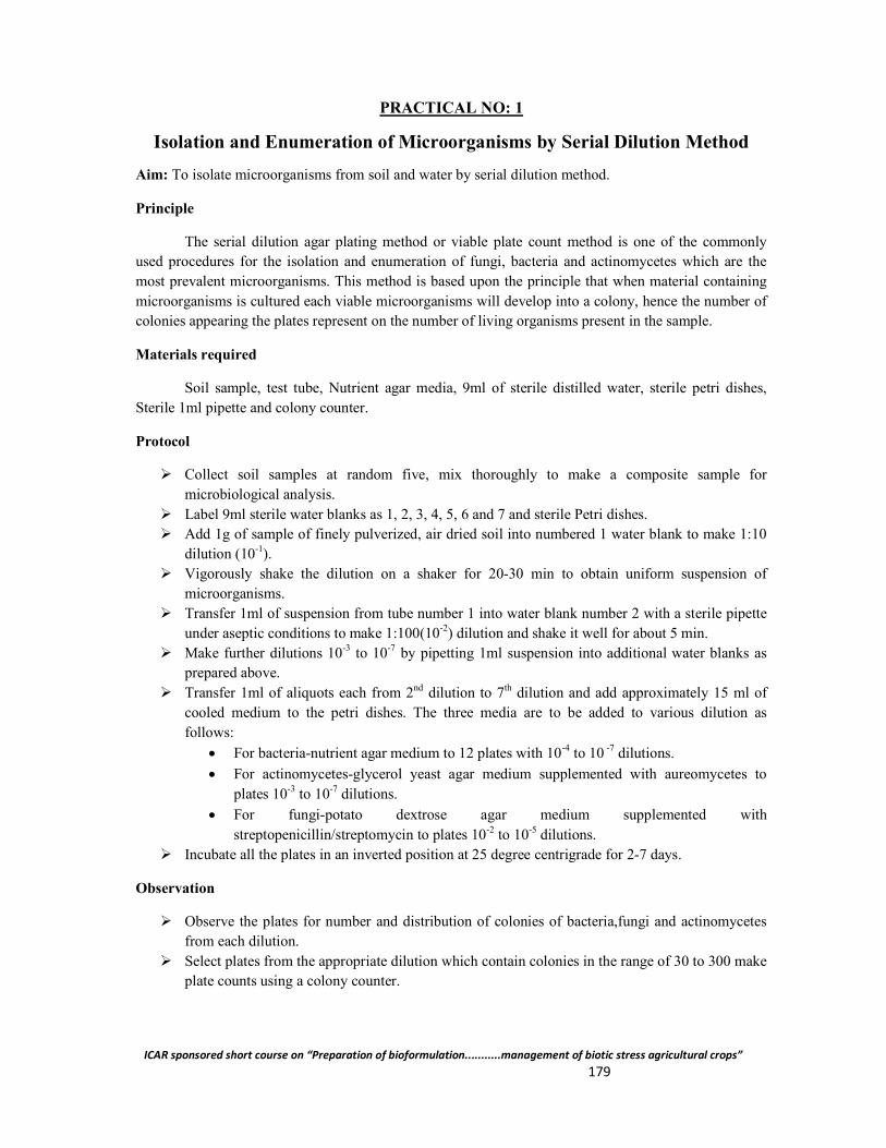
ICAR sponsored short course on “Preparation of bioformulation...........management of biotic stress agricultural crops” 179
PRACTICAL NO: 1
Isolation and Enumeration of Microorganisms by Serial Dilution Method
Aim: To isolate microorganisms from soil and water by serial dilution method.
Principle
The serial dilution agar plating method or viable plate count method is one of the commonly used procedures for the isolation and enumeration of fungi, bacteria and actinomycetes which are the most prevalent microorganisms. This method is based upon the principle that when material containing microorganisms is cultured each viable microorganisms will develop into a colony, hence the number of colonies appearing the plates represent on the number of living organisms present in the sample.
Materials required
Soil sample, test tube, Nutrient agar media, 9ml of sterile distilled water, sterile petri dishes, Sterile 1ml pipette and colony counter.
Protocol
Collect soil samples at random five, mix thoroughly to make a composite sample for microbiological analysis.
Label 9ml sterile water blanks as 1, 2, 3, 4, 5, 6 and 7 and sterile Petri dishes. Add 1g of sample of finely pulverized, air dried soil into numbered 1 water blank to make 1:10
dilution (10-1). Vigorously shake the dilution on a shaker for 20-30 min to obtain uniform suspension of
microorganisms. Transfer 1ml of suspension from tube number 1 into water blank number 2 with a sterile pipette
under aseptic conditions to make 1:100(10-2) dilution and shake it well for about 5 min. Make further dilutions 10-3 to 10-7 by pipetting 1ml suspension into additional water blanks as
prepared above. Transfer 1ml of aliquots each from 2nd dilution to 7th dilution and add approximately 15 ml of
cooled medium to the petri dishes. The three media are to be added to various dilution as follows:
For bacteria-nutrient agar medium to 12 plates with 10-4 to 10 -7 dilutions.
For actinomycetes-glycerol yeast agar medium supplemented with aureomycetes to plates 10-3 to 10-7 dilutions.
For fungi-potato dextrose agar medium supplemented with streptopenicillin/streptomycin to plates 10-2 to 10-5 dilutions.
Incubate all the plates in an inverted position at 25 degree centrigrade for 2-7 days.
Observation
Observe the plates for number and distribution of colonies of bacteria,fungi and actinomycetes from each dilution.
Select plates from the appropriate dilution which contain colonies in the range of 30 to 300 make plate counts using a colony counter.
ICAR sponsored short course on “Preparation of bioformulation...........management of biotic stress agricultural crops” 180
Calculate the number of organisms per gram of soil by applying the formula:
Number of colonies*dilution factor Viable cells/g dry soil = ---------------------------------------------------------------------
Dry weight of soil
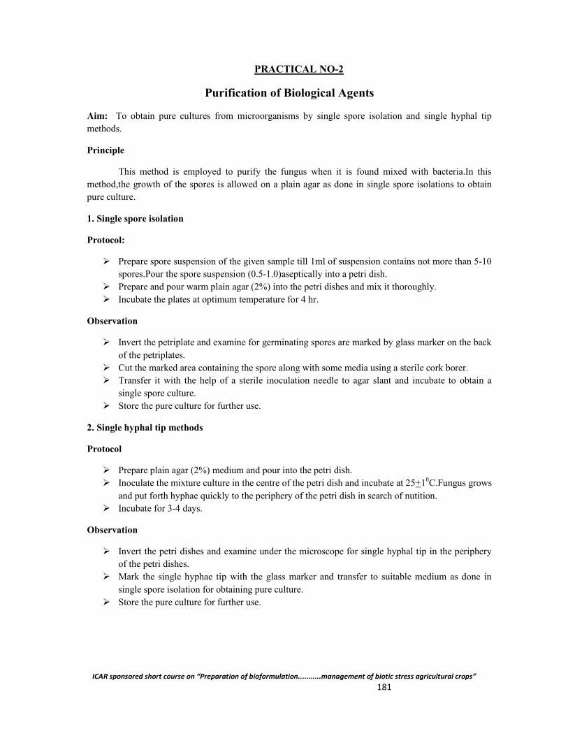
ICAR sponsored short course on “Preparation of bioformulation...........management of biotic stress agricultural crops” 181
PRACTICAL NO-2
Purification of Biological Agents
Aim: To obtain pure cultures from microorganisms by single spore isolation and single hyphal tip methods.
Principle
This method is employed to purify the fungus when it is found mixed with bacteria.In this method,the growth of the spores is allowed on a plain agar as done in single spore isolations to obtain pure culture.
1. Single spore isolation
Protocol:
Prepare spore suspension of the given sample till 1ml of suspension contains not more than 5-10 spores.Pour the spore suspension (0.5-1.0)aseptically into a petri dish.
Prepare and pour warm plain agar (2%) into the petri dishes and mix it thoroughly. Incubate the plates at optimum temperature for 4 hr.
Observation
Invert the petriplate and examine for germinating spores are marked by glass marker on the back of the petriplates.
Cut the marked area containing the spore along with some media using a sterile cork borer. Transfer it with the help of a sterile inoculation needle to agar slant and incubate to obtain a
single spore culture. Store the pure culture for further use.
2. Single hyphal tip methods
Protocol
Prepare plain agar (2%) medium and pour into the petri dish. Inoculate the mixture culture in the centre of the petri dish and incubate at 25+10C.Fungus grows
and put forth hyphae quickly to the periphery of the petri dish in search of nutition. Incubate for 3-4 days.
Observation
Invert the petri dishes and examine under the microscope for single hyphal tip in the periphery of the petri dishes.
Mark the single hyphae tip with the glass marker and transfer to suitable medium as done in single spore isolation for obtaining pure culture.
Store the pure culture for further use.
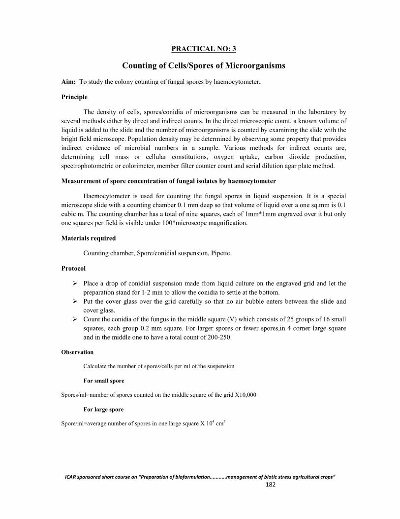
ICAR sponsored short course on “Preparation of bioformulation...........management of biotic stress agricultural crops” 182
PRACTICAL NO: 3
Counting of Cells/Spores of Microorganisms
Aim: To study the colony counting of fungal spores by haemocytometer.
Principle
The density of cells, spores/conidia of microorganisms can be measured in the laboratory by several methods either by direct and indirect counts. In the direct microscopic count, a known volume of liquid is added to the slide and the number of microorganisms is counted by examining the slide with the bright field microscope. Population density may be determined by observing some property that provides indirect evidence of microbial numbers in a sample. Various methods for indirect counts are, determining cell mass or cellular constitutions, oxygen uptake, carbon dioxide production, spectrophotometric or colorimeter, member filter counter count and serial dilution agar plate method.
Measurement of spore concentration of fungal isolates by haemocytometer
Haemocytometer is used for counting the fungal spores in liquid suspension. It is a special microscope slide with a counting chamber 0.1 mm deep so that volume of liquid over a one sq.mm is 0.1 cubic m. The counting chamber has a total of nine squares, each of 1mm*1mm engraved over it but only one squares per field is visible under 100*microscope magnification.
Materials required
Counting chamber, Spore/conidial suspension, Pipette.
Protocol
Place a drop of conidial suspension made from liquid culture on the engraved grid and let the preparation stand for 1-2 min to allow the conidia to settle at the bottom.
Put the cover glass over the grid carefully so that no air bubble enters between the slide and cover glass.
Count the conidia of the fungus in the middle square (V) which consists of 25 groups of 16 small squares, each group 0.2 mm square. For larger spores or fewer spores,in 4 corner large square and in the middle one to have a total count of 200-250.
Observation
Calculate the number of spores/cells per ml of the suspension
For small spore
Spores/ml=number of spores counted on the middle square of the grid X10,000
For large spore
Spore/ml=average number of spores in one large square X 104 cm3

ICAR sponsored short course on “Preparation of bioformulation...........management of biotic stress agricultural crops” 183
Name and addres of the participants of teh short:
Sl. No.
Name Position Address
01 Dr. R. C. Shakywar
Assistant Professor
College of Horticulture and Forestry (Central Agricultural University), Pasighat-791102, Arunachal Pradesh
02 Dr. Angad Singh Rajput
Assistant Professor
Dau Kalyan Singh College of Agriculture and Research Station, Alesur, Bhatapara-493118, Distt- Balodabazar-Bhatapara, Chhattisgarh
03 Mr. Apurba Das
Assistant Professor
College of Sericulture, Assam Agricultural University, Jorhat-785013, Assam
04 Ms. E. Lireni Kikon
Subject Matter Specialist
KVK, Longleng. Post Box-10 Longleng, Nagaland Pin-798625, ICAR-ZPD, Zone-III, Barapani, Umiam
05 Mr. Swapan Deb Roy
Assistant Professor
College of Agriculture, Tripura, Lembucherra-799210, Tripura
06 Dr. Rajesh Ramdas Waghunde
Assistant Professor
College of Agriculture, N.A.U., Bharuch Zadeshwar road, Maktampur patia Bharuch-392012, Gujarat
07 Dr. Munmi Borah
Assistant Professor
Department of Plant Pathology, Assam Agricultural University, Jorhat-785013, Assam
08 Mr. Tarun Chandra Kalita
Assistant Professor
Sarat Chandra Singha College of Agriculture, Assam Agricultural University, Rangamati, Dhubri-783376, Assam
09 Ms. Himadri Kaushik
Jr. Research Fellow cum PhD final Year
Department of Plant Pathology, Assam Agricultural University, Jorhat-78013, Assam
10 Mr. Jeevan B. Scientist Central Muga Eri Research and Training Institute, Central Silk Board, Lahdoigarh, Jorhat-785700, Assam
11 Mr. Joseph Thomas
Sociologist Tejas, Pala 686 575, Kerala
12 Dr. Sekhar Bandopdhyaya
Assistant Professor
Department of Plant Pathology, Uttar Banga Krishi Viswavidyalaya (UBKV), Pundibari, Cooch Behar-736165, West Bengal
13 Dr. B. Temjenmenla
Horticulture Officer
State Department of Horticulture Nagaland (LKR), Zunheboto- 798620, Nagaland
14 Dr. Nayan Kishor Adhikary
Scientist Institute of Agricultural Science University of Calcutta (IASCU), Hazra Road, Ballygunge, Kolkata-700019, West Bengal
ICAR sponsored short course on “Preparation of bioformulation...........management of biotic stress agricultural crops” 184
15 Dr. Vikas Singh
Assistant Professor
Dau Kalyan Singh College of Agriculture and Research Station, Alesur, Bhatapara-493118, Distt- Balodabazar-Bhatapara, Chhattisgarh
16 Dr. G. Subrahmanyam
Scientist Central Muga Eri Research and Training Institute, Central Silk Board, Lahdoigarh, Jorhat-785700, Assam
17 Dr. Monimala Saikia
Assistant Professor
College of Sericulture, Assam Agricultural University, Jorhat-785013, Assam
18 Dr. Pranab Barma
Subject Matter Specilalist
Darjeeling Krishi Vigyan Kendra, UBKV, Kalimpong, Darjeeling-734301, West Bengal
19 Ms. Sarodee Boruah
Jr. Research Fellow cum PhD final Year
Department of Plant Pathology, Assam Agricultural University, Jorhat-785013, Assam
20
Mr. Brajendra Deva Sarma
Assistant Director cum PhD scholar
Spices Board, Mizoram; Presently PhD final Year Student of Department of Plant Pathology, Assam Agricultural University, Jorhat-785013, Assam
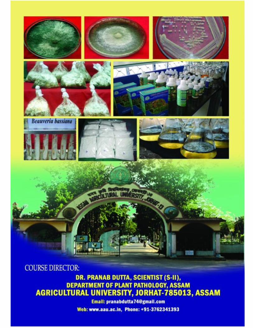
ICAR sponsored short course on “Preparation of bioformulation...........management of biotic stress agricultural crops” 185