IAN-2021-details.pdf - Indian Academy of Neurosciences
349
IAN INTERNATIONAL E-CONFERENCE XXXIX Annual Meeting of Indian Academy of Neurosciences Neuron-glia Interaction: Recent Concepts and Advances December 15, 2021 Programme Jointly Organized by Indian Institute of Science Education and Research-Kolkata Mohanpur - 741 246, West Bengal, India CSIR-Indian Institute of Chemical Biology Kolkata - 700 032, West Bengal, India School of Sciences, Netaji Subhas Open University Kolkata – 700064, West Bengal, India.
-
Upload
khangminh22 -
Category
Documents
-
view
0 -
download
0
Transcript of IAN-2021-details.pdf - Indian Academy of Neurosciences

IAN INTERNATIONAL E-CONFERENCE
XXXIX Annual Meeting of
Indian Academy of Neurosciences
Neuron-glia Interaction: Recent Concepts and Advances
December 15, 2021
Programme
Jointly Organized by
Indian Institute of Science Education and Research-Kolkata Mohanpur - 741 246, West Bengal, India
CSIR-Indian Institute of Chemical Biology Kolkata - 700 032, West Bengal, India
School of Sciences, Netaji Subhas Open University Kolkata – 700064, West Bengal, India.

IAN INTERNATIONAL E-CONFERENCE
XXXIX Annual Meeting of
Indian Academy of Neurosciences
Wednesday, December 15, 2021 10:30 hrs.
IAN Executive Committee Meeting
Opening Session 13:30-13:35 hrs.
Welcome Jayasri Das Sarma Organizing Secretary Laxmi T. Rao Joint Organizing Secretary IAN 2021
13:35-13:40 hrs.
Genesis of the Conference & about the Organizers Subhas C. Biswas, Joint Organizing Secretary, IAN 2021
13:40-13:50 hrs.
Neuroscience Research in Kolkata Debjani Guha, (Secretary, IAN Kolkata Chapter) Professor, Department of Neuroscience, Calcutta University, Kolkata Tushar K. Ghosh, (Treasurer, IAN Kolkata Chapter) Professor, Department of Physiology, Calcutta University, Kolkata
13:50-14:00 hrs.
Guest of Honour Vinay K. Khanna, CSIR-IITR, Lucknow
14:00-14:05 hrs.
Vote of Thanks Anirban Ghosh, Joint Organizing Secretary, IAN 2021
14:05-14:15 hrs.
Short Break
14:15-16:15 hrs.
Session – I
Neuro-Glia in Health Chairpersons: Laxmi T Rao, NIMHANS, Bengaluru Kiranmai S Rai, MMMC-MAHE, Manipal Pradeep Punnakal, PGIMER, Chandigarh Epileptiform activity impairs synaptic plasticity in the rat hippocampus Shobi Veleri, ICMR-NIN, Hyderabad The fundamental function of cilia in health and diseases Bhupesh Mehta, NIMHANS, Bengaluru A quest to unravel potential indicators of early diabetic retinopathy in the inner retina K Vijayalakshmi, NIMHANS, Bengaluru Responses of Oligodendroglia to Cerebrospinal Fluid from Sporadic Amyotrophic Lateral Sclerosis patients turn protective to motor
Session – II
Neural Circuits and Behavior Chairpersons: Anindya G. Roy, NBRC, Manesar Monika Sadananda, MU, Mangalore
Vatsala Thirumalai, NCBS, Bengaluru Can fish tell time? Kavita Babu, CNS-IISc, Bengaluru Studying neuropeptide based circuits through worm locomotion Nitin Gupta, IIT, Kanpur Odor processing in the mosquito brain Rupak Datta, IISER, Kolkata Neurological manifestations of MPS VII: Lessons from a fly model

IAN INTERNATIONAL E-CONFERENCE
XXXIX Annual Meeting of
Indian Academy of Neurosciences
neurons Yogananda S. Markandeya, NIMHANS, Bengaluru Cav-1 in Health and Disease of the Brain
Anamika Sharma, NCBS, Bengaluru Modulation of flight and feeding behaviours in Drosophila melanogaster requires presynaptic IP3Rs in dopaminergic neurons
16.15–16.25 hrs.
Short Break
16.25–18.25 hrs
Session – III
IAN-FAONS Symposium Neuroprosthetics Chairpersons: Renu Wadhwa, AIST, Tsukuba, Japan Supratim Roy, IISc, Bengaluru Ranil de Silva, Institute for Combinatorial Advanced Research and Education (KDU-CARE), Ratmalana, Sri Lanka Cut-off scores/ norms in cognitive screening instruments: a Sri Lanka experience Manojit Pramanik, NTU, Singapore Intracranial hypotension (IH) detection with novel photoacoustic imaging Shyamanta M Hazarika, IIT, Guwahati Motor Imagery Induced Mental Fatigue: Towards an Adaptive Brain Machine Interface Deepak Joshi, AIIMS, New Delhi AI –supported FES system for neuro prosthesis development in SCI and stroke patients Nitish V. Thakor, Johns Hopkins University, USA Machine to Brain Interface: Providing Sensory Feedback to Amputees Sunil Kaul, AIST, Tsukuba, Japan Concluding remarks
Session – IV
Neuronal regulation
Chairpersons: Vinay K. Khanna, CSIR-IITR, Lucknow Surendra K. Trigun, BHU, Varanasi Meenakshi Bawari, Assam University, Silchar Neurotoxicological evaluation of subacute oral administration of methanol extract of medicinal plant Persicaria hydropiper (L.) Delabre in swiss albino mice Sulagna Das, Albert Einstein College of Medicine, New York Local regulation of gene expression in neurons: Insights from single mRNA imaging Manorama Patri, Ravenshaw University, Cuttack Microbiome-Linked Crosstalk in the Gastro-intestinal Exposome Towards Mental Health Prachi Srivastava, AMITY University, Lucknow miRNA and Mammalian Circadian Clock: A Crosstalk Vijay Paramanik, IGNTU, Amarkantak Genistein mediated signaling in learning and memory Rahul Basu, NIH/NIAID, USA A strategy to identify genes which contribute to increased La Crosse Virus

IAN INTERNATIONAL E-CONFERENCE
XXXIX Annual Meeting of
Indian Academy of Neurosciences
susceptibility in children
18.25–18.35 hrs
Short Break
18.35–19.10 hrs
Training Brains to Understand the Brain: Career Choices in Neuroscience: Panel Discussion Chairpersons: Laxmi T. Rao, NIMHANS, Bengaluru K P Mohanakumar, IUCBR, Kottayam Speakers:
1. Shruthi S Sharma, NIMHANS, Bengaluru 2. Debanjana Chakravarty, IISER, Kolkata 3. Fareeha Saadi, IISER, Kolkata 4. Sukanya Sarkar, CSIR-IICB, Kolkata 5. Sayedha Zehra Hyder, NIMHANS, Bengaluru 6. Rituparna Chaudhuri, NBRC, Manesar 7. Shruthi Sridhar, CNS-IISc, Bengaluru 8. Naveen Gowda, Center for Brain Research-IISc, Bengaluru 9. Avishek Roy, AIIMS, New Delhi 10. Deeksha Rathore, RNTMC, Udaipur 11. Dr Jitendra Sinha, Amity University, Noida 12. Dr Rakesh Kumar, JU, Gwalior
19:15 hrs. onwards
Company Presentation Chairperson: Jayasri Das Sarma, IISER, Kolkata Company: BD, Citizen, Perkin Elmer, Gentech
20.00 hrs. Concluding remark Jayasri Das Sarma, IISER, Kolkata

IAN INTERNATIONAL E-CONFERENCE
XXXIX Annual Meeting of
Indian Academy of Neurosciences
Theme: Neuro-glia in Health and Disease
December 16 - 19, 2021
Programme
Jointly Organized by
Indian Institute of Science Education and Research-Kolkata Mohanpur - 741 246, West Bengal, India
CSIR-Indian Institute of Chemical Biology Kolkata - 700 032, West Bengal, India
School of Sciences, Netaji Subhas Open University Kolkata –700064, West Bengal, India
IAN INTERNATIONAL E-CONFERENCE
XXXIX Annual Meeting of
Indian Academy of Neurosciences
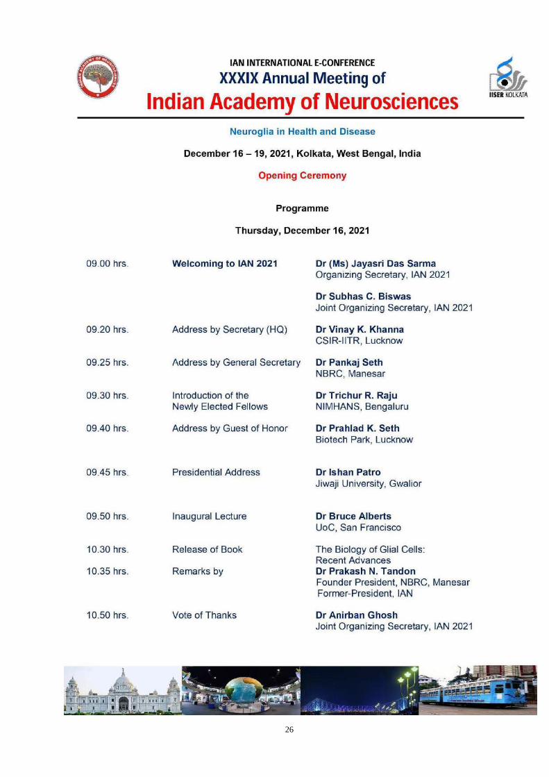
Neuroglia in Health and Disease
December 16 – 19, 2021, Kolkata, West Bengal, India
Opening Ceremony
Programme
Thursday, December 16, 2021
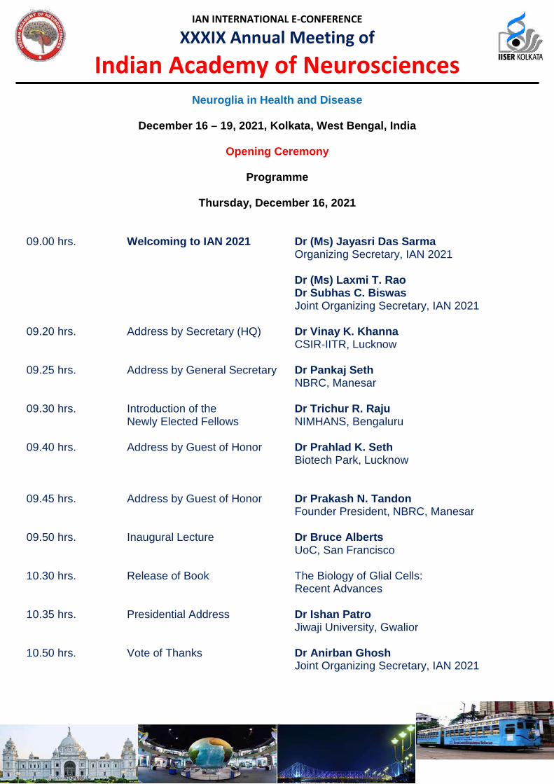
09.00 hrs. Welcoming to IAN 2021 Dr (Ms) Jayasri Das Sarma Organizing Secretary, IAN 2021
Dr (Ms) Laxmi T. Rao Dr Subhas C. Biswas
Joint Organizing Secretary, IAN 2021 09.20 hrs. Address by Secretary (HQ) Dr Vinay K. Khanna CSIR-IITR, Lucknow 09.25 hrs. Address by General Secretary Dr Pankaj Seth NBRC, Manesar 09.30 hrs. Introduction of the Dr Trichur R. Raju
Newly Elected Fellows NIMHANS, Bengaluru
09.40 hrs. Address by Guest of Honor Dr Prahlad K. Seth Biotech Park, Lucknow
09.45 hrs. Address by Guest of Honor Dr Prakash N. Tandon Founder President, NBRC, Manesar 09.50 hrs. Inaugural Lecture Dr Bruce Alberts UoC, San Francisco 10.30 hrs. Release of Book The Biology of Glial Cells:
Recent Advances
10.35 hrs. Presidential Address Dr Ishan Patro Jiwaji University, Gwalior 10.50 hrs. Vote of Thanks Dr Anirban Ghosh
Joint Organizing Secretary, IAN 2021

IAN INTERNATIONAL E-CONFERENCE
XXXIX Annual Meeting of
Indian Academy of Neurosciences
Day 1, Thursday; December 16, 2021 12.00 hrs Tulsabai Somani Educational Trust Award
Chairpersons: Abbas A Mahdi, KGMU, Lucknow Vinay K. Khanna, CSIR-IITR, Lucknow
12.00 –12.10 hrs Madhavi Joshi, Nirma University, Ahmedabad
Extreme glycemic fluctuations debilitate NRG1, ErbB receptors and Olig1 function: Association with regeneration, cognition and mood alterations during diabetes
12.10 –12.20 hrs Bhanu Prakash Tewari, University of Virginia at Charlottesville, USA
Perineuronal nets regulate homeostatic functions of Astrocytes 12.20 –12.30 hrs Anugya Srivastava, CSIR-IITR, Lucknow
Involvement of hippocampal AMPA Receptor trafficking in cadmium induced cognitive deficits in rats -Attenuation by Quercetin
12.30 –12.40 hrs Abass Alao Safiriyu, IISER, Kolkata
Two consecutive prolines in the fusion peptide of Murine-β-Coronavirus spike protein predominantly determine its neuroglial tropism and neuropathogenesis.
12.40 –12.50 hrs Bhavna Daswani, Sophia College, Mumbai
Influence of estrogen receptor beta agonist on C6 glioma cells 12.50 –13.00 hrs Sajeev Kaur, AIIMS, New Delhi
Temporal effects of low intensity magnetic field on sensory and motor functions, morphology and cortical electrical activity after spinal cord injury in adult rats
13.00 –13.10 hrs Sreeja Kumari Dhanya, NCBS, Bengaluru
Role of STIM1 and SEPT7 in regulating gene expression and synaptic components in mouse Purkinje Neurons
13.10 –13.20 hrs Short Break
IAN INTERNATIONAL E-CONFERENCE
XXXIX Annual Meeting of
Indian Academy of Neurosciences
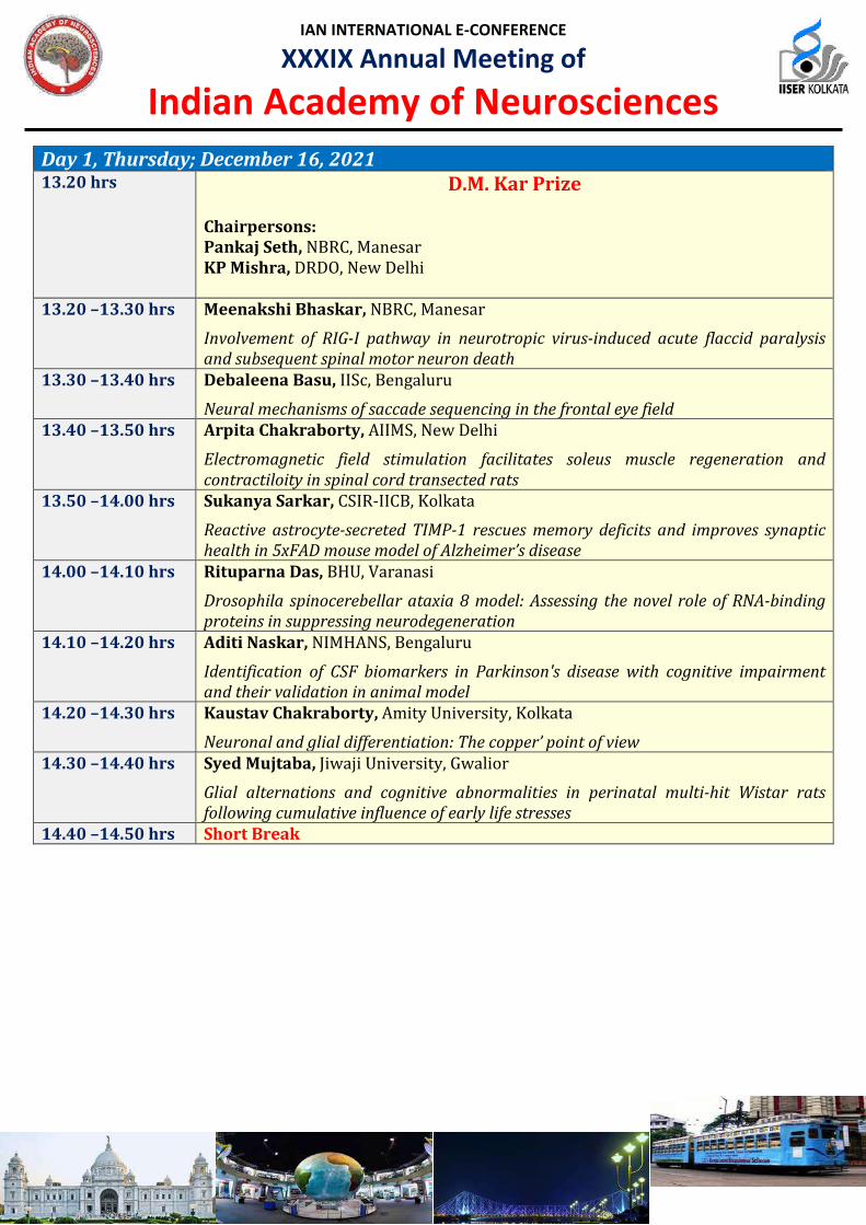
Day 1, Thursday; December 16, 2021 13.20 hrs D.M. Kar Prize
Chairpersons: Pankaj Seth, NBRC, Manesar KP Mishra, DRDO, New Delhi
13.20 –13.30 hrs Meenakshi Bhaskar, NBRC, Manesar
Involvement of RIG-I pathway in neurotropic virus-induced acute flaccid paralysis and subsequent spinal motor neuron death
13.30 –13.40 hrs Debaleena Basu, IISc, Bengaluru
Neural mechanisms of saccade sequencing in the frontal eye field 13.40 –13.50 hrs Arpita Chakraborty, AIIMS, New Delhi
Electromagnetic field stimulation facilitates soleus muscle regeneration and contractiloity in spinal cord transected rats
13.50 –14.00 hrs Sukanya Sarkar, CSIR-IICB, Kolkata
Reactive astrocyte-secreted TIMP-1 rescues memory deficits and improves synaptic health in 5xFAD mouse model of Alzheimer’s disease
14.00 –14.10 hrs Rituparna Das, BHU, Varanasi
Drosophila spinocerebellar ataxia 8 model: Assessing the novel role of RNA-binding proteins in suppressing neurodegeneration
14.10 –14.20 hrs Aditi Naskar, NIMHANS, Bengaluru
Identification of CSF biomarkers in Parkinson's disease with cognitive impairment and their validation in animal model
14.20 –14.30 hrs Kaustav Chakraborty, Amity University, Kolkata
Neuronal and glial differentiation: The copper’ point of view 14.30 –14.40 hrs Syed Mujtaba, Jiwaji University, Gwalior
Glial alternations and cognitive abnormalities in perinatal multi-hit Wistar rats following cumulative influence of early life stresses
14.40 –14.50 hrs Short Break

IAN INTERNATIONAL E-CONFERENCE
XXXIX Annual Meeting of
Indian Academy of Neurosciences
Day 1, Thursday; December 16, 2021 17.00 –17.30 hrs Key Note Lecture
Chairpersons: Shashi B. Singh, NIPER, Hyderabad Subrata Sinha, AIIMS, New Delhi Sumantra Chattarji, NCBS, Bengaluru, India Autism and "Astro"logy
17.30 –17.40 hrs Short Break 17.40 –18.10 hrs Distinguished Lecture
Chairpersons: Prahlad K. Seth, Biotech Park, Lucknow Jayasri Das Sarma, IISER, Kolkata Avindra Nath, NINDS, NIH, USA Retroviral elements in brain development
18.10 –18.20 hrs Short Break 18.20 –20.20 hrs Symposium I
Molecular Mechanisms of Neurodegeneration Chairpersons: Oishee Chakrabarti, SINP, Kolkata Aurnab Ghose, IISER, Pune Ramanujan S. Hegde, Cambridge, UK Protein quality control of orphaned proteins Richard Morimoto, Northwestern University, USA Proteostasis Collapse in Aging and Neurodegenerative Diseases
Symposium II
NeuroCOVID Chairpersons: Pallab Bhattacharya, NIPER, Ahmedabad Shukla Prasad, BHU, Varanasi P.N Sylaja, SCTIMST, Trivandrum COVID 19 and Stroke Pankaj Seth, NBRC, Manesar Molecular mechanisms for SARS-CoV2 mediated neuronal death
Symposium III Brain Response to Environmental Stress: From Man to Molecule
Chairpersons: Shashi B. Singh, NIPER-Hyderabad Kalpana Barhwal, AIIMS, Bhubaneswar Kalpana Barhwal, AIIMS, Bhubaneswar Class switching of carbonic anhydrase isoforms mediates remyelination in CA3 hippocampal neurons during chronic hypoxia S Muthuraju, University of Houston, USA Role of neuroinflammation in the mediation of addictive behaviors following induction of socio-psychological

IAN INTERNATIONAL E-CONFERENCE
XXXIX Annual Meeting of
Indian Academy of Neurosciences
Richard J. Youle, NIH, USA How PINK1- and parkin-mediated mitophagy and neurodegeneration Henriques Dias, CECAD, Germany Cellular quality control by Mitofusins and the E4 ubiquitin ligase Ufd2 Ana-Mafalda Escobar Mahua Maulik, IISER, Kolkata Gap junction intercellular communication in demyelinating neurodegenerative pathology
Dileep R. Yavagal, Miami, USA Large vessel occlusion stroke in COVID19 Sudhir Shah, Department of Neurology, SVPIMSR and NHL Municipal Medical College & Sterling Hospital, Ahmedabad, Gujarat, Neurological manifestations in individuals following COVID-19 Debanjana Chakrabarty, IISER, Kolkata Nexus between CD4+ T cells and microglia / macrophages in murine-CoV induced neuroinflammatory demyelination
stressors K P Mishra, DIPAS, DRDO, Delhi Inhibition of Mac1 scavenger receptor induces M2 microglial polarization and provides neuroprotection under hypobaric hypoxic stress B. N. Srikumar, NIMHANS, Bengaluru Development and characterization of a rat model of post-finasteride syndrome Suryanarayanan Biswal, NCBS, Bengaluru Epigenetic cross-talk at synaptic sites: A bridge towards coping with chronic hypoxic stress

IAN INTERNATIONAL E-CONFERENCE
XXXIX Annual Meeting of
Indian Academy of Neurosciences
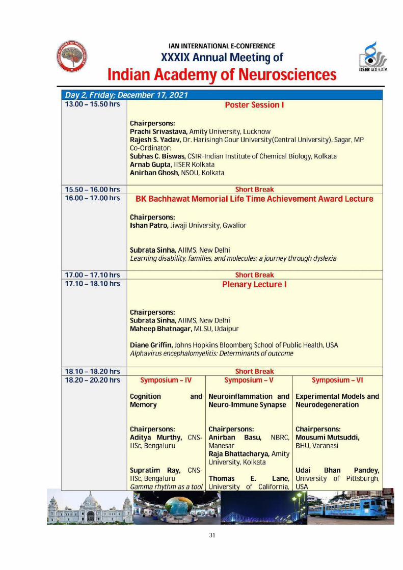
Day 2, Friday ; December 17, 2021 13.15 – 15.50 hrs Poster Session I
Chairpersons: Prachi Srivastava, Amity University, Lucknow Rajesh S. Yadav, Dr. Harisingh Gour (Central University), Sagar Co-Ordinator: Subhas C. Biswas, CSIR-Indian Institute of Chemical Biology, Kolkata Arnab Gupta, IISER, Kolkata Anirban Ghosh, NSOU, Kolkata
15.50 – 16.00 hrs Short Break 16.00 – 17.00 hrs BK Bachhawat Memorial Life Time Achievement Award Lecture
Chairpersons: Ishan Patro, Jiwaji University, Gwalior Raj D. Mehra, New Delhi Subrata Sinha, AIIMS, New Delhi Learning disability, families, and molecules: a journey through dyslexia
17.00 – 17.10 hrs Short Break 17.10 – 16.10 hrs Plenary Lecture I
Chairpersons: Subrata Sinha, AIIMS, New Delhi Maheep Bhatnagar, MLSU, Udaipur Diane Griffin, Johns Hopkins Bloomberg School of Public Health, USA Alphavirus encephalomyelitis: Determinants of outcome
16.10 – 16.20 hrs Short Break 16.20 – 20.20 hrs Symposium – IV
Cognition and Memory Chairpersons: Aditya Murthy, CNS-IISc, Bengaluru Akshay Anand, PGIMER, Chandigarh Supratim Ray, CNS-IISc, Bengaluru Gamma rhythm as a tool
Symposium – V Neuroinflammation and Neuro-Immune Synapse Chairpersons: Anirban Basu, NBRC, Manesar Raja Bhattacharya, Amity University, Kolkata Thomas E. Lane, University of California, USA
Symposium – VI Experimental Models and Neurodegeneration Chairpersons: Mousumi Mutsuddi, BHU, Varanasi Aditya B. Pant, CSIR-IITR, Lucknow Udai Bhan Pandey, University of Pittsburgh, USA

IAN INTERNATIONAL E-CONFERENCE
XXXIX Annual Meeting of
Indian Academy of Neurosciences
to investigate brain function in health and disease Monika Sadananda, Mangalore University Modelling for treatment resistant depression (TRD): Neurobehaviours and monoaminergic neurochemistry across ages in the female Wistar Kyoto rat Balaji J, CNS-IISc, Bengaluru Spatially correlated reorganisation rather than addition of new spines underlies encoding of related memories Mehdi Hayat Shahi, AMU, Aligarh Shh-Gli1-BDNF nexus, synaptic plasticity and depression Kumari Anshu, UoW, Madison, USA Altered attentional processing in the prenatal valproic acid (VPA) model of autism
Neutrophils enhance demyelination in a model of coronavirus-induced neurologic disease Denise Fitzgerald, Queen's University Belfast Medicine, UK The role of T cells in CNS remyelination Long-Jun Wu, Mayo Clinic College of Medicine, USA Microglia-astrocyte interaction in a mouse model of neuromyelitis optica Kalipada Pahan, Rush University, Chicago, USA Stop paying tolls in the CNS to halt neurodegeneration Fareeha Saadi, IISER, Kolkata CD40-CD40 ligand axis in neurotropic mouse hepatitis virus-induced neuroinflammation and demyelination
Identifying hidden GEMs using genetic approaches Sandhya Koushika, TIFR, Mumbai Traffic jams in neurons and implications for neurodegenerative disease Bina Pillai, CSIR-IGIB, New Delhi Inherited RNAs in zebrafish influence brain development: The story of Durga Surajit Sarkar, University of Delhi, Delhi Excavating trans-cellular propagation of human tau aggregates in Drosophila disease models Lucky Sarkar, IISER, Kolkata Azadirachta indica A. Juss bark extract and its Nimbin isomers restrict β-coronaviral infection and replication

IAN INTERNATIONAL E-CONFERENCE
XXXIX Annual Meeting of
Indian Academy of Neurosciences
Day 3, Saturday; December 18, 2021 13.00 – 15.15 hrs
Poster Session II Chairpersons: Vijay Paramanik, IGNTU, Amarkantak Rajendra K. Shukla, Autonomous State Medical College, Bahraich Co-Ordinator: Anirban Ghosh, Netaji Subhas Open University, Kolkata Joy Chakraborty, CSIR-Indian Institute of Chemical Biology
15.15 – 15.30 hrs
Break
15.30 – 16.00 hrs
KT Shetty Memorial Oration Chairpersons: Mahendra K. Thakur, BHU, Varanasi BSS Rao, NIMHANS, Bengaluru Rajat Sandhir, Panjab University, Chandigarh Altered insulin signaling as a pathogenic mechanism in sporadic Alzheimer’s disease: GSK3 beta as a potential therapeutic target
16.00 – 16.10 hrs Short Break 16.10 – 18.10 hrs Symposium VII
Rare Genetic Variants and Neurological Disorders: Studies from Indian Population Chairpersons: S. Ganesh, IIT, Kanpur M. M. Srinivas Bharath, NIMHANS, Bengaluru K Thangaraj, CDFD Hyderabad Dual genetic origin of neuromuscular disorders
Symposium VIII Stem Cell Plasticity in Neuronal Injury Chairpersons: Pallab Bhattacharya, NIPER, Hyderabad Malancha Ta, IISER, Kolkata Hajime Hirase, University of Copenhagen, Denmark Enhancement of remote memory by optogenetic activation of astrocytic Gq
Symposium IX
Normal Ageing Versus Dementia Chairpersons: Sasanka Chakrabarti, MMIMSR - MMDU, Mullana-Ambala Ashima Bhattacharjee, Amity University, Kolkata Marco Bisaglia, University of Padova, Italy Metal dyshomeostasis and neurodegenerative

IAN INTERNATIONAL E-CONFERENCE
XXXIX Annual Meeting of
Indian Academy of Neurosciences
Anuranjan Anand, JNCASR, Bengaluru Human epilepsy genetics: a drift from the ion channel genes involvement B K Thelma, University of Delhi, Delhi Newer genetic insights from familial and sporadic Parkinson's disease Debasmita Pankaj Alone, NISER, Khurda, Odisha New insights into the pathogenesis of pseudoexfoliation glaucoma Rashmi Parihar, IIT, Kanpur A crosstalk between stress granules biogenesis, autophagy and neuropathology: A study on Lafora neurodegenerative disease model
signaling Nikolaus Plesnila, Munich, Germany The pericyte response to ischemic stroke Norshariza Nordin, Universiti Putra Malaysia, Malaysia Neuroregenerative properties of centellaasiatica on oxidative stress-induced stem cell-derived neural cells Ravi Shankar Akundi, South Asian University, New Delhi Exogenous ATP modulates inflammation in the brain through sustained cyclooxygenase-2 (COX-2) synthesis Deepaneeta Sarmah, NIPER, Ahmedabad Stem cell therapy for ischemic stroke: Exploring the role of mitochondria towards neuroprotection
diseases Ravindra K Rawal, CSIR-NEIST, Jorhat, Assam Paradigms in drug discovery against neurodegenerative disorders: A path forward Vivek Swarup, University of California, USA Single-nucleus chromatin accessibility and transcriptomics identify key regulators of Alzheimer's disease Atanu Biswas, IPGMER, Kolkata Many faces of Alzheimer’s disease - do we call it a syndrome? Upasana Ganguly, MMIMSR - MMDU, Mullana-Ambala Linking ferroptosis, mitochondria and alpha-synuclein in Parkinson’s disease neurodegeneration: investigating the effects of iron, erastin, and rotenone in SH-SY5Y cells

IAN INTERNATIONAL E-CONFERENCE
XXXIX Annual Meeting of
Indian Academy of Neurosciences
18.10 – 18.20 hrs Short Break 18.20 – 19.20 hrs Oral Session
Chairpersons: Kamalesh K. Gulia, SCTIMST, Trivandrum Shyam Diwakar, Amrita Vishwa Vidyapeetham, Amritanagar 1. Payal Trivedi, Amity University, Lucknow 2. Geethu Krishna, NIMHANS, Bengaluru 3. Madhumita P. Ghosh, Amity University, AUUP, Noida 4. Sushree Abhidhatri Shrama, University of Hyderabad, Hyderabad 5. Sreelakshmi Sadanandan, CUSAT, Kochi
19.20 – 19.30 hrs Short Break 19.30 – 20.00 hrs Plenary Lecture II
Chairpersons: Avindra Nath, Clinical Director, NINDS, NIH, USA P. Satish Chandra, Former Director, NIMHANS, Bengaluru Stanley Perlman, Professor, University of Iowa, USA Animal Models for COVID-19
20.00 – 20.10 hrs Short Break 20.10 – 22.10 hrs Symposium X
Neural Plasticity and Repair in Neurotraumatic Injury Chairpersons: Suman Jain, AIIMS, New Delhi Sumana Chakravarty, CSIR-IICT, Hyderabad Indrani Dutta, NIMHANS, Bengaluru Regulation of exogenous transplantation of Dental Pulp stem cells on endogenous Schwann cell regeneration and function: implications in Diabetic Neuropathy
Symposium XI Clinical and Cellular Basis of Neurodegeneration Chairpersons: Phalguni A. Alladi, NIMHANS, Bengaluru Nihar R. Jana, IIT, Kharagpur
Ravi Yadav, NIMHANS, Bengaluru Glia targeted therapies for treatment of Movement Disorders Patients
Symposium XII Epigenetics and Drug Repurposing for Neurodegenerative Diseases Chairpersons: Amal Bera, IIT, Madras Arun K. Ray, Ex-Professor of Molecular Medicine, Bose Institute, Calcutta Rajnish Kumar Chaturvedi, CSIR-IITR, Lucknow Drug repurposing for neuroregeneration in Alzheimer's disease

IAN INTERNATIONAL E-CONFERENCE
XXXIX Annual Meeting of
Indian Academy of Neurosciences
Ajay Pal, Columbia University Medical Center. New York Paired motor cortex and spinal cord epidural stimulation facilitates sensorimotor plasticity and improves forelimb function after cervical spinal cord injury in rats- Suman Jain, AIIMS, New Delhi Cortical plasticity in complete spinal cord injury rats following magnetic field exposure Bhagwati Saxena, Nirma University, Ahmedabad TLR4-Mediated Neuroinflammatory responses in traumatic brain injuries: potential mechanisms and therapeutic opportunities Shalini Das Gupta, University of Eastern Finland, Finland Circulating plasma biomarkers of traumatic brain injury
Sumana Chakravarty, CSIR-IICT, Hyderabad Investigating the sex difference in neuroglial alterations in cerebral ischemia using animal models. Sharmili Vidyadaran, Universiti Putra Malaysia iPSC derived Microglia – what have we learnt lately? Phalguni Anand Alladi, NIMHANS, Bengaluru Neurons and glia point up distinct ultrastructural signatures in mice substantia nigra in response to MPTP - the glia have skill sets to survive. Vidyadhara DJ, Yale University School of Medicine, USA Endolysosomal system dysfunction in Parkinson’s disease- evidence from recent studies
Kaviraja Udupa, NIMHANS, Bengaluru Exploring the utility of Neuromodulation and other novel therapeutic regimens to modulate Neuroglia and Gut-brain axis in the management of Parkinsonian disorders Arvind Kumar, CSIR-CCMB, Hyderabad Epigenetic regulation of hippocampal neurogenesis and altered cognitive circuitry- Role of PRMT5 Madhavrao C, All India Institute of Medical Sciences, Mangalagiri, Kerala Angiotensin receptor blocker exhibited favorable effects on oxidative stress and anti-inflammatory parameters of brain in MPTP induced animal models of Parkinson’s Disease: A preclinical study Akanksha Kushwaha, Banaras Hindu University, Varanasi Suv39h1 inhibition recovers memory decline in scopolamine induced amnesic mice

IAN INTERNATIONAL E-CONFERENCE
XXXIX Annual Meeting of
Indian Academy of Neurosciences
Day 4, Sunday; December 19, 2021 13.00 – 15.00 hrs Symposium–XIII
Microglia: Past Controversies and Current Concepts Chairpersons: Nisha Patro, Jiwaji University, Gwalior Ranil De Silva, KDU-CARE), Ratmalana, Sri Lanka Anna Victoria Molofsky, University of California, San Francisco Microglia, memories, and the extracellular space Bozena Kaminska- Kaczmarek, Warszawa, Poland Heterogeneity or plasticity? Dissecting the role of microglia and brain macrophages in stroke, brain tumors and depression Marie Ève Tremblay, University of Victoria, Canada Dark microglia in health and neurodegeneration Susmita Jha, IIT, Jodhpur Discovering dopamine-induced microglia extracellular traps
Symposium–XIV
Translational Research in Movement Neurosciences
Chairpersons: Hrishikesh Kumar, INK, Kolkata Nilkanta Chakrabarty, Calcutta University, Kolkata Stuart Baker, Newcastle University, UK Neural circuits for strength and weakness John Rothwell, London, UK Using neurophysiology to probe mechanisms of recovery post-stroke- Mark R Baker, Newcastle University, UK Animal models of amyotrophic lateral sclerosis Mandar Jog, Western University, London, Ontario Canada Spinal cord stimulation therapy for gait dysfunction in Parkinson’s
Symposium–XV Neuro-Glial Cells in Ageing and Neurodegeneration Chairpersons: Umesh C. Srivastava, NASI, Prayagraj Sunil K. Hota, DIPAS-DRDO, New Delhi Ramen Saha, University of California, Merced, USA Mechanisms of neuronal activity-induced gene transcription and their implications for neurodevelopmental disorders Dharmendra K. Khatri, NIPER, Hyderabad Transcriptional and epigenetic influences in neurodegenerative disease Rahul Kumar, NIPER, Hyderabad Epigenetic regulation of microglial plasticity in hypoxic brain Saroj Kumar Das, Siksha 'O' Anusandhan University, Bhubaneswar Insight underpinning potential impact of bisphenol A towards

IAN INTERNATIONAL E-CONFERENCE
XXXIX Annual Meeting of
Indian Academy of Neurosciences
Debabrata Ghosh, CSIR-IITR, Lucknow Post-transcriptional regulation of microglial CD200R1 expression
Disease Supriyo Choudhury, Institute of Neurosciences, Kolkata Start react effect in chronic stroke patients
development of neurodegenerative diseases Sudeshna Das, San Diego, USA Physiology of microglia in aging brain
15.00 – 16.00 hrs IAN General Body Meeting
16.00 – 17.00 hrs
Cultural Programme
17.00 – 18.00 hrs Break 18.00 – 18.45 hrs Plenary Lecture III
Chairpersons: Trichur R. Raju, NIMHANS, Bengaluru P. K. Sarkar, Kolkata M.V. Padma Srivastava, AIIMS, New Delhi Covid & Brain
18.45 – 18.55 hrs Short Break 18.55 – 20.55 hrs Symposium – XVI
Astroglia in the CNS Health and Disease Chairpersons: Pankaj Seth, NBRC, Manesar Subhas C. Biswas, CSIR-IICB, Kolkata Shane A. Liddelow, NYU, New York What do reactive astrocytes (really) do? Anusha Mishra, Oregon Health and Science University, USA
Symposium – XVII Glioma Biology Chairpersons: Ellora Sen, NBRC, Manesar Anirban Ghosh, NSOU, Kolkata, Simone Niclou, Luxembourg Institute of Health, Luxembourg Challenges of tumor cell plasticity for the treatment of Glioblastoma Dinorah Friedmann, Tel Aviv University, Israel Adoptive T-cell
Symposium – XVIII
Neuropharmaceuticals Chairpersons: Gurucharan Kaur, GNDU, Amritsar Anita Jagota, UoH, Hyderabad Anita Jagota, UoH, Hyderabad Therapeutic effects of various antioxidants on age induced alterations in circadian rhythm and Neurodegeneration Kenneth Shindler, University of Pennsylvania,

IAN INTERNATIONAL E-CONFERENCE
XXXIX Annual Meeting of
Indian Academy of Neurosciences
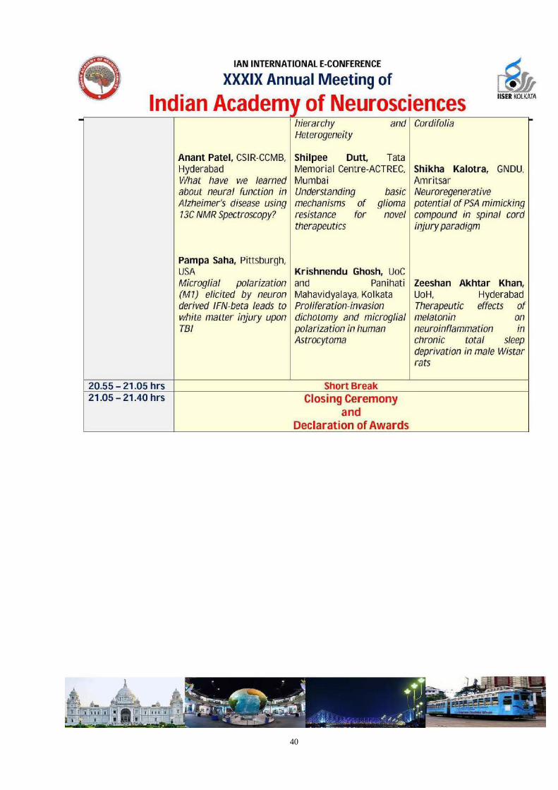
Neurovascular coupling in health and disease Sarika Singh, CSIR-CDRI, Lucknow Protein degradation mechanisms and neurodegenerative diseases Anant Patel, CSIR-CCMB, Hyderabad What have we learned about neural function in Alzheimer’s disease using 13C NMR Spectroscopy? Pampa Saha, Pittsburgh, USA Microglial polarization (M1) elicited by neuron derived IFN-beta leads to white matter injury upon TBI
Immunotherapy targeting both glioma cells and tumor derived endothelial cells Kumar Somasundaram, Indian Institute of Sciences, Bengaluru Cancer stem-like cells: understanding tumor hierarchy and Heterogeneity Shilpee Dutt, Tata Memorial Centre-ACTREC, Mumbai Understanding basic mechanisms of glioma resistance for novel therapeutics Krishnendu Ghosh, UoC and Panihati Mahavidyalaya, Kolkata Proliferation-invasion dichotomy and microglial polarization in human Astrocytoma
Philadelphia, USA International Collaboration to establish a plant extract-based treatment for COVID-19 and Pan-β-Coronavirus infections Gurcharan Kaur, GNDU, Amritsar Neurotherapeutic potential of Tinospora Cordifolia Shikha Kalotra, GNDU, Amritsar Neuroregenerative potential of PSA mimicking compound in spinal cord injury paradigm Zeeshan Akhtar Khan, UoH, Hyderabad Therapeutic effects of melatonin on neuroinflammation in chronic total sleep deprivation in male Wistar rats
20.55 – 21.05 hrs Short Break 21.05 – 21.35 hrs Closing Ceremony
and Declaration of Awards

IAN INTERNATIONAL E-CONFERENCE
XXXIX Annual Meeting of
Indian Academy of Neurosciences
Theme: Neuroglia in health and disease
December 16 - 19, 2021
Dr DM Kar Prize
Number of Prize – 1, Age limit - 35 years, Mode of presentation – Oral Tulsabai Somani Educational Trust Award
Number of Award – 1, Age limit - 40 years, Mode of presentation – Oral
Guidelines for Award Session
The presenter should be member of Indian Academy of Neurosciences.
The presentation will be for 8 minutes followed by discussion/question(s) if any for 2 minutes. There will be minus points for candidates if aided by mentors or colleagues and / or not keeping time.
The presenters are advised to organize their presentation in the following way-
Prior art, Rationale/Hypothesis, Methods, Salient observations (Figures/Tables) with discussion and conclusion.
Judgment is made on the following points –
Clear objectives, Approach to the problem, Findings in relation to aims and objectives, Conclusion, Clarity of presentation, Ability to answer the question (s).
The decision of the referees will be final.

1

2
Message from the President

3
President Prof. Ishan Kumar Patro Jiwaji University Gwalior, Madhya Pradesh, India
IBRO and FAONS Representative Dr. Prahlad Kishore Seth
NASI Senior Scientist, Platinum Jubilee Fellow &
Former Director, CSIR-IITR Biotech Park, Lucknow, Uttar Pradesh, India
Vice President Dr. Raghu Vemuganti University of Wisconsin Madison WI, USA
Vice President Dr . Suman Jain All India Institute of Medical Sciences Ansari Nagar, New Delhi, India
Dean Dr. Trichur R Raju National Institute of Mental Health and
NeuroSciences Bengaluru, Karnataka, India

4
General Secretary Dr. Pankaj Seth
National Brain Research Centre Manesar, Haryana, India
Secretary HQ Dr. Vinay Kumar Khanna
CSIR-Indian Institute of Toxicology Research
Lucknow, Uttar Pradesh, India
Immediate Past President Dr . Shashi Bala Singh
National Institute of Pharmaceutical Education and Research, Hyderabad
Telangana, India
Immediate Past General Secretary Dr. Mahendra Kumar Thakur
Banaras Hindu University Varanasi, Uttar Pradesh, India

5
Message from the Organizers-IAN 2021
On behalf of the Organizing committee, it is our pleasure to welcome you all to the International XXXIX Annual E-Meeting of Indian Academy of Neurosciences (IAN) 2021 with the unique theme of “ Neuro-glia in health and disease.” Since its inception in 1982, IAN has nurtured and promoted neuroscience research in India. The annual IAN meeting provides a unique platform for Indian neuroscientists to share their new findings, views, and thoughts through Distinguished lectures, a series of symposiums, talks, oral presentations, and posters. Moreover, it has regularly held meetings at different centers involved in neuroscience research. Besides, the Academy also has organized and sponsored symposia, workshops, and conferences in areas of cutting-edge neuroscience. During the 38th Annual conference of the IAN last year, the Indian Institute of Science Education and Research Kolkata (IISER-K), in coordination with Netaji Subhas Open University (NSOU) and CSIR-Indian Institute of Chemical Biology (IICB), was bestowed with the responsibility to host the IAN International e-Conference - XXXIX Annual Meeting of Indian Academy of Neurosciences 2021. Due to the ongoing pandemic, XXXIX Annual Meeting will be conducted virtually on December 16-19th, 2021.
Themes of the IAN-2021 include Glial Cell Development, Role of Glia in Aging and Dementia; Glial Adaptations after Injury and Disease; Role of Glia in Learning and Memory; and Glial Regulation of Circuits and Behaviour as well as Neuroprosthetics, Neuronal Regulation, Neuro-COVID, Neurodegeneration, Cognition and memory, Neuro-inflammation and neuroimmune synapse, Neural Stem cell plasticity, Translational neuroscience, Glioma biology, Neural Circuits and behavior and related topics. An exceptional roster of both national and international speakers representing the leaders in glial biology will present the most current research in Glial Biology. The young budding scientists have conducted a panel discussion on "Training Brains to Understand the Brain: Career Choices in Neuroscience." We believe this panel discussion will provide fundamental insights and perspectives of neuroscience research as career choices. As a preface of IAN-2021, four satellite symposiums were organized under the banner of IAN-2021. The first curtain-raiser satellite symposium on "Brainwave: Connection to cognition" was organized by Amity University Kolkata, on September 10, 2021, followed by the second satellite symposium on “Translational Neurophysiology and Cognition” organized by the Department of Physiology, University of Calcutta, third satellite symposium on November 16, 2021, on “Neurological Disorders- from Molecular to Mechanism” by CSIR-IICB Kolkata and IISER Kolkata organized the fourth Satellite program on December 15, 2021, on " Neuron-Glia interaction: Recent concepts and advances."
Although we had to organize the annual meeting via a virtual meeting due to the pandemic, like every dark cloud has a silver lining, the positive side of this virtual conference is that we have received an enormous response from all over the world, with almost 300 participants and over 125 eminent speakers ranging from senior scientist to budding young scientists to discuss their research in the conference over these four days. We are also thrilled to share that Dr. Bruce Alberts kindly agreed to grace the inaugural ceremony of the conference with his distinguished deliberation on " The Great Complexity of Biology, from Cells to Tissues."

6
My sincere thanks to the IAN Executive Committee for bestowing trust in IISER Kolkata, CSIR- IICB Kolkata, and Netaji Subhas Open University to host the meeting this year.
We extend to all the participants a very hearty and warm welcome. Make the best of these four days of scientific extravaganza! My sincere prayer to the Almighty for the help in conducting the annual meeting of IAN with perfection without any technical glitches and making it a valuable asset to neuroscience education and research.
Organizing Committee,
Jayasri Das Sarma, Professor, IISER Kolkata, India
Subhas C. Biswas, Senior Principal Scientist, and Professor, CSIR-IICB Kolkata
Anirban Ghosh, Associate Professor, Netaji Subhas Open University, Kolkata

7

8

9

10

11
History of IAN
1.0 PRODROMAL DEVELOPMENTS INSTRUMENTAL IN THE FOUNDATION OF THE INDIAN ACADEMY OF NEUROSCIENCES
Concurrent with the development of highly refined experimental and analytical techniques that enabled in-depth investigations at levels of intensity and with a degree of precision not attainable earlier, occurred an upsurge of interest in the multidisciplinary Neurosciences in India in the late seventies. It was then fully realized that the contribution of one discipline was hardly known to investigators in other fields of brain research. A plea for having such an organization was made at an International Symposium on Central Synaptic Transmission held at Central Drug Research Institute, Lucknow in October 1974 and attended by neuroscientists from 11 countries besides India and several other symposia etc. in various parts of the country.
A pressing need was felt to bring researchers engaged in different disciplines of neurosciences (viz. Neuroanatomy, Neurophysics, neurophysiology, Neurochemistry, Neurobiology, Neuropathology and clinical disciplines like Neurology, Neurosurgery and Psychiatry along with Bioengineering and Mathematical modelling of the brain, etc.) together on a common platform where their knowledge and methodology could be shared and a road-map for a future effective collaborative interdisciplinary research of the highest standards be designed.
2.0 THE MOUNTING INTEREST IN MULTIDISCIPLINARY NEUROSCIENCES IN INDIA DEMOLISHES PSEUDO - BARRIERS OF WATER-TIGHT DISCIPLINES
2.1 Late Prof. K. P. Bhargava, the then. Director-Professor of Upgraded Department of Pharmacology and Therapeutics and Principal of King George's Medical College Lucknow, in collaboration with Prof. B.N. Dhawan of Central Drug Research Institute and Dr. P .K. Seth of Industrial Toxicology Research Centre, Lucknow approached eminent neuroscientists of the country for establishing a Society of Neurosciences (India). The response to this proposal was overwhelming

12
2.2 Consequently, a meeting of prominent neuroscientists was held at the Central Drug Research Institute, Lucknow on February 10 and 11, 1982. which was addressed by Prof. Merton Sandler, an eminent neuroscientist from the United Kingdom. Everyone present agreed that a Neuroscience Society of India (NSI) be established. It was later found that a society with the same objectives and the same name was already registered in Chennai but was not very active. Prof. K.P. Bhargava requested his former student Prof. Mahdi Hasan, the then Director of the Interdisciplinary Brain Research at Jawahar Lal Nehru Medical College, Aligarh to approach Prof. B. Ramamurthi, the then President of the Neuroscience Society of India for the organization of a Conference of Neurosciences Society of India (NSI) at Aligarh so that its activities and membership could be enlarged. Accordingly Prof. Hasan obtained the consent of Late Prof. Ramamurthi and organized a meeting of Neurosciences Society of India at Jawahar Lal Nehru Medical College, AMU, Aligarh on February 5 and 6, 1983. Prof. Ramamurthi sent his presidential address but on the day of the conference he communicated his inability to attend and preside over the conference. Other office bearers of NSI also stayed away from the meeting
3.0 BIRTH OF THE INDIAN ACADEMY OF NEUROSCIENCES AT ALIGARH
3.1 On February 5, 1983, seventy neuroscientists from all over the country and USA gathered at Aligarh to attend the Neuroscience Society of India Conference (NSI) felt distressed over the absence of the office-bearers of the NSI.
3.2 The need for a continuously active forum of neuroscientists of India was stressed by all the participants. The consensus was that NSI was unlikely to meet the aspirations and needs of the neuroscience community of the country. Prof. S.S. Parmar of the Department of Physiology, University of North Dakota School of Medicine, Grand Forks, USA and an international votary of multidisciplinary Neurosciences, thereupon proposed that a new organization should be formed with wider spectrum of membership and activities. It was decided to form Indian Academy of Neurosciences and get it registered as a new organization.
3.3 Prof. Parmar assured the gathering that large number of Indian neuroscientist from USA will form the Academy and actively contribute to its activity.
3.4 A general body meeting approved the formation of the Academy and adopted its constitution. It would be named Indian Academy of Neurosciences (IAN) and would build up international linkages and welcome foreign neuroscientists as members. For this purpose, the post of a Secretary, International Affairs was provided in the new constitution. The first elections to the various posts of the IAN were held and Prof. P.N. Saxena, Head of Pharmacology presided over the conference at Aligarh. Besides a plenary lecture by Prof. S.S. Parmar, 45 research papers were presented and a poster session was held.
4.0 REGISTRATION OF THE ACADEMY
Dr. P.K. Seth, the former General Secretary of the Indian Academy of Neurosciences, got the Academy formally registered at Lucknow under the Societies Registration Act of 1860 on July 9, 1984.

13
5.0 ANNUAL CONFERENCES OF IAN
5.1 The third annual conference of the IANS was organized at Lucknow by the Industrial Toxicology Research Centre in collaboration with IBRO on Feb. 4- 6, 1984. Also, an ITRC-IBRO symposium on "Neurotoxic Substances and. Human Health" was held concurrently.
5.2 The Fourth Annual Conference of the Indian Academy of Neurosciences was held jointly with the Asian Congress of Pharmacology at New Delhi which Prof. K.P. Bhargava presided.
5.3 Thereafter, successive annual conferences of IAN were held at Tirupati (1987), Calcutta (1988), Chandigarh (1989), Lucknow (1990) and Delhi (1991)
5.4 At the Annual Conference of IAN held at Jawahar Lal Nehru University, New Delhi, presided over by late Dr. Darab Dastoor, efforts were made for the merger of the Neuroscience Society of India (NSI) and the Indian Academy of Neurosciences into one common Neuroscience organization but unfortunately the initiative did not succeed.
5.5 The 11th,12th,13th,14thand 15thAnnual Conferences of the Indian Academy of Neurosciences were successfully held at Lucknow (1992), Jhansi (1994), Vellore (1995), Mumbai (1996) and Bangalore (1997) respectively.
5.6 During the Bangalore conference, a symposium on "Molecules to Behaviour" was organized jointly with the Neuroscience Society of India as a tribute to the memory of Prof. B. K. Bachhawat, an eminent neuroscientist of the country and a former president of IAN.
5.7 The 16th- 23rdAnnual Conferences of the IANS were held at New Delhi (1998). Gwalior (1999), Lucknow (2001), Kolkata (2002), Udaipur (2003), Hyderabad (2004). Gwalior (2005) and Bangalore (2005).
5.8 The 24thannual conference marked the beginning of Silver Jubilee Year of the Academy and was organized at Lucknow from December 17- 20, 2006. Prof. P.N. Tandon, Presided over the Conference. The 25111 annual conference of the Academy was held at Banaras Hindu University, Varanasi from November 22 - 25,2007 and was presided over by Prof. B.N. Dhawan.
5.9 Regular features of the annual meeting are plenary lectures and symposia on current topics in neurosciences in addition to the oral and poster presentations. Workshops on techniques of neurosciences have also been organised. The annual meeting and symposia bring together basic scientists and clinical researchers to discuss common problems inherent in their area of research. The Academy encourages the participation of young neuroscientists so that they get an opportunity to interact with eminent senior colleagues. The endeavor has been helpful to stimulate and widen their outlook for future research.
6.0 INTERNATIONAL SYMPOSIA / WORKSHOPS

14
In addition to organizing the Annual National Conference, the Academy has been sponsoring and co-sponsoring International Symposia and Conferences to promote the cause of neurosciences and to create awareness in public regarding the progress of various facets of brain research
A list of major International Symposia / Workshops co-sponsored by the IANS is given below:
1. ITRC-IBRO Symposium on Neurotoxic Substances and their Impact on Human Health, at Industrial Toxicology Research Centre, Lucknow 1984.
2. Neuroscience Component of the Asian Congress of Pharmacology at Hotel Ashok, New Delhi on January 15 - 19, 1985.
3. Satellite symposium of the Asian Congress of Pharmacology on "Brain Neurotransmission Mechanisms and Hypertension" held at the Upgraded Department of Pharmacology, King George's Medical College, Lucknow, January 21 - 22, 1985.
4. Indo-U.S. Workshop on Chemistry and Biology of Centrally Acting Peptides, Central Drug Research Institute, Lucknow 1987.
5. Indo-US Workshop on Current Approaches for Receptor Studies in Neurobiology Central Drug Research Institute, Lucknow (1991).
6. 2nd Congress of Toxicology in Developing Countries, New Delhi (1991).
7. Colloquium on Cellular and Molecular Advances Neuropharmacology Central Drug Research Institute, Lucknow (1992).
8. Colloquium on Advances in Neurotransmitter Receptors: Cellular in and Molecular mechanisms Central Drug Research Institute, Lucknow (1994).
9. Symposium on Cellular and Molecular Mechanisms of Centrally Acting Agents Industrial Toxicology Research Centre, Lucknow (1997).
10. International Symposium on Molecular Toxicology and Environmental Health Industrial Toxicology Research Centre, Lucknow (2003).
7.0 MEMBERSHIP AND FELLOWSHIP
The Academy has sizable number of dedicated neuroscientists as life members spread all over the country and abroad. The Academy also elects eminent neuroscientists as Fellows and Honorary Members. The Academy has over 800 life members
8.0 CREATION OF FELLOWSHIP AND ELECTION OF FOUNDER FELLOWS AND THE FIRST CONVOCATION OF THE IANS
8.1 The Fifth Annual Conference of the IAN was held at Aligarh on December 7- 9,1986. It was decided to create a new category of membership designated as Fellow to be elected from among the members on basis of their distinguished contribution

15
8.2 The following eight Founder Fellows were elected
1. Dr. B.N. Dhawan, Lucknow 2. Dr. D.K. Ganguly, Kolkata 3. Dr. D.K. Dastur, Mumbai 4. Dr. Mahdi Hasan, Aligarh 5. Dr. S.S. Parmar, Grand Forks, USA 6. Dr. V.K. Selvarajan, Chennai 7. Dr. P.K. Seth, Lucknow 8. Dr. K.C. Singhal, Aligarh.
8.3 The first convocation of IAN was presided over by Dr. John Autian, from USA on December 7, 1986 at the Auditorium of the M.U. Institute of Ophthalmology, Gandhi Eye Hospital, Aligarh.
8.4 Fellows have been elected every year and Fellowship conferred at convocation at the next Annual Conference of the Academy
8.5 The total number of Fellows is 57.
9.0 HONORARY MEMBERS
9.1 Eminent Indian and Foreign scientist whose association with the Academy will be valuable for its activities are elected Honorary Members of the Academy. They enjoy all the privileges of the members but do not have to pay membership or admission fee.
9.2 The Academy currently has 28 honorary members.
10.0 AWARDS OF THE ACADEMY
10.1 Professor B.K. Bachhawat Memorial Life Time Achievement Award:
The Academy has initiated an annual award in the memory of late Prof. B.K. Bachhawat, one of the stalwarts of Neuroscience in India and former President of the Academy. The award is given to an outstanding Neuroscientist, on the basis of his/her research contributions and consists of a scroll and a plaque. The awardee has to give an oration on the topic of his/her choice preferably at the annual conference and to provide a manuscript of the oration for publication in the Annals of Neurosciences.
The Awardee must be a Fellow Honorary Member 1 Life Member of the Indian Academy of Neurosciences and have a standing of at least 10 years in Neurosciences.
The neuroscientist to be honored is selected by the Executive Committee based on the recommendation of the search committee and the award is announced during the inaugural function of the annual meeting of the Academy. Prof. P.N. Tandon (2003), Prof B.N. Dhawn (2004), Prof. Mahdi Hasan (2005), Prof. P.K. Seth (2007) and Prof. S.S. Parmar (2008) have been honored with the award.

16
10.2 Tulsabai Somani Educational Trust Award
The award is given for best oral presentation at the annual conference of IAN by young neuroscientist and carries cash prize of RS.1,500/ -(Rupees One thousand five hundred only), memento and certificate
Eligibility: The candidate / nominee should essentially be a member of Indian Academy of Neurosciences and below 40 years of age at the time of conference.
Procedure: Requisite copies of the abstract clearly marked as PAPER FOR TULSABAI SOMANI EDUCATIONAL TRUST AWARD should be forwarded to the Organizing Secretary before the last date of abstract submission. A copy of the abstract should also be sent to the General Secretary / Secretary (HQ). The papers awarded shall be published in the official journal of the Academy 'Annals of Neuroscience.
10.3 Dr. D. M. Kar Prize
The award is given for best oral presentation at the annual conference of IAN by youneuroscientist and carries a memento and a certificate.
Eligibility: The candidate / nominee should essentially be a member of Indian Academy of Neurosciences and below 35 years of age at the time of conference.
Procedure: Requisite copies of the abstract clearly marked as PAPER FOR Dr. D. M. KAR PRIZE should be forwarded to the Organizing Secretary before the last date of abstract submission. A copy of the abstract should also be sent to the General Secretary / Secretary (HQ). The papers awarded shall be published in the official journal of the Academy 'Annals of Neuroscience'.
10.4 Professor S. S. Parmar Research Foundation Award
There are two awards, which are given for best papers presented in the Poster session. The award carries cash prize of Rs. 500/- (Rupees Five hundred only) and certificate.
Eligibility: The candidate / nominee should essentially be a member of Indian Academy of Neurosciences and below 35 years of age at the time of conference.
Procedure: Requisite copies of the abstract clearly marked as PAPER FOR PROFESSOR S.S. PARMAR RESEARCH FOUNDATION AWARD should be forwarded to the Organizing Secretary before the last date of abstract submission. A copy of the abstract should also be sent to the General Secretary / Secretary (HQ). The papers awarded shall be published in the official journal of the Academy 'Annals of Neuroscience'.
10.5 Jyotsnamoyee Raghunath Bhattacharya Prize
The award is given for published research paper and carries cash prize of Rs. 1,000/-(Rupees One thousand only), medal and certificate.

17
Eligibility: The paper should be in the field of basic neuroscience including the disciplines - Neuroanatomy, Neurophysiology, Neurochemistry, Neuropharmacology, Neuroimmunology and Neurotoxicology and should have been published during the past 2 years. The work should have been done in India. In case of multi-authored publication. the awardee should be the first author and at least one of the authors should be a member of the Academy.
Procedure: Requisite copies (generally three) of the reprint / paper clearly marked as PAPERFORJYOTSNAMOYEE RAGHUNATH BHATTACHARYA PRIZE should be sent to the General Secretary / Secretary (HQ) of the Academy before the last date for peer reviewing.
10.6 John Miller Travel Award(s)
The Academy provides John Miller travel Award(s) that covers for a round trip II Class Sleeper travel to the candidate presenting a paper at the annual conference on competitive basis. Depending on availability of funds two travel awards upto the maximum of Rs. 800/- each may be provided.
Eligibility: The age of the candidate / nominee should not exceed 35 years.
Procedure: Interested persons should send an application duly forwarded by their mentors along with required copies of the abstract of paper and a certificate of II Sleeper class fare to the Organizing Secretary. A copy of the same should also be sent to the General Secretary and Treasurer (Dr. A.K. Agrawal) on or before the last date. The selected candidate(s) are informed before the conference and actual fare towards travel by II Class Sleeper is provided at the time of conference.
10.7 Professor R. Nath Memorial Travel Award (s)
The academy awards Dr. R. Nath Memorial Travel Fellowship to the candidates seeking support for participation in the annual conference of Indian Academy of Neurosciences. Six to eight travel awards consisting of actual travel reimbursement by AC-3 tier up to a maximum of Rs. 1,500/- are available. The travelling fellowships will be provided on competitive basis and selection will be made by the Organizing Secretary by constituting a committee in consultation with Treasurer and Secretary (HQ).
Eligibility: The applicant! nominee should be below 35 years of age and present a paper at the annual conference essentially and may not be receiving travel support from other sources.
Procedure: Interested persons should send an application duly forwarded by their mentors along with required copies of the abstract of paper. They should send a statement of about 250 words how his/her participation will help in further research send a letter of support from the Guide / Supervisor.
The above papers should reach the Organizing Secretary before the last date as advertised. A set of papers should also be sent to the Treasurer on or before the last date. The selected candidate(s) will be informed before the conference and

18
reimbursement of fare will be done on the last day of the conference.
11.0 PUBLICATIONS
1. Annals of Neurosciences - Quarterly scientific publication of the Academy Stated in 1991 and currently published 15 volumes
2. Newsletter- Started in 1985 and being published once/ twice a year. 3. Membership Directory - Published at regular intervals.
12.0 INTERNATIONAL LINKAGES
The Academy has established International linkage with the following major International Neuroscience Organization
1. International Brain Research Organization 2. Federation of Asian and Oceanic Neuroscience Societies 3. Neuroscience Division of Association of Scientist of Indian origin
Federation, USA
13.0 LOCAL BRANCHES
To promote the activities of neuroscience in a region it was decided that a branch of the Academy may be formed in the city having more than 10 members. At present the Academy has local branches at Aligarh, Gwalior, Kolkata and Lucknow.
14.0 SILVER JUBILEE CELEBRATION OF INDIAN ACADEMY OF, NEUROSCIENCES
Starting the journey from Lucknow in 1982, the Academy completed 25 glorious years of its existence in 2006. It was decided to commemorate the silver jubilee with the organization of 24th Annual meeting of the Academy at Lucknow on December17- 2006 followed by year-round activities at various neuroscience centers and IAN local chapters. The silver jubilee celebrations were concluded at Banaras Hindu University, Varanasi with the organization of 25th Silver Jubilee Conference on November 22 -2007. A number of eminent neuroscientists from different parts of the country and globe participated in these conferences to make the events successful and memorable. Following symposia were held to commemorate the silver jubilee of the Academy in the year 2007.

19
1. Symposium on The Expanding Frontiers of Neurosciences, May 11th 2007, Department of Psychiatry, University of Illinois at Chicago, 1601 W. Taylor St., Chicago, IL, 60612.
2. Symposium on Neurodegeneration and Neuroregeneration : Current Trends and Future Strategies, July 30, 2007, Industrial Toxicology Research Centre Lucknow.
3. Symposium on Current Trends in Auditory Research, September 21-22, 2007, Department of Anatomy, Maulana Azad Medical College, New Delhi.
4. Symposium on Glial Neurobiology, October 23, 2007, School of Neurosciences, Jiwaji University Gwalior

20
Sponsors

21

22

23
Gen-Tech

24
Program

25
26

27

28

29

30
31

32

33

34

35

36

37

38

39
40

41

42

43
Satellite Symposia-1
Satellite Symposium for the Indian Academy of Neurosciences (IAN) Annual Conference) Jointly organised by Indian Academy of Neurosciences and Amity Institute of Biotechnology, Amity University
Kolkata
September 10, 2021
Sl No
Time
Title
Speaker
1. 8.30 – 9.15 AM
Saraswati bandana followed by the inaugural address
Prof. Sanjay Kumar
Vice Chancellor,
Amity University
Kolkata Prof. Ankita
Chakraborty Pro Vice-
Chancellor, Amity
University Kolkata
Dr Santanu Palchaudhuri
Assistant Director,
Amity Institute of Biotechnology
2. 9:15 – 9.30 AM Introduction Prof. Jayasri Das Sarma
(IISER-Kolkata)
3. 9.30 – 9.45 AM Introduction to IAN IAN representative

44
4. 9.45 – 10.00 AM Introduction to the satellite symposium Dr. Ashima Bhattacharjee and
Dr. Raja Bhattacharya
(Conveners, Amity University Kolkata)
Scientific
Session I
Sl No
Time
Title
Speaker
5. 10.05 – 10.35 AM Opposing activities of neuropeptides impose differential activity states to
regulate the feeding drive Dr. Aurnab Ghosh
(IISER, Pune)
6. 10.40 – 11.10 AM Ube3a and its link with autism spectrum disorder
Prof. Nihar Ranjan Jana
(IIT-Kharagpur)
7. 11.15 – 11.45 AM How we perceive brightness: a journey beyond Simple Cells through the parallel visual pathways
Dr. Kuntal Ghosh
(ISI, Kolkata)
11.45 – 12.00… BREAK
8. 12.00 – 1.00 PM Flash Talk (7 minutes each)
Lectures by students/scholars
(6 talks talks selected by reviewing committee for a 7 min.s talk and time will be strictly
maintained).
9. 1.00 – 1.30 PM – Lunch
Scientific
Session II
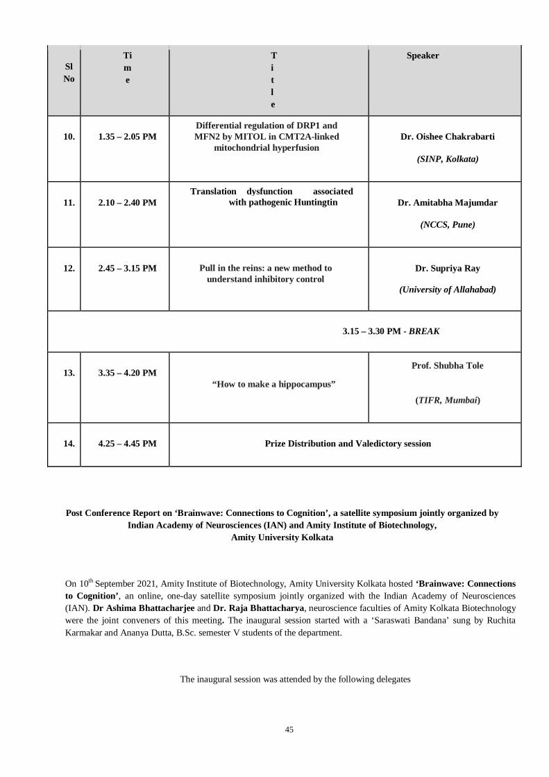
45
Sl No
Time
Title
Speaker
10. 1.35 – 2.05 PM Differential regulation of DRP1 and MFN2 by MITOL in CMT2A-linked
mitochondrial hyperfusion Dr. Oishee Chakrabarti
(SINP, Kolkata)
11. 2.10 – 2.40 PM Translation dysfunction associated
with pathogenic Huntingtin Dr. Amitabha Majumdar
(NCCS, Pune)
12. 2.45 – 3.15 PM Pull in the reins: a new method to understand inhibitory control
Dr. Supriya Ray
(University of Allahabad)
3.15 – 3.30 PM - BREAK
13. 3.35 – 4.20 PM
“How to make a hippocampus”
Prof. Shubha Tole
(TIFR, Mumbai)
14. 4.25 – 4.45 PM Prize Distribution and Valedictory session
Post Conference Report on ‘Brainwave: Connections to Cognition’, a satellite symposium jointly organized by Indian Academy of Neurosciences (IAN) and Amity Institute of Biotechnology,
Amity University Kolkata
On 10th September 2021, Amity Institute of Biotechnology, Amity University Kolkata hosted ‘Brainwave: Connections
to Cognition’, an online, one-day satellite symposium jointly organized with the Indian Academy of Neurosciences (IAN). Dr Ashima Bhattacharjee and Dr. Raja Bhattacharya, neuroscience faculties of Amity Kolkata Biotechnology were the joint conveners of this meeting. The inaugural session started with a ‘Saraswati Bandana’ sung by Ruchita Karmakar and Ananya Dutta, B.Sc. semester V students of the department.
The inaugural session was attended by the following delegates

46
(i) Dr Santanu Palchaudhuri, Assistant Director, Amity Institute of Biotechnology, Amity University Kolkata.
(ii) Shri. Arunasis Chakraborty, Deputy Director General, Amity Foundation for Science, Technology &
Innovation Alliances (AFSTIA).
(iii) Dr. Rajiv Sharma, Director General, Amity Foundation for Science, Technology & Innovation Alliances (AFSTIA) and Sr. Vice President, Ritnand Balved Education Foundation (RBEF).
(iv) Prof. Jayasri Das Sarma, Department of Biological Sciences, Indian Institute of Science Education and Research, Kolkata.
The delegates provided their words of encouragement which set the meeting to a nice start. Dr Rajiv Sharma stressed on the importance of maintaining a long-term collaboration with IAN. He congratulated the campus, the department and the conveners for organising such a curtain raiser event for this year’s IAN’s annual meeting. This was followed by Prof Das Sharma’s speech where she introduced the audience to IAN and its dignitaries and requested them to say a few words. The speakers included:
(i) Prof Pankaj Seth, General Secretary (IAN), National Brain Research Centre
(ii) Dr. Subhas C. Biswas, Joint organiser of the IAN society meeting 2021, CSIR-Indian Institute of Chemical Biology.
(iii) Dr. Anirban Ghosh, Joint organiser of the IAN society meeting 2021, Netaji Subhas Open University.
(iv) Dr. Pallab Bhattacharya, Associate Professor, NIPER, Ahmedabad.
Dr Ashima Bhattacharjee and Dr. Raja Bhattacharya, the joint conveners of the meeting next discussed about the organisation of the meeting, the challenges and advantages imposed by the pandemic and relevance of the meeting in the current context. The conference included two scientific sessions and a student talk session. The speakers for the first scientific session included:
(i) Dr. Aurnab Ghose, IISER, Pune
(ii) Prof. Nihar Ranjan Jana, IIT-Kharagpur

47
(iii) Dr. Kuntal Ghosh, ISI, Kolkata
This session was chaired by Dr. Kavita Babu (Associate Professor, Indian Institute of Science, Bangalore) and Dr Raja Bhattacharya (Associate Professor and Ramalingaswamy fellow, Amity Institute of Biotechnology, Amity University Kolkata). This was followed by a very interesting student talk session. Out of a total of 23 submitted abstracts, 6 were selected for presentation in this session by the expert panel. Each presenter was allowed 7 minutes to present his/her work, followed by a 3-minute interaction with the expert judges and participants. The selected speakers were as follows:
(i) Rahul Kumar, McGill University, Canada
(ii) Siddharth R. Venkatesh, Indian Institute of Science, Bangalore
(iii) Diptankar Bandyopadhyay, CSIR-Indian Institute of Chemical Biology
(iv) Anushka Deb, Tata Institute of Fundamental Research
(v) Fareeha Saadi, Indian Institute of Science Education Research, Kolkata
(vi) Debmita Chatterjee, Saha Institute of Chemical Biology
The judges for this session were:
(i) Dr Aurnab Ghose, IISER-Pune
(ii) Dr. Arnab Gupta, IISER-Kolkata
(iii) Dr. Amitabha Majumdar, NCCS, Pune

48
It was challenging to select the winners taking into consideration the quality of work, presentation and interaction. Diptankar Bandyopadhyay was awarded the first prize. Rahul Kumar got the second prize and Siddharth Venkatesh received the third prize. Prizes were sponsored by Patel Chem de Drugs. The second scientific session included the following speakers:
(i) Dr. Oishee Chakrabarti, SINP, Kolkata
(ii) Dr. Amitabha Majumdar, NCCS, Pune
(iii) Dr. Supriyo Ray, University of Allahabad
The session was chaired by Prof. Birendra Nath Mallick (Director of the Amity Institute of Neurosciences and Neuropsychology, Amity University, Noida) and Dr. Ashima Bhattacharjee (Associate Professor, Amity Institute of Biotechnology, AUK). The last scientific session included a plenary talk by Prof. Shubha Tole, Tata Institute of Fundamental Research. This session was chaired by Dr. Raja Bhattacharya. Prof. Jayasri Das Sarma announced the prizes for the student talk session and delivered the concluding remarks. Dr. Sandipan Chakraborty (Amity Institute of Biotechnology, Amity University, Kolkata) delivered the vote of thanks.
49
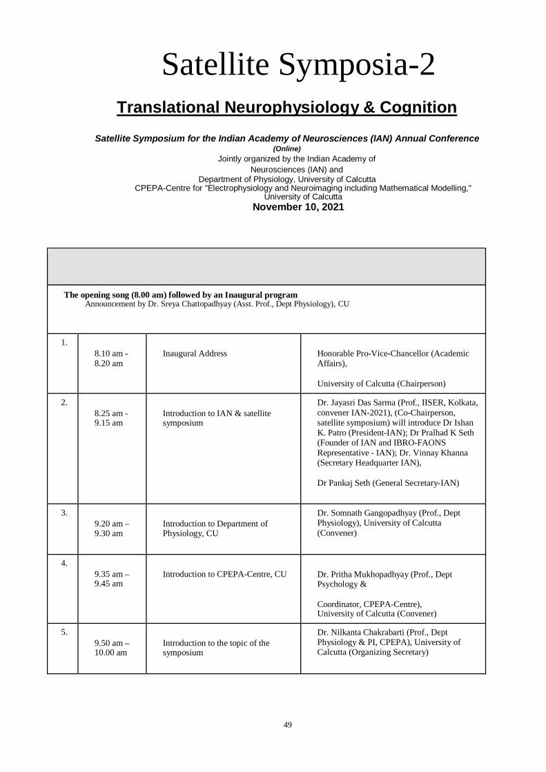
Satellite Symposia-2 Translational Neurophysiology & Cognition
Satellite Symposium for the Indian Academy of Neurosciences (IAN) Annual Conference (Online)
Jointly organized by the Indian Academy of Neurosciences (IAN) and
Department of Physiology, University of Calcutta CPEPA-Centre for "Electrophysiology and Neuroimaging including Mathematical Modelling,"
University of Calcutta November 10, 2021
The opening song (8.00 am) followed by an Inaugural program Announcement by Dr. Sreya Chattopadhyay (Asst. Prof., Dept Physiology), CU
1. 8.10 am - 8.20 am
Inaugural Address Honorable Pro-Vice-Chancellor (Academic Affairs),
University of Calcutta (Chairperson)
2. 8.25 am - 9.15 am
Introduction to IAN & satellite symposium
Dr. Jayasri Das Sarma (Prof., IISER, Kolkata, convener IAN-2021), (Co-Chairperson, satellite symposium) will introduce Dr Ishan K. Patro (President-IAN); Dr Pralhad K Seth (Founder of IAN and IBRO-FAONS Representative - IAN); Dr. Vinnay Khanna (Secretary Headquarter IAN),
Dr Pankaj Seth (General Secretary-IAN)
3. 9.20 am – 9.30 am
Introduction to Department of Physiology, CU
Dr. Somnath Gangopadhyay (Prof., Dept Physiology), University of Calcutta (Convener)
4. 9.35 am – 9.45 am
Introduction to CPEPA-Centre, CU Dr. Pritha Mukhopadhyay (Prof., Dept Psychology &
Coordinator, CPEPA-Centre), University of Calcutta (Convener)
5. 9.50 am – 10.00 am
Introduction to the topic of the symposium
Dr. Nilkanta Chakrabarti (Prof., Dept Physiology & PI, CPEPA), University of Calcutta (Organizing Secretary)

50
Break: 10 min
Scientific Session-I: "Clinical case studies."
Invited talks Chaired by: Dr. Debasish Bandyopadhyay, (Prof., Dept Physiology & PI, CPEPA), CU
6. 10.10 am –10.40 am Dr. Hrishikesh Kumar
(Neurologist, INK, Kolkata)
Title: Brain simulation in Lewy body disease
7. 10.45 am –11.15 am Dr. Atanu Biswas
(Neurologist & Prof., BIN, IPGME&R, Kolkata)
Title: COVID19 and cognitive impairment
Break: 10 min
Scientific Session-II: "Psychobehavioural approaches."
Invited talks Chaired by Dr. Sanjukta Das (Prof., Dept Psychology & PI, CPEPA) & Dr. Swarup Poria (Prof., Dept Appl Math & PI, CPEPA), CU
8. 11.25 am –11.55 am Dr. Laxmi T. Rao
(Prof., Neurophysiology, NIMHANS, Bengaluru)
Title: Causes and consequences of anxiety-induced changes in cognition and behavior
9. 12.00 am - 12.30 pm
Dr. Manas Kumar Mandal
(Prof., Humanities & Social Sci., IIT-Kgp)
Title: Cognitive Neuroscience: a translational approach
Lunch Break: 1 hr.
Scientific Session-III: "Molecular interactions to systems biology."
Invited talks Chaired by Dr. Biswadev Bishayi (Prof., Dept Physiology), CU
10. 1.30 pm –
2.00 pm Dr. Amal Bera
(Prof., Biotechnology, IIT-Madras)
Title: Pannexin-P2X7 receptor interaction and its role in Parkinson's Disease

51
11. 2.05 pm –
2.35 pm Dr. Basant K. Tiwary
(Prof., Bioinformatics, Pondicherry University)
Title: Molecular co-evolution of cognition & speech in human
Break: 10 min
Scientific Session-IV: Presentation by research scholars/students (interdisciplinary approaches)
Part-1: Research scholar presentation Chaired by Dr. Sanjit Dey (Prof., Dept Physiology & PI, CPEPA), CU
12. 2.45 pm –
2.55 pm Presentation -1
Selected candidates after reviewing the submitted abstracts by the members of the scientific committee (faculty members of Physiology and PI-members of CPEPA-centre).
13. 3.00 pm –
3.10 pm Presentation -2
14. 3.15 pm –
3.25 pm Presentation -3
Part-2: Students (M.Sc.) presentation Chaired by Dr. Roshnara Misra (Assoc. Prof., Dept Physiology), CU
15. 3.35 pm –
3.45 pm Presentation -4
Selected candidates after reviewing the submitted abstracts by the members of the scientific committee (faculty members of Physiology and PI-members of CPEPA-centre).
16. 3.50 pm –
4.00 pm Presentation -5
17. 4.05 pm –
4.15 pm Presentation -6
Break: 15 min

52
Valedictory session: Announcement by Dr. Amit Bandopadhyay (Asst. Prof., Dept Physiology), CU
18. 4.30 pm –
5.00 pm Announcement of the rank of presentation by scholar/students followed by a valedictory session
Satellite Symposium for the Indian Academy of Neurosciences (IAN) Annual Conference (Online)
Jointly organized by the Indian Academy of Neurosciences (IAN) and Department of Physiology, University of Calcutta
CPEPA-Centre for "Electrophysiology and Neuroimaging including Mathematical Modelling," University of Calcutta
November 10, 2021
Title of your symposium
Translational Neurophysiology & Cognition
Theme of your symposium
Transdiciplinary approaches of research in higher brain function can reveal the neurophysiological basis of cognitive sciences for health and diseases. The research fields including in vitro cell culture method, in vivo experiments with animals and humans, and bioinformatics/computational evaluations can contribute ample of evidences in support of the structural and functional aspects of brain to define cognition. Our mission covered the scientific sessions with the focus of the "Clinical case studies", "Psychobehavioural approaches" and "Molecular interactions to systems biology".
Name of the speakers and their topic that was covered.

53
Name of the speaker
Title of the talk Keywords of the topic covered
1. Dr. Hrishikesh Kumar (Neurologist, INK, Kolkata)
Brain simulation in Lewy body disease
Parkinson disease (PD); case studies; Deep brain stimulation; non-invasive technique and its neurophysiological basis for recovery of motor movement in PD patients
2. Dr. Atanu Biswas (Neurologist & Prof., BIN, IPGME&R,
Kolkata)
COVID19 and cognitive impairment
Symptomatic analysis of neurological problems in COVID19; MRI studies and structural/functional changes in brain areas; possible neuro- pathophysiology of COVID-19
3. Dr. Laxmi T. Rao (Prof., Neurophysiology, NIMHANS,
Bengaluru)
Causes and consequences of anxiety-induced changes in cognition and behavior
Animal (rodent) model of anxiety; fear and anxiety association; Electrophysiological assessment of brain activation related to fear & anxiety.
4. Dr. Manas Kumar Mandal (Prof., Humanities & Social Sci., IIT-Kgp)
Cognitive Neuroscience: a translational approach
Cognitive neuroscience & higher brain function; transdisciplinary approach and the translational nature of cognitive science; dementia; SWOT analysis of the present status of cognitive neuroscience in the country.
No. of registered participants
Total: 175
IAN members: 14

54
Non-IAN members: 102
Undergraduate/Graduate: 59
Students presentation and award information
(I) PhD student: 8 candidates submitted abstract
1. Selected/awarded for best abstract: 3 abstracts for 10 min presentation each 3 PhD students presented their talks (highlighted in table below)
2. Other 5 abstracts considered for 5 min presentation each 3 PhD students out of 5 presented their talks
(II) MSc students: 1 candidate submitted abstract and presented 10 min talk
Model of work Institute Method
applied Title of the work
Human Amity University Noida (U.P.) Questionnaire Clinicoepidemiological
study of
Postpartum depression in women from Northern India
National Institute of Biomedical
Genomics, Kalyani
Bioinformatics/ Computational, qPCR
A study of genomic copy number variations reveals NLGN1 is over
represented in primary angle closure glaucoma patients
Selected/awarded
Rat Dept.of Physiology, University of Calcutta
Molecular analysis Behavioural tests
The optimum dose of streptozotocin (STZ) required for induction of memory impairments and neuroinflammation following
intracerebroventricular
Selected/awarded

55
(ICV) injection
Dept.of Physiology, University of Calcutta
Molecular analysis
Thyroid Hormone Action in Synapse
under ROS-induced Stressed Condition
Mice Dept.of Physiology, University of Calcutta
Molecular analysis Behavioural tests
Combination treatment of ciprofloxacin and dexamethasone reduces the severity of S. aureus induced brain abscess via neuroendocrine-immune interaction of TLR-2 and glucocorticoid receptor leading to behavioral improvement
Chick
Department of zoology. S.S.J. Campus, Almora. Kumaun University,
Nainital.
Histology Effect of acute-stress on neuronal characteristics of the dorsolateral forebrain of 30-day-old chick, Gallus domesticus
Selected/awarded

56
Department of zoology. S.S.J. Campus, Almora. Kumaun
University, Nainital.
Histology To evaluate the effect of unpredictable chronic mild stress in the hippocampal complex of 30 days old chick.
Not presented
Cell line
CSIR-CDRI,
Lucknow
Molecular analysis
Effect of Angiotensin Converting Enzyme 2 Activation on LPS Induced Inflammation in Microglia
Not presented
Organizers experience
The online meeting continued uninterrupted with very organized manner and finished in time. The meeting inaugurated with “University song” (written by Nobel Laureate Rabindranath Tagore on 1937) of University of Calcutta followed by the Inaugural speech of honourable Pro- Vice Chancellor (Academy Affairs), University of Calcutta. The inaugural program was made more gleeful with deliberation of speech by Dr Pralhad K Seth (Founder of IAN and IBRO- FAONS Representative - IAN), Dr. Pankaj Seth (General Secretary-IAN) and Dr. Jayasri Das Sarma (convener IAN-2021). The Departmental introduction was delivered by Prof. Somnath Gangopadhyay, Department of Physiology and Dr. Pritha Mukhopadhyay, CPEPA-centre, under University of Calcutta. Average 70 participants attended throughout the program. Participants interacted with invited speakers after end of each talk through online chat box and the speakers provided their answers online immediately. Total eight PhD scholars and one M.Sc. student presented their talks very nicely. The program was delightfully fulfilled by joint venture of all concerned faculty members of the University of Calcutta.

57
Satellite symposia-III
Time Title Speaker
09.00 -
9.15 AM
Inaugural Address by Director Prof. Arun Bandopadhyay
CSIR-Indian Institute of Chemical Biology, Kolkata.
09.15-
09.25 AM
Introduction to IAN Meeting - 2021 Prof. Jayasri Das Sarma
IISER, Kolkata, organizing secretary IAN-2021, Co- Chairperson, satellite symposium.
09.25-
09.35 AM
Introduction to IAN Prof. Ishan K. Patro (President-IAN)
09.35-
09.45 AM
Introduction to IBRO-FAONS Prof. Prahlad K Seth (Founder of IAN and IBRO-FAONS Representative - IAN)
09.45-
09.50 AM
Introduction to the satellite symposium Prof. Subhas C. Biswas
CSIR-Indian Institute of Chemical Biology, Kolkata.

58
09.50-
10.00 AM
Address by senior IAN member Dr. P.K. Sarkar
CSIR-Indian Institute of Chemical Biology, Kolkata.
Scientific session I
Chair: Prof. Suvendra Bhattacharyya; Co-chair: Dr. Ramalingam Natarajan
10.05-
10.50 AM
Plenary lecture
“Serotonin 2A receptors: from
mitochondria to mood”
Prof. Vidita Vaidya
Tata Institute of Fundamental Research, Mumbai.
10.55- 11-
25 AM
Glycogen and neurodegeneration Prof. S. Ganesh
Indian Institute of Technology, Kanpur.
11.30-
12.00 PM
Bendless regulates mitochondrial
dynamics in neurodegenerative mutants
Dr. Manish Jaiswal
TIFR-Centre for Interdisciplinary Sciences, Hyderabad.
12.05-
12.35 PM
Brain Bile Acid Receptor at the Interface of
Metabolic & CNS Disorders
Dr. Prem N. Yadav
CSIR-Central Drug Research Institute, Uttar Pradesh.
12.35-01.15 PM Lunch Break
Scientific session II(talks by the post-doctoral fellows)
Chair: Prof. Krishnanada Chattopadhyay; Co-chair: Dr. Nakul Maity
01.20-
01.35 PM
Understanding the effect of Chitosan nanoparticles in a zebrafish model of
Alzheimer’s Disease
Dr. Suraiya Saleem
Indian Institute of Technology, Madras.
01.40-
01.55
Amyloid-β perturbs endosomal maturation affecting miRNP recycling to promote an
Dr. Dipayan De
Indian Institute of Chemical Biology, Kolkata.

59
PM inflammatory response in glial cells
Scientific session III (flash talks by PhD students)
Chair: Prof. Krishnanada Chattopadhyay; Co-chair: Dr. Nakul Maity
02.00-
02.55 PM
Five abstracts will be chosen for the presentation.
03.00-03.15 PM Break
Scientific session IV
Prof. Saikat Chakrabarty; Co-chair: Dr. Prem Tripathy
03.20-
03.50 PM
Mechanistic understanding of neurodegeneration and regeneration in
Brain Diseases
Dr. Shubha Shukla
CSIR-Central Drug Research Institute, Uttar Pradesh.
03.55-
04.25 PM
Trafficking of glutamate receptors: Implications in the brain
Dr. Samarjit Bhattacharyya
Indian Institute of Science Education and Research,
Mohali.
04.30-
05.00 PM
Amyloid oligomers: Still an enigma? Prof. Sudipta Maiti
Tata Institute of Fundamental Research, Mumbai.
Valedictory
session
05.10-
05.45 PM
Concluding remark Prof. Pankaj Seth (General Secretary-IAN)
Announcement of winners of Flash
talks by PhD students
Prof. Vinay Khanna (Secretary Headquarter-IAN)

60
Vote of thanks Dr. Joy Chakraborty
CSIR-Indian Institute of Chemical Biology, Kolkata.
3rd Satellite Symposium of Indian Academy of Neurosciences Annual Conference – 2021
A satellite symposium of Indian Academy of Neurosciences (IAN) annual conference - 2021 was organized by CSIR-Indian Institute of Chemical Biology (CSIR-IICB) on 16-11-2021. It was a single day meeting on hybrid platform (virtual and in person) based on the theme: “Neurological Disorders: From Molecule to Mechanism”. The meeting was supported by Prof. Arun Bandopadhyay (Director, CSIR-IICB) and organized by the committee comprised of Prof. Subhas C. Biswas, Prof. Suvendra Nath Bhattacharyya, Prof. Krishnananda Chattopadhyay, Prof. SaikatChakrabarti, Dr. PremTripathi and Dr. Joy Chakraborty. The program was divided into inaugural session, four scientific sessions followed by the valedictory session. All the scientific sessions were followed by brief questions and answers period, which was highly interactive.The inaugural address was given by Prof. Arun Bandopadhyay, Director, CSIR-IICB. Prof. Jayasri Das Sarma, IISER, Kolkata, organizing secretary IAN-2021 introduced the basic goals of IAN. Dr. Ishan K. Patro (President-IAN) discussed the key factors which led to the theme of this year’s IAN main meeting and then he opened the session. Prof. Subhas C. Biswas delivered the introduction about the neuroscience community at CSIR-IICB and driving factors to determine the theme of this satellite meeting. Dr. P.K. Sarkar, senior IAN member and former scientist, CSIR-IICB, delivered a brief talk about how neuroscience evolved during the last decade and how IAN always supported the investigators to follow the innovative ideas.
Scientific session I was chaired by Prof. Subhas C. Biswas and Co-chaired by Dr. Prem Tripathi. The first talk was given by Prof. S. Ganesh, Indian Institute of Technology, Kanpur. His presentation was focused on how glycogen accumulation during stress can become neurotoxic. Next, Prof. Vidita Vaidya, Tata Institute of Fundamental Research, discussed about the role of serotonin receptors in discrete regions of brain on animal anxiety level as well as mitochondria synthesis. Dr. Manish Jaiswal, TIFR, gave a very interesting talk on Bandless protein and showed the effect of this gene on mitochondrial morphology via Marf in Drsophila. Dr. Prem N. Yadav, CSIR-Central Drug Research Institute, delivered a talk about his recent findings on the status of TGR5 during the ischemic cerebral stroke.
Scientific session II was chaired by Prof. Suvendra Nath Bhattacharyya and co-chaired by Prof. Saikat Chakrabarti. This session was for the final year PhD students or young post-doctoral fellows to convey their research objectives and findings. Dipayan De, CSIR-IICB, delivered a talk on endosomal maturation and miRNP recycling in Alzheimer’s disease models. Dr. Suraiya Saleem, Indian Institute of Technology, Madras, could not deliver her talk because of demise of a close relative. On her place, Sukanya Sarkar, final year PhD student, CSIR-IICB discussed how cytokines, especially TIMP-1, released from the reactive astrocytes can alter the disease course of Alzheimer’s disease in a mouse model.
Scientific session III was a competitive session where young PhD students were given 10 minutes to showcase their initial findings. The five talks were selected on basis of the merit of abstracts submitted by the students. Aishwarya Raj, NIMHANS, opined her views on extracellular aggregated and non-

61
aggregated α-synuclein on astrocyte health. Bidisha Das, CSIR-IICB, discussed how Zn can alter the function of SOD1 aggregates and how the mutation near the Zn binding site of SOD1 can influence the severity of amyotrophic lateral sclerosis. Chayan Banerjee, CSIR-IICB, revealed that flavonoids isolated from an indigenous plant of north east India can inhibit Monoamine oxidase and may have therapeutic implications against the behavioural decline in Parkinson’s disease. Esha Pandit, CSIR-IICB, discussed about her finding that a mutant variant of α-synuclein can change the secondary structure as well as the toxicity of the protein. Ankita Sarkar, Presidency University, showed that PARP1 inhibition can counter rotenone mediated neurotoxicity.
Scientific session IV was chaired by Prof. Krishnanada Chattopadhyay and co-chaired by Dr. Joy Chakraborty. Dr. Shubha Shukla, CSIR-Central Drug Research Institute, discussed about the regeneration of neurons in brain in different contexts. Dr. Samarjit Bhattacharyya, Indian Institute of Science Education and Research, gave a talk on the role of glutamate receptor trafficking on neuronal plasticity and memory formation. Prof. Sudipta Maiti, Tata Institute of Fundamental Research, delivered a talk on how size of the protein oligomers can alter their interaction influenced localization on membranes.
During the valedictory session concluding remarks were delivered by Prof. Pankaj Seth (General Secretary-IAN). He discussed how IAN will continue to support the research on neuroscience in India. Winners of the flash talk by the early PhD students were announced by Prof. Vinay Khanna (Secretary Headquarter-IAN). Aishwarya Raj and Bidisha Das won the prizes for their excellent talks. In the end, vote of thanks was delivered by Dr. Joy Chakraborty, CSIR-IICB.

62
Satellite symposia-IV

63

64

65

66

67

68
Distinguished Speakers
Dr. Bruce Alberts UoC, San Francisco
Inaugural Lecture
Dr. Avindra Nath NINDS, NIH, USA
Distinguished Lecture
Dr. Diane Griffin Johns Hopkins Bloomberg
School of Public Health,USA Plenary Lecture I
Dr. Stanley Perlman University of Iowa, USA
Plenary lecture II
Dr. Padma Srivastava AIIMS,New Delhi Plenary Lecture III

69
Distinguished Speakers
Dr. Bruce Alberts, UoC, San Francisco
Dr.Bruce Alberts, Department of Biophysics and Biochemistry, University of California, San Francisco (UCSF), San Francisco, California, is one of the prominent biochemists. He served two six-year terms (1993-2005) as the president of the National Academy of Sciences (NAS), USA. He has done significant work studying the protein complexes that enable chromosome replication during cell division. He is the original author for “The Bible” for biochemists Molecular Biology of the Cell. His strong commitment to improving science and mathematics education is commendable. President Barack Obama honored Dr. Bruce Alberts with the National Medal of Science award in 2014 and the Lasker-Koshland Special Achievement Award in Medical Science in 2016. Dr. Alberts served as Editor-in-Chief of Science magazine (2009-2013) and as one of the first three United States Science Envoys (2009-2011). During his tenure at the NAS, Alberts was instrumental in developing the landmark National Science Education standards implemented in school systems nationwide. From 2000 to 2009, he served as the co-chair of the Inter-Academy Council, a new organization in Amsterdam governed by the Presidents of 15 national academies of sciences and established to provide scientific advice to the world. Committed to his international work to the promotion of the "creativity, openness and tolerance that are inherent to science," Alberts believes that "scientists all around the world must now band together to help create more rational, scientifically-based societies that find dogmatism intolerable." Widely recognized for his work in biochemistry and molecular biology, Alberts has earned many honors and awards, including 16 honorary degrees. He currently serves on the advisory boards of more than 25 non-profit institutions, including the Gordon and Betty Moore Foundation.

70
Dr. Avindra Nath, M.D, NINDS, NIH, USA
Dr. Avindra Nath, M.D. is currently the clinical director at NINDS, NIH, USA. He received his M.D. degree from Christian Medical College in India in 1981 and completed a residency in Neurology from the University of Texas Health Science Center in Houston. He subsequently completed a fellowship in Multiple Sclerosis and Neurovirology at the same institution and then a fellowship in Neuro-AIDS at NINDS. He joined NIH in 2011 as the Clinical Director of NINDS, the Director of the Translational Neuroscience Center, and Chief of the Section of Infections of the Nervous System. His team works on understanding the pathophysiology of retroviral infections of the nervous system and the development of new diagnostic and therapeutic approaches for these diseases. He served as the former Vice President, International Society of Neurovirology (elected), President, International Society of Neurovirology (elected) and has received numerous awards, including Pioneer award, International Society of Neurovirology, Lifetime achievement award, Association of Indian Neurologists of America, American Academy of Neurology, Silver Award for Research in HIV infection, Children’s Hospital of Philadelphia, NIH Directors award for Ebola response team, HHS Secretary’s Award for Distinguished Service for Ebola Clinical Research Response Team, NIMH Directors award.
Dr. Diane E. Griffin, M.D., Ph.D., Johns Hopkins Bloomberg School of Public Health
Prof. Diane E. Griffin, MD PhD is University Distinguished Service Professor and former Chair of the W. Harry Feinstone Department of Molecular Microbiology and Immunology at the Johns Hopkins Bloomberg School of Public Health and Vice President of the US National Academy of Sciences . Her research focuses on alphaviruses and acute encephalitis; and measles virus, immune response and vaccines. She has received numerous awards namely American Academy of Microbiology, National Academy of Sciences, National Academy of Medicine, Maryland Women's Hall of Fame, Pioneer Award International Society for Neurovirology, Rudolf Virchow Medal University of Wurzburg, Wallace Sterling Lifetime Alumni Achievement Award Stanford University, Gilman Scholar Johns Hopkins University, FASEB Excellence in Science Award, Association of American Physicians,

71
Maxwell Finland Award National Foundation for Infectious Diseases, Millipore Sigma Alice C. Evans Award American Society for Microbiology and American Philosophical Society. She was the founding director of the Johns Hopkins Malaria Research Institute and past president of the American Society for Virology, the Association of Medical School Microbiology and Immunology Chairs and the American Society for Microbiology.
Prof. Stanley Perlman, MD, PhD, University of Iowa, USA
Dr.Stanley Perlman, MD, PhD is a Professor of Microbiology and Immunology at the University of Iowa, USA. He completed his PhD in Biophysics from the Massachusetts Institute of Technology and MD in Medicine from the University of Miami School of Medicine. He is a fellow of the American Academy of Microbiology. His team focuses on the human respiratory coronavirus infections: SARS (Severe Acute Respiratory Syndrome)-coronavirus, Middle East Respiratory syndrome (MERS)-coronavirus, SARS-CoV-2 (COVID-19), human coronavirus-OC43, and human coronavirus-NL63 as well as the immunological and viral factors involved in MHV induced demyelination.
Prof. M V Padma Srivastava MD, DM. FRCP (Edin), FAMS, F.N.A.Sc., FIAN, FNA
Dr. M V Padma Srivastava, MD, DM. FRCP (Edin), FAMS, F.N.A.Sc., FIAN, FNA, is a Professor and Head, Depatrment of Neurology and Chief, of Neurosciences Center in AIIMS, New Delhi. .The Government of India awarded her the fourth highest civilian honour of the Padma Shri, in 2016, for her contributions to medical science. Her research interest is primarily Stroke, Vascular Dementia and Multiple Sclerosis. She is the National Co-ordinator for the SITS-NEW registry for thrombolysis for stroke data from India, the National Co-ordinator for the SITS-SEARS registry with the Karolinska University and is an active member of the National Stroke Surveillance program for India, the National Stroke Registry, the National Prevention Programs for Noncommunicable Diseases of India, Stroke delivery in India and is the author of Management of Stroke Guidelines and Tele stroke Guidelines for India. She is the past President of the Indian Stroke Association. She is the honorary professor at UCLAN, UK& the visiting professor to the Department of Neurology, UMASS, Boston, & University of Cincinnati. She is

72
on board as task force member for several programs including the FIST, under DST, Govt. of India, Neurosciences Task Force of DBT, ICMR and others. She is the recipient of several orations such as Vimla Virmani Oration, Achanta Laxmipathy Oration, Baldev Singh Oraiton from NAMS, K.L.Wig Oration from API, Mridula Kambhoj Oration, Dr V.P Sharma Oration from NASI, Veera Raghava Reddy Oration from NIMS, Hyderabad, Endowment Oration by Andhra Medical College, Prof. Kuppachhi Krishna Murthy Memorial Oration by APAPICON. She has 326 publications in indexed journals and has on going several projects including center of excellence for Stroke Recovery from ICMR, including assistive devices, brain stimulation and stem cells

73

74
Introduction to the Chairpersons
Dr. Laxmi T Rao
Dr. Laxmi T Rao is a renowned Scientist; she has been a faculty at NIMHANS, Bengaluru since 2005. She obtained her PhD at the Neurophysiology Department at NIMHANS in 2000. Dr. Laxmi T Rao received her post doc from Otto-von-Guericke University, Magdeburg, Germany in 2002. She is the Secretary of APPI Bangalore Chapter. She is also a member of Society for Neuroscience (SfN), USA. She currently heads a lab at NIMHANS that studies the various behavioural assays for cognitive functions in animal models, polysomnographic studies to study sleep architecture, different stages of fear cognition and physiographs to study cardiovascular and respiratory physiology. She serves as an Editorial board member for various journals of repute. She has over 36 publications. Her research interests are on Extinction as a Therapeutic Tool for Post-Traumatic Stress Disorder (PTSD) and for various Mood Disorders, Use of Early Maternal Separation Stress as an Animal Model to Unravel the Neural circuits and the cellular mechanisms underpinning passive (extinction) & active (emotion regulation) strategies to regulate adult behavioral changes and Brain plasticity mechanisms and functional recovery following ischemic injury.
Dr. Kiranmai S Rai
Dr.Kiranmai S Rai is currently the Head of Department of Physiology at Melaka Manipal Medical Centre, MAHE, Manipal, Karnataka. She completed her MSc and PhD at Kasturba Medical College [KMC], Manipal, Karnataka, India and her Post-Doctoral Research Associate Fellowship training at Duke University Medical Centre & University of North Carolina at Chapel Hill, North Carolina, USA. She has won several awards including the prestigious Dr TMA Pai Gold Medal & Prof Baldev Singh Oration National Award 2017 by the Association of Physiologists and Pharmacologists of India. She is a Life member of Association of Physiologists and Pharmacologists of India (APPI); Life member of Indian Academy of Neurosciences (IAN) 2010; Member of International Society for Stem Cell Research (ISSCR)-2006-present; Member of Society for Neurosciences (SfN), USA, & Executive committee and editorial board member of “Cognitive Neuroscience Society of India”. Her area of research focus is on the Role of meditation on brain structure and function that induces positive thoughts and happiness; & the role of choline and DHA as a strategy for neuroprotection.She has published more than 35 publications including 2 recent ones in Nature journals.
Dr. Anindya Ghosh Roy
Dr. Anindya Ghosh Roy completed his PhD from Tata Institute of Fundamental Research, Mumbai in 2005. He then proceeded to pursue his post-doctoral research at Columbia University, New York from 2005 to 2007 and then at the University of California, San Diego from 2007-2013. Finally, it was in 2019 that he joined National Brain Research Centre, Gurgaon.His research mainly focuses on understanding the neuronal development and regeneration using C.elegans as the model organism. He is further interested to

75
understand how the nervous system repairs following injury.He received the young Scientist award in 2006 by Indian National Science Academy. He was also the recipient of the Welcome Trust DBT Intermediate Fellowship from 2013-2019.
Dr. Monika Sadananda
Dr. Monika Sadananda completed her post graduation from Karnatak University. She acquired first prize during her masters and was the proud recipient of the highly prestigious Dr. M. Appaswamy Rao Prize for securing the first rank. She then went on to complete her PhD from the University of Bielefeld Germany. She carried out her post-doctoral research at the University of Bielefeld Germany, and then at the Philipps University , Marburg, Germany is an eminent scientist and the Chairperson of the Department of Biosciences, Mangalore University. Her research areas are mainly neuroscience, immunology and biotechnology. She is the recipient of several prestigious awards namely the young scientist project fellowship. She also received the Graduierten Foerderung doctoral fellowship awarded by the German Government. She has presented papers at multiple national and international conferences. She has peer-reviewed research publications in national and international journals.
She has organized national and international seminars/workshops, among them International Brain Research Organization (IBRO)-sponsored lecture series and IAS-INSANASI-sponsored lecture workshop. She has been the member of several different committees- The Institutional Stem Cell Research Committee, the Institutional Biosafety Committee and the Indian Society of Chronobiology are just a few of them.
Dr. Rajnikanth Mishra
Dr Mishra is a full Professor in the Dept. of Zoology, BHU. His academic career ranging from bachelors up till PhD is in BHU. His group is trying to understand mechanisms of functions of paired box genes and proteins (Pax6 and Pax5) in neurodegenerative diseases, neuropathy and immunological surveillance.
Dr.Prasun Kumar Roy
Dr Roy is Coordinator/Head, School of Bio-Medical Engineering, and Professor, Neuro-Imaging Laboratory, Indian Institute of Technology, B.H.U., Varanasi. He had earlier been Director / Incharge, National Brain Research Centre, Gurgaon. At IIT (BHU) he is also Coordinator of the Healthcare domain, NCPS National Mission on Data Analytics & Predictive Technology, sponsored by DST. Dr Roy was trained in Radiology at the Institute of Postgraduate Medical Education and Research, Calcutta University, and had been Research Scientist at The University of Connecticut, and Clinical Visiting Associate Professor, Medical College of Wisconsin, USA.Dr Roy has received the prestigious Tata Innovation Fellowship Award, Dept. of Bio-Technology, Govt. of India. His research areas are Neuroscience & Neurotechnology, Medical Computing, Bioimaging and Lifestyle-related Brain disorders. He is a Fellow of three national academies: the National Academy of Medical Sciences, the Indian National Academy of Engineering, and the National Academy of Sciences – India.
Dr. Renu Wadhwa
Dr. Renu Wadhwa obtained her first Ph.D. from the Guru Nanak Dev University, India, and the second Ph.D. from the University of Tsukuba, Japan. She underwent her post-doctoral training at the University

76
of Newcastle Upon Tyne, England, and RIKEN, Japan. She has been working in Japan for the last 29 years, mostly leading a research team working on the mechanisms of cell proliferation controls at the Biomedical Research Institute, National Institute of Advanced Industrial Science and Technology (AIST), Tsukuba Science City, Japan. Her primary research interest is understanding the molecular mechanisms of aging and cancer using normal and cancer cells as model systems. She first cloned a novel member of the heat shock protein (hsp70) family in 1993 and named it ‘mortalin.’ Since then, she has made several original findings describing the functional characteristics of this protein and its role in cancer and age-related disorders. She has more than 200 publications in international peer-reviewed journals with many invited/plenary talks in international conferences. She has served as a member of AACR (1997-2000) and President of the 86th Annual Meeting of the Japanese Tissue Culture Association held at AIST Tsukuba. She is serving as Prime Senior Researcher in AIST and leader of DBT (Department of Biotechnology, Government of India)-AIST (Japan) International Laboratory for Advanced Biomedicine, Japan (DAILAB) at AIST, Japan. She has been on the editorial boards and has served as a reviewer of several scientific journals. As an honorary academic position, she has served as an Associate Professor at the University of Tokyo and Professor at the Yonsei University College of Medicine (Seoul). Presently, she is also serving as a Professor, School of Integrative and Global Majors (SIGMA), University of Tsukuba, Japan; Adjunct Professor, Department of Bioengineering, Hanyang University, Seoul, Korea, and Honorary Scientist, Faculty of Medicine, University of Sydney, Children's Medical Research Institute, Australia. She is a Fellow of the Geriatric Society of India (FGSI), the Indian Academy of Neurosciences (FIAN), and the Biotech Research Society of India (FBRSI).
Dr. Supratim Ray
Dr. Supratim Ray is an associate professor at the Centre of Neuroscience department, IISC Banglore. Supratim Ray did his B.Tech in Electrical Engineering from IIT Kanpur and Ph.D. in Biomedical Engineering from the Johns Hopkins School of Medicine, USA. His post-doctoral training was in the Department of Neurobiology at Harvard Medical School, USA. He joined the Center for Neuroscience, IISc Banglore, in June 2011. His research is focused on studying the neural mechanisms of selective attention and gamma oscillations and defining the relationship between neuronal firing and local field potentials.
Dr. Surendra Kumar Trigun
Dr. Trigun obtained his bachelors and masters degrees in 1979 and 1981 respectively from BHU. He received his PhD degree from BHU in 1986. Currently he is working as Professor in Banaras Hindu University where his research interests include understanding Anticancer agents in in vivo tumor models and Neurobiology of Hepatic-encephalopathy. He has received several awards and fellowships like the ‘Senior Scientist Medal’ of ICCB in 2009, AMBO fellowship in 2000, Better Opportunities for Young Scientists in Chosen Areas of Science and Technology (BOYSCAST) Fellowship of DST in 2002. He has also served as member of the Executive Council: Association of Gerontology (India) in 2009, Secretary and Executive council member of AGI, Member in Advisory Board: SNT Memorial Research Foundation: 2005-0nward. He has also been a Member in the Editorial/Reviewer Board of various Scientific Journals like Journal of Paediatric Neurology, AJBR (African Journal of Biochemistry Research, Scientific Journals International (SJI)

77
Dr. Debjani Guha Prof.Debjani Guha is currently the Secretary of IAN Kolkata Chapter.She has been associated with the IAN Kolkata chapter sice its inception. She serves as a Professor of Neuroscience at S.N. Pradhan Centre For Neurosciences,University of Calcutta.
Dr. Sumana Chakravarty
Dr. Sumana Chakravarty is a Principal Scientist in Applied Biology, IICT has completed her Ph.D. from Banaras Hindu University, Varanasi, in Zoology in 1997. She has more than twenty years of experience in biomedical research including 10 long years of international experience, first as postdoctoral fellow in different fields of biology like physiology and neurobiology and then served as faculty in the Department of Psychiatry, University of Texas Southwestern Medical Center at Dallas, Texas, USA. With her outstanding credentials she was awarded the “Ramalingaswami Re-entry Fellowship”, to return to India, the first woman to win this prestigious award from DBT in 2009, and joined IICT Hyderabad. She has published more than 80 publications in highly rated biomedical journals. The major focus of her research is to understand the sexual dimorphisms in cellular and molecular mechanisms behind various mood disorders and neurological disorders affecting brain and behaviour by using mouse and zebrafish models. She was recently awarded the prestigious SERB-POWER fellowship 2021 as an outstanding woman researcher/innovator in Indian R n D labs. 7 Ph.D. and 34 master students’ have graduated/completed projects from her lab and 6 students are presently pursuing their doctoral studies under her guidance.
Dr. Abbas Ali Mahdi
Dr. Abbas Ali Mahdi is affiliated to the Department of Biochemistry, King George medical University, where Dr. Abbas Ali Mahdi is currently working as Professor . Previously, he served as the Vice Chancellor of Era University, Lucknow, during 2017 – 2019. Dr. Abbas Ali Mahdi has authored and co-authored several national and international publications and also worked as a reviewer for reputed professional journals. Dr. Abbas Ali Mahdi is having an active association with different societies and academies around the world. Dr. Abbas Ali Mahdi has received several awards for his contributions to the scientific community. Dr. Abbas Ali Mahdi's major research interest involves Clinical Biochemistry, Molecular Biology, Immunology, Free Radical Biology, Natural Products, Medical Elementology, Food Adulteration & Toxicology. He has more than three decades of research and teaching experience, with over 350 research publications, several book chapters, and more than eleven thousand citations.
Dr. Vinay K. Khanna
Dr. Vinay Khanna is the general secretary headquarter of IAN. Dr. Khanna is a Senior Principal Scientist & Professor at the Developmental Toxicology Laboratory, Systems Toxicology & Health Risk Assessment Group, CSIR - Indian Institute of Toxicology Research.Owing to his significant work in neuroprotection and Toxicology ,he has several publications in international peer reviewed journals.

78
Dr. Pankaj Seth
Dr. Pankaj Seth is an eminent neuroscientist of our country, currently working as a Prof. in Division of cellular and molecular neuroscience, National Brain Research Center, Manesar. Dr. Seth did his PhD from University of Kanpur in 1996 and recipient of several reputed awards and funding, his research interest is mainly focused with NeuroAIDS and NeuroCovid, He has expertise in the field of Medical Biochemistry, stemcells and ZIKA virus induced pathogenesis.
Dr. K P Mishra
Dr. K.P. Mishra is currently working as Scientist-E at the Directorate General Life Sciences, DRDO-HQ, New Delhi. Prior to his posting at DRDO-HQ he was at Defence Institute of Physiology & Allied Sciences (DIPAS), Delhi. His area of research is Immunophysiology, Viral Immunology and Immunomodulation. He is an elected member of the National Academy of Sciences India and the National Academy of Medical Sciences, India. He is on the panel of the editorial board of several journals. He has published more than 65 Research papers in national and international journals.
Dr. Subrata Sinha
Dr. Subrata Sinha obtained MBBS and MD (Biochemistry) from the All India Institute of Medical Sciences (AIIMS), New Delhi. His postgraduate research was under the guidance of Professors GP Talwar and LM Srivastava availing the ICMR Talent Search Scheme Fellowship. He did his PhD and postdoctoral training under the guidance of Dr GE Neal and Dr TA Cannors at the MRC Toxicology Unit, Cashalton, UK and Professor CJ Marshall at the Chester Beauty Institute for Cancer Research, London. He worked on the mechanism of oncogene activation in aflatoxin induced liver cancer. On returning from England, he joined the National Institute of Immunology as TSS Fellow. Since 1988, he has been working as a Faculty Member, Biochemistry Department, AIIMS. He has served as executive director of Regional Centre of Biotechnology from 2015-2016. He was director of National Brain Research Centre Manesar, Haryana from 2010 to 2017. He has published plenty of good quality research articles in various peer reviewed journals and has several US based and Indian based Patents.
Professor Sinha was awarded Swarnajayanti Fellowship by DST (1998). He was elected Fellow of the Indian Academy of Sciences, Bangalore in 2003, National Academy of Sciences (India), Allahabad in 2005 and the National Academy of Medical Sciences, 2007. He also served as the president of Indian academy of neuroscience from 2015 to 2017.
He has been awarded several prestigious rewards such as Sun Pharma Award Medical Sciences Basic research in 2015, The Vishwanath Memorial Lecture Award of the Indian National Science Academy in 2015, The V S Khanolkar Memorial Lecture of the National Academy of Medical Sciences in 2014, The D N Prasad Memorial Lecture of the National Institute of Mental Health and Neurosciences, Bangalore, 2012.

79
Dr. Prahlad K Seth
Dr. Seth is the founding C.E.O, of Biotech Park and was associated with the Biotechnology City, Lucknow Project, as the Nodal Officer, since its initiation by the Department of Biotechnology, Government of India. He is the founding member of IAN and actively leading the academy.Prof. Seth was the former Director of Indian Institute of Toxicology Research, Lucknow, he is Adjunct Professor of Toxicology Jamia Hamdard University, New Delhi and honorary Professor of Biochemistry, Lucknow University, Lucknow.He has made significant contributions to Toxicology, Neurosciences, Molecular Biology and Biotechnology during the last four decades.His pioneering contributions to neurosciences include delineation of the mechanisms of action of chemicals acting on the central nervous system, validating peripheral markers for CNS and demonstrating the presence and role of drug metabolizing enzymes in brain and their role action of drugs as chemicals, Dr. Seth demonstrated presence of dopamine (DA) D2 and Serotonin (5-HT2A) receptors in human platelet membranes and nitric oxide in PMNs with pharmacological responsiveness similar to brain and showed that human platelets and lymphocytes are useful in clinical research.On the applied side, Prof. Seth's laboratory standardized the test systems for the evaluation of the safety of plastics used in the packaging, storage and delivery of food, medicines and cosmetics for the first time in India. These protocols have now been adopted by BIS and Central Committee of Food Standards, Ministry of Health and Family Welfare, Government of India.In view of his academic contributions, Dr Seth has been invited to serve as Visiting Scientist and Visiting Professor at several U.S. institutions (NIH, FDA) and universities. In recognition of his contribution, he has been elected fellow of national and international scientific academies and bodies and has been conferred with several national and international awards.He has published over 240 research papers, contributed more than 50 review articles / book chapters and reports and holds 10 patents.
Dr. Jayasri Das Sarma
Dr. Jayasri Das Sarma did her Ph.D. in collaboration with the Indian statistical institute and Indian Institute of Chemical Biology and her post-doctoral work at the University of Pennsylvania, Philadelphia, USA, and then successively joined Thomas Jefferson University Neurology Department as Assistant Professor.is a full professor at the Department of Biological scinces,IISER Kolkata and an Adjunct Associate Professor, Department of Ophthalmology; University of Pennsylvania; Philadelphia, USA.She is a elected fellow of the West Bengal Academy of Science and Technology (WAST) and has been recently elected as a fellow of The National Academy of Sciences(NASI).
Lab webpage: https://www.iiserkol.ac.in/~dassarmaj/index.html
Dr. Oishee Chakrabarti
Dr. Oishee Chakrabarti is an associate professor at the Department of Biophysics and Structural Genomics Division, Saha Institute of Nuclear Physics.She completed her PhD form the National Center for Biological sciences ,India and her post doctoral work at Harvard University and NIH. Dr. She has also been awarded the National Women Bioscientist Award (2017 &2018).Her research focuses on the involvement of mitochondira in cancer and neurological disorders.

80
Dr. Aurnab Ghose
Dr.Aurnab Ghose is an Associate Professor at the department of biology ,IISER Pune.He completed his Masters from the University of Leicester, UK and his PhD research was conducted at The Beatson Institute for Cancer Research, UK. Followed by postdoctoral research at the Dept. of Cell Biology, Harvard Medical School, USA.His group employs quantitative cell biology, biophysical measurements, activity imaging and behavioural analysis to explore the ontogeny and homoeostasis of neural circuits, with emphasis on cytoskeleton remodelling and cellular biomechanics. Functional modulation of neural circuits by neuropeptides and integration of internal states with behavioural outputs.
Dr. Pallab Bhattacharya
Dr. Pallab Bhattacharya is serving as Dean and Associate Professor in the Department of Pharmacology and Toxicology at NIPER-Ahmedabad which is an Institute of National Importance under Govt. of India. He obtained his PhD degree from Indian Institute of Technology (BHU), Varanasi and further had his post-doctoral training at University of Miami Miller School of Medicine, USA and Harvard Medical School, Boston. He has been trained under the mentorship of Prof. Dileep R Yavagal and Nobel Laureate Prof.Andrew Schally. He had an advanced training in stem cell biology from Marine Biological Laboratory (MBL), Woods Hole and Massachusetts Institute of Technology (MIT),USA. He is presently a visiting scientist at Harvard Medical School, Boston and Aarhus University Denmark. His research interest is to test the safety and efficacy of intra-arterial delivery of mesenchymal stem cells in small and large animal models of ischemic stroke and to decipher mechanisms of stem cell mediated neuroprotection. His research group is also investigating the strategies for mitochondria protection and exploring the cross talk between mitochondria and endoplasmic reticulum following stroke. His lab also works in stem cell engineering and nanoparticle based targeted drug delivery to the brain as therapeutic strategies for stroke intervention.
Dr. Shukla Prasad
Dr. Prasad did his MSc and PhD from Banaras Hindu University in 1980 and 1988 respectively. He is currently working as professor in the Dept. of Zoology, BHU his research work mainly focused on understanding the molecular mechanism of ageing and effects of age and age associated diseases on learning, memory and cognition. He is recipient of multiple national and international awards including Lenox-Boyd trust award, AMBO, International fellowship, Osaka, Japan, NASI fellow and many more.
Dr. Kalpana Barhwal
Dr. Kalpana Barhwal is Associate Professor in the department of Physiology, AIIMS Bhubaneshwar. Dr. Barhwal graduated from Defence Institute of Physiology and Allied Sciences. Her research interests are cognitive physiology and stress physiology. She was awarded with the DST Fast Track Young Scientist scheme 2013.

81
Dr. Prachi Srivastava
Dr. Prachi Srivastava is an assistant Professor from Amity Institute of Biotechnology. Her research work is mainly focused on Structural Biology and Protein-Protein Interactions. She did her PhD in 2004 at Lucknow University with collaboration of IITR CSIR and CSM Medical University Lucknow. She received numerous awards like the Parashakti Academic Excellence Award Received through AIBAS from Lucknow Campus for year 2017 and BRP-1st (By Honorable Governor of U.P. Shri Vishnu Kant Shastri) in XXI Annual Session Academy of Environmental Biology (AEB), held at Kumarganj (Faizabad) during 6-8 November, 2000.
Dr. Rajesh Yadav
Dr Rajesh Yadav is currently an Assistant Professor in the Department of Criminology and Forensic Science at Dr. Harisingh Gour Vishwavidyalaya, Sagar. His research interests lie in the field of Neurotoxicology, and Forensic Neuroscience.He did his graduate studies from Jamia Hamdard University and his Masters from Bundelkhand University, Jhansi. He won the JYOTSNAMOYEE - RAGHUNATH BHATTACHARYA YOUNG SCIENTIST Award in 2009, and in 2012, he received ISN-APSN-IBRO TRAVEL FELLOWSHIP from International Brain Research Organization (IBRO). His numerous accolades include various travel grants like the MELVIN YAHR TRAVEL AWARD and CONGRESS TRAVEL AWARD.
Dr. Ishan Patro
Dr.Ishan Patro received his Ph.D. And M.Phil. Degree from Kurukshetra University, Kurukshetra. He had his Post-Doctoral at MRC Neurochemical Pathology Unit, Newcastle upon Tyne, UK, and Dept. of Anatomy, University of Cologne, Germany. He is the former Vice Chancellor of Ravenshaw University, Cuttack and Professor of Zoology/ Neuroscience at Jiwaji University, Gwalior. Professor Patro has made significant contributions to glial neurobiology with special reference to the role of microglia and astrocytes in neurodegeneration and the perpetuating effect of glia on the health of the neurons. His group was first to report presence of microglia in dorsal root ganglia, phenotypically similar to those in the CNS with distinctive pathophysiological role in peripheral nerve injury. His group is very actively engaged in deciphering the impact of Maternal protein malnutrition (PMN) or perinatal exposure to Poly I:C (mimicking viral infection), lipopolysaccharide (mimicking bacterial infection) or deltamethrin that delayed formation and migration of granule cells, alteration in the level of several neuroregulatory proteins; impaired maturation, functional development of neurons, motor coordination and behavior. He received A.V. Tilak Award of the Association of Gerontology (India) for the best research paper in1988, Defence Research and Establishment Award (Best Paper Award for Biological Sciences) in 2010, K.T. Shetty Memorial Oration of the Indian Academy of Neurosciences in 2012, Late Prof. R.K Shrivastava Memorial Oration, Indian Science Congress Association (Sagar Chapter) in 2017and B.K. Bachawat Memorial Life-time Achievement Award of the Indian Academy of Neurosciences in 2018. He is a Fellow of the Indian Academy of Neurosciences, Collegium Internationale Neuro-Psychopharmacologicum and National Academy of Sciences. He was the Project Coordinator of the DBT National Initiative on Glial Cell Research in Health and Disease, with CCMB, CDRI, JNU, JU, NBRC and NIMHANS as

82
participating Institutes. His outstanding contribution to Human Resource Development in Neuroscience (M.Sc. and Ph.D. in Neuroscience since 2001) by establishment of India’s 1st UTD of Neuroscience at Jiwaji University, Gwalior deserves a special mention.
Dr. Maheep Bhatnagar
Dr. Maheep Bhatnagar, Dean and Chairman Faculty of Science at M.L.Sukhadia University is an internationally acclaimed neurobiologist, known for his extensive research contribution in the area of neurochemistry, aging, neurodegeneration and neuroprotection. His research is published in prestigious journals like Brain research, Neurobiology and aging, Brain and Behaviour, Thorax, Regulatory peptides, Proceedings of New York academy of sciences, Cellular and molecular Biology, Phytotherapy Research, ethnopharmacology, Herbal pharmacotherapy etc. Prof. Bhatnagar is also on the editorial board of several International and national journals and is reviewer for Neurochemistry, Neurobiology, Pesticide Biology, J Alternative Medicine, Cell tissue research, Ind.J.Exp. Biology, Medical Science Monitor etc. Professor Bhatnagar was President of the Indian Academy of Neurosciences ( 2004-05) and Society of Science and Environment ( 2005-2006). He served as Guest Scientist at Cellular and Mol. Neurobiology Div, Karolinska Institute and was invited as Visiting Professor to the Div. of Biotechnology, Shanxi University, China. He was a Governor’s Nominee to Navsari Agri University, Gujarat. He is also recipient of Biotechnology award, CV Kapoor foundation distinguished teacher award , Bharat Jyoti award and Prof. D.M. Kar award. He is fellow of several societies viz, Fellow of World Congress of Cell and Molecular Biology (France), Royal Microscopical Society (UK); Zoological Society of London (UK) ,Indian Academy of Neurosciences ( India), National environmental science Academy, Society of Science and Environment and also life member of several societies.
Dr. Aditya Murthy
Dr. Murthy’s did his bachelors and Masters at St. Xavier’s college and Mumbai University studying biology. His doctoral training was in the Department of Neurobiology at the University of Pittsburgh where he examined the neural mechanisms involved in the processing of motion in the visual system. For his postdoctoral training he studied the primate visuomotor system to more directly relate neural activity to psychological functions and behavior. His lab studies the neural and computational basis of movement planning and control with an emphasis to understand the basis of flexibility and control that is the hallmark of intelligent action. From the perspective of behaviour, his lab seeks to understand the nature of computations that enable motor control; from the perspective of the brain his lab seeks to understand the contribution of circumscribed neural circuits to motor behavior; and by recording the electrical activity of neurons and muscles his lab seek to understand how such computational processes are implemented by the brain.
Dr. Raja Bhattacharya
Dr Raja Bhattacharya did his PhD from University of Calcutta in 2007. Dr. Raja Bhattacharya then pursued his postdoctoral research in neurobiology with the model organism Caenorhabditis elegans, first at Albert Einstein College of Medicine, New York and later moved to University of Massachusetts,

83
Worcester, USA for a second. His research interests are directed to understand how environmental conditions and internal state of animals modify neural circuits to alter behaviour. He utilizes a combined approach, integrating behavioural genetics with cell-specific neuronal ablation and optogenetic techniques to study this problem in the genetically tractable model organism C. elegans. He is a recipient of various awards/fellowships like Ramalinga swami re-entry fellowship of the Department of Biotechnology, Ministry of Science and Technology, Govt. of India (November, 2017), selected as senior Research Associate (Scientists’ Pool Scheme) CSIR, Govt. of India (April, 2017).
Dr. Anirban Basu
Dr. Anirban Basu is a Senior Scientist and Professor at National Brain Research Center, Manesar, Haryana, India. He received his Ph.D. degree in Immunology from the CSIR-Indian Institute of Chemical Biology, Kolkata. He then obtained Postdoctoral training in Neuro-immunology at Neural and Behavioral Science Dept. at Pennsylvania State University College of Medicine, Hershey, Pennsylvania. So far, he has trained eight Masters students, thirteen PhD students and ten postdoctoral fellows, and numerous short term and long term research trainees in his lab. Dr. Basu has long been interested in curing diseases of the nervous system. His current research is focused on identifying the role of microglia and neural stem/progenitor cells
in the healthy and diseased central nervous system, with specific reference to Central Nervous System (CNS) infections, and neurodegenerative diseases. The group of students who currently work with him is testing strategies to develop disease-modifying therapy by abrogating inflammation in CNS disorders. Dr Basu is a recipient of Tata Innovation Fellowship from DBT and J C Bose Fellowship from SERB. He is also an elected fellow of all three national academy of sciences in India, and American Academy of Microbiology.
Dr. Mousumi Mutsuddi
Dr. Mousumi Mutsuddi is a faculty at the Department of Molecular and Human Genetics at the Banaras Hindu University. With more than two decades of research experience in the field of Genetics, she has contributed to the academic as well as national and international research community. After garnering a fulfilling post-doctoral experience from Whitehead Institute and working as a Scientist at Broad Institute, Massachusetts Institute of Technology, USA she moved back to India and established her research laboratory at the Department of Molecular and Human Genetics at Banaras Hindu University. The major goal of her research is to advance our understanding of the genetic and molecular mechanisms responsible for neurodegenerative diseases and neuronal development. She has been a visiting scientist at National Institute of Health, Bethesda USA, University of Valencia, Spain and Massachusetts Institute of Technology, USA. She has been awarded prestigious SERB-POWER Fellowship (2021), 'INSA International Bilateral Exchange Programme Visiting Fellowship Award' (2019) and has been also awarded Visiting Research Faculty (VRF) to North Eastern Region, by Department of Biotechnology, for her outstanding contribution to Biotechnology (2017). She has been executive board member of Indian Society of Cell Biology (2012-2015) and Indian Drosophila Board (2014-2017) and life member of Indian

84
Academy of Neuroscience. Research work from her laboratory has been published in reputed international journals.
Dr. Vijay Paramanik
Dr.Vijay Paramanik is an assistant professor in the Zoology Department, Indira Gandhi National Tribal University Amarkantak, Madhya Pradesh, India. He did his MSc, Ph.D., and Post-doc from BHU Varanasi. He was a postdoctoral research associate at West Virginia University, Morgantown, USA. Dr. Vijay Paramanik has over a decade of experience in neurobiology and receptor biology. Dr. Paramanik focuses on studying Phytoestrogen(s) mediated functions through estrogen receptor (ER) α and β in the brain. In addition, Dr. Paramanik is involved in identifying Indian Traditional Medicines, their biochemical characterization, and preparing ayurinformatics data.
Dr. Rajendra K. Shukla
Dr. Rajendra K. Shukla is currently an Assistant Professor in the Department of Biochemistry at MSD Autonomous State Medical College, Bahraich, Uttar Pradesh. His research interests in the field of Neurotoxicology and Neuropsychiatric Disorders. He did his PhD in the Medical Biochemistry at IITR- CSIR, Lucknow with collaboration of IIMS&R, Lucknow. His numerous awards include various travel grants like the ISN, APSN, JNS, FAONS and International Congress of Parkinson’s Disease and Movement Disorders. He has published numerous research articles in international peer-reviewed journals and members of various editorial board.
Dr. MK Thakur
Dr MK Thakur is UGC-BSR Faculty fellow at the Department of Zoology, Banaras Hindu University. He is former Professor and Head, Department of Zoology, and Coordinator of DBT-Interdisciplinary School of Life Sciences, BHU. His area of specialization is Neurobiology of Aging. He has published over 150 research papers and two books on ‘Molecular and Cellular Neurobiology’ and ‘Brain Aging and Therapeutic Interventions’. Twenty-four students have received PhD degree under his supervision and they are well placed in different institutions of India and abroad. He is a fellow of Indian Academy of Neurosciences (IAN), National Academy of Medical Sciences (NAMS), and National Academy of Sciences, India (NASI). He has been awarded INSA Medal for Young Scientists, ICMR Marwah award, BHU Gold Medal, DAAD, Rockefeller Foundation, MRC and JSPS fellowship. He is presently President, Society for Neurochemistry (India), and former President of IAN, and Association of Gerontology (India).
Dr. B S Shankaranarayana Rao
Dr. B S S Rao is a professor of neurophysiology at NIMHANS. Prof Rao obtained his MPhil in 1992 and PhD in 1996 from NIMHANS, Bangalore. His main research interest are cellular and molecular mechanisms of learning, amelioration of stress and depression induced cognitive deficits and so on.

85
Dr. S. Ganesh
Dr. S. Ganesh, is an Indian geneticist, molecular biologist and a Professor at the Department of Biological sciences and Bio-engineering of the Indian Institute of Technology, Kanpur since 2002. He is well known for his pioneering studies on Lafora progressive myoclonic epilepsy and other neurodegenerative disorders, Prof. Ganesh is an elected fellow of the Indian Academy of Sciences and the National Academy of Sciences, India. The Department of Biotechnology of the Government of India awarded him the National Bioscience Award for Career Development, one of the highest Indian science awards, for his contributions to biosciences in 2008. He has also received B. M. Birla Science Prize of the B. M. Birla Science Foundation. The Science Research Council of the Department of Atomic Energy selected him for the Outstanding Research Investigator Award in 2010 and The National Academy of Sciences, India elected him as a fellow in 2012; the same year as he received the CDRI Award of the Central Drug Research Institute. Likewise he has received numerous awards and for his insightful contribution in science and research.
Dr. M.M. Srinivas Bharath
Dr. M.M. Srinivas Bharath, Professor and Head of the Department NIMHANS, Bengaluru. He completed his Ph.D. (Biochemistry) 2002 Department of Biochemistry, Indian Institute of Science, Bangalore, and Karnataka, India. His area is focused on Animal models to understand neurotoxicology and CNS disease pathogenesis; Redox and mitochondrial dynamics in muscle and brain during health and disease and neurotoxicological paradigms. He has been awarded with renowned 2020 KT Shetty memorial Oration, Indian Academy of Neurosciences 2019 Sir CV Raman State Award for Young Scientists, Karnataka State Council for Science and Technology, Govt. of Karnataka 2015-16 Member, National Academy of Sciences, India (NASI), Allahabad 2011-12 Young investigator award by the Asia-Pacific Society for Neurochemistry.
Dr. Malancha Ta
Dr. Malancha Ta is an Associate professor at the Department of Biological Sciences, IISER Kolkata. She did her bachelor's from Delhi university and her master's degree from the School of Biotechnology, JNU, and obtained her Ph.D. from the National Institute of Immunology, New Delhi. She did her Post-doctorate from the National Institute of Diabetes and Digestive and Kidney Diseases, USA. After that, she joined Stempeutics Research Pvt Ltd. as Sr Principal Scientist. She has been working in the area of stem cell biology and is currently working to study the proliferation, immunomodulatory properties, and migration of human mesenchymal stem cells under different physiological stress-like conditions.
Dr. Sasanka Chakrabarti
Dr. Chakrabarti is currently the head of the Department of Biochemistry at Maharishi Markandeshwar Institute of Medical Sciences and Research. He is also an associate editor of the prestigious journal,

86
Bioenergetics Communications. His research interests include Alzheimer's disease, brain aging and Type-2 diabetes.
Dr. Ashima Bhattacharjee
Dr. Ashima Bhattacharjee is currently an associate professor and Ramanujan Fellow at The Amity University of Biotechnology, Kolkata. She is also a visiting faculty in University of Calcutta. She completed her M.Sc. from University of Calcutta and PhD from Indian Institute of Chemical Biology as a CSIR fellow. She also completed her post-doctoral training from Johns Hopkins University School of medicine, United States. Her research group is interested in understanding the role of the complex interplay of cellular copper and redox
homeostasis during neuronal and glial differentiation and neuron-glia communication and also to understand the role of genetic mutations in ATP7B on its cellular functions and causation of hepatocellular carcinoma.
Dr. Kamalesh K. Gulia
Dr.Kamlesh K. Gulia is a Scientist in the division of Sleep research at Sree Chitra Tirumal Institute for Medical Sciences and Technology, Trivandrum , she did her MSc. And PhD from University of Delhi in 1988 and 1996 respectively, she did her post-doctoral research in National Brain Research Centre, Manesar in 2009. She is an avid researcher in the field of sleep physiology, arousal system, Machine learning and Artificial intelligence.She has several awards and accolades such as Women Scientist award by Department of Science and Technology in 2008, Award for young scientist at 4th Asian Sleep Research Society Congress at Zhuhai, China in 2004.
Dr. Avindra Nath
Dr. Avindra Nath, M.D. is currently the clinical director at NINDS, NIH, USA. He received his M.D. degree from Christian Medical College in India in 1981 and completed a residency in Neurology from the University of Texas Health Science Center in Houston. He subsequently completed a fellowship in Multiple Sclerosis and Neurovirology at the same institution and then a fellowship in Neuro-AIDS at NINDS. He joined NIH in 2011 as the Clinical Director of NINDS, the Director of the Translational Neuroscience Center, and Chief of the Section of Infections of the Nervous System. His team works on understanding the pathophysiology of retroviral infections of the nervous system and the development of new diagnostic and therapeutic approaches for these diseases. He served as the former Vice President, International Society of Neurovirology (elected), President, International Society of Neurovirology (elected) and has received numerous awards, including Pioneer award, International Society of Neurovirology, Lifetime achievement award, Association of Indian Neurologists of America, American Academy of Neurology, Silver Award for Research in HIV infection, Children’s Hospital of Philadelphia,

87
NIH Directors award for Ebola response team, HHS Secretary’s Award for Distinguished Service for Ebola Clinical Research Response Team, NIMH Directors award.
Dr. P Satish Chandra
Dr. P Satish Chandra is currently Advisor & Senior Consultant in Neurology , Apollo Institute of Neurosciences Bangalore, India – since November 2017; Hon. Professor, University of Liverpool , UK since 2014. He is Former Senior Professor of Neurology, Director & Vice Chancellor, National Institute of Mental Health & Neuro Sciences ( NIMHANS), Bangalore, India from 2010-2016. During his tenure, NIMHANS got status of ‘Institute of National Importance’ (INI) by an act of Parliament in India. He served as Faculty in the Department of Neurology at NIMHANS (1982 – 2017) for more than 35 years and Developed Comprehensive Epilepsy Programme including MEG and PET – MRI for the first time in India. He has published numerous research article in the field of Epilepsy. For his immense hard work he received several prestigious awards like “Distinguished Academician” by Apollo Group of Hospitals in 2020, Ambassador For Epilepsy’ Award in 2017 by International League Against Epilepsy (ILAE) & International Bureau for Epilepsy (IBE), Sir. M.Visvesvaraya Senior Scientist State Award for the year 2016 for lifetime contribution to the development of Science & Technology in Karnataka , India - 2016.
Dr. Suman Jain
Dr Suman Jain is Professor at department of Physiology, All India Institute of Medical Sciences, New Delhi, India. She is a neurophysiologist and her areas of interest and expertise are synaptic plasticity and neuroregeneration. She did her graduation in Human Biology from AIIMS and then obtained her Ph.D. degree in Physiology from AIIMS, Delhi. She has been actively involved in deciphering the translational potential of repetitive Transcranial magnetic stimulation and the use of nanoparticles in patients as well as animal models of spinal cord injury, Parkinson’s disease, Alzheimer’s disease, Cerebral Palsy and other neurological disorders. Her lab has expertise and complete set up in conducting numerous electrophysiological, behavioral, and biochemical techniques in human subjects and animal models. She is recipient of AIIMS Research Excellence Award and many other travel and academic awards. She has number of publications in peer reviewed reputed national and international journals. She is Vice President of Indian Academy of Neurosciences and Secretary of IAN Delhi-NCR Chapter. Dr Jain is also associated with many National and International Scientific bodies; active member of Research Funding Committee, North American Spine Society, Member of International union of Physiology and Pharmacology, member Project review committee ICMR, AIIMS, etc.
Dr. Phallguni Anand Alladi
Dr. Phallguni Anand Alladi pursued her undergraduate and postgraduate degree in Microbiology from Gujarat University. She accomplished her PhD degree from All India Institute of Medical Sciences, New

88
Delhi. She then proceeded for her post doctoral Research at the Department of Neurology, NIMHANS. She was also a visiting professor at the Institute of Neurophysiology, Goethe University, Frankfurt-am-Main, Germany. She is now an eminent scientist at the Department of Clinical Psychopharmacology & Neurotoxicology at NIMHANS. Her research mainly delves into deciphering the age related changes in the human basal ganglia components in people of different ethnicity suffering from Parkinson's disease. She further endeavours to study the role of different proteins in different neurodegenerative diseases. She is the recipient of the prestigious Jyotsanamoyee Raghunath Bhattacharya Award from Indian Academy of Neuroscience. She has also received “Prof. A Namasivayam Award” from “Indian Association of Biomedical Scientists. Dr. Phallguni A Alladi has published multiple articles in several reputed national and international peer reviewed journals.
Dr. Nihar Ranjan Jana
Dr. Nihar Ranjan Jana completed his graduation in physiology from Midnapore College and then he pursued his masters degree in the same subject from Calcutta University. He obtained his PhD degree in endocrinology from Visva-Bharati University and then proceeded for his post-doctoral research at RIKEN Brain Science Institute, Japan. He had also worked as a visiting scientist at RIKEN Brain Science Institute. After treading back to India he joined the National Brain Research Centre (NBRC) as a faculty member in 2001 and served as a faculty till May 2018. Presently he is a neuroscientist at the school of biosciences , IIT Kharagpur. His research primarily focuses on understanding different neurodegenerative disorders and designing effective therapeutics for the same. He is the Fellow of National Academy of Sciences , India. He has also received the highly prestigious TATA Innovation Fellowship (2014) of the Department of Biotechnology, Govt. of India. He is also the recipient of Professor KT. Shetty Memorial award from Indian Academy of Neurosciences. He has published numerous articles in a plethora of national and international peer-reviewed journals.
Dr. Amal Kanti Bera
Dr. Bera did both his Bachelors and Masters in human Physiology and PhD on Mitochondrial Electrophysiology from the University of Delhi. Then he moved to do postdocs across the Globe in Israel and the US. He is currently a Professor at IIT Madras. He was a research associate at both UT Southwestern Medical Centre at Dallas, Texas and Albert Einstein College of Medicine, New York. He works on the structure-function relationship of ion channels, gap junctions and the neuronal diseases associated with them. He received numerous fellowships like the ICMR International Fellowship in 2016 and INSA International Fellowship in 2018. He also received the Fulbright Nehru Academic and Professional Excellence Fellowship from the United States-India Educational Foundation (USIEF) IN 2018. He also Received the Prof. P A Kurup Endowment Lecture Award from the Society of Biological Chemists, India in 2019.

89
Dr. Arun K. Ray
Dr.Arun K. Ray did his Ph.D. in Physiology from Calcutta University in 1974. Joined Bose Institute, Calcutta, as a faculty in 1976 in Department of Animal Physiology (later renamed as Department of Molecular Medicine ), retired as Senior Professor in 2007, served as Consultant in Rural Biotechnology and Emeritus Scientist (CSIR) in Bose Institute, Calcutta, up to 2012, his research specializations are : Animal Biotechnology, Molecular Endocrinology, Thyroid hormone - adult mammalian brain interaction. He is a fellow of Alexander von Humboldt Foundation, Germany, Society of Reproductive Biology and Comparative Endocrinology, India, Zoological Society, Calcutta. He received Prof. H.P.C. Shetty Gold Medal from Asian Fisheries Society, Bose Institute Foundation Day award, and a number of Memorial Oration Award from Physiological Society of India, also served as Editor-in-chief, Journal of Physiology and Allied Science, India.
Dr. Nisha Patro
Dr. Nisha Patro is currently School of Studies in Neuroscience, Jiwaji University, Gwalior, Her Ph.D. research was conducted at the Kurukshetra University, Kurukshetra and Post-Doctoral training at MRC Neurochemical Pathology Unit, Newcastle upon Tyne, UK. She has 39 years of research and 30 years of teaching experience. She has been awarded the ‘Jyotsanamayee Raghunath Bhattacharya Prize’ for Best Paper published in 2009-10 by the Indian Academy of Neurosciences. She has participated in the DBT National Initiative on Glial Cell Research in Health and Disease. She has significantly contributed in the areas of glia development and the role of glia in the development of the brain and published about 60 research articles.
Dr. Ranil De Silva
Dr. K. Ranil D. de Silva is a professor at Institute for Combinatorial Advanced Research and Education (KDU-CARE), Sri Lanka. He has established one of the largest bio-banks in South Asia: A “Brain Bank'' with the most comprehensive neuropathology and molecular genetics performed and a Bio-Repository DNA bank with neurological disorders, including rare diseases. As Principal supervisor producing Double Doctoral and Master’s Degree in neuroscience with prestigious international universities.
Dr. Hrishikesh Kumar
Dr. Hrishikesh Kumar is head of neurology at Institute of Neurosciences Kolkata. His research interest is the field of neurology which deals with movement disorders (like Parkinson’s disorder, Gait disorder and Tremor). He is trained in the discipline from UCL, London, University of Western Ontario, Canada. Dr. Kumar is also a founding member of the Movement Disorder Society of India and also represents India in the Asia-Oceanian chapter of the global Movement Disorder Society as a member of their education committee, In addition to that, he is the founder president of Parkinson’s disease Patients Welfare Society Kolkata (PDPWSK) He has been awarded different accolades including Patricia Harris award for the best

90
studentship, Vishishtha Chikitsa Samman in 2019 by Chief minister of West Bengal, India Academy of Neurology Queen Square fellowship (2006-2007). Dr. Kumar has published several papers, book chapters in neurology. He is currently head of the research wing of INK and supervises a team of 25 doctoral candidates.
Dr. Nilkanta Chakrabarty
Dr. Nilkanta Chakrabarty of University of Calcutta, India, is currently the Head of the Department at Department of Physiology. He completed B.Sc. in 1989 from Vidyasagar University, India and M.Sc. from the University of Calcutta, India in 1991. Prof.Chakrabarty then obtained his MPhil degree from University of Calcutta, India in 1992. He achieved his Ph.D. from University of Calcutta (Bose Institute, Kolkata) in 2001 and then moved to Morehouse School of Medicine, Atlanta, Georgia, USA for postdoctoral research in 2005. He has also served as the Director at S. N. Pradhan Centre for Neurosciences, University of Calcutta from 2007 to 2009 as well as the Principal Investigator at Centre with Potential for Excellence in Particular Area (CPEPA)-UGC centre for “Electrophysiology & Neuroimaging studies including Mathematical Modelling”, University of Calcutta from 2012-2019. Additionally he also continues to act as a Member of the Academic Committee at the Department of Sports Science in University of Calcutta from 2018. He is also a member of the Physiological Society of India, Zoological Society of India, Indian Science Congress Association. His current research interest covers the molecular and cellular studies on ageing and neurodegenerative diseases (Parkinson; Alzheimer, Epilepsy), brain imaging and integrative physiology of cellular and molecular cognition, as well as neuro-computational approach of cognitive physiology.
Dr. U. C Srivastava
Dr. U. C Srivastava is serving as a professor in the Department of Zoology, University of Allahabad, Allahabad. He was the Council Member and Treasurer for The National Academy of Sciences, India from 2008 to 2011. He was the President 98th Indian Science Congress from 2010 to 2011 in Animal Veterinary and Fishery Sciences. He was the Organizing Secretary of International Symposium on Neurosciences and XXXI Annual Conference of Indian Academy of Neurosciences October 25th-27th, 2013. He is a recipient of several prestigious awards like Prof. G.K. Manna Memorial Award of ISCA in 2012, Prof U.S. Srivastava Memorial Lecture Award of NASI in 2013. He has also served as Managing Editor for The Proceedings of the National Academy of Sciences, a springer publication.
Dr. Sunil Kumar Hota
Dr. Sunil Kumar Hota joined DRDO as a scientist in 2009 after completing his Ph.D from Defence Institute of Physiology and Allied Sciences.
During his tenure at Defence Institute of High Altitude Research, Leh, Ladakh he contributed towards psycho-physiological attributes of cognitive impairment at high altitude following a man to molecule

91
approach. His contribution towards understanding of cellular and molecular changes associated with acute and chronic global hypoxia that include role of neuroglobin as a regulator of hypoxic response, neurotrophin signalling and hypoxia induced precocious ageing have provided a new dimension to the understanding of neuronal responses to hypoxia. He has also worked on translational research involving identification of prophylactic and therapeutic potential of trans-Himalayan plants derived bioactive compounds in hypoxic neurodegeneration.
As Additional Director of Life Sciences Cluster at DRDO HQs, Dr Hota has contributed towards futuristic research areas of DRDO that include human machine interfaces and shock wave mitigation. He also holds the credit of contributing towards establishment and operationalization of World’s Highest Terrestrial Research Station at Chang La Pass which is registered in Guiness Book of World Records.
Dr Hota has more than 50 peer reviewed publications and 13 patents to his credit.
He is presently contributing towards futuristic aspects of Man-Machine Interfaces and Soldier Assist Technologies at Defence Institute of Physiology and Allied Sciences.
Dr. Tara Shankar Roy
Dr. Roy is currently the Head of the department of Department of Anatomy in North Delhi Municipal Medical College and Hindu Rao Hospital, New Delhi. Previously he worked at All India Institute of Medical Sciences, New Delhi. He completed Bachelor of Medicine degree from University of Calcutta, Doctor of Medicine degree and PhD from AIIMS New Delhi. He joined All India Medical Sciences as assistant professor of Human Anatomy in 1992. He received postdoctoral training at Duke University Medical Center, Durham and US Environmental Protection Agency (EPA) RTP, NC, USA. His research interests include neurobiology, developmental biology and toxicology. Dr. Roy has so far authored 70 peer-reviewed publications and his work has appeared in many prestigious journals including Hearing Research, Brain Research, Developmental Brain Research, Journal of Pharmacology and Experimental Therapeutics, Teratology, Brain Research Bulletin, Journal of Anatomy, Clinical Anatomy, Journal of Anatomical Society of India. His publication has been cited in Gary’s Anatomy- the Bible of Medicine. Dr. Roy has also received continuous extramural research funding as Principal Investigator for over 21 years from various sources such as the Ministry of Health and Family Welfare Government of India, Department of Science and Technology (DST) and Indian Council of Medical Research (ICMR). Dr. Roy’s laboratory is interested in developing clinically applicable strategies that are efficacious for reducing the inflammation pancreas after acute and chronic pancreatitis, neurotoxicology of nicotine and chlorpyriphos and neural aging.
Dr. Trichur R. Raju
Dr. Trichur R. Raju is a Senior Professor of Neurophysiology and the Former Dean at the National Institute of Mental Health and Neurosciences, Bengaluru. The primary areas of Prof. Raju’s research interests include animal models of motor neuron disease, trophic factors, CNS regeneration and role of stem cells in neuronal plasticity. His research work spans multiple areas of neurodevelopment,

92
neurodegeneration, adult neural plasticity, nervous system regeneration and even the use of complementary approaches such as yoga and Ayurveda for the treatment of neuropsychiatric disorders. Prof. Raju has immensely contributed to the discovery of biomarkers in neurodegenerative disorders and establishing the central roles of glia and trophic factors during development. Prof. Raju has received numerous awards and honours in his career, including being elected as a Fellow of Indian Academy of Sciences and a Fellow of Indian Academy of Neuroscience. He has received the most coveted oration awards from the Association of Physiologists and Pharmacologists of India and Physiological Society of India. As a member of the Global membership and Chapters Committee of Society of Neuroscience, he plays a significant role in the promotion of Neuroscience globally. Apart from his scientific endeavours, Prof. Raju has been an expert member of the Law Commission of India, chaired the ethics committee of various institutes, served as a mentor and expert for Neuroscience/Physiology/Neurology across the country, as well as dealt with several institutional administrative affairs as a Dean.
Dr. P. K. Sarkar
Dr. Pranab Kumar Sarkar obtained his PhD degree from the Univ. of Calcutta in 1963, followed by post-doctoral studies at University of California, San Francisco and Harvard University. He was an Assistant Professor at Illinois Institute of Technology, Chicago after which he joined CSIR-IICB as the Head of the Division of Cell Biology & Physiology before retiring formally in 1999 as a Director Grade Scientist. Dr. Sarkar has also served as CSIR Emeritus Scientist (1999-2004), INSA Senior Scientist (2004-2009), INSA Honorary Scientist (2009-2014) and as an INSA Fellow. His Honors and Awards include : Member, Guha Research Conference, Life-Member Society of Biological Chemistry; President of the Society of Neuroscience India, Fellow of the Indian Academy of Sciences Bangalore and Fellow of the Indian National Science Academy FNA. Dr. Sarkar has done seminal work in investigating the molecular basis of thyroid hormone action in brain development.
Dr. Subhas Chandra Biswas
Dr. Subhas Chandra Biswas is a Senior Principal Scientist at the CSIR-Indian Institute of Chemical Biology, Kolkata and Professor of Biological Sciences at the Academy of Scientific and Industrial Research, Ghaziabad. He is also an elected fellow of West Bengal Academy of Science and Technology. Dr. Biswas did his Ph.D. (1998) from Jadavpur University, Kolkata followed by a postdoctoral stint (1998 – 2009) at Columbia University, USA. The prime focus of his lab is to identify the signaling pathways implicated in the pathogenesis of Alzheimer’s (AD) and Parkinson’s diseases (PD) and also to develop new therapeutic targets and screen specific drug candidates for these diseases.
Dr. Ellora Sen
Dr.Ellora Sen,Scientist VI at National Brain Research Centre. She completed her Ph.D. in Immunology and Infectious diseases, Indian Institute of Chemical Biology, Calcutta, India and did her post doctoral training at the Department of Microbiology & Immunology, Pennsylvania State University and

93
Rutgers University .She has received number of awards like the Biotechnologist Award (IYBA) Department of Biotechnology, Government of India, NASI–SCOPUS Young Scientist Award conferred jointly by National Academy of Science and Elsevier.Her group studies inflammation and hypoxia regulated transcriptional signaling network that affect genes associated with survival, resistance to apoptosis and immune evasion in Glioblastoma multiforme (GBM).
Dr. Anirban Ghosh
Dr Anirban Ghosh is an Associate Professor in Zoology, at the School of Sciences, Netaji Subhas Open University (NSOU). He did his graduate studies on the Role of Microglia in Immunomodulation in Brain Tumor Induced Animal Model, in 2006 from the University of Calcutta, where he did his masters too in 2000. Along with teaching he continues his research on the biology of microglia in the developing and maturing brain and proliferation, invasion dichotomy in brain tumour, particularly in astrocytoma. Research of Dr. Ghosh is funded by SERB-Young Scientist project and support from UGC, ICMR and CSIR. He received the Summer Research Fellowship 2014 from IAS-INSA-INAS in Indian Institute of Science (IISc), Bangalore, “J.L. Bhaduri Memorial Medal for Excellence in Research for 2014-15” by ‘The Zoological Society, Kolkata’ in 2015. He also received the Research Excellence Medal in “Neo-classical Approaches in Zoological Sciences” in Madras University, Chennai, on February, 2017. Dr. Ghosh also serves as the reviewer of different journals and funding agencies. He is associate editor of ‘Proceedings of the Zoological Society’ (Springer-Nature) and Editorial Board Member of ‘Advances in Medicine’ (Hindawi).
Dr. Gurucharan Kaur
Dr. Gurucharan Kaur is professor in the of the Department of Biotechnology, Guru Nanak Dev University, Amritsar. She has completed her PhD from JNU, New Delhi and M.Sc in Biology GNDU, Amritsar. Among various prestigious awards she got UGC Career Awardee for Young Teachers, she is a fellow of Indian Academy of Neuroscience. Her expertise lies in the field of medical biotechnology (molecular neuroscience). Prof Kaur and her lab is pioneer in providing the pre-clinical evidence for Withania somnifera and Tinospora cordifolia extracts as potential agents with capability for neuronal differentiation, an attractive supplementary therapeutic approach for the treatment of brain cancers.
Dr. Anita Jagota
Dr. Anita Jagota is professor in the department of Animal Biology, University of Hyderabad. She completed her PhD from JNU, New Delhi. She has her expertise in Neurobiology, Neurodegeneration and Brain Aging, Molecular Chronobiology. Her research centered towards the understanding of cellular and molecular mechanisms involved in neural regulation of circadian rhythms, Identification of biomarkers of clock disfunction and their therapeutic interventions in clock disfunction in aging and neurodegeneration which includes Parkinson’s and Alzheimer’s Disease. Her lab is also exploring the Role of hormones in the cellular and molecular mechanisms underlying Postembryonic neural development. She is fellow of Indian Academy of Neuroscience and Telangana Academy of Sciences and Editorial Board Member of Biogerontology.

94
Abstracts Session- I Pre-Conference
Epileptiform activity impairs synaptic plasticity in the rat hippocampus
Pradeep Punnakkal
Department of Biophysics, PGIMER, Chandigarh.
Epilepsy is a neurological disorder, characterized by the occurrence of recurrent seizures. It is estimated that about 1 % of world population is affected by different forms of epilepsies. Temporal lobe epilepsy (TLE), the most common type of epilepsy in humans, seizure arises in a restricted part of the limbic system in the mesial part of the temporal lobe - hippocampus, parahippocampus and amygdala. The process that leads to epilepsy (epileptogenesis) involves temporal changes in the structure and function of neuron or neuronal networks (Ben-Ari and Dudek, 2010). TLE patients suffer from memory related problems are common. The real cause of the memory and cognitive impairment is still undercover. Long term potentiation (LTP) and long term depression (LTD) are the well characterised cellular memory models. In order to understand the cellular memory process in TLE, LTP was investigated in patient samples and animal models by several groups, but LTD was not studied with the same interest in epilepsy research. Moreover, LTP and LTD were observed in the same synapses adding to the fact of bidirectional nature of synaptic plasticity, which is necessary for proper function of the synapses and memory consolidation. Here we induced epileptiform activity in rat hippocampal slices and characterised LTD in the CA1 Schaffer-collateral synapses. We found that epileptiform activity impaired LTD, and application of LTD protocol 1Hz, 900 stimuli ended in inducing 20% LTP instead of LTD in Schaffer-collateral synapses, whereas in control slices without epileptiform activity the protocol induced 20% LTD. We also induced LTD using another protocol by the application of 30 M NMDA application (3 min) in hippocampal slices and observed the same result as in the case of low frequency stimulation protocol. These experiments confirm the plasticity preference of the CA1 synapses after epileptiform activity. The loss of LTD in CA1 Schaffer-collateral synapses may be a reason behind the memory impairment in the TLE.

95
The fundamental functions of cilia in health and diseases
Shobi Veleri
Drug Safety Division, ICMR-NIN, Hyderabad
The living matter evolves from simple to complex in terms of structure and function. The blue print for this phenomenon is scripted and coded in nucleic acids and embedded in genome. The genes encode proteins that mostly drive the fundamental biological processes of the cells. Genetic programs activate differentially select modules in different cellular contexts. The development is a major genetic program and nutrients are inevitable for the development. The cells have inbuilt systems to sense the availability of nutrient in the environment. The non-availability of nutrients during embryonic stunts development. Embryonic malnutrition could even impact the adult life and can lead to non-communicable diseases (NCDs) like diabetes, obesity and cardiovascular diseases (CVDs) in adulthood. The molecular basis of metabolic disorders are not fully understood. Many genes involved in the metabolic processes are evolutionarily conserved from algae to mammals. These genes’ functions are being elucidated in simple model organisms, which provided unforeseen molecular insight in to basis of metabolic disorders in human. The current understanding of the cilia gene’s role in health and metabolic diseases will be discussed.

96
A quest to unravel potential indicators of early diabetic retinopathy in the inner retina
Bhupesh Mehta
Department of Biophysics
National Institute of Mental Health & Neuro Sciences (NIMHANS), Bangalore, India.
Diabetic retinopathy (DR), clinically defined as a microvascular disease, is a long-term complication of diabetes. This disease is the leading cause of irreversible blindness in the working-class population around the globe. Unfortunately, its symptoms appear only at later stages of the disease when retinal detachment has already begun and blindness is inevitable. Hence, there is an urgent need for a non-invasive early diagnosis to warrant any hope of prevention or cure for this disease. To unravel potential indicators for early detection of DR, we exploit the signatures of synaptic crosstalk between the inner retinal neurons in the well laminated retina of rats with Type1 diabetes progression using both non-invasive and invasive means. Type1 diabetic rats in early stage of diabetes show significant reduction in the amplitude of a-wave (rod photo response) and b-wave (inner retina) in their scotopic electroretinogram recordings taken non-invasively. In addition, a novel wave component (N-29) following the b-wave appears more prominent with diabetes progression. The oscillatory potentials of 16-week diabetics exhibit a phase shift indicating inner retinal dysfunction, most likely in the rod bipolar cell (RBC) to AII amacrine cell neurotransmission. Looking forward to these waveform changes in the scotopic pathway of diabetics, we performed paired patch clamp recordings from RBC – AII pairs in acute rat retina slices. Interestingly the synaptic pairs showed a 30% reduction in synaptic transmission from the RBC in early diabetics. These results support our hypothesis that neuronal dysfunction precedes vascular defects and by monitoring the retina function closely using a combination of invasive and non-invasive methodology in animals, holds promise for its application for early clinical interventions (non-invasively) in diabetic individuals, years before the manifestation of DR.

97
Responses of Oligodendroglia to Cerebrospinal Fluid from Sporadic Amyotrophic Lateral Sclerosis patients turn protective to motor neurons
V. Ramya1#, Nisha Sarkar1#, Savita Bhagat1#, Rajkumar Pradhan1, Anu Mary Varghese1, Atchayaram Nalini2, Talakad. N. Sathyaprabha1, Trichur. R. Raju1 and K. Vijayalakshmi1*
Departments of 1Neurophysiology and 2Neurology, 3A.S. Paintal Distinguished Scientist Chair of ICMR,
National Institute of Mental Health and Neuro Sciences (NIMHANS), Bengaluru, India. Cerebrospinal fluid from Amyotrophic Lateral Sclerosis patients (ALS-CSF) induces marked gliosis and degeneration of motor neurons. Our earlier studies reveal that ALS-CSF induces activation of both, microglia and astroglia skewing them towards detrimental forms with the release of pro-inflammatory cytokines and other neurotoxic substances resulting in obvious neuroinflammatory responses. Amongst the glial cells, role of oligodendrocytes in ALS has been overlooked since demyelination is not a prominent feature of ALS. However, recent studies point towards additional role of oligodendrocytes including trophic and metabolic support to the neighbouring neurons. With reduced trophic support from activated astrocytes and microglia as well as altered glucose metabolism in the degenerating motor neurons, it is intriguing to examine the role of oligodendrocytes in sporadic ALS. To investigate this, human oligodendrocyte cell line, MO3.13 was exposed to CSF from sporadic ALS patients (ALS-CSF) at 10%v/v for 48hrs and expression of oligodendrocyte specific proteins viz. CNPase as well as Olig2 was studied using immunocytochemistry followed by confocal microscopy and Western blotting. ALS-CSF affected the viability of MO3.13 cells as evidenced by live cell imaging and MTT assay. Expression of CNPase as well as Olig2 was found to be significantly reduced in cells exposed to ALS-CSF. Further, to investigate the effect of the observed oligodendroglial changes on motor neurons, NSC-34 motor neuronal cells were co-cultured with MO3.13 cells or supplemented with conditioned medium of the MO3.13 cells which were exposed to ALS-CSF. Live cell imaging experiments reveal better survival of NSC-34 cells upon co-culture with MO3.13 co-cultures as evidenced by absence of both cytoplasmic vacuolation as well as beading of neurites and better differentiation of the motor neuronal cells. Enhanced lactate levels and increased expression of its transporter, MCT-1 with sustained expression of trophic factors namely, GDNF and BDNF by MO3.13 cells hint towards metabolic and trophic support provided by the surviving oligodendrocytes. These findings indicate that oligodendrocytes are indeed the “lone hero” to the degenerating motor neurons when the astrocytes and microglia turn topsy-turvy.
Keywords: MO3.13 human oligodendrocyte cell line; NSC-34 motor neuron cell line; ALS-CSF
Acknowledgements: ICMR & DBT, Govt. of India and National Institute of Mental Health & Neuro Sciences, Bangalore, India.

98
Cav-1 in Health and Disease of the Brain
Yogananda S. Markandeya
Department of Biophysics, National Institute of Mental Health & neurosciences (NIMHANS), Bengaluru,
INDIA
Caveolins are the scaffolding proteins know to associate with multiple signaling molecules along with ion channels. Caveolins exist in three isoforms (Cav-1, -2 and -3), Cav-1 is found in different types of neurons in CNS and PNS. Altered expression of Cav-1 has been linked to aging, and several other neurodegenerative diseases such as Alzheimer, Schizophrenia, Huntington and brain tumors. On other hand synapse-targeted Cav1 demonstrated improved synaptic plasticity, memory and motor function in mouse models. However, the role of Cav1 during excitotoxicity is not clear. Excitotoxicity referees to excessive activation of glutamatergic receptor linked to calcium dysregulation mitochondrial dysfunction leading to neuronal cell death. Which been observed as common final pathway in most of the neurodegenerative diseases including epilepsy. Here in this study we investigated how the altered Cav-1 expression regulate excitotoxic mechanism in neurons. We have employed rat cortical neurons, intracellular calcium, mitochondrial membrane potential and Reactive oxygen species were accessed. Our results demonstrates that on Cav-1 knockdown glutamate challenge increases intracellular calcium levels, this causes sustained calcium influx. Also, glutamate challenge increases ROS and causes mitochondrial dysfunction. On the other hand, glutamate in neurons overexpressed with Cav-1 caused reduced intracellular calcium, reactive oxygen formation and protects mitochondrial function. Thus, our results suggest that normal expression of Cav-1 is very critical for the normal functioning of neurons.

99
Lithium as modulator on Acid and Alkaline phosphatase’s specific activity in malignant brain tumor (glioblastoma) LN229 cell lines for anticancer drug conjugates
Prabha M*, Nirmala, Prajwala S D, Bharath kumar H N, Lavanya, Sahana S N and K N C Murthy
Email: [email protected]
Background:-Glioblastoma (GBM) is malignant Brain tumor transformed astrocytes supports and protects neurons. One of the reasons for cancer cell survival is the activation of oncogenes, cancerous protein content and their expression. Therefore, it is essential to study various proteins, enzymes and their expression levels in cell lines. In our earlier studies it was known that lithium chloride is modulator found to increase the activity of hydrolytic enzymes (carboxyl esterase anticancer drug metabolizing enzyme) in glioma cell lines.
Materials and Methods: LN229 cell lines culture maintained and total protein content, Acid Phosphatase (ACP) and Alkaline phosphatase (ALP) assays were performed by spectrophotometer. The LN229 cell lines treated with lithium as modulator for both phosphatases and specific activities determined.
Results: The LN229 Glioblastoma cell lines treated with 1µM and 10 µM lithium significantly decreased total protein 1824 and 397.7mg respectively as compared to control 1950 mg.
Acid phosphatase exhibited significantly elevated specific activity of 268 and 422.077 when treated with the 0.1 µM and 0.5 µM LiCl2 respectively for LN229 cell lines as compared to control 8.3 µmol/min/mg of protein.
LN229 cell lines treated with 1 µM and 10µM lithium showed higher specific activity of Alkaline phosphatase of 1.379 and 67.7 respectively as compared to control 0.741 µmol/min/mg of proteins.
Discussion and conclusions: The effect of Lithium on phosphatases enhances specific activity and behavior of tumor cell to the enzyme modulator may provide an answer for understanding the cancerous cell properties. This exactly confirms that Lithium reduces cancerous protein and positive modulator for ACP specific activity enhances the lysosomal function for anticancerous activity and ALP for variation membrane integrity and removal of phosphate in cancer cells. Therefore lithium can be conjugated with anti-cancerous drugs for their metabolic efficiency and targeting on Brain tumor cells for better treatment in future.
Key words: Glioblastoma cell lines, Acid Phosphatase, Alkaline phosphatase, specific activity, Lithium
Acknowledgement: Authors would like to sincerely thank Ramaih medical college staff and Faculty coordinators of Projects Dept of Biotechnology, Ramaiah Institute of Technology, Bangalore

100
Session- II Pre- conference
Can fish tell time?
Vatsala Thirumalai
NCBS, Bangaluru
Neural mechanisms that allow animals to keep track of multi-second time intervals are not clearly understood. Further, the role of the cerebellum in this process is under debate. We show that when periodic optic flow is presented, cerebellar Purkinje cells in larval zebrafish show simple spike and climbing fiber activity that ramps up in anticipation of flow onset. A distinct subset of Purkinje cells reports prediction error signals mediated solely by climbing fiber inputs when the stimulus interval is altered. These signals develop in 2 trials and can quickly adapt to new intervals if stimulus timing is altered. Robust predictive and prediction error signals are associated with faster behavioral responses and the modulation of behavioral latency is dependent on the cerebellum. Based on our results, we built an algorithmic model that explains how Purkinje cells mediate acquisition and updating of an internal model of stimulus time intervals.

101
Studying neuropeptide based circuits through worm locomotion
Kavita Babu
CNS-IISc, Bengaluru
Neurotransmitters and Neuropeptides regulates diverse neuronal processes and their dysregulation can lead to multiple neurological diseases. Our lab is interested in studying aspects of neuropeptide function in behavioral circuits. To this end we use behavioral genetics and calcium imaging on moving animals to study peptides, their receptors and the neurons they function through. My talk will describe the role of a neuropeptide called FLP-18 in maintaining C. elegans locomotion. Our data indicates that FLP-18 is required to maintain both reversal length and number of reversals in worms, with flp-18 mutants showing increased reversals and increased numbers of reversals. I will also describe a recent screen in our lab to look at aspects of local food search and global food search in worms and the peptides involved in this process.

102
Odor processing in the mosquito brain
Nitin Gupta
IIT Kanpur
Most insects rely heavily on their sense of smell (olfaction) to find food, mates, or hosts. Female mosquitoes use the smells and exhaled carbon dioxide from hosts to locate them for their next blood meal. Although the odor molecules that make host smells are well known, it remains unclear how the brain activity generated by specific odors leads to attraction, while other odors lead to aversion or no preference. We are using whole-cell patch-clamp electrophysiology to measure the responses of different neurons in the mosquito brain to understand the representations of odors with different behavioral preferences. In this talk, I will discuss our recent observations from the analysis of odor responses in the brains of the dengue mosquito, Aedes aegypti.

103
Neurological manifestations of MPS VII: Lessons from a fly model
Rupak Dutta
IISER Kolkata
Mucopolysaccharidosis VII (MPS VII) is a lysosomal storage disorder caused by mutation in the β-glucuronidase (β-GUS) gene. The disease is characterized by multiple organ failure and premature death. But the mechanisms that underlie the disease progression still remain obscure. To address this shortcoming, we developed a fly model of MPS VII by knocking out the CG2135 gene, the β-GUS orthologue in Drosophila. The CG235-/- fly mimicked the cardinal features of MPS VII like reduced lifespan, locomotor defect and neuropathological abnormalities. Neurodegeneration, particularly the loss of dopaminergic neurons, and muscle atrophy due to extensive apoptosis was implicated as the basis of locomotor deficits in this fly. Autophagy defect and reduced mitochondrial turnover was also evident in the brain of the CG235-/- fly, which may explain the mechanism of neurodegeneration in MPS VII. The neuromuscular pathologies and movement disability of the CG235-/- fly could be corrected by treatment with resveratrol, thus providing a therapeutic lead. This novel MPS VII model holds the key to deeper exploration of the disease mechanism and drug discovery.

104
Modulation of flight and feeding behaviours in Drosophila melanogaster requires presynaptic IP3Rs in dopaminergic neurons.
Anamika Sharma
NCBS, Bengaluru
Intracellular calcium homeostasis is essential for a range of cellular events like growth, secretion, metabolism, transcription of genes, apoptosis etc. Inositol-1, 4, 5-trisphosphate receptor (IP3R) is one among the proteins that helps maintain intracellular Ca2+ homeostasis. Previous genetic studies in Drosophila have revealed a role of IP3R mediated Ca2+ signalling in the formation and regulation of flight circuit. Cellular mechanism(s) by which IP3Rs modulate neuronal function for specific behaviours remain speculative, in vertebrates and invertebrates. To address this, we generated an inducible dominant negative form of the IP3R (IP3RDN). Flies with neuronal expression of IP3RDN exhibit flight deficits. Expression of IP3RDN helped identify key flight-modulating dopaminergic neurons with axonal projections in the mushroom body. Flies with attenuated IP3Rs in these dopaminergic neurons exhibit shortened flight bouts and a disinterest in seeking food. Further experiments showed reduced excitability as well as reduced dopamine release upon cholinergic stimulation as underlying cellular mechanisms. Our findings suggest that various neuromodulatory signals received by these neurons stimulate IP3/Ca2+ signals to regulate pre-synaptic cellular physiology with significant impact on flight and feeding behaviour.
105
Evaluation Of Anti-Depression Activity Of Linochialaven Emulsion Against Stress Induced Depression Using Zebra Fish Model
Sumithra M*
Chitra V Shilpa Karna, K.R.Bharadhwaj ,Murugn, M .Gowthamraj,
Department of pharmacology, SRM College of pharmacy, SRMIST kattankulathur, Tamilnadu, India.
* SRM institute of science and technology, kattankulathur.
EMail:[email protected]
Abstract: Adolescent depression is a prevalent mental health problem, with a prevalence of 4–5% in mid-to-late adolescence [1]. It is a significant risk factor for suicide, as well as social and educational difficulties. As a result, it's critical to recognise and treat this condition. In drug production for depressive disorder, an important approach is needed to resolve the current drug's side effects. Hence, the development of novel antidepressant models is a pressing need in biomedicine. The psychopharmacological function of linochiavalen emulsion has been demonstrated in zebra fish, an aquatic species with high sensitivity in screening antidepressants against Phobia induced depression.
In this study, 5 groups of 15 fish in each group were separated, and depression was induced by exposing the fish to the dark environment for 7 days, followed by the treatment with 1 % and 2% Linochiavalen emulsion (5ml/litre) to the fish tank for next 5 days , Citapralom (5 mg/litre) taken as standard. Behavioural parameters like- Circling movement, number of top enteries, and freezing episode are monitored.
The spectral analysis results showed that there was no chemical reaction between the oils, and also the phytomolecule Omega 3 faaty acid basic functional groups were confirmed through the FTIR analysis, then the antioxidant assay by DPPH method was found to be 500µg/ml. The emulsion-treated group's behavioural parameter were successfully reversed from the depression-induced group, and the histopathological test further confirmed the efficacy of the emulsion. Overall research concludes that the emulsion has good impact on stress induced depression.
Keywords: Linochiavalen emulsion, Anti-depression, FT-IR Spectral analysis, DPPH assay, Zebrafish

106
Session –III Pre-Conference
Viral Encephalitis and Iron Dysregulation: Role of Hepcidin-Ferroportin axis in
Japanese Encephalitis Viral infection
Alok Kumar *1, Gajendra Singh1
1Sanjay Gandhi Postgraduate Institute of Medical Sciences, Department of Molecular Medicine and
E-mail ID: [email protected]
Background: Both iron accumulation and neuroinflammatory response are recognized as pathological hallmarks of disease progression in Japanese encephalitis (JE) viral infection. However, the detailed mechanism underlying is not well understood. The goal of the present study was to analyze iron accumulation and its regulation during the neuroinflammatory response of JE viral infection.
Material and Methods: In the study, we performed a detailed analysis of iron accumulation, iron transporter (both exporter and importer) expression and hepcidin regulation using western blot, real-time polymerase chain reaction, biochemical and immunohistochemistry analysis, and correlated with host neuroinflammatory response, microglia activation, neuronal loss and neuronal dysfunction through 7 days post-JE viral infection.
Results: We noted iron level and iron storage protein highly upregulated and mRNA level of the iron transporter markers (both exporter and importer) significantly altered in cortical tissue at 3 days and 7 days post-JE viral infection. We further observed that hepcidin level increases and ferroportin level decreases at 7days post-JE viral infection, suggesting that the hepcidin-ferroportin axis is a key regulatory pathway for disease outcome of JE viral infection.
Discussion and Conclusions: Collectively, our data indicate that dysregulation in hepcidin-ferroportin axis, alteration in iron transporter markers result in iron accumulation that contributes to microglia activation, oxidative stress, host neuroinflammatory response, and neuronal loss in JE viral infection.
Acknowledgements (funding Source); This study was supported by the Ramalingaswami re-entry fellowship (BT/RLF/Re-entry/13/2014) from the Department of Biotechnology, Ministry of Science and Technology, Government of India, to Alok Kumar.
Keywords: Cytokine production, Microglial cells, (Neuro)inflammation
107
Neuronal Classes in the Central Field of Parahippocampus in Eudynamys scolopaceus
Durgesh Singh
Department of Zoology, S.S. Khanna Girls’ Degree College, Prayagraj-211003
Email: [email protected]
Background and Rationale: Dorsomedial surface in avian telencephalon is occupied by a region referred to as hippocampus which has been observed to be differentiated into five regions namely: Lateral hippocampus (HCl), Medial hippocampus (HCm), Central field of parahippocampus (PHc), Parahippocampal area (APH) and Crescent field (CF) [Srivastava et al. 2007]. Hippocampus in birds has been reported to play important role in learning, memory and sexual behaviour [Bingman and Able 2005; Hampton and Shettleworth 1996; Atoji and Wild 2006]. PHc region is the central part of APH region hence named so.
Methods: Golgi Colonnier method was employed to study the neuronal diversity in PHc region of E. scolopaceus.
Results:The PHc region was observed to be populated with only multipolar neurons. The multipolar neurons observed in this region possessed medium to large sized soma with multiple dendritic arborization. The soma ranged between 25.7-28.77µm and dendritic field ranged between 163.22x116.71-210.49x243.18µm.
Discussion and Conclusions: Multipolar neurons contribute to major neuronal diversity in avian brain which was observed in present case. As far as PHC region is concerned, the presence of only multipolar neurons specifies specialization of this region in terms of neuronal type and circuitry. Large dendritic field and axonal projection of multipolar neurons of PHc region innervating the adjacent regions can be correlated with better ramification and connectivity among different regions of hippocampus which inturn can help in better functioning of hippocampus.
Ethics statement: All the experimental protocols followed were in accordance to Institutional Animal Ethical care guidelines.
Acknowledgement: This research was supported by the financial support in form of Basic Research Fellowship (UGC-BSR No. F-7-16/2007 (BSR) Order No. 2012/2480) awarded to Mr. Durgesh Singh.
108
Seasonal dynamics in neuronal spacing within dorsolateral forebrain in Psittacula krameri
Sippy Singh
Department of Zoology, S.S. Khanna Girls’ Degree College, Prayagraj-211003
Email: [email protected]
Background and Rationale: Dorsolateral surface of avian telencephalon is occupied by dorsolateral forebrain which is differentiated into two subregions: Intermediate Corticoid region (CI) and Dorsolateral Corticoid region (CDL). CI constitutes a zone of transition between both dorsomedial forebrain and hyperpallium apicale whereas CDL has been considered as a part of avian limbic system [Atoji and Wild 2005] thereby indicating its important role in emotion, cognition and memory.
Methods: Cresyl violet staining method was employed to study the soma of neurons and the spacing between adjacent soma during breeding and non-breeding of Psittacula krameri
Results: Spaces between neuronal cells in CI and CDL were observed to increase in breeding P. krameri. The differences in neuronal spacing between non-breeding and breeding phase of female and male parrot showed significant differences.
Discussion and Conclusions: Increase in neuronal spaces during breeding time of bird reflects an increase in dendritic field and axonal projections of neurons [Singh and Srivastava 2013] during breeding time as observed in both female and male parrot which is quite similar to the case observed in RA neurons in canaries [Hill and DeVoogd 1991] and Area X in song sparrows [Thompson and Brenowitz 2005] where the neuronal spacing was observed to increase under breeding conditions and this increase was correlated with greater dendritic and axonal arborizations.
Ethics statement: All the experimental protocols followed were in accordance to Institutional Animal Ethical care guidelines.
Acknowledgement: This research was supported by the financial support in form of Junior Research Fellowship (UGC-JRF No. 20-6/2008(ii)EU-IV) awarded to Ms. Sippy Singh.

109
Antistress efficacy of the hydroalcoholic extract of the Benincasa hispida (Thunb.)
Cogn. Fruit Pulp: A Preclinical Study
Anshul Shakya*, Hans Raj Bhat, Surajit Kumar Ghosh
Department of Pharmaceutical Sciences, School of Science and Engineering, Dibrugarh University,
Dibrugarh – 786 004, Assam, India
Email: [email protected]
Background: Benincasa hispida (Thunb.) Cogn., (Familly: Cucurbitaceae), is an Ayurvedic ‘Rasayana’. It has been indicated for anabolic, brain tonic, memory enhancer, and vitalizer properties. The current investigation was carried out to evaluate the anti-stress potential of the hydro-alcoholic extract of Benincasa hispida fruit pulp (HABH) using a validated rat model.
Materials and Methods: A standardized HABH (30, 100, and 300 mg/kg body weight/day) was administered orally in rats. An unpredictable footshock stress paradigm was conducted one hour after the treatment on the rats assigned to the stress groups (i.e. vehicle and/or drug-treated groups) for 21 days. Further, range of stress markers viz. change in body weight, organ weight of the adrenal gland and spleen, plasma corticosterone level, the plasma level of pro-inflammatory cytokines, the expression levels of the glucocorticoid receptor (GR), mineralocorticoid receptor (MR) in the brain, oxidative stress and mitochondrial bioenergetics were estimated using validated procedures.
Results: Repeated daily for 21-days administration of standardized HABH (30, 100, and 300 mg/kg/day, p.o.) treated stressed rats were found significant (p<0.05) when compared with vehicle-treated stressed rats for the range of quantified stress markers.
Discussion and Conclusions: Stress can be defined as a state of impaired homeostasis such as eliciting significant levels of plasma corticosterone; pro-inflammatory molecules, oxidative stress, and expression level of GR and MR as well as altering mitochondrial bioenergetics in the brain and periphery. The outcome of our findings is evidence of the fact that daily treatments with HBAH afford protection against all subchronic footshock stress-triggered pathologies studied, and that their efficacies are qualitatively quite analogous to that of another Ayurvedic herbal adaptogen, Withania somnifera.

110
1-Triacontanol cerotate isolated from Marsilea quadrifolia Linn. Protects pyramidal neurons in medial prefrontal cortex and improves memory in chronic epileptic rats
Adhikari Snehunsu, Suryavanshi Chinmay, Christofer Thomas*
AIIMS Kalyani, West Bengal, India
BACKGROUND: Treatment resistance and cognitive impairment are the most important hindering factor associated with antiepileptic drugs. We previously demonstrated that 1-Triacontanol cerotate; active component isolated from Marsilea quadrifolia Linn. Showed promising antiepileptic property and neuroprotection. The current study was carried out to study whether the active component has neuroprotective role prefrontal cortical neurons and improve memory retention in chronic epileptic rats.
METHODS: Two-month-old adult male Wistar rats were randomly divided into five groups; I- Vehicle Control, II- Animals received 30 mg kg-1 bw of PTZ ip once in every 48 hrs, III-Animals received 200 mg kg-1 bw Sodium Valproate 30 minute prior to the PTZ challenge, IV and V-Animals received 40 and 80 mg kg-1 bw TAC orally 30 minutes prior to the PTZ challenge respectively. Memory performance was tested using passive avoidance, following which brains were further processed for Cresyl violet staining for cell densities.
RESULTS: 1-Triacontanol cerotate was able to minimize the loss of pyramidal cells in medial prefrontal cortex. These cellular changes were behaviourally responded as improved memory demonstrated by better retention seen after 24 and 48 hours.
CONCLUSION: The current study strongly implicates that 1-Triacontanol cerotate has potent neuroprotective role and augments special memory deficit in chronic epileptic rats.
Key words: 1-Triacontanol cerotate; Marsilea quadrifolia Linn; Medial prefrontal cortex, Learning and Memory

111
Prenatal vibratory stress and postnatal maternal separation affects the spatial learning in rat neonates
Ashwini LS1, Kiranmai S Rai2, Somashekar Shetty3, Guruprasad Rao3, Padmanabha Udupa4, Mohandas Rao KG1
1 Department of Anatomy, Melaka Manipal Medical College, MAHE, Manipal. Karnataka, India
2 Department of Physiology, Melaka Manipal Medical College, MAHE, Manipal. Karnataka, India
3 Department of Biochemistry, Melaka Manipal Medical College, MAHE, Manipal. Karnataka, India
4 Department of Biochemistry, Kasturba Medical College, MAHE, Manipal. Karnataka, India
Background: Prenatal stress (PS) has been proven to cause abnormal cognitive, behavioral, and psychosocial outcomes. In the present day, pregnant women are exposed to vibrations while travelling and it has been reported to affect the developing brain of the fetus. However, there is a need to establish the effect of prenatal vibratory stress on behavior of neonates through experimental evidence. Objective: To assess the effect of prenatal vibratory stress and postnatal maternal separation stress on spatial learning behavior of rat neonates.
Methods: Pregnant Wistar rats (dams) were divided into normal control (NC_M), vehicle control (VC_M) and 3 hour / day vibratory stress from gestation day 7-16 (VS_M) groups. The neonates born to the dams of control groups were allocated into NC_N and VC_N groups. The neonates born to VS_M group were further categorised into prenatally stressed control (PS_N) and prenatally stressed and postnatal maternal separation for 6 hours from postnatal day 1-7 (double stressed, DS_N) groups. Neonates on postnatal day 45 and dams were subjected to Morris water maze tests to study their spatial learning behavior. Blood was analyzed for serum cortisol level estimation in dams and their neonates.
Results: There was significant (p<0.05) decrease in spatial learning performance of VS_M group rats compared to NC_M group rats. Significant (p<0.001) decrease in number of entries into the target quadrant was observed in the PS_N and DS_N groups compared to normal control group. Serum cortisol levels were significantly (p<0.001) increased in VS_M and PS_N and DS_N groups compared to control groups. There was significant (p<0.01) linear correlation observed between increase in serum cortisol levels and average speed of animals to reach the target quadrant in the PS_N and DS_N groups compared to normal control group. Conclusion: Prenatal vibratory stress and postnatal maternal separation stress has detrimental effect on spatial learning behavior of dams and their neonates.
Keywords: Spatial learning, prenatal stress, herbal extracts, Morris water maze, maternal separation

112
Session –IV Pre-Conference
Cut-off scores/ norms in cognitive screening instruments: a Sri Lanka experience
De Silva Karunamuni Ranil D
Neuro-Molecular diagnostics and Neuro-BioBank Institute for Combinatorial Advanced Research and Education (KDU-CARE),
General Sir John Kotelawala Defence University (KDU), Ratmalana, Sri Lanka
Digital neuropsychological test batteries need to be well validated, precise and reliable software providing sensitive digital measures of cognitive function testing quickly, outside standard clinical settings, via online assessments. Thus, crucial to provide correlation between underlying brain circuits, neurochemical systems, protein biomarkers and digital cognitive scores in developing validated cognitive clinical markers and surrogate end points. Sri Lanka is a multi-ethnic, multi-cultural, multi religious and multi linguistic country, which has one of the fastest aging populations in the world. Identifying those individuals with dementia at an early stage is one of the first steps in tackling the problem and the use of cost effective, rapid and sensitive cognitive screening tools are essential in countries such as Sri Lanka and the developing world. Scores obtained from cognitive screening instruments have been shown to vary in different target populations with respect to age, level of education attainment and socio-cultural issues. Thus, our aim was to derive norms/ cut-off scores for; 1) Addenbrooke’s Cognitive Examination – Revised (ACE-r) in an urban Sri Lankan cohort and to compare it with the existing scales previously validated in Sri Lanka; the Mini Mental Status Examination (MMSE) and the Montreal Cognitive Assessment (MOCA), 2) Activities of Daily Living (ADL) scale - Sinhalese version of the modified Bristol and Blessed scale, 3) Geriatric Depression Scale (GDS) and for the MMSE based on age, gender and level of formal education among the elderly living in care homes in the Western Province in Sri Lanka. Mindfulness has emerged as an important health concept based on evidence that mindfulness interventions reduce symptoms and improve health-related quality of life. In this context, we investigated frontal EEG wave pattern variations of long-term meditators (n=6) before and during meditation. Frontal alpha power increment observed in long-term meditators who practiced meditation techniques earlier. These studies open valuable methodology in validating cut-off scores/ norms obtained from cognitive screening instruments that have been shown to vary in different target populations with respect to age, level of education attainment and socio-cultural issues, basis for the digital neuropsychological test battery development.
113
Intracranial hypotention (IH) detection with novel photoacoustic imaging
Manojit Pramanik
School of Chemical and Biomedical Engineering,
Nanyang Technological University, Singapore Photoacoustic imaging is a novel hybrid imaging technique combining both optical and ultrasound imaging. We developed a low-cost, portable, desktop photoacoustic tomography (PAT) system using pulsed laser diodes. With multiple transducers we achieved an imaging speed of 2 frames/second. This type of circular scan PAT system can be used for various pre-clinical application. One such example is the detection of Intracranial hypotension (IH). Intracranial hypotension is a pathophysiological condition of reduced intracranial pressure caused by the low cerebrospinal fluid (CSF) volume due to dural injuries like lumbar puncture, trauma, or surgery. A timely diagnosis of IH is important to prevent the debilitating complications associated with it such as orthostatic headache, cerebral venous thrombosis, coma, etc. In this study, a pulsed laser diode (PLD) based PAT imaging system was used to examine the changes in the cerebral venous sinuses of the brain due to IH, induced through CSF extraction. Our results provide a conclusive evidence that the PLD-PAT can be successfully employed for the diagnosis of IH. We also used deep learning based technique to improve the image quality of such PAT system, which will benefit IH detection as well.

114
Motor Imagery Induced Mental Fatigue: Towards an Adaptive Brain Machine Interface
Shyamanta M Hazarika
Biomimetic Robotics and Artificial Intelligence Lab Mechanical Engineering
Indian Institute of Technology Guwahati
The idea of using Brain Machine Interfaces (BMIs) for Robotic Neurorehabilitation is often accomplished using electroencephalography (EEG) signals - brain signals generated through motor imagery (MI) i.e., mental imagination of a particular task. MI have been shown to lead to mental fatigue (MF) and deterioration of EEG signals separability. Progress on MI induced MF is limited. However, characterizing MI induced MF has the potential of evolving a whole new class of adaptive BMIs. In this talk, I shall discuss the inter-relationship between MI EEG and MF: a. whether prolonged sequences of MI produce MF and b. whether MF affects MI EEG. The talk will include recent work on EEG characterizing of MI induced MF; MI and MF correlation and introduce adaption of feature extraction for MI BCIs by tracking MF. I shall present results on development of an adaptive BMI for robotic neurorehabilitation.

115
AI –supported FES system for neuro prosthesis development in SCI and stroke patients
Deepak Joshi
AIIMS, New Delhi
Last decade has witnessed tremendous and encouraging results from neuroprosthesis development for rehabilitation in Stroke and SCI patients. One such intervention which has gain popularity in neuroprosthesis is functional electrical stimulation (FES). Despite of the huge potential of FES, its use in open loop mode has restricted its full benefits in neuroprosthesis development. This open loop choice is primarily due to limited modelling knowledge of complex sensorimotor control in impaired movements like in stroke and SCI. In this talk, an AI-based approach including muscle synergy based muscle impairment assessment will be discussed. Some preliminary results on healthy individuals to show the effectiveness of AI-based approach will be presented. Finally, the challenges in the development of FES-based neuroprosthesis will be presented. Some experimental videos will be the part of talk for demonstration purpose.

116
Machine to Brain Interface: Providing Sensory Feedback to Amputees
Nitish V. Thakor
Johns Hopkins University, USA
Brain machine interface (BMI) is an emerging, attractive area of research and clinical translation. Neural signals are used to control machines like computers (by patients who have ALS or even paralysis), but also of late, prosthetic limbs. Sources of brain signals are EEG, Electrocorticogram, and even direct neural action potential activity recorded using microelectrodes. These techniques have resulted in the use of prosthetic hands and even fingers by amputees. Now the research attention has shifted to the opposite: to give the prosthetic limb sensory perception, a feeling such as touch, temperature, back. In this regard, I will introduce the topic of Machine to Brain Interface (MBI) whereby we successfully acquired tactile information for prosthetic limbs and converted into neural signals mimicking nerve signals. Further, we used models of tactile receptors to convey grasp (slip), touch (palpation) and finally roughness/sharpness (and pain!). The field of BMI and MBI has captured the interest of neuroengineers (and imagination of the research communities!) who now focus on developing neural interfaces, There are many technological challenges, clinical translation barriers, and further considerations of surgery, cost, ethical constraints on deployment. But like implantable cochlear implant, retinal prosthesis, and deep brain stimulators, BMI/MBI will overcome the challenges and the implantable neural prosthesis will, hopefully, be a reality in the coming years.

117
Session-V Pre-conference
Neurotoxicological evaluation of subacute oral administration of methanol extract of medicinal plant Persicaria hydropiper ( L.) Delabre in swiss albino
mice
Meenakshi Bawari , Sanjit Namasudra
Department of Life Science and Bioinformatics, Assam University (Central) Silchar, Assam, India.
Background: In recent times there has been a huge surge in the use of herbal drugs . Persicaria hydropiper (L.)Delabre, belongs to the family-Polygonaceae is used in traditional medicine systems as diuretic, sedative , antiseptic. This study aims to evaluate neurotoxicity induced by Persicara hydropiper ( L. ) Delabre in mice. Material and Methods: The methanol extract of leaves of Persicaria hydropiper (L.)Delabre (MEPH) was prepared. Qualitative phytochemical analysis and GCMS analysis of MEPH was performed. Behavioral study and transmission electron microscopy(TEM) of the brain sections was performed in different groups of mice administered with various doses of MEPH. Results: Qualitative analysis revealed the presence of various phytoconstituents in MEPH. GC-MS chromatogram analysis of MEPH indicates the presence of 57 phytochemical constituents. On comparison of the mass spectra of these constituents with the NIST library, the 57 phytocompounds were characterized and identified. High dose of MEPH induced significant adverse effects in mice subjected to morris water maze test,open field test and elevated plus maze test. TEM images of brain showed ultrastructural damage at high dose. Discussion & Conclusions: Medicinal plants have various pharmacological beneficial activities. Also owing to its multiple phytoconstituents the herbal extracts may produce undesired side effects on consumption. There is considerable augmentation on the use of Persicaria hydropiper (L.)Delabre for various ailments. Thus the comprehensive toxicological evaluation of leaf extract on the nervous system provides deep insight for the optimum dose and proper use of this medicinal plant. Acknowledgements: UGC-RGNF Fellowship, GCMS analysis – AIRF , JNU, N. Delhi, TEM analysis – SAIF, NEHU, Shillong , Meghalaya.

118
Local regulation of gene expression in neurons: Insights from single mRNA imaging
Sulagna Das, and Robert H. Singer
Albert Einstein College of Medicine, NY, USA. Spatio-temporal regulation of gene expression is essential for maintenance and strengthening of neuronal connectivity in brain. For a highly asymmetric cell like the neuron, decentralized gene expression several microns away from the cell body in dendrites and in axons empowers the cell to respond to rapid changes in environment. These responses result in long-lasting changes at the synapses, which is the basis of several cognitive processes including learning and memory. A significant portion of the transcriptome is transported over long distances along neuronal processes to be locally translated. As such, mRNAs serve as the blueprints to rapidly produce multiple copies of the protein when and where the cell needs them. While ensemble biochemical measurements give an estimate of gene expression levels in the population, detection of individual mRNA and protein in real time provides unprecedented resolution into the molecular dynamics in situ and the heterogeneity in the population. By fluorescent labeling of endogenous mRNAs using bacteriophage-derived stem loops and high-resolution imaging, we follow the life cycle of mRNAs from transcription to synaptic localization and translation. We have focused on two memory-associated mRNAs- Arc (an immediate early gene) and β-actin (constitutive), to study how global and local activity regulates transcription, transport, and localization kinetics of these mRNAs in time scales of long-term memory. The differential regulation of these localized mRNAs indicates how a precise control of RNA dynamics is important for long term changes at synapses during memory formation and consolidation.

119
Microbiome-Linked Crosstalk in the Gastro-intestinal Exposome Towards Mental Health
Manorama Patri
Neurobiology Laboratory, Department of Zoology, School of Life Sciences,
Ravenshaw University, Odisha, India
Benzo[a]pyrene (B[a]P), a polycyclic aromatic hydrocarbon (PAH) is known for its neurotoxic potential causing behavioral alterations in animal models. Thus the present study was conducted on Zebrafish model to address the potential role of microbes on B[a]P-induced learning and memory deficit leading to neurodegenerative disease like phenotype. We investigated the role of probiotic, Lactobacillus rhamnosus GG (LGG) in modulating host behavior by the metabolic processes of toxicant and microbes interaction. Waterborne B[a]P exposure was carried out for a stipulated period of 21 days at a concentration of 20 µg/L. Learning and memory was assessed by T-maze and explorative behavior was assessed by novel tank diving test. The findings of the present study advocated that chronic exposure to B[a]P significantly impaired the reference learning memory in zebrafish. Chronic exposure to B[a]P significantly reduced the distance travelled and velocity in novel tank. To address the possible role of microbes in B[a]P-induced learning and memory impairment showing progressive neurodegenerative phenotypes affecting dopaminergic system. The tyrosine hydroxylase positive cells, D1 and D2 receptor expression were analysed and results showed significant reduction in receptor expression with locomotor impairment. The study showed remarkable behavioral modulation in presence of LGG in toxicant-microbiota relationship providing a major clue for improving animal and human health. Therefore, the findings of the present study address the potential role of LGG in B[a]P-induced neurodegenerative phenotypes possibly leading to Parkinson’s syndrome. Keywords: Gut Microbiota; Probiotics; Lactobacillus; Brain function; Behavior; Zebrafish
120
miRNA and Mammalian Circadian Clock: A Crosstalk
Prachi Srivastava1 and Anshul Tiwari2
1. Amity Institute of Biotechnology, Amity University Uttar Pradesh, Lucknow Campus 226028 2. Channing Division of Network Medicine, Brigham and Women’s Hospital, Harvard Medical School,
Boston, MA, USA
Background: Review is exploring possible ways for unlocking miRNAs' usefulness as effectors of circadian physiology and pathology in neurodegenerative diseases and clock physiology. Transcription translation feedback loops, post-transcriptional and post-translational modification are used to generate systems. Research suggests that additional non-coding RNA-based processes are necessary to keep the clock functioning efficiently. Materials and Methods: Review comprises studies related to circadian clocks and its importance in regulation of different disorders. Circadian clocks are endogenous oscillators that regulate behavior and physiology over the course of a 24-hour cycle, attaining time-dependent homeostasis with the external environmental systems. It implies different strategic analysis to prove all suggestive aspect. Result of Review: Recent researches compiled suggest that additional non-coding RNA based processes are also necessary to keep the clock functioning efficiently. MicroRNAs are a critically essential element in the regulation of circadian rhythm and many other physiological activities. Circadian imbalance not only disrupts the sleep/wake cycle and periodic physiological activity, but it also plays role in development of disorders including insomnia and neurodegenerative diseases. This new vision is important to put forward in research analysis in disorders. Discussion: MicroRNA dysfunction is rapidly being recognized as a cause of sporadic neurodegenerative disorders via unregulated genes involved in neurodegenerative disease pathogenesis, some of which are also causal genes in hereditary neurodegenerative diseases. Such findings of different studies suggest that anomalies in circadian miRNA expression might serve as biomarkers for development of neurodegenerative diseases in future. Furthermore, manipulating miRNA expression early in course of a disease might be utilized to treat neurodegenerative disorders for new light.
Keywords: miRNA, circadian rhythm, microRNA, neurodegenerative disorder

121
Genistein mediated signaling in learning and memory
Vijay Paramanik
Cellular and Molecular Neurobiology & Drug Targeting Laboratory
Department of Zoology, Indira Gandhi National Tribal University, Amarkantak (MP)-484 887
Email:[email protected]
Genistein (GEN) is a well known phytoestrogen abundantly found in soya. Soya and its products are used as food component in many countries including India. GEN is chemically analogous to estradiol and binds to estrogen receptor (ER)s. GEN acts through ERs and mimic estrogen action. After binding to ERs, GEN regulates several brain functions including learning and memory. The expression of ERK1/2 was increased in GEN treated mouse as compared to control mouse. Moreover, open field and novel objective recognition showed elevation in learning and memory behavior in GEN treated mouse. In addition, GEN treated primary neuron culture showed increase in the expression of ERK1/2, BDNF and TrkB as compared to untreated neurons. Taken together, GEN showed increase in the expression of candidate learning and memory proteins both in mouse and cell culture. Such study may be helpful to understand GEN mediated learning and memory involving ERK1/2, BDNF and TrkB and its therapeutic perspectives.
Keywords: Genistein, Estrogen, Brain, Learning and Memory
122
A strategy to identify genes which contribute to increased La Crosse Virus susceptibility in children
Rahul Basu1, 2, Vinod Nair, Clayton W1. Winkler, Tyson A. Woods1, Jing Sun2, Karin E. Peterson1 and Iain D. C. Fraser2
1 Laboratory of Persistent Viral Diseases, Rocky Mountain Laboratories, National Institute of Allergy and Infectious Diseases, National Institutes of Health, 903 S. 4th Street, Hamilton, MT 59840, USA
2Signaling Systems Section, Laboratory of Immune System Biology, National Institute of Allergy and Infectious Diseases, National Institutes of Health, 4 Memorial Drive, Bethesda 20892, MD, USA
La Crosse virus (LACV), a mosquito-borne orthobunyavirus, is one of the leading causes of pediatric arboviral encephalitis in the USA. Mice provide an in vivo model for age-dependent LACV susceptibility as weanling mice will contract LACV encephalitis, while adult mice are resistant. Encephalitis in weanling mice is associated with virus-mediated damage of the blood brain barrier (BBB), resulting in vascular leakage in the olfactory bulb/tract (OB/OT) region of the mouse brain. To examine mechanisms of LACV-induced BBB breakdown and infection of the CNS, we analyzed brain capillary endothelial cells (BCECs) isolated from weanling and adult mice to determine if there were age-dependent response(s) of BCECs to LACV infection. Ex vivo cultured BCECs from weanling, but not adult mice, had detectable infected cells after several days. Further analysis of BCECs from uninfected mice, infected in vitro, showed that weanling BCECs were more susceptible to virus infection than adult BCECs, with higher numbers of infected cells. Plaque assay and active Caspase-3 staining demonstrated that weanling BCEC cultures produced a higher amount of released virus, increased BCEC apoptosis and also induced a greater bystander cytopathic effect. Using RNA-seq, we identified 35 genes as candidate regulators of age-dependent susceptibility of BCEC to LACV. We have conducted a focused siRNA screen of these genes in a mouse endothelialpolyoma cell line and identified a subset of genes that specifically regulate viral infection. The top hits were examined to understand which phase of viral infection (attachment or entry/ replication/ viral trafficking/ export and egress) is impacted by their perturbation. In ongoing work, we are further evaluating the roles of these genes in an in vivo model of LACV-induced BBB leakage.
This work was supported by the Intramural Research Program of the National Institute of Allergy and Infectious Disease.

123
Session VI- Pre Conference
The intervention of early signs of neurodegenerative diseases
Sudip Paul
Department of Biomedical Engineering North-Eastern Hill University, Shillong, India
E-mail: [email protected]
Abstract:
Neurodegenerative diseases are conditions that primarily affect the human cranial nerves. Neurons are the building blocks of the nervous system, including the brain and spinal cord. Neurons do not replicate or replace themselves, so the body cannot replace them if they become damaged or die. Examples of neurodegenerative diseases include Parkinson's disease, Alzheimer's disease, and Huntington's disease. Symptoms of neurodegenerative disorders include memory loss, apathy, anxiety, excitement, loss of inhibition, and mood swings. Even after many years of efforts to understand the early diagnosis of these diseases, they remain an open issue. Various researchers have endeavored to design different technologies and clinical basis technologies for detecting the early stages of neurodegenerative diseases. International guidelines to see in vivo the pathological processes underlying progressive cognitive and behavioral disorders recommend using biological and topographical markers that can reflect neuropathological deformities of the brain. To do. Recently, various Ehealth technologies that can detect diseases based on smartphone applications and web applications have been designed for the development of healthcare. These techniques can monitor disease progression and sit at home to provide post-management support to patients and caregivers. Clinicians can also use it from early diagnosis of illness. These application-based disease diagnostic systems are very user-friendly and cost-effective.

124
Drosophila olfaction: New Insights
Jawaid Ahsan, Md Zeeshan Ali, Anushree, Sushma Kumari Singh, Shubham Gudadhe
Central University of South Bihar, Gaya, Bihar
Background: Brains have to decide whether and how to respond to detected stimuli based on complex sensory input and underlying previous experiences and memory. The fruit flies can differentiate many different odours with remarkable sensitivity. The fruit flies could be trained to attract or avoid certain odours.
Materials and methods: We are trying to develop and establish novel training paradigms for the larvae and adult flies to get better learning indices based on natural cues which fruit flies normally experience in wild and in more natural conditions rather than cues in a laboratory presented in a more artificial way.
Results: We have standardized various training and testing paradigms for the measurement of olfaction, learning and memory in Drosophila melanogaster. We are able to generate the dose response curves for the novel odours not been tested so far in behavioural paradigms. We have found various neurological effects of the exposure of Arsenic on Drosophila melanogaster and currently we are trying to understand the mechanism of these effects.
Discussion and conclusions: Drosophila is able to detect various odours at different concentrations in these new paradigms and this response could be measure both qualitatively and quantitatively. The fruit flies could be trained in novel ways and these paradigms could be used to measure the behavioural defects due to arsenic exposure. These studies would help researchers in understanding the olfaction, learning and memory in a better way using fruit fly as model system.
Acknowledgements: We are thankful to UGC and Central University of South Bihar, Gaya for funding this research.

125
Renal Epithelial Sodium Channel Inhibitors Exhibits Significant Anti-Convulsant Properties In Chemical And Electric Seizure Model Screening Tests In Wistar Albino Rats
1Chavan M, *2Mythili Bai K, 3Saniya K, 4Prakash KG
1Dept. of Pharmacology, 2 Dept. of Physiology and 3, 4 Dept. of Anatomy, SMIMS,
Tamil Nadu, AIMSR, Kerala, and
Dr. Pinnamaneni Siddhartha Institute of Medical Sciences & Research Foundation [PSIMS],
Andhra Pradesh, India
Background: Epilepsy is neurological condition with hyper excitability of neurons in the brain. The current drugs for this medical condition have many drawbacks and hence research on better drug molecules with safety profile is warranted to screen the drugs in laboratory animal models.
Amis And Objectives: To screen the anti-convulsant properties of renal epithelial sodium channel inhibitors in pentylenetetrazole and maximal electroshock seizure screening model tests in Wistar albino rats
Matrials And Methods: This study included Group-I: Control for PTZ, Group-II: Standard for PTZ [Sodium valproate 150 mg/Kg BW i.p], Group-III: Amiloride for PTZ [0.5 mg/kg BW i.p], Group-IV: Triamterene for PTZ [9 mg/kg BW i.p], Group-V: Control for MES, Group-VI: Standard for MES [Diphenylhydantoin 25 mg/Kg BW i.p], Group-VII: Amiloride for MES [0.5 mg/kg BW i.p] and Group-VIII: Triamterene for MES [9 mg/kg BW i.p]. The statistical significance of data was tested by using one way ANOVA with Bonferroni as a Post hoc test. The P value of less than 0.05, was set to be as statistically significant.
Results: The sodium valproate [Standard drug] in Group II and experimental test drugs [Amiloride & Triamterene] in Group III and IV respectively, showed statistical significant reduction in onset, duration, number of seizures in one hour and score of seizures when compared to their respective control group [Group-I] in PTZ model. Similarly in MES model, the diphenylhydantoin [Standard drug] in Group VI and experimental test drugs [Amiloride & Triamterene] in Group VII and VIII respectively, also exhibited statistical significant reduction in scores of seizures when compared to its respective control group [Group-V].
Conclusion: Amiloride and triamterene exhibited significant anti-convulsant properties in
pentylenetetrazole and maximal electroshock seizure model screening tests in Wistar albino rats

126
Endothelial cells stimulate proliferation of human glial progenitors and their specification towards astrocytic lineage.
Asmita Dasgupta*
Department of Biochemistry and Molecular Biology, Pondicherry University, Pondicherry -
605014, India.
Email: [email protected]
Background: Progenitor cells are continuously exposed to the milieu of humoral and cell surface or matrix interactions available to them. In this context, the role of human endothelial cells’ contribution towards the differentiation of human glial progenitors lacks experimental evidence.
Methods: The endothelial influence on progenitors was queried using insert co-culture of sorted human glial progenitor cells with human umbilical vein endothelial cells followed by measuring the progenitor sphere size, count, sphere dissociation followed by cell count and thymidine incorporation. The differentiation of the progenitors was followed by immunocytochemistry.
Results: In co-culture with the endothelial cells, under proliferative conditions, the human glial progenitors show increased proliferation and sphere formation. In contrast, under differentiating conditions, the progenitors show increased differentiation to astrocytes, with a concomitant decrease in differentiation to oligodendrocytes, compared to no co-culture controls. Taqman gene expression assay for selective soluble factors was performed for the two co-culture partner cells to reveal differential enrichment of transcripts for these factors.
Discussion and Conclusion: The results of this study raises questions on the safety of transplantation therapies with sorted human glial progenitor cells. It is postulated that the endothelial cell mediated increased proliferation and astrocytic differentiation of the human glial progenitor cells as seen in this coculture system may recapitulate the genesis of glioma and the study may have implication towards initiation of glial tumors.
Acknowledgements: Acknowledgements are due to University of Rochester Medical Centre for the Postdoctoral Research Associateship to the author in the area of Cell and Gene Therapy under Neurology Fellowship Program. The study was funded by NINDS grants to the laboratory.

127
Tinospora cordifolia extract enhances recuperating from oxidative stress caused by prenatal vibratory stress in rat neonates
Mohandas Rao KG1, Ashwini LS1, Kiranmai S Rai2, Somashekar Shetty3, Guruprasad Rao3, Padmanabha Udupa4
1 Department of Anatomy, Melaka Manipal Medical College, MAHE, Manipal. Karnataka, India
2 Department of Physiology, Melaka Manipal Medical College, MAHE, Manipal. Karnataka, India
3 Department of Biochemistry, Melaka Manipal Medical College, MAHE, Manipal. Karnataka, India
4 Department of Biochemistry, Kasturba Medical College, Manipal, MAHE, Karnataka, India
Background: Detrimental effect of prenatal stress on cognitive, behavioral, and psychosocial parameters has been established. Tinospora cordifolia (TC) is one of the medhya drugs in Ayurveda and is believed to possess antistress, antiulcer, anti-oxidative, and learning and memory enhancing properties. However, experimental evidence to show its oxidative stress attenuating property is lacking.
Objective: To evaluate the antioxidant effect of TC in rat neonates subjected to prenatal vibratory stress.
Methods: Pregnant Wistar rats (dams) were divided into normal control (NC_M), vehicle control (VC_M), 3 hours/day vibratory stress from gestation day 7-16 (VS_M) and vibratory stress+ 6mg/kg body weight /day TC treatment from gestation day 7-16 groups (VS+TC_M) (n=6/group). The neonates born to these dams were allocated into NC_N, VC_N, VS_N and VS+TC_N groups. After the treatment period [at postnatal day 45], blood was extracted from the neonates for the estimation of plasma glutathione (GSH) and malondialdehyde (MDA) levels. Results: There was a significant (p<0.001) decrease in plasma GSH level in the stressed neonates compared to that of control and TC treated neonates. Plasma MDA levels were significantly (p<0.001) increased in prenatally vibratory stressed neonatal group rats compared to control and TC treated group rats.
Conclusion: Treatment with TC extract has an attenuating effect on oxidative stress caused by prenatal vibratory stress in rat neonates. Keywords: prenatal stress, antioxidants, herbal extracts, oxidative stress

128
Effect of polyphenolic acid on tMCAO induced brain injury in hyperlipidemic rats
Shrivastav Sonika, Panchal Shital S.
Institute of Pharmacy, Nirma University, Ahmedabad, India
Email: [email protected]
Background: Ischemic stroke is the second largest cause of death at Global Scenario. Atherosclerosis is the major risk factor for development of stroke. Oryzanol (OZ) has hypocholesterolemic and antioxidant effect. Present study was aimed to evaluate effect of Oryzanol on transient middle cerebral artery occlusion (tMCAO) induced brain injury in hyperlipidemic rats and find out the possible mechanism lying beneath the effect.
Material and Methods: SD rats of either sex were used for the study, divided into five groups: Normal control group, Hyperlipidemic control group, sham control, Hyperlipidemic cerebral ischemic group, and OZ (100mg/kg p.o) treated hyperlipidemic cerebral ischemic group. Hyperlipidemia was induced by providing high fat diet (20% ground nut oil, 0.5% cholesterol, 1% cholic acid) for 32 days. The treatment with the OZ was started from the first day of treatment with high fat diet for 32 days. Serum biochemical parameters were measured on 33rd day, cerebral ischemia was induced by tMCAO. OZ treatment was continued after the induction of ischemia from 34th day to 48th day. Animals were sacrificed, blood and brain were collected to assess various parameters.
Results: High fat diet significantly increased total cholesterol and lipid levels and increase oxidative stress and Na+ K+ ATPase activity when compared to the normal group. OZ treatment significantly reduced in lipids, CRP, LDH, oxidative stress level and Na+ K+ ATPase activity. After tMCAO brain hemisphere weight difference, cerebral infarct volume and neurological score was increased which is reduced by the OZ treatment.
Conclusion: OZ has shown potent anti-hyperlipidemic activity as well as protective effect towards brain injury in tMCAO induced ischemic stroke.

129
Abstract for Tulsabai Somani Educational Trust Award
Extreme Glycemic Fluctuations Debilitate NRG1, ErbB Receptors and Olig1 Function: Association with Regeneration, Cognition and Mood Alterations During Diabetes
Madhavi Joshi, · Dhriti P. Shah, · Amee Krishnakumar Institute of Science, Nirma University, Ahmedabad, Gujarat.
Background:Neuronal regeneration is crucial for maintaining intact neural interactions for perpetuation of cognitive and emotional functioning. The NRG1-ErbB receptor signaling is crucial for regeneration and also associated with learning and mood stabilization by modulating synaptic transmission. Extreme glycemic stress is known to affect NRG1- ErbB-mediated regeneration in brain. Materials and Methods: The alterations in NRG1, ErbB-receptor subtypes were assessed to study regenerative potential, both in rodents along with neuronal and glial cell models of hyperglycemia and hypoglycemia. The pro-oxidant and anti-oxidant status was determined. The spatial memory and anxiogenic behaviour of experimental rodents were tested using ‘T-maze and elevated plus maze. Results: Extreme glycemic fluctuations caused increase in pro-oxidants’ status and dampened antioxidant system, leading to degeneration in sensory cortex, hippocampus and corpus callosum during diabetes, which was exaggerated by hypoglycemic episodes. Altered expression of NRG1, ErbB receptor subtypes, syntaxin1 and olig1 was observed during diabetes and hypoglycemia. ‘T’-maze test and elevated plus maze revealed impairment in spatial memory and anxiogenic behavior due to glycemic stress. Discussion and Conclusion:Our data revealed that the extreme glycemic discrepancies during diabetes and recurrent hypoglycemia lead to altered expression of NRG1, ErbB receptor subtypes, Syntaxin1 and Olig1 that shows association with impaired regeneration, synaptic dysfunction, demyelination, cognitive deficits and anxiety. Keywords: NRG1-ErbB signaling · Syntaxin1 · Diabetes · Sensory cortex · Corpus callosum · Hippocampus Acknowledgement: This work was supported by research grants from NERF, Nirma University, Ahmedabad (ISNU/12–13/ E1AF/AN) to Dr. Amee Krishnakumar and Departmental budget for doctoral studies to Ms. Madhavi Joshi (ISNU/E1A07A/2014–2020). The Fluorescence Microscopy Facility used for the study was set up with grants provided by DST FIST, Govt. of India (SR/FST/LSI-682/2016) to Institute of Science, Nirma University, Ahmedabad. Dr. Madhavi Joshi thanks DST-SERB for Senior Research Fellowship (EMR/2015/001027). Ms. Dhriti Shah thanks ICMR for Senior Research Fellowship (45/52/2019-BIO/BMS).

130
Perineuronal nets regulate homeostatic functions of Astrocytes
Bhanu Prakash Tewari and Harald Sontheimer
Department of Neuroscience, University of Virginia at Charlottesville, Virginia, USA.
Background: Neurons and glial cells are embedded in a heterogeneous extracellular matrix which forms lattice-like perineuronal nets (PNNs) around GABAergic parvalbumin (PV) neurons. Deposition of PNN closes the critical period of plasticity in several brain regions. Due to a high density of negatively charged proteoglycans, PNNs influence neuronal firing properties and the diffusion and homeostasis of extracellular ions. Materials and methods: We used immunohistochemistry and confocal imaging to study astrocyte-PNN spatial interface and expression of synaptic and astrocytic components in the PNN perforations, patch-clamp electrophysiology to study the effect of PNN disruption on astrocytic homeostatic functions, and intracranial AAV injections and transgenic mice to spatiotemporally manipulate PNNs in-vivo. Results: We hypothesized that the condensation of PNN locks spatial interface between astrocytic processes and PV neurons, thereby allowing PNN perforations to funnel K+ ions and glutamate towards astrocytic processes. Our data suggest that PNN perforations contain all components of a typical synapse including astrocytic processes with differential expression of homeostatic proteins. PNN depletion changes the spatial interface between astrocytes and PV neurons and disturbs astrocytic glutamate and K+ uptake. Discussion and conclusions: Our results suggest that the PNN condensation on PV neurons limits pericellular astrocytic coverage. To overcome this spatial constraint, PNN perforations act as funnels to direct K+ ions and glutamate towards astrocytic processes to aid their homeostatic functions. PNN depletion eliminates the spatial constraint, however, astroglial uptake of K+ and glutamate is negatively impacted. In conclusion, our results suggest a novel astrocyte-PNN physicochemical interaction that constitutes a pericellular microenvironment around PV neurons to support astrocytic homeostatic functions. Acknowledgements: This work was supported by NIH Grants 1R01NS036692-01A1 and 1R01CA227149-01A1.

131
Involvement of hippocampal AMPA Receptor trafficking in cadmium induced cognitive deficits in rats -Attenuation by Quercetin
Anugya Srivastava*1, Anima Kumari*2, Aditya Bhushan Pant*, Vinay Kumar Khanna*
* Developmental Toxicology Laboratory, Systems Toxicology & Health Risk Assessment, CSIR-Indian Institute of Toxicology Research, Lucknow - 226 001, Uttar Pradesh, India
*Academy of Scientific and Innovative Research (AcSIR), Lucknow. Email: [email protected]
Background and Rationale: High incidences of cognitive deficits in population on exposure to cadmium, a heavy metal widely distributed in the environment is a cause of concern. Recently, we found that cadmium affects N-Methyl-D-aspartate receptor (NMDA-R) signaling and STriatal Enriched protein tyrosine Phosphatase (STEP) proteins in hippocampus leading to cognitive deficits. Continuing with the leads, the study is focused to assess involvement of α-amino-3-hydroxy-5-methyl-4-isoxazolepropionic acid receptors (AMPA-Rs) proteins in rat hippocampus. Further, protective potential of quercetin, a polyphenolic natural bioflavonoid has also been investigated.
Materials and Method: Male rats of Wistar strain (150± 20gm) obtained from CSIR-IITR central animal breeding colony were randomly divided into four treatment groups. Rats in treatment groups I and II were treated either with cadmium (5mg/kg, body weight p.o. for 28 days) or quercetin (25mg/kg body weight p.o. for 28 days) respectively. Rats in treatment group III were treated with cadmium and quercetin simultaneously. The fourth group of rats was treated with vehicle and served as control. Rats were sacrificed 24 h after the last treatment dose. The brain was removed and hippocampus dissected out and processed for transcriptional and translational studies. For histopathological studies, brain was fixed, sectioned and stained with H&E and Cresyl violet. Results: A decrease in mRNA expression and protein levels of AMPA-R subunits (GluA1, GluA2, and GluA3) in hippocampus was observed in cadmium treated rats as compared to those in the control group. Alterations in the levels of proteins associated with AMPA-R downstream signalling (GRIP1, PSD95, CaMKIIα/pCaMKIIα) was also observed in hippocampus of cadmium treated rats. These changes were associated with increase in the levels of STEP61/pSTEP61 which resist synaptic plasticity as observed by us earlier. Cadmium treatment caused degeneration of pyramidal neurons with darkly stained nuclei with shrunken hyperchromatic cytoplasm in hippocampus in histological sections as compared to controls. Simultaneous treatment with quercetin was found to attenuate cadmium induced changes in rat hippocampus.
Conclusion: The results suggest that cadmium induced cognitive deficits in rats may be due to alterations in the expression of AMPA-R subunits and its signalling proteins in hippocampus. It is interesting that quercetin has the ability to protect cadmium induced synaptic plasticity possibly due to its radical scavenging and antioxidant property.
Acknowledgement: Financial support for the study provided by the CSIR-Indian Institute of Toxicology Research, Lucknow and Research Fellowship provided by the Department of Science & Technology, New Delhi, India.

132
Two consecutive prolines in the fusion peptide of Murine-β-Coronavirus spike protein predominantly determine its neuroglial tropism and neuropathogenesis.
Abass Alao Safiriyu1*, Manmeet Singh1, Debnath Pal2 and Jayasri Das Sarma1
1 Department of Biological Sciences, Indian Institute of Science Education and Research-Kolkata, Mohanpur, Nadia, West Bengal 741246, India
2 Department of Computational and Data Sciences, Indian Institute of Science, Bengaluru 560012, India
Background: A recent in vitro and in vivo murine β-Coronavirus Mouse Hepatitis Virus (MHV-A59)-RSA59 pathogenesis studies suggest a crucial role for two consecutive prolines at the center of the host membrane-virus interaction initiator domain of the spike protein, called the fusion peptide that heightens the virus fusogenicity and infectivity.
Methods: The role of two consecutive prolines in the fusion peptide of the spike protein on neuroglial tropism and neuropathogenesis of MHV was studied by comparing two consecutive prolines containing recombinant demyelinating strain RSA59 (PP) and proline added mutant of impaired non-demyelinating strain; RSMHV2 (PP) and its parental strain RSMHV2 (P).
Results: Comparative neuroglial cell tropism and neuropathogenesis studies demonstrated that the presence of two consecutive proline significantly enhances the neuroglial cell tropism, viral spread, and its neuropathogenesis compared to single proline-containing mutants both in vitro in neuroglial cells in continuous culture, neonatal primary glial cells, and in vivo in mice brain.
Conclusion: Two consecutive prolines in the fusion peptide of the spike protein regulate the neuroglial cell tropism and viral antigen spread through neurons and their neuropathogenesis, emphasizing that the presence of two consecutive prolines in fusion peptide promotes a more ordered, compact, and rigid structure in the spike protein.
Acknowledgment: The work is supported by the Department of Biotechnology (BT/PR4530/MED/30/715/2012/2016) research grant. We thank IISER Kolkata animal house and central imaging facilities. We thank the Ministry of Human Resource and Development (MHRD), India, for A.A.S fellowship.

133
Influence of estrogen receptor beta agonist on C6 glioma cells
Bhavna Daswani, Sonali Dhaval, Yasmin Khan
Dept. of Life Sciences, Sophia College (Autonomous), Mumbai
Background: There is accumulating evidence that gliomas show a male preponderance. This led to the realization that female sex hormones, especially estrogen, may contribute to tumor suppression. Interestingly, ER-α is considered pro-tumorigenic, whereas, ER- β is anti-tumorigenic. Hence, estradiol’s protective action may be mediated via Estrogen Receptor -beta (ER-β).
Material and Methods: We studied the effect of Diarylpropionitrile (DPN), an ER-β agonist, on proliferation (using MTT assay), migration (scratch assay) and morphology (Giemsa staining and Phalloidin staining) on C6 glioma cells. We also explored ‘hub genes’ and ‘pathways’ used by ER-β by a combination of proteomic and bioinformatic tools in silico.
Results: We show that DPN can inhibit the proliferation and migration of C6 glioma cells, besides altering its morphology. Further, using proteomic and bioinformatic tools, we also show the mechanisms that govern the downstream effects of ER-β and its modulation of the protein repertoire in glioma cells.
Discussion and Conclusion: In this study, we found that DPN has anti-tumorigenic properties using C6 glioma cells. We chose this agonist as compared to previously studied ER-β agonists, DPN is highly specific and has higher binding affinity to ER-β than ER-α. Also, DPN can cross the blood brain barrier and has been studied for safety in menopause related clinical studies. Given the anti-tumorgenic role of DPN in glioma, it would be interesting to explore whether DPN can enhance the effects of existing therapeutic agents.

134
Temporal Effects Of Low Intensity Magnetic Field On Sensory And Motor Functions, Morphology
And Cortical Electrical Activity After Spinal Cord Injury In Adult Rats
Sajeev Kaur* and Suman Jain
Department of Physiology, All India Institute of Medical Sciences, New Delhi, India
Email: [email protected]
Spinal cord injury (SCI) is a neurotraumatic disorder characterized by sensory and motor deficits, chronic pain, urinary and bowel dysfunction, osteoporosis, and bedsores. Our study showed the effects of low intensity magnetic field after complete SCI on sensory and motor functions, morphology, and cortical electrical activity at different time points. The injury was done at T13 level in adult male Wistar rats. A low intensity magnetic field (LI-MF, 17.96 µT, 50 Hz) was given for 2h/day for 5, 12, 32 days. The behavioral parameters (BBB score, grip strength test, and von Frey test), EEG and CV staining were done. After SCI, there was a decrease in locomotor function, increased forelimb muscular strength, development of tactile allodynia, decrease in cortical electrical activity, and reduction of lesion area and volume after SCI. After LI-MF exposure for 32 days, there was a significant recovery in locomotor function, muscular strength, tactile allodynia, lesion area and volume, and cortical electrical activity. There was no effect after 5, 12 days exposure except on muscular strength (after 12 days).The results suggested that LI-MF exposure for 32 days showed beneficial effects on maladaptive functions following complete SCI. It attenuated functional, electrical, and morphological deficits in a rat model of SCI. The magnetic field stimulates neural tissue by its ability to induce an electric field by penetrating soft tissues and bones, reaching deeper neural structures. This leads to the conductive microenvironment that modulates the neurotrophic factors, decreases apoptosis, releases neurotransmitters, axonal growth, and oxidative stress.

135
Role of STIM1 and SEPT7 in regulating gene expression and synaptic components in mouse Purkinje Neurons
Sreeja Kumari Dhanya 1, 2, Prof. Gaiti Hasan1
1National Centre for Biological Sciences (NCBS), Bangalore-560065, India 2SASTRA University, Thanjavur-613401, India
Purkinje neurons (PNs) are GABAergic neurons that play an important role in coordination of vertebrate
movements. They mediates in the integration of synaptic inputs from other regions of the brain and cerebellum and
generating the sole output of the cerebellar cortex to the deep cerebellar nuclei (Albus, 1971; Ito, 2006; Dean et al.,
2010). Purkinje neurons within the cerebellum are vulnerable to many cellular perturbations resulting in its aberrant
function and degeneration as observed in many of the Spino-cerebellar Ataxias (SCAs; Robinson et al., 2020). A
very important aspect of PN function depends on intracellular Ca2+ signals through stimulation of the metabotropic
Glutamate receptor 1 and intracellular Ca2+ release through ER-resident Ca2+ channel, the inositol 1,4,5-
trisphosphate receptor 1 (IP3R1). Many studies have reported the importance of intracellular Ca2+ signaling in PN
function and deranged calcium signaling cascade is associated with neuronal degeneration and motor deficits as
observed in mice mutants of mGluR1, IP3R1 and a Ca2+ binding protein Calbindin (Airaksinen et al., 1997; Ogura et
al., 2001; Sugawara et al., 2013).
Stromal interaction molecule 1 (STIM1) which is an ER-Ca2+ sensor and an essential component of store operated
Ca2+ entry (SOCE) is also expressed abundantly in PNs (Skibinska-Kijek et al., 2009). Previous studies have
reported the importance of STIM1 in regulating mGluR1 dependent synaptic transmission and dendritic ER Ca2+
refilling in PNs (Hartmann et al., 2014). Moreover, loss of STIM1 in PNs was also demonstrated to regulate their
intrinsic excitability, plasticity and cerebellar memory consolidation (Ryu et al., 2017). However, importance of
STIM1 mediated calcium signaling in regulating the PN functions and morphology needs further investigation. To
understand how changes in intracellular Ca2+ signaling lead to age-dependent deficits in PN function, as observed in
the SCAs, we investigated molecular and cellular changes across a longitudinal time frame in the PNs of mice with
cell-specific knockout of STIM1. Our data demonstrate that mice with complete knock-out of STIM1 in PNs exhibit
several age-dependant changes. These include altered gene expression in PNs, that correlates with increased
synapses between climbing fibre (CF) axons and Purkinje neuron (PN) dendrites and a reduced ability to learn a
motor coordination task. Our data provides evidence that STIM1 dependent Ca2+ homeostasis and signaling helps to
maintain the expression of multiple key components of synaptic architecture and function in ageing animals.
Understanding the functional significance of STIM1 mediated calcium signaling in PN function motivated us to
further investigate if altering septin7 levels exclusively in the purkinje neurons could by any chance rescue the
motor deficits observed in the STIM1 knockout (STIM1PKO) mice. The rationale behind this hypothesis was
influenced by the previous study from our lab that identified dSEPT7, which are GTP-binding proteins that act as a
‘molecular brake’ on the activation of Orai channels in Drosophila neurons (Deb et al., 2016). Interestingly, partial
genetic depletion of dseptin7 has been found to rescue the flight defects in Drosophila with IP3R mutations or STIM
knockdown indicating store independent opening of dOrai channels on lowering dSEPT7 levels thus compensating

136
for the reduced function of IP3R or STIM in drosophila neurons. We tested the effect of reducing and removing
SEPT7 in mouse Purkinje neurons (PN) with loss of STIM1. Removal of either one or two copies of the SEPT7
gene in STIM1KO PNs restored expression of a subset of genes, including several in the category of neuron
projection development. Importantly, rescue of gene expression in these animals is accompanied by normal CF-PN
innervation and an improved ability to learn a motor coordination task in ageing mice. Thus, negative regulation of
SOCE by SEPT7 in PNs, further modulates cerebellar circuit function in STIM1KO animals. Our findings are
relevant in the context of identifying SEPT7 as a putative therapeutic target for various neurodegenerative diseases
caused by reduced intracellular Ca2+ signaling.
References:
Airaksinen, M. S., Eilers, J., Garaschuk, O., Thoenen, H., Konnerth, A. and Meyer, M. (1997) ‘Ataxia and altered dendritic calcium signaling in mice carrying a targeted null mutation of the calbindin D28k gene’, Proc. Natl. Acad. Sci. U. S. A., 94(4), pp. 1488–1493. doi: 10.1073/pnas.94.4.1488.
Albus, S. (1971). 'A theory of cerebellar function'. Math. Biosci., 10, pp. 25–61. doi:10.1016/j.gaitpost.2012.10.015
Dean, P., Porrill, J., Ekerot, C. F. and Jörntell, H. (2010) ‘The cerebellar microcircuit as an adaptive filter: Experimental and computational evidence’, Nat. Rev. Neurosci. Nature Publishing Group, 11(1), pp. 30–43. doi: 10.1038/nrn2756.
Deb, B. K., Pathak, T. and Hasan, G. (2016) ‘Store-independent modulation of Ca 2+ entry through Orai by Septin 7’, Nat. Commun., 7, p. 11751. doi: 10.1038/ncomms11751.
Hartmann, J., Karl, R. M., Alexander, R. P. D., Adelsberger, H., Brill, M. S., Rühlmann, C., et al. (2014) ‘STIM1 Controls Neuronal Ca2+ Signaling, mGluR1-Dependent Synaptic Transmission, and Cerebellar Motor Behavior’, Neuron, 82(3), pp. 635–644. doi: 10.1016/j.neuron.2014.03.027.
Ito, M. (2006) ‘Cerebellar circuitry as a neuronal machine’, Prog. Neurobiol., 78(3–5), pp. 272–303. doi: 10.1016/j.pneurobio.2006.02.006.
Ogura, H., Matsumoto, M. and Mikoshiba, K. (2001) ‘Motor discoordination in mutant mice heterozygous for the type 1 inositol 1,4,5-trisphosphate receptor’, Behav. Brain Res., 122(2), pp. 215–219. doi: 10.1016/S0166-4328(01)00187-5.
Robinson, K. J., Watchon, M. and Laird, A. S. (2020) ‘Aberrant Cerebellar Circuitry in the Spinocerebellar Ataxias’, Front. Neurosci., 14, p. 707. doi: 10.3389/fnins.2020.00707.
Ryu, X. C., Jang, X. D. C., Jung, X. D., Kim, X. Y. G., Shim, X. H. G., Ryu, H., et al. (2017) ‘STIM1 Regulates Somatic Ca 2 ϩ Signals and Intrinsic Firing Properties of Cerebellar Purkinje Neurons’, 37(37), pp. 8876–8894. doi: 10.1523/JNEUROSCI.3973-16.2017.
Skibinska-Kijek, A., Wisniewska, M. B., Gruszczynska-Biegala, J., Methner, A. and Kuznicki, J. (2009) ‘Immunolocalization of STIM1 in the mouse brain’, Acta Neurobiol. Exp. (Wars)., 69(4), pp. 413–428.
Sugawara, T., Hisatsune, C., Le, T. D., Hashikawa, T., Hirono, M., Hattori, M., et al. (2013) ‘Type 1 inositol trisphosphate receptor regulates cerebellar circuits by maintaining the spine morphology of purkinje cells in adult mice’, J. Neurosci., 33(30), pp. 12186–12196. doi: 10.1523/JNEUROSCI.0545-13.2013.

137
D.M. Kar Prize
Involvement of RIG-I pathway in Neurotropic virus-induced acute flaccid paralysis and subsequent spinal motor neuron death
Meenakshi Bhaskara, Sriparna Mukherjee,a and Anirban Basua,#
a National Brain Research Centre, Manesar, Haryana-122052, India
Poliomyelitis like illness is a common manifestation associated with neurotropic virus infection. Functional loss and death of motor neurons in spinal cord often lead to reduced muscle tone and paralysis, which subsequently result in clinical symptoms like movement disorders, cognitive impairment and long term neurological sequelae amongst survivors. Despite several reports on molecular basis of encephalopathy, the pathogenesis of flaccid paralysis upon viral infection remained largely unknown. The present study for the first time aims to elucidate the mechanism responsible for limb paralysis by studying clinical isolates of Japanese encephalitis virus (JEV) and Chandipura virus (CHPV) causing clinical-AFP (Acute flaccid paralysis) in vast region of south-east Asia and Indian subcontinent. Experimental model for studying virus-induced AFP was generated by intra-peritoneal infection of 10-day old BALB/c mice. Mice were subjected to a series of behavioural tests to assess gait, neurodegeneration and locomotory behaviour. Progressive decline in motor performance of infected animals was found when compared with mock. Paralysis was correlated with death of motor neuron (MN) by studying various cell death-assays both in vivo and in vitro. Furthermore, this study demonstrates that upon infection MNs trigger extrinsic apoptotic signalling through RIG-I dependent pathway via activation of transcription factor IRF-3 and IRF-7. Once activated, this pathway leads to interferon-independent extrinsic apoptosis of motor neurons. Both gene silencing experiments using specific RIG-I siRNA and in-vivo morpholino abrogated cellular apoptosis, thus validating important role of PRR RIG-I in MN death. Hence from our experimental observations, we are hypothesizing that host innate antiviral response might play a principle role in deterioration of motor functioning and pathogenesis of flaccid paralysis upon neurotropic virus infection.
Reference: M Bhaskar, S Mukherjee, A Basu. (2021) Involvement of RIG-I pathway in neurotropic virus-induced acute flaccid paralysis and subsequent spinal motor neuron death. 12:e02712-21. (mBio in press)

138
Neural mechanisms of saccade sequencing in the frontal eye field
Debaleena Basu1*, Naveen Sendhilnathan2, and Aditya Murthy1
1Centre for Neuroscience, Indian Institute of Science, Bengaluru, Karnataka 560012, India 2Department of Neuroscience, Columbia University in the City of New York, NY 10027, USA
Email: [email protected]
Background: Saccadic eye movements are directed to objects of interest in the visual scene. Prior to execution of single saccades, neural activity ramps up from baseline to threshold with varying growth rates. How the ramping activity is modulated for multiple saccades is unknown and forms the basis of our study.
Materials & Methods: We conducted single-unit recordings from the frontal eye field (FEF) of two macaque monkeys as they performed a task requiring the execution of two saccades in a sequence. We hypothesized that FEF, a higher-order oculomotor area in the brain, would show modulations related to the planning of sequential saccades.
Results: Our results show that when saccades are planned in parallel, processing bottlenecks arise: the presaccadic ramping activity of FEF movement neurons exhibited a slower growth rate and a higher threshold, leading to longer reaction times. The inhibitory modulations were greater for the second saccade plan. Our experimental results were predicted by a computational model in which activity for the first saccade plan inhibited the second and vice versa.
Discussion & Conclusion: Mutual inhibition between co-active saccade plans appears to underlie processing bottlenecks during sequential saccade planning. We believe that the bottleneck is of the ‘capacity sharing’ type, in which multiple saccade plans complete for the brain’s limited processing capacity by mutual inhibition. This leads to lengthening of the saccade plans, and probably functions to limit the extent of parallel processing, thereby minimizing interference between the plans and allowing for correct ordinal control of the sequence.

139
Electromagnetic field stimulation facilitates soleus muscle regeneration and contractiloity in spinal cord transected rats
Arpita Chakraborty1, M.C Sharma2, Suman Jain1
Department of Physiology1& Pathology2, All India Institute of Medical Sciences,
New Delhi-110029
Email: [email protected]
Background: Spinal cord injury (SCI) along with unloading of muscles leads to muscle atrophy, altered phenotype and metabolic chaos. Skeletal muscle is heterogeneous tissue which can adjust phenotypic properties in response to functional demands. Objective of our study was to determine the effect of EMF stimulation on muscle contractility and regeneration. Methods: Adult male rats were subjected to spinal cord transection at T13 level and 24h thereafter exposed to EMF for 14 days (2h/day,17.96µT).On 15th day, electrophysiological recording of contractility was performed in Sham, SCI and EMF groups (n=6). 10µm sections of soleus muscle were taken to perform hematoxylin-eosin staining and Immunohistochemistry.
Results: Twitch and tetanic force significantly decreased in SCI as compared to Sham. A significant increase in contractile force was observed in EMF group as compared to SCI. In SCI significant necrotic myofibers were evident as compared to EMF. Immunostaining for embryonic myosin heavy chain and Type IIA showed significant increase and decrease in positive fibers respectively in EMF as compared to SCI, suggesting muscle regeneration and preservation of slow muscle type morphology after EMF stimulation.
Conclusion: This is the first study which suggests electromagnetic field stimulation promotes muscle regeneration and improves muscle contractile force after spinal cord transection.
Ethics statement:The study was approved by Institutional Animal Ethics Committee (Ethical number: 936/IAEC/2016).
Acknowledgement: The study was supported by grants from AIIMS, New Delhi.

140
Reactive astrocyte-secreted TIMP-1 rescues memory deficits and improves synaptic health in 5xFAD mouse model of Alzheimer’s disease
Sukanya Sarkar, Kusumika Gharami,Ramesh Kumar Paidi and Subhas Chandra Biswas
Cell Biology & Physiology Division, CSIR-Indian Institute of Chemical Biology, 4, Raja S. C. Mullick Road, Kolkata – 32
Background: Astrocytes become reactive by undergoing dramatic remodeling in response to any pathological insult to the brain. Reactive astrocyte response in Alzheimer’s disease (AD) remains poorly understood. Earlier we detected tissue inhibitor of matrix metalloproteinase-1 (TIMP-1) secreted by reactive astrocytes as an essential neuroprotective cytokine against Amyloid-β (Aβ) toxicity.
Materials and Methods: Primary neurons were treated with recombinant TIMP-1 to study its role in cell death pathways activated by Aβ through western blotting and immunocytochemistry. TIMP-1 was stereotaxically injected in 6-month-old 5xFAD mice and were behaviorally assessed for cognitive recovery. Concomitantly, immunohistochemistry and western blotting from tissue and pure synaptosomes were performed.
Results: TIMP-1 inhibited pro-apoptotic protein expressions and improved autophagy flux in vitro by regulating Akt phosphorylation. TIMP-1 achieved this by binding to transmembrane receptor CD63. TIMP-1 expressions were lower in hippocampi and cortices of 5xFAD mice compared to age-matched wild-type mice. TIMP-1 infusion alleviated cognitive abnormalities of 5xFAD mice by enhancing synaptic protein expressions and spine density in hippocampus and cortex. Further, we observed improved BDNF levels in viable synaptosomes from TIMP-1 treated 5xFAD mice.
Discussion and Conclusions: Increased phosphorylation at AktS473 inhibited apoptotic pathway and corrected impaired autophagy flux rescuing neurons from Aβ toxicity. TIMP-1 ameliorated memory deficits in 5xFAD mice that maybe linked to underlying TIMP-1-induced improvements in synaptic protein expressions, spine density and upregulated BDNF levels. TIMP-1 additionally prevented GSK3β activity linked to LTD induction. Thus, reactive astrocyte secreted TIMP-1 may be implicated in mediating neuronal survival and cognitive recovery in AD and is proposed as a potential therapeutic candidate.
Acknowledgement: The work was supported in part by one of the 12th Five Year Plan Projects, miND (BSC0115) of CSIR, Govt. of India and by the Department of Biotechnology, Govt. of India, Project BT/PR14383/MED/12/475/2010.

141
Drosophila Spinocerebellar Ataxia 8 Model: Assessing the Novel Role of RNA-Binding Proteins in suppressing Neurodegeneration
Rituparna Das, Mousumi Mutsuddi
Banaras Hindu University, Uttar Pradesh, India.
Ever since the development of Drosophila models of human neurodegenerations SCA3 and Huntington’s disease in 1998, it has become one of the most commonly employed in vivo platforms for testing and validating novel therapeutics and unravelling underlying molecular mechanisms associated with the diseases. Drosophila models have helped to identify the molecular mechanisms underlying the RNA expansion disorders like myotonic dystrophy 1, 2, c9orf7ALS/FTD, SCA8 and FXTAS. Utilizing the Drosophila model of Spinocerebellar Ataxia 8 (SCA8) which expresses the pathogenic ATXN8OS transgenes, we have revealed the RNA binding proteins Spoonbill, Muscleblind, Splitends and Staufen as novel modulators of the disease. Interestingly, some of these RBPs have been implicated in other RNA expansion neurodegenerative diseases like DM1 also, thus proving the robustness of the genetic screens employed, as well as indicating a common disease mechanism underlying the RNA expansion associated neurodegeneration. We have demonstrated that Spoonbill ameliorates the pathogenicity by reducing the pathogenic transcripts via its KH domain. A proteomic screen led to the identification of a novel interacting partner of Spoonbill which we refer to as dSIP1. HA tagged gain-of-function allele of dSIP was generated which turned out to be a potent suppressor of SCA8 pathogenicity phenotypically as well as at the molecular level. We also observed that the rescue of pathogenicity is dependent on the presence of Spoonbill protein. Interestingly, a pronounced depletion of natural RNA target of dSIP also occurs during SCA8 pathogenesis, suggesting recruitment of dSIP to the pathogenic RNA foci. In a separate transcriptomic analysis along with targeted RNAi screen we have also identified some novel targets that may be implicated in the pathogenicity at a wide range, starting from molecular to systemic levels. In brief, we have shown that some vital RNA binding proteins like Spoonbill and dSIP ameliorates SCA8 pathogenesis by reducing the pathogenic transcripts and ribonuclear foci formation. Our Drosophila SCA8 model has aided in identification of important RNA binding proteins Spoonbill and dSIP as suppressors of neurodegeneration along with depletion of pathogenic SCA8 transcripts.

142
Identification of CSF biomarkers in Parkinson's disease with cognitive impairment and their validation in animal model
Aditi Naskar 1a #, Albert Stezin 2a, Arpitha Dharmappa 3a, Shantala Hegde 3a, Mariamma Philip 4a, Nitish Kamble 5a, Jitender Saini 6a, Sandhya K 7b, Utpal Tatu 8c, Ravi Yadav 5a, Pramod Kumar Pal 5a,
Phalguni Anand Alladi 1a
1. Department of Clinical Psychopharmacology & Neurotoxicology 2. Department of Clinical Neurosciences 3. Department of Clinical Psychology 4. Department of Biostatistics 5. Department of
Neurology 6. Department of Neuroimaging & Interventional Radiology 7. Department of Anaesthesiology 8. Department of Biochemistry
a. National Institute of Mental Health and Neuro Sciences, Bengaluru, India
b. Bangalore Medical College and Research Institute, Bengaluru, India
c. Indian Institute of Science, Bengaluru, India
Background: Cognitive impairment (CI) is a debilitating non-motor symptom of Parkinson’s disease (PD). PDCI is primarily diagnosed by clinical and neuropsychological examinations. The final confirmation is possible only at autopsy. Therefore, there is an unmet need to identify biomarker of PDCI at an early stage and their appropriate validation.
Materials & Methods: Identified and validated proteins in CSF of non-neurological control, PDCI, PD and NPH using Q-TOF LC/MS-MS and ELISA respectively. Intraperitoneally injected two highly up regulated proteins in C57BL/ 6J mice to determine their pathogenic potential by assessing neurobehavioral and neuroanatomical correlates of PD using behavioral tests, stereology, densitometry and morphometry.
Results: According to LC-MS/MS and ELISA fibrinogen and CFAH were the highly up regulated proteins in CSF of PDCI. Fibrinogen injected mice took more time to climb down the pole and loss of dopaminergic phenotype was observed. Both fibrinogen and CFAH injected mice showed shorter stride length in gait analysis, cognitive decline in NOR test, loss of dopaminergic neurons and striatal TH expression, hypertrophy of DA neurons, compensatory hypertrophy of CA 1 and subiculum neurons.
Discussion and conclusions: Fibrinogen and CFAH were the two highly upregulated proteins injected in C57BL/ 6J mice. Fibrinogen induced PD like motor symptom like bradykinesia whereas shuffling of gait and cognitive decline were reflected in both the groups. Additionally, these proteins induced neuropathological correlates of PD’s motor and cognitive deficits. Thus, our study is the first of its kind to provide objective evidence of putative roles of fibrinogen and CFAH as biomarkers and their pathogenic potential for PDCI.
143
Neuronal and Glial Differentiation: The ‘Copper’ point of View
Kaustav Chakraborty1, Sumanta Kar2, Reshma Bhagat3, Pankaj Seth3, Arnab Gupta2, Ashima Bhattacharjee1
1Amity Institute of Biotechnology, Amity University Kolkata, India,2 Department of Biological Sciences,
Indian Institute of Science Education and Research Kolkata, India, 3National Brain Research Centre, Manesar, India.,
Background-Brain has the second highest level of copper in the body. Both copper accumulation (Wilson’s Disease) and copper deficiency (Menkes Disease) involves neuronal symptoms. Besides, dysregulation of the cellular copper homeostasis has been linked with Alzheimer’s Disease, Parkinson’s Disease and Prion Disease. However the role of copper and its regulation in the neuron and glia is still enigmatic. This study aims to identify the role of the cellular copper homeostasis towards neuronal and glial differentiation.
Materials and Methods- PC-12, C6 cells were taken as the cell-based model for neuronal and glial differentiation respectively. Besides, neurons differentiated from the human brain derived neuronal progenitor cells are also utilized.
Results-Our study demonstrate that neuronal differentiation accompanies increased intracellular copper. A major part of the intracellular copper is channelized towards the lumen of the Trans Golgi Network (TGN), Cytosol and mitochondria .But cytosol and mitochondria also serve as major sites of copper utilisation in the differentiated neurons. On the other hand, glial differentiation involves lowering of the intracellular copper level. The cytosolic copper dependent pathways are involved in triggering both neurite generation and neuronal survival. The copper dependent activation of ERK1/2 is essential for neuronal survival. It is upregulated during the differentiation of both PC 12 derived neurons and those differentiated from the human brain derived neuronal progenitor cells. On the other hand, glial differentiation involves lowering of the intracellular copper level. Nevertheless, copper is seen sequestered into ATP7A containing intracellular vesicles, in addition to the TGN. Such vesicular localisation, under low copper, has never been observed before. Consequently, copper dependent ERK1/2 activation is significantly downregulated in the glia.
Discussion and conclusions- The study presents the first systematic attempt to identify the role of the intracellular copper towards the neuronal and glial differentiation. Intracellular copper triggers pathways for both the neurite generation and survival in the neurons.

144
Glial alternations and cognitive abnormalities in perinatal multi-hit Wistar rats following cumulative influence of early life stresses
Syed Mujtaba*, Nisha Patro, Ishan Patro
School of Studies in Neuroscience, Jiwaji University, Gwalior *
Email id- [email protected]
Background: Early life stress appears to have long-term impacts on humans and animals, including an increased risk of neurological abnormalities, mental problems, and age-related dysfunction. Protein malnutrition, infections (viral/bacterial) and neurotoxicant exposure all have been linked to affect brain development and increased susceptibility to neuropsychiatric illnesses. The present study was designed to investigate the multi-hit stress induced glial alternations and cognitive deficits in F1 rats at various ages of maturation.
Methodology: Female Albino rats (n=32; 220-240 gm b.wt.) were shifted to the control (20% protein) or experimental LP (8% protein) diets, 15 days prior to conception and maintained on the same diets throughout gestational and lactational periods. A set of pups born to both control/LP dams were intraperitoneally administered either with Deltamethrin (DLT) 0.7mg/kg b.wt.) from PND 1 to 7, or LPS (bacterial endotoxin; 0.3mg/kg b.wt.) at PND 3 and 5 or both DLT and LPS, representing 8 experimental groups (Control, LP, Control+LPS, LP+LPS, Control+DLT, LP+DLT, Control+LPS+DLT and LP+DLT+LPS). The rats from various groups and age-points were transcardially perfused, fixed, and the brain were dissected out. Immunohistochemistry to assess the response of astrocytes and microglia in hippocampus and a battery of neurobehavioral and cognitive tests were performed at the age of 1, 3 and 6 months in F1 rats.
Results: GFAP and S100β upregulation was observed in LPS+DLT co-injected group with prominent reactive astrogliosis and glial scar formation. Microglial hypertrophy and dystrophy was also evident in the LPS+DLT co-infected group. In addition these double-hit rats exhibited hyperactivity, low anxiety, and decreased spatial learning and memory.
Discussion and Conclusion: The present findings reveal that perinatal multi-stress induce chronic activation of astrocytes and microglia leading to the impaired behavioural (anxiolytic, hyperactivity) and cognitive (spatial memory loss with poor learning, impaired retention and integration) abilities thus indicate early onset of neurodegenerative disease phenotypes.
Acknowledgment: SAP UGC-BSR and ICMR.

145
Symposium – I
Protein quality control of orphaned proteins
Ramanujan S. Hegde
MRC Laboratory of Molecular Biology, Cambridge, UK.
A large proportion of the cell’s proteins must be assembled into multi-protein complexes. Proteins that fail to assemble properly must be recognized by the cell and promptly degraded to avoid the accumulation of aberrant and potentially harmful proteins. Our group has taken a biochemical strategy to identify cytosolic factors that recognize unassembled proteins that we term orphans. We have used reconstitution approaches and experiments in mammalian cell culture to understand how they function. I will describe our ongoing efforts to identify quality control factors that identify orphan proteins and selectively target them for degradation. These factors play crucial roles in maintaining cytosolic protein homeostasis, and when mutated, may contribute to diseases of protein misfolding such as neurodegeneration. Conversely, they may be particularly crucial for facilitating rapid grow of mutation-ridden cancer cells in need to robust quality control systems.

146
Proteostasis Collapse in Aging and Neurodegenerative Diseases
Richard I. Morimoto
Department of Molecular Biosciences, Rice Institute of Biomedical Research Northwestern University, Evanston, IL
Aging is the major risk factor for neurodegenerative diseases, and is associated with the programmed decline of cell protective stress responses and the loss of proteostasis that is essential to prevent misfolded and aggregated proteins that accumulate in Alzheimer’s disease, Frontal Temporal Dementia, ALS, Parkinson’s disease and Huntington’s disease. Proteostasis collapse in aging is regulated by an epigenetic program that signals from germline stem cells to somatic tissues at the onset of reproductive maturity and results in the loss of the heat shock response and organellar unfolded protein responses that regulate the functional properties of the proteostasis network. This organismal decline in proteostasis occurs at reproductive maturity, regulated by signaling from germline stem cells, and can be reset genetically by enhancing the activity of the transcription factors HSF-1 or DAF-16, by preventing the epigenetic repression of cell stress responses and by blocking the signal from the germline to soma. As these events occur early in adulthood, we propose that these pre-early events of aging provide a molecular understanding of risks associated with protein conformational diseases.

147
How PINK1- and Parkin-mediated Mitophagy and Neurodegeneration
Richard Youle
Surgical Neurology Branch, NINDS, NIH, Bethesda, MD USA
PINK1 and Parkin, both mutated in familial PD, normally work intimately together to initiate autophagy of impaired mitochondria. When mitochondria are damaged, Pink1 senses the damage and accumulates specifically on the outer membrane of damaged mitochondria where it phosphorylates ubiquitin chains. These phosphorylated ubiquitin chains on the outer mitochondrial membrane bind to cytosolic Parkin and activate Parkin’s E3 ubiquitin ligase activity yielding a feedback amplification loop that leads to autophagy of individual damaged mitochondria. Downstream of Parkin the machinery that mediates autophagosome recognition of damaged mitochondria links this pathway to genes mutated in ALS. Optineurin and the kinase TBK1, both mutated in familial ALS cases, participate in mitophagy in addition to NDP52. Optineurin and NDP52 bind to ubiquitin chains on mitochondria and also recruit autophagy machinery proteins, including the upstream kinase Ulk1 and the downstream autophagosome marker, LC3, to induce engulfment of the damaged mitochondria. Interestingly, in a murine model of exhaustive, the product of the kinase PINK1 (phospho-S65 ubiquitin) is detected to increase in the heart, representing a signature of PINK1 activity. Although mutations in Parkin and PINK1 in man lead to PD, mice lacking either or both genes have no PD related phenotypes. However, Drosophila display muscle and neuron defects in the absence of PINK1 or Parkin. Interestingly, an inflammatory signature is found in Drosophila lacking Parkin and may stem from mtDNA inadequately eliminated by mitophagy. Consistently, humans with PD lacking Parkin or PINK1 display increased mtDNA in serum and signs of inflammation. Mitophagy may eliminate mitochondria to prevent innate immune activation.

148
Cellular quality control by Mitofusins and the E4 ubiquitin ligase Ufd2
Mafalda Escobar-Henriques
CECAD, Germany
Mitochondria are dynamic organelles constantly undergoing fusion, fission, transport, and anchoring events, which empower mitochondria with a very interactive behaviour.Mitofusins, effector proteins of membrane fusion, are the first determinants deciding on whether communication and content exchange between different mitochondrial populations should occur.
Mitofusin defects are intimately linked to neurodegeneration. Mutations in mitofusin 2 cause the peripheral neuropathy Charcot-Marie-Tooth Type 2A, strikingly accounting for approximately 90% of the most severe cases of CMT. Moreover, low mitofusin levels, caused by their ubiquitylation and proteasomal turnover, are linked to Parkinson’s disease.
Mitofusinsare upfront sensors and transmitters of cellular stress, integrated by theirubiquitylation, performed by several E3 enzymes in response to many different cellular stimuli. However, the molecular mechanisms allowing coordinated cellular responses are largely unknown. We identified the E4 ubiquitin ligase Ufd2/UBE4A/UBE4B as the common E4 ubiquitin ligase allowing to convergethose multiple metabolic inputs into an integrated molecular response mechanism.

149
Gap junction intercellular communication in demyelinating neurodegenerative pathology
Mahua Maulik
Department of Biological Sciences, IISER Kolkata
Background: Demyelination is a pathological hallmark of human neurological disease multiple sclerosis. Viral-induced acute encephalomyelitis can progress to chronic progressive demyelination. Alterations of gap junction proteins connexin 43 expressed in astrocytes and Cx47 expressed in oligodendrocytes, and their altered metabolic coupling might play a significant role in the process of progressive demyelination.
Methods: Current study is designed to understand the kinetics of altered metabolic coupling of gap junction proteins Cx43 and Cx47 with the demyelination pathology in the spinal cord tissue harvested from murine β-coronavirus infected C57BL/6 mice using mRNA and protein expression studies.
Results: Our studies demonstrate that murine β-coronavirus infection causes significant downregulation of Cx43 levels and destabilizes Cx47 expression in oligodendrocytes during the acute stage of encephalo-myelitis. Following clearance of the infectious viral particles from the system, while Cx43 levels get restored during the chronic stage, Cx47 expression remains downregulated.
Conclusion: Restoration of the destabilization of the Cx43-Cx47 axis may help to reduce the severity of the demyelination and the remyelination process. Understanding whether the alteration of Cx43 in astrocytes is associated with altered oligodendrocyte gap junctions and myelin protein expression, which are also known to be disrupted in multiple sclerosis lesions, is crucial for the development of future therapeutic strategies.

150
Symposium – II
Molecular mechanisms for SARS-CoV2 mediated neuronal death
Archana Mehta, Rituparna Chaudhuri and Pankaj Seth
Molecular and Cellular Neuroscience Division, National Brain Research Centre, Manesar (Gurgaon)-122051, India
Abstract: The COVID-19 pandemic has devastating consequences across the globe, and most of the mortalities were due to severe lung injury following SARS-CoV2 infection. There is now convincing evidence to suggest that 30-50% of COVID-19 survivors report some degree of neurological problems. Histopathological findings from COVID-19 brain autopsies, studies on 2D neuronal cultures and 3D brain organoids report significant neuronal loss, culminating into immediate and long-term neurological problems in Long COVID patients. Direct viral invasion into the central nervous system has been proven by identification of viral particles by transmission electron microscopy, and detection of viral RNA and proteins by qRT-PCR and immunohistochemistry in post-mortem brains of COVID-19 patient. In this study, we screened all 29 SARS-CoV-2 proteins which may cause neuronal death by lentiviral-mediated overexpression in primary human neurons. We identified the SARS-CoV-2 accessory protein, Orf6 that caused maximum cell death in human neurons as measured by live-dead cell assay and other cell death assays. Necroptosis, rather than apoptosis was found to be the predominant cell death pathway being triggered by Orf6 in these cells. We are currently exploring the detailed molecular mechanism of necroptosis in human neurons. Our work is significant because detailed understanding of SARS-CoV-2 induced neuronal death pathways can lead to identification of molecular targets for designing interventions to prevent neurological damage.
151
Large Vessel Occlusion Stroke in the Covid-19 Pandemic
Dileep R. Yavagal
Miami, USA Stroke is the leading cause of disability and 2nd leading cause of death worldwide. Among the subtypes of stroke, large vessel occlusion (LVO) ischemic stroke while comprising of < 1/3rd of ischemic strokes contributes disproportionately towards 5/8ths of permanent disability. The severe acute respiratory syndrome coronavirus 2 (SARS-CoV-2) infection is associated with higher rates of vascular thrombosis, including stroke, as compared to other coronaviruses and seasonal viruses. There is a 7·6-fold increase in the odds of stroke with COVID-19 compared with influenza. Several studies have shown the incidence of cerebrovascular disease in COVID 19 to range from 1% to 6%. Among the stroke subtypes associated with COVID-19 infection, we have found that LVO was the predominant subtype in our study across 2 continents, occurring at a significantly younger age and affecting African Americans at a disproportionately higher rate. Several other studies starting with the first report of 5 cases of large vessel occlusion in young patients COVID-19 patients without any other vascular risk factors in New York City at the height of the first pandemic wave, are consistent with the findings of a striking increase in the rate of this most severe form of stroke occurring at a mean age that is nearly two decades earlier.
The proposed mechanisms for this increased rate of ischemic stroke include evidence of a hypercoagulable state from systemic inflammation and cytokine storm and direct viral-induced endotheliopathy predisposing to thrombosis.
Prevention of stroke in COVID-19 patients with anticoagulation has a strong rationale and accumulating evidence but there is no consensus yet on the specific preventive protocols. The first-line treatment for LVO stroke is mechanical thrombectomy with or without IV tPA. Data from our study and others confirm the robust effectiveness of this treatment in COVID-19 associated LVO, not diminished in efficacy or with increased risk in these patients.
In this talk, we will discuss the overall rate of LVO in COVID-19 based on the accumulated data from multiple studies, the primary mechanisms of LVO stroke in COVID-19, the currently recommended best practices for prevention, and its treatment.

152
Neurological Manifestations in individuals following COVID-19
Sudhir Shah
Department of Neurology, SVPIMSR and NHL Municipal Medical College & Sterling Hospital, Ahmedabad, Gujarat, India.
The COVID-19 pandemic is one of the most challenging health care crises to occur over the decade. COVID-19 has been associated with high mortality in individuals with underlying medical conditions and the elderly. Not much is known about the outcomes of COVID-19 patients with underlying neurological conditions or neurological outcomes in individuals after COVID-19 infection. The causative agent for COVID-19, severe acute respiratory syndrome coronavirus 2 (SARS-CoV-2), is one of the many coronaviruses known to affect humans and previous SARS-CoV outbreaks have shown neurological manifestations in affected individuals. Although being a respiratory virus, several neurological manifestations have been associated with SARS-CoV-2 such as headache, dizziness, myalgias and severe incidences of encephalopathy, cerebrovascular conditions, etc. However, the outcome of neurological manifestations in COVID-19 patients still debatable. The mechanism of neurological effects following COVID-19 is not completely understood, however, several direct and indirect mechanisms taken up by the virus is said to play an important role. Direct route involves the invasion of the virus through the bloodstream or by nerve endings. Indirect mechanisms include rampant immune system activation generating cytokine storm leading to demyelination. Others mechanisms include blood clotting abnormalities and metabolic abnormalities which over a course of time lead to encephalopathy. As per reports, 1 in 3 recovered COVID-19 patients were diagnosed with neurological conditions. Hence, it becomes imperative to discuss the long-term management of individuals with neurological manifestations following COVID-19.

153
COVID 19 and Stroke
PN Sylaja
Sree Chitra Tirunal Institute for Medical Sciences and Technology,Thiruvananthapuram,Kerala
Stroke is one of the most common neurological complication in patients with COVID 19.Though ischemic stroke is the commonest subtype, haemorrhagic stroke and cerebral venous thrombosis has been described in varying frequencies.It constitutes about 0.4 to 8.1% of patients infected with COVID 19 infection.The mechanisms of stroke are multifactorial due to disseminated thrombosis, inflammatory vasculitis, endothelial inflammation and direct invasion of virus. Most of the time a combination of factors are responsible. Studies have shown that cryptogenic and cardioembolic strokes are the most common etiologies in patients with COVID 19 and large vessel occlusions are common. The stroke severity has been shown to be higher in patients with stroke and COVID 19.Though patients received revascularization therapies, the inhospital mortality is higher in patients with COVID 19 and stroke.The outcomes are worse in patients with stroke who have severe COVID 19 infection and a high level of inflammatory markers. There has been an decrease in stroke admissions with milder strokes avoiding hospital admissions and delay in patients reaching the hospital. Mechanical thrombectomy has significantly reduced during the pandemic.Increasing awareness among the public, telestroke and hospital based protected stroke code protocols have improved the management of stroke globally during this pandemic.Regarding endovascular thrombectomy guidelines have been published for pathway to perform endovascular thrombectomy,by preparing angiographic suites with infectious disease protocols,minimizing the number of health care staff and maintaining negative pressure environments.

154
Nexus between CD4+ T cells and microglia/macrophages in murine-CoV induced neuroinflammatory demyelination
Debanjana Chakravarty, Fareeha Saadi, Jayasri Das Sarma
IISER Kolkata
Background: Murine- coronavirus (m-CoV) infection mounts neuroinflammation by activating neuroglial cells, astrocytes, and microglia. Microglia and astrocyte activation promotes the innate immune arm of host immunity, leading to the recruitment of peripheral leucocytes. While peripheral myeloid cells enter the inflamed CNS and promote neuroinflammation, peripheral T cells interact with the inflamed CNS cells to ameliorate the virus load and reinstate homeostasis. Although the functions of individual immune cells in neuroinflammation have been known for a long time, the interaction between T cells and CNS resident glial cells is still unclear.
Methods: Towards this, our studies compared Wild-type mice with CD4-/- mice and wild-type mice of different age groups (4-,5-,6-week-old) and incorporated histopathological, immunohistochemistry, flow cytometry, and real-time PCR analyses to compare between the groups at different times post-infection.
Results: CD4 deficient mice showed that CD4+ T cells' interaction with microglia/macrophages is critical to maintaining the homeostasis in the inflamed CNS. This maintenance of homeostasis is vital to protect gray matter neuronal damage, white matter demyelination, and the inflammation of dorsal root ganglion. In the absence of CD4+ T cells, combined severe tissue damage corroborated with prolonged virus persistence leads to insistent phagocytosis by microglia/macrophages, worsening the white matter disease process. Therefore, these studies highlighted the protective role of CD4+ T cells in RSA59 induced neuroinflammation. Additionally, our studies in wild-type mice also highlighted the neuroprotective role of Tregs in combination with CD4+ T cells in adult mice compared to young mice.
Conclusion: Our studies, for the first time, clearly demonstrated the neuro-protective role of CD4+ T cells via its interaction with microglia/macrophages against m-COV infection and emphasized that these cellular functions and interactions are strengthened in an age-specific manner, as the mice transits from a juvenile 4-week-old to an adult phase of 6 weeks old.
Acknowledgment and Funding: The authors want to thank UPENN and IISER Kolkata Animal Facility for all the animal-related work and the Department of Biotechnology for the project funding, the Ministry of Education, and CSIR for funding DC and FS.

155
Symposium – III
Class switching of carbonic anhydrase isoforms mediates remyelination in CA3 hippocampal neurons during chronic hypoxia
Kalpana Barhwal
Department of Physiology
All India Institute of Medical Sciences (AIIMS), Bhubaneswar, Odisha. India
Chronic exposure to hypoxia results in cerebral white matter hyperintensities, increased P300 latency, delayed response and impairment in working memory. Despite burgeoning evidence on role of myelination in nerve conduction, the effect of chronic hypoxia on myelination of hippocampal neurons has been less studied. The present study provides novel evidence on alterations in myelination of hippocampal CA3 neurons following chronic hypoxic exposure. Sprague Dawley rats exposed to global hypobaric hypoxia simulating altitude of 25,000 ft showed progressive demyelination in CA3 hippocampal neurons on 14 days followed by remyelination on 21 and 28 days. The demyelination of CA3 neurons was associated with increased apoptosis of both oligodendrocyte precursor cells (OPCs) and mature oligodendrocytes (OLs), peroxidation of myelin lipids, and nitration induced reduced expression of Carbonic Anhydrase II (CAII). Prolonged hypoxic exposure of 21 and 28 days on the other hand resulted in peroxisome proliferator-activated receptor alpha (PPARα) induced upregulation of Carbonic Anhydrase IV (CAIV) expression in mature oligodendrocytes through iNOS mediated mechanisms along with reduction in lipid peroxidation and remyelination. Inhibition of carbonic anhydrase activity on the other hand prevented remyelination of CA3 neurons. Based on these findings we propose a novel iNOS mediated mechanism for regulation of myelination in hypoxic hippocampal neurons through class switching of carbonic anhydrases.

156
Role of neuroinflammation in the mediation of addictive behaviors following induction of socio-psychological stressors.
Muthuraju Sangu
Neuroscientist, University of Houston, Texas, USA.
Alcohol addiction is causing significant negative impact on society and health issues among adults worldwide. Although, many factors are involved to mediate alcohol drinking behavior, socio-psychological stressors among them contribute a major role in the alcohol addiction. Stress exposure had been closely associated with the alteration of physiological and psychological functioning, which in turn influences behavioral changes. Prolonged and unbearable stress levels especially during adolescence are recognized to induce disadvantageous effects including the development of psychological disorders and promoting alcohol and/or other drug addiction. Studies had long been evaluating stress and its effects. However, the precise and comprehensive understanding regarding the mechanism remained poorly understood. Therefore, the present talk discusses the effects of acute and chronic restraint stress and social isolation and therapeutic approach to minimize or eliminate the negative effect of stress in alcoholism. Numerous evidence had connected the involvement of toll-like receptor 4 (TLR) in the activation of neuroimmune systems with the emergence of psychological dysfunction and addiction but the direct causal link with stress remained elusive. To achieve possible outcome in the behavioral consequence following stress, various behavioral assessments including elevated-plus maze, light-dark exploration test, open field maze, wooden beam walking test, wire hanging test and two-bottles choice ethanol preference analysis and molecular techniques including histology, immunohistochemistry and gene expression studies were carried out using Swiss Albino mice model following acute (3 days) and chronic (14 days) restraint stress and social isolation. The results revealed that both acute and chronic stress induced the emergence of anxiety. No deficit found in locomotor, motor coordination, and neuromuscular ability following stress exposure. No significant morphological changes were found between acute and chronic-stressed animals in the prefrontal cortex and hippocampus of all groups. Besides, results also demonstrated the increased expression of TLR4 in the prefrontal cortex of acute-stressed animals, c-Fos and GFAP in prefrontal cortex and hippocampus. The antagonism of TLR4 significantly reduced the emergence of anxiety-like behavior following prolonged injection of LPS-RS, and suppression of voluntary ethanol seeking and consumption behavior, but the advantageous effect was time and duration specific. As a conclusion, stress induced the emergence of anxiety and voluntary ethanol seeking and consumption possibly through TLR4-mediated neuroinflammation in the prefrontal cortex. Consequently, it is implying that antagonism of TLR4 could have a therapeutic value to mitigate the impact of stress-induced anxiety and voluntary ethanol seeking.
Keywords: Restraint stress, social isolation, anxiety, voluntary ethanol consumption and TLR4-mediated neuroinflammation.

157
Inhibition of Mac1 scavenger receptor induces M2 Microglial Polarization and Provides Neuroprotection under Hypobaric Hypoxic stress
KP Mishra, Sudeshna Das, Lilly Ganju and Shahsi bala Singh
Defence Institute of Physiology and Allied Sciences, Delhi-110054
Exposure to hypobaric hypoxia is known to promote inflammatory responses that leads to tissue damage and loss of cognitive functions as well as memory impairment. The brain resident macrophage (Microglia) polarize towards M1 cells under hypoxic condition. Targetting of Macrophage scavenger receptor Mac1 polarized M1 macriphages towards M2 macrophages which subverts inflammatory responses. In this study we implicate the Mac1 receptor in microglial phenotype switching, neurodegeneration and working memory impairment. RNA mediated silencing of Mac1 in both in vitro (Primary neuron glia culture) and in vivo (Adult male rat) model showed significance decrease on hypoxia induced expression of MAC-1 cascade proteins like GSK3-β, PDK1, PKC. Delivery of Mac1 siRNA also supressed expression of M1 phenotypic markers, inflammatory chemokines and proinflammatory cytokines, but on the other hand it is upregulating M2 phenotypic markers and anti-inflammatory cytokines. Neurodegenerative and synaptic plasticity markers were also modulated significantly by this strategy. In vivo eight arm radial maze study revealed significant downregulation in the number of working memory errors in a time dependant manner after intracerebroventricular injection of Mac1 siRNA. Thereby, Using Mac1 siRNA to treat hypoxia induced memory impairment may be a promising modality for hypoxia induced memory impairment.

158
Development and characterization of a rat model of post-finasteride syndrome
R. Sasibhushana, Ashutosh Ahire, K. Mrudula, B. S. Shankaranarayana Rao, B.N. Srikumar
Department of Neurophysiology, National Institute of Mental Health and Neuro Sciences (NIMHANS),
Bengaluru, India
Email: [email protected]
The enzyme 5α-Reductase (5α-R) catalyzes the formation of dihydroxytestosterone, which is involved in male
pattern hair loss and benign prostatic hyperplasia. Finasteride inhibits 5α-R and is used to treat both these
conditions. Several clinical studies show that chronic finasteride treatment induces persistent depression, suicidal
thoughts and cognitive impairment. The neurobiological mechanisms underlying the effects of finasteride are not
clearly understood. In an effort to address this, we are developing an animal model of finasteride-induced
depression and cognitive dysfunction. We subjected male/female rats to repeated finasteride administration over a
period of 7 or 10 days. We evaluated depression, anxiety and cognitive function in several paradigms. Repeated
finasteride administration resulted in increased immobility in the forced swim test, decreased grooming in the splash
test, decreased open arm exploration in the elevated plus maze and impaired social interaction. Further, the
antidepressant effect of fluoxetine was diminished following finasteride administration. Memory was impaired in
the radial arm maze task and cognitive flexibility in the attentional set-shifting task was also decreased following
finasteride administration. When the allopregnanolone levels were examined in the frontal cortex and hippocampus,
we observed a decrease following finasteride administration. Finasteride administration (100mg/Kg, s.c.) also
significantly decreased AChE activity in the frontal cortex and hippocampus. In summary, our results indicate that
repeated finasteride administration induces treatment-resistant depression-like behavior, co-morbid with anxiety and
cognitive dysfunction, potentially through cholinergic and neurosteroid mechanisms. These results provide insights
into the involvement of neurosteroids in depression and cognitive function and has potential for development of
novel therapeutics to treat neuropsychiatric diseases.
Keywords: finasteride, treatment-resistant depression, anxiety, finasteride, neurosteroid, cognition
Acknowledgements: Finasteride gift sample was kindly provided by Dr. Reddy’s laboratories and this work is
supported by research grants from NIMHANS and SERB, DST, Govt. of India. RS and KM were supported by
fellowships by CSIR and NIMHANS, respectively.

159
Epigenetic cross talk at synaptic sites: A bridge towards coping with chronic hypoxic stress
Suryanarayan Biswal1 2, Debashree Das2, Kalpana Kumari Barhwal2 3, Tapas Chandra Nag4
Sunil Kumar Hota2 5
1 Centre for Brain Development and Repair, National Centre for Biological Sciences, Bangalore, India.
2 Defence Institute of High Altitude Research, DRDO, Leh-Ladakh, Jammu & Kashmir, India.
3 Department of Physiology, All India Institute of Medical Sciences, Bhubaneswar, Odisha, India.
4 Department of Anatomy, All India Institute of Medical Sciences, New Delhi, India.
5 Defence Institute of Physiology & Allied Sciences, DRDO, Timarpur, New Delhi, India.
Exposure to chronic hypoxic stress causes hippocampal dependent cognitive dysfunction resulting in deficit of spatial learning and memory. The major contributing factor for such impairment is neurodegeneration in the hippocampal CA3 region. Our earlier studies suggested that the neuronal damage is progressive during the initial phase of exposure but maintains a plateau on prolonged exposure. This stagnation in neurodegeneration indicates towards an adaptation mechanism of the CA3 neurons to chronic hypoxic stress. The present study aimed at understanding the underlying molecular mechanisms that are responsible for this adaptation of CA3 neurons to prolonged exposure. The detailed analysis of CA3 pyramidal neurons showed that there was dendritic atrophy and reduction in total spine density on chrobic exposure. Moreover reduction in the PSD length and thickness was also observed, despite the significant accumulation of synaptic vesicles at the pre-synaptic terminals. Molecular investigations revealed that there was decrease in the expression of the synaptic protein SNAP-25, resulting in reduced vesicular docking at the presynaptic sites and cleavage of the cell adhesion molecule NLG1 by metalloprotease MMP-9 eliciting AMPAR internalization at the postsynaptic sites, which cause destabilization of the synaptic molecular structure. This study therefore provides evidence for activity-dependent retrograde synaptic plasticity in hippocampal CA3 neurons. These phenomena are governed by epigenetic factors and cause synaptic remodeling to prevent excitotoxic neurodegeneration on prolonged exposure to global hypobaric hypoxia.
160
Symposium – IV
Gamma rhythm as a tool to investigate brain function in health and disease
Supratim Ray
CNS-IISc, Bengaluru
Brain signals often show intrinsic oscillations at “gamma” frequency range (30-80 Hz), which can be induced by presenting visual stimuli such as bars and gratings. Stimulus-induced gamma oscillations are modulated by high-level cognitive processes such as attention and memory and are abnormal in patients suffering from mental disorders such as Autism and Schizophrenia. Gamma oscillations thus provide ways to investigate neural processes in health and disease. Using EEG recordings from a large cohort of elderly subjects (>50 years), I will discuss how stimulus induced gamma oscillations vary with healthy ageing and with early onset of Alzheimer’s Disease (AD). I will also discuss their reliability over two recordings separated by a year or more and also their connectivity patterns. Together, these results suggest that gamma oscillations can be used as a biomarker in early diagnosis of mental disorders such as AD.
161
Modelling for treatment resistant depression (TRD): Neurobehaviours and monoaminergic neurochemistry across ages in the female Wistar Kyoto rat
Monika Sadananda
Brain Research Laboratory, Biotechnology Unit, Department of Biosciences,
Mangalore University, Mangalagangothri, Karnataka.
Research on neurological disorders using animal is carried out mainly in males leading to a gender bias in our understanding of underlying mechanisms spurred by causative genetic and/or environmental triggers. Females are vulnerable to stress-related major depression which emerges during adolescence and less responsive to conventional antidepressants. Rapid ultrastructural, molecular and neurochemical changes during adolescence, particularly of monoaminergic pathways, underlie brain development of implicated brain areas. Therefore, female animal models are necessary to identify the development trajectories and time windows for better clinical interventions. Here, it is done using the Wistar-Kyoto (WKY) rat which is an established model for endophenotypic depression and stress vulnerability. A battery of tests to screen for anxiety and depressive-like profiles was used to quantify neurobehaviours at postnatal days (P) 30, 40, 60 and 90. Brain tissues of candidate brain areas, hippocampus, prefrontal cortex and striatum were assayed for monoamines (dopamine, serotonin, norepinephrine) and their metabolites using HPLC. Female WKY rats exhibited variations in levels of norepinephrine, dopamine and serotonin and their metabolites in the hippocampus, dorsal/ventral striatum and prefrontal cortex when compared to age-matched Wistars. Reduced levels of norepinephrine, dopamine and serotonin were observed in striatum of WKY rats during P30. Hippocampus exhibited decreased levels of dopamine, whereas medial prefrontal cortex showed increased concentration of the dopamine metabolite homovanillic acid. Similar differences were not observed in other age groups. Decrease in the levels of biogenic amines in brain areas of adolescent female WKY rats indicates high vulnerability to develop stress-related affective disorders during adolescence. Additional work using females or male-female comparisons is required to close the gender gap that exists in the establishment of animal models for neurological disorders.

162
Spatially correlated reorganisation rather than addition of new spines underlies encoding of related memories
Balaji J
Centre for Neuroscience, Indian Institute of Science, Bangalore – 560012.
Email: [email protected]
Formation of new memory requires NMDA receptor activation. Nevertheless, subsequent acquisition of related memories of distinct events can be acquired independent of NMDA receptor activation provided they share some similarity. Little is known about how such memories of related events are encoded at the synaptic level while preserving their specificity. Apart from changes in number and strength of synapses clustering of spines has been postulated and observed to encode information. Functional value of such spine clusters has often been debated theoretically in the context of memory precision and storage capacity but not in terms of encoding related memories. Using in vivo longitudinal imaging and a novel 1D spatial autocorrelation we find1 that related memory formation dominantly results in clustered loss of spines proximal to spines gained during initial memory encoding, rather than a simple gain of new spines. These observations reveal at synaptic scale how our brain integrates new information when it is related to existing memory rather than encoding afresh. Remarkably, we find such integration is independent of NMDAr activation
Figure: Section of mice brain (200 x 200 x 150 µm) that is imaged in vivo is reconstructed in 3D to show the neuronal architecture. The scale bar is 50 µm. Location of the imaging area (RSc) in the mice brain is shown as an illustration. The area shown within red square is enlarged to show the spines located on the dendrites in images obtained at 4 time points (Baseline1(BL1), Baseline2(BL2), First-Training(L1) and Second Training (L2). Scale bar in the enlarged image corresponds to 10 µm.

163
References:
1. Kumar, S., Kumar, M. P., Kaushik, Y. & Jayaprakash, B. Clustered Loss of Dendritic Spines Characterizes Encoding of Related Memory. bioRxiv 2020.12.17.423264 (2020) doi:10.1101/2020.12.17.423264.

164
Shh-Gli1-BDNF nexus, synaptic plasticity and depression
Mehdi Hayat Shahi, Mohd Tayyab and M. Mobarak Hossain
Interdisciplinary Brain Research Centre (IBRC), J.N.Medical College, Aligarh Muslim University, Aligarh Uttar Pradesh-202002, India
Email: [email protected]
Background: Depression is one of the prominent psychiatric problems and prevailing globally. Still many factors are unexplored for the main causes of depression. There must be a strong correlation of cell signalling pathways, BDNF and synaptic plasticity to revive from the depression. In this study we attempted to explore Shh-Gli1-BDNF nexus, neural plasticity and depression in in vivo model.
Materials and Methods: We induced the depression in the adult male Wistar rats with chronic unpredictable mild stress (CUMS) and observed the behavioural changes to check the depression status. Rats were randomly selected into four groups (n=6 each group) 1. Control group, 2. protectant group receive Naringenin for 4 weeks (including 1 week of pre-treatment) 3. CUMS group 4. CUMS+Naringenin for 4 weeks.
Results: We observed the decreased in the total distance travelled by CUMS group as compared to control (p<0.01) in the open field test (OFT) and total distance travelled by CUMS+Naringenin showed significantly enhancement in the total distance travelled as compare to control and CUMS (p<0.01). Moreover Naringenin reversed the despair behaviour due to CUMS as observed in Forced swim test (FST) (p<0.05). Moreover, CUMS decreased the expression of BDNF, Sonic hedgehog (Shh) and Gli1 in hippocampus as compared to control and however, decreased expression of BDNF, Shh and Gli1 were recovered in CUMS+naringenin group.
Discussion: Our results showed the anti-depressant and neurogenesis effect of Naringenin and these effects are potentially under regulation of Shh-Gli1-BDNF nexus. Therefore, this regulation might be essential for the promotion of synaptic plasticity from the revival of depressive disorder.
Ethics Statement: This study has been approved from the Institutional Ethics Committee, J.N.M.C A.M.U., Aligarh
165
Altered attentional processing in the prenatal valproic acid (VPA) rat model of autism
Kumari Anshua, Ajay Kumar Naira, Shoba Srinathb & Laxmi T Raoa
a Department of Neurophysiology; b Department of Child and Adolescent Psychiatry; National Institute of Mental Health and Neurosciences (NIMHANS), Bengaluru
Background: Early attentional deficits in autism spectrum disorder (ASD) have been proposed to play a causal role in the development of core symptoms: deficits in social cognition and communication. We assessed attention in a prenatal valproate (VPA) exposure rat model ASD and examined the underlying neural mechanism of changes in attention. Methods: After validating the VPA model for autism phenotype, we evaluated attention using five-choice serial reaction time task (5CSRTT). To understand underlying neural mechanism, we examined local field potentials (LFP) from orbitofrontal cortex (OFC) and posterior parietal cortex (PPC) – regions implicated in reward and attentional processing, and ASD pathophysiology. Results: VPA rats showed reduced social approach behavior and increased repetitive behavior. Additionally, VPA rats had delayed ontogeny of developmental milestones and reduced empathy (pro-social behavior). VPA rats took more sessions to complete the 5-CSRTT training. Parametric task manipulations further decreased accuracy with increased incorrect responses. VPA rats showed reduced LFP power in beta frequency band and decreased coherence between OFC and PPC in theta, beta and gamma frequency bands while performing the attention task. Conclusions: The prenatal VPA model of ASD exhibits robust attentional deficits at behavior level. Additionally, the asynchronous network interactions between OFC and PPC regions are suggestive of impaired functional connectivity in VPA rats. This study provides evidence pertaining to the neural mechanisms of atypical attentional processing due to prenatal valproate exposure. Acknowledgments: This work was supported by University Grants Commission (UGC), Government of India for the fellowship to K.A. and National Institute of Mental Health and Neurosciences (NIMHANS), Bengaluru for the infrastructure facilities and support to carry out the work.

166
Symposium – V
Neutrophils enhance demyelination in a model of coronavirus-induced neurologic disease
Thomas E. Lane
Department of Neurobiology & Behavior
University of California, Irvine
Intracranial inoculation of the neuroadapted JHM strain of mouse hepatitis virus (JHMV) into susceptible strains of mice results in an acute encephalomyelitis and chronic immune-mediated demyelination. Previous studies from our laboratory have demonstrated an important role for neutrophils in contributing to demyelination in JHMV-infected mice. However, potential mechanisms by which neutrophils augment white matter damage have not been well characterized. Using JHMV infection of transgenic mice, in which expression of the neutrophil chemoattractant chemokine CXCL1 is under the control of a tetracycline-inducible promoter active within GFAP-positive cells, results in sustained CXCL1 expression within the CNS that correlates with increased neutrophil numbers in both the brain and spinal cord throughout disease. We used flow cytometry and protein analysis to characterize neutrophils that migrate to the CNS both morphologically and phenotypically before damage to CNS occurs. In addition, we performed single cell RNA sequencing (scRNAseq) on CD45+ cells isolated from the spinal cords of JHMV-infected transgenic mice to examine how sustained neutrophil recruitment into the CNS affects the immunological landscape during the damage phase of disease. Neutrophils that migrate to the CNS following JHMV infection have a distinct morphology and expression profile, which correlates with increased proinflammatory neutrophil associated protein levels. Additionally, during the damage phase of disease, neutrophils augment other infiltrating immune cells to a more inflammatory state. This study highlights the role of neutrophils in responding to murine coronavirus infection of the CNS and their sensitivity to the microenvironment which can induce morphologic and expression profile changes.
167
The role of T cells in CNS remyelination
Denise Fitzgerald
Queen's University Belfast Medicine, UK Background: Regeneration of myelin (remyelination) in the Central Nervous System (CNS) holds potential to restore function to patients with a range of neurological diseases such as Multiple Sclerosis (MS). However, currently we lack therapeutics to induce remyelination. Our lab is investigating the role of T cell in remyelination and we hypothesise that subsets of T cells differentially influence CNS remyelination
Methods: We use a range of in vitro, ex vivo and in vivo murine mouse models as well and experimental models using human samples, to examine the effect of T cells on oligodendrocyte progenitor cell (OPC) survival, proliferation, differentiation and axonal myelination and remyelination.
Results: We discovered that the regulatory T cell (Treg) subset of CD4+ T cells support efficient remyelination in the murine CNS. Deficiency of Treg impairs CNS remyelination and this was rescued by administration of Treg in vivo. Among the regenerative mechanisms of Treg is production of the matricellular protein, CCN3, which promotes oligodendrocyte differentiation and myelination in organotypic brain slice culture. Non-classical mechanisms of T cell activation are also implicated in Treg-driven oligodendrocyte differentiation.
Conclusions: Regenerative mechanisms of T cells may represent novel therapeutic opportunities to promote myelin regeneration in the CNS.
Acknowledgements: This abstract reflects the work of the Fitzgerald Lab team and collaborators and was funded by a number of funders including the Wellcome Trust, BBSRC and Dept. for the Economy (Northern Ireland).

168
Microglia-astrocyte interaction in a mouse model of neuromyelitis optica
Long-Jun Wu
Department of Neurology, Mayo Clinic
Neuromyelitis optica (NMO) is a severe inflammatory autoimmune CNS disorder triggered by binding of an IgG autoantibody to the aquaporin 4 (AQP4) water channel on astrocytes. Activation of cytolytic complement has been implicated as the major effector of tissue destruction that secondarily involves myelin. However, cellular and molecular mechanisms that precede the cytolytic astrocyte-centered lesion of NMO are largely unknown. We investigated early precytolytic events in the evolving pathophysiology of NMO in mice by continuously infusing IgG (NMO patient serum–derived or AQP4-specific mouse monoclonal), without exogenous complement, into the spinal subarachnoid space. Using this novel mouse model of NMO, we found that motor impairment and sublytic NMO-compatible immunopathology were IgG dose dependent and AQP4 dependent. By selectively deleting microglia, we demonstrated an unanticipated central role for microglia in NMO pathogenesis that involves early complement component signaling. In vivo spinal cord imaging revealed a striking physical interaction between microglia and astrocytes that required signaling from astrocytes by the C3a fragment of their upregulated complement C3 protein. Astrocytes remained viable but lost AQP4. Furthermore, despite astrocyte activation and AQP4 downregulation by NMO-IgG, microglial activation and interaction with astrocytes, and motor impairment were attenuated in mice lacking C3aR. Therefore, previously unappreciated crosstalk between astrocytes and microglia involving early-activated CNS-intrinsic complement components and microglial C3a receptor signaling appears to be a critical driver of the precytolytic phase in the evolving NMO lesion, including initial motor impairment. Our study highlights the role of astrocyte-microglia interaction in NMO and suggests microglia as a therapeutic target in the treatment of NMO.

169
Stop paying tolls in the CNS to halt neurodegeneration
Kalipada Pahan
Rush University, USA Pathways to control the spreading of α-synuclein (α-syn) and amyloid β (Aβ) and associated neuropathologies in various neurodegenerative disorders are unclear. Toll-like receptors (TLRs) serve as important links between innate and adaptive immunity primarily by responding to microbial and viral insults. Activation of TLRs except TLR3 depends on its association with the adapter protein MyD88. We found that preformed α-syn fibrils (PFF) and fibrillar Aβ1-42 specifically increased the association between TLR2 and MyD88. Since there was no specific inhibitor of TLR2, to target induced TLR2 from a therapeutic angle, we engineered a peptide corresponding to the TLR2-interacting domain of MyD88 (TIDM) that inhibited only TLR2, but not other TLRs. After intranasal administration, wtTIDM peptide entered into the hippocampus, reduced hippocampal glial activation, lowered Aβ burden, attenuated neuronal apoptosis, and improved memory and learning in 5XFAD mouse model of Alzheimer’s disease (AD). Similarly, in PFF-seeded mouse model of Parkinson’s disease (PD), nasal wtTIDM peptide reached the nigra, reduced nigral glial inflammation, decreased α-syn spreading, protected dopaminergic neurons, and improved locomotor activities. Therefore, selective targeting of the activated status of one component of the innate immune system by wtTIDM peptide may be beneficial in AD, PD as well as other disorders in which TLR2/MyD88 signaling plays a role in disease pathogenesis.

170
CD40-CD40 Ligand axis in Neurotropic Mouse Hepatitis Virus-induced Neuroinflammation and Demyelination
Fareeha Saadi a, Debanjana Chakravartya, Saurav Kumara, Mithila Kamblea, Jayasri Das Sarmaa, b,*
aDepartment of Biological Sciences, Indian Institute of Science Education and Research Kolkata, India. bDepartment of Ophthalmology University of Pennsylvania Scheie Eye Institute, Philadelphia, USA.
Background: A broad spectrum of molecular and cellular processes is regulated by CD40- CD40 Ligand, including the initiation and progression of cellular immunity in neuroinflammation. Blockade of CD40-CD40L interaction is host-protective in EAE, an autoimmune model for MS, but not much is known about their role in viral-induced demyelination.
Methods: The current study employed histopathological analyses along with flow cytometry, viral titer estimation, and mRNA and protein estimation to investigate at the molecular level, CD4-microglia nexus using CD40L-/- and CD40-/- mice in comparison with C57BL/6 wildtype.
Results: CD40 and CD40L in the CNS are modulated upon RSA59 infection, and CD40L and CD40-deficient mice are more susceptible to RSA59 infection than wildtype due to reduced microglia/macrophage activation, significantly dampened CD4+ T recruitment but an unwanted accumulation of neutrophils during acute neuroinflammation. Still, severe chronic stage demyelination is mediated by phagocytic microglia/macrophages, axonal loss, and persistent poliomyelitis during chronic infection.
Conclusions: CD40-CD40L is host-protective against RSA59-induced demyelination. This suggests a novel target in designing prophylaxis for virus-induced demyelination and axonal degeneration, in contrast to immunosuppression which holds only for autoimmune mechanisms of inflammatory demyelination.
Acknowledgments and Funding: This work was supported by a Department of Biotechnology, India, research grant (BT/PR 20922/MED/122/37/2016). We thank the Animal facility at the National Centre of Cell Science, Pune, India, for providing the CD40L-/- mice (Jackson Laboratory, B6.129S2-Cd40lgtm1Imx/J, Stock no. 002770) used in the study. We thank the IISER-Kolkata animal facility for providing the necessary support. We thank the Council of Scientific and Industrial Research (CSIR) India for providing fellowships to F.S. and S.K.; the Ministry of Education (MoE), India for providing fellowship to D.C; and the University Grants Commission (UGC), India, for providing fellowship to M.K.

171
Symposium – VI
Identifying hidden GEMs using genetic approaches
Udai Pandey
Department of Pediatrics, Human Genetics & Neurology
University of Pittsburgh Medical Center Pittsburgh, PA
GEMIN5, an RNA-binding protein is essential for assembly of the SMN complex. It facilitates the formation of small nuclear ribonucleoproteins (snRNPs), the building blocks of spliceosomes. It is also involved in regulating the splicing of pre-mRNAs and has been shown to bind snRNA-binding protein of the SMN complex. We identified autosomal recessive variants in the GEMIN5 gene in 25 unrelated patient families with total 40 affected individuals. These variants have not been reported in any publicly available databases. The probands presented with developmental delay, central hypotonia, ataxia and cerebellar atrophy. We found that mutations in GEMIN5 perturb the subcellular distribution, stability, and expression of GEMIN5 protein and its interacting partners in patient iPSC-derived neurons, suggesting a potential loss-of-function mechanism. GEMIN5 mutations result in disruption of snRNP complex assembly formation in patient iPSC neurons. Finally, knocking out endogenous Gemin5 in mice caused early embryonic lethality, suggesting that Gemin5 expression is crucial for normal development. These findings collectively provide evidence that pathogenic variants in GEMIN5 perturb physiological functions and result in a neurodevelopmental delay and ataxia syndrome.

172
Traffic jams in neurons and implications for neurodegenerative disease
Sandhya P. Koushika
DBS-TIFR, Mumbai, India
Cargo movement in neurons is essential to maintain the architecture and function of the nervous system. We asked if there are rules for cargo traffic in neurons. We learnt that traffic jams occur naturally in healthy neurons in part due to physical crowding. An attractive hypothesis is that vesicular transport may be optimized to prevent the build-up of static traffic jams that could block transport. A simulation model describing key features of axonal cargo transport, benchmarking the model against our experiments in the touch neurons of C. elegans was developed. We demonstrate, both in simulations and in neurons, that suppressing reversals leads to larger and more stationary vesicle clusters being formed while also reducing flux. Our simulation results support the view that the physiological significance of clusters is located in their role as dynamic reservoirs of cargo vesicles.
173
Inherited RNAs in zebrafish influence brain development: The story of Durga
Beena Pillai
CSIR-Institute of Genomics & Integrative Biology
New Delhi, India.
Life starts as a single cell zygote arising from the fusion of gametes which provide the genetic material and cytoplasmic components like cell organelles and messenger RNAs. We have shown previously that a number of non-coding RNAs, including microRNAs and long non-coding RNAs are selectively inherited from the parents. We have been exploring the epigenetic and long-term regulatory role of this pool of inherited ncRNAs (incRNA) in brain development using zebrafish as a model.
We have teased out the role of a novel incRNA Durga and the role it might play during early development of the nervous system. Pursuing the same locus in mammalian systems, we find extensive expression of ncRNAs from the Kalirn locus, the spatio-temporal dynamics of which seem to coincide with stages of neuronal differentiation in vitro and in vivo. Further, we use meta-analysis of transcriptomics data to identify the differential expression of these ncRNAs from the Kalirn locus in neurodevelopmental and neuropsychiatric diseases in which Kalirn is already implicated. These studies may reveal the involvement of ncRNA in regulation of the Kalrn locus.

174
Excavating trans-cellular propagation of human tau aggregates in Drosophila disease models
Surajit Sarkar
Department of Genetics, University of Delhi South Campus, New Delhi, India.
Tauopathies such as Alzheimer’s disease, Parkinson’s disease, Fronto-temporal dementia etc. represent a class of devastating neurodegenerative disorders which involve transformation of physiological tau proteins in their pathogenic form due to genetic and/or sporadic factor(s). Some of the key events of disease etiology includes hyperphosphorylation of tau, their aggregation in the form of neurofibrillary tangles (NFTs) and/or paired helical filaments (PHFs), and subsequent trans-cellular propagation of these neurotoxic protein aggregates across the nervous system. In spite of several past attempts to deduce the details of trans-cellular spreading of tau aggregates; the mechanistic in-depth of this phenomenon is still enigmatic, largely due to lack of an appropriate in-vivo model. For the first time, we report that human tau protein possesses an inherent property to form neurofibrillary tangles and propagate trans-cellularly in the Drosophila nervous system. Also, trans-cellular migration of the neurotoxic tau aggregates in Drosophila can be efficiently restricted by suitable genetic modifier(s) and/or drug molecule(s). Our study offers an easy and rapid in-vivo system for inclusive investigation of tau migration pathology, and screening of novel genetic modifiers and/or drug molecules with to restrict the trans-cellular spreading of neurotoxic tau aggregates.

175
Azadirachta indica A. Juss bark extract and its Nimbin isomers restrict β-coronaviral infection and replication
Lucky Sarkar,1* Lauren Oko, Soham Gupta, Andrew N. Bubak, Bishnu Das, Parna Gupta, Abass Alao
Safiriyu, Chirag Singhal, Ujjwal Neogi, David Bloom, Arup Banerjee, Ravi Mahalingam, Randall J. Cohrs, Michael Koval, Kenneth S Shindler, Debnath Pal, Maria Nagel, and Jayasri Das Sarma#1
1Department of Biological Sciences,
Indian Institute of Science Education and Research Kolkata, Mohanpur, Nadia- 741246, West Bengal, India
Email: [email protected] Background and rationale: Emerging mutations in the SARS-CoV-2 genome challenge vaccine development and antiviral therapy. Ethnomedicinal Azadirachta indica A. Juss (Neem) bark extract (NBE) restricts viral replication, spread, cell-to-cell fusion, and consecutive hepato-neuro pathogenesis in a prototype murine β-Coronavirus (m-CoV)-RSA59. Methods: The antiviral efficacy of NBE or one of its isolated compounds enriched in Nimbin was assessed in SARS-CoV-2 infected Vero E6/A549-ACE2 cells and m-CoV-RSA59 infected Neuro-2A cells. Effects of in vivo intranasal or oral NBE administration on viral load, inflammatory response, and histopathological changes were assessed in m-CoV-RSA59-infected C57BL/6 mice. Results: NBE administered pre-and post-infection inhibits SARS-CoV-2 and m-CoV-RSA59 infection and replication in vitro, with reduced Envelope and Nucleocapsid gene expression. NBE ameliorates neuroinflammation and hepatitis in vivo by restricting viral replication and spread. Isolated fraction of NBE enriched in Nimbin isomers shows significant inhibition of m-CoV-RSA59 infection in vitro. In silico studies revealed that NBE could target Spike protein and RNA-dependant-RNA-Polymerase of m-CoV and SARS-CoV-2 with high affinity. Conclusions: NBE or isolated compounds may be the effective antiviral agent that can inhibit the SARS-CoV-2 and m-CoV replication and infectivity. The majority of compounds in NBE have a triterpenoids origin that may allow them to competitively target panoply of viral proteins to inhibit mouse and different strains of human coronavirus infections, suggesting its potential as an antiviral against pan-β-Coronaviruses. Ethics statement: Use of C57BL/6 male mice (Jackson Laboratory, USA) and all experimental procedures were reviewed following good animal ethics approved by the IAEC committee at IISER Kolkata, India. Animal protocols adhered to the guidelines of the CPCSEA, India. Acknowledgment: This work is endorsed by Indo-U.S. Science & Technology Forum (IUSSTF) Virtual Networks for COVID-19 (Ref: IUSSTF/VN-COVID/107/2020), India.

176
Symposium – VII
Dual genetic origin of neuromuscular disorders
K. Thangaraj
CSIR-Centre for Cellular and Molecular Biology, Hyderabad Centre for DNA Fingerprinting and Diagnostics, Hyderabad
Mitochondrial diseases are clinically and genetically heterogeneous group of disorders. Diagnosis and treatment of mitochondrial diseases remains challenging as they are caused by mutations in either mitochondrial DNA (mtDNA) or nuclear genes or both. Recent studies have reported that around 90% of mitochondrial disorders are due to mutations in nuclear genes, which control mitochondrial functions. Based on the mtDNA profile, we have analysed the exons and intron-exon boundaries of common nuclear genes such as; POLG, C10orf2 and MPV17 in more than 300 unrelated patients with mitochondrial disorders and identified several novel and reported pathogenic mutations. Further, we performed exome sequencing of about 100 individuals, who were normal for mtDNA and known nuclear genes, and identified mutations several novel nuclear genes. Charecterization and functional analysis of novel genes are in progress, which would be discussed at the time of presentation.

177
Human epilepsy genetics: a drift from the ion channel genes involvement
Anuranjan Anand
Molecular Biology and Genetics Unit, and Neuroscience Unit Jawaharlal Nehru Centre for Advanced Scientific Research, Bengaluru, India
Epilepsy is a brain disorder in which neuronal activity becomes abnormal, causing seizures, unusual behaviors, and sensations. While epilepsy is usually thought of as being caused by mis-regulation of well-known ion-channel genes, identification of variants in non-channel genes from our and several other laboratories is providing evidence for the existence of hitherto unknown biological mechanisms in the causation of the disorder. Employing whole genome and candidate gene studies on a set of epilepsy patients, we have identified potentially pathogenic mutations in the EFHC2, EFHC1 and CDC20B genes. Studies examining functional correlates of these gene mutations suggest their roles in cell division in cultured mammalian cells.

178
Newer genetic insights from familial and sporadic Parkinson's disease
Thelma B K
Dept. of Genetics, University of Delhi South Campus, New Delhi
Familial Parkinson's disease (PD) from mostly transethnic populations continues to contribute to disease causal gene discovery. Moderate to large sized informative families from India serve as a rich resource in this endeavour. Though the findings thereof may be generally private, their contribution to understanding disease biology cannot be undermined; so also their potential for personalized therapeutics. Conversely, uncovering the genetic etiology of the larger sporadic PD burden remains a global challenge. Analysis of the cumulative contribution of common and rare variants reported in PD seems promising. Novel findings from the laboratory using both these disease forms would be discussed.
179
New insights into the pathogenesis of pseudoexfoliation glaucoma
Debasmita Pankaj Alone
School of Biological Sciences, National Institute of Science Education and Research (NISER), Homi Bhabha National Institute (HBNI), Khurda, Odisha, India.
Pseudoexfoliation glaucoma (PEXG) is an age-related neurodegenerative disease involving degeneration of the optic nerve head leading to irreversible blindness. The damage is believed to occur due to increased intraocular pressure arising due to blockage of the aqueous humour outflow in the eye by deposition of protein aggregates, a characteristic of pseudoexfoliation (PEX). The etiology of PEX is multifactorial and involves both genetic and epigenetic factors in its pathogenesis. Molecular chaperones play a pivotal role in maintaining proteostasis and clearing off the protein aggregates. My talk will focus on our recent findings which help our understanding of the genetic and epigenetic regulation of molecular chaperones, clusterin and heat shock protein 70, respectively, in PEX pathology. Varying results across different ethnicities imply involvement of other unexplored factors in PEX pathology. I will further discuss our endeavour in search for novel candidates in PEX pathogenesis.

180
A Crosstalk Between Stress Granules Biogenesis, Autophagy And Neuropathology: A Study On Lafora Neurodegenerative Disease Model
Rashmi Parihar
Central Experimental Animal Facility, Indian Institute of Technology, Kanpur
The cellular stress response process is an intrinsic behavior of a cell to survive under physiological stress by adapting the environmental changes. One of the such pro-survival mechanism for the cells under stress is the formation of stress granules (SG) in the cytoplasm to transiently suppress the translation and to promote degradation of impaired mRNAs and pathological proteins. The SGs are ensembles of RNA-protein complex, dynamic and are formed only under stress. Defect in SGs biogenesis and/or dispersal has been found to be associated with aging, myopathy, developmental diseases and various neurodegenerative disorders. However, a mechanistic understanding of how aberrant SG formation leads to neurodegenerative diseases is not clearly understood. The current study on the Lafora neurodegenerative models suggests a causal relationship between SGs and neuropathology. Lafora disease (LD) is a genetic form of neurodegenerative disorder caused by defects in the gene coding for Lafora phosphatase or the malin ubiquitin ligase. Earlier we have shown that the E3 ubiquitin ligase malin is recruited to the processing bodies and regulate the mRNA degradation, and thus the loss of this function could be associated with the Lafora neurodegenerative disorder (or Lafora disease – LD), caused by the mutations in the gene coding for malin. The LD can also be caused by defects in another gene coding for a protein phosphatase, named laforin. The processing bodies are also known to interact with SG and compliment their function. Therefore, in the present study we looked at the role of laforin and malin during stress granule assembly and disassembly. For this we have used primary fibroblasts or neurons isolated from the wild-type or the laforin-/malin-deficient mice were exposed to various stressors, and the dynamics of SGs were monitored. We have observed loss of laforin or malin led to an increased number of SGs in fibroblasts and neurons upon exposure to physiological stress (heat shock, oxidative stress, ER stress or proteasome blockade) and their delayed disassembly during recovery due to the impaired autophagy. Further, the results were confirmed using laforin-/malin-deficient mice brain sections. SGs were also seen in the neurons of the LD mice exposed a heat shock, suggesting a direct role for laforin and malin in neuronal stress response. Since the affected neurons in the Lafora disease are known to experience oxidative stress and proteo-lytic blockade, the current set of finding suggests that the stress-induced changes in the SG dynamics in LD neurons could contribute to some of the symptoms of LD.

181
Symposium – VIII
Optic means to activate astrocytic Gq signaling and induce single-vessel stroke in mouse cortex
Hajime Hirase
Center for Translational Neuromedicine, University of Copenhagen, Denmark
Astrocytes interfaces both the cerebral vasculature and synapses. Astrocytes elicit transient Ca2+ elevations primarily induced by G protein-coupled receptors (GPCRs), yet their role in vivo remains unknown. To address this, transgenic mice with astrocytic expression of the optogenetic Gq-type GPCR, Optoα1AR, were established, in which transient Ca2+ elevations similar to those in wild type mice were induced by brief blue light illumination. Activation of cortical astrocytes resulted in an adenosine A1 receptor-dependent inhibition of neuronal activity. At the behavioral level, repeated astrocytic activation in the anterior cortex gradually affected novel open field exploratory behavior, and remote memory was enhanced in a novel object recognition task. At the vascular end, we have recently established a method to induce thrombosis in a targeted single vessel using a two-photon laser scanning microscope. The combination of in vivo astrocytic activation and microstroke induction in the live may develop to a powerful tool to explore novel therapeutic strategies.
References:
Iwai et al., Frontiers Neural Circuits (2021) https://doi.org/10.3389/fncir.2021.658343
Fukuda et al., bioRxiv (2021) https://doi.org/10.1101/2021.11.10.466928

182
The pericyte response to ischemic stroke
Nikolaus Plesnila
University of Munich (LMU), Germany
Neurons are very sensitive to cerebral ischemia, however, very little is known about the sensitivity of
vascular cells to ischemic stroke, particularly pericytes. Therefore, we aimed to characterize the fate of
pericytes in the acute phase after experimental stroke by profiling the extent and mechanism of cell death
in this cell population. PDGFRb-eGFP/NG2-DsRed double reporter mice were subjected to occlusion of
the middle cerebral artery (MCAo) by an intraluminal filament. TUNEL, CollagenIV, and Ki67 staining
were used to assess acute pericyte loss following ischemic stroke combined with analyses of
PDGFRbEGFP and NG2DsRed reporter mice to investigate surviving pericyte density, proliferation, cell
cycle entry, activation, and coverage. Our results indicate that within the region where neurons die (as
indicated by loss of NeuN staining) after 1 h of MCAo the majority of pericytes survives. Specifically,
pericyte survival is about 50% adjacent to the MCA territory in striatal and lower cortical layers and
increases significantly to 80% toward the anterior and the cortical surface of the brain. Loss of pericytes is
coupled with a concomitant increase in TUNEL+ PDGFRb expressing pericytes (30%) in the infarct area.
Further on, we report that surviving PDGFRb+ pericytes compensate widespread vascular disruption by
significantly expanding their coverage within the basement membrane, activating and expressing cell
cycle specific markers (Ki67) and proliferate in both the ischemic core and peri-infarct region. Taken
together, our results demonstrate that pericytes are more resistant to transient cerebral ischemia than
neurons and possess the ability to activate, enter the cell cycle and proliferate following reperfusion of the
occluded artery in both the infarct core and peri-infarct region. These results demonstrate that depending
on their location a large proportion of pericytes is resistant to extended periods of cerebral ischemia.

183
Neuroregenerative Properties of Centella asiatica on Oxidative Stress-Induced Stem Cell-derived Neural Cells
Nur ‘Izzati Mansor1,2, Nuratiqah Azmi1,2, King Hwa Ling1,2, Rozita Rosli1,2,3, Zurina Hassan4 and Norshariza Nordin1,2
1 Genetics & Regenerative Medicine Research Group, Faculty of Medicine and Health Sciences, Universiti Putra Malaysia, Serdang, Selangor, Malaysia.
2 Medical Genetics Unit, Department of Biomedical Science, Faculty of Medicine and Health Sciences, Universiti Putra Malaysia, Serdang, Selangor, Malaysia.
3 UPM-MAKNA Cancer Research Laboratory, Institute of Bioscience, Universiti Putra Malaysia, Serdang, Selangor, Malaysia.
4 Centre for Drug Research, Universiti Sains Malaysia, Gelugor, Penang, Malaysia.
Oxidative stress and reactive oxygen species (ROS) have been reported to be associated with the development of neurodegenerative diseases, as characterized by neuronal cell death and dystrophic neurites as observed in Alzheimer’s disease, Parkinson’s disease, and stroke. Neuroregenerative property is the ability of a therapeutic agent to repair and restore the normal function of degenerated neurons. A variety of herbal plants have been shown to contain valuable nutritional benefits including those that demonstrate good for memory and learning as well as exhibiting antioxidant properties. Our work aims to investigate the neuroregenerative effects of Centella asiatica (L.) Urban (CA) in an in vitro oxidative stress-induced model. Neural cells derived from transgenic mouse embryonic (46C) and rat amniotic fluid (R3) stem cell lines were used to establish hydrogen peroxide (H2O2)-induced oxidative stress and apoptotic death in vitro models. Restoration of cell survival and neurite outgrowth and decrease in ROS activity of the damaged 46C- and R3-derived neurons were observed after 48 hours of treatment with CA. The treatment also demonstrated upregulation of antioxidant and neuronal-specific marker genes, suggesting their contribution in neuritogenic effect. These findings suggest the potential of CA in inhibiting neurodegeneration by ameliorating oxidative damage and promoting neuronal regeneration.
Keywords: Neuroregenerative, embryonic stem cell, 46C, amniotic fluid stem cells, antioxidant, oxidative stress

184
ATP release following neuronal injury enhances microglial activation through
sustained cyclooxygenase-2 (COX-2) synthesis
Ravi Shankar Akundi
South Asian University, New Delhi, India.
Inflammation in the brain is a common occurrence in response to varied insults such as bacterial infections, stroke, traumatic brain injury and neurodegenerative disorders. An important mediator of inflammation is prostaglandin E2 (PGE2), produced by the enzymatic activity of cyclooxygenases (COX) 1 and 2. Use of nonsteroidal anti-inflammatory drugs (NSAIDs) reduce inflammation in the short-term but have limited success in cases of chronic inflammation. We proposed that inflammation is modulated in response to tissue injury with the release of adenosine triphosphate (ATP) by injured cells into the extracellular milieu. We show that this extracellular ATP (eATP) synergistically increases the levels of COX-2 in LPS-activated microglia and the same pattern is seen in systemic inflammation mediated by macrophages and monocytes. The sustained synthesis of COX-2 protein in these cells was shown due to stabilized levels of COX-2 mRNA found more than 36 h post-stimulation. The eATP-dependent increase in COX-2/PGE2 levels in LPS-activated cells could be abolished by the use of suramin, a pan-P2 receptor antagonist. We further identified specific purinergic (P2) receptors in both macrophages and microglia following LPS stimulation. Targeting P2 receptors through the use of P2 receptor-based anti-inflammatory drugs (PBAIDs), therefore, provides a therapeutic alternative to reduce systemic and brain inflammation.
185
Mesenchymal Stem Cell Therapy for Ischemic Stroke: Exploring Modulation of SIRT1 Mediated Inflammasome Signaling Towards Neuroprotection
Deepaneeta Sarmah1, Aishika Datta1, Harpreet Kaur1, Kiran Kalia1, Anupom Borah2, Anne Marie
Rodriguez3, Dileep R. Yavagal4, Pallab Bhattacharya1
1Department of Pharmacology and Toxicology, National Institute of Pharmaceutical 5Education and Research (NIPER), Ahmedabad, Gandhinagar, Gujarat, India.
2Cellular and Molecular Neurobiology Laboratory, Department of Life Science and Bioinformatics, Assam University, Silchar, Assam, India.
3Universite´ Paris-Est Cre´teil, INSERM, IMRB, 94010 Cre´teil, France. 4Department of Neurology and Neurosurgery, University of Miami Miller School of Medicine, Miami,
Florida, USA. Background: Stroke affects millions across the globe due to its long-term effects. The present study aims to understand the role of MSCs in regulating the brain SIRT1 levels following stroke and the involvement of SIRT1 in regulating inflammasome signaling towards reduction of cellular apoptosis to render neuroprotection. Materials and Methods: Ovariectomized Sprague Dawley rats were infused intraarterially with 1*105 MSCs at 6 hrs post MCAo. Following 24 hrs of MCAo, animals were examined for functional and behavioral outcomes. Brains were harvested for molecular studies. Inhibition study with SIRT1 specific inhibitor was also performed. Results: Significant improvement in motor functional and behavioral outcomes and reduction in infarct size was observed following infusion of MSCs intraarterially at 6 hrs post stroke. Increase in average neuronal length and density were also observed. Increased expression of SIRT1, BDNF and concomitant reduction in the expression of different inflammatory and apoptotic markers in the brain cortical regions were observed following MSCs treatment. Discussion and Conclusions: Results from our study provides primary evidence that IA MSCs therapy post-stroke regulates SIRT1 to modulate NFκB pathway towards mitigating inflammasome signaling and cellular apoptosis. The intraarterial (IA) approach for administering MSCs is highly relevant in the present clinical scenario. Our study is the first to report that neuroprotective effects of IA MSCs in rodent focal ischemia is mediated by SIRT1 regulation of inflammasome signaling. Acknowledgements: Department of Pharmaceuticals, Ministry of Chemical and Fertilizers, Govt. of India; National Institute of Pharmaceutical Education and Research (NIPER) Ahmedabad, Gandhinagar, India; Indo French Centre for Promotion of Advanced Research (IFCPAR/CEFIPRA) for Raman Charpak Fellowship 2018 (IFC/4122/RCF 2018/1356).

186
Symposium –IX
Metal Dyshomeostasis And Neurodegenerative Diseases
Marco Bisaglia
Physiology, genetics and behavior unit, Department of Biology, University of Padova, Italy
The involvement of copper and iron in numerous physiological processes makes these metal ions essential for human life. Alterations in copper and iron homeostasis might have deleterious consequences, and several neurodegenerative disorders, including Alzheimer’s disease and Parkinson’s disease, have been associated with impaired copper and iron levels. In this presentation, the molecular mechanisms through which copper and iron can exert their toxicity will be described, by considering, in particular, how these metals can interfere with other cellular processes known to play a role in neurodegenerative disorders, such as, oxidative stress and protein aggregation. The potential interplay between copper and iron in the pathogenesis of both Alzheimer’s and Parkinson’s diseases will be also discussed together with the recently described regulated form of cell death, referred to as ferroptosis, which is characterized by iron-dependent lipid peroxidation. Overall, the recent discoveries discussed in this presentation show how either metal ions deficiency or excessive levels can promote detrimental effects, highlighting the importance of preserving their homeostasis and opening unexplored therapeutic avenues in the definition of novel disease-modifying drugs.

187
Paradigms in Drug Discovery against Neurodegenerative Disorders: A Path Forward
Ravindra K. Rawal
CSIR-North East Institute of Science and Technology, Jorhat-785006, Assam
Neurodegenerative disorders have brought upsetting upshots on human health and are emerging at an uncontrollable pace in the modern age. The major neurological disorders are associated with CNS including tumors, Alzheimer’s disease, Parkinson’s disease, Huntington’s disease, Epilepsy, Seizures, and Sclerosis, etc. Although there are therapies available clinically due to the poor early diagnosis and complex pathophysiology of these disorders, no complete treatment is available to date; therefore these ailments have been proved to be the most life-threatening health burdens all around the globe. Also, there are several obstacles in drug design against these disorders such as poor blood-brain barrier penetration, poor supply of drugs to the brain, and associated unwanted neuro-immune activities. To solve all these problems several strategies have been adopted to discover and develop efficacious therapeutic agents against these neurodegenerative disorders. These include classical animal and cellular models, induced pluripotent cell models, target-based drug design, structure-based drug design, polypharmacology, proteinopathies, high throughput screening, identification of biomarkers, radiotherapies, drug repositioning, peptide hormones, targeting of mitochondrial dysfunction, machine-based drug development, and in silico approaches. Beyond this, medicinal chemists have broadly explored the concept of multi-target-directed ligands to bypass the traditional one drug-one target therapies. Against Alzheimer’s disease (AD) several drugs are in clinical phases which display anti-beta amyloid, nerve growth factor-like activity, or drugs with the mixed mechanism of action along with cholinesterase inhibition/secretase activity. Other approaches include immunotherapy, metal ion interactions ensuing oxidative reactions as well as hormonal regulation. In the case of Parkinson’s disease various therapeutic approaches based on L-DOPA, adenosine receptor antagonism, MAO-B inhibition, ion channel modulation, AMPA receptor antagonism, astrocyte-modulating agents, 5-HT(1A) agonists, and alpha(2)-adrenergic receptor antagonists, which are targeted at preventing or ameliorating Parkinson's disease-related or L-DOPA-induced dyskinesias. Huntington's disease therapy envisages a Phase III drug, LAX-101, which displays antiapoptotic properties by promoting membrane stabilization and mitochondrial integrity. Also, natural products belonging to plant-derived products like Lunasin, Polyphenols, Alkaloids, and Tannins are potential therapeutic candidates for AD whereas Resveratrol and flavonoids seem to be dietary components with specific neuroprotective action and positive effects on human cognitive decline. Beyond all these advancements, still very few have cleared the clinical stages and only a few are available in the market. Therefore, medicinal chemists and researchers have to identify the bottlenecks of the drug discovery processes and must resolve the loopholes.

188
Single-nucleus chromatin accessibility and transcriptomics identify key regulators of Alzheimer's disease
Samuel Morabito1,3, , Emily Miyoshi2,3, Neethu Michael2,3, Saba Shahin2,3, Alessandra Cadete Martini3,4,
Elizabeth Head3,4, Justine Silva3, Kelsey Leavey3, Mari Perez-Rosendahl3,4, Vivek Swarup2,3
1Mathematical, Computational and Systems Biology (MCSB) Program, University of California, Irvine, CA
92697, USA
2Department of Neurobiology and Behavior, University of California, Irvine, CA 92697, USA
3Institute for Memory Impairments and Neurological Disorders (MIND), University of California, Irvine, CA
92697, USA
4Department of Pathology and Laboratory Medicine, University of California, Irvine, CA 92697, USA
The gene-regulatory landscape of the brain is highly dynamic in health and disease, coordinating a menagerie of biologi-cal processes across distinct cell types. Here, we present a multi-omic single-nucleus study of 191,890 nuclei in late-stage Alzheimer’s disease (AD), accessible through our web portal, profiling chromatin accessibility and gene expression in the same biological samples and uncovering vast cellular heterogeneity. We identified cell-type-specific, disease-associated candidate cis-regulatory elements and their candidate target genes, including an oligodendrocyte-associated regulatory module containing links to APOE and CLU. We describe cis-regulatory relationships in specific cell types at a subset of AD risk loci defined by genome-wide association studies, demonstrating the utility of this multi-omic single-nucleus approach. Trajectory analysis of glial populations identified disease-relevant transcription factors, such as SREBF1, and their regulatory targets. Finally, we introduce single-nucleus consensus weighted gene coexpression analysis, a coexpression network analysis strategy robust to sparse single-cell data, and perform a systems-level analysis of the AD transcriptome.

189
Many faces of Alzheimer’s disease - do we call it a syndrome?
Atanu Biswas
Department of Neurology, Institute of Post Graduate Medical Education & Research (IPGMER) and Bangur Institute of Neuroscience (BIN), Kolkata
Tremendous advancement has happened in the understanding of Alzheimer disease (AD) in last two decades. Clinically, there are typical and atypical variants. Atypical variants or focal-onset diseases are difficult to differentiate from other dementias. Young-onset disease is different from late-onset one. Genetic variants, although constitute a minuscule, provided clues to the evolution of pathophysiology of the disease. This patho-mechanism, however, cannot be applied to the majority of patients who are late-onset and have no family history. Many other disease mechanisms proposed including metabolic, inflammatory, etc. are supported by genome wide association studies (GWAS), where some new genes have been found to be associated with the disease. Things become more complex when typical presentation of multi-domain amnestic presentation in older people is found to have TDP-43 related pathology. More complex is the disease than what we thought earlier and it is now clear that the disease cannot be clinically, pathologically or genetically restricted to a uniform pattern.

190
Linking ferroptosis, mitochondria and alpha-synuclein in Parkinson’s disease neurodegeneration: investigating the effects of iron, erastin, and rotenone in SH-SY5Y
cells
Upasana Ganguly, Gourav Chandan, Reena Saini, Sasanka Chakrabarti
Central Research Cell, Maharishi Markandeshwar Deemed University, India
Background - Ferroptosis is a novel form of programmed cell death and is distinct from other modes of cell death based on biochemical, morphological, and genetic parameters, but the distinction is not very well-defined. In the present study, we compared the effects of ferroptotic triggers like ferric ammonium citrate (FAC) and erastin with rotenone [a toxin widely used to develop Parkinson’s Disease (PD) models] on SH-SY5Y cells in terms of cell viability, mitochondrial function, and oxidative stress response.
Materials and Methods - SH-SY5Y cells were treated with ferrostatin-1 and liproxstatin-1 (ferroptosis inhibitors) or cyclosporine A (mitochondrial permeability transition pore inhibitor) and then co-incubated with 400 µM Ferric ammonium citrate (FAC) or 10 µM erastin or 0.5 µM rotenone for 48h and analyzed for cell death (LDH assay and trypan blue assay), mitochondrial function (membrane potential measurement by TMRE and ATP assay), ROS generation (DCFDA assay), lipid peroxidation (MDA levels), and α-synuclein protein levels (western blotting).
Results - We observed that FAC, erastin, and rotenone caused increased production of ROS and lipid peroxidation markers associated with mitochondrial dysfunction and cell death which were all prevented significantly by ferrostatin-1, liproxstatin-1 and cyclosporine A. Finally, we also studied the involvement of α-synuclein as a mediator of ferroptosis in our model system.
Discussion and Conclusion – Our study implies that ferroptosis is linked to mPTP (mitochondrial permeability transition pore) activation and PD-neurotoxin rotenone triggers a ferroptotic death. α-synuclein plays a dual role in ferroptosis – it enhances oxidative stress and acts as a mediator of oxidative damage to the cells.
Acknowledgement – Prof. Sasanka Chakrabarti acknowledges the financial support from Indian Council of Medical Research (ICMR), Government of India. The authors also acknowledge the financial support from Maharishi Markandeshwar University (Deemed to be), Mullana, Ambala.
191
Oral Session- I
Differential Expression Analysis of Brain Transcriptome Data in Autism spectrum disorder
Payal Trivedi 1, Pradyumn Sing2, Prachi Srivastava1
1 Amity Institute of Biotechnology, Amity University, Lucknow Campus Uttar Pradesh, India. 2 Ram Manohar Lohia Institute of Medical Sciences, Lucknow, Uttar Pradesh, India,
Background: Autism spectrum disorder is a neurodevelopmental disorder having deficits in social communication skills and having repetitive behaviours. It is the emerging issue of current era spreading worldwide as one in eight children is afflicting by this disorder in India which predicts that its prevalence will rise in the coming years. Materials & methods: Two RNA-Seq datasets i.e GSE64018 and GSE62098 were chosen from Gene Expression Omnibus (GEO) database of ASD and control samples. Sex and age were main insertion criteria.The differential gene expression of individual RNA-Seq datasets were analyzed by using Limma package of R bioconductor in which DESeq2 software was used. Results: Findings of current studies are quite interesting and reveals that from total 38181 genes of both datasets when processing and analysis was further done by computational annotations results was significant. It is revealed that 3315 genes were found to be differentially expressed in selected datasets. Results also showed the linearity in identification of several ASD-related genes that were reported earlier, which leads to a better mechanistic approach towards the functional enrichment pathways in near future. Discussions & Conclusion: The aim of present study is to analyse the differential gene expression profile of brain cortex of ASD patients. To carry over the study two datasets of RNA-seq studies are used which are publically available on specific databases with the aim to find out the novel expression of ASD-related genes. This preliminary outcome may lead us to give a promising therapeutic and prophylactic approach towards this problematic social and mental issue to get positive outcome.
Keywords: Neurodevelopmental disorders, Autism spectrum disorder, Differential gene expression, Transcriptomics.

192
Relevance of Elevated Prolidase in Alzheimer’s disease: Enzymatic or Non-Enzymatic?
Geethu Krishna1, Sarada Subramanian1, Palanimuthu T Sivakumar2, Ajit B Dahale2.
1Department of Neurochemistry 2Department of Psychiatry
National Institute of Mental Health & Neurosciences, Bengaluru, India. Alzheimer’s disease (AD) is characterized by the loss of functional synapses with concomitant cognitive impairment associated with neurodegeneration. The extracellular matrix (ECM) which ensures the connections between the neurons may be either weakened or destroyed by the accumulation of amyloid ß plaques characteristic to AD. Prolidase plays a major role in the metabolism of collagen, a major structural element of ECM. Prolidase is a matrix metalloproteinase capable of cleaving imidodipeptides containing C-terminal proline or hydroxyproline. It catalyzes the rate-limiting step during collagen recycling and is essential in collagen metabolism/turnover and matrix remodelling. In the present study we analysed the plasma samples of 49 AD subjects and 22 healthy controls. The prolidase activity in the samples were measured by using a colorimetric method and the amount of enzymatically liberated proline was assayed using Chinard’s method. The mean prolidase activity in AD subjects (5.62 ± 2.05 U/mL) was found to be significantly higher than in the control group (4.45 ± 0.92 U/mL) with a p-value of 0.0016. These results indicate that the increased prolidase activity might accelerate the degradation of ECM which reduces the integrity of ECM and fails to establish functional neuronal circuits in AD. Prolidase based therapeutic approaches may be considered.

193
Insulin like growth factor-1 in combination with dopamine alleviates dopamine deficiency and protects neural retina from proliferative diabetic retinopathy
Shikha Upreti and Madhumita P. Ghosh
Center for Medical Biotechnology, Amity Institute of Biotechnology
Amity University, AUUP, Noida Background: Diabetic retinopathy (DR) involves neurodegeneration accompanied with vascular damage leading to vision loss. Angiogenesis characterizes the disease progression from the Non Proliferative Diabetic Retinopathy (NPDR) into the advanced stage known as Proliferative Diabetic Retinopathty (PDR). Dopamine (DA) deficiency in addition to low levels of insulin-like growth factor - 1(IGF-1) initiates NPDR. Regulation of IGF-1 levels with adequate DA may delay the onset of angiogenesis. At the same time IGF-1 is proangiogenic manifesting neovascularization in the PDR stage.
Materials and Method : A group of 40 Wistar rats were maintained for a period of 8, 12 and 16 weeks after induction of diabetes with streptozotocin (STZ) and subsequently treated with DA and DA with IGF-1 in combination.. The retinae were assessed for morphological changes through H & E staining and TEM, DA level analyzed by HPLC, recceptor levels by RT-PCR and immunofluorescence.
Results: Improved retinal morphology were observed in response to DA as well as combination of DA and IGF-1. DA levels were significantly low in 16 weeks as compared to 12 weeks in retina and these levels were supported by DA levels in serum. The levels of VEGFR1 and VEGFR2 were enhanced in 12 and 16 weeks.Dopamine receptors DR1, DR2,DR4 and IGF-1R were also decreased in these time points which could be augmented by administration of DA in combination with IGF-1.
Discussion: Inhibition of angiogenic factors causing vascular proliferation needs to be well timed in order to prevent the progression of NPDR to PDR stage. L-DOPA at concentrations of 10mg/kg body was able to attenuate IGF-1 induced hypervascularization as visible through H & E staining and TEM. The symptoms of PDR like onset of neovascularization due to disruptions in dopaminergic neurons and increased IGF-1 levels could be prevented by combination of DA and IGF-1.This allowed to restrict the associated pathologies to the less-invasive NPDR stage.
Acknowledgement: The authors are grateful to Department of Svience and Technology for financial support.

194
Studies of Daily Rhythms of Various Inflammatory and Parkinson’s Disease Associated Markers in Microglia in Aging and Rotenone Induced Parkinson’s Disease (RIPD) Rat
Model
Sushree Abhidhatri Shrama and Anita Jagota
Neurobiology and molecular Chronobiology Laboratory, Department of Animal Biology, School of Life Sciences, University of Hyderabad, Hyderabad , India.
Mammalian circadian system is comprised of a master clock i.e. Suprachiasmatic nucleus (SCN) which perceives the light cue and in turn synchronizes all the peripheral clocks. Microglial cells are the lineage of macrophages which account for 10-15% of all the cells found within the brain. They are the first and chief active form of defence in the central nervous system (CNS). It is also regarded as the peripheral clock as its activation is regulated by SCN over a circadian day. Microglia exerts two polar activation states such as classical activation (M1 phenotype) and alternative activation (M2 phenotype). In the classical activation state it releases all the pro-inflammatory cytokines which act against all the foreign pathogens and antigens. Microglia switches from classical activation state to alternative activation state releasing all the anti-inflammatory cytokines which help in tissue repair. Aging as well as many age associated neurodegenerative disorders such as Parkinson’s disease (PD) are associated with the impairment in the switching mechanism of microglia from classical activation state to alternative activation state as a result there will be continuous accumulation of activated microglia leading a state called microgliosis. PD is associated with mutation in genes associated with proteosomal degradation pathway such as Park2, Lrrk2, Snca and Dj-1 leading to accumulation of Lewy bodies containing α-syncluein that eventually mediates microgliosis which in turn secrete several pro-inflammatory cytokines such as TNF-α, IL-6, IL-1β and also generates superoxide, nitric oxide etc. Therefore exploiting the beneficial properties of microglia cells by modulating their polarization states provides great potential for the treatment of PD. Materials and Methods: Male Wistar rats were used as experimental model to establish Rotenone induced Parkinson’s disease model (RIPD). Microglia were isolated at four different time point such as zeitgeber time (ZT)-0, 6, 12 and 18 from all the groups studied by percoll gradient method. Real Time PCR was done to measure the gene expression, Graph Pad prism 8.0, Sigma stat 11.0 software were used for statistical analysis. Results: We have observed differential expression pattern of all the genes studied in case of 3, 12 and 24 months groups as well as in RIPD group. Discussion and conclusion: Our work helps to understand the rhythmicity and robustness of various inflammatory and PD associated markers in the principal immune cell such as microglia. Hence give insights into the role of clock in the regulation of peripheral clock like microglia upon aging and in the progression of neurodegeneration in case of PD. Acknowledgment: I thank to ICMR for SRF fellowship, ICMR- grant, DBT- grant for funding to AJ. The proposed work has been approved by Institutional Animal Ethics Committee (IAEC), University of Hyderabad and there is no conflict of interest. Key words: Microglia, Parkinson’s disease (PD), Aging, Inflammatory markers, Lewy bodies, α-synuclein

195
Role of Pituitary Adenylate Cyclase Activating Polypeptide (PACAP) in neurite growth of differentiating Neuro-2a cells exposed to Oxidative Stress
Sreelakshmi Sadanandan , Payal P.B. , Saipriya P.P. and Ajith Vengellur
Department of Biotechnology, Cell and Molecular Biology Lab, Cochin University of Science and Technology (CUSAT), Cochin
Background: Expression of neuropeptide PACAP is frequently found to be increased during oxidative stress. Their role in regulation of transcription factor TCF4 implicated in neuronal differentiation remains unanswered. The purpose of this study was to evaluate the effect of neuropeptide PACAP on TCF4 expression under oxidative stress in Neuro-2a cells. Materials and Methods: Neuro-2a cells were subjected to oxidative stress using H2O2and the cells were cultured in medium with PACAP. Cells were differentiated using ATRA and neurite formation recorded at different time points by imaging. TCF4 mRNA levels in differentiated Neuro-2a cells were evaluated by quantitative PCR. Results: The cells exposed to oxidative stress showed higher neurite count and neurite length than the control group. In the presence of PACAP, neurite count was significantly increased but there was no increase in neurite length. Quantitative PCR demonstrated that TCF4 mRNA is significantly upregulated in the cells exposed to oxidative stress regardless of presence of PACAP. A positive correlation was observed with TCF4 mRNA expression level and oxidative stress. Conclusions: Our findings showed that TCF4 mRNA is upregulated in differentiating neurons exposed to oxidative stress. Though PACAP influenced neurite growth, an effect on TCF4 at mRNA level could not be effectively established. Further ex- periments have to be conducted to look at changes in TCF4 at protein level and its transcriptional efficiency under PACAP treatment. Acknowledgement: DST-INSPIRE Junior Research Fellowship and Department of Biotechnology, CUSAT. Funding support: DST- INSPIRE Junior Research Fellowship Conflict of interest: No conflict of interest.

196
Symposium –X
Regulation of exogenous transplantation of Dental Pulp stem cells on endogenous Schwann cell regeneration and function: implications in Diabetic Neuropathy
Indrani Datta
Department of Biophysics, National Institute of Mental Health and Neurosciences, Bengaluru
Deficiency of angiogenic and neurotrophic factors under long term diabetes is known to lead to Schwann cell degeneration, clinically manifested as Diabetic Neuropathy (DN). While impaired glucose metabolism is well established to be the initiator of degeneration, newer studies have recognized impairments in the endogenous diabetic bone marrow niche being the cause of microvascular dysfunction. Using the STZ-induced Type I diabetic rat model, we have shown that endogenous BM-MSCs displayed reduced self-renewal and migration toward pathological stimuli along with impaired calcium signalling. Significantly, these impairments preceded the onset of neuropathy, thereby suggesting that the impairment of regenerative functions of endogenous BM-MSCs may be a crucial factor in affecting Schwann cell regeneration. Dental Pulp Stem Cell (DPSC) transplantation restored Nerve Conduction Velocity (NCV), improved hyperalgesia, grip strength, and motor co-ordination in addition to the reduction of systemic inflammation. More importantly, two doses of DPSCs transplanted intramuscularly proved to have a better sustained steady effect as opposed to a single dose or intravenous administration. Using a combination of in vivo, in vitro and NIR imaging based biodistribution studies, we delineated the following possible ways by which DPSCs could have exerted their effects: i) by modulating the endogenous BM-MSCs, ii) by improving Schwann cell survival and function, and/or iii) by improving neuro-vasculature. Despite the minimal recovery in endogenous BM-MSC functions, a complete restoration of NCV and motor function along with improvement in neuro-vasculature was achieved upon DPSC transplantation. It effectively improved NCV and restored motor co-ordination via improving sciatic nerve architecture, myelination, and Schwann cell regeneration by posing as ‘cell replacement’ for the dysfunctional endogenous BM-MSCs.

197
Paired motor cortex and spinal cord epidural stimulation facilitates sensorimotor plasticity and improves forelimb function after cervical spinal cord injury in rats
Ajay Pal1, HongGeun Park1, Aditya Ramamurthy1, Ahmet S. Asan1, Thelma Bethea1, Meenu Johnkutty1,
Jason B. Carmel1,2
Departments of 1Orthopedics and 2Neurology, Columbia University, New York, NY 10032, USA.
Background: Previous efforts to promote associative plasticity have targeted cortex, with variable and moderate effects. In addition, the targeted circuits are inferred, rather than tested directly. In contrast, we sought to target the strong convergence between motor and sensory systems in the spinal cord. Methods: We developed spinal cord associative plasticity (SCAP), precisely timed pairing of motor cortex and dorsal spinal cord stimulation, to target this interaction. We tested the hypothesis that properly timed paired stimulation would strengthen the sensorimotor connections in the spinal cord and improve recovery after SCI. Results: We observed that the effect of paired stimulation depended on cortical descending efferent and spinal proprioceptive afferent connections. Selective inactivation of either of these pathways abrogated the pairing effects. When the optimized SCAP was applied repeatedly, motor cortex and spinal cord evoked responses were increased 2- to 3-fold and remained elevated for hours after pairing ended. This effect was just as strong in rats with cervical SCI as in uninjured rats, demonstrating that spared connections after moderate SCI are sufficient to support this plasticity. Rats with SCAP had significantly improved dexterity (t-test, p = 0.024) compared with sham stimulation rats 50 days after SCI. In addition, rats with SCAP had diminished hyperreflexia that emerged during the 10 days of stimulation and persisted for the entire study period (t-test, p = 0.0001) with no augmentation of injury-induced neuropathic pain (t-test, p = 0.51). Conclusion: We conclude that SCAP strengthens sensorimotor connections within the spinal cord, resulting in decreased hyperreflexia and improved forelimb function after moderate SCI. Keywords: plasticity; spinal cord; motor cortex; electrical stimulation; injury Funding: NINDS-NIH-R01NS115470 and the Travis Roy Foundation. Acknowledgements: We thank Aldo Sandoval-Garcia and Walter Voit for making the spinal electrodes.

198
Cortical Plasticity in Complete Spinal Cord Injury Rats Following Magnetic Field Exposure
Suman Jain, Sajeev Kaur and K P Kochhar
Department of Physiology All India Institute of Medical Sciences, Ansari Nagar, New Delhi. India
Background: Spinal cord injury is generally followed by long term reorganization of cortical topographic maps, which has pivotal role in recovery and rehabilitation of sensory and motor dysfunction. External magnetic field (MF) stimulation has shown recovery of locomotor, sensorimotor function with attenuation of lesion volume. The present study aims to explore the effect of EMF on cortical plasticity. Methods: Complete transection spinal cord injury model was established at T13 level. Low-intensity magnetic field (17.46 µT, 50 Hz) was given for 2h/day as an intervention for 5 or 10 or 32 days. All the rats were subjected to a battery of behavioral tests (BBB score, grip strength test, and von Frey test), EEG and forearm evoked potential recordings and immunofluorescence for Nogo-A and BDNF. Results: EMF exposure significantly improved (p≤ 0.01) locomotor behavior
and tactile allodynia, whereas, a significant reduction in forearm grip
strength was observed on all study periods. SCI+MF group showed significant
increase in δ power and θ power in frontal and parietal on day 32. These
observations were associated with significant decrease in expression of
Nogo- A+ cells and increase in BDNF+ cell in primary motor cortex, sensory
cortex at day 32 following EMF exposure. Conclusion: The results suggest recovery of cortical maps and plasticity by magnetic field stimulation in spinal cord injured rats. Keywords: Spinal cord injury; magnetic field stimulation; reorganization; allodynia; grip strength Funding: CSIR-UGC fellowship

199
TLR4-Mediated Neuroinflammatory Responses in Traumatic Brain Injuries: Potential Mechanisms and Therapeutic Opportunities
Bhagawati Saxena
Department of Pharmacology, Institute of Pharmacy, Nirma University, India
Background: TLR4 is one of the Toll-like receptors (TLRs), which fall in the family of pattern recognition receptors. TLR4 is a key player in innate immunity and involved in the pathogenesis of traumatic brain injury (TBI). Targeting TLR4-mediated neuroinflammation provides a potential therapeutic opportunity for the treatment of TBI. Taxifolin is a phytochemical with potent anti-inflammatory activity. The present study aimed to investigate neuroprotective role of taxifolin against experimental models of TBI.
Experimental Methods: To study the interaction of taxifolin with TLR4, docking studies were performed using Autodock 4.2 software. Rats were assigned into four groups; control and TBI groups pretreated with vehicle, and two TBI groups pretreated with different doses of taxifolin (2 and 5 mg/kg/day, i.p., five consecutive days). Except for the control, all other groups were subjected to TBI using Marmarou's weight-drop method. 24 h after TBI, locomotor function was evaluated. Lastly animals were scarified and in addition to histopathological studies, the TLR4 expression, estimation of lipid peroxidation, nitric oxide and myeloperoxidase in brain tissue, blood-brain barrier (BBB) integrity and brain edema were also performed.
Result: Insilco docking studies showed that the taxifolin showed binding free
energy value (∆G) with TLR4 of -4.38 kcal/mol. Results of in-vivo studies showed that weight-drop induced TBI caused functional disability in the rats as indicated by impairment in locomotor activities. The TBI also resulted in augmented expression of TLR4 in rat brain. The results also showed disruption in the BBB integrity, increased brain edema, and increased nitric oxide, lipid peroxidation, myeloperoxidase and neuronal death in the brain of the rats exposed to trauma. Pretreatment with taxifolin (1 and 5 mg/kg) ameliorated neurochemical and behavioral consequences of trauma as well as attenuated the expression of TLR4.
Conclusion: This study revealed that taxifolin can be considered as a potential candidate for managing the functional disabilities associated with TBI because of its TLR4 receptor inhibiting activity.

200
Circulating plasma biomarkers of traumatic brain injury
Shalini Das Gupta
A. I. Virtanen Institute for Molecular Sciences, University of Eastern Finland, PO Box 1627, FI-70211 Kuopio, Finland
Traumatic brain injury (TBI) is defined as an injury to the brain caused by an external mechanical force. Each year, about 69 million people worldwide are affected by TBI. However, due to its heterogeneity and complex pathophysiology, identifying sensitive and specific biomarkers for TBI remains challenging. Minimally-invasive circulating blood-based biomarkers have the potential for TBI diagnosis, prognosis, clinical outcome prediction, and the identification of patients at risk of developing secondary pathologies. Thus, the aim of this work was to identify circulating microRNA (miRNA) and protein biomarkers of TBI from the blood plasma of rats undergoing the experimental lateral fluid percussion injury (FPI) model of TBI, and to examine the translational potential of the preclinically identified miRNA biomarkers in human TBI patient plasma. Our studies identified elevated levels of the brain-enriched miR-124-3p in the plasma of the rats at 2 d post-TBI in comparison to sham-operated controls. Importantly, this elevation linearly associated with the cortical lesion area developed at 2 mo post-TBI. Further, high-throughput small RNA sequencing from the plasma of the rats at 2 d post-TBI revealed elevated levels of miR-9a-3p, miR-136-3p, and miR-434-3p, and the levels of these miRNAs increased with a corresponding increase in injury severity. A subpopulation of human mTBI patients also exhibited elevated miR-9-3p and miR-136-3p levels within 2 d post-injury. Apart from circulating miRNAs, we also observed elevated levels of the clusterin protein in the ipsilateral cortex, hippocampus, and thalamus of the rats with lateral FPI from 7 d-12 mo post-injury, whereas plasma clusterin levels were downregulated at 2-6 h post-TBI.
In conclusion, acutely altered plasma levels of miR-124-3p, miR-9a-3p, miR-136-3p, and miR-434-3p, as well as clusterin protein were identified as novel biomarkers of TBI. miR-124-3p was a prognostic biomarker for chronic lesion severity. miR-9a-3p, miR-136-3p, and miR-434-3p were biomarker candidates of injury severity. Clusterin, a well-known risk factor for Alzheimer’s disease (AD), was also altered after TBI, indicating a plausible shared pathology between TBI and AD that needs further exploration.

201
Symposium– XI
Glia targeted therapies for treatment of Movement Disorders Patients
Ravi Yadav
NIMHANS, Bengaluru
Email: [email protected]
Parkinson's disease (PD) is the 2ndmost common neurodegenerative disorder and a significant cause of morbidity and mortality in elderly. In the quest of exploring the pathogenesis of PD, the role of glia has been demonstrated. Microglia forms a critical backbone in the neuroinflammation and is a key finding shown in many studies on alpha-synucleinopathies. It has been shown that those who have a history of NSAID intake have a relatively lesser prevalence of Parkinson’s disease. Also, activation of microglia leads to a chain of inflammatory cascade leading to possible further damage in PD. Thus, novel therapeutic targets keeping glia in mind are being developed for the prevention and treatment of PD. In this talk we are going to review the progress in this area.

202
Investigating sex difference in neuroglial changes upon cerebral ischemia using animal models
Sumana Chakravarty
Applied Biology, CSIR- Indian Institute of Chemical Technology (IICT), Hyderabad
Email: [email protected]
Stroke incidence, especially the burden of cerebral ischemia is increasing exponentially in global population. It not only causes mortality but also produces severe long-term disability in survived patients. It is well documented that brain diseases affect women differently from men. Especially, gender-specificity has been identified as a risk factor for stroke repeatedly in which male are more prone to stroke and women are protected from stroke until menopause. It has been demonstrated that older females have greater prevalence of and worse outcome after, ischemic stroke than do males and younger females. Considering sex as a biological variable, it is necessary to make progress in women's healthcare. In this context, we wanted to understand the regulatory mechanisms in cerebral ischemia induced neural damage in both the sexes using animal models. By using zebrafish model, our earlier report indicated that although female zebrafish showed more hypoxia-ischemia induced neural damage, yet showed faster recovery than male. Analysis of mRNA and protein expression levels of some characteristic hypoxic-ischemic markers showed notable sex-specific differences. Reduced mitochondrial enzyme activity was observed for both male (∼50%) and female (∼70%) zebrafish 1h post-hypoxia; however, at later time points, female zebrafish showed ∼80% recovery by regaining normal metabolic activity as early as 8h post-hypoxia, whereas in male similar recovery was noted at 12h post-hypoxia. The sex difference was found to be associated with higher rate of neural proliferation post-hypoxia, as shown by the specific neuronal markers/proteins that are known to have a role in repair mechanisms. Interestingly, in mouse cerebral ischemic models too we observed that females recover faster than males, considering different post-ischemia time points when evaluating through neurodeficit score and motor behavioral paradigms. Additionally, differential pattern of mitochondrial enzyme activity in the affected brain areas of males and females was observed. The gender difference in neuroglial changes following acute hypoxia were also found. These results highlight the need to better understand the molecular mechanisms underlying this differential susceptibility to the cerebrovascular diseases across the sexes and provide validation for the need for the sex-specific therapeutic development.

203
Microglia – what have we learnt lately?
Sharmili Vidyadaran
Neuroinflammation Group, Department of Pathology, Faculty of Medicine and Health Sciences, Universiti Putra Malaysia, MALAYSIA.
Microglia research continues to reveal new information about this tissue-resident immune cell of the central nervous system. In this talk, recent findings including an update on microglia phenotype and discriminatory microglial markers will be discussed. Information brought by technologies such as single-cell RNA sequencing requires us to rethink the way we consider these cells. These new lessons are an opportunity to perform more precise microglia research, and hopefully, lead to breakthroughs in therapeutics.

204
Neurons and Glia paint distinct ultrastructural signatures in mice substantia nigra in response to MPTP - Glia have the skill sets to survive.
Phalguni Anand Alladi
Department of Clinical Psychopharmacology and Neurotoxicology
National Institute of Mental Health and Neurosciences, Bengaluru -560029, INDIA
Email: [email protected]
Studies on autopsied substantia nigra of patients and animal models propose gliosis as a triggering factor of neuronal loss in Parkinson’s disease (PD). Different mice strains are variably sensitive to MPTP, which can be exploited to understand the ethnic bias in PD prevalence, noted vide the epidemiology-based studies comparing Caucasians and non-Whites. We had earlier likened divergent MPTP-sensitivity of C57BL/6J and CD-1 mice with differential susceptibility to PD, based on differences in neuronal numbers. Both astroglia and microglia being maladaptive to age-associated alterations within their niche, we examined whether the variability was also incumbent to inter-strain differences in glial features of C57BL/6J and CD-1 mice. Stereological counts showed marginally more microglia and marginally fewer astrocytes in the substantia nigra of MPTP-susceptible C57BL/6J mice, suggesting possibility of an immune-vigilant state. MPTP-induced microgliosis and astrogliosis in both strains, suggests their involvement in pathogenesis. In the ventral-midbrain, pro-inflammatory cytokine TNF-α, was augmented at middle-age in both strains that reduced at old-age, suggesting middle-age as a critical, inflamm-aging associated time-point, but was specifically high in C57BL/6J. CD-1 had higher levels of anti-inflammatory cytokine TGF-β. Interestingly, ultrastructural observations of elongated astroglial/microglial mitochondria vis-à-vis the shrunken ones in neurons, suggest a scale up of their functions with neurotoxic consequences. Thus, astroglia and microglia temper aging and susceptibility to Parkinson’s disease. Detailed investigations on glia are warranted to better understand their involvement in the disease pathology and reconnoiter the opportunities to put them on the dart board.

205
Role of auxilin in pathogenesis of Parkinson’s disease
Vidyadhara DJ
Depts. of Neurology and Neuroscience, Yale University School of Medicine, CT, USA.
Recent developments in Parkinson’s disease (PD) genetics increasingly point at endolysosomal (E-L) system dysfunction as a key pathway affected in PD. Striatal dopaminergic terminal that degenerate early in PD rely heavily on synaptic vesicle (SV) recycling for neurotransmission. It is mediated through clathrin mediated endocytosis (CME), an integral part of the E-L system. Auxilin, a brain-specific co-chaperone plays a central role in CME by uncoating clathrin-coated vesicles (CCVs), facilitating SV endocytosis and recycling. Loss-of-function mutations of auxilin (PARK19) cause early-onset PD. In our study, we are using auxilin-knockout (KO) mice to elucidate the mechanisms through which auxilin deficiency, and more broadly clathrin-uncoating deficits lead to PD. We observed auxilin KO mice displaying cardinal features of PD, including progressive motor deficits, synucleinopathy, nigral dopaminergic loss, and neuroinflammation. We performed whole-brain and synaptosome proteomics of auxilin KOs which revealed dopamine dyshomeostasis. CCVs proteomics suggested defective SV protein stoichiometry, which might result in defective dopamine sequestration and contribute to dopamine dyshomeostasis. Congruently, neurochemistry revealed accumulation of toxic metabolites of cytosolic dopamine. Decreased dopamine reuptake kinetics was also seen in auxilin KOs, potentially due to dopamine transporter misrouting in the axonal membrane deformities of the dorsal striatum. These observations suggest that the loss of auxilin and CME dysfunction leads to defective striatal dopamine kinetics and compartmentalization, initiating PD pathogenesis.

206
Symposium – XII
Drug repurposing for neuroregeneration in Alzheimer's disease
Rajnish Kumar Chaturvedi
CSIR-IITR, Lucknow
Abstract not Received

207
Exploring utility of Neuromodulation as novel therapeutic regimens in the investigation and treatment of Neurodegenerative disorders
Kaviraja Udupa
Departments of Neurophysiology, National Institute of Mental Health & Neuro Sciences (NIMHANS), Bengaluru, India
There is expanding interest in various brain stimulation techniques such as non-invasive brain stimulation (NIBS): repetitive TMS and transcranial direct current stimulation (tDCS), transcranial alternating brain stimulation (tACS), transcutaneous auricular vagal nerve stimulation (taVNS) and electroconvulsive therapy (ECT) and invasive deep brain stimulation (DBS). Transcranial magnetic stimulation (TMS) has been utilized in the investigation and treatment of many neurological and psychiatric disorders for the last three decades. Unlike other brain stimulation techniques, for e.g. electroconvulsive therapy and deep brain stimulation, TMS is shown to be non-invasive, safe and effective mode of investigation to explore the physiology of cortical circuits in health and disease. TMS usually delivered to motor cortex to obtain characteristic response from the muscles of hand, which is having maximal motor cortical representation along with face as observed in motor homunculus. Generally, single or paired pulses of TMS could be used to investigate cortical excitability functions, which are altered in various neuropsychiatric disorders. rTMS and various patterns of stimuli delivered over length of time alters the excitability of stimulated region for extended period of time (so called plasticity effects) thus providing therapeutic potentials. In this review, I’ll be describing emerging trends of other modes of stimulation as well as their therapeutic implications in neurodegenerative disorders such as Parkinsonian disorders.
In ongoing studies we are investigating factors affecting attention deficit hyperactivity disorders, Parkinsonian disorders, the efficacy of dual stimulation of tDCS and high frequency rTMS in patients with Major depression (commonest mental health disability) and investigate the medical refractoriness in depression by using a comprehensive battery of clinical and investigative modalities of TMS. Further, we are investigating mechanisms of potential Ayurvedic add-on therapies in couple of projects involving patients with depression or DMD. In these projects we are exploring various neurochemical factors to understand the interaction between excitatory and inhibitory neurotransmitter systems using paired pulse TMS protocols and neurochemical investigations. I shall briefly discuss these neurophysiologic perspectives of brain stimulation, possible combination of various modes of brain stimulation and mechanistic basis of these stimulation protocols.
Although rTMS treatment has been successfully considered as add-on to physiotherapies in management of unilateral stroke, clinical depression and various other neuropsychiatric disorders, we are still lacking the clear understanding of their mechanisms of action in neurodegenerative conditions. Many studies involving clinical & behavior changes, imaging measures, molecular and animal models of neurological conditions have looked into various modes of mechanisms of action of these brain stimulation modalities. Further, based on scientific studies, we know brain stimulation alter the excitability and plasticity of stimulated region and its connected regions based on polarity, frequency and patterns of stimulation. Further, newer protocols of stimulation, tailor made protocols depending on the genetic and clinical profiles of patients, newer brain stimulation techniques and possible combination of various modes of brain stimulation, which are emerging in research and clinical practice of these neuropsychiatric conditions. These newer protocols or combination of brain stimulation techniques may potentially augment both understanding the pathophysiology as well as treatment strategies for various disabling neurodegenerative disorders.

208
Epigenetic regulation of hippocampal neurogenesis and altered cognitive circuitry- Role of PRMT5
Arvind Kumar
CSIR-Centre for Cellular and Molecular Biology (CCMB), Hyderabad Hippocampus, an important structure for learning and memory, is related to formation of associative or relational memories besides modulating mood behaviours. Associative memories are important for having an appropriate response to environmental stimuli upon subsequent interactions. Hippocampal plasticity is pivotal to meet the challenges of representing continuously changing contexts and to reconstruct associative memories in response to subtle changes in contexts. In addition to regular plasticity mechanisms, hippocampus and hippocampus dependent behaviours are remodelled with generation of new neurons (neurogenesis) in response to environmental stimuli. This hippocampal neurogenesis provides for maintenance of hippocampal memory capacity and cognitive flexibility- processes with considerable importance for memory, cognition, mood and acquisition of drug addiction. Research has shown dysregulations in hippocampal neurogenesis in several psychiatric disorders like depression and addiction. However, the molecular mechanisms mediating such dysregulations in hippocampal neurogenesis are not completely understood. Epigenetic regulatory mechanisms have recently been implicated in mediating drug-induced neuroplasticity by regulating hippocampal neural stem or progenitor cells (NSCs/NPCs) proliferation and differentiation. However, most studies involve histone lysine acetylation and methylation. There is hardly any information on histone arginine methylation, another important histone-based epigenetic mechanism, in addiction-associated neural and behavioural changes.
Using mouse model of drug abuse, we found alcohol-induced cognitive decline, reduced neurogenesis and transcriptional dysregulation of one of the Protein Arginine Methyltransferases, Prmt5, in dentate gyrus. Furthermore, using in-vitro primary NSC/NPC culture, we observed attenuated Prmt5 levels associated with diminished proliferating potential, upon alcohol exposure. To have insights into the role of Prmt5 in hippocampal neurogenesis, we profiled genomic occupancy of PRMT5 and its target H4R3me2(s) with RNA sequencing data (PolyA containing RNA transcripts and PolyA deficient RNA transcripts) analysis on the proliferating and differentiating neonatal hippocampal NSCs/NPCs. Overall, our results led us to uncover hundreds of target genes through which PRMT5/H4R3me2 appears to regulate proliferation and differentiation of NPCs. In-depth analysis of high throughput NGS data (ChIP-Seq and RNA-Seq) highlight transcription regulatory functions of PRMT5 in neurogenesis and alcohol-induced neural and behavioural changes. For the first time, interestingly, we report the role of extra-coding RNAs (ecRNA) and possible cross talk between histone arginine methylation and DNA methylation in neurogenesis. This outcome will help in future to pinpoint the exact role PRMT5 and H4R3me2 play in mediating chronic alcohol’s effects on altered or dysregulated hippocampal neurogenesis and plasticity.

209
Angiotensin Receptor Blocker Exhibited Favourable Effects On Oxidative Stress And Anti-Inflammatory Parameters Of Brain In Mptp Induced Animal Models Of Parkinson’s Disease: A
Preclinical Study
1 Madhavrao C, 2 Prakash KG, 3 Bannur BM, 4 Saniya K, 5 Mythili Bai K
1Dept. of Pharmacology, 2,3,4 Dept. of Anatomy & 5 Dept. of Physiology, All India Institute of Medical Sciences [AIIMS], Mangalagiri, AIMSR, Kerala, and Dr. Pinnamaneni Siddhartha Institute of Medical
Sciences & Research Foundation [PSIMS], Andhra Pradesh, India
Background: Parkinson’s disease is a chronic progressive neurodegenerative disorder associated with oxidative stress and neuroinflammation. Angiotensin receptor blockers are clinically used in hypertension found to possess anti-oxidant and anti-inflammatory properties.
Aims And Objectives: To screen the neuroptotective role of angiotensin receptor blocker on oxidative stress and anti-inflammatory parameters of brain in MPTP induced animal models of Parkinson’s disease
Materials And Methods: The grant of approval for the study was obtained from the CPCSEA registered institutional animal ethics committee, this study was conducted in accordance with the international as well as national GLP standards. Wistar albino rats of either sex weighing between 180 -250 g were randomized into different groups. Experiments were performed as per the standard protocol and the established reported literature.
The study had total 4 different groups comprising of Groups I to IV for MPTP model [Vehicle control, Negative control, Standard and Experimental test drug group respectively]; GraphPad Instat 3.0 version software was used to analyze the statistical significant difference among different study groups. The P<0.05 was considered as statistically significant.
Results: The oxidative stress parameters [GSH-Px, GSH, CAT, SOD, GST and lipid peroxidation] were measured. The oxidative stress parameters GSH-Px, GSH, CAT, SOD and GST were significantly decreased in the negative control group [Group II] when compared to the vehicle control group [Group I] with P<0.01. All the above parameters were restored significantly in the experimental test drug [Group IV] and standard drug [Group III] and it was statistically significant [P<0.05] when compared to the Group II. The lipid peroxidation was significantly increased in the Group II [Negative control] when compared to the vehicle control [Group I] with P<0.001. The lipid peroxidation in standard drug [Group III] and experimental test drug [Group IV] was similar to the vehicle control group [Group I] with P>0.05. The anti-inflammatory parameter MPO was also restored with the experimental test drug with significant P value of less than 0.001. Conclusion: Angiotensin receptor blocker exhibited significant favorable effects on oxidative stress and anti-inflammatory parameters of brain in MPTP induced animal models of Parkinson’s disease.

210
Suv39h1 inhibition recovers memory decline in scopolamine-induced amnesic mice
Akanksha Kushwaha and Mahendra Kumar Thakur
Biochemistry and Molecular Biology Laboratory, Department of Zoology, Institute of Science, Banaras
Hindu University, Varanasi - 221 005
Email: [email protected]
Background: Pharmacological animal models based on the administration of memory impairing drugs, particularly
neurotransmitter agonists and antagonists, have been extensively used to study the mechanism of amnesia and
delineating the therapeutic targets. In this regard, scopolamine-induced amnesic mouse model is well recognized
animal model of memory impairment. Memory and synaptic plasticity processes require transcription of neuronal
immediate early genes (nIEGs) which are regulated through epigenetic modifications. H3K9 trimethylation and its
specific methyltransferase Suv39h1 is relatively unexplored during amnesia.
Material and method: Swiss albino male mice of 10 weeks were administered with 3mg/kg BW i.p., of
scopolamine hydrobromide for 7 days. Suv39h1 siRNA was used for its silencing. Behavioral validation of model
was done by novel object recognition test. Expression analysis of proteins was done by western blotting and
immunofluorescence. Gene specific H3K9me3 levels were checked by ChIP-qRT PCR.
Results: Recognition memory declines in amnesic mice. H3K9me3 and Suv39h1 levels increase in amnesic
hippocampus. Arc and BDNF are downregulated during amnesia. Gene specific H3K9me3 levels were are altered
during amnesia. Hippocampal subregions show differential pattern of H3K9me3. siRNA mediated silencing of
Suv39h1 restored the levels of Arc and BDNF and mice show improvement in recognition memory. H3K9me3
levels at Arc promoter did not show significant alteration after Suv39h1 inhibition but at BDNF promoter it was
significantly altered.
Discussion and conclusion: This study was carried out to explore the regulation of nIEGs by H3K9me3 during
amnesia. We found that there was increase in H3K9me3 specific methyltransferase Suv39h1 during amnesia. So, we
silenced the expression this Suv39h1 by siRNA. It was found that Suv39h1 silencing recovered memory and
increased the expression of Arc and BDNF. Taken together, our study provides evidences for the involvement of
Suv39h1in memory decline during scopolamine-induced amnesia.
Acknowledgements: The work was financially supported by Department of Science & Technology
(EMR/2015/002178), University Grants Commission (UGC) and Indian Council of Medical Research (ICMR),
India to MKT. Akanksha Kushwaha acknowledge Council of Scientific & Industrial Research (CSIR), India for
Senior Research Fellowship.

211
Symposium – XIII
Microglia, memories, and the extracellular space
Anna Victoria Molofsky
University of California, San Francisco
Abstract: Microglia are professional phagocytes of the central nervous system and are major players in neurodegenerative diseases. Our group found that the IL-1 family cytokine Interleukin-33 (IL-33) promotes hippocampal synapse formation and memory consolidation by driving engulfment of the extracellular matrix, and that this process declines in the aging brain. In ongoing work, we find that a Type-I interferon responsive microglial subset can engulf whole neurons and is enriched around plaques in a 5xFAD model of Alzheimer’s disease. In this talk, I will discuss the impact of these distinct cytokine-dependent microglial states on phagocytic function , and its implications for balancing synaptic plasticity and neuronal survival in health and disease.

212
Dark microglia in health and neurodegeneration
Marie-Ève Tremblay
Division of Medical Sciences, University of Victoria, Victoria BC, V8W 2Y2 Canada
Microglia are actively involved in almost every steps of neurodevelopment and neuropathological anomalies. Different subsets and morpho-functional variations of microglia are observed at differently conditioned states in brain. We are now discussing on our recent characterization of an ultrastructurally distinct microglial phenotype that is predominantly associated with pathological states, using a combination of immunocytochemical electron microscopy, array tomography and focused-ion beam scanning electron microscopy with 3D reconstruction. They are designated as dark microglia. This dark microglia are rare in steady state conditions, but become prevalent upon chronic stress, aging, and Alzheimer’s disease pathology, where they account for two-thirds of the normal microglial population. The findings indicate that dark microglia could represent a subset of cells that become stressed as a result of their hyperactive involvement with the remodeling of neuronal circuits across development, stress-induced plasticity, aging, and neurodegenerative disease.

213
Heterogeneity or plasticity? Dissecting the role of microglia and brain macrophages in stroke, brain tumors and depression
Bozena Kaminska
Laboratory of Molecular Neurobiology, Nencki Institute of Experimental Biology, Polish Academy of Sciences, Warsaw, Poland.
Microglia and the central nervous system (CNS) border-associated macrophages (BAMs) are immune cells residing in CNS. Cell type lineage tracing, transcriptomic and proteomic studies provided a compelling evidence of the distinct ontogeny of microglia and BAMs from other tissue macrophages. Microglia play a pivotal role in health and disease, rapidly reacting to the changes in their microenvironment. This plasticity is attributed to the ability of microglia to adapt a context-specific phenotype. Previous studies of CNS immune cells did not permit a clear dissection of immune subpopulations in the diseased brain and characterization of their phenotypes. These difficulties are prominent in CNS pathologies, in which peripheral macrophages and other immune cells infiltrate into the damaged brain and perform specific functions. Recent transcription profiling studies of immunosorted cells combined with in single-cell technologies allow for the first time studying CNS myeloid cells at high resolution and dissect discrete subpopulations or different origin or functionality. These studies, including ours, revealed a spectrum of discrete functional states both under homeostatic and pathological conditions as well as the heterogeneity of brain macrophages in brain diseases. The unforeseen heterogeneity of microglia and immune infiltrates has been detected in brain pathologies such as neurodegenerative disorders, stroke, brain tumors and depression. Plasticity of microglia from the inflammatory to neuroprotective phenotype and immune-regulatory functions of infiltrating macrophages have been revealed in stroke, brain tumors and CNS neurodegenerative disorders. I will summarize the findings from those studies and the current state of knowledge about a functional diversity of microglia under physiological and pathological conditions. New findings on the pharmacological manipulation of microglia and their role in depression will be disclosed. Altogether, those findings demonstrate a necessity for the precise defining functions and phenotypes of microglia and infiltrating monocytes/macrophages and shows the greater understanding at a single cell resolution is essential to understand brain pathologies and design future immune-modulating therapies.
Studies were supported by grants National Science Center 2017/27/B/NZ3/01605 and ERA-NET-NEURON/18/2018 from the National Center of Research and Development and, Poland

214
Discovering Dopamine-Induced Microglia Extracellular Traps
Sushmita Jha
Department of Bioscience and Bioengineering Indian Institute of Technology Jodhpur
Dopamine plays a central role in our brain’s pleasure and reward systems, movement and cognition, but its role in regulating innate immunity is not clear. Here we show for the first time that dopamine can induce DNA based extracellular traps in primary human microglia and BV2 microglia cell line. These traps are formed independent of reactive oxygen species, actin polymerization and cell death. The traps are functional and capture FITC tagged Escherichia coli even when reactive oxygen species or actin polymerization is inhibited. We show that microglia extracellular traps are present in Glioblastoma multiforme microenvironment. This is crucial because Glioblastoma multiforme secrete dopamine. Our results demonstrate that dopamine plays a significant role in neuro-inflammation by inducing microglia extracellular traps.
215
Post-transcriptional regulation of microglial CD200R1 expression
Debabrata Ghosh
CSIR-Indian Institute of Toxicology Research, Lucknow, Uttar Pradesh
CD200R1 is a surface receptor expressed in microglia (brain macrophage) as well as blood macrophages. It is well associated with neuro-inflammatory response. Its expression can be influenced by various endogenous and exogenous factors, but the mechanism is relatively unexplored. We investigated a post-transcriptional regulation of CD200R1 expression following arsenic exposure where micro RNAs (miRNAs) plays an important role. Our study revealed that arsenic-induced promoter demethylation increased the level of miR129-5p, which in turn down regulates the expression of CD200R1 by binding to its 3’-untranslated region (3’-UTR). The role of miR-129-5p has also been validated in vivo by injecting anti-miR-129-5p. Anti-miR129-5p reversed the expression of CD200R1 and microglial TNF-α & IL-6. Arsenic-induced alteration in the level of CD200R1 and miR-129-5p was reproduced in human microglial cell line, CHME3. Taken together, the study revealed that miR129-5p is a post-transcriptional regulator of CD200R1 expression.

216
Symposium– XIV
Neural circuits for strength and weakness
Stuart N. Baker
Newcastle University, Newcastle upon Tyne, UK.
Weakness is a key diagnostic sign, but what are the underlying neural substrates? In the periphery, this is uncontroversial: pathology in muscle or motor nerves will clearly cause weakness; death of spinal motoneurons (as seen in motoneuron disease) has similar effects. However, at the level of pre-synaptic inputs to motoneurons, the causes of weakness are less clear. Most clinical accounts focus on monosynaptic inputs from the corticospinal tract, but the role of this system in generating forceful contractions is not clear. In this talk, I will describe some of our recent work examining the relative contributions of corticospinal vs reticulospinal inputs to motoneurons in production of strong contractions. This suggests that the corticospinal tract is best suited to precise control, but does not contribute to the high motoneuron drive required for strong contractions. By contrast, cells in the reticular formation increase their firing monotonically with increasing contraction strength. A more nuanced understanding of the underlying causes of clinical weakness would therefore emphasise a failure to activate reticulospinal inputs to motoneurons. Finally, I will describe our work on the impact of a strength training regime on descending pathways. We have shown that after resistance training, it is the reticulospinal, not corticospinal, tract which strengthens its outputs to the spinal cord. Understanding the neuroanatomy of strength and weakness may allow better rehabilitative programs, which aim to enhance recovery from weakness by targeting reticulospinal circuits.

217
Using neurophysiology to probe mechanisms of recovery post-stroke
John Rothwell
Emeritus Professor of Human Neurophysiology UCL Queen Square Institute of Neurology
3rd floor, 33 Queen Square, London WC1N 3BG, UK
In pre-clinical models, rehabilitation training early after stroke produces larger gains compared with delayed training. It is thought that this is because stroke is followed by a 2-3 week period of increased neural plasticity during which connections can be formed (and movements learned) more rapidly than in the normal adult brain. Training in this period leads to increased behavioural benefit. I will describe an investigation in stroke patients to test whether there is any evidence for a similar period of increased plasticity in the human brain. Our measure of motor cortical “plasticity” used transcranial magnetic stimulation (TMS). Repetitive TMS activates synapses repeatedly in the cortex and this leads to a transient increase or decrease in their effectiveness that is analogous to early long term potentiation/depression (LTP, LTD) described in animals. The effectiveness of this protocol is gauged by measuring the increase or decrease in the motor evoked potentials (MEPs) tested before and after rTMS. We followed two sets of 30 patients longitudinally from onset to 6 months post-stroke. The results suggest that early after stroke, the plasticity is large and that is declines over time to 6 months. This is compatible with the idea that there is in human stroke patients, as in animals, a period of increased neural plasticity which may be relevant for the timing of rehabilitation.

218
Animal Models of Amyotrophic Lateral Sclerosis
Mark R Baker
Newcastle University, Newcastle upon Tyne, UK.
Amyotrophic lateral sclerosis (ALS) is a rare progressive and rapidly fatal neurodegenerative disorder, characterised by weakness, wasting and progressive paralysis. Whilst clinically, weakness is initially focal, as disease progresses, weakness typically spreads to affect muscles innervated by anatomically interconnected neuronal populations. Currently, there is no cure for ALS.
There is now increasing evidence that disease progression in ALS is determined by the spread of dysfunctional cellular proteins (including TDP-43), via ‘prion-like’ protein-protein interaction, from an initial focus of disease and that the same abnormal proteins trigger changes within motor neurons that impair cellular function and ultimately result in cell death.
Over the past quarter of a century a long list of compounds has been trialled in patients with ALS based on data from pre-clinical studies showing disease-modifying effects in rodent models of ALS; most of these trials have failed to show efficacy. As a consequence, some in the research community have begun to question the relevance of animal models to pre-clinical drug development in ALS, particularly in the light of recent advances in our understanding of the underlying molecular pathology.
In this talk I will briefly review the role of animal models of ALS in research and drug development, with a particular focus on non-human primate models of ALS.

219
Spinal cord stimulation therapy for gait dysfunction in Parkinson’s Disease
Mandar Jog
Western University, London, Ontario Canada
Abstract Not Received

220
StartReact effect in chronic stroke patients
Supriyo Choudhury, A. Shobhana, Ravi Singh, Dwaipayan Sen, Sidharth Shankar Anand, Shantanu Shubham, Mark R. Baker1 & Stuart N. Baker1
Institute of Neurosciences, Kolkata, India,
1 Newcastle University, Newcastle upon Tyne, United Kingdom.
Recent evidence from studies in monkeys and humans suggests that the reticulospinal tract may contribute to the restoration of arm and hand function after stroke. In this study, we evaluated a marker of reticulospinal tract output (the StartReact effect) in stroke survivors with varying degrees of motor recovery. We recruited 95 consecutive stroke patients who had a stroke from 6 months to 12 years prior to recruitment, and 19 healthy subjects. All participants were asked to respond to a flash of light with a rapid flexion of the wrist; Randomly, the flash was paired with either a quiet or loud (startling) sound. The average difference in electromyographic (EMG) response latency after flash with a quiet sound compared to that of flash with a loud sound measures the StartReact effect. Upper extremity function was assessed with the Action Research Arm Test (ARAT), spasticity was graded using the Modified Ashworth Scale (MAS) and active angular motion of the wrist using an electrogoniometer. We found, StartReact was significantly greater in stroke patients than in healthy controls (78.4 vs 45.0 ms, P < .005). StartReact showed a significant negative correlation with the ARAT score and the degree of active wrist movement. The StartReact effect was significantly greater in patients with high spasticity scores. We speculate that in some patients with severe damage to their corticospinal tract, recovery led to strengthening of reticulospinal connections and an enhanced StartReact effect, but this did not occur for patients with milder impairment who could use surviving corticospinal connections to mediate recovery.

221
Symposium–XV
Mechanisms of Neuronal Activity-Induced Gene Transcription and Their Implications for Neurodevelopmental Disorders
Ramen Saha
Saha Laboratory of Neuroepigenetics Molecular and Cell Biology
University of California – Merced, California, USA Email: [email protected]
The coupling of neuronal excitation at the membrane with transcription in the nucleus leads to the induction of activity-regulated genes (ARGs), whose transcriptional products mediate many subsequent neuronal processes and are therefore critical in neurodevelopment and thereafter, in normal functioning of the brain. Recent work from our laboratory and others has shown that transcribed ARGs can be classified based on their temporal induction kinetics into three distinct classes of genes: rapid immediate early genes (rIEGs), delayed immediate early genes (dIEGs), and secondary genes that require de novo protein translation1. Rapid IEGs are unique among all genes transcribed and are marked by paused RNA Polymerase II (Pol2) near their promoters2. Upon stimulation, the pioneer Pol2 undertakes productive elongation and pre-mRNA production within 2-3 minutes, a process amplified by sustained stimulation via the MAP kinase pathway-dependent recruitment of additional rounds of Pol2. Despite such remarkable insight into the transcriptional mechanism, the following significant lacuna in our understanding remains unanswered: how does productively elongating Pol2 overcome nucleosomal barriers within the gene body? We tackled this question in the current project by hypothesizing that Pol2 productive elongation is facilitated by co-transcriptional ATP-dependent chromatin remodeling. Such remodeling was hypothesized to be mediated by the neuronal BAF (nBAF or mammalian SWI/SNF) complex, a megadalton-sized protein complex composed of 29 genes and 12-15 subunits. We used novel pharmacological inhibitors, degraders, and RNAi of several nBAF subunits in primary cortical rat neurons to show that the nBAF complex is required for activity-induced transcription of all neuronal rIEGs. Using Arc as a model rIEG to study underlying mechanisms further, our ChIP assays reveal that neuronal activity triggers accumulation of BAF complex subunits in the Arc gene body. Furthermore, inhibition, degradation, or knockdown of nBAF attenuates Pol2 levels at Arc promoter and gene body. To uncouple transcription elongation from initiation, we performed ChIP assays after pharmacologically inhibiting Pol2 recruitment. These assays revealed that in the absence of a functional BAF complex, transcriptionally competent Pol2 accumulates within the Arc gene body, likely due to ineffective clearance of nucleosomal barriers. Taken together, our data set suggests that the nBAF complex regulates Pol2 productive elongation and provides a broader understanding of the mechanisms that regulate transcription during neuronal activity. Given that mutations in several of these BAF subunits are strongly associated with neurodevelopmental disorders, such as autism spectrum disorders, our findings may provide insights into awry transcriptional mechanisms underlying these disorders. 1. Tyssowski, K. M. et al. Different Neuronal Activity Patterns Induce Different Gene Expression Programs. Neuron 98, 530-546.e11 (2018). 2. Saha, R. N. et al. Rapid activity-induced transcription of Arc and other IEGs relies on poised RNA polymerase II. Nat. Neurosci. 14, 848–856 (2011).

222
Transcriptional and Epigenetic Influences in Neurodegenerative Disease
Dharmendra K. Khatri
NIPER Hyderabad
ABSTRACT: Chronic phase of type 2 diabetic conditions have been associated with certain cerebral complications, that we term as “diabetic encephalopathy”, which includes neurobehavioral dysfunctional patterns and morphological alterations of neurons, especially within the hippocampus. Epigenetic alterations in the brain are well known to affect age-associated disorders, however its association with the evolving diabetes-induced damage in the brain is still not fully understood. DNA hypermethylation within the neurons, tend to silent the gene expression of several regulatory proteins. Molecular chaperones and synaptic proteins are important functional regulators for maintaining the synaptic transmission in the hippocampus. The findings in the study have shown an increase in global DNA methylation within the HFD/STZ-induced diabetic mice brain. Inhibiting DNA methylation, restored the levels of hsf1, the master regulator for molecular chaperones as well as an integral protein for maintaining synaptic fidelity, and also the chaperones hsp40, 60 and 70 in the hippocampus of the diabetic mice. Rescue in synaptic proteins PSD95 and synaptophysin, important for synaptic transmission, further confirmed positive reinforcements. Astrocyte activation (GFAP) were further significantly decreased in the 5-azadeoxycytidine group (DNMT inhibitor), suggesting an attempt in restoring neuroinflammation, further evidenced by decrease in proinflammatory cytokines TNF , IL-6, and mediators iNOS and phospho-NFkβ. Our results suggest that changes in DNA methylation advocate epigenetic dysregulation and its involvement in disrupting the synaptic exactitude in the hippocampus of diabetic mice model, providing an insight into the pathophysiology of diabetic encephalopathy.

223
Role of Histone acetylation in the regulation of gene expression and memory impairment under hypobaric hypoxia exposure
Rahul Kumar1, Aastha Dheer2, Dr Shashi Bala Singh1
1. National Institute of Pharmaceutical Education and Research
Abstract Hypobaric hypoxia at higher altitudes usually impairs cognitive function. Previous studies suggested that epigenetic modifications are the culprits for this condition. Here, we set out to determine how hypobaric hypoxia mediates epigenetic modifications and how this condition worsens neurodegeneration and memory loss in rats. In the current study, different duration of hypobaric hypoxia exposure showed a discrete pattern of histone acetyltransferases and histone deacetylases (HDACs) gene expression in the hippocampus when compared with control rat brains. The level of acetylation sites in histone H2A, H3 and H4 was significantly decreased under hypobaric hypoxia exposure compared to the control rat's hippocampus. Additionally, inhibiting the HDAC family with sodium butyrate administration (1.2 g/kg body weight) attenuated neurodegeneration and memory loss in hypobaric hypoxia exposed rats. Moreover, histone acetylation increased at the promoter regions of brain derived neurotrophic factor (BDNF); thereby its protein expression was enhanced significantly in hypobaric hypoxia exposed rats treated with HDAC inhibitor compared with hypoxic rats. Thus, BDNF expression upregulated cAMP response element binding protein (CREB) phosphorylation by stimulation of PI3K/GSK3β/CREB axis, which counteracts hypobaric hypoxia-induced spatial memory impairment. In conclusion, these results suggested that sodium butyrate is a novel therapeutic agent for the treatment of spatial memory loss associated with hypobaric hypoxia, and also further studies are warranted to explore specific HDAC inhibitors in this condition. KEYWORDS: brain derived neurotrophic factor, histone acetyl transferases, histone deacetylase, hypobaric hypoxia

224
Insight underpinning potential impact of bisphenol A towards development of neurodegenerative diseases
Pradyumna Kumar Sahoo, Lilesh Kumar Pradhan and Saroj Kumar Das*
Neurobiology Laboratory, Centre for Biotechnology, Siksha ‘O’ Anusandhan (Deemed to be University), Bhubaneswar, Odisha-751003, India
Email : [email protected]; [email protected]
Exposure to anthropogenic toxicants and their health effects has been well recognized in reported works. In this regards, the day to day life of human being is not auxiliary from exposure to bisphenol-A (BPA) and its products. Several reports have shown the discovery of BPA in foetus, blood, muscle, placenta and even in brain. Therefore, the neurotoxic potential of BPA and it’s possible association with precocious development of neurodegenerative phenotypes cannot be ignored. In this line, risk evaluation following chronic BPA exposure towards development of neurodegenerative phenotypes and altered neuromorphology is the foremost requirement. With the existing knowledge on BPA induced neurotoxicity in inducing neurobehavioral deficits, we were mainly concerned in whether this neurobehavioral transformation and neurodegeneration is an consequence of precocious development of Parkinsonism like phenotypes in zebrafish. We were also anxious about whether the precocious motor dysfunction has been associated with transformed Parkinson disease (PD) relevant proteins and augmented neurodegeneration through increased expression of cleaved caspase-3 expression in zebrafish brain. Our basic findings showed that BPA itself signified as a primary factor in inducing PD phenotypes in zebrafish through augmented oxidative stress and neurodegeneration in diencephalon and telencephalon of zebrafish brain. Similarly, checking out the level of PD relevant proteins in zebrafish brain, we found a concentration dependent transformation following chronic BPA exposure. In summary, the gross observation denotes the precocious induction of PD phenotypes in zebrafish following chronic BPA exposure. Our preliminary prophylactic screening of natural compounds like taurine and quercetin show significant neuroprotection against BPA-induced neurotoxicity. We are presently trying to established the other neurological outcomes following anthropogenic toxicant exposure. However, prophylactic intervention of natural compounds against BPA-induced neurodegeneration might be a novel approach.
Ethics statement:
All experiments conducted on zebrafish were in accordance to the guidelines of the institutional animal ethics committee (IAEC), Siksha ‘O’ Anusandhan (Deemed to be University).
Acknowledgements: The authors acknowledge the financial support from UGC-FRPS (SRG) and ICMR (Extramural) to SKD and Siksha ‘O’ Anusandhan (Deemed to be University) for providing the infrastructure facility and necessary support.

225
Physiology of microglia in aging brain
Sudeshna Das
San Diego, USA
Abstract Not Received

226
Symposium – XVI
What do reactive astrocytes (really) do?
Shane A. Liddelow
NYU, New York
The study of astrocyte reactivity requires careful identification of heterogeneity via transcriptomic profiling, followed by identification using cell based systems to model their functional alterations compared to physiologically ‘normal’ astrocytes. Further validation by confirmation in rodent models of disease and in human cells and postmortem tissue provides corroboration of biologically important reactive astrocyte sub-states.
We recently completed a large well-powered scRNAseq analysis of astrocyte reactivity profiles following acute systemic inflammation, highlighting several transcriptomically defined sub-states. Further, using integration with other published datasets we find specific disease-associated substates in rodent models of Alzheimer’s disease, demyelination, and an acute stab wound. Following, we produced in vitro models to further study the functional alterations of these substates of reactive astrocytes, and used snRNASeq from human post-mortem Alzheimer’s disease patients for cross-specific integration.
In parallel studies, we used animal models of neurodegeneration: the bead occlusion model of glaucoma, and the SOD1G93A model of amyotrophic lateral sclerosis to highlight two key insights. First, we report that neurons must be susceptible to astrocyte-mediated cell death; and second, disease-associated mutations alone may not mediate reactivity states, but instead lower their threshold for ‘activation’.
Combined these studies highlight the importance of functional validation of transcriptomically defined reactive astrocyte sub-states, and identify a specific set of stop-gaps in the mechanism of reactive astrocyte mediated neurotoxicity.

227
Neurovascular coupling in health and disease
Zhenzhou Li1,2, Heather L. McConnell1, Teresa L. Stackhouse1, Martin M. Pike1, Wenri Zhang1, and Anusha
Mishra1
1Oregon Health & Science University, Portland, OR, USA; 2General Hospital of Ningxia Medical University, Ningxia, China
Astrocytes are important mediators of neurovascular coupling (NVC)—the process by which active brain regions increase their local blood flow. Astrocytes mediate NVC, in part, by synthesizing and releasing vasoactive metabolites of arachidonic acid. We recently showed that astrocytes are particularly important for healthy capillary NVC; this is regulated by a calcium-dependent process in astrocytes resulting in the synthesis of prostaglandin E2, an AA metabolite that causes capillary dilation. NVC is impaired in many neurological conditions including stroke. As reactive astrocytes are a prominent feature of the post-stroke brain, we investigated whether neurovascular coupling is impaired at the capillary level, the microvascular compartment regulated by astrocytes. Indeed, experimental stroke results in a reduction of capillary NVC by ~75% in otherwise intact cortical tissue outside the infarct border, mimicking clinical findings. This decrease in capillary responsiveness is not explained by a decrease in local neuronal activity or a loss of vascular contractility. Capillary NVC is attenuated due to increased synthesis of 20-HETE, a vasoconstrictor derived from arachidonic acid, which in turn is caused by decreased nitric oxide levels. Further, inhibiting 20-HETE synthesis increases cerebral blood flow in the peri-infarct cortex in vivo. These data identify 20-HETE, likely synthesized from astrocyte-derived arachidonic acid, as a vasoconstrictive mechanism that impairs capillary neurovascular coupling after stroke. Our results suggest that the brain’s energy supply may be significantly reduced after stroke in regions previously believed to be asymptomatic and that 20-HETE inhibition may restore healthy neurovascular coupling post-stroke.

228
Protein degradation mechanisms and neurodegenerative diseases
Sarika Singh
Division of Neuroscience and Ageing Biology and Division of Toxicology and Experimental Medicine,
CSIR-Central Drug Research Institute, Lucknow-226031, UP, India Academy of Scientific & Innovative Research (AcSIR), Ghaziabad 201002, India
Abstract –Neurodegenerative diseases of CNS are characterized by selective neurodegeneration of specific neurons of particular brain regions. The pathogenesis of such diseases primarily involve the abnormal accumulation and aggregation of specific proteins which may deposit in intracellular inclusions or as extracellular aggregates which remain specific for each disease. Both genetic mutations and environmental factors can instigate the protein misfolding and their consequent aggregations to initiate or facilitate disease pathology. This hypothesis is further validated by findings of last more than one decade’s reports suggested the impairment of protein quality control and clearance system such as ubiquitin proteasome system (UPS), chaperon mediated autophagy and macroautophagy. This suggests that, similar pathological mechanisms may underlie the pathogenesis of different neurodegenerative disorders. However, findings suggested that UPS plays major role in degradations of such misfolded or unfolded proteins. UPS-execution essentially requires the tagging of substrate protein with ubiquitin, an essential core component of UPS, with concerted action of three enzymes E1(ubiquitin-activating), E2 (ubiquitin-conjugating) and E3 (Ub ligases). Among them E3 ubiquitin ligase plays regulatory role and therefore may participate in pathological impairments. Upstream to UPS the functional implications of endoplasmic reticulum (ER) cannot be ignored as this is the precise organelle for protein synthesis and processing. The observed protein aggregation related neurodegenerative signaling is well associated with findings of ER stress and reflects the contribution of functional impairment of ER in disease pathology. Both glia and neurons exhibited the diverse susceptibility towards degenerative conditions and therefore, must be considered during etiological investigations.

229
What have we learned about neural function in Alzheimer’s disease using 13C NMR Spectroscopy?
Anant Bahadur Patel
NMR Microimaging and Spectroscopy, CSIR-Center for Cellular and Molecular Biology, Habshiguda, Uppal Road, Hyderabad 500007, India
Email: [email protected];[email protected]
Alzheimer’s disease (AD) is the most common neurodegenerative disorder associated with gradual deterioration of cognitive functions, personality and memory. Although AD was discovered more than a century ago, the biomarker for the diagnosis of the disease as well as the therapeutic intervention for the cure of the disease is not available. We are investigating the pathophysiology of AD using genetic, and chemical models. These studies involve extensive measurements of cognitive function, and neuronal and astroglial metabolic activity across the brain.
Neurometabolic measurements are carried out by 1H-[13C]-NMR spectroscopy in conjunction with an infusion of 13C labeled glucose and acetate. The metabolism of [1,6-13C2]glucose labels glutamate-C4/3/2 in the glutamatergic, and GABA-C2/3/4 in the GABAergic neurons while glutamine-C4 is labeled from neurotransmitter cycling. The metabolism of [2-13C]acetate in the astrocytic TCA cycle labels glutamine-C4 followed by GABA-C2 and glutamate-C4. The kinetics of 13C labeling of amino acids are used to determine the flux through different pathways.
Our analyses indicate that the neuronal metabolic activity is decreased while the astroglial function is increased with the progress of AD in APP-PS1 mice. Moreover, neurometabolic changes precede neurodegeneration and clinical symptom in AD. These findings were further established using other models (transgenic and chemical) of AD. Additionally, we have shown that intervention with riluzole, and Withania somnifera alleviates memory and neurometabolic activity in APP-PS1 mice. In this presentation, I will be discussing these findings in detail.
Acknowledgements: The study was supported by funding from CSIR-CCMB (NCP/MLP0139) and network project (BSC0208).

230
Microglial polarization (M1) elicited by neuron derived IFN-beta leads to white matter injury upon TBI
Tanusree Sen1, Pampa Saha 1, Rajaneesh Gupta1, Lesley M. Foley2, Tong Jiang1, Olena S. Abakumova1, T. Kevin Hitchens2, and Nilkantha Sen1
1Department of Neurological Surgery, University of Pittsburgh, Pittsburgh, Pennsylvania 15213, and
2Animal Imaging Center, School of Medicine, University of Pittsburgh, Pittsburgh, Pennsylvania 15044
Abstract: Persistent endoplasmic reticulum (ER) stress in neurons is associated with activation of inflammatory cells and subsequent neuroinflammation following traumatic brain injury (TBI); however, the underlying mechanism remains elusive. We found that induction of neuronal-ER stress, which was mostly characterized by an increase in phosphorylation of a protein kinase R-like ER kinase (PERK) leads to release of excess interferon (IFN)β due to atypical activation of the neuronal-STING signaling pathway. IFNβ enforced activation and polarization of the primary microglial cells to inflammatory M1 phenotype with the secretion of a proinflammatory chemokine CXCL10 due to activation of STAT1 signaling. The secreted CXCL10, in turn, stimulated the T-cell infiltration by serving as the ligand and chemoattractant for CXCR3+ T-helper 1 (Th1) cells. The activation of microglial cells and infiltration of Th1 cells resulted in white matter injury, characterized by impaired myelin basic protein and neurofilament NF200, the reduced thickness of corpus callosum and external capsule, and decline of mature oligodendrocytes and oligodendrocyte precursor cells. Intranasal delivery of CXCL10 siRNA blocked Th1 infiltration but did not fully rescue microglial activation and white matter injury after TBI. However, impeding PERK-phosphorylation through the administration of GSK2656157 abrogated neuronal induction of IFNβ, switched microglial polarization to M2 phenotype, prevented Th1 infiltration, and increased Th2 and Treg levels. These events ultimately attenuated the white matter injury and improved anxiety and depressive-like behavior following TBI.

231
Symposium – XVII
Challenges of tumor cell plasticity for the treatment of Glioblastoma
Simone P. Niclou, PhD Director, Department of Oncology
Group Leader, NORLUX Neuro-Oncology Laboratory Luxembourg Institute of Health, Luxembourg Adjunct Professor, University of Bergen, Norway
Tumor cell plasticity recently emerged as a major contributor to intra-tumoral heterogeneity and the development of treatment resistance in Glioblastoma. Plasticity is the ability to reversibly adapt the cellular phenotype to changing intrinsic or extrinsic conditions. It has been shown that Glioblastomas are composed of cells of variable dynamic cellular states that interact actively with each other and with the microenvironment thereby creating an adaptive tumor ecosystem. Data from different lab including ours underlying the concept of tumor cell plasticity will be presented and associated challenges and opportunities for treatment will be discussed. Key words: Glioblastoma, diffuse glioma, tumor heterogeneity, tumor plasticity, single cell RNA-Seq, treatment resistance

232
Adoptive T-cell Immunotherapy targeting both glioma cells and tumor derived endothelial cells
Dinorah Friedmann-Morvinski
Sagol School of Neuroscience Department of Biochemistry and Molecular Biology
The George S. Wise Faculty of Life Sciences Tel Aviv University, Tel Aviv 69978, Israel
Email: [email protected]
Glioblastoma (GBM) is the most common and lethal form of brain cancer. Cancer immunotherapy holds a lot of potential as a targeted therapy designed with high affinity to locate specifically the tumor cells and act directly on them, however, its applicability in the context of GBM seems limited, in part, by the lack of ubiquitously expressed tumor antigens. Previously, using a tumor homing-peptide targeted nanoparticles platform, we identified p32/gC1qR/HABP, a mitochondrial protein that is also expressed at the cell surface of tumor cells and tumor derived endothelial cells (TDECs). In this study, human and murine glioma cells were evaluated for expression of p32 by flow cytometry and confocal microscopy. We designed a second generation p32-specific CAR and both human and murine CAR engineered lymphocytes were tested in vitro against human and murine glioma lines. The antitumor efficacy of this CAR was evaluated in a syngeneic mouse GBM model and in patient derived xenograft model, showing in both models a significant survival extension in the treated groups. Antiangiogenic activity of the CARs was evaluated in vitro against tumor derived endothelial cells, resulting in activation of the engineered T cells and a significant cytotoxic effect. Confocal microscopy analysis of tumor sections, showed reduced blood vessels (vWF staining) in the p32-CAR T treated group, supporting the potential anti-angiogenic effect of these CARs. Collectively, our studies identified a previously uncharacterized biomarker, p32, expressed both in glioma cells and TDECs that holds potential for serving as a novel CAR target with a dual function for cancer immunotherapy in gliomas.
233
Cancer stem-like cells: understanding tumor hierarchy and heterogeneity
Kumar Somasundaram
Microbiology and Cell Biology Indian Institute of Science
Bangalore 560012 Email: [email protected]
Inter- and Intra-tumoral genetic heterogeneity is a well-documented phenomenon and complicates cancer diagnosis and treatment. It is now well established that cancer stem-like cells (CSCs), also known as tumor-initiating cells (TICs) or tumor-propagating cells (TPCs), evolve through genetic and epigenetic alterations. Like normal stem cells, CSCs can self-renew and possess long-term growth potential but are not subject to regular controls that limit growth. The CSCs have a dichotomous division pattern, as they are capable of self-renewal and give rise to the differentiated cells that form the bulk of the tumor. CSCs alone can initiate a tumor even though they are present in negligible proportions in a tumor. In contrast, the differentiated tumor cells form the bulk of the tumor, yet they cannot initiate a tumor. The massive imbalance in the proportions of CSCs and differentiated bulk tumor cells raises several interesting questions. The differentiated bulk tumor cells-do have any specific essential functions, or they are there to make up the tumor mass. Secreted factors play a significant role in executing paracrine signaling involving feedback events between CSCs, differentiated bulk tumor cells, and stromal cells, thus setting up aggressive tumor growth. Hence, we set out to identify the differentially abundant proteins in the secretomes from glioma stem-like cells (GSCs) and the differentiated glioma cells (DGCs). We subjected the secretomes from GSCs and matched DGCs to nano-LC-FT-MS/MS (nano-flow liquid chromatography coupled to Fourier transform tandem mass spectrometry). This investigation revealed several proteins showing significant differential abundances. The talk will cover the findings of this analysis and a detailed examination of the importance of a DGC-specific high abundant protein for glioma tumor growth and tumor angiogenesis.

234
Understanding basic mechanisms of glioma resistance for novel therapeutics
Shilpee Dutt
Tata Memorial Centre Advanced Centre for Treatment Research & Education in Cancer
Navi Mumbai, India Email: [email protected]; [email protected]
In our lab we are trying to address the fundamental issue of radiation resistance and recurrence in glioblastoma multiforme (GBM). GBM is the most aggressive and malignant primary brain tumor with median overall survival of barely 18 months. Despite multimodal therapy recurrence in GBM is inevitable making GBM the most difficult brain tumor to treat. GBM recurrence is due to the residual disease cells that are left after surgery and radio-therapy. Therefore, it is imperative to eliminate residual cells to prevent recurrence and improve patient outcome. With this aim, we developed a cellular model system and in vivo orthotopic mouse (PDOX) model system from clinically relevant naïve primary GBM patient samples and cell lines that mimic clinical scenario of GBM resistance and recurrence. Using these systems, we are able to capture the rare population of cells (residual resistant cells) that survive radiotherapy and are responsible for relapse. We recently showed that irradiation induce DNA double strand breaks (DSBs) in residual cells followed by recruitment of DNA repair proteins, ATM and ATR at DSBs, that activate H2AX and downstream effector proteins Chk1 and Chk2. Non Homologous End Joining Proteins (NHEJ) proteins Ku80 and Artemis are stabilized while Homologous Recombination (HR) BRCA1 and Mre11 are downregulated. Furthermore, residual cells upregulate SETMAR methyltransferase and corresponding histone modification H3K36me2 levels. H3K36me2 directly recruit Ku80 to preferentially activate NHEJ pathway. Inducible knockdown of SETMAR in residual cells significantly reduced H3K36me2, Ku80 recruitment and DDR enforcing therapy induced irreversible senescence. Contrarily mutating H3K36A abrogated H3K36me2 and Ku80 recruitment at leading to complete apoptosis and abrogation of tumorigenicity in vitro and in vivo. Together, in this presentation I will present data that provides important molecular insights into the survival mechanisms of inaccessible from patient biopsies residual cells and offer therapeutic opportunity to prevent GBM relapse through eradication of residual cells.

235
Proliferation-invasion dichotomy and microglial polarization in human astrocytoma
Krishnendu Ghosh1,2, Pritha Bhattacharjee2 and Anirban Ghosh1,3#
1Immunobiology Laboratory, Department of Zoology, Panihati Mahavidyalaya, Sodepur, West Bengal, India. 2Environmental Epigenomics Laboratory, Department of Environmental Science, University of Calcutta, Kolkata,
India. 3Department of Zoology, School of Science, Netaji Subhas Open University, Salt Lake, Kolkata, West Bengal, India.
Correspondence: Anirban Ghosh: [email protected], [email protected]; Krishnendu Ghosh: [email protected]
Abstract: Glial cells are critical in maintaining the CNS integrity and plasticity as well as fine tuning of neuronal circuitry. But glial dysfunction owing to abnormal proliferative and invasive aggression of these cells turns out to be one of most outrageous cancer, namely, glioma with median survival of 2-3 years and having high recurrence effect. The hallmark properties of proliferation, invasion and angiogenesis with the infiltrated macrophages in glioma are expected to be tightly coupled or cross-linked, but not related so far. The present study is aimed to find a relationship between this featured quadrangle from lower to higher grades of post-operative glioma tissues and their invading subsets. MRI and histopathology suggested low and high grade of grouped astrocytoma were further cohorted into whole cell non-invading type and selective invading population by treating with mimicked ECM condition. Elevated Ki67 associated proliferation in lower grades was supported with VEGF-dependent neo-angiogenic maintenance which found a decrease unlikely in higher grades. In contrast, MMP-2 and 9 associated invasions augmented high in higher grades with the dominant presence of CD204+ M2 polarized macrophages and a general increase in global DNMT1 associated methylation. Marked differences found in ECM invading cellular subsets of higher grades showing high proliferative capacity indicating rationally for recurrence, contrasting the nature of gross tumor tissue of the same grade. Thus in lower grades, the neoplastic lesion is more inclined to its growth while in higher grade more disposed towards tissue wreckage in support with cellular environmental milieu whereas the cellular variants and subsets of invaded cells showed different trends. Therefore, some operational dichotomy or coupling among cellular variants in glioma is active in determining its low to high-grade transition and aggressive progression. Keywords: Glioma, Astrocytoma, Proliferation, Invasion, Angiogenesis, M2 microglia, Dichotomy.

236
Symposium – XVIII
Therapeutic effects of various antioxidants on Age induced alterations in circadian
rhythms and Neurodegeneration
Anita Jagota
Neurobiology and Molecular Chronobiology Laboratory, Department of Animal Biology, School of Life Sciences,
University of Hyderabad, 500046, India
Email: [email protected]
Background: The suprachiasmatic nucleus (SCN) in hypothalamus is the circadian clock which regulates neuronal, endocrine and behavioral rhythms through a network of interconnected transcriptional and translational feedback loops. Age induced stoichiometric alterations in interactomes of daily chronomics in neurodegenerative changes in the functional integrity of Circadian Timing System (CTS) were studied. Materials and Methods:Effect of administration of melatonin and various herbal antioxidants especially Curcumin from Curcuma longa (turmeric) and withanolides and alkaloids from Withania somnifera (Ashwagandha) on daily rhythms in various parameters in SCN in three age groups (3 (adult), 12 and 24 months (m)) of male Wistar rats were studied. Results: The interactions between various 5-HT metabolism components were altered significantly by 12 m and further by 24 m. The m-RNA expression rhythms in clock genes such as bmal1, per1, per2, cry1, and cry2 showed significant changes in phases in 12 and 24 m with abolition of daily rhythms of cry1, cry2 and bmal1 in 24m. Differential alterations with aging in the levels and chronomics of 2-D protein profiles and locomotor rhythms were observed. Discussion: Administration of melatonin and various herbal antioxidants showed differential effects in therapeutic restoration of various parameters affecting aging, neurodegeneration and clock function. This work may prove useful towards targeting novel treatments for circadian dysfunction, good health and longevity.
Acknowledgments: Research grants from UGC, CSIR, ICMR, DST and DBT to Prof. Anita Jagota are acknowledged.

237
International Collaboration to Establish a Plant Extract-Based Treatment for COVID-19 and Pan-β-Coronavirus Infections
Lucky Sarkar, 1 Lauren Oko, 2 Soham Gupta, 3 Andrew N. Bubak, 2 Bishnu Das, 4 Parna Gupta, 4Abass Alao
Safiriyu, 1 Chirag Singhal, 1 Ujjwal Neogi, 2 David Bloom, 5 Arup Banerjee, 6 Ravi Mahalingam, 2 Randall J. Cohrs, 2 Michael Koval, 7 Kenneth S Shindler, 8,10 Debnath Pal, 9 Maria Nagel, 2 and Jayasri Das Sarma* 1,10
1 Department of Biological Sciences, Indian Institute of Science Education and Research Kolkata, Kolkata, Mohanpur, Nadia
2 Department of Neurology, University of Colorado Denver School of Medicine, Aurora, CO 80045, USA
3 Division of Clinical Microbiology, Department of Laboratory Medicine, Karolinska Institutet, Stockholm, Sweden
4 Department of Chemical Sciences, Indian Institute of Science Education and Research Kolkata, Kolkata, Mohanpur, Nadia
5 Department of Molecular Genetics and Microbiology, University of Florida College of Medicine, Gainesville, Florida, USA
6 Laboratory of Virology, Regional Centre for Biotechnology, Faridabad, Haryana, India Translational Health Science & Technology Institute, Faridabad, Haryana, India
7 Division of Pulmonary, Allergy, Critical Care and Sleep Medicine, Department of Medicine, Emory University School of Medicine, Atlanta, GA 30322, USA
8 Department of Neurology, University of Pennsylvania, Philadelphia, PA 19104, USA 9 Department of Computational and Data Sciences, Indian Institute of Science, Bangalore
Karnataka-560012, India 10 Department of Ophthalmology, University of Pennsylvania, Philadelphia, PA, USA
Background: SARS-CoV-2 β-coronavirus induces the COVID-19 disease that caused a world-wide pandemic. Vaccine development for this specific virus has occurred at remarkable speed, but effectiveness is at risk of waning as the virus mutates, and may not be adaptable to novel coronaviruses that emerge in the future. A multi-center international collaborative team was formed to examine the potential role ofethnomedicinal Azadirachta indica A. Juss (Neem) bark extract (NBE) as a pan-coronavirus treatment that restricts viral replication, spread, cell-to-cell fusion, and pathogenesis induced by both SARS-CoV-2 and a prototype murine β-coronavirus (m-CoV-RSA59). Methods: The antiviral efficacy of NBE was assessed in SARS-CoV-2 infected human cell cultures, and m-CoV-RSA59 infected mouse cells. Effects of in vivo NBE administration on viral load, inflammatory response, and histopathological changes were assessed in m-CoV-RSA59-infection. Results: NBE administered pre-and post-infection inhibits SARS-CoV-2 and m-CoV-RSA59 infection and replication in vitro, with reduced Envelope and Nucleocapsid gene expression. NBE ameliorates neuroinflammation and hepatitis in vivo by restricting viral replication and spread. Isolated fraction of NBE enriched in Nimbin isomers shows potent inhibition of m-CoV-RSA59 infection in vitro. In silico studies revealed that NBE could target Spike and RdRp of m-CoV and SARS-CoV-2 with high affinity. Conclusions:Results show that a strong collaborative effort drives a multi-faceted approach to identifying and understanding novel anti-viral therapies. Specifically, NBE is a potent inhibitor of both SARS-CoV-2 and m-CoV-RSA59.Important compounds in NBE may allow them to competitively target key viral proteins to inhibit mouse and different strains of human coronavirus infections, suggesting a potential role as an antiviral therapy against pan-β-Coronaviruses.

238
Neurotherapeutic potential of Tinospora cordifolia
Gurcharan Kaur
Department of Biotechnology, Guru Nanak Dev University, Amritsar Email: [email protected]
Background: T. Cordifolia, also known as Heavenly Elixir due to its important medicinal properties one of the popular Rasayana herbs of Ayurveda. The ongoing research work in our lab is aimed to explore the neurotherapeutic potential of T. cordifolia in the light of various preclinical and clinical studies from literature. Methodology: To validate the traditional use of T. cordifolia extracts like hydroalcoholic (TCE) and whole stem powder (TCP), animal model systems of acute sleep deprivation and high fat diet induced obesity have been tested for their anxiolytic activity. Further primary cerebellar neurons challenged with glutamate were tested for their neuroregenerative potential. Result: T. cordifolia extracts showed potential activity to ameliorate anxiety-like behaviour, and cognitive impairments in acute sleep deprived and high fat diet fed rats. . Extensive data has also been generated on chemical characterization of the Tinospora. cordifolia stem powder and butanol fraction showed excellent neuroregenerative activity Further, cell survival and synaptic plasticity pathway proteins were identified as underlying mechanism(s) of potential beneficial effects of TCE, B-TCE and TCP. Discussion And Conclusion: The data provides pre-clinical evidence for the potential activity of T. cordifolia in the management of anxiety and cognitive impairments along with adiposity, dyslipidemia in high fat diet induced obesity and sleep deprivation model systems. Based on these preliminary observations, it may be suggested that this plant has therapeutic potential to treat lifestyle associated metabolic disorders. Keywords: Tinospora cordifolia; Obesity, Neuroregeneration; Cell survival; Synaptic plasticity
Ethical Statement: Animal care and procedures were followed in accordance with the guidelines of the Animal Ethical Committee, Guru Nanak Dev University, Amritsar, India.
Acknowledgement: This work is financially supported by DBT and DST-CSRI, GOI grants.

239
Neuroregenerative potential of PSA mimicking compound in spinal cord injury paradigm
Shikha Kalotra, Vedangana Saini, and Gurcharan Kaur
Department of Biotechnology, Guru Nanak Dev University Amritsar-143005, Punjab
Email: [email protected], [email protected]
Background: Spinal cord injury (SCI) is one of the most devastating event with no effective clinical therapies available. Thus, to develop the efficient PSA based therapy, the current study was designed to test the PSA mimicking compound, 5-noyloxytryptamine oxalate in combination with collagen/laminin scaffold for its potential to repair SCI.
Material and methods: For in vivo study, compression injury was performed using calibrated forceps in mice and subsequently treated with hydrogel containing compounds at the injury site. Locomotor recovery in mice was assessed by beam walk, BMS score etc. for two weeks after treatment and underlying molecular mechanism of 5-NOT was also investigated.
Results: Mice treated with 5-NOT and CA containing hydrogel showed higher (~75 %) overall recovery index as compared to vehicle and SCI group mice without scaffold implantation over two weeks. These effects were observed to be associated with Erk-MAPK and AKT/BAD cell survival pathway. 5-NOT containing hydrogel also promoted the reinnervation of monoaminergic, glutamatergic and GABAergic neurons as well as reduced astrogliosis at the injury site.
Discussion and conclusion: The data suggests that 5-NOT may be a potential agent to promote functional recovery after SCI. The compound targeted at multiple sites such as excitatory and inhibitory neurotransmission, mitochondrial permeability, astrogliosis, cell survival and synaptic plasticity pathway for promoting repair of SCI.
Keywords: Polysialic acid, PSA mimetic, 5-nonyloxytryptamine oxalate, hydrogels, Collagen-laminin, spinal cord injury. Ethical Statement: Animal care and procedures were followed in accordance with the guidelines of the Animal Ethical Committee, Guru Nanak Dev University, Amritsar, India.
Acknowledgement: This work is financially supported by DST-SERB, GOI grant.

240
Therapeutic effects of melatonin on chronic total sleep deprivation in male Wistar rats
Zeeshan A. Khan and Anita Jagota
Neurobiology and Molecular Chronobiology Laboratory, Department of Animal Biology, School of Life Sciences, University of Hyderabad, Hyderabad 500046, Telangana, India.
Background And Rationale: Sleep-wake cycleis under homeostatic and circadian regulation. Sleep deprivation (SD), chronic sleep loss due to demanding social and work pressure in the 21st century is leading to sleep disorders detrimental to health. Administration of melatonin, messenger of darkness and a potent antioxidant, for treating sleep loss needs further insights. Methodology: Adult male Wistar rats were subjected to chronic total SD (15 days) by gentle handling (GH) method from Zeitgeber time (ZT)0 – ZT6 (6 hours). Gross locomotor activity rhythms followed by the gene expression rhythms for SD biomarker, clock and inflammatory genes and the induced NO levels were checked. Result: Perturbations in the expression of clock and SD biomarker genes and biomarkers for inflammation were observed. The deprivation of sleep affects the biological clock there by altering the dynamics of clock gene expression which in turns affects the physiology and behaviour. These were found to be tapered upon exogenous melatonin administration. Discussion And Conclusion: Though melatonin is the candidate drug for curing various symptoms of clock misalignments the present work may help identify novel drug targets for sleep disorders. Our study gives further insight into the association between the chronic total SD and Chrono-disruption. A thorough knowledge on the inter-link between sleep and biological clock will make it possible to implement feasible chrono-therapeutic approaches to the impending issues of SD in the younger generation. Keywords: Sleep-wake cycle, Inflammation, Aging, Biological Clock Dysfunction Acknolegement: Prof. AJ is thankful to ICMR, DBT, UGC, CSIR, DST-NANO, UPE I & II, CREA for funding. ZAK is thankful to UoH for BBL fellowship.

241
Poster Abstracts
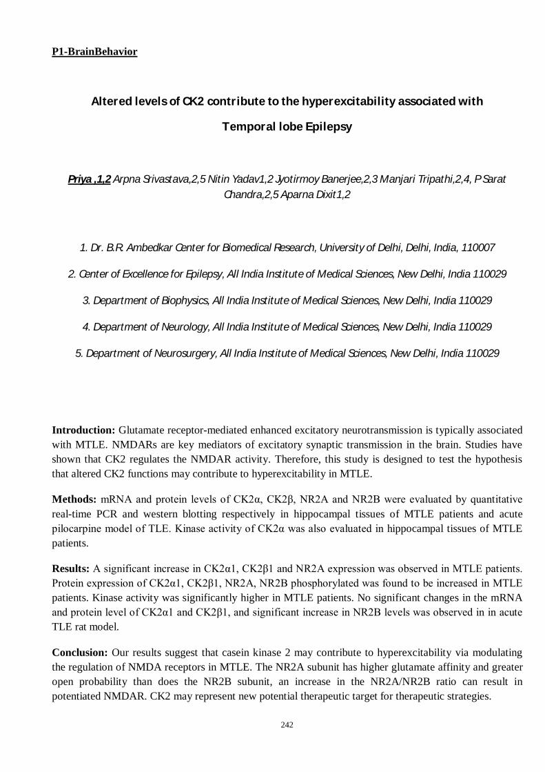
242
P1-BrainBehavior
Altered levels of CK2 contribute to the hyperexcitability associated with
Temporal lobe Epilepsy
Priya ,1,2 Arpna Srivastava,2,5 Nitin Yadav1,2 Jyotirmoy Banerjee,2,3 Manjari Tripathi,2,4, P Sarat Chandra,2,5 Aparna Dixit1,2
1. Dr. B.R. Ambedkar Center for Biomedical Research, University of Delhi, Delhi, India, 110007
2. Center of Excellence for Epilepsy, All India Institute of Medical Sciences, New Delhi, India 110029
3. Department of Biophysics, All India Institute of Medical Sciences, New Delhi, India 110029
4. Department of Neurology, All India Institute of Medical Sciences, New Delhi, India 110029
5. Department of Neurosurgery, All India Institute of Medical Sciences, New Delhi, India 110029
Introduction: Glutamate receptor-mediated enhanced excitatory neurotransmission is typically associated with MTLE. NMDARs are key mediators of excitatory synaptic transmission in the brain. Studies have shown that CK2 regulates the NMDAR activity. Therefore, this study is designed to test the hypothesis that altered CK2 functions may contribute to hyperexcitability in MTLE.
Methods: mRNA and protein levels of CK2α, CK2β, NR2A and NR2B were evaluated by quantitative real-time PCR and western blotting respectively in hippocampal tissues of MTLE patients and acute pilocarpine model of TLE. Kinase activity of CK2α was also evaluated in hippocampal tissues of MTLE patients.
Results: A significant increase in CK2α1, CK2β1 and NR2A expression was observed in MTLE patients. Protein expression of CK2α1, CK2β1, NR2A, NR2B phosphorylated was found to be increased in MTLE patients. Kinase activity was significantly higher in MTLE patients. No significant changes in the mRNA and protein level of CK2α1 and CK2β1, and significant increase in NR2B levels was observed in in acute TLE rat model.
Conclusion: Our results suggest that casein kinase 2 may contribute to hyperexcitability via modulating the regulation of NMDA receptors in MTLE. The NR2A subunit has higher glutamate affinity and greater open probability than does the NR2B subunit, an increase in the NR2A/NR2B ratio can result in potentiated NMDAR. CK2 may represent new potential therapeutic target for therapeutic strategies.

243
P2-BrainBehavior
Early-life stress and risk-taking behaviour in adolescence
Abanti Chowdhury, B S Shankaranarayana Rao and Laxmi T Rao
Department of Neurophysiology, National Institute of Mental Health and Neuro Sciences (NIMHANS),
Bengaluru
Introduction
Evidences indicate that adolescent period is a time when an individual’s body and behaviour undergo a dramatic change. There is an increased novelty and sensation seeking behaviour, emotional instability, and impulsivity which eventually leads to an increased willingness to take risk. However, it is not known whether early life stress has any impact on such risk-taking behaviour and whether nutritional intervention, fats (namely polyunsaturated fatty acids) per se can be considered as a viable strategy to ameliorate the impact of stress induced changes in brain behaviour. Accordingly, the aim of the present study is to assess whether stress during early stage of life has any impact on risk-taking behaviour at adolescence. The study also tried to identify an interventional approach to restore the effects of early-life stress on risky decision-making.
Materials and methods
Maternal separation and isolation (MS) stress has been done to induce stress in the rats during early life. In this procedure pups were separated from the mother and kept in separate cages daily from post-natal day (PND) 4 to PND 13 for 6 hours. 1 ml poly-unsaturated fats (PUFA) were fed along with standard diet to one group for 21 days (PND 22- PND 42). A novel paradigm has been designed in our laboratory to evaluate the risk-taking behaviour when the rats reached adolescent age (PND 45). The behaviour was analysed using an animal motion-tracking software.
Results
It was observed that learning was not affected by maternal separation and isolation stress. Risk-assessment was reduced, while, risk taking behaviour was elevated in MS rats. PUFA has been found to restore the enhanced risk-taking behaviour in the MS rats.
Discussion
Due to vigorous search for sensation and elevated curiosity, adolescents might take the risk of indulging in activities that might have long-lasting impact in later stages of life like the use of tobacco, alcohol or drugs which in turn will lead to physical and mental health deterioration. In order to maintain an optimal risk-taking behaviour, the factors modifying this behaviour in adolescent age must be identified and our study lists early-life stress as one of the factors. Nutrition is a very crucial component during growing years and
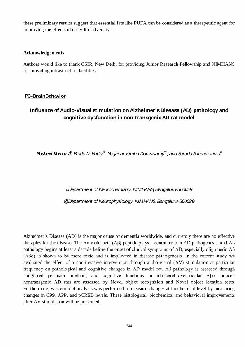
244
these preliminary results suggest that essential fats like PUFA can be considered as a therapeutic agent for improving the effects of early-life adversity.
Acknowledgements
Authors would like to thank CSIR, New Delhi for providing Junior Research Fellowship and NIMHANS for providing infrastructure facilities.
P3-BrainBehavior
Influence of Audio-Visual stimulation on Alzheimer's Disease (AD) pathology and cognitive dysfunction in non-transgenic AD rat model
Susheel Kumar J#, Bindu M Kutty@, Yoganarasimha Doreswamy@, and Sarada Subramanian#
#Department of Neurochemistry, NIMHANS, Bengaluru-560029
@Department of Neurophysiology, NIMHANS, Bengaluru-560029
Alzheimer’s Disease (AD) is the major cause of dementia worldwide, and currently there are no effective therapies for the disease. The Amyloid-beta (Aβ) peptide plays a central role in AD pathogenesis, and Aβ pathology begins at least a decade before the onset of clinical symptoms of AD, especially oligomeric Aβ (Aβo) is shown to be more toxic and is implicated in disease pathogenesis. In the current study we evaluated the effect of a non-invasive intervention through audio-visual (AV) stimulation at particular frequency on pathological and cognitive changes in AD model rat. Aβ pathology is assessed through congo-red perfusion method, and cognitive functions in intracerebroventricular Aβo induced nontransgenic AD rats are assessed by Novel object recognition and Novel object location tests. Furthermore, western blot analysis was performed to measure changes at biochemical level by measuring changes in C99, APP, and pCREB levels. These histological, biochemical and behavioral improvements after AV stimulation will be presented.

245
Acknowledgements
ICMR-JRF for SKJ is gratefully acknowledged
P4-BrainBehavior
Differential sensitivity to GABA agonist in an endophenotypic model of depression and anxiety
Akshata T. Inavally and Monika Sadananda
Brain Research Laboratory, Biotechnology Unit, Department of Biosciences,
Mangalore University, Mangalagangothri - 574 199, Karnataka
Abstract
Background: Major depressive disorder is comorbid with anxiety with prevalence of treatment resistance depression. Wistar-Kyoto rat demonstrates depressive endophenotype with face/construct/predictive validities in open field, elevated-plus-maze, and forced-swim-test, with dysfunctional HPA-axis and brain neurochemistry and is suggested as treatment-resistant model. Anxiolytic Diazepam binds to GABAA receptors increasing affinity for endogenous GABA.
Materials and methods: We evaluated the anxiolytic effect of 1mg/kg b.w. DZP, administered orally for 10 days in Wistar/WKY rats with behavioural testing from 6th day on a comprehensive battery of nerobehavioural paradigms such as open-field (OF), Elevated-plus-maze (EPM), light-dark-box (LDB) and forced-swim-test (FST). Ultrasonic-vocalizations and cognition-based-test on novel-object-recognition (NOR) were also analysed.
Results: Result showed strain-specific effects in EPM with increased open arm time and entries (p<0.05) in DZP-treated Wistars compared to vehicular group, but no effect in WKY. DZP had no effect in OF, LDB and FST in both Wistars and WKY. DZP significantly reduced discrimination index in Wistars (p<0.01) suggesting amnesia, but had no effect in WKY.
Discussion and Conclusions: Results indicated differential strain effects in EPM, though DZP had no effect in OF, LDB and FST in both Wistars and WKY. The results also showed the differential sensitivity of paradigms to anxiolytic effect of DZP. A strain-specific difference exists in response to DZP suggesting differential sensitivity of GABAA receptor and showed that DZP had no effect in WKY, corroborating evidences of it being a model for TRD.

246
Acknowledgement:
ATI is a DBT-JRF and is grateful to DBT, Govt. of India for the fellowship.
P5-BrainBehavior
Evaluation of colour preferences in Zebrafish under normal and hyperglycaemic condition
Manisha Pallavi
Central University of Jharkhand
Background: Colour plays an important role in the detection of food and it can be linked to the selection of mates also. The selection of a particular colour in movable organisms could be the best clue for the evaluation of many behavioural parameters as well as an indication of many stressed conditions and retinopathy due to hyperglycaemia.
Material and Methods: In this study, we probed the colour preference in three different groups, control (adult), larvae, and hyperglycaemic zebrafish. A plus-maze having four different coloured arms (B; Blue, G; Green, R; Red, Y; Yellow) was used for evaluation of deviation in colour preference in a different age as well as control vs hyperglycaemic condition.
Result: A significant change in colour preference was observed in the different groups of zebrafish. The larvae zebrafish preferred Green over the other four colours (G>B>Y>R) whereas in control adult zebrafish the pattern was slightly different (G>R>B>Y). Hyperglycaemic zebrafish preferred blue colour over all other colours (B>G>Y>R).
Discussion and Conclusions: Although, colour preference for food is well documented whether these preferences evolved separately in taxa or inherited by ancestors is still a matter of investigation. A prolonged increase in plasma glucose and hyperglycaemia also leads to retinopathy. The selection of a particular colour in movable organisms could be the best clue for the evaluation of many behavioural parameters as well as an indication of many stressed conditions and retinopathy due to hyperglycaemia.
Acknowledgement

247
The authors are thankful to DBT for Builder Project, Department of Life Sciences, Central University
of Jharkhand, Ranchi.
P6-BrainBehavior
The time dependant changes in some memory parameters in intracerebroventricular streptozotocin (STZ) injected rats are correlated with hippocampal level of COX2,
PGE2 and TNFα
Nicky Singh1, Rupsa Ghosh1, Debasish Bandyopadhyay2 and Tusharkanti Ghosh1
1Neurophysiology Laboratory, Dept. of Physiology, UCST, University of Calcutta, 92 A.P.C Road, Kolkata-700009, West Bengal, India
2Oxidative Stress and Free Radical Biology Laboratory, Dept. of Physiology, UCST, University of Calcutta, 92 A.P.C Road, Kolkata-700009, West Bengal, India
Abstract:
Background: Intracerebroventricular (ICV) injection of streptozotocin (STZ) in rats induces experimental dementia but the mechanism of memory impairments is not clearly understood. The time dependent effect of STZ on memory parameters and neuroinflammatory markers in hippocampus may indicate the mechanism of memory impairments in this experimental rat model of Alzheimer’s disease.
Materials and methods: Working memory errors (WME), reference memory errors (RME), latency to enter first baited arm (L1B) and latency to enter four baited arms (L4B) in a Radial Arm Maze (RAM) along with the concomitant levels of COX2, PGE2 and TNF α in hippocampus were measured at 3-hour, 24-hour, 7th day and 21stday after ICV STZ injection in rats.
Results: WME and RME were increased at 24- hour, 7th day and 21st day after ICV STZ injection and the errors were progressively elevated with time. WME, RME and L4B were increased progressively from 6th day to 21st day after ICV STZ injection in 21-day study. The neuroinflammatory markers (COX 2 PGE 2, TNFα) in hippocampus were increased significantly at 3-hour, 24- hour,7th day and 21st day after ICV STZ injection and the levels were gradually increasing with time.
Discussion and conclusion: The time dependant changes of WME and RME are correlated significantly with the gradual higher levels of the observed hippocampal inflammatory markers, but L1B and L4B are not correlated with these markers. Probably the neural circuitries for WME and RME through hippocampus are increasingly affected

248
with higher levels of inflammatory markers. The inflammatory markers are not able to significantly influence the neural circuitry for L1B and L4B. The injection shock might stop the movements of rats at 3-hour study.
Keywords: working memory error, reference memory error, latency to enter first baited arm, COX 2, PGE2, TNFα, Alzheimer’s disease
Acknowledgement: This research work has been funded DST, CSRI, Govt. Of India [RefernceNo.SR/CSRI/353/2016(G) dt.24.4.2017]
P7-BrainBehavior
Evaluation of neuronal remodeling after stress removal in hippocampal complex of chick, Gallus domesticus
Kavita Tamta, Adarsh Kumar, Hemlata Arya, Ram Chandra Maurya
Department of Zoology (DST-FIST SPONSORED), S.S.J. Campus, Kumaun University, Nainital.
Background: Various studies have proposed that the avian dorsomedially situated HCC-hippocampal complex (consisting of hippocampus proper and parahippocampal area) shows homology with the mammalian HCC and is more prone to chronic stress. The aim of present study is to evaluate the neuronal remodeling after stress removal in the hippocampal complex of a chick.
Materials and Methods: In the present study we used the Golgi Cox method. Chicks were divided into two groups Group-1 non-stressed (4+4 week), Group-2 Stressed (for 4 weeks) and treated with five types of chronic mild stress (food deprivation, isolation, dark, wind & cold temperature) and after 4 weeks they were kept under normal environmental conditions.
Results: The multipolar and stellate neurons show increase in their number while the pyramidal neurons show decrease. In all neuronal cells we found an insignificant increase in their spine number, dendritic field and distance of secondary branches from the soma center. The projection neurons show decrease in their soma diameter but show an increase in their axonal length while the stellate neurons show increase in their soma diameter but a decrease in their axonal length, after stress removal.
Discussion and Conclusions: The avian HCC plays an important role in spatial navigation, learning, memory, social behavior, imprinting and sexual behavior. The present study established that the hippocampal complex plays a significant role in normalizing and re-establishing the homeostasis in the animal to survive. Thus, the present study provides valuable insights about the recovery of chicks into normal state after stress removal.

249
Ethics statement: All the experimental procedures were carried out according to the guidelines of the Animal Ethics Committee of the Kumaun University, Nainital.
Acknowledgement: The authors thank the Head of the Department of Zoology, S.S.J. Campus, Kumaun University, Nainital for providing essential facilities for the present investigation. The present research study has received funding from UGC Fellowship No. F. 82-1/2018 (UGC ref. No.2856). There is no conflict of interest among authors.
P8-BrainBehavior
Acute-stress induced neuronal plasticity in the corticoid complex of 15-day-old chick,
Gallus domesticus
Adarsh Kumar, Kavita Tamta, Hemlata Arya, Ram Chandra Maurya
Department of Zoology (DST-FIST SPONSORED), S.S.J. Campus, Kumaun University, Nainital.
Background and rationale: Several stress studies conducted in birds have shown that a single stress exposure may impair or improve memory and learning processes. The present study aims to investigate the role of single acute-stress (AS) in inducing structural plasticity in neurons of the corticoid complex (CC) in 15-day-old chick.
Materials & Methods: The present research study is based on Cresyl Violet (for regional identification), Golgi Colonnier and Golgi Cox staining technique (for the morphological evaluation of the neurons of the corticoid complex in 15-day-old chick, Gallus domesticus).
Results: In dorso laterally situated corticoid complex three groups of spinous neurons: the multipolar, the pyramidal and the stellate neurons were observed. The stellate neurons have shown a significant decrease as well as an increase in their spine density in both CI-intermediate and CDL-dorsolateral corticoid subfields, whereas the multipolar neurons had shown a significant increase in their spine density in the CDL region only, which could be linked to more effect of stress in this region.
Discussion & Conclusions: The avian CDL- dorsolateral corticoid subfield corresponds to the entorhinal cortex of the mammals, on the basis of neuronal morphology and bidirectional connections between adjacent areas. The present study will establish that slight modifications in natural stimuli or environmental changes faced by the animal may affect their dorsolateral forebrain which shows neuronal plasticity that help in the development of an adaptive capacity in the animal to survive under changing environmental conditions.
Ethics Statement: The present research work was approved by IAEC with protocol no. KUDOPS/106 and all the experimental procedures were carried out according to the guidelines of the Animal Ethics Committee of the Kumaun University, Nainital, Uttarakhand,India.
Acknowledgement: The authors thank the Head, Department of Zoology (DST FIST Sponsored), Kumaun University, Soban Singh Jeena Campus Almora, for providing essential infrastructural support for the present research work. This research did not receive any specific grant from funding agencies in the public, commercial or not-for-profit sectors. All the authors declare that they have no conflict of interest among them to proclaim.
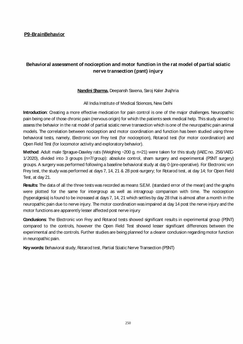
250
P9-BrainBehavior
Behavioral assessment of nociception and motor function in the rat model of partial sciatic
nerve transection (psnt) injury
Nandini Sharma, Deepansh Saxena, Saroj Kaler Jhajhria
All India Institute of Medical Sciences, New Delhi
Introduction: Creating a more effective medication for pain control is one of the major challenges. Neuropathic pain being one of those chronic pain (nervous origin) for which the patients seek medical help. This study aimed to assess the behavior in the rat model of partial sciatic nerve transection which is one of the neuropathic pain animal models. The correlation between nociception and motor coordination and function has been studied using three behavioral tests, namely, Electronic von Frey test (for nociception), Rotarod test (for motor coordination) and Open Field Test (for locomotor activity and exploratory behavior).
Method: Adult male Sprague-Dawley rats (Weighing ~200 g, n=21) were taken for this study (IAEC no. 256/IAEC-1/2020), divided into 3 groups (n=7/group): absolute control, sham surgery and experimental (PSNT surgery) groups. A surgery was performed following a baseline behavioral study at day 0 (pre-operative). For Electronic von Frey test, the study was performed at days 7, 14, 21 & 28 post-surgery; for Rotarod test, at day 14; for Open Field Test, at day 21.
Results: The data of all the three tests was recorded as mean± S.E.M. (standard error of the mean) and the graphs were plotted for the same for intergroup as well as intragroup comparison with time. The nociception (hyperalgesia) is found to be increased at days 7, 14, 21 which settles by day 28 that is almost after a month in the neuropathic pain due to nerve injury. The motor coordination was impaired at day 14 post the nerve injury and the motor functions are apparently lesser affected post nerve injury
Conclusions: The Electronic von Frey and Rotarod tests showed significant results in experimental group (PSNT) compared to the controls, however the Open Field Test showed lesser significant differences between the experimental and the controls. Further studies are being planned for a clearer conclusion regarding motor function in neuropathic pain.
Key words: Behavioral study, Rotarod test, Partial Sciatic Nerve Transection (PSNT)

251
P10-BrainBehavior
Effect of unpredictable chronic mild stress in the hippocampal region of 2-week-old chick, Gallus domesticus.
Hemlata Arya, Adarsh Kumar, Kavita and Ram Chandra Maurya
Department of Zoology (DST-FIST SPONSORED), S.S.J Campus Almora, Kumaun University, Nainital
Background: In birds, hippocampal complex (HCC) is a curved strip of tissue that occupies the dorsomedial surface of telencephalon, it is subdivided into two major areas namely parahippocampal area (APH) and hippocampus proper (Hp). It is known that the mammalian HP is similar to the medial part of the avian dorsal cortex (i.e., HP) in terms of origin, development and topography. In the present work different morphological characteristics of neurons were studied by using Golgi Cox method.
Materials and Methods: 5-day old chicks are divided into two groups: Group 1 and Group 2 each contain four chicks. Group 1 is the non-stressed group whereas Group 2 was given unpredictable chronic mild stress for 2 weeks. On 15th day all the chicks were anaesthetized then sacrificed for golgi staining analysis.
Results: The number of multipolar and pyramidal neurons increases whereas stellate neurons decrease in UCMS chick and the mean dendritic field is less in UCMS chick as compared to NS. Mean soma diameter of multipolar and stellate neurons is more than pyramidal neurons in UCMS chick as compared to NS. Distance of secondary branches from soma decreases in UCMS chick than NS and visible spine number is observed less in UCMS chick.
Discussion and Conclusions: The result obtained in the present study indicates that the HCC is more prone to stress than other regions of brain. Moreover, UCMS causes the remarkable changes in various characteristics of neurons such as, number of neurons, soma diameter, dendritic field, visible spine number and the distance of secondary branches from soma in the hippocampal complex of avian brain.
Ethics statement: The experimental procedures were carried out according to the guidelines of the Animal Ethics Committee of the Kumaun University, Nainital.
Acknowledgement: We would like to thank the Department of Zoology, S.S.J Campus, Almora, Kumaun University, Nainital for infrastructure support during the investigation. There is no conflict of interest.

252
P11-BrainBehavior
Bacillus coagulans Unique IS-2 reverses maternal separation and chronic unpredictable mild stress generated anxiety- and depression-like phenotypes in rats
by modulating microbiome gut-brain axis
Shubham Ghuge1, Aditya Singh1, S. S. Srilakshmi1, Manisurya1, Siva Nageswara Rao Gajula1, Rajesh Sonti1, Manoj P. Dandekar1
1Department of Pharmacology and Toxicology, National Institute of Pharmaceutical Education and
Research (NIPER), Hyderabad, Email: [email protected]
Background: Despite availability of several antidepressant agents, many patients with depression remain treatment-refractory. Probiotics have garnered a notable attention in the management of depression. We examined the effects of Bacillus coagulans Unique IS-2 on anxiety- and depression-like phenotypes using maternal separation (MS) and chronic-unpredictable mild stress (CUMS) in Sprague-Dawley (SD) rats.
Methods: Both male and female SD rats subjected to MS+CUMS. The probiotic treatment was provided for 6-weeks via drinking water. The neurobehavioral changes were assessed using sucrose-preference test (SPT), open-field test (OFT), elevated-plus maze test (EPM), and forced-swimming test (FST). Post-behavioral studies rat’s blood, brain and intestine samples were collected.
Results: Stress-exposed rats drank less sucrose solution (SPT), increased passivity (FST), and explored less in open-arms (EPM). These stress-generated neurobehavioral aberrations reversed by 6-week probiotic treatment. Body weight and locomotor activity in OFT remained unchanged. Probiotic treatment reversed, the decreased levels of BDNF and serotonin and increased levels of CRP, TNF-α and dopamine, in the hippocampus and/or prefrontal cortex. Treatment with probiotic also restored the villi/crypt ratio and Goblet-cell count, and levels of kynurenine and kynurenic-acid.
Discussion and Conclusion: This study suggests an important role of Bacillus coagulans Unique IS-2 in the reversal of MS+CUMS generated anxiety- and depression-like behaviours in rats. No prominent sex-specific changes were noted. Administration of Bacillus coagulans Unique IS-2 positively modulated the systemic levels of metabolites, villi/crypt ratio, Goblet-cell and brain neurotransmitters indicating remodelling of the microbiome gut-brain axis. Thus, Bacillus coagulans Unique IS-2 may be evaluated further as a potential therapeutic option for alleviating the mood disorders.
Acknowledgements: This study is funded by DoP, Ministry of Chemicals and Fertilizers, Government of India and Unique Biotech Ltd., Hyderabad.

253
P12-BrainBehavior
Metabolic and epigenetic alterations in hippocampal tissues induced by repetitive mild Blast
Megha Kumari1, 2 ,Palkin Arora1,Priyanka Sharma1, Yasha Hasija2, Poonam Rana1, Maria M Dsouza1,Namas
Chandra4, Richa Trivedi 1#
1 Radiological, Nuclear and Imaging Sciences (RNAIS), Institute of Nuclear Medicine and Allied Science (INMAS), DRDO, Delhi, India
2 Department of Biotechnology, Delhi Technological University, Delhi, India.
3 Center for Injury Bio-mechanics, Materials, and Medicine at New Jersey Institute of Technology, Newark, United States.
Warfare affected people suffer from blast-induced traumatic brain injury (blTBI). Blast explosions are becoming more and more devastating as current IEDs evolve. blTBI can cause both primary and secondary injuries to the brain tissue, which can affect the brain's metabolic and cognitive functions. Repeated blTBI is common for people resides in war zone, and it can affect the brain's metabolic and cognitive functions. The hippocampus is responsible for learning, memory, and the limbic system, and it is extremely vulnerable to brain injuries. The purpose of this study was to determine the metabolic and histological changes in the hippocampus of rats after 24 hours of injury. All rats except sham were exposed to various blast intensities. Rats were put into four groups for the study: i) Sham; ii) Mild TBI (mi); iii) Moderate TBI (mo); and iv) Repetitive Mild TBI (rm TBI). After 24-hour of TBI, hippocampal tissues were taken for proton nuclear magnetic resonance spectroscopy (1H NMR spectroscopy) and immunohistochemistry (IHC) investigation. Only rmTBI animals showed significant changes in glutamate, NAA, acetate, creatinine/creatine, and phosphotidyl choline/choline levels. IHC findings showed the number of AH3 positive cells in rm TBI tissues was notably lower than in miTBI and sham rats. This could be related to an epigenetic change caused by TBI 24 hours after the injury. Astrogliosis was also found in the hippccampal tissue of the miTBI and moTBI rats, but not in the rmTBI rats.Our study demonstrates the metabolic alterations post blast induced TBI at acute timepoint. These metabolite specific changes post rmTBI is associated with a significant decrease in acetylation in the hippocampus. The study also reported an increase in number of astrocytes in mild and mod TBI, while no change was observed in rmTBI. Conclusively the study suggests metabolic and epigenetic alterations after repeated exposure to mild blast.

254
P13-Neuromodulator
Structure-function insights into the role of D-amino acid oxidase mutations implicated Amyo-trophic Lateral Sclerosis
Shumayila Khan*, Upma Dave, James Gomes#
# corresponding author Kusuma School of Biological Sciences Indian Institute of Technology Delhi Hauz Khas, New Delhi 110016, India
Background: D-amino acid oxidase (DAO) maintains motoneuron function by regulating D-serine levels and N-methyl-D-aspartate-receptor activation. Mutations in DAO are associated with Amyo-trophic Lateral Sclerosis. We aim to understand the pathogenic basis of known DAO mutations and a novel mutation identified in ALS patients by our group, through biophysical and biochemical as says.
Methodology: His-tagged wild-type and mutant DAO proteins were purified and enzymatic activity was measured through a colorimetric assay providing FAD as a cofactor and D-alanine as a substrate. Size exclusion chromatography, circular dichroism spectrophotometry and tryptophan fluorescence were used to study the change in conformation and stability of the mutant proteins.
Results: Assay results show that the different DAO mutants have reduced enzymatic activity as compared to the wild-type protein. Moreover, biophysical data suggests that the mutations cause the protein to attain an altered conformation as well as a higher oligomeric state as compared to the wild type DAO protein.
Discussion and conclusion: The instability of the mutant proteins resulting from the changes in con formation and oligomeric state may hamper the normal functioning of the proteins. Similarly, reduced enzymatic activity may hinder the degradation of D-serine which may accumulate and alter the homoeostasis of the motoneurons, consequently contributing to the pathogenic effect of the mutants.

255
P14-Neuromodulator
Long-term impact of saturated fatty acid rich diet at adolescence on ischemic stroke-induced motor deficit at late adulthood
Rini P.G.1, Vikas V. 2 and Laxmi T. Rao1
1 Department of Neurophysiology, National Institute of Mental Health and Neurosciences (NIMHANS),
Bengaluru; 2Department of Neurosurgery, NIMHANS, Bengaluru
Introduction: The present study has made an attempt to delineate the role of long-term intake of high saturated fatty acid during adolescence in modulating the risk for ischemic stroke at adulthood. The study has used 100 Sprague Dawley rats. The dietary supplements were fed to the rats throughout the adolescence age from PND 22 to PND 50 of age. The diet was either normal standard nutrition or saturated fatty acid-rich diet.
Materials and Methods: At PND 75, surgery was performed in all four groups – SFA Stroke, SFA-Sham, NC Stroke and NC Sham groups. Stroke group received ischemic induction by the occlusion of middle cerebral artery (MCAO) using the monofilament. The study has adopted ischemic stroke procedure based on the classical Koizumi MCAO method. 24 h after the induction of MCAO, stroke-induced changes in neurological deficits were assessed by Garcia scoring method. Subsequently, the study has investigated the gait analysis. In parallel, a group of rats were exposed to TTC staining to confirm the ischemic stroke injury in the motor cortex. After 21 days of ischemic stroke induction, the study has evaluated the long-term impact of ischemic stroke on the ambulatory behavior of rats.
Results: The result showed no difference in motor deficit after 24h of ischemic stroke induction between NC-Stroke and SFA-Stroke group. However, 21 days after the MCAO, we observed a significant difference in ambulatory behaviour between SFA-Stroke and NC-Stroke group. While, the study has observed significant deficit in neurological functions after 24h and 17 days of MCAO between NC-Stroke and SFA-Stroke group.

256
Discussion: In summary, the present study has found a clear-cut difference in motor deficit between normal stroke group and SFA stroke group. Natural locomotor ability was significantly reduced in SFA stroke group, and reduced velocity even after 3 weeks of MCA occlusion. By reducing the high-saturated fatty acid rich diet and use of proper balanced diet at adolescence, it will be highly beneficial in accelerate the recovery and enhance the quality of life after stroke.
Acknowledgment: This project is supported by the NIMHANS M. Phil. fellowship program.

257
P15-Neuromodulator
Therapeutic activity of Vitamin D3 supplementation on oxidative stress, motor impairment, neuromuscular coordination and spatial memory in 3-nitropropionic
acid induced mouse model of Huntington’s disease
Manjari SKV1, Shuvadeep Maity1, Sonata Yu Ku2 and Pragya Komal1*
2The Hong Kong Polytechnic University, Hong Kong, kowloon Hong Kong; 1Department of Biological Sciences, Birla Institute of Technology and Sciences – Pilani, Hyderabad, Telangana, India – 500078; * Corresponding author-
Background: Oxidative stress plays a vital role in the disease progression of various neurodegenerative diseases like Huntington’s disease (HD). Certain antioxidant compounds like Vitamin D3 (VD) are shown to be a key regulator of oxidative stress as observed in other neurological disorders like Alzheimer’s and Parkinson’s disease. However, limited work is available to forecast the role of VD in combating oxidative stress in HD. Hence, in the present study, we aimed to explore the neuroprotective and anti-oxidative effects of Vitamin D3 (VD) supplementation in 3-nitropropionic acid (3-NP) induced mouse model of HD.
Methodology: In the present study, C57BL/6 male mice (3-4 months of age; n=8-15 animals per group) were divided into four different groups namely; Group I: Vehicle treated with sterile saline, Group II: animals were induced intraperitoneal (i.p) with 75mg/kg of 3-NP in three consecutive doses as described previously by Fernagut et al. Group III: animals injected i.p with only 500IU/kg of VD for 15 days and Group IV: 500IU/kg (12.5 µg/kg) of VD was given for 15 days through i.p to post 3-NP (75mg/kg) injected animals. Behavioral tests like locomotor activity, gait analysis, rotarod analysis, and morris water maze were carried out for a total period of 30 days. After 30 days, animals were decapitated and striatal tissues were isolated to check the mRNA expression of neurotrophins (nerve growth factor (NGF), brain derived neurotrophic factor (BDNF)) and key antioxidative enzymes (superoxide dismutase (SOD), glutathione peroxidase (Gpx) and catalase (Cat)).
Results: Our behavior results reflect that post VD treatment to 3-NP animals (Group IV) significantly rescued spatial memory deficits, locomotion dysfunction and neuromuscular coordination when compared with 3-NP induced HD mice (Group II). VD supplemented 3-NP induced animals (Group IV) showed significant increase in mRNA expression of key neurotrophic factors like nerve-growth factor and brain-derived neurotrophic factor reflecting rescue action of VD for neurons undergoing degeneration. The key antioxidant markers i.e SOD, GpX and Cat were found to be increased in HD animals which got significantly subsided on VD supplementation (Group IV) when compared with 3-NP induced HD animals (Group II), corroborating an antioxidative property of VD in HD.
Conclusion: Overall, our current study infer that VD modulates the expression of anti-oxidative markers under pathological conditions and correspondingly increases the expression of key neurotrophins vital for survival of neurons undergoing loss in function.
Keywords: Huntington’s disease, 3-nitropropionic acid, Vitamin D, behaviour, nerve growth factor, brain derived neurotrophic factor, superoxide dismutase, glutathione peroxidase, catalase.

258
P16-Neuromodulator
Heavy metals as risk factor for neurodegenerative disorders through edible fishes in Gangetic Belt of Uttar Pradesh
Bhargawi Mishra
Institute of Medical Sciences, BHU
Background: River Ganga is the longest and holiest river of India. Research has been conducted in the Gangetic belt of Uttar Pradesh in a hospital based prevalence to study the spectrum of neurological disorders, Gangetic fishes as dietary intake and heavy metals concentration, to find out environmental trigger for the disease.
Materials and methods: Amyotrophic Lateral Sclerosis, Parkinson's disease and Multiple system atrophy were clinically screened among neurodegenerative disorders. Demographic detail studied through questionnaire. Intravenous blood was subjected to Atomic Absorption Spectroscopy(AAS) technique to observe metal content after consent. Five commonly edible Gangetic fish tissues were also subjected to AAS for metal (lead, manganese, chromium)content as dietary intake.
Result: Lead, manganese and chromium were detected in blood and fish tissues, . Manganese ranges from 1.0007 mg/L to 37.1447mg/L, lead from 0.219 to 6.0525mg/L, and chromium ranged between 3.442mg/L to 10.1821 mg/Lin fish tissue. In human, average manganese blood content was in 0.892mg/L in Parkinson's disease.0.109 mg/L in ALS and in 0.171mg/L in MSA. While average blood content of lead was in 0.092 mg/L in ALS, 0.109 mg/L in Parkinson's and 0.131in MSA. and chromium was estimated as 0.385mg/L in Parkinson's disease,0.132mg/L in ALS and 0.054 mg/L in MSA.
Discussion and conclusion: Fishes are common bioindicator for metal pollution and an important link in the food chain. Accumulation of lead, chromium and manganese in fish tissues shows an environmental and dietary trigger for neurological diseases in Gangetic belt. Patients suffering from Neurological disorders also bear the load of lead, manganese and chromium in their blood, which need further investigation for the effect of these metals in causing the disease
Acknowledgement: Department of Neurology. Institute of Medical Sciences. Banaras Hindu University

259
P17-Neuromodulator
Tinospora cordifolia Extract Ameliorates Rotenone-induced Oxidative Stress and Apoptosis via Enhancing Mitochondrial Function
Hagera Dilnashin*, Surya Pratap Singh
Department of Biochemistry, Institute of Science, Banaras Hindu University, Varanasi-221005 (U.P.), India.
Background: Neuroprotection targeting mitochondrial dysfunction has been proposed as an important therapeutic strategy for Parkinson’s disease. Tinospora cordifolia has emerged as a novel medicinal plant that protects neurons from oxidative stress. In this study, we investigated the neuroprotective effects of Tinospora cordifolia and the underlying mechanisms in the classic rotenone-induced Parkinsonism.
Material and Methods: Mice were divided into four experimental groups: control, rotenone (2 mg/kg body wt., subcutaneous), Tinospora cordifolia extract (TCE, 200 mg/kg body wt., oral) + rotenone, and TCE only]. Mice were pre-treated with TCE for a week and then simultaneously injected with ROT for 35 days.
Results: TCE administration significantly improved locomotor performance and increased tyrosine hydroxylase expression in the substantia nigra pars compact of rotenone-intoxicated mice. Furthermore, TCE improved mitochondrial dysfunction via counteracting the decline in mitochondrial electron transport chain complex activity evoked by ROT. Similarly, TCE suppressed ROT-induced imbalance of Bax/Bcl-2 ratio and activation of caspase-3.
Discussion and Conclusion: This study demonstrates the neuroprotective effects of TCE against rotenone-induced apoptosis in mice. The Bax/Bcl-2 ratio, mitochondrial dysfunction, and expression of caspase-3 were seen to be significantly increased on rotenone-intoxication. However, TCE was potent in protecting the neurons against rotenone-induced cytotoxicity through the regulation of oxidative stress-mediated mitochondrial dysfunction and apoptosis in the mouse model of PD. Taken together, our results suggested that TCE attenuated rotenone-induced oxidative stress, through the regulation of mitochondrial functions.
Keywords: Tinospora cordifolia extract; Parkinsonism; Mitochondrial Dysfunction; Apoptosis;
Rotenone.
Acknowledgement: HD is sincerely thankful to BHU, India, for fellowship. The authors are also thankful to the head of the Department of Biochemistry, IOS, BHU, for providing the basic departmental facility and ISLS, IMS, BHU, for their central instrument facility.

260
P18-Neuromodulator
Can tasimelteon be the key for neuroinflamation?
Mrinalini R
SRM College of Pharmacy, SRMIST
Microglia happens to be a core component behind neuroinflammation and the associated neurodegenerative diseases like Alzheimer’s Disease (AD). The inflammatory function of microglia in AD pathology remains to be the most studied subject, yet with not many clinically significant results. A well-established hallmark of neuroinflammation is the cytokine release. A pre-requisite for the cytokine release is the activation of inducible complexes known as inflammasomes. The most notable inflammasome for microglia is NLRP3 (Nod-like receptor family pyrin domain containing 3). NLRP3 is a viable target since it’s known to be behind both cytokine release and amyloid beta deposits, which form the pathophysiology of AD. In order to target NLRP3, we further need to target the molecules involved in both NLRP3 priming and activation pathways. One such key molecular target is NF-κB (Nuclear Factor kappa-light-chain-enhancer of activated B cells), the translocation of which, to nucleus, results in the increased production of cytokines ultimately via NLRP3 inflammasome activation. Similarly, TLR (Toll-like Receptor) are the surface receptor targets for the pathway. We are considering a recently FDA-regulated drug called tasimelteon for its ability to suppress neuroinflammation in any one or many ways around NLRP3 activation by both in-silico and in-vitro methods. The vast results from in-silico by employing tools like AutoDock 4.2.6, ProTox-II, and Pass along with the results from in-vitro analysis using SH-SY5Y cell line is depicted. The basis for selecting tasimelteon is the scientifically sound results obtained from using melatonin, an endogenous neurohormone working via the same receptors as tasimelteon.
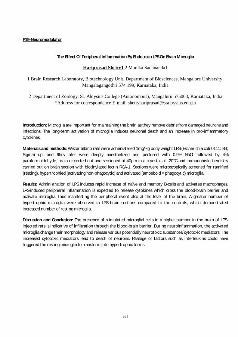
261
P19-Neuromodulator
The Effect Of Peripheral Inflammation By Endotoxin LPS On Brain Microglia
Hariprasad Shetty1,2 Monika Sadananda1
1 Brain Research Laboratory, Biotechnology Unit, Department of Biosciences, Mangalore University, Mangalagangothri 574 199, Karnataka, India
2 Department of Zoology, St. Aloysius College (Autonomous), Mangaluru 575003, Karnataka, India *Address for correspondence E-mail: [email protected]
Introduction: Microglia are important for maintaining the brain as they remove debris from damaged neurons and infections. The long-term activation of microglia induces neuronal death and an increase in pro-inflammatory cytokines.
Materials and methods: Wistar albino rats were administered 1mg/kg body weight LPS (Escherichia coli 0111: B4; Sigma) i.p. and 6hrs later were deeply anesthetized and perfused with 0.9% NaCl followed by 4% paraformaldehyde, brain dissected out and sectioned at 40µm in a cryostat at -20°C and immunohistochemistry carried out on brain section with biotinylated lectin RCA-1. Sections were microscopically screened for ramified (resting), hypertrophied (activating non-phagocytic) and activated (amoeboid = phagocytic) microglia.
Results: Administration of LPS induces rapid increase of naïve and memory B-cells and activates macrophages. LPS-induced peripheral inflammation is expected to release cytokines which cross the blood-brain barrier and activate microglia, thus manifesting the peripheral event also at the level of the brain. A greater number of hypertrophic microglia were observed in LPS brain sections compared to the controls, which demonstrated increased number of resting microglia.
Discussion and Conclusion: The presence of stimulated microglial cells in a higher number in the brain of LPS-injected rats is indicative of infiltration through the blood-brain barrier. During neuroinflammation, the activated microglia change their morphology and release various potentially neurotoxic substances/cytotoxic mediators. The increased cytotoxic mediators lead to death of neurons. Passage of factors such as interleukins could have triggered the resting microglia to transform into hypertrophic forms.

262
P20-Neuromodulator
Taurine restores bisphenol-A induced neurobehavioral and neuromorphological alterations via modulating brain anti-oxidant defense system
Lilesh Kumar Pradhan1#, Pradyumna Kumar Sahoo1# and Saroj Kumar Das1*
1Neurobiology Laboratory, Centre for Biotechnology, Siksha ‘O’ Anusandhan (Deemed to be University), Bhubaneswar-751003, India
Abstract
Background
Bisphenol A (BPA) has been recognized as a causative agent for various health maladies including oxidative stress, neuroinflammation, mood dysfunction, carcinogenicity, cognitive impairment etc. However, literature regarding the role of BPA-induced altered neurobehavioral response and brain morphology and probable neuroprotective efficacy of taurine against BPA-induced neurotoxicity is limited.
Materials and methods
The current experimental paradigm includes 21 days of exposure of standardized dose BPA with standardized dose of taurine co-supplementation. To illustrate the neuroprotective role of taurine against BPA-induced neurotoxicity we have conducted neurobehavioral analysis to determine anxiety like behavior, biochemical studies related to oxidative stress and neuromorphological studies.
Results
The current findings indicate that taurine ameliorated BPA-induced neurobehavioral alterations in novel tank diving test (NTDT) and light dark preference test (LDPT). Biochemical studies reveal the potential therapeutic efficacy of taurine in contrast to BPA-induced oxidative stress. Taurine also shows significant neuroprotective effect against BPA-induced increased neuronal pyknosis and chromatin condensation in the periventricular grey region (PGZ) of zebrafish brain.
Discussions and conclusions
In essence, the findings of the current study advocate that taurine may act as an effective intervention against BPA-induced oxidative stress-mediated neurobehavioral and neuromorphological transformations in zebrafish brain through modulation of brain antioxidant defense system. However, elucidating the underlying mechanism of BPA-induced neuropathological manifestation and taurine-mediated neuroprotection might provide novel strategies for the prevention or treatment of BPA-persuaded neurological conditions.
Acknowledgements
The authors acknowledge the Centre for Biotechnology, SOA University for providing the infrastructure facility and necessary support. Dr. Saroj Kumar Das is the recipient of UGC start-up and ICMR extramural research grants.

263
P21-Neuromodulator
Quercetin attenuates Bisphenol-A induced oxidative stress and neurodegenerative phenotypes in zebrafish
Pradyumna Kumar Sahoo1, Lilesh Kumar Pradhan1, Saroj Kumar Das1
1 Neurobiology Laboratory, Centre for Biotechnology, Siksha ‘O’ Anusandhan (Deemed to be university),
Bhubaneswar 751003, India
Background and rationale: Bisphenol-A (BPA; 4,4'-(propane-2,2-diyl) diphenol) is the key element widely used in the production of plastics. Unsafe disposal BPA can leach out into the waterbodies and can causes several diseases like inflammation, oxidative stress, cancer, hypertension, obesity etc. The neurotoxic potential of BPA and its amelioration through natural compound intervention is elusive in literature.
Methods: To find out the neuroprotective role of quercetin against BPA induced neurotoxicity, the experiment was assigned into five different groups (Naïve, control, BPA, quercetin, quercetin+ BPA). The altered neurobehavioral pattern, oxidative stress parameters including several enzymatic and non-enzymatic assays, and the histopathological observation corresponds to altered neurobehavioral pattern in zebrafish brain was examined.
Results: Chronic waterborne exposure of BPA altered bottom dwelling and scototaxic behaviour in zebrafish was restored by quercetin +BPA co-supplemented groups. Also, the oxidative stress marker including LPx and PC was reduced, meanwhile antioxidant marker such as, GSH to GSSG ratio, GPx, SOD and GST level was reinstated in quercetin +BPA groups. The neuromorphological alteration in the PGZ (periventricular grey zone) responsible concur the change in neurobehavioral responses was also restored in zebrafish.
Conclusions: Overall observation showed that the chronic waterborne exposure of BPA trigger neurobehavioral alteration, inducing oxidative stress, limiting antioxidant enzyme, and neuromorphological alteration in zebrafish. Furthermore, our observation shows quercetin used as a prophylactic strategy against BPA induced neurotoxicity can able to restored the altered neurobehavioral pattern, oxidative stress, increasing antioxidant activity via maintaining the cellular integrity in zebrafish brain.
Ethical statement: All the experiment was approved by the principle and guidelines as per the Institute animal ethical approval (IAEC) committee from the university.
Acknowledgement: The author acknowledges the Siksha ‘O’ Anusandhan (Deemed to be University) for providing necessary infrastructure facility and basic financial support.

264
P22-Neuromodulator
Statin renders post-stroke neuroprotection by regulating mitochondrial dynamics
Namrata Vadak1*, Deepaneeta Sarmah1, Aishika Datta1, Antra Chaudhary, Vrushali Thongire1, Harpreet Kaur, Vishal Gupta1, Pallab Bhattacharya1
1National Institute of Pharmaceutical Education and Research, Ahemdabad (NIPER-A), Palaj, Gandhinagar-382355, Gujarat, India
Abstract: Background: Cholesterol lowering drugs such as statins are reported to lower the risk of stroke. Following stroke, mitochondrial dynamics dysregulation is one of the major reasons of neuronal death. Therefore, our study aims to explore the neuroprotective role of statin by regulating the mitochondrial dynamics in rat model of ischemic stroke.
Materials and Methods: Focal stroke was induced in male SD rats by middle cerebral artery occlusion. The prophylactic group received simvastatin 3 days prior stroke induction and the treatment group received simvastatin 3 days post-stroke. Animals were assessed for neurological deficit score and motor co-ordination. Brains were harvested for further biochemical and molecular studies.
Results: Prophylactic and post-stroke simvastatin administration showed reduction in infarct size, and neurodeficit score while improvement in motor co-ordination. Normalization of biochemical parameters were achieved in both prophylactic and post stroke simvastatin administered animals. Dysregulation of mitochondrial dynamics was alleviaviated as evident by relevant gene and protein expression studies.
Discussion & Conclusion: Dysregulation of mitochondrial dynamics is one of the major reasons leading towards mitochondrial dysfunction. Post-stroke mitochondrial dysfunction triggers various cell death pathways. Our study provides promising evidence on neuroprotective effects of simvastatin by regulating mitochondrial dynamics. The therapy is clinically relevant and may be used as an adjunct stroke therapy.
Acknowledgement: National Institute of Pharmaceutical Education and Research, Ahmedabad, Department of Pharmaceuticals, Ministry of Chemicals and Fertilizers, Government of India.

265
P23-Neuromodulator
Identification of potential inhibitors of PARP-1, a regulator of caspase-independent cell death pathway, from Moringa oleifera phytochemicals for combating neurotoxicity: A structure-based in-silico study
Smriti Singh1*, Prof. Surya Pratap Singh2
Smriti Singh: [email protected]
1. Interdisciplinary School of Life Sciences, Institute of Sciences, Banaras Hindu University, Varanasi
2. Department of Biochemistry, Institute of Sciences, Banaras Hindu University, Varanasi
Abstract
Background: Poly(ADPribose) polymerase1 reverses DNA damage by repairing DNA nicks and damage in normal cellular environment. However, during abnormal conditions such as stroke and other neurological disorders, over activation of PARP1 leads to neuronal cell death via a caspase-independent programmed cell death pathway. Strategies involving PARP1 inhibition or knockout have been shown to be useful in combating neurocytotoxicity.
Material and methods: In-silico docking study was performed Auto dock Vina docking tool. Active Site prediction of the PARP1 protein was done on Castp server. The preparation of the protein and ligands (M. oleifera phytochemicals) for docking was done on Autodock 4.2. Protein-ligand interaction was analysed using Pymol and LigPlot.
Results: Total 16 reported ligands were docked with PARP1 protein; 13 structures were found to be binding to catalytic domain of PARP1 with higher binding affinity. Docking analysis of the FDA approved compounds PJ34 and Tazaloparib (highly potent inhibitors of PARP1) was also done. Docking study results of ligand was compared to that of approved compounds and it was found that most of the phytochemicals showed high binding affinity and number of hydrogen bonds in comparison to approved compounds.
Discussion and Conclusion: Our study establishes the potential of Moringa oleifera phytochemicals as novel inhibitors of PARP-1. Most the phytochemicals used in this study possess the ability to cross BBB, so these compounds can be further developed into therapeutics to confer neuroprotection by exploiting their potential to inhibit PARP-1. Further in-vivo studies are being carried out to evaluate their PARP-1 inhibition capacity and ability to reverse cell death in MPTP-induced Parkinsonian mice model.

266
P24-Neuromodulator
Caveolin-1 Modulates Excitotoxic Signaling In Rat Hippocampal Neurons
Swapna K. K, Bhupesh Mehta, Yogananda S. Markandeya
Department of Biophysics, National Institute of Mental Health & Neurosciences (NIMHANS),
Bengaluru, 5600029, INDIA E mail: [email protected], [email protected]
Background: Caveolins, a group of integral membrane proteins exist in three isoforms (Cav-1, -2 and -3) play an important role in cell signaling by scaffolding various signaling molecules. Loss of Cav-1 expression is reported in Huntington’s disease, schizophrenia, Alzheimer’s disease, cancers and aging. However, the role of Cav-1 in neuronal glutamatergic excitotoxicity is not known. We hypothesize that Cav-1 expression protects neurons from glutamate excitotoxicity.
Objective: We aim to identify the role of Cav-1 in glutamate mediated excitotoxicity with emphasis on intracellular calcium, mitochondrial function and oxidative stress.
Methods: 10 Day in vitro (DIV) primary rat hippocampal neurons were transduced with lentiviral ShRNA Cav-1 and scrambled ShRNA. Transduced neurons were then selected using puromycin and all the experiments were performed on 13-14 DIV. Intracellular calcium, reactive oxygen species (ROS) generation and mitochondrial functions were assessed by using live cell fluorescence imaging. All data were analyzed using statistical software Origin. Statistical significance was determined by student t-test and P < 0.05 was considered statistically significant.
Results: Glutamate (100µM) caused rise in [Ca2+]i in biphasic manner in hippocampal neurons. Peak [Ca2+]i was decreased by 29% and caused irreversible sustained secondary calcium in Cav-1 knock down (KD) neurons (0.63047 ± 0.05, n= 19) compared to the controls (0.8890 ± 0.06, n = 23). Glutamate induced robust increase (3.3-fold) in ROS formation with loss of Cav-1 (7.7069 ± 0.2, n=18) as compared to the controls (2.33664 ± 0.3, n=31). Further glutamate stimulation caused increased mitochondrial depolarization in Cav-1KD (0.833 ± 0.1, n=18) with respect to the controls (0.50184 ± 0.03, n=43). On the other hand, mitochondrial uncoupler CCCP caused significant decrease in Rh123 fluorescence in Cav-1KD neurons (1.27131 ± 0.14) with respect to the controls (2.3722 ± 0.03). However, in the absence of extra cellular Ca2+ glutamate did not affect [Ca2+]i, ΔΨm and ROS generation.
Conclusion: Cav-1 modulates glutamatergic signaling via regulation of intracellular calcium and mitochondrial function.
Ethics: Approved by NIMHANS IAEC. Funding: SERB-DST Extramural for YSM.

267
Conflict of interest: None
P25-Neuromodulator
Caveolin-1 Modulates Voltage Gated Calcium Channels in Rat Cortical Neurons
Madhura Nimonkar, Devaraj T P, Bhupesh Mehta, Yogananda Markandeya
Department of Biophysics, National Institute of Mental Health & neurosciences (NIMHANS), Bangalore, 5600029, India. E mail: [email protected]
Background: Caveolins are a group of integral membrane proteins crucial for the caveolar lipid raft formation in mammalian cells. These proteins play an essential role in cell signaling by scaffolding various signalling molecules and form signalling micro-domains within the cell membrane. There are three Caveolin isoforms, Caveolins 1-3 in humans. Recent studies found that L-type voltage gated calcium channels (L-VGCC) are associated with Caveolin-1 (Cav-1) rich lipid raft fractions in cerebellar granule neurons. However, the role of Cav-1 in the modulation of neuronal VGCC functions is not well understood. We hypothesize that Cav-1 is associated with L-VGCC and modulate its function in neurons.
Methods: Cultured primary rat cortical neurons of 8-10 days in vitro (DIV) were transduced with lentiviral shRNA Cav-1 and Scrambled ShRNA. Cav-1 silenced neurons were selected using puromycin and all the experiments were carried out after 14-15 DIV. Intracellularcalcium was measured using ratiometric calcium indicator dye Fura-2 AM and neurons were depolarised with 60mM KCl stimulation. Cav-1 over-expression was performed with Lipofectamine mediated transfection of Cav-1 mCherry plasmid. Plasma membrane lipid rafts were disrupted by cholesterol depletion using Methyl-β-cyclodextrine. Individual calcium channels were identified using calcium channel-specific blockers ω-Agatoxin IV A (P/Q-type), ω- Conotoxin GVIA (N-type) and Nifedipine (L-type). Additionally, exocytosis was analysed using FM1-43.
Result: KCl depolarisation elicits increased [Ca2+]I in the control neurons (1.396 ± 0.087, n = 63) as compared to Cav-1 knocked down (0.465 ±0.033, n=51). Additionally, there is significant difference between the area under the curve in control cells (62.043±2.388, n=79) and Cav-1 knockdown cells (15.500±0.429, n=142). Cav-1 over-expression does not alter Ca2+ response upon depolarisation with KCl. Moreover, cholesterol depletion from the neuronal membrane significantly reduces [Ca2+]I in Cav-1 knockdown neurons (0.203±0.0.027, n=39) as opposed to control (1.396 ± 0.087, n = 63). We also observed that knockdown of Cav-1 inhibits Ca2+ influx through L-VGCC. Furthermore, our study found that inhibition of L- VGGC by loss of Cav-1 could also result in decreased exocytosis.

268
Our investigation suggests that targeting Cav-1 in neurons could be a novel way to improve synaptic transmission in neurodegenerative diseases and aging.
Conclusion: Cav-1 modulates L-type VGCC and regulates neurotransmitter release in rat primary cortical neurons.
Ethics: Approved by NIMHANS IAEC. Funding: SERB-DST Extramural and NIMHANS intermural grant. Conflict of interest: None
P26-Neuromodulator
Neuroprotective role of Curcumin in Rotenone induced mouse model of Parkinson's disease
Aaina Singh Rathore
Banaras Hindu University
Parkinson’s disease is a neurodegenerative disorder of motor neurons with reduction of dopamine level in the striatum as a result of the death of dopaminergic neurons in the substantia nigra pars compacta. Curcumin is studied to target multiple molecular pathways thus showing potential neuroprotective effects. The present study was undertaken to investigate the anti-inflammatory and anti-oxidative effects of Curcumin in parkinsonian mice model along with its effect on the aggregation of misfolded α-Synuclein protein. The PD mouse model was developed with subcutaneous infusion of Rotenone (2mg/kg b.wt.) administered every day for 35 days in conjugation with oral administration of Curcumin (100mg/kgb.wt.). It was observed that Rotenone stimulated increased expression of the various pro-inflammatory mediators involving TNF-α, IL-6, and IL-1β whereas Curcumin treatment alleviated the effect. The lowering of neural inflammation was seen to ameliorate the aggravated level of ROS production consequently reducing the oxidative stress. This was observed by the augmentation of the GSH level and SOD activity on Curcumin administration. Results of immunoblotting have also suggested the down-regulation of a-synuclein aggregation and up-regulation of the TH protein expression in rotenone-intoxicated mouse. Thus, the results suggest the reduction of oxidative stress, inflammatory response and inhibition of α-synuclein aggregation. Hence, our results suggest that curcumin may be a potential drug in the therapy for PD and other related synucleopathies.
269
P27-Neuromodulator
Screening the anti-neuroinflammatory role of oleuropein in rotenone induced parkinsonian mouse model
Richa Singh
Banaras Hindu University
BACKGROUND: Parkinson's disease (PD), the second most common age-related neurodegenerative condition (SNpc), causes the loss of dopaminergic (DA) neurons and the formation of α-synuclein aggregation in the substantia nigra pars compacta (SNpc). One of the characteristics of PD pathogenesis is chronic neuroinflammation. According to epidemiological and genetic studies, neuroinflammation appears to play a role in the pathophysiology of Parkinson's disease, A variety of polyphenls have been reported which exhibit the pharmacological effect and shows the neuroprotective role in PD. Oleuropein is a glycosylated seco-iridoid, found in Olive leaves and fruits. Oleuropein reveals the various forms of pharmacological effect like- anti cancer, anti-diabetics, anti-viral and neuroprotective effect. The aim of this study was to observe the anti-inflammatory role of Oleuropein in PD.
MATERIALS AND METHODS: All protocols for animals (Swiss albino mice) on which the experiments were carried out was approved by the Animals Ethics Committee of our institute. Mice were divided into four groups: Control, Rotenone, Rotenone administrated with Oleuropein and the positive control group. The treatment group were first pretreated with Oleuropein for 7 days and further the doses continue with dose of rotenone for 35 days. After completion of dosing, the mice were sacrificed and behavior test was performed. The protein expression related with neuroinflammation in PD was also detected through western blot analysis and immunohistochemistry.
RESULTS: The Behaviour analysis showed the increase in motor impairment in rotenone group as compared to control and the treatment group showed significant decrease in motor impairment. The expression of α-synuclein and neuroinflammatory markers like- NFκB elevated in case of rotenone group than control and diminished in case of treated group. The immunohistochemistry analysis also revealed the increase in expression of NFκB in rotenone

270
group and decrease in treatment group and the number of tyrosine hydroxylase positive cells were less in rotenone group and significantly more in treatment group.
DISCUSSION AND CONCLUSION: Researchers have suggested that the rotenone intoxication causes change in redox potential which results in increase in neuroinflammatory markers and upregulating the microgial cella in sunstantia nigra pars compacta (SNpc) in case of PD. Rotenone inhibits the complex I of the mitochondrial electron transport chain in dopaminergic neuronal cells of substantia nigra pars compacta leading to decrease in antioxidant enzymes like - Superoxide dismutase and Catalase and causes increase in ROS production and promotes neuroinflammation. Our study suggests that Oleuropein ameliorate the motor impairment and alleviate the neuroinflammation in PD.
P28-Neuromodulator
Flavonoid isolated from an indigenous plant, inhibits Monoamine oxidase in brain and elevates striatal dopamine levels: therapeutic implications against Parkinson’s disease
Chayan Banerjee1, 3*, Sumangal Nandi1, Joy Chakraborty 1, 3#, Deepak Kumar 2, 3#
1Department of Cell Biology and Physiology, CSIR-Indian Institute of Chemical Biology-TRUE campus, Kolkata, India
2 Department of Organic and medicinal Chemistry, CSIR-Indian Institute of Chemical Biology, Kolkata, India
3Academy of Scientific and Innovative Research (AcSIR), CSIR- Human Resource Development Centre, Ghaziabad, India.
* Presenting author
#Co-corresponding author
Background: Flavonoid, a class of plant secondary metabolite, is known to possess diverse biological activities including monoamine oxidase (MAO) inhibition. MAO, mainly an outer mitochondrial membrane protein metabolizes monoamine neurotransmitters in brain compartments. Pathophysiological conditions characterize by low neurotransmitter levels, usage of flavonoids can give symptomatic relief by delaying neurotransmitter degradation.
Materials and methods: Column chromatography and HPLC are used to isolate pure compounds from crude plant extract. For determining MAO activity spectrofluorimetric assay is performed. SH-SY5Y cell and Drosophila are used to determine toxicity of the compounds. Finally, we checked the therapeutic potential of the compounds in toxin-induced parkinsonian mice.
Results: We characterized four flavonoids (coded M1 – M4), from leaf extract of an indigenous plant, all four showed MAO inhibition. M1 and M2 have no cellular toxicity in-vitro. They did not alter neuronal survival, mitochondrial distribution, ROS generation, neuro-motor behavior, and long-term survivability in-vivo. Only M1 restricted MAO-B activity and elevated striatal DA levels in C57BL/6 mice. Finally, M1 was able to attenuate motor incoordination and improved striatal DA levels in toxin-induced parkinsonian mice.
271
Discussion and conclusion: In present study, we successfully completed the initial characterization of major constituent flavonoids in leaf extract of an indigenous plant and evaluated their MAO inhibitory properties. M1 showed selective MAO-B inhibition and did not affect cell survival or mitochondrial function. M1 improved DA levels in striatum and attenuated motor incoordination in parkinsonian mice. The results suggest M1 may be beneficial in disease conditions that seek MAO-B inhibition as a potential therapeutic improvements.
Acknowledgement: CB is a recipient of senior research fellowship from UGC, Govt. of India. The work is financed by CSIR-IICB.
P29-Neuromodulator
Neuroprotective Property of the Fruit Pulp Extract of Benincasa hispida (Thunb.) Cogn. Against Stress-induced Cognitive Impaired Rats
Anshul Shakya, Surajit Kumar Ghosh, Obaidur Rahman, Rajdeep Sarma
Department of Pharmaceutical Sciences, School of Science and Engineering, Dibrugarh University,
Dibrugarh, Assam, India
Background: Benincasa hispida (Thunb.) Cogn., (Familly: Cucurbitaceae), is an Ayurvedic climbing herb. It has been indicated for anabolic, brain tonic, memory enhancer, and vitalizer properties. The present study was carried out to assess the neuroprotective property of the hydro-alcoholic extract of Benincasa hispida fruit pulp (HABH) on stressed rats.
Materials and Methods: A standardized HABH (30, 100, and 300 mg/kg body weight/day) was administered orally in rats. One hour after the treatment rats assigned to the stress groups (i.e. vehicle and/or drug-treated groups) were subjected to unpredictable footshock stress once daily for 1 h for 21 days. Further, associated markers of stress and cognitive impairment were estimated using the Morris water maze task and validated biochemical procedures.
Results: HABH treated stressed rats were found significant (p<0.05) when compared with vehicle treated stressed rats for various stress markers. Further, repeated daily for 21-days administration of standardized HABH (30, 100 and 300 mg/kg/day, p.o.) were found significant (p<0.05) on learning task i.e. time spent in the targeted quadrant and memory task i.e. escape latency on Morris-water-maze task in subchronic footshock stressed rats.

272
Discussion and Conclusions: Cognitive disturbances are not only the hallmarks of dementia but also are often encountered in patients suffering from many comorbid pathological conditions.
Our present result showed significant prevention of memory dysfunction in stressed rats by HABH pretreatments in Moris-water-maze tasks in comparison with vehicle-treated stressed rats. HABH causes an increase in central cholinergic transmission and might be involved in the maintenance of the nootropic effects in stressed rats.
P30-Neuromodulator
Ameliorative Potential of the Benincasa hispida (Thunb.) Cogn. Fruit Pulp Extract Against Stress-induced Ulcers in Rats
Anshul Shakya, Surajit Kumar Ghosh, Rajdeep Sarma, Obaidur Rahman
Department of Pharmaceutical Sciences, School of Science and Engineering, Dibrugarh University,
Dibrugarh Assam, India
Background: Ayurvedic climbing herb, named as Benincasa hispida (Thunb.) Cogn. is a member of ‘Cucurbitaceae’. It has been indicated for anabolic, brain tonic, memory enhancer, and vitalizer properties. The present study was carried out to assess the ulcer preventive potential of the hydro-alcoholic extract of Benincasa hispida (HABH) on stressed rats.
Materials and Methods: A standardized HABH (30, 100, and 300 mg/kg BW/day) was administered orally in rats.1 hour after the treatment rats assigned to the stress groups were subjected to unpredictable footshock (2 mA) stress through a grid floor once daily for 1 h for 21 days. Further, associated markers of stress and ulcers were quantified using validated protocols.
Results: The statistical analysis suggest that stress-induced irregularities viz. increase in the mass of adrenal gland, elevated level of the plasma corticosterone level, pro-inflammatory cytokines, oxidative stress, degree of ulcers, and associated features were significantly (P<0.05) prevented in a dose-dependent manner by the pretreatment of the HABH (30, 100 and 300 mg/kg/day, p.o.) for the 21 days in stressed rats, alike standard drug i.e. WS (100 mg/kg/day, p.o.) treated stressed rats when compared with the vehicle-treated stressed rats.

273
Discussion and Conclusions: Gastrointestinal ulceration is an inevitable consequence of stressful events and the intensity of ulcers depends on the duration and or type of stress exposure. The damage caused by stress-induced ulcers involves the reactive oxygen species and pro-inflammatory cytokines. The HABH pretreatment significantly prevented the oxidative load and pro-inflammatory cytokines as well as increased the mucin activity; therefore preventing the development of stress-induced ulcers in rats.
P31-Neuromodulator
Neurodevelopmental Defects in Prenatal Valproic Acid-Induced mice model
Sreyashi Chandra, Dr Prem Tripathi
Valproic acid(VPA) is an antiepileptic and mood stabilizing drug known to cause neurodevelopmental defects in the children of women exposed to it during their pregnancy. Prenatal administration of VPA in rodents is used as an ASD model. I am studying how Prenatal VPA treatment affects hippocampal neurogenesis from developmental perspective.
VPA was gavaged at 300mg/kg body weight of pregnant FVB/N mice once daily from E12.5 to E14.5. BrdU was administered at various timepoints of interest. Immunohistochemistry was used to assess the changes in various neural progenitors, neural stem cells, immature neuron and mature neuronal populations labelled by the respective markers.
We have observed some changes in neuronal progenitor populations in embryonic mice hippocampus, especially in the dentate neuroepithelial region. A change in Dentate migratory stream has also been observed in the later embryonic and early postnatal stages.
So far, the birth dating data indicates a decrease in overall neurogenesis, with decrease/ displacement in neural stem cell and intermediate stem cell pools. From the BrdU and other markers it appears that the Dentate Migratory Stream is compromised by VPA. The data from embryonic brains suggest that the Dentate neuroepithelium might itself be affected, which needs further study for more clarification.
I want to thank my PI Dr Prem Prakash Tripathi for helping and guiding me in this work. I would also like to thank CSIR- IICB for funding this work.
P32-Neuromodulator
Neuroprotective Potential of Limonene in LPS Induced Parkinson’s Disease in Rats
Maanvi, Pratyancha Sharma*, Rahul Deshmukh*
*Department of Pharmaceutical Sciences and Technology

274
Maharaja Ranjit Singh Punjab Technical University, Punjab, India
Background: Parkinson's disease is 2nd most disabling neurodegenerative disorder characterized by
progressive loss of dopaminergic neurons in basal ganglia. There are different pathologies that count for
PD like neuro-inflammation. This study highlights the role of oxidative stress and inflammation in
progression of PD where limonene shows antioxidant and anti-inflammatory response.
Method: Wistar rats (200-250gm) of either sex were taken and divided into 6 groups. Animals were
treated with three different doses of limonene i.e. (25, 50, 100 mg/kg p.o.) for 21 day, starting 1 day prior
to LPS 5µl(1mg/ml PBS) infused stereotaxically into the SN of rats.
Results: The catalepsy technique was used to analyze the clinical signs of Parkinson's disease in rats. LPS
significantly increased oxidative stress indicators in the animals and decreased systemic antioxidant synthesis.
Biochemically limonene significantly reduced the oxidative–nitrative stress, as evidenced by decrease in
malondialdehyde and nitrite levels, and restored the reduced catalase, glutathione and Superoxide dismutase
levels. The observed beneficial effects of limonene in motor defects may be due to its ability reduce oxidative
burden.
Conclusion: LPS is potent inflammatory biomarker but this also has major role in development of
oxidative stress. Interaction between reactive oxygen species and dopamine leads to its depletion &
considered as one of the major mechanism for dopaminergic degeneration in PD. LPS caused significant
elevation in malondialdehyde, nitrite and decline in SOD, catalase & glutathione, indicating development
of oxidative-nitrosative stress. Limonene treatment significantly reduced striatal MDA and nitrite
concentration & restored antioxidant enzyme activities.
P33-Neuromodulator
Neuroprotective Effect of Perillyl alcohol in Experimental Sporadic Alzheimer’s disease
Rupali Sharma, Dolly Chauhan, Rahul Deshmukh
Department of Pharmaceutical Sciences and Technology
Maharaja Ranjit Singh Punjab Technical University, Punjab, India

275
Background: Alzheimer’s is most common neurodegenerative disorder characterized by memory loss due
to accumulation of amyloid- β fibrils. There are different pathologies that count for AD like neuro-
inflammation. This study highlights the role of oxidative stress and inflammation in progression of AD
whereas Perillyl alcohol (POH) shows antioxidative & anti-inflammatory properties.
Material: Sprague Dawley ICV-STZ (3mg/kg) rats (250-300g, 7-8 weeks) were treated with Perillyl alcohol
treated with three different doses of (25, 50 and 100mg/kg p.o.) for 13 days from 15th to 27th day.
Weight measured on 1st and 28th day. Behavioral analysis done using Open Field Test and Morris Water
Maze.
Result: Perillyl alcohol treatment significantly attenuates declined body weight in ICV-STZ induced
dementia in rats, showed significant increase in crossing number, and active time. Dementia induced rats
were unable to discriminate familiar and novel objects, which is improved by POH. POH attenuates STZ
induced acquisition deficits and retention time, increased MDA and nitrite levels. POH attenuated
depleted levels of GSH, Catalase, SOD comparable to standard drug donepezil, POH decreased AChE
levels in cortex and hippocampus.
Discussion: Study outcomes demonstrate neuroprotective potential of Perillyl alcohol against ICV-STZ
administration of streptozotocin induced behavioural and biochemical abnormalities. Biventricular STZ
infusion produced significant impairment in learning, memory, cholinergic hypofunction and elevation in
hippocampal oxidative stress in rats. Rats showed poor learning, memory and unable to discriminate
between familiar & novel objects in MWM and ORT. POH administration significantly attenuated STZ
induced biochemical consequences. Observed beneficial effects of POH include restored cholinergic
functions and antioxidant mechanisms.
Key Words: Alzheimer’s, neuro-inflammation, Perillyl alcohol, streptozotocin, neurodegeneration.

276
P34-Neuromodulator
Chronic caffeine treatment disrupts the circadian rhythm in Drosophila
Aishwarya Segu, Nisha. N. Kannan
IISER Thiruvananthapuram
The circadian clock governs the timing of sleep-wake cycles as well as of other behavioural, physiological and metabolic processes. While the endogenous circadian clock mediates the timing of sleep, homeostatic mechanisms modulate the amount and depth of sleep. Evidence from previous studies showed that caffeine intake promotes wakefulness, whereas adult-stage specific caffeine treatment not only suppresses sleep but also delays the phase of circadian rhythm in Drosophila. In humans, caffeine is consumed on a daily basis and hence it is important to understand the effect of prolonged caffeine intake on circadian and homeostatic regulation of sleep. In the present study we examined the differential effect of acute and chronic caffeine treatment on sleep ontogeny as well as on circadian and homeostatic regulation of sleep in Drosophila. The results of our study showed that acute caffeine treatment reduces day and night sleep in mature flies through the homeostatic pathway whereas it reduced only the day sleep in young flies. Chronic caffeine treatment did not exert any significant effect on sleep in young flies. On the other hand, it delayed the timing of sleep in mature flies and in addition flies under higher caffeine concentration reduced the morning and evening anticipatory activity under 12 hour: 12 hour light: dark cycles. These flies also exhibited either a longer free running period or arrhythmicity under constant darkness. The results of our study showed that acute caffeine treatment suppresses sleep through the homeostatic pathway whereas prolonged caffeine treatment disrupts the circadian rhythm in mature flies.
P35-Neuromodulator
Evaluation of Anxiolytic Effect of Saraswata Churna in the Pilocarpine Induced Rat Model of Epilepsy

277
PUGAZHANDHI B.¹, Sudarshan S.²
¹ TMA Pai PhD. Research Scholar, Melaka Manipal Medical College (Manipal Campus), Manipal Academy of Higher Education, Manipal, Karnataka -576104, India.
² Associate Professor, Department of Anatomy, Melaka Manipal Medical College (Manipal Campus), Manipal Academy of Higher Education, Manipal, Karnataka -576104, India.
Aim and Background: The aim of the present study was to evaluate the anxiolytic effect of Saraswata Churna (SC) in attenuating the anxiety and depression in pilocarpine induced rat model of epilepsy. Saraswata Churna, an ayurvedic preparation has been used in treating many cases of neurological disorders. Depression and anxiety are common psychiatric symptoms in patients with epilepsy, exerting a profound negative effect on health-related quality of life.
Objective: To study the effect of Saraswata Churna (SC) in anxiety and depression through elevated plus maze test. The elevated plus maze test is a widely used behavioural test for rodents and it has been validated to assess the anti-anxiety effects of pharmacological agents and steroid hormones, and to define brain regions and mechanisms underlying anxiety-related behavior.
Methodology: Four month old adult male Wistar rats (n=24) were randomly divided into four groups (n= 6/group) as Normal Control (NC), Pilocarpine Group (PI), Phenytoin Group (PH) and Saraswata Churna (SC). Epilepsy model was created by a single intraperitoneal injection (270mg/kg.b.w.) of pilocarpine. At the end of 24 hours and 48 hours post first seizure occurrence, Phenytoin 30 mg/kg.b.w. (i.p.) and SC (308 mg/kg.b.w. oral) were given to the respective groups. After 14 days of experimental period, the anxiety behaviour of rodents were assessed by using the ratio of time spent on the open arms to the time spent on the closed arms through the elevated plus maze apparatus. This device is in the shape of a cross which is raised to a height of 40 cm from the floor. The device has two open arms without walls of the size 50 x 10 x 59 cm. Perpendicular to the open arms are two other arms closed by walls. The test takes a total of 5 minutes per animal. The rat was placed in the centre of the maze, facing an open arm. Anxious rats are averse to heights and open spaces. A rat exploring the open arms was designated as "less anxious" and a rat that is confined to the closed arms of the device was described as "anxious."
Measured variables were a) Time spent in the open arm and b) Number of entries into the closed arm. Values obtained were used for statistical analysis using SPSS 16.0 and data was expressed as mean and standard deviation.
Result:
Time Spent in the Open Arm:
The time spent in the open arm of the elevated plus maze apparatus by the animals are: Normal Control (NC) group (19mins), Pilocarpine Group (PI) group (13mins), Phenytoin (PH) treated group (18mins), and

278
the drug Saraswata Churna (SC) treated group (20mins). This result shows that SC treated group spent more time in open arm in comparison to the PI and PH group which shows the anxiolytic effect of SC is significant in reducing the anxiety.
Number of Entries into the Closed Arm:
Number of entries attempted by the rats in all the four groups are: Normal Control group (38 times), Pilocarpine Group (45 times), Phenytoin treated group (39 times), and the drug Saraswata Churna treated group is (40 times).This result suggests that the animals in the PH treated group and SC treated groups shows less number of entries into the closed and is significant when compared to PI group.
Conclusion: Supplementation of SC has the potential to mitigate the functional/behavioural alteration in the epilepsy induced by pilocarpine. The present study demonstrates that SC has the efficiency to improve anxiolytic effect of rodents in the pilocarpine induced rat model of epilepsy.
P36-Neuromodulator
Exploring the role of immune-inflammatory, and oxidative stress pathway-related markers in inducing peripheral neuropathy in alcohol use disorder patients.
Nikhitha Sreenivas1, Monojit Debnath1, Lekhansh Shukla2, Pratima Murthy2, Madhu Nagappa3
1. Department of Human Genetics, National Institute of Mental Health and Neurosciences,
Bangalore, India.
2. Centre for Addiction Medicine, National Institute of Mental Health and Neurosciences,
Bangalore, India.
3. Department of Neurology, National Institute of Mental Health and Neurosciences, Bangalore, India.
Background: Peripheral neuropathy is the commonest neurological complication of alcohol use disorder (AUD), however, the mechanism(s) underlying neuropathy in alcohol use disorder (AUD) are inadequately understood.
Aims: To examine the role of the crucial mediators of immune-inflammatory and oxidative stress pathways in causing peripheral neuropathy in patients with AUD.

279
Methods: Prospective evaluation of 15 AUD patients with neuropathy (AUD-NP, mean age=35.13±6.5 years), 15 AUD patients without neuropathy (AUD-NNP, mean age=35.06±6.8 years) and 9 healthy controls (mean age=37.89±7.2 years) without AUD was carried out. Plasma levels of IL-1β, IL-6, IL-17A, IFN-γ and TNF-α as well as contents of oxidative stress marker, glutathione peroxidase (GPX), catalase (CAT) and superoxide dismutase (SOD) and total antioxidant were assayed.
Results: Significant group differences for the plasma IL-17A [H(2)=6.29, p=0.044] and TNF- α [H(2)=6.64, p=0.036] levels were observed; IL-17A was significantly elevated in AUD-NP as compared to AUD-NNP, while TNF-α levels were decreased in both AUD-NP and AUD-NNP as compared to healthy controls. Non-parametric analysis revealed significantly reduced total antioxidants in AUD and the decrease was more prominent in AUD-NP [H(2)=4.78, p=0.09]. Besides this, GPX was significantly decreased (test statistics=10.21, p=0.001) in AUD-NP compared to AUD-NNP. No significant differences were observed for lipid peroxidation as well as CAT and SOD contents.
Conclusion: The present study provides preliminary evidence towards a disrupted immune- inflammatory and oxidative stress pathways in causing peripheral neuropathy in patients with AUD. Further validation of these findings will provide important insights into the precise mechanistic basis of peripheral neuropathy in patients with AUD.
Acknowledgement: We thank DST-SERB for generously funding the work and NIMHANS for supporting and encouraging our research.
P37-Neuromodulator
Can nutritional intervention modulate orexin peptides in adult rats exposed to perinatal undernutrition?
Damodara Gowda K. Ma, *, Kishore Aa, Harshini Ullalb
a Department of Physiology, K.S. Hegde Medical Academy, Nitte (Deemed to be University), Deralakatte, Mangalore-575018, India.
b Department of Biotechnology, N. M. A. M. institute of Technology, Karkala, India.
*Corresponding Author

280
Background: The orexin system was primarily involved in the stimulation of food intake. Perinatal nutritional stress predisposes metabolic disorders in adult life. However, its effect on orexin system in the brain needs exploration. This study focussed on modulation orexin peptides A and B by Astaxanthin in perinatally undernourished Wistar rats.
Methods: Male Wistar rats born to dams undernourished during gestation and lactation period, and treated with or without Astaxanthin (AsX, 12 mg/kg) were used. The orexin peptides A & B at different parts of brain at 1, 6, and 12 months of postnatal life were measured and compared with normal rats. Results: The mean concentration of Orexin-A and Orexin-B in perinatally undernourished rats compared with normal rats showed a significant increase (p<0.001) in the cerebral cortex, hypothalamus, and hippocampus at 1st, 6th, and 12th month of postnatal life, respectively. However, it was significantly decreased (p<0.001) in the cerebral cortex, hypothalamus, and hippocampus in undernourished but Astaxanthin treated animals’ when compared with control at different time points of postnatal life.
Conclusion: The findings showed that perinatal undernutrition increases orexin-A and B in adult life. The nutritional enrichment by AsX effectively modulated orexin-A and B levels in the brain tissue. Therefore, Astaxanthin supplementation during the critical period of development contributes to brain physiology and, hence disclose a promising action of AsX as a nutraceutical with a potential to combat adult obesity.
Keywords: Perinatal Undernutrition, Astaxanthin, Orexin-A, Orexin-B.
Acknowledgement: This study was supported by Nitte (Deemed to be University): [Grant Number NU/REG/S3/2014-15/197].
P38-Neuromodulator
Xanthine Oxidase Inhibitor And Ginkgo Biloba Possess Significant Antidepressant Properties In Standard Animal Models
1Chavan M, 2Mythili Bai K, 3Prakash KG, 4Saniya K
1
Dept. of Pharmacology, 2Dept. of Physiology and 3,4 Dept. of Anatomy, SMIMS, Tamil Nadu, AIMSR,
Kerala, and Dr. Pinnamaneni Siddhartha Institute of Medical Sciences & Research Foundation [PSIMS], Andhra Pradesh, India

281
Background: Depression is a very common mood disorder and current available drugs are associated with many side effects including prolonged lag period for their Pharmacodynamic actions. Xanthine oxidase inhibitor and Ginkgo Biloba possess to have many pharmacological actions with milder side effects.
Aims & Objectives: To evaluate the antidepressant properties of xanthine oxidase inhibitor and ginkgo biloba in standard animal models
Materials & Methods: As per the research proposal, all the experimental animals were placed in the preclinical research laboratory for one week for acclimatization. This study was done on Wistar albino rats using two standard animal models for screening of antidepressant properties and which were Tail Suspension Test [TST] and Forced Swim Test [FST]. Simple randomization method was adopted for segregation of experimental animals into different groups and the groups were Group-I: Control for TST, Group-II: Standard for TST [Imipramine], Group-III: Xanthine oxidase inhibitor for TST, Group-IV: Ginkgo Biloba for TST, Group-V: Control for FST, Group-VI: Standard for FST [Imipramine], Group-VII: Xanthine oxidase inhibitor for FST and Group-VIII: Ginkgo Biloba for FST. The experimental design and protocol was approved by the IAEC.
Results: In this study, the standard drug [Imipramine] in Group II and experimental test drugs [Xanthine oxidase inhibitor and Ginkgo Biloba] in Groups III and IV respectively, exhibited significant decline in total immobility time in total 5 minutes TST in comparison to the Group I [Vehicle Control for TST model]. In another screening model also, the standard drug [Imipramine] in Group VI and experimental test drugs [Xanthine oxidase inhibitor and Ginkgo Biloba] in Groups VII and VIII respectively, also exhibited significant decline in immobility time in last 4 minutes of total 6 minutes FST in comparison to the Group V [Vehicle Control for FST model].
Conclusion: Xanthine oxidase inhibitor and ginkgo biloba possess significant antidepressant properties in standard animal models.
P39-Neuromodulator
Preclinical Testing Of Biguanides And Sulfonylureas For Their Antiepileptic Properties
1Chavan M, 2Mythili Bai K, 3Prakash KG, *4Saniya K

282
1 Dept. of Pharmacology, 2 Dept. of Physiology and 3,4 Dept. of Anatomy, SMIMS, Tamil Nadu, AIMSR, Kerala,
and Dr. Pinnamaneni Siddhartha Institute of Medical Sciences & Research Foundation [PSIMS], Andhra Pradesh, India
Background: Epilepsy is associated with many complications affecting the quality of life. Prevention of recurrence of seizures is the important goal of anti-seizure drugs. The available current drugs biguanides and sulfonylurea possess to suppress the oxidative stress and certain kind of cytokines in the brain.
Aims And Objectives: To evaluate the antiepileptic properties of biguanides and sulfonylureas using standard screening tests in Wistar albino rats
Materials And Methods: In this study, healthy, adult Wistar albino rats were randomly divided into different groups. The experimental animals were provided with standard food pellets and water as per the CPCSEA norms with twelve hours of dark and light cycle. The animals were kept in the preclinical research laboratory for about a week for their acclimatization. The animal ethics committee [AEC] permission was granted for to carry the study as per the protocol.
This study included a total of eight groups, each with six animals per group and the groups were Group-I: Control for PTZ, Group-II: Standard for PTZ [Sodium valproate], Group-III: Metformin for PTZ, Group-IV: Glibenclamide for PTZ, Group-V: Control for MES, Group-VI: Standard for MES [Diphenylhydantoin], Group-VII: Metformin for MES and Group-VIII: Glibenclamide for MES. For data with normal (Gaussian) distribution, one way ANOVA [Parametric test] followed by Bonferroni post hoc test was used and Kruskal Wallis test [Non-parametric test] followed by Dunn’s post hoc test was used [For data with non-normal (non-Gaussian) distribution].
Results: The study results were presented in the form of Mean±SE for parameters which followed normal Gaussian distribution and Median±SE [For parameters which followed non-normal Gaussian distribution]. The four parameters in PTZ model, like duration of seizures, number of seizures, onset of seizures and scores of seizures in test drug groups [Group III & Group IV i.e Metformin and Glibenclamide respectively] were compared to the control group [Group I]. The statistical analysis revealed that, all the above four parameters were found statistically significant different from the control group in respect to the test drug groups [i.e Group III & Group IV] with P < 0.001. Similarly, in MES model, it was revealed that, more than 69% of experimental animals were free from the occurance of tonic hind limb extension in experimental test drug groups [Metformin and Glibenclamide]. It was also noted that, the scores of seizures in MES screening model were significantly decreased in both the test drug groups [Metformin and Glibenclamide] when compared to the Group V [P < 0.05] Conclusion: Metformin and glibenclamide possess significant antiepileptic properties in pentylenetetrazole and maximal electroshock seizure standard screening tests in Wistar albino rats. P74-Dr. S.S.Parmar Award

283
Synergistic impact of maternal protein deprivation and LPS induced bacterial infection on astroglial activation and chondroitin sulphate proteoglycan (CSPG) expression in rat cerebellum
Urmilla John, Nisha Patro and Ishan K Patro
School of Studies in Neuroscience, Jiwaji University, Gwalior, India
*Email id: [email protected]
Background: The high prevalence of poor nutritional status and infectious diseases regularly unite into a vicious circle placing the individuals at a high vulnerability. The present study was designed to explore the cumulative impact of perinatal protein malnutrition (PMN) and Lipopolysaccharide (LPS) exposure on glial activation, synaptic function as well as on chondroitin sulphate proteoglycan (CSPG) expression in a rat model. Methodology: Female Wistar albino rats (3 month old) were switched to control (20% protein) or low protein (8% protein) diet for 15 days followed by breeding. A set of pups born to control/LP mothers and maintained on respective diets constituted the control and LP groups. A separate set of pups (control/LP) exposed to bacterial infection by a single intraperitoneal injection of LPS (0.3mg/ kg body weight) on postnatal day (PND) 9 constituted Control+LPS and LP+LPS groups. Standard immunohistochemical procedures were performed to measure the relative fluorescence intensities and number of expressing cells for CSPG, GFAP, S100- and PSD-95 at PND 21 and 6 months of age. Sholl analysis carried out on camera lucida Golgi images revealed dendritic arborisation and spine count in Purkinje neurons. Results: Cumulative exposure of PMN and LPS induced marked astroglial activation, disorientation and hypertrophy of Bergamann glial fibres as well as aberrant expression of CSPGs dominantly by reactive astrocytes participating in the formation of glial scar. Furthermore, PSD-95 expression was impaired, potentially inhibiting dendritic growth and leading to a significant decrease in the spine density of Purkinje neurons. Discussion and Conclusions: The results thus suggest that singular or synergistic exposure of PMN and LPS lead to glial activation, aberrant CSPG expression, reduced synaptic protein level and a deleterious impact on dendritogenesis and normal spine growth, which could attribute to synaptic dysregulation and potentially contributing to the emergence of schizophrenic like phenotypes. Acknowledgment DBT-HRD for the infrastructure

284
P75-Dr. S.S.Parmar Award
Effect of magnetic field stimulation therapy on cognitive and electrophysiological alterations in progression of Alzheimer’s disease
Reena Chittora, Avishek Roy, Suman Jain
Department of Physiology, AIIMS, New Delhi, India
Background:
Alzheimer’s disease (AD) is an irreversible, progressive brain disorder slowly compromising learning and memory skills. It is an approaching public health crisis which presently lacks an effective treatment. Recently, the brain oscillation analysis is a novel tool in early diagnosis and prediction of disease progression which is independent of plaque formation. Further, non-invasive therapies propose a promising alternative approach to traditional pharmacological interventions for AD. The present study aims to unravel the effect of low intensity, short duration magnetic field stimulation on AD disease progression in STZ rat model.
Materials and Methods:
Adult Male Wistar rats were taken as study model and divided into three groups: Sham, AD and AD+MF. For development of AD, STZ was injected intracerebroventricularly, at a dose of 3mg/kg body weight stereotaxically. AD+MF animals were exposed to low intensity Magnetic field from 8th day of STZ treatment till 15th day. Passive Avoidance Task was used for cognitive assessment and lesion volume was assessed by cresyl violet staining. Electrocortical recordings from frontal and Occipital lobes were done for EEG wave power analysis.
Results:
MF exposure has significant beneficial effect on cognition as evident by increase in step down latency (memory retention) in comparison to AD rats. Histological analysis revealed increase in the number of healthy, viable neurons after MF treatment in CA3 and CA1 sub-regions of hippocampus (**P value < 0.001; *P value < 0.01). Electrocorticogram from frontal and occipital lobes showed reduced power of EEG waves in AD rats in comparison to sham. While MF exposure for short duration of 8 days only, increased the wave power in comparison to AD.
Conclusion:
Present findings suggest that magnetic field stimulation has the ability to ameliorate the cognitive deficits in STZ rat model of AD by attenuating neurodegeneration or neuronal cell loss which is also reflected in altered cortical activity. Thus, EEG can be a potential tool to assess disease progression as well as the effect of intervention like Magnetic field stimulation, which is non-invasive strategy and has potential therapeutic value in treating progression of Alzheimer’s disease as earlier as symptomatic disease occurrence.
Acknowledgement: Authors wish to thank ICMR for financial assistance. The overall mentorship and supervision were provided by Prof. Suman Jain.

285
P76-Dr. S.S.Parmar Award
Age induced alterations in daily rhythms of oxidative stress biomarkers in central pacemaker SCN and other brain regions (Cerebral Cortex, Hippocampus and Cerebellum) of male wistar rats.
Mohamad Sultan Khan and Anita Jagota* Neurobiology and Molecular Chronobiology Laboratory, Department of Animal Biology, School of Life Sciences,
University of Hyderabad, 500046, India
*Correspondence: [email protected]
Abstract:
Background: Aging is associated with disruptions in endogenous levels of many protective antioxidant enzymeswhich show 24h oscillations, coordinated and entrained by SCN. To understand the link between circadian rhythms,oxidative stress, and aging in different brain regions, three important biomarkers Catalase, Lipid peroxidase (LPO), and Glutathione-S-Transferase (GST) were studied.
Materials and Methods: Various age groups 3months (m), 12m, and 24m male wistar rats were used. Daily rhythms of antioxidant enzymes such as Catalase,LPO, andGST were studied in four different brain regions (SCN, Cortex, Cerebellum, and Hippocampus) at variable time points (Zeitgeber Time (ZT) 0, 6, 12 and 18).
Results: In SCN and hippocampus, catalase showed maximum decline in itsactivity(~30%) in 24mbut in Cortex and Cerebellum least activity (~35% decline) was found in 12m. GST activity was attenuated with age showing max decline-(87%) in Hippocampus, (56%) in cortex, (51%) in cerebellum and (32%) in SCN. Further, least antioxidant activities were observed during night time-(ZT12/ZT18). Maximum alterations in mean 24h levels were observed in middle age-12m showing almost~40-50% decrease as compared to 3m.
Discussion: In present study, we have found differential rhythmic expression patterns of antioxidant enzymes in a tissue specific manner. SCN showed maximumenzymatic activity (k30/mg)at ZT18; Hippocampus and Cerebellum at ZT12, and Cortex at ZT6. The results suggest that aging is accompanied by attenuation of antioxidant activities in brain leading to cellular oxidative damage of neurons. Hence maintenance of antioxidant enzyme activities may prove to be important mechanisms for mitigating damage to achieve healthy brain aging.
Acknowledgments:

286
Research grants from CSIR, ICMR, and DBT to Prof. Anita Jagota are acknowledged and Mohamad Sultan Khan is thankful to University of Hyderabad for fellowship (BBL).

287
P77-Dr. S.S.Parmar Award
Interferon-Induced Tetratricopeptide Repeat Protein (Ifit2) deficiency exacerbates the neurological manifestation of murine coronavirus induced neuroinflammation and chronic progressive
demyelination
1Madhav Sharma, 1Debanjana Chakravarty, 2Ajay Zalavadia, 2Amy Burrows, 2Patricia Rayman, 2Nikhil
Sharma, 3Cornelia Bergmann, #Ganes C. Sen2, 1‡Jayasri Das Sarma
1Department of Biological Sciences, Indian Institute of Science Education and Research Kolkata,
Mohanpur, West Bengal, India, 2Department of Inflammation and Immunity, Lerner Research Institute, Cleveland Clinic, Ohio, United States, 3Department of Neurosciences, Cleveland Clinic, Ohio, United States
Email: [email protected]
‡Corresponding author’s email address: [email protected]
#Senior Author: Ganes C. Sen (Email ID: [email protected])
Phone: +91-7906650079
Presenting author: Madhav Sharma*
Background and rationale: Ifit2 play an important role in providing protective antiviral immunity. Intracranial inoculation of RSA59 in Ifit2-/- mice caused pronounced morbidity and mortality accompanied by uncontrolled virus replication with significantly impaired microglial activation and reduced recruitment of NK1.1+T cells and CD4+T cells in the brain along with reduced expression of CX3CR1.
Methods: Ifit2-/- and Ifit2+/+ (WT) mice were intracranially inoculated with RSA59, an isogenic recombinant Strain of MHV-A59. Ifit2-/- and wild type infected mice compared for their clinical outcome and neuropathological manifestations of the spinal cord at day 5 post-infection (peak of acute neuroinflammation) and day 30 p.i. (peak of chronic inflammation).
Results: Spinal cords of RSA59 infected Ifit2-/- mice showed higher viral replication and spread accompanied by reduced recruitment of CD4+T cells and increased number of neutrophils. Infected Ifit2-/- mice showed impaired microglial activation but developed severe chronic demyelination. Reduced number of CD4+T and effector CD8+ T cells were observed in Cervical-lymph-node of these mice in addition to limited permeability of Blood-Brain-Barrier. The reductionist approach showed upregulation of Ifit2 mRNA in primary astrocytes upon RSA59 infection.
Conclusion and Discussion: Ifit2 deficiency causes uncontrolled viral replication and spread in the grey and white matter of the spinal cord whereas in WT mice RSA59 is restricted to grey matter and grey-white matter junction.

288
ZO-1, a tight junction protein, expression suggest that Blood-brain-barrier is less permeable in Ifit2-/- mice compared to WT mice. Therefore, Ifit2 is not only required for efficient T cell priming in the CLN but also protects mice from developing severe chronic neuro-inflammatory demyelination.
Ethics statement:
All studies were carried out in strict accordance with all provisions of the Animal Welfare Act, the Guide for the Care and Use of Laboratory Animals, and the PHS Policy on Human Care and Use of Laboratory Animals. All animal experiments were performed in compliance with protocols approved by the Cleveland Clinic Institutional Animal Care and Use Committee (PHS assurance number A3047-01).
Acknowledgment:
This work was supported by the National Institutes of Health grant: R01-CA068782, Antiviral actions of Interferons to GS. The authors thank the LRI Biological Resources Unit for assistance with animal care and handling. The authors also thank the members of the LRI Imaging Core. The authors also thank CSIR and IISER Kolkata for funding MS and DC respectively.
P78-Dr. S.S.Parmar Award
Normal age-associated changes in the architecture of human striatum
1Jyothi.H. J, 2Anita Mahadevan, 2Yasha.T.C, 1Phalguni Anand Alladi
1Department of Clinical Psychopharmacology and Neurotoxicology, NIMHANS, Bengaluru, 2Department
of Neuropathology, NIMHANS, Bengaluru.
Correspondence: [email protected]
Background: Age is the biggest risk for developing Parkinson’s disease (PD). Earlier studies showed age-associated preservation of substantia nigra pars compacta of Asian-Indians vis-à-vis the Caucasians. Here, we aim to identify the age-related changes in the post-synaptic density protein-95 (PSD-95), dopamine receptors (D1R and D2R) and tyrosine hydroxylase (TH) in striatum.
Materials/Methods: We used archived striatal tissues of normally aging individuals who succumbed to road-traffic accidents (tissue donation/procurement, autopsy and preservation managed by HBTR, NIMHANS). Formalin-fixed paraffin embedded 5µm thick sections were stained by immunohistochemistry for PSD-95, D1R and D2R. Additionally, 40µm thick striatal cryosections were immunoflourescently labelled for tyrosine hydroxylase (TH).
Results: Mean optical density (Image-J, NIH, USA) determined for PSD-95, revealed a statistically significant reduction in expression as well as a moderate negative correlation between age and PSD-95 (n=25, r= -0.6151; p= 0.0011). No changes were noted in the expression of D1R (n=21; r = -0.0253; p=0.9131) and D2R (n=21; r = -

289
0.0936; p=0.6865). Mean TH expression in striatal matrix (LAS-X software, Leica Microsystems, Germany) too showed no alterations with age (n=19; r = -0.0123; p=0.0425).
Discussion and Conclusions: The presence of higher TH expression in the matrix compared to striosomes confirms the presence of more dopaminergic terminals in this compartment. Age associated stable expression of dopamine receptors and TH, suggests the preservation of direct and indirect pathways of the striatum and their dopaminergic innervations. However, the reduction in PSD-95 suggests sub-threshold neurodegeneration. These observations highlight the sub-threshold striatal pathology in our aging population and may partly explain the lower prevalence of PD in Asian-Indians.
Acknowledgements
We would like to thank HBTR, NIMHANS for the autopsied human tissues. ICMR, India for SRFship to Ms. Jyothi H.J; CSIR, India for grant to PAA.
P79-Dr. S.S.Parmar Award
Lambda-cyhalothrin induced dopaminergic dysfunctions in rats: involvement of
P38 /JNK MAPK / Nrf2-ARE Signaling
Anima Kumari1, Anugya Srivastava*2, Aditya Bhushan Pant*, Vinay Kumar Khanna*
*Developmental Toxicology Laboratory, Systems Toxicology & Health Risk Assessment Group, CSIR-Indian Institute of Toxicology Research, Lucknow - 226 001, Uttar Pradesh, India, *Academy of Scientific and Innovative Research (AcSIR), CSIR-IITR Campus, Lucknow, Uttar Pradesh, India. Email: <[email protected]>
Introduction: Enhanced inflammation in brain dopaminergic alterations on exposure to lambda-cyhalothrin (LCT), a new generation type II synthetic pyrethroid has been demonstrated by us. To further understand the mechanisms, the present study is focused to find out involvement of cellular redox homeostatic mechanism and cell survival / stress response pathway in LCT induced dopaminergic dysfunctions in rats.
Materials and Method: Male Wistar rats were exposed to any of the three doses of LCT (0.5, 01, 03 mg/kg body weight) for 45 days. The fourth group of rats was given corn oil identically and served as control. Rats were

290
sacrificed and brains dissected out to isolate substantia nigra and corpus striatum and processed for transcriptional and translational studies.
Results: A significant decrease in the mRNA expression and protein level of Nrf2, HO-1 and increase in Keap1 was evident both in substantia nigra and corpus striatum of LCT exposed rats, compared to controls. An increase in the levels of JNK2, JNK3, p-P38, Bax and cytosolic cytochrome C and decrease in Bcl- XL levels also observed both in substantia nigra and corpus striatum of LCT treated rats. Changes were more intense in substantia nigra and at higher dose (3mg/kg body weight).
Conclusion: The findings of the study exhibit that impairments in the mechanisms of cellular redox homeostasis and cell survival contribute significantly in LCT induced dopaminergic deficits.
Acknowledgment
This work was supported by the CSIR-Indian Institute of Toxicology Research, Lucknow and Academy of Scientific and Innovative Research, Ghaziabad. The study was funded by University Grant Commission, Grant Number-21/06/2015(1) EU-V.
Conflict of Interest
There is no conflict of interest.
P80-Dr. S.S.Parmar Award
Muscarinic receptor-mediated activation of PKC attenuates the scopolamine-induced amnesia by restoring hippocampal immediate early gene expression
Bhabotosh Barman, Akanksha Kushwaha and Mahendra Kumar Thakur
Department of Zoology, Banaras Hindu University, Varanasi-221005, India
Background: Scopolamine acts as an antagonist for both muscarinic and nicotinic receptors and causes loss of memory termed as amnesia. These receptors regulate the expression of several neuronal plasticity related genes including immediate early genes (IEGs). Previously our lab showed that scopolamine downregulated hippocampal IEGs leading to amnesia in mice.
Materials and methods:: Young male Swiss albino mice were intraperitoneally injected with scopolamine hydrobromide, pilocarpine hydrochloride (muscarinic receptor agonist) and bryostatin 1 (PKC activator). Novel object recognition test was performed to check the memory. Protein and mRNA expression was checked by immunoblotting and qRT-PCR, respectively. Kinase activity was measured by ELISA.
Results: Pilocarpine recovered the scopolamine-induced amnesia by restoring the hippocampal IEGs expression in mice. Scopolamine downregulated the activity of PKC and activation of muscarinic receptor restored the reduced PKC activity and CREB phosphorylation. To investigate the sole involvement of PKC during amnesia we activated
291
PKC by bryostatin 1 and it resulted in restoration of hippocampal IEGs expression and memory. Such recovery of IEGs expression after PKC activation was mediated through the upregulation of CREB phosphorylation.
Discussion and conclusions: IEGs including Arc, EGR1, Homer1 and Narp are activated rapidly or transiently just after neuronal stimulation and thus they regulate hippocampal dependent memory. Pilocarpine and bryostatin 1 mediated activation of muscarinic receptor and PKC, respectively, recovered the scopolamine-induced amnesia by restoring hippocampal IEGs expression and CREB phosphorylation. These findings suggest the muscarinic receptor- mediated PKC activation as a therapeutic target to treat amnesia.
Acknowledgements:
The authors acknowledge the use of qRT-PCR facility at the Interdisciplinary School of Life Sciences, BHU. BB and AK thank CSIR, India for senior research fellowship. The work was supported by grants from ICMR, DST and UGC, Government of India to MKT.
P81-Dr. S.S.Parmar Award
Role of Tinospora cordifolia in amelioration of neurological outcome of
menopause in middle aged female rats
Anmol Bhandari1, Shikha Kalotra1, Aditya Sunkaria1 and Gurcharan Kaur1
1Medical Biotechnology Laboratory, Department of Biotechnology, Guru Nanak Dev
University, Amritsar-143005, India
Background: Sex hormones decline during perimenopause contributes to various neuropathologies linked with anxiety, depression and cognitive deficits. Thus this period can serve as important therapeutic window to intervene for menopause associated neurological outcomes. Current study was designed to evaluate the neurotherapeutic potential of Tinospora cordifolia in middle aged acyclic female rats.
Material and methods: Wistar acyclic female rats were provided with ad libitum supply of food and water for 12 weeks. Daily calories intake and weekly body weight were recorded. After 12 weeks, EPM and NOR tests were performed followed by expression study of various
molecular markers in PFC and hippocampus regions of the brain.

292
Results: Animals were divided in three group (n=7), namely LY (young, 3-4 months); LM (middle aged, 12-13 months); and LTM (middle aged, 12-13 months and supplemented with TCP). After 12 weeks, EPM and NOR tests were performed on these rats and TCP supplemented animals showed better performance on these tests. TCP also modulated the expression of different molecular markers in PFC and hippocampus regions in the brain. Discussion and conclusion: Higher cognitive functions, and synaptogenesis in PFC and hippocampus regions of brain are reported to be linked with expressional level of sex hormones. Better performance in neurobehavioural tests and modulation of the expression of different molecular markers associated with cell survival and apoptosis; synaptic plasticity and neuroinflammatory pathways in PFC and hippocampus regions of brain suggest that TCP may be a potential agent to ameliorate the neurological outcome of menopause in middle aged females.
Acknowledgement: Authors acknowledge the Department of Biotechnology (DBT), GOI for
providing infrastructure facility.
P40-Mol Neurobiol
High mobility group box 1 is a potential target for the inhibition of LPS induced neurobehavior impairment and neuroinflammation
Devlina Ghosh1,2*
, Aditi Singh1, Alok Kumar3, Neeraj Sinha2
1Amity Institute of Biotechnology, Amity University Uttar Pradesh, Lucknow Campus, Gomti Nagar Extension, Lucknow 226028, India
2 Centre of Biomedical Research, SGPGIMS-Campus, Raibareli Road, Lucknow 226014, India.
3Department of Molecular Medicine and Biotechnology, Sanjay Gandhi Postgraduate Institute of Medical Sciences (SGPGIMS), Lucknow, India.
*Corresponding/presenting author: Devlina Ghosh; Email ID: [email protected]
Background: In case of neuronal insult, high mobility group box1 (HMGB1), a ubiquitous nuclear protein is released from neurons/glia cells extracellularly and activates the NF-kβ pathway with the secretion of several proinflammatory cytokines. Thus triggering/ aggravating neuroinflammation. We here used glycyrrhizin (GL) to target HMGB1 for modulation of neuroinflammatory responses, including microglia activation.
Material Methods: We analyzed nitric oxide and reactive oxygen species production in microglia cells (Bv2) after LPS stimulation with or without GL pretreatment. We studied changes in proinflammatory markers and

293
neurobehavioral analysis such as cognitive/anxiety and depressive-like behavior in mice (BALB/c) after LPS stimulation with/without GL.
Result: We noted that LPS stimulated microglia cells, when pretreated with GL, show a significant reduction in nitric oxide release and secretion of reactive oxygen species. Furthermore, the mRNA level of key proinflammatory cytokines such as TNFα, IL1β, iNOS, and IL6 in LPS induced BV2 cells and mice model takes a plunge in the presence of GL. Our behavioral study supports the fact that HMGB1 inhibition by using GL treatment improves cognitive function while reducing anxiety/ depressive symptoms.
Discussion and Conclusion: LPS mediated activated microglia are associated with neuronal dysfunction such as anxiety and depression-like behavior. Our study demonstrates that HMGB1 inhibition by using GL treatment improves cognitive function while reducing anxiety/ depressive symptoms in the LPS induced neuroinflammatory mouse model by regulating the microglia proinflammatory response. Targeting HMGB1 using GL presents a promising therapeutic possibility for controlling neuroinflammatory response and improving neurobehavioral dysfunction outcomes.
Acknowledgment: This study was supported by the Department of Biotechnology, Ministry of Science and Technology, Government of India.
Keywords: Glycyrrhizin, neuroinflammatory response, proinflammatory cytokines, nitrite
P41-Mol Neurobiol
Prostaglandin receptor EP2 as target for attenuation of neuroinflammation in two-hit model of Alzheimer’s disease.
Avijit Banik, Radhika Amaradhi, Daniel Lee, Michael Sau, Wenyi Wang, Ray Dingledine and Thota Ganesh*
Department of Pharmacology and Chemical Biology, School of Medicine, Emory University, Atlanta GA 30322.
*Corresponding author: [email protected] First author: [email protected]
Background: Neuroinflammatory pathways in Alzheimer’s disease (AD) brains are widely investigated to identify disease-modifying targets. Prostaglandin receptors, downstream of Cyclooxygenase-2 pathway, act as key inflammatory modulators in many neurodegenerative disorders including AD brain. Here, we examined whether prostanoid receptor EP2 antagonist can ameliorate neuroinflammation in AD brains.
Materials and Methods: 5xFAD transgenic mouse model of AD were used in the study. A second cohort of mice were intraperitoneally injected with lipopolysaccharide (LPS) (0.5 mg/kg/week) as a second hit to induce additional level of peripheral and brain inflammation. Mice were treated with a potent and selective EP2 antagonist (projected dose of 100 mg/kg/daily) in drinking water. Blood and brains were harvested for further analysis.

294
Results: Complete blood count (CBC) analysis revealed an inflammatory effect of LPS, which was not altered by the EP2 antagonist. The brain tissue analysis revealed that in only two hit female mice the levels of proinflammatory mediators (EP2, IL-1β, TNFα, IL-6, CCl2) and glial markers (Iba1, GFAP, CD11b, S110B) were significantly reduced by the EP2 antagonist treatment. It was further confirmed by attenuated levels of GFAP and Iba1 histological signals in different regions of brain. There was no change in the amyloid pathology by the drug treatment.
Conclusion: Overall, our findings suggest a therapeutic effect of EP2 antagonism in ameliorating chronic neuroinflammation in AD brain. Further investigations are needed to elucidate the underlying mechanisms involving in this anti-inflammatory effect.
Acknowledgement: This work was supported by National Institutes of Health grants: NIA, U01 AG052460 (T.G.), and, NINDS, R21/R33 NS10167 (T.G.).
Keywords:
Prostaglandin-E2; EP2 antagonist; TG11-77.HCl; pro-inflammatory; astrogliosis, microgliosis; lipopolysaccharide
P42-Mol Neurobiol
Transcriptomic Analysis to functional enrichment of Available High Throughput Genomic Data Set of Intellectual Disability Patients
Prekshi Garg1, Farrukh Jamal2, Prachi Srivastava1*
1. Institute of Biotechnology, Amity University Uttar Pradesh, Lucknow Campus 226028 2. Dr.Rammanohar Lohia Avadh University, Ayodhya-224001, Uttar Pradesh
AIB Communication Number: AiB/RA/2021/395
Abstract
Background: Intellectual disability (ID) is an early childhood neurodevelopmental disorder characterized by impaired intellectual functioning and adaptive behavior. It is one of the major concerns in the field of neurodevelopmental disorders across the globe. Timely monitoring and preventions are much imperative for such disorders as their succession is at fetal development.
Methodology: 6 transcriptomic studies were retrieved from GEO and BioProject and were subjected to quality check and trimming prior to alignment. Normalization and differential expression analysis was done using DESeq2 and edgeR packages of R studio. Functional analysis was further done using GSEA to identify upregulated biological processes involved in ID.

295
Result: The present study deals with identification of differentially expressed genes (DEGs) and establishment of their up regulated biological processes involved in the development of ID. Our findings revealed a well evidential fact that E2F targets and late estrogen response are strongly related to neurodevelopmental delay and furthermore, other pathways like glycolysis, oxidative phosphorylation and MYC targets V1 and V2 also contribute to the onset of intellectual disability.
Conclusion: Our findings revealed a very significant output in reference to the identification of related biological processes and pathways found to be involved in the progression of ID. The current study concludes that monitoring the level of E2F targets, estrogen and genes related to oxidative phosphorylation and glycolysis during the developmental stage of an individual can help in early detection of intellectual disability as disease and about other related neurodevelopmental disorders as well.
Keywords: Intellectual disability, transcriptome analysis, neurodevelopmental delay, meta-analysis
P43-Mol Neurobiol
Alteration in miR-155-5p, miR-24-p and miR-186-5p contributes to modulation of GABAA receptors in focal cortical dysplasia (FCD)
1 Arpna Srivastava, 2Aparna Banerjee Dixit, 3 Manjari Tripathi, 4 Ramesh Daddamani, 5Meher Chand Sharma, 4P Sarat Chandra, 6Jyotirmoy Banerjee
1. MEG Resource Facility, Department of Neurology, All India Institute of Medical Sciences, New Delhi, India 110029 2. Dr. B.R. Ambedkar Center for Biomedical Research, University of Delhi, Delhi, India, 110007 3. Department of Neurology, All India Institute of Medical Sciences, New Delhi, India 110029 4. Department of Neurosurgery, All India Institute of Medical Sciences, New Delhi, India 110029 5. Department of Pathology, All India Institute of Medical Sciences, New Delhi, India 110029 6. Department of Biophysics, All India Institute of Medical Sciences, New Delhi, India 110029
7.
Background: Enhanced spontaneous GABAA receptor activity is associated with FCD. miRNAs are potentially involved in the regulation of GABAA receptor subunit expression and activity. This study was aimed to determine the expression of GABAA receptor subunits and their regulatory miRNAs in patients with FCD.
Material & Methods: Expression of GABAA receptor sub units (α1 & α4) and miRNAs (miR-155, miR-186 and miR-24) were evaluated using real-time PCR in surgically resected brain tissues of FCD patients. miRNAs levels were also observed in serum of FCD patients. GABAA receptor activity was measures using patch-clamp technique.
Results: Significant increase in GABAR α4, miR-155-5p, and miR-24-p and decrease in miR-186-5p was observed in tissue samples of FCD, whereas expression of GABAR α1 was found to be unaltered. Expression of miR-155-5p and miR-24-5p were found to be increased also in serum of FCD patients. We also observed increased frequency of GABAA receptor-mediated synaptic transmission in brain samples of FCD patients.

296
Discussion & Conclusions: Our results revealed that there was a significant positive correlation between the GABAA receptor function, α4 subunit expression and miR-24-p levels, and a negative correlation between the GABAR α4 expression and miR-186-5p levels. Collectively, we provided evidence supporting that alteration in GABAA receptor subunit expression may be mediated in part by microRNAs in FCD.
Acknowledgement: The present study is supported by the intramural grant AIIMS, New Delhi (IRG A788
P44-Mol Neurobiol
Understanding autophagy defects and deciphering the role of Sertad1 in autophagy regulation in Alzheimer’s disease (AD)
Naqiya Ambareen*, Subhas C Biswas
4, Raja S.C. Mullick Road, Jadavpur, Kolkata- 700032
Background: Autophagy plays a critical role in clearance of protein aggregates. In AD, autophagy is dysregulated at various steps. Amyloid-β (Aβ) treatment elevates expression of Sertad1 and is associated with neurodegeneration. We try to establish a link between Aβ induced neuron loss and autophagy dysfunction.
Materials and Methods: To check autophagy progression in AD, transgenic mice cortical and hippocampal tissue lysates were prepared, followed by Western blot. Sertad1 levels upon Amyloid-β treatment in primary cortical neurons was done using Western blotting and immunocytochemistry. In primary cortical neurons, shRNA mediated Sertad1 downregulation was performed and percentage viability was assessed.
Results: Here, we characterized key autophagy markers such as LC3, p62, Beclin1 and ATG5 in the cortex and hippocampus of transgenic mice of various ages and find a significant increase in their expression as compared to age-matched controls. Further, we find that Sertad1 is upregulated upon amyloid-β treatment and shRNA against Sertad1 rescues neuron death associated with it.
Discussion and conclusion: In this study, we find that autophagosome markers are significantly upregulated with age in mice model of Alzheimer’s disease signifying increased autophagosome synthesis. Sertad1 is upregulated upon Aβ treatment and shRNA mediated knockdown of Sertad1 shows protection against Aβ induced neurotoxicity. Since amyloid-β treatment causes dysfunctional autophagy and an elevated Sertad1 expression, we will further try to establish a link between the two occurrences.
297
P45-Mol Neurobiol
Trans-differentiation of Adult Rat Peripheral Blood Mononuclear Cells (PBMNC’s) into Neuronal lineage
1Prabha Prakash*,
1 Ayswaria Deepti, 2Lissy K Krishnan, 1Ajith Vengellur, 1 Baby Chakrapani P.S.
1 Centre for Neuroscience, Department of Biotechnology, Cochin University of Science and Technology, Kerala, India
2 Sree Chitra Tirunal Institute for Science and Technology, Trivandrum, Kerala
Background: Peripheral Blood is an accessible source of distinct multipotent progenitor cell populations for basic and clinical applications. The multi-differentiation potential of Peripheral Blood Mononuclear Cells (PBMNC’s) can be employed to regenerate tissues and restore function after injury. Here, we focus on the differentiation of PBMNC into neuronal lineage for treating various neurodegenerative diseases.
Materials: Peripheral blood was collected from adult Sprague Dawley (SD) rats in strict accordance with the guidelines of the Institutional Animal Ethics Committee (IAEC). PBMNCs were separated by density gradient centrifugation and cultured under standard conditions. Immunocytochemical analysis, RNA sequencing, RT-PCR and Western Blotting were performed accordingly.
Results: Immunostaining revealed that the blood cell marker CD45 was detectable on Day 4 but was undetectable after culturing on complete NBA medium at day 12. Nestin, β-III tubulin, Nurr1, Tuj1, Tyrosine Hydroxylase (TH) and Synaptophysin were detectable at different time intervals in culture. Based on TH expression, the efficiency of transdifferentiation was estimated to be 90% of the attached cells. Interestingly, PBMNC’s could not be converted using an incomplete NBA medium.
Discussion and Conclusions: Our results show that adult rat PBMNC’s can be transdifferentiated into the neuronal lineage. The generation of neuronal cells from PBMNC’s has important practical applications. Previously, fibroblasts and astrocytes were converted to neuronal cells using small molecules. Although, isolation of fibroblasts and astrocytes requires painful biopsy. In contrast, a large amount of PBMNC’s can be obtained from human blood and proposed as an autologous treatment regime for various neurodegenerative diseases.
Acknowledgements: This work was supported by Kerala State Council for Science and Technology (KSCSTE) and Seed Money Grant by Cochin University of Science and Technology.
P46-Mol Neurobiol
Elucidation of the role of SQSTM1 variant in Amyotrophic Lateral Sclerosis

298
Nidhi Singh*
, James Gomes#
Kusuma School of Biological Sciences Indian Institute of Technology, Delhi Hauz Khas, New Delhi -110016, India
#Corresponding author Email: [email protected]
Background: Sequestosome1 (SQSTM1/p62) is a multi-domain adaptor protein which plays crucial role in regulating apoptosis, inflammation, oxidative stress-response and autophagy pathways. In our study involving next generation sequencing of DNA samples of Indian ALS patients, we identified a rare missense mutation in the SQSTM1 gene. In this study, we will examine how SQSTM1 associated mutation causes ALS pathogenesis.
Materials and Methods: Wild type SQSTM1 was cloned in FLAG-HA-pcDNA3.1 expression vector. The mutant was created by site directed mutagenesis and confirmed through Sanger Sequencing. Cell death assays, cell localization studies, aggregation study and real time PCR were carried out to understand the pathophysiology of the novel mutation in ALS.
Results: After successful cloning and mutant generation, our aim is to demonstrate the plausible effect of the identified mutant as compared to wild type in neuronal cell line. The results of cell death assays, localization studies and aggregation study helps to understand the effect of mutant in ALS pathogenesis. Real time PCR results explain the p62 associated protein partners to know the SQSTM1 domain involved in downstream pathways.
Discussion and conclusion: ALS and various other neurodegenerative disorders exhibit aggregation of SQSTM1 encoded protein due to mutations in the gene. Different domains of SQSTM1 interact with various signaling proteins and mutation in any of the SQSTM1 domains can potentially lead to dysfunction of the protein. Overall, this work brings about a better understanding of SQSTM1 mutation in ALS. Thereby, developing a scope for the therapeutic interventions in future to combat neurodegenerative disorders such as ALS.
Acknowledgements: The authors thankfully acknowledge the funding received from the Kusuma Trust UK and the financial assistantship and infrastructure support of the Indian Institute of Technology, Delhi.
P47-Mol Neurobiol
ICAM-1 improves neuronal health and cognitive impairments in a transgenic model of Alzheimer’s disease by inhibiting NF-κB signaling pathway
Subhalakshmi Guha, Soumita Goswami*, Ramesh K Paidi,

299
Pampa Saha, Subhas C Biswas
4, Raja S.C. Mullick Road, Jadavpur, Kolkata- 700 032
BACKGROUND: Astrocytes undergo an inflammatory process in response to Amyloid-β1-42 (Aβ1-42) where a number of pro- and anti-inflammatory molecules get upregulated. Here, we investigated the role of astrocyte secreted soluble intercellular adhesion molecule-1 (sICAM-1) in neuronal survival against Aβ1-42 and in improving the cognitive deficits in 5xFAD mice model of AD.
MATERIALS &METHODS: To assess the effect of ICAM-1 in vitro, we performed cell viability assay, western blot, immunocytochemistry. To determine the effect of ICAM-1 in vivo we did immunohistochemistry, TUNEL assay, behavioral assays such as novel object recognition, open filed, elevated plus maze, passive avoidance, contextual & cue dependent fear conditioning tests.
RESULTS: Aβ-treated primary astrocyte led to an upregulation of sICAM-1 in the conditioned medium that contributed significantly in improving the neuronal survivability. rrICAM-1 treatment in neurons resulted in increased expression of anti-apoptotic proteins and reduction in pro-apoptotic proteins. Further, in 5xFAD mice, rrICAM-1 not only reduced the amyloid plaque burden and cell death in brain, but also improved the cognitive skills and memory. Finally, rrICAM-1 reduced the level of NF-ĸB and its downstream effectors in AD.
DISCUSSION: Astrocyte-neuronal cross talk is majorly mediated by soluble factors like cytokines. Here we report that in response to Aβ1-42, sICAM-1 level increases both in vivo and in vitro. Moreover, administration of rrICAM-1 in 5xFAD mice ameliorates cognitive and learning deficits. We have also shown that ICAM-1 mediates neuroprotection by neutralizing pro-apoptotic signalling of NF-κB. Finally, our data suggest that NF-κB could be a potential therapeutic target for AD as inhibiting NF-κB reverses cognitive impairments.
ACKNOWLEDGEMENT: The work was supported in part by one of the 12th
Five Year Plan Projects, miND (BSC0115) of CSIR, Govt. of India and by the
Department of Biotechnology, Govt. of India, Project BT/PR14383/MED/12/475/2010.
P48-Mol Neurobiol
Neuronal stem/progenitor cells derived from different niches of adult mouse brain exhibit different characteristics.
Ayswaria Deepti* 1, 2, Bins KC 1, 2, Aswathi Nair 1, 2, Anagha S Menon 1, 2, Baby Chakrapani P
S1, 2

300
1 Centre for Neuroscience, Department of Biotechnology, Cochin University of Science and Technology, Kerala, India
2 Department of Biotechnology, Cochin University of Science and Technology, Kerala, India
Background: In the mature brain, Neural Stem Cells (NSCs) act as the tissue-specific stem cells generated at two brain niches, SVZ and DG. These cells act as endogenous repair mechanisms during the late stages of life. We found that the cells from these two regions act differently.
Materials and methods: Neural stem cells are extracted from two regions of the brain and cultured in vitro. The cells are RNA sequenced, and dominantly expressed genes of each region were analysed bioinformatically for its characteristics, Ontology and lineage prediction. The results were validated for cellular characteristics and proliferation.
Results: Bioinformatic analysis of dominantly expressed genes in SVZ and DG showed both glial and neuronal differentiation, but SVZ NSCs exhibited more neuronal differentiation, and DG showed an inclination towards glial lineage. Pathway and ontology analysis showed that SVZ cells were more inclined to regulate proliferation and cell cycle while DG cells exhibited a differentiation profile. The cell proliferation profile supported these findings as neurosphere size and percentage cell growth were higher in SVZ.
Discussion and Conclusion: The NSCs of SVZ are rapidly proliferating than that of DG and along with this, the comparatively smaller size of neurospheres of DG can be considered as a sign of differentiation which is supported by the bioinformatic analysis cuing to axon guidance. Neuronal differentiation is more promoted in SVZ NSCs, while glial differentiation is promoted by DG NSCs. These findings will be helpful while selecting cells for stem cell therapy in regenerative medicine.
Acknowledgements: This work was supported by Kerala State Council for Science and Technology (KSCSTE) and UPDF funds by Cochin University of Science and Technology
P49-Mol Neurobiol
Japanese Encephalitis Virus-mediated perturbation of microRNAs expression in human microglial cells
Meghana Rastogi*ϯ
and Sunit K. Singhϯ
*ϯMolecular Biology Unit, Institute of Medical Sciences, Banaras Hindu University, Varanasi-221005.
Background:
Japanese Encephalitis Virus (JEV) is a neurotropic arbovirus that causes neuroinflammation. Microglial cells are involved in immune surveillance and maintain homeostasis within CNS. JEV primarily infects neurons and activates microglial cells which leads to neuroinflammation. MicroRNAs are small non-coding RNAs that regulate gene expression and perturbs during pathological conditions.

301
Material and Methods:
The human microglial cells were infected with JEV at different time points. The microRNA expression was studied by qPCR, protein expression was studied through immunoblotting during JEV infection. The microRNAs/siRNAs were overexpressed and knockdown with or without JEV infection. The type-I interferon and pro-inflammatory responses were studied through luciferase assay.
Results:
JEV infection upregulates the expression of miR-374b-5p and miR-155 in human microglial cells. The miR-374b-5p targets PTEN. PTEN protein is a negative regulator of the PI3K/AKT pathway and modulates the type-I interferon response as a part of immune evasion. miR-155 target the PELI1. PELI1 is an E3 ubiquitin ligase protein that suppresses the pro-inflammatory responses (IL-6 and TNF-α) during JEV infection and may help with JEV viral persistence and replication in microglial cells.
Discussion/Conclusion
As a part of immune evasion strategy, JEV infection in human microglial cells modulates the host antiviral response via microRNAs. In addition, JEV infection suppresses the pro-inflammatory responses through different intermediates to promote viral replication, making microglial cells a reservoir for infection. Therefore, our study on the microRNA-gene pairs uncovers the underlying mechanism involved in JEV-mediated neuropathogenesis. In addition, the knowledge will help in developing therapeutics against JEV infection.
Acknowledgement
Authors are highly acknowledging the funding support (Grant No: BT/PR8706/AGR/36/767/2013) from the Department of Biotechnology (DBT), Govt. of India. MR was a recipient of CSIR-Senior Research Fellowship.
P50-Mol Neurobiol
ZIKV NS1 modulates expression of miRNA-101-3p to suppress VE-Cadherin and Claudin-5 in Human Brain Microvascular Endothelial Cells
Utkarsh Bhardwaj
Molecular Biology Unit, Institute of Medical Sciences, Banaras Hindu University, Varanasi-221005
Background: Zika virus (ZIKV) is a neurotropic virus that causes microcephaly in newborns and Guillain-Barré syndrome (GBS) in adults. ZIKV infected cells secretes NS1 protein, which disrupts Blood-brain-barrier (BBB) integrity. The microRNAs are non-coding RNAs, which regulate the gene expression by binding to the 3’ UTR of the target gene.

302
Material and Methods: Human brain microvascular endothelial cells were exposed to ZIKV-NS1 at different doses for varying time points. The targeting of hsa-miR-101-3p to VE-Cadherin was proved by transfection of hsa-miR-101-3p mimic and inhibitor in ZIKV-NS1-exposed human brain microvascular endothelial cells. Protein expression was quantified through immunoblotting and immunofluorescence. The miRNA expression was quantified by qRT-PCR.
Results: VE-Cadherin and Claudin-5 expression were suppressed in ZIKV-NS1-exposed human brain microvascular endothelial cells. The expression of hsa-miR-101-3p was upregulated in ZIKV-NS1-exposed human brain microvascular endothelial cells. Overexpression of hsa-miR-101-3p suppressed the expression of VE-Cadherin and Claudin-5 in ZIKV-NS1-exposed human brain microvascular endothelial cells. Knockdown of hsa-miR-101-3p rescued the VE-Cadherin and Claudin-5 expression in ZIKV-NS1-exposed human brain microvascular endothelial cells.
Discussion and Conclusions: BBB regulates the traffic of immune cells and other substances into the brain. Compromise in the junctional proteins of human brain microvascular endothelial cells may be detrimental to the BBB. We reported the effect of ZIKV-NS1 on BBB integrity. This could be one of the strategies utilized by the Zika virus to enter in the brain.
Acknowledgements (must include funding source): The authors acknowledge the support provided through the Institute of Eminence Scheme (IoE-6031) of Banaras Hindu University, Varanasi, India.
P51-Mol Neurobiol
Altered Expression Of Histone Deacetylases 2, 4 & 6 In Cortical
Dysplasia And Non-Cortical Dysplasia Pathology
Sonali Kumar1*, Arpna Srivastava2, Jyotirmoy Banerjee3, Manjari Tripathi4, P Sarat Chandra2, Fouzia Siraj5, Aparna Dixit1
1. Dr. B.R Ambedkar centre for biomedical research (ACBR), University of Delhi, India 2. Department of Neurosurgery, All India institute of medical sciences (AIIMS), New Delhi, India
3. Department of Biophysics, All India institute of medical sciences (AIIMS), New Delhi, India 4. Department of Neurology, All India institute of medical sciences (AIIMS), New Delhi, India
5. Department of Pathology, ICMR- National Institute of Pathology, Vardhman Mahavir Medical College and Safdarjung Hospital, New Delhi, India
Background: Previous studies have demonstrated the effectiveness of HDAC inhibitors as effective antiepileptic drugs. HDAC levels have been found to be altered in temporal lobe epilepsy (TLE). In the present study, comparative analysis of HDACs (2, 4 &6) was done in FCD and MTLE patients to determine pathology specific alterations.

303
Materials and Methods: Quantitative real-time PCR was used to assess HDAC2, 4, and 6 mRNA levels in surgically resected tissue specimens from MTLE and FCD patients. Protein expression of HDAC2, 4, and 6 was investigated using ELISA in MTLE patients and western blotting in FCD patients.
Results: HDAC2, 4, and 6 mRNA levels increased significantly in MTLE patients, while HDAC2 levels decreased significantly in FCD patients. In FCD, a significant decrease in HDAC2 mRNA was observed when compared to MTLE. No significant changes in the levels of HDAC 4, and 6 were observed in FCD as compared to control, both at the mRNA and protein level.
Discussion and Conclusion: This was the first study that demonstrated an alteration in the levels of HDACs in FCD as compared to a non-CD pathology (MTLE) providing a rationale for conducting further extrapolatory studies on the role of HDACs in CD vs non-CD pathology.
Acknowledgements: This work was supported by Council of Scientific & Industrial Research (CSIR) India and MEG resource facility, funded by Department of Biotechnology, Ministry of Science & Technology, Govt. of India [BT/MED/122/SP24580/2018]
P52-Mol Neurobiol
Chandipura virus activates NF-κBp65 by increasing the expression of hsa-miR-21-5p in human microglial cells.
Neha Pandey*ϯ, Meghana Rastogi
ϯ, Sunit K. Singh
ϯ
ϯMolecular Biology Unit, Institute of Medical Sciences, Banaras Hindu University, Varanasi-221005,
India.
Background: Chandipura virus (CHPV) is a neurotropic virus which belongs to Rhabdoviridae family. CHPV infections are reported from Central and Western regions of India. CHPV infection causes acute encephalitis. CHPV leads to neuronal apoptosis and activates microglial cells. The microRNAs (miRNAs) are small non-coding RNAs which regulate the gene expression.
Material and Methods: Human microglial cells were infected with CHPV. The hsa-miR-21-5p expression was quantified through qPCR. Expression of PTEN, AKT, NF-κBp65, IL-6, and TNF-α was investigated using western blotting and NF-κBp65 promoter activity was studied through luciferase assay. Overexpression and knockdown approaches of hsa-miR-21-5p were used in this study.
Results: CHPV infection in human microglial cells increased the expression of hsa-miR-21-5p. The hsa-miR-21-5p targeted and reduced the expression of PTEN, promoting the phosphorylation of AKT and NF-κBp65 proteins. The phosphorylation of NF-κBp65 increased the expression of IL-6 and TNF-α in CHPV infected human microglial cells. In human microglial cells, overexpression of hsa-miR-21-5p reduced PTEN expression and promoted further

304
phosphorylation of AKT and NF-κBp65 proteins. The expression was restored after inhibiting hsa-miR-21-5p with a hsa-miR-21-5p inhibitor.
Discussion: Viral infections often dysregulate the expression of cellular miRNAs which in turn, affect the expression of their target genes. CHPV infection in human microglial cells increased the expression of hsa-miR-21-5p and supressed the expression of PTEN protein. PTEN is a negative regulator of PI3K/AKT signalling. Suppression of PTEN promoted the activation of NF-ĸBp65. Activation of NF-ĸBp65 during CHPV infection induced pro-inflammatory response which may contribute in CHPV mediated neuroinflammation.
Acknowledgements: The authors highly acknowledge the funding support received from Department of Biotechnology, Ministry of Science and Technology, Govt. of India, (BT/PR27501/MED/122/142/2018) and Institutions of Eminence Scheme (IoE-6031) of Banaras Hindu University, Varanasi, India.
P53-Mol Neurobiol
CNTF induced self-renewal of GFAP+ NSPCs isolated from the SVZ region of adult mouse brain and its differentiation in to the neuronal lineage.
Bins K C 1, 2
, Dr. Unnikrishnan Sivan 1, 2
, Dr. Ajith Vengellur 1, 2
, Dr. Baby Chakrapani P.S 1, 2
1 Centre for Neuroscience, Department of Biotechnology, Cochin University of
Science and Technology, Kerala, India
2 Department of Biotechnology, Cochin University of Science and Technology, Kerala, India
Background: Subventricular Zone (SVZ) of adult mice brain is one of the prominent areas of adult neurogenesis, and the Neural Stem Progenitor Cells (NSPCs) isolated from this area and cultured as Neurospheres in vitro has been widely used as a model system to study the role of neural stem cells in various neurodegenerative diseases. CNTF effect on these neural stem cells has been thought to direct them towards an astrocytic fate (Johe et al., 1996; Bonni et al., 1997; Koblar et al., 1998).
Materials and methods: NSPCs were isolated from the SVZ region of 8 weeks old BALB/C mice and were cultured as neurospheres invitro. NSPCs were treated with 20ng/mL CNTF. Lineage commitment of NSPCs was characterized using the markers Nestin, GFAP, Ki67, Tuj1. mRNA expression levels were assessed using the primers Nestin, Sox2, Thbs4, CD15, GFAP, Tuj1, Notch1. BrdU and RNA Seq analysis (Day 3) of the samples were also done.
Results: Immunostaining with Nestin and BrdU showed CNTF induced reduction in progenitor proliferation. However, CNTF treatment did not alter the number of GFAP+ population until Day 9. CNTF treatment also increased the co-expression GFAP+ population with Ki67, and Tuj1, markers for proliferation and Immature neurons, respectively. Real-time PCR analysis showed reduced mRNA expression levels of Nestin, Sox2 (progenitor markers), and Increased expression of Notch 1 and Tuj1, whereas the GFAP expression did not show any

305
significant changes. RNA Sequence analysis revealed the Neural Stem Cell maintenance genes to be upregulated under CNTF treatment.
Discussion: The reduction in proliferation of NSPCs under CNTF treatment observed would have been due to these progenitors being self-renewed and because a small population of these cells is differentiated into the neuronal lineage. In fact, the GFAP+ population did not reduce, rather maintained in a plateau and a significant increase in the Notch1 mRNA levels, as observed from real-time analysis, shows that the primary role of CNTF is to induce the self-renewal of progenitors. Our immunocytochemical analysis also shows that the subset of this GFAP+ population co-express Tuj1 and becomes Tuj1+ Cells. Hence, the results of this study suggest that CNTF induces the self-renewal of NSPCs isolated from the adult mice brain and directs a small population to the neuronal lineage.
Acknowledgements:
a.) KSCSTE-YIPB Project, Kerala State Council for Science, Technology and Environment, Trivandrum, Kerala.
b.) KSCSTE Research Fellowship, Kerala State Council for Science, Technology and Environment, Trivandrum, Kerala.
P54-Mol Neurobiol
Glucose hypometabolism induces mitochondrial dysfunction, amyloid beta peptide accumulation and neural cell death preventable by beta-hydroxybutyrate: implications in the pathogenesis and
treatment of Alzheimer’s disease.
Gourav Chandan1, Upasana Ganguly1 Adesh Saini2 and Sasanka Chakrabarti1
1 Department of Biochemistry, M M Institute of Medical Sciences & Research
2 Department of Biotechnology, M M Engineering College Maharishi Markandeshwar Deemed University, Mullana, Haryana, India
Background: Alzheimer’s disease (AD), the most common form of dementia in the elderly, predominantly appears in the sporadic form, but a small subset of patients suffer from the familial disease. The etiopathogenesis of AD is complex and multifactorial, but the dominant view (Amyloid Cascade Hypothesis) holds that the accumulation of oligomeric amyloid beta peptide (Aβ42), derived from the precursor protein APP, play a central role in the neurodegenerative process in the AD brain through multiple toxic effects.
Material and methods: In the present study, SH-SY5Y cells (human neuroblastoma cell line) were exposed to WZB117, an inhibitor of glucose transporter (GLUT), for 48 h to create a condition of glucose hypometabolism.

306
Results: We observed decreased mitochondrial membrane potential, increased ROS production, decreased ATP production, increased BACE1 activity and an accumulation of Aβ42 in SH-SY5Y cells under glucose hypometabolic stress. This was associated with significant loss of cell viability. All these alterations were significantly prevented when SH-SY5Y cells were provided with alternative energy substrate like β-hydroxybutyrate which is a ketone body used by the staving brain.
Discussion and conclusions: An important characteristic feature of AD is decreased cerebral glucose metabolism which is observed in the AD brain from the early to the advanced stage of the disease. However, the relationship of excess accumulation of amyloid beta, decreased glucose metabolism and neurodegeneration in AD brain has not been explored so far. Our results apparently provide metabolic links between glucose hypometabolism and altered amyloid homeostasis which has clear implications in AD pathogenesis and treatment.
Acknowledgements: The authors thank Maharishi Markandeshwar University, Mullana, Haryana, India, for providing the facilities to conduct the work.
P55-Mol Neurobiol
Microglial phenotype: a key player in neuronal cell survival and neurodegeneration
Sharanjot Kaur1
, Monisha Dhiman1, Anil K. Mantha2
1Department of Microbiology, School of Biological Sciences, Central University of Punjab, Ghudda,
Bathinda, Punjab, India.
2Department of Zoology, School of Biological Sciences, Central University of Punjab, Ghudda, Bathinda,
Punjab, India.
Background: AD is the most common form of dementia; however, its etiology is still not clear. Neuroinflammation is emerging to be a cause associated with its onset.
Materials and Methods: In this study neuronal (differentiated SH-SY5Y) cells and microglial cells (THP1) cells stimulated with PMA, were used to evaluate inflammatory and oxidative stress responses upon amyloid beta (Aβ), ferulic acid (FA) and pre-treatment of FA followed by the Aβ stress induction in both cells as well as in co-culture model.

307
Results: Aβ increased ROS, RNS, and decreased mitochondrial membrane potential in neurons; however, neurons co-cultured with THP1 further increased ROS and RNS. Both the cells showed increase in NLRP3 and pro-inflammatory cytokines levels upon Aβ treatment. FA pre-treatment followed by Aβ treatment decreased ROS, RNS, NLRP3, iNOS, nNOS and normalised SOD1 levels. Neurons co-cultured with THP1 increased iNOS expression upon Aβ treatment; however, FA pre-treatment displayed increase in SOD1, APE1 expression and decrease in nNOS expression.
Discussion and Conclusion: These findings suggest that homeostatic microglial cells (FA treated THP1 cells) protect the neurons from Aβ stress mediated responses by inducing increase in antioxidant and neuroprotective proteins’ expression, SOD1 and APE1, and on the other hand decreasing the iNOS and NLRP3 expression. Furthermore, Aβ treated (activated) microglial cells increase the expression of nNOS and NLRP3, thus leading to increase in ROS & RNS, mitochondrial dysfunction and DNA damage.
Key words: Neurons, THP1 cells, neuroinflammation, Alzheimer’s disease, Amyloid beta.
P56-Mol Neurobiol
Intra-arterial mesenchymal stem cell treatment alleviates post-stroke vasogenic edema by regulating aquaporin 4 expression in animal model of ischemic stroke
Aishika Datta1*, Deepaneeta Sarmah1, Harpreet Kaur1, Kamisetty Leela Mounica1, and Pallab Bhattacharya1
1Department of Pharmacology and Toxicology, National Institute of Pharmaceutical Education and
Research (NIPER), Ahmedabad, Gandhinagar, Gujarat, India.
Background: Upregulation of Aquaporin 4 (AQP4) water transport channels are well reported in post-stroke edema. Studies from our lab have demonstrated that 1X 10⁵ mesenchymal stem cell (MSCs) administered via intra-arterial (IA) route is neuroprotective. Our study aims to explore the regulation of post-stroke edema by AQP4 after IA-MSCs administration.
Materials and Methods: Ovariectomized female SD rats were infused with 1X 10⁵ IA-MSCs at 6hrs post-middle cerebral artery occlusion. Rats were evaluated for neurodeficit scores and motor functions at 24hrs post-stroke. Brains were harvested for estimation of infarct size and edema. Cortical brain regions were harvested for further biochemical and molecular studies.

308
Results: Administration of IA-MSCs exhibited neuroprotection as evident by the reduction of infarct volume, edema and neurological deficit scores as well as improvement in motor functions. Decrease in blood brain barrier (BBB) permeability and normalisation of biochemical parameters were achieved. Reduction in gene and consequent protein expression of AQP4 were observed within cortical brain region following IA-MSCs administration.
Discussion and conclusion: Post-stroke edema is one of the important factors leading to post stroke mortality. Our study provides a preliminary evidence of IA-MSCs mediated alleviation of post-stroke vasogenic edema by regulating AQP4 expression, thereby rendering neuroprotection. The novelty of our study lies on the intervention of stroke by IA-MSCs to resolve vasogenic edema which is clinically relevant.
Acknowledgement: National Institute of Pharmaceutical Education and Research (NIPER) Ahmedabad, Department of Pharmaceuticals, Ministry of Chemicals and Fertilizers, Government of India and Indian Council of Medical Research (ICMR), New Delhi for the senior research fellowship grant of Miss Aishika Datta (File No. 45/13/2020-PHA/BMS).
P57-Mol Neurobiol
Differences in gut microbiome composition between persons with parkinson’s disease and healthy comparison subjects
1,2 Dania Jose*, 3Syam K, 3Asha Kishore, 3Divya KP, 2Sarita G Bhat, 4Manoj Pillai, 12Baby Chakrapani
PS
1Centre for Neuroscience, Department of Biotechnology, Cochin University of Science and Technology
2 Department of Biotechnology, Cochin University of Science and Technology, Kerala, India
3Sree Chitra Tirunal Institute for Science and Technology, Trivandrum, Kerala
4Department of Applied Chemistry
Background: Identification of the potential role of gut microbiota in the pathogenesis of human diseases has become a research focus worldwide, in particular Parkinson’s Disease (PD). In this study, we examined the gut microbiome associated with Parkinson’s disease and subsequently to identify and to validate potential prognostic biomarkers of Parkinson’s disease.

309
Methods: This study investigated gut microbial community structure of 20 PD subjects and 20 age matched healthy controls using 16s Metagenomics Sequencing. Furthermore, 8 of these samples were used for analysis of specific gut microbial gene markers. The 16s amplicon and Whole-genome sequencing (WGS) was performed on Illumina MiSeq system and data analysis was done by QIIME2 module plugin integrated with R package.
Results: The alpha (within sample) diversity of the PD patients was much higher than that of the healthy volunteers (Chao1, H=16.3, P=<0.001 observed features=17.4, P=<0.001), Shannon entropy, H=17.5, P=<0.001) and between sample (beta) diversity using PERMANOVA indicated, the bacterial compositions of PD and HV samples differ significantly (Bray-Curtis P = 0.03). Additionally, Marker gene analysis showed 32 bacterial markers for accurately differentiating PD patients from the controls.
Conclusion: Our study shows altered gut microbiota in PD patients. Importantly, we found inflammation-associated microbial genera may play roles in PD progression. The abnormal colonization of gut microbial flora with gram negative bacteria may trigger Parkinson’s disease by altering the immune system and damage of dopaminergic neurons by aggregation of α-synuclein and generation of Lewy bodies. In addition, we identified a set of 32 gut microbial gene markers to identify patients with Parkinson’s disease, representing the first set of its kind in Indian population.
Ethics Statement: This study was approved by the Institutional Ethics Committee (IEC) of Sree Chitra Tirunal Institute for Medical Sciences and Technology (SCTIMST). After complete description of the study written informed consent was obtained from all subjects.
Acknowledgements: This work was supported by Indian Council of Medical Research (ICMR).
P58-Mol Neurobiol
Brain Thyroid Status in MPTP-induced Parkinsonism in Adult Male Mice
Priyobrata Sinha1,2, Nilkanta Chakrabarti2,**, Nabanita Ghosh1, Soham Mitra1, Shauryabrota Dalui1, Arindam Bhattacharyya1,*
1Immunology Lab, Department of Zoology, University of Calcutta, 35, Ballygunge Circular Road,
Kolkata-700019, India
2Department of Physiology, University of Calcutta, 92, Acharya Prafulla Chandra Road, Kolkata-700009,
India
Email address:
Priyobrata Sinha: [email protected]
*Corresponding Author: [email protected]

310
** Joint Corresponding Author: [email protected]
Background: MPTP treatment is associated with oxidative stress, nigrostriatal dopaminergic neuronal loss and responsible for the development of Parkinson’s like disease (PD) in rodents. Due to lack of information, the thyroid and deiodinase enzyme status in different brain regions in MPTP treated mice had been executed in the present study.
Materials and Methods: Thyroid status in different brain regions and in hypothalamo-pituitary-blood-thyroid axis and associated dopaminergic neurodegeneration in substatia nigra (SN) were measured by ELISA and western blot respectively. Cellular, molecular changes in thyroid and neuropathological alteration including changes in brain deiodinases were checked by histology, biochemical assays, western blot and RT-PCR respectively.
Results: MPTP depleted TyrH protein expression in SN, differentially altered mRNA of brain deiodinases and elevated ROS levels in brain regions with maximum elevation in hippocampus. MPTP altered cellular morphology, RNA: protein ratio, TPO protein expression and concomitantly depleted TPO mRNA expression, elevated TSH levels and differentially altered T4 level in thyroid. In pituitary, TSH altered differently whereas T3, T4 remains elevated. Notably, thyroid status remained unaltered in hypothalamus except initial depletion of T4 level.
Discussion and Conclusion: Alteration in thyroid hormone levels and deiodinase enzymes status in different brain regions along with cellular and molecular changes in thyroid tissue and alteration of thyroid status in periphery are associated with nigral dopaminergic neuronal loss and elevated ROS level in different brain regions. These brain regional alterations of thyroid hormone and deiodinases might help to combat the adverse situation after MPTP treatment. But these alterations failed to depict the hypo or hyperthyroid conditions.
Acknowledgement: This work is supported by University of Calcutta and funded by Neuroscience Taskforce, DBT, Govt. of India (BT/PR3986/MED/30/677/2011).
P59-Mol Neurobiol
Dopamine can lead to enhanced mitochondrial fission by increased translocation of Dynamin related protein 1 (DRP1)
Rupsha Mondal 1,3
, Chayan Banerjee1,2,3, Joy Chakraborty1,2
1Department of Cell Biology and Physiology, CSIR-Indian Institute of Chemical Biology-TRUE, Kolkata,
India 2
CSIR-Indian Institute of Chemical Biology, Kolkata, India

311
3Academy of Scientific and Innovative Research (AcSIR), CSIR-Human Resource Development Centre,
Ghaziabad, India
Background: Dopamine (DA) mediated oxidative stress is one of the plausible causes of neurodegeneration in Parkinson’s disease (PD). DA supplementation therapy for the disorder does not protect the surviving neurons at substantia nigra, if not escalate the process. But how DA exerts the detrimental effect, especially on mitochondria is mostly uncharacterised.
Materials And Methods: Cell viability assays, confocal microscopy and immunoblotting were performed to characterize DA cytotoxicity. L-DOPA treated C57BL/6 mice (5 and 10 mg/kg, for 5 days) were used to determine the in vivo effect of enhanced DA levels. We used either student’s t test or one-way ANOVA for analyzing the statistical significance.
Results: We found that DA oxidation by Monoamine oxidase increases mitochondrial ROS levels and afterwards leads to mitochondrial fission. DA treatment also leads to partial loss of mitochondrial electron transport chain complex I mediated respiration. Translocation of DRP1 on mitochondria and degradation of OPA1 was found to be the cause of increased fission. Striatum isolated from mice brain after L-DOPA treatment showed a decrease in p-S637-DRP1 levels, suggesting a shift towards fission.
Discussion And Conclusions: Translocation of DA mediated DRP1 onto mitochondria was inhibited in presence of MAO inhibitor or ROS scavenger, suggesting an involvement of MAO-ROS-DRP1 pathway which could lead to apoptosis. Also decreased p-S637-DRP1 levels in L-DOPA treated mice indicated a shift towards fission. Since, mitochondrial fission precedes Cytochrome-c re-localization and subsequent initiation of apoptosis, our study highlights the risk of prolonged DA supplementation therapy, especially during PD progression.
Acknowledgements: The study is supported by grant from Indian Council of Medical Research (ICMR). RM is supported by Council of Scientific and Industrial Research (CSIR)-Senior Research Fellowship. CB is supported by The University Grants Commission (UGC)-Senior Research Fellowship.

312
P60-Mol Neurobiol
Anti-inflammatory effect of Angiotensin Converting Enzyme 2 Activation mitigates LPS Induced glial activation by modulating ACE2/Angiotensin (1-7)/Mas Receptor axis
Priya Tiwari, Kashif Hanif
Department of Pharmacology, CSIR-Central Drug Research Institute, Lucknow, India.
Background: Angiotensin Converting Enzyme 2 /Angiotensin (1-7)/Mas receptor (ACE2/Ang (1-7)/
MasR), anti-inflammatory axis of Renin Angiotensin System (RAS), has been found to be neuroprotective
in a number of studies. However, the role of ACE2/Ang-(1-7)/MasR axis has not been explored in glial
activation, an integral aspect of neuroinflammation.
Materials and Methods: Diminazene aceturate (DIZE), an ACE2 activator, was used to evaluate the
effect of ACE2/Ang-(1-7)/MasR axis in LPS induced astroglial (C6) and microglial (BV2) cells. After 24
hr treatment, cells were harvested to check the effect of ACE2 activation on markers of glial activation,
neuroinflammation and oxidative stress.
Results: ACE2 activation by DIZE efficiently prevented LPS-induced changes by decreasing astroglial
and microglial activation, inflammatory signaling, cell migration via upregulation of ACE2/Ang (1-
7)/MasR signaling. Further, activation of ACE2/Ang (1–7)/MasR axis decreased oxidative stress by
suppressing the level of reactive oxygen species (ROS) and up regulating glutathione (GSH), hence
imparting neuroprotection. In addition, activation of ACE2/Ang (1-7)/MasR axis by DIZE significantly
suppressed proinflammatory ACE/Ang II/AT1R axis by reducing Ang II level.
Discussion and Conclusion: Neuroinflammation leads to over activation of ACE/Ang II/AT1R axis and
suppression of ACE2/Ang (1-7)/MasR axis. Our study showed that ACE2/Ang (1-7)/MasR axis activation
reduced glial activation by down regulating Ang II and up regulating ACE2, Ang (1-7) and Mas receptor
expression. This further lead to a decrease in the level of ROS and pro-inflammatory cytokine (TNF-α)
and increase in level of GSH and anti-inflammatory cytokine (IL-10). This study demonstrated that
activation of ACE2/Ang-(1-7)/MasR axis can be milked therapeutically for neuroprotection.
Acknowledgement: The authors want to acknowledge Council of Scientific and Industrial Research
(CSIR) and Department of Biotechnology (BT/PR27480/MED/122/138/2018), New Delhi, India for the

313
financial grants. Award of research fellowship to Ms. Priya Tiwari from University Grant Commission,
New Delhi, India is also greatly acknowledged.
P61-Mol Neurobiol
Human iPSC derived model of Amyotrophic Lateral Sclerosis: Potential association of lysosomal and autophagy dysregulation
UA Ansari1,2*, R Negi1,2, P Vatsa1,2, VK Khanna1,2 & AB Pant1,2
1 System Toxicology & Health Risk Assessment Group, CSIR-Indian Institute of Toxicology Research (CSIR-IITR),
Lucknow, Uttar Pradesh, India.
2 CSIR-Human Resource Development Centre (CSIR-HRDC) Campus, Academy of Scientific and Innovative
Research (AcSIR), Ghaziabad, Uttar Pradesh, India
Background: Amyotrophic Lateral Sclerosis (ALS) is a fatal neurodegenerative disease. The primary cellular events in the causation of the disease involves abnormalities in RNA metabolism, excitotoxicity, SOD1 mediated toxicity, apoptosis, inflammatory response, cytoskeletal derangements, and mitochondrial dysfunction. Transactive response DNA binding protein 43 (TDP-43) has been demonstrated as one of the causes of ALS. Though, the progressive etiology of the disease is unclear, the role of autophagy and lysosomal dysregulation has been suggested. The present investigation was carried out to identify the role of the cellular mechanism involved in sporadic ALS under using hiPSC derived neuronal progenitor cells.
Materials and Methods: Healthy human induced pluripotent stem cells (iPSCs) were used to differentiate into neural progenitor cells (NPCs) under specific growth conditions. Following the characterization, the colonies carrying the mutation for TDP-43 were allowed to differentiate into TDP-43 linked mutated NPCs and used to decipher the cellular mechanisms of the disease.
Results: Our findings of the expression of autophagy and lysosomal activity have shown their association with ALS. Markers viz; LAMP1, LC3B, and P62 in mutated NPCs corroborated ALS-like pathology in contrast to normal NPCs. The experimental exposure to gamma-secretase inhibitor (GSI-10mM) and mitoxantrone has shown a significant recovery in the altered expression level of autophagy marker and lysotracker level, which confirms its therapeutic potential.
Discussion and Conclusion: The studies are on the way to further generate the endeavor of applicability of the human iPSCs derived in vitro model of ALS to understand the exact mechanism of this fatal neurodegenerative disorder and as a potential recovery tool towards the development of new drugs to cure and improve the disease condition.

314
Acknowledgment: The study is partially sponsored by Indo-Brazil Project (DBT, New Delhi), and CSIR, New Delhi
P62-Mol Neurobiol
Potential Role Of Endogenous Neural Progenitors In Regeneration Of Hippocampal Pyramidal Neurons Following Neurodegeneration
Jhilik Dey1 ,2
& Prem Prakash Tripathi*1 ,2
1 Cell biology and Physiology Division, CSIR-Indian Institute of Chemical Biology (CSIR- IICB), Jadavpur,
Kolkata, India.
2 IICB- Translational Research Unit of Excellence (IICB- TRUE), Kolkata, India.
Background: Seizure induced neurodegeneration in hippocampus provides a powerful model for investigating adult neurogenesis as a response to injury in an attempt for brain regeneration following neurodegeneration. Our present study investigates the response of endogenous neural progenitors following seizure and their roles in hippocampal regeneration.
Materials and Methods: Kainic Acid was administered once intraperitoneally at dose of 30mg/kg of animal body weight following with BrdU injection at various time points at dose 100 mg/kg to mark the new-born neurons. Transcardial perfusion was performed; brain samples were fixed and processed by cryosection. Immunofluorescence was performed using routine procedure.
Results: Seizure induced lesion at CA1 and CA3 regions of hippocampus were detected following KA treatment by Nissl staining. Also, TUNEL+ cells were observed at those injury sites (CA3 and CA1). Our result showed that there is massive activation, proliferation and migration of the endogenous neuronal progenitors
315
at injury sites following KA induced neurodegeneration. Around 45 days of post KA treatment, reappearance of pyramidal neurons at injury sites was observed.
Discussions: In response to neurodegeneration following seizure, robust proliferation of endogenous neuronal progenitors in dentate gyrus of adult brain has taken place. These new born neural progenitors may inturn , have subsequently migrated at the injury sites (CA3 and CA1) to generate new neurons there. Thus, the presence of new neural progenitors at injury sites, following with gradual regeneration of hippocampus pyramidal neurons shows the endogenous repair potential of the brain to restore brain functions.
Acknowledgement: The authors gratefully acknowledge the financial assistance from DST/ECR/2017/000466.
P63-Mol Neurobiol
Regeneration of retinal ganglion cells through stem cell therapy
Neilanjan Maitra and Madhumita P. Ghosh
Amity Institute of Biotechnology
AUUP, Noida
Email: [email protected]
The degeneration of the retina resulting from progressive damage of the retinal layers and specific cell types are pathological challenges in eye disease like glaucoma, diabetic retinopathy (DR), age related macular degeneration (ARMD) and optic neuritis. The structural deformities of the retina ranging from retinal ganglion layer to the photoreceptors marks the symptoms of the diseases. The causative factors leading to degeneration of these cells in glaucoma, DR, retinitis pigmentosa etc are varied but research in this direction evolved with multiple treatment strategies. Regeneration of the neural retina is difficult as it stops growing after ten days of birth but trials with antioxidants, flavenoids, several compounds turned effective to some extent with promising results from stem cell therapy. A retinal ganglion cell (RGC) is a type of neuron located near the inner surface (the ganglion cell layer) of the retina of the eye. It receives visual information from photoreceptors via two intermediate neuron types: bipolar cells and amacrine cells. Retinal ganglion cells collectively transmit image-forming and non-image forming visual information from the retina to several regions in the thalamus, hypothalamus, and mesencephalon, or midbrain. We aim at developing stem cell cultures of young postnatal retina of PND0 stage, isolation of RGCs through FACS and introduce them in animal models of RGC degeneration. Integration of newly formed RGCs isolated from culture into the damaged ganglion layer will be evaluated through H&E staining, transmission electron microscopy, expression of RGC specific NMDA receptors through RT-PCR and proteins through western
316
blotting. Regeneration of retinal ganglion cells through stem cell therapy would prevent vision loss in glaucoma and diabetic retinopathy.
Keywords: Retinal ganglion cells, stem cells, regenerative therapy glaucoma, NMDA receptors
P64-Mol Neurobiol
Alleviation of Endoplasmic Reticulum Stress Following Statin Administration in Rodent Model of Ischemic Stroke
Antra Chaudhary1*, Aishika Datta1, Deepaneeta Sarmah1, Namrata Vadak1, Harpreet Kaur1, Vishal Gupta1, Pallab Bhattacharya1
1National Institute of Pharmaceutical Education and Research, Ahmedabad (NIPER-A), Palaj,
Gandhinagar-382355, Gujarat, India.
Background: Statin is reported to reduce stroke risk clinically. Following stroke, endoplasmic reticulum stress can increase neuronal death that may result in impaired functional outcomes. Present study aims to investigate the neuroprotective role of statin towards alleviating endoplasmic reticulum stress in rat model of ischemic stroke.
Materials & Methods: Stroke was induced in male SD rats via middle cerebral artery occlusion. The prophylactic group received simvastatin 3 days prior stroke induction and the treatment group received simvastatin 3 days post-stroke. Animals were assessed for neurological-deficit scores and motor coordination. Brains were harvested for biochemical and molecular studies.
Result: Simvastatin administration in both prophylactic and post-stroke exhibits decrease in neurological deficit score, improvement in motor coordination, and decrease in brain infarct volume. Normalization of oxidative stress parameters (MDA and nitrite) and antioxidant enzyme activity (GSH) were achieved in both the simvastatin-administered groups. ER stress was alleviated as evident by different ER-stress related gene and protein expression studies.
Discussion & Conclusion: ER stress is one of the major hallmarks of post-stroke neuronal death. Our study reports that statin can be neuroprotective by alleviating ER stress and reducing neuronal apoptosis. Therefore, this study may be of clinical relevance as statin can emerge as a promising adjunct therapy for stroke.
Acknowledgement: National Institute of Pharmaceutical Education and Research, Ahmedabad, Department of Pharmaceuticals, Ministry of Chemicals and Fertilizers, Government of India.

317
P65-Brain IS
In silico network analysis for the identification of novel targets for SCN1A gene
Aparna Khatri
Institute of Medical Sciences, BHU
Background: Febrile seizures are most common seizures of pediatric group and occur in age-dependent manner, affecting 2–5% of children without any previous history of CNS or viral infections. The aim of our study is to identify the novel targets for SCN1A gene.
Methods: Literature review for candidate genes associated with febrile seizure and generalized epilepsy with febrile seizure plus and dravet syndrome were done using PubMed, PMC, Google Scholar, Science Direct. Network analysis of selected candidate genes was done based on molecular function and biological process using Cytoscape software. Selection of candidate proteins target receptors for AEDs on the basis of first neighbour, structural retrieval analysis, verification of selected receptors was done using RAMPAGE, and PDBsum server. Molecular Docking calculation and analysis was performed using YASARA and BIOVIA Discovery studio software.
Results: We found 156 epilepsy genes and 62 pediatric genes were screened excluding neonatal group. In this investigation we found SCN1A, SCN9A, HCN2 AND FGF12 proteins as a potential target receptor. Further, SCN1A protein receptor was used to screen suitable anti-epileptic drugs using molecular docking investigation.
Conclusion: In silico network study of SCN1A gene has provided various target receptors for anti-epileptic drug targeting and this will help in better understanding of disease mechanisms, analysis and knowledge of molecular structure of protein. The study of receptor proteins through this can be explored and utilized further for curative purpose. This research provides a systems biology approach to excerpt information from interaction networks of gene expression. We show how large-scale computational integration of heterogeneous datasets, PPI network analyses, functional databases and published literature may support the detection and assessment of possible potential therapeutic targets in the disease.
Keywords: SCN1A, Pediatric epilepsy, Molecular docking, Anti-epileptic drugs, translational bioinformatics, Genetic disease, System medicine, Protein-protein interaction network.

318
P66-Brain IS
Origin of soul: a mathematical conjecture
Sreedhar Varadarajan
SVYASA ( Yoga institute ) – Bengaluru, India
BACKGROUND: The Hypothesis is that GOD created Everything from Nothing through SOMETHING ( Leibnitz – German Mathematician and Philosopher ). ( 1 ) is represented as GOD / energy. Everything (∞) is assumed to be Man / self, endowed with freewill. Nothing ( 0 ) is assumed as Consciousness / mind. SOMETHING (x) is assumed as SOUL/spirit (pineal gland – Descartes).
MATERIALS :
a ) Inner Engineering – Sadhguru – Penguin books.
b ) Spiritual Brain – Beauregard and O’ Leavy – Harper One.
METHODS : Non – Formal Thinking
RESULTS :
( 1 ) --- (∞) --- ( 0 ) --- ( x )
GOD created everything from nothing through something
(vayu/air) Man Consciousness SOUL
(agni/fire) Living Matter (varuna/water)
Non – Living Matter (boomi/earth)
CONCLUSION :
Clinical and Experimental studies need to be done to prove the hypothesis.

319
P67-Brain IS
Systematic roles of PPAR'S and nuclear receptors in NASH
Padaleeswaran T
SRM Institute of science and Technology
Non-alcoholic liver fatty disease (NAFLD) affects up to one-third of the population in many industrialised nations, and it is on the rise. (1) Because most NAFLD pathophysiology involves some form of metabolic disarray, and it might be resistant to insulin, several risk factors for NAFLD development were supported. In patients with NAFLD, both hepatic and cardiovascular mortality are more likely to occur. For a definitive diagnosis, a liver biopsy is required; however, non-invasive imaging, biochemical and genetic testing will provide future doctors with a wealth of information, as well as the opportunity for increased intelligence and tailored therapy based on aetiology. Their treatment is primarily dependent on the stage of the disease, which highlights the need of stratifying the risk. For example, we explained how NAFLD works and the Nuclear receptors involved in differential diagnosis, here we have involved in the suppression of hepatic inflammation, because due to the non-availability of proper treatment drug for NAFLD, This helps us better understand this illness and treat NAFLD patients more effectively.(2) The PPAR- α, β / δ,γ are the master regulators of lipid and glucose metabolism in multiple cell types, it is not surprising that this family of nuclear receptors and this plays a major role in the drug development for NAFLD.(3) Here we are considering the activation of nuclear receptors by studying the in-vitro and in silico methods. The results from in-silico is obtained by employing the tools like AutoDock4.2.6,ProTox-II,Pass.
REFERENCES:
1. Tanaka N, Aoyama T, Kimura S, Gonzalez FJ. Targeting nuclear receptors for the treatment of fatty liver disease. Pharmacology & therapeutics. 2017 Nov 1;179:142-57. 2. Pu S, Wu X, Yang X, Zhang Y, Dai Y, Zhang Y, Wu X, Liu Y, Cui X, Jin H, Cao J. The therapeutic role of xenobiotic nuclear receptors against metabolic syndrome. Current drug metabolism. 2019 Jan 1;20(1):15-22. 3. Dixon ED, Nardo AD, Claudel T, Trauner M. The role of lipid sensing nuclear receptors (PPARs and LXR) and metabolic lipases in obesity, diabetes and NAFLD. Genes. 2021 May;12(5):645.

320
P68-Brain IS
Advanced bio-inspired designing approaches of nanoparticles (nanotechnology) for the therapeutics & early diagnosis of Alzheimer’s diseases& its application in biomedical field.
Abhinaba Gupta1*, Jayita Mandal2, Debolina Dey3, Shibam Boral3, Arup Pradhan3 Dipanjan Ghosh3, and Dr. Nirmal Kr Pradhan 4
PG Students, Department of Physiology, Hooghly Mohsin College, Hooghly, Pin-712101.
UG Students, Department of Zoology, University Of Calcutta, Pin-700073.
Students of Medical laboratory Technology, Ruby General Hospital, Ruby, Kolkata
Associate Professor, Department of Physiology, Hooghly Mohsin College, Hooghly, Pin-712101.
Abstract:
Introduction: Alzheimer’s disease (AD) is an incurable and highly debilitating condition characterized by the progressive degeneration and death of nerve cells, which leads to manifestation of disabilities in cognitive functioning. The most commonly used hypothesis to describe the pathophysiology of AD is amyloid Cascade Hypothesis saying that the Aβ aggregation is the major contributor leading to progressive neurodegeneration in AD. In recent years, the development of biosensors for determination of AD’s main biomarkers has made remarkable progress, particularly based on the tremendous advances in nanoscience and nanotechnology. Bio-Inspired Design or Bioinspiration is the development of novel materials, devices, and structures inspired by solutions found in biological systems and biological evolution and refinement which has occurred over millions of years. A fluorescent nanoparticle for detecting markers and a novel kit for the early diagnosis of AD. The kit consists of a probe molecule comprising an oligonucleotide capable of detecting one or more AD-specific microRNAs (miRNAs) and biomarkers related to AD.
Aims & objectives: The aim of this study is to highlights the role of nanoparticles in early detection of AD, effective drug targeting to brain and theranostic (diagnosis and therapy) approaches in AD’s management.
Materials & methods: Applications of nanotechnology in AD therapy including neuroprotections against oxidative stress and anti-amyloid therapeutics, neuroregeneration and drug delivery beyond the blood brain barrier (BBB) are discussed and analyzed. Nanoparticles offer the opportunity to design smart therapeutics carriers that can simultaneously cross the BBB and deliver the payload to the specific targets. The technology has recently offered some antiamyloid neuroprotective approaches against the cellular and synaptic toxicity of oligomeric and fibrillar (polymeric) Aβ species.
321
Discussion & conclusion: Nanotechnology encompasses a recent and overwhelming group of atomic- and molecular-based techniques capable of arranging atoms and molecules in specially designed and controlled positions. Nanomaterials represent promising tools in reversing multi-drug resistance (MDR) effect in cancer cells. MDR refers to the ability of tumor cells to survive or become resistant to the treatment of a wide variety of drugs. Nanopharmaceuticals field gains a new player through these bio-inspired materials, by the platforms that are being developed . The final goal in nanomedicine is to realize safe and effective therapy and, if possible, align to the tailor-made drugs in personalized medicine.
Key words: Nanotechnology, Alzheimer’s diseases, MDR, Aβ amyloid.
P69-Brain IS
Identification of novel target genes and pathways in rotenone induced parkinson’s disease: An in silico Approach
Anurag Singh1, Neha Srivastava1,2, Prachi Srivastava1*1Amity Institute of Biotechnology, Amity University Uttar Pradesh – Lucknow Campus, Uttar Pradesh, India – 226028, 2Dr. A. P. J. Abdul Kalam Technical University, Lucknow, Uttar Pradesh, India
*Corresponding Author’s Email – [email protected]
AIB Communication number - AIB/RA/2021/402
Abstract
Background: Rotenone pesticide is a potentially lethal neurotoxin known to inhibit the mitochondrial complex I and it is closely associated with the pathogenesis of Parkinson’s Disease (PD) in humans. In this context, the identification of genes and pathways may play a crucial role in understanding insight of disease mechanism.
Materials and Methods: In the present work we have utilized Systems biology approach. The gene expression data (GSE35642) of rotenone exposed neuroblastoma cells was retrieved from the GEO database. The R studio packages for DEGs and Cytoscape-plug-in were used to identify the differentially expressed genes (DEGs) in PD.
Results: Based on R result we identified 22,251 DEGs out of which only 32 genes were found
to be significant. Then, the protein-protein interaction network was built and analyzed by
inputting 32 significant genes using Cytoscape plug-ins. Based on network topology

322
parameter betweenness centrality and node degree SMAD3, MAX and NFYC genes were
obtained as key gene in network. These play an important role in rotenone induced PD
pathway and are important in PD pathogenesis.
Conclusion: The gene expression and network study conclude the potential role of SMAD3
in regulation of microglial activity in PD Patients, along with MAX and NFYC genes.
Therefore, targeting these genes and pathway could lead to a possible treatment option for
rotenone induced Parkinson’s disease in humans.
Keywords – Parkinson’s Disease, Rotenone, DEGs, Cytoscape, String, In Silico approach.
P70-Brain IS
Development of a structural MRI repository of children and adults from the Indian population
Mahender Kumar Singh and Nandini C. Singh
National Brain Research Centre, NH-8, Manesar, Gurgaon – 122052, Haryana (India)
Abstract:
Material and Methods: We present five datasets of MRI data that includes structural (T1-weighted) MRI data from a large set of healthy participants from five age groups. Group 1 - children (5-12 years), Group 2 adolescents (13-18 years), Group 3 - young adults (19-40 years) and Group 4 mature adults (41-55 years), Group 5 - elderly (56-80 years). All participants were scanned on a 3T Philips Achieva scanner at the NBRC National Neuroimaging Facility. For each dataset, data is provided in both raw and pre-processed form. The processed images will be available in NIFTI format and can be accessed via a web-based platform.
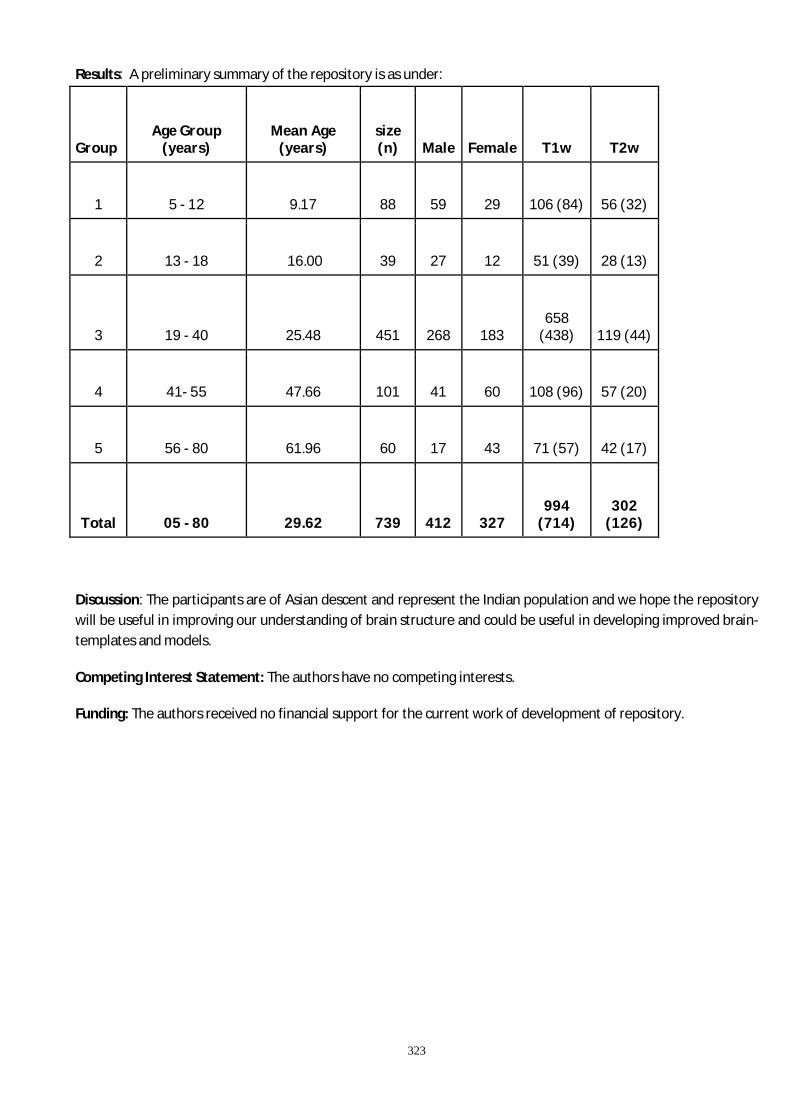
323
Results: A preliminary summary of the repository is as under:
Group Age Group
(years) Mean Age
(years) size (n) Male Female T1w T2w
1 5 - 12 9.17 88 59 29 106 (84) 56 (32)
2 13 - 18 16.00 39 27 12 51 (39) 28 (13)
3 19 - 40 25.48 451 268 183 658
(438) 119 (44)
4 41- 55 47.66 101 41 60 108 (96) 57 (20)
5 56 - 80 61.96 60 17 43 71 (57) 42 (17)
Total 05 - 80 29.62 739 412 327 994
(714) 302
(126)
Discussion: The participants are of Asian descent and represent the Indian population and we hope the repository will be useful in improving our understanding of brain structure and could be useful in developing improved brain-templates and models.
Competing Interest Statement: The authors have no competing interests.
Funding: The authors received no financial support for the current work of development of repository.
324
P71-Brain IS
Time dependent microstructural alterations in the hippocampus of moderate closed head injured rats
Palkin Arora, Kavita Singh, Richa Trivedi, Maria M D’souza
Radiological, Nuclear and Imaging Sciences (RNAIS), Institute of Nuclear Medicine and Allied Sciences
(INMAS), Delhi, India
Background: Closed head injury (CHI) is the most common insult from accidents, sports-injury and assaults. Since, trauma-associated injury is found to be associated with learning and memory deficits, hippocampal tissue alterations can be a causative source. Neuroimaging plays a critical role in detection of injury specific structural changes. Therefore, the present study involved time dependent assessment of microstructural changes in the hippocampus of injured rats using DTI and glial pathology using histological analysis.
Methods: The rats were subjected to modified Marmarou's weight drop model where brass rod held at a height of 50 cm was allowed to free fall by gravity above the sagittal midway of the rat brain. The rats were then subjected to MR imaging including diffusion tensor imaging (DTI) at day0, 4hours, day1, day5, day10 and day30 post injury. Histological analysis was also performed to assess changes in the glial population of the hippocampal region.
Results: DTI showed significant changes in mean diffusivity (MD) values at all timepoints. Similarly, both axial diffusivity (AD) and radial diffusivity (RD) values were reduced at all timepoints post injury. However, fractional anisotropy (FA) values were increased only during day1 post injury. Immunohistochemical analysis showed reactive glial pathology in the hippocampus.
Conclusion: Moderate TBI is associated with microstructural changes in the hippocampus region of the rats. Additionally, the structural alterations in the brain correlate with histological analysis. Therefore, DTI can serve as an important non-invasive method to assess the neuronal trauma severity.

325
P72- Suttee Nag Prize
Intermittent theta burst stimulation as a therapeutic technique in complete spinal cord injury patients: A pilot study
Deeksha Patel1, Aliya Mufti1, Bhavuk Garg2, Nand Kumar3, AK Dinda4, Nishank Mehta2, KP Kocchar1, Suman Jain1
1Department of Physiology, All India Institute of Medical Sciences, New Delhi, India
2Department of Orthopedics, All India Institute of Medical Sciences, New Delhi, India
3Department of Psychiatry, All India Institute of Medical Sciences, New Delhi, India
4Department of Pathology, All India Institute of Medical Sciences, New Delhi, India
Corresponding author
Email address: [email protected] (Deeksha Patel)
Abstract:
Objective: Spinal cord injury (SCI) impairs sensory and motor function affecting daily life activities. iTBS is a therapeutic technique which alter excitability of neurons. Previous study shown improvement in spasticity after iTBS in incomplete SCI patients. The study was design to investigate the effect of iTBS in complete SCI patients.
Methods: 6 spinal cord injury patients (3 Sham; 3 Real) aged between 18-70 years received 10 sessions of iTBS twice a day for 5 consecutive days. AMT, MEP, Visual Analog Pain Scale (VAS), Beck depression inventory and WHO Quality of Life (WHOQOL) have been assessed before and after the intervention.
Result: Then mean AMT was decreased in real group patients after the completion of intervention in compare to the sham group patients. The average MEP has been increased in the real group compared to the sham group. Similarly VAS and Beck depression inventory score also decreased in the real group at the end of complete intervention. Quality of Life in real group has also improved in compare to sham group after the intervention.
Conclusion: The results of this study suggested that iTBS could be a promising interventional tool in spinal cord injury patients. Intervention with iTBS over the parameters established in this study have generated motor gains and consequent functional recovery in patients with SCI. Motor gains and functional recovery have contributed to an improve in quality of life and decrease in pain.
Keywords: Spinal Cord Injury (SCI), Intermittent Theta Burst Stimulation (iTBS), Motor Evoked Potential (MEP).
Acknowledgements: This work was supported by the Department of Biotechnology (DBT) Ministry of Science and Technology, Government of India.

326
P73-Suttee Nag Prize
Deleterious effect of extracellular α-synuclein on astrocytic function
Aishwarya Raj, Alka Kaushal, Indrani Datta
National Institute of Mental Health and Neuro Sciences (NIMHANS) – Bangalore, Karnataka, India
Abstract
Background: Presence of α-synuclein has been established in the CNS microenvironment during PD disease progression, thereby not limiting its presence in DA neurons, but also in astrocytes; risking, important neuro-supportive functions of astrocytes. This study aims to assess the effect of various forms of α-synuclein on anti-oxidation machinery and glutamate metabolism. Materials and methods: WT and A53T/A30P double mutant α-synuclein were used in monomeric and aggregated forms; added extracellularly to primary astrocytes culture. Association of peptides with astrocytes were examined. Oxidative and nitrative stress were analyzed. Apart from glutathione and glutamate content, gene and/or protein expression of enzymes associated with these were also studied. Results: Monomers were engulfed while aggregates were bound to membrane. Peptide engulfed astrocytes showed increase in oxidative stress generation, contributed by decrease in Nrf2; increased iNOS, further aggravated by decreased glutathione content and related enzymes like glutathione-synthetase, -peroxidase and -reductase. Glutamate content increased in these cells contributed by increased glutamate transporter expression, de-novo synthesis of glutamate by pyruvate-carboxylase and decreased glutamine synthase and glutamate dehydrogenase expression. Highest dysfunction was observed with mutant aggregated α-synuclein treatment. Discussion and conclusions: The presence of α-synuclein did lead to astrocytic dysfunction. The impact was higher in the case of aggregated and/or mutated peptide treatment. The study not only helps in understanding the effect of α-synuclein species on astrocyte function but also suggests that ‘one size fit all’ approach cannot be adopted for tackling monomer and aggregate induced changes in cellular functions. This might allow us to consider these niche cells as potential targets for therapeutic strategies.

327

328
Prof. Jayasri Das Sarma with students oraganizers at IISER Kolkata

329