
Hypnotic Depth and Response to Suggestion Under Standardized Conditions and During fMRI Scanning
27
Intl. Journal of Clinical and Experimental Hypnosis, 55(1): 32–58, 2007 Copyright © International Journal of Clinical and Experimental Hypnosis ISSN: 0020-7144 print / 1744-5183 online DOI: 10.1080/00207140600995844 HYPNOTIC DEPTH AND RESPONSE TO SUGGESTION UNDER STANDARDIZED CONDITIONS AND DURING fMRI SCANNING David A. Oakley 1 University College London, UK Quinton Deeley Institute of Psychiatry, London, UK Peter W. Halligan Cardiff University, Cardiff, UK Abstract: Hypnosis is a potentially valuable cognitive tool for neuroimaging studies. However, understandable concern that Magnetic Resonance Imaging (MRI) in particular may adversely affect hypnotic procedures remains. Measurements of hypnotic depth and responsiveness to suggestions were taken using a standardized proce- dure that met all the requirements for functional MRI (fMRI). Testing outside the scanning environment showed reliable and stable changes in subjective hypnotic depth, with no carryover once the hypnosis had been terminated. Within-subject comparisons showed that the magnitude and pattern of these changes and the degree of respon- siveness to hypnotic suggestion were not discernibly affected by the fMRI environment. It is concluded that hypnosis can be employed as a discrete and reliable cognitive tool within fMRI neuroimaging settings. There is rapidly growing interest in employing hypnosis as a tool in cognitive neuropsychological research (Oakley, 2006) and in using brain-imaging techniques to explore hypnosis and the phenomena created by suggestion following hypnotic induction (Kihlstrom, 2003; Rainville & Price, 2003; Ray & Oathes, 2003; Spiegel, 2003; Woody & McConkey, 2003; Woody & Szechtman, 2003). To Manuscript submitted October 10, 2005; final revision received May 19, 2006. 1 Address correspondence to David A. Oakley, Hypnosis Unit, Department of Psychology (Bedford Way Building), University College London, Gower Street, London WC1E 6BT, UK. E-mail: [email protected] 32
-
Upload
independent -
Category
Documents
-
view
1 -
download
0
Transcript of Hypnotic Depth and Response to Suggestion Under Standardized Conditions and During fMRI Scanning

Intl. Journal of Clinical and Experimental Hypnosis, 55(1): 32–58, 2007Copyright © International Journal of Clinical and Experimental HypnosisISSN: 0020-7144 print / 1744-5183 onlineDOI: 10.1080/00207140600995844
HYPNOTIC DEPTH AND RESPONSE TOSUGGESTION UNDER STANDARDIZED
CONDITIONS AND DURINGfMRI SCANNING
David A. Oakley1
University College London, UK
Quinton Deeley
Institute of Psychiatry, London, UK
Peter W. Halligan
Cardiff University, Cardiff, UK
Abstract: Hypnosis is a potentially valuable cognitive tool forneuroimaging studies. However, understandable concern thatMagnetic Resonance Imaging (MRI) in particular may adversely affecthypnotic procedures remains. Measurements of hypnotic depth andresponsiveness to suggestions were taken using a standardized proce-dure that met all the requirements for functional MRI (fMRI). Testingoutside the scanning environment showed reliable and stable changesin subjective hypnotic depth, with no carryover once the hypnosishad been terminated. Within-subject comparisons showed that themagnitude and pattern of these changes and the degree of respon-siveness to hypnotic suggestion were not discernibly affected by thefMRI environment. It is concluded that hypnosis can be employedas a discrete and reliable cognitive tool within fMRI neuroimagingsettings.
There is rapidly growing interest in employing hypnosis as atool in cognitive neuropsychological research (Oakley, 2006) andin using brain-imaging techniques to explore hypnosis and thephenomena created by suggestion following hypnotic induction(Kihlstrom, 2003; Rainville & Price, 2003; Ray & Oathes, 2003; Spiegel,2003; Woody & McConkey, 2003; Woody & Szechtman, 2003). To
Manuscript submitted October 10, 2005; final revision received May 19, 2006.1Address correspondence to David A. Oakley, Hypnosis Unit, Department of
Psychology (Bedford Way Building), University College London, Gower Street, LondonWC1E 6BT, UK. E-mail: [email protected]
32

HYPNOTIC DEPTH AND fMRI 33
date most studies have used Positron Emission Tomography (PET)in combination with hypnosis to investigate auditory hallucinations(Szechtman, Woody, Bowers, & Nahmias, 1998), color processing(Kosslyn, Thompson, Costantini-Ferrando, Alpert, & Spiegel, 2000),pain perception (Hofbauer, Rainville, Duncan, & Bushnell, 2001;Rainville, Duncan, Price, Carrier, & Bushnell, 1997), voluntary motorcontrol (Blakemore, Oakley, & Frith, 2003; Halligan, Athwal, Oakley,& Frackowiak, 2000), malingering (Ward, Oakley, Frackowiak, &Halligan, 2003), consciousness (Rainville, Hofbauer, Bushnell, Duncan,& Price, 2002), phantom-limb pain (Rosén,Willoch, Bartenstein, Berner,& Røsjø, 2001;Willoch et al., 2000), and the hypnotic state itself (Maquetet al., 1999; Rainville et al., 1999). With a notable early exception thatused hypnosis to investigate phantom-limb movements (Ersland et al.,1996), functional Magnetic Resonance Imaging (fMRI) has been rela-tively little used in hypnosis-related research until recently. The past 2years, however, have seen a growth in hypnosis studies using fMRI toinvestigate pain (Derbyshire, Whalley, Stenger, & Oakley, 2004; Raij,Numminen, Narvarnen, Hiltunen, & Hari, 2005; Schultz-Stubner et al.,2004) and attentional processes (Egner, Jamieson, & Gruzelier, 2005).
This trend seems set to continue and indeed fMRI is fast becomingthe neuroimaging technique of choice for this kind of work. fMRI has anumber of technical advantages over PET, particularly with regard togreater temporal and spatial resolution (Ray & Oathes, 2003). UnlikePET, it is not invasive and does not involve exposure to sources ofradiation; the procedure when used experimentally is not restricted tomale subjects, as is the case in the United Kingdom for PET; and thesame individual can be used for repeated scans over a shorter timeperiod. There are however some practical disadvantages to using fMRIfor hypnosis studies, most notably that the participant is typicallyclosely confined in a complete body scanning tube and the equipmentis very noisy when it is operating. This creates an unpleasant, claustro-phobic environment for the participant and, significantly for hypnosisstudies, makes communication more difficult. More recent dedicatedfMRI brain scanners have overcome some of these problems with thescanning field being limited to the head area and the developmentof more efficient verbal communication systems between the exper-imenter and the person in the scanner. Concerns remain, however,that the fMRI scanning environment could interfere with the processof hypnotic induction to a greater extent than that of PET. There isalso the possibility, given the design of many neuroimaging studies,that unplanned changes in hypnotic depth could occur over the courseof extended standardized hypnosis sessions, especially where theseinvolve the delivery and removal of targeted suggestions. In addi-tion, the need to maintain hypnosis for the long periods that many

34 DAVID A. OAKLEY ET AL.
studies require may serve to attenuate suggested phenomena that areof interest to the experimenters.
This study set out to monitor self-reported hypnotic depth with anexperimental protocol (using a standardized hypnosis induction proce-dure based on Gruzelier, 1998) that was designed to be compatiblewith an fMRI scanning environment. Since fMRI experimental designstypically employ standardized periods of timewithin which images areacquired under controlled cognitive, sensory, and/or behavioral condi-tions, the protocol included measures that enable subjective changesin hypnotic depth to be monitored over time (Woody & McConkey,2003).
To ascertain which elements of a standardized hypnotic procedureare required to produce a substantial and stable level of hypnoticdepth for experimental purposes, these procedures were carried out indiscrete steps. The procedure also included an evaluation of the effectson hypnotic depth of providing and removing a targeted suggestion(in this case, limb paralysis). The complete procedure was initiallytested outside the scanner and then, using the same protocol, repeatedin the scanning environment for a subset of the same participants.The MR scanner was operated normally to acquire fMRI data duringpreselected time blocks as indicated by the experimental design.
Hence, the main aim of this study was to quantify and to comparechanges in hypnotic depth in response to the same protocol inside andoutside an MR scanner. For this reason, imaging data acquired in theMR scanner will be separately reported.
There is currently no objective measure for the intensity ofa hypnotic experience or hypnotic depth, though a number ofapproaches have been adopted over the years for monitoring theparticipant’s subjective experience. McConkey, Wende, & Barnier(1999) have recently developed a more sophisticated version of anearlier technique involving the participant manually moving thepointer on a dial to indicate changes in the intensity of their experienceof hypnosis and hypnotically suggested effects on a moment-to-moment basis. Unfortunately, limitations of space in the fMRI envi-ronment and the nature of the motor function tests to be used inthe scanner precluded use of this device in our study. Instead, weadopted the more practical method of verbal self-reporting similar tothe hypnotic-depth scale described by Le Cron (1953) and later devel-oped as the Long Stanford Scale (Tart, 1970).
A common feature of both PET and fMRI studies is that becauseof their expense it is usual to pretest potential experimental partici-pants both for their hypnotic susceptibility and for their specific abilityto produce any suggested phenomena required within the experi-mental design. Participants had two experiences of hypnosis prior tothe commencement of the study. In addition to serving as screening

HYPNOTIC DEPTH AND fMRI 35
procedures, these experiences provided a benchmark for participantsagainst which their subsequent subjective depth estimations couldbe made.
In summary, this study was designed to provide an accountof changes in subjective depth of hypnotic experience over timein an experimental protocol directly compatible with those usedin fMRI neuroimaging studies when either hypnosis itself or theeffects of targeted suggestions given after a hypnotic inductionprocedure are the principal focus of interest. In particular, we explorethe predictability, stability, and strength of hypnosis and hypnoticphenomena both within and outside the scanning environment.
Method
ParticipantsForty-six students scoring eight or above (out of a possible total of
12) on the Harvard Group Scale of Hypnotic Susceptibility, Form A(HGSHS:A; Shor & Orne, 1962) from the departments of Psychologyand Medicine at University College London were identified from adatabase maintained by the authors and were invited to participatein the present study. Of these, 22 responded positively and weretested for their ability to experience a leg paralysis in response todirect suggestion in hypnosis (limb-paralysis screening). All reportedan involuntary paralysis with no observable movement when theywere asked to try to move the leg. Two of this group were left-handedand 2 were unavailable for further testing, leaving a group of 18 right-handed participants who took part in the first phase of the presentstudy (off-line testing). Ten of these 18 were females. The mean ageof the group was 22.3 years (range, 18–35; SD=4.69) and the meanHGSHS:A score was 10.28 (range, 8–12; SD=1.18). Half were testedusing suggestions for leg paralysis and half for hand/arm paralysis.All paralyses were on the left side.
Following the off-line testing phase of the study, 10 of the sameparticipants went on to be tested in the fMRI scanning environment(fMRI testing) and 8 completed the full sequence of experimental stagesdescribed below. This final subgroup was composed of equal numbersof males and females. Four had been previously tested with a hand/arm paralysis and four with a leg paralysis. All suggested paralyses inthis second phase of the study were of the left hand/arm. The meanage of this group was 22.43 years (range, 18–35; SD=5.83) and themean HGSHS:A score was 10.13 (range 8–12; SD=1.46).
Limb Paralysis ScreeningAll 22 participants who accepted the invitation to take part in the
studywere tested individually in an experimental room for their ability

36 DAVID A. OAKLEY ET AL.
to produce limb paralysis in response to suggestion in hypnosis. Theywere seated in a reclining armchair with their legs horizontal to thefloor supported on a footstool. Just before the hypnotic induction,each participant was asked to describe a real or imaginary “specialplace” where they would feel safe and relaxed and could “spend sometime for themselves” (Heap & Aravind, 2002), and detailed notes weretaken. They were made aware that a limb paralysis would be suggestedand that when they were asked to try to move either of their limbs theyshould actively try to do so. The required movement of raising the legso that the foot was a few inches above the stool was described to themand they were then asked to perform this movement with each legin turn. The hypnotic induction procedure that followed commencedwith the participant being asked to close his or her eyes, followed byinstructions for regular breathing with color imagery (breathing outa color representing tension or stress and replacing it with a “calm,tranquil” color), systematic direct muscle relaxation with suggestionsof muscles “letting go,” “relaxing,” etc., and then descent imagery—either garden steps or lift (elevator) according to the participant’spreference—accompanied by counting from one to ten. The final stageof the induction was engagement with their own special-place experi-ence guided by the experimenter on the basis of the previously writtennotes. As a further hypnotic deepening procedure, a count of one toten was used once the special place had been established, and then theparticipant was asked to describe aloud their experience in the specialplace with the suggestion that this would intensify their involvement inthe experience. A left-sided leg paralysis was then suggested based onthe paralysis script outline given below. The behavioral test consistedof asking the participant to try first to raise their right leg and then tryto raise their left leg. They were allowed to try for 1 minute in eachcondition. Following this the paralysis suggestions were reversed, thebehavioral test was repeated, and the special-place experience wasreinstated. Finally, hypnosis was terminated with counting backwardsfrom three to one accompanied by statements reorienting the partic-ipant to the experimental room and of returning to their everydaylevels of alertness.
All participants confirmed that they had actively tried to movethe appropriate leg when instructed to do so. Despite their attempts,however, none of them succeeded in raising their “paralyzed” leftleg during the first behavioral test (i.e., following the paralysissuggestion)—though some showed visible signs of individual muscletwitches. In contrast, they were all able to raise their right legs. Allparticipants moved both legs during the second behavioral test (i.e.,following reversal of the paralysis suggestion). Following terminationof hypnosis, participants were asked to rate the degree of involun-tariness they experienced in trying to move their left leg when it

HYPNOTIC DEPTH AND fMRI 37
was paralyzed, in moving their left leg when the paralysis had beenremoved, and in moving their right leg when the left leg was para-lyzed on 100mm Visual Analog Scales (VAS; where one end of thescale is labeled completely voluntary and the other completely involun-tary). Overall, mean for perceived involuntariness of the failure tomove during suggested paralysis was 93.00 (SD=7.18); this comparedwith a mean involuntariness score of 17.22 (SD=17.15) for movementof the left leg when the paralysis was removed and 14.67 (SD=17.85)for the right leg when the left leg was paralyzed.
Off-Line TestingThe 18 participants in this first phase of the study were tested indi-
vidually in an experimental room set up as it was for the limb paralysisscreening sessions. To evaluate behaviors that would be possible foruse in an MRI scanner, the motor response under investigation for halfof the participants was the movement of an imaginary pedal by anankle movement of their left or right foot at the rate of once every 3seconds. For the other participants, it was the movement of an imagi-nary joystick once forward, once back, and then returning to the centralposition upon hearing an instruction to move, using the hand andwrist of their left or right hand. As before, they were told that theyshould always try to move the pedal/joystick in the designated wayat all stages of the study when asked to do so by the experimenter.The participants were also introduced to a scale of hypnotic depthwhere 0=not hypnotized at all and 10= as deeply hypnotized as you haveever been before. They were instructed to use numbers above 10 if theyfelt they were hypnotized more deeply than on any previous occasion.Then followed a hypnosis and motor-response testing sequence thatfollowed a proposed protocol for future experimental studies usingfMRI. All stages of the experimental procedure incorporated 2-minuteintervals (mock MRI blocks), during which no instruction was given,to represent fMRI data acquisition blocks. A hypnotic depth score wasasked for at the beginning and end of each of these intervals with therequest: “Please give me the number on your scale now.”
The sequence of experimental stages is summarized in Table 1and as the horizontal axis in Figure 1. For Stage 1 (MOTOR 1), theparticipant was asked to close their eyes and to carry out the appro-priate movements with their left and right foot/hand in alternationover a 2-minute interval. Stage 2 (OPEN 1) consisted of a 2-minuterest interval with eyes open and Stage 3 (CLOSED 1) a 2-minuterest interval with eyes closed. Stages 4, 5, and 6 were based closelyon a three-step model of hypnotic induction described by Gruzelier(1998). The steps are (a) fixation on an object and listening to thehypnotist’s voice; (b) a letting-go procedure comprising suggestionsof fatigue at continued fixation, tiredness, and eye closure, together

38 DAVID A. OAKLEY ET AL.Ta
ble1
MeanHyp
notic
Depth
Ratings
DuringOff-Line
Testing
PREH
YPN
OSIS
HYPN
OSIS
POST
HYPN
OSIS
(1)MOTO
R1
b0.11
(0.32)
(4)FIXATE
1b
2.06
(1.98)
(12)
CLO
SED
2b
1.39
(1.97)
a0.39
(0.61)
a2.44
(1.92)
a2.11
(2.17)
b/amean
0.25
(0.43)
b/amean
2.25
(1.92)
b/amean
1.75
(1.99)
(2)OPE
N1
b0.50
(0.62)
(5)AFT
ERCOUNT
b6.94
(1.63)
(13)
OPE
N2
b0.94
(1.47)
a0.94
(1.06)
a7.06
(1.83)
a0.89
(1.28)
b/amean
0.72
(0.79)
b/amean
7.00
(1.65)
b/amean
0.92
(1.34)
(3)CLO
SED
1b
1.22
(1.35)
(6)SP
ECIA
LPL
ACE1b
8.22
(1.73)
a1.56
(1.58)
a8.44
(1.98)
b/amean
1.39
(1.44)
b/amean
8.33
(1.81)
(7)MOTO
R/P
ARAL
b9.06
(2.29)
a9.44
(2.79)
b/amean
9.25
(2.51)
(8)MOTO
R/N
ORM
b8.44
(1.79)
a8.50
(2.18)
b/amean
8.47
(1.96)
(9)SP
ECIA
LPL
ACE2b
9.00
(1.94)
a9.00
(2.03)
b/amean
9.00
(1.94)
(10)
BEFO
RECOUNT
b9.00
(2.09)
a9.00
(2.74)
b/amean
9.00
(2.39)
(11)
FIXATE
2b
2.94
(2.36)
a2.67
(2.09)
b/amean
2.81
(2.14)
Notes.Means
ofde
pthratin
gstake
nbe
fore
(b)an
dafter(a)each
2-minute(m
ockMRI)
timeinterval
andthemeanratin
gforeach
oftheseintervals(b/a
mean)
aresh
ownforall1
3expe
rimen
tals
tage
s.Stan
dard
deviations
aresh
ownin
parenthe
ses.

HYPNOTIC DEPTH AND fMRI 39
Figure 1. Mean subjective hypnotic depth for all 13 stages of Phase 1 (Off-line testing)—based on 18 participants. See text for further explanation.
with deep relaxation and counting; and (c) instructions for relaxedand passive imagery (see “Hypnosis Scripts” below). Our Stage 4(FIXATE 1) consisted of a 2-minute interval preceded by instructionsto the participant to continue to listen for the experimenter’s voiceand to fixate visually on a small spot on the wall set just above thenormal line of sight. Stage 5 (AFTER COUNTING) was a 2-minuteinterval preceded by instructions to continue to fixate on the spot andbreathe regularly, accompanied by suggestions of muscle relaxation,the eyelids becoming heavy and tired, spontaneous eye-closure, andfinally by the experimenter counting from one to twenty. For Stage6 (SPECIAL PLACE 1), the participant was asked to enter into thesame special-place experience that had been used during the earlierlimb-paralysis screening with the suggestion that they could this time

40 DAVID A. OAKLEY ET AL.
“let the scene unfold like in a dream” and to remain there for the next2-minute interval. At the end of this interval, the participant was askedto “leave your special place and all the imagery associated with it butremain as relaxed and hypnotized as you are now.”
In Stage 7 (MOTOR PARALYSIS), suggestions for left-sided paral-ysis used the paralysis-script outline presented belowwith appropriatesubstitutions of the words leg/arm, hip/shoulder, and toes/fingersfor the two subgroups of participants in this part of the study. Therefollowed a 2-minute interval during which participants attempted tomove their left and right feet/hands to carry out the required move-ments as in Stage 1 (MOTOR 1). In Stage 8 (MOTOR NORMAL),suggestions for removal of the paralysis were given followed by a2-minute interval during which the same motor test as in Stages 1 and7 were repeated. Stage 9 (SPECIAL PLACE 2) was a repetition of thespecial-place experience plus a 2-minute interval as described for Stage6. Stage 10 (BEFORE COUNT) involved a 2-minute interval immedi-ately after the participant had left the special-place experience and theassociated imagery but remained in the same relaxed and hypnotizedstate. The suggestions and instructions that were introduced at Stage5 were then reversed, and the participant returned to “wide-awake”feelings by means of suggestions of the eyelids becoming less and lesstired and heavy until the eyes felt like opening, along with countingin reverse order from twenty to one. At the end of this count, theparticipant was instructed as in Stage 4 to continue to listen for theexperimenter’s voice and to fixate visually on the spot on the wallover a 2-minute interval (Stage 11: FIXATE 2). Stage 12 (CLOSED 2)consisted of a 2-minute rest interval with eyes closed, and Stage 13(OPEN 2) was a 2-minute rest interval with eyes open.
None of the participants were observed to move their hand or footwhen attempting to carry out the intended action with their para-lyzed limb—though all were capable of doing so when the paral-ysis was removed or when using their right side. When testing wascomplete, participants rated the involuntariness of their inability tomove the paralyzed left hand\foot and in moving the right hand/footwhen the left side was paralyzed on 100 mm VAS as in the limb-paralysis screening phase of the study. Additional 100 mm VAS wereused in this phase to assess the difficulty involved in attempting tomove under these conditions. For involuntariness, one end of thescale was labeled completely voluntary and the other completely invol-untary. For difficulty, one end of the scale was labeled moved easilyand the other impossible to move. Complete sets of involuntarinessand difficulty data were collected for 15 and 16 of the participants,respectively.
The two special-place conditions were intended to represent discretesteps in the hypnotic process and similar VAS were completed by 16

HYPNOTIC DEPTH AND fMRI 41
of the participants at the end of the session to measure the strengthof special-place imagery on the two occasions (the two ends of thescale were labeled no imagery at all and very strong imagery). In addi-tion, these participants were asked to say whether there was imagerypresent immediately before the first special-place condition, duringthe limb-movement testing phase that followed termination of thefirst special-place condition, and immediately after termination of thesecond special-place condition. If the participant reported that imagerywas present at any of these times, they were asked to rate its strengthon VAS and to say if it was the special place or other imagery. If thelatter, they were asked to describe the imagery they experienced atthese times. The overall average time taken from the first experimentalstage (MOTOR 1) to the final one (OPEN 2) was approximately 45–50minutes.
Scripts
Hypnosis ScriptsThe following are verbatim scripts used in the second phase of
this study (fMRI-environment testing) for the three steps of hypnoticinduction and for reversal of hypnosis, based closely on scripts usedby Gruzelier and Jamieson (Graham Jamieson, November 20, 2002,personal communication). Apart from minor modifications to reflectthe practical conditions of the scanning environment, such as referenceto lying on the scanner table rather than reclining in a chair, the useof actual joysticks and a fixation cross instead of a spot on the wall,these scripts are identical to the ones used in the off-line testing phaseof the study.
Hypnosis Step 1. Stage 4: FIXATE 1. Eyes are open, fixated on target.This follows an eyes-closed rest condition.
Open your eyes. Just lie comfortably on the table. Keep your arms wherethey are, relaxed by your side with your hands holding the joysticks.Look at the cross in front of you, which I shall refer to as the target.Please look steadily at the target and while concentrating on the targetpay attention to my voice. Focus your mind on what I ask you to thinkabout—keeping your gaze fixed upon the target. If you find your mindwandering at any time, just bring your thoughts back to the target andto my words.
Please give me the number on your scale now.Stay looking at the target and listening for my voice.
TEST/SCAN. Offline – silent break for 2 minutes. fMRI environ-ment – MRI scan acquisitions; no communication from experimenterfor 2 minutes, 12 seconds. Then experimenter asks: “Please give methe number on your scale now.”

42 DAVID A. OAKLEY ET AL.
Hypnosis Step 2. Stage 5: AFTER COUNTING. Eye fatigue at fixa-tion, eye closure, tiredness, relaxation, and counting.
Now you may feel you have stared for long enough but continue to lookat the target for a little longer. Your eyes will feel tired and will shortlystart to close. Breathe gently and easily and as you breathe out and relaxmore and more your eyes will begin to close all by themselves. Just letthis happen and when your eyes have fully closed, please say “yes” sothat I know. Breathe in and out and each time you breathe out you willfeel more deeply relaxed—deeply relaxed. Feel the muscles of your faceletting go ! ! ! and the relaxation spreading through your facial musclesinto your forehead and into the muscles of your scalp. Feel those musclesletting go ! ! ! and the feelings of relaxation moving through your head! ! ! around and behind your eyes and into the muscles of your jaw.And the relaxation continues to move down through your body ! ! ! toyour neck ! ! ! throat, shoulders. Your shoulders feeling limp, heavy,and relaxed.
Feelings of relaxation extend along your arms ! ! ! down to yourelbows ! ! ! to your wrists ! ! ! your hands ! ! ! yours fingers. Your armsfeel heavy. You feel deeply and peacefully relaxed. Your eyelids arebecoming heavier and heavier ! ! ! heavier and heavier. [IF CLOSED:“Your eyelids closed and heavy.” IF NOT CLOSED: “And if they havenot already closed, they will soon do so.”].
Relaxation moves across your shoulders ! ! ! into your chest ! ! !spreading like a wave through your body ! ! ! moving down to yourwaist. Your breathing is easy and regular. Each time you breathe outyou go deeper and deeper ! ! ! feeling more and more relaxed. Wavesof relaxation spread from your waist to your hips ! ! ! to your legs ! ! !down to your knees ! ! ! to your ankles ! ! ! down to your feet ! ! ! toyour toes. As you become more and more relaxed, your body may feelheavy ! ! ! or perhaps a little numb. You may begin to have this pleasantfeeling of numbness and heaviness in your legs and feet, in your handsand arms, throughout your body ! ! ! as though you were settling deepinto the surface beneath you. Your eyelids feel heavy and tired ! ! ! [IFCLOSED: “! ! ! remaining tightly closed ! ! ! heavier and heavier. Youreyelids seem weighted down ! ! ! pulled down by the weight.” IF NOTCLOSED: “! ! ! and if they are not closed yet, they will begin to closesoon as they feel heavier and heavier ! ! ! just say ‘yes’ when they haveclosed completely. Your eyelids seem weighted down ! ! ! pulled downby the weight ! ! ! so heavy ! ! ! just allow them to close by themselvesnow ! ! ! let them close ! ! !” CONTINUE UNTIL subject says, “Yes.”].
You are going to become even more relaxed. It is easier to relax withyour eyes closed. So keep them closed now. You feel deeply relaxed ! ! !as you continue to listen to my voice. Just keep your thoughts on whatI am saying. Soon I shall begin counting from one to twenty. As I countyou will feel yourself going down further ! ! ! and further into a deepstate of relaxation, however, you will be able to do all the things youare asked to do without it disturbing your deep state of relaxation. And

HYPNOTIC DEPTH AND fMRI 43
you can find that background sounds bother you less and less as timegoes by—just letting them slip to the back of your mind.
One ! ! ! two ! ! ! down, down into a deep state of relaxation ! ! !three ! ! ! four ! ! ! five ! ! ! more and more deeply relaxed ! ! ! six ! ! !seven ! ! ! you are sinking deeper and deeper ! ! ! eight ! ! ! nine ! ! !ten ! ! ! half way ! ! ! eleven ! ! ! twelve ! ! ! thirteen ! ! ! fourteen ! ! !deeply relaxed ! ! ! hearing my voice clearly ! ! ! fifteen ! ! ! sixteen ! ! !seventeen ! ! ! eighteen ! ! ! deeper ! ! ! deeper ! ! ! more and more relaxed! ! ! nineteen ! ! ! twenty ! ! ! deeply relaxed. Just remain in that deeplyrelaxed state for now.
Please give me the number on your scale now.Just relax deeper and deeper as time goes by and I will speak to
you again shortly.
TEST/SCAN. Offline – silent break for 2 minutes. fMRI environ-ment – MRI scan acquisitions; no communication from experimenterfor 2 minutes, 12 seconds. Then experimenter asks: “Please give methe number on your scale now.”
Hypnosis Step 3. Stage 6: SPECIAL PLACE 1. Relaxed and passiveimagery.
As you relax deeper and deeper ! ! ! allow a scene—your special place—tocome to mind and begin to experience your self as part of that scene—there in your special place—just letting the scene unfold like in a dream! ! ! just allowing the images to shift and change as they will ! ! ! in evermore pleasant and relaxing ways. There in your special place. [Identifyindividual special place and set it up using the information from thestandard sheet].
If everything is O.K. and you have the feeling of being there in yourspecial place, please let me know by saying “yes.” Just remain in yourspecial place enjoying the imagery.
Please give me the number on your scale now.I will speak to you again shortly. Stay in your special place.
TEST/SCAN. Offline – silent break for 2 minutes. fMRI environ-ment – MRI scan acquisitions; no communication from experimenterfor 2 minutes, 12 seconds. Then experimenter asks: “Please give methe number on your scale now.”
Leave your special place now and all the imagery associated with it butremain as relaxed and hypnotized as you are now—please let me knowby saying “yes” when you have done that.
Reversal of hypnosis. Returning to alert feelings, eyes becoming lesstired and heavy, reverse count twenty to one, and eyes opening.
In a moment, I will count back from twenty to one, and as I do justreturn to alert wide-awake feelings so that you are fully alert and wide

44 DAVID A. OAKLEY ET AL.
awake when I get to one. Keep your eyes closed for now, but as I countyour eyes will feel less and less tired and start to open. Just let thishappen, and when your eyes have fully opened please say “yes” so thatI know. O.K., just returning to wide-awake, alert feelings now as I count.
Twenty ! ! ! nineteen ! ! ! eighteen ! ! ! returning to wide-awakefeelings ! ! ! seventeen ! ! ! sixteen ! ! ! fifteen ! ! ! more and more alert! ! ! fourteen ! ! ! thirteen ! ! ! twelve ! ! ! eleven ! ! ! ten ! ! ! half way! ! ! nine ! ! ! eight ! ! ! seven ! ! ! back to normal wide-awake feelings! ! ! no more heaviness or numb feelings ! ! ! six ! ! ! five ! ! ! four ! ! ! allthe muscles throughout your body back to their normal state of tensionand tone ! ! ! three ! ! ! two ! ! ! and one, wide awake, fully alert.
Motor Test/Paralysis ScriptsThe following are verbatim scripts and script outlines for motor
testing and limb paralysis as used in the second phase of the study(fMRI-environment testing). Apart from the use of taped instructionscueing the joystick movements these are identical to the scripts usedin the first (off-line testing) phase of the study.
Hypnosis motor test. MOTOR/NORM: arms normal. Apart fromreferences to remaining hypnotized, this script is identical to that usedfor the motor test (MOTOR 1) prior to hypnosis and, for phase twoonly, the motor test (MOTOR 4) after hypnosis.
With your eyes closed, remaining relaxed and hypnotized. You willhear recorded instructions at regular intervals. The instruction will say:“rest,” which simply means not attempting to do anything; “left,” whichmeans try to move the left joystick forwards and backwards once withyour left hand each time; and “right,” which means try to move theright joystick forwards and backwards once with your right hand eachtime. The instructions will come at regular intervals. Don’t guess what iscoming, just listen and follow the instructions to the best of your ability.
Please give me the number on your scale now.In all stages, listen to the recorded instruction. Remember, some-
times you will be asked to try to move, sometimes not. Remain as relaxedand hypnotized as you are now.
TEST/SCAN. Offline – 2 minutes during which experimenterrequests right and left limb movements. fMRI environment – 7minutes, 42 seconds of MRI scan acquisitions during which prere-corded instructions are given at 3-second intervals for 7 minutes, 30seconds: “rest,” “left,” and “right,” which cue the appropriate subjectresponses. Then the experimenter says, “Please give me the numberon your scale now.”
Hand/arm paralysis suggestions. Script outline: Key suggestions forleft arm/hand, from shoulder to tips of fingers.

HYPNOTIC DEPTH AND fMRI 45
1. Muscles are floppy, relaxed, so relaxed subject is unable to movebut retaining their grip on the joystick. Later the word paralyzed isintroduced—as in “that left hand and arm are becoming paralyzed fromyour shoulder right down to the tips of your fingers and quite unableto move—floppy and unable to move.”
2. Sensation of touch remains normal and subject’s grip on the joystickremains strong.
3. Muscles out of touch with subject’s thoughts, wishes, and intentions—sothat even if subject tries to move, the muscles will fail to respond in anyway, As though the left arm and hand are no longer part of subject,“quite unable to respond.”
After these suggestions (1–3 above) have been administered, subjectis asked to say “yes” when he or she begins to feel the suggestedchanges. Suggestions continue until the “yes” response is given.Subject is then asked to allow the feelings to strengthen until the lefthand/arm is “completely paralyzed from your shoulder right down tothe tips of your fingers—completely unable to move the left joystick—even if you try.” Suggestions continue. Subject is asked to say “yes”when paralysis is as strong as it can be. Then, “Even though your lefthand and arm are paralyzed and unable to move your right hand andarm remain normal and you are able to move the right joystick easilywhenever you want to.”
Hypnosis motor test. MOTOR/PARAL: left arm paralyzed.
Just remain as relaxed and hypnotized as you are now with that left armand hand paralyzed and unable to move and the right arm completelynormal but retaining a grip on both joysticks.
As before, you will hear recorded instructions at regular inter-vals. When you hear the word “rest,” just relax—do not attempt to doanything. Do not try to move either joystick but your left arm and handwill continue to feel paralyzed. Listen to the word “rest” as it is repeatedbut do not attempt to move either joystick.
When you hear the word, “left,” please try to move the left joystickforwards and backwards once with your left hand each time, but yourleft arm and handwill remain paralyzed and unable to move the joystick,floppy and unable to move.
When you hear the word, “right,” please try to move the rightjoystick forwards and backwards once with your right hand each time—your right arm and hand will remain normal and able to move easily,normally, and able to move the joystick easily.
Please say, “yes” to confirm that your left arm and hand are stillcompletely paralyzed and unable to move at all.
Please say, “yes” to confirm that your right arm and hand are stillcompletely normal and able to move easily.
In all stages, listen to the recorded instruction. Sometimes you willbe asked to move, sometimes not. The instructions will come at regular

46 DAVID A. OAKLEY ET AL.
intervals. It is important that you follow the instructions to the best ofyour ability.
Please give me the number on your scale now.
TEST/SCAN. Offline – 2 minutes during which experimenterrequests right and left limb movements. fMRI environment – 7minutes, 42 seconds of MRI scan acquisitions during which prere-corded instructions are given at 3-second intervals for 7 minutes, 30seconds: “rest,” “left,” and “right,” which cue the appropriate subjectresponses. Then the experimenter says, “Please give me the numberon your scale now.”
fMRI-Environment Testing
For this second phase of the study, all the procedures were carriedout while the participants were lying enclosed in a GE Signa 1.5Tesla system (General Electric, Milwaukee, WI) whole-body magnet,immersed up to their waist. A quadrature birdcage head coil was usedfor radiofrequency transmission and reception. The fMRI scanner washoused in the Maudsley Hospital in London, and all instructions fromthe experimenter were conveyed via earphones from a microphonelocated in the scanning control room. As this proved to be a techni-cally more reliable method for use in the scanner, all participants weretested in this phase of the study for hand and wrist movements usingstandard computer analog joysticks adapted to remove most metal.The filtered signal from each joystick was interfaced to the computersvia a custom-built interface box. As with the imaginary joysticks usedin off-line testing, they were to be moved once forward, once backwardand returned to rest upon verbal instruction. As an actual joystickwas available for each hand, the additional suggestion was given thatirrespective of paralysis suggestions the participant’s fingers/handwould retain their grip on the relevant joystick throughout. The verbalinstructions pertaining to the joystick movements were also simplifiedand presented via a digital recording for this part of the study (seemotor test/paralysis scripts above).
In contrast to the off-line phase, we included a fourth stage ofmotor testing after reversal of the hypnotic state in the fMRI-testingphase. This additional stage was introduced to explore any carryovereffects on brain function of the suggestions of limb paralysis afterremoval of the suggestion and reversal of the hypnotic state. Due totime limitations, this stage was acquired in only 6 subjects.
Each of the four motor-testing blocks, during which MR activationswere acquired, comprised a total of 15 30-second epochs (five “rest,”five “right,” and five “left”), each epoch consisting of 10 consecutiverepetitions of the relevant command repeated at 3-second intervals.

HYPNOTIC DEPTH AND fMRI 47
These epochs were presented in a pseudorandomized order. The totalstimulus time for each motor block was 7 minutes, 30 seconds in atotal scan time of 7 minutes, 42 seconds.
The other five stages of the testing protocol up to the point at whichhypnosis was terminated were each followed by a 2-minute 12-secondblock of MR acquisition (including an initial 12 seconds of dummyacquisitions). For each of these five nonmotor stages of the experi-mental protocol, this resulted in five blocks of image acquisition thatwere concatenated into a single experimental run to allow contrasts ofdiscrete stages. It is important to note that although the scan data arenot reported the scanner was activated in the normal way during eachof the scanning periods that were embedded in the testing protocol.The final nonmotor stage of the procedure (posthypnosis, eyes open)did not include a scanning block. Further, as part of the normal proce-dure for checking for scanner drift, a standardized visual/auditory(VISAUD) stimulus set consisting of a changing checkerboard visualdisplay and spoken words that had not occurred during off-line testingwas presented once before testing commenced, once in the middle oftesting (immediately before the reversal of hypnosis), and once whentesting was complete (stimulus duration 4 minutes, 48 seconds in atotal scan time of 4 minutes, 56 seconds). In addition, prior to thecommencement of baseline motor testing localizer scans, high resolu-tion GE (Gradient Echo) scans for fMRI image registration, and a highresolution three-dimensional MR volume, using a T1-weighted SPGR(Spoiled Gradient Recall) acquisition strategy, were acquired.
Apart from the above mentioned differences, and the use of aprojected fixation cross, all the stages that formed part of the procedurefor this phase of the study were carried out exactly as described for theoff-line testing phase and are identified here with the same names. Toaccommodate the testing protocol within the time available for scan-ning, some of the later stages used in the off-line testing were dropped.The fMRI-environment testing phase of the study commenced withthree prehypnosis stages: MOTOR 1 testing, followed by OPEN 1 andCLOSED 1. There were then five hypnosis stages: FIXATE 1, AFTERCOUNT, SPECIAL PLACE 1, MOTOR NORMAL, MOTOR PARAL-YSIS. The sequence as described above is identical to the off-line proce-dure up to and including SPECIAL PLACE 1. In this second phase ofthe study, motor testing with both limbs normal (MOTOR NORMAL)preceded testing with the left limb paralyzed (MOTOR PARALYSIS).This was to avoid any possible carryover effects of hypnotic paralysisinto normal limb-movement testing in anticipation of the proposedfMRI analysis. Following MOTOR PARALYSIS, the hypnosis reversalprocedure that was described in the off-line testing phase was carriedout. The final stage of the procedure was an eyes-open alert state(OPEN 2), and for 6 of the participants there was a further posthypnotic

48 DAVID A. OAKLEY ET AL.
motor block (MOTOR 4). This sequence of stages is summarized inTable 2 and in the horizontal axis to Figure 2. Even with these amend-ments, because of varying delays between each of the stages that wereintroduced by operating requirements of the scanner itself, the inclu-sion of VISAUDs, localizer scans, structural scans and the longer motortesting intervals compared to Phase 1, the duration of this part of thestudy for the 6 participants who completed all ten stages was approx-imately 2 hours and 15 minutes within which there was a minimumof 1 hour 43 minutes of scanning time (the other two participantsomitted the final motor block, corresponding to 7 min 42 seconds lessof completed MR scanning).
Corresponding to the procedure in the off-line testing phase ofthe study, hypnotic depth measurements were taken immediatelybefore and after each block of MR acquisition. In addition, a singlehypnotic depth measurement was taken immediately after the reversalof hypnosis in the alert, eyes-open state (OPEN 2). Postsession VASmeasures were again used to assess the involuntariness and difficultyof attempted joystick movements during the left-hand paralysis condi-tion, the strength of special-place imagery and the strength and natureof imagery experienced before the special-place condition and duringjoystick movement testing after its termination. Full sets of data werecollected for all 8 participants on all measures except for right arm,special place, and posthypnosis measures for one of them.
All data analysis was carried out using SPSS Version 11.03 forMacintosh OSX. The criterion for statistical significance was set atp< .05 (two-tailed).
Results
Off-Line TestingMean hypnotic depth ratings before (b) and after (a) each of the
2-minute intervals (mock MRI blocks) embedded in the 13 stages ofthe off-line screening phase of the study are shown in Table 1. To testfor changes in subjective hypnotic depth within mock-MRI intervalsand across all stages of the experimental procedure, a 13 (stages –within subjects) ! 2 (before interval, after interval – within subjects)! 2 (arm, leg – between subjects) analysis of variance (ANOVA) wasperformed on the depth ratings (b and a values shown in Table 1). Inthe analysis that follows, the term stage is used to refer to stages of theoverall procedure (MOTOR 1, OPEN 1, CLOSED 1, etc.) and intervalrefers to the 2-minute mock-MRI intervals that accompanied each ofthese stages.
A significant main effect of hypnotic depth was found for stages,F(12, 192)=177.89, p< .001, and for interval, F(1, 16)=4.70, p< .05.

HYPNOTIC DEPTH AND fMRI 49
Table2
MeanHyp
notic
Depth
Ratings
DuringfM
RIEn
vironm
entTe
sting
PREH
YPN
OSIS
HYPN
OSIS
POST
HYPN
OSIS
(1)MOTO
R1
b0.50
(0.53)
(4)FIXATE
1b
1.13
(1.36)
(9)OPE
N2
0.57
(0.53)
a1.00
(1.07)
a1.25
(1.28)
b/amean
0.75
(0.76)
b/amean
1.19
(1.31)
(2)OPE
N1
b0.63
(0.74)
(5)AFT
ERCOUNT
b6.75
(1.49)
(10)
MOTO
R4
b1.00
(0.63)
a0.63
(0.74)
a6.88
(1.89)
a1.17
(0.41)
b/amean
0.63
(0.74)
b/amean
6.81
(1.60)
b/amean
1.08
(0.49)
(3)CLO
SED
1b
0.88
(0.83)
(6)SP
ECIA
LPL
ACE1
b8.13
(1.36)
a1.38
(1.30)
a7.88
(2.17)
b/amean
1.13
(1.06)
b/amean
8.00
(1.71)
(7)MOTO
R/P
ARAL
b8.71
(0.76)
a8.57
(0.79)
b/amean
8.64
(0.69)
(8)MOTO
R/N
ORM
b8.43
(0.79)
a7.71
(0.49)
b/amean
8.07
(0.61)
Notes.Means
ofde
pthratin
gstake
nbe
fore
(b)an
dafter(a)each
oftheMRIscan
ning
blocks
and
themeanratin
gforeach
ofthe
blocks
(b/a
mean)
aresh
ownforalle
xperim
entalstage
sexcept
forpo
sthy
pnosisStag
e9(O
PEN
2)whe
reasing
lede
pthratin
gwas
take
n.Th
eb-adu
ratio
nis
7minutes,4
2second
sforMOTO
Rtest
scan
s(Stage
s1,
7,8,
and10
)an
d2minutes,1
2second
sforStag
es2,
3,4,
5,an
d6.
Stan
dard
deviations
aresh
ownin
parenthe
ses.
50 DAVID A. OAKLEY ET AL.
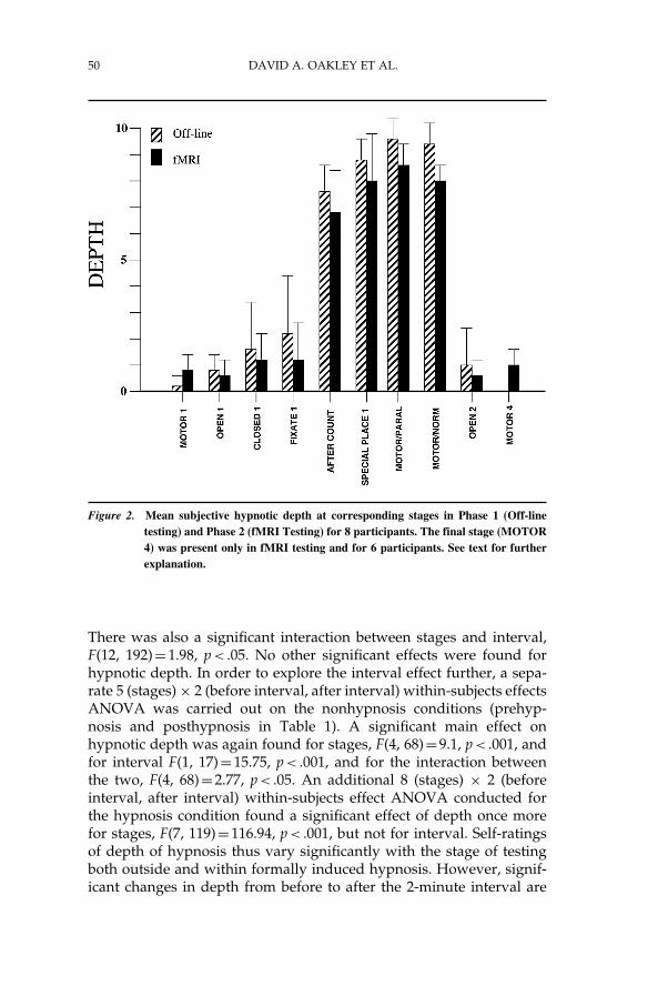
Figure 2. Mean subjective hypnotic depth at corresponding stages in Phase 1 (Off-linetesting) and Phase 2 (fMRI Testing) for 8 participants. The final stage (MOTOR4) was present only in fMRI testing and for 6 participants. See text for furtherexplanation.
There was also a significant interaction between stages and interval,F(12, 192)=1.98, p< .05. No other significant effects were found forhypnotic depth. In order to explore the interval effect further, a sepa-rate 5 (stages)! 2 (before interval, after interval) within-subjects effectsANOVA was carried out on the nonhypnosis conditions (prehyp-nosis and posthypnosis in Table 1). A significant main effect onhypnotic depth was again found for stages, F(4, 68)=9.1, p< .001, andfor interval F(1, 17)=15.75, p< .001, and for the interaction betweenthe two, F(4, 68)=2.77, p< .05. An additional 8 (stages) ! 2 (beforeinterval, after interval) within-subjects effect ANOVA conducted forthe hypnosis condition found a significant effect of depth once morefor stages, F(7, 119)=116.94, p< .001, but not for interval. Self-ratingsof depth of hypnosis thus vary significantly with the stage of testingboth outside and within formally induced hypnosis. However, signif-icant changes in depth from before to after the 2-minute interval are

HYPNOTIC DEPTH AND fMRI 51
restricted to the nonhypnosis conditions. Table 1 illustrates that withthe exception of OPEN 2 mean hypnotic depth increased across theinterval in the nonhypnosis conditions.
Subsequent analysis of the depth ratings was carried out using themean values for each of the 2-minute intervals. These means (plusSDs) are shown graphically in Figure 1. Related-means t tests wereused to evaluate depth changes over the various stages of the session.Compared to the initial eyes-open (OPEN 1) stage, there is a signif-icant increase in depth during eye-fixation (FIXATE 1), t(17)="4.57,p< .001. The numerically larger increase in depth following theeye-closure and counting induction (AFTER COUNT) is signifi-cant compared to both the initial eyes-closed stage (CLOSED 1),t(17)="14.43, p< .001, and depth during fixation (FIXATE 1),t(17)="10.85, p< .001. The introduction of the special-place experi-ence (SPECIAL PLACE 1) produces a further significant, albeit numer-ically smaller, increase in depth when compared to the eye-closureand counting procedure (AFTER COUNT), t(17)="6.23, p< .001.The introduction of limb paralysis (MOTOR/PARAL) also results ina further small but significant increase in depth compared to thepreceding special place (SPECIAL PLACE 1), t(17)="3.34, p< .01.Conversely, there is a numerically large and significant drop indepth scores at the end of the hypnosis condition from the intervalsampled just before the reversal of eye-closure and counting proce-dure (BEFORE COUNT) to the second fixation stage (FIXATE 2),t(17)=10.83, p< .001, to produce depth levels at FIXATE 2 that arenot significantly different from the first fixation stage (FIXATE 1).Consistent with this lack of persistence of hypnotic depth following thehypnosis reversal procedure, the depth at the posthypnosis eyes-closedstage (CLOSED 2) is not significantly different from the comparableprehypnosis eyes-closed stage (CLOSED 1).
During the hypnosis condition when the left limb was paralyzed(MOTOR/PARAL), related-means t tests revealed a significant differ-ence in the mean strength of the sense of involuntariness reported byparticipants when attempting tomove their left or right limbs, left limb,89.07, SD=15.09; right limb, 5.87, SD=9.51; t(14),=17.87, p< .001, andin the difficulty of movement, left limb, 87.50, SD=19.79; right limb,16.31, SD=20.19; t(15)=9.652, p< .001. No significant differences werefound due to whether the upper or lower limbs (arm vs. leg) were usedfor motor-paralysis testing. Also, there was no significant difference inmean strength of imagery reported between the first (SPECIAL PLACE1, 76.19, SD=20.35) and the second time that special-place imagerywas introduced (SPECIAL PLACE 2, 83.56, SD=15.91). Two partic-ipants reported spontaneously experiencing special-place imagery inhypnosis before SPECIAL PLACE 1 (mean strength, 49.0; range, 39–59)and 7 reported the persistence of special-place imagery after SPECIAL

52 DAVID A. OAKLEY ET AL.
PLACE 2 (mean strength, 52.3; range, 29–93), but none reportedspecial-place imagery during the MOTOR/PARAL stage of testing.Imagery other than special place was reported by 1 participant beforeSPECIAL PLACE 1 (image of “descending through water,” strength,71) and another participant reported other imagery (“like watchingcrystals form,” strength, 59) after SPECIAL PLACE 2. Two partici-pants reported other imagery during MOTOR/PARAL—one of “visu-alizing left paralyzed leg” (strength, 64) and the other the “image of acomputer joystick in my left hand” (strength, 70).
fMRI-Environment TestingMean hypnotic depth ratings were taken immediately before and
after the MRI acquisition blocks for the 8 participants in each of thefirst 8 stages of this phase of the study that were shared with theoff-line study (see Table 2). In the following analysis, stage refers tothe stages of the overall procedure, and block refers to the MRI acqui-sition blocks. In order to explore changes in subjective hypnotic depthwithin MRI acquisition blocks and across stages separately for theprehypnosis and the hypnosis conditions, two ANOVA designs wereused. A 3 (stages) ! 2 (before block, after block) ANOVA carried outon the hypnotic depth measurements for the prehypnosis conditionshowed no significant effect for either stages or blocks. A similar5 (stages) ! 2 (before block, after block) ANOVA for the hypnosiscondition, however, found a significant depth of hypnosis effect forstages, F(4, 24)=58.95, p< .001, but again not for blocks. As in theoff-line testing phase, therefore, depth of hypnosis varied significantlyover stages within the hypnosis condition but remained stable duringthe blocks of MR acquisition. The significant changes in depth acrossstages and within the 2-minute (mock MRI) intervals seen in the off-line testing phase outside hypnosis, however, are no longer present inthis second phase of testing.
Mean hypnotic depth scores for the 8 participants who progressedto the second phase of the study are shown for the 9 stages commonto both phases (off-line testing and fMRI-environment testing) of thestudy in Figure 2 (for comparison with off-line testing, the fMRI-environment testing data forMOTORNORMAL andMOTOR PARAL-YSIS are shown in Figure 2 and Table 2 in reverse of the actualorder of testing). A 9 (stages) ! 2 (phases) within-subjects ANOVAfound a significant overall effect for stages, F(8, 40)=294.39, p< .001.Although inspection of the data in Figure 2 suggests slightly lowerdepth measurements during fMRI-environment testing for all stagesexcept the initial motor test (MOTOR), there was no significant effectfor experimental phase, that is, when comparing off-line and fMRIenvironment testing results across stages.

HYPNOTIC DEPTH AND fMRI 53
The data for this second phase of the study allow a comparison ofmean subjective hypnotic depth before and after hypnosis as a meansof investigating the possibility of carry-over of hypnotic effects fromthe hypnosis condition to the posthypnosis condition. Related-meanst tests comparing mean hypnotic depths at OPEN1 versus OPEN2 andat MOTOR 1 versus MOTOR 4 revealed no significant difference inthe before and after measures indicating that there is no carryover ofsubjective hypnotic depth from the hypnosis condition to the posthyp-nosis condition.
Related-means t tests were used to explore the strength of thereported experience of involuntariness and difficulty in moving theright or left joystick during paralysis of the left arm (MOTOR/PARAL)for these 8 participants in both phases of the study (off-line testingand fMRI-environment testing). There were no significant differencescomparing off-line with fMRI-environment testing on either involun-tariness or difficulty measures. The differences between the left andright limb were, however, significant for both phases of the study forinvoluntariness (off-line testing: left limb, 94.88, SD=11.32; right limb,6.43, SD=13.58; t[6]=13.82, p< .001. fMRI-environment testing: leftlimb, 92.75, SD=10.65; right limb, 1.00, SD=1.00; t[7]=23.8, p< .001)and for difficulty (off-line testing: left limb, 97.50, SD=3.25; right limb,10.43; SD=12.69; t[6]=17.04, p< .001; fMRI-environment testing: leftlimb, 96.38, SD=4.57; right limb, 0.72, SD=0.95; t[7]=57.83, p< .001).The mean strength of imagery in these same participants during thespecial-place stage (SPECIAL PLACE 1) for off-line testing, 87.00,SD=6.73, was not significantly different from that for fMRI testing,81.89, SD=17.71. In the fMRI-environment testing phase, none of the 8participants reported spontaneous special-place imagery either beforethe first special-place stage (SPECIAL PLACE 1) or during motortesting while the left hand/arm was paralyzed (MOTOR/PARAL).Three participants reported nonspecial-place imagery before theSPECIAL PLACE 1 stage of testing (“walking down a staircase”(visual), strength 100; “random visual imagery, e.g., faces,” strength,81; “descending stairs,” strength, 13), but none reported other imageryduring MOTOR/PARAL.
Discussion
The present study examined responsiveness to suggestion andsubjective depth of hypnotic experience over time in an experimentalprotocol designed to allow hypnotic phenomena to be investigatedboth outside the fMRI-scanning environment (off-line testing) andduring the fMRI-scanning procedure itself (fMRI-environment testing).The main aim was to formally establish whether a typical fMRI-scanning environment produces any potentially adverse effects on

54 DAVID A. OAKLEY ET AL.
both subjective hypnotic depth and the participants’ ability to respondto specific suggestions. A number of features of the fMRI environmentmight be expected to have a negative impact on both of these. Themost notable are the noise and the claustrophobic environment of thescanner itself, but other possibly adverse factors include the need todeliver instructions and suggestions to the participant remotely viaearphones, the longer time taken to complete the tests, and the distrac-tion caused by other events related to scanning protocols, such asVISAUDs, localizer scans, and structural scans. The results obtainedin the two phases of the study, however, confirm that none of thesefeatures had any significant effect on either hypnotic depth or respon-siveness to suggestions.
The sequence and magnitude of the changes in subjective hypnoticdepth across the various stages of the procedure both within andoutside hypnosis were the same in the fMRI environment as they hadbeen during off-line testing in a normal experimental setting. Impor-tantly, once the hypnotic procedures had been initiated, subjectivedepth remained stable within both the mock-MRI intervals of the off-line-testing phase and the actual MRI-acquisition blocks in the fMRIenvironment. The latter is particularly notable, because some of theseMRI blocks (those used for motor testing) were 7 minutes and 42seconds long compared to the standard 2-minute mock-MRI intervalsused in the off-line phase of the study. There was no evidence fromeither phase of the study of any carryover of subjective hypnotic deptheffects from the hypnosis to the posthypnosis condition.
Participants reported experiencing the suggested limb paralysiswith the same degree of involuntariness and difficulty in initiatingmovement in both phases of the study, and there was no apparenteffect of the fMRI environment on strength or persistence of suggestedspecial-place imagery. It was also a consistent observation from bothphases of the study that the introduction of special-place imagery andof testing for hypnotic paralysis were accompanied by small but signif-icant increases in hypnotic depth. The level of spontaneous imagerywas low in both phases of the study, and none of the participantsreported imagery of either the special place or of any other type duringmotor testing in hypnosis. Nevertheless, the occurrence of unintendedimagery, particularly the persistence of special-place imagery if it isused, should be monitored in imaging studies as its presence willaffect the pattern of activity associated with intended experimentalconditions.
One observation that emerged from both phases of this studythat has practical significance for both clinical and experimentalapplications is that the most significant effect on hypnotic depthwas contributed by the combination of eye closure, relaxation, andcounting procedures used as the second step of our standardized

HYPNOTIC DEPTH AND fMRI 55
hypnotic-induction procedure. Hence, where time is a considerationand the achievement of hypnotic depth is the major aim, proceduresequivalent to induction Step 1 (eye fixation) and induction Step 3 (inour study represented by the special place) could be omitted. Thefirst step could perhaps be replaced with a simple eye-closure requestor instruction. It remains possible, however, that this step representsan important part of the hypnotic process, such as the engagement offrontal attentional networks (Gruzelier, 1998), that is not reflected inthe magnitude of subjective hypnotic depth change associated with it.Equally, there may be good reasons, especially in clinical settings, toinstitute the special-place procedure, other than for increasing hypnoticdepth. Kalisch et al. (2005), for example, have recently shown in anfMRI experiment that the special-place procedure used in the presentstudy is effective in attenuating the subjective and physiologicalcorrelates of anticipatory anxiety and reduces responsiveness to painstimuli.
Overall, these data provide a clear picture of the changes in subjec-tive hypnotic depth associated with a standardized three-step hypnosisinduction procedure, such as that described by Gruzelier (1998), anddemonstrate that this pattern of changes can be maintained in the fMRIenvironment without reduction in depth of hypnosis. In addition, ourstudy provides the first direct evidence that an fMRI environmentand the temporal constraints it imposes do not seem to impair theparticipants’ ability to experience the effects of hypnotic suggestion,though this observation needs to be replicated and extended beyondlimb paralysis and special-place imagery. This outcome supports theview that hypnosis and suggestion within hypnosis are reliable toolsfor use in fMRI neuroimaging studies.
References
Blakemore, S. J., Oakley, D. A., & Frith, C. D. (2003). Delusions of alien control in thenormal brain. Neuropsychologia, 41, 1058–1067.
Derbyshire, S. W. G., Whalley, M. G., Stenger, V. A., & Oakley, D. A. (2004). Cere-bral activation during hypnotically induced and imagined pain. NeuroImage, 23,392–401.
Egner, T., Jamieson, G., & Gruzelier, J. (2005). Hypnosis decouples cognitive controlfrom conflict monitoring processes of the frontal lobe. NeuroImage, 27, 969–978.
Ersland, L., Rosén, G., Lundervold, A., Smievoll, A. I., Tillung, T., Sundberg, H., &Hugdahl, K. (1996). Phantom limb imaginary fingertapping causes primary motorcortex activation: An fMRI study. NeuroReport, 8, 207–210.
Gruzelier, J. H. (1998). A working model of the neurophysiology of hypnosis: A reviewof the evidence. Contemporary Hypnosis, 15, 3–21.
Halligan, P. W., Athwal, B. S., Oakley, D. A., & Frackowiak, R. S. J. (2000). Imaginghypnotic paralysis. Implications for conversion hysteria. Lancet, 355, 986–987.
Heap, M., & Aravind, K. K. (2002). Hartland’s medical and dental hypnosis: 4th Edition.Edinburgh, UK: Churchill Livingstone.

56 DAVID A. OAKLEY ET AL.
Hofbauer, R. K., Rainville, P., Duncan, G. H., & Bushnell, M. C. (2001). Cortical repre-sentation of the sensory dimension of pain. Journal of Neurophysiology, 86, 402–411.
Kalisch, R., Wiech, K., Crtichley, H. D., Seymour, B., O’Doherty, J. P., Oakley, D. A.,Allen, P., & Dolan, R. J. (2005). Anxiety reduction through detachment: Subjectivephysiological and neural effects. Journal of Cognitive Neuroscience, 17, 1–11.
Kihlstrom, J. F. (2003). The fox, the hedgehog, and hypnosis. International Journal ofClinical and Experimental Hypnosis, 51, 166–189.
Kosslyn, S. M., Thompson,W. L., Costantini-Ferrando, M. F., Alpert., N. M., & Spiegel, D.(2000). Hypnotic visual illusion alters color processing in the brain. American Journalof Psychiatry, 157, 1279–1284.
Le Cron, L. M. (1953). A method of measuring the depth of hypnosis. Journal of Clinicaland Experimental Hypnosis, 1, 4–7.
Maquet, P., Faymonville, M. E., Degueldre, C., Delfiore, G., Franck. G., Luxen, A., &Lamy, M. (1999). Functional neuroanatomy of hypnotic state. Biological Psychiatry,45, 327–333.
McConkey, K. M., Wende, V., & Barnier, A. J. (1999). Measuring change in the subjectiveexperience of hypnosis. International Journal of Clinical and Experimental Hypnosis, 47,23–30.
Oakley, D. A. (2006). Hypnosis as a tool in research: Experimental psychopathology.Contemporary Hypnosis, 23, 3–14.
Raij, T. T., Numminen, J., Narvarnen, S., Hiltunen, J., & Hari, R. (2005). Brain correlatesof subjective reality of physically and psychologically induced pain. Proceedings ofthe National Academy of Sciences of the United States of America, 102, 2147–2151.
Rainville, P., Duncan, G. H., Price, D. D., Carrier B., & Bushnell, M. C. (1997). Painaffect encoded in human anterior cingulate but not somatosensory cortex. Science,277, 968–971.
Rainville, P., Hofbauer, R. K., Bushnell, M. C., Duncan, G. H., & Price, D. D. (2002).Hypnosis modulates activity in brain structures involved in the regulation ofconsciousness. Journal of Cognitive Neuroscience, 14, 887–901.
Rainville, P., Hofbauer, R. K., Paus, T., Duncan, G. H., Bushnell, M. C., & Price, D. D.(1999). Cerbral mechanisms of hypnotic induction and suggestion. Journal of CognitiveNeuroscience, 11, 110–125.
Rainville, P., & Price, D. D. (2003). Hypnosis phenomenology and the neurobiologyof consciousness. International Journal of Clinical and Experimental Hypnosis, 51,105–129.
Ray, W. J., & Oathes, D. (2003). Brain-imaging techniques. International Journal of Clinicaland Experimental Hypnosis, 51, 97–104.
Rosén, G., Willoch, F., Bartenstein, P., Berner, N., & Røsjø, S. (2001). Neurophysiologicalprocesses underlying the phantom limb pain experience and the use of hypnosisin its clinical management: An intensive examination of two patients. InternationalJournal of Clinical and Experimental Hypnosis, 49, 38–55.
Schultz-Stubner, S., Krings, T., Meister, I. G., Rex, S., Thron, A., & Rossaint, R. (2004).Clinical hypnosis modulates functional magnetic resonance imaging signal intensitiesand pain perception in a thermal stimulation paradigm. Regional Anaesthesia and PainMedicine, 29, 594–556.
Shor, R. E., & Orne, E. C. (1962). Harvard Group Scale of Hypnotic Susceptibility, Form A.Palo Alto, CA: Consulting Psychologists Press.
Spiegel, D. (2003). Negative and positive visual hypnotic hallucinations: Attendinginside and out. International Journal of Clinical and Experimental Hypnosis, 51,130–1146.
Szechtman, H., Woody, E., Bowers, K. S., & Nahmias, C. (1998). Where the imaginalappears real: A positron emission tomography study of auditory hallucinations.Proceedings of the National Academy of Sciences, 95, 1956–1960.

HYPNOTIC DEPTH AND fMRI 57
Tart, C. T. (1970). Self-report scales of hypnotic depth. International Journal of Clinical andExperimental Hypnosis, 18, 105–125.
Ward, N. S., Oakley, D. A., Frackowiak, R. S. J., & Halligan, P. W. (2003). Differen-tial brain activations during intentionally simulated and subjectively experiencedparalysis. Cognitive Neuropsychiatry, 8, 295–313.
Willoch, F., Rosén, G., Tolle, T. R., Oye, I., Wester, H. J., Berner, N., Schwaiger, M.,& Bartenstein, P. (2000). Phantom limb in the human brain: Unraveling neuralcircuitries of phantom limb sensations using Positron Emission Tomography. Annalsof Neurology, 48. 842–849.
Woody, E. Z., & McConkey, K. M. (2003). What we don’t know about the brain andhypnosis, but need to: A view from the Buckhorn Inn. International Journal of Clinicaland Experimental Hypnosis, 51, 309–338.
Woody, E. Z., & Szechtman, H. (2003). How can brain activity and hypnosis inform eachother? International Journal of Clinical and Experimental Hypnosis, 51, 232–255.
Hypnosetiefe und Reaktion auf Suggestion unter standardisiertenBedingungen und während fMRT-Messungen
David A. Oakley, Quinton Deeley und Peter W. HalliganZusammenfassung: Hypnose ist ein möglicherweise sehr wertvolles kogni-tives Instrument für bildgebende Studien. Allerdings existiert Besorgnisdarüber, dass insbesondere bei Magnetresonanztomographie-Messungenhypnotische Vorgänge beeinträchtigt werden könnten. Messungen derhypnotischen Tiefe und Ansprechbarkeit auf Suggestionen wurden imRahmen einer standardisierten Prozedur erhoben, welche allen Vorgabender funktionellen Magnetresonanztomographie-Messungen gerecht wurde.Tests außerhalb des Scanners wiesen zuverlässige und stabile Verän-derungen der subjektiven Hypnosetiefe auf. Es gab keine Folgeeffektenachdem die Hypnose beendet worden war. Vergleiche innerhalb einzelnerVersuchspersonen zeigten, dass Größe und Art dieser Veränderungen sowiedas Ausmaßder Ansprechbarkeit auf hypnotische Suggestionen von derfMRT-Umgebung nicht erkennbar beeinträchtigt wurden. Daraus wirdgeschlossen, dass Hypnose als eigenständiges und zuverlässiges kogni-tives Instrument im Rahmen von fMRT -Bildgebungsstudien eingesetztwerden kann.
Ralf SchmaelzleUniversity of Konstanz, Konstanz, Germany
La profondeur de l’état hypnotique et la réponse à des suggestions dansdes conditions standardisées et durant un examen d’imagerie par
résonance magnétique fonctionnelle (IRMF)
David A. Oakley, Quinton Deeley et Peter W. HalliganRésumé: L’hypnose constitue un outil cognitif potentiellement précieuxpour la recherche en neuro-imagerie. On continue pourtant de s’inquiéterà juste titre du fait que l’examen par IRM (imagerie par résonance magné-tique), en particulier, puisse avoir une incidence négative sur la procé-dure hypnotique. Des mesures ont été prises de la profondeur de l’étathypnotique et de la réceptivité aux suggestions, à l’aide d’une procédure

58 DAVID A. OAKLEY ET AL.
standardisée satisfaisant à toutes les exigences de l’imagerie par résonancemagnétique fonctionnelle (IRMF). Les essais mené hors scintigraphie ontmontré des changements fiables et stables dans la profondeur subjectivede l’état hypnotique, sans effet résiduel une fois l’hypnose terminée. Lescomparaisons chez un même sujet ont démontré que l’ampleur et le modèlede ces changements et le degré de réceptivité à la suggestion hypnotiquen’étaient pas affectés de façon notable par l’IRMF. On a pu conclure qu’enneuro-imagerie, l’hypnose peut être employée comme outil cognitif distinctet fiable.
Johanne ReynaultC. Tr. (STIBC)
La profundidad hipnótica y la respuesta a las sugestiones durantecondiciones estandarizadas y durante el fMRI
David A. Oakley, Quinton Deeley, y Peter W. HalliganResumen: La hipnosis es una herramienta cognitive potencialmente valiosapara los estudios de imagen cerebral. Sin embargo, existe una preocupacióncomprensible de que la Imagen de Resonancia Magnética (MRI) en partic-ular pueda afectar adversamente los procedimientos hipnóticos. Tomamosmedidas de profundidad hipnótica y responsividad a las sugestiones usandoun procedimiento estandarizado que cumple todos los requisitos para elMRI funcional (fMRI). La prueba afuera del ambiente de muestreo mostrócambios estables y confiables en la profundidad hipnótica subjetiva, sinningún efecto ulterior ya que la hipnosis había terminado. Las compara-ciones intra-sujetos mostraron que la magnitud y patrón de estos cambios yel grado de respuesta a las sugestiones hipnóticas no fueron sensiblementeafectadas por el ambiente del fMRI. Se concluye que se puede emplear a lahipnosis como una herramienta cognitiva específica y confiable dentro delambiente de muestreo del fMRI.
Etzel CardeñaUniversity of Lund, Lund, Sweden