Hypertonic Saline Solution Drives Neutrophil from Bystander Organ to Infectious Site in...
9
Hypertonic Saline Solution Drives Neutrophil from Bystander Organ to Infectious Site in Polymicrobial Sepsis: A Cecal Ligation and Puncture Model Mariana Cardillo Theobaldo, Flavia Llimona, Ricardo Costa Petroni, Ester Correia Sarmento Rios, Irineu Tadeu Velasco, Francisco Garcia Soriano* Emergency of Medicine Division-Faculdade de Medicina da Universidade de Sa ˜o Paulo, Sa ˜o Paulo, Brazil Abstract The effects of hypertonic saline solution (HSS) have been shown in several animal models of ischemia and shock. Literature has shown potential benefits of HSS modulating inflammatory response after sepsis in an animal model. We studied the HSS effects in sepsis through cecal ligation and puncture (CLP) in Balb-C mice. Groups studied: 1- CLP without treatment (CLP-C); 2- CLP treated with normal saline solution NaCl 0.9% – 34 ml/Kg (CLP-S); 3- CLP treated with HSS NaCl 7.5% – 4 ml/Kg (CLP- H); and 4- group (Basal) without no CLP or treatment. Volume infusion was always applied 30 min after CLP. Lung and peritoneal lavage were harvested after 6h and 24h of CLP to analyze cytokines amount, oxide nitric, lipid peroxidation and neutrophil infiltration. Neutrophil infiltration, ICAM-1, CXCR-2, and CXCL-1 in lung were reduced by HSS (CLP-H) compared to CLP-C or CLP-S. Neutrophil in peritoneal lavage was increased in 24h with HSS (CLP-H) compared to CLP and CLP-S. Peritoneal CXCR-2 was increased in CLP-C and CLP-S but presented a lower increase with HSS (CLP-H) after 6 hours. GRK-2 presented difference among the groups at 24 h, showing a profile similar to neutrophil infiltration. Pro-inflammatory cytokines (TNF-a and IL-6) were reduced by HSS treatment; CLP-S increased TNF-a. IL-10 was increased in lung tissue by the HSS treatment. The oxidative stress (TBARS and nitric oxide biochemistry markers) was reduced with HSS. Animal survival was 33.3% in CLP-C group, 46.6% in CLP-S group and 60% in the CLP-H group after the sixth day. The HSS protects the animal against sepsis. Our results suggest that the volume replacement modulate pro and anti-inflammatory mediators of an inflammatory response, but HSS presented a more effective and potent effect. Citation: Theobaldo MC, Llimona F, Petroni RC, Rios ECS, Velasco IT, et al. (2013) Hypertonic Saline Solution Drives Neutrophil from Bystander Organ to Infectious Site in Polymicrobial Sepsis: A Cecal Ligation and Puncture Model. PLoS ONE 8(9): e74369. doi:10.1371/journal.pone.0074369 Editor: Jorge I. F. Salluh, D’or Institute of Research and Education, Brazil Received November 7, 2012; Accepted August 4, 2013; Published September 17, 2013 Copyright: ß 2013 Theobaldo et al. This is an open-access article distributed under the terms of the Creative Commons Attribution License, which permits unrestricted use, distribution, and reproduction in any medium, provided the original author and source are credited. Funding: This study was supported by FAPESP grant 2009/15530-0. The funders had no role in study design, data collection and analysis, decision to publish, or preparation of the manuscript. Competing Interests: The authors have declared that no competing interests exist. * E-mail: [email protected] Introduction Sepsis and septic shock are characterized by an acute systemic immune response to a variety of bacterial infections. The host response could be triggered by bacteria, viruses and fungi [1]; most cases are Gram-negative bacteria (60%) and the remainder of Gram-positive [2,3]. Host receptors recognize distinct bacterial components and initiate the signaling for the inflammatory response [4–7]. Septic shock is the most severe response of systemic infection and is a major cause of morbidity and mortality in non-cardiac intensive care units (ICUs) around the world [8,9]. Approximately 25% to 35% of all septic episodes end in death [10], with high mortalities rates in both underdeveloped and developing countries [8]. In the United States approximately 750,000 patients are treated for severe sepsis yearly with a mortality rate of 30–50% and an estimated $17 billion in health care costs [9]. In Brazil, several studies already showed high mortality rates [8]. Patients suffering from septic peritonitis experience a higher mortality rate (60% to 80%) [11]. Despite advances in diagnosis, antibiotic therapy and supportive care, mortality has remained high and disproportionately affects the chronically ill and the aged [9].The exaggerated proinflammatory response during sepsis may result in many of the injurious and sometimes fatal physiological symptoms of the disease. Strategies targeting a single mediator have failed as an effective treatment in sepsis. Treatments able to modulate the amplifier aspects of inflammation will be more efficient [12–14]. In spite of increased knowledge on septic mechanism up to now the therapeutic used has not added improvement in survival patients [15]. The therapy consists basically in source control and hemody- namic support with volume expansion. Recently the positive volume balance has been associated to pulmonary and abdominal complications [16]. The pioneering study by Velasco et. al. (1980) showed that small amounts of 7.5% saline solution restored vital parameters and decreased mortality in dogs submitted to severe hemorrhagic shock [17]. Since the 1980’s, the Hypertonic Saline Solution (HSS) has been extensively studied, at a dose of 4 ml/kg into a peripheral vein [17–20]. Recent clinical trial showed a benefit of HSS in septic patients [21]. Experimental studies showed beneficial effects of HSS modulating inflammatory response, as for instance the expression and release of cytokines, free radicals, augmenting interleukin-10, and reducing oxidative burst [22]. Hypertonic saline solution PLOS ONE | www.plosone.org 1 September 2013 | Volume 8 | Issue 9 | e74369
Transcript of Hypertonic Saline Solution Drives Neutrophil from Bystander Organ to Infectious Site in...
Hypertonic Saline Solution Drives Neutrophil fromBystander Organ to Infectious Site in PolymicrobialSepsis: A Cecal Ligation and Puncture ModelMariana Cardillo Theobaldo, Flavia Llimona, Ricardo Costa Petroni, Ester Correia Sarmento Rios, Irineu
Tadeu Velasco, Francisco Garcia Soriano*
Emergency of Medicine Division-Faculdade de Medicina da Universidade de Sao Paulo, Sao Paulo, Brazil
Abstract
The effects of hypertonic saline solution (HSS) have been shown in several animal models of ischemia and shock. Literaturehas shown potential benefits of HSS modulating inflammatory response after sepsis in an animal model. We studied the HSSeffects in sepsis through cecal ligation and puncture (CLP) in Balb-C mice. Groups studied: 1- CLP without treatment (CLP-C);2- CLP treated with normal saline solution NaCl 0.9% – 34 ml/Kg (CLP-S); 3- CLP treated with HSS NaCl 7.5% – 4 ml/Kg (CLP-H); and 4- group (Basal) without no CLP or treatment. Volume infusion was always applied 30 min after CLP. Lung andperitoneal lavage were harvested after 6h and 24h of CLP to analyze cytokines amount, oxide nitric, lipid peroxidation andneutrophil infiltration. Neutrophil infiltration, ICAM-1, CXCR-2, and CXCL-1 in lung were reduced by HSS (CLP-H) comparedto CLP-C or CLP-S. Neutrophil in peritoneal lavage was increased in 24h with HSS (CLP-H) compared to CLP and CLP-S.Peritoneal CXCR-2 was increased in CLP-C and CLP-S but presented a lower increase with HSS (CLP-H) after 6 hours. GRK-2presented difference among the groups at 24 h, showing a profile similar to neutrophil infiltration. Pro-inflammatorycytokines (TNF-a and IL-6) were reduced by HSS treatment; CLP-S increased TNF-a. IL-10 was increased in lung tissue by theHSS treatment. The oxidative stress (TBARS and nitric oxide biochemistry markers) was reduced with HSS. Animal survivalwas 33.3% in CLP-C group, 46.6% in CLP-S group and 60% in the CLP-H group after the sixth day. The HSS protects theanimal against sepsis. Our results suggest that the volume replacement modulate pro and anti-inflammatory mediators ofan inflammatory response, but HSS presented a more effective and potent effect.
Citation: Theobaldo MC, Llimona F, Petroni RC, Rios ECS, Velasco IT, et al. (2013) Hypertonic Saline Solution Drives Neutrophil from Bystander Organ to InfectiousSite in Polymicrobial Sepsis: A Cecal Ligation and Puncture Model. PLoS ONE 8(9): e74369. doi:10.1371/journal.pone.0074369
Editor: Jorge I. F. Salluh, D’or Institute of Research and Education, Brazil
Received November 7, 2012; Accepted August 4, 2013; Published September 17, 2013
Copyright: � 2013 Theobaldo et al. This is an open-access article distributed under the terms of the Creative Commons Attribution License, which permitsunrestricted use, distribution, and reproduction in any medium, provided the original author and source are credited.
Funding: This study was supported by FAPESP grant 2009/15530-0. The funders had no role in study design, data collection and analysis, decision to publish, orpreparation of the manuscript.
Competing Interests: The authors have declared that no competing interests exist.
* E-mail: [email protected]
Introduction
Sepsis and septic shock are characterized by an acute systemic
immune response to a variety of bacterial infections. The host
response could be triggered by bacteria, viruses and fungi [1]; most
cases are Gram-negative bacteria (60%) and the remainder of
Gram-positive [2,3]. Host receptors recognize distinct bacterial
components and initiate the signaling for the inflammatory
response [4–7].
Septic shock is the most severe response of systemic infection
and is a major cause of morbidity and mortality in non-cardiac
intensive care units (ICUs) around the world [8,9]. Approximately
25% to 35% of all septic episodes end in death [10], with high
mortalities rates in both underdeveloped and developing countries
[8]. In the United States approximately 750,000 patients are
treated for severe sepsis yearly with a mortality rate of 30–50%
and an estimated $17 billion in health care costs [9]. In Brazil,
several studies already showed high mortality rates [8]. Patients
suffering from septic peritonitis experience a higher mortality rate
(60% to 80%) [11]. Despite advances in diagnosis, antibiotic
therapy and supportive care, mortality has remained high and
disproportionately affects the chronically ill and the aged [9].The
exaggerated proinflammatory response during sepsis may result in
many of the injurious and sometimes fatal physiological symptoms
of the disease. Strategies targeting a single mediator have failed as
an effective treatment in sepsis. Treatments able to modulate the
amplifier aspects of inflammation will be more efficient [12–14]. In
spite of increased knowledge on septic mechanism up to now the
therapeutic used has not added improvement in survival patients
[15].
The therapy consists basically in source control and hemody-
namic support with volume expansion. Recently the positive
volume balance has been associated to pulmonary and abdominal
complications [16]. The pioneering study by Velasco et. al. (1980)
showed that small amounts of 7.5% saline solution restored vital
parameters and decreased mortality in dogs submitted to severe
hemorrhagic shock [17]. Since the 1980’s, the Hypertonic Saline
Solution (HSS) has been extensively studied, at a dose of 4 ml/kg
into a peripheral vein [17–20].
Recent clinical trial showed a benefit of HSS in septic patients
[21]. Experimental studies showed beneficial effects of HSS
modulating inflammatory response, as for instance the expression
and release of cytokines, free radicals, augmenting interleukin-10,
and reducing oxidative burst [22]. Hypertonic saline solution
PLOS ONE | www.plosone.org 1 September 2013 | Volume 8 | Issue 9 | e74369
appears as a way to modulate excessive inflammation, effective
hemodynamic support with no risk for volume overload damages.
The aim of this study is to analyze the role of hypertonic saline
in the inflammatory profile of sepsis and neutrophil migration into
the lung and the peritoneal cavity. We studied in experimental
sepsis, through model of cecal ligation and puncture (CLP), the
hypertonic solution effects on inflammatory response. The model
of CLP in mice is very well established and can address the
objectives of our study about sepsis and the hypertonic solution
treatment avoiding using dogs or rabbits.
Methods
Ethical approvalProcedures were performed in accordance the Guide for the
Care and Use of Laboratory Animals published by the US
National Institutes of Health. The study protocol was approved by
the Research Ethics Committee of the USP School of Medicine
(Comissao de Etica para Analise de Projetos de Pesquisa do
HCFMUSP - http://www.hcnet.usp.br/adm/dc/cappesq/) (#0333/08). For experimental procedures, animals were anesthe-
tized with pentobarbital 20 mg/mL.
ProceduresSepsis induction – Cecal ligation and punction, and
treatments. A total of 297 male BALB/c mice, 8 wk old,
25 g of mean weight, subjected to CLP, were used in this study, as
described previously [23]. The animals were provided from our
School Facility, they are specific pathogen free (SPF) in climatized
facility, kept in automatic dark/side cycle (http://www.biot.fm.
usp.br/). Under aseptic conditions, a 2 cm midline laparotomy
was performed to allow exposure of the cecum with adjoining
intestine. The cecum was tightly ligated with a 3.0 silk suture at its
base, below the ileocecal valve, and was perforated twice with a
22-gauge needle (top and bottom). The cecum was gently
squeezed to extrude a small amount of feces from the perforation
sites. The cecum was then returned to the peritoneal cavity and
the laparotomy was closed with 4.0 silk sutures. In addition, n =
24 animals were used for control purposes. All animals were then
returned to their cages with free access to food and water.
The animals were divided into four groups: the group without
treatment cecal ligation and puncture (CLP-C); septic treated with
hypertonic saline solution 7.5% (4 ml/kg) (CLP-H); the third
group treated with normal saline solution 0.9% (34 ml/kg) (CLP-
S); and the sham group (Basal) to indicate the basal values without
both CLP and treatment. The groups CLP-H and CLP-S were
treated 30 min. after CLP. The volume (saline and hypertonic
saline solution) was infused in the tail vein of the animals [24]. The
animals were anesthetized and sacrificed to collect samples of
peritoneum lavage and lung after 6h and 24h of CLP. The samples
were collected by a blinded collaborator, the tissues were
numbered, and the code kept with the collaborator up to the
end of measurements. We used the lower number of animals
necessary to each experiment, for biochemical assays we used
between 6–8 animals.
Tissue Preparation. Frozen tissue (100 mg) was pulverized
in liquid nitrogen. Samples were homogenized in NP40 buffer
containing 135 mM NaCl, 20 mM Tris (pH 8,0), 10% glycerol,
and proteolytic enzyme inhibitors (40 ug/mL phenylmethylsufo-
nylfluoride 1 mM; Sigma, St, Louis, MO). After debris separation
through centrifugation for 40 min at 10,000 rpm, the supernatants
were collected and protein concentration was determined by the
Bradford method (Bio Rad, Hercules, CA). Samples were stored at
–80uC until assayed.
Measurement of cytokines, chemokines and adhesion
molecules. The concentration of Tumor Necrosis Factor -a,
Interleukin (IL) -10, IL-6, Inter-Cellular Adhesion Molecule 1
(ICAM-1), Vascular cell adhesion protein 1 (VCAM-1) and
neutrophil chemoattractant chemokine (C-X-C) motif ligand 1
(CXCL-1) were measure in lung tissues by enzyme-linked
immunosorbent assay (ELISA) using a DuoSet kit (R&D SystemsH,
Minneapolis, MN, USA) [23].
Chemokine (C-X-C motif) Receptor 2 (CXCR2), G
protein-coupled receptor kinase 2 (GRK2), ICAM and
Chemokine (C-X-Cmotif) ligand 1 (CXCL-1)
expression. Six and twenty four hours after surgery, mice were
sacrificed; samples of lung and cells of peritoneal cavity were
collected. The total RNA was extracted with TRIzol (Invitrogen,
Carlsbad, CA, USA). The amount of total RNA was determined
spectrophotometrically (Nanoview,GE, Pittsburgh, PA, USA) at
260 nm, and RNA integrity was confirmed by electrophoresis on
1% agarose gels and staining with 0.1 mg/L ethidium bromide.
mRNA analysis was performed using a (Life Technologies, Grand
Island, NY, USA) Real-time PCR with a SYBR-green fluores-
cence system. The sequences of the primer pairs were as follows: G
protein-coupled receptor kinase 2 (GRK2) forward 59-ccctctcac-
catctctgagc-39; GRK2 reverse 59-cggttggggaacaagtagaa-39; Che-
mokine (C-X-C) Receptor 2 (CXCR2) forward 59-tctgctacgggtt-
cacactg-39; CXCR2 reverse 59-ggaggaagccaagaatctcc-39; ICAM-1
forward 59-cgaaggtggttcttctgagc-39; ICAM-1 reverse 59-gtctgctga-
gacccctcttg-39; CXCL-1 forward 59-tgttgtgcgaaaagaagtgc-39;
CXCL-1 reverse 59-cgagacgagaccaggagaaa-39. As housekepping
we used b2M. The sequence of the primer pairs of b2M was b2M
forward 59-CATGGCTCGCTCGGTGACC-39; b2M reverse 59-
AATGTGAGGCGGGTGGAACTG-39. To validate and stan-
dardize the method, we used positive and negative known control
samples for all genes, including the housekepping. The method
was procedure as describe previously [25].
Nitrite. Tissue (lung) nitrite levels were measured by means of
the classic Griess method as described previously [26]. We used
50 mL of the sample, that were prepared as describe above in
tissue preparation, to measured nitrite at 540 nm absorbance by
using 50 ml of reaction with Griess reagent (sulfanilamide and
naphthalene–ethylene diamine dihydrochloride). We wait 10 min
at room temperature before read. Amounts of nitrite in the tissue
were estimated by a standard curve.
Thiobarbituric Acid-Reactive (Tbars). Thiobarbituric ac-
id-reactive formation was used to quantify the lipid peroxidation in
tissues and measured as thiobarbituric acid-reactive as described
previously [27]. Tissues were homogenized (100 mg/mL) in
1.15% KCl buffer. A total of 100 mL of the homogenates were
then added to a reaction mixture consisting of 750 mL of 0.8%
thiobarbituric acid, 100 mL of 8.1% sodium dodecyl sulfate (SDS),
750 mL of 20% acetic acid (pH 3.5), and 300 mL of distilled water.
The mixture was then heated at 90uC for 60 min. After cooling at
4uC, the samples were cleared by centrifugation (10,000 g, 10 min)
and their absorbance was measured at 532 nm, using 1,1,3,3-
tetramethoxypropane as an external standard. The level of lipid
peroxides was expressed as mmol malondealdeide/mg of protein.
Peritoneal neutrophils countFor the purpose of cells counting as described previously [28],
28 mice (7 CLP, 7 CLP-H, 7 CLP-S and 7 Basal) were used. After
6 and 24 h, the surviving animals (n = 7/group) were anesthetized
and euthanized then peritoneal lavage was obtained in sterile
physiological saline solution. The total cells were counted using a
Handheld automated cell counter (MilliporeH - Billerica, MA,
USA). The quantification was performed using a cytocentrifuge;
Neutrophil Hypertonic Direct to Infectious Focus
PLOS ONE | www.plosone.org 2 September 2013 | Volume 8 | Issue 9 | e74369
slides were prepared by centrifugation of each sample at 900 g for
6 min (Cytospin 2, Shandon Scientific, Pittsburgh, PA). These
slides were stained by Diff Quick stain, and differential counts of at
least 300 cells were made according to standard morphologic
criteria. The result is expressed in cell x104/peritoneum.
Myeloperoxidase assayFor myeloperoxidase assay we procedure as described previ-
ously [27]. Tissues were homogenized (50 mg/ml) in 0.5%
hexadecyltrimethylammonium bromide in 10 mM 3-N-orpholi-
nopropanesulfonic acid (MOPS) and centrifuged at 15,000 g for
40 min. The suspension was then sonicated three times for 30s. An
aliquot of supernatant was mixed with a solution of 1.6 mM
tetramethylbenzidine and 1 mM hydrogen peroxide. Activity was
measured spectrophotometrically as the change in absorbance at
650 nm at 37uC, using a Spectramax microplate reader. Results
are expressed as milliunits of myeloperoxidase (MPO) activity per
milligram of protein, which were determined with the Bradford
assay.
ImmunochemistryFor immunochemistry we procedure as described previously
[26]. In the lung, paraffin sections (3 mm) were deparaffinized in
xylene and then rehydrated in decreasing concentrations (100%,
95%, and 75%) of ethanol followed by a 10-min incubation in PBS
(pH 7.4). Sections were treated with 0.3% hydrogen peroxide for
15 min to block endogenous peroxidase activity and they were
then rinsed briefly in PBS. After blocking for nonspecific sites,
slides were incubated in a humid chamber overnight at 4uC with
the primary ICAM-1 antibody (Santa cruzH, CA, USA). After
washing with PBS, slides were incubated following extensive
washing (565 min) with PBS, immunoreactivity was detected with
a biotinylated goat anti-rabbit secondary antibody and the avidin-
biotin-peroxidase complex (ABC) both supplied in the Vector Elite
kit (Vector Laboratories, Burlingame, CA). Diaminobenzidine
(DAB) was used as the chromogen, and the slides were
counterstained with hematoxylin.
Survival rateCLP was induced in mice, and 30 min later treated with
hypertonic saline 7.5% or normal saline solution 0.9%, as
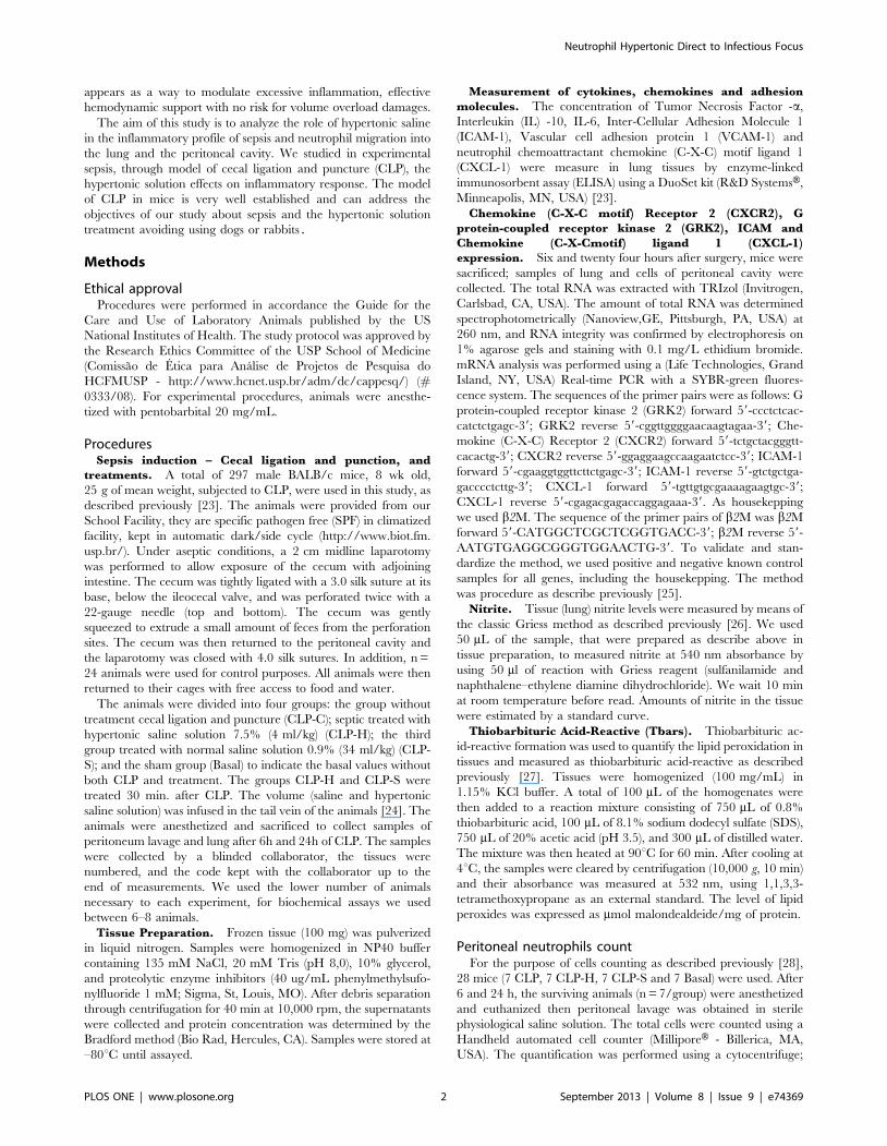
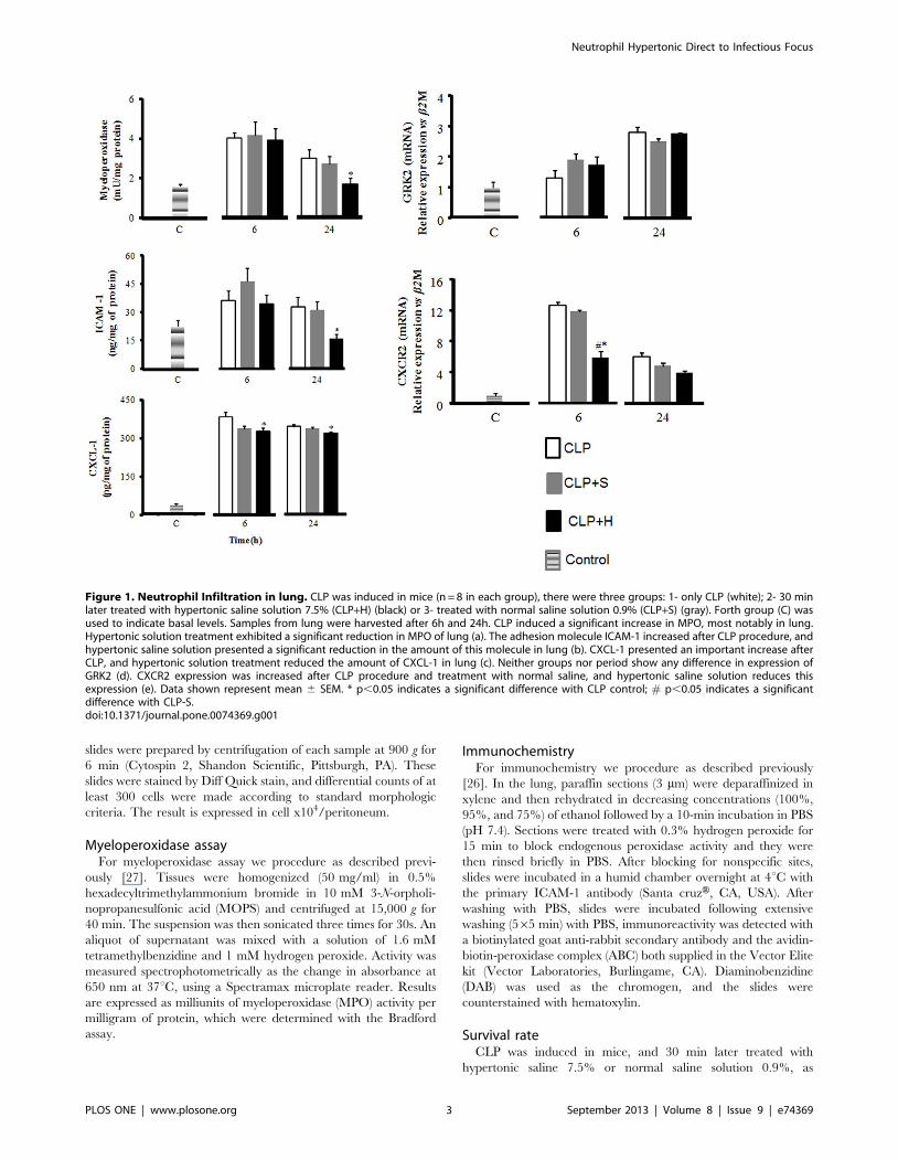
Figure 1. Neutrophil Infiltration in lung. CLP was induced in mice (n = 8 in each group), there were three groups: 1- only CLP (white); 2- 30 minlater treated with hypertonic saline solution 7.5% (CLP+H) (black) or 3- treated with normal saline solution 0.9% (CLP+S) (gray). Forth group (C) wasused to indicate basal levels. Samples from lung were harvested after 6h and 24h. CLP induced a significant increase in MPO, most notably in lung.Hypertonic solution treatment exhibited a significant reduction in MPO of lung (a). The adhesion molecule ICAM-1 increased after CLP procedure, andhypertonic saline solution presented a significant reduction in the amount of this molecule in lung (b). CXCL-1 presented an important increase afterCLP, and hypertonic solution treatment reduced the amount of CXCL-1 in lung (c). Neither groups nor period show any difference in expression ofGRK2 (d). CXCR2 expression was increased after CLP procedure and treatment with normal saline, and hypertonic saline solution reduces thisexpression (e). Data shown represent mean 6 SEM. * p,0.05 indicates a significant difference with CLP control; # p,0.05 indicates a significantdifference with CLP-S.doi:10.1371/journal.pone.0074369.g001
Neutrophil Hypertonic Direct to Infectious Focus
PLOS ONE | www.plosone.org 3 September 2013 | Volume 8 | Issue 9 | e74369
described above. We analyzed the survival rate for 168h (7 days).
Each group (CLP-C, CLP-S and CLP-H) was composed by 30
animals. After recovery from anesthesia, mice were monitored
three times daily. Moribund mice were euthanized by CO2
inhalation and cervical dislocation.
Statistical AnalysisAll values were expressed as mean6standard error of the mean
(SEM). For the biochemical measurements, the means from the
experimental groups were compared by analysis of variance
(ANOVA), and Tukey test was used as post hoc test to compare
individual groups. To compare the survival curves among different
groups of treatment, log rank test was used. These summary
measures all represent ‘time to event measures’ and were analyzed
using Kaplan Meier survival fit analyses and the Logrank test for
statistical significance. This test investigates the null hypothesis
that the Kaplan Meier curves for all groups are identical (i.e. that
the treatment did not change the time taken to reach the event
being analysed). Low P-values are therefore indicative of
differences between curves that did not occur due to chance.
Statistical significance was assigned to p,0.05.
Results
Neutrophil infiltrationWe used myeloperoxidase quantification to measure neutrophil
infiltration in lung. CLP produced an increase in neutrophil
amount in the lung at the period of 6h and 24h post sepsis. Normal
saline treatment did not reduce at any time period the amount of
neutrophil in the tissue. On the other hand, hypertonic was able in
reduce neutrophil infiltration at 24h periods compared to CLP-C
group and normal saline group (Figure 1a).
ICAM e VCAM measurementWe also measured the amount of ICAM-1 (Figure 1b) and
VCAM-1 (data not shown) in lung. CLP induced an increase of
ICAM-1 amount in the lung. Normal saline group presented a
similar profile of ICAM-1 at 6 and 24h periods. Interesting
hypertonic produced a progressive decrease of ICAM-1 amount in
the lung with a significant decrease at 24h (Figure 1b). CLP did
not induce any change in VCAM-1 amount in lung. In addition,
the treatments with normal saline or hypertonic solution infusion
also did not induce any alteration in lung levels of VCAM-1 in all
groups at 6h and 24h periods (data not shown).
CXCL-1The data of CXCL-1 in lung of CLP and the groups treated
with normal saline or hypertonic saline solution are presented in
the Figure 1c. CLP induced an important increase of CXCL-1
amount in the lung matching neutrophil infiltration. CLP and
CLP normal saline groups did not show a significant decrease of
CXCL-1 at 24h periods. Interesting hypertonic produced a
significant decrease of CXCL-1 amount in the lung (Figure 1c).
Chemokine (C-X-C) Receptor 2 (CXCR2) and G protein-coupled receptor kinase 2 (GRK2) expression in lung
We measured the expression of GRK2 and CXCR2 in lung
(Figure 1d and e, respectively). Neither groups and nor period
show any difference in expression of GRK2. CLP and CLP+S
groups induced an increase of CXCR2 expression in relation to
CLP+H that reduces this expression at 6h. Even at 24h we didn’t
found any statistical difference, the group CLP+H has a tendency
to reduce the levels of CXCR2 in comparison to CLP group.
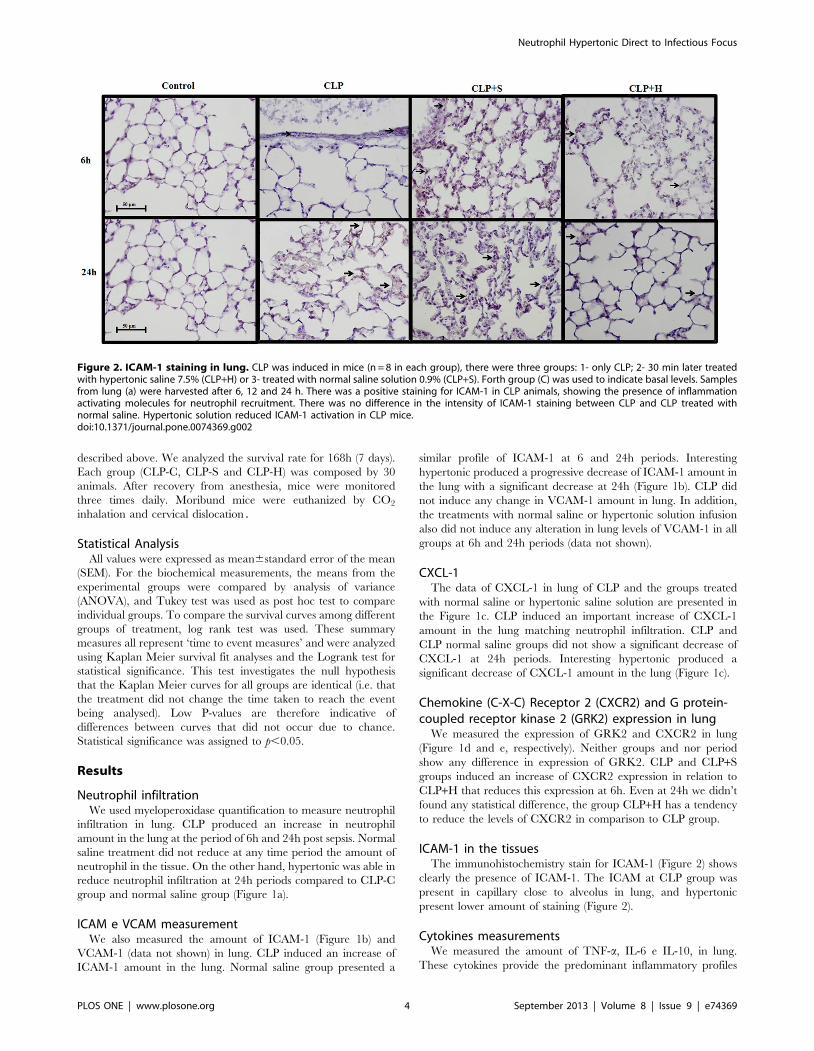
ICAM-1 in the tissuesThe immunohistochemistry stain for ICAM-1 (Figure 2) shows
clearly the presence of ICAM-1. The ICAM at CLP group was
present in capillary close to alveolus in lung, and hypertonic
present lower amount of staining (Figure 2).
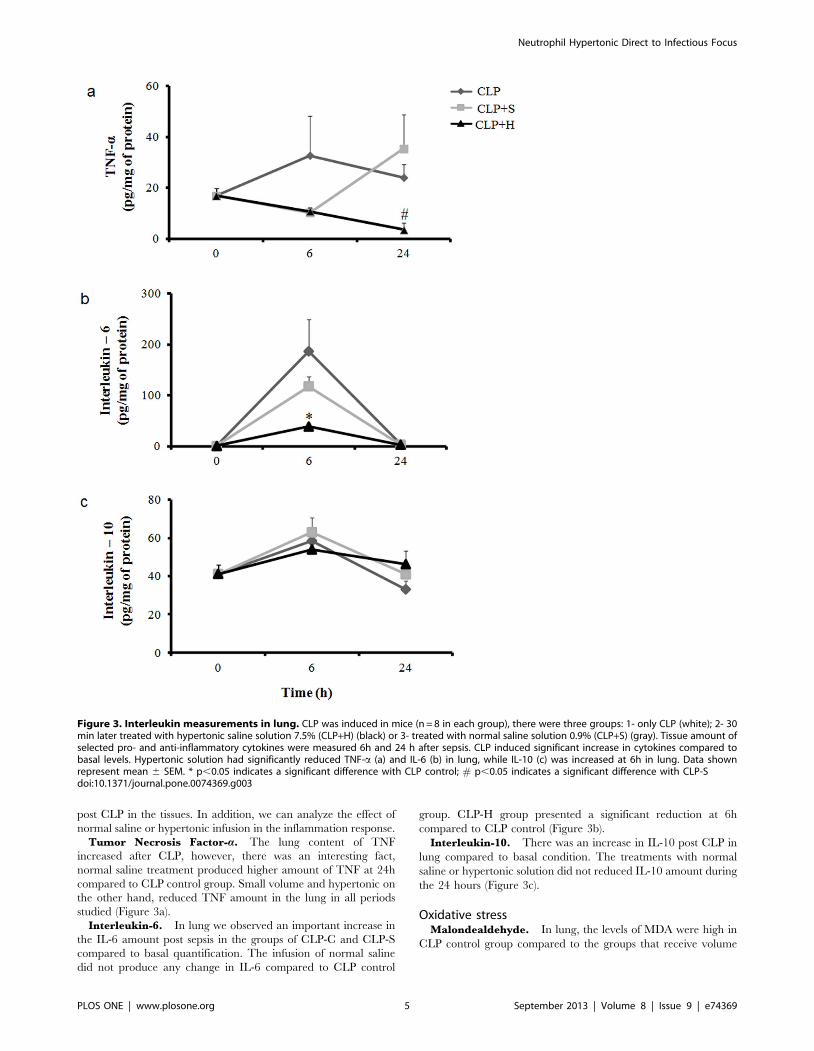
Cytokines measurementsWe measured the amount of TNF-a, IL-6 e IL-10, in lung.
These cytokines provide the predominant inflammatory profiles
Figure 2. ICAM-1 staining in lung. CLP was induced in mice (n = 8 in each group), there were three groups: 1- only CLP; 2- 30 min later treatedwith hypertonic saline 7.5% (CLP+H) or 3- treated with normal saline solution 0.9% (CLP+S). Forth group (C) was used to indicate basal levels. Samplesfrom lung (a) were harvested after 6, 12 and 24 h. There was a positive staining for ICAM-1 in CLP animals, showing the presence of inflammationactivating molecules for neutrophil recruitment. There was no difference in the intensity of ICAM-1 staining between CLP and CLP treated withnormal saline. Hypertonic solution reduced ICAM-1 activation in CLP mice.doi:10.1371/journal.pone.0074369.g002
Neutrophil Hypertonic Direct to Infectious Focus
PLOS ONE | www.plosone.org 4 September 2013 | Volume 8 | Issue 9 | e74369
post CLP in the tissues. In addition, we can analyze the effect of
normal saline or hypertonic infusion in the inflammation response.
Tumor Necrosis Factor-a. The lung content of TNF
increased after CLP, however, there was an interesting fact,
normal saline treatment produced higher amount of TNF at 24h
compared to CLP control group. Small volume and hypertonic on
the other hand, reduced TNF amount in the lung in all periods
studied (Figure 3a).
Interleukin-6. In lung we observed an important increase in
the IL-6 amount post sepsis in the groups of CLP-C and CLP-S
compared to basal quantification. The infusion of normal saline
did not produce any change in IL-6 compared to CLP control
group. CLP-H group presented a significant reduction at 6h
compared to CLP control (Figure 3b).
Interleukin-10. There was an increase in IL-10 post CLP in
lung compared to basal condition. The treatments with normal
saline or hypertonic solution did not reduced IL-10 amount during
the 24 hours (Figure 3c).
Oxidative stressMalondealdehyde. In lung, the levels of MDA were high in
CLP control group compared to the groups that receive volume
Figure 3. Interleukin measurements in lung. CLP was induced in mice (n = 8 in each group), there were three groups: 1- only CLP (white); 2- 30min later treated with hypertonic saline solution 7.5% (CLP+H) (black) or 3- treated with normal saline solution 0.9% (CLP+S) (gray). Tissue amount ofselected pro- and anti-inflammatory cytokines were measured 6h and 24 h after sepsis. CLP induced significant increase in cytokines compared tobasal levels. Hypertonic solution had significantly reduced TNF-a (a) and IL-6 (b) in lung, while IL-10 (c) was increased at 6h in lung. Data shownrepresent mean 6 SEM. * p,0.05 indicates a significant difference with CLP control; # p,0.05 indicates a significant difference with CLP-Sdoi:10.1371/journal.pone.0074369.g003
Neutrophil Hypertonic Direct to Infectious Focus
PLOS ONE | www.plosone.org 5 September 2013 | Volume 8 | Issue 9 | e74369
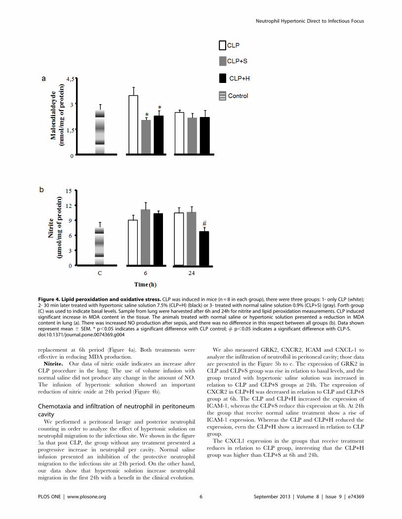
replacement at 6h period (Figure 4a). Both treatments were
effective in reducing MDA production.
Nitrite. Our data of nitric oxide indicates an increase after
CLP procedure in the lung. The use of volume infusion with
normal saline did not produce any change in the amount of NO.
The infusion of hypertonic solution showed an important
reduction of nitric oxide at 24h period (Figure 4b).
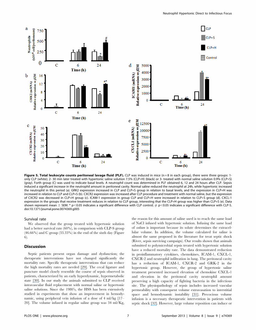
Chemotaxia and infiltration of neutrophil in peritoneumcavity
We performed a peritoneal lavage and posterior neutrophil
counting in order to analyze the effect of hypertonic solution on
neutrophil migration to the infectious site. We shown in the figure
5a that post CLP, the group without any treatment presented a
progressive increase in neutrophil per cavity. Normal saline
infusion presented an inhibition of the protective neutrophil
migration to the infectious site at 24h period. On the other hand,
our data show that hypertonic solution increase neutrophil
migration in the first 24h with a benefit in the clinical evolution.
We also measured GRK2, CXCR2, ICAM and CXCL-1 to
analyze the infiltration of neutrofhil in peritoneal cavity; those data
are presented in the Figure 5b to e. The expression of GRK2 in
CLP and CLP+S group was rise in relation to basal levels, and the
group treated with hypertonic saline solution was increased in
relation to CLP and CLP+S groups at 24h. The expression of
CXCR2 in CLP+H was decreased in relation to CLP and CLP+S
group at 6h. The CLP and CLP+H increased the expression of
ICAM-1, whereas the CLP+S reduce this expression at 6h. At 24h
the group that receive normal saline treatment show a rise of
ICAM-1 expression. Whereas the CLP and CLP+H reduced the
expression, even the CLP+H show a increased in relation to CLP
group.
The CXCL1 expression in the groups that receive treatment
reduces in relation to CLP group, interesting that the CLP+H
group was higher than CLP+S at 6h and 24h.
Figure 4. Lipid peroxidation and oxidative stress. CLP was induced in mice (n = 8 in each group), there were three groups: 1- only CLP (white);2- 30 min later treated with hypertonic saline solution 7.5% (CLP+H) (black) or 3- treated with normal saline solution 0.9% (CLP+S) (gray). Forth group(C) was used to indicate basal levels. Sample from lung were harvested after 6h and 24h for nitrite and lipid peroxidation measurements. CLP inducedsignificant increase in MDA content in the tissue. The animals treated with normal saline or hypertonic solution presented a reduction in MDAcontent in lung (a). There was increased NO production after sepsis, and there was no difference in this respect between all groups (b). Data shownrepresent mean 6 SEM. * p,0.05 indicates a significant difference with CLP control; # p,0.05 indicates a significant difference with CLP-S.doi:10.1371/journal.pone.0074369.g004
Neutrophil Hypertonic Direct to Infectious Focus
PLOS ONE | www.plosone.org 6 September 2013 | Volume 8 | Issue 9 | e74369
Survival rateWe observed that the group treated with hypertonic solution
had a better survival rate (60%), in comparison with CLP-S group
(46.66%) and C group (33.33%) in the end of the sixth day (Figure
6).
Discussion
Septic patients present organ damage and dysfunction; the
therapeutic interventions have not changed significantly the
mortality rate. Specific therapeutic interventions that can reduce
the high mortality rates are needed [29]. The cecal ligature and
puncture model closely resemble the course of sepsis observed in
patients, characterized by an early hyperdynamic, hypermetabolic
state [30]. In our study the animals submitted to CLP received
intravascular fluid replacement with normal saline or hypertonic
saline solutions. Since the 1980’s, the HSS has been extensively
studied in experiments that show an improvement in hemody-
namic, using peripheral vein infusion of a dose of 4 ml/kg [17–
20]. The volume infused in regular saline group was 34 ml/Kg,
the reason for this amount of saline used is to reach the same load
of NaCl infused with hypertonic solution. Infusing the same load
of sodium is important because its solute determines the extracel-
lular volume. In addition, the volume calculated for saline is
almost the same proposed in the literature for treat septic shock
(River, sepsis surviving campaign). Our results shown that animals
submitted to polymicrobial sepsis treated with hypertonic solution
have a reduced mortality rate. The data demonstrated reduction
in proinflammatory cytokines, chemokines, ICAM-1, CXCL-1,
CXCR-2 and neutrophil infiltration in lung. The peritoneal cavity
has a reduction of ICAM-1, CXCR-2 and GRK-2 in the
hypertonic group. However, the group of hypertonic saline
treatment presented increased elevation of chemokine CXCL-1
and elevation in the peritoneal cavity neutrophil amount
preserving a high capacity of fighting bacteria in the infectious
site. The physiopathology of sepsis includes increased vascular
permeability with consequent volume extravasation to interstitial
space and hemodynamic instability [31]. Precocious volume
infusion is a necessary therapeutic intervention in patients with
septic shock [32]. However, large volume reposition can induce or
Figure 5. Total leukocyte counts peritoneal lavage fluid (PLF). CLP was induced in mice (n = 8 in each group), there were three groups: 1-only CLP (white); 2- 30 min later treated with hypertonic saline solution 7.5% (CLP+H) (black) or 3- treated with normal saline solution 0.9% (CLP+S)(gray). Forth group (C) was used to indicate basal levels. A neutrophil count was determined in PLF obtained 6, 12 and 24 hours after CLP. Sepsisinduced a significant increase in the neutrophil amount in peritoneal cavity. Normal saline reduced the neutrophil at 24h, while hypertonic increasedthe neutrophil in this period (a). GRK2 expression increased in CLP and CLP+S group in relation to basal levels, and the expression in CLP+H wasincreased in relation to CLP and CLP+S (b). CXCR2 expression was increased after CLP procedure and treatment with normal saline, but the expressionof CXCR2 was decreased in CLP+H group (c). ICAM-1 expression in group CLP and CLP+H were increased in relation to CLP+S group (d). CXCL-1expression in the groups that receive treatment reduces in relation to CLP group, interesting that the CLP+H group was higher than CLP+S (e). Datashown represent mean 6 SEM. * p,0.05 indicates a significant difference with CLP control; # p,0.05 indicates a significant difference with CLP-S.doi:10.1371/journal.pone.0074369.g005
Neutrophil Hypertonic Direct to Infectious Focus
PLOS ONE | www.plosone.org 7 September 2013 | Volume 8 | Issue 9 | e74369

aggravate pulmonary interstitial edema and increase intra-
abdominal pressure [16,33]. Lung water accumulation exacer-
bates respiratory failure and makes mechanical ventilation
necessary. An increment in abdominal pressure reduces venous
return decreasing cardiac output, with the consequences of lower
tissue perfusion and organ damage. These data demonstrate the
relevance of small volumes solutions infusion as an alternative to
treat septic patients.
Neutrophil are the first line of defense against infection
challenge and key part of the innate immunity [34]. Their arsenal
of proteases and reactive oxygen species make neutrophil very
efficient for killing bacteria, and at the same time may cause host
tissues damage [35,36]. CLP group presented in the lung an
increased neutrophil infiltration along the 24 hours period post
sepsis. The lung is a bystander distant target organ in peritoneal
sepsis disease that is frequently damaged by the inflammation.
Normal saline was ineffective in control neutrophil infiltration in
lung. On the other hand, the treatment with hypertonic solution
presented a significant reduction in neutrophil infiltration along
24h period. Neutrophil recruitment to the site of inflammation is
mediated by adhesion molecules such as, selectins, integrins,
immunoglobulin superfamily, chemokines and G-protein coupled
receptors [37]. ICAM-1 is an important adhesion molecule that
participates on neutrophil recruitment. There is a mechanistic
explanation for the action of hypertonic solution that is through
adhesion molecules in sepsis, reducing neutrophil infiltration and
organ damage. CLP-H group presented a time course of ICAM-1
in the lung parallel to neutrophil infiltration and at 24h the
reduction was significant in both ICAM-1 and neutrophil. In
addition, there was lower amount of proinflammatory cytokines
(TNF-a, IL-6) in the lung which permits to reduced ICAM
activation and consequent less neutrophil. ICAM-1 and VCAM-1
are present constitutively on endothelial cells in vitro and in vivo, the
expression of ICAM-1 can be augmented by a variety of
inflammatory mediators, such as tumor necrosis factor a (TNF-
a) and endotoxin [38]. On the other hand, there was a reduction
of CXCL-1 and CXCR-2 amount in lung tissue of animals treated
with hypertonic solution in all periods measured. Taken together
these data can explain the mechanistic action of hypertonic by
increasing GRK-2 which reduces CXCR-2 in endothelial cells,
also hypertonic solution reduced expression of ICAM_1 and
CXCL-1.
The final hypertonic effect presented a lower inflammation in
lung and in addition to this, lower amount of superoxide and nitric
oxide. The superoxide production was reduced early by hyper-
tonic solution treatment, at 6h and was kept at the baseline. On
the other hand, nitric oxide was affected late by hypertonic
treatment. These results are consistent with previous results found
in experimental pancreatitis treated with hypertonic solution.
Nitric oxide has beneficial effects, and high levels of superoxide
and nitric oxide produce peroxinitrite, that is more reactive and
toxic to cell [29]. In that way hypertonic treatment reducing
preferentially superoxide potentiate the nitric oxide beneficial [26].
Interestingly hypertonic saline increased neutrophil amount in
the, infectious site, peritoneal cavity. The literature has shown as a
determinant factor in the sepsis survival the quantity of neutrophil
in the infectious focus [37,39]. The phagocytes are necessary in
order to eliminate the invaders bacteria. After 24 hours of cecal
ligation and puncture, animals that received HSS had fewer
bacteria in serum, lower formation of abscesses in liver and lungs,
and less pulmonary and hepatic injury [40]. However, neutrophil
migration to bystander organs, as for instance lung, causes a cost
without benefit. Our data are in agreement to Jones et al, their
study showed that an inhibition of neutrophil migration to the site
of infection as consequence develop an increase remote organ
neutrophil sequestration and injury [41]. Interesting hypertonic
solution reduced neutrophil infiltration in lung, through its action
on ICAM-1, CXCR-2 and CXCL-1. The partial and less potent
anti-inflammatory effect of hypertonic saline was sufficient for
bystander organs, where the inflammation is not the focus. On the
other hand, in the infectious focus predominates the hemodynamic
effect of hypertonic solution increasing perfusion and neutrophil
delivery [20]. In that way CXCR-2 and ICAM-1, a neutrophil
receptor and an endothelial adhesion molecule were reduced by
hypertonic solution as in the lung. However, the action of
hypertonic solution was a favorable increase in peritoneal
macrophage CXCL-1 release improving neutrophil migration to
the infectious site.
The survival was 33% in CLP-C with no treatment, on the
other hand volume expansion showed a reduction in mortality.
The animals receiving normal saline presented a 47% survival and
the hypertonic group showed a significant higher survival (60%).
Following the initial microbial interaction there is widespread
activation of innate immune response, the function of host defense
is the elimination of the invading organism or destruction of
foreign tissue. Inflammation is the price paid for an effective
defense, that consist in releasing pro-inflammatory cytokines, such
as TNF-a, IL-1, IL-6 [42,43], however, an excessive activation can
lead to multiple organ dysfunction and death [1]. The subsequent
effect, and sometimes concomitant, is the release of anti-
inflammatory mediators, such as a few cytokines (IL-10, IL-4,
IL-13, transforming growth factor b (TGF-b) [43]. Hypertonic
saline solution did not reduce completely pro-inflammatory
pathways, modulating the balance in favor to the anti-inflamma-
tory cytokines compared to pro-inflammatory cytokines, main-
taining the ability to fight bacteria efficiently and at same time
reducing organ damage [40]. Our data corroborate the recent
study in septic patients that showed benefits of hypertonic solution
in the treatment compared to normal saline [21].
Figure 6. Survival experiments in CLP: the role of hypertonicsaline solution treatment. CLP was induced in mice, three groupswere analyzed: 1- CLP without any treatment; 2- CLP with normal saline0.9% treatment; and 3- CLP treated with hypertonic saline solution 7.5%(n = 30 in each group). The animals were observed for 7 days, andmortality was recorded every 12h. We observed that the group treatedwith hypertonic solution had a better survival rate (60%), in comparisonwith CLP-S group (46.66%) and C group (33.33%) in the end of theseventh day. No change in mortality was observed after Day 5 in eitherexperimental group. *p,0.05, Log-Rank test.doi:10.1371/journal.pone.0074369.g006
Neutrophil Hypertonic Direct to Infectious Focus
PLOS ONE | www.plosone.org 8 September 2013 | Volume 8 | Issue 9 | e74369
Conclusion
Hypertonic saline (NaCl7.5%) improves the protection against
infection by increasing the infiltration of neutrophils in the
peritoneum, i.e. the focus of infection, however, at the same time
on the distant target organ decreases the infiltration lung
neutrophils, these two actions associated may explain the
protective effect of hypertonic saline solution. Finally, hypertonic
solution can reduce the mortality of septic animals [12].
Author Contributions
Conceived and designed the experiments: FGS MCT ITV ECSR RCP.
Performed the experiments: MCT FL ECSR RCP FGS. Analyzed the
data: FGS MCT ECSR RCP. Contributed reagents/materials/analysis
tools: MCT ECSR RCP FL. Wrote the paper: FGS ITV MCT.
References
1. Nduka OO, Parrillo JE (2009) The pathophysiology of septic shock. Critical care
clinics 25(4):677–702, vii.2. Alberti C, Brun-Buisson C, Burchardi H, Martin C, Goodman S, et al. (2002)
Epidemiology of sepsis and infection in ICU patients from an internationalmulticentre cohort study. Intensive care medicine 28(2):108–121.
3. Angus DC, Linde-Zwirble WT, Lidicker J, Clermont G, Carcillo J, et al. (2001)
Epidemiology of severe sepsis in the United States: analysis of incidence,outcome, and associated costs of care. Crit Care Med 29(7):1303–1310.
4. Majcherczyk PA, Langen H, Heumann D, Fountoulakis M, Glauser MP, et al.(1999) Digestion of Streptococcus pneumoniae cell walls with its major
peptidoglycan hydrolase releases branched stem peptides carrying proinflam-matory activity. The Journal of biological chemistry 274(18): 12537–12543.
5. Morath S, Geyer A, Hartung T (2001) Structure-function relationship of
cytokine induction by lipoteichoic acid from Staphylococcus aureus. The Journalof experimental medicine 193(3):393–397.
6. Shimazu R, Akashi S, Ogata H, Nagai Y, Fukudome K, et al. (1999) MD-2, amolecule that confers lipopolysaccharide responsiveness on Toll-like receptor 4.
The Journal of experimental medicine 189(11):1777–1782.
7. Takeuchi O, Hoshino K, Kawai T, Sanjo H, Takada H, et al. (1999) Differentialroles of TLR2 and TLR4 in recognition of gram-negative and gram-positive
bacterial cell wall components. Immunity 11(4):443–451.8. Conde KA, Silva E, Silva CO, Ferreira E, Freitas FG, et al. (2013), Differences
in Sepsis Treatment and Outcomes between Public and PrivateHospitals inBrazil: A Multicenter Observational Study. PLoS One 6;8(6):e64790.
9. Frazier WJ, Xue J, Luce WA, Liu Y (2012) MAPK signaling drives inflammation
in LPS-stimulated cardiomyocytes:theroute of crosstalk to G-protein-coupledreceptors. PLoS One 7(11):e50071.
10. Bone RC, Balk RA, Cerra FB, Dellinger RP, Fein AM, et al. (1992) Definitionsfor sepsis and organ failure and guidelines for the use of innovative therapies in
sepsis. The ACCP/SCCM Consensus Conference Committee. American
College of Chest Physicians/Society of Critical Care Medicine. Chest101(6):1644–1655.
11. Holzheimer RG, Muhrer KH, L’Allemand N, Schmidt T, Henneking K (1991)Intraabdominal infections: classification, mortality, scoring and pathophysiology.
Infection 19(6):447–452.12. Abraham E (1999) Why immunomodulatory therapies have not worked in
sepsis. Intensive care medicine 25(6):556–566.
13. Glauser MP, Heumann D, Baumgartner JD, Cohen J (1994) Pathogenesis andpotential strategies for prevention and treatment of septic shock: an update.
Cinical infection diseases 18(Suppl 2):S205– S216.14. Remick DG, Garg SJ, Newcomb DE, Wollenberg G, Huie TK, et al. (1998)
Exogenous interleukin-10 fails to decrease the mortality or morbidity of sepsis.
Critical care medicine 26(5):895–904.15. Friedman G, Silva E, Vincent JL (1998) Has the mortality of septic shock
changed with time. Crit Care Med 26(12):2078–2086.16. Wiedemann HP, Wheeler AP, Bernard GR, Thompson BT, Hayden D, et al.
(2006) National Heart L, and Blood Institute Acute Respiratory DistressSyndrome (ARDS) Clinical Trials Network: Comparison of two fluid-
management strategies in acute lung injury. The new England jornal of
medicine 354(24):2564–2575.17. Velasco IT, Pontieri V, Rocha e Silva M Jr, Lopes OU (1980) Hyperosmotic
NaCl and severe hemorrhagic shock. The American journal of physiology239(5):H664–673.
18. Noppens RR, Kelm RF, Lindemann R, Engelhard K, Werner C, et al. (2012)
Effects of a single-dose hypertonic saline hydroxyethyl starch on cerebral bloodflow, long-term outcome, neurogenesis, and neuronal survival after cardiac
arrest and cardiopulmonary resuscitation in rats*. Critical care medicine40(7):2149–2156.
19. Rocha e Silva M, Velasco IT, Nogueira da Silva RI, Oliveira MA, Negraes GA
(1987) Hyperosmotic sodium salts reverse severe hemorrhagic shock: othersolutes do not. The American journal of physiology 253:H751–762.
20. Velasco IT, Rocha e Silva M, Oliveira MA, Silva RI (1989) Hypertonic andhyperoncotic resuscitation from severe hemorrhagic shock in dogs: a
comparative study. Crit Care Med 17(3):261–264.
21. Van Haren FM, Sleigh J, Boerma EC, La Pine M, Bahr M, et al. (2012)
Hypertonic fluid administration in patients with septic shock: a prospectiverandomized controlled pilot study. Shock 37(3):268–275.
22. Petroni RC, Teodoro WR, Guido MC, Barbeiro HV, Abatepaulo F, et al. (2012)
Role of focal adhesion kinase in lung remodeling of endotoxemic rats. Shock37(5):524–530.
23. Dal-Pizzol F, Di Leone LP, Ritter C, Martins MR, Reinke A, et al. (2006)
Gastrin-releasing peptide receptor antagonist effects on an animal model ofsepsis. American journal of respiratory and critical care medicine 173(1):84–90.
24. Zapata-Sirvent RL, Hansbrough JF, Greenleaf G (1995).Effects of small volume
bolus treatment with intravenous normal saline and 7.5 per cent saline incombination with 6 per cent dextran-40 on metabolic acidosis and survival in
burned mice. Burns: journal of the international society for Burn Injuries21(3):185–90.
25. Melo ES, Barbeiro DF, Gorjao R, Rios EC, Vasconcelos D, et al. (2010). Gene
expression reprogramming protects macrophage from septic-induced cell death.Molecular immunology 47(16):2587–93.
26. Rios EC, Moretti AS, Velasco IT, Souza HP, Abatepaulo F, et al. (2011)
Hypertonic saline and reduced peroxynitrite formation in experimentalpancreatitis. Clinics 66.(3):469–476.
27. Soriano FG, Liaudet L, Marton A, Hasko G, Batista Lorigados C, et al.
(2001).Inosine improves gut permeability and vascular reactivity in endotoxicshock. critical care medicine 29(4):703–8.
28. Leendertse M, Willems RJ, Giebelen IA, Roelofs JJ, Bonten MJ, et al. (2009).
Neutrophils are essential for rapid clearance of Enterococcus faecium in mice.Infection and immunity 77(1):485–91.
29. Soriano FG, Lorigados CB, Pacher P, Szabo C (2011) Effects of a potent
peroxynitrite decomposition catalyst in murine models of endotoxemia andsepsis. Shock 35(6):560–566.
30. Rittirsch D, Hoesel LM, Ward PA (2007) The disconnect between animal
models of sepsis and human sepsis. Journal of leukocyte biology 81(1):137–143.
31. Soriano FG, Liaudet L, Szabo E, Virag L, Mabley JG, et al. (2002) Resistance toacute septic peritonitis in poly(ADP-ribose) polymerase-1-deficient mice. Shock
17(4):286–292.
32. Rivers E, Nquyen B, Havstad S, Ressler J, Muzzin A, et al. (2001) Early goal-directed therapy in the treatment of severe sepsis and septic shock. The New
England journal of medicine 345(19):1368–1377.
33. Malbrain ML, Deeren D, Potter TJ (2005) Intra-abdominal hypertension in thecritically ill: it is time to pay attention. Current opinion in critical care 11(2):156–
171.
34. Pinheiro da Silva F, Soriano FG (2009) Neutrophils recruitment during sepsis:Critical points and crossroads. Frontiers in Bioscience 14:4464–4476.
35. Henson PM, Johnston RB Jr (1987) Tissue injury in inflammation. Oxidants,
proteinases, and cationic proteins. The Journal of clinical investigation79(3):669–674.
36. Weiss SJ (1989) Tissue destruction by neutrophils. The New England journal of
medicine 320(6):365–376.
37. Alves-Filho J, de Freitas A, Spiller F, Souto F, Cunha F (2008) The role of
neutrophils in severe sepsis. Shock 30 Suppl 1:3–9.
38. Albelda SM, Smith CW, Ward PA (1994) Adhesion molecules and inflammatoryinjury. The FASEB journal: official publication of the Federation of American
Societies for Experimental Biology 8:504–512.
39. Craciun FL, Schuller ER, Remick DG (2010) Early enhanced local neutrophilrecruitment in peritonitis-induced sepsis improves bacterial clearance and
survival. Journal of immunology 185 (11): 6930-8. Epub 2010 Nov 1.
40. Oliveira RP, Velasco I, Soriano FG, Friedman G (2002) Clinical review:Hypertonic saline resuscitation in sepsis. Critical Care medicine 6(5):418–423.
41. Mercer-Jones MA, Heinzelmann M, Peyton JC, Wickel D, Cook M, et al. (1997)
Inhibition of neutrophil migration at the site of infection increases remote organneutrophil sequestration and injury. Shock. 8(3):193–9.
42. Cohen J (2002) The immunopathogenesis of sepsis. Nature 420(6917):885–891.
43. Dinarello CA (1997) Proinflammatory and anti-inflammatory cytokines as
mediators in the pathogenesis of septic shock. Chest )112(6 Suppl:321S–329S.
Neutrophil Hypertonic Direct to Infectious Focus
PLOS ONE | www.plosone.org 9 September 2013 | Volume 8 | Issue 9 | e74369