Human Platelet Lysates Successfully Replace Fetal Bovine Serum in Adipose-Derived Adult Stem Cell...
11
Journal of Advanced Biotechnology and Bioengineering, 2014, 2, 000-000 1 E-ISSN: 2311-1755/14 © 2014 Synergy Publishers Human Platelet Lysates Successfully Replace Fetal Bovine Serum in Adipose-Derived Adult Stem Cell Culture Caroline Rauch, Jaqueline Wechselberger, Elisabeth Feifel and Gerhard Gstraunthaler * Division of Physiology, Innsbruck Medical University, A-6020 Innsbruck, Austria Abstract: Fetal bovine serum (FBS) is still the gold standard as a cell culture medium additive due to its high level of growth stimulatory factors. Although supplementation of growth media with FBS is common practice in cell and tissue culture, FBS bears a number of disadvantages and its use has been questioned recently: (1) an ill-defined medium supplement, (2) qualitative and quantitative batch-to-batch variations, and (3) animal welfare concerns regarding the harvest of bovine fetal blood. Recently, we were able to show the capacity of human platelet -granule lysates to replace FBS in a variety of human and animal cell culture systems. Thus, lysates of human donor platelets may become a valuable non animal-derived substitute for FBS in cultures of mammalian cells and in human and animal stem cell technology. Stem cells may become the future for human-based alternative to animal testing, in vitro toxicology, and drug safety assessment. New stem cell-based test systems are continuously established, and their performance under animal- derived component free culture conditions has to be defined in prevalidation and validation studies. In order to accomplish these tasks, adipose-derived mesenchymal stem cells (ADSC) were expanded in media supplemented with platelet lysates. Proliferation assays by resazurin and WST-8 compared with direct cell counting confirmed the growth promoting effect of platelet lysate, comparable to high FBS. Furthermore, we established culture conditions that ADSC kept their undifferentiated state as determined by CD73, CD90 and CD105 expression and the lack of negative marker CD45. Preliminary tests whether ADSC can be differentiated towards adipogenic, osteogenic, or chondrogenic phenotypes under platelet lysate supplemented growth conditions were also successful. Keywords: Fetal bovine serum, platelet lysates, non-animal alternatives, human adult stem cells, stem cell-based approaches. INTRODUCTION The supplementation of basal culture media with fetal bovine serum (FBS) is common practice in mammalian cell and tissue culture. FBS provides hormones, growth factors and cytokines, attachment and spreading factors, fatty acids and lipids, vitamins, and trace elements [1-3]. However, the use of FBS has recently been questioned for a number of reasons [4]. Therefore, a major challenge in cell and tissue culture today, aside from the thread of cross-contamination of human cell lines [5, 6] and the contamination of cultures by mycoplasma [7-9], is the search for alternatives to the use of FBS in cell culture media. The arguments for a replacement of FBS are threefold: (1) FBS is an ill-defined supplement with high batch-to- batch variations added to a fully defined basal medium, (2) concerns on animal welfare, when blood is drawn from unborn bovine fetuses, and (3) the dependence from FBS availability at the global market [3, 4, 10, 11]. In addition, there exist additional barriers for the use of FBS in tissue engineering and in future stem cell therapy in regenerative medicine [12-14] as well as in *Address correspondence to this author at the Division of Physiology, Innsbruck Medical University, Fritz-Pregl-Strasse 3, A-6020 Innsbruck, Austria; Tel: ????????????; Fax: ????????????; E-mail: [email protected] stem cell-based approaches for in vitro toxicology and drug testing and safety assessment [15-21]. Aside from a theoretical health risk of using xenogenic serum, FBS is a source for of the non-human sialic acid N- glycolylneuraminic acid (Neu5Gc), which is internalized into the cultured cell membrane, stimulating the immunogenicity of the cells [22]. Thus, xenogenic culture methodology imperils any transplantation success, since an immune response would kill the cells in vivo, and any future therapeutic treatment is not feasible [23, 24]. Thus, for any development of stem cell therapies, definition of safe culture conditions is essential. These include animal-derived component- free or chemically defined culture conditions. In this context, considerable efforts are undertaken to grow human embryonic and adult stem cells under strict xeno-free culture conditions to eliminate or reduce the risk of adverse side effects due to FBS constituents [25-32]. It is well recognized, that serum and not plasma supports growth and proliferation of cells in culture [33], which is due to the presence of mitogenic factors in the plasma fraction [34]. During the clotting process in vitro - as seen in wound healing in vivo [35-37] - a broad spectrum of growth factors and other active molecules are released from the -granules of activated thrombocytes [38-40].
Transcript of Human Platelet Lysates Successfully Replace Fetal Bovine Serum in Adipose-Derived Adult Stem Cell...
Journal of Advanced Biotechnology and Bioengineering, 2014, 2, 000-000 1
E-ISSN: 2311-1755/14 © 2014 Synergy Publishers
Human Platelet Lysates Successfully Replace Fetal Bovine Serum in Adipose-Derived Adult Stem Cell Culture
Caroline Rauch, Jaqueline Wechselberger, Elisabeth Feifel and Gerhard Gstraunthaler*
Division of Physiology, Innsbruck Medical University, A-6020 Innsbruck, Austria
Abstract: Fetal bovine serum (FBS) is still the gold standard as a cell culture medium additive due to its high level of growth stimulatory factors. Although supplementation of growth media with FBS is common practice in cell and tissue
culture, FBS bears a number of disadvantages and its use has been questioned recently: (1) an ill-defined medium supplement, (2) qualitative and quantitative batch-to-batch variations, and (3) animal welfare concerns regarding the harvest of bovine fetal blood.
Recently, we were able to show the capacity of human platelet -granule lysates to replace FBS in a variety of human and animal cell culture systems. Thus, lysates of human donor platelets may become a valuable non animal-derived substitute for FBS in cultures of mammalian cells and in human and animal stem cell technology.
Stem cells may become the future for human-based alternative to animal testing, in vitro toxicology, and drug safety assessment. New stem cell-based test systems are continuously established, and their performance under animal-derived component free culture conditions has to be defined in prevalidation and validation studies. In order to
accomplish these tasks, adipose-derived mesenchymal stem cells (ADSC) were expanded in media supplemented with platelet lysates. Proliferation assays by resazurin and WST-8 compared with direct cell counting confirmed the growth promoting effect of platelet lysate, comparable to high FBS. Furthermore, we established culture conditions that ADSC
kept their undifferentiated state as determined by CD73, CD90 and CD105 expression and the lack of negative marker CD45. Preliminary tests whether ADSC can be differentiated towards adipogenic, osteogenic, or chondrogenic phenotypes under platelet lysate supplemented growth conditions were also successful.
Keywords: Fetal bovine serum, platelet lysates, non-animal alternatives, human adult stem cells, stem cell-based
approaches.
INTRODUCTION
The supplementation of basal culture media with
fetal bovine serum (FBS) is common practice in
mammalian cell and tissue culture. FBS provides
hormones, growth factors and cytokines, attachment
and spreading factors, fatty acids and lipids, vitamins,
and trace elements [1-3]. However, the use of FBS has
recently been questioned for a number of reasons [4].
Therefore, a major challenge in cell and tissue culture
today, aside from the thread of cross-contamination of
human cell lines [5, 6] and the contamination of
cultures by mycoplasma [7-9], is the search for
alternatives to the use of FBS in cell culture media. The
arguments for a replacement of FBS are threefold: (1)
FBS is an ill-defined supplement with high batch-to-
batch variations added to a fully defined basal medium,
(2) concerns on animal welfare, when blood is drawn
from unborn bovine fetuses, and (3) the dependence
from FBS availability at the global market [3, 4, 10, 11].
In addition, there exist additional barriers for the use
of FBS in tissue engineering and in future stem cell
therapy in regenerative medicine [12-14] as well as in
*Address correspondence to this author at the Division of Physiology, Innsbruck Medical University, Fritz-Pregl-Strasse 3, A-6020 Innsbruck, Austria; Tel: ????????????; Fax: ????????????; E-mail: [email protected]
stem cell-based approaches for in vitro toxicology and
drug testing and safety assessment [15-21]. Aside from
a theoretical health risk of using xenogenic serum, FBS
is a source for of the non-human sialic acid N-
glycolylneuraminic acid (Neu5Gc), which is internalized
into the cultured cell membrane, stimulating the
immunogenicity of the cells [22]. Thus, xenogenic
culture methodology imperils any transplantation
success, since an immune response would kill the cells
in vivo, and any future therapeutic treatment is not
feasible [23, 24]. Thus, for any development of stem
cell therapies, definition of safe culture conditions is
essential. These include animal-derived component-
free or chemically defined culture conditions. In this
context, considerable efforts are undertaken to grow
human embryonic and adult stem cells under strict
xeno-free culture conditions to eliminate or reduce the
risk of adverse side effects due to FBS constituents
[25-32].
It is well recognized, that serum and not plasma
supports growth and proliferation of cells in culture [33],
which is due to the presence of mitogenic factors in the
plasma fraction [34]. During the clotting process in vitro
- as seen in wound healing in vivo [35-37] - a broad
spectrum of growth factors and other active molecules
are released from the -granules of activated
thrombocytes [38-40].
2 Journal of Advanced Biotechnology and Bioengineering, 2014, Vol. 2, No. 1 Rauch et al.
A number of recent studies evidenced that human
mesenchymal stem cells can be cultivated without FBS
in media supplemented with fresh frozen plasma and
platelets, platelet lysates, platelet-rich plasma or
platelet-derived growth factors (reviewed in [41]). The
cells exhibited proliferation and migration capacity,
clonogenic efficiency, and the capacity to differentiate
towards the adipogenic, osteogenic, or chondrogenic
lineage [42-53].
Human adipose tissue, obtained by liposuction, is a
promising source for adult mesenchymal stem cells,
called adipose-derived stem cells (ADSC) [54-61]. The
isolated cells exhibit typical characteristics of
mesenchymal stem cells, such as adherence to plastic
culture vessels, the ability to form colonies, and the
potential to differentiate into specific cell lineages. We
recently successfully introduced cell-free human
platelet lysates (PL) to grow and maintain anchorage-
dependent and -independent human and animal cell
lines [38]. In the present work, these studies were
extended to human adult mesenchymal stem cells,
ADSC.
The present paper differs considerably from recently
published work on this topic [42-45, 47, 48, 52, 62-64],
and even goes beyond. (1) The mode of preparation of
PL from thrombapheresis donor bags is a cell free
extract after ultrafiltration, free of donor serum, each lot
thoroughly characterized by growth factor ELISA and
determination of protein content [38]. (2) ADSC were
cultured on different substrata, comparing negatively
and positively charged culture surfaces with collagen-
coated culture dishes. (3) The quantification of growth
and proliferation of ADSC was performed with WST-8
and resazurin assays, respectively, and was compared
with classical growth curves obtained by direct counting
of monolayer cell density. (4) The proof of CD marker
expression, indicating the mesenchymal
undifferentiated state of ADSC was performed in
accordance with routine testing protocols [65].
MATERIALS AND METHODS
Platelet Lysate Preparation
The preparation of human platelet lysates (PL) were
described elsewhere [38]. The source material were
expired human donor thrombocytes, that were
immediately used at the end of the shelf life (i.e. 5 days
after donation). In brief, human thrombocyte
concentrates, obtained by apheresis at the blood bank
of the Innsbruck Medical University Hospital, were
transferred under sterile conditions into 250 ml
centrifugation cups and centrifuged at 6,000 g for 20
min in order to remove platelet additive solution (PAS)
and donor serum [38]. The supernatant was aspirated
and the platelets were washed with 0.9% NaCl.
Platelets were resuspended in 15 ml 0.9% NaCl with a
final cell count of ~1.5 1010
platelets/ml. The
suspension was stored at –20° C before lysate
preparation by three freeze/thawing cycles. Aliquots of
the platelet lysate were again stored at –20° C for use
within 4 weeks. Before addition to serum-free culture
media, aliquots were thawed and spun at 8,000 g for
10 min and supernatants were taken.
Cell Culture
Adipose-derived human stem cells (ADSC), isolated
from liposuction material [59], were purchased from
Lonza Walkersville, Inc. (Cat. No. PT-5006)
(www.lonza.com).
Cultures were incubated at 37°C in a 5% CO2 and
95% air atmosphere. Routinely, cultures were fed three
times a week. Cell culture media were a 1:1 (v/v)
mixture of DMEM (Dulbecco`s Modified Eagle`s
Medium) base (Cat. No. D5030, Sigma-Aldrich), with
5.5 mM D-glucose, 2 mM L-glutamine, 26.2 mM
NaHCO3, and HAM´s F-12 nutrient mixture (Cat. No.
N6760, Sigma-Aldrich), supplemented with 50 g/ml
gentamicin (Cat. No. 15750-037, GIBCO), 2.5 g/ml
amphotericin B (Cat. No. A2411, Sigma-Aldrich) and
10% FBS (Biochrom/Berlin, Germany) or 5% PL.
Monolayers showing 80% of confluency were
subcultured using 0.25% TrypLE™ (GIBCO) and
0.02% EDTA in Ca2+
- and Mg2+
-free buffered saline.
TrypLE™ is a novel recombinant trypsin preparation for
serum-free cell culture that does not require
inactivation by serum and/or protease inhibitors. Tissue
culture plasticware and culture dishes were from
Greiner (Greiner Bio-One, Kremsmünster, Austria),
Sarstedt (Sarstedt, Inc. Newton, NC) and Becton
Dickinson (Becton Dickinson Labware, Two Oak Park,
Bedford, MA).
Cell Counting
Light microscopy was used to estimate the number
of cells per area (cell density). Cell number was
determined by counting the cell nuclei within ten test
squares of 0.04 mm2 on photomicrographs taken
randomly over the respective petri dish, considering the
“forbidden line rule” [2, 66].
Human Platelet Lysates Successfully Replace Fetal Bovine Serum Journal of Advanced Biotechnology and Bioengineering, 2014, Vol. 2, No. 1 3
Proliferation Assays
Two test systems were used, a tetrazolium-based
assay (WST-8), and the resazurin assay. Both assays
measure the activity of dehydrogenases in cultured
cells as an indirect parameter of cell viability and
proliferation, respectively [67-69]. Assays were
performed with cells grown in 24-well plates. The WST-
8 assay is a water-soluble tetrazolium-based test
system. WST-8 solution (Sigma-Aldrich, Cat.No.
96992) was added at a dilution of 1:10 to cell culture
media in 24 wells and incubated for 2 hours at 37°C.
WST-8 is reduced by dehydrogenases within living
cells to give a yellow-colored product (formazan), which
is determined at 450 nm. Formazan formation
correlates with the number of metabolically active cells
in the culture [2].
The resazurin assay (Alamar Blue, Sigma-Aldrich,
Cat.No. R7017) is another method used for the
measurement of metabolic activity of living cells.
Culture medium of 24-well plate cultures was removed
and fresh medium was added containing alamar blue in
a final concentration of 44 M. The bioreduction of the
dye reduces the amount of the oxidized form
(resazurin, blue) and concomitantly increases the
fluorescent intermediate (resorufin, red). Resazurin
reduced to resorufin was measured at 540 nm
excitation and 590 nm emission. Resazurin reduction is
directly proportional to the number of viable cells [70].
Immunofluorescence Staining of State of Differentiation
Cells were seeded on glass cell culture slides from
Becton Dickinson (Becton Dickinson Labware, Two
Oak Park, Bedford, MA). After seven days of incubation
with 10% FBS, 5% PL or serum-free medium, cells
were fixed in 100% ice-cold methanol for 30 min at –
20° C. Fixed cells were incubated at room temperature
in blocking buffer (1% (w/v) BSA in PBS).
Subsequently cells were washed with phosphate-
buffered saline (PBS) and incubated with primary
antibodies for one hour at room temperature. The
primary antibodies (mouse anti-human CD45, No.
640265, BD Transduction Laboratories™, mouse anti-
human CD105, No. 611314, BD Transduction
Laboratories™, mouse anti-human CD73, No. bs-
0372R, Bioss Inc., and mouse anti-human CD90, No.
bs-0778R, Bioss Inc.) were diluted to a final
concentration of 10 g/ml in PBS with 0.1% BSA (w/v).
Thereafter, the cells were washed with PBS and
incubated for 30 min with 10 g/ml of an Alexa488-
conjugated anti-mouse secondary antibody (Invitrogen,
A11059). Culture slides were mounted in 3 mg/ml p-
phenylene-diamine glycerol on a microscope slide.
RESULTS
Influence of Different Surfaces on the Growth of Adipose-Derived Stem Cells
In order to determine the most supportive culture
substratum in terms of surface charge and coating with
extracellular matrix material, respectively, adipose-
derived stem cells (ADSC) were grown on negatively
charged polystyrol (Becton-Dickinson), on positively
charged dishes (Cell+, Sarstedt), on poly-D-lysine-
treated dishes, which resulted in a positive surface
charge, and on culture dishes coated with collagen
type I (BD BioCoat™, Becton-Dickinson). Poly-D-
lysine-treated dishes are equivalent to Cell+ dishes in
terms of a positive surface charge. ADSC were seeded
into DMEM/Ham F-12 medium, supplemented with
10% FBS, 24 hours before switching to the respective
culture media (10% FBS, 5% platelet lysates, PL or
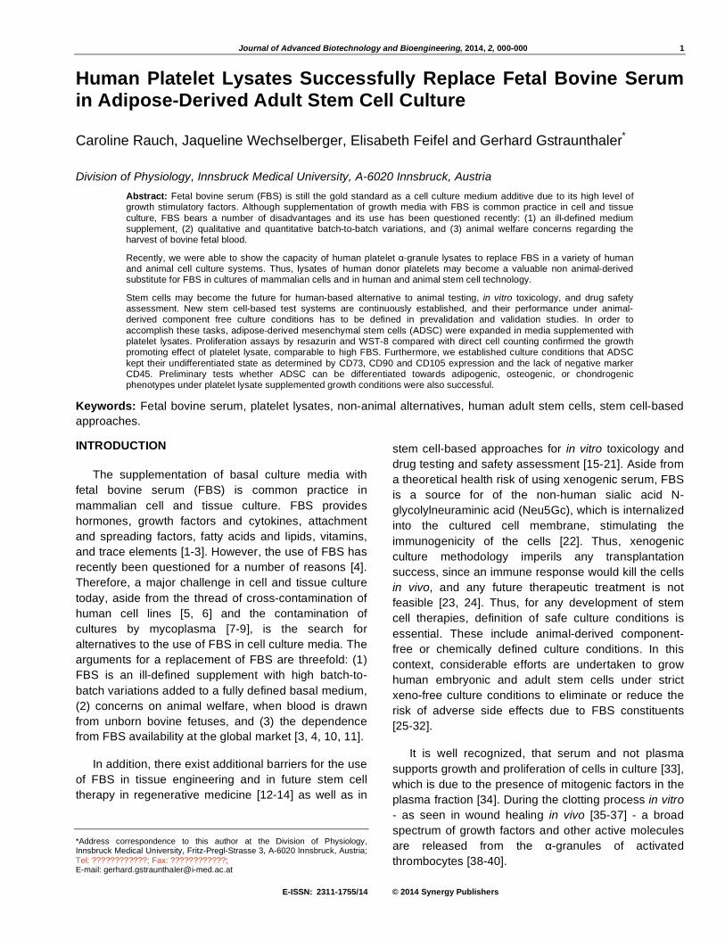
serum-free, SF). In this series of experiments, PL
treated cells show the highest proliferation rates and no
significant differences between the culture surfaces
tested, including Cell+ dishes (data not shown), could
be detected (Figure 1).
Comparison of FBS Batches with Platelet Lysates on Growth of Adipose-Derived Stem Cells
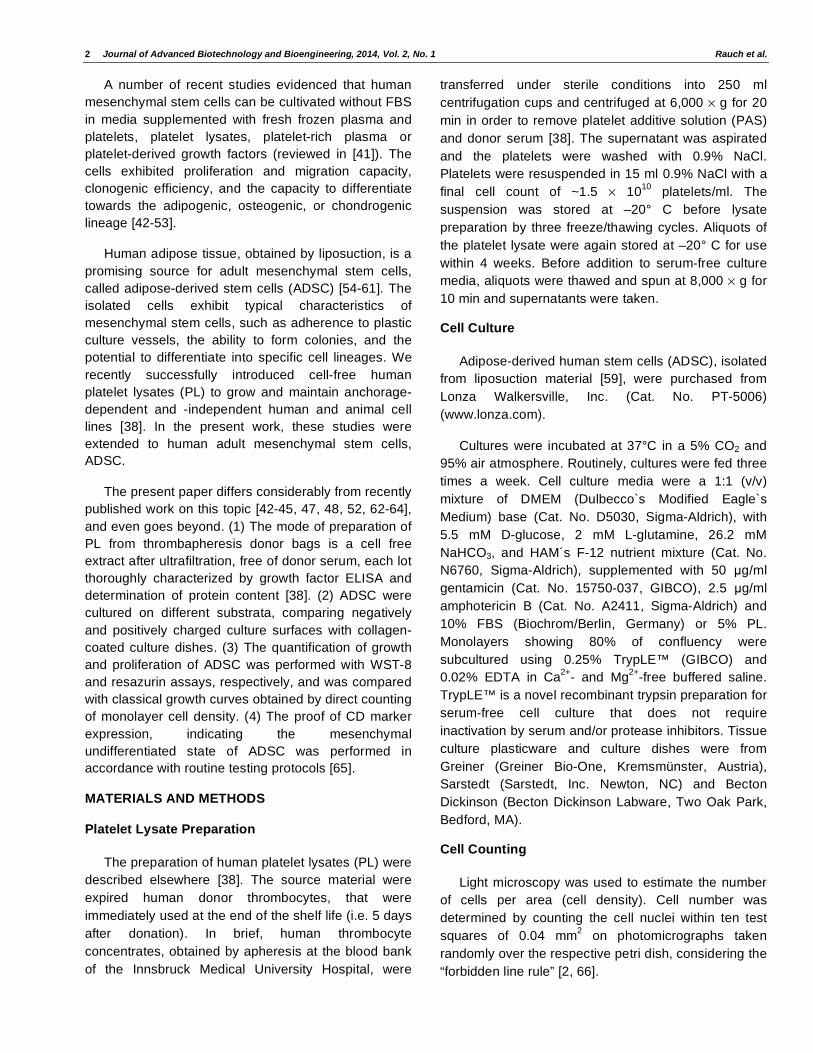
In the growth and proliferation experiments
summarized in Figure 1 it was noted, that growth
curves obtained with cultures in FBS-supplemented
DMEM/Ham F-12 media differed only slightly (p<0.1)
from growth curves of serum-free cultures, although
growth and proliferation under FBS was apparent at
daily microscopical inspection. However, the results
obtained with PL could never be achieved. Therefore,
FBS batches of different vendors were tested. The
results of a representative series of experiments are
shown in Figure 2. As can be seen, batches of FBS
differed in their growth promoting capacity, determined
by WST-8 assays, however, PL were still superior in all
experiments tested.
Growth Promoting Effect of Platelet Lysates on Mesenchymal Stem Cells
ADSC were grown in DMEM/Ham F-12 media,
containing either 10% FBS or 5% PL. In addition, a 1%
FBS/5% PL combination was tested for additive effects.
Serum-free culture conditions (SF, no additions) served
4 Journal of Advanced Biotechnology and Bioengineering, 2014, Vol. 2, No. 1 Rauch et al.
Figure 1: Cells were grown on polystyrol, on poly-D-lysine coated and on collagen type I-coated dishes. Quantification of cell growth experiments was performed by in situ-cell counting (A), and by WST-8 (B) and resazurin assays (C) [2]. Cell counts were determined from 10 randomly taken photomicrographs (means ± SD). WST-8 and resazurin values are expressed as means ± SD of 3 - 6 independent series of experiments. The growth curves with FBS are statistically significant from serum-free curves with p-values <0.1 in unpaired Student’s t-test. Growth curves with PL differ from FBS with p<0.05.
comparison of FBS batches
0 1 2 3 4 5 6 7 80.0
0.5
1.0
1.5
2.0
serum batch 2
serum batch 1
SF
PL
Time (days)
WS
T-8
Red
ucti
on
Figure 2: Comparison of FBS batches on growth and proliferation of ADSC. Cells were grown on polystyrol dishes in DMEM/Ham F-12 supplemented with either 10% FBS from different vendors (batch 1, Biochrom; batch 2, GIBCO), or 5% PL. SF, serum-free media without any supplementation. WST-8 read-outs are given as means ± SD of four independent series of experiments.
as controls. Cells were seeded onto polystyrol either
with FBS containing, or serum-free media 24 h before
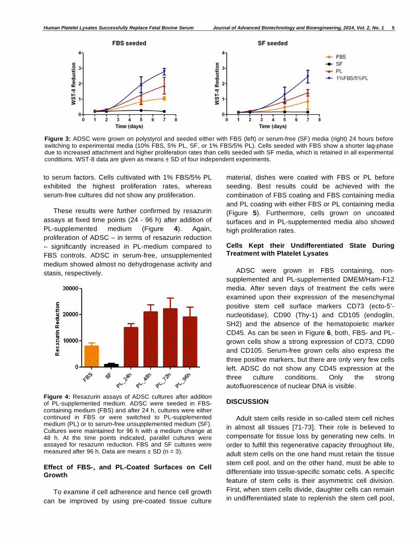
starting the experiment. As depicted in Figure 3, cells
seeded in FBS-containing medium before switching to
experimental media exhibit a shorter lag-phase, which
can be explained by an improved cell attachment due
Human Platelet Lysates Successfully Replace Fetal Bovine Serum Journal of Advanced Biotechnology and Bioengineering, 2014, Vol. 2, No. 1 5
to serum factors. Cells cultivated with 1% FBS/5% PL
exhibited the highest proliferation rates, whereas
serum-free cultures did not show any proliferation.
These results were further confirmed by resazurin
assays at fixed time points (24 - 96 h) after addition of
PL-supplemented medium (Figure 4). Again,
proliferation of ADSC – in terms of resazurin reduction
– significantly increased in PL-medium compared to
FBS controls. ADSC in serum-free, unsupplemented
medium showed almost no dehydrogenase activity and
stasis, respectively.
Figure 4: Resazurin assays of ADSC cultures after addition of PL-supplemented medium. ADSC were seeded in FBS-containing medium (FBS) and after 24 h, cultures were either continued in FBS or were switched to PL-supplemented medium (PL) or to serum-free unsupplemented medium (SF). Cultures were maintained for 96 h with a medium change at 48 h. At the time points indicated, parallel cultures were assayed for resazurin reduction. FBS and SF cultures were measured after 96 h. Data are means ± SD (n = 3).
Effect of FBS-, and PL-Coated Surfaces on Cell Growth
To examine if cell adherence and hence cell growth
can be improved by using pre-coated tissue culture
material, dishes were coated with FBS or PL before
seeding. Best results could be achieved with the
combination of FBS coating and FBS containing media
and PL coating with either FBS or PL containing media
(Figure 5). Furthermore, cells grown on uncoated
surfaces and in PL-supplemented media also showed
high proliferation rates.
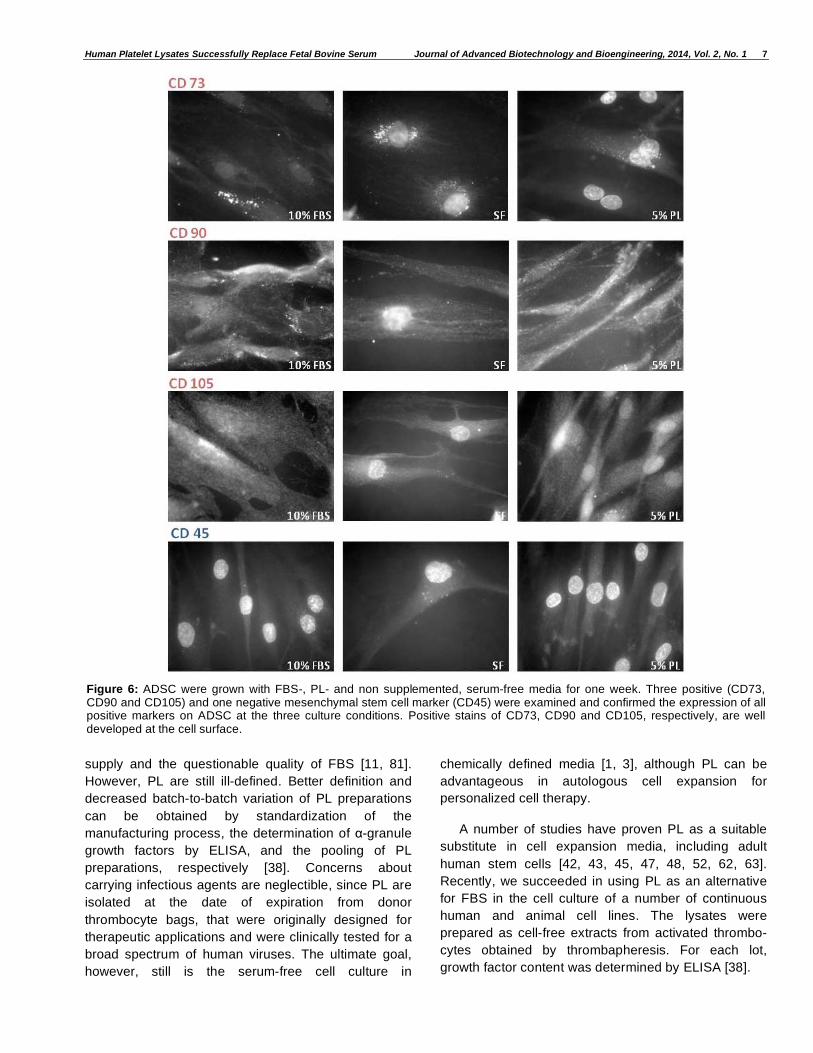
Cells Kept their Undifferentiated State During Treatment with Platelet Lysates
ADSC were grown in FBS containing, non-
supplemented and PL-supplemented DMEM/Ham-F12
media. After seven days of treatment the cells were
examined upon their expression of the mesenchymal
positive stem cell surface markers CD73 (ecto-5’-
nucleotidase), CD90 (Thy-1) and CD105 (endoglin,
SH2) and the absence of the hematopoietic marker
CD45. As can be seen in Figure 6, both, FBS- and PL-
grown cells show a strong expression of CD73, CD90
and CD105. Serum-free grown cells also express the
three positive markers, but there are only very few cells
left. ADSC do not show any CD45 expression at the
three culture conditions. Only the strong
autofluorescence of nuclear DNA is visible.
DISCUSSION
Adult stem cells reside in so-called stem cell niches
in almost all tissues [71-73]. Their role is believed to
compensate for tissue loss by generating new cells. In
order to fulfill this regenerative capacity throughout life,
adult stem cells on the one hand must retain the tissue
stem cell pool, and on the other hand, must be able to
differentiate into tissue-specific somatic cells. A specific
feature of stem cells is their asymmetric cell division.
First, when stem cells divide, daughter cells can remain
in undifferentiated state to replenish the stem cell pool,
Figure 3: ADSC were grown on polystyrol and seeded either with FBS (left) or serum-free (SF) media (right) 24 hours before switching to experimental media (10% FBS, 5% PL, SF, or 1% FBS/5% PL). Cells seeded with FBS show a shorter lag-phase due to increased attachment and higher proliferation rates than cells seeded with SF media, which is retained in all experimental conditions. WST-8 data are given as means ± SD of four independent experiments.

6 Journal of Advanced Biotechnology and Bioengineering, 2014, Vol. 2, No. 1 Rauch et al.
Figure 5: Cells were seeded on uncoated, FBS- and PL-coated cell culture dishes. The combinations of FBS coating and FBS containing media, PL coating with either FBS or PL containing media and uncoated surfaces and PL supplemented media show the best growth results. WST-8 values are expressed as means ± SD of three independent experiments.
a property called self-renewal. Second, daughter cells
can differentiate into specific cell types to supply all the
somatic cells of the tissue, called multipotency. Self-
renewal and multipotency are the defining
characteristics of stem cells [74].
When innovative culture systems for human stem
cells are applied i.e. the replacement of high contents
of FBS in culture media by non-animal derived
components, like human platelet lysates (PL) [41], the
stem cell attributes of self-renewal and multipotency
have to be retained under those culture conditions.
Before applying PL as an alternative to FBS, the
following questions have to be answered as
prerequisites for a successful stem cell culture system:
(1) Can adult human stem cells e.g. ADSC, be cultured
in the presence of PL? (2) Can the cells be maintained
in undifferentiated state? (3) Can the cells be triggered
to differentiate into specific lineages?
The first two questions are positively answered in
the present study, also a preliminary answer for the
third question can delivered. However, a full answer
about the differentiation potential of ADSC in PL-
supplemented media would go beyond the present
paper, and will be given in a subsequent publication.
Bone marrow aspirates and adipose tissue, derived
from liposuction, are rich sources for adult human
mesenchymal stem cells. The generation of this type of
cells is – in contrast to human embryonic stem cells –
ethically acceptable, that paved the way for a vast
amount of research concerning their potential use in
regenerative medicine. However, any future clinical
application of adult stem cells is impeded by the use of
FBS as an animal-derived growth supplement in
expansion culture media, due to the possibility of
introducing xenogenic molecules into human stem cells
[22]. This fact called for the search and the
development of alternatives to FBS that are animal-
derived component-free and safe for any therapeutic
application.
In addition, human stem cell cultures gained
importance as innovative human-based alternative to
animal testing, in vitro toxicology, and drug testing and
safety assessment [15-21, 75-78]. Thus, also for a
successful application of human stem cell-based
testing systems under fully humanized test conditions,
animal-derived component-free culture protocols are
mandatory.
Cell-free extracts of human donor thrombocytes
(PL) have been established as a human-based, xeno-
free surrogate for FBS (reviewed in [41, 79, 80]). The
use of PL in culturing human cells may overcome some
of the critical aspects in the FBS dilemma [3, 4, 28], like
the presence of bovine sialic acids, that may act as
xenoantigens [22] which imperils any future therapeutic
use of FBS-grown (stem) cells [24], or the limited global
Human Platelet Lysates Successfully Replace Fetal Bovine Serum Journal of Advanced Biotechnology and Bioengineering, 2014, Vol. 2, No. 1 7
supply and the questionable quality of FBS [11, 81].
However, PL are still ill-defined. Better definition and
decreased batch-to-batch variation of PL preparations
can be obtained by standardization of the
manufacturing process, the determination of -granule
growth factors by ELISA, and the pooling of PL
preparations, respectively [38]. Concerns about
carrying infectious agents are neglectible, since PL are
isolated at the date of expiration from donor
thrombocyte bags, that were originally designed for
therapeutic applications and were clinically tested for a
broad spectrum of human viruses. The ultimate goal,
however, still is the serum-free cell culture in
chemically defined media [1, 3], although PL can be
advantageous in autologous cell expansion for
personalized cell therapy.
A number of studies have proven PL as a suitable
substitute in cell expansion media, including adult
human stem cells [42, 43, 45, 47, 48, 52, 62, 63].
Recently, we succeeded in using PL as an alternative
for FBS in the cell culture of a number of continuous
human and animal cell lines. The lysates were
prepared as cell-free extracts from activated thrombo-
cytes obtained by thrombapheresis. For each lot,
growth factor content was determined by ELISA [38].
Figure 6: ADSC were grown with FBS-, PL- and non supplemented, serum-free media for one week. Three positive (CD73, CD90 and CD105) and one negative mesenchymal stem cell marker (CD45) were examined and confirmed the expression of all positive markers on ADSC at the three culture conditions. Positive stains of CD73, CD90 and CD105, respectively, are well developed at the cell surface.

8 Journal of Advanced Biotechnology and Bioengineering, 2014, Vol. 2, No. 1 Rauch et al.
In the present study, ADSC, derived from adipose
tissue after liposuction, were used as adult stem cell
model [59]. The minimal criteria defining multipotent
mesenchymal stromal cells, proposed by Dominici et
al. [65], include – among others – the cells’ potency to
adhere to plastic culture surfaces. However, neither
more precise information is given about the surface
treatment and/or charge of the culture plastic, nor a
systematic study on the nature of the plastic surface
has been performed yet [82]. To this end, ADSC were
cultured on different substrata, comparing negatively
and positively charged culture surfaces with collagen-
coated culture dishes in culture media containing FBS
or PL. In order to quantify growth and proliferation of
ADSC, WST-8 and resazurin assays were performed
and the data compared with classical growth curves
after direct counting of monolayer cell density. As can
be seen from Figure 1, no significant differences in
rates of cell proliferation could be observed with
respect to cell surface treatment. Clear differences,
however, can be seen in terms of culture media
supplements (for comparison of different FBS batches
see also Figure 2). Highest proliferation rates of ADSC
were found in media supplemented with 5% PL. In
addition, the diagrams within each assay method to
determine the rate of proliferation (cell counting, WST-
8, resazurin) are well comparable, as are the growth
curves among each experimental group. This is, for the
first time, a systematic comparison of classical growth
curves, obtained by direct counting of cell density, with
two indirect proliferation assays, measuring the activity
of cellular dehydrogenases as indicators for cell
viability [2, 68, 69].
However, considerable differences in initial cell
adhesion were observed depending on the composition
of the seeding medium (Figures 3 and 4). It is a well
known fact, that FBS, aside from hormones, growth
factors and cytokines, vitamins, and trace elements,
also contains attachment and spreading factors,
facilitating the attachment of cells after seeding into
fresh culture vessels [2, 3]. Therefore, in some serum-
free culture protocols for sensitive cells, pre-coating of
culture dishes is required [1]. These facts are well
reflected by the different lag-phases of ADSC, when
either seeded in FBS-containing or in serum-free
medium (Figure 3).
In order to further support these findings and to test
whether PL may contain attachment and spreading
factors, culture dishes were coated with either FBS or
PL before seeding (Figure 5). The results are in good
accordance with the data of the previous series of
experiments. Again, cell attachment, and growth and
proliferation, respectively, of ADSC was improved after
pre-coating with FBS or PL, shown by a shorter lag-
phase and steeper log-phases, in contrast to seeding
of cells into uncoated plastic culture dishes.
A prerequisite for successful application of adult
stem cells in cell replacement therapy and in vitro-
toxicology, respectively, is the expansion and
maintenance of the cells in their undifferentiated state,
thus to retain the multipotential nature and the
proliferative capacity of the cells. In order to test
whether ADSC maintain their undifferentiated state
under PL culture conditions, cells were grown in the
respective experimental media and the expression of
specific surface markers, characteristic for defining
multipotent mesenchymal stem cells, was determined
[65]. ADSC cultured in FBS- or in PL-supplemented
culture media stained positive for the mesenchymal
markers CD73, CD90, and CD105, while cells were
negative for the hematopoietic marker CD45 (Figure 6).
To summarize, human thrombocyte extracts
(platelet lysates, PL) can successfully replace the high
contents of FBS in human adult mesenchymal stem
cell cultures, as shown previously for continuous
human and animal cell lines [38]. In the present study,
in culture media supplemented with 5% PL, ADSC
attached to polystyrene culture dishes, grew out and
proliferated at rates equal or higher than control
cultures with 10% FBS. In addition, under PL culture
conditions, ADSC retained their undifferentiated
phenotype. Thus, stem cell culture media
supplemented with PL provide a fully “humanized”
culture system for human adult mesenchymal stem
cells (hMSC). In the future, stem cells under
appropriate culture conditions may transform the way in
which therapeutics are discovered and validated [83].
In addition, this culture system can also be applied for
human induced pluripotent stem cells (iPS) [84]. The
unique attributes of iPS are the generation of patient-
and disease-specific cells for early drug discovery and
safety assessment [85-89]. A promising approach is
the setup of an European iPS Cell Bank by the recently
initiated StemBANCC Project, funded by IMI, the
Innovative Medicines Initative [90]. Also in this
multinational project xeno-free culture conditions are
mandatory prerequisites for successful applications of
iPS for drug screening and safety assessment studies.
A consistent source of cells cultured at the highest
quality available to guarantee powerful test results will
be the future challenge in in vitro toxicology [91, 92].
Or, as pointed out recently, “Are our cell cultures good
enough for regulatory decision taking?” [93, 94].

Human Platelet Lysates Successfully Replace Fetal Bovine Serum Journal of Advanced Biotechnology and Bioengineering, 2014, Vol. 2, No. 1 9
CONFLICT OF INTEREST
The authors declare that there is no conflict of
interests regarding the publication of this article.
ACKNOWLEDGEMENTS
This study was generously supported by SET,
Foundation for the Promotion of Alternate and
Complementary Methods to Reduce Animal Testing,
Frankfurt a. M., and the ProCare Private Foundation,
Zürich.
REFERENCES
[1] Gstraunthaler G. Alternatives to the use of fetal bovine serum: serum-free cell culture. ALTEX 2003; 20: 275-81.
[2] Gstraunthaler G, Lindl T. Zell- und Gewebekultur. Allgemeine Grundlagen und spezielle Anwendungen. 7. Aufl., Berlin – Heidelberg: Springer-Spektrum 2013.
[3] van der Valk J, Brunner D, De Smet K, et al. Optimization of chemically defined cell culture media – Replacing fetal
bovine serum in mammalian in vitro methods. Toxicol in Vitro 2010; 24: 1053-63. http://dx.doi.org/10.1016/j.tiv.2010.03.016
[4] Brunner D, Frank J, Appl H, et al. Serum-free cell culture:
The serum-free media interactive online database. ALTEX 2010; 27: 53-62.
[5] Alston-Roberts C, Barallon R, Bauer SR, et al. Cell line misidentification: the beginning of the end. A Report from the American Type Culture Collection Standards Development
Organization Workgroup ASN-0002. Nature Rev Cancer 2010; 10: 441-8.
[6] Gstraunthaler G. The Bologna Statement on Good Cell Culture Practice (GCCP) – 10 years later. Proceedings of the
7th World Congress on Alternatives & Animal Use in the Life
Sciences, Rome, Italy, 2009. ALTEX 2010; 27: 141-6.
[7] Cobo F, Cortes JL, Cabrera C, et al. Microbiological contamination in stem cell cultures. Cell Biol Int 2007; 31: 991-5. http://dx.doi.org/10.1016/j.cellbi.2007.03.010
[8] Drexler HG, Uphoff CC. Mycoplasma contamination of cell
cultures: Indices, sources, effects, detection, elimination, prevention. Cytotechnology 2002; 39: 75-90. http://dx.doi.org/10.1023/A:1022913015916
[9] Young L, Sung J, Stacey G, Masters JR. Detection of
mycoplasma in cell culture. Nature Protocols 2010; 5: 929-34. http://dx.doi.org/10.1038/nprot.2010.43
[10] Fujimoto B. Fetal bovine serum – supply vs. demand. Art to Science 2002; 21: 1-4.
[11] Gstraunthaler G, Lindl T, van der Valk J. A plea to reduce or
replace fetal bovine serum in cell culture media. Cytotechnology 2013; 65: 791-3. http://dx.doi.org/10.1007/s10616-013-9633-8
[12] Giordano A, Galderisi U, Marino IR. From the laboratory bench to the patient’s bedside: An update on clinical trials
with mesenchymal stem cells. J Cell Physiol 2007; 211: 27-35. http://dx.doi.org/10.1002/jcp.20959
[13] Klimanskaya I, Rosenthal N, Lanza R. Derive and conquer:
sourcing and differentiating stem cells for therapeutic applications. Nature Rev Drug Discovery 2008; 7: 131-42. http://dx.doi.org/10.1038/nrd2403
[14] Körbling M, Estrov Z. Adult stem cells for tissue repair – a
new therapeutic concept? New Engl J Med 2003; 349: 570-
82. http://dx.doi.org/10.1056/NEJMra022361
[15] Chapin RE, Stedman DB. Endless posibilities: Stem cells and the vision for toxicology testing in the 21
st century. Toxicol
Sciences 2009; 112: 17-22. http://dx.doi.org/10.1093/toxsci/kfp202
[16] Davila J, Cezar GG, Thiede M, et al. Use and application of stem cells in toxicology. Toxicol Sciences 2004; 79: 214-23. http://dx.doi.org/10.1093/toxsci/kfh100
[17] Leist M, Bremer S, Brundin P, et al. The biological and ethical basis of the use of human embryonic stem cells for in vitro test systems or cell therapy. ALTEX 2008; 25: 163-90.
[18] Leist M, Hartung T, Nicotera P. The dawning of a new age of toxicology. ALTEX 2008: 25: 103-14.
[19] Rohwedel J, Guan K, Hegert C, Wobus AM. Embryonic stem cells as an in vitro model for mutagenicity, cytotoxicity and embryotoxicity: present state and future prospects. Toxicol In
Vitro 2001; 15: 741-753. http://dx.doi.org/10.1016/S0887-2333(01)00074-1
[20] Vojnits K, Bremer S. Challenges using pluripotent stem cells for safety assessments of substances. Toxicology 2010; 270:
10-17. http://dx.doi.org/10.1016/j.tox.2009.12.003
[21] Wobus AM, Löser P. Present state and future perspectives of using pluripotent stem cells in toxicology research. Arch Toxicol 2011; 85: 79-117. http://dx.doi.org/10.1007/s00204-010-0641-6
[22] Martin MJ, Muotri A, Gage F, Varki A. Human embryonic
stem cells express an immunogenic nonhuman sialic acid. Nature Med 2005; 11: 228-32. http://dx.doi.org/10.1038/nm1181
[23] Conley BJ. Young JC, Trounson AO, Mollard R. Derivation,
propagation and differentiation of human embryonic stem cells. Int J Biochem Cell Biol 2004; 36: 555-67. http://dx.doi.org/10.1016/j.biocel.2003.07.003
[24] Halme DG, Kessler DA. FDA Regulation of stem-cell–based therapies. New Engl J Med 2006; 355: 1730-5. http://dx.doi.org/10.1056/NEJMhpr063086
[25] Klimanskaya I, Chung Y, Meisner L, et al. Human embryonic stem cells derived without feeder cells. Lancet 2005; 365: 1636-41. http://dx.doi.org/10.1016/S0140-6736(05)66473-2
[26] Ludwig TE, Levenstein ME, Jones JM, et al. Derivation of
human embryonic stem cells in defined conditions. Nature Biotechnol 2006; 24: 185-7. http://dx.doi.org/10.1038/nbt1177
[27] Mallon BS, Park K-Y, Chen KG, et al. Toward xeno-free
culture of human embryonic stem cells. Int J Biochem Cell Biol 2006; 38: 1063-75. http://dx.doi.org/10.1016/j.biocel.2005.12.014
[28] Mannello F, Tonti GA Concise Review: No breakthroughs for human mesenchymal and embryonic stem cell culture. All
that glitters is not gold! Stem Cells 2007; 25: 1603-9. http://dx.doi.org/10.1634/stemcells.2007-0127
[29] McDevitt TC, Palecek SP. Innovation in the culture and derivation of pluripotent human stem cells. Curr Opin
Biotechnol 2008; 19: 527-33. http://dx.doi.org/10.1016/j.copbio.2008.08.005
[30] Meng G, Liu S, Krawetz R, et al. A novel method for generating xeno-free human feeder cells for human embryonic stem cell culture. Stem Cells Dev 2008; 17: 413-
22. http://dx.doi.org/10.1089/scd.2007.0236
[31] Pedersen RA. Feeding hungry stem cells. Nat Biotechnol 2002; 20: 882-3. http://dx.doi.org/10.1038/nbt0902-882
[32] Richards M, Fong C-Y, Chan W-K, et al. Human feeders
10 Journal of Advanced Biotechnology and Bioengineering, 2014, Vol. 2, No. 1 Rauch et al.
support prolonged undifferentiated growth of human inner
cell masses and embryonic stem cells. Nat Biotechnol 2002; 20: 933-6. http://dx.doi.org/10.1038/nbt726
[33] Gospodarowicz D, Ill CR. Do plasma and serum have different abilities to promote cell growth? Proc Natl Acad Sci
USA 1980; 77: 2726-30. http://dx.doi.org/10.1073/pnas.77.5.2726
[34] Balk SD, Levine SP, Young LL, et al. Mitogenic factors present in serum but not in plasma. Proc Natl Acad Sci USA
1981; 78: 5656-60. http://dx.doi.org/10.1073/pnas.78.9.5656
[35] Barrientos S, Stojadinovic O, Golinko MS, et al. Growth factors and cytokines in wound healing. Wound Rep Reg 2008; 16: 585-601. http://dx.doi.org/10.1111/j.1524-475X.2008.00410.x
[36] Nurden AT, Nurden P, Sanchez M, et al. Platelets and wound healing. Front Biosci 2008; 13: 3525-48. http://dx.doi.org/10.2741/2947
[37] Werner S, Grose R. Regulation of wound healing by growth factors and cytokines. Physiol Rev 2003; 83: 835-70.
[38] Rauch C, Feifel E, Amann E-M, et al. Alternatives to the use
of fetal bovine serum: platelet lysates as a serum substitute in cell culture media. ALTEX 2010; 28: 305-16.
[39] Reed GL, Fitzgerald ML, Polgar J. Molecular mechanisms of platelet exocytosis: insights into the "secrete" life of thrombocytes. Blood 2000; 96: 3334-42.
[40] Rendu F, Brohard-Bohn B. The platelet release reaction: granules´ constituents, secretion and functions. Platelets
2001; 12: 261-73. http://dx.doi.org/10.1080/09537100120068170
[41] Bieback K. Platelet lysates as replacement for fetal bovine serum in mesenchymal stromal cell cultures. Transfus Med
Hemother 2013; 40: 326-35. http://dx.doi.org/10.1159/000354061
[42] Bernardo ME, Avanzini MA, Perotti C, et al. Optimization of in vitro expansion of human multipotent mesenchymal stromal cells for cell-therapy approaches: further insights in the
search for fetal calf serum substitute. J Cell Physiol 2007; 211: 121-30. http://dx.doi.org/10.1002/jcp.20911
[43] Bieback K, Hecker A, Kocaoemer A, et al. Human
alternatives to fetal bovine serum for the expansion of mesenchymal stromal cells from bone marrow. Stem Cells 2009; 27: 2331-41. http://dx.doi.org/10.1002/stem.139
[44] Blande IS, Bassenze V, Lavini-Ramos C, et al. Adipose
tissue mesenchymal stem cell expansion in animal serum-free medium supplemented with autologous human platelet lysate. Transfusion 2009; 49: 2680-5. http://dx.doi.org/10.1111/j.1537-2995.2009.02346.x
[45] Doucet C, Ernou I, Zhang Y, Llense J-R, et al. Platelet lysates promote mesenchymal stem cell expansion: a safety substitute for animal serum in cell-based therapy
applications. J Cell Physiol 2005; 205: 228-36. http://dx.doi.org/10.1002/jcp.20391
[46] Gruber R, Karreth F, Kandler B, et al. Platelet-released supernatants increase migration and proliferation, and decrease osteogenic differentiation of bone marrow-derived
mesenchymal progenitor cells under in vitro conditions. Platelets 2004; 15: 29-35. http://dx.doi.org/10.1080/09537100310001643999
[47] Kocaoemer A, Kern S, Klüter H, Bieback K. Human AB
serum and thrombin-activated platelet-rich plasma are suitable alternatives to fetal calf serum for the expansion of mesenchymal stem cells from adipose tissue. Stem Cells
2007; 25: 1270-8. http://dx.doi.org/10.1634/stemcells.2006-0627
[48] Lange C, Cakiroglu F, Spiess A-N, et al. Accelerated and
safe expansion of human mesenchymal stromal cells in
animal serum-free medium for transplantation and regenerative medicine. J Cell Physiol 2007; 213: 18-26. http://dx.doi.org/10.1002/jcp.21081
[49] Müller AM, Davenport M, Verrier S, et al. Platelet lysate as a serum substitute for 2D static and 3D perfusion culture of
stromal vascular fraction cells from human adipose tissue. Tissue Eng A 2009; 15: 869-75. http://dx.doi.org/10.1089/ten.tea.2008.0498
[50] Müller I, Kordowich S, Holzwarth C, et al. Animal serum-free
culture conditions for isolation and expansion of multipotent mesenchymal stromal cells from human BM. Cytotherapy 2006; 8: 437-44. http://dx.doi.org/10.1080/14653240600920782
[51] Reinisch A, Bartmann C, Rohde E, et al. Humanized system
to propagate cord blood-derived multipotent mesenchymal stromal cells for clinical application. Regen Med 2007; 2: 371-82. http://dx.doi.org/10.2217/17460751.2.4.371
[52] Schallmoser K, Bartmann C, Rohde E, et al. Human platelet lysate can replace fetal bovine serum for clinical-scale expansion of functional mesenchymal stromal cells.
Transfusion 2007; 47: 1436-46. http://dx.doi.org/10.1111/j.1537-2995.2007.01220.x
[53] Vogel JP, Szalay K, Geiger F, et al. Platelet-rich plasma improves expansion of human mesenchymal stem cells and retains differentiation capacity and in vivo bone formation in
calcium phosphate ceramics. Platelets 2006; 17: 462-9. http://dx.doi.org/10.1080/09537100600758867
[54] Fraser JK, Wulur I, Alfonso Z, Hedrick MH fat tissue: an underappreciated source of stem cells for biotechnology. Trends Biotechnol 2006; 24: 150-4. http://dx.doi.org/10.1016/j.tibtech.2006.01.010
[55] Gimble JM, Guilak F. Adipose-derived adult stem cells: isolation, characterization, and differentiation potential. Cytotherapy 2003; 5: 362-9. http://dx.doi.org/10.1080/14653240310003026
[56] Kronsteiner B, Wolbank S, Peterbauer A, et al. Human
mesenchymal stem cells from adipose tissue and amnion influence T-Cells depending on stimulation method and presence of other immune cells. Stem Cells Dev 2011; 20:
2115-26. http://dx.doi.org/10.1089/scd.2011.0031
[57] Wolbank S, Peterbauer A, Fahrner M, et al. Dose-dependent immunomodulatory effect of human stem cells from amniotic
membrane: a comparison with human mesenchymal stem cells from adipose tissue. Tissue Eng 2007; 13: 1173-83. http://dx.doi.org/10.1089/ten.2006.0313
[58] Wolbank S, Peterbauer A, Wassermann E, et al. Labelling of human adipose-derived stem cells for non-invasive in vivo
cell tracking. Cell Tissue Banking 2007; 8: 163-77. http://dx.doi.org/10.1007/s10561-006-9027-7
[59] Zuk PA. The adipose-derived stem cell: Looking back and looking ahead. Mol Biol Cell 2010; 21: 1783-7. http://dx.doi.org/10.1091/mbc.E09-07-0589
[60] Zuk PA, Zhu M, Ashjian P, et al. Human adipose tissue is a
source of multipotent stem cells. Mol Biol Cell 2002; 13: 4279-95. http://dx.doi.org/10.1091/mbc.E02-02-0105
[61] Zuk PA, Zhu M, Mizuno H, et al. Multilineage cells from
human adipose tissue: Implications for cell-based therapies. Tissue Eng 2001; 7: 211-28. http://dx.doi.org/10.1089/107632701300062859
[62] Cholewa D, Stiehl T, Schellenberg A, et al. Expansion of adipose mesenchymal stromal cells is affected by human
platelet lysate and plating density. Cell Transplant 2011; 20: 1409-22. http://dx.doi.org/10.3727/096368910X557218
[63] Horn P, Bokermann G, Cholewa D, et al. Impact of individual

Human Platelet Lysates Successfully Replace Fetal Bovine Serum Journal of Advanced Biotechnology and Bioengineering, 2014, Vol. 2, No. 1 11
platelet lysates on isolation and growth of human
mesenchymal stromal cells. Cytotherapy 2010; 12: 888-98. http://dx.doi.org/10.3109/14653249.2010.501788
[64] Jonsdottir-Buch SM, Lieder R, Sigurjonsson OE. Platelet lysates produced from expired platelet concentrates support growth and osteogenic differentiation of mesenchymal stem
cells. PLoS ONE 2013; 8: e68984. http://dx.doi.org/10.1371/journal.pone.0068984
[65] Dominici M, Le Blanc K, Mueller I, et al. Minimal criteria for defining multipotent mesenchymal stromal dells. The
International Society for Cellular Therapy position statement. Cytotherapy 2006; 8: 315-317. http://dx.doi.org/10.1080/14653240600855905
[66] Pfaller W, Gstraunthaler G, Loidl P. Morphology of the differentiation and maturation of LLC-PK1 epithelia. J Cell
Physiol 1990; 142: 247-54. http://dx.doi.org/10.1002/jcp.1041420205
[67] Berridge MV, Tan AS. Characterization of the cellular reduction of 3-(4,5-dimethylthiazol-2-yl)-2,5-diphenyltetra-
zolium bromide (MTT): subcellular localization, substrate dependence, and involvement of mitochondrial electron transport in MTT reduction. Arch Biochem Biophys 1993;
303: 474-82. http://dx.doi.org/10.1006/abbi.1993.1311
[68] Mosmann T. Rapid colorimetric assay for cellular growth and survival: application to proliferation and cytotoxicity assays. J Immunol Methods 1983; 65: 55-63. http://dx.doi.org/10.1016/0022-1759(83)90303-4
[69] Scudiero DA, Shoemaker RH, Paull KD, et al. Evaluation of a
soluble tetrazolium/formazan assay for cell growth and drug sensitivity using human and other tumor cell lines. Cancer Res 1988; 48: 4827-33.
[70] Jennings P, Koppelstaetter C, Aydin S, et al. Cyclosporine A
induces senescence in renal tubular epithelial cells. Am J Physiol Renal Physiol 2007; 293: F831-8. http://dx.doi.org/10.1152/ajprenal.00005.2007
[71] Fuchs E, Tumbar T, Guasch G. Socializing with the neighbors: stem cells and their niches. Cell 2004; 116: 769-
78. http://dx.doi.org/10.1016/S0092-8674(04)00255-7
[72] Jones DL and Wagers AJ. No place like home: anatomy and function of the stem cell niche. Nature Rev Molec Cell Biol
2008; 9: 11-21. http://dx.doi.org/10.1038/nrm2319
[73] Scadden DT. The stem-cell niche as an entity of action. Nature 2006; 441: 1075-9. http://dx.doi.org/10.1038/nature04957
[74] Snipper HJ, Clevers H. Tracking adult stem cells. EMBO Rep
2011; 12: 113-22. http://dx.doi.org/10.1038/embor.2010.216
[75] Bhogal N, Grindon C, Combes R, Balls M. Toxicity testing: creating a revolution based on new technologies. Trends Biotechnol 2005; 23: 299-307. http://dx.doi.org/10.1016/j.tibtech.2005.04.006
[76] De Kock, J, Rodrigues RM, Bolleyn J, et al. Focus on stem cells as sources of human target cells for in vitro research and testing. ALTEX Proceedings 2012; 1: 541-8.
[77] Pellizzer C, Bremer S, Hartung T. Developmental toxicity
testing from animal towards embryonic stem cells. ALTEX 2005; 22: 47-56.
[78] Sartipy P, Björquist P, Strehl R, Hyllner J. The application of
human embryonic stem cell technologies to drug discovery. Drug Discov Today 2007; 12: 688-99. http://dx.doi.org/10.1016/j.drudis.2007.07.005
[79] Gottipamula S, Muttigi MS, Kolkundkar U, Seehtaram RN.
Serum-free media for the productionm of human mesenchymal stromal cells: a review. Cell Prolif 2013; 46: 608-27. http://dx.doi.org/10.1111/cpr.12063
[80] Tekkatte C, Gunasingh GP, Cherian KM, Sankaranarayanan
K. “Humanized” stem cell culture techniques: the animal serum controversy. Stem Cells Int 2011; 2011: 504723. http://dx.doi.org/10.4061/2011/504723
[81] Brindley DA, Davie NL, Culme-Seymor EJ, Mason C, Smith
DW, Rowley JA. Peak serum: implications of serum supply for cell therapy manufacturing. Regen Med 2012; 7: 7-13. http://dx.doi.org/10.2217/rme.11.112
[82] Baker M. Stem cells in culture: defining the substrate. Nature Methods 2011; 8: 293-7. http://dx.doi.org/10.1038/nmeth0411-293
[83] Rubin LL Stem cells and drug discovery: the beginning of a new era? Cell 2008; 132: 549-52. http://dx.doi.org/10.1016/j.cell.2008.02.010
[84] Yamanaka S. Induced pluripotent stem cells: Past, present, and future. Cell Stem Cell 2012; 10: 678-83. http://dx.doi.org/10.1016/j.stem.2012.05.005
[85] Balls M. The conflict over animal experimentation: is the field of battle changing? ATLA 2012; 40: 189-91.
[86] Balls M. FRAME and the pharmaceutical industry. ATLA 2012; 40: 295-300.
[87] Bellin M, Marchetto MC, Gage FH, Mummery CL. Induced pluripotent stem cells: the new patient? Nature Rev Molec Cell Biol 2012; 13: 713-26. http://dx.doi.org/10.1038/nrm3448
[88] Grskovic M, Javaherian A, Strulovici B, Daley GQ. Induced pluripotent stem cells – opportunities for disease modelling and drug discovery. Nature Rev Drug Discov 2011; 10: 915-29.
[89] Park I-H, Arora N, Huo H, et al. Disease-specific induced
pluripotent stem cells. Cell 2008; 134: 877-86. http://dx.doi.org/10.1016/j.cell.2008.07.041
[90] StemBANCC – Stem cells for biological assays of novel drugs and predictive toxicology; www.stembancc.org
[91] Basketter DA, Clewell H, Kimber I, et al. A roadmap for the development of alternative (non-animal) methods for systemic toxicity testing. ALTEX 2012; 29: 5-91.
[92] Hartung T. A toxicology for the 21st century – Mapping the
road ahead. Toxicol Sciences 2009; 109: 18-23. http://dx.doi.org/10.1093/toxsci/kfp059
[93] Hartung T. Food for thought . . . on cell culture. ALTEX 2007; 24: 143-7.
[94] Hartung T. Are our cell cultures good enough for regulatory
decision taking? Plenary Lecture at the 5th Annual Quasi-Vivo
User Group Meeting, Liverpool, UK, 2013.