Human identification from washed blood stains
13
Ünsal Sapan et al. Bull Natl Res Cent (2021) 45:148 https://doi.org/10.1186/s42269-021-00600-3 RESEARCH Human identification from washed blood stains Tuğba Ünsal Sapan 1,2* , Işıl Tuna Erdoğan 1 and Sevil Atasoy 1,2,3 Abstract Background: Among the physical evidence, bloodstain is one of the most common biological pieces of evidence at the crime scene, especially in violent crimes. Bloodstains are frequently seen at the crime scene and can be easily detected due to their color and structure. Because blood and bloodstains can potentially be evidence of a crime, offenders often tend to clean or wash them to get rid of relevant evidence. Some researchers think that washed bloodstains cannot obtain enough quality DNA for DNA profiling. However, some studies have shown that blood- stains on a piece of fabric can be used for DNA profiling even after washing. This study, it is aimed to determine whether a sufficient amount of DNA can be obtained for DNA profiling used for forensic purposes from blood-stained nylon and cotton fabrics washed at different temperatures such as 40 °C, 60 °C and 90 °C. Results: In this study, bloodstains were created on cotton and nylon fabrics in a representative crime scene. After washing with laundry detergent at 40 °C, 60 °C and 90 °C was performed, studies were conducted in order to make bloodstains visible and obtain DNA for genetic profiling. The result is that more DNA is lost due to easy exposure to external factors in the non-absorbent nylon fabric type compared to the absorbent cotton fabric. Moreover, the rates of obtaining DNA from bloodstains on different types of fabrics washed at several temperatures are shown in this study. Conclusions: It was determined that while a sufficient amount of DNA for the forensic genetic profiling can be obtained from cotton and nylon fabrics washed at 40 and 60 °C, a sufficient amount could not be obtained from the ones washed at 90 °C. And that shows even if bloodstained fabrics are washed at 60 °C, the fabrics still can be consid- ered as physical evidence of the crime and can be associated with the scene, perpetrator and victim triangle. Keywords: Forensic genetics, Blood stains, DNA recovery, Washed blood stains, Human identification © The Author(s) 2021. Open Access This article is licensed under a Creative Commons Attribution 4.0 International License, which permits use, sharing, adaptation, distribution and reproduction in any medium or format, as long as you give appropriate credit to the original author(s) and the source, provide a link to the Creative Commons licence, and indicate if changes were made. The images or other third party material in this article are included in the article’s Creative Commons licence, unless indicated otherwise in a credit line to the material. If material is not included in the article’s Creative Commons licence and your intended use is not permitted by statutory regulation or exceeds the permitted use, you will need to obtain permission directly from the copyright holder. To view a copy of this licence, visit http://creativecommons.org/licenses/by/4.0/. Background In recent years, by the help of developing technology, DNA profiles which allow individualization serve as a significant evidence to investigate criminal cases. Due to this reason, analyses of biological materials which are obtained from the crime scene are extremely important and it is possible to carry these analyses to better points (Bond and Hammond 2008; Kamphausen et al. 2015; Unsal et al. 2011). In murder or violent events, it is critical to reveal bloodstains at the scene for crime scene investigation. Since the crime scene investigation generally covers violent crimes, the most common biological material is blood and therefore bloodstains (Andrews and Coquoz 1994; Stojanović 2019; Mushtaq et al. 2015; Bucka et al. 2011). Bloodstains have long been one of the most important body fluid for forensic scientists in crime research. e shape, size and relationship of bloodstains with the sur- rounding items can provide important ideas about how the case occurred and it is possible to reconstruct the incident with its careful examination. e morphologi- cal examination of stains (bloodstain pattern analysis) Open Access Bulletin of the National Research Centre *Correspondence: [email protected] 1 Institute of Addiction and Forensic Sciences, Üsküdar University, Üsküdar University Altunizade-Main Campus, Üsküdar, 34662 Istanbul, Turkey Full list of author information is available at the end of the article
-
Upload
khangminh22 -
Category
Documents
-
view
0 -
download
0
Transcript of Human identification from washed blood stains

Ünsal Sapan et al. Bull Natl Res Cent (2021) 45:148 https://doi.org/10.1186/s42269-021-00600-3
RESEARCH
Human identification from washed blood stainsTuğba Ünsal Sapan1,2* , Işıl Tuna Erdoğan1 and Sevil Atasoy1,2,3
Abstract
Background: Among the physical evidence, bloodstain is one of the most common biological pieces of evidence at the crime scene, especially in violent crimes. Bloodstains are frequently seen at the crime scene and can be easily detected due to their color and structure. Because blood and bloodstains can potentially be evidence of a crime, offenders often tend to clean or wash them to get rid of relevant evidence. Some researchers think that washed bloodstains cannot obtain enough quality DNA for DNA profiling. However, some studies have shown that blood-stains on a piece of fabric can be used for DNA profiling even after washing. This study, it is aimed to determine whether a sufficient amount of DNA can be obtained for DNA profiling used for forensic purposes from blood-stained nylon and cotton fabrics washed at different temperatures such as 40 °C, 60 °C and 90 °C.
Results: In this study, bloodstains were created on cotton and nylon fabrics in a representative crime scene. After washing with laundry detergent at 40 °C, 60 °C and 90 °C was performed, studies were conducted in order to make bloodstains visible and obtain DNA for genetic profiling. The result is that more DNA is lost due to easy exposure to external factors in the non-absorbent nylon fabric type compared to the absorbent cotton fabric. Moreover, the rates of obtaining DNA from bloodstains on different types of fabrics washed at several temperatures are shown in this study.
Conclusions: It was determined that while a sufficient amount of DNA for the forensic genetic profiling can be obtained from cotton and nylon fabrics washed at 40 and 60 °C, a sufficient amount could not be obtained from the ones washed at 90 °C. And that shows even if bloodstained fabrics are washed at 60 °C, the fabrics still can be consid-ered as physical evidence of the crime and can be associated with the scene, perpetrator and victim triangle.
Keywords: Forensic genetics, Blood stains, DNA recovery, Washed blood stains, Human identification
© The Author(s) 2021. Open Access This article is licensed under a Creative Commons Attribution 4.0 International License, which permits use, sharing, adaptation, distribution and reproduction in any medium or format, as long as you give appropriate credit to the original author(s) and the source, provide a link to the Creative Commons licence, and indicate if changes were made. The images or other third party material in this article are included in the article’s Creative Commons licence, unless indicated otherwise in a credit line to the material. If material is not included in the article’s Creative Commons licence and your intended use is not permitted by statutory regulation or exceeds the permitted use, you will need to obtain permission directly from the copyright holder. To view a copy of this licence, visit http:// creat iveco mmons. org/ licen ses/ by/4. 0/.
BackgroundIn recent years, by the help of developing technology, DNA profiles which allow individualization serve as a significant evidence to investigate criminal cases. Due to this reason, analyses of biological materials which are obtained from the crime scene are extremely important and it is possible to carry these analyses to better points (Bond and Hammond 2008; Kamphausen et al. 2015; Unsal et al. 2011).
In murder or violent events, it is critical to reveal bloodstains at the scene for crime scene investigation. Since the crime scene investigation generally covers violent crimes, the most common biological material is blood and therefore bloodstains (Andrews and Coquoz 1994; Stojanović 2019; Mushtaq et al. 2015; Bucka et al. 2011).
Bloodstains have long been one of the most important body fluid for forensic scientists in crime research. The shape, size and relationship of bloodstains with the sur-rounding items can provide important ideas about how the case occurred and it is possible to reconstruct the incident with its careful examination. The morphologi-cal examination of stains (bloodstain pattern analysis)
Open Access
Bulletin of the NationalResearch Centre
*Correspondence: [email protected] Institute of Addiction and Forensic Sciences, Üsküdar University, Üsküdar University Altunizade-Main Campus, Üsküdar, 34662 Istanbul, TurkeyFull list of author information is available at the end of the article

Page 2 of 13Ünsal Sapan et al. Bull Natl Res Cent (2021) 45:148
enables experts to “physically” reconstruct the incident, while biological examination of blood and the stains provides individualization of crime to criminal. Usually perpetrators clean up the scene and wash the clothes or fabrics in order to get rid of the evidence from expert examination. (Bucka et al. 2011; Kulstein and Wiegand 2018; Salahuddin et al. 2018).
In recent years, cleaning of bloodstains with water and chemicals has often been observed as a result of the post-crime behavior of perpetrators. Today, the permanence of DNA on cleaned materials has become the subject of
many scientific researches along with speculations and discussions. Although most researchers have the opin-ion that washed bloodstains cannot provide sufficient DNA quality for profiling, several studies have shown that washed bloodstains can be used for DNA profiling (Bucka et al. 2011; Hofmann et al. 2019; Arjun Rao and Ashish 2016; Kamphausen et al. 2015).
This study aimed to determine whether a sufficient amount of DNA can be obtained for DNA profiling from bloodstained nylon and cotton fabrics washed at various temperatures.
Table 1 Number of fabric samples washed and DNA extracted
Cotton fabric Nylon fabric
Qiagen DNA extrac-tion kit
40 °C 60 °C 90 °C 40 °C 60 °C 90 °C 6 pieces
Organic DNA extrac-tion method
40 °C 60 °C 90 °C 40 °C 60 °C 90 °C 6 pieces
1 donor 12 pieces
10 donors 120 pieces
Fig. 1 The original trace of the blood stain after washing the cotton fabrics with blood stains at 40, 60, 90°C

Page 3 of 13Ünsal Sapan et al. Bull Natl Res Cent (2021) 45:148
MethodsSamples, substrates and washingBlood samples (6 ml) used in the study were taken from 10 unrelated donors with a injector. All samples were obtained after written informed consent with Uskudar University Non-Interventional Research Ethics Board approval in accordance with the Declaration of Helsinki and national laws. Blood samples taken from 10 donors were used to create bloodstained cotton and nylon fab-rics. Each donor’s blood was dripped onto 12 separate pieces of 10 × 10 cm fabric as 1 ml each. (First DNA
Table 2 Washing specifications for cotton and nylon fabrics
Fabric type Temperature (°C) Operating duration (min)
Cotton 90 180
60 90
40 90
Nylon 90 180
60 90
40 60
Cotton Nylon
40
60
90
Fig. 2 Visualization of bloodstains with luminol in fabrics washed at 40, 60 and 90ºC

Page 4 of 13Ünsal Sapan et al. Bull Natl Res Cent (2021) 45:148
isolation kit group: 3 different fabrics for 40, 60 and 90 degrees were applied on both cotton and nylon fabric, 6 fabric samples in total; second type of DNA isolation kit group: 3 different fabrics for 40, 60 and 90 degrees both cotton and nylon applied on the fabric, 6 fabric samples in total.) Since 12 pieces of blood-stained fabric were
obtained from each donor, a total of 120 samples were studied for 10 donors (Table 1).
The most common cotton and nylon fabrics were cho-sen to create representative crime scene. There were no fabric donors for this study. The fabric samples used in the study were supplied from an independent store to retain equal standards for each sample. The new branded fabrics were autoclaved and dried in room conditions for sterilization. Furthermore, fabrics were exposed to UV light for 60 min to avoid a contamination. Next, the fab-rics were cut into 10 × 10 cm pieces with sterile scissors. In order to represent a realistic crime scene, all collected blood samples were dropped as a 1 ml immediately on fabrics with the same injector to create stains. No antico-agulants were added.
Each stained fabrics were washed with powder laun-dry detergent (Persil) using the recommended amount (1 cup ~ 150 g) by the brand. The washing machine (Regal Pratica 7100 TY) was operated at 40 °C, 60 °C and 90 °C (1000 rpm). Washing temperatures and corresponding operation times are given in Table 2. We have used fixed programmes for the washing machine. The duration and
Fig. 3 Luminol score for bloodstains washed at 40, 60 and 90 °C
Fig. 4 Full profile of donor obtained from the whole blood sample

Page 5 of 13Ünsal Sapan et al. Bull Natl Res Cent (2021) 45:148
temperature values given in the manuscript are specifica-tions of those washing programmes. In order to mimic a realistic situation, we have not altered the programme features. Therefore, washing durations are not chosen as a contributing factor. In order to avoid any transfer from previous washing, the machine was operated empty at 90 °C after each washing. Each stained fabric was dried in a clean area under room conditions, approximately
20–25 °C with 45% humidity. (12 fabrics per donor: 40 °C, 60 °C and 90 °C, 2 different extraction methods and nylon-cotton fabrics separately).
Luminol application and documentationPioneer Forensic brand Luminol kit was used in this study. According to the protocol; the mixed compound was sprayed onto the washed bloodstained samples and examined in the dark. It was evaluated whether there was a blue metallic glare or not. Pictures were taken with a Sony SLT-A35 Alpha 35 DSLR digital camera with Tam-ron AF 18–200 mm f/3.5–6.3 XR Di II LD Aspherical [IF] Macro Lens with an exposure time of 30 s (e.g. f8 for 30 s at 400 ISO) using tripod as suggested in the literature (Edler et al. 2017; Kamphausen et al. 2015).
DNA extraction and quantificationLuminous areas of the fabrics of each sample after expo-sition to luminol were cut into 0.5 cm pieces with sterile
Table 3 Washing temperature and corresponding luminol score for both fabric types
Fabric Temperature (°C) Luminol score
Cotton 40 3
60 3
90 1
Nylon 40 3
60 2
90 0
Fig. 5 DNA profile of blood stain in washed cotton fabric at 40 °C extracted by organic DNA extraction method

Page 6 of 13Ünsal Sapan et al. Bull Natl Res Cent (2021) 45:148
scissors. The pieces were transferred to a 1.5 ml Eppen-dorf tube with the help of disposable forceps. DNA was extracted from a total of 120 blood samples col-lected from 10 healthy volunteers by spin colon-based extraction method (QIAamp DNA Mini Kit) and Phe-nol–Chloroform Isoamyl Organic method separately. Allsheng-Nano 400A spectrophotometer device was used for quantitation of extracted DNA.
DNA amplification and electrophoresisPCR reaction for DNA of each donor obtained from the whole blood sample was performed using the GlobalFiler™ PCR Amplification Kit (Thermo Fisher Scientific) according to the manufacturer protocol with a reaction volume of 25 μL and 30 PCR amplification cycles.
PCR products were subsequently prepared for CE by adding 1 µl of each amplified product to 10 µl Hi-Di for-mamide (Applied Biosystems) and 0.5 µl GS-600 LIZ size standard (Applied Biosystems), respectively. Separation
Fig. 6 DNA profile of blood stain in washed nylon fabric at 40 °C extracted by organic DNA extraction method
Table 4 Statistical parameters for the amount of extracted DNA from 60 samples as cotton and nylon by using spin colon extraction method
In order to categorically compare extracted DNA amount, each value is normalized by the highest extracted amount
*According to the data, extracted amounts were in an agreement with a Normal Distribution. Therefore mean and standard deviation are the two parameters enough to describe such a distribution. Besides, in order to quantitatively interpret the results, we have normalized the extracted amounts for each sample by using highest value. This normalization enabled us to categorically compare DNA amount in terms of means
Fabric type and washing temperature
Amount of DNA (normalized)
Fabric Temperature (°C)
Mean SD 95% CI
Cotton (30 samples) 40 0.597 0.191 0.472–0.722
60 0.343 0.154 0.242–0.444
90 0.175 0.098 0.111–0.239
Nylon (30 samples) 40 0.534 0.137 0.444–0.624
60 0.279 0.118 0.199–0.359
90 0.157 0.069 0.107–0.207

Page 7 of 13Ünsal Sapan et al. Bull Natl Res Cent (2021) 45:148
and detection were performed with ABI 3500 Genetic Analyzer (Thermo Fischer Scientific) using filter set G5 and POP4 polymer (Applied Biosystems). Samples were genotyped with GeneMapper ™ ID X Software (Thermo Fischer Scientific). In this way, the full profile of the donor was obtained.
ResultsVisibility of washed bloodstainsSixty bloodstained nylon and cotton fabric pieces from 10 donors were washed at 40 °C, 60 °C, and 90 °C.
It was observed that the original contour of the blood-stain remained on the cotton fabric washed at 40° C and 60 °C.
No bloodstain traces were observed with naked eyes on the nylon fabric washed at 60 °C.
No bloodstain traces were observed with naked eyes on the nylon and cotton fabrics washed at 90 °C (Fig. 1). In this study, the luminol score scheme used by Hofmann et al. (2019) preferred. Since the score table is sharp and distinct, all researchers (3 person) observed and agreed
on the results of the luminol scores. Thus, there was no interobserver error.
When the spots were exposed to Luminol, a metallic blue shine was observed in all pieces washed at 40–60–90 °C (Fig. 2). Moreover, it was found that the shining was less in the nylon fabric compared to the cotton fabric.
In order to assess visibility of the stains, luminol score scheme (Hofmann et al. 2019) has been followed. Lumi-nol score and corresponding luminescence property of the stains are given below:
• 0 → No luminescence• 1 → Hardly visible, tentative luminescence• 2 → Notable luminescence• 3 → Apparent luminescence with distinct contour
borders
The assessment of luminescence for each fabric and graphical representation are given in Table 3 and Fig. 3, respectively.
Fig. 7 DNA profile of blood stain in washed cotton fabric at 60 °C extracted by organic DNA extraction method
Page 8 of 13Ünsal Sapan et al. Bull Natl Res Cent (2021) 45:148
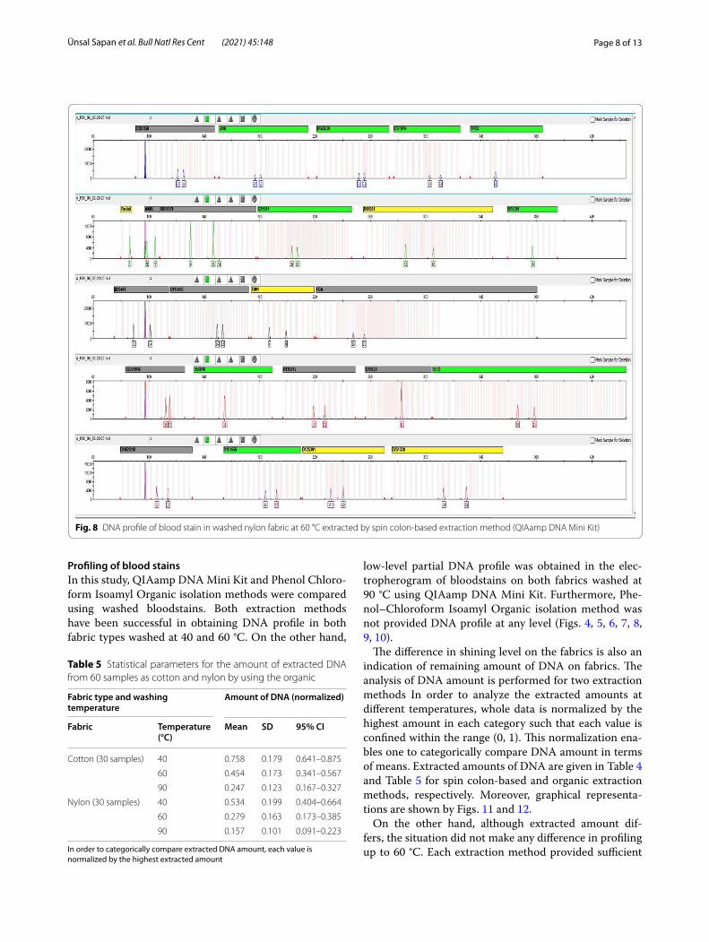
Profiling of blood stainsIn this study, QIAamp DNA Mini Kit and Phenol Chloro-form Isoamyl Organic isolation methods were compared using washed bloodstains. Both extraction methods have been successful in obtaining DNA profile in both fabric types washed at 40 and 60 °C. On the other hand,
low-level partial DNA profile was obtained in the elec-tropherogram of bloodstains on both fabrics washed at 90 °C using QIAamp DNA Mini Kit. Furthermore, Phe-nol–Chloroform Isoamyl Organic isolation method was not provided DNA profile at any level (Figs. 4, 5, 6, 7, 8, 9, 10).
The difference in shining level on the fabrics is also an indication of remaining amount of DNA on fabrics. The analysis of DNA amount is performed for two extraction methods In order to analyze the extracted amounts at different temperatures, whole data is normalized by the highest amount in each category such that each value is confined within the range (0, 1). This normalization ena-bles one to categorically compare DNA amount in terms of means. Extracted amounts of DNA are given in Table 4 and Table 5 for spin colon-based and organic extraction methods, respectively. Moreover, graphical representa-tions are shown by Figs. 11 and 12.
On the other hand, although extracted amount dif-fers, the situation did not make any difference in profiling up to 60 °C. Each extraction method provided sufficient
Fig. 8 DNA profile of blood stain in washed nylon fabric at 60 °C extracted by spin colon-based extraction method (QIAamp DNA Mini Kit)
Table 5 Statistical parameters for the amount of extracted DNA from 60 samples as cotton and nylon by using the organic
In order to categorically compare extracted DNA amount, each value is normalized by the highest extracted amount
Fabric type and washing temperature
Amount of DNA (normalized)
Fabric Temperature (°C)
Mean SD 95% CI
Cotton (30 samples) 40 0.758 0.179 0.641–0.875
60 0.454 0.173 0.341–0.567
90 0.247 0.123 0.167–0.327
Nylon (30 samples) 40 0.534 0.199 0.404–0.664
60 0.279 0.163 0.173–0.385
90 0.157 0.101 0.091–0.223

Page 9 of 13Ünsal Sapan et al. Bull Natl Res Cent (2021) 45:148
amount and quality for profiling up to this temperature. However, at 90 °C, the sufficient amount/quality could not be obtained for full profile. Only spin colon-based extraction method was able to provide just a low-level partial DNA profile (3 STR out of 24–12.5%). The profil-ing percentages for both methods are given in Table 6, Fig. 13.
DiscussionThe most common biological evidence at the crime scene is bloodstains. For this reason, bloodstain is one of the most important evidence in forensic science that can help investigators to solve a crime. It can link a suspect to a crime and also help reconstruct the crime scene. Criminals often tend to clear up and eliminate evidence from the scene (Voskoboinik et al. 2018). After a criminal act, a perpetrator would try washing bloodstains either by hand washing in cold water or using a regular wash-ing machine. These attempts may cause the bloodstains
to change and eventually to be partially or completely removed. In addition, bloodstains can be found on vari-ous surfaces at the scene. Due to this reason, fabrics such as nylon and cotton, which are mostly encountered at the scene, were chosen in this study. It is known that in washed blood-stained nylon or cotton fabric samples, a genotyping problem can be experienced with classi-cal STR loci, which occupy more space on DNA, and so generally no results can be obtained (Unsal et al. 2011). In this study, these situations were examined in detail whether the temperature and fabric type factors that could have an effect on DNA recovery from the washed blood-stained fabrics in forensic genetic applications.
Between the fabrics used in this study, cotton is one of the most common plant fibers and has high absorbency and moisture retention. Polyamide fiber is the first syn-thetic fiber produced in the world. The word nylon is used as a general name for polyamide fibers. Consider-ing that it is a non-stain and bright, durable, slippery and
Fig. 9 DNA partial profile of blood stain in washed cotton fabric at 90 °C extracted by spin colon-based extraction method (QIAamp DNA Mini Kit)

Page 10 of 13Ünsal Sapan et al. Bull Natl Res Cent (2021) 45:148
light fabric, it is aimed to compare the difference between the two types of fabric selected due to its low absorbency and moisture retention.
DNA can be transferred without any primary contact to objects at the scene or, indirectly, other people that are not relevant to crime. In the study of Voskoboinik et al. (2018), it was reported that in scenarios that may cause second-ary DNA transfer, washed blood-stained fabrics can cause DNA transfer to other laundry by hand washing or wash-ing machine (Voskoboinik et al. 2018). In this study, donors’ fabrics were washed separately and the washing machine was run at 90 °C in an empty state in order to prevent pos-sible transfer after each washing. As a result of the washes, bloodstains on both nylon and cotton fabric types were invisible to the naked eyes. However, it has been observed that the original contour of the blood stain could not be erased on the cotton fabric washed at 40 °C and 60 °C, despite washing (Fig. 1). It was observed that the blood
Fig. 10 DNA profile of blood stain in washed nylon fabric at 90 °C extracted by spin colon-based extraction method (QIAamp DNA Mini Kit)
Fig. 11 Graphical representation of the amount of extracted DNA by using spin colon-based extraction method

Page 11 of 13Ünsal Sapan et al. Bull Natl Res Cent (2021) 45:148
stains of all samples washed in 90 °C washing machine with frequently used detergent and fabric softener were wiped off from cotton and nylon fabric. Then, when all of these samples tested were sprayed with Luminol spray, bloodstains on cotton and nylon-type fabrics washed at 40 °C, 60 °C and 90 °C were visualized in all samples (Fig. 2). This shows that even if the blood cells are no longer visible, they are still on the fabrics. In this way, it was observed that blood-stained fabrics washed at high temperatures can still be valuable tool for DNA analysis. In the studies of Caro-line Edler et al. it was stated that the luminol examination after washing (30, 60 and 95° C) shows glow, but there may be samples showing less glare, and this low glow contains denatured blood residues. However, the authors added that blood is more likely to react with some laundry detergent
ingredients and they noted that it inhibits chemilumines-cence. In addition, in the study of Edler et al., it was stated that there was no significant difference in Luminol reaction intensity between the blood collected in EDTA tubes and freshly collected blood that would reflect the crime scene. This result supports the conclusion that blood stains can be made visible in forensic investigations by using luminol in washed fabrics to detect hidden blood stains which was applied in our study (Edler et al. 2017). This result shows that, regardless of the washing temperature and detergent usage, washing in household washing machines is not suf-ficient to completely remove visible or hidden bloodstains from cotton fabric. However, due to the fact that the con-ditions of the study were created and representative of a controlled crime scene, the staining was not done in tem-perature conditions where different crime scenes could be unique, and it was not tested in blood stain splash models. It is obvious that by increasing the amount of blood, it will be more possible to obtain a DNA profile. As stated in the study by Erin L. Houston, there is no difference in DNA obtaining from the amount of blood between 5 and 10 ml in washed blood stained clothes. Again, the same study suggests that more studies should be done to observe the effects of initial blood stain amount variability to obtain DNA profile using standard techniques (Houston 2016).
When the fabric type and DNA recovery after washing are compared, better DNA profile can be obtained from absorbent cotton surfaces compared to non-absorbent nylon fabric. DNA on the non-absorbent nylon fabric is more exposed to external effects than the cotton fabric surface. Washing blood-stained fabrics in the washing machine with laundry detergents at higher temperatures or longer durations, it is more difficult to obtain DNA when non-absorbent nylon fabric is used, this also has an effect on DNA profiling (Stojanović 2019).
It has been observed that DNA can be obtained rela-tively easily from cotton fabric type compared to nylon surface, but it is thought that the amplicon size is observed with gradually decreasing peak heights due to the degradation of the DNA in the increased tempera-ture or chemical exposure conditions. In the study by Stojanović, it was stated that all these interactions are caused by the degradation of biological material after washing the blood stains (Stojanović 2019).
In this study, QIAamp DNA Mini Kit and Phenol Chlo-roform Isoamyl Organic isolation methods were com-pared in washed blood stains. Two isolation methods at 40 °C and 60 °C were successful in obtaining DNA pro-files in two fabric types. In 2017, Caroline Edler et al. used the QIAamp DNA Mini Kit to obtain DNA from washed blood-stained fabrics in their study and gave par-allel results with our study (Edler et al. 2017).
Fig. 12 Graphical representation of the amount of extracted DNA by using organic method
Fig. 13 Profile percentage of STR loci for two methods. The percentage is based on the number of STR locus out of 24

Page 12 of 13Ünsal Sapan et al. Bull Natl Res Cent (2021) 45:148
The study of Erin L. Houston in 2016, they isolated the DNA material from washed fabrics by using the Phenol–Chloroform Isoamyl extraction method, but reported that this method is a very intense chemical process to extract the DNA profile of the person from common crime scene sam-ples or washed fabrics (Houston 2016). It has been stated that there may be failures in obtaining a person profile with the organic method, and it has also been reported that the compounds of some laundry detergents create a quantity loss in DNA isolation. The result of this study was associated with the phenol chloroform isoamylalcohol organic DNA isolation method used in our study, and the lack of results from nylon and cotton fabrics washed at 90° C (Fig. 8).
The efficiency in obtaining DNA is inversely propor-tional to the washing temperature. Incomparable peaks were formed on fabric pieces washed at 90 °C. This situation resulted in parallel with the study conducted by Andrews and Coquoz (1994), and the researchers reported that no DNA could be obtained from the aga-rose gel electrophoresis results washed at 95 °C in the samples in which DNA isolation was provided with the
Chelex protocol in fabrics (Andrews and Coquoz 1994). However, in our study, the washing procedure was stud-ied at 90° C and more accurate and stronger results were obtained by making electrophoretic separation through capillary electrophoresis, which is accepted in modern forensic sciences.
ConclusionsIn crime scenes that include violence such as murder, theft and sexual assault, bloodstains can often be found on clothes and fabrics washed at high temperatures in order to conceal the crime, and therefore it is thought that DNA profiling cannot be retrieved. With this study, it is shown that the DNA profile can be obtained after washing the blood-stained clothes with the mission of concealing the crime and destroying the evidence, and it has the potential to be a material evidence that can estab-lish the crime scene, victim, perpetrator triangle and also enable the reconstruction of the crime scene.
In this study, DNA profile can be obtained from fabrics of blood-stained cotton and nylon fabrics at 40° C and 60° C by washing them in a washing machine with laun-dry detergent with fabric softener, but it was concluded that DNA profile could not be obtained at 90° C despite the fact that the amount of DNA was determined. Since the bloodstain on the nylon fabric is more exposed to external effects than the cotton fabric surface, it is dis-advantageous compared to the absorbent cotton fabric in obtaining DNA; it was observed that sufficient DNA for DNA profiling could not be obtained from the nylon fabric at 90° C. This showed that the fabric type is a fac-tor affective the DNA extraction and analysis. Within the framework of these results, it can be said that the wash-ing temperature and fabric type greatly affect the obtain-ing of the person’s DNA profile.
As a result this study, it is shown that DNA can be obtained from laundry washed fabrics up to 60° C with the mission of hiding the evidence after the incident or for any other reason. All in all the washed blood-stained evidence which is associated with the crime scene, the perpetrator and the victim triangle is an important tool for the interest of justice.
AbbreviationsDNA: Deoxyribonucleic acid; CE: Capillary electrophoresis; CI: Confidence Interval; PCR: Polymerase chain reaction; UV: Ultraviolet.
AcknowledgementsWe would like to thank Üsküdar University Institute of Addiction and Forensic Sciences for the use of the laboratory.
Authors’ contributionsContributing authors in this manuscript are “TS”, “IE” and “SA”. IE performed the preparation of the samples and DNA Isolation steps. TE and IE carried out PCR electrophoresis and interpretation of results together. TE was a major
Table 6 DNA profile containing alleles
Allelic dropout was observed at bold-marked loci
40 °C–60 °C Cotton40 °C–60 °C Nylon
90 °C Cotton90 °C Nylon
Loci Alleles Loci Alleles
CSF1PO 11–13 CSF1PO –
D10S1248 13–15 D10S1248 –
D12S391 17.3–20 D12S391 –
D13S317 10–12 D13S317 –
D16S539 11–12 D16S539 –
D18S51 13–18 D18S51 18 (Allele drop-out)
D19S433 13.2–14.2 D19S433 –
D1S1656 15–17 D1S1656 –
D21S11 29–3 D21S11 30 (Allele drop–out)
D22S1045 16–17 D22S1045 –
D2S1338 19–23 D2S1338 –
D2S441 11–14 D2S441 14(Allele drop–out)
D3S1358 11–16 D3S1358 –
D5S818 11–11 D5S818 –
D7S820 11–11 D7S820 –
D8S1179 9–13 D8S1179 –
FGA 19–21 FGA –
TH01 6–9 TH01 –
TPOX 8–8 TPOX –
vWA 16–17 vWA –
Y InDel 2 Y InDel –
Amelogenin XY Amelogenin –
DYS391 10 DYS391 –
SE33 19–22 SE33 –
Page 13 of 13Ünsal Sapan et al. Bull Natl Res Cent (2021) 45:148
contributor in writing the manuscript. SA acted as a consultant in processes and took part in writing manuscript with TE. All authors have read and approved the manuscript.
FundingNot applicable.
Availability of data and materialNot applicable.
Declarations
Ethics approval and consent to participateThis study was carried out in Üsküdar University Institute of Addiction and Forensic Science Laboratories. All samples were obtained after written informed consent with Uskudar University Non-Interventional Research Ethics Board approval of the in accordance with the Declaration of Helsinki and national laws. The study was initiated following the approval of Üsküdar Uni-versity Non-Interventional Research Ethics Committee, number of 61351342 /2019-339.
Consent for publicationNot applicable.
Competing interestsThe authors declare that they have no competing interests.
Author details1 Institute of Addiction and Forensic Sciences, Üsküdar University, Üsküdar University Altunizade-Main Campus, Üsküdar, 34662 Istanbul, Turkey. 2 Depart-ment of Forensic Sciences, Faculty of Engineering and Natural Sciences, Üsküdar University, 34662 Istanbul, Turkey. 3 United Nations International Narcotics Control Board, Vienna, Austria.
Received: 18 March 2021 Accepted: 1 August 2021
ReferencesAndrews C, Coquoz R (1994) PCR DNA typing of washed stains. In: Bär W, Fiori
A, Rossi U (eds) Advances in forensic haemogenetics, vol 5. Springer, Berlin, Heidelberg. https:// doi. org/ 10. 1007/ 978-3- 642- 78782-9_ 90
Arjun Rao I, Ashish P (2016) Identification of Blood Stains on Different Fabrics after Washing with Routinely Used Detergents in India. Int J Foren Sci 1(1):000102
Bond JW, Hammond C (2008) The value of DNA material recovered from crime scenes. J Forensic Sci 53(4):797–801
Bucka U, Kneubuehla B, Näthera S, Albertini N, Schmidt L, Thali M (2011) 3D bloodstain pattern analysis: ballistic reconstruction of the trajectories of blood drops and determination of the centres of origin of the blood-stains. Forensic Sci Int 206(1–3):22–28
Edler C, Gehl A, Kohwagner J, Walther M, Krebs O, Augustin C, Klein A (2017) Blood trace evidence on washed textiles—a systematic approach. Int J Legal Med 131(4):1179–1189
Hofmann M, Adamec J, Anslinger K, Bayer B, Graw M, Peschel O, Schulz MM (2019) Detectability of bloodstains after machine washing. Int J Legal Med 133(1):3–16
Houston EL (2016) The effects of various laundering factors on the recoverabil-ity of DNA. Browse All Theses and Dissertations 1576:1–139
Kamphausen T, Fandel SB, Gutmann JS, Poetsch Bajanowski T (2015) Every-thing clean? Transfer of DNA traces between textiles in the washtub. Int J Legal Med 129:709–714
Kulstein G, Wiegand P (2018) Comprehensive examination of conventional and innovative bodyfluid identification approaches and DNA profiling of laundered blood- and saliva-stained pieces of cloths. Int J Legal Med 132(1):67–81
Mushtaq S, Rasool N, Firiyal S (2015) Detection of dry bloodstains on different fabrics after washing with commercially available detergents. Aust J Forensic Sci 48:87–94
Salahuddin Z, Yasir Zahoor M, Kalsoom S, Rakha A (2018) You can’t hide encoded evidence: DNA recovery from different fabrics after washing. Aust J Forensic Sci 50(1):355–360
Stojanović I (2019) Detection of bloodstains on cotton fabric after washing. Acta Med Med 58(1):24–27
Unsal T, Filoglu G, Sipahi E, Altuncul H (2011) Rayimoglu G)New Mini STR Loci D10S1248, D14S1434, D22S1045, D4S2364, D2S441, D1S1677 validation and optimization on blood and blood spots. Forensic Sci Int Genet Suppl Ser 3:e473–e474
Voskoboinik L, Amiel M, Reshef A, Gafny R, Barash M (2018) Laundry in a wash-ing machine as a mediator of secondary and tertiary DNA transfer. Int J Legal Med 132(2):373–378
Publisher’s NoteSpringer Nature remains neutral with regard to jurisdictional claims in pub-lished maps and institutional affiliations.