
“Er cömerdin er nâkesin ozan bilir”: Divan Edebiyatında Patrona Hitap ve Beklenti
HSP72 Protects Cells from ER Stress-induced Apoptosisvia Enhancement of IRE1a-XBP1 Signaling through aPhysical InteractionSanjeev Gupta1., Ayswaria Deepti1., Shane Deegan1, Fernanda Lisbona2, Claudio Hetz2, Afshin Samali1*
1 Apoptosis Research Centre, School of Natural Sciences, NUI Galway, Galway, Ireland, 2 Institute of Biomedical Sciences, FONDAP Center for Molecular Studies of the Cell,
University of Chile, Santiago, Chile
Abstract
Endoplasmic reticulum (ER) stress is a feature of secretory cells and of many diseases including cancer, neurodegeneration,and diabetes. Adaptation to ER stress depends on the activation of a signal transduction pathway known as the unfoldedprotein response (UPR). Enhanced expression of Hsp72 has been shown to reduce tissue injury in response to stress stimuliand improve cell survival in experimental models of stroke, sepsis, renal failure, and myocardial ischemia. Hsp72 inhibitsseveral features of the intrinsic apoptotic pathway. However, the molecular mechanisms by which Hsp72 expression inhibitsER stress-induced apoptosis are not clearly understood. Here we show that Hsp72 enhances cell survival under ER stressconditions. The UPR signals through the sensor IRE1a, which controls the splicing of the mRNA encoding the transcriptionfactor XBP1. We show that Hsp72 enhances XBP1 mRNA splicing and expression of its target genes, associated withattenuated apoptosis under ER stress conditions. Inhibition of XBP1 mRNA splicing either by dominant negative IRE1a or byknocking down XBP1 specifically abrogated the inhibition of ER stress-induced apoptosis by Hsp72. Regulation of the UPRwas associated with the formation of a stable protein complex between Hsp72 and the cytosolic domain of IRE1a. Finally,Hsp72 enhanced the RNase activity of recombinant IRE1a in vitro, suggesting a direct regulation. Our data show thatbinding of Hsp72 to IRE1a enhances IRE1a/XBP1 signaling at the ER and inhibits ER stress-induced apoptosis. These resultsprovide a physical connection between cytosolic chaperones and the ER stress response.
Citation: Gupta S, Deepti A, Deegan S, Lisbona F, Hetz C, et al. (2010) HSP72 Protects Cells from ER Stress-induced Apoptosis via Enhancement of IRE1a-XBP1Signaling through a Physical Interaction. PLoS Biol 8(7): e1000410. doi:10.1371/journal.pbio.1000410
Academic Editor: Jeffrey W. Kelly, Scripps Research Institute, United States of America
Received October 26, 2009; Accepted May 20, 2010; Published July 6, 2010
Copyright: � 2010 Gupta et al. This is an open-access article distributed under the terms of the Creative Commons Attribution License, which permitsunrestricted use, distribution, and reproduction in any medium, provided the original author and source are credited.
Funding: This publication has emanated from research conducted with the financial support of Science Foundation Ireland under grant number 06/RFP/BIC002,05/IN3/B851 (to AS); and Enterprise Ireland, and FONDECYT no. 1100176 (to CH), FONDAP grant no. 15010006, Millennium Nucleus no. P07-048-F, and ICGEB(CH); CONICYT fellowship (FL). The funders had no role in study design, data collection and analysis, decision to publish, or preparation of the manuscript.
Competing Interests: The authors have declared that no competing interests exist.
Abbreviations: 6-OHDA, 6-hydroxy dopamine; AIF, apoptosis-inducing factor; AIP1, ASK1-interacting protein 1; ALS, Amyotrophic Lateral Sclerosis; ATF6,activating transcription factor 6; BDNF, brain-derived neurotrophic factor; BI-1, Bax inhibitor-1; DMEM, Dulbecco’s modified Eagle’s medium; ER, endoplasmicreticulum; ERN1, endoplasmic reticulum-to-nucleus signaling 1; IRE1a, inositol requiring 1; NGF, nerve growth factor; PERK, double-stranded RNA-activatedprotein kinase (PKR)-like ER kinase; PTP 1B, Protein Tyrosine Phosphatase 1B; RT, reverse transcription; UPR, unfolded protein response; XBP1, X-box bindingprotein; XBP1u, unspliced XBP1
* E-mail: [email protected]
. These authors contributed equally to this work.
Introduction
The human Hsp70 family consists of at least 12 members [1,2].
Of these, the two best studied members are the constitutive or
cognate Hsp70 (Hsc70) and a stress inducible form of cytosolic
Hsp70 (Hsp72). Hsc70 is constitutively and ubiquitously expressed
in tissues and has a basic and essential function as molecular
chaperone in the folding of proteins [1,2]. The second is an
inducible form, called Hsp72, which is expressed at low levels
under normal conditions and its expression is induced upon
exposure to environmental stress that causes protein misfolding in
the cytosol, such as heat shock, exposure to heavy metals, anoxia,
and ischemia [1,2]. Hsp72 has strong cytoprotective effects and
functions as a molecular chaperone in protein folding, transport,
and degradation. Moreover, the cytoprotective effect of Hsp72 is
also related to its ability to inhibit apoptosis [3,4]. Hsp72 has been
shown to inhibit apoptosis by several distinct mechanisms [3,5,6].
It can prevent the formation of an active apoptosome by binding
directly to Apaf-1, in in vitro conditions [7,8]. Additionally, it has
been shown that Hsp72 functions upstream of the caspase cascade
by inhibiting the release of cytochrome c from the mitochondria
[9,10,11]. Inhibition of cytochrome c release may be achieved by
the ability of Hsp72 to prevent Bax translocation into the
mitochondrial membrane in response to stress [9,10,11]. It has
also been shown that Hsp72 inhibits apoptosis by suppressing
JNK, a stress-activated protein kinase, thereby blocking an early
component of a stress-induced apoptotic pathway [12]. Further, it
has been shown that Hsp72 binds to apoptosis-inducing factor
(AIF), another apoptogenic factor released from the mitochondria,
thereby preventing the chromatin condensation and cell death that
result from AIF [13,14,15].
Physiological or pathological processes that disrupt protein
folding in the endoplasmic reticulum (ER) lead to ER stress and
trigger a set of signaling pathways termed the unfolded protein
PLoS Biology | www.plosbiology.org 1 July 2010 | Volume 8 | Issue 7 | e1000410

response (UPR) [16]. This concerted and complex cellular
response transmits information about the protein-folding status
in the ER lumen to the cytosol and nucleus to increase protein-
folding capacity [17,18]. However, cells undergo apoptosis if these
mechanisms of cellular adaptation are unable to alleviate the stress
[19]. The three major transmembrane sensors of ER stress in
metazoans are IRE1a (inositol requiring 1; ERN1, endoplasmic
reticulum-to-nucleus signaling 1), PERK [double-stranded RNA-
activated protein kinase (PKR)-like ER kinase; PEK, pancreatic
eukaryotic initiation factor 2a kinase; EIF2AK3], and ATF6
(activating transcription factor 6) [17,18]. IRE1a, the prototype
ER stress sensor, is evolutionarily conserved from yeast to humans
and the cytoprotective output of IRE1a is present across all
eukaryotes. IRE1a is a Ser/Thr protein kinase and endoribonu-
clease that, upon activation, initiates the unconventional splicing
of the X-box binding protein (XBP1) mRNA [20]. The spliced
XBP1 form is a highly active transcription factor and one of the
key regulators of ER folding capacity [21,22]. In response to ER
stress, IRE1a splices a 26 nucleotide long intron of unspliced
XBP1 mRNA (XBP1u), generating an active and stable transcrip-
tion factor XBP1s. XBP1s regulates several UPR target genes
including ER chaperones (BIP/GRP78, ERdj4, ERdj5, HEDJ,
GRP58, and PDIP5), ERAD components (EDEM, HERP, and
p58IPK), transcription factors (CHOP and XBP1), and other
proteins related to the secretory pathway [23].
The activation of IRE1a is regulated by a complex protein
platform at the ER membrane, known as the UPRosome [24,25].
BAX and BAK form a protein complex with the cytosolic domain
of IRE1a, which requires their conserved BH1 and BH3 domains
[26]. Similarly, ASK1-interacting protein 1 (AIP1) has been shown
to associate with IRE1a and to enhance the dimerization of
IRE1a, suggesting a direct role for AIP1 in regulating IRE1aactivity [27]. We have shown that Bax inhibitor-1 (BI-1) also binds
to IRE1a and has an inhibitory effect on IRE1a signaling [28,29].
Furthermore, ER localized Protein Tyrosine Phosphatase 1B (PTP
1B) has been show to potentiate the IRE1a signaling pathway,
however its interaction with IRE1a has not been determined [30].
It has been proposed that binding of anti- and pro-apoptotic
proteins to IRE1a controls the amplitude of IRE1a signaling and
determines cell fate during conditions of ER stress.
The cytoprotective role of Hsp72 has been demonstrated in
many tissues and its role as a neuroprotectant has been
demonstrated in vitro and in animal models of neuronal
degeneration in vivo [31,32,33]. Transgenic mice overexpress-
ing the hsp72 gene show significant protection during focal
cerebral ischemia [34,35]. Further, injection of a vector carrying
the hsp72 gene into the rat hippocampal CA1 region provides
protection to cells in the vicinity of the injection site following
10 min of global ischemia [36]. Despite a large number of
studies demonstrating neuroprotection by the chaperone Hsp72,
in both animal [31,34,35] and cell culture models of ischemia
[33,37], the mechanisms of protection are poorly understood.
The presence of chronic ER stress has been extensively
described in neurodegenerative conditions linked to protein
misfolding and aggregation, including Amyotrophic Lateral
Sclerosis (ALS), prion-related disorders, and conditions such as
Parkinson’s, Huntington’s, and Alzheimer’s disease. We rea-
soned that Hsp72 may provide cytoprotection by modulation of
UPR signaling pathways emanating from the ER membrane.
Here we have evaluated the effect of Hsp72 on UPR signaling.
Overall our results identify Hsp72 as a new component of
UPRosome where binding of Hsp72 to IRE1a enhances IRE1a-
XBP1 signaling at the ER, promoting adaptation to ER stress
and cell survival.
Results
Hsp72 Expression Inhibits ER Stress-Induced ApoptosisUpstream of Mitochondria
Neuroprotective effects of Hsp72 overexpression have been
reported in numerous studies during ischemia-like conditions in
neuronal cells [15,31,32]. To assess the effect of Hsp72
expression on ER stress-induced apoptosis, we generated stable
clones of PC12 cells expressing the inducible form of Hsp70
(Hsp72). The level of Hsp72 expression in PC12 cells used in
this study was within the normal physiological range, because
ectopic Hsp72 expression is comparable to the level of Hsp72
induced during thermotolerance in PC12 cells (Figure 1A). For
induction of thermotolerance, cells were subjected to 1 h of heat
shock at 42uC60.5uC and processed after a 6 h recovery at
37uC. To determine the effect of Hsp72 expression on ER
stress-induced apoptosis, control (Neo) and Hsp72-expressing
(Hsp72) PC12 cells were treated with either 0.25 mM thapsi-
gargin or 1 mg/ml tunicamycin for 48 h. We observed that
Hsp72 expression partially protected PC12 cells from ER stress-
induced cell death (Figure 1B, C). ER stress-induced caspase
activity was found to be significantly reduced in Hsp72 cells as
compared with Neo cells (Figure 1D). In agreement with
reduced caspase activity, Hsp72 cells showed reduced processing
of pro-caspase-3 to active caspase-3 (Figure 1E). These results
suggest that caspase activity is required for ER stress-induced
apoptosis and that Hsp72 can inhibit the ability of the cell to
activate the caspase cascade.
The loss of mitochondrial membrane potential (DYm) and
MOMP is a hallmark of apoptosis [38,39]. Previous studies have
shown that Hsp72 inhibits apoptosis by preventing mitochondrial
outer membrane permeabilization and cytochrome c release
[10,11]. Next we evaluated the effect of Hsp72 on the dissipation
Author Summary
The endoplasmic reticulum (ER) is responsible for produc-tion and folding of secreted proteins. When the proteinfolding machinery cannot keep up with demand, mis-folded proteins accumulate, leading to a state of ER stressthat contributes to diseases such as cancer, neurodegen-eration, diabetes, and myocardial infarct. The unfoldedprotein response (UPR) is an intracellular signaling networkactivated in response to ER stress. It initially tries to restorenormal ER homeostasis, but if the damage is too severecell death pathways mediated by cytosolic and mitochon-drial proteins are activated. The molecular mechanismsinvolved in the transition of the UPR from a protective toan apoptotic phase are unclear. IRE1a is an ER membraneprotein that acts as a sensor of ER stress. A number ofproteins can interact with IRE1a to regulate its function,which includes an RNase activity responsible for inducingthe unconventional splicing of the transcript for adownstream signaling protein called XBP-1. Here, wereport that Hsp72, a stress-inducible cytosolic molecularchaperone, can bind to and enhance the RNase activity ofIRE1a, providing an important molecular link between theheat shock response and the ER stress response. Impor-tantly, increased production of active XBP-1 was necessaryfor Hsp72 to exert its prosurvival effect under conditions ofER stress. Our results suggest a mechanism whereby Hsp72overexpression helps cells adapt to long-term ER stress invivo by enhancing the pro-survival effects of the IRE1a/XBP1 branch of the UPR.
Role of Hsp72 in the Unfolded Protein Response
PLoS Biology | www.plosbiology.org 2 July 2010 | Volume 8 | Issue 7 | e1000410

Role of Hsp72 in the Unfolded Protein Response
PLoS Biology | www.plosbiology.org 3 July 2010 | Volume 8 | Issue 7 | e1000410

of DYm and release of cytochrome c to the cytosol upon exposure
to ER stress stimuli. To quantify DYm, TMRE, a potentiometric
fluorescent dye that incorporates into mitochondria in a DYm-
dependent manner, was used. Cells were either left untreated or
treated with 0.25 mM thapsigargin. The cells were then incubated
with TMRE for 30 min and analyzed by a flow cytometer. A drop
in DYm was observed in Neo cells following thapsigargin
treatment (Figure 2A). The expression of Hsp72 inhibited the loss
of DYm (Figure 2A). At 48 h, loss of DYm was detected in 80%–
90% of Neo cells treated with thapsigargin or tunicamycin,
respectively (Figure 2B). However, at the same time point,
thapsigargin or tunicamycin only induced loss of DYm in 50%
of the Hsp72 cells (Figure 2B). To further study the involvement of
mitochondria in ER stress-induced cell death, we assessed the
release of cytochrome c into the cytosol. Western blot analysis of
the cytosolic extracts of cells showed that exposure of Neo cells to
thapsigargin for 24 h triggered release of cytochrome c from
mitochondria (Figure 2C). However, at the same time point, the
release of cytochrome c induced by thapsigargin was significantly
reduced in Hsp72 cells (Figure 2C). These results suggest that
Hsp72 may be acting upstream of MOMP to inhibit ER stress-
induced apoptosis.
Figure 2. Hsp72 prevents ER stress-induced loss of mitochondrial membrane potential and cytochrome c release. The control (Neo)and Hsp72 expressing (Hsp72) PC12 cells were treated with (0.25 mM) Tg for the indicated time points. (A) Mitochondrial membrane potential wasassessed by TMRE staining and flow cytometry. A representative image of three independent experiments is shown. (B) Following treatment, cellswere incubated with TMRE (100 nM). Mitochondrial membrane potential was monitored by measuring the fluorescence intensity at 582 nm (FL2).Average and error bars represent mean 6 SD from three independent experiments. (C) Cytosolic extracts were prepared as described in Materials andMethods and resolved by SDS-PAGE followed by Western blotting using antibodies against cytochrome c and b-actin.doi:10.1371/journal.pbio.1000410.g002
Figure 1. Hsp72 protects PC12 from apoptosis induced by ER stress. (A) Immunoblotting of total protein from indicated cells was performedusing antibodies against Hsp72 and b-actin. (B) The control (Neo) and Hsp72 expressing (Hsp72) PC12 cells were either untreated (Un) or treated with(0.25 mM) thapsigargin (Tg) or (2 mg/ml) tunicamicin (Tm) for 48 h. Reduction in cell viability was determined by MTT assay. Average and error barsrepresent mean 6 SD from three independent experiments performed in triplicate. (C) Cells were treated as in (B), and apoptosis was determinedwith annexin-V/PI staining followed by FACS analysis. Percentages of cells positive for both annexin-V and PI are shown. Average and error barsrepresent mean 6 SD from three independent experiments. (D) Cells were treated as in (B), and DEVDase activity was measured as described inMaterials and Methods. Average and error bars represent mean 6 SD from four independent experiments performed in duplicate. (E) The control(Neo) and Hsp72 expressing (Hsp72) PC12 cells were treated with (0.25 mM) Tg for the indicated time and Western blotting of total protein wasperformed using antibodies against caspase-3 and b-actin. * indicates a statistical significance between Neo and Hsp72 cells; p,0.05.** indicates a statistical significance between Neo and Hsp72 cells; p,0.005.doi:10.1371/journal.pbio.1000410.g001
Role of Hsp72 in the Unfolded Protein Response
PLoS Biology | www.plosbiology.org 4 July 2010 | Volume 8 | Issue 7 | e1000410

Hsp72 Expression Enhances XBP1 mRNA Splicing underER Stress Conditions
Activation of the UPR and regulation of protein quality control
is essential to restore cellular homeostasis and prevent ER stress-
induced apoptosis [18,19]. To investigate the possible regulation of
the UPR by Hsp72, we compared the activation of IRE1a/XBP1
and PERK/CHOP axis in Neo and Hsp72 cells. First we
determined the levels of XBP1 mRNA splicing by semi-
quantitative RT-PCR and production of spliced XBP1 protein
by Western blotting. Notably, upon treatment with thapsigargin
Hsp72 cells displayed increased levels of the spliced XBP1 mRNA
as compared to Neo cells, demonstrating a sustained signaling over
time and late inactivation (Figure 3A,B). In agreement with the
increased XBP1 mRNA splicing, enhanced expression of XBP1s
protein was also observed in Hsp72 cells undergoing ER stress
when compared with Neo cells (Figure 3D). Since JNK activation
is also induced downstream of IRE1a activation, we next
determined the effect of Hsp72 on JNK activation during ER
stress signaling. Activation of JNK was detected by Western
blotting with a phospho-specific antibody. ER stress-induced JNK
phosphorylation was reduced in Hsp72 cells as compared to Neo
cells (Figure 3C). Activation of the PERK/CHOP axis, a parallel
pathway activated by ER stress, was also examined by measuring
phosphorylation of eIF-2a, a direct target of PERK, and
expression of CHOP. The level of ER stress-induced phosphor-
ylation of eIF-2a and induction of CHOP was not significantly
different in Hsp72 cells as compared to Neo cells, although Hsp72
cells showed slightly earlier kinetics in eIF-2a phosphorylation
(Figure 3E). In conditions of ER stress, cellular adaptation is
mediated by modulating the expression of a cohort of so-called
UPR target genes. The IRE1a/XBP1 arm of the UPR specifically
mediates the induction of specific target genes such as EDEM1,
ERdj4, and P58IPK [22,40]. Analysis of gene expression profiles by
quantitative RT-PCR revealed that induction of EDEM1, ERdj4,
Figure 3. ER stress-induced activation of IRE1a/XBP1 axis is increased in Hsp72 expressing cells. (A) The control (Neo) and Hsp72 expressing(Hsp72) PC12 cells were treated with (0.1 mM) Tg for indicated time points. RT-PCR analysis of total RNA was performed to simultaneously detect bothspliced and unspliced XBP1 mRNA and GAPDH. Size of PCR products: unspliced XBP1 = 289 bp, spliced XBP1 = 263 bp. The image is presented inverted forgreater clarity. (B) In the experiment described in (A), XBP1 mRNA splicing was calculated after densitometric analysis of the XBP1s PCR products. Averageand error bars represent mean 6 SD from three independent experiments. (C–E) The control (Neo) and Hsp72 expressing (Hsp72) PC12 cells were treatedwith (0.25 mM) Tg for the indicated time points. (C) Immunoblotting of total protein was performed using antibodies against phospho-JNK and b-actin. (D)Immunoblotting of total protein was performed using antibodies against spliced XBP1 and b-actin. (E) Immunoblotting of total protein was performedusing antibodies against CHOP, phospho-eIF-2a, total eIF-2a, and b-actin. (F) The control (Neo) and Hsp72 expressing (Hsp72) PC12 cells were treated with(0.1 mM) Tg for 12 h, and the expression level of indicated genes was quantified by real-time RT-PCR, normalizing against GAPDH. Average and error barsrepresent mean 6 SD from two independent experiments performed in triplicate. * indicates a statistical significance between Neo and Hsp72 cells; p,0.05.** indicates a statistical significance between Neo and Hsp72 cells; p,0.005.doi:10.1371/journal.pbio.1000410.g003
Role of Hsp72 in the Unfolded Protein Response
PLoS Biology | www.plosbiology.org 5 July 2010 | Volume 8 | Issue 7 | e1000410

HERP, P58IPK, and GRP78 was significantly enhanced in Hsp72
cells as compared to Neo cells (Figure 3F). Taken together, these
observations suggest that Hsp72 specifically regulates ER stress
signaling through the modulation of the IRE1a/XBP1 axis of the
UPR.
Increased XBP1s Protein Is Required for Enhanced CellSurvival Induced by Hsp72 Under ER Stress Conditions
Recently it has been shown that experimental prolonging of
IRE1a signaling independent of ER stress can promote cell
adaptation to protein folding stress and survival [9,41]. Our data
show that the ability of Hsp72 to inhibit ER stress-induced
apoptosis correlates with enhanced production of spliced XBP1.
To determine the role of XBP1s in the cytoprotective effects of
Hsp72, we used a dominant negative mutant of IRE1a to
compromise the production of spliced XBP1 and evaluated its
effect on the protection mediated by Hsp72. Expression vectors for
various mutants of IRE1a (IRE1a KA, IRE1a DC, and IRE1aDRNase) (Figure 4A) were transfected into PC12 cells, and the
levels of XBP1 mRNA splicing were examined upon ER stress.
We observed that the three mutants of IRE1a reduced ER stress-
induced splicing of XBP1 as compared to control pcDNA
transfected cells (Figure 4B). Further experiments were performed
with the IRE1a DRNase because the compromised kinase domain
in IRE1a KA or the lack of kinase domain in IRE1a DC may alter
the downstream events mediated by the kinase domain of IRE1ain addition to abrogating its endoribonuclease activity. The effect
of IRE1a DRNase on cell viability was determined by measuring
b-galactosidase activity after treatment with ER stress-inducing
agents thapsigargin and tunicamycin, and two other apoptosis-
Figure 4. Increased production of spliced XBP1 contributes to cytoprotective function of Hsp72 against ER stress-inducedapoptosis. (A) Schematic presentation of wild-type and mutant IRE1a plasmids. (B) PC12 cells were transfected with indicated IRE1a plasmids. 24 hpost-transfection, cells were either untreated (Un) or treated with (0.25 mM) Tg for 6 h. RT-PCR analysis of total RNA was performed to simultaneouslydetect both spliced and unspliced XBP1 mRNA and GAPDH. The image is presented inverted for greater clarity. (C) pCMV.SPORT-bGAL was co-transfected with either pcDNA3.1 or IRE1a DRNase expression plasmid in control (Neo) or Hsp72 expressing (Hsp72) PC12 cells. 24 h post-transfection, cells were either left untreated (Un) or treated with (0.25 mM) Tg for 48 h, (2 mg/ml) Tm for 48 h, (150 nM) staurosporine (STS) for 16 h,or (25 mg/ml) etoposide (ETOP) for 24 h. The reduction in cell viability was determined by measuring the reduction in b-galactosidase activity afterthe drug treatments. Average and error bars represent mean 6 SD from three independent experiments performed in triplicate. ** indicates astatistical significance between Hsp72 and Hsp72 plus IRE DRNase cells; p,0.005. (D) Hsp72 expressing PC12 cells were transduced with lentivirusexpressing indicated XBP1 targeting shRNA. RT-PCR analysis of total RNA was performed to simultaneously detect unspliced XBP1 mRNA and GAPDH.The image is presented inverted for greater clarity. (E) The control (Neo), Hsp72 expressing (Hsp72), and Hsp72 cells expressing indicated shRNAswere either untreated (Un) or treated with (0.25 mM) Tg, (2 mg/ml) Tm, (150 nM) staurosporine (STS), or (25 mg/ml) etoposide (ETOP). The reduction incell viability was determined by MTT assay. Average and error bars represent mean 6 SD from three independent experiments performed intriplicate. ** indicates a statistical significance between Hsp72 and Hsp72 plus XBP1 shRNA cells; p,0.005.doi:10.1371/journal.pbio.1000410.g004
Role of Hsp72 in the Unfolded Protein Response
PLoS Biology | www.plosbiology.org 6 July 2010 | Volume 8 | Issue 7 | e1000410

inducing agents that do not act through ER stress, staurosporine
and etoposide [42]. The IRE1a DRNase mutant was co-
transfected with b-galactosidase plasmid into Neo and Hsp72
cells and the reduction in reporter enzyme activity was used to
determine whether a gene has a detrimental effect on cell survival
[42]. We observed that IRE1a DRNase mutant specifically
reversed the protective effect of Hsp72 on ER stress-induced
apoptosis, but it did not affect the protection against etoposide and
staurosporine (Figure 4C). To further confirm the role of increased
XBP1s protein in the cytoprotective effects of Hsp72, we knocked
down XBP1s levels by introducing XBP1 targeted shRNAs into
Hsp72 cells and then assessed their effects on cell survival. We
found that all four shRNAs were able to silence XBP1s expression
to varying degrees (Figure 4D). Notably, the protective effect of
Hsp72 during ER stress-induced apoptosis was abrogated in four
independent subclones of Hsp72 cells expressing XBP1 targeted
shRNAs (Figure 4E). These results suggest that all four XBP1
targeting shRNAs are able to neutralize the effect of Hsp72
overexpression on ER stress-induced production of spliced XBP1
and apoptosis. The knockdown of XBP1 did not alter the
cytoprotective effects of Hsp72 on staurosporine- or etoposide-
induced apoptosis (Figure 4E). Collectively, these results suggest
that Hsp72 enhances survival under ER stress conditions possibly
by upregulation of the adaptive responses initiated by the IRE1a/
XBP1 branch of the UPR.
Hsp72 Forms a Protein Complex with IRE1aTo determine the mechanism by which Hsp72 regulates IRE1a
activity, we first explored the possibility of a physical interaction
between Hsp72 and IRE1a. For this purpose, Hsp72 cells were
transfected with IRE1a FL-HA or IRE1a DC-HA (Figure 5A) and
interaction of Hsp72-IRE1a was determined by co-immunopre-
cipitation assays. The Hsp72-IRE1a complex was detected in the
absence of ER stress and required the cytosolic C-terminal region
of IRE1a, which encodes the kinase and endoribonuclease
domains (Figure 5C). Further, the interaction of Hsp72 with
IRE1a was not altered in cells undergoing ER stress triggered by
thapsigargin treatment (Figure 5C). Under similar conditions,
Hsc70, the constitutive form of Hsp72 did not interact with IRE1a(Figure 5C). Hsp72 consists of three structural motifs: an N-
terminal ATPase domain, a C-terminal substrate binding domain,
and a C-terminal sequence EEVD (Figure 5B). Hsp72 function
requires coordinated action of all three domains. To map the
critical domain of Hsp72 required for IRE1a binding, we
transfected IRE1a FL-HA into PC12 cells expressing Hsp72,
DATPase-Hsp72, or DEEVD-Hsp72 (Figure 5B) and association
of IRE1a with wild-type and mutant Hsp72 was determined by
co-immunoprecipitation assays. We observed that wild-type
Hsp72 and DEEVD-Hsp72 associated with IRE1a (Figure 5D).
However, DATPase- Hsp72 failed to interact with IRE1a,
demonstrating that the ATPase domain of Hsp72 is necessary
for interaction of Hsp72 with IRE1a (Figure 5D). We were able to
detect a physical interaction of endogenous Hsp72 with ectopically
expressed IRE1a FL-HA as well as endogenous IRE1a in HEK
293 cells by immunoprecipitations (Figure 5E–F).
Based on the results of our immunoprecipitation experiments,
we then monitored the possible effects of Hsp72 on the
endoribonuclease activity of IRE1a in an in vitro assay. We have
recently established an in vitro assay to monitor the endoribonu-
clease activity of purified IRE1a [28]. The cytosolic version of
human IRE1a (recIRE1DN) was expressed and purified from
insect cells, and then incubated with a mixture of total mRNA and
ATP in the absence or presence of increasing concentrations of
recombinant Hsp72. After 1 h of incubation, mRNA was re-
extracted, and the cleavage of XBP1 mRNA in the splicing site
was monitored by RT-PCR. As a control, actin levels were
monitored. The activity of recIRE1DN was enhanced by the
presence of recombinant Hsp72 in a dose dependent manner
(Figure 5G). These results indicate that the effects of Hsp72 on
IRE1a activity can be reconstituted in vitro, suggesting a direct
regulation.
The critical role of the Hsp72 ATPase domain in IRE1abinding prompted us to determine its role in ER stress-mediated
IRE1a signaling. We evaluated the induction of EDEM1, ERdj4,
HERP, P58IPK, and GRP78 in cells expressing Hsp72 or
DATPase-Hsp72. Quantitative RT-PCR analysis revealed that
induction of EDEM1, ERdj4, HERP, P58IPK, and GRP78 was
significantly enhanced only in Hsp72 cells. Notably, induction of
EDEM1, ERdj4, HERP, P58IPK, and GRP78 in DATPase-Hsp72
expressing cells was comparable to Neo cells (Figure 6A). The
examination of ER stress-induced apoptosis and caspase activity in
cells expressing Hsp72 or DATPase-Hsp72 revealed that wild-type
Hsp72 expressing cells were more resistant to ER stress-induced
apoptosis and caspase activation (Figure 6B–C). There was no
significant difference in ER stress-induced apoptosis and caspase
activation in DATPase-Hsp72 and Neo cells (Figure 6B–C).
Collectively, these results show that the ability of Hsp72 to bind to
IRE1a correlates with increased induction of UPR target genes
downstream of IRE1a/XBP1 and protection against ER stress-
induced apoptosis.
Hsp72 Regulates IRE1a-XBP1 in Physiological ModelsMammalian cells, when exposed to a non-lethal heat shock,
have the ability to acquire a transient resistance to subsequent
exposures at elevated temperatures, a phenomenon termed
thermotolerance. We have previously shown that mild heat shock
preconditioning can induce expression of Hsp72 and protect PC12
cells against a number of cytotoxic agents [43]. To evaluate the
effect of Hsp72 on IRE1a-XBP1 axis in physiological conditions,
we examined the acquisition of thermotolerance in control and
XBP1 knockdown PC12 cells. For this purpose parental PC12 cells
were transduced with control (PGIPZ) and XBP1 targeting
shRNA (XBP1 shRNA) expressing lentiviral particles. Mild heat
shock preconditioning induced the expression of Hsp72 in control
and XBP1 knockdown PC12 cells to comparable levels (Figure 7A–
B). However, the knockdown of XBP1 specifically abrogated heat-
induced acquisition of resistance against ER stress-induced
apoptosis in PC12 cells (Figure 7C), but not against etoposide or
staurosporine (Figure 7C). These results suggest an important role
for regulation of IRE1a/XBP1 axis by Hsp72 in attainment of ER
stress tolerance induced upon heat preconditioning. More
importantly, these data provide evidence of a molecular crosstalk
between the cytosolic heat shock response and the UPR.
The main physiological function of the XBP1 axis of the UPR is
to modulate secretory pathway function, enhancing protein
secretion [44,45,46]. PC12 is a cell line derived from a
pheochromocytoma of the rat adrenal gland and secretes
neurotrophins such as nerve growth factor (NGF) and brain-
derived neurotrophic factor (BDNF). Therefore, we monitored the
secretion of NGF and BDNF in Neo and Hsp72 cells after
exposure to sublethal dose of either thapsigargin, tunicamycin, or
6-hydroxy dopamine (6-OHDA), a commonly used drug to mimic
Parkinson’s disease-like features in animals that also triggers ER
stress [47,48], to modulate ER physiology. First we determined the
effect of Hsp72 expression on 6-OHDA-induced death in PC12
cells. We found that Hsp72 cells were resistant to 6-OHDA
induced death as compared to Neo cells (Figure S1). In addition
secretion of NGF and BDNF into the cell-culture media of Hsp72
Role of Hsp72 in the Unfolded Protein Response
PLoS Biology | www.plosbiology.org 7 July 2010 | Volume 8 | Issue 7 | e1000410

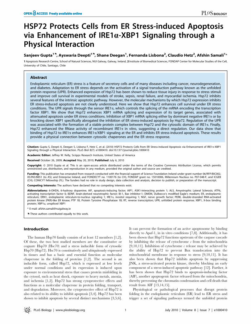
Figure 5. Hsp72 forms a protein complex with IRE1a, and the ATPase domain of Hsp72 is critical for IRE1a binding. (A) Schematicdiagram for IRE1a structure domains and expression constructs. (B) Schematic diagram for Hsp72 structure domains and expression constructs. (C)Hsp72 expressing PC12 cells (Hsp72) were transfected with empty vector (EV) or expression vectors for IRE1a FL-HA or IRE1a DC-HA. After 24 h, cellswere either untreated (Un) or treated with (0.25 mM) Tg for 12 h and then the co-precipitation of Hsp72 with IRE1a FL-HA or IRE1a DC-HA wasevaluated by IP and Western blot. (D) PC12 cells expressing the indicated Hsp72 constructs were transfected with IRE1a FL-HA. Lysate fromuntransfected PC12 cells (UT) was used as a negative control. Co-precipitation of wild-type, DATPase, and DEEVD mutant of Hsp72 with IRE1a FL-HAwas evaluated by IP and Western blot. (E) HEK 293 cells were transiently transfected with IRE1a FL-HA expression vector or empty vector (EV). After48 h, IRE1a FL-HA was immunoprecipitated and its association with endogenous Hsp72 was assessed by Western blot. (F) Endogenous Hsp72 was
Role of Hsp72 in the Unfolded Protein Response
PLoS Biology | www.plosbiology.org 8 July 2010 | Volume 8 | Issue 7 | e1000410
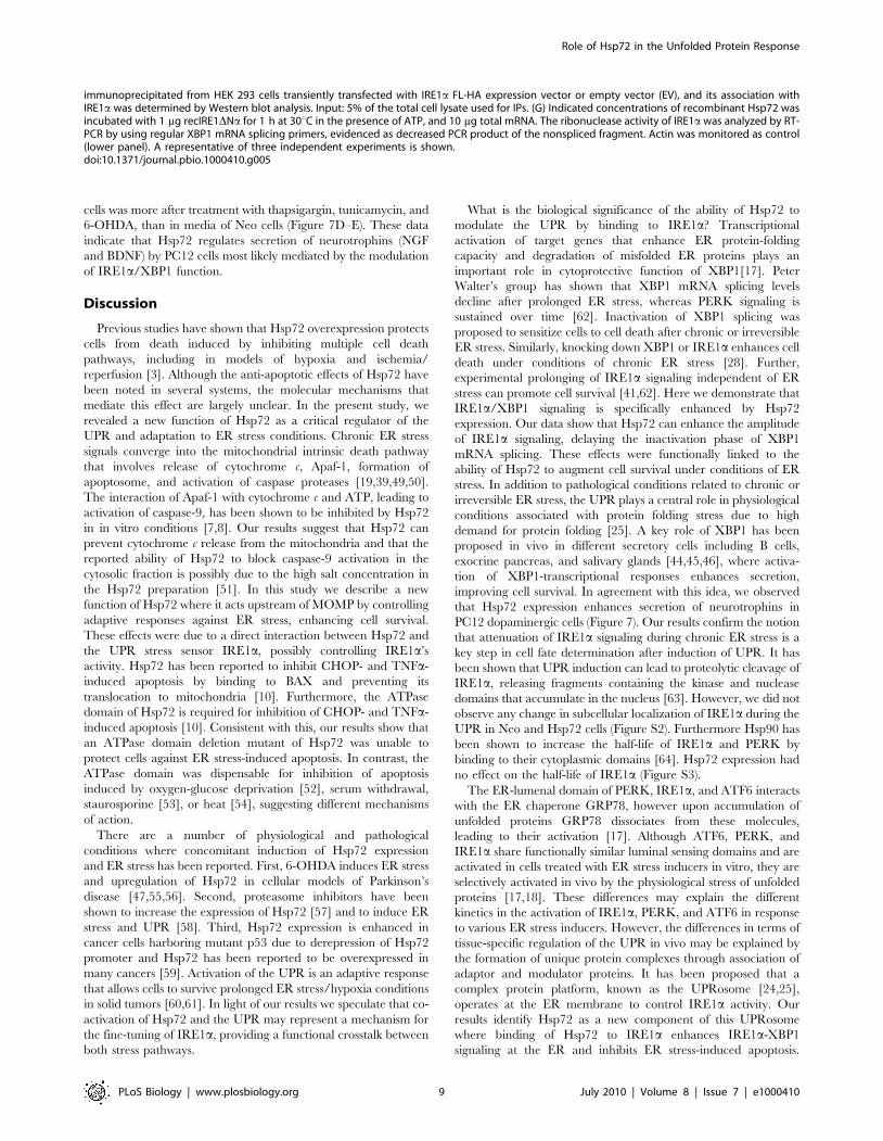
cells was more after treatment with thapsigargin, tunicamycin, and
6-OHDA, than in media of Neo cells (Figure 7D–E). These data
indicate that Hsp72 regulates secretion of neurotrophins (NGF
and BDNF) by PC12 cells most likely mediated by the modulation
of IRE1a/XBP1 function.
Discussion
Previous studies have shown that Hsp72 overexpression protects
cells from death induced by inhibiting multiple cell death
pathways, including in models of hypoxia and ischemia/
reperfusion [3]. Although the anti-apoptotic effects of Hsp72 have
been noted in several systems, the molecular mechanisms that
mediate this effect are largely unclear. In the present study, we
revealed a new function of Hsp72 as a critical regulator of the
UPR and adaptation to ER stress conditions. Chronic ER stress
signals converge into the mitochondrial intrinsic death pathway
that involves release of cytochrome c, Apaf-1, formation of
apoptosome, and activation of caspase proteases [19,39,49,50].
The interaction of Apaf-1 with cytochrome c and ATP, leading to
activation of caspase-9, has been shown to be inhibited by Hsp72
in in vitro conditions [7,8]. Our results suggest that Hsp72 can
prevent cytochrome c release from the mitochondria and that the
reported ability of Hsp72 to block caspase-9 activation in the
cytosolic fraction is possibly due to the high salt concentration in
the Hsp72 preparation [51]. In this study we describe a new
function of Hsp72 where it acts upstream of MOMP by controlling
adaptive responses against ER stress, enhancing cell survival.
These effects were due to a direct interaction between Hsp72 and
the UPR stress sensor IRE1a, possibly controlling IRE1a’s
activity. Hsp72 has been reported to inhibit CHOP- and TNFa-
induced apoptosis by binding to BAX and preventing its
translocation to mitochondria [10]. Furthermore, the ATPase
domain of Hsp72 is required for inhibition of CHOP- and TNFa-
induced apoptosis [10]. Consistent with this, our results show that
an ATPase domain deletion mutant of Hsp72 was unable to
protect cells against ER stress-induced apoptosis. In contrast, the
ATPase domain was dispensable for inhibition of apoptosis
induced by oxygen-glucose deprivation [52], serum withdrawal,
staurosporine [53], or heat [54], suggesting different mechanisms
of action.
There are a number of physiological and pathological
conditions where concomitant induction of Hsp72 expression
and ER stress has been reported. First, 6-OHDA induces ER stress
and upregulation of Hsp72 in cellular models of Parkinson’s
disease [47,55,56]. Second, proteasome inhibitors have been
shown to increase the expression of Hsp72 [57] and to induce ER
stress and UPR [58]. Third, Hsp72 expression is enhanced in
cancer cells harboring mutant p53 due to derepression of Hsp72
promoter and Hsp72 has been reported to be overexpressed in
many cancers [59]. Activation of the UPR is an adaptive response
that allows cells to survive prolonged ER stress/hypoxia conditions
in solid tumors [60,61]. In light of our results we speculate that co-
activation of Hsp72 and the UPR may represent a mechanism for
the fine-tuning of IRE1a, providing a functional crosstalk between
both stress pathways.
What is the biological significance of the ability of Hsp72 to
modulate the UPR by binding to IRE1a? Transcriptional
activation of target genes that enhance ER protein-folding
capacity and degradation of misfolded ER proteins plays an
important role in cytoprotective function of XBP1[17]. Peter
Walter’s group has shown that XBP1 mRNA splicing levels
decline after prolonged ER stress, whereas PERK signaling is
sustained over time [62]. Inactivation of XBP1 splicing was
proposed to sensitize cells to cell death after chronic or irreversible
ER stress. Similarly, knocking down XBP1 or IRE1a enhances cell
death under conditions of chronic ER stress [28]. Further,
experimental prolonging of IRE1a signaling independent of ER
stress can promote cell survival [41,62]. Here we demonstrate that
IRE1a/XBP1 signaling is specifically enhanced by Hsp72
expression. Our data show that Hsp72 can enhance the amplitude
of IRE1a signaling, delaying the inactivation phase of XBP1
mRNA splicing. These effects were functionally linked to the
ability of Hsp72 to augment cell survival under conditions of ER
stress. In addition to pathological conditions related to chronic or
irreversible ER stress, the UPR plays a central role in physiological
conditions associated with protein folding stress due to high
demand for protein folding [25]. A key role of XBP1 has been
proposed in vivo in different secretory cells including B cells,
exocrine pancreas, and salivary glands [44,45,46], where activa-
tion of XBP1-transcriptional responses enhances secretion,
improving cell survival. In agreement with this idea, we observed
that Hsp72 expression enhances secretion of neurotrophins in
PC12 dopaminergic cells (Figure 7). Our results confirm the notion
that attenuation of IRE1a signaling during chronic ER stress is a
key step in cell fate determination after induction of UPR. It has
been shown that UPR induction can lead to proteolytic cleavage of
IRE1a, releasing fragments containing the kinase and nuclease
domains that accumulate in the nucleus [63]. However, we did not
observe any change in subcellular localization of IRE1a during the
UPR in Neo and Hsp72 cells (Figure S2). Furthermore Hsp90 has
been shown to increase the half-life of IRE1a and PERK by
binding to their cytoplasmic domains [64]. Hsp72 expression had
no effect on the half-life of IRE1a (Figure S3).
The ER-lumenal domain of PERK, IRE1a, and ATF6 interacts
with the ER chaperone GRP78, however upon accumulation of
unfolded proteins GRP78 dissociates from these molecules,
leading to their activation [17]. Although ATF6, PERK, and
IRE1a share functionally similar luminal sensing domains and are
activated in cells treated with ER stress inducers in vitro, they are
selectively activated in vivo by the physiological stress of unfolded
proteins [17,18]. These differences may explain the different
kinetics in the activation of IRE1a, PERK, and ATF6 in response
to various ER stress inducers. However, the differences in terms of
tissue-specific regulation of the UPR in vivo may be explained by
the formation of unique protein complexes through association of
adaptor and modulator proteins. It has been proposed that a
complex protein platform, known as the UPRosome [24,25],
operates at the ER membrane to control IRE1a activity. Our
results identify Hsp72 as a new component of this UPRosome
where binding of Hsp72 to IRE1a enhances IRE1a-XBP1
signaling at the ER and inhibits ER stress-induced apoptosis.
immunoprecipitated from HEK 293 cells transiently transfected with IRE1a FL-HA expression vector or empty vector (EV), and its association withIRE1a was determined by Western blot analysis. Input: 5% of the total cell lysate used for IPs. (G) Indicated concentrations of recombinant Hsp72 wasincubated with 1 mg recIRE1DNa for 1 h at 30uC in the presence of ATP, and 10 mg total mRNA. The ribonuclease activity of IRE1a was analyzed by RT-PCR by using regular XBP1 mRNA splicing primers, evidenced as decreased PCR product of the nonspliced fragment. Actin was monitored as control(lower panel). A representative of three independent experiments is shown.doi:10.1371/journal.pbio.1000410.g005
Role of Hsp72 in the Unfolded Protein Response
PLoS Biology | www.plosbiology.org 9 July 2010 | Volume 8 | Issue 7 | e1000410

Our results suggest that Hsp72 can bind to the monomeric and
nonphosphorylated cytoplasmic tail of IRE1a. Furthermore the
interaction of Hsp72 and IRE1a is not affected by ER stress
mediated phosphorylation and oligomerization of IRE1a. What is
the molecular mechanism by which Hsp72 stimulates the RNase
activity of IRE1a? There are several possibilities by which Hsp72
might regulate RNase activity of IRE1a. Hsp72 may regulate
IRE1a either by allosteric interactions or by altering the binding of
Figure 6. The ATPase domain of Hsp72 is necessary for activation of IRE1a/XBP1 axis and inhibition of ER stress-induced apoptosis.(A) The control (Neo), wild-type Hsp72, and DATPase Hsp72 expressing PC12 cells were treated with (0.25 mM) Tg for 12 h and the expression levels ofindicated genes were quantified by real-time RT-PCR, normalizing against GAPDH. Average and error bars represent mean 6 SD from twoindependent experiments performed in triplicate. ** indicates a statistical significance between Hsp72 and Hsp72 DATPase cells; p,0.005. (B) Thecontrol (Neo), wild-type Hsp72, and DATPase Hsp72 expressing PC12 cells were either untreated (Un) or treated (0.25 mM) Tg or (2 mg/ml) Tm for48 h, and cell viability was determined using MTT assay. Average and error bars represent mean 6 SD from three independent experimentsperformed in triplicate. ** indicates a statistical significance between Hsp72 and Hsp72 DATPase cells; p,0.005. (C) The control (Neo), wild-typeHsp72, and DATPase Hsp72 expressing PC12 cells were treated as in (B), and DEVDase activity was measured as described in Materials and Methods.Average and error bars represent mean 6 SD from four independent experiments performed in duplicate. ** indicates a statistical significancebetween Hsp72 and Hsp72 DATPase cells; p,0.005.doi:10.1371/journal.pbio.1000410.g006
Role of Hsp72 in the Unfolded Protein Response
PLoS Biology | www.plosbiology.org 10 July 2010 | Volume 8 | Issue 7 | e1000410
other regulatory proteins (BAX, BAK, BI-1, AIP1, and RACK) to
IRE1a. Our results showing that recombinant Hsp72 can enhance
the RNase activity of purified IRE1a in in vitro conditions suggest
an allosteric mechanism (Figure 5G). However it is possible that in
a cellular context other mechanisms such as altering the binding of
other regulatory proteins (BAX, BAK, BI-1, AIP1, and RACK) to
IRE1a are also involved. Previous reports and our data collectively
support a model in which a fine balance of anti- and pro-apoptotic
Figure 7. Regulation of IRE1a-XBP1 by Hsp72 contributes to thermotolerance against ER stress and increased secretion ofneurotrophins. (A) PC12 cells were transduced with lentivirus expressing control non-targeting shRNA or XBP1 targeting shRNA. RT-PCR analysis oftotal RNA was performed to simultaneously detect unspliced XBP1 mRNA and GAPDH. The image is presented inverted for greater clarity. (B) Thecontrol (PGIPZ) or XBP1 shRNA expressing (XBP1 shRNA) PC12 cells were heat shocked for 1 h at 42uC and left to recover for 6 h. Western blots onwhole cell lysates were carried out to check the expression of Hsp72 after heat shock with b-actin as loading control. (C) Normal (PGIPZ C, XBP1shRNAC) and thermotolerant (PGIPZ HS, XBP shRNA HS) control and XBP1 shRNA expressing PC12 cells were either untreated (Un) or treated with (0.25 mM)Tg for 48 h, (2 mg/ml) Tm for 48 h, (150 nM) staurosporine (STS) for 16 h, or (25 mg/ml) etoposide (ETOP) for 24 h. The reduction in cell viability wasdetermined by MTT assay. Average and error bars represent mean 6 SD from three independent experiments performed in triplicate. (D–E) Thecontrol (Neo) and Hsp72 expressing (Hsp72) PC12 cells were either untreated (Un) or treated with (0.1 mM) Tg, (0.5 mg/ml) Tm, or (50 mM) 6-OHDA for24 h. Culture supernatant was analyzed for NGF and BDNF according to the conditions as described in Materials and Methods. Average and error barsrepresent mean 6 SD from three independent experiments performed in triplicate. * indicates a statistical significance between Neo and Hsp72 cells;p,0.05. ** indicates a statistical significance between Neo and Hsp72 cells; p,0.005.doi:10.1371/journal.pbio.1000410.g007
Role of Hsp72 in the Unfolded Protein Response
PLoS Biology | www.plosbiology.org 11 July 2010 | Volume 8 | Issue 7 | e1000410

proteins at the ER membrane modulates the amplitude of IRE1asignaling, thereby regulating the cellular sensitivity to ER stress
conditions.
Materials and Methods
Cell Culture and TreatmentRat pheochromocytoma PC12 cells (obtained from ECACC)
were cultured in Dulbecco’s modified Eagle’s medium (DMEM)
from Sigma (D6429) supplemented with 10% heat inactivated
horse serum, 5% foetal bovine serum, and 1% penicillin/
streptomycin (Sigma) at 37uC, 5% CO2 in humidified incubator.
Appropriate number of cells was seeded 24 h prior to treatment.
To induce apoptosis, cells were treated with 0.25 mM thapsigargin,
2 mg/ml tunicamycin, 150 nM staurosporine, or 25 mg/ml
etoposide for the indicated time periods. Stock solutions of 6-
Hydroxydopamine were made freshly in sodium metabisulfite
(1 M) prior to experiment. PC12 cells were treated with 200 mM
6-OHDA for 24 h before analysis. All reagents were from Sigma-
Aldrich unless otherwise stated.
Plasmids and TransfectionThe plasmid expressing wild type Hsp72, DATPase-Hsp72, or
DEEVD-Hsp72 under the CMV promoter were kind gifts from
Dr. Tomomi Gotoh, Kumamoto University, Japan [10]. The
expression vector for wild type IRE1a, IRE1a KA, IRE1a DC,
and IRE1a DRNase under the CMV promoter were kind gifts
from Dr. Kazunori Imaizumi, University of Miyazaki, Japan [65]
and expression plasmids for wild type IRE1a-HA or IRE1a DC-
HA are reported previously [28]. The plasmids containing
shRNAs targeting rat XBP1 were obtained from GeneCopoeia,
Rockville, USA (RSH045024-HIV U6). Transfections of cells
were carried out using Lipofectamine 2000 (Invitrogen) according
to the manufacturer’s protocol.
Cell Viability AssayViability of cells after treatment was analyzed by MTT assay.
After 48 h of treatment, 1 mg/ml concentration of MTT ((3-(4, 5-
dimethylthiazol-2-yl)-2, 5-diphenyl tetrazonium bromide) was
added to the wells and incubated at 37uC for 3 h. The reaction
was stopped with a stop mix containing 20% SDS in 40%
dimethyl formamide. The color intensity is measured at 550 nm
and percentage cell viability is calculated using the untreated
samples as 100%.
Annexin V StainingExternalization of phosphatidylserine (PS) to the outer leaflet of
the plasma membrane of apoptotic cells was assessed with annexin
V-FITC as described earlier [66]. Briefly, cells were collected by
centrifugation at 350 g, washed once in ice-cold calcium buffer
(10 mM HEPES/NaOH, pH 7.4, 140 mM NaCl, 2.5 mM
CaCl2), and incubated with annexin V-FITC or with annexin
V-PE for 15 min on ice. Prior to analysis 300 ml of binding buffer
containing 4 ml of PI (50 mg/ml) was added and analyzed on a
FACSCalibur flow cytometer (Becton Dickinson).
Analysis of DEVDase ActivityCells were harvested and pelleted by centrifugation at 350 g.
After washing in PBS, cell pellets were re-suspended in 50 ml of
PBS and 25 ml was transferred to duplicate wells of a microtiter
plate and snap-frozen in liquid nitrogen. To initiate the reaction,
50 mM of the caspase substrate carbobenzoxy-Asp-Glu-Val-Asp-7-
amino-4-methyl-coumarin (DEVD-AMC, Peptide Institute Inc.)
in assay buffer (100 mM HEPES, pH 7.5, 10% sucrose, 0.1%
CHAPS, 5 mM DTT and 0.0001% Igepal-630, pH 7.25) was
added to cell lysates. Liberated free AMC was measured by a
Wallac Victor 1420 Multilabel counter (Perkin Elmer Life
Sciences) using 355 nm excitation and 460 nm emission wave-
lengths at 37uC at 60 s intervals for 25 cycles. The data were
analyzed by linear regression and enzyme activity was expressed as
nM of AMC released 6min216mg21 total cellular protein.
Measurement of DYm
Mitochondrial transmembrane potential was determined by
using the fluorescent probe tetramethylrhodamine ethyl ester
(TMRE, Molecular Probes) as previously described [67]. Briefly,
cells were trypsinized and incubated with TMRE at RT for
30 min in the dark and analyzed by flow cytometry using a
FACSCalibur instrument.
RNA Extraction, RT-PCR, and Real Time RT-PCRTotal RNA was isolated using RNeasy kit (Qiagen) according to
the manufacturer’s instructions. Reverse transcription (RT) was
carried out with 2 mg RNA and Oligo dT (Invitrogen) using 20 U
Superscript II Reverse Transcriptase (Invitrogen). The cDNA
product was subjected to 25–35 cycles of PCR using the forward
primer 5-TTACGAGAGAAAACTCATGGGC-3 and reverse
primer 5-GGGTCCAACTTGTCCAGAATGC-3 specific for
Rat XBP1. GAPDH (forward: ACCACAGTCCATGCCATC;
reverse: TCCACCACCTGTTGCTG) was used as an endoge-
nous control. Real-time PCR method to determine the induction
of UPR target genes has been described previously[68]. Briefly,
cDNA products were mixed with 26TaqMan master mixes and
206 TaqMan Gene Expression Assays (Applied Biosystems) and
subjected to 40 cycles of PCR in StepOnePlus instrument (Applied
Biosystems). Relative expression was evaluated with DDCT
method.
Preparation of Cytosolic ExtractsThe cells were washed in ice-cold PBS and lysed using cell lysis
and mitochondrial intact buffer (CLAMI) containing 250 mM
sucrose, 70 mM KCl dissolved in 16PBS with 0.5 mM DTT and
2.5 mg/ml Pepstatin and 0.2 g/ml digitonin. The cells were
allowed to swell on ice for 5 min. The cell suspension was
centrifuged at 400 g for 5 min and the pellet was removed. The
supernatant was transferred to a clean eppendorf tube and the
mitochondrial and microsomal fractions were separated by
spinning at 20,000 g for 5 min. The cytosolic fraction was
removed and prepared for Western blot by adding 56 sample
buffer.
Western BlottingCells were washed once in ice-cold PBS and lysed in whole cell
lysis buffer (20 mM HEPES pH 7.5, 350 mM NaCl, 0.5 mM
EDTA, 1 mM MgCl2, 0.1 mM EGTA, and 1% NP-40) after
stipulated time of treatments and boiled at 95uC with Laemmli’s
SDS-PAGE sample buffer for 5 min. Protein concentration was
determined by Bradford method. Equal amounts (20 mg/lane) of
protein samples were run on an SDS polyacrylamide gel. The
proteins were transferred onto nitrocellulose membrane and
blocked with 5% milk in PBS-0.05% Tween. The membrane
was incubated with the primary antibody Hsp72 (Stressgen SPA-
810), Caspase-3 (Cell Signaling Technology, Cat# 9662),
Cytochrome c (BD Pharmingen, Cat# 556433), XBP1 (Santa
Cruz Biotechnology, Inc, Cat# sc-7160), CHOP (Santa Cruz
Biotechnology, Inc, Cat# sc-793), phosphorylated eIF2a (Cell
Signaling Technology, Cat# 3597), total eIF2a (Cell Signaling
Role of Hsp72 in the Unfolded Protein Response
PLoS Biology | www.plosbiology.org 12 July 2010 | Volume 8 | Issue 7 | e1000410

Technology, Cat# 2103), phosphorylated JNK (Cell Signaling,
Cat# 9255S), IRE1a (Cell Signaling Technology, Cat# 3294S),
or b-Actin (Sigma, Cat# A-5060) for 2 h at room temperature or
overnight at 4uC. The membrane was washed 3 times with PBS-
0.05% Tween and further incubated in appropriate horseradish
peroxidase-conjugated secondary antibody (Pierce) for 90 min.
Signals were detected using West pico chemiluminescent substrate
(Pierce).
ImmunoprecipitationsImmunoprecipitation of HA-tagged wild-type IRE1a or IRE1a
DC was performed using Pierce Profound mammalian HA tagged
IP/Co-IP kit (23615). Briefly, cell lysates were incubated with HA-
agarose slurry in IP column overnight. Agarose beads were washed
twice with TBS containing 0.05% Tween. Protein complexes were
extracted by boiling the beads with 26 lane marker buffer and
analyzed by Western blotting as described above. For immuno-
precipitation of Hsp72, cleared protein extracts were incubated
with anti-Hsp72 polyclonal antibody (Stressgen SPA-811) over-
night at 4uC, followed by 100 ml of a 12% suspension of protein A-
Sepharose for 1 h at 4uC, and then washed three times with TBS-
0.05% Tween. Protein complexes were eluted by boiling in 26lane marker buffer and analyzed by Western blotting as described
above.
In Vitro IRE1a Activity AssaysThe effect of Hsp72 on activity of IREa was monitored using
recombinant human IRE1aDN-HIS produced as GST fusion
protein using the Prescission Protease cleaved system. IRE1a DN
was incubated with recombinant Hsp70 (Stressgene) in a total
volume of 50 ml for 1 h at 30uC with 10 mg of total mRNA as
substrate (obtained from mouse brain cortex because of minimal
basal levels of spliced XBP1 mRNA) in a buffer containing 20 mM
HEPES (pH 7.3), 1 mM DTT, 10 mM magnesium acetate,
50 mM potassium acetate, and 2 mM ATP. Then, mRNA was
re-extracted with 500 ml of Trizol, and the endoribonuclease
activity of IRE1a was monitored by RT-PCR using the XBP1
mRNA splicing assay that employs a set of primers that closely
surround the processing site. Using this method, we observed a
decrease in the amount of nonspliced XBP1 mRNA due to its
cleavage by IRE1aN-HIS as we previously described [28].
Knockdown of XBP1We generated stable subclones of PC12-Hsp72 with reduced
levels of XBP1 by targeting XBP1 mRNA with shRNA using the
lentiviral expression vector psiHIV-U6 (GeneCopoeia). The
targeting sequences identified for rat XBP1 were XBP1 shRNA1:
5-actgcgcgagatagaaaga-3; XBP1 shRNA2: 5- gttgcctcttcagattctg-3;
XBP1 shRNA3: 5-gagagccaaactaatgtgg-3; and XBP1 shRNA4: 5-
ctgaggtcttcaaaggtat-3.
Heat Preconditioning of PC12 CellsCells were seeded in T25 flasks 24 h prior to heat precondi-
tioning. The flasks were sealed with parafilm and immersed in
water bath set at 42uC for 1 h. The cells were left to recover for 6
h at 37uC before treating with apoptosis inducing agents. Media
was changed prior to treatment.
ELISA for BDNF and NGF ReleaseCells were treated with 0.1 mM Tg or 0.5 mg/ml Tm or 50 mM
6-OHDA for 24 h to induce UPR. Culture media was analyzed
for NGF or BDNF release using b-NGF (DY 256) or BDNF (DY
248) DuoSet ELISA development kit according to manufacturer’s
protocol (R&D Systems). The amount of NGF or BDNF released
into the media was calculated using the standard curve generated
in parallel with recombinant NGF and BDNF.
Statistical AnalysisAll the experiments were repeated at least 2 times. Results are
expressed as mean 6 standard deviation. Statistical analyses of the
results were done with Student’s t test using Graphpad (http://
www.graphpad.com).
Supporting Information
Figure S1 Overexpression of Hsp72 protects PC12 cellsfrom 6-OHDA treatment induced cell death. The control
(Neo) and Hsp72 expressing (Hsp72) PC12 cells were either
untreated (Un) or treated with 200 mM 6-OHDA for 24 h.
Reduction in cell viability was analyzed by sub G1 peak. Average
and error bars represent mean 6 SD from three independent
experiments performed in triplicates.
Found at: doi:10.1371/journal.pbio.1000410.s001 (0.06 MB TIF)
Figure S2 Subcellular localization of IRE1a is notaffected by Hsp72. Hsp72 and Neo expressing cells weretransfected with IRE1a-GFP. After 24 h of treatment with Tg,
cells were fixed in 3.7% formaldehyde and the coverslips were
mounted using Vectashield mounting medium with DAPI (H-
1200). Cells were visualized using a Nikon microscope fitted with
appropriate filters.
Found at: doi:10.1371/journal.pbio.1000410.s002 (0.56 MB TIF)
Figure S3 Overexpression of Hsp72 does not affect thehalf-life of IRE1. (A) Control (Neo) and Hsp72 expressing
(Hsp72) PC12 cells were treated with (10 ng/ml) Tg for the
indicated time. Immunoblotting of total protein was performed
using antibodies against IRE1a and b-actin. (B) In the experiment
described in (A), band density of IRE1a was calculated by
densitometry and normalized against b-actin. Average and error
bars represent mean 6 SD from two independent experiments.
Found at: doi:10.1371/journal.pbio.1000410.s003 (0.17 MB TIF)
Acknowledgments
We are grateful to Dr. Sandra Healy for critical reading of this manuscript
and to the Technical Officers and administrative teams in Biochemistry,
School of Natural Sciences, NUI, Galway. We would like to thank Dr.
Tomomi Gotoh, Kumamoto University, Japan, and Dr. Kazunori
Imaizumi, University of Miyazaki, Japan, for sharing invaluable reagents
generated in their laboratories.
Author Contributions
The author(s) have made the following declarations about their
contributions: Conceived and designed the experiments: SG FL CH AS.
Performed the experiments: SG AD. Analyzed the data: SG AD FL CH.
Contributed reagents/materials/analysis tools: SD. Wrote the paper: SG
AD FL CH AS.
References
1. Morimoto RI, Kline MP, Bimston DN, Cotto JJ (1997) The heat-shock response:
regulation and function of heat-shock proteins and molecular chaperones. Essays
Biochem 32: 17–29.
2. Tavaria M, Gabriele T, Kola I, Anderson RL (1996) A hitchhiker’s guide to the
human Hsp70 family. Cell Stress Chaperones 1: 23–28.
3. Garrido C, Schmitt E, Cande C, Vahsen N, Parcellier A, et al. (2003) HSP27
and HSP70: potentially oncogenic apoptosis inhibitors. Cell Cycle 2: 579–
584.
4. Samali A, Orrenius S (1998) Heat shock proteins: regulators of stress response
and apoptosis. Cell Stress Chaperones 3: 228–236.
Role of Hsp72 in the Unfolded Protein Response
PLoS Biology | www.plosbiology.org 13 July 2010 | Volume 8 | Issue 7 | e1000410

5. Samali A, Cotter TG (1996) Heat shock proteins increase resistance to apoptosis.
Exp Cell Res 223: 163–170.
6. Lanneau D, Brunet M, Frisan E, Solary E, Fontenay M, et al. (2008) Heat shock
proteins: essential proteins for apoptosis regulation. J Cell Mol Med 12:
743–761.
7. Saleh A, Srinivasula SM, Balkir L, Robbins PD, Alnemri ES (2000) Negativeregulation of the Apaf-1 apoptosome by Hsp70. Nat Cell Biol 2: 476–483.
8. Beere HM, Wolf BB, Cain K, Mosser DD, Mahboubi A, et al. (2000) Heat-
shock protein 70 inhibits apoptosis by preventing recruitment of procaspase-9 to
the Apaf-1 apoptosome. Nat Cell Biol 2: 469–475.
9. Bivik C, Rosdahl I, Ollinger K (2007) Hsp70 protects against UVB induced
apoptosis by preventing release of cathepsins and cytochrome c in human
melanocytes. Carcinogenesis 28: 537–544.
10. Gotoh T, Terada K, Oyadomari S, Mori M (2004) hsp70-DnaJ chaperone pairprevents nitric oxide- and CHOP-induced apoptosis by inhibiting translocation
of Bax to mitochondria. Cell Death Differ 11: 390–402.
11. Stankiewicz AR, Lachapelle G, Foo CP, Radicioni SM, Mosser DD (2005)
Hsp70 inhibits heat-induced apoptosis upstream of mitochondria by preventing
Bax translocation. J Biol Chem 280: 38729–38739.
12. Gabai VL, Meriin AB, Yaglom JA, Volloch VZ, Sherman MY (1998) Role of
Hsp70 in regulation of stress-kinase JNK: implications in apoptosis and aging.
FEBS Lett 438: 1–4.
13. Gurbuxani S, Schmitt E, Cande C, Parcellier A, Hammann A, et al. (2003) Heatshock protein 70 binding inhibits the nuclear import of apoptosis-inducing
factor. Oncogene 22: 6669–6678.
14. Kroemer G (2001) Heat shock protein 70 neutralizes apoptosis-inducing factor.
ScientificWorld Journal 1: 590–592.
15. Matsumori Y, Hong SM, Aoyama K, Fan Y, Kayama T, et al. (2005) Hsp70
overexpression sequesters AIF and reduces neonatal hypoxic/ischemic brain
injury. J Cereb Blood Flow Metab 25: 899–910.
16. Lin JH, Walter P, Yen TS (2008) Endoplasmic reticulum stress in disease
pathogenesis. Annu Rev Pathol 3: 399–425.
17. Ron D, Walter P (2007) Signal integration in the endoplasmic reticulum
unfolded protein response. Nat Rev Mol Cell Biol 8: 519–529.
18. Schroder M, Kaufman RJ (2005) The mammalian unfolded protein response.Annu Rev Biochem 74: 739–789.
19. Szegezdi E, Logue SE, Gorman AM, Samali A (2006) Mediators of endoplasmic
reticulum stress-induced apoptosis. EMBO Rep 7: 880–885.
20. Calfon M, Zeng H, Urano F, Till JH, Hubbard SR, et al. (2002) IRE1 couplesendoplasmic reticulum load to secretory capacity by processing the XBP-1
mRNA. Nature 415: 92–96.
21. Yamamoto K, Yoshida H, Kokame K, Kaufman RJ, Mori K (2004) Differential
contributions of ATF6 and XBP1 to the activation of endoplasmic reticulum
stress-responsive cis-acting elements ERSE, UPRE and ERSE-II. J Biochem136: 343–350.
22. Lee AH, Iwakoshi NN, Glimcher LH (2003) XBP-1 regulates a subset of
endoplasmic reticulum resident chaperone genes in the unfolded protein
response. Mol Cell Biol 23: 7448–7459.
23. Yoshida H, Matsui T, Yamamoto A, Okada T, Mori K (2001) XBP1 mRNA is
induced by ATF6 and spliced by IRE1 in response to ER stress to produce a
highly active transcription factor. Cell 107: 881–891.
24. Hetz C, Glimcher L (2008) The daily job of night killers: alternative roles of theBCL-2 family in organelle physiology. Trends Cell Biol 18: 38–44.
25. Hetz C, Glimcher LH (2009) Fine-tuning of the unfolded protein response:
assembling the IRE1alpha interactome. Mol Cell 35: 551–561.
26. Hetz C, Bernasconi P, Fisher J, Lee AH, Bassik MC, et al. (2006) ProapoptoticBAX and BAK modulate the unfolded protein response by a direct interaction
with IRE1alpha. Science 312: 572–576.
27. Luo D, He Y, Zhang H, Yu L, Chen H, et al. (2008) AIP1 is critical in
transducing IRE1-mediated endoplasmic reticulum stress response. J Biol Chem
283: 11905–11912.
28. Lisbona F, Rojas-Rivera D, Thielen P, Zamorano S, Todd D, et al. (2009) BAX
inhibitor-1 is a negative regulator of the ER stress sensor IRE1alpha. Mol Cell
33: 679–691.
29. Xu C, Xu W, Palmer AE, Reed JC (2008) BI-1 regulates endoplasmic reticulumCa2+ homeostasis downstream of Bcl-2 family proteins. J Biol Chem 283:
11477–11484.
30. Gu F, Nguyen DT, Stuible M, Dube N, Tremblay ML, et al. (2004) Protein-
tyrosine phosphatase 1B potentiates IRE1 signaling during endoplasmic
reticulum stress. J Biol Chem 279: 49689–49693.
31. Yenari MA, Giffard RG, Sapolsky RM, Steinberg GK (1999) The neuropro-
tective potential of heat shock protein 70 (HSP70). Mol Med Today 5: 525–531.
32. Giffard RG, Yenari MA (2004) Many mechanisms for hsp70 protection fromcerebral ischemia. J Neurosurg Anesthesiol 16: 53–61.
33. Xu L, Lee JE, Giffard RG (1999) Overexpression of bcl-2, bcl-XL or hsp70 in
murine cortical astrocytes reduces injury of co-cultured neurons. Neurosci Lett
277: 193–197.
34. Plumier JC, Krueger AM, Currie RW, Kontoyiannis D, Kollias G, et al. (1997)
Transgenic mice expressing the human inducible Hsp70 have hippocampal
neurons resistant to ischemic injury. Cell Stress Chaperones 2: 162–167.
35. Rajdev S, Hara K, Kokubo Y, Mestril R, Dillmann W, et al. (2000) Mice
overexpressing rat heat shock protein 70 are protected against cerebralinfarction. Ann Neurol 47: 782–791.
36. Kelly S, Zhang ZJ, Zhao H, Xu L, Giffard RG, et al. (2002) Gene transfer of
HSP72 protects cornu ammonis 1 region of the hippocampus neurons from
global ischemia: influence of Bcl-2. Ann Neurol 52: 160–167.
37. Papadopoulos MC, Sun XY, Cao J, Mivechi NF, Giffard RG (1996) Over-
expression of HSP-70 protects astrocytes from combined oxygen-glucose
deprivation. Neuroreport 7: 429–432.
38. Zamzami N, Kroemer G (2001) The mitochondrion in apoptosis: how Pandora’s
box opens. Nat Rev Mol Cell Biol 2: 67–71.
39. Gupta S, Kass GE, Szegezdi E, Joseph B (2009) The mitochondrial death
pathway: a promising therapeutic target in diseases. J Cell Mol Med.
40. Yoshida H, Matsui T, Hosokawa N, Kaufman RJ, Nagata K, et al. (2003) A
time-dependent phase shift in the mammalian unfolded protein response. Dev
Cell 4: 265–271.
41. Lin JH, Li H, Zhang Y, Ron D, Walter P (2009) Divergent effects of PERK and
IRE1 signaling on cell viability. PLoS ONE 4: e4170. doi:10.1371/journal.pone.
0004170.
42. Miura M, Yuan J (2000) Transient transfection assay of cell death genes.
Methods Enzymol 322: 480–492.
43. Quigney DJ, Gorman AM, Samali A (2003) Heat shock protects PC12 cells
against MPP+ toxicity. Brain Res 993: 133–139.
44. Iwakoshi NN, Lee AH, Vallabhajosyula P, Otipoby KL, Rajewsky K, et al.
(2003) Plasma cell differentiation and the unfolded protein response intersect at
the transcription factor XBP-1. Nat Immunol 4: 321–329.
45. Lee AH, Chu GC, Iwakoshi NN, Glimcher LH (2005) XBP-1 is required for
biogenesis of cellular secretory machinery of exocrine glands. EMBO J 24:
4368–4380.
46. Reimold AM, Iwakoshi NN, Manis J, Vallabhajosyula P, Szomolanyi-Tsuda E,
et al. (2001) Plasma cell differentiation requires the transcription factor XBP-1.
Nature 412: 300–307.
47. Ryu EJ, Harding HP, Angelastro JM, Vitolo OV, Ron D, et al. (2002)
Endoplasmic reticulum stress and the unfolded protein response in cellular
models of Parkinson’s disease. J Neurosci 22: 10690–10698.
48. Matus S, Lisbona F, Torres M, Leon C, Thielen P, et al. (2008) The stress
rheostat: an interplay between the unfolded protein response (UPR) and
autophagy in neurodegeneration. Curr Mol Med 8: 157–172.
49. Kim I, Xu W, Reed JC (2008) Cell death and endoplasmic reticulum stress:
disease relevance and therapeutic opportunities. Nat Rev Drug Discov 7:
1013–1030.
50. Gupta S, Cuffe L, Szegezdi E, Logue SE, Neary C, et al. (2010) Mechanisms of
ER stress-mediated mitochondrial membrane permeabilization. Int J Cell
Biol;170215.
51. Steel R, Doherty JP, Buzzard K, Clemons N, Hawkins CJ, et al. (2004) Hsp72
inhibits apoptosis upstream of the mitochondria and not through interactions
with Apaf-1. J Biol Chem 279: 51490–51499.
52. Sun Y, Ouyang YB, Xu L, Chow AM, Anderson R, et al. (2006) The carboxyl-
terminal domain of inducible Hsp70 protects from ischemic injury in vivo and in
vitro. J Cereb Blood Flow Metab 26: 937–950.
53. Ravagnan L, Gurbuxani S, Susin SA, Maisse C, Daugas E, et al. (2001) Heat-
shock protein 70 antagonizes apoptosis-inducing factor. Nat Cell Biol 3:
839–843.
54. Volloch V, Gabai VL, Rits S, Sherman MY (1999) ATPase activity of the heat
shock protein hsp72 is dispensable for its effects on dephosphorylation of stress
kinase JNK and on heat-induced apoptosis. FEBS Lett 461: 73–76.
55. Holtz WA, O’Malley KL (2003) Parkinsonian mimetics induce aspects of
unfolded protein response in death of dopaminergic neurons. J Biol Chem 278:
19367–19377.
56. Gorman AM, Szegezdi E, Quigney DJ, Samali A (2005) Hsp27 inhibits 6-
hydroxydopamine-induced cytochrome c release and apoptosis in PC12 cells.
Biochem Biophys Res Commun 327: 801–810.
57. Bush KT, Goldberg AL, Nigam SK (1997) Proteasome inhibition leads to a
heat-shock response, induction of endoplasmic reticulum chaperones, and
thermotolerance. J Biol Chem 272: 9086–9092.
58. Szokalska A, Makowski M, Nowis D, Wilczynski GM, Kujawa M, et al. (2009)
Proteasome inhibition potentiates antitumor effects of photodynamic therapy in
mice through induction of endoplasmic reticulum stress and unfolded protein
response. Cancer Res 69: 4235–4243.
59. Ciocca DR, Calderwood SK (2005) Heat shock proteins in cancer: diagnostic,
prognostic, predictive, and treatment implications. Cell Stress Chaperones 10:
86–103.
60. Koumenis C, Naczki C, Koritzinsky M, Rastani S, Diehl A, et al. (2002)
Regulation of protein synthesis by hypoxia via activation of the endoplasmic
reticulum kinase PERK and phosphorylation of the translation initiation factor
eIF2alpha. Mol Cell Biol 22: 7405–7416.
61. Harris AL (2002) Hypoxia–a key regulatory factor in tumour growth. Nat Rev
Cancer 2: 38–47.
62. Lin JH, Li H, Yasumura D, Cohen HR, Zhang C, et al. (2007) IRE1 signaling
affects cell fate during the unfolded protein response. Science 318: 944–949.
63. Niwa M, Sidrauski C, Kaufman RJ, Walter P (1999) A role for presenilin-1 in
nuclear accumulation of Ire1 fragments and induction of the mammalian
unfolded protein response. Cell 99: 691–702.
64. Marcu MG, Doyle M, Bertolotti A, Ron D, Hendershot L, et al. (2002) Heat
shock protein 90 modulates the unfolded protein response by stabilizing
IRE1alpha. Mol Cell Biol 22: 8506–8513.
Role of Hsp72 in the Unfolded Protein Response
PLoS Biology | www.plosbiology.org 14 July 2010 | Volume 8 | Issue 7 | e1000410

65. Ogata M, Hino S, Saito A, Morikawa K, Kondo S, et al. (2006) Autophagy is
activated for cell survival after endoplasmic reticulum stress. Mol Cell Biol 26:9220–9231.
66. Concannon CG, FitzGerald U, Holmberg CI, Szegezdi E, Sistonen L, et al.
(2005) CD95-mediated alteration in Hsp70 levels is dependent on proteinstabilization. Cell Stress Chaperones 10: 59–65.
67. Samali A, Cai J, Zhivotovsky B, Jones DP, Orrenius S (1999) Presence of a pre-
apoptotic complex of pro-caspase-3, Hsp60 and Hsp10 in the mitochondrial
fraction of jurkat cells. EMBO J 18: 2040–2048.
68. Samali A, Fitzgerald U, Deegan S (0307) Methods for monitoring endoplasmic
reticulum stress and the unfolded protein response. Int J Cell Biol.
Role of Hsp72 in the Unfolded Protein Response
PLoS Biology | www.plosbiology.org 15 July 2010 | Volume 8 | Issue 7 | e1000410
Copyright © 2022 FDOKUMEN


![Saugu[!] Asmundar, er kalladur er Kappabani :](https://static.fdokumen.com/doc/165x107/63264a17051fac18490dae0e/saugu-asmundar-er-kalladur-er-kappabani-.jpg)