Paleointensity during a chron C26r excursion recorded in west Greenland lava flows
Upload
independentCategory
view
1download
0
1
How gravity and muscle action control medio-lateral center of mass excursion during slow walking: a simulation study. Karen Jansen1, Friedl De Groote2, Jacques Duysens3,4, Ilse Jonkers1
1 Human Movement Biomechanics Research Group, Department of Kinesiology, KU Leuven, Belgium
2 Department of Mechanical Engineering, KU Leuven, Belgium.
3 Department of Kinesiology, KU Leuven, Belgium
4 Department of Research, Development & Education, St. Maartenskliniek, Nijmegen, The Netherlands
Corresponding author:
Karen Jansen
Faculty of Kinesiology and Rehabilitation Sciences
Human Movement Biomechanics Research Group
Tervuursevest 101 – box 1501
3001 Heverlee, Belgium
E-mail: [email protected]
Tel.: +32 16 3 29009
Other authors:
Friedl De Groote
Department of Mechanical Engineering
Celestijnenlaan 300b - box 2420
3001 Heverlee, Belgium
E-mail: [email protected]
Jacques Duysens
Movement Control and Neuroplasticity Research Group
Tervuursevest 101 – box 1501
3001 Heverlee, Belgium
E-mail: [email protected]
Ilse Jonkers
Human Movement Biomechanics Research Group
Tervuursevest 101 – box 1501
3001 Heverlee, Belgium
2
E-mail: [email protected]
Acknowledgments
This work was funded by a grant of the KU Leuven Research council (IDO/07/012) and a grant of the
Flemish research council (KN 1.5.017.08).
3
Abstract
Maintaining mediolateral (ML) balance is very important to prevent falling during walking, especially
at very slow speeds. The effect of walking speed on support and propulsion of the center of mass (COM)
has been focus of previous studies. However, the influence of speed on ML COM control and the
associated coupling with sagittal plane control remains unclear. Simulations of walking at very slow and
normal speeds were generated for twelve healthy subjects. Our results show that gluteus medius
(GMED) contributions to ML stability decrease, while its contributions to sagittal plane accelerations
increase during very slow compared to normal walking. Simultaneously the destabilizing influence of
gravity increases in ML direction at a very slow walking speed. This emphasizes the need for a tight
balance between gravity and gluteus medius action to ensure ML stability. When walking speed
increases, GMED has a unique role in controlling ML acceleration and therefore stabilizing ML COM
excursion. Contributions of other muscles decrease in all directions during very slow speed. Increased
contributions of these muscles are therefore required to provide for both stability and propulsion when
walking speed increases.
Keywords
Stability; center of mass acceleration; walking speed; simulations; mediolateral balance

4
1. Introduction
A high incidence of falls is an important problem in modern society. In addition to the high
associated cost on health care, these falls also have a major impact on the quality of life1. Many of these
falls occur during walking.
To maintain balance during walking, control of the mediolateral (ML) motion of the center of mass
(COM) is crucial. Accordingly, deviations of the gait pattern in the ML direction are often suggested to be
a valid predictor of falls. Previous studies suggested step width (SW) and especially SW variability to be
related to fall risk2. During steady-state gait, older adults reduce the ML COM accelerations as a
compensatory mechanism to improve ML stability3.
These modifications in COM movement and therefore its control are likely to become important at
very slow speeds. Den Otter et al.4 found specific bursts of muscle activity at very slow speeds and
argued that these might be attributed to increased demands on postural stability. The direct relation to
ML COM control still needs to be further investigated. As several studies found indeed that slower
walking in elderly is associated with an increased risk of falls5, it is highly relevant to understand how
slow walking affects the ML COM control. To date, the relation between the ML and sagittal plane COM
control remains unclear. In the sagittal plane, support and progression are coupled and similar muscles
contribute to both in parallel. Principle component analysis of experimental electromyography (EMG)
patterns shows that a reduced set of muscle activation modules can generate both support and
propulsion6. This is also confirmed using muscle driven simulations7-9. Furthermore muscle contributions
to both progression and support generally decrease proportionally when walking speed decreases10-11.
Extending these insights to the ML COM control is less clear. Some experimental studies on posture
suggest that ML and anterior-posterior (AP) motions have a coupled control12. In contrast, other
researchers believe that COM control in the sagittal plane and ML stabilization are independent of each
5
other during quiet stance13 and balance perturbations14. Proof also exists for an independent control
during gait: with decreasing walking speed, local dynamic stability in AP and vertical direction is
enhanced, but stability in ML direction reduced15. This indicates that separate control of stability is
required in different directions. Likewise, analysis of passive walking models suggests that during
walking ML stability is actively controlled, while passive stability is provided in the sagittal plane16.
Simulation studies of steady-state walking17 investigated how individual muscles control ML balance.
They concluded that muscles responsible for sagittal plane accelerations also accelerated the COM
laterally (i.e. away from the midline) and that COM stability in all directions is controlled by similar
muscle groups. However, a recent study18 showed that control of ML accelerations of the COM requires
additional synergies to the ones controlling the COM in the sagittal plane. Hence, these studies do not
allow to uniformly conclude on sagittal plane versus ML COM control. Previous simulation studies10,11
mainly explored the effect of speed on the muscle contributions to COM control in the sagittal plane.
The study of Pandy et al.17 investigated ML COM stability, but only reported one single speed. Only
recently, a study of John et al.19 explored the effect of walking speed on muscle contributions to the
control of ML body motion. However, slowest speeds reported in their study are still largely above the
walking speeds previously reported in for instance stroke patients and the subjects involved were
children. Therefore, when exploring the role of individual muscle contributions to ML and sagittal plane
COM control it seems advantageous to investigate the trade-off between sagittal plane and ML control
also at very slow walking speeds and in an adult population.
In the present study, the effect of walking speed on ML and sagittal plane COM control is
investigated using simulations of walking at normal and very slow speed. More particularly, we
investigate if a decrease in speed affects similarly the muscle contributions in ML, vertical and AP
direction. If a common control exists for the different planes, one would expect that with decreasing
speed the observed decreases in muscle action (EMG) would have similar effects on the muscle
6
contributions to COM accelerations in the various planes. However, if changes in muscle contributions
to COM acceleration are specific for a given plane this would argue for separate control. One important
muscle to consider is the gluteus medius (GMED). This muscle decreases EMG activity with decreasing
gait speed20. Based on a simulation study Liu et al.10 concluded that GMED contributions to support are
‘relatively constant across walking speeds’. However, a significant difference is reported between free
and slow walking speed, with higher GMED contributions for the slow speeds.
Based on the assumption that the functional demands for progression decrease whereas stability
remains equal or also decreases when walking at very slow speeds, it is hypothesized that muscles that
act mainly in the sagittal plane will decrease their contributions in this plane, but maintain their ML
contribution. In contrast, muscles preferentially contributing to ML stability such as GMED mainly
decrease their contribution to ML accelerations of COM but maintain their contributions in the sagittal
plane. This will therefore confirm an uncoupling of the COM control in the ML and sagittal plane.
2. Methods
2.1. Experimental data
We collected three-dimensional kinematic and kinetic data of twelve healthy subjects (age: 25.8 ±
4.0 years, weight: 71.1 ± 8.9 kg, leg length: 0.9 ± 0.04 m) walking on treadmill at a speed of 1 km/h (very
slow) and 4 km/h (normal). The speed of 1 km/h was chosen because it is at the low end of the range of
preferred speeds in stroke patients21.
All subjects gave their informed consent prior to data collection and the experimental protocol was
approved by the local ethical committee. Marker (active infrared LEDs) trajectories were collected at

7
100Hz using a two-beam camera system (Krypton, Nikon Metrology NV, Belgium). The marker protocol
consisted of six technical clusters and 16 additional individual markers; this protocol was described more
detailed in a previous paper22. Ground reaction forces (GRF) and torques were measured at 1000 Hz
using a force-plate instrumented split-belt treadmill (Forcelink, The Netherlands). EMG data were
collected bilaterally at 1000 Hz for tibialis anterior, gastrocnemius lateralis, soleus (SOL), vastus lateralis,
rectus femoris (RF), biceps femoris and semitendinosus using a wireless EMG system (Zero-wire EMG,
Aurion, Italy). As part of the post-processing, the raw EMG signal was band-pass filtered between 10-
500Hz using a fourth order digital Butterworth filter and RMS was calculated using a 50 ms time window.
2.2. Simulations
Subject-specific simulations of both walking speeds were generated using a dedicated workflow in
OpenSim23. In a first step, data of a static trial was used to scale a generic musculoskeletal model24 to
match the anthropometry of the subject. This generic model consists of 27 degrees of freedom
(Supplemental Table 1). The leg and trunk joints were actuated by 92 Hill-type muscle-tendon units and
the arms were driven by torque actuators. An in house developed Kalman smoothing algorithm25
calculated the joint angles that minimize the difference between experimental and model markers. A
residual reduction algorithm26 reduced dynamic inconsistencies between the model kinematics and the
measured GRF (Supplemental Fig. 1). Computed Muscle Control computed the most optimal muscle
excitations patterns required to track the experimental walking task26. The calculated muscle activations
were compared to the subject’s measured EMG and visually verified (Supplemental Fig. 2). Simulated
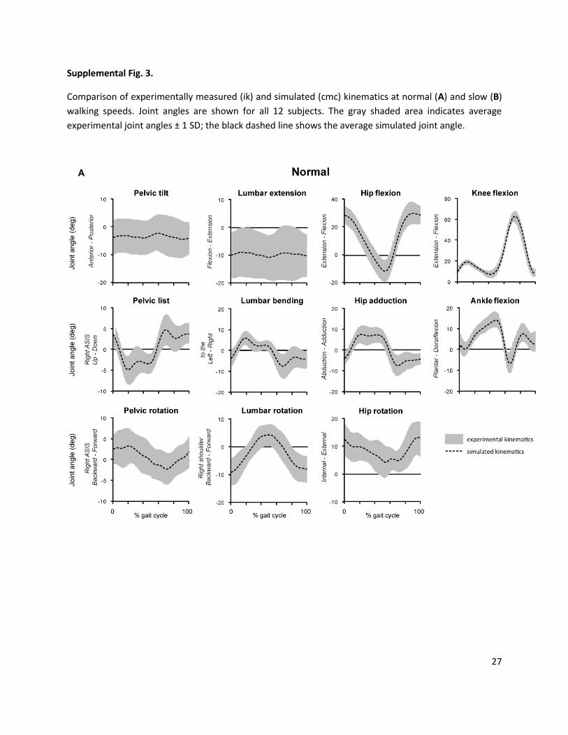
kinematics and kinetics closely tracked experimental kinematics and kinetics (Supplemental Fig. 3-4).
8
2.3. Analysis muscle function
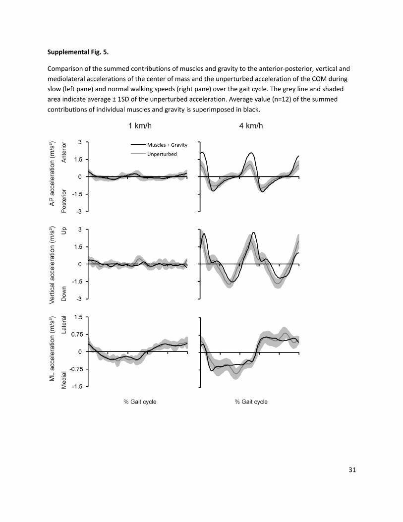
A perturbation analysis8 computed the AP, vertical and ML COM accelerations induced by a specific
muscle or gravity. The force of each muscle or gravity was subsequently increased with 1N and the
equations of motion were integrated forward over a time window of 0.03s to evaluate the effect on the
COM. To allow changes in the GRF and moments during the perturbation, linear and torsional springs
were added between the model’s feet and the floor. We verified the validity of the simulations by
comparing the summed muscle contributions to the COM acceleration with the COM accelerations in
the reference simulation (Supplemental Fig. 5).
Contributions of muscles and gravity were calculated over the entire gait cycle, which was further
divided into subphases (Fig. 2-3). While most simulation studies8,10-11,17 consider head, arms and trunk as
a single rigid body, this study used a model that included separate arms segments. This allowed us to
identify the contribution of the arm dynamics to the COM accelerations.
In a post processing step, positive and negative contributions were separately averaged over each
subphase and subsequently over all subjects. To simplify data analysis, contributions of smaller muscles
with similar function were summed.
We performed a Wilcoxon’s test for matched pairs (Statistica, Statsoft Inc, USA) to test for significant
differences in average contributions between normal and very slow walking.
3. Results
3.1 Total muscle contributions versus gravity
9
At a very slow speed, COM acceleration decreased (Fig. 1B) mainly due to a decrease in the total
muscle contribution (Fig. 2, SUM). Gravity on the other hand showed mixed results: While the
downward acceleration due to gravity decreased at a very slow speed, the contribution to anterior
acceleration increased (Fig. 2, GRAVITY) during the entire gait cycle. Similarly, contributions to lateral
acceleration during single stance and to medial acceleration during swing increased at a very slow
speed.
3.2 Individual muscle contributions
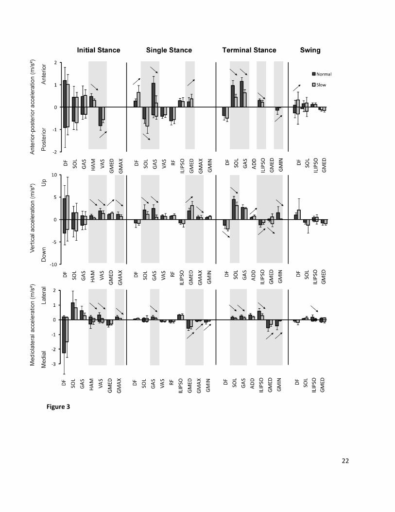
At both speeds, plantarflexors (GAS & SOL), quadriceps (RF & VAS), DF and GMED contributed most
to the AP and vertical COM acceleration during stance. Hip abductors (GMED & GMIN), ILIPSO,
plantarflexors and VAS were the main contributors to ML accelerations (Fig. 3). The influence of the arm
movement is negligible during both speeds (maximum arm contributions < 0.1 m/s2).
At initial stance, contributions from HAM, VAS and GMAX to anterior-posterior, upward and lateral
(i.e. away from the midline) acceleration of the COM decreased significantly at a very slow speed
compared to normal speed. However, contribution from GMED to support (i.e. upward acceleration)
increased at a very slow speed. In the other muscles (GAS, SOL and DF), no significant differences were
found between the different walking speeds.
During single stance, contributions of GAS to propulsion (i.e. anterior acceleration), support and
lateral acceleration decreased during very slow compared to normal speed. Similarly, GMAX
contributions to support and medial acceleration (i.e. towards the midline) decreased. On the other
hand, increased contributions were found from GMED to propulsion and support, while contributions to
medial acceleration decreased. ILIPSO and QUAD contributions remained relatively constant between
10
the two speeds. Increased contributions were found for DF to propulsion and SOL to posterior
acceleration.
In terminal stance, contributions of plantarflexors (GAS and SOL) to propulsion, support and lateral
acceleration all decreased. ILIPSO contribution to propulsion, downward and lateral acceleration also
decreased, similar as GMIN contributions in the opposite directions. Similar as in previous phases,
contributions of hip abductor GMED increased in the sagittal plane, but decreased in medial direction.
During swing, contributions of most muscles were small at both speeds. The contribution of ILIPSO
to lateral acceleration decreased during very slow walking. In contrast, an increase was found of DF
contribution to propulsion at very slow speed.
In general, at very slow speed, muscles contributions to mediolateral acceleration either decreased
or remained constant compared to normal speed. A similar pattern was found for muscles contributions
to sagittal plane COM acceleration. GMED consistently increased its contribution to sagittal plane COM
control.
4. Discussion
In literature, correlations were found between an increase of fall risk, ML deviations of the COM
and slower walking speeds2,4. The purpose of this study was therefore to gain more insight in the
influence of speed on ML COM control, and additionally how this ML control is coupled with sagittal
plane control. Therefore, we generated subject specific 3D simulations of gait at very slow and normal
speeds.
11
Mediolateral control of the COM
When walking speed decreases from normal to very slow, resulting medial COM accelerations
decreased during single stance. Two factors contribute to this decrease:
Firstly, during very slow walking, gravity’s destabilizing contribution to lateral acceleration (away
from the midline) increases. The increased contribution of gravity to lateral COM acceleration (Fig. 2) is
indeed associated with increased ML COM displacement (Fig. 1A). The higher destabilizing action of
gravity might well be the reason why even healthy individuals increase their step width27 at very slow
speeds, as this counteracts the influence of gravity (cf. Pandy et al.17).
Secondly, during very slow walking, both net and individual muscle contributions to medial COM
acceleration decrease significantly compared to normal walking. Especially decreased abductor (i.e.
GMIN and GMED) contributions (Fig. 3) to medial COM acceleration are responsible for this decrease.
The excessive lateral COM movement (Fig. 1A) during slow walking speeds, also reported in
previous studies27, can therefore be related to a combined decrease of the restraining muscle action in
medial direction and increased destabilizing GRAV action in lateral direction.
Coupling of control in the different planes
Gravity
Similar to its role in controlling ML acceleration, an increase of gravity contribution to forward
propulsion was found at slow compared to normal walking (Fig. 2). In combination with a large decrease
in muscle propulsive action, this leads to a very high relative contribution of gravity to forward
propulsion during slow walking. To compensate for this, a less pronounced decrease (in single stance)
and even increase (in terminal stance) of total muscle contributions to posterior acceleration are found
during slow walking. Hereby, a higher backward tilting moment is induced to maintain balance in the
sagittal plane.
12
In contrast with AP and ML directions, a decreased contribution of gravity to downward
acceleration at very slow speed is found in this study (Fig. 2), which is consistent with an increase of
skeletal alignment10. An uncoupling of gravity’s contributions to COM control in the different directions
therefore exists.
Muscles
For most muscles a coupled control between sagittal plane and ML stability was shown, with muscle
contributions that decreased at very slow compared to normal speed in both the sagittal plane (cf. Liu et
al.10) and mediolateral direction (cf. John et al.19). However, the decrease of ML GMED contributions
combined with the increase of sagittal plane contributions during very slow walking, suggests that an
uncoupling of GMED muscle function underlies the change in walking speed. This confirms the
assumption of a separate control mechanism for sagittal plane and ML balance control15-16.
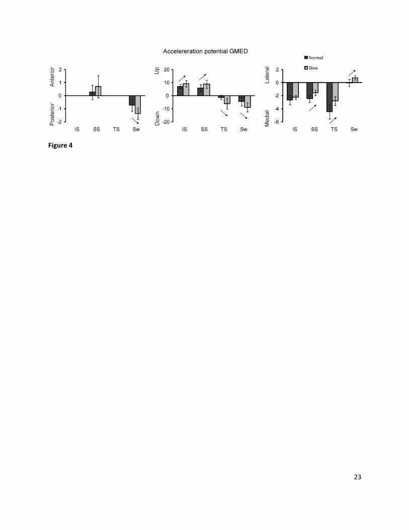
Further analysis of our results (Fig. 4) shows an uncoupling in acceleration potential of GMED, i.e.
the acceleration per Newton of force. The acceleration potential of GMED in AP (Sw: +90%) and UD (IS:
+31%, SS: +46%, TS: +384%, Sw: +103%) direction increased at very slow compared to normal walking
speed, while the potential in ML (SS: -36%, TS: -37%) direction decreased. The force of GMED is the
same in all directions. Therefore, the uncoupling of total of GMED contribution between the different
planes, i.e. the product of the total muscle force with the potential acceleration, mainly results from the
uncoupling of acceleration potential in the different directions.
The reduced ML potential of GMED at lower speeds may potentially threaten ML stability as less ML
stability will be provided at slow walking speed. As contribution of GRAV to ML acceleration increases a
critical balance needs to be satisfied to restrict excessive lateral motion of the COM during very slow
walking.
13
Our results are in agreement with previous speed-dependent changes found by Liu et al.10 and John
et al.19. Similar as in Liu et al.10, contributions of GMAX, HAM and VAS to support decreased at our very
slow compared to the normal walking speed during early stance. During late stance, a decrease was
found in soleus contribution to support. Likewise, GMED contribution increased instead of decreased at
very slow speed. Our results on muscle contribution in antero-posterior direction also agree with Liu et
al.10; VAS contribution to posterior acceleration decreased during early stance, and SOL contribution to
propulsion decreased during late stance. In agreement with John et al.19, we found decreased
contributions of VAS and GMAX to lateral accelerations of the COM at initial stance during very slow
walking. However, no significant changes were found in initial stance contributions to medial
accelerations. GMED contributions to medial accelerations decreased in both single and terminal stance.
A similar decrease of SOL contribution to lateral acceleration during TS was found. To our knowledge,
this study is the first to confirm decreased GAS contribution during TS to acceleration of COM in all three
directions.
Methodological considerations/limitations
A potential limitation of our study is that subjects walked on a split-belt treadmill. No significant
differences were found in SW between slow and normal walking (0.22 ± 0.03m vs. 0.23± 0.03m, p=0.64),
while SW was expected to increase during slow compared to normal walking. This might be due to the
use of a split-belt treadmill since the gap in between the belts might slightly bias the base of gait28
compared to single-belt walking. However, the fact that there was no change in SW may actually be an
advantage for the present study. Indeed the increase in SW related to walking speed found in
overground walking, makes it difficult to evaluate the isolated effect of speed. The unchanged SW allows
evaluating the separate effect of speed on the ML acceleration of the COM, without any confounding
effects of SW. Furthermore, no significant differences were found in frontal plane joint moments, ML
14
GRF or GMED EMG activity during the stance phase in split-belt treadmill walking compared to
overground walking in literature29. Frontal plane dynamic stability was also insensitive to overground or
treadmill walking30.
In this study, contributions of centrifugal, Coriolis and inertial forces were not taken into account.
However, Anderson and Pandy7 found that contributions to support during stance are relatively small.
Furthermore, Pandy et al.17 showed contributions from centrifugal and Coriolis forces to ML
accelerations to be small compared to muscle contributions, especially during single stance.
Clinical implications
Slow walking seems to be advantageous for older people, because it requires less muscle force.
However, while COM acceleration in ML direction decreases compared to normal speed, this is
accompanied by an increase in ROM of the COM. This increased ROM induces higher demands on
muscle coordination to keep equilibrium between gravity and muscle contributions. In elderly people, a
reduction of their coordination capacity might jeopardize their ability to maintain balance at lower
speeds. This will result in an increase in variability and might eventually lead to falls.
In conclusion, at very low walking speeds a vicious circle might arise in which increasing ROM and
increasing destabilizing acceleration of gravity reinforce each other, eventually leading to falls. A tight
coordination between the action of GMED towards the mid-line and lateral action of gravity away from
the mid-line is therefore crucial to maintain stability. Increased contributions of most muscles are
important for both stability and propulsion when walking speed increases. However, GMED has a unique
role in controlling ML acceleration and therefore stabilizing ML COM excursion.
Conflict of interest statement

15
No conflicts of interest, financial or otherwise, are declared by the author(s).
16
References
1. Hanley A, Silke C, Murphy J. Community-based health efforts for the prevention of falls in the elderly. Clin Interv Aging 2011;6:19-25
2. Hausdorff JM. Gait variability: methods, modeling and meaning. J Neuroeng Rehabil 2005;2:19
3. Hernandez A, Silder A, Heiderscheit BC, Thelen DG. Effect of age on center of mass motion during human walking. Gait Posture 2009;30:217-222
4. Den Otter AR, Geurts AC, Mulder T, Duysens J. Speed related changes in muscle activity from normal to very slow walking speeds. Gait Posture 2004;19:270-278
5. Bergland A, Jarnlo GB, Laake K. Predictors of falls in the elderly by location. Aging Clin Exp Res 2003;15:43-50
6. Ivanenko YP, Poppele RE, Lacquaniti F. Five basic muscle activation patterns account for muscle activity during human locomotion. J Physiol 2004;556:267-282
7. Anderson FC, Pandy MG. Individual muscle contributions to support in normal walking. Gait Posture 2003;17:159-169
8. Liu MQ, Anderson FC, Pandy MG, Delp SL. Muscles that support the body also modulate forward progression during walking. J Biomech 2006;39:2623-2630
9. Neptune RR, Clark DJ, Kautz SA. Modular control of human walking: a simulation study. J Biomech 2009;42:1282-1287
10. Liu MQ, Anderson FC, Schwartz MH, Delp SL. Muscle contributions to support and progression over a range of walking speeds. J Biomech 2008;41:3243-3252
11. Neptune RR, Sasaki K, Kautz SA. The effect of walking speed on muscle function and mechanical energetics. Gait Posture 2008;28:135-143
12. Jones SL, Henry SM, Raasch CC, Hitt JR, Bunn JY. Responses to multi-directional surface translations involve redistribution of proximal versus distal strategies to maintain upright posture. Exp Brain Res 2008;187:407-417
13. Winter DA, Prince F, Frank JS, Powell C, Zabjek KF. Unified theory regarding A/P and M/L balance in quiet stance. J Neurophysiol 1996;75:2334-2343
14. Kung UM, Horlings CG, Honegger F, Duysens JE, Allum JH. Control of roll and pitch motion during multi-directional balance perturbations. Exp Brain Res 2009;194:631-645
15. Bruijn SM, van Dieen JH, Meijer OG, Beek PJ. Is slow walking more stable? J Biomech 2009;42:1506-1512
17
16. Bauby CE, Kuo AD. Active control of lateral balance in human walking. J Biomech 2000;33:1433-1440
17. Pandy MG, Lin YC, Kim HJ. Muscle coordination of mediolateral balance in normal walking. J Biomech 2010;43:2055-2064
18. Allen JL, Neptune RR. Three-dimensional modular control of human walking. J Biomech 2012;45:2157-63
19. John CT, Seth A, Schwartz MH, Delp SL. Contributions of muscles to mediolateral ground reaction force over a range of walking speeds. J Biomech 2012;45:2438-43.
20. Hof AL, Elzinga H, Grimmius W, Halbertsma JP. Speed dependence of averaged EMG profiles in walking. Gait Posture 2002;16:78-86
21. Den Otter AR, Geurts AC, Mulder T, Duysens J. Gait recovery is not associated with changes in the temporal patterning of muscle activity during treadmill walking in patients with post-stroke hemiparesis. Clin Neurophysiol 2006;117:4-15
22. Jansen K, De Groote F, Massaad F, Meyns P, Duysens J, Jonkers I. Similar muscles contribute to horizontal and vertical acceleration of center of mass in forward and backward walking: implications for neural control. J Neurophysiol 2012;107:3385-96
23. Delp SL, Anderson FC, Arnold AS, Loan P, Habib A, John CT, Guendelman E, Thelen DG. OpenSim: open-source software to create and analyze dynamic simulations of movement. IEEE Trans Biomed Eng 2007;54:1940-1950
24. Hamner SR, Seth A, Delp SL. Muscle contributions to propulsion and support during running. J Biomech 2010;43:2709-2716
25. De Groote F, De Laet T, Jonkers I, De Schutter J. Kalman smoothing improves the estimation of joint kinematics and kinetics in marker-based human gait analysis. J Biomech 2008;41:3390-3398
26. Thelen DG, Anderson FC. Using computed muscle control to generate forward dynamic simulations of human walking from experimental data. J Biomech 2006;39:1107-1115
27. Orendurff MS, Segal AD, Klute GK, Berge JS, Rohr ES, Kadel NJ. The effect of walking speed on center of mass displacement. J Rehabil Res Dev 2004;41:829-834
28. Altman AR, Reisman DS, Higginson JS, Davis IS. Kinematic comparison of split-belt and single-belt treadmill walking and the effects of accommodation. Gait Posture 2012;35:287-291
29. Lee SJ, Hidler J. Biomechanics of overground vs. treadmill walking in healthy individuals. J Appl Physiol 2008;104:747-755
30. Rosenblatt NJ, Grabiner MD. Measures of frontal plane stability during treadmill and overground walking. Gait Posture 2010;31:380-384
18
Figure captions
Fig. 1. Averaged (n=12) displacements (A) and accelerations (B) of the center of mass (COM) over the
gait cycle during slow and normal walking speed. Mediolateral center of mass excursions increased
substantially in slow compared to normal walking speeds; center of mass accelerations over the gait
cycle decreased significantly for all directions with decreasing walking speed. Shaded bars indicate
periods of double-limb support during slow (grey) and normal (black) walking speed.
Fig. 2. Contributions of summed right leg muscles (SUM, left pane) and gravity (right pane) to the
anterior-posterior, vertical and mediolateral accelerations of the center of mass during slow and normal
walking speeds. (A) Absolute contributions over the gait cycle. (B) Contributions averaged over the sub
phases of the gait cycle (n = 12, mean + 1 SD). A positive sign indicates contributions to respectively
anterior, upward or lateral acceleration of the COM, a negative sign indicates contributions to
respectively posterior, downward or medial acceleration of the COM. If contributions of both signs are
present, it reflects a reversed contribution within that particular phase of the gait cycle.
Initial stance was from ipsilateral heel contact to contralateral toe off. Single stance was from
contralateral toe off to contralateral heel contact, terminal stance was from contralateral heel contact
to ipsilateral toe off and swing form ipsilateral toe off to ipsilateral heel contact. Statistically significant
differences (p < 0.05) are indicated with ↑ or ↓.
19
Fig. 3. Contributions of individual muscles to anterior-posterior (top), vertical (middle) and mediolateral
(bottom) accelerations of the center of mass, averaged over the initial stance, single stance, terminal
stance and swing (n=12; mean + 1 SD). GMAX consists of the three parts of gluteus maximus, GMED
consists of the three parts of the gluteus medius. GMIN includes the different parts of gluteus minimus.
ILIPSO consists of iliacus and psoas and ADD consists of adductor longus, brevis and magnus. HAMS
combines semimembranosus, semitendinosus, and biceps femoris long head. VAS consists of vastus
lateralis, intermedius and medialis. GAS consists of the medial and lateral parts of the gastrocnemius;
Dorsiflexors (DF) consist of tibialis anterior, extensor digitorum and extensor hallucis. Statistically
significant differences (p < 0.05) are indicated with ↑ or ↓. As contributions from ipsilateral and
contralateral side are symmetric, only ipsilateral contributions are shown.
Fig. 4. Acceleration potential (10-3 x m/s2 x 1/N) of gluteus medius (GMED) in the different directions,
averaged over the initial stance (IS), single stance (SS), terminal stance (TS) and swing (Sw) (n=12; mean
± 1 SD). GMED acceleration potentials are only reported in phases with significant total GMED
contributions. Statistically significant differences (p < 0.05) are indicated with ↑ or ↓.

20
Figure 1

21
Figure 2
22
Figure 3
23
Figure 4

24
Supplementary Material
Supplemental Table 1.
Joint Type of joint # DOF total
Hip ball-and-
socket
3 6
Knee hinge 1 2
Ankle hinge 1 2
Subtalar Locked / /
Metatarsophalangeal Locked / /
Pelvis Free 6 6
Back ball-and-
socket
3 3
Shoulder ball-and-
socket
3 6
Elbow hinge 1 2
Forearm rotation Locked / /
Wrist Locked / /
27
Degrees of freedom (DOF) in the generic musculoskeletal model

25
Supplemental Fig. 1.
A. Residual Forces (N) and Moments (Nm) acting on the pelvis after residual reduction (RRA). Residuals
are subsequently averaged over the gait cycle and over the different subjects for normal and slow
walking speed. Error bars show ± 1SD. B. Reserve Actuators forces (N) acting on the different joints to
compensate for insufficient muscle strength during CMC. Forces are subsequently averaged over the
gait cycle and over the different subjects for normal and slow walking speed. Error bars show ± 1SD
across subjects.

26
Supplemental Fig. 2. Comparison of experimentally measured electromyography signals (EMG) and simulated muscle
activations (from CMC) during FW at normal (A) and slow speed (B). The gray line and shaded area
indicate average EMG ± 1 SD, while the black line indicates average simulated muscle activation ± 1 SD
(dashed line). Both EMG and simulated activations are normalized to the maximum value of each
subject over the two trials. EMG/activations are shown for biceps femoris (BF), semitendinosus (ST),
rectus femoris (RF), lateral vastus (LVAS), lateral gastrocnemius(LGAS), soleus (SOL), and tibialis anterior
(TA).
27
Supplemental Fig. 3.
Comparison of experimentally measured (ik) and simulated (cmc) kinematics at normal (A) and slow (B)
walking speeds. Joint angles are shown for all 12 subjects. The gray shaded area indicates average
experimental joint angles ± 1 SD; the black dashed line shows the average simulated joint angle.
28
29
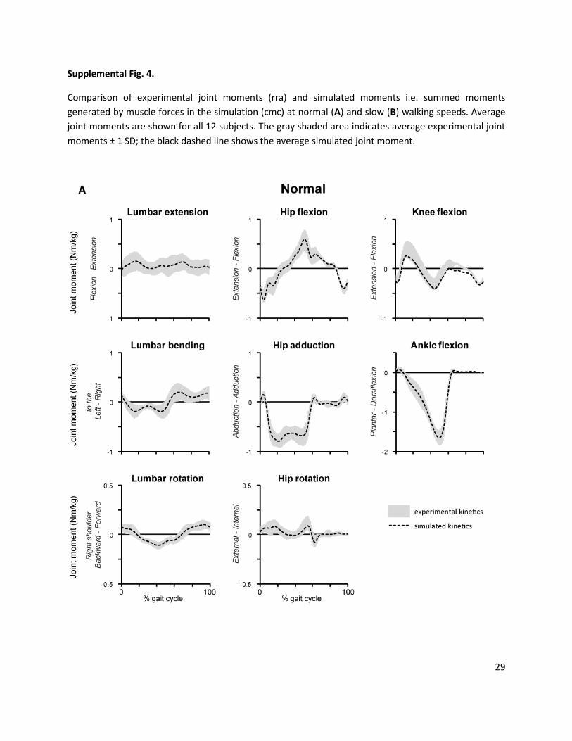
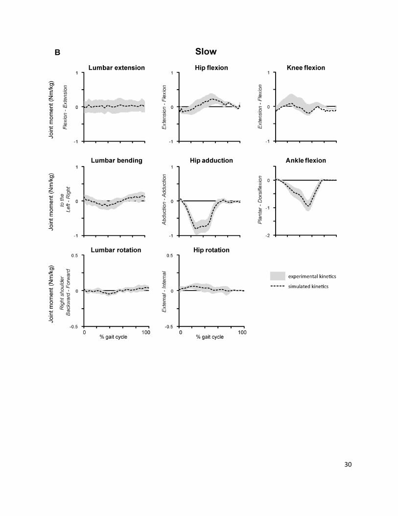
Supplemental Fig. 4.
Comparison of experimental joint moments (rra) and simulated moments i.e. summed moments
generated by muscle forces in the simulation (cmc) at normal (A) and slow (B) walking speeds. Average
joint moments are shown for all 12 subjects. The gray shaded area indicates average experimental joint
moments ± 1 SD; the black dashed line shows the average simulated joint moment.
30
31
Supplemental Fig. 5.
Comparison of the summed contributions of muscles and gravity to the anterior-posterior, vertical and
mediolateral accelerations of the center of mass and the unperturbed acceleration of the COM during
slow (left pane) and normal walking speeds (right pane) over the gait cycle. The grey line and shaded
area indicate average ± 1SD of the unperturbed acceleration. Average value (n=12) of the summed
contributions of individual muscles and gravity is superimposed in black.
Copyright © 2022 FDOKUMEN