Host epithelial-viral interactions as cause and cure for asthma
13
Host epithelial-viral interactions as cause and cure for asthma Michael J. Holtzman, Dhara Patel, Yong Zhang, and Anand C. Patel Drug Discovery Program, Division of Pulmonary and Critical Care Medicine, Department of Internal Medicine, Department of Cell Biology and Physiology, and Department of Pediatrics, Washington University School of Medicine, St. Louis, Missouri Abstract Research on the pathogenesis of asthma has concentrated on initial stimuli, genetic susceptibilities, adaptive immune responses, and end-organ alterations (particularly in airway mucous cells and smooth muscle) as critical steps leading to disease. Recent evidence indicates that the innate immune cell response to respiratory viruses also contributes to the development of inflammatory airway disease. We further develop this concept by raising the issue that the interaction between host airway epithelial cells and respiratory viruses is another aspect of innate immunity that is also a critical determinant of asthma. We also introduce a rationale for how antiviral performance at the epithelial cell level might be improved to prevent acute infectious illness and chronic inflammatory disease caused by respiratory viruses. Introduction One of the major tasks facing medical research is to define the pathogenesis of chronic inflammatory diseases. In the case of asthma, the approach to understanding chronic inflammation has implicated a broad array of cell types, cell-cell interactions, and cellular products. One leading scheme for integrating this information is based on the classification of the adaptive immune system, and especially the responses of T helper (Th) cells into T helper type 1 (Th1) cells that mediate delayed-type hypersensitivity reactions and selectively produce interleukin (IL)-2 and interferon (IFN)-γ, and Th2 cells that promote B-cell dependent humoral immunity and selectively produce IL-4, IL-5, and IL-13. Under this scheme, an up-regulated Th2 and perhaps a down-regulated Th1 response is thought to drive the development of asthma. The newer contributions of Th17 (IL-17-producing) and Treg (IL-10- and TGF-β-producing) subsets of T cells are also proposed to contribute to inflammatory airway disease by skewing the system towards a Th2 response [1, 2]. In general, the Th2 hypothesis is based on observations of the response to allergen challenge in mouse models of asthma and in humans with allergic asthma [3, 4]. However, it has been pointed out that a Th2-biased response does not account for the epidemiological link between respiratory viral infection and the subsequent development of asthma [5]. Indeed, the broader issue of the relationship between acute viral infection and chronic inflammatory disease remains uncertain. In an effort to understand this issue, we identified the likely steps leading from viral infection to inflammatory disease using respiratory viral infection and © 2011 Elsevier Ltd. All rights reserved. Address correspondence to M.J.H., Washington University School of Medicine, Campus Box 8052, 660 South Euclid Avenue, St. Louis, MO 63110. Tel. 314-362-8970; Fax: 314-362-9009; [email protected]. Publisher's Disclaimer: This is a PDF file of an unedited manuscript that has been accepted for publication. As a service to our customers we are providing this early version of the manuscript. The manuscript will undergo copyediting, typesetting, and review of the resulting proof before it is published in its final citable form. Please note that during the production process errors may be discovered which could affect the content, and all legal disclaimers that apply to the journal pertain. NIH Public Access Author Manuscript Curr Opin Immunol. Author manuscript; available in PMC 2012 August 1. Published in final edited form as: Curr Opin Immunol. 2011 August ; 23(4): 487–494. doi:10.1016/j.coi.2011.05.010. NIH-PA Author Manuscript NIH-PA Author Manuscript NIH-PA Author Manuscript
-
Upload
independent -
Category
Documents
-
view
0 -
download
0
Transcript of Host epithelial-viral interactions as cause and cure for asthma
Host epithelial-viral interactions as cause and cure for asthma
Michael J. Holtzman, Dhara Patel, Yong Zhang, and Anand C. PatelDrug Discovery Program, Division of Pulmonary and Critical Care Medicine, Department ofInternal Medicine, Department of Cell Biology and Physiology, and Department of Pediatrics,Washington University School of Medicine, St. Louis, Missouri
AbstractResearch on the pathogenesis of asthma has concentrated on initial stimuli, geneticsusceptibilities, adaptive immune responses, and end-organ alterations (particularly in airwaymucous cells and smooth muscle) as critical steps leading to disease. Recent evidence indicatesthat the innate immune cell response to respiratory viruses also contributes to the development ofinflammatory airway disease. We further develop this concept by raising the issue that theinteraction between host airway epithelial cells and respiratory viruses is another aspect of innateimmunity that is also a critical determinant of asthma. We also introduce a rationale for howantiviral performance at the epithelial cell level might be improved to prevent acute infectiousillness and chronic inflammatory disease caused by respiratory viruses.
IntroductionOne of the major tasks facing medical research is to define the pathogenesis of chronicinflammatory diseases. In the case of asthma, the approach to understanding chronicinflammation has implicated a broad array of cell types, cell-cell interactions, and cellularproducts. One leading scheme for integrating this information is based on the classificationof the adaptive immune system, and especially the responses of T helper (Th) cells into Thelper type 1 (Th1) cells that mediate delayed-type hypersensitivity reactions and selectivelyproduce interleukin (IL)-2 and interferon (IFN)-γ, and Th2 cells that promote B-celldependent humoral immunity and selectively produce IL-4, IL-5, and IL-13. Under thisscheme, an up-regulated Th2 and perhaps a down-regulated Th1 response is thought to drivethe development of asthma. The newer contributions of Th17 (IL-17-producing) and Treg(IL-10- and TGF-β-producing) subsets of T cells are also proposed to contribute toinflammatory airway disease by skewing the system towards a Th2 response [1, 2].
In general, the Th2 hypothesis is based on observations of the response to allergen challengein mouse models of asthma and in humans with allergic asthma [3, 4]. However, it has beenpointed out that a Th2-biased response does not account for the epidemiological linkbetween respiratory viral infection and the subsequent development of asthma [5]. Indeed,the broader issue of the relationship between acute viral infection and chronic inflammatorydisease remains uncertain. In an effort to understand this issue, we identified the likely stepsleading from viral infection to inflammatory disease using respiratory viral infection and
© 2011 Elsevier Ltd. All rights reserved.Address correspondence to M.J.H., Washington University School of Medicine, Campus Box 8052, 660 South Euclid Avenue, St.Louis, MO 63110. Tel. 314-362-8970; Fax: 314-362-9009; [email protected]'s Disclaimer: This is a PDF file of an unedited manuscript that has been accepted for publication. As a service to ourcustomers we are providing this early version of the manuscript. The manuscript will undergo copyediting, typesetting, and review ofthe resulting proof before it is published in its final citable form. Please note that during the production process errors may bediscovered which could affect the content, and all legal disclaimers that apply to the journal pertain.
NIH Public AccessAuthor ManuscriptCurr Opin Immunol. Author manuscript; available in PMC 2012 August 1.
Published in final edited form as:Curr Opin Immunol. 2011 August ; 23(4): 487–494. doi:10.1016/j.coi.2011.05.010.
NIH
-PA Author Manuscript
NIH
-PA Author Manuscript
NIH
-PA Author Manuscript
asthma as a template for this process (as outlined in Fig. 1). Here, we review three majoradvances that lead to a substantial revision of this virus-disease connection. First, wedevelop the experimental and clinical evidence that the link between acute infection andchronic disease of the airway unexpectedly depends on immune cells of the innate ratherthan the adaptive immune system; second, we extend this concept to the airway epithelialcell and the proposal that high-level viral replication at this cellular site is required to triggerthe innate immune cell activation that in turn drives asthma; and third, we introducestrategies that could improve antiviral defense at the airway epithelial cell level and therebyhelp to prevent acute infectious illness and chronic asthmatic disease. We conclude byshowing how these advances provide for a new virus-disease paradigm.
Introducing the innate immune cells for chronic postviral diseaseOne of the initial objectives for understanding the role of respiratory viruses in thepathogenesis of asthma was to define the immune program for postviral disease. This goalrequired a high-fidelity experimental model of postviral asthma in humans, whererespiratory syncytial virus (RSV) is implicated. However, we recognized (and confirmed)the shortcomings of using RSV for an experimental model in mice [6], and thereforesubstituted the corresponding mouse paramyxovirus, Sendai virus (SeV). The change inapproach provided for cardinal features of human disease, including acute bronchiolitisfollowed by chronic (perhaps lifelong) airway inflammation, mucus overproduction, andhyperreactivity that depend on genetic susceptibility [7-9]. We used this model to identify anew immune axis that translates viral infection into chronic airway disease. When the acutelung disease appears in this model (at 3 weeks after viral inoculation), it depends on animmune response that features expression and activation of the high-affinity IgE receptor(FcεRI) on conventional lung dendritic cells (cDCs) and consequent CCL28 production torecruit IL-13-producing CD4+ T cells to the airways [10, 11]. In addition, when the chroniclung disease develops fully (at 7 weeks after inoculation), it is driven instead by an innateimmune response that relies on invariant natural killer T (iNKT) cells that are programmedto activate macrophages to produce IL-13 [12, 13]. The interaction between iNKT cells andmacrophages depends on contact between the semi-invariant Vα14Jα18-TCR on lung iNKTcells and the oligomorphic MHC-like protein CD1d on macrophages as well as NKT cellproduction of IL-13 that binds to the IL-13 receptor (IL-13R) on the macrophage. Thisinnate immune axis is also activated in the lungs of humans with severe asthma or COPDbased on detection of increased numbers of iNKT cells and alternatively-activated (M2)IL-13-producing macrophages in the lung [12, 14-16]. Together, the findings identify anadaptive immune response that mediates acute disease and an innate immune response thatdrives chronic obstructive lung disease in experimental and clinical settings (as summarizedin Fig. 2).
The viral and immune mechanisms for postviral asthma (for SeV and related viruses) arestill under study, but even at this stage, we recognize that a critical element of theexperimental model is the severity of acute infection. As discussed further in the nextsection, this requirement for severity may be the reason why others missed the effect of SeVand other viruses in experimental models and in humans with milder degrees of acuteillness. In contrast, we tailored our experimental approach to match clinical observationsfrom our group and others that the children with the most severe manifestations of viralbronchiolitis are the ones that are marked for the subsequent development of chronic asthma[17-19]. A natural corollary of this issue is that the severity of illness often depends on thelevel of virus and in turn on the flux between viral replication and clearance in the primaryhost cell. Since this cell is most often the airway epithelial cell (and within this cellpopulation, it is often the ciliated mucosal epithelial cell) [20], this additional component ofinnate mucosal immunity was a prime candidate for further study.
Holtzman et al. Page 2
Curr Opin Immunol. Author manuscript; available in PMC 2012 August 1.
NIH
-PA Author Manuscript
NIH
-PA Author Manuscript
NIH
-PA Author Manuscript
Moving upstream to the airway epithelial cellDespite being the primary home to respiratory viruses (or perhaps because of it), airwayepithelial cells contain a potent antiviral system based mainly on IFN production andsubsequent expression of IFN-stimulated genes (ISGs). An abbreviated scheme for thiscomplex IFN-based network features a master regulator known as STAT1 (as diagrammedin Fig. 3). If this network is genetically defective (e.g., due to STAT1 deficiency) in mice orman, the host often succumbs to lethal viral infection [21]. Moreover, the magnitude of thisantiviral IFN response, as judged by the level of IFN production and signaling, appears tocorrelate with the level of protection against infection. For example, in a study of peripheralblood mononuclear cells (PBMCs) obtained at the time of birth, it was found that the levelof IFN-γ production in response to RSV infection can predict the likelihood of respiratorytract infection in the first year of life [22]. In this case, the higher the IFN production, thenthe less likely the child is to develop an infection, and in turn, the less likely to developserious respiratory or wheezing illnesses. Similarly, decreased induction of IFN-αproduction was found in RSV-inoculated PBMCs that were isolated from children andadults with asthma versus normal control subjects [23]. A comparable deficiency in IFN-γproduction was detected in human rhinovirus (HRV)-inoculated PBMCs isolated fromasthmatics compared to normal controls [24]. Others have also reported that circulatingplasmacytoid dendritic cells (pDCs) from asthmatics produce less IFN-α than cells fromnormals after inoculation with influenza A virus (IAV) [25].
This concept that there is a deficiency in IFN-dependent control of respiratory viruses inasthma has been extended to the study of airway epithelial cells in experimental models andin humans. In particular, a deficiency of IFN signaling in airway epithelial cells will likelycompromise host defense against respiratory viruses and promote the subsequentdevelopment of experimental asthma in mouse models [26-33]. These findings imply that adeficiency in IFN-dependent antiviral defense at the level of airway epithelial cells mightalso be found in humans with more severe infections and subsequent asthma. In support ofthis possibility, studies from the labs of Donna Davies and Sebastian Johnston show adeficiency in IFN-β and IFN-λ, production in response to HRV inoculation in airwayepithelial cells cultured from asthmatic versus normal subjects [34, 35]. A recent follow-upstudy indicated that the bronchial epithelial cell response to IFN-β in asthma was nodifferent from normal [36], suggesting that the defect lies in IFN production rather thansignaling. However, others reported that asthma is also characterized by a genetic increase inthe levels of an endogenous inhibitor of IFN signaling known as SOCS1 [37], suggestingthat a signaling defect may also be found in some patients with asthma. Meanwhile, there isevidence that control of viral replication and production of IFN may also be defective inairway epithelial cells from patients with other types of inflammatory airway disease, e.g.,COPD and cystic fibrosis, but any possible abnormalities or underlying mechanisms stillneed to be better defined [38-40].
Even in the case of asthma, not all reports agree on a defect in IFN-dependent control ofviral replication. In a study from James Gern's lab, airway epithelial cells from asthmaticand control subjects exhibited no difference in viral levels after inoculation with HRV [41].Similarly, a study from Homer Boushey's and Pedro Avila's labs found no difference inHRV levels in asthma versus normal subjects, and in this case, cells were cultured under air-liquid interface conditions to better achieve ciliated cell differentiation [42]. Moreover,reports from two different labs (including Johnston's lab) show no statistical difference inHRV levels in asthma versus control subjects after controlled inoculation in vivo [43, 44].Other reports indicate that the levels of IFN-γ in airway epithelium or IFN-λ in sputum aresimilar in asthmatic versus normal subjects [45, 46]. However, we do not yet havecomprehensive reports of IFN (type I, II, and/or III) level or signal during viral infection. In
Holtzman et al. Page 3
Curr Opin Immunol. Author manuscript; available in PMC 2012 August 1.
NIH
-PA Author Manuscript
NIH
-PA Author Manuscript
NIH
-PA Author Manuscript

fact, precise quantification of IFN and corresponding viral level in vivo are clinicalbiomarkers with significant methodological challenges.
In addition to technical concerns, the divergent results in studies of cultured airwayepithelial cells could also be explained by the proposal for a close relationship between levelof viral replication, subsequent acute illness, and finally chronic disease. In particular, itappears that HRV replicates relatively inefficiently in well-differentiated airway epithelialcells and in mouse in vivo models as well [47, 48]. A lower level of viral replication couldexplain why the anti-HRV response has not appeared to be reproducible in vitro and whyHRV infection has not appeared to be sufficient for postviral asthma in vivo in experimentalmodels in mice. By contrast, SeV is particularly adapted for vigorous viral replication in theexperimental mouse model, and this element of the model is essential for the subsequentillness and development of chronic obstructive lung disease. Similarly in humans, despitethe difficult in comparing viral levels in asthmatics versus normals, if one concentrates onjust the asthmatic population that is susceptible to virus-induced disease, there remains atight relationship between viral load and severity of illness [44].
Improving innate immunityTaken together, there appears to be a direct relationship between viral level and both theseverity of acute illness and the likelihood of chronic disease in experimental models and inhumans with asthma. Moreover, the capacity of the host to control viral level appears to bedeficient in asthma, perhaps at the level of IFN production and/or signaling in airwayepithelial cells and likely in immune cells in the airway as well. Even if some of these tenetsturn out to be wrong, there still stands to be significant benefit for the normal and theasthmatic host to improve control over viral infection. So the question still remains as tohow best to achieve that goal given the absence of vaccines for asthmagenic viruses. In thatregard, nearly as soon as it was recognized that physiologic levels of IFNs were required fornormal host defense, it was proposed that excessive levels of IFNs might provide therapeuticbenefit. Indeed, transgenic overexpression of IFN-encoding genes in mice may protectagainst experimental infection and inflammatory disease, and administration of recombinantIFN is commonly used for infectious, autoimmune, and cancerous conditions in humans[49]. Unfortunately, this approach is limited by toxicity, so that excessive IFN might harmthe normal host [50]. In fact, this strategy suggests that there is little safe reserve in the IFNsystem that can be utilized for benefit in vivo. For these reasons and others, it might be moreprudent to aim at increasing the efficiency of endogenous IFN and thereby potentiatedownstream signal transduction [33].
Although an improvement in IFN efficacy might be desirable, it is made difficult by thecomplexity of the IFN signaling pathway. Nonetheless, support for an approach to increaseIFN signaling can be found in ongoing experimental work directed at STAT1. In particular,a double-cysteine substitution of native STAT1 (designated STAT1-CC) leads to a markedlyincreased responsiveness to IFN stimulation [51]. The consequence is a markedimprovement in ISG expression and control of viral replication, at least in vitro. Initialexperiments suggest that a similar benefit can be achieved in a transgenic mouse model invivo. Thus, current efforts aim to develop therapeutics that mimic this benefit and therebycorrect a possible defect in host defense that might contribute to chronic inflammatorydiseases such as asthma. In particular, the assay methods developed for studies of IFNsignaling can also be used to identify small molecular weight compounds that mimic Stat1-CC and enhance IFN efficacy in a manner that has distinct advantages over currenttherapies, including IFN itself [52].
Holtzman et al. Page 4
Curr Opin Immunol. Author manuscript; available in PMC 2012 August 1.
NIH
-PA Author Manuscript
NIH
-PA Author Manuscript
NIH
-PA Author Manuscript
ConclusionHere we summarize new work on the pathogenesis of asthma to support three breakthroughissues: (1) respiratory viral infections drive long-term activation of an innate immune cellresponse and in turn chronic obstructive lung disease in an experimental model thatresembles virus-induced asthma in humans; (2) this type of innate immune response dependson the severity of acute infection and in turn the tissue levels of virus, so that proper controlof virus by airway epithelial cells via IFN-dependent signaling would prevent asthma undernormal conditions but promote it under deficient conditions; and (3) smart mechanisms existfor improving IFN signaling as a means for improving antiviral defense and preventingsevere infection and consequent asthma. These concepts lead to a revised scheme for thesteps leading from respiratory viral infection to asthma (as summarized in Fig. 4). Themodel also provides a framework for therapeutic strategies that improve antiviral defense atthe level of airway epithelial cells and thereby overcomes common respiratory viruses thatpose a serious public health problem in terms of acute respiratory illness and chronic airwaydisease
AcknowledgmentsOur research on this topic is supported by grants from the National Institutes of Health (National Heart, Lung, andBlood Institute and National Institute of Allergy and Infectious Diseases).
References1. Wang YH, Liu YJ. The IL-17 cytokine family and their role in allergic inflammation. Curr Opin
Immunol. 2008; 20:697–702. [PubMed: 18832032]2. Wilson MS, Pesce JT, Ramalingam TR, et al. Suppression of murine allergic airway disease by
IL-2:anti-IL-2 monoclonal antibody-induced regulatory T cells. J Immunol. 2008; 181:6942–6954.[PubMed: 18981114]
3. Poston RN, Chanez P, Lacoste JY, et al. Immunohistochemical characterization of the cellularinfiltration in asthmatic bronchi. Am Rev Respir Dis. 1992; 145:918–921. [PubMed: 1554221]
4. Ying S, Durham SR, Corrigan CJ, et al. Phenotype of cells expressing mRNA for TH2-type(interleukin 4 and interleukin 5) and TH1-type (interleukin 2 and interferon γ) cytokines inbronchoalveolar lavage and bronchial biopsies from atopic asthmatic and normal control subjects.Am J Respir Cell Mol Biol. 1995; 12:477–487. [PubMed: 7742012]
5. Holtzman MJ, Byers DE, Benoit LA, et al. Immune pathways for translating viral infection intochronic airway disease. Adv Immunol. 2009; 102:245–276. [PubMed: 19477323]
6. Graham BS, Perkins MD, Wright PF, Karzon DT. Primary respiratory syncytial virus infection inmice. J Med Virol. 1988; 26:153–162. [PubMed: 3183639]
7**. Walter MJ, Morton JD, Kajiwara N, et al. Viral induction of a chronic asthma phenotype andgenetic segregation from the acute response. J Clin Invest. 2002; 110:165–175. This articleestablished that an acute respiratory viral infection can lead to chronic inflammatory lung diseasein an experimental model system. [PubMed: 12122108]
8**. Patel AC, Morton JD, Kim EY, et al. Genetic segregation of airway disease traits despiteredundancy of chloride channel calcium-activated (CLCA) family members. Physiol Genomics.2006; 25:502–513. This article established the genetic susceptibility of the process leading froman acute respiratory viral infection to chronic inflammatory lung disease in an experimentalmodel system and genetically segregated two distinct disease traits for chronic obstructive lungdisease. [PubMed: 16569774]
9. Tyner JW, Kim EY, Ide K, et al. Blocking airway mucous cell metaplasia by inhibiting EGFRantiapoptosis and IL-13 transdifferentiation signals. J Clin Invest. 2006; 116:309–321. [PubMed:16453019]
10**. Grayson MH, Cheung D, Rohlfing MM, et al. Induction of high-affinity IgE receptor on lungdendritic cells during viral infection leads to mucous cell metaplasia. J Exp Med. 2007;
Holtzman et al. Page 5
Curr Opin Immunol. Author manuscript; available in PMC 2012 August 1.
NIH
-PA Author Manuscript
NIH
-PA Author Manuscript
NIH
-PA Author Manuscript
204:2759–2769. This article established the up-regulation of high-affinity IgE receptors on lungdendritic cells using a mouse model of respiratory viral infection and showed how this immuneevent leads to chronic inflammatory lung disease in an experimental model system. [PubMed:17954569]
11. Cheung DS, Ehlenbach SJ, Kitchens RT, et al. Cutting Edge: CD49d+ neutrophils induce FcεRIexpression on lung dendritic cells in a mouse model of postviral asthma. J Immunol. 2010;185:4983–4987. [PubMed: 20876348]
12**. Kim EY, Battaile JT, Patel AC, et al. Persistent activation of an innate immune responsetranslates respiratory viral infection into chronic inflammatory lung disease. Nat Med. 2008;14:633–640. This article established the innate immune basis for how an acute respiratory viralinfection can lead to chronic inflammatory lung disease in an experimental model system and inhumans with asthma and chronic obstructive pulmonary disease. [PubMed: 18488036]
13. Benoit LA, Holtzman MJ. New immune pathways from chronic post-viral lung disease. Ann NYAcad Sci. 2010; 1183:195–210. [PubMed: 20146716]
14. Agapov E, Battaile JT, Tidwell R, et al. Macrophage chitinase 1 stratifies chronic obstructive lungdisease. Am J Respir Cell Mol Biol. 2009; 41:379–384. [PubMed: 19491341]
15. Byers DE, Holtzman MJ. Alternatively activated macrophages as cause or effect in asthma. Am JRespir Cell Mol Biol. 2010; 43:1–4. [PubMed: 20587775]
16. Byers DE, Holtzman MJ. Alternatively activated macrophages and airway disease. Chest. 2010 inpress.
17. Sigurs N, Bjarnason R, Sigurbergsson F, Kjellman B. Respiratory syncytial virus bronchiolitis ininfancy is an important risk factor for asthma and allergy at age 7. Am J Respir Crit Care Med.2000; 161:1501–1507. [PubMed: 10806145]
18**. Sigurs N, Gustafsson PM, Bjarnason R, et al. Severe respiratory syncytial virus bronchiolitis ininfancy and asthma and allergy at age 13. Am J Respir Crit Care Med. 2005; 171:137–141. Thisarticle and related ones from this research group provided the epidemiologic evidence for aconnection between respiratory viral infection and asthma in childhood. [PubMed: 15516534]
19. Castro M, Schweiger T, Yin-Declue H, et al. Cytokine response after severe respiratory syncytialvirus bronchiolitis in early life. J Allergy Clin Immunol. 2008; 122:725–733.
20. Ibricevic A, Pekosz AS, Walter MJ, et al. Influenza virus receptor specificity and cell tropism inmouse and human airway epithelial cells. J Virol. 2006; 80:7469–7480. [PubMed: 16840327]
21*. Dupuis S, Jouanguy E, Al-Hajjar S, et al. Impaired response to interferon-a/b and lethal viraldisease in human STAT1 deficiency. Nat Genet. 2003; 33:388–391. This article and related onesfrom this research group established the requirement for IFN signaling in antiviral host defense inhumans by identifying the consequences for genetic deficiency of this signaling pathway.[PubMed: 12590259]
22. Sumino KC, Visness CM, Schwarz J, et al. Antiviral interferon responses at birth can predictrespiratory illness in the first year of life. Am J Respir Crit Care Med. 2010; 181:A2503.
23. Gelhar K, Bilitewski C, Reinitz-Rademacher K, et al. Impaired virus-induced interferon-alpha2release in adult asthmatic patients. Clin Exp Allergy. 2006; 36:331–337. [PubMed: 16499644]
24. Papadopoulos NG, Stanciu LA, Papi A, et al. A defective type I response to rhinovirus in atopicasthma. Thorax. 2002; 57:328–332. [PubMed: 11923551]
25. Gill MA, Bajwa G, George TA, et al. Counterregulation between the FceRI pathway and antiviralresponses in human plasmacytoid dendritic cells. J Immunol. 2010; 184:5999–6006. [PubMed:20410486]
26*. Look DC, Keller BT, Rapp SR, Holtzman MJ. Selective induction of intercellular adhesionmolecule-1 by interferon-γ in human airway epithelial cells. Am J Physiol. 1992; 263:L79–L87.This article was the first in a series to establish the functional and molecular biology of the IFNsignal transduction system in airway epithelial cells. [PubMed: 1353306]
27. Look DC, Pelletier MR, Holtzman MJ. Selective interaction of a subset of interferon-g responseelement binding proteins with the intercellular adhesion molecule-1 (ICAM-1) gene promotercontrols the pattern of expression on epithelial cells. J Biol Chem. 1994; 269:8952–8958.[PubMed: 7907595]
Holtzman et al. Page 6
Curr Opin Immunol. Author manuscript; available in PMC 2012 August 1.
NIH
-PA Author Manuscript
NIH
-PA Author Manuscript
NIH
-PA Author Manuscript
28. Look DC, Pelletier MR, Tidwell RM, et al. Stat1 depends on transcriptional synergy with Sp1. JBiol Chem. 1995; 270:30264–30267. [PubMed: 8530443]
29. Walter MJ, Look DC, Tidwell RM, et al. Targeted inhibition of interferon-g-dependent ICAM-1expression using dominant-negative Stat1. J Biol Chem. 1997; 272:28582–28589. [PubMed:9353323]
30. Look DC, Roswit WT, Frick AG, et al. Direct suppression of Stat1 function during adenoviralinfection. Immunity. 1998; 9:871–880. [PubMed: 9881977]
31. Lo MS, Brazas RM, Holtzman MJ. Respiratory syncytial virus nonstructural proteins NS1 and NS2mediate inhibition of Stat2 expression and type I interferon responsiveness. J Virol. 2005;79:9315–9319. [PubMed: 15994826]
32**. Shornick LP, Wells AG, Zhang Y, et al. Airway epithelial versus immune cell Stat1 function forinnate defense against respiratory viral infection. J Immunol. 2008; 180:3319–3328. This articleprovides the best evidence to date for the proposed role of the airway epithelial cell system inmediating mucosal immunity and antiviral host defense in vivo. [PubMed: 18292557]
33. Zhang Y, Hinojosa ME, Holtzman MJ. Viral and host strategies to take advantage of the innateimmune response. Am J Respir Cell Mol Biol. 2010; 43:507–510. [PubMed: 20971885]
34*. Wark PA, Johnston SL, Bucchieri F, et al. Asthmatic bronchial epithelial cells have a deficientinnate immune response to infection with rhinovirus. J Exp Med. 2005; 201:937–947. This articleprovided evidence that a defect in airway epithelial cell production of IFN after viral infectionmight be found in human asthma. [PubMed: 15781584]
35. Contoli M, Message SD, Laza-Stanca V, et al. Role of deficient type III interferon-l production inasthma exacerbations. Nat Med. 2006; 12:1023–1026. [PubMed: 16906156]
36. Cakebread JA, Xu Y, Grainge C, et al. Exogenous IFN-b has antiviral and anti-inflammatoryproperties in primary bronchial epithelial cells from asthmatic subjects exposed to rhinovirus. JAllergy Clin Immunol. 2011 in press.
37. Harada M, Nakashima K, Hirota T, et al. Functional polymorphism in the suppressor of cytokinesignaling 1 gene associated with adult asthma. Am J Respir Cell Mol Biol. 2007; 36:491–496.[PubMed: 17099141]
38. Zheng S, De BP, Choudhary S, et al. Impaired innate host defense causes susceptibility torespiratory virus infections in cystic fibrosis. Immunity. 2003; 18:619–30. [PubMed: 12753739]
39. Schneider D, Ganesan S, Comstock AT, et al. Increased cytokine resonse of rhinovirus-infectedairway epithelial cells in chronic obstructive pulmonary disease. Am J Respir Crit Care Med.2010; 182:332–340. [PubMed: 20395558]
40. Mallia P, Message SD, Gielen V, et al. Experimental rhinovirus infection as a human model ofchronic obstructive pulmonary disease exacerbation. Am J Respir Crit Care Med. 2011; 183:734–742. [PubMed: 20889904]
41. Bochkov YA, Hanson KM, Keles S, et al. Rhinovirus-induced modulation of gene expression inbronchial epithelial cells from subjects with asthma. Mucosal Immunol. 2010; 3:69–80. [PubMed:19710636]
42. Lopez-Souza N, Favoreto S, Wong H, et al. In vitro susceptibility to rhinovirus infection is greaterfor bronchial than for nasal airway epithelial cells in human subjects. J Allergy Clin Immunol.2009; 123:1384–1390. [PubMed: 19428098]
43. DeMore JP, Weisshaar EH, Vrtis RF, et al. Similar colds in subjects with allergic asthma andnonatopic subjects after inoculation with rhinovirus-16. J Allergy Clin Immunol. 2009; 124:245–252. [PubMed: 19596142]
44. Message SD, Laza-Stanca V, Mallia P, et al. Rhinovirus-induced lower respiratory illness isincreased in asthma and related to virus load and Th1/2 cytokine and IL-10 production. Proc NatlAcad Sci U S A. 2008; 105:13562–13567. [PubMed: 18768794]
45. Sampath D, Castro M, Look DC, Holtzman MJ. Constitutive activation of an epithelial signaltransducer and activator of transcription (Stat1) pathway in asthma. J Clin Invest. 1999; 103:1353–1361. [PubMed: 10225979]
46. Bullens DM, Decraene A, Dilissen E, et al. Type III IFN-lambda mRNA expression in sputum ofadult and school-aged asthmatics. Clin Exp Allergy. 2008; 38:1459–1467. [PubMed: 18564328]
Holtzman et al. Page 7
Curr Opin Immunol. Author manuscript; available in PMC 2012 August 1.
NIH
-PA Author Manuscript
NIH
-PA Author Manuscript
NIH
-PA Author Manuscript
47. Bartlett NW, Walton RP, Edwards MR, et al. Mouse models of rhinovirus-induced disease andexacerbation of allergic airway inflammation. Nat Med. 2008; 14:199–204. [PubMed: 18246079]
48. Newcomb DC, Sajjan US, Nagarkar DR, et al. Human rhinovirus 1B exposure inducesphophatidylinositol 3-kinase-dependent airway inflammation in mice. Am J Respir Crit Care Med.2008; 177:1111–1121. [PubMed: 18276942]
49. Borden EC, Sen GC, Uze G, et al. Interferons at age 50: past, current and future impact onbiomedicine. Nat Rev Drug Discov. 2007; 6:975–990. [PubMed: 18049472]
50. Biggioggero M, Gabbriellini L, Meroni PL. Type I interferon therapy and its role in autoimmunity.Autoimmunity. 2010; 43:248–254. [PubMed: 20298125]
51**. Zhang Y, Takami K, Lo MS, et al. Modification of the Stat1 SH2 domain broadly improvesinterferon efficacy in proportion to p300/CREB-binding protein coactivator recruitment. J BiolChem. 2005; 280:34306–34315. This article provided the initial molecular strategy forenhancement of IFN signal transduction and consequent improvement in antiviral host defense.[PubMed: 16107341]
52. Heilman, C. NIAID influenza antiviral development workshop: new generation; 2009. p.1-31.www3.niaid.nih.gov/topics/Flu
Holtzman et al. Page 8
Curr Opin Immunol. Author manuscript; available in PMC 2012 August 1.
NIH
-PA Author Manuscript
NIH
-PA Author Manuscript
NIH
-PA Author Manuscript

Research highlights
• Placing antiviral defense in the context of innate mucosal immunity
• Understanding how a defect in antiviral defense causes asthma
• Identifying the precise defect in antiviral defense in asthma
• Devising a strategy to improve antiviral defense
Holtzman et al. Page 9
Curr Opin Immunol. Author manuscript; available in PMC 2012 August 1.
NIH
-PA Author Manuscript
NIH
-PA Author Manuscript
NIH
-PA Author Manuscript
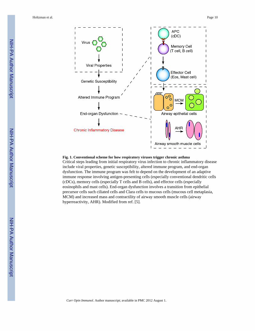
Fig. 1. Conventional scheme for how respiratory viruses trigger chronic asthmaCritical steps leading from initial respiratory virus infection to chronic inflammatory diseaseinclude viral properties, genetic susceptibility, altered immune program, and end-organdysfunction. The immune program was felt to depend on the development of an adaptiveimmune response involving antigen-presenting cells (especially conventional dendritic cells(cDCs), memory cells (especially T cells and B cells), and effector cells (especiallyeosinophils and mast cells). End-organ dysfunction involves a transition from epithelialprecursor cells such ciliated cells and Clara cells to mucous cells (mucous cell metaplasia,MCM) and increased mass and contractility of airway smooth muscle cells (airwayhyperreactivity, AHR). Modified from ref. [5].
Holtzman et al. Page 10
Curr Opin Immunol. Author manuscript; available in PMC 2012 August 1.
NIH
-PA Author Manuscript
NIH
-PA Author Manuscript
NIH
-PA Author Manuscript
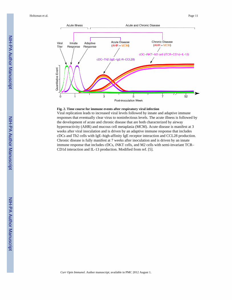
Fig. 2. Time course for immune events after respiratory viral infectionViral replication leads to increased viral levels followed by innate and adaptive immuneresponses that eventually clear virus to noninfectious levels. The acute illness is followed bythe development of acute and chronic disease that are both characterized by airwayhyperreactivity (AHR) and mucous cell metaplasia (MCM). Acute disease is manifest at 3weeks after viral inoculation and is driven by an adaptive immune response that includescDCs and Th2 cells with IgE–high-affinity IgE receptor interaction and CCL28 production.Chronic disease is fully manifest at 7 weeks after inoculation and is driven by an innateimmune response that includes cDCs, iNKT cells, and M2 cells with semi-invariant TCR–CD1d interaction and IL-13 production. Modified from ref. [5].
Holtzman et al. Page 11
Curr Opin Immunol. Author manuscript; available in PMC 2012 August 1.
NIH
-PA Author Manuscript
NIH
-PA Author Manuscript
NIH
-PA Author Manuscript
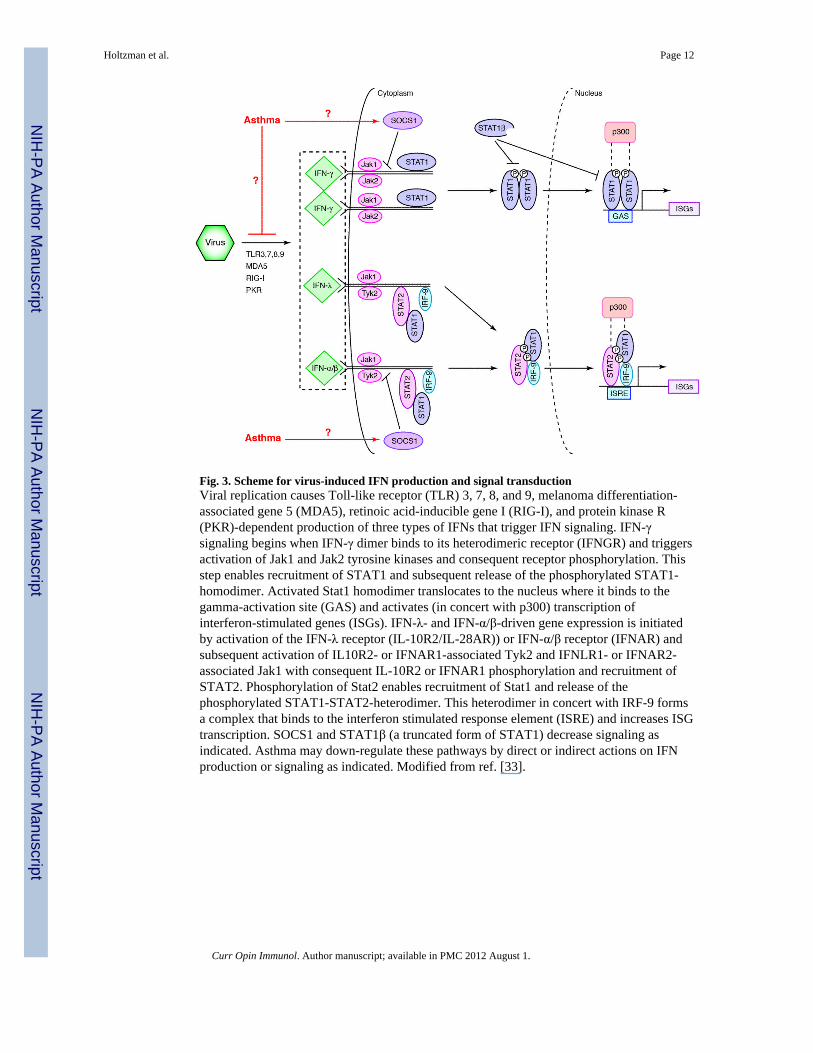
Fig. 3. Scheme for virus-induced IFN production and signal transductionViral replication causes Toll-like receptor (TLR) 3, 7, 8, and 9, melanoma differentiation-associated gene 5 (MDA5), retinoic acid-inducible gene I (RIG-I), and protein kinase R(PKR)-dependent production of three types of IFNs that trigger IFN signaling. IFN-γsignaling begins when IFN-γ dimer binds to its heterodimeric receptor (IFNGR) and triggersactivation of Jak1 and Jak2 tyrosine kinases and consequent receptor phosphorylation. Thisstep enables recruitment of STAT1 and subsequent release of the phosphorylated STAT1-homodimer. Activated Stat1 homodimer translocates to the nucleus where it binds to thegamma-activation site (GAS) and activates (in concert with p300) transcription ofinterferon-stimulated genes (ISGs). IFN-λ- and IFN-α/β-driven gene expression is initiatedby activation of the IFN-λ receptor (IL-10R2/IL-28AR)) or IFN-α/β receptor (IFNAR) andsubsequent activation of IL10R2- or IFNAR1-associated Tyk2 and IFNLR1- or IFNAR2-associated Jak1 with consequent IL-10R2 or IFNAR1 phosphorylation and recruitment ofSTAT2. Phosphorylation of Stat2 enables recruitment of Stat1 and release of thephosphorylated STAT1-STAT2-heterodimer. This heterodimer in concert with IRF-9 formsa complex that binds to the interferon stimulated response element (ISRE) and increases ISGtranscription. SOCS1 and STAT1β (a truncated form of STAT1) decrease signaling asindicated. Asthma may down-regulate these pathways by direct or indirect actions on IFNproduction or signaling as indicated. Modified from ref. [33].
Holtzman et al. Page 12
Curr Opin Immunol. Author manuscript; available in PMC 2012 August 1.
NIH
-PA Author Manuscript
NIH
-PA Author Manuscript
NIH
-PA Author Manuscript
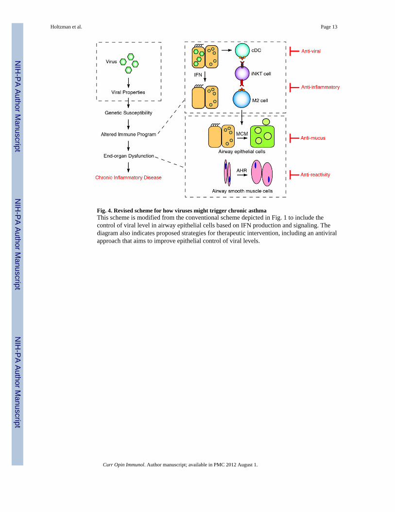
Fig. 4. Revised scheme for how viruses might trigger chronic asthmaThis scheme is modified from the conventional scheme depicted in Fig. 1 to include thecontrol of viral level in airway epithelial cells based on IFN production and signaling. Thediagram also indicates proposed strategies for therapeutic intervention, including an antiviralapproach that aims to improve epithelial control of viral levels.
Holtzman et al. Page 13
Curr Opin Immunol. Author manuscript; available in PMC 2012 August 1.
NIH
-PA Author Manuscript
NIH
-PA Author Manuscript
NIH
-PA Author Manuscript