Hong Kong - the International League Against Epilepsy
119
13 th - 16 th MAY 2016 HONG KONG 11 th ASIAN & OCEANIAN EPILEPSY CONGRESS FINAL PROGRAMME AND ABSTRACT BOOK 11th Asian & Oceanian Epilepsy Congress 13-16 May 2016 Hong Kong International Bureau for Epilepsy IBE www.epilepsyhongkong2016.org
-
Upload
khangminh22 -
Category
Documents
-
view
2 -
download
0
Transcript of Hong Kong - the International League Against Epilepsy
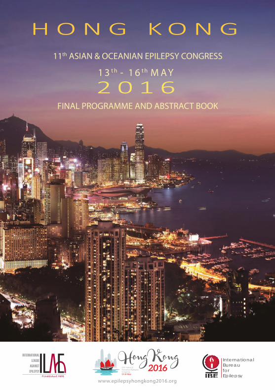
1 3 t h - 1 6 t h M A Y
2 0 1 6
H O N G K O N G11th ASIAN & OCEANIAN EPILEPSY CONGRESS
FINAL PROGRAMME AND ABSTRACT BOOK
11th Asian & Oceanian Epilepsy Congress 13-16 May
2016Hong Kong
InternationalBureau
forEpilepsyIBE
www.epilepsyhongkong2016.org
www.epilepsyhongkong2016.org
11th Asian & Oceanian Epilepsy Congress
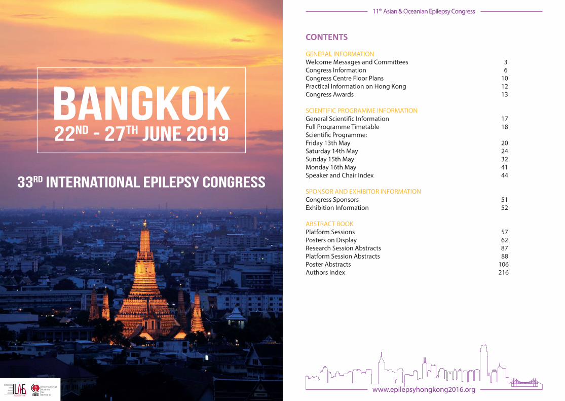
CONTENTS
GENERAL INFORMATION Welcome Messages and Committees 3Congress Information 6Congress Centre Floor Plans 10Practical Information on Hong Kong 12Congress Awards 13
SCIENTIFIC PROGRAMME INFORMATIONGeneral Scienti�c Information 17Full Programme Timetable 18Scienti�c Programme: Friday 13th May 20Saturday 14th May 24Sunday 15th May 32Monday 16th May 41Speaker and Chair Index 44
SPONSOR AND EXHIBITOR INFORMATIONCongress Sponsors 51Exhibition Information 52
ABSTRACT BOOKPlatform Sessions 57Posters on Display 62Research Session Abstracts 87Platform Session Abstracts 88Poster Abstracts 106Authors Index 216
InternationalBureauforEpilepsyIBE
33rd international epilepsy congress
BAngkok22nd - 27th JUNE 2019
BAngkok

3www.epilepsyhongkong2016.org
11th Asian & Oceanian Epilepsy Congress
Fina
l Pro
gram
me
www.epilepsyhongkong2016.org
11th Asian & Oceanian Epilepsy Congress
GENERAL INFORMATION
Denise CHAPMANCo-chair, Scienti�c
Organising Committee
Byung-In LEE Co-chair, Scienti�c
Organising Committee
Ada YUNGCo-chair, Scienti�c
Organising Committee
WELCOME MESSAGE FROM THE SCIENTIFIC ORGANISING COMMITTEE CHAIRS
Dear Friends and Colleagues,
We are delighted that you could join us here in Hong Kong for the 11th Asian & Oceanian Epilepsy Congress (AOEC) and have no doubt that you will �nd this Congress to be a truly rewarding experience.
Our colleagues from the Scienti�c Organising Committee have produced an excellent scienti�c programme and with input from our friends in ASEPA the highest educational quality is guaranteed. The Congress opens with the Chairmen’s Symposium on “Autoimmune Encephalitis” on Friday afternoon and the main sessions this year focus on “Genetics in Epilepsy”, “Epilepsy and Behaviour” and “New Paradigms of AED Therapy”. A careful blend of parallel sessions and practical sessions ensure that all recent scienti�c, clinical and social developments in epilepsy are comprehensively covered.
Organised by the local and regional IBE chapters, the Epilepsy and Society Symposium will take place on Sunday. The programme will be of great interest to sta� from community organizations supporting people living with epilepsy as well as appealing directly to the people living with epilepsy themselves.
Make sure to attend the platform sessions and visit the poster area to hear and read about the latest research and data on epilepsy from throughout the region. The two best platform presentations and two best posters will receive the Tadokoro Award on Monday morning. We would like to acknowledge the hard work of the members of the Abstract Review Committee in reviewing the congress abstracts.
Famed for its tower-studded skyline, Hong Kong is a unique destination that has absorbed cultural in�uences from around the globe and proudly proclaims itself to be Asia’s World City. Hong Kong is also known for its lively food scene, from Cantonese dim sum to extravagant high tea, and is a shopper’s paradise with options spanning from atmospheric night markets to the city’s innumerable bespoke tailors. Sightseeing opportunities abound and with transport options by the fabulous Tram network here in Wanchai or the nearby Star Ferry you can get anywhere in Hong Kong easily and cheaply.
We look forward to welcoming you to this vibrant city of Hong Kong over the next few days.
With our best wishes,

5www.epilepsyhongkong2016.org
11th Asian & Oceanian Epilepsy Congress
Fina
l Pro
gram
me
4 www.epilepsyhongkong2016.org
11th Asian & Oceanian Epilepsy Congress
Final Programm
e
Athanasios COVANISPresident IBE
Emilio PERUCCA President ILAE
WELCOME MESSAGE FROM THE PRESIDENTS OF THE INTERNATIONAL LEAGUE AGAINST EPILEPSY (ILAE) AND THE INTERNATIONAL BUREAU FOR EPILEPSY (IBE)
Dear Friends,
On behalf of the International Bureau for Epilepsy (IBE) and the International League Against Epilepsy (ILAE), it is our pleasure to welcome you to Hong Kong for the 11th Asian & Oceanian Epilepsy Congress (AOEC).
Both ILAE and IBE are grateful to our regional committees for their admirable work in advancing epilepsy knowledge especially through education and training and also for improving the quality of care as well as the quality of life for people living with epilepsy. Being here amongst you is the best way for us to understand the most important issues for epilepsy in this vast region and to learn how ILAE and IBE can best serve our members.
We would like to commend the members of the Scienti�c Organising Committee for constructing an exceptional scienti�c programme with international appeal. Beyond the session rooms, you can create new projects for your community by networking with those of similar minds and it is a perfect opportunity to meet old friends and make new acquaintances.
Hong Kong may be Asia’s business hub, but this vibrant, dynamic city also enjoys international notoriety for its intriguing and exhilarating leisure opportunities that will enrich your stay and leave a lasting impression. I am sure that many of you will have researched the restaurants here in Hong Kong given how notorious it is for its unrivalled choices of cuisine but if you need any particular recommendations the local team at the Customer Services desks in HKCEC will be happy to help you.
We wish you a most memorable and educational experience here in Hong Kong and hope to meet many of you in the next few days.
With warm regards,
CONGRESS COMMITTEES
SCIENTIFIC ORGANISING COMMITTEEDenise CHAPMAN (Australia), Co-chairByung-In LEE (South Korea), Co-chairAda YUNG (Hong Kong), Co-chair
Ding DING (China) John DUNNE (Australia) Shih Hui LIM (Singapore) Guoming LUAN (China) P. SATISHCHANDRA (India) Vinod SAXENA (India)Tatsuya TANAKA (Japan)
ABSTRACT REVIEW COMMITTEEDerrick CHAN (Singapore), Co-chairHowan LEUNG (Hong Kong), Co-chair
Andrew BLEASEL (Australia)Leonor CABRAL-LIM (Philippines)Eric CHAN (Hong Kong)Yotin CHINVARUN (Thailand)Mark COOK (Australia)Eva FUNG (Hong Kong)Josephine CASANOVA-GUTIERREZ (Philippines)Simon HARVEY (Australia)Akio IKEDA (Japan)Yuwu JIANG (China)Sunao KANEKO (Japan)
Heung-Dong KIM (South Korea)Kurnia KUSUMASTUTI (Indonesia)Patrick KWAN (Australia)Weiping LIAO (China)Kheng Seang LIM (Malaysia)Man Mohan MEHNDIRATTA (India)Zarine MOGAL (Pakistan)Ernest SOMERVILLE (Australia)Chong Tin TAN (Malaysia)Venus TANG (Hong Kong)Manjari TRIPATHI (India)

7www.epilepsyhongkong2016.org
11th Asian & Oceanian Epilepsy Congress
Fina
l Pro
gram
me
6 www.epilepsyhongkong2016.org
11th Asian & Oceanian Epilepsy Congress
Final Programm
e
CONGRESS INFORMATION
FACILITIES TIMETABLE
Friday Saturday Sunday Monday
Registration 13:00-18:30 07:00-18:00 07:00-18:00 07:30-13:00
Speakers Room 12:00-17:30 07:00-17:30 07:00-17:30 07:30-11:00
Posters on Display - 09:00-17:00 09:00-17:00 -
Exhibition ** 09:00-16:30 09:00-16:30 -
Co�ee Break Morning - 10:30-11:00 10:30-11:00 10:30-11:00
Co�ee Break Afternoon - 16:00-16:30 16:00-16:30 -
Lunch -
12:30-12:40
Available in Eisai’s Satellite Symposium
only
- -
Cafeteria 12:30-18:30 10:00-17:00 10:00-17:00 -
Cloakroom 13:00-20:30 07:15-19:15 07:15-18:15 07:45-13:00
** Some exhibition stands may be open during Friday afternoon and evening during sessions and the Welcome Ceremony and Reception
CAFETERIAA cafeteria is located on the main conference �oor outside Conference Hall C. Please view the opening hours in the Facilities Timetable.
CERTIFICATE OF ATTENDANCE A Certi�cate of Attendance will be available for all delegates for collection from the Registration Counter on the ground �oor of the HKCEC on Sunday or from the Registration Desk outside the Theatres on the main conference �oor of HKCEC on Monday.
CLOAKROOMA small cloakroom is available for congress delegates beside Theatre 1 on the main conference �oor of HKCEC. There is no charge for this service but space is limited. Please view the opening hours in the Facilities Timetable.
COFFEE BREAKSCo�ee and tea will be served in the Exhibition Area on the main conference �oor of HKCEC from 10:30-11:00 and also from 16:00-16:30 on Saturday and on Sunday. On Monday it will be served from 10:30-11:00 in the Theatre Foyer on the main conference �oor.
CONGRESS SECRETARIAT OFFICEMembers of the Congress Secretariat can be contacted at the Registration Counter during the Congress. For queries arising after the Congress, please contact:
11th Asian & Oceanian Epilepsy Congress, ILAE/IBE Congress Secretariat, 7 Priory O�ce Park, Stillorgan Road,Blackrock, Co. Dublin, A94 FN26, Ireland.
Tel: +353 1 2056720Fax: +535 1 2056156Email: [email protected] Website: www.epilepsyhongkong2016.org
EXHIBITIONA trade exhibition will be held in conjunction with the 11th AOEC. This is an integral part of the event, offering delegates the opportunity to learn about the latest developments in products and services relevant to the field of epilepsy. The Exhibition Area is located in the Convention Foyer on the main conference floor of HKCEC. Please view the opening hours in the Facilities Timetable.
HKCEC CUSTOMER SERVICE DESKHKCEC o�er a Customer Service Desk where you can receive information on the Congress and on local amenities; basic business centre services can also be carried out here. The HKCEC Customer Service Desk can be found on the main conference �oor and is open for congress hours.
CONGRESS INFORMATION

9www.epilepsyhongkong2016.org
11th Asian & Oceanian Epilepsy Congress
Fina
l Pro
gram
me
8 www.epilepsyhongkong2016.org
11th Asian & Oceanian Epilepsy Congress
Final Programm
e
LANGUAGEEnglish is the o�cial language of the 11th AOEC.
LIABILITY AND INSURANCEThe International League Against Epilepsy (ILAE), the International Bureau for Epilepsy (IBE) and its agents do not accept any liability whatsoever for death, personal injury, accidents, theft, loss or damage to persons, property or belongings of participants or accompanying persons, either before, during or following the Congress, tours or their stay in Hong Kong. It is therefore recommended that participants arrange their own personal health, accident and travel insurance.
LUNCHEisai Co., Ltd. are o�ering lunch to delegates attending their Satellite Symposium in Conference Hall A&B on Saturday from 12:30-12:40. Otherwise lunch may be purchased in the cafeteria on the main conference �oor or in the various catering outlets in HKCEC.
POSTERSPosters are on display on the mezzanine �oor and also in the Theatre Foyer on the main conference �oor of HKCEC. Posters will be on display from 09:00-17:00 on Saturday and on Sunday. Poster presenters are required to set up their posters between 08:00-09:00 on Saturday morning. Posters must be removed between 17:00-18:00 on Sunday. Presenting authors must be in attendance at their posters on Saturday from 14:10-15:00 and on Sunday from 12:45-13:30.
REGISTRATIONThe Registration Counter is located on the ground �oor of HKCEC; on Monday it will move upstairs outside the Theatres on the main conference �oor. Congress bags can be collected from this point. Please note that name badges must be worn at all times.
SCIENTIFIC EXHIBIT OF POSTERSOn Sunday from 11:00-14:00, Eisai Co., Ltd is holding a Scienti�c Exhibit of posters in Room V104 which is located beside Conference Hall C on the main conference �oor of HKCEC. All are welcome.
SMOKING POLICYHKCEC is a non-smoking area.
SPEAKERS ROOMThe Speakers Room is in Room V103 which is located between Theatre 1 and Theatre 2 on the main conference �oor of HKCEC. Facilities to review and amend presentations are available to all speakers and those presenting a platform session. Please note that all speakers and platform presenters should submit their final PowerPoint presentations to the main desk in the Speakers Room no later than 2 hours in advance of their session. Speakers in early morning sessions are required to submit their material before 17:00 on the day prior to their scheduled session.
VENUE INFORMATIONThe 11th AOEC is taking place at the Hong Kong Convention and Exhibition Centre (HKCEC).
Venue Address: Hong Kong Convention and Exhibition Centre (HKCEC),I Expo Drive, Wanchai, Hong Kong.Tel: +852 2582 8888Website: www.hkcec.com
WELCOME CEREMONY AND RECEPTIONThe Welcome Ceremony of the 11th AOEC will take place in Convention Hall A&B on the main conference �oor of HKCEC on Friday at 18:30. This special event will give you a chance to learn more about the many activities of ILAE and IBE as well as about Hong Kong and its culture.
Following the Welcome Ceremony, all delegates are invited to join the Welcome Reception which will be held in the Exhibition Area on the main conference �oor of HKCEC; it is the perfect opportunity to catch up with friends and colleagues from the region and beyond.
WHEELCHAIR ACCESSAll session rooms in the HKCEC are wheelchair accessible.
WIFIThere is free wi� in the venue for all registered delegates. In order to log-on, connect to the “HKCEC_Free_WiFi” network and when you open your internet browser, the venue’s log-on web page will ask you to press the “I agree” button after reading the terms and conditions.
CONGRESS INFORMATIONCONGRESS INFORMATION

11www.epilepsyhongkong2016.org
11th Asian & Oceanian Epilepsy Congress
Fina
l Pro
gram
me
10 www.epilepsyhongkong2016.org
11th Asian & Oceanian Epilepsy Congress
Final Programm
e
CONGRESS CENTRE FLOOR PLANSGROUND FLOOR, HKCEC
To Mezzanine and Ground Floors
CONGRESS CENTRE FLOOR PLANSMAIN CONFERENCE FLOOR: LEVEL 1, HKCEC
Gift Shop
Escalators up to Mezzanine and
Convention Floors
MEZZANINE FLOOR, HKCEC
EXHIBITION AREA
POSTER AERA
REGISTRATION AREA
SCIENTIFIC EXHIBIT
BUSINESS MEETINGROOMS
SESSION ROOMS
SPEAKERS ROOM
ESCALATOR
RESTROOM
COFFEE BREAKS
ELEVATOR/LIFT
CLOAKROOM
EXHIBITION AREA
POSTER AERA
REGISTRATION AREA
SCIENTIFIC EXHIBIT
BUSINESS MEETINGROOMS
SESSION ROOMS
SPEAKERS ROOM
ESCALATOR
RESTROOM
COFFEE BREAKS
ELEVATOR/LIFT
CLOAKROOM

13www.epilepsyhongkong2016.org
11th Asian & Oceanian Epilepsy Congress
Fina
l Pro
gram
me
12 www.epilepsyhongkong2016.org
11th Asian & Oceanian Epilepsy Congress
Final Programm
e
SURNAME FIRST NAME COUNTRY
DAI Jindong China
HAO Bin China
KOHILA Krishnan Malaysia
LI Yan China
SAMANT Shruti India
WANG Ruofan China
ZHAO Jing China
PRACTICAL INFORMATION ON HONG KONG
ABOUT HONG KONGHong Kong, with its world-famous skyline, o�ers something to everyone with an abundance of ancient and modern attractions on o�er. The city is famed for its seafood, Cantonese and international cuisine, and its amazing shopping opportunities. Soaring Victoria Peak is still a favourite spot for its magni�cent views across the iconic harbour to Kowloon. The frantic pace of life on Hong Kong Island gives way to the mountainous interior and its remote beauty, perfect for walking, hiking, trekking, and exploring the ecosystems. An alternative is a boat trip to a selection of the other islands in the chain, home to famous temples, stilted �shing villages, and small, secluded bays with glorious beaches.
CITY TRANSPORTHong Kong is internationally famous for its safe, a�ordable and reliable public transport system that keeps the city moving at its trademark lightning speed. The subway system (MTR) connects almost all of Hong Kong and is highly e�cient with trains every minute in rush hour and buses have routes covering the entire city. Alternatively you can switch gears by hopping on an unhurried tram or ferry and savour the city at an old-world pace. You will �nd “urban taxis” in Hong Kong Island and Kowloon, easily distinguished by being painted red with silver roofs. Most taxi drivers speak some English but just in case, it is a good idea to have your destination written down in Chinese.
ELECTRICITYThe standard electrical voltage in Hong Kong is 220 volts AC, 50Hz. Most hotel bathrooms also have outlets for 100 volts, but if not, you will need a transformer for any appliance or electrical equipment. The majority of electrical outlets in Hong Kong take a three-pronged square plug. You can buy an inexpensive adaptor for your electrical equipment at most convenience stores.
TAXES AND TIPPINGThere is no sales tax in Hong Kong; hotels and restaurants may add a 10% service charge.Tipping is not a Chinese custom; however in Hong Kong, tipping is becoming more expected.
TIME ZONEHong Kong is 7 hours ahead of GMT in May.
WATERHong Kong’s tap water is safe to drink without any further �ltration or treatment and complies with World Health Organisation drinking water guidelines.
CONGRESS AWARDS
THE ASIAN OCEANIAN OUTSTANDING ACHIEVEMENT EPILEPSY AWARDThe Asian Oceanian Outstanding Achievement Epilepsy Award recognises and pays tribute to medical and non-medical professionals for their extraordinary contributions to epilepsy care in this region. The award is bestowed on Yuan-gui HUANG (China), Patrick KWAN (Australia), K.V. MURALIDHARAN (India) and Manjari TRIPATHI (India) and will be given out during the Welcome Ceremony on Friday.
THE GOLDEN LIGHT AWARDThe Golden Light Award (IBE) will be presented to 10 recipients during the Welcome Ceremony on Friday and also at the Epilepsy & Society Symposium on Sunday. The award is bestowed on M AMARJARGAL (Mongolia), Amrita BHASHYAM (India), Rosalind CHEE (Malaysia), Jeong Ja JEE (South Korea), Yin Chan LOKE (Singapore), Kym MEERS (Australia), Wai Hung NG (Hong Kong), Kun Hoo RHEE (Nepal), Zhi-Gang WANG (China) and Laura Liu YI (Taiwan).
THE TADOKORO AWARD In order to encourage young researchers in epileptology in the region, there will be best presentation prizes for both platform and poster presentations. Dr. TADOKORO (Japan) contributed generously to the activities of the ILAE Commission on Asian and Oceanian Affairs (CAOA).
The first and second prize for both platform and poster presentation are US$300 and US$200 respectively and the recipients will be announced on Monday morning before the main session.
ILAE AND IBE BURSARY AWARDS The Bursary Award scheme was established to assist delegates to attend the 11th AOEC. A particular emphasis was given to those coming from developing regions, which are locally active in the �eld of epilepsy. A total of 26 Bursary Award Recipients were selected by the IBE and ILAE Regional Committee chairs; funding for these awards was provided by the International League Against Epilepsy (ILAE) and the International Bureau for Epilepsy (IBE).
The 11th AOEC IBE Bursary Award recipients are:

15www.epilepsyhongkong2016.org
11th Asian & Oceanian Epilepsy Congress
Fina
l Pro
gram
me
14 www.epilepsyhongkong2016.org
11th Asian & Oceanian Epilepsy Congress
Final Programm
e
SURNAME FIRST NAME COUNTRY
ASRANNA Ajay I.P. India
CRUZ Maria Teresa Philippines
GOIT Rajesh Kumar Nepal
GUPTA Swapan India
HLAING Chaw Su Myanmar
JOSHI Mandeep Dutta Nepal
KARUNARATNE Kasun Sri Lanka
KUMAR Vijay India
MISHRA Monika India
PHAM Duc-Hung Vietnam
PUANGMANY Phoumavong Lao
RAMANUJAM Bhargavi India
SIMJEE Shabana Pakistan
TALWAR Palak India
TANVEER Mehwish Pakistan
VELMURUGAN Jayabal India
YAMPAYON Kittika Thailand
ZHANG Yujiao China
CONGRESS AWARDS
ILAE AND IBE BURSARY AWARDS The 11th AOEC ILAE Bursary Award recipients are:
C
M
Y
CM
MY
CY
CMY
K
11th AOEC Final Programme_300dpi.pdf 1 16/3/16 12:17 pm
17www.epilepsyhongkong2016.org
11th Asian & Oceanian Epilepsy Congress
Fina
l Pro
gram
me
16 www.epilepsyhongkong2016.org
11th Asian & Oceanian Epilepsy Congress
Final Programm
e
SCIENTIFIC PROGRAMME INFORMATION
GENERAL SCIENTIFIC INFORMATION
ASEPA EEG CERTIFICATION EXAMINATION PART I Part I ASEPA EEG Certification Examinations will take place during the 11th AOEC. For further details, please contact the Registration Counter.
ASEPA PRE-CONGRESS TEACHING COURSESTwo ASEPA pre-congress teaching courses will take place on Friday from 08:30-12:00 in Theatre 1 and 2; one is entitled “Diagnosis: Is it a Seizure?” and the second is “Diagnosis: Localization of Seizures”.
Please note that a separate registration is required to attend either of these teaching courses; you may register for them in the Theatre Foyer on the main conference �oor. The registration fee for congress delegates is US$20 and it is US$40 for non-congress delegates.
ASEPA WORKSHOP ON CHAPTER LEADERSHIP AND MANAGEMENTASEPA have organized a workshop on Chapter Leadership and Management that takes place on Saturday from 11:00-13:00 in S228 on level 2. This workshop is by invitation only.
EPILEPSY & SOCIETY SYMPOSIUMAn exciting programme that will be of great interest to both individuals living with epilepsy and to sta� from community organisations supporting people with epilepsy will take place on Sunday from 09:30-16:00 in Theatre 2. This programme has been developed by local and regional committees of the International Bureau for Epilepsy (IBE).
Please note that a separate registration is required for this programme; please enquire at the Registration Counter.
ILAE / CAOA CHAPTER CONVENTION The ILAE/CAOA Chapter Convention will take place on Friday from 12:00-13:45 in Convention Hall C for pre-invited ILAE members.
THE MASAKAZU SEINO MEMORIAL LECTUREProf. Masakazu SEINO (Japan) was instrumental in the establishment of the Asian and Oceanian Regional Commission of ILAE (CAOA) and built the foundation for the outstanding progress of epileptology in the region. In order to commemorate his exceptional contribution to the region after his death in 2007, the CAOA set up the Masakazu Seino Memorial Lecture in 2008 which is regarded as one of the highlights of the programme of the AOEC. The CAOA would like to thank UCB Japan for their generous contribution to this lecture.

19www.epilepsyhongkong2016.org
11th Asian & Oceanian Epilepsy Congress
Fina
l Pro
gram
me
18 www.epilepsyhongkong2016.org
11th Asian & Oceanian Epilepsy Congress
Final Programm
e
Lunch kindly sponsored by Eisai Co., LtdSatellite Symposium: Eisai Co., Ltd
DILEMMAS AND CHALLENGES IN THE MANAGEMENT OF GENERALIZED TONIC-CLONIC SEIZURES:CAN WE DO MORE?
12:40-14:10
ASEPA WORKSHOP ON CHAPTER LEADERSHIP
AND MANAGEMENT 11:00-13:00
(By Invitation Only)
Platform Session: CLINICAL NEURO-
PHYSIOLOGY & NEUROIMAGING
15:00-16:00
Platform Session: TREATMENT AND EPIDEMIOLOGY
15:00-16:00
CAOA Research Task Force Session:
15:00-16:00
Platform Session: BASIC SCIENCE AND GENETICS
15:00-16:00
Poster Viewing 14:10-15:00
Poster Viewing 12:30-13:30
Satellite Symposium: UCB Pharma EPILEPSY THROUGH THE AGES
13:30-15:00
Epilepsy & Society Symposium
09:30-16:00 (separate registration
fee)
Parallel Sessions: 11:00-12:30
SURGERY OF MR-NEGATIVE
EPILEPSY
EPILEPTOLOGY OF NEUROLOGICAL INTENSIVE CARE
ASEPA Didactic Lecture: OBESITY, OSTEOPOROSIS AND EPILEPSY: WHY AND WHAT TO DO?
08:15-09:00
ASEPA Didactic Lecture: EPILEPSY AND MEMORY
07:30-08:15
Practical Session - Debate:
THE NEW CLASSIFICATION OF
EPILEPSY 16:30-17:30
Platform Session: PAEDIATRIC
EPILEPTOLOGY 16:30-17:30
Practical Session - Workshop:
HFOS AND THE EPILEPTOGENIC ZONE
16:30-17:30
Practical Session - Video:
EPILEPTIC SEIZURES OR NOT?
16:30-17:30
Platform Session: STATUS
EPILEPTICUS 16:30-17:30
Coffee Break 16:00-16:30
Friday 13th May Monday 16th May
ASEPA Didactic Lecture: CAN I STOP MY DRUGS?
08:00-08:45
ASEPA Didactic Lecture: EPILEPSY TREATMENT - CAN GENETICS GUIDE US?
07:30-08:15
ASEPA Didactic Lecture: CONTEMPORARY MANAGEMENT OF WOMEN WITH EPILEPSY ACROSS THE LIFESPAN
08:15-09:00 AWARDS CEREMONY 08:45-09:00
Saturday 14th May Sunday 15th May
ASEPA Pre-Congress Teaching
Course 1: DIAGNOSIS: IS IT A
SEIZURE? 08:30-12:00
(separate registration fee)
Main Session: EPILEPSY AND BEHAVIOUR
09:00-10:30
Poster Set-up
Post
ers:
09:
00-1
7:00
ASEPA Pre-Congress Teaching
Course 2: DIAGNOSIS:
LOCALIZATION OF SEIZURES 08:30-12:00
(separate registration fee)
Satellite Symposium: GSK TREATMENT OPTIONS AND MANAGEMENT
OF EPILEPSY: LAST 25 YEARS AND BEYOND17:30-19:00
Platform Session: ADULT
EPILEPTOLOGY 15:00-16:00
Platform Session: PYSCHOSOCIAL
ISSUES 15:00-16:00
CAOA Global Campaign Task Force Session:
DIAGNOSIS OF EPILEPSY IN LOW-MIDDLE INCOME
COUNTRIES 15:00-16:00
Satellite Symposium: Sanofi - Access to Medicines
PAVING THE WAY TO BETTER ACCESS TO EPILEPSY CARE
16:30-18:00
Practical Session - Debate:
IS THE FIRST SEIZURE EPILEPSY?
16:30-17:30
Platform Session: SURGERY16:30-17:30
Practical Session - Video: USUAL AND
UNUSUAL SEIZURES ACROSS THE AGE
RANGES16:30-17:30
CAOA Global Campaign Task Force Session:
THE WHO RESOLUTION AND THE LAOS
PROJECT 16:30-17:30
WELCOME RECEPTION 19:30-20:30
Post
ers:
09:
00-1
7:00
Poster Removal
MINIMALLY INVASIVE EPILEPSY SURGERY
THE BURDEN OF EPILEPSY IN THE ASIAN OCEANIAN
REGION
KETOGENIC DIET THERAPY
WELCOME CEREMONY 18:30-19:30
Chairman's Symposium: AUTOIMMUNE ENCEPHALITIS
14:00-15:30
CAOA Peadiatric Task Force Session:
AN UPDATE OF EPILEPTIC ENCEPHALOPATHY
15:00-16:00
Masakazu Seino Memorial Lecture: EXPLORING THE MYSTERIES OF EEG:
CAN INFRASLOW AND DC SHIFT IMPROVE EPILEPSY TREATMENT?
15:30-16:15Coffee Break 16:00-16:30
Main Session: GENETICS IN EPILEPSY
09:00-10:30
Coffee Break 10:30-11:00
Post Main Session:
BRAIN SOMATIC MUTATIONS 11:00-12:30
Main Session: NEW PARADIGMS OF AED THERAPY
09:00-10:30
Coffee Break: 10:30-11:00 Coffee Break: 10:30-11:00
Post Main Session: THE USE OF REPURPOSE
DRUGS 11.00-12:30
STEREO-EEG AND BRAIN
NETWORKS
GENDER ISSUES IN AED THERAPY
Parallel Sessions: 11:00-12:30
THE ADVENT OF DEVICES IN THE
MANAGEMENT OF EPILEPSY
Post Main Session: EPILEPSY AND
COMORBIDITIES11.00-12:30
Parallel Sessions: 11:00-12:30
SCIENTIFIC PROGRAMME - FULL PROGRAMME TIMETABLE SCIENTIFIC PROGRAMME - FULL PROGRAMME TIMETABLE
Lunch kindly sponsored by Eisai Co., LtdSatellite Symposium: Eisai Co., Ltd
DILEMMAS AND CHALLENGES IN THE MANAGEMENT OF GENERALIZED TONIC-CLONIC SEIZURES:CAN WE DO MORE?
12:40-14:10
ASEPA WORKSHOP ON CHAPTER LEADERSHIP
AND MANAGEMENT 11:00-13:00
(By Invitation Only)
Platform Session: CLINICAL NEURO-
PHYSIOLOGY & NEUROIMAGING
15:00-16:00
Platform Session: TREATMENT AND EPIDEMIOLOGY
15:00-16:00
CAOA Research Task Force Session:
15:00-16:00
Platform Session: BASIC SCIENCE AND GENETICS
15:00-16:00
Poster Viewing 14:10-15:00
Poster Viewing 12:30-13:30
Satellite Symposium: UCB Pharma EPILEPSY THROUGH THE AGES
13:30-15:00
Epilepsy & Society Symposium
09:30-16:00 (separate registration
fee)
Parallel Sessions: 11:00-12:30
SURGERY OF MR-NEGATIVE
EPILEPSY
EPILEPTOLOGY OF NEUROLOGICAL INTENSIVE CARE
ASEPA Didactic Lecture: OBESITY, OSTEOPOROSIS AND EPILEPSY: WHY AND WHAT TO DO?
08:15-09:00
ASEPA Didactic Lecture: EPILEPSY AND MEMORY
07:30-08:15
Practical Session - Debate:
THE NEW CLASSIFICATION OF
EPILEPSY 16:30-17:30
Platform Session: PAEDIATRIC
EPILEPTOLOGY 16:30-17:30
Practical Session - Workshop:
HFOS AND THE EPILEPTOGENIC ZONE
16:30-17:30
Practical Session - Video:
EPILEPTIC SEIZURES OR NOT?
16:30-17:30
Platform Session: STATUS
EPILEPTICUS 16:30-17:30
Coffee Break 16:00-16:30
Friday 13th May Monday 16th May
ASEPA Didactic Lecture: CAN I STOP MY DRUGS?
08:00-08:45
ASEPA Didactic Lecture: EPILEPSY TREATMENT - CAN GENETICS GUIDE US?
07:30-08:15
ASEPA Didactic Lecture: CONTEMPORARY MANAGEMENT OF WOMEN WITH EPILEPSY ACROSS THE LIFESPAN
08:15-09:00 AWARDS CEREMONY 08:45-09:00
Saturday 14th May Sunday 15th May
ASEPA Pre-Congress Teaching
Course 1: DIAGNOSIS: IS IT A
SEIZURE? 08:30-12:00
(separate registration fee)
Main Session: EPILEPSY AND BEHAVIOUR
09:00-10:30
Poster Set-up
Post
ers:
09:
00-1
7:00
ASEPA Pre-Congress Teaching
Course 2: DIAGNOSIS:
LOCALIZATION OF SEIZURES 08:30-12:00
(separate registration fee)
Satellite Symposium: GSK TREATMENT OPTIONS AND MANAGEMENT
OF EPILEPSY: LAST 25 YEARS AND BEYOND17:30-19:00
Platform Session: ADULT
EPILEPTOLOGY 15:00-16:00
Platform Session: PYSCHOSOCIAL
ISSUES 15:00-16:00
CAOA Global Campaign Task Force Session:
DIAGNOSIS OF EPILEPSY IN LOW-MIDDLE INCOME
COUNTRIES 15:00-16:00
Satellite Symposium: Sanofi - Access to Medicines
PAVING THE WAY TO BETTER ACCESS TO EPILEPSY CARE
16:30-18:00
Practical Session - Debate:
IS THE FIRST SEIZURE EPILEPSY?
16:30-17:30
Platform Session: SURGERY16:30-17:30
Practical Session - Video: USUAL AND
UNUSUAL SEIZURES ACROSS THE AGE
RANGES16:30-17:30
CAOA Global Campaign Task Force Session:
THE WHO RESOLUTION AND THE LAOS
PROJECT 16:30-17:30
WELCOME RECEPTION 19:30-20:30
Post
ers:
09:
00-1
7:00
Poster Removal
MINIMALLY INVASIVE EPILEPSY SURGERY
THE BURDEN OF EPILEPSY IN THE ASIAN OCEANIAN
REGION
KETOGENIC DIET THERAPY
WELCOME CEREMONY 18:30-19:30
Chairman's Symposium: AUTOIMMUNE ENCEPHALITIS
14:00-15:30
CAOA Peadiatric Task Force Session:
AN UPDATE OF EPILEPTIC ENCEPHALOPATHY
15:00-16:00
Masakazu Seino Memorial Lecture: EXPLORING THE MYSTERIES OF EEG:
CAN INFRASLOW AND DC SHIFT IMPROVE EPILEPSY TREATMENT?
15:30-16:15Coffee Break 16:00-16:30
Main Session: GENETICS IN EPILEPSY
09:00-10:30
Coffee Break 10:30-11:00
Post Main Session:
BRAIN SOMATIC MUTATIONS 11:00-12:30
Main Session: NEW PARADIGMS OF AED THERAPY
09:00-10:30
Coffee Break: 10:30-11:00 Coffee Break: 10:30-11:00
Post Main Session: THE USE OF REPURPOSE
DRUGS 11.00-12:30
STEREO-EEG AND BRAIN
NETWORKS
GENDER ISSUES IN AED THERAPY
Parallel Sessions: 11:00-12:30
THE ADVENT OF DEVICES IN THE
MANAGEMENT OF EPILEPSY
Post Main Session: EPILEPSY AND
COMORBIDITIES11.00-12:30
Parallel Sessions: 11:00-12:30
Theatre 1
Theatre 2
Conference Hall A&B
Conference Hall C
Room S228, Level 2
Session Rooms

21www.epilepsyhongkong2016.org
11th Asian & Oceanian Epilepsy Congress
Fina
l Pro
gram
me
20 www.epilepsyhongkong2016.org
11th Asian & Oceanian Epilepsy Congress
Final Programm
e
SCIENTIFIC PROGRAMME - FRIDAY 13TH MAY
Conference Hall A&B Theatre 1 Theatre 2
Satellite Symposium: Sanofi - Access to Medicines
PAVING THE WAY TO BETTER ACCESS TO EPILEPSY CARE
16:30-18:00
ASEPA Pre-Congress Teaching Course: DIAGNOSIS: LOCALIZATION OF SEIZURES
08:30-12:00 (separate registration fee)
ASEPA Pre-Congress Teaching Course: DIAGNOSIS: IS IT A SEIZURE?
08:30-12:00 (separate registration fee)
Masakazu Seino Memorial Lecture: EXPLORING THE MYSTERIES OF EEG: CAN
INFRASLOW AND DC SHIFT IMPROVE EPILEPSY TREATMENT?
15:30-16:15
Welcome Reception 19:30-20:30
The Chairmen's Symposium: AUTOIMMUNE ENCEPHALITIS
14:00-15:30
Welcome Ceremony 18:30-19:30
SCIENTIFIC PROGRAMME - FRIDAY 13TH MAY08:30-12:00 ASEPA TEACHING COURSE Theatre 1
DIAGNOSIS: IS IT A SEIZURE?Chairs: Shih Hui LIM (Singapore) and Ernest SOMERVILLE (Australia)
Non-epileptic events in children Hian-Tat ONG (Singapore)
Syncope vs seizure: clinical features and evaluation Ernest SOMERVILLE (Australia)
Distinguishing sleep disorders from seizures Yotin CHINVARUN (Thailand)
Co�ee Break
Migraine and epilepsy – interface, overlap and diagnosis Shih Hui LIM (Singapore)
Non-epileptic psychogenic events Venus TANG (Hong Kong)
What kind of epilepsy is it and does it matter? (The importance of a precise diagnosis) Parthasarthy SATISHCHANDRA (India)
08:30-12:00 ASEPA TEACHING COURSE Theatre 2
DIAGNOSIS: LOCALIZATION OF SEIZURES Chairs: John DUNNE (Australia) and Byung-In LEE (South Korea)
Seizure semiology - how good is it? Yushi INOUE (Japan)
Surface EEG techniques - getting the best yield Andrew BLEASEL (Australia)
MEG: is it worth it? Hermann STEFAN (Germany)
Co�ee Break
CT and MRI: a practical guide Graeme JACKSON (Australia)
Functional imaging - SPECT, PET Byung-In LEE (South Korea)
Invasive techniques Sinclair LIU (China)
23www.epilepsyhongkong2016.org
11th Asian & Oceanian Epilepsy Congress
Fina
l Pro
gram
me
22 www.epilepsyhongkong2016.org
11th Asian & Oceanian Epilepsy Congress
Final Programm
e
14:00-15:30 THE CHAIRMEN’S SYMPOSIUM Conference Hall A&B
AUTOIMMUNE ENCEPHALITIS Chairs: Denise CHAPMAN (Australia), Byung-In LEE (South Korea) and Ada YUNG (Hong Kong)
The pathogenesis of autoimmune encephalitis Stephen REDDEL (Australia)
The clinical and radiological aspects of autoimmune encephalitis Ada YUNG (Hong Kong)
Laboratory diagnosis Sang Kun LEE (South Korea)
Therapeutic strategies and outcome Ming LIM (United Kingdom)
15:30-16:15 THE MASAKAZU SEINO MEMORIAL LECTURE Conference Hall A&B
Chair: Shih Hui LIM(Singapore)
Exploring the mysteries of EEG:Can infraslow and DC shift improve epilepsy treatment?Akio IKEDA (Japan)
16:30-18:00 SATELLITE SYMPOSIUM: SANOFI - ACCESS TO MEDICINES Theatre 1
PAVING THE WAY TO BETTER ACCESS TO EPILEPSY CAREChairs: Howan LEUNG (Hong Kong) and Pierre-Marie PREUX (France)
Improving access to epilepsy care in Cambodia: ECIR programmeChhour CHANNARA (Cambodia)
DheVELoP programme: domestic health visitors to improve access to care for people with epilepsy in Lao PDR – Preliminary resultsPhetvongsinh CHIVORAKOUN (Lao PDR)
WHO: reducing the treatment gap – The Vietnam projectTruong Le Van NGOC (Vietnam)
Developing access to healthcare: What role for the pharmaceutical industry?Robert SEBBAG (France)
18:30-19:30 WELCOME CEREMONY Conference Hall A&B
SCIENTIFIC PROGRAMME - FRIDAY 13TH MAY
12th EUROPEANCONGRESS ONEPILEPTOLOGY
11th-15th
SEPT2016

25www.epilepsyhongkong2016.org
11th Asian & Oceanian Epilepsy Congress
Fina
l Pro
gram
me
24 www.epilepsyhongkong2016.org
11th Asian & Oceanian Epilepsy Congress
Final Programm
e
SCIENTIFIC PROGRAMME – SATURDAY 14TH MAY
Conference Hall A&B Theatre 1 Theatre 2 Conference Hall C Room 228 (level 2)Mezzanine Floor &
Theatre Foyer
Lunch is kindly provided by Eisai Co. Ltd. for those attending their Satellite Symposium
Satellite Symposium: GSKTREATMENT OPTIONS AND MANAGEMENT OF EPILEPSY: LAST 25 YEARS AND BEYOND
17:30-19:00
Parallel Session: KETOGENIC DIET THERAPY
11:00-12:30
CAOA Global Campaign Task Force Session: DIAGNOSIS OF EPILEPSY IN
LOW-MIDDLE INCOME COUNTRIES15:00-16:00
Debate: IS THE FIRST SEIZURE EPILEPSY?
16:30-17:30
Parallel Session: THE BURDEN OF EPILEPSY IN THE ASIAN
OCEANIAN REGION 11:00-12:30
Poster ViewingPoster Viewing
Coffee Break
ASEPA Didactic Lecture: OBESITY, OSTEOPOROSIS AND EPILEPSY: WHY
AND WHAT TO DO? 08:15-09:00
ASEPA Didactic Lecture: EPILEPSY AND MEMORY
07:30-08:15
CAOA Peadiatric Task Force Session: AN UPDATE OF EPILEPTIC ENCEPHALOPATHY
15:00-16:00
Platform Session: ADULT EPILEPTOLOGY
15:00-16:00
Platform Session: SURGERY
16:30-17:30
Platform Session: PYSCHOSOCIAL ISSUES
15:00-16:00
CAOA Global Campaign Task Force Session: THE WHO RESOLUTION AND THE LAOS PROJECT
16:30-17:30
Coffee Break
Main Session: EPILEPSY AND BEHAVIOUR
09:00-10:30
Parallel Session: MINIMALLY INVASIVE EPILEPSY SURGERY
11:00-12:30
Post Main Session: EPILEPSY AND COMORBIDITIES
11:00-12:30
ASEPA WORKSHOP: CHAPTER LEADERSHIP AND MANAGEMENT
11:00-13:00 (By Invitation Only)
Satellite Symposium: Eisai Co., Ltd. DILEMMAS AND CHALLENGES IN THE
MANAGEMENT OF GENERALIZED TONIC-CLONIC SEIZURES: CAN WE DO MORE?
12:40-14:10
POSTERS ON DISPLAY 09:00-17:00
Coffee Break
Video Quiz: USUAL AND UNUSUAL SEIZURES ACROSS THE
AGE RANGES 16:30-17:30
POSTER SET-UP 08:00-09:00
Coffee Break
27www.epilepsyhongkong2016.org
11th Asian & Oceanian Epilepsy Congress
Fina
l Pro
gram
me
26 www.epilepsyhongkong2016.org
11th Asian & Oceanian Epilepsy Congress
Final Programm
e
SCIENTIFIC PROGRAMME – SATURDAY 14TH MAY07:30-08:15 ASEPA DIDACTIC LECTURE Theatre 1
Chair: Rabindra SHRESTHA (Nepal)
Epilepsy and memory Marco MULA (United Kingdom)
08:15-09:00 ASEPA DIDACTIC LECTURE Theatre 1Chair: Muzharul MANNAN (Bangladesh)
Obesity, osteoporosis and epilepsy: why and what to do? Terence O'BRIEN (Australia)
09:00-10:30 MAIN SESSION Conference Hall A&B
EPILEPSY AND BEHAVIOURChairs: Athanasios COVANIS (Greece) and Kousuke KANEMOTO (Japan)
Psychosis in chronic epilepsy Naoto ADACHI (Japan) Aggression in epilepsy Sung Pa PARK (South Korea)
Suicide and suicidal behaviour in epilepsy Marco MULA (United Kingdom)
Behavioural problems in children with epilepsy Toshibsaburo NAGAI (Japan)
11:00-12:30 POST MAIN SESSION Theatre 1 EPILEPSY AND COMORBIDITIES Chairs: Zhen HONG (China) and Parthasarthy SATISHCHANDRA (India)
Dementia and cognitive impairment Yushi INOUE (Japan) Epilepsy and vascular risk Yao-Chung CHUANG (Taiwan)
Sleep disordersSang-Ahm LEE (South Korea) SUDEPTorbjörn TOMSON (Sweden)
11:00-12:30 PARALLEL SESSION Conference Hall A&B MINIMALLY INVASIVE EPILEPSY SURGERYChairs: Tatsuya TANAKA (Japan) and Tak-Lap POON (Hong Kong) LITT Ashwini SHARAN (USA) The role of endoscopy for epilepsySarat CHANDRA (India) Minimally invasive stereotactic radiofrequency thermocoagulation for hypothalamic hamartoma Shigeki KAMEYAMA (Japan)
Bipolar electro coagulation for refractory epilepsy in eloquent cortexes and insular Guoming LUAN (China)
11:00-12:30 PARALLEL SESSION Conference Hall C
THE BURDEN OF EPILEPSY IN THE ASIAN OCEANIAN REGION Chairs: Robert COLE (Australia) and Vinod SAXENA (India) Epidemiology of epilepsy in Asia: an overview Chong Tin TAN (Malaysia) Mortality and SUDEP in Asia Ding DING (China)
Employment and marriage of PWE in Asia Kheng Seang LIM (Malaysia)
The impact of epilepsy renaming on stigma Eva FUNG (Hong Kong)
SCIENTIFIC PROGRAMME – SATURDAY 14TH MAY

29www.epilepsyhongkong2016.org
11th Asian & Oceanian Epilepsy Congress
Fina
l Pro
gram
me
28 www.epilepsyhongkong2016.org
11th Asian & Oceanian Epilepsy Congress
Final Programm
e
11:00-12:30 PARALLEL SESSION Theatre 2
KETOGENIC DIET THERAPY Chairs: Derrick CHAN (Singapore) and Anannit VISUDTIBHAN (Thailand)
The anti-epileptic mechanisms of dietary treatment Heung Dong KIM (South Korea)
The ketogenic diet and speci�c epilepsy syndromes Hirokaza OGUNI (Japan)
Making the ketogenic diet work in the Asian food cultureDerrick CHAN (Singapore) Ketogenic diet therapy: the future Sheffali GULATI (India)
11:00-13:00 ASEPA WORKSHOP (BY INVITATION ONLY) Room S228, level 2
CHAPTER LEADERSHIP AND MANAGEMENT WORKSHOPChairs: Shih Hui LIM (Singapore) and Chong Tin TAN (Malaysia)
An introduction to leadership and management Shih Hui LIM (Singapore)
Communication between CAOA and its chapters Sunao KANEKO (Japan)
How to raise funds, budget and resolve con�icts Chong Tin TAN (Malaysia)
How do I lead and manage ILAE? Emilio PERUCCA (Italy)
How do I lead and manage CAOA? Byung-In LEE (South Korea)
How do I lead and manage a big chapter? Shichuo LI (China)
How do I lead and manage a chapter with limited resources? Rabindra SHRESTHA (Nepal)
SCIENTIFIC PROGRAMME – SATURDAY 14TH MAY12:40-14:10 SATELLITE SYMPOSIUM: EISAI CO., LTD. Conference Hall A&B
DILEMMAS AND CHALLENGES IN THE MANAGEMENT OF GENERALIZED TONIC-CLONIC SEIZURES: CAN WE DO MORE?Chair: Ada YUNG (Hong Kong)
The burden and challenges in generalized tonic-clonic seizures; clinical e�cacy and safety of perampanel in PGTC seizuresEugen TRINKA (Austria)
Current evidence and contemporary management of generalized tonic-clonic seizures Terence O'BRIEN (Australia)
From bench to bedside: Perampanel in real-world clinical settingsVicente VILLANUEVA (Spain)
15:00-16:00 CAOA’S PAEDIATRIC TASK FORCE SESSION Conference Hall A&B
AN UPDATE OF EPILEPTIC ENCEPHALOPATHY Chairs: Heung Dong KIM (South Korea) and Karen KWONG (Hong Kong)
The genetic implication of epileptic encephalopathy Yuwu JIANG (China)
Medical treatment; an updateShang-Yeong KWAN (Taiwan)
Surgical treatment Taisuke OTSUKI (Japan)
Developmental outcomesKaren KWONG (Hong Kong)
15:00-16:00 PLATFORM SESSION Theatre 1
ADULT EPILEPTOLOGYChair: Man Mohan MEHNDIRATTA (India)
SCIENTIFIC PROGRAMME – SATURDAY 14TH MAY
Refer topage 57

31www.epilepsyhongkong2016.org
11th Asian & Oceanian Epilepsy Congress
Fina
l Pro
gram
me
30 www.epilepsyhongkong2016.org
11th Asian & Oceanian Epilepsy Congress
Final Programm
e
15:00-16:00 PLATFORM SESSION Theatre 2
PSYCHOSOCIALChair: Kheng Seang LIM (Malaysia)
15:00-17:30 CAOA’S GLOBAL CAMPAIGN TASK FORCE SESSION Conference Hall C
PART 1: DIAGNOSIS OF EPILEPSY IN LOW-MIDDLE INCOME COUNTRIESChairs: Ernest SOMERVILLE (Australia) and Chong Tin TAN (Malaysia)
Epilepsy diagnosis by smartphonesVictor PATTERSON (United Kingdom)
Epilepsy diagnosis by smart nursesErnest SOMERVILLE (Australia)
Epilepsy diagnosis by EEG: Helpful servant or dangerous master?John DUNNE (Australia)
Co�ee Break
PART 2: THE WHO RESOLUTION ON EPILEPSY AND OVERCOMING THE TREATMENT GAP IN LAOSChairs: Ernest SOMERVILLE (Australia) and Chong Tin TAN (Malaysia)
The WHO resolution on epilepsy: what does it mean for the future?Emilio PERUCCA (Italy)
Overcoming the treatment gap in Laos: Epidemiology study, treatment gap project, Lao Epilepsy SocietyPhetvongsinh CHIVORAKOUN (Laos)
Overcoming the treatment gap in Laos: Neurology training, EEG service and overcoming the management gap in epilepsyChong Tin TAN (Malaysia)
SCIENTIFIC PROGRAMME – SATURDAY 14TH MAY
Refer topages 57-58
16:30-17:30 PRACTICAL SESSION: VIDEO QUIZ Conference Hall A&B
USUAL AND UNUSUAL SEIZURES ACROSS THE AGE RANGESChairs: Yotin CHINVARUN (Thailand) and Pongkiat KANKIRAWATANA (USA)
Presenters: Yotin CHINVARUN (Thailand)Andrew BLEASEL (Australia) Pongkiat KANKIRAWATANA (USA)
16:30-17:30 PRACTICAL SESSION: DEBATE Theatre 1
IS THE FIRST SEIZURE EPILEPSY? Chair: Byung-In LEE (South Korea)
ForTorbjörn TOMSON (Sweden)
AgainstJohn DUNNE (Australia)
16:30-17:30 PLATFORM SESSION Theatre 2
SURGERYChair: Tatsuya TANAKA (Japan)
17:30-19:00 SATELLITE SYMPOSIUM: GSK Theatre 1
TREATMENT OPTIONS AND MANAGEMENT OF EPILEPSY: LAST 25 YEARS AND BEYONDChair: Anannit VISUDTIBHAN (Thailand)
Treatment options for epilepsyAnannit VISUDTIBHAN (Thailand)
Management of epilepsyKa Yeung FONG (Hong Kong)
SCIENTIFIC PROGRAMME – SATURDAY 14TH MAY
Refer topage 58

33www.epilepsyhongkong2016.org
11th Asian & Oceanian Epilepsy Congress
Fina
l Pro
gram
me
32 www.epilepsyhongkong2016.org
11th Asian & Oceanian Epilepsy Congress
Final Programm
e
SCIENTIFIC PROGRAMME – SUNDAY 15TH MAY
Conference Hall A&B Theatre 1 Theatre 2 Conference Hall C Room S228 (level 2)Mezzanine Floor &
Theatre Foyer
Workshop: HFOS AND THE EPILEPTOGENIC ZONE
16:30-17:30
Parallel Session: GENDER ISSUES IN AED THERAPY
11:00-12:30
Coffee Break
Video Quiz: EPILEPTIC SEIZURES OR NOT?
16:30-17:30
Debate: THE NEW CLASSIFICATION OF EPILEPSY
16:30-17:30
Platform Session: BASIC SCIENCE AND GENETICS
15:00-16:00
Satellite Symposium: UCB Pharma EPILEPSY THROUGH THE AGES
13:30-15:00
Platform Session: CLINICAL NEUROPHYSIOLOGY
AND NEUROIMAGING15:00-16:00
Parallel Session: STEREO-EEG AND BRAIN NETWORKS
11:00-12:30
Main Session: NEW PARADIGMS OF AED THERAPY
09:00-10:30
Post Main Session: THE USE OF REPURPOSE DRUGS
11:00-12:30
Parallel Session: THE ADVENT OF DEVICES IN THE
MANAGEMENT OF EPILEPSY 11:00-12:30
ASEPA Didactic Lecture: EPILEPSY TREATMENT - CAN GENETICS
GUIDE US? 07:30-08:15
ASEPA Didactic Lecture: CONTEMPORARY MANAGEMENT OF WOMEN
WITH EPILEPSY ACROSS THE LIFESPAN 08:15-09:00
Coffee Break
Epilepsy & Society Symposium: 09:30-16:00Poster Viewing
Platform Session: TREATMENT AND EPIDEMIOLOGY
15:00-16:00
Poster Viewing
POSTER REMOVAL 17:00-18:00
Coffee Break
POSTERS ON DISPLAY 09:00-17:00
Coffee Break
Platform Session: STATUS EPILEPTICUS
16:30-17:30
Platform Session: PAEDIATRIC EPILEPTOLOGY
16:30-17:30
CAOA Research Task Force Session: 15:00-16:00
35www.epilepsyhongkong2016.org
11th Asian & Oceanian Epilepsy Congress
Fina
l Pro
gram
me
34 www.epilepsyhongkong2016.org
11th Asian & Oceanian Epilepsy Congress
Final Programm
e
SCIENTIFIC PROGRAMME – SUNDAY 15TH MAY07:30-08:15 ASEPA DIDACTIC LECTURE Theatre 1
Chair: Zarine MOGAL (Pakistan)
Epilepsy treatment - can genetic tests guide us? Weiping LIAO (China)
08:15-09:00 ASEPA DIDACTIC LECTURE Theatre 1Chair: Nyan TUN (Myanmar)
Contemporary management of women with epilepsy across the lifespanTorbjörn TOMSON (Sweden)
09:00-10:30 MAIN SESSION Conference Hall A&B
NEW PARADIGMS OF AED THERAPYChairs: Jason FONG (Hong Kong) and Kurnia KUSUMASTUTI (Indonesia)
The advent of newer AEDs; has it changed our practice?Emilio PERUCCA (Italy)
Rational polytherapy Byung-In LEE (South Korea)
Newer AEDs with novel targetsMartin BRODIE (United Kingdom)
Antiepileptogenic drug therapy; is it feasible?Terence O'BRIEN (Australia)
11:00-12:30 POST MAIN SESSION Theatre 1 THE USE OF REPURPOSE DRUGS IN EPILEPSY Chairs: Terence O'BRIEN (Australia) and Chung Yan G FONG (Hong Kong)
Rapamycin and its' derivativesHoon-Chul KANG (South Korea)
DiureticsHelen CROSS (United Kingdom)
Cardiac and other drugs in epilepsyMan Mohan MEHNDIRATTA (India)
Cannabis and marijuana in epilepsySolomon MOSHÉ (USA)
11:00-12:30 PARALLEL SESSION Conference Hall A&B
STEREO-EEG AND BRAIN NETWORKSChairs: Graeme JACKSON (Australia) and Yushi INOUE (Japan)
Is seizure semiology arising from the symptomatic zone or the brain networks? An overview from SEEG study Guo Guang ZHAO (China)
Seizure semiology generated from insular and opercular cortexSinclair LIU (China)
HFO synchronization and epilepsy networkAkio IKEDA (Japan)
SEEG mapping for eloquent cortex and epileptogenic zoneChong WONG (Australia)
11:00-12:30 PARALLEL SESSION Conference Hall C
GENDER ISSUES IN AED THERAPYChairs: Leonor CABRAL-LIM (Philippines) and Eric CHAN (Hong Kong)
Hormones, seizures and AEDsLei CHEN (China)
The adverse e�ects of AEDs in women with epilepsyDong ZHOU (China)
The adverse e�ects of AEDs in men with epilepsyEric CHAN (Hong Kong)
Teratogenesis and breast feedingFrank VAJDA (Australia)
SCIENTIFIC PROGRAMME – SUNDAY 15TH MAY

37www.epilepsyhongkong2016.org
11th Asian & Oceanian Epilepsy Congress
Fina
l Pro
gram
me
36 www.epilepsyhongkong2016.org
11th Asian & Oceanian Epilepsy Congress
Final Programm
e
11:00-12:30 PARALLEL SESSION Room S228, level 2
THE ADVENT OF DEVICES IN THE MANAGEMENT OF EPILEPSYChairs: Xian Lun ZHU (Hong Kong) and Shang-Yeong KWAN (Taiwan)
Invasive monitoring of epilepsyMark COOK (Australia)
Non-invasive monitoring of epilepsyPatrick KWAN (Australia)
Invasive therapeuticsMartha MORRELL (USA)
Non-invasive therapeuticsYuping WANG (China)
13:30-15:00 SATELLITE SYMPOSIUM: UCB PHARMA Theatre 1
EPILEPSY THROUGH THE AGESChair: Terence O'BRIEN (Australia)
Treating women with epilepsy of childbearing potentialHsiang-Yu YU (Taiwan)
Transitioning care of epilepsy patients from pediatric to adult neurology; a patient perspectiveNobukazu NAKASATO (Japan)
The challenges of ensuring individualised patient care for people with epilepsyDing DING (China)
15:00-16:00 CAOA’S RESEARCH TASK FORCE SESSION Room S228, level 2 CAOA'S RESEARCH TASK FORCE SESSIONChairs: Chong Tin TAN (Malaysia) and Akio IKEDA (Japan)
Research PrioritiesPatrick KWAN (Australia)
Outstanding Paper from the Congress Abstracts: Basis of loss of consciousness in absence epilepsy using simultaneous EEG-fMRIGanne CHAITANYA (India)
Outstanding Paper from the Congress Abstracts: Use of antiepileptic drugs and risk of dyslipidemia: a large-scale cross-sectional studyYoshiaki YAMAMOTO (Japan)
SCIENTIFIC PROGRAMME – SUNDAY 15TH MAY
Refer topage 87
15:00-16:00 PLATFORM SESSION Conference Hall A&B BASIC SCIENCE AND GENETICSChair: Xiaofeng YANG (China)
15:00-16:00 PLATFORM SESSION Theatre 1 CLINICAL NEUROPHYSIOLOGY AND NEUROIMAGING Chair: Ziyi CHEN (China)
15:00-16:00 PLATFORM SESSION Conference Hall C
TREATMENT AND EPIDEMIOLOGYChair: Leonor CABRAL-LIM (Philippines)
16:30-17:30 PRACTICAL SESSION: VIDEO SESSION Conference Hall A&B
EPILEPTIC SEIZURES OR NOT?Chair: Josephine CASANOVA-GUTIERREZ (Philippines)
Presenters:Derrick CHAN (Singapore)Josephine CASANOVA-GUTIERREZ (Philippines)Andrew BLEASEL (Australia)
16:30-17:30 PRACTICAL SESSION: DEBATE Theatre 1
THE NEW CLASSIFICATION OF EPILEPSYChair: Emilio PERUCCA (Italy)
ForHelen CROSS (United Kingdom)
AgainstErnest SOMERVILLE (Australia)
SCIENTIFIC PROGRAMME – SUNDAY 15TH MAY
Refer topages 58-59
Refer topages 59-60
Refer topage 60

39www.epilepsyhongkong2016.org
11th Asian & Oceanian Epilepsy Congress
Fina
l Pro
gram
me
38 www.epilepsyhongkong2016.org
11th Asian & Oceanian Epilepsy Congress
Final Programm
e
16:30-17:30 PRACTICAL SESSION: WORKSHOP Theatre 2
HFOS AND THE EPILEPTOGENIC ZONEChairs: Seung Bong HONG (South Korea) and Akio IKEDA (Japan)
An introduction of HFOs in partial epilepsySeung Bong HONG (South Korea)
High-frequency oscillations - recent advances from animal models of epilepsy Premysl JIRUSKA (Czech Republic)
Advanced techniques in HFO analysisAkio IKEDA (Japan)
16:30-17:30 PLATFORM SESSION Conference Hall C
PAEDIATRIC EPILEPTOLOGYChair: Heung Dong KIM (South Korea)
16:30-17:30 PLATFORM SESSION Room S228, level 2
STATUS EPILEPTICUSChair: Wa Hou TAI (Macau)
SCIENTIFIC PROGRAMME – SUNDAY 15TH MAY
Refer topage 61
Refer topages 60-61
SCIENTIFIC PROGRAMME – SUNDAY 15TH MAY09:30-16:00 EPILEPSY & SOCIETY SYMPOSIUM Theatre 2 09:30-09:40 OPENING CEREMONY INCLUDING GROUP PHOTOS
OPENING ADDRESSESAthanasios COVANIS (Greece)Dr. Wing-man KO, Secretary for Food and Health (Hong Kong)
09:40-09:45 AWARDS CEREMONY
THE GOLDEN LIGHT AWARDSOfficiated by: Denise CHAPMAN (Australia)
09:45-11:20 NAMING ISSUE OF EPILEPSY IN HONG KONG AND AROUND THE WORLD
Chairs: Ding DING (China) and Vinod SAXENA (India)
Revisiting renaming epilepsy in Hong KongDr. Wing-man KO, Secretary for Food and Health (Hong Kong) Anchor Hung (Hong Kong)
Education Programmes and International Epilepsy Caring Day (ECD) in ChinaShichuo LI (China)
The development of an International Day on Epilepsy (and its implications)Ann LITTLE (Ireland)
Discussion Co�ee Break
Experience on renaming epilepsy in South KoreaByung-In LEE (South Korea)

41www.epilepsyhongkong2016.org
11th Asian & Oceanian Epilepsy Congress
Fina
l Pro
gram
me
40 www.epilepsyhongkong2016.org
11th Asian & Oceanian Epilepsy Congress
Final Programm
e
11:20-14:20 HOW TECHNOLOGY IMPROVES DIAGNOSIS, SELF-MANAGEMENT AND COMMUNITY EDUCATIONChairs: Robert COLE (Australia) and Parthasarthy SATISHCHANDRA (India) Diagnosing epilepsy via mobile appVictor PATTERSON (United Kingdom)
Epilepsy inclusion app in Hong KongEva FUNG (Hong Kong)
Social media marketing and combating stigma Denise CHAPMAN (Australia)
Discussion
Lunch
Public awareness programmes on epilepsy in Hong KongClaudia SCHLESINGER (Hong Kong)
14:20-15:20 ENHANCING MEDICAL AND NURSING CARE FOR PERSONS WITH EPILEPSYChairs: Man Mohan MEHNDIRATTA (India) and Muzharul MANNAN (Bangladesh)
Epilepsy care in Glasgow (Scotland) for adults - neurologists and nurse specialists Martin BRODIE (United Kingdom)
Collaborative epilepsy care by doctors and nurse specialists respectively in Singapore Derrick CHAN (Singapore)Martha KAO (Singapore)
Discussion
15:20-15:50 PERFORMANCE BY “KIDS ON THE BLOCK”(a puppet show on epilepsy)The Society for the Relief of Disabled Children (SRDC) The Hong Kong Society for Rehabilitation (HKSR)
15:50-16:00 CONCLUDING REMARKS Athanasios COVANIS (Greece)
SCIENTIFIC PROGRAMME – SUNDAY 15TH MAY SCIENTIFIC PROGRAMME – MONDAY 16TH MAY
Theatre 1 Theatre 2 Room S228 (level 2)
Awards Ceremony 08:45-09:00
Main Session: GENETICS IN EPILEPSY
09:00-10:30
Parallel Session: EPILEPTOLOGY OF NEUROLOGICAL
INTENSIVE CARE 11:00-12:30
Parallel Session: MR-NEGATIVE EPILEPSY
11:00-12:30
Post Main Session: BRAIN SOMATIC MUTATIONS
11:00-12:30
ASEPA Didactic Lecture: CAN I STOP MY DRUGS?
08:00-08:45
Coffee Break

43www.epilepsyhongkong2016.org
11th Asian & Oceanian Epilepsy Congress
Fina
l Pro
gram
me
42 www.epilepsyhongkong2016.org
11th Asian & Oceanian Epilepsy Congress
Final Programm
e
SCIENTIFIC PROGRAMME – MONDAY 16TH MAY08:00-08:45 ASEPA DIDACTIC LECTURE Theatre 1
Chair: Ana Marie JAVELOSA (Philippines)
Can I stop my drugs? (Management of the well-controlled patient) Mark COOK (Australia)
08:45-09:00 AWARDS CEREMONY Theatre 1
09:00-10:30 MAIN SESSION Theatre 1
GENETICS IN EPILEPSYChairs: Eva FUNG (Hong Kong) and Sung Eun KIM (South Korea)
The impact of genetics on the landscape of epilepsySam BERKOVIC (Australia)
How to interpret the results of a genetic test for epilepsyShinichi HIROSE (Japan)
Management of refractory epilepsy based on genetic informationYuwu JIANG (China)
Implementation of pharmacogenomic testing in epilepsy: Lost in translation?Patrick KWAN (Australia)
11:00-12:30 POST MAIN SESSION Room S228, level 2
BRAIN SOMATIC MUTATIONSChairs: Sam BERKOVIC (Australia) and Sunao KANEKO (Japan) Somatic mutations in mTOR pathway Hoon-Chul KANG (South Korea)
Somatic mutation in Sturge-Weber SyndromeNaomichi MATSUMOTO (Japan)
Detection of germline and somatic mutations in cerebral cortical malformations Richard LEVENTER (Australia)
NGS based low-allele fraction variant detection for epilepsy genome analysisSangwoo KIM (South Korea)
11:00-12:30 PARALLEL SESSION Theatre 2
SURGERY OF MR-NEGATIVE EPILEPSY Chairs: Guoming LUAN (China) and Yotin CHINVARUN (Thailand)
When should we consider surgery?Howan LEUNG (Hong Kong)
SEEG or SDE?Andrew BLEASEL (Australia)
Finding the focus by advanced-MRI Graeme JACKSON (Australia)
Source localization approaches with EEG, MEG and EMEGHermann STEFAN (Germany)
11:00-12:30 PARALLEL SESSION Theatre 1
EPILEPTOLOGY OF NEUROLOGICAL INTENSIVE CAREChairs: Kheng Seang LIM (Malaysia) and Manjari TRIPATHI (India)
Continuous EEG in neurocritical careJohn DUNNE (Australia)
The management of SEManjari TRIPATHI (India)
The management of non-convulsive SEEugen TRINKA (Austria)
Non-conventional drug treatment in super-refractory SEShih Hui LIM (Singapore)
SCIENTIFIC PROGRAMME – MONDAY 16TH MAY

45www.epilepsyhongkong2016.org
11th Asian & Oceanian Epilepsy Congress
Fina
l Pro
gram
me
44 www.epilepsyhongkong2016.org
11th Asian & Oceanian Epilepsy Congress
Final Programm
e
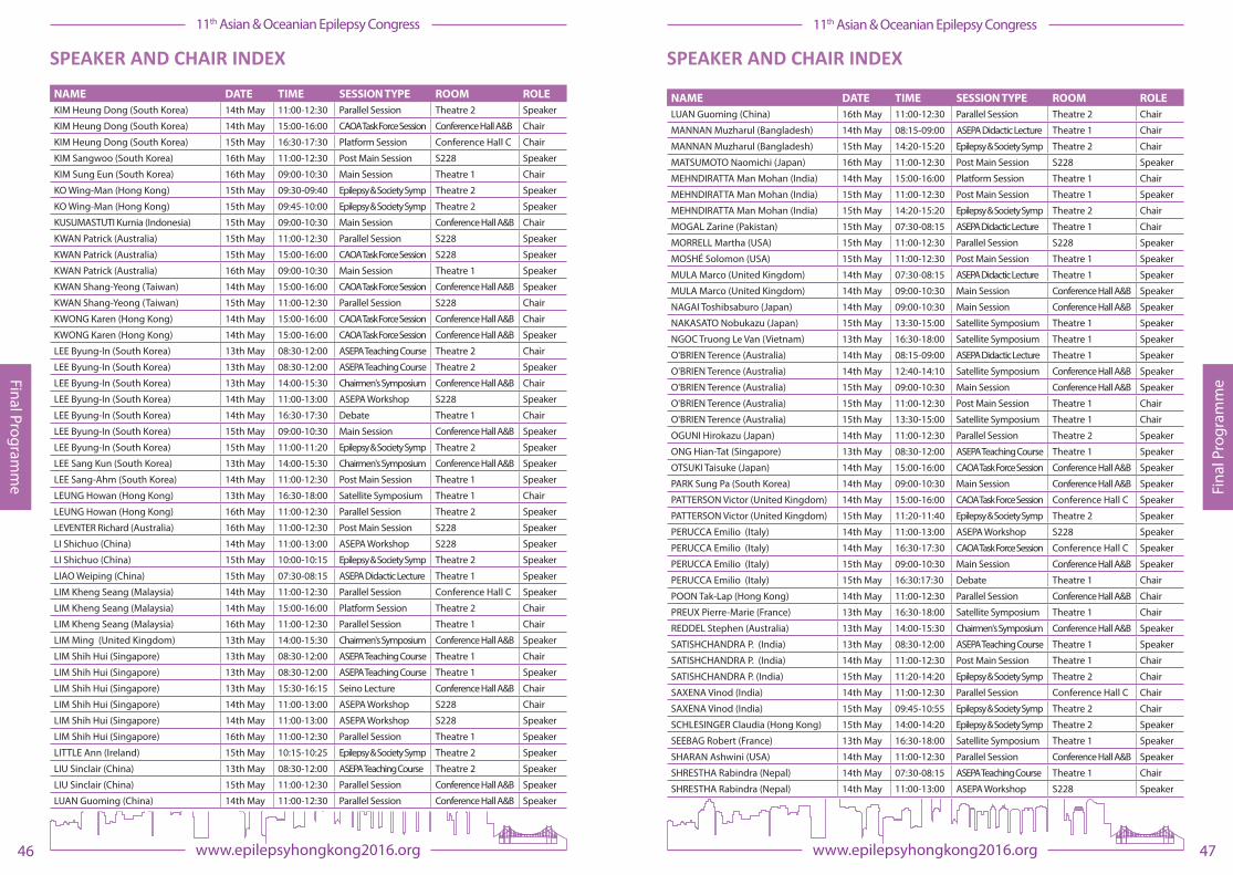
SPEAKER AND CHAIR INDEX
NAME DATE TIME SESSION TYPE ROOM ROLEADACHI Naoto (Japan) 14th May 09:00-10:30 Main Session Conference Hall A&B Speaker
BERKOVIC Sam (Australia) 16th May 09:00-10:30 Main Session Theatre 1 Speaker
BERKOVIC Sam (Australia) 16th May 11:00-12:30 Post Main Session S228 Chair
BLEASEL Andrew (Australia) 13th May 08:30-12:00 ASEPA Teaching Course Theatre 2 Speaker
BLEASEL Andrew (Australia) 14th May 16:30-17:30 Video Session Conference Hall A&B Presenter
BLEASEL Andrew (Australia) 15th May 16:30-17:30 Video Session Conference Hall A&B Presenter
BLEASEL Andrew (Australia) 16th May 11:00-12:30 Parallel Session Theatre 2 Speaker
BRODIE Martin (United Kingdom) 15th May 09:00-10:30 Main Session Conference Hall A&B Speaker
BRODIE Martin (United Kingdom) 15th May 14:20-14:40 Epilepsy & Society Symp Theatre 2 Speaker
CABRAL-LIM Leonor (Philippines) 15th May 11:00-12:30 Parallel Session Conference Hall C Chair
CABRAL-LIM Leonor (Philippines) 15th May 15:00-16:00 Platform Session Conference Hall C Chair
CASANOVA-GUTIERREZ Josephine (Philippines) 15th May 16:30-17:30 Video Session Conference Hall A&B Chair
CASANOVA-GUTIERREZ Josephine (Philippines) 15th May 16:30-17:30 Video Session Conference Hall A&B Presenter
CHAITANYA Ganne (India) 15th May 15:00-16:00 CAOA Task Force Session S228 Speaker
CHAN Derrick (Singapore) 14th May 11:00-12:30 Parallel Session Theatre 2 Chair
CHAN Derrick (Singapore) 14th May 11:00-12:30 Parallel Session Theatre 2 Speaker
CHAN Derrick (Singapore) 15th May 14:40-15:05 Epilepsy & Society Symp Theatre 2 Speaker
CHAN Derrick (Singapore) 15th May 16:30-17:30 Video Session Conference Hall A&B Presenter
CHAN Eric (Hong Kong) 15th May 11:00-12:30 Parallel Session Conference Hall C Chair
CHAN Eric (Hong Kong) 15th May 11:00-12:30 Parallel Session Conference Hall C Speaker
CHANDRA Sarat (India) 14th May 11:00-12:30 Parallel Session Conference Hall A&B Speaker
CHANNARA Chhour (Cambodia) 13th May 16:30-18:00 Satellite Symposium Theatre 1 Speaker
CHAPMAN Denise (Australia) 13th May 14:00-15:30 Chairmen's Symposium Conference Hall A&B Chair
CHAPMAN Denise (Australia) 15th May 09:40-09:45 Epilepsy & Society Symp Theatre 2 Chair
CHAPMAN Denise (Australia) 15th May 12:00-12:15 Epilepsy & Society Symp Theatre 2 Speaker
CHEN Lei (China) 15th May 11:00-12:30 Parallel Session Conference Hall C Speaker
CHEN Ziyi (China) 15th May 15:00-16:00 Platform Session Theatre 1 Chair
CHINVARUN Yotin (Thailand) 13th May 08:30-12:00 ASEPA Teaching Course Theatre 1 Speaker
CHINVARUN Yotin (Thailand) 14th May 16:30-17:30 Video Session Conference Hall A&B Chair
CHINVARUN Yotin (Thailand) 14th May 16:30-17:30 Video Session Conference Hall A&B Presenter
CHINVARUN Yotin (Thailand) 16th May 11:00-12:30 Parallel Session Theatre 2 Chair
CHIVORAKOUN Phetvongsinh (Laos) 13th May 16:30-18:00 Satellite Symposium Theatre 1 Speaker
CHIVORAKOUN Phetvongsinh (Laos) 14th May 16:30-17:30 CAOA Task Force Session Conference Hall C Speaker
CHUANG Yao-Chung (Taiwan) 14th May 11:00-12:30 Post Main Session Theatre 1 Speaker
COLE Robert (Australia) 14th May 11:00-12:30 Parallel Session Conference Hall C Chair
COLE Robert (Australia) 15th May 11:20-14:20 Epilepsy & Society Symp Theatre 2 Chair
COOK Mark (Australia) 15th May 11:00-12:30 Parallel Session S228 Speaker
COOK Mark (Australia) 16th May 08:00-08:45 ASEPA Didactic Lecture Theatre 1 Speaker
COVANIS Athanasios (Greece) 14th May 09:00-10:30 Main Session Conference Hall A&B Chair
COVANIS Athanasios (Greece) 15th May 09:30-09:40 Epilepsy & Society Symp Theatre 2 Speaker
CROSS Helen (United Kingdom) 15th May 11:00-12:30 Post Main Session Theatre 1 Speaker
CROSS Helen (United Kingdom) 15th May 16:30-17:30 Debate Theatre 1 Speaker
SPEAKER AND CHAIR INDEX
NAME DATE TIME SESSION TYPE ROOM ROLEDING Ding (China) 14th May 11:00-12:30 Parallel Session Conference Hall C Speaker
DING Ding (China) 15th May 09:45-10:55 Epilepsy & Society Symp Theatre 2 Chair
DING Ding (China) 15th May 13:30-15:00 Satellite Symposium Theatre 1 Speaker
DUNNE John (Australia) 13th May 08:30-12:00 ASEPA Teaching Course Theatre 2 Chair
DUNNE John (Australia) 14th May 15:00-16:00 CAOA Task Force Session Conference Hall C Speaker
DUNNE John (Australia) 14th May 16:30-17:30 Debate Theatre 1 Speaker
DUNNE John (Australia) 16th May 11:00-12:30 Parallel Session Theatre 1 Speaker
FONG Chung Yan G (Hong Kong) 15th May 11:00-12:30 Post Main Session Theatre 1 Chair
FONG Jason (Hong Kong) 15th May 09:00-10:30 Main Session Conference Hall A&B Chair
FONG Ka Yeung (Hong Kong) 14th May 17:30-19:00 Satellite Symposium Theatre 1 Speaker
FUNG Eva (Hong Kong) 14th May 11:00-12:30 Parallel Session Conference Hall C Speaker
FUNG Eva (Hong Kong) 15th May 11:40-12:00 Epilepsy & Society Symp Theatre 2 Speaker
FUNG Eva (Hong Kong) 16th May 09:00-10:30 Main Session Theatre 1 Chair
GULATI She�ali (India) 14th May 11:00-12:30 Parallel Session Theatre 2 Speaker
HIROSE Shinichi (Japan) 16th May 09:00-10:30 Main Session Theatre 1 Speaker
HONG Seung Bong (South Korea) 15th May 16:30-17:30 Workshop Theatre 2 Chair
HONG Seung Bong (South Korea) 15th May 16:30-17:30 Workshop Theatre 2 Speaker
HONG Zhen (China) 14th May 11:00-12:30 Post Main Session Theatre 1 Chair
HUNG Anchor (Hong Kong) 15th May 09:45-10:00 Epilepsy & Society Symp Theatre 2 Speaker
IKEDA Akio (Japan) 13th May 15:30-16:15 Seino Lecture Conference Hall A&B Speaker
IKEDA Akio (Japan) 15th May 11:00-12:30 Parallel Session Conference Hall A&B Speaker
IKEDA Akio (Japan) 15th May 15:00-16:00 CAOA Task Force Session S228 Chair
IKEDA Akio (Japan) 15th May 16:30-17:30 Workshop Theatre 2 Chair
IKEDA Akio (Japan) 15th May 16:30-17:30 Workshop Theatre 2 Speaker
INOUE Yushi (Japan) 13th May 08:30-12:00 ASEPA Teaching Course Theatre 2 Speaker
INOUE Yushi (Japan) 14th May 11:00-12:30 Post Main Session Theatre 1 Speaker
INOUE Yushi (Japan) 15th May 11:00-12:30 Parallel Session Conference Hall A&B Chair
JACKSON Graeme (Australia) 13th May 08:30-12:00 ASEPA Teaching Course Theatre 2 Speaker
JACKSON Graeme (Australia) 15th May 11:00-12:30 Parallel Session Conference Hall A&B Chair
JACKSON Graeme (Australia) 16th May 11:00-12:30 Parallel Session Theatre 2 Speaker
JAVELOSA Ana Marie (Philippines) 16th May 08:00-08:45 ASEPA Didactic Lecture Theatre 1 Chair
JIANG Yuwu (China) 14th May 15:00-16:00 CAOA Task Force Session Conference Hall A&B Speaker
JIANG Yuwu (China) 16th May 09:00-10:30 Main Session Theatre 1 Speaker
JIRUSKA Premysl (Czech Republic) 15th May 16:30-17:30 Workshop Theatre 2 Speaker
KAMEYAMA Shigeki (Japan) 14th May 11:00-12:30 Parallel Session Conference Hall A&B Speaker
KANEKO Sunao (Japan) 14th May 11:00-13:00 ASEPA Workshop S228 Speaker
KANEKO Sunao (Japan) 16th May 11:00-12:30 Post Main Session S228 Chair
KANEMOTO Kousuke (Japan) 14th May 09:00-10:30 Main Session Conference Hall A&B Chair
KANG Hoon-Chul (South Korea) 15th May 11:00-12:30 Post Main Session Theatre 1 Speaker
KANG Hoon-Chul (South Korea) 16th May 11:00-12:30 Post Main Session S228 Speaker
KANKIRAWATA Pongkiat (USA) 14th May 16:30-17:30 Video Session Conference Hall A&B Chair
KANKIRAWATA Pongkiat (USA) 14th May 16:30-17:30 Video Session Conference Hall A&B Presenter
KAO Martha (Singapore) 15th May 14:40-15:05 Epilepsy & Society Symp Theatre 2 Speaker
47www.epilepsyhongkong2016.org
11th Asian & Oceanian Epilepsy Congress
Fina
l Pro
gram
me
46 www.epilepsyhongkong2016.org
11th Asian & Oceanian Epilepsy Congress
Final Programm
e
SPEAKER AND CHAIR INDEX
NAME DATE TIME SESSION TYPE ROOM ROLEKIM Heung Dong (South Korea) 14th May 11:00-12:30 Parallel Session Theatre 2 Speaker
KIM Heung Dong (South Korea) 14th May 15:00-16:00 CAOA Task Force Session Conference Hall A&B Chair
KIM Heung Dong (South Korea) 15th May 16:30-17:30 Platform Session Conference Hall C Chair
KIM Sangwoo (South Korea) 16th May 11:00-12:30 Post Main Session S228 Speaker
KIM Sung Eun (South Korea) 16th May 09:00-10:30 Main Session Theatre 1 Chair
KO Wing-Man (Hong Kong) 15th May 09:30-09:40 Epilepsy & Society Symp Theatre 2 Speaker
KO Wing-Man (Hong Kong) 15th May 09:45-10:00 Epilepsy & Society Symp Theatre 2 Speaker
KUSUMASTUTI Kurnia (Indonesia) 15th May 09:00-10:30 Main Session Conference Hall A&B Chair
KWAN Patrick (Australia) 15th May 11:00-12:30 Parallel Session S228 Speaker
KWAN Patrick (Australia) 15th May 15:00-16:00 CAOA Task Force Session S228 Speaker
KWAN Patrick (Australia) 16th May 09:00-10:30 Main Session Theatre 1 Speaker
KWAN Shang-Yeong (Taiwan) 14th May 15:00-16:00 CAOA Task Force Session Conference Hall A&B Speaker
KWAN Shang-Yeong (Taiwan) 15th May 11:00-12:30 Parallel Session S228 Chair
KWONG Karen (Hong Kong) 14th May 15:00-16:00 CAOA Task Force Session Conference Hall A&B Chair
KWONG Karen (Hong Kong) 14th May 15:00-16:00 CAOA Task Force Session Conference Hall A&B Speaker
LEE Byung-In (South Korea) 13th May 08:30-12:00 ASEPA Teaching Course Theatre 2 Chair
LEE Byung-In (South Korea) 13th May 08:30-12:00 ASEPA Teaching Course Theatre 2 Speaker
LEE Byung-In (South Korea) 13th May 14:00-15:30 Chairmen's Symposium Conference Hall A&B Chair
LEE Byung-In (South Korea) 14th May 11:00-13:00 ASEPA Workshop S228 Speaker
LEE Byung-In (South Korea) 14th May 16:30-17:30 Debate Theatre 1 Chair
LEE Byung-In (South Korea) 15th May 09:00-10:30 Main Session Conference Hall A&B Speaker
LEE Byung-In (South Korea) 15th May 11:00-11:20 Epilepsy & Society Symp Theatre 2 Speaker
LEE Sang Kun (South Korea) 13th May 14:00-15:30 Chairmen's Symposium Conference Hall A&B Speaker
LEE Sang-Ahm (South Korea) 14th May 11:00-12:30 Post Main Session Theatre 1 Speaker
LEUNG Howan (Hong Kong) 13th May 16:30-18:00 Satellite Symposium Theatre 1 Chair
LEUNG Howan (Hong Kong) 16th May 11:00-12:30 Parallel Session Theatre 2 Speaker
LEVENTER Richard (Australia) 16th May 11:00-12:30 Post Main Session S228 Speaker
LI Shichuo (China) 14th May 11:00-13:00 ASEPA Workshop S228 Speaker
LI Shichuo (China) 15th May 10:00-10:15 Epilepsy & Society Symp Theatre 2 Speaker
LIAO Weiping (China) 15th May 07:30-08:15 ASEPA Didactic Lecture Theatre 1 Speaker
LIM Kheng Seang (Malaysia) 14th May 11:00-12:30 Parallel Session Conference Hall C Speaker
LIM Kheng Seang (Malaysia) 14th May 15:00-16:00 Platform Session Theatre 2 Chair
LIM Kheng Seang (Malaysia) 16th May 11:00-12:30 Parallel Session Theatre 1 Chair
LIM Ming (United Kingdom) 13th May 14:00-15:30 Chairmen's Symposium Conference Hall A&B Speaker
LIM Shih Hui (Singapore) 13th May 08:30-12:00 ASEPA Teaching Course Theatre 1 Chair
LIM Shih Hui (Singapore) 13th May 08:30-12:00 ASEPA Teaching Course Theatre 1 Speaker
LIM Shih Hui (Singapore) 13th May 15:30-16:15 Seino Lecture Conference Hall A&B Chair
LIM Shih Hui (Singapore) 14th May 11:00-13:00 ASEPA Workshop S228 Chair
LIM Shih Hui (Singapore) 14th May 11:00-13:00 ASEPA Workshop S228 Speaker
LIM Shih Hui (Singapore) 16th May 11:00-12:30 Parallel Session Theatre 1 Speaker
LITTLE Ann (Ireland) 15th May 10:15-10:25 Epilepsy & Society Symp Theatre 2 Speaker
LIU Sinclair (China) 13th May 08:30-12:00 ASEPA Teaching Course Theatre 2 Speaker
LIU Sinclair (China) 15th May 11:00-12:30 Parallel Session Conference Hall A&B Speaker
LUAN Guoming (China) 14th May 11:00-12:30 Parallel Session Conference Hall A&B Speaker
SPEAKER AND CHAIR INDEX
NAME DATE TIME SESSION TYPE ROOM ROLELUAN Guoming (China) 16th May 11:00-12:30 Parallel Session Theatre 2 Chair
MANNAN Muzharul (Bangladesh) 14th May 08:15-09:00 ASEPA Didactic Lecture Theatre 1 Chair
MANNAN Muzharul (Bangladesh) 15th May 14:20-15:20 Epilepsy & Society Symp Theatre 2 Chair
MATSUMOTO Naomichi (Japan) 16th May 11:00-12:30 Post Main Session S228 Speaker
MEHNDIRATTA Man Mohan (India) 14th May 15:00-16:00 Platform Session Theatre 1 Chair
MEHNDIRATTA Man Mohan (India) 15th May 11:00-12:30 Post Main Session Theatre 1 Speaker
MEHNDIRATTA Man Mohan (India) 15th May 14:20-15:20 Epilepsy & Society Symp Theatre 2 Chair
MOGAL Zarine (Pakistan) 15th May 07:30-08:15 ASEPA Didactic Lecture Theatre 1 Chair
MORRELL Martha (USA) 15th May 11:00-12:30 Parallel Session S228 Speaker
MOSHÉ Solomon (USA) 15th May 11:00-12:30 Post Main Session Theatre 1 Speaker
MULA Marco (United Kingdom) 14th May 07:30-08:15 ASEPA Didactic Lecture Theatre 1 Speaker
MULA Marco (United Kingdom) 14th May 09:00-10:30 Main Session Conference Hall A&B Speaker
NAGAI Toshibsaburo (Japan) 14th May 09:00-10:30 Main Session Conference Hall A&B Speaker
NAKASATO Nobukazu (Japan) 15th May 13:30-15:00 Satellite Symposium Theatre 1 Speaker
NGOC Truong Le Van (Vietnam) 13th May 16:30-18:00 Satellite Symposium Theatre 1 Speaker
O'BRIEN Terence (Australia) 14th May 08:15-09:00 ASEPA Didactic Lecture Theatre 1 Speaker
O'BRIEN Terence (Australia) 14th May 12:40-14:10 Satellite Symposium Conference Hall A&B Speaker
O'BRIEN Terence (Australia) 15th May 09:00-10:30 Main Session Conference Hall A&B Speaker
O'BRIEN Terence (Australia) 15th May 11:00-12:30 Post Main Session Theatre 1 Chair
O'BRIEN Terence (Australia) 15th May 13:30-15:00 Satellite Symposium Theatre 1 Chair
OGUNI Hirokazu (Japan) 14th May 11:00-12:30 Parallel Session Theatre 2 Speaker
ONG Hian-Tat (Singapore) 13th May 08:30-12:00 ASEPA Teaching Course Theatre 1 Speaker
OTSUKI Taisuke (Japan) 14th May 15:00-16:00 CAOA Task Force Session Conference Hall A&B Speaker
PARK Sung Pa (South Korea) 14th May 09:00-10:30 Main Session Conference Hall A&B Speaker
PATTERSON Victor (United Kingdom) 14th May 15:00-16:00 CAOA Task Force Session Conference Hall C Speaker
PATTERSON Victor (United Kingdom) 15th May 11:20-11:40 Epilepsy & Society Symp Theatre 2 Speaker
PERUCCA Emilio (Italy) 14th May 11:00-13:00 ASEPA Workshop S228 Speaker
PERUCCA Emilio (Italy) 14th May 16:30-17:30 CAOA Task Force Session Conference Hall C Speaker
PERUCCA Emilio (Italy) 15th May 09:00-10:30 Main Session Conference Hall A&B Speaker
PERUCCA Emilio (Italy) 15th May 16:30:17:30 Debate Theatre 1 Chair
POON Tak-Lap (Hong Kong) 14th May 11:00-12:30 Parallel Session Conference Hall A&B Chair
PREUX Pierre-Marie (France) 13th May 16:30-18:00 Satellite Symposium Theatre 1 Chair
REDDEL Stephen (Australia) 13th May 14:00-15:30 Chairmen's Symposium Conference Hall A&B Speaker
SATISHCHANDRA P. (India) 13th May 08:30-12:00 ASEPA Teaching Course Theatre 1 Speaker
SATISHCHANDRA P. (India) 14th May 11:00-12:30 Post Main Session Theatre 1 Chair
SATISHCHANDRA P. (India) 15th May 11:20-14:20 Epilepsy & Society Symp Theatre 2 Chair
SAXENA Vinod (India) 14th May 11:00-12:30 Parallel Session Conference Hall C Chair
SAXENA Vinod (India) 15th May 09:45-10:55 Epilepsy & Society Symp Theatre 2 Chair
SCHLESINGER Claudia (Hong Kong) 15th May 14:00-14:20 Epilepsy & Society Symp Theatre 2 Speaker
SEEBAG Robert (France) 13th May 16:30-18:00 Satellite Symposium Theatre 1 Speaker
SHARAN Ashwini (USA) 14th May 11:00-12:30 Parallel Session Conference Hall A&B Speaker
SHRESTHA Rabindra (Nepal) 14th May 07:30-08:15 ASEPA Teaching Course Theatre 1 Chair
SHRESTHA Rabindra (Nepal) 14th May 11:00-13:00 ASEPA Workshop S228 Speaker
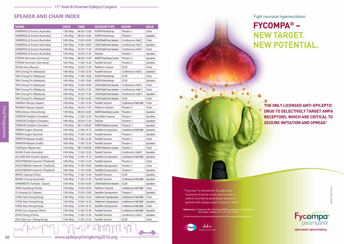
48 www.epilepsyhongkong2016.org
11th Asian & Oceanian Epilepsy Congress
Final Programm
e
SPEAKER AND CHAIR INDEX
NAME DATE TIME SESSION TYPE ROOM ROLESOMERVILLE Ernest (Australia) 13th May 08:30-12:00 ASEPA Workshop Theatre 1 Chair
SOMERVILLE Ernest (Australia) 13th May 08:30-12:00 ASEPA Workshop Theatre 1 Speaker
SOMERVILLE Ernest (Australia) 14th May 15:00-16:00 CAOA Task Force Session Conference Hall C Chair
SOMERVILLE Ernest (Australia) 14th May 15:00-16:00 CAOA Task Force Session Conference Hall C Speaker
SOMERVILLE Ernest (Australia) 14th May 16:30-17:30 CAOA Task Force Session Conference Hall C Chair
SOMERVILLE Ernest (Australia) 15th May 16:30-17:30 Debate Theatre 1 Speaker
STEFAN Hermann (Germany) 13th May 08:30-12:00 ASEPA Teaching Course Theatre 2 Speaker
STEFAN Hermann (Germany) 16th May 11:00-12:30 Parallel Session Theatre 2 Speaker
TAI Wa Hou (Macau) 15th May 16:30-17:30 Platform Session S228 Chair
TAN Chong Tin (Malaysia) 14th May 11:00-12:30 Parallel Session Conference Hall C Speaker
TAN Chong Tin (Malaysia) 14th May 11:00-13:00 ASEPA Workshop S228 Chair
TAN Chong Tin (Malaysia) 14th May 11:00-13:00 ASEPA Workshop S228 Speaker
TAN Chong Tin (Malaysia) 14th May 15:00-16:00 CAOA Task Force Session Conference Hall C Chair
TAN Chong Tin (Malaysia) 14th May 16:30-17:30 CAOA Task Force Session Conference Hall C Chair
TAN Chong Tin (Malaysia) 14th May 16:30-17:30 CAOA Task Force Session Conference Hall C Speaker
TAN Chong Tin (Malaysia) 15th May 15:00-16:00 CAOA Task Force Session S228 Chair
TANAKA Tatsuya (Japan) 14th May 11:00-12:30 Parallel Session Conference Hall A&B Chair
TANAKA Tatsuya (Japan) 14th May 16:30-17:30 Platform Session Theatre 2 Chair
TANG Venus (Hong Kong) 13th May 08:30-12:00 ASEPA Teaching Course Theatre 1 Speaker
TOMSON Torbjörn (Sweden) 14th May 11:00-12:30 Post Main Session Theatre 1 Speaker
TOMSON Torbjörn (Sweden) 14th May 16:30-17:30 Debate Theatre 1 Speaker
TOMSON Torbjörn (Sweden) 15th May 08:15-09:00 ASEPA Didactic Lecture Theatre 1 Speaker
TRINKA Eugen (Austria) 14th May 12:40-14:10 Satellite Symposium Conference Hall A&B Speaker
TRINKA Eugen (Austria) 16th May 11:00-12:30 Parallel Session Theatre 1 Speaker
TRIPATHI Manjari (India) 16th May 11:00-12:30 Parallel Session Theatre 1 Chair
TRIPATHI Manjari (India) 16th May 11:00-12:30 Parallel Session Theatre 1 Speaker
TUN Nyan (Myanmar) 15th May 08:15-09:00 ASEPA Didactic Lecture Theatre 1 Chair
VAJDA Frank (Australia) 15th May 11:00-12:30 Parallel Session Conference Hall C Speaker
VILLANEUVA Vicente (Spain) 14th May 12:40-14:10 Satellite Symposium Conference Hall A&B Speaker
VISUDTIBHAN Anannit (Thailand) 14th May 11:00-12:30 Parallel Session Theatre 2 Chair
VISUDTIBHAN Anannit (Thailand) 14th May 17:30-19:00 Satellite Symposium Theatre 1 Chair
VISUDTIBHAN Anannit (Thailand) 14th May 17:30-19:00 Satellite Symposium Theatre 1 Speaker
WANG Yuping (China) 15th May 11:00-12:30 Parallel Session S228 Speaker
WONG Chong (Australia) 15th May 11:00-12:30 Parallel Session Conference Hall A&B Speaker
YAMAMOTO Yoshiaki (Japan) 15th May 15:00-16:00 CAOA Task Force Session S228 Speaker
YANG Xiaofeng (China) 15th May 15:00-16:00 Platform Session Conference Hall A&B Chair
YU Hsiang-Yu (Taiwan) 15th May 13:30-15:00 Satellite Symposium Theatre 1 Speaker
YUNG Ada (Hong Kong) 13th May 14:00-15:30 Chairmen's Symposium Conference Hall A&B Chair
YUNG Ada (Hong Kong) 13th May 14:00-15:30 Chairmen's Symposium Conference Hall A&B Speaker
YUNG Ada (Hong Kong) 14th May 12:40-14:10 Satellite Symposium Conference Hall A&B Chair
ZHAO Guo-Guang (China) 15th May 11:00-12:30 Parallel Session Conference Hall A&B Speaker
ZHOU Dong (China) 15th May 11:00-12:30 Parallel Session Conference Hall C Speaker
ZHU Xian Lun (Hong Kong) 15th May 11:00-12:30 Parallel Session S228 Chair
human health care NEW TARGET. NEW POTENTIAL.
Fycompa® is indicated for the adjunctivetreatment of partial-onset seizures with orwithout secondarily generalized seizures inpatients with epilepsy aged 12 years or older.
*
FYCOMPA® – NEW TARGET. NEW POTENTIAL.
Fight neuronal hyperexcitation
THE ONLY LICENSED ANTI-EPILEPTIC DRUG TO SELECTIVELY TARGET AMPA RECEPTORS, WHICH ARE CRITICAL TO SEIZURE INITIATION AND SPREAD1
References: 1. Rogawski MA. Revisiting AMPA receptors as an antiepileptic drug target. Epilepsy Curr. 2011(2);11:56-63.
For further drug efficacy and safety information kindly refer to the Fycompa prescribing information
AR-F
C-A
G-1
5J-0
1

51www.epilepsyhongkong2016.org
11th Asian & Oceanian Epilepsy Congress
Fina
l Pro
gram
me
50 www.epilepsyhongkong2016.org
11th Asian & Oceanian Epilepsy Congress
Final Programm
e
CONGRESS SPONSORS
The 11th Asian & Oceanian Epilepsy Congress (AOEC) would like to thank the Hong Kong Tourist Board for their contribution towards the 11th AOEC.
INDUSTRY SPONSORS Platinum Sponsor:
Silver Sponsor:
Sponsors:
These companies have provided funding towards the costs of the Congress, but have had no input into or in�uence over the programme schedule or content. Sponsoring companies’ sta� will be present at the Congress and sponsoring companies may have exhibition stands promoting their products.
SPONSOR AND EXHIBITOR INFORMATION

53www.epilepsyhongkong2016.org
11th Asian & Oceanian Epilepsy Congress
Fina
l Pro
gram
me
52 www.epilepsyhongkong2016.org
11th Asian & Oceanian Epilepsy Congress
Final Programm
e
EXHIBITION INFORMATION
EXHIBITION OPENING HOURS
Friday 13th May **
Saturday 14th May 09.00 – 16:30
Sunday 15th May 09.00 – 16:30
** Exhibition stands may be open during the afternoon and evening of Friday 13th May during sessions and the Welcome Ceremony and Reception
LIST OF EXHIBITORS
Exhibitor Stand No: Ad-Tech Medical B03
BESA GmbH B09
Courtagen Life Sciences, Inc. D01
EB Neuro S.p.A. B07
Eisai Co., Ltd. A01
Electrical Geodesics Inc. B10
Epilepsy Organisations in Hong Kong D04
Hong Kong Epilepsy Society (HKES) D03
International Bureau for Epilepsy (IBE) A02
International League Against Epilepsy (ILAE) A02
Janssen B06 & B08
John Libbey Eurotext B02
Lifelines iEEG C03
LivaNova C04
Micromed S.p.A. B04
Natus Neurology, Inc. C05
Nihon Kohden Singapore Pte Ltd C01
Sano� B01
The Anita Kaufmann Foundation B05
UCB Pharma A03
EXHIBITION INFORMATION
EXHIBITION FLOOR PLAN
To Conference Hall A&B and Conference Hall C
To Theatre 1and Theatre 2
To Mezzanine and Ground Floors
Epilepsy Through The Ages 11th Asian & Oceanian Epilepsy Congress Satellite Symposium
Sunday 15th May 2016 | HKCEC, Theatre 1 | 13:30–15:00
Chairperson – Professor Terence O’Brien James Stewart Chair of Medicine & Head of Dept. of Medicine, Royal Melbourne Hospital,University of Melbourne; Consultant Neurologist, Royal Melbourne Hospital, Australia
© UCB Biopharma SPRL 2016. All rights reserved. This symposium is sponsored by UCB
SEA-0031-032016
Treating women with epilepsy of child-bearing potential
Associate Professor Hsiang-Yu, YuAssistant Professor, National Yang-Ming University Attending Doctor, Neurologic Institute, Taipei Veterans General Hospital, Taiwan
Transitioning care of epilepsy patients from pediatric to adult neurology (a patient perspective)
Professor Nobukazu Nakasato
Chair of the Department of Epileptology, Tohoku University Graduate School of Medicine Director of Tohoku University Hospital Epilepsy Centre, Japan
The challenges of ensuring individualised patient care for people with epilepsy
Associate Professor Ding Ding
Associate Professor & Neuro-epidemiologist, Institute of Neurology, Fudan University, WHO Collaborating Centre for Research & Training in Neurosciences, China
Guoqiong, living with Epilepsy
SEDE DEL CONGRESOCancun International
Convention Centre, Cancun ICCBoulevard Kukulcan Km. 9 1er Piso
Zona Hotelera C.P. 77500Cancún , Quintana Roo, México
M E X I C Owww.epilepsycancun2016.org
IX CONGRESO LATINOAMERICANO DE EPILEPSIA
CANCÚN 20-23 AUGUST 2016

57www.epilepsyhongkong2016.org
11th Asian & Oceanian Epilepsy Congress
Abs
trac
t Boo
k
www.epilepsyhongkong2016.org
11th Asian & Oceanian Epilepsy Congress
Adult Epileptology Saturday 14th May; 15:00-16:00 Theatre 1Refer to pages 88-90 for abstracts
Chair: Man Mohan MEHNDIRATTA (India)
P003 Efficacy and safety of adjunctive lacosamide for the treatment of partial-onset seizures in Chinese and Japanese adults: a multicenter, double-blind, randomized, placebo-controlled study Xinlu DU (China)
P004 Comparison of relapse risk between drug discontinuation and continuation at different periods of seizure remission in adult patients with focal epilepsy Rong-Yuan ZHENG (China)
P005 Determinant of health-related quality of life in patients with epilepsy from Malaysia Sherrini BAZIR AHMAD (Malaysia)
P006 Evaluation of headache disorders in patients of idiopathic epilepsy Sagar BOSE (India)
P007 Can ictal-MEG accurately localize the epileptogenic zone? Bhargavi RAMANUJAM (India)
Psychosocial Saturday 14th May; 15:00-16:00 Theatre 2Refer to pages 90-92 for abstracts
Chair: Kheng-Seang LIM (Malaysia)
P008 Epilepsy interaction with behavior, emotions & learning Mahmoud I ABEIDAH (Qatar)
P009 Breastfeeding initiation, duration and exclusivity among Chinese mothers with epilepsy: A retro-prospective study in a developing country Nanya HAO (China)
P010 Factors affecting the employability in people with epilepsy (PWE) Kheng Seang LIM (Malaysia)
PLATFORM SESSIONS
ABSTRACT BOOK
59www.epilepsyhongkong2016.org
11th Asian & Oceanian Epilepsy Congress
Abs
trac
t Boo
k
58 www.epilepsyhongkong2016.org
11th Asian & Oceanian Epilepsy Congress
Abstract Book
P018 Study of transplantation of neural stem cells in therapy of epilepsy in developing rats Yuanyuan DAI (China)
P019 MicroRNA-132 silencing decreases epileptogenesis via the P250GAP/Cdc42 signaling pathway Xue FAN (China)
P020 Targeting mammalian target of rapamycin down-regulates the expression of P-glycoprotein in a pharmacoresistant epilepsy rat model kindled by coriaria lactone Xiaosa CHI (China)
P021 Association of genetic and non-genetic factors with phenytoin-induced severe cutaneous adverse drug reactions inThai patients Kittika YAMPAYON (Thailand)
P022 Predictions of regression of intellectual disability and measuring efficacy of medication in 286 Japanese cohort of Dravet syndrome with SCN1A missense and truncation mutations Atsushi ISHII (USA)
Clinical Neurophysiology and Neuroimaging Sunday 15th May; 15:00-16:00 Theatre 1Refer to pages 97- 99 for abstracts
Chair: Ziyi CHEN (China)
P023 A novel model-based method for real-time detection of electrographic seizures Armen SARGSYAN (Australia)
P024 Cortical morphometric abnormalities in patients with ‚hot-water epilepsy´ K RAGHAVENDRA (India)
P025 Using maximum distributed peak point to detect baseline for automatic detection of HFOs Guoping REN (China)
P026 EEG functional connectivity characterization of focal vs generalized inter-ictal epileptiform discharges, and possible network implications in the prediction of ictal evolving patterns Alberto PINZON (USA)
PLATFORM SESSIONS
P011 Epilepsy, interictal psychopathology and vitamin D3 levels - Are they related? Palak TALWAR (India)
P012 Positive psychology interventions for neurological disorders: a systematic review Siew Tim LAI (Malaysia)
Surgery Saturday 14th May; 16:30-17:30 Theatre 2Refer to pages 92-94 for abstracts
Chair: Tatsuya TANAKA (Japan)
P013 The outcome of bipolar electro-coagulation with lesionectomy in the treatment of epilepsy involving eloquent areas Feng ZHAI (China)
P014 Robotics in neurosurgical stereotactic interventions: Oblique intrainsular electrodes implanted of patients with epilepsy Afif AFIF (France)
P015 Predictive analysis of perioperative cognitive function in patients with hypothalamic hamartoma, who underwent surgery of stereotactic radiofrequency thermocoagulation Masaki SONODA (Japan)
P016 MRI negative patients with medial temporal lobe epilepsy were more effective to stereotactic radiofrequency thermocoagulation of amygdalohippocampal complex compared with MRI positive patients Quanjun ZHAO (China)
P017 Seizure onset and propagation in tuberous sclerosis: intracranial EEG analysis Lakshminarayanan KANNAN (Australia)
Basic Science and GeneticsSunday 15th May; 15:00-16:00 Conference Hall A&BRefer to pages 94-96 for abstracts
Chair: Xiaofeng YANG (China)
PLATFORM SESSIONS

61www.epilepsyhongkong2016.org
11th Asian & Oceanian Epilepsy Congress
Abs
trac
t Boo
k
60 www.epilepsyhongkong2016.org
11th Asian & Oceanian Epilepsy Congress
Abstract Book
P034 The ketogenic diet in patients younger than 3 years old Se Hee KIM (South Korea)
P035 Precise epileptogenic zone location with stereotactic electroencephalography navigated by ROSA in patients with focal cortical dyplasia in children Changqing LIU (China)
P036 Risk factors for relapse of infant spasms after spasms-free Jian Min ZHONG (China)
P037 Comparison of radiological outcome of 7 days versus 28 days albendazole therapy along with steroid in patients with parenchymal neurocysticercosis with lesion load ≤ 5: a randomized control study Vijay KUMAR (India)
Status Epilepticus Sunday 15th May; 16:30-17:30Room S228, level 2Refer to pages 103-105 for abstracts
Chair: Wa Hou TAI (Macau)
P038 Analysis of 247 children with status epilepticus: Clinical features and prognosis Ji WANG (China)
P039 Status epilepticus is associated with cardiac ventricular abnormalities in children See Wai CHAN (USA)
P040 Status epilepticus in the elderly patients: A national data study in Thailand Somsak TIAMKAO (Thailand)
P041 Effect of continuous electroencephalography monitoring on clinical outcomes of status epilepticus Apinya JIRARAT (Thailand)
P042 Prevalence and characteristics of status epilepticus in patients with inflammatory etiology: Results from Korean initiative for status epilepticus registry Jung-Won SHIN (South Korea)
PLATFORM SESSIONS
P027 Juvenile myoclonic epilepsy may be a disorder of cortex rather than thalamus: An effective connectivity analysis Sung Eun KIM (South Korea)
Treatment and EpidemiologySunday 15th May; 15:00-16:00 Conference Hall CRefer to pages 99-101 for abstracts
Chair: Leonor CABRAL-LIM (Philippines)
P028 Comparative long-term effectiveness research of the usual antiepileptic drugs as a monotherapy for focal epilepsy in adult patients Hui-Qin XU (China)
P029 High sensitivity C-reactive protein level as marker of atherosclerosis in epilepsy patients with first generation antiepileptic drugs Luh Ari INDRAWATI (Indonesia)
P030 Characteristics of early drug treatment for new-onset epilepsy in older persons Edward FAUGHT (USA)
P031 Acute symptomatic seizures in patients with acute ischemic stroke Se-Jin LEE (South Korea)
P032 Referral pattern for epilepsy surgery after evidence-based recommendations in India, a developing country perspective Ajay ASRANNA I.P. (India)
Paediatric Epileptology Sunday 15th May; 16:30-17:30Conference Hall CRefer to pages 101-103 for abstracts
Chair: Heung Dong KIM (South Korea)
P033 GRIN2A mutations in epilepsy-aphasia spectrum disorders Yuehua ZHANG (China)
PLATFORM SESSIONS
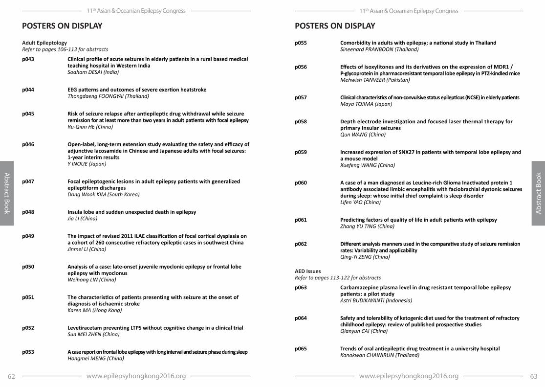
63www.epilepsyhongkong2016.org
11th Asian & Oceanian Epilepsy Congress
Abs
trac
t Boo
k
62 www.epilepsyhongkong2016.org
11th Asian & Oceanian Epilepsy Congress
Abstract Book
p055 Comorbidity in adults with epilepsy; a national study in Thailand Sineenard PRANBOON (Thailand)
p056 Effects of isoxylitones and its derivatives on the expression of MDR1 / P-glycoprotein in pharmacoresistant temporal lobe epilepsy in PTZ-kindled mice Mehwish TANVEER (Pakistan)
p057 Clinical characteristics of non-convulsive status epilepticus (NCSE) in elderly patients Maya TOJIMA (Japan)
p058 Depth electrode investigation and focused laser thermal therapy for primary insular seizures Qun WANG (China)
p059 Increased expression of SNX27 in patients with temporal lobe epilepsy and a mouse model Xuefeng WANG (China)
p060 A case of a man diagnosed as Leucine-rich Glioma Inactivated protein 1 antibody associated limbic encephalitis with faciobrachial dystonic seizures during sleep: whose initial chief complaint is sleep disorder Lifen YAO (China)
p061 Predicting factors of quality of life in adult patients with epilepsy Zhang YU TING (China)
p062 Different analysis manners used in the comparative study of seizure remission rates: Variability and applicability Qing-Yi ZENG (China)
AED Issues Refer to pages 113-122 for abstracts
p063 Carbamazepine plasma level in drug resistant temporal lobe epilepsy patients: a pilot study Astri BUDIKAYANTI (Indonesia)
p064 Safety and tolerability of ketogenic diet used for the treatment of refractory childhood epilepsy: review of published prospective studies Qianyun CAI (China)
p065 Trends of oral antiepileptic drug treatment in a university hospital Kanokwan CHAINIRUN (Thailand)
POSTERS ON DISPLAYPOSTERS ON DISPLAY
Adult Epileptology Refer to pages 106-113 for abstracts
p043 Clinical profile of acute seizures in elderly patients in a rural based medical teaching hospital in Western India Soaham DESAI (India)
p044 EEG patterns and outcomes of severe exertion heatstroke Thongdaeng FOONGYAI (Thailand)
p045 Risk of seizure relapse after antiepileptic drug withdrawal while seizure remission for at least more than two years in adult patients with focal epilepsy Ru-Qian HE (China)
p046 Open-label, long-term extension study evaluating the safety and efficacy of adjunctive lacosamide in Chinese and Japanese adults with focal seizures: 1-year interim results Y INOUE (Japan)
p047 Focal epileptogenic lesions in adult epilepsy patients with generalized epileptiform discharges Dong Wook KIM (South Korea)
p048 Insula lobe and sudden unexpected death in epilepsy Jia LI (China)
p049 The impact of revised 2011 ILAE classification of focal cortical dysplasia on a cohort of 260 consecutive refractory epileptic cases in southwest China Jinmei LI (China)
p050 Analysis of a case: late-onset juvenile myoclonic epilepsy or frontal lobe epilepsy with myoclonus Weihong LIN (China)
p051 The characteristics of patients presenting with seizure at the onset of diagnosis of ischaemic stroke Karen MA (Hong Kong)
p052 Levetiracetam preventing LTPS without cognitive change in a clinical trial Sun MEI ZHEN (China)
p053 A case report on frontal lobe epilepsy with long interval and seizure phase during sleep Hongmei MENG (China)
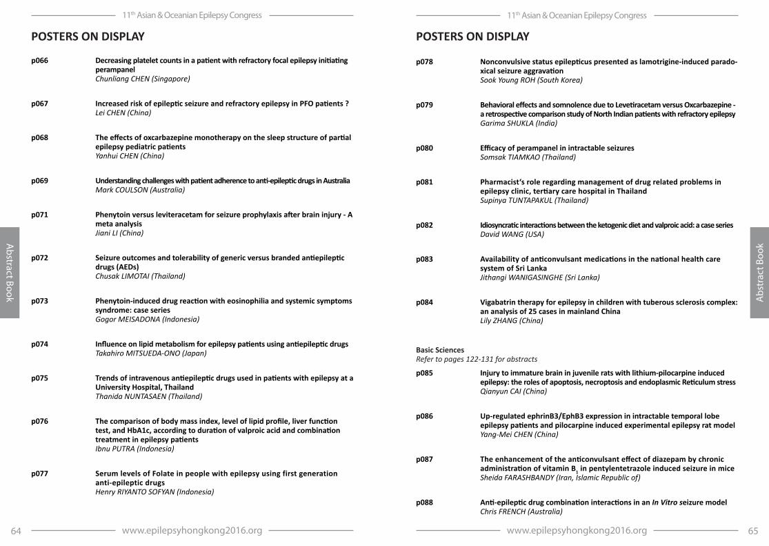
65www.epilepsyhongkong2016.org
11th Asian & Oceanian Epilepsy Congress
Abs
trac
t Boo
k
64 www.epilepsyhongkong2016.org
11th Asian & Oceanian Epilepsy Congress
Abstract Book
p078 Nonconvulsive status epilepticus presented as lamotrigine-induced parado-xical seizure aggravation Sook Young ROH (South Korea)
p079 Behavioral effects and somnolence due to Levetiracetam versus Oxcarbazepine - a retrospective comparison study of North Indian patients with refractory epilepsy Garima SHUKLA (India)
p080 Efficacy of perampanel in intractable seizures Somsak TIAMKAO (Thailand)
p081 Pharmacist‘s role regarding management of drug related problems in epilepsy clinic, tertiary care hospital in Thailand Supinya TUNTAPAKUL (Thailand)
p082 Idiosyncratic interactions between the ketogenic diet and valproic acid: a case series David WANG (USA)
p083 Availability of anticonvulsant medications in the national health care system of Sri Lanka Jithangi WANIGASINGHE (Sri Lanka)
p084 Vigabatrin therapy for epilepsy in children with tuberous sclerosis complex: an analysis of 25 cases in mainland China Lily ZHANG (China)
Basic Sciences Refer to pages 122-131 for abstracts
p085 Injury to immature brain in juvenile rats with lithium-pilocarpine induced epilepsy: the roles of apoptosis, necroptosis and endoplasmic Reticulum stress Qianyun CAI (China)
p086 Up-regulated ephrinB3/EphB3 expression in intractable temporal lobe epilepsy patients and pilocarpine induced experimental epilepsy rat model Yang-Mei CHEN (China)
p087 The enhancement of the anticonvulsant effect of diazepam by chronic administration of vitamin B1 in pentylentetrazole induced seizure in mice Sheida FARASHBANDY (Iran, Islamic Republic of)
p088 Anti-epileptic drug combination interactions in an In Vitro seizure model Chris FRENCH (Australia)
POSTERS ON DISPLAY
p066 Decreasing platelet counts in a patient with refractory focal epilepsy initiating perampanel Chunliang CHEN (Singapore)
p067 Increased risk of epileptic seizure and refractory epilepsy in PFO patients ? Lei CHEN (China)
p068 The effects of oxcarbazepine monotherapy on the sleep structure of partial epilepsy pediatric patients Yanhui CHEN (China)
p069 Understanding challenges with patient adherence to anti-epileptic drugs in Australia Mark COULSON (Australia)
p071 Phenytoin versus leviteracetam for seizure prophylaxis after brain injury - A meta analysis Jiani LI (China)
p072 Seizure outcomes and tolerability of generic versus branded antiepileptic drugs (AEDs) Chusak LIMOTAI (Thailand)
p073 Phenytoin-induced drug reaction with eosinophilia and systemic symptoms syndrome: case series Gogor MEISADONA (Indonesia)
p074 Influence on lipid metabolism for epilepsy patients using antiepileptic drugs Takahiro MITSUEDA-ONO (Japan)
p075 Trends of intravenous antiepileptic drugs used in patients with epilepsy at a University Hospital, Thailand Thanida NUNTASAEN (Thailand)
p076 The comparison of body mass index, level of lipid profile, liver function test, and HbA1c, according to duration of valproic acid and combination treatment in epilepsy patients Ibnu PUTRA (Indonesia)
p077 Serum levels of Folate in people with epilepsy using first generation anti-epileptic drugs Henry RIYANTO SOFYAN (Indonesia)
POSTERS ON DISPLAY
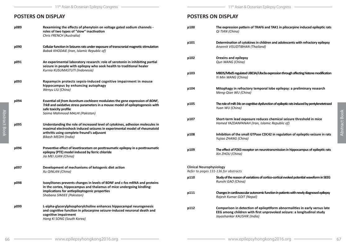
67www.epilepsyhongkong2016.org
11th Asian & Oceanian Epilepsy Congress
Abs
trac
t Boo
k
66 www.epilepsyhongkong2016.org
11th Asian & Oceanian Epilepsy Congress
Abstract Book
p100 The expression pattern of TRAF6 and TAK1 in pilocarpine induced epileptic rats Qi TIAN (China)
p101 Determination of cytokines in children and adolescents with refractory epilepsy Anannit VISUDTIBHAN (Thailand)
p102 Orexins and epilepsy Qun WANG (China)
p103 MBD5/Mbd5 regulated UBE3A/Ube3a expression through affecting histone modification Yi Min WANG (China)
p104 Mitophagy in refractory temporal lobe epilepsy: a preliminary research Meng-Qian WU (China)
p105 The role of miR-34c on cognitive dysfunction of epileptic rats induced by pentylenetetrazol Yuan WU (China)
p107 Short-term lead exposure reduces chemical seizure threshold in mice Hamed YAZDANPANAH (Iran, Islamic Republic of)
p108 Inhibition of the small GTPase CDC42 in regulation of epileptic-seizure in rats Yujiao ZHANG (China)
p109 The effect of P2X3 receptor on neurotransmission in hippocampus of epileptic rats Xin ZHOU (China)
Clinical Neurophysiology Refer to pages 131-136 for abstracts
p110 Study of the reason of variations of cortico-cortical evoked potential waveform in SEEG Runshi GAO (China)
p111 Changes in cardiovascular autonomic function in patients with newly diagnosed epilepsy Rajesh Kumar GOIT (Nepal)
p112 Comparison in detection of epileptiform abnormalities in early versus late EEG among children with first unprovoked seizure: a longitudinal study Jayashankar KAUSHIK (India)
POSTERS ON DISPLAY
p089 Rexamining the effects of phenytoin on voltage gated sodium channels - roles of two types of “slow“ inactivation Chris FRENCH (Australia)
p090 Cellular function in Seizures rats under exposure of transcranial magnetic stimulation Babak KHODAIE (Iran, Islamic Republic of)
p091 An experimental laboratory research: role of serotonin in inhibiting partial seizure in people with epilepsy who seek health to traditional healer Kurnia KUSUMASTUTI (Indonesia)
p093 Rapamycin protects sepsis-induced cognitive impairment in mouse hippocampus by enhancing autophagy Wenyu LIU (China)
p094 Essential oil from Aconitum cochleare modulates the gene expression of BDNF, TrkB and oxidative stress parameters in a mouse model of epileptogenesis with safe toxicity profile Saima Mahmood MALHI (Pakistan)
p095 Understanding the role of increased level of cytokines, adhesion molecules in maximal electroshock induced seizures in experimental model of rheumatoid arthritis using complete freund‘s adjuvant Bikash MEDHI (India)
p096 Preventive effect of levetiracetam on posttraumatic epilepsy in a posttraumatic epilepsy (PTE) model induced by ferric chloride Jia MEI JUAN (China)
p097 Development of mechanisms of ketogenic diet action Xu QINLAN (China)
p098 Isoxylitones prevents changes in levels of BDNF and c-fos mRNA and proteins in the cortex, hippocampus and thalamus of mice undergoing kindling: implications for antiepileptogenic properties Shabana SIMJEE (Pakistan)
p099 L-alpha-glycerylphosphorylcholine enhances hippocampal neurogenesis and cognitive function in pilocarpine seizure-induced neuronal death and cognitive impairment Hong Ki SONG (South Korea)
POSTERS ON DISPLAY

69www.epilepsyhongkong2016.org
11th Asian & Oceanian Epilepsy Congress
Abs
trac
t Boo
k
68 www.epilepsyhongkong2016.org
11th Asian & Oceanian Epilepsy Congress
Abstract Book
p125 The analysis of clinical symptoms and EEG: a children with anti-NMDAR encephalitis, whos initial symptoms are mental and behavior abnormality and seizures Yao LIFEN (China)
p126 Prevalence and predictors of subclinical seizures during scalp video-EEG monitoring in patients with epilepsy Shan WANG (China)
p127 Analysis of the efficacy and electroencephalogram by transcutaneous vagus nerve stimulation for refractory epilepsy Aihua LIU (China)
p128 The cost analysis of electroencephalography in a university hospital Saengjan NAJAKHUN (Thailand)
p129 EEG changes in 3p25.3 syndrome: a case report and comparison with reported cases in literature Ali AL OTAIBI (Saudi Arabia)
p130 Comparative study on Scalp EEG between before and after hemispherotomy in refractory epilepsy due to hemispheric lesions Wen WANG (China)
p131 The clinical and EEG characteristics of anti-NMDA receptor encephalitis in 12 cases Yuan WU (China)
p132 Association of electroencephalogram patterns with mortality in patients with altered mental state Kwan Ming YAM (Hong Kong)
Epidemiology Refer to pages 140-142 for abstracts
p133 Domestic health visitors improve access to care for people living with epilepsy in Lao PDR: Preliminary results Phetvongsinh CHIVORAKOUN (Lao People`s Democratic Republic)
p134 Incidence and organization of seizures and epilepsies (2010) of new-onset unprovoked seizures in a cohort of children accessing government primary schools: A study in south India Shyam JAISWAL (India)
POSTERS ON DISPLAY
p113 Electrophysiological evaluation of twin patients with familial myoclonus epilepsy Masako KINOSHITA (Japan)
p114 Case study of repetitive transcranial magnetic stimulation of the motor cortex for thalamic pain Hua LIN (China)
p115 The distribution of interictal, preictal and postictal high frequency activities and their relationship with surgical outcome in partial epilepsy Chae Jung PARK (South Korea)
p116 The characteristic and threshold of afterdischarges (ADs) caused by electrical cortical stimulation Zhiwei REN (China)
p117 Neuro physiologic peculiarities in idiopathic and symptomatic epilepsy Nazakat K. RUZIMOVA (Uzbekistan)
p118 Quantitative MRI assessment of the differences in lateralization of language-related brain activation in patients with temporal lobe epilepsy Azamatjon UMAROV (Uzbekistan)
p119 Giant somatosensory evoked magnetic field (SSEF) in patients with progressive myoclonic epilepsy (PME): MEG based assessment Jayabal VELMURUGAN (India)
p120 Temporal and spatial characteristics of inter-ictal and ictal high frequency oscillations (80-200Hz) (HFO) in absence epilepsy Jayabal VELMURUGAN (India)
p121 Lateralizing value of low frequency band MEG beamformer source imaging in temporal lobe epilepsy Di WANG (China)
EEG Refer to pages 136-140 for abstracts
p123 Intramuscular dexmedetomidine for pediatric electroencephalogram sedation Maria Teresa CRUZ (Philippines)
p124 Role of interictal electroencephalography in predicting seizure relapse, a promising result: Report from a tertiary care centre, north India Biplab DAS (India)
POSTERS ON DISPLAY
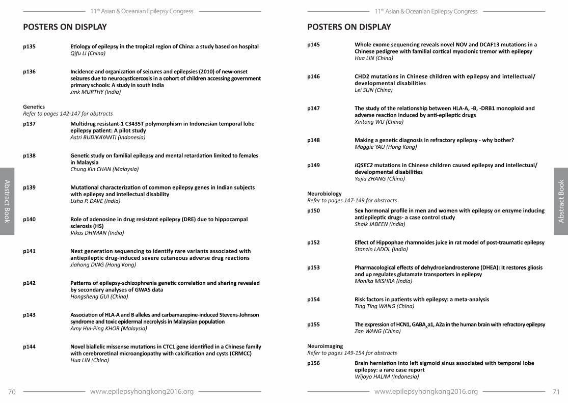
71www.epilepsyhongkong2016.org
11th Asian & Oceanian Epilepsy Congress
Abs
trac
t Boo
k
70 www.epilepsyhongkong2016.org
11th Asian & Oceanian Epilepsy Congress
Abstract Book
p145 Whole exome sequencing reveals novel NOV and DCAF13 mutations in a Chinese pedigree with familial cortical myoclonic tremor with epilepsy Hua LIN (China)
p146 CHD2 mutations in Chinese children with epilepsy and intellectual/developmental disabilities Lei SUN (China)
p147 The study of the relationship between HLA-A, -B, -DRB1 monoploid and adverse reaction induced by anti-epileptic drugs Xintong WU (China)
p148 Making a genetic diagnosis in refractory epilepsy - why bother? Maggie YAU (Hong Kong)
p149 IQSEC2 mutations in Chinese children caused epilepsy and intellectual/developmental disabilities Yujia ZHANG (China)
Neurobiology Refer to pages 147-149 for abstracts
p150 Sex hormonal profile in men and women with epilepsy on enzyme inducing antiepileptic drugs- a case control study Shaik JABEEN (India)
p152 Effect of Hippophae rhamnoides juice in rat model of post-traumatic epilepsy Stanzin LADOL (India)
p153 Pharmacological effects of dehydroeiandrosterone (DHEA): It restores gliosis and up regulates glutamate transporters in epilepsy Monika MISHRA (India)
p154 Risk factors in patients with epilepsy: a meta-analysis Ting Ting WANG (China)
p155 The expression of HCN1, GABAAa1, A2a in the human brain with refractory epilepsy Zan WANG (China)
Neuroimaging Refer to pages 149-154 for abstracts
p156 Brain herniation into left sigmoid sinus associated with temporal lobe epilepsy: a rare case report Wijoyo HALIM (Indonesia)
POSTERS ON DISPLAY
p135 Etiology of epilepsy in the tropical region of China: a study based on hospital Qifu LI (China)
p136 Incidence and organization of seizures and epilepsies (2010) of new-onset seizures due to neurocysticercosis in a cohort of children accessing government primary schools: A study in south India Jmk MURTHY (India)
Genetics Refer to pages 142-147 for abstracts
p137 Multidrug resistant-1 C3435T polymorphism in Indonesian temporal lobe epilepsy patient: A pilot study Astri BUDIKAYANTI (Indonesia)
p138 Genetic study on familial epilepsy and mental retardation limited to females in Malaysia Chung Kin CHAN (Malaysia)
p139 Mutational characterization of common epilepsy genes in Indian subjects with epilepsy and intellectual disability Usha P. DAVE (India)
p140 Role of adenosine in drug resistant epilepsy (DRE) due to hippocampal sclerosis (HS) Vikas DHIMAN (India)
p141 Next generation sequencing to identify rare variants associated with antiepileptic drug-induced severe cutaneous adverse drug reactions Jiahong DING (Hong Kong)
p142 Patterns of epilepsy-schizophrenia genetic correlation and sharing revealed by secondary analyses of GWAS data Hongsheng GUI (China)
p143 Association of HLA-A and B alleles and carbamazepine-induced Stevens-Johnson syndrome and toxic epidermal necrolysis in Malaysian population Amy Hui-Ping KHOR (Malaysia)
p144 Novel biallelic missense mutations in CTC1 gene identified in a Chinese family with cerebroretinal microangiopathy with calcification and cysts (CRMCC) Hua LIN (China)
POSTERS ON DISPLAY
73www.epilepsyhongkong2016.org
11th Asian & Oceanian Epilepsy Congress
Abs
trac
t Boo
k
72 www.epilepsyhongkong2016.org
11th Asian & Oceanian Epilepsy Congress
Abstract Book
p168 Indications for and findings of neuropsychological assessment in pediatric neurology clinics: an audit Catherine Anne COX (Singapore)
p169 Cyclophosphamide-responsive anti-NMDAr encephalitis in a Filipino adolescent girl: Her journey towards recovery Maria Teresa CRUZ (Philippines)
p170 Depression and cognitive performance in elderly subjects - a South Indian experience Kumarpillai GOPUKUMAR (India)
p171 McGurk effect in left and right hemispheric epilepsy Ravish KENI (India)
p172 Visual, auditory verbal and logical memory functions and depression in children with refractory mesial temporal sclerosis (MTS) Shobha NANDAVAR (India)
p173 The impact of postoperative outcomes on neuropsychological tests in patients with intractable temporal lobe epilepsy (TLE) Toshiaki OTSUBO (Japan)
p174 Some aspects of neuro-psychological development in children with post-stroke epilepsy Furkat SAMADOV (Uzbekistan)
p175 Analysis of cognitive function in refractory frontal epilepsy based on electroencephalogram Zhaohui SUN (China)
p176 Depressive symptoms and motor, attention and executive functions in children with refractory mesial temporal sclerosis (MTS) M.V. VARSHINI (India)
p177 Lateral and gender differences in short-term effects of anterior temporal lobectomy on cognitive function of temporal lobe epilepsy patients Siyu WANG (China)
POSTERS ON DISPLAY
p157 Availability of 11C-Methionine PET to detect epileptogenic lesion Ryosuke HANAYA (Japan)
p158 Impact of insular morphology on feasibility of long-axis cannulation for stereoelectroencephalographic recording Michael LANG (USA)
p159 Impact of trajectory planning with susceptibility weighted imaging for intracranial electrode implantation Michael LANG (USA)
p160 Asymmetry in hippocampal memory networks associated with verbal memory in mesial temporal epilepsy: Preliminary findings Hong LI (China)
p161 Altered regional homogeneity in secondary generalized neocortical seizure Syu-Jyun PENG (Taiwan, Republic of China)
p162 Resting state functional networks in epilepsy: recent advances in magnetoencephalography studies Jiechuan REN (China)
p163 Automated T2 relaxometry of the hippocampus in temporal lobe epilepsy Gavin P WINSTON (United Kingdom)
p164 Functional Connectivity in patients with juvenile myclonic epilepsy: MEG-based Brain Network Yao XINGQI (China)
p165 Increased interhemispheric resting-state functional connectivity in juvenile myoclonic epilepsy: A resting-state fMRI study Tianhua YANG (China)
p166 Functional mapping and characteristics of motor cortex Li ZHE (China)
Neuropsychology Refer to pages 154-158 for abstracts
p167 Outcome of epilepsy surgery in patient of temporal lobe epilepsy: Preliminary findings in Hong Kong Iris CHAN (Hong Kong)
POSTERS ON DISPLAY

75www.epilepsyhongkong2016.org
11th Asian & Oceanian Epilepsy Congress
Abs
trac
t Boo
k
74 www.epilepsyhongkong2016.org
11th Asian & Oceanian Epilepsy Congress
Abstract Book
p190 Small focal cortical dysplasia type II lesions are associated with sleep-related epilepsy Shuang WANG (China)
p191 Neuropathology of patients with refractory epilepsy in a tertiary referral center in Hong Kong. A review of 55 cases Amanda KAN (Hong Kong)
p193 Frequent rhabdomyolysis in anti-NMDA receptor encephalitis Jung-Ah LIM (South Korea)
p194 Symptomatic epileptic seizure in neurointensive care unit Nino LOBJANIDZE (Georgia)
p195 Characteristics of cognitive impairments in idiopathic epilepsy Nazakat K. RUZIMOVA (Uzbekistan)
p196 A patient with dangerous postictal heart rate variability Dong Jin SHIN (South Korea)
p197 Chemical stability study of hospital prepared diazepam rectal suppository Patcharin SUPAPSOPHON (Thailand)
p198 Serum matrix metalloproteinase-2: a potential biomarker for diagnosis of epilepsy Rui WANG (China)
p199 “Cocktail” therapy can repair early hippocampal damage (functional changes) in epilepsy patients Wei-Min WANG (China)
p200 Long-term lipid profile in patient taking ketogenic diet/ modified Atkin‘s diet Karen YAM (Hong Kong)
p201 The effect of cathodal transcranial direct current stimulation in one patient with refractory epilepsy after surgery failure Dongju YANG (China)
p202 The pathogenesis of poststroke seizures and epilepsy Huajun YANG (China)
POSTERS ON DISPLAY
Others Refer to pages 159-168 for abstracts
p178 The role of serum auto antibodies to the neuro mediators‘ receptors in the pathogenesis of epilepsy Rano B. AZIZOVA (Uzbekistan)
p179 Interrelation of cognitive potentials and neuroimmunologic values in epilepsy Rano B. AZIZOVA (Uzbekistan)
p180 The values of P300 cognitive potentials in cases of idiopathic and symptomatic forms of epilepsy Rano B. AZIZOVA (Uzbekistan)
p181 The association between aged at onset, history of febrile seizures and medication adherence with quality of life in epilepsy patients Sesmi BETRIS (Indonesia)
p182 Adherence to medication among persons with epilepsy: an exploration in Hong Kong Mike KT CHEUNG (Hong Kong)
p183 Quality of epilepsy health care in Hong Kong from patients‘ perspective Mike KT CHEUNG (Hong Kong)
p184 The effects of reducing seizure activity in Mozart music play more than a sound stimulation Po-Ching CHOU (Taiwan, Republic of China)
p185 Spot “Epilepsy” through canvas Sanjaya FERNANDO (Sri Lanka)
p186 Excessive daytime sleepiness in epilepsy patients and related factors Ismi Adhanisa HAMDANI (Indonesia)
p187 Autoimmune encephalitis in Hong Kong: A retrospective study of clinical characteristics and outcomes Ting Hin Adrian HUI (Hong Kong)
p188 A case of refractory seizure with cognitive impairment due to anti-GABA encephalitis Ting Hin Adrian HUI (Hong Kong)
p189 A study on the self-management behavior among persons with epilepsy in HK and its implications for health and social care services Anchor TF HUNG (Hong Kong)
POSTERS ON DISPLAY

77www.epilepsyhongkong2016.org
11th Asian & Oceanian Epilepsy Congress
Abs
trac
t Boo
k
76 www.epilepsyhongkong2016.org
11th Asian & Oceanian Epilepsy Congress
Abstract Book
p215 KCNT1 mutations in Chinese children with epilepsy and intellectual/developmental disabilities Yuwu JIANG (China)
p216 Clinical study of levetiracetam and phenobarbital in the treatment of neonatal seizures Li JIE (China)
p217 Associate factors for frontal lobe dysfunctions in children with frontal lobe epilepsy Hideaki KANEMURA (Japan)
p218 Mesial temporal lobe epilepsy in a patient with Peutz-Jeghers syndrome Joon Won KANG (South Korea)
p219 Assessment of the functional status of brain in children with symptomatic epilepsy depending on the form of children‘s cerebral palsy Anvar KASIMOV (Uzbekistan)
p220 Prognosis of epilepsy progressing in cases of children‘s cerebral palsy Anvar KASIMOV (Uzbekistan)
p221 Etiology and characteristics of epilepsy after pediatric hematopoietic stem cell transplantation: a case series Saeyoon KIM (South Korea)
p222 The clinical characteristics of unprovoked seizure after febrile seizure Sorina KIM (South Korea)
p223 Preliminary outcomes of a proposed treatment strategy for childhood absence epilepsy Won Seop KIM (South Korea)
p224 Study about juvenile myoclonic epilepsy (JME) in children Ashok KUMAR (India)
p225 Cytokine profile of children in drug-resistant forms of epilepsy Marzhan LEPESSOVA (Kazakhstan)
p226 Analyze sleep architecture in children with benign epilepsy with centro-temporal spikes Dongqing LI (China)
POSTERS ON DISPLAY
Paediatric Epileptology Refer to pages 168-189 for abstracts
p203 Surgical indications and timing for medically intractable epilepsy in children Lixin CAI (China)
p204 The clinical audit of effectiveness, tolerability and side effects of different medical treatment modalities in a case of refractory Landau Kleffner syndrome in a tertiary regional referral center in HKSAR Mario CHAK (Hong Kong)
p205 The clinical audit of seizure outcome and side effect of ketogenic diet or modified atkins diet in refractory paedictric epilepsy in regional referral center in HKSAR Mario CHAK (Hong Kong)
p206 Paroxysmal non-epileptic events in Rett syndrome Chew Thye CHOONG (Singapore)
p207 Epilepsy surgery in Vietnam: a review of 42 cases at national hospital of pediatrics Anh Tuan DANG (Vietnam)
p208 Hippocampal malrotation: Is It epileptogenic? Özgür DUMAN (Turkey)
p209 cEEG findings and outcomes in children with alteration of consciousness: preliminary report of a prospective observational study Thitiporn FANGSAAD (Thailand)
p210 High IL-1RA plasma level as protective factor in Balinese children with idiopathic generalized epilepsy Anna Marita GELGEL (Indonesia)
p213 Histone deacetylase inhibitor SAHA attenuates post-seizures hippocampal microglia TLR4 / MYD88 signaling and regulates TLR4 gene expression via histone acetylation Qing-Peng HU (China)
p214 Analysis of effectiveness of surgical treatment and predictive factors for prognosis in children with epileptic spasms Taoyun JI (China)
POSTERS ON DISPLAY

79www.epilepsyhongkong2016.org
11th Asian & Oceanian Epilepsy Congress
Abs
trac
t Boo
k
78 www.epilepsyhongkong2016.org
11th Asian & Oceanian Epilepsy Congress
Abstract Book
p239 Genetic characteristics of SCN1A mutations in 547 Dravet syndrome Aijie LIU (China)
p240 Phenotype and SCN1A gene mutation in familial Dravet syndrome Aijie LIU (China)
p241 Gender effects and Hemispheric lateralization in benign focal epilepsy in childhood with centrotemporal spikes (BECTS) in Uzbekistan Azamatjon UMAROV (Uzbekistan)
p242 Subacute scelrosing panencephalitis in a child: A case report Jun WANG (China)
p243 A study on functional outcomes of hemispherotomy in pediatric patients with intractable epilepsy Haibo YANG (China)
p244 Analysis of clinical features of 943 children with epilepsy in capital institute of peadiatrics inpatient from 2010 to 2014 Shenghai YANG (China)
p245 Clinical and genetic analysis of two Chinese infants with Mabry syndrome Zhixian YANG (China)
p246 Utility of high dose hormonal therapy in patients with infantile spasms Maggie Lo Yee YAU (Hong Kong)
p247 Comparison of antibody titers of NSP4 protein between rotavirus gastroenteritis patients with and without seizures Jung Sook YEOM (South Korea)
p248 A case of de novo terminal deletion of 2q37 including the gene HDAC4 presenting with BDMR and epilepsy Jeesuk YU (South Korea)
p249 Characteristic long-standing fever and Angelman syndrome: a case report and review of the literature Tao YU (China)
p250 Correlation between interictal cerebral glucose hypometabolism and IQ in children with epilepsy Qiongxiang ZHAI (China)
POSTERS ON DISPLAY
p227 The clinical features of epilepsy in alternating hemiplegia of childhood with ATP1A3 mutations Yuehua ZHANG (China)
p228 Surgical treatment of intractable epilepsy in children caused by both frontal and insular malformation of cortical development Qingzhu LIU (China)
p229 Hemispherotomy for pediatric catastrophic epilepsy: Indications and prognostic factors Hao LUO (China)
p230 Clinical and epidemiological analysis of epilepsy in children of 0-15 years old in Almaty city Naziya MENDIGALIYEVA (Kazakhstan)
p231 Familial chronic lymphocytic inflammation with pontine perivascular enhancement responsive to Steroids (CLIPPERS syndrome) associated with seizures Atchayaram NALINI (India)
p232 Clinico-etiological profile of infants with first seizure: An observational study from a developing country (India) Niraj Kumar NIKUNJ (India)
p233 Efficacy of lidocaine in a patient with malignant migrating partial seizures in infancy Tetsuo OHYAMA (Japan)
p234 Prognostic factors in predicting the failure of childhood epilepsy treatment: serum drug levels and patient characteristics Mahmoud OMIDBEIGI (Iran, Islamic Republic of)
p235 Neurometabolic deficiency in genesis of epilepsy in children Furkat SAMADOV (Uzbekistan)
p236 Clinical features and gene mutations of migrating focal seizures of infancy Yuehua ZHANG (China)
p237 Classification of epilepsy in children using ILAE report in pediatric neurology clinic Madhavi SHELKE (India)
POSTERS ON DISPLAY

81www.epilepsyhongkong2016.org
11th Asian & Oceanian Epilepsy Congress
Abs
trac
t Boo
k
80 www.epilepsyhongkong2016.org
11th Asian & Oceanian Epilepsy Congress
Abstract Book
p263 Diagnostic study for the Indonesian version of the Generalized Anxiety Disorder-7 (GAD-7) scale in adult epileptic patients Astri BUDIKAYANTI (Indonesia)
p264 Development of therapeutic educational program for children and adolescents with chronic illness and their parents Soyong EOM (South Korea)
p265 Apathy and its predictors in people with epilepsy Gun-Hee LEE (South Korea)
p266 Epilepsy in elderly people and affective disorders Vladimir MIKHAILOV (Russian Federation)
p267 Antiepileptic drug profile associated with behavioral disturbance in patients with epilepsy in Hong Kong Adrian WONG (Hong Kong)
Seizure Semiology Refer to pages 192-196 for abstracts
p268 My special case: A rare neurological status but not epilepticus; the contrast of the Tow Episodes Mahmoud ABEIDAH (Qatar)
p269 A case of frontal lobe epilepsy (FLE) which is easily misdiagnosed as rapid eye movement behavior disorder (RBD) Yan CHEN (China)
p270 Incidence and localizing value of vertigo in patients with epilepsy: Video-EEG monitoring study Hye Jin LEE (South Korea)
p271 Six cases reports for leucine-rich glioma-inactivated-1 protein antibody positive limbic encephalitis Zhimei LI (China)
p272 Correlating seizure semiology with electroencephalography Hiba MAHMUD (Pakistan)
p273 Evaluation of periodic slow head nodding seizure after West syndrome Eiji NAKAGAWA (Japan)
POSTERS ON DISPLAY
p251 Novel CaBP4 mutation in a pedigree with autosomal dominant nocturnal frontal lobe epilepsy Qiongxiang ZHAI (China)
p252 Haematological effects of newer antiepileptic drugs treatment in children with epilepsy Qiongxiang ZHAI (China)
p253 Different antiepilepsy drugs effects on the serum levels of uric acid of children with epilepsy Qiongxiang ZHAI (China)
p254 Genetic characteristics and clinical features of KCNQ2 gene mutations in a cohort of early-onset epileptic encephalopathy patients Qingping ZHANG (China)
p255 Clinical features of Dravet syndrome patients with PCDH19 mutations Yuehua ZHANG (China)
p257 Protocol optimization for treatment of infantile spasms with high dose prednisone combined with topiramate Jian Min ZHONG (China)
p258 Clinical features and electroencephalographic characteristics of tuberous sclerosis complex in children with epileptic seizures Jian Min ZHONG (China)
Prognosis Refer to page 189 for abstract
p259 Statin treatment can prevent the occurence of poststroke seizures Geun-Ho LEE (South Korea)
Psychiatry Refer to pages 189-192 for abstracts
p260 The prevalence and influence factors of major depression in epilepsy with Indonesian version of Neurological Disorders Depression Inventory for Epilepsy (NDDI-E) Lilir AMALINI (Indonesia)
p262 Prevalence and characteristics of epileptic patients with psychiatric disorders Andriani Putri BESTARI (Indonesia)
POSTERS ON DISPLAY
83www.epilepsyhongkong2016.org
11th Asian & Oceanian Epilepsy Congress
Abs
trac
t Boo
k
82 www.epilepsyhongkong2016.org
11th Asian & Oceanian Epilepsy Congress
Abstract Book
p285 The differences of knowledge, attitude, and understanding of cognitive function towards childhood epilepsy among government and private primary school teachers in Medan Tuntungan, Indonesia Rivita Putri NASARI (Indonesia)
p286 No Problem: Computer-naïve health workers can use a tablet-based epilepsy diagnosis app Shruti SAMANT (India)
p287 The unsung heroes: An exploratory study on caregiver burden of caregivers for adults with epilepsy Wan Yen TAN (Malaysia)
p288 National data of seizure-related injuries in Thailand Somsak TIAMKAO (Thailand)
p289 Traffic accident in person with epilepsy Somsak TIAMKAO (Thailand)
p290 Knowledge, attitudes and experiences in parents of children admitted with acute febrile seizures in Sri Lanka Jithangi WANIGASINGHE (Sri Lanka)
p291 Japanese patients‘ satisfaction with epilepsy care and influencing factors Toshio YAMAUCHI (Japan)
Status Epilepticus Refer to pages 202-204 for abstracts
p292 Nonconvulsive status epilepticus amongst comatose and noncomatose patients: Should they be viewed similarly? Soaham DESAI (India)
p293 Evaluation of prophylactic and neuroprotective effects of chronic daily levetiracetam, topiramate and lacosamide administration on PTZ-induced status epilepticus in newborn rats Özgür DUMAN (Turkey)
p294 Stroke-like migraine attack after radiation therapy syndrome: a case presented with status epilepticus Yû GAO (China)
POSTERS ON DISPLAY
p274 A video-EEG based semiological analysis in epilepsy children Zhiwei REN (China)
p275 Seizure semiology in patients with bilateral temporal lobe epilepsy in Uzbekistan Azamatjon UMAROV (Uzbekistan)
p276 Are generalized seizures just partial seizures disguised? David WANG (USA)
p277 The application of magnetoencephalography in presurgical evaluation of insular lobe epilepsy: eight cases report and review of literature Li ZHE (China)
Social Issues Refer to pages 196-202 for abstracts
p278 Epilepsy and how the community responses in a developing country Mandeep Dutta JOSHI (Nepal)
p279 Disparity in the knowledge on myths and misbelieves regarding epilepsy: a study of different strata in a hospital populace Kasun KARUNARATNE (Sri Lanka)
p280 Patient behavior, and factors driving choice of hospital and physician among epilepsy patients Mieko KAWAKUBO (Japan)
p281 The experiences of women with epilepsy in northern India Neha KINARIWALLA (United Kingdom)
p282 Alternative treatment in epileptic clinic at a tertiary hospital in Thailand Sunee LERTSINUDOM (Thailand)
p283 Effectiveness of animation in facilitating learning in epilepsy in school-aged children Chusak LIMOTAI (Thailand)
p284 Acceptance factors of mobile apps for seizure self-management among caregivers of people with epilepsy in children and adolescents: Questionnaire study Xu LIU (China)
POSTERS ON DISPLAY
85www.epilepsyhongkong2016.org
11th Asian & Oceanian Epilepsy Congress
Abs
trac
t Boo
k
84 www.epilepsyhongkong2016.org
11th Asian & Oceanian Epilepsy Congress
Abstract Book
p306 Surgical outcomes in patients with electrocorticography (EcoG) guided epilepsy surgery-experiences of a tertiary care centre in India Vivek IYER (India)
p307 Acute postoperative seizures in patients with focal cortical dysplasia Shuang WANG (China)
p308 Surgical treatment of drug-resistant epilepsy Olga KOCHETKOVA (Russian Federation)
p309 The surgical treatment of hemispheric epilepsy Jiuluan LIN (China)
p310 Seizure outcome in malformations of cortical development : medical versus surgical management Deepak MENON (India)
p311 Describe early result of corpus callostomy for the treatment of the children with intractable epilepsy in Hanoi, VietNam Duc Lien NGUYEN (Vietnam)
p312 Epileptogenic cortex resection for nonlesional perirolandic epilepsy DuanYu NI (China)
p313 Shifting of semiology from frontal to temporal lobe in multi-focal focal cortical dysplasia Tak Lap POON (Hong Kong)
p314 Surgical outcomes of tuberous sclerosis complex intractable epilepsy patients with different disease courses: A comparative and correlative analysis Runshi GAO (China)
p316 Outcome of epilepsy surgery for drug-resistant epilepsy in Thailand, a single center experience Kanjana UNNWONGSE (Thailand)
p317 Neuropsychological changes in temporal lobe epilepsy patients before and after surgery Di WANG (China)
p318 Long-term outcomes and experiences of epilepsy surgery in our hospital Takamichi YAMAMOTO (Japan)
POSTERS ON DISPLAY
p295 Topiramate for refractory nonconvulsive status epilepticus in elderly Jooyong KIM (South Korea)
p296 Functional disability and factor related to mortality in status epilepticus: a preliminary study Jong-Geun SEO (South Korea)
p297 The role of immunotherapy in management of super-refractory status epilepticus Nasim TABRIZI (Iran, Islamic Republic of)
Surgery Refer to pages 204-213 for abstracts
p298 The clinical audit of seizure outcome of paediatric epilepsy surgery according to pathological etiologies in a tertiary regional referral center in HKSAR Mario CHAK (Hong Kong)
p299 The follow-up analysis of hemispheric epilepsy with surgical treatment (93 cases report) Jindong DAI (China)
p300 Trend of epilepsy surgery service in regional hospital of Hong Kong over past 11 years, 2005-2015 KW FONG (Hong Kong)
p301 Characteristics of motor semiology of epileptic seizure originated from dorsolateral frontal lobe - an analysis based on stereoelectroencephalography Qiang GUO (China)
p302 Using stereoelectroencephalography in epilepsy presurgical evaluation (78 cases‘ experience from South China) Qiang GUO (China)
p303 Functional MRI and cortical Stimulation in eloquent area epilepsy surgery Swapan GUPTA (India)
p304 Long term outcome in children with intractable epilepsy who were followed up for more than 10 years after epilepsy surgery Hiroshi HOSOYAMA (Japan)
p305 Feasibility of magnetoencephalography and dynamic changes of epileptic discharges in epilepsy patients carrying vagus nerve stimulation Koji IIDA (Japan)
POSTERS ON DISPLAY
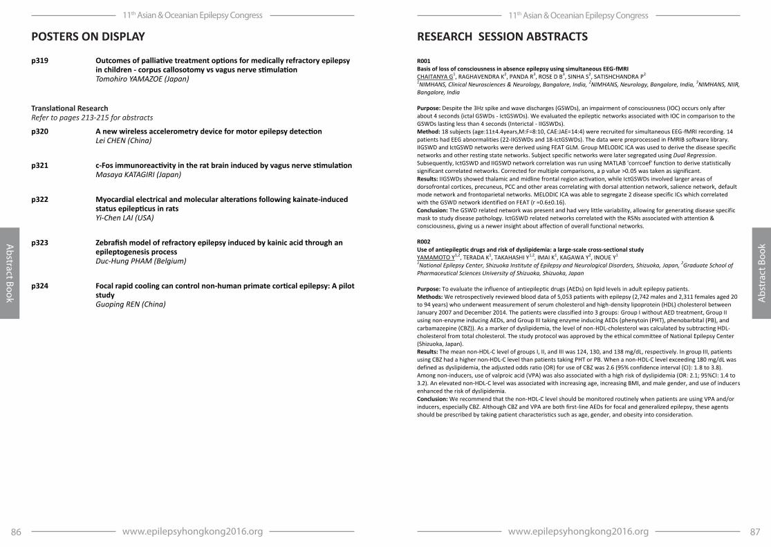
87www.epilepsyhongkong2016.org
11th Asian & Oceanian Epilepsy Congress
Abs
trac
t Boo
k
86 www.epilepsyhongkong2016.org
11th Asian & Oceanian Epilepsy Congress
Abstract Book
RESEARCH SESSION ABSTRACTS
p319 Outcomes of palliative treatment options for medically refractory epilepsy in children - corpus callosotomy vs vagus nerve stimulation Tomohiro YAMAZOE (Japan)
Translational Research Refer to pages 213-215 for abstracts
p320 A new wireless accelerometry device for motor epilepsy detection Lei CHEN (China)
p321 c-Fos immunoreactivity in the rat brain induced by vagus nerve stimulation Masaya KATAGIRI (Japan)
p322 Myocardial electrical and molecular alterations following kainate-induced status epilepticus in rats Yi-Chen LAI (USA)
p323 Zebrafish model of refractory epilepsy induced by kainic acid through an epileptogenesis process Duc-Hung PHAM (Belgium)
p324 Focal rapid cooling can control non-human primate cortical epilepsy: A pilot study Guoping REN (China)
POSTERS ON DISPLAY
R001 Basis of loss of consciousness in absence epilepsy using simultaneous EEG-fMRI CHAITANYA G1, RAGHAVENDRA K2, PANDA R3, ROSE D B3, SINHA S2, SATISHCHANDRA P2 1NIMHANS, Clinical Neurosciences & Neurology, Bangalore, India, 2NIMHANS, Neurology, Bangalore, India, 3NIMHANS, NIIR, Bangalore, India Purpose: Despite the 3Hz spike and wave discharges (GSWDs), an impairment of consciousness (IOC) occurs only after about 4 seconds (ictal GSWDs - IctGSWDs). We evaluated the epileptic networks associated with IOC in comparison to the GSWDs lasting less than 4 seconds (Interictal - IIGSWDs). Method: 18 subjects (age:11±4.4years,M:F=8:10, CAE:JAE=14:4) were recruited for simultaneous EEG-fMRI recording. 14 patients had EEG abnormalities (22-IIGSWDs and 18-IctGSWDs). The data were preprocessed in FMRIB software library. IIGSWD and IctGSWD networks were derived using FEAT GLM. Group MELODIC ICA was used to derive the disease specific networks and other resting state networks. Subject specific networks were later segregated using Dual Regression. Subsequently, IctGSWD and IIGSWD network correlation was run using MATLAB 'corrcoef' function to derive statistically significant correlated networks. Corrected for multiple comparisons, a p value >0.05 was taken as significant. Results: IIGSWDs showed thalamic and midline frontal region activation, while IctGSWDs involved larger areas of dorsofrontal cortices, precuneus, PCC and other areas correlating with dorsal attention network, salience network, default mode network and frontoparietal networks. MELODIC ICA was able to segregate 2 disease specific ICs which correlated with the GSWD network identified on FEAT (r =0.6±0.16). Conclusion: The GSWD related network was present and had very little variability, allowing for generating disease specific mask to study disease pathology. IctGSWD related networks correlated with the RSNs associated with attention & consciousness, giving us a newer insight about affection of overall functional networks. R002 Use of antiepileptic drugs and risk of dyslipidemia: a large-scale cross-sectional study YAMAMOTO Y1,2, TERADA K1, TAKAHASHI Y1,2, IMAI K1, KAGAWA Y2, INOUE Y1 1National Epilepsy Center, Shizuoka Institute of Epilepsy and Neurological Disorders, Shizuoka, Japan, 2Graduate School of Pharmaceutical Sciences University of Shizuoka, Shizuoka, Japan Purpose: To evaluate the influence of antiepileptic drugs (AEDs) on lipid levels in adult epilepsy patients. Methods: We retrospectively reviewed blood data of 5,053 patients with epilepsy (2,742 males and 2,311 females aged 20 to 94 years) who underwent measurement of serum cholesterol and high-density lipoprotein (HDL) cholesterol between January 2007 and December 2014. The patients were classified into 3 groups: Group I without AED treatment, Group II using non-enzyme inducing AEDs, and Group III taking enzyme inducing AEDs (phenytoin (PHT), phenobarbital (PB), and carbamazepine (CBZ)). As a marker of dyslipidemia, the level of non-HDL-cholesterol was calculated by subtracting HDL-cholesterol from total cholesterol. The study protocol was approved by the ethical committee of National Epilepsy Center (Shizuoka, Japan). Results: The mean non-HDL-C level of groups I, II, and III was 124, 130, and 138 mg/dL, respectively. In group III, patients using CBZ had a higher non-HDL-C level than patients taking PHT or PB. When a non-HDL-C level exceeding 180 mg/dL was defined as dyslipidemia, the adjusted odds ratio (OR) for use of CBZ was 2.6 (95% confidence interval (CI): 1.8 to 3.8). Among non-inducers, use of valproic acid (VPA) was also associated with a high risk of dyslipidemia (OR: 2.1; 95%CI: 1.4 to 3.2). An elevated non-HDL-C level was associated with increasing age, increasing BMI, and male gender, and use of inducers enhanced the risk of dyslipidemia. Conclusion: We recommend that the non-HDL-C level should be monitored routinely when patients are using VPA and/or inducers, especially CBZ. Although CBZ and VPA are both first-line AEDs for focal and generalized epilepsy, these agents should be prescribed by taking patient characteristics such as age, gender, and obesity into consideration.

89www.epilepsyhongkong2016.org
11th Asian & Oceanian Epilepsy Congress
Abs
trac
t Boo
k
88 www.epilepsyhongkong2016.org
11th Asian & Oceanian Epilepsy Congress
Abstract Book
PLATFORM SESSION ABSTRACTSPLATFORM SESSION ABSTRACTS
P003 Efficacy and safety of adjunctive lacosamide for the treatment of partial-onset seizures in Chinese and Japanese adults: a multicenter, double-blind, randomized, placebo-controlled study DU X1, INOUE Y2, LIAO W3, MENG H4, WANG X5, WANG W6, ZHOU L7, ZHANG L8, TOGO O9, TENNIGKEI F10, HONG Z11 1UCB Pharma, Shanghai, China, 2Shizuoka Institute of Epilepsy and Neurological Disorders, Shizuoka, Japan, 3The Second Affiliated Hospital of Guangzhou Medical University, Guangzhou, China, 4The First Hospital of Jilin University, Changchun, China, 5The First Affiliated Hospital of Chongqing Medical University, Chongqing, China, 6The First Affiliated Hospital of Kunming Medical University, Kunming, China, 7The First Affiliated Hospital of Sun Yat-sen University, Guangzhou, China, 8The First Affiliated Hospital of Harbin Medical University, Harbin, China, 9UCB Pharma, Tokyo, Japan, 10UCB Pharma, Monheim am Rhein, Germany, 11Huashan Hospital, Shanghai, China Rationale: A randomized, double-blind, placebo-controlled study (EP0008; NCT01710657) evaluated the efficacy and safety of adjunctive lacosamide treatment in Chinese and Japanese adults with uncontrolled partial-onset (focal) seizures (POS), with or without secondary generalization. Methods: After 8 weeks of baseline, patients aged 16-70 years recruited from study centers in China and Japan were randomly assigned to receive adjunctive lacosamide 400mg/day, 200mg/day, or placebo for 4 weeks of Titration (started at 100mg/day), followed by 12 weeks of Maintenance. The primary outcome was the change in seizure frequency/28 days from Baseline to Maintenance. Efficacy and safety evaluations were performed on the full analysis set (FAS) and safety set (SS), respectively. Results: ve all, atients eceive t eat ent ( a ≥ ost-baseline efficacy measurement (FAS); 485 (88.7%) completed the study. Demographics and baseline characteristics were similar between treatment groups. Among 544 patients (FAS), 77.8% were taking 2-3 concomitant AEDs. Mean (SD) seizure frequency/28 days from Baseline to Maintenance changed from 20.70 (28.06) to 11.92 (18.27), 31.31 (89.91) to 20.56 (44.53), and from 26.71 (57.90) to 23.28 (52.14) in the lacosamide 400mg/day (median change: -4.50), lacosamide 200mg/day (-3.33), and placebo (-1.22) groups, respectively. The percentage reduction in seizure frequency over placebo/28 days from Baseline to Maintenance was statistically significant for lacosamide 400mg/day (39.6% [95% CI: 30.5%, 47.6%], p< 0.001) and lacosamide 200mg/day (29.4%[18.7%, 38.7%], p< 0.001). The proportions of patients who were seizure free during the Maintenance period was 5.4%, 4.7%, and 0% in patients receiving lacosamide 400mg/day, 200mg/day, or placebo. The overall incidence of treatment-emergent adverse events in all lacosamide groups was 72.2%; the most common were dizziness (25.9%), nasopharyngitis (14.3%) and somnolence (10.2%). Conclusions: Adjunctive lacosamide (400 and 200mg/day) was efficacious in reducing POS frequency in Chinese and Japanese patients with a safety and tolerability profile consistent with previous trials. Funding: UCB Pharma-sponsored. P004 Comparison of relapse risk between drug discontinuation and continuation at different periods of seizure remission in adult patients with focal epilepsy HE R-Q1,2, WANG X-S1, ZENG Q-Y1, ZHU P1, BAO Y-X1, XIA N-G1, XU H-Q1, ZHENG R-Y1 1The First Affiliated Hospital of Wenzhou Medical University, Neurology, Wenzhou, China, 2The Three Affiliated Hospital of Wenzhou Medical University ( Ruian People's Hospital), Neurology, Wenzhou, China Purpose: To explore the optimal timing of antiepileptic drug (AEDs) discontinuation in adult patients with focal epilepsy Method: Adopted by Wenzhou Epilepsy Follow Up Registry Database (WEFURD), 446 adult patients with focal epilepsy were recruited, who were seizure free for more than 2 years between January 2003 and June 2013, were divided into AEDs discontinuation group and continuation group depending on whether AEDs withdrewal, and were followed up prospectively for at least 1 year or until a seizure relapse. Then compared the relapse risk of AED discontinuation subgroup with that of continuation subgroup at different periods of seizure remission(2- < 3, 3- < 4, 4- < 5, 5- < 6, 6- < 7 years). The relapse rate of each corresponding subgroups were analyzed by Kaplan-Meier method. The comparisons of relapse risk between each corresponding subgroups were investigated by log-rank method and the confusions were adjusted by Cox proportional hazard regression model. Results: The number of patients discontinued AEDs after seizure-free for 2- < 3, 3- < 4, 4- < 5, 5- < 6, 6- < 7 years were 65, 61, 32, 15, 11, respectively. And the corresponding number for AEDs continuation were 262, 221, 151, 109, 83, respectively. Patients who discontinued AED after 2- < 5 years of seizure-freedom had a significant higher risk of seizure relapse compared with the corresponding continuation, with the hazard ratio (HR) 1.8 (95%CI 1.25-2.74), 2.6 (95%CI 1.73-4.18), 3.4 (95%CI 1.77-6.54) in remission time of 2- < 3, 3- < 4, 4- < 5 years, respectively. While rather lower relapse risk in corresponding subgroups after 5- < 6, 6- < 7 years of seizure-freedom were similar with the HR 1.32 (95%CI 0.36-4.70), 1.12 (95%CI 0.21-6.10), respectively. Conclusion: The optimal timing of AEDs discontinuation for adult patients with focal epilepsy is seizure-free for at least 5 years.
P005 Determinant of health-related quality of life in patients with epilepsy from Malaysia BAZIR AHMAD S1, TAN C T1, LIM K S1, WO M C M1, TAN W Y1, PERUCCA E2 1University of Malaya, Neurology Division, Department of Medicine, Kuala Lumpur, Malaysia, 2University of Pavia Medical School, Pavia, Italy Purpose: Identifying the significant predictors of health-related quality of life (HRQOL) is crucial in managing patients with epilepsy (PWE). We aim to evaluate the impact of socio-economic profiles, disease and treatment-related variables on (HRQOL) on PWE in Malaysia. Method: This is a single-center, observational study, aiming for an 18-month prospective assessment of health outcomes in patients with epilepsy. Patients were required to independently answer five validated self-administered questionnaires; Beck Depression Inventory II (BDI-II), Social Stigma Scale, Seizure Severity Scale (SSS), Adverse Events Profile (AEP) and Quality of Life in Epilepsy (QOLIE-31). Results: A total of 195 patients were recruited over 9-month period. Mean age was 37.6 years old (SD 15.1), predominantly male (52.3%). Mean age of seizure onset was 22.2 years old (SD 15.7) with an average seizure duration of 15.5 years (SD 12.7). 41% of them were married/co-habiting, 51.8% had received higher education and 55.9% were employed. Majority of patients came from a low socio-economic background. 71.8% of patients had focal epilepsy and 25.1% had generalized epilepsy. The aetiology for 40% of cases were unknown, followed by 34.9% structural and 24.1% genetic. Most of them were in remission (51.8%) and 21% were refractory patients. 64.6% of patients were on monotherapy. Seizure severity scale correlated significantly with number of AED, seizure frequency and AEP score (p< 0.05). The mean BDI-II score in our cohort was 9.1 (SD 9.4, range 0-46), which was within the normal range and the average QOLIE-31 total score was 52.6 (SD 9.2, range 28.1-72.6). Patients in the uncontrolled/refractory group had a relatively worse score (p< 0.05 for BDI II, p< 0.001 for QOLIE-31). Depression, seizure severity, AED side-effects, social stigma were significant negative predictors for HRQOL (p< 0.05). Conclusion: Seizure severity, depression, social stigma and AED toxicity are significant predictors for HRQOL in PWE. P006 Evaluation of headache disorders in patients of idiopathic epilepsy GANGULY G1, UJJAWAL R1, BOSE S2, ALAK P1 1West Bengal University of Health Sciences, Bangur Institute of Neurosciences, Kolkata, India 2West Bengal University of Health Sciences, K P C Medical College, Kolkata, India Purpose: Various types of headache, especially migraine is considered a relevant co-morbidity in epilepsy. Headache in idiopathic epilepsy may have diagnostic and therapeutic implications. Method: We reporting clinical data on 109 patients of Idiopathic epilepsy. All eligible patients of idiopathic epilepsy were recruited from OPD & epilepsy clinic. Headache was classified according to ICHD III and headache disability before and after treatment was calculated according to Henry Ford Headache Disability Index(HDI). The data was compared to normal and the statistical analysis was done by using SPSS4. Results: Out of 109 cases, 71(65.13%) had headache. Cases were diagnosed as JME (28,25.7%) followed by epilepsy with febrile seizure plus (21,19.3%) and Juvenile Absence Epilepsy (18,16.5%). Other types of seizure noted were Idiopathic Generalized Epilepsy with GTCS only, Benign Epilepsy with Centro-Temporal Spikes, Panayiotopoulos syndrome and Gastaut syndrome. Out of those who had headache, 21(19.27%) cases had post ictal headache and remaining had primary headache (50,45.87%). Most common primary headache type was migraine without aura (31,28.4%) and frequent episodic Tension type headache(TTH) (15,13.8%). Other headaches were noted infrequent tension type, Migraine with aura, chronic migraine, medication overuse. Among primary headache migraine was more commonly seen in cases as compared to controls, which was statistically significant(p=0.029). Additionally the occurrence of GTCS in a particular IGE was associated with headache (Chi2 = 4.75, p=0.029, Odds ratio= 3.51). Additional drug requirement was high in primary headache as compared to control. HDI was reduced to a significant amount in the cases after additional treatment. Conclusion: Apart from post ictal headache, TTH and migraine were more common in cases. Migraine most commonly associated with occurrence of GTCS. Majority of patients of Migraine and TTH required additional therapy, but surprisingly there was significant reduction in the headache frequency and reduction/ improvement in HDI.
91www.epilepsyhongkong2016.org
11th Asian & Oceanian Epilepsy Congress
Abs
trac
t Boo
k
90 www.epilepsyhongkong2016.org
11th Asian & Oceanian Epilepsy Congress
Abstract Book
PLATFORM SESSION ABSTRACTSPLATFORM SESSION ABSTRACTS
P007 Can ictal-MEG accurately localize the epileptogenic zone? RAMANUJAM B1, BHARTI K2, VISHWANATHAN V2, CHANDRA P S1, GARG A3, TRIPATHI M1, BAL C4, TRIPATHI M4 1All India Institute of Medical Sciences, Neurology, New Delhi, India, 2National Brain Research Centre, Manesar, India, 3All India Institute of Medical Sciences, Neuroradiology, New Delhi, India, 4All India Institute of Medical Sciences, Nuclear Medicine, New Delhi, India Purpose: The objective of this study was to determine if an ictal-MEG study added useful information towards delineating the epileptogenic and/or the ictal-onset zone. Method: Patients with DRE routinely undergo Video-EEG (VEEG) monitoring, MRI-brain (epilepsy protocol), ictal and interictal SPECT, FDG-PET and MEG as part of presurgical evaluation. Out of 310 patients who underwent MEG, 37 had a seizure during the MEG-study. Their Ictal and interictal-MEG data were analyzed by both DANA and CURRY software by different technologists blinded to each other's results. The VEEG localization was done by an epileptologist independently, PET and SPECT reported by nuclear medicine specialists blinded to other results. Concordance of the Ictal-MEG localization with the lesion on MRI was checked; if substrate-negative on MRI, concordance with SPECT and/or PET was noted. Results: Most seizures were focal (30), only 7 patients had secondary generalizations. Inter-ictal data of all 37 showed abnormal findings in the form of either spikes, sharps or slow waves and source analysis done with equivalent current dipole model showed focal clustering in 29 out of 37 patients who got convulsions during acquisition. Ictal findings were concordant with the MRI in 85 % of those with an abnormal MRI substrate. In those with no substrate in the MRI (14), ictal MEG was concordant with the ictal-onset zone on scalp EEG in 56%, with SISCOS in 50% and with PET in 63% . MEG done again in 5 patients in the latter group, and found consistent with the previous MEG cluster. Interictal and ictal-MEG data were convergent in their clusters to about 68%. Conclusion: Ictal-MEG is useful in localizing the ictal-onset zone in the presurgical evaluation of DRE-patients. P008 Epilepsy interaction with behavior, emotions & learning ABEIDAH M I1 1Hamad Medical Corporation, Neurodevelopmental Pediatrics, Doha, Qatar Background: Epilepsy is more common in people with developmental/ learning disabilities than in the general population. Likewise many children with epilepsy are likely to present with spectrum of psychosocial, behavioral or emotional problems and the more severe the epilepsy the more likely it is that a person will also have these disorders. There are also a lot of misconceptions about the link between epilepsy and violent behavior. Objectives: In this presentation we will be looking into the complexities of these relationships between epilepsy, behavior, learning and other related issues including epilepsy, autism and ADHD connection. With the aid of videos and other materials we will demonstrate that managing epilepsy in these children can be a challenging yet also a rewarding experience. We will discuss the difficulties we face as clinicians when we look after people with a high level of needs. We will share with you our personal practice and experience in providing specialist service for children and young people with epilepsy and developmental disabilities at the secondary and tertiary levels. P009 Breastfeeding initiation, duration and exclusivity among Chinese mothers with epilepsy: A retro-prospective study in a developing country HAO N1, WU M1, JIANG H1, ZOU X1, ZHOU D1 1West China Hospital, Sichuan University, Department of Neurology, Chengdu, China Purpose: to 1) estimate the breastfeeding initiation, duration and exclusivity rates, as well as common reasons for early weaning among Chinese mothers with epilepsy; 2) identify their potential perinatal correlations with selected sociodemographic and clinical factors. Method: a detailed investigation was conducted in China using a semi-structured questionnaire from December 2013 to May 2015. Data about breastfeeding behaviors, sociodemographic, obstetric, and epileptic variables were collected from 281 Chinese mothers with epilepsy. Descriptive analysis, followed by univariate and multivariate logistic regression analyses, was applied to analyze the data from this survey. Results: the breastfeeding initiation rate among participant mothers with epilepsy was 59.4%. At 3 months, any breastfeeding rate was 49.5% and exclusive breastfeeding rate was 36.3%. At 6 months, about one third (33.1%) of mothers with epilepsy kept breastfeeding their babies and 12.8% of enrolled infants were exclusively breastfed. During lactation, fear of potential AEDs exposure via breastfeeding, frequent seizure onset, and insufficient breast milk supply were the most common reasons for early breastfeeding cessation. Participant mothers with epilepsy who lived in rural
areas or had term delivery were inclined to breastfeed their babies. And mothers who lived in rural areas or have gestational non-active epilepsy were more likely to have long-term breastfeeding. Non-polytherapy is of benefit to all described breastfeeding behaviors. Conclusion: Chinese mothers with epilepsy have lower prevalence of eligible breastfeeding behaviors compared to general population. Good seizure control and optimal therapy during gestation and lactation were to the benefit of breastfeeding implementation. Targeted intervention programs enhancing antenatal care services and breastfeeding consultation are needed for this specific group. P010 Factors affecting the employability in people with epilepsy (PWE) WO M C M1, LIM K S1, CHOO W Y2, TAN C T1 1University of Malaya, Medicine, Kuala Lumpur, Malaysia, 2University of Malaya, Social and Preventive Medicine, Kuala Lumpur, Malaysia Purpose: This study aimed to examine demographic, clinical and psychological factors associated with employability in PWE. Method: Clinical, demographical and psychosocial details were collected through self-administered questionnaire at neurology clinic in University of Malaya Medical Center, Malaysia. The psychological measures were Work Self-Determination Index (WSDI), Rosenberg Self-Esteem Scale (SES), Multidimensional Scale of Perceived Social Support (MSPSS), and Quality Of Life In Epilepsy (QOLIE-3 . E lo a ilit as eas e sing e lo ent atio, it a atio ≥ 90% (ER90) classified as high employability. Results: Of 146 participants, the mean age was 34.40 (SD 10.89), 52% female, 50.7% Chinese, 50% single and 55.5% had tertiary education and more than half had high employability (64.4%). A majority of participants had focal epilepsy (72.6%), with mean seizure frequency of 14.90/year (SD 45.82). Variables that significantly distinguished high employability group included seizure type, ability to work (indicated by education level, work performance affected by seizures, ability to travel independently and ability to cope with stress at), lower self-perceived stigma (ESS), higher self-determination (WSDI), self-esteem (SES), perceived social support (MSPSS), and family overprotection. Those with high employability reported a better quality of life measured by QOLIE-31 (mean 59.20 ± SD 13.79) compared to those with low employability (mean 54.18 ± SD 12.98). Regression analysis showed that education level (OR 3.42, CI: 1.461 - 7.999), self-determination (WSDI; OR 1.087, CI: 1.012 - 1.168), family overprotection (OR 0.76, CI: 0.610 - 0.947), and seizure type (OR 0.24, CI: 0.079 - 0.731) were predictive of employability in PWE. Furthermore, there was no evidence in mediation on causal chain among independent variables (WSDI and tertiary education level) and high employability (ER90%, dependent variable). Conclusion: Demographic factor (education level), clinical factor (seizure type) and psychological factors (work self-determination and family overprotection) were important factors affecting the employability in PWE. P011 Epilepsy, interictal psychopathology and vitamin D3 levels - Are they related? TALWAR P1, SUDARSHAN C Y1, BEGUM S1 1J.J.M. Medical College, Psychiatry, Davangere, India Purpose: To measure Vitamin D3 levels in patients having epilepsy with interictal psychopathology, without psychopathology & healthy controls and assess its relationship to epilepsy & psychopathology. Method: This is a hospital based cross sectional study. The sample consisted of group 46 patients having interictal psychopathology, 40 epilepsy patients without psychopathology and 50 healthy controls. Sociodemographic data of the three groups and illness-related variables in patients were elicited after taking informed consent. Vitamin D3 levels of were assessed in all three groups and compared. The relationship between severity of low levels of vitamin D3, sociodemographic & illness related variables was analysed amongst the groups. Results: Data was analysed using SPSS 22.0. Vitamin D3 levels were in deficiency range for all the three groups (< 20 ng/ml). Significant gender difference in Vitamin D3 levels (less in females than males) was present in patients than controls. Muslim controls had significantly lower Vitamin D3 levels than Hindu controls. Vitamin D3 levels were significantly lower in the patients with interictal psychopathology as compared to controls and patients not having psychopathology (14.66±7.38 vs 17.87±6.06 vs 16.1±4.82, F=3.17,p< 0.04), despite significantly longer duration of exposure to sunlight in minutes as compared to controls (138.0 ±123.66 vs 44.6±70.45; t=4.594, p< 0.00). Vitamin D3 levels in patients were negatively correlated with age, frequency of seizure, duration of illness & treatment and number of drugs used while exposure to sunlight was positively correlated.Patients with interictal psychopathology having deficiency of Vitamin D3 (Vitamin D3 - < 20 ng/ml) were significantly older and less educated than those having insufficiency of vitamin D3(Vitamin D3 - 20 to 30 ng/ml). Conclusion: Norms for deficiency/ insufficiency for Indian population may need revision. Vitamin D deficiency may result in

93www.epilepsyhongkong2016.org
11th Asian & Oceanian Epilepsy Congress
Abs
trac
t Boo
k
92 www.epilepsyhongkong2016.org
11th Asian & Oceanian Epilepsy Congress
Abstract Book
PLATFORM SESSION ABSTRACTSPLATFORM SESSION ABSTRACTS
poor seizure control. Interictal psychopathology per se was associated with significantly lower Vitamin D3 levels. This has therapeutic Implications. P012 Positive psychology interventions for neurological disorders: a systematic review LAI S T1, LIM K S1, TAN C T1, LOW W Y2 1University of Malaya, Division of Neurology, Department of Medicine, Faculty of Medicine, Kuala Lumpur, Malaysia, 2University of Malaya Medical Center, Health Research Development Unit, Kuala Lumpur, Malaysia Purpose: Happiness is a psychological state that reflects on individual's experience of positive feelings and life satisfaction, which, is often a neglected concept in the healthcare climate. In the realm of neurological diseases, happiness can diminish via direct physical damage or a reaction towards poor prognosis. Beyond treatment aspects of symptomology, positive psychology (PoP) addresses how the scientific study of well-being promotes health. This systematic review aimed to synthesize the evidence of using positive psychology interventions (PPI) in this population. From the review evidence, we also aimed to explore whether the benefits can be extrapolated to epilepsy patients. Method: PoP interventions (PPI) are defined as therapies or intentional activities aimed at cultivating positive thoughts, feelings and behaviors. The electronic databases included PUBMED, EMBASE, MEDLINE, PSYCINFO, and secondary search dated from [Jan 1980 - Dec 2015]. Studies reporting the use of (PPI) in neurological populations were independently assessed and screened by three reviewers using review-specific criteria. Results: Of the 661 articles identified through databases, 16 studies were finally included in this review. Five groups of therapies were found: mindfulness-based approaches, written expression of positive emotions, meaning-making interventions, creative-expressive therapy and cognitive-based approaches. Findings revealed these interventions promoted positive changes in neurological diseases, in which patients reported enhanced quality of life, fewer symptoms distress, and reduction in depressive symptoms. Coverage has been limited in epilepsy; however patients with epilepsy (PWE) have benefited from such holistic therapy in terms of coping and managing the course of their disease. Nevertheless, the disparity of the interventions and some methodological issues limit the outcomes. Conclusion: PPIs were shown to promote well-being across various neurological disorders, but the widespread recognition has not extended to the epilepsy community. P013 The outcome of bipolar electro-coagulation with lesionectomy in the treatment of epilepsy involving eloquent areas ZHAI F1, LUAN G1, Jian Zhou, Tianfu Li 1Beijing Sanbo Brain Hospital, Capital Medical University, Neurosurgery, Beijing, China Purpose: Although resection of an epileptogenic region is the main procedure of epilepsy surgery, epileptogenic areas in functionally critical cortex can not be approached in that manner. We demonstrated previously that Bipolar Electro-Coagulation on Functional Cortex (BCFC) is a safe and effective approach for epilepsy involving eloquent areas. Here we report the results of BCFC with lesionectomy for patients whose epileptogenic foci are partially overlapped with eloquent areas. Method: 50 patients involving eloquent arears who underwent lesionectomy combined with BCFC between March 2007 to March 2013 were retrospectively reviewed with regard to seizure and neurologic outcome. Results: Fellow up 24-54 months, averaged 36 months. Engel class I outcome was achieved in 25 (50%) patients, class II in 10 (20%), class III in 9 (18%) and class IV in 6 (12%); 6 (12%) patients developed mild hemiparesis. All these complications recovered within 1~12 months. Conclusion: BCFC with lesionectomy is a safe and effective operation to treat epilepsy involving eloquent areas. Compared with lesionectomy with MST, the outcome of BCFC with lesionectomy is similar, but the complication is less. P014 Robotics in neurosurgical stereotactic interventions: Oblique intrainsular electrodes implanted of patients with epilepsy AFIF A1,2 1Lyon 1 University, Neurosurgery A, Neurological Hospital, Lyon, France, 2Department of Anatomy, INSERM U 1028, Pain Center, Pierre Wertheimer Hospital, Hospices Civils de Lyon, Lyon, France Objective: This study is to investigate: The feasibility, the safety and the utility of chronic depth electrodes stereotactically implanted by a robotic arm in the insular cortex of patients suffering from drug refractory focal epilepsy. Methods: A total number of 32 electrodes in 29 patients (in Grenoble University Hospital) were successfully implanted within the insula. 220 contacts were available for insula recording. Electrode insertion was guided by a robotic arm (Neuromate, Renishawmayfield, Switzerland) connected to the stereotactic frame and driven by stereotactic planning
software. The targetting of the insula is planned on a pre-surgical T1-MRI. The fusion between the preoperative 3D MRI and the postoperative 3D CT scan enabled us to identify the contact location in three dimensions. Results: No morbidity occurred during the surgical step and the chronic SEEG recording or stimulation procedure. Clinical responses have been identified in terms of gyral and sulcal anatomy.They were classified into: painful responses, sensitivomotor responses, speech disturbance, oropharengeal responses, auditory phenomena and neuro-vegetative phenomena. Conclusion: The advantages of the oblique approach are: 1. The implantation of electrodes within the insula using robotic arm appears in our study to be safe. 2. This approach can explore all insular regions by avoidance of the sylvian vascular network. 3. This approach offers a better sampling of insular EEG activity (until 10 contacts/electrode) than that obtained by the classical lateral trans opercular approach (1 ½ contacts/electrode). 4. Thisapproach has allowed us to develop the first anatomo-functional organization scheme of the insular cortex according to its gyri and sulci. P015 Predictive analysis of perioperative cognitive function in patients with hypothalamic hamartoma, who underwent surgery of stereotactic radiofrequency thermocoagulation SONODA M1, MASUDA H1, SHIROZU H1, ITO Y1, AKAZAWA K2, KAMEYAMA S1 1Nishi-Niigata Chuo National Hospital, Hypothalamic Hamartoma Center, Niigata, Japan, 2Niigata University Medical and Dental Hospital, Department of Medical Informatics, Niigata, Japan Purpose: Hallmark of hypothalamic hamartoma (HH) syndrome is gelastic seizure. Additionally, over half of the patients exhibit cognitive impairment referred to as epileptic encephalopathy. But the pathophysiological mechanism of epileptic encephalopathy was still unclear. To clarify its nature, we statistically analyzed pre- and postoperative profiles of cognitive function from our clinical records. Method: We retrospectively reviewed consecutive 91 patients with HH who underwent stereotactic radiofrequency thermocoagulation (SRT) for medically intractable gelastic seizures between October 1997 and December 2014 in our institution. The patients were examined with WAIS- , WISC- or Tanaka-Binet Intelligence Scale and categorized into WAIS/ WISC group (n=65) , junior group (n=13) and elder group of T-B group (n=13) based on neuropsychological examination and the age at surgery. Clinical features among each groups and difference between pre- and postoperative cognitive function were statistically compared. Stepwise multiple regression analyses were performed to evaluate independent predictive factors for preoperative cognitive function and the percentage postoperative change of IQ in WAIS/WISC group. Results: Eighty six patients (95%) were followed up at 1 year following SRT, and 58 (67 %) patients achieved complete seizure freedom at postoperative 1 year. The patients in all groups had significantly improved cognitive function following surgery. Multiple linear regressions analysis of preoperative cognitive function revealed that the generalized paroxysms (p < 0.001) were negative predictor and that intra-type HH (p = 0.001) are positive that for preoperative full IQ. Moreover, multiple linear regressions revealed that preoperative IQ (p < 0.001) and the duration of non-gelastic seizures (p = 0.020) were negative predictors to postoperative cognitive change rate. Conclusion: Our results of predictive analysis of pre- and postoperative cognitive function provide a robust evidence to encourage the patients with HH and their families to challenge early surgical intervention for achievement of better cognitive outcome. P016 MRI negative patients with medial temporal lobe epilepsy were more effective to stereotactic radiofrequency thermocoagulation of amygdalohippocampal complex compared with MRI positive patients ZHAO Q1 1The 306 Hospital of PLA, Neurosurgery, Beijing, China Purpose: To explore the therapeutic efficacy of depth-electrode-guided stereotactic radiofrequency thermocoagulation (RFTC) of the amygdalohippocampal complex for the treatment of intractable medial temporal lobe epilepsy (MTLE) between MRI negative and positive patients. Method: A total of 127 cases of MTLE were retrospectively studied after depth-electrode-guided RFTC of the amygdalohippocampal complex. A preoperative MRI scan (T1, T2, Flair sequence) was performed in all patients, in which 67 cases were positive for brain lesions and 60 cases were negative. Results: After 24-83 months follow-up, 54.69% (70/127) of patients were in Engel class I-III, with 32.28% (41/127) of patients in Engel class I. A total of 50.00% (30/60) of patients in the MRI-negative group became seizure-free after treatment, but only 16.42% (11/67) of cases in the MRI-positive group were seizure-free at the time of follow-up. A
95www.epilepsyhongkong2016.org
11th Asian & Oceanian Epilepsy Congress
Abs
trac
t Boo
k
94 www.epilepsyhongkong2016.org
11th Asian & Oceanian Epilepsy Congress
Abstract Book
PLATFORM SESSION ABSTRACTSPLATFORM SESSION ABSTRACTS
significant difference in seizure-free outcome (P < 0.001) was obtained between the MRI-positive and MRI-negative groups. Conclusion: MTLE patients with a preoperative MRI scan negative for lesions show better seizure control after RFTC of the amygdalohippocampal complex than MRI-positive patients. RFTC of the amygdalohippocampal complex significantly reduces epileptic discharges, but does not thoroughly deal with structural lesions observed by MRI. MRI-positive MTLE patients show better results with surgical resection, and therefore, must be carefully selected for RFTC of the amygdalohippocampal complex. P017 Seizure onset and propagation in tuberous sclerosis: intracranial EEG analysis KANNAN L1, BAILEY C1, VOGRIN S2, MAIXNER W3, HARVEY A S1 1The Royal Children's Hospital, Department of Neurology, Melbourne, VIC, Australia, 2Murdoch Childrens Research Institute, Melbourne, VIC, Australia, 3The Royal Children's Hospital, Department of Neurosurgery, Melbourne, VIC, Australia Purpose: Uncertainties exist about epileptogenic substrate(s), underlying mechanisms of seizure generation and seizure propagation patterns in tuberous sclerosis (TSC). We aimed to describe spatio-temporal electrophysiological patterns of seizure onset and propagation on intracranial EEG (iEEG) in children with TSC. Methods: Retrospective analysis of iEEG in 10 children with TSC, who had ictal recordings from concurrent surface and depth electrodes sampling two or more tubers, was performed. Electrode contacts in relation to tuber centre, tuber rim and perituberal cortex were determined. Patient specific bipolar montages were reformatted with tuber centre, tuber rim and perituberal cortex channels. Inter-ictal epileptiform discharges (IEDs) and ictal rhythms were analysed for location, field, morphology, rhythmicity, latencies and temporal dispersion. Quantitative EEG-signal analyses (pairwise cross-correlation and root-total power (RTP) analysis) were performed in a subgroup. Results: Patients were aged 2-13 years (median 3.8). 15 electro-clinically distinct seizures types (EDS) were recorded. Seizure onset was within a single tuber, in the tuber centre with or without involvement of tuber rim; perituberal cortex was not involved in seizure onset. Cross-correlation analysis confirmed this finding. 9/15 seizure onset tubers showed continuous, periodic IEDs on an attenuated background. Seizure onsets were recorded from 15/33 tubers with independent IEDs, but none of the seven tubers with only propagated IEDs and none of the eight tubers with no IEDs (p=0.003). Stereotyped seizure propagation from tuber-to-tuber was observed in 10/15 EDS. Propagated ictal rhythms were indistinguishable from seizure onset rhythms. Intraictal activation of distinct ictal rhythms were noted during seizure propagation in 6/10 EDS. RTP analysis demonstrated the spatial and temporal seizure propagation dynamics quantitatively. Conclusions: Our findings suggest that seizures arise in the tuber centre and not in perituberal cortex, that continuous periodic IEDs recorded on an attenuated background indicate epileptogenic tubers, and that tuber-to-tuber seizure propagation could potentially lead to false localisation. P018 Study of transplantation of neural stem cells in therapy of epilepsy in developing rats DAI Y1 1Department of Pediatrics, the Affiliated Hospital of Xuzhou Medical College, Xuzhou, China Purpose: After the transplantation of neural stem cells (NSCs) from the hippocampus in newborn rats on the developmental epileptic rats, to observe the survival of neural stem cells and the impact of the dentate gyrus mossy fiber sprouting. Method: The neural stem cells were identified with immunofluorescence staining, which were extracted and cultured and purified from the hippocampus of newborn SD rats 24 hours after birth. Take the 21-day-old SD rats, to establish rat kindling model with epilepsy by PTZ-injection, divided into 4 groups: (1) no seizures (normal control group); (2) epilepsy with no treatment group (epilepsy group); (3) treated with PBS transplantation after the seizures (sham group); (4) treated with BrdU-labeled NSCs transplantation after the seizures (treatment group). 7 days after the seizures, BrdU-labeled NSCs transplanted into the lateral ventricle in rats. After 8 weeks using immunohistochemical method to observe the hippocampus dentate gyrus BrdU positive cells changes, timm silver staining method to observe the hippocampus MFS situation. Results: The cells gathered suspension growth and formed typical neural spheres, immunofluorescence staining can detect the positive expression of nestin and BrdU, and the cultured cells with the ability of self-renewal and pluripotent differentiation. These cells after transplantation into the hippocampus of rats 8 weeks can detect its presence to around migration. The normal control group is no seizures, mossy fiber sprouting did not change significantly; after the success of model making, the mossy fiber Timm's staining particles ratings in epilepsy group increased with the normal control group, the difference was statistically significant(P< 0.05); both the epilepsy group and the sham group, MFS score was not statistically significant(P 0.05); compared with the sham group, the scores for mossy fiber sprouting in the treatment group were lower, and the difference was significant statistically(P< 0.05).
Conclusion: NSCs after transplantation can survive and effectively suppress seizures. P019 MicroRNA-132 silencing decreases epileptogenesis via the P250GAP/Cdc42 signaling pathway YUAN J1, HUANG H1, ZHOU X1, LIU X1, XU T1, OU S1, CHEN Y1, FAN X1 1the Second Affiliated Hospital of Chongqing Medical University, Neurology, Chongqing, China Purpose: Increasing evidence suggests that epilepsy is the result of synaptic reorganization and the formation of a pathological expiatory loop in the central nervous system (CNS); however, the mechanisms that regulate the epileptogenesis process are not well understood. We proposed that mir-132 and p250GAP may play important roles in this process by activating the downstream Rho family of GTPases. Method: By using a magnesium-free medium-induced epileptic model of cultured hippocampal neurons, we firstly investigated whether mir-132 regulates downstream GTPase activity through p250GAP, then, we tested the electrophysiological and neuroprotective effect of mir-132/p250GAP in cultured neurons. In addition, we verified the effect of mir-132 in vivo by silencing mir-132 in a lithium-pilocarpine-induced epileptic mouse model and compared the incidence of chronic spontaneous seizure in control group and mir-132 silencing group. Results: In the magnesium-free medium-induced epileptic model of cultured hippocampal neurons the mir-132 might regulates downstream Cdc42 activity through p250GAP since Cdc42 was significantly activated in our experimental model. Silencing mir-132 can inhibit the electrical excitability level of cultured epileptic neurons, whereas silencing of p250GAP had the opposite effect. Silencing mir-132 has a neuroprotective effect in the epileptic model, but this effect did not occur through the p250GAP pathway. In vivo, silencing mir-132 can inhibit the aberrant formation of dendritic spines and chronic spontaneous seizure in a lithium-pilocarpine-induced epileptic mouse model. Conclusion: In general, silencing of mir-132 can suppress spontaneous seizure activity through the mir-132/p250GAP/Cdc42 pathway by regulating the morphology and electrophysiology of dendritic spines. Moreover, mir-132 may serve as a potential target for the development of anti-epileptic drugs. P020 Targeting mammalian target of rapamycin down-regulates the expression of P-glycoprotein in a pharmacoresistant epilepsy rat model kindled by coriaria lactone CHI X1, CHENG H1, LI J1, ZHOU D1 1Department of Neurology, West China Hospital, Sichuan University, Chengdu, China Purpose: In spite of emerging new antiepileptic drugs, pharmacoresistance is still a major problem in clinical treatment. The prominent hypothesis of pharmacoresistant mechanism is that the overexpressed P-glycoprotein (Pgp) at the blood-brain barrier limited access of drugs the central nervous system. The mammalian target of rapamycin (mTOR) has been proved to mediate multidrug resistance in tumor by inducing Pgp overexpression. Therefore, we treated the pharmacoresistant epilespsy rat model with the mTOR inhibitor, rapamycin, to investigate the potential role of mTOR in pharmacoresistant mechanisms. Method: We established a pharmacoresistant epilepsy SD rat model by repeated injections of coriaria lactone , and kindled rats were treated with rapamycin at different doses of 1,3,and 10 mg/kg every other day for four weeks. Rapamycin-treated rats were sacrificed at various time points (0h,1h,8h,24h,3d,7d,14d, and 28d) after rapamycin administration for Western blotting and immunohistochemical analysis. Results: The overexpression of both phosopho-S6 (P-S6) and Pgp were detected in kindled rats compared to normal rats. For rapamycin-treated rats, the P-S6 rose in the first 24h, peaked at 8h, decreased after 24h, and returned to normal by 14d (all P< 0.05). The Pgp showed the same pattern of change with P-S6. After 28d, the expressive levels of P-S6 and Pgp in kindled rats with rapamycin were significantly lower than kindled rats without treatment (all P< 0.05). In addition, the moderating effect of 10mg/kg rapamycin on P-S6 and Pgp was the most obvious when compared with other doses at the same time point. Conclusion: The current study indicates that the rapamycin down-regulates the expression of P-glycoprotein by mTOR signaling pathway, and the inhibitory effect is dose-dependent. Therefore, mTOR signaling pathway has a potential role in drug-resistant mechanisms, and may be a potential drug target for pharmacoresistant epilepsy.
97www.epilepsyhongkong2016.org
11th Asian & Oceanian Epilepsy Congress
Abs
trac
t Boo
k
96 www.epilepsyhongkong2016.org
11th Asian & Oceanian Epilepsy Congress
Abstract Book
PLATFORM SESSION ABSTRACTSPLATFORM SESSION ABSTRACTS
P021 Association of genetic and non-genetic factors with phenytoin-induced severe cutaneous adverse drug reactions in Thai patients YAMPAYON K1, SUKASEM C2, LIMWONGSE C3, CHINVARUN Y4, TEMPARK T5, KIJSANAYOTIN P1 1Faculty of Pharmaceutical Sciences, Chulalongkorn University, Department of Pharmacology and Physiology, Bangkok, Thailand, 2Faculty of Medicine Ramathibodi Hospital, Mahidol University, Department of Pathology, Bangkok, Thailand, 3Faculty of Medicine Siriraj Hospital, Mahidol University, Department of Medicine, Bangkok, Thailand, 4Phramongkutklao Hospital, Neurology Unit, Bangkok, Thailand, 5Faculty of Medicine, Chulalongkorn University, Department of Pediatrics, Bangkok, Thailand Purpose: Several studies suggested CYP2C9*3 and HLA-B*15:02 as predictive genetic markers for PHT-induced severe cutaneous adverse drug reactions (SCARs) and SJS/TEN. However, no association study including non-genetic factors has been done. This study aimed to investigate the association of genetic variants together with non-genetic factors with phenotype specific PHT-induced SCARs in Thai patients. Method: Thirty-six PHT-induced SCARs patients confirmed diagnosis by dermatologist (15 SJS/TEN and 21 DRESS/HSS) and 100 PHT-tolerant control patients were studied. Variants in HLA-B, CYP2C9 and CYP2C19 genes were genotyped and selected as candidate covariates together with non-genetic factors. Conditional logistic regression analysis was performed to determine the association of multiple factors with PHT-induced SCARs. The study protocol was approved by each institutional ethics committee. Results: The final regression model showed that in specific phenotype analysis, HLA-B*13:01 was strongly associated with PHT-induced DRESS/HSS (adjusted OR = 18.84, p = 4.97×10⁻⁴). In addition, CYP2C19*3 and omeprazole co-medicated increased the risk of PHT-induced DRESS/HSS (adjusted OR = 11.60, p = 0.030; adjusted OR =5.38, p = 0.034, respectively). Moreover, HLA-B*56:02 or 56:04 was associated with PHT-induced DRESS/HSS but couldn´t be calculated actually statistical values in regression analysis because of quasi-complete separation. Whereas CYP2C9*3 and HLA-B*15:02 were associated only with SJS/TEN (adjusted OR = 22.71, p = 0.007; adjusted OR = 5.79, p = 0.043, respectively) and HLA-B*13:01 was slightly associated with PHT-induced SJS/TEN (adjusted OR = 7.50, p = 0.047). Conclusion: This study is the first to report that CYP2C19*3 and non-genetic factor, enzyme inhibitor, were associated with PHT-induced DRESS/HSS in Thai patients. These findings suggest that genetic variations in the immune system and drug metabolism, and non-genetic factors may work in concert in pathogenesis of PHT-induced SCARs. Acknowledgements: We would like to thank The 90th anniversary of Chulalongkorn University Fund and Pharmaceutical Science Research Fund for financial support. P022 Predictions of regression of intellectual disability and measuring efficacy of medication in 286 Japanese cohort of Dravet syndrome with SCN1A missense and truncation mutations ISHII A1,2,3, WATKINS J C2, CHEN D1, HIROSE S3, HAMMER M F1, the Japanese Febrile Seizures and Epilepsy Working Group 1Arizona Research Laboratories Division of Biotechnology, Tucson, AZ, USA, 2Department of Mathematics, University of Arizona, Tucson, AZ, USA, 3Department of Pediatrics, School of Medicine, Fukuoka University, Fukuoka, Japan Purpose: Dravet syndrome (DS) is an epileptic encephalopathy of infantile onset that is caused by SCN1A mutations or microdeletions. Despite having identified mutations in SCN1A for over 70% cases, there are few studies that correlate genotype and phenotype. Here we investigate the effects of truncation and missense mutations on clinical features of DS patients. Method: A total of 286 Japanese DS patients were divided into two groups according to whether they had a truncation or a missense mutation, and then each group was analyzed for incidence, onset age, progression of intellectual disability, and efficacy of medication. Results: The incidence of DS patients with SCN1A mutation is estimated to be at least 1: 33,400 Japanese births based on the number of live births in 2009, with no gender difference. The onset age has no association with mutation type. The logistic regression of intellectual disability showed truncation mutations lead to a more rapid progression to severe phenotype (p=0.009). This progression is influenced by onset age only in cases of missense mutations (p=0.014). As an add-on medication, clonazepam and bromide show significant efficacy for cases with missense mutations (both p=0.016), whereas there are no associations between efficacy and mutation type with stiripentol and levetiracetam. Conclusion: The analysis based on mutation type provides evidence that the progression of intellectual disability of DS with missense mutation is slower and depends on onset age. Our findings about medication efficacy suggest that mutation type may be informative in guiding add-on therapy protocols.
P023 A novel model-based method for real-time detection of electrographic seizures SARGSYAN A1, MELKONIAN D1 1Kaoskey Pty. Ltd., Sydney, NSW, Australia Purpose: Timely detection of seizure occurrence in epileptic patients and alerting caregivers for prompt intervention would drastically improve the quality of life of epileptic patients and reduce mortality. Despite remarkable development in this field, there are no, to our knowledge, algorithms that could reliably and promptly detect the seizure occurrences by real-time EEG analysis. Method: We propose a novel method for real-time electrographic seizure detection. The key part of the method is decomposition of the EEG signal into elementary components (Fragmentary decomposition, FD), using original technique of short-term Fourier transform. FD creates accurate explicit model of the signal and provides opportunity for computer reconstructions of different sets of defined signal components. This approach provides a more elaborate way for waveform analysis which identifies specific shape of each peak in the time course of non-stationary signal. The components of the model signal are then processed by an original temporal pattern recognition algorithm, which may be tuned for recognition of any specific combination of model components. Results: The method was successfully tested on EEG recordings from WAG/Rij rats (animal model of absence epilepsy), and human intracranial and scalp EEG. In most cases the seizures were detected significantly before they gain the full strength - mostly before the spike phase initiates, and always before the spikes' amplitudes reach large values. In some patients unique precursor events preceded seizure onset for several seconds. In these cases the detection of the precursor events in fact turns into accurate seizure prediction. Conclusion: The proposed method may be used for real-time fast and reliable detection of electrographic seizures in the EEG. Its ability to detect short epileptogenic events and other specific patterns (which may be small in amplitude) makes the method useful in research directed to seizure prediction and in seizure detection device application. P024 Cortical morphometric abnormalities in patients with 'hot-water epilepsy´ RAGHAVENDRA K1, CHAITANYA G2, SINHA S1, SAINI J3, ROSE D B3, BHAVANI S B4, PANDA R3, SATISHCHANDRA P1 1NIMHANS, Neurology, Bangalore, India, 2NIMHANS, Clinical Neurosciences & Neurology, Bangalore, India, 3NIMHANS, NIIR, Bangalore, India, 4NIMHANS, Clinical Neurosciences, Bangalore, India Purpose: To determine the cortical thickness (CTh), cortical surface area (CSA), cortical volume (CV), curvature and sulcal depth (SD) in patients with hot-water epilepsy (HWE). Method: Freesurfer © software was used to identify differences in CTh, CSA, curvature, and SD between 100 patients with HWE (M:F=73:27; age:27.67±11yrs) and 57 matched controls (M:F=41:16;age:25.26±4.82yrs). Generalized linear model was used to determine the morphometric differences (FDR corrected p< 0.05) between patients and controls and correlated with clinical variables. Results: Asymmetric abnormalities of CTh, CSA, CV, curvature and SD were seen across widespread temporo-fronto-parietal and insular cortices bilaterally. A decrease in cortical thickness associated with decreased CSA was noted in posterior frontal, lateral temporal cortices and anterior-isthmus cingulate. CV was additionally decreased in precuneus, fusiform, postcentral, inferior-parietal, insula. Correlation with clinical variables, showed decreased CTh in the left medial orbito-frontal inferior temporal and pars triangularis and reduced CSA of posterior cingulate on right side among those with early age at onset. Probands with family history of epilepsy had significantly thinner right parietal, left lingual and opercular cortices. Drug naive patients had CTh abnormalities in the left superior parietal, precuneus and right parietal, precentral cortices. Patients with additional spontaneous seizures had thickened entorhinal cortex. Conclusion: HWE, a geographically specific disorder, due to gene-environment interaction and phenotypically associated with TLE, have multiple and extensive morphometric abnormalities predominantly in insular, precentral, postcentral, precuneus, lingual and bilateral temporo-frontal regions. These observations may unravel the understanding of the neurobiological basis of HWE. P025 Using maximum distributed peak point to detect baseline for automatic detection of HFOs REN G1, YAN J2, YU Z1, WANG D1, MEI S3, LI X2, LI Y3, WANG X4, YANG X1 1Xuanwu Hospital, Capital Medical University; Beijing Institute for Brain Disorders, Neuroelectrophysiological Laboratory; Center of Epilepsy, Beijing, China, 2Beijing Normal University, State Key Laboratory of Cognitive Neuroscience and Learning, IDG/McGovern Institute for Brain Research, Beijing, China, 3Haidian Hospital, Functional Neurology and Neurosurgery, Beijing, China, 4Haidian Hospital; Beijing Children's Hospital, Capital Medical University, Functional Neurology and Neurosurgery; Department of Neruology, Beijing, China

99www.epilepsyhongkong2016.org
11th Asian & Oceanian Epilepsy Congress
Abs
trac
t Boo
k
98 www.epilepsyhongkong2016.org
11th Asian & Oceanian Epilepsy Congress
Abstract Book
PLATFORM SESSION ABSTRACTSPLATFORM SESSION ABSTRACTS
Purpose: Interictal high frequency oscillations (HFOs) are a promising candidate biomarker for defining the epileptogenic zone. Most current automated HFOs detectors use a solid-state baseline, so they perform well in channels with rare HFOs but lose accuracy in active channels. We propose a new algorithm using the method of maximum distributed peak points to calculate dynamic baseline to improve the accuracy of HFOs detection in both inactive and active channels. Method: Intracranial EEG data from 6 patients were processed with automated detectors. HFOs and baselines were identified by two experienced analysts. For automatic detection, the data were band-passed between 80-200 Hz (ripples) and 200-500 Hz (fast ripples). We plotted the amplitude for each peak point and found a linear association in the amplitude of the maximum distribution area. We extrapolated the linear graph to meet 100% on the x-axis and denoted this point on the y-axis A. We calculated the baseline by taking the mean amplitude below point A. A 5 second moving window was used to form a dynamic baseline. HFOs were defined as activity containing at least eig t consec tive ea s ≥ 3 an t o ea s ≥ a ove t e ean aseline a lit e . inal es lts o t e a to atic etecto e e co a e it t e an al analysis results. Results: The sensitivity and specificity of our detector was 75.66% and 80.43% for ripples; 75.91% and 80.39% for fast ripples, respectively. The rank correlation between visualized and automated detection of HFOs was significant for all recordings (r=0.9525, p< 0.0001). Conclusion: Our new algorithm calculates a dynamic EEG baseline using the method of maximum distributed peak points. It can effectively and accurately detect HFOs in both inactive and active channels. P026 EEG functional connectivity characterization of focal vs generalized inter-ictal epileptiform discharges, and possible network implications in the prediction of ictal evolving patterns PINZON A1,2,3, CABRERIZO M3, RAJAEI H3, JANWATTANAPONG P3, GARCIA S1, MORENO C1, ADJOUADI M3 1The Neurology Group, Miami, FL, USA, 2Baptist Health Neuroscience Center, Epilepsy Program, Miami, FL, USA, 3Florida International University, College of Engineering and Computing, Miami, FL, USA Purpose: This study evaluates electroencephalographic (EEG) functional connectivity maps obtained from selected generalized epileptiform discharges (GED) and from focal epileptiform discharges (FED) on EEG data. Method: 3D spike source localization of the same inter-ictal discharges was obtained by calculating the inverse problem. The spatiotemporal modeling was represented by the use of the moving dipole, rotating dipole and CDR-based method (SLORETA), and then superimposed on magnetic resonance imagery (MRI) in in order to present the possible epileptogenic regions where discharges can be originating from. Results were then evaluated in conjunction with functional connectivity analysis in order to validate the propagation of the spikes. 3D software programming based on the CURRY platform (multimodal neuro-imaging software) was adapted for analyzing scalp EEG data and reconstructing superimposed images in 2 adults with either primary generalized or focal epilepsy. Results of EEG source localization with their respective connectivity maps FED and GEP are presented. Results: There are significantly more connections (symmetry and density) in the GED group as expected from a primarily abnormal epileptic network. FED are more limited in density and distribution. Conclusion: Ictal propagation characteristics are likely related to connectivity network characteristics in both generalized and focal ictal events. P027 Juvenile myoclonic epilepsy may be a disorder of cortex rather than thalamus: An effective connectivity analysis KIM S E1, PARK K M1 1Haeundae Paik Hospital, Inje University College of Medicine, Busan, South Korea Purpose: Although juvenile myoclonic epilepsy has been considered as a disorder of thalamo-cortical circuit, it is not determined the causality relationship between thalamus and cortex. The aim of this study was to evaluate whether juvenile myoclonic epilepsy is a disorder of thalamus or cortex. Method: Twenty-nine patients with juvenile myoclonic epilepsy and 20 normal controls were enrolled in this study. In addition, we included 10 patients with childhood absence epilepsy as a disease control group. Using whole-brain T1-weighted MRIs, we analyzed the volumes of the structures, including hippocampus, thalamus, and total cortex, with FreeSurfer 5.1. We also investigated the effective connectivity among these structures using SPSS Amos 21. Results: The structural volumes in juvenile myoclonic epilepsy were not different from those in normal controls. There was a statistically significant effective connectivity from the total cortex to the thalamus in the patients with juvenile myoclonic epilepsy (from the cortex to the right and left thalamus, p=0.020 and p=0.032, respectively). In addition, a significant effective connectivity from the hippocampus to the ipsilateral thalamus was revealed (from the right and left hippocampus
to the thalamus, p=0.004 and p=0.006, respectively). Unlike the patients with juvenile myoclonic epilepsy, neither the patients with childhood absence epilepsy nor normal controls had a significant effective connectivity from the total cortex to the thalamus or from the thalamus to the cortex. Conclusion: The connectivity of brain in patients with juvenile myoclonic epilepsy could be different from that in patients with childhood absence epilepsy, and the cortex rather than the thalamus might play a critical role in the pathogenesis of juvenile myoclonic epilepsy. P028 Comparative long-term effectiveness research of the usual antiepileptic drugs as a monotherapy for focal epilepsy in adult patients ZENG Q-Y1,2, ZHU P1, HE R-Q1, WANG X-S1, XIA N-G1, ZHENG R-Y1, XU H-Q1 1The First Affiliated Hospital of Wenzhou Medical University, Wenzhou, China, 2Ningbo Medical Treatment Center Lihuili Hospital, Ningbo, China Purpose: To compare long-term effectiveness of carbamazepine (CBZ), valproate (VPA), lamotrigine (LTG), topiramate (TPM), levetiracetam (LEV) and oxcarbazepine (OXC) as monotherapy for adult patients with focal epilepsy in clinical practice during five years and explore the influence factors to provide evidence for decision making of monotherapy. Method: This was a prospective and observational clinical cohort study. Through Wenzhou Epilepsy Follow Up Registry Database, adult focal epilepsy patients who were prescribed with the six AEDs as monotherapy were recruited. Recruitment was from January 2004 to June 2013. Long-term follow-up was conducted until June 2014. The long-term outcome of effectiveness was retention rate. Thirteen confounders were included in Cox regression model to analyse for potential risk factors, adjust influence, minimize the bias, and then to compare the effectiveness during five years. Results: This study included 1207 cases that received monotherapy: CBZ (n=179), VPA (n=254), LTG (n=266), TPM (n=134), LEV (n=65) and OXC (n=309). Retention rates of CBZ, VPA, LTG, TPM, LEV and OXC at the fifth year were 40.3%, 28.0%, 60.2%, 37.9%, 55.5% and 50.8% respectively. The retention rate of LTG was the highest, significantly higher than CBZ (0.70 [0.52-0.94]), VPA (0.45 [0.34-0.58]) and TPM (TPM vs. LTG, 1.55 [1.14-2.11]). VPA was the lowest, significantly lower than TPM (0.69 [0.52-0.92]), LEV (0.56 [0.36-0.86]), OXC (0.47 [0.37-0.60]), CBZ(VPA vs. CBZ, 1.57 [1.20-2.05]). Conclusion: This study showed long-term effectiveness of six usual AEDs as monotherapy for adult patients with focal epilepsy in clinical practice during five years. LTG had the best performance concerning long-term effectiveness, while VPA did not, that LTG is an appropriate first choice, but VPA not for adult focal epilepsy patients. LEV and OXC had commendable retention rates that they could be regarded as relatively optimal choices for decision-making. P029 High sensitivity C-reactive protein level as marker of atherosclerosis in epilepsy patients with first generation antiepileptic drugs INDRAWATI L A1, WIRATMAN W1, BUDIKAYANTI A1, JUSMAN S W A2, OCTAVIANA F1 1University of Indonesia, Neurology, Jakarta, Indonesia, 2University of Indonesia, Biochemistry, Jakarta, Indonesia Purpose: There is increased cardiovascular events and atherosclerosis risk in patients treated with first generation antiepileptic drugs (AED) (Mintzer S. Curr Opin Neurol. 2010;23:164-169). High sensitivity C-reactive protein (hs-CRP) is biomarker of cardiovascular event and atherosclerosis. This study compare hs-CRP level between epilepsy patients treated with first generation AED (carbamazepine, phenytoin, phenobarbital, valproate) and healthy subjects, and also compare hs-CRP level between inducer and non-inducer AED, duration and number of AED. Method: This was a cross sectional study comparing study group (epilepsy patients treated with those AED) and control group (healthy subjects), age and sex were matched. All subjects underwent interview, physical examination, laboratory investigations and medical record review. Exclusion criteria were stroke, ischemic heart disease, arrhythmia, hypertension, diabetes, autoimmune disease, infection, malignancy, taking statin, hormonal therapy, overweight, pregnancy, smoking and cholesterol >240 mg/dl. Hs-CRP examination was postponed minimum 3 weeks interval from last tonic clonic seizure. Results: There were 44 subjects for each group. Hs-CRP of study group (1.19 (0.27-9.13) mg/L) was significantly higher than control (0.745 (0.13-4.9) mg/L). Hs-CRP in inducer AED group tended to be higher (1.785 (0.27-9.13)) than non-inducer group (0.77 + 0.36 mg/L) and control group. However, there was no difference in non inducer AED group compared to control. Polytherapy group (2.255 (0.43-8.67) mg/L) tended to have higher hs-CRP than monotherapy (1.105 (0.27-9.13) mg/L). Among polytherapy group, combination of 2 or more inducer AED had higher hs-CRP ((3.11 (1.80-8.67) mg/L) than combination of inducer and non-inducer AED (0.96 (0.43-4.59) mg/L). No significant correlation between last seizure type, AED type and duration with hs-CRP. However, by multivariate analysis there was dependently significant correlation between number and type of AED and hs-CRP. Conclusion: Hs-CRP in patients treated with first generation AED was significantly higher than control. Also, tends to be higher in inducer AED and polytherapy group.
101www.epilepsyhongkong2016.org
11th Asian & Oceanian Epilepsy Congress
Abs
trac
t Boo
k
100 www.epilepsyhongkong2016.org
11th Asian & Oceanian Epilepsy Congress
Abstract Book
PLATFORM SESSION ABSTRACTSPLATFORM SESSION ABSTRACTS
P030 Characteristics of early drug treatment for new-onset epilepsy in older persons FAUGHT E1, DAI C2, RICHMAN J2, MARTIN R2, SZAFLARSKI J2, FUNKHOUSER E2, BROWN A2, JUAREZ L2, PIPER L2, PISU M2, QUIET (Quality in Epilepsy Treatment) Care Study 1Emory University, Neurology, Atlanta, GA, USA, 2University of Alabama at Birmingham, Birmingham, AL, USA Purpose: The incidence of epilepsy is high in persons over 65 years of age: in this age group, antiepileptic drug (AED) side effects and drug interactions may be common.Our purpose was to determine the frequency of use of individual AEDs and whether this was correlated with racial or demographic differences. Method: Medicare is the USA insurance program for persons over age 65. We conducted a retrospective analysis of deidentified claims data for a 5% random sample of the Medicare population with Part D drug coverage, augmented with additional observations from minority racial groups. New-onset epilepsy was defined by a diagnostic code for epilepsy or two or more codes for seizures, and one or more AED prescriptions during 2009 with no such codes or AED prescriptions during the previous year. Results: Of 633,710 persons in the sample, 3706 met criteria for new-onset epilepsy. For European Americans (EA) and Asian Americans levetiracetam (LEV) was the most common initial AED; for African Americans (AA), it was phenytoin (PHT). Among the EA group, the five most common first AEDs in order were LEV, PHT, valproate, gabapentin, then lamotrigine, for AAs they were PHT, LEV, valproate, gabapentin, then carbamazepine. Among those taking more than one AED, LEV was the most common second drug for EA and AA. Median days from initial seizure claim to first prescription was 30, with a mean of 60 days, with no racial differences. AAs were more likely to be eligible for Medicaid, a program for low-income citizens, and more likely to reside in areas of high poverty and low education. Conclusion: Older enzyme-inducing drugs such as PHT and CBZ are not ideal first AED choices for older persons, but are still commonly used, especially in minority groups. Possible reasons include formulary restrictions and access to medical care. P031 Acute symptomatic seizures in patients with acute ischemic stroke LEE S-J1 1Yeungnam University Hospital, Neurology, Daegu, South Korea Purpose: The aim of this study was to establish the incidence, predictive factors, characteristics, and clinical outcomes of acute symptomatic seizures (ASS) after acute ischemic stroke (AIS). Method: In total, 2,528 consecutive patients with first-ever AIS were included. Patients with a history of epilepsy or provoked seizures due to tumor, head trauma, brain surgery, or high fever were excluded. Onset seizure (OS) and ASS were defined as seizures occurring within 24 hours and 7 days after AIS, respectively. The incidence of ASS, type of seizures, presence of late unprovoked seizure (LUS), MRI, and electroencephalogram were analyzed. Results: ASS and OS occurred in 23 patients (0.9%) and 15 patients (0.6%), respectively; 20 of the patients with ASS (87.0%) had partial seizures and 4 (17.4%) developed status epilepticus. The incidence rates of ASS were 1.3%, 0.3%, and 0.2% in AIS caused by large-artery atherosclerosis, small-vessel occlusion, and cardioembolism, respectively. Of the 23 patients with ASS, the cortex was involved in 19 (82.6%), 16 patients (69.6%) had medium-sized to large lesions, the anterior circulation territory was involved in 21 patients (91.3%), and 4 patients (17.4%) developed MRI-confirmed hemorrhagic transformation of the lesions. Epileptiform discharges were observed in 9 (45%) of the 20 patients with ASS. Three of the 23 patients with ASS (13.0%) developed LUS. Conclusion: The incidence of ASS was 0.9% and was highest in the large-artery atherosclerosis group. The development of ASS was significantly associated with cortical involvement, medium-sized to large lesions, and lesions in the anterior circulation territory. Three of 23 patients (13%) developed LUS. P032 Referral pattern for epilepsy surgery after evidence-based recommendations in India, a developing country perspective ASRANNA I.P. A1, RADHAKRISHNAN A1 1Sree Chitra Tirunal Institute for Medical Sciences and Technology, Neurology, Trivandrum, India Purpose: Despite class I evidence for effectiveness of surgery in temporal lobe epilepsy (TLE) and recommendations for referral to surgical epilepsy center on failing appropriate trials of first-line antiepileptic drugs(AEDs) in TLE, surgical treatment remains underutilized even in developed countries. We studied the patterns of referral to a tertiary epilepsy surgery centre in South India and analysed the trends in referral patterns over time. Method: Data of patients who underwent long-term VEEG monitoring in our Institute ,a tertiary referral centre for all epilepsy sub-types which has the largest epilepsy surgery program in South Asia as a part of pre-surgical evaluation were analysed from the prospectively maintained records. They were divided into three groups as five year periods during which
they underwent pre surgical evaluation, Group 1: Year 2000-2004; Group 2: Year 2005-2009; Group 3: Year 2010-2014. Referral data with particular reference to duration of epilepsy before referral, age at onset of seizures and number of AEDs tried before referral was analysed. Results: 1362 patients who fulfilled inclusion criteria were included in the study; 388 in Group 1, 485 in Group 2 and 491 in Group 3. Age of onset of seizures was comparable across the groups, there was no significant difference between them (p =0.16). There was no significant difference in median duration of epilepsy before referral across the 3 groups (p=0.58) or the number of AEDs tried before referral (p=0.928). 85.34% of patients in Group1, 88.88% patients in Group 2 and 80.08% patients in Group 3 were diagnosed to have mesial temporal sclerosis. Conclusion: Despite recommendations for early surgery in drug resistant TLE, no significant trend for an earlier referral for pre-surgical evaluation was seen in this study. This calls for intensification of efforts to increase awareness about benefits of early surgery in drug resistant TLE amongst medical practitioners. P033 GRIN2A mutations in epilepsy-aphasia spectrum disorders YANG X1, QIAN P1, XU X1, LIU A1, LIU X1, WU X1, YANG Z1, ZHANG Y1 1Peking University First Hospital, Pediatrics, Beijing, China Purpose: Epilepsy-aphasia spectrum (EAS) are a group of epilepsy syndromes denoting an association between epilepsy, speech and language disorders and the EEG signature of centro-temporal spikes. Mutations in GRIN2A, enco ing t e 2 subunit of the N-methyl-d-aspartate (NMDA) receptor, have been identified as a monogenic cause for EAS. Here, we report GRIN2A mutations in a cohort of Chinese patients with EAS. Method: Patients with Landau-Kleffner syndrome (LKS), epileptic encephalopathy with continuous spike-and-wave during sleep (ECSWS), atypical benign partial epilepsy (ABPE), and benign childhood epilepsy with centrotemporal spikes (BECTS) were recruited. Mutation screening of GRIN2A was performed using PCR and Sanger sequencing. Results: 122 probands, including 9 LKS, 26 ECSWS, 42 ABPE and 45 BECTS were enrolled. The mean age of seizure or aphasia onset was 5 years (range 10 months-11 years). GRIN2A mutation screening detected 4 pathogenic missense mutations including c.2278G>A (p.G760S), c.4153G>T(p.D1385Y), c.1364G>A(p.C455Y) and c.691T>C(p.C231R) in four probands respectively. Four probands with GRIN2A mutation comprised one case with LKS and three cases with ABPE. To LKS, the mutation rate was 11.1% (1/9). To ABPE, the mutation rate was 7.2% (3/42). No GRIN2A mutation was found in the 26 probands with ECSWS and 45 probands with BECTS. In 25(20.5%) of 122 probands had a positive family history of febrile seizures or epilepsy. Conclusion: GRIN2A gene is a rare causative gene in Chinese patients with EAS, suggesting the possibility of other gene involved in the pathogenesis of EAS. P034 The ketogenic diet in patients younger than 3 years old KIM S H1, BLACKFORD R2, LOWMAN W2, NORDLI D2 1Severance Children's Hospital, Yonsei University College of Medicine, Seoul, South Korea, 2Ann & Robert H. Lurie Children's Hospital of Chicago, Northwestern University, Chicago, IL, USA Purpose: To evaluate efficacy, predictive factors for the favorable outcome and adverse events of the ketogenic diet (KD) in patients younger than 3 years old. Method: We retrospectively evaluated 111 patients under 3 years old who were started on the KD at Ann & Robert H. Lurie Children's Hospital of Chicago. Results: One hundred and nine patients were included. The median age at the seizure onset was 4 (IQR 1 - 6) months old. The median number of previously attempted medications by the time of the KD initiation was 4 (IQR 3 - 5). Approximately half (56%) of the patients had West syndrome. The mean age at the initiation of the KD was 1.4 ± 0.8 years old. The youngest patient was 3 weeks old. Every child reached a fat to non-fat ratio of 3:1 except one. The form of the food that was used included solid foods (33%), liquid foods (32%) and a combination of both (33%). After 3 months, 39% (42/109) experienced more than 50% seizure reduction. Among 42, 20 (18%) achieved complete seizure control. Interestingly, patients with a confirmed genetic mutation or a chromosomal abnormality showed a better response to the KD than the other patients (p = 0.03). Genetic abnormalities included mutations of BRAF, CDG 1p, CDKL5, SCN1A, SCN2A, SCN8A, KCNQ2, DCX, EEF1A2, GFAP, GRIN2A, MTO, POLG, RARS2, TSC2, SLC35A2 and SLC6A8 genes. Only 12 (11%) withdrew from the diet before 3 months. The most common cause for withdrawal was diet rigidity. Adverse events rarely caused early withdrawal (2%). Conclusion: The KD is an effective and safe treatment option that should be actively considered for intractable epilepsy in infants. The KD may be implemented earlier when the patient is diagnosed with a positive genetic test result which is known to respond to the KD favorably.
103www.epilepsyhongkong2016.org
11th Asian & Oceanian Epilepsy Congress
Abs
trac
t Boo
k
102 www.epilepsyhongkong2016.org
11th Asian & Oceanian Epilepsy Congress
Abstract Book
PLATFORM SESSION ABSTRACTSPLATFORM SESSION ABSTRACTS
P035 Precise epileptogenic zone location with stereotactic electroencephalography navigated by ROSA in patients with focal cortical dyplasia in children LIU C1, LUAN G1 1Beijing Sanbo Brain Hospital of Capital Medical University, Beijing, China Purpose: To evaluate the application of robotized stereotactic assistant (ROSA) navigated intracranial electrode implantation in precise epileptogenic zone localization. Method: The data of 15 patients with drug-resistant epilepsy in capital medical university sanbo brain hospital from March 2012 to September 2014 who underwent ROSA navigated intracranial electrode implantation, and after resection operation confirmed by pathology with foca cortical dysplasia. We retrospectively analyzed the clinical data of PISE under 14 receiving resective surgery after epileptogenic foci located by SEEG, including age at surgery, age of onset, course of epilepsy, type of seizures, medication, video electroencephalography (vEEG)and MRI pattern, surgery data, pathyology and seizure remission after surgery. Results: 15 PISE with ages of onset of 20 days to 11 yrs, and epilepsy course of 2yrs to 22yrs, all medically intractable. Two patients showed a normal MRI finding, 4 with obvious MRI findings, 9 with obscure finding, and all with a discordant vEEG pattern. SEEG located EF on frontal lobe in 5 PISE, temporal in 2, central in 1, insular in 1, multiple foci in 5, and multiple lobes in 1. All foci located by SEEG were resected with surgery, and all patients were acquire effective followed -up, from 8 to 36 months. In the 15 patient's follow-up, 10 achieved Engel class I, 3 class II, 1 classIII, and 1 class. All patients with postoperative pathology were all Focal Cortical Dysplasia, 2 patient FCD IA, 3 patient FCD IB, 6 patient FCD IIA, 4 patient FCDIIB. Conclusion: For intractable epilepsy in children, Focal cortical dysplasia is the most common pathogeny, when non-invasive assessment couldn't find the epileptogenic foci, SEEG is an effective pre surgical assessment method for PISE with discordant findings of other preoperative examination, especially the ROSA navigated sterotactic electrode implantation. Which was a microinvasive, short time, less-complication, safe-guaranteed and precise technique. P036 Risk factors for relapse of infant spasms after spasms-free YI Z S1, ZHA J1, WU H P1, YU X Y1, ZHONG J M1 1Jiangxi Children's Hospital, Nanchang, China Purpose: To analyze the potential risk factors for relapse of infantile spasms (IS) through following up the responders with IS after the different treatment protocols. Methods: Sixty-nine cases were collected in the Department of Pediatric Neurology of Jiangxi Children's Hospital from May, 2011 to Sep, 2013, who had complete cessation of spasms after the various treatment protocols. The follow-up was performed on these patients until spasms seizure relapse or at least one year for those without recurrence. According to the literature review, we selected 8 possible risk factors of ISs relapse (gender, age at onset, course of disease, etiology, the forms of hypsarrhythmia, developmental quotient, the response time, treatment protocols), and then used Logistic regression to analyze the relationship of various potential risk factors and the relapse of spasms. Results: 1. The recurrence rate at 6 months and 12 months were 40.6% (28/69) and 43.5% (30/69) respectively. 2. Among the various potential factors, the age at onset and the responsive time were close relation to the ISs recurrence. Namely, the non-classic type(early-onset and late-onset o e e o e li el to ela se t an t e classic t e ( 2=6.605, P=0.010); the responders with the responsive time of beyond 1 week were more likely to relapse than those with the responsive time
it in ee ( 2=5.341, P=0.021). There was significant difference between the two groups (P< 0.05). 3.Logistic regression analysis demonstrated that age at onset (Wlad=3.603) was most closely related to the relapse of spasms. Conclusion: 1.The relapse rate of IS in children was high and the majority (93.3%) of them relapsed within 6 months. So we should explore a long-term, rational and effective clinical management solution. 2. The age at onset and the response time were very important risk factors of the ISs recurrence, and the former was more significant. P037 Comparison of radiological outcome of 7 days versus 28 days albendazole therapy along with steroid in patients with parenchymal neurocysticercosis with lesion load ≤ 5: a randomized control study KUMAR V1, GULATI S1, CHAKRABARTY B1, KUMAR A1, PANDEY R M1, GUPTA Y K1, KABRA M1 1All India Institute of Medical Sciences (AIIMS), New Delhi, India Purpose: To compare the efficacy of 7 days albendazole therapy versus 28 days albendazole therapy in patients with parenchymal neurocysticercosis upto 5 lesions in terms of percentage of cysts undergoing complete resolution or calcification on CT Head at the end 6 months after initiation of albendazole therapy.
Method: An open labelled non inferiority randomized controlled study was conducted in children with parenchymal neurocysticercosis upto 5 lesions at a tertiary care teaching hospital in North India. Forty eight children (60 cysts, Group A) and 49 children (66 cysts, Group B) received 7 days and 28 days of albendazole (15mg/kg/day) respectively. Both the groups received dexamethasone 0.6 mg/kg day in four divided doses 2 days prior to starting albendazole and 3 days along with it. The children were being followed up at 1, 2 and 4 weeks, 3 and 6 months. Five and 9 children were lost to follow up in groups A and B respectively. Clinical, laboratory and radiological parameters were monitored in these visits according to a predesigned proforma. Results: Group A and B were comparable on baseline data. The percentage of cyst resolution or calcification were 58.33 % and 37.8 % in group A and B respectively. The difference between the two using generalized estimating equation was 20 % (95% CI: 5.8-35 %) by intention to treat analysis and 16.2 % (95% CI: 2.5-32 %) by per protocol analysis, hence non inferiority was proved. The two groups did not differ in terms of seizure recurrence, other neurological symptoms and adverse drug effects. Conclusion: Seven days albendazole treatment is as effective as 28 days treatment in children with parenchymal neurocysticercosis upto 5 lesions. P038 Analysis of 247 children with status epilepticus: Clinical features and prognosis LIU T-T1, WANG J1, LU G-P1, WANG Y1 1Children's Hospital of Fudan University, Shanghai, China Status epilepticus is one of the most common neurological emergencies in childhood. However, few studies evaluated this population, particularly in China. Objectives: To summarize the clinical features and outcomes among Chinese children. Methods: Children aged from 29 days to 18 years with SE were included in Children's Hospital of Fudan University. The demography, etiology, seizure types, treatment and outcome were analyzed. The statistics were conducted by SPSS19. Results: There were 247 SE patients (F: M = 1.25:1). The mean age was 2.4 years. Most of them had acute symptomatic SE (53.06%) including viral encephalitis (48.46%). Most of them had convulsive status epilepticus (83.4%); others had non-convulsive status epilepticus (16.6%). Most of them had combination therapy (74.89%). Lengths of follow-up ranged from 1 month to 3 years after onset of SE. Most of cases were bad outcome (73.80%). Etiology and neuroimaging were significant risk predictors by multiple logistic regression analysis. Mortality rate was 16.95%. Recurrence rate was 20.76%. Most of cases died within 1 month after onset of SE (65.0%). Most of cases also recurred within 1 month (81.63%). Duration of SE and mechanical ventilation were related to death by multiple logistic regression analysis. The number of antiepileptic drugs and anesthetic treatment were related to recurrence of SE by Cox survival analysis. Conclusion: The proportion of SE was highest in children younger less than 1 year. The most common etiology of SE in children was acute symptomatic, especially viral encephalitis. The clinical features between CSE and NCSE were different. Ris acto s et een ec ence an eat e e i e ent. T e eat ate as ig e in cases it ation o E ≥ 3 o s and mechanical ventilation. aintenance t eat ent ≥ 3 E s an anest etic t eat ent e e elate to ig e ec ence rate. These results suggested we can improve the prognosis by terminating seizure in time. P039 Status epilepticus is associated with cardiac ventricular abnormalities in children CHAN S W1, BUBOLZ B1, CHUN A1, NGUYEN L2, ANDERSON A1, LAI Y-C1 1Baylor College of Medicine, Houston, TX, USA, 2Rice University, Houston, TX, USA Purpose: Status epilepticus (SE) represents a prevalent neurological emergency in children that is commonly associated with tachycardia and hypertension. Here we seek to investigate whether SE can also affect cardiac electrical properties and to identify contributing factors in children. Method: We retrospectively reviewed pediatric emergency center (EC) visits with primary diagnosis of SE from 1/1/2011 to 12/31/2013. 15-lead EKG were included if: 1) obtained within 24 h of EC visit, 2) no cardiac medications, 3) no history of heart disease or channelopathy. Children with SE were categorized into those with epilepsy (E, n=14), and without epilepsy (NE, n=16). Age, gender and ethnicity-matched control children (C, n=30) met the inclusion criteria and had no seizure history. A pediatric cardiologist blinded to the diagnosis interpreted EKGs, verifying standard interval measurements and morphology. The correlation between QT and RR intervals was analyzed based on 10 QT and RR intervals per EKG using linear regression. Values are expressed as mean±SEM. Results: 435 children presented with SE; 30 met inclusion criteria. PR, QRS and QTc intervals were normal in control and SE groups. Compared with the controls, the SE groups exhibited higher heart rates (109±6 vs 128±5 bpm, control vs SE, n=30/group, p< 0.05), an increased relative risk for abnormal EKG seen as axis deviation, ST-T wave abnormalities (RR=4.95
105www.epilepsyhongkong2016.org
11th Asian & Oceanian Epilepsy Congress
Abs
trac
t Boo
k
104 www.epilepsyhongkong2016.org
11th Asian & Oceanian Epilepsy Congress
Abstract Book
PLATFORM SESSION ABSTRACTSPLATFORM SESSION ABSTRACTS
[1.20-20.37], p< 0.05), and weaker QT/RR correlation (C: r2=0.83, NE: r2=0.66, E: r2=0.61). The epileptic group also had a flatten regression line (C: 0.29±0.01, NE: 0.28±0.02, E: 0.17±0.01, p< 0.0001). No clinical factors were associated with EKG abnormalities. Conclusion: SE can adversely influence the electrical properties of ventricular myocardium, particularly in children with epilepsy. Therefore, these children may be at a higher risk for SE-induced ventricular instability. Studies are ongoing to examine the contribution of epilepsy-related factors to the EKG abnormalities. P040 Status epilepticus in the elderly patients: A national data study in Thailand TIAMKAO S1, PRANBOON S2, THEPSUTHAMMARAT K3, SAWANYAWISUTH K1, Integrated Epilepsy Research Group, KhonKaen University, KhonKaen, Thailand 1Department of Medicine, Faculty of Medicine, KhonKaen University, KhonKaen, Thailand, 2Nursing Division, Srinagarind Hospital, KhonKaen University, KhonKaen, Thailand, 3Clinical Epidemiology Unit, Faculty of Medicine, KhonKaen University, KhonKaen, Thailand Purpose: There are limited data in term of incidence, characterize the demographics, comorbidity, outcome and prognostic factor and in-hospitalized mortality of status epilepticus (SE) in the elderly patients in Thailand. Method: We retrospectively explored national data in Thailand for reimbursement of all SE in elderly patients admitted in the fiscal year 2004-2 2. E in el e l atients (age ≥ ea s e e iagnose an sea c e ase on ( o the national database with Universal Health Coverage Insurance. Results: There were 3,326 SE in elderly patients. The national incidence of SE was lowest at 1.83 patients/100,000/year in 2004 and highest at 8.78 patients/100,000 /year in 2012 The average age was 72.02 years and most were males (1,379 patients; 58.8%).At discharge, 66% of patients were improved, while 18.94% were not improved, and in-hospital mortality rate was 14.5%.The first three common co-morbid conditions were hypertension (1,072 patients; 32.2%), diabetes mellitus (543 patients; 16.3%), and previous stroke (423 patients; 12.7%). The common complications were respiratory failure (1,556 patients; 46.8%), pneumonia (569 patients; 17.1%) and septicemia (387 patients; 11.6%). The mean (SD) hospital stay was 7.74 (16.36) days.Predictor of poor outcome were older age > 80 years (odd ratio (OR)= 1.67, 95%CI 1.33-2.10), female (OR= 1.53, 95%CI 1.29-1.80) and hospitals level; secondary care (OR=0.30, 95%CI 0.24-0.39) and tertiary care (OR=0.23, 95%CI 0.18-0.29). Out of 11 co-morbid conditions, three of them including chronic renal failure previous stroke and CNS infection were significantly associated with poor outcomes. Additionally, complications including respiratory failure,pneumonia,septicemia,shock and procedure intervention by cardiopulmonary resuscitation and retained foley's catheter were significant factors of poor outcomes. Conclusion: SE in elderly patients in Thailand were increasing annually .Factors associated with poor outcome in admitted elderly SE patients were age, gender, hospital level, co-morbid conditions, complications of SE, and procedural interventions by the national data. P041 Effect of continuous electroencephalography monitoring on clinical outcomes of status epilepticus JIRARAT A1, UNNWONGSE K1 1Prasat Neurological Institute, Neurology, Bangkok, Thailand Purpose: NCS guideline recommends continuous EEG monitoring (c-EEG) in patients with status epilepticus (SE). However no direct evidence to support the benefit of c-EEG on the outcome of SE. Here we compared clinical outcomes of patients with or without c-EEG during their SE course. Method: We reviewed our patient database for adult diagnosed with SE from 2008 to 2014. Eligible patients were categorized into c-EEG (> 24 hrs monitoring) and non c-EEG group. Clinical outcomes were defined as in-hospital death and mRS on discharge. Factors associated with death were also identified. Results: Of 58 patients, 23 patients (39.6%) underwent c-EEG. Patients in c-EEG group were younger (p=0.037), less likely to have epilepsy (p=0.016), and more likely to have cryptogenic etiology (p=0.004) of SE. Patients with c-EEG were more likely to have SE refractory to AEDs (p< 0.001) and anesthesia (p=0.025), and were more likely given immunosuppressant; IVMP (p< 0.001) and PLEX (p=0.007). Complications were higher in the c-EEG group (p=0.03). However GCS, mRS, SE severity score (STESS), and ICU morbidity score (SAPS II) on admission were comparable between both groups. Overall in-hospital death (30.4% vs 20.9%, p=0.52) and mRS on discharge (mRS=4 vs 5, p=0.91) were not different, whereas subgroup analysis, poor mRS was found in non-c-EEG group with unfavorable SAPS II score (> 40) (p=0.031). Significant longer SE duration (p< 0.001) and ventilator use (p=0.042) were observed in c-EEG group. Unfavorable SAPS II score (OR 7.23, p=0.033) and SE severity (super-refractory SE) (OR 9.32, p=0.044) independently predicted death. Conclusion: No difference on clinical outcomes between SE patients who had c-EEG and who had not c-EEG. However c-
EEG group appeared to be sicker. To draw absolute conclusion of effect of c-EEG is rather challenge. High ICU morbidity score and severe type of SE independently predict mortality. P042 Prevalence and characteristics of status epilepticus in patients with inflammatory etiology: Results from Korean initiative for status epilepticus registry SHIN J-W1, KIM Y S2, LEE S Y3, KIM K K4, KIM D W5, KIM H K6, JUNG K-Y7, MOON H8, CHO Y-W8, KIM J-M9, CHU K7, LEE S K7 1CHA University, CHA Medical Center, Neurology, Seongnam-si, Gyeonggi-do, South Korea, 2Samsung Changwon Hospital, Neurology, Changwon, South Korea, 3Kangwon Univertisy Hospital, Kangwon, South Korea, 4Dongguk University Ilsan Hospital, Gyeonggi-do, South Korea, 5Konkuk University Medical Center, Seoul, South Korea, 6National Medical Center, Seoul, South Korea, 7Department of Neurology, Comprehensive Epilepsy Center, Seoul National University Hospital, Seoul, South Korea, 8Dongsan Medical Center, Keimyung University, Daegu, South Korea, 9Chungnam National University Hospital, Daejeon, South Korea Purpose: Inflammatory related status epilepticus (SE) is currently increasing. However, studies on SE of inflammatory etiology are rare. Therefore, we investigated the prevalence and characteristics of SE in patients with inflammatory etiologies. Method: In this observational cohort study, we screened our prospective registry of adults with SE between September, 2013 and September, 2015. We classified inflammatory etiologies into 4 categories: infectious, autoimmune, others, unknown. Demographic and clinical data were compared between patients with inflammatory etiologies and other etiologies. Results: Among the 234 patients with SE, 30 (12.8 %) were inflammatory. 8 out of 30 (26 %) patients had infectious etiology (3 herpes simplex virus, 1 neurocysticercosis, 1 progressive multifocal leukoencephalopathy, 1 cytomegalovirus, 1 Cryptococcus, 1 Influenza virus), and 2 patients (6 %) had autoimmune pathology (1 anti-GAD encephalitis, 1 anti-MA2 encephalitis). Patients who had unknown etiology were 14 (46 %) and comprised a largest proportion of inflammatory etiologic categories. Patients with inflammatory etiology are younger (44.9 ± 22.1 vs 60 ± 17.9, p=0.001) and had more acute etiology (66.7 % vs 35.8 %, p=0.002). Episodes of noncovulsive SE were more frequent in patients with inflammatory etiology (51.7 % vs 38 %, p=0.218), although it was not statistically significant. Patients with and without inflammatory etiologies had similar clinical outcome and a proportion of refractory to initial antiepileptic treatment. Conclusion: Inflammatory SE etiologies were relatively common in our study, and unknown etiologies comprised a large proportion. Unknown etiology might be antibody negative autoimmune disease. Given the rise of autoimmune disease now days, further study will be necessary for unknown inflammatory etiology.
107www.epilepsyhongkong2016.org
11th Asian & Oceanian Epilepsy Congress
Abs
trac
t Boo
k
106 www.epilepsyhongkong2016.org
11th Asian & Oceanian Epilepsy Congress
Abstract Book
POSTER ABSTRACTSPOSTER ABSTRACTS
p043 Clinical profile of acute seizures in elderly patients in a rural based medical teaching hospital in Western India DESAI D1, DESAI S2,3 1Pramukhswami Medical College, Karamsad, Medicine, Anand, India, 2Pramukhswami Medical College, Karamsad, Neurology, Anand, India, 3Shree Krishna Hospital, Neurology, Anand, India Purpose: To present clinical profile of seizures in elderly [age >60] in rural medical teaching hospital in Western India. Method: Retrospective study of patients with seizures or status epilepticus between 1 Jan 2014 to 31 Dec 2015 in Pramukhswami Medical College, Karamsad. Amongst 338 patients, 87 patients had age > 60. We evaluated them for aetiology, semiology, imaging, EEG findings and outcome. Results: Of 93 patients [49 males], mean age 68.9 (60-92), 29 patients had single provoked seizures. Of these, 13 patients had hyponatremia, 3 hypoglycaemia, 8 drug/substance induced [3 flouroquinolone, 3 beta 2 agonists and theophylline, 2 alcohol] 2 uraemia, 2 accelerated hypertension and 1 hepatic encephalopathy. Amongst the 64 patients with unprovoked seizures, 55 had symptomatic epilepsy while 9 had late onset idiopathic epilepsy. Amongst the 55 patients with symptomatic epilepsy, 31 had old strokes, 8 old head injury associated gliosis. All patients with provoked seizures had generalised tonic clonic convulsions. Amongst symptomatic epilepsy; 37 had focal seizures with secondary generalization, 13 had focal dyscognitive seizures, 5 had non-convulsive status epilepticus. 67 had presented in status epilepticus. EEG was contributory only in patients with non-convusive status epilepticus. We did not perform EEG in patients with abnormal Imaging and regained sensorium. 11/29 patients with provoked seizures needed anti-epileptics. Amongst symptomatic / idiopathic epilepsies, 53 received monotherapy [26 levetiracetam, 18 phenytoin, 9 oxcarbazepine]. 6 patients required 2 AEDs. 3 patients with limbic encephalitis needed immunomodulation for refractory seizures. Conclusion: The most common etiological factor of provoked seizures in elderly is hyponatremia. Symptomatic epilepsy is the commonest cause,. Secondarily generalized tonic clonic seizure the commonest type of presentation in elderly. EEG does not have a major role in seizures in elderly except in patients presenting with nonconvulsive status epilepticus. Most patients respond well to monotherapy and have good prognosis. p044 EEG patterns and outcomes of severe exertion heatstroke FOONGYAI T1, SIRILERT B1, UDOMMONGKOL C1, SITHINAMSUWAN P1, CHINVARUN Y1 1Phramongkutkhao Hospital, Neurology, Ratchathewi, Thailand Purpose: To identify EEG pattern, outcome and predictor in patients diagnosed with severe exertional heatstroke (EHS). Method: A cross-sectional study was conducted in Division of Neurology, Phramongkutkhlao Army Hospital. Medical records and EEG of patients diagnosed with severe EHS during 2013-2015 were reviewed. Milder patients not requiring ICU placement were excluded. Demographic data, EEG patterns, treatments, and outcomes were collected. Good outcome included complete recovery, while poor outcome included death and neurological deficits. Results: Total 12 patients diagnosed with severe EHS were included. Mean age was 22.33 years (SD 3.3). All were male, developing EHS during military training. There were 10(83.3%) developing seizures at the presentation while 12 (100%) developed comatose state. EEG background amplitude was more common to be moderate than low amplitude. Infrequent epileptiform discharges were identifying in only 1 patient. Alpha coma was detected in 3 patients (25%). All patients were treated with external cooling. Poor outcome group were 1) Death [2 patients(16.7%)] and 2) neurological deficit at discharge [4 patients(33.3%)]. Neurological deficits were cognitive impairment 1 patient(25%), epilepsy 1(25%), critical illness neuropathy 2(50%), spasticity 1(25%), and dysarthia 1(25%). By univariate analysis, good outcome seemed to be associated with the presence of beta activity in EEG, 4 patients(66.7%) [versus poor outcome 1 patient(16.7%)], p-value0.079. Conclusion: Seizures from heatstroke represent acute severe neuronal injury and were not associated with developing epilepsy. Motality in severe EHS was 16.7% and neurological deficit at discharge was 33.3%. The presence of beta activity in EEG may predict good outcome. p045 Risk of seizure relapse after antiepileptic drug withdrawal while seizure remission for at least more than two years in adult patients with focal epilepsy HE R-Q1,2, XIA N-G2, ZENG Q-Y2, ZHU P2, BAO Y-X2, WANG X-S2, XU H-Q2, ZHENG R-Y2 1The Three Affiliated Hospital of Wenzhou Medical University ( Ruian People's Hospital), Neurology, Wenzhou, China, 2The First Affiliated Hospital of Wenzhou Medical University, Neurology, Wenzhou, China
Purpose: To determine the risk factors of seizure relapse after antiepileptic drug (AED) withdrawal in adult patients with focal epilepsy. Method: Adopted by Wenzhou Epilepsy Follow Up Registry Database (WEFURD), 200 adult patients with focal epilepsy were recruited, who met the inclusion criteria of this study, were beginning AEDs withdrawal after seizure-free for at least more than 2 years, registered between January 2003 and June 2013, and were followed up prospectively for at least 1 year or until a seizure relapse. The risk of recurrence and the time to seizure relapse were analyzed by the Kaplan-Meire method, and the predictive factors were identified by the Cox proportional hazard regression model. Results: A total of 99 patients had an unprovoked relapse during the follow-up time. and the recurrence probability of 12, 24, 48, 60 and 84 months after AED withdrawal was 24.0%, 20.4%, 2.7%, 4.6% and 0.98%, respectively. The two independent risk factors for the recurrence were,before withdrawal, a longer duration of active epilepsy with the hazard ratio (HR) were 1.15 (95%CI 1.04-1.27) and a shorter seizure-free period with the HR was 2.08for those in remission of 2 to < 3 years (95% CI =1.06-4.06), 2.33 for those in remission of 3 to < 4 years (95% CI = 1.21-4.50), and 1.53 for those in remission of 4 to < 5 years (95% CI = 0.72-3.26), when compared with patients with a seizure-free period ≥ 5 years. Conclusion: The high-risk period of a seizure relapse is within the first 2 years after withdrawal, and until at least 5 years after withdrawal patients are almost no longer relapse (relapse rate < 1%). A seizure-free period for less than 4 years is a risk factor for AED withdrawal in adult patients with focal epilepsy. p046 Open-label, long-term extension study evaluating the safety and efficacy of adjunctive lacosamide in Chinese and Japanese adults with focal seizures: 1-year interim results INOUE Y1, DU X2, SASAMOTO H3, TOGO O3, TENNIGKEIT F4 1Shizuoka Institute of Epilepsy and Neurological Disorders, Shizuoka, Japan, 2UCB Pharma, Shanghai, China, 3UCB Pharma, Tokyo, Japan, 4UCB Pharma, Monheim am Rhein, Germany Rationale: A multicenter, open-label, extension (OLE) of a randomized, double-blind study evaluating the safety and tolerability of long-term adjunctive lacosamide therapy in Chinese and Japanese patients with partial-onset (focal) seizures (EP0009; NCT01832038) is ongoing. Methods: Patients completing the double-blind study could enter the OLE at a lacosamide dose of 200 mg/day. Lacosamide dose could be adjusted no faster than 100 mg/day per week to 100-400 mg/day. Changes in concomitant AEDs (1-3 AEDs) were permitted. Primary safety variables included treatment-emergent adverse events (TEAEs) and withdrawals due to AEs. Efficacy variables included the percent change in focal seizure frequency per 28 days from EP0008 Baseline. A 1-year interim analysis is presented. Results: Four hundred seventy three patients have entered the OLE and received ≥1 lacosamide dose, 351 patients (74.2%) were ongoing, and 122 patients (25.8%) discontinued (lack of efficacy: n=45 [9.5%]; AEs: n=35 [7.4%]); 361 patients (76.3%) were taking 2-3 concomitant AEDs. The mean (SD) duration of lacosamide exposure was 396.6 (165.3) days; the mean (SD) modal dose was 296.9 (91.1) mg/day. Overall, 357 patients (75.5%) experienced ≥1 TEAE; 203 patients (49.2%) experienced a drug-related TEAE. The most commonly (>4%) reported drug-related TEAEs in the double-blind and extension studies were dizziness (22.9%, 17.8% respectively), somnolence (8.8%, 5.7%), diplopia (4.4%, 2.3%), headache (4.1%, 3.8%), and vomiting (4.1%, 3.2%). 31 patients (6.6%) in EP0009 discontinued due to TEAEs, mostly dizziness (n=7, 1.5%). Median percent change in focal seizure frequency from the double-blind Baseline to Maintenance (36.8% and 48.8% for 200 mg/day and 400 mg/day lacosamide) continued over time during the OLE Treatment period (55.2%, n=471). Conclusions: In this OLE interim analysis, long-term adjunctive lacosamide therapy had a favorable long-term safety and tolerability profile and maintained seizure reduction over time in Chinese and Japanese patients with partial-onset (focal) seizures. Funding: UCB Pharma-sponsored. p047 Focal epileptogenic lesions in adult epilepsy patients with generalized epileptiform discharges JEONG M1, KIM D W2 1Konkuk University Hospital, Neurology, Seoul, South Korea, 2Konkuk University Hospital, Seoul, South Korea Purpose: Epileptic seizures can be largely classified into generalized and partial seizures, and the differentiation of them depends on the clinical features and EEG findings. MRI abnormalities with epileptogenic potency are often found in adult epilepsy patients with generalized epileptiform discharges on EEG, but there is limited information on the clinical implication of the MRI abnormalities. We performed the present study to investigate the incidence and characteristics of adult patients with generalized epileptiform discharge with potentially epileptogenic lesions.
109www.epilepsyhongkong2016.org
11th Asian & Oceanian Epilepsy Congress
Abs
trac
t Boo
k
108 www.epilepsyhongkong2016.org
11th Asian & Oceanian Epilepsy Congress
Abstract Book
POSTER ABSTRACTSPOSTER ABSTRACTS
Method: Data of clinical features and results of EEG and MRI of all epilepsy patients who were registered in the epilepsy clinic at Konkuk University hospital from the 2008 to 2015 were analyzed retrospectively. Epileptic seizures were largely divided into generalized and partial seizures, and the presence of generalized epileptiform discharge was essential in the diagnosis of generalized seizure. Results: While 1247 patients were classified to have partial seizures, 218 patients were classified to have generalized seizures. Five of 218 patients (2.3%) with generalized seizures had potentially epileptogenic lesions. There were two men and three women, and the mean age of onset was 23 years (range from 18 to 28 years). The lesions were located in the mesial frontal area in two patients, basal frontal in one patient, hippocampus in one patient, and parietal area in one patient. Interestingly, all these patients have congenital or acquired during early life, including focal cortical dysplasia, and cerebromalatic change due to perinatal injury. Conclusion: The presence of potentially epileptogenic lesions in patients with generalized seizure may be an incidental finding, but it can be suggested that some adult onset epilepsy patients with generalized epileptiform discharges may have focal onset seizures, and this may have significant clinical implication in the choice of antiepileptic drugs and consideration of surgical treatment. p048 Insula lobe and sudden unexpected death in epilepsy LIN W1, LI J1 1First Hospital of Jilin University, Changchun, China Purpose: Sudden unexpected death in epilepsy (SUDEP) is a major cause of death in patients with refractory epilepsy, particularly those with chronic epilepsy. The etiopathogenetic mechanisms underlying SUDEP have not been elucidated.Thus, we try to explore the underling causation of SUDEP. Method: We check out the articles in recent years about SUDEP, and review the possible causes of SUDEP. Results: Autonomic dysregulation of cardiac or respiratory function is thought to underlie SUDEP. Ictal discharge that originates in cortex can, primarily or secondarily, involve insula lobe through epileptogenic signal networks, leading on to cardiorespiratory dysfunction, central apnea, arrhythmias, and sudden death in epileptic patients. Conclusion: Insula lobe appears to be instrumental in the causation of SUDEP. p049 The impact of revised 2011 ILAE classification of focal cortical dysplasia on a cohort of 260 consecutive refractory epileptic cases in southwest China LI J1, HUANG C1 1West China Hospital, Sichuan University, Chengdu, China Purpose: To re-examine pathology diagnosis of refractory epilepsy using the revised 2011 ILAE classification of focal cortical dysplasia (FCD) and to estimate the misdiagnosis rate of FCD in our institution and associated underlying causes; Method: Consecutive surgical cases of refractory epilepsy from July 2011 to Jun 2013 were included. Clinical data and surgery outcome also were reviewed. A questionnaire investigation was carried out to explore potential causes for misdiagnosis. A short-term training program on FCD was carried out to improve pathological diagnosis accuracy. Results: 260 cases (177 male and 83 female) were included, of who mean age at surgery was 25.1 years and mean disease duration was 7.0 years. Temporal lobe epilepsy occupied 154 cases (59.2%) and extra-temporal lobe epilepsy occupied 106 cases (40.8%).Pathological diagnosis was changed in 59 cases (83.1%) after re-examination. The three most common pathologies were FCD (27.3%), hippocampus sclereosis (HS) (15%) and vascular malformation (12.3%). The most common subtype of FCD was FCD III (63.4%, 45/71), followed by FCD II (35.2%, 25/71) and FCD I (1.4%, 1/71). Age at seizure onset was younger and frontal lobe epilepsy was more frequent in FCD II than in FCD III. 47% of all FCD patients achieved Engel class I at two-year follow-up. The prognosis of FCD IIIa was poorer than HS. Questionnaire investigation suggested most participant pathologists lack sufficient knowledge and training courses on the new classification. The diagnostic sensitivity for FCD subtypes were significantly improved by two to six folds after a short-term training (p=0.043, 0.034 and 0.043, FCD I, II and III respectively). Conclusion: FCD is an important etiological factor of pharmaceutical resistant epilepsy in southwest China. The new classification of FCD may be helpful for pre-estimating surgery outcome. It is essential to provide continuing training to improve pathological diagnosis on FCD in developing country. p050 Analysis of a case: late-onset juvenile myoclonic epilepsy or frontal lobe epilepsy with myoclonus LIN W1, YU J1, ZHANG X1 1First Hospital of Jilin University, Changchun, China
Purpose: We report a case of frontal lobe epilepsy (FLE) with myoclonus as the presenting symptom, to improve the knowledge of this disease. Method: History, clinical manifestations, auxillary examination results and treatment are described. Results: A 38-year-old female patient presented at our hospital due to paroxysmal limb tremors companied by seizures for 20 years. There was no family history or any other significant past medical history apart from the seizures. Twenty-four-hour Electroencephalography (EEG) background was normal, whereas with occasional sharp, slow waves in the frontal zone. Head magnetic resonance imaging (MRI) examination was normal. Positron emission tomography-computed tomography (PET-CT) indicated asymmetry of metabolism between the two frontal lobes, in the absence of any other specific hallmark abnormalities. The patient was diagnosed as a case of FLE with myoclonus, and readily responded to treatment with levetiracetam (LEV). Conclusion: FLE is a rare clinical entity that may be difficult to distinguish from juvenile myoclonic epilepsy (JME). Early diagnosis and treatment is important, owing to the distinctly different pharmacotherapy for these two disorders. p051 The characteristics of patients presenting with seizure at the onset of diagnosis of ischaemic stroke MA K1, LEUNG H1, LEUNG T1, SOO Y1, MOK V1, WONG K S1 1Chinese University of Hong Kong, Hong Kong, Hong Kong Purpose: Acute symptomatic seizure occurs in approximately 5-8% of the ischaemic stroke popultation. Seizure as a presentation of ischaemic stroke is an even rarer phenomenon, accounting for approximately 2.4% of the total number of ischaemic stroke. Yet, the study of the characteristics of these patients may help clinicians understand the pathophysiology and ultimately elucidate the feasibility towards modern day therapeutic approach including thrombolytic therapy. Method: Consecutive patients presenting with ischaemic stroke were evaluated since 1 Jan 2005 for 51 months. Patients were recruited after their index stroke, defined by the acute onset of neurological symptoms due to vascular causes lasting 24h or longer. Exclusion criteria include intracranial haemorrhage, subdural haematoma, subarachnoid haemorrhage, venous sinus thrombosis. Patients with "limb-shaking" transient ischaemic attacks were excluded. The follow-up period for seizure was 2 years. Results: Stroke parameters: A total of 67 patients were recruited. 41.8% of patients had National Institute of Health Stroke Score >7 and 56.7% had cardioembolism as their stroke mechanism. Transient complete occlusion and/or partial recanalization occurred in 35.8%. Multiple territory infarct was found in 25.3% and haemorrhagic transformation in 16.4%. Seizure parameters: Status epilepticus occurred in 28.3%. Acute clustering of seizures within the first 7 days was found in 28.3%. Seizure recurrence beyond the 7-day period associated with another acute stroke was found in 10.4%. Unprovoked seizure recurrence occurred in 14.9%. Epileptiform EEG was reported in 32.8%. Conclusion: Within the subgroup of patients with ischaemic stroke and seizure upon presentation, a higher proportion of patients had transient complete occlusion and/or partial recanalization compared with patients with either multiple territory infarct or haemorrhagic transformation. It may well be argued that thrombolytic therapy is still feasible in this group of patients if vessel imaging can be performed in time. p052 Levetiracetam preventing LTPS without cognitive change in a clinical trial YAN LI W1, MEI ZHEN S1 1First Hospital of Shanxi Medical University, Neurology Department, Taiyuan, China Purpose: To evaluate the effect of Levetiracetam (LEV) on late post-traumatic seizure (LTPS) and cognitive change in patients with brain injury. Method: 114 patients with traumatic brain injury (TBI) were selected from neurosurgery and emergency department of Shanxi Medical University. All patients were randomly divided into three groups: Group A received levetiracetam 500 mg/day, b.i.d, and group B received sodium valproate 500 mg/day,q.d. 38 cases was as control group (group C). Group A and group B patients accepted drugs after trauma within 7 days for 30 days. Cognitive change was tested with mini-mental state examination (MMSE). The incidence of epilepsy, electroencephalogram (EEG) and cognitive function were evaluated before treatment and after 12 months after brain injury. Results: Of 114 cases, medication compliance was 99.12%. One patient lost during follow-up, no patients died. 15 of 113 subjects developed post-traumatic.1) 12-mouth cumulative incidence of LPTE in three groups was 2.63%, 18.42%, 18.91% separately. There were significant difference between group A and group B (P< 0.05), also between group A and group C (P< 0.05).There was not significant difference between group B and group C (P>0.05).2) EEG of the group A improved significantly 12 months after brain injury in group A compared with before treatmeat(p< 0.05), and it did not improved

111www.epilepsyhongkong2016.org
11th Asian & Oceanian Epilepsy Congress
Abs
trac
t Boo
k
110 www.epilepsyhongkong2016.org
11th Asian & Oceanian Epilepsy Congress
Abstract Book
POSTER ABSTRACTSPOSTER ABSTRACTS
significantly 12 months after brain injury in group B and C compared with before (P> 0.05).3) There was no significant difference of MMSE score among three groups 12 months after brain injury compared with before treatmeat (P> 0.05). Conclusion: Levetiracetam may be preventing post-traumatic epilepsy, and does not improved cognitive function. p053 A case report on frontal lobe epilepsy with long interval and seizure phase during sleep MENG H1, CUI L1, WANG Z1, LV Y1, LIN W1 1First Hospital of Jilin University, Changchun, China Purpose: This paper reports a rare case of frontal lobe epilepsy for a long interval and seizure during sleeping phase,to improve the knowledge of this disease. Method: We analyse the patient´s case history, the features of seizures and auxiliary examination in detail and comprehensive. Results: A 32-year-old woman suffered from convulsions for four years. The seizures often last a few seconds, and the manifestations included the right hemiplegia's tic and rigor, pain and frightened feeling. There is no unconsciousness during most of the seizures, unconsciousness and urinary incontinence occasionally. She experienced cluster seizures every four months at a frequency of dozens of times each night, and every seizure last one month. The seizures were not well controlled after administrating various anti-epileptic drugs(lamotrigine, Levetiracetam,etc.). Auxiliary examinations: The head MRI was normal during the seizures.24-hours dynamic brain electrical monitor showed the normality of background activity, while monitoring seizures in NREM-Ⅱ. She lifted her right limb rigidly, the lower one in especial, with pain expression and conciousness. The EEG showed low一voltage fast activity and quantities of artefact. The voltage descended apparently after seizure. And slow waves were monitored in left frontal region during sleep.She was diagnosed with frontal lobe epilepsy and was given carbamazepine 0.2 three times one day in the recent seizure for adjustment. Conclusion: The case is a special pattern of frontal lobe epilepsy while the pathogenesis and pathogenic genes are needed to be further detected and determined. p055 Comorbidity in adults with epilepsy; a national study in Thailand PRANBOON S1, TIAMKAO S2, USANAWARONG T2, THEPSUTHAMMARAT K3, SAWANYAWISUTH K2,4, on behalf of Integrated Epilepsy Research Group 1Khon Kaen University, Nursing Division, Srinagarind Hospital, Khon Kaen, Thailand, 2Khon Kaen University, Faculty of Medicine, Department of Medicine, Khon Kaen, Thailand, 3Khon Kaen University, Faculty of Medicine, Clinical Epidemiology Unit, Khon Kaen, Thailand, 4Khon Kaen University, Research Center in Back, Neck Other Joint Pain and Human Performance, Khon Kaen, Thailand Purpose: To study co-morbidities of adults with epilepsy in Thailand. Method: A retrospectively study explored the national data in Thailand for reimbursement of co-morbidities of epilepsy patients age of 15 or over who admitted in the fiscal year 2004-2012. People with epilepsy (PWE) were diagnosed and searched based on ICD 10 (G40) from the national database with Universal Health Coverage Insurance office. Results: There were 139,867 patients. Most were males (92,972 patients; 66.5%).The top five most common co-morbidity of PWE were hypertensive disease (10%), diabetes mellitus (4.6%), head injury (2.8%), stroke (2.3%) and psychosis (1.9%). Co-morbidities affecting poor outcomes of adults with epilepsy were diabetes mellitus, chronic renal failure, cirrhosis , psychosis, stroke, CNS infection, schizophrenia (p< 0.001), alcoholic (p=0.001) and brain tumor (p=0.004). Conclusion: Hypertensive disease is the major co-morbidity of PWE in Thailand. In addition, comorbidity associate with poor outcome. p056 Effects of isoxylitones and its derivatives on the expression of MDR1 / P-glycoprotein in pharmacoresistant temporal lobe epilepsy in PTZ-kindled mice TANVEER M1 1H.E.J. Research Institute of Chemistry, International Center for Chemical and Biological Sciences, Neuropharmacology, Karachi, Pakistan Purpose: Refractory epilepsy is the major concern in this era. One of the main cause of refractory epilepsy is the over expression of multi drug transporter; p-glycoprotein (P-gp). Therefore, need to identify and evaluate the cause and treatment of refractory epilepsy is paramount. In our study we will incorporate the isoxylitones and its derivatives to evaluate their effects on the expression of p-glycoprotein in pharmacoresistant epileptic mice. Method: In order to evaluate the expression of p-glycoprotein, we established the pharmacoresistant mouse model by
treating the kindled animals with phenobarbital over an extended time period. In addition to behavioral observations and scoring, p-gp levels in brain were evaluated by PCR. Our next step is to use the compounds having anticonvulsant and anti-epileptic activity. The compounds with potential effects will be further explored for cellular and molecular mechanisms. Results: P-glycoprotein expression was increased in our pharmacoresistant kindled animals. The treatment with phenobarbital initially suppress the over expression of p-glycoprotein, however, continuous treatment till 30 days results in rise of p-glycoprotein levels in brain, indicating the development of pharmacoresistant model. Conclusion: The over-expression of P-glycoprotein in the brain of mice with pharmacoresistant epilepsy is due to a combination of drug effects and epileptic seizures. Since our model was validated therefore isoxylitones and its derivatives are being used in this pharmacoresistant model. p057 Clinical characteristics of non-convulsive status epilepticus (NCSE) in elderly patients TOJIMA M1, ISHIMOTO T1, MINAMIYAMA S1, MURAKAMI G1, MITSUEDA T1, MATSUI M1 1Otsu Red Cross Hospital, Otsu, Japan Purpose: Non-convulsive status epilepticus (NCSE) is one of the important differential diagnoses of consciousness disturbance in elderly patients. However, few studies describing the prognostic factor of NCSE have been performed. Thus, we investigated the clinical characteristics of elderly NCSE patients. Method: Subjects were elderly (aged 65 or older) NCSE patients who were hospitalized in our institution between April 2014 and March 2015. We investigated the patients' clinical information, including age, sex, clinical course, imaging studies, and EEG findings. We also examined which factors influenced prognosis by comparing modified Rankin Scale (mRS) scores between admission and in discharge. Results: Eleven patients (male: 5, female: 6) were selected during this period. Mean age was 76.8±11.4 years. Seven patients (64%) had no history of epilepsy. Cerebrovascular disease was the most frequent underlying disease (45%). Longer time required for diagnosis, longer seizure duration and prolonged hospitalization period tended to worsen the mRS score, but this tendency was not statistically significant. Seven patients (64%) were administered carbamazepine (CBZ, 329±150 mg/day), with an average concentration level during the seizure-free period of 6.8±2.4 µg/ml. Polytherapy was performed in five patients (45%). Cranial MRI diffusion weighted imaging indicated high intensity areas in the bilateral cerebral cortex in one patient, corresponding with seizure foci and propagating area. EEG showed various findings, including lateralized periodic discharges, spikes, sharp waves, rhythmic slow waves, and evolution patterns. Evolution patterns were recorded in 4 patients (36%); however, their clinical symptoms and EEG findings improved well after anti-epileptic drug (AED) administration. Conclusion: The clinical and EEG findings in NCSE patients were varied. AED medication may be more effective for patients recorded ictal EEG, and a low dosage of AED may control their seizures. p058 Depth electrode investigation and focused laser thermal therapy for primary insular seizures WANG Q1,2,3, HOGAN E4 1Beijing Tiantan Hospital/Capital Medical University, Neurology, Beijing, China, 2Beijing Institute for Brain Disorders, Epilepsy Center, Beijing, China, 3National Clinical Research Center for Neurologic Diseases, Beijing, China, 4Washington University School of Medicine, Neurology, St. Louis, MO, USA Purpose: We utilized intraoperative MRI-guided stereotactic depth electrode recording and laser interstitial thermal therapy (LITT) for treatment of a patient with insular epileptic seizures. Method: A 54 year old man with a history of refractory complex partial seizures for 17 years underwent video-EEG, which showed seven complex partial seizures with left hemispheric diffuse EEG onset and evolution. A brain MRI revealed encephalomalacia in the left insula. A PET scan revealed hypometabolism in the left insula. An ictal SPECT injection during a typical complex partial seizure revealed hyperperfusion in the left insula in the region of MRI-defined encephalomalacia. He underwent stereotactic placement of left insular depth electrodes and left frontotemporal strip electrodes with intraoperative stereotactic MRI navigation. The intracranial video-EEG showed five complex partial seizures with left insular onset followed by left frontal and temporal propagation.Ablation of the insular lesion using laser ablation interstitial thermal therapy (LITT) was performed to target the seizure-onset zone, and surrounding encephalomalacia, including both the anterior, and posterior insula. Results: Postoperatively, the patient reported symptoms of anxiety, with associated mild dysarthria, which improved over the first six months of follow-up. He remained seizure free at his last follow-up, six months after surgery. Conclusion: This case shows multimodality localization of left insular seizures, followed by LITT for surgical treatment of the epileptogenic focus, demonstrating usefulness of these diagnostic and treatment approaches for insular seizures.

113www.epilepsyhongkong2016.org
11th Asian & Oceanian Epilepsy Congress
Abs
trac
t Boo
k
112 www.epilepsyhongkong2016.org
11th Asian & Oceanian Epilepsy Congress
Abstract Book
POSTER ABSTRACTSPOSTER ABSTRACTS
p059 Increased expression of SNX27 in patients with temporal lobe epilepsy and a mouse model DU C1, WANG X1 1The First Affiliated Hospital, Chongqing Medical University, Neurology, Chongqing, China Purpose: Sorting nexin 27 (SNX27) is a member of sorting nexin family, in addition to the common structure of the family, it has a unique PDZ domain. Although previous studies have indicated that SNX27 is associated with AMPA receptor transport and its deficiency can lead to excitatory synaptic dysfunction, the pathologic role in epilepsy remain unclear.The aim of present study is to investigate the relationship between epilepsy and SNX27 expression. Method: We studied the expression pattern and distribution of SNX27 in patients with temporal lobe epilepsy(TLE) and in chronic mouse epileptic model induced by pilocarpine by Western blot, quantitative real-time PCR and immunofluorescence. Results: The results suggested that the expression of SNX27 increased remarkably in epileptic patients and in experimental epileptic mouse. Double immunofluorescence labeling studies have showed that SNX27 protein is mainly expressed in neurons. Conclusion: Our results are the first to indicate that the abnormal expression of the SNX27 in TLE brain tissue and may play an important role in TLE. p060 A case of a man diagnosed as Leucine-rich Glioma Inactivated protein 1 antibody associated limbic encephalitis with faciobrachial dystonic seizures during sleep: whose initial chief complaint is sleep disorder QU Y1, YAO L1, HOU X1 1The 1st Affiliated Hospital of Harbin Medical University, Harbin, China Purpose: To report the case of a 57-year-old man with Leucine-rich Glioma Inactivated protein 1(LGI1) antibody associated limbic encephalitis(LE) with faciobrachial dystonic seizures during sleep, sleep disorder, short-term memory loss and hyponatremia. Method: During the twenty days before admitted, he developed a range of sleep disorder's symptoms,including somnolence,several times of cataplexy,loss of consciousness, sleep paralysis and hallucination. On admission, he gradually presented frequent dystonia in his right hand with oral automatism during sleep and short-term memmory impairment. In addition to the typical symptoms, complex partial seizures, abepithymia, drive reduction and personality changes were also presented. 24-hour dual video electroencephalography(EEG) monitoring seized a total of seven times of epileptic seizure activity at the bitemporal lobes, while brain magnetic resonance imaging(MRI) demonstrated normal. Cerebrospinal fluid(CSF) characteristics were unnoticeable apart from the apparent increase of plasma cell percentage, which was up to 5 percents. No signs of tumor were detected by systemic examination. Positive results, that LGI1 antibody showed high titers in both serum and CSF, helped to confirm the diagnosis of this disease. With conventional therapies, including anti-epileptic, corticosteroids and intravenous immunoglobulin (IVIG), he had an obvious improvement of symptoms. Results: The characteristic of this case is that faciobrachial dystonic seizures totally appeared during sleep, furthermore his initial chief complaint is sleep disorder. Conclusion: This case warns that patients with LGI1 antibody associated LE can initially only complain about a seires of sleep disorder's symptoms, expecially at early stages of this disease. Even indeed, sleep disorder assuredlly belongs to the clinical manifestations of ILG1 antibody associated LE. Last but not the least, conventional immunotherapy as early as possible probably does a lot of good to patients. p061 Predicting factors of quality of life in adult patients with epilepsy YU TING Z1, MEI ZHEN S2 1First Hospital of Shanxi Medical University, Neurology Department, Taiyuan Shanxi, China, 2First Hospital of Shanxi Medical University, Neurology Department, Tai Yuan, China Purpose: The study was to access the influence factors on the quality of life of adult patients with epilepsy. Method: 207 adult outpatients with epilepsy from neurology department of the First Hospital of Shanxi Medical University. The patients were aged 18 or older. The patients were investigated by the quality of life in Epilepsy Inventory(QOLIE-31) of Chinese version and general condition questionnaire. Using univariate analysis to access which variables were associated with QOLIE-31 overall and subscale scores. The variables contained gender, age, onset-age, marital status, etiology, emotional disorders, the number of AEDs, seizure type, seizure frequency, duration of disease, economic situation, employment status, education, type of AEDs used and family history. Comparison between two groups was done by
independent sample T-test analysis. The data of multiple groups were compared with a one-way analysis of variance (ANOVA).The correlation between age, onset-age and QOLIE-31 scores were examined by pearson correlation analysis. Variables that were significantly associated with the outcome in the univariate analysis (P-value < 0.05) were included in the multivariate logistic regression analysis to access which variables were associated with QOLIE-31 overall score. Results: The univariate analysis indicated that seizure type, type of AEDs used and family history were not the factors on quality of life in the patients. Gender, age, onset-age, marital status, etiology, emotional disorders, seizure frequency, duration of disease, economic situation, employment status, education and the number of AEDs may have an impact on the quality of life in the patients(p< 0.05). Multivariate logistic regression indicated that married, emotional disorders and long duration of disease were the risk factors for quality of life. High education was the protective factor for quality of l ife. Conclusion: Marital status, education, emotional disorders and duration of disease were the main factors on quality of life of those patients. p062 Different analysis manners used in the comparative study of seizure remission rates: Variability and applicability ZENG Q-Y1,2, ZHU P1, HE R-Q1, WANG X-S1, XIA N-G1, WANG Y3, ZHENG R-Y1, XU H-Q1 1The First Affiliated Hospital of Wenzhou Medical University, Wenzhou, China, 2Ningbo Medical Treatment Center Lihuili Hospital, Ningbo, China, 3The Public Health College of Wenzhou Medical University, Wenzhou, China Purpose: To investigate the variability and applicability of four methods in analyzing seizure remission rates. Method: Data from a prospective cohort study of 258 adult patients with focal epilepsy who received valproate (VPA) and lamotrigine (LTG) as monotherapy regularly in daily practice for the first time in the Epilepsy Center of the First Affiliated Hospital of Wenzhou Medical University were collected. Four analysis methods (ratio method, survival analysis method 1 (SAM1) -adopted by Marson, survival analysis method 2 (SAM2) -adopted by Brodie and survival analysis method 3 (SAM3) modified by us) were used for evaluating seizure remission rates in three years. The variability in the results of analysis and the applicability of four analysis methods were investigated. Results: The results which were analysed respectively by ratio method, SAM2 and SAM3 showed high concordances with the seizure remission rates of VPA and LTG in 3 years were 31.2% and 46.2%, 31.2% and 46.2%, 32.5% and 48.9% respectively, and also the seizure remission rate of LTG was significantly higher than that of VPA (p< 0.05). But only the results of SAM1 showed a significant difference from that of other methods as the seizure remission rates of LTG and VPA increased exponentially at 63.0% and 83.4% and the comparison results between seizure remission rates of LTG and VPA showed no significance(p>0.05). Conclusion: There is variability between the results of four analysis methods for comparing long-term efficacy of monotherapy with VPA or LTG. The applicability of SAM1 should be questioned for poor consistency and large deviation as compared to the other three ones. The SAM3 is suitable for application for analyzing seizure remission rates in long-term observational studies with certain value for generalization. p063 Carbamazepine plasma level in drug resistant temporal lobe epilepsy patients: a pilot study BUDIKAYANTI A1,2, LOUISA M3, CHRYSWITHA C3, SOFIYAH R2, KAMAL B4, KHOSAMA H5, OCTAVIANA F1,2, INDRAWATI L A1, WIRATMAN W1,2, RANAKUSUMA T1,2, SETIANINGSIH I4, SETIABUDY R3 1Faculty of Medicine University of Indonesia, Neurology, Jakarta, Indonesia, 2Cipto Mangunkusumo Hospital, Jakarta, Indonesia, 3Faculty of Medicine University of Indonesia, Clinical Pharmacology, Jakarta, Indonesia, 4Eijkman Institute, Jakarta, Indonesia, 5Faculty of Medicine Sam Ratulangi University, Neurology, Manado, Indonesia Purpose: Until now, carbamazepine (CBZ) is still the drug of choice in the management of temporal lobe epilepsy (TLE). Almost two third of TLE patients, however, do not respond to CBZ. Auto induction property of CBZ on its own metabolism makes it hard to predict whether CBZ plasma level is within therapeutic range or not. CBZ therapeutic plasma level is between 4-12mcg/mL. This pilot study was performed to find out about carbamazepine plasma level in drug resistant TLE (DRE) patients. Method: A cross sectional study was performed at Cipto Mangunkusumo Hospital from June to December 2015. TLE patients included were those treated with CBZ 200-1600mg, had normal liver function and albumin plasma level, and also do not experience any side effects. High Performance Liquid Chromatography (HPLC) method was used to determine CBZ concentration in plasma. Results: There were 38 patients included in the study, however, 9 were excluded due to inconclusive syndrome (1 patient), CBZ dosage below 200mg (3 patients) and laboratory errors (5 patients). Among the 29 eligible patients, 17 were DRE and 12 were drug responsive (DRV). Plasma level of CBZ range between 6.41 to 14.96mcg/mL. In the DRE group, median CBZ plasma level was 10.44mcg/mL and median CBZ dosage was 800mg. In the DRV group, median CBZ plasma level was 7.54mcg/mL and median CBZ dosage was 400mg.
115www.epilepsyhongkong2016.org
11th Asian & Oceanian Epilepsy Congress
Abs
trac
t Boo
k
114 www.epilepsyhongkong2016.org
11th Asian & Oceanian Epilepsy Congress
Abstract Book
POSTER ABSTRACTSPOSTER ABSTRACTS
CBZ plasma level ≥12mcg/mL was found in 4 subjects and all of them were in the DRE group, so did 7 of 9 patients who had CBZ dosage more than 800 mg. Meanwhile, all subjects in the DRV group had CBZ plasma level within therapeutic range and 7 of 12 subjects had CBZ dosage 400mg or less. Conclusion: DRE patients had higher dosage of CBZ and still failed to respond despite of the relatively high plasma level of CBZ. p064 Safety and tolerability of ketogenic diet used for the treatment of refractory childhood epilepsy: review of published prospective studies CAI Q1, LUO R1, ZHOU Z2, WAN C1, LI S1, GAN J1 1West China Second University Hospital, Sichuan University, Chengdu, China, 2West China Hospital, Sichuan University, Chengdu, China Objectives: To systematically review the available evidence from prospective studies on the safety and tolerability of the ketogenic diet (KD) used for the treatment of refractory childhood epilepsy. Data sources: A bibliographic search was performed in the MEDLINE, and three Chinese Biomedical Literature Databases with the aim to retrieve prospective studies that monitored adverse effects (AEs) in children receiving KD therapy for refractory epilepsy. Study selection: Prospective studies written in either English or Chinese that reported AEs after using the classic or medium-chain triglyceride (MCT) KD to treat children with intractable epilepsy were included in the review. Results: A total of 43 studies were retrieved, including 7 randomized controlled trials. The reported AEs were listed in table 1. The most common AEs included gastrointestinal disturbance (38.6%), hyperlipidemia (12%), hyperuricemia (4.6%), lethargy (4.2%) and infectious diseases (4.0%). Severe AEs, such as respiratory failure, thrombocytopenic purpura, and pancreatitis, occurred in no more than 0.5% of children (table 1). The efficacy and tolerability of different KD regimens in randomized controlled trials were presented in table 2. The retention rates of the diet for 1 year and 2 years were 52.3% and 20.5%, respectively. Reasons for discontinuing the diet were revealed in table 3. Nearly half of the patients discontinued because of lack of efficiency. Side effects were not the main reason to drop out KD (table 3). A total of 22 deaths after initiation of the diet were reported (table 4), 7 of which occurred after stopping the diet. None of the deaths was attributed to KD in the literature. Conclusions: KD is a relatively safe dietary therapy. However, as it can cause various adverse effects, KD should be implemented under careful medical supervision. Continuous follow-up is needed to address long-term impact of the diet on the overall health of children. p065 Trends of oral antiepileptic drug treatment in a university hospital CHAINIRUN K1, TIAMKAO S2, LERTSINUDOM S3, on behalf of the Integrated Epilepsy Research Group 1Pharmacy Department, Srinagarind Hospital, KhonKaen University, Khonkaen, Thailand, 2Department of Medicine, Faculty of Medicine, KhonKaen University, Khon Kaen, Thailand, 3Division of Clinical Pharmacy, Faculty of Pharmaceutical Sciences, KhonKaen University, Khonkaen, Thailand Purpose: This study aimed to evaluate the uses of both new and standard oral antiepileptic drugs (AEDs) in a university hospital. These trends may be baseline data for patient care system. Method: This study was a retrospective study and conducted at Srinagarind Hospital, Khon Kaen Univeresity, Thailand. The study period was between January 1, 2005 and November 30, 2014. Data of oral AEDs use were retrieved from the electronic database of the hospital. Descriptive statistics were used to analyze data. Results: There were 13 oral AEDs available in our hospital. Of those, five AEDs were standarad group including Carbamazepine, Phenobarbital, Phenytoin, Sodium valproate, and Clonazepam and eight new AEDs group including Gabapentin, Lamotrigine, Levetiracetam, Oxcarbazepine, Pregabalin, Topiramate, Vigabatrin, and Zonisamide. Prescriptions of both group were increasing annually from 2005 to 2014. The new AEDs group had prescriptions of 4,483,832 more tablets in 2014 than 2005. The prescriptions for the new AEDs group were double of standard AED prescription in 2014. The total cost of AEDs prescription in 2014 was higher than in 2005; 222,883.8 USD in the new AEDs group and only 76,733.8 USD in the standard AEDs group, differences drugs cost ;145,149.9 USD. Gabapentin was the highest prescribed of AEDs (923,723 tablets; 37.0%), followed by Lamotrigine and Levetiracetam were the two highest prescribed new AEDs (285,941 tablets; 8.3% and 265,306 tablet; 7.7%, respectively). Conclusion: The prescriptions of AEDs were increasing particularly the new AEDs. Gabapentin was the most common use AED.
p066 Decreasing platelet counts in a patient with refractory focal epilepsy initiating perampanel CHEN C1, CHAN D W S2 1KK Women's and Children's Hospital, Department of Pharmacy, Singapore, Singapore, 2KK Women's and Children's Hospital, Department of Paediatric Medicine, Neurology Service, Singapore, Singapore Purpose: To highlight a potential adverse event (AE) related to perampanel. Perampanel is a newly antiepileptic drug (AED) for adjunctive treatment of partial seizures in patients aged ≥12. Known AEs included central nervous-system related AEs, commonly including dizziness, headache, fatigue and irritability. Also reported were psychiatric AEs including aggression, depression and suicidal ideation. There is no literature on the usage and AE profile in patients aged below 12. Additionally, there are no published reports on blood-related AEs due to perampanel use. Method: A 10 year-old Arab with a background of traumatic brain injury presented for rehabilitation. He had a right hemiparesis and refractory epilepsy with frequent daily seizures. He was on levetiracetam, oxcarbazepine, zonisamide and clobazam but there were still up to nine seizures daily. The child's father gave informed consent for initiation of perampanel. Baseline full blood count (FBC) and liver function (LFTs) were performed and perampanel initiated at 2mg every night (Platelet count = 287 x 109/L) and followed-up weekly. On the first week at 2mg daily, LFTs and FBC were normal. After two weeks, seizures decreased to two episodes. Perampanel was increased to 4mg daily. Platelet count was 223 then 174 a week later. Perampanel was then increased to 6mg daily, with platelet count = 153 with no clinical bleeding nor bruising. Caregivers reported no inter-current fever, other illness nor adjustments to the doses of the other anticonvulsants. The perampanel dose was reduced back to 4mg daily and maintained. Platelet count increased to 203 the next week. The Naranjo adverse drug reaction probability score obtained was 7, showing that perampanel was a “probable” cause for this observation. Conclusion: Currently there is a lack of studies involving blood monitoring for younger patients starting on perampanel. Serial FBC monitoring is recommended in children on multiple anticonvulsants starting perampanel. p067 Increased risk of epileptic seizure and refractory epilepsy in PFO patients? CHEN L1, ZHU X2, HE S1, ZHOU D2, NING M3 1West China Hospital in Sichuan University, Neurology, Chengdu, China, 2West China Hospital in Sichuan University, Chengdu, China, 3Department of Massachusetts General Hospital, Neurology, Cardio-Neurology Clinic, Boston, MA, USA Purpose: Patent foramen oval (PFO) can be found in 25% normal people, and PFO is associated with several neurologic disorders, but it is unclear whether PFO is much more found in epilepsy patients and associated with the prognosis of epileptic seizure. Aim to find the relationship of PFO and epileptic seizure,we investigated the clinical features of epilepsy patients with PFO and without PFO. Method: With IRB accept, a continuous registration database was established to collect epilepsy patients with PFO or without PFO in recent one year. There were 200 epilepsy patients were included in this study who received transthoracic echocardiogram (TTE) test with informed consent, and all of them have no special cause for epilepsy. Result:80 patients in this study have PFO (40%), and 120 patients of them have no PFO. The two groups are age, sex, and race matched. In the PFO positive group, male and female patients are equal, from 10~45 years old, and most of them are under 30 years old. Nearly 60% patients with PFO are generalized tonic clonic seizure and 40% are complex partial or simple partial epilepsy, in additional, there is no statistical difference of seizure types in non-PFO group. However, the mean length of seizure period are longer in PFO group than in non-PFO group, and the incidence of refractory epilepsy were more in PFO group then in non-PFO group. Conclusion: The occurrence rate of PFO were found higher in epilepsy patients than in normal people, which indicate that the individuals with PFO may be at a moderately increased risk of epilepsy, and may be one of the reason for drug-resistance in epilepsy patients. p068 The effects of oxcarbazepine monotherapy on the sleep structure of partial epilepsy pediatric patients CHEN Y1, KOMBA L B2 1Fujian Medical University Affiliated Union Hospital, Pediatric, Fuzhou, China, 2Fujian Medical University Affiliated Union Hospital, Fuzhou, China Purpose: Little and contradicting effects are known about the effects of new antiepileptic drugs on the sleep structure. The purpose for this research was to study the effects of oxcarbazepine (OXC) monotherapy on sleep structure of patients with partial onset epilepsy.

117www.epilepsyhongkong2016.org
11th Asian & Oceanian Epilepsy Congress
Abs
trac
t Boo
k
116 www.epilepsyhongkong2016.org
11th Asian & Oceanian Epilepsy Congress
Abstract Book
POSTER ABSTRACTSPOSTER ABSTRACTS
Method: The subjects of this study were fifteen children (n=15) who visited the pediatric outpatient clinic of the Affiliated union hospital of Fujian medical university between January, 2014 to December, 2014 and diagnosed with partial onset epilepsy. Oxcarbazepine was the drug of choice for their seizure control. The study was a prospective self-control study in which patients underwent before treatment (T0) and after treatment (T1) polysomnographic recordings. Basic demographics such as age, sex, seizure type were obtained by using self-designed questionnaire. In this paper means of sleep structure parameters before and after treatment were obtained and analyzed using student's t-test. Results: OXC was associated with significant increased in total sleep time (80.73±113.99 min, P=0.016), sleep efficiency (8.68±13.21%, P=0.023), REM sleep percentage (4.81±8.64 %, P=0.049), REM latency (170.93±257.98 min, P=0.022) and significant improvement in wake time after sleep onset (52.87±84.04 min, P=0.029). There was an observed non significant difference in the number of awakenings between baseline and after treatment group. There was a tremendous decrease of REM sleep percentage on the before treatment group (T0) followed by an obvious increase on the after treatment group (T1). No significant changes were seen on sleep stages 1(N1), stage 2(N2) stage 3 and 4(N3). All patients had tolerated the dose and had a good control of their seizures. Conclusion: The findings show that in children with partial onset epilepsy, OXC incremented sleep and might have good restorative effects on the patients sleep. p069 Understanding challenges with patient adherence to anti-epileptic drugs in Australia COULSON M1, CHAPMAN D2, IRELAND C3, MCMURTRIE Y4, BROOKS-ROONEY C5, MILES G5 1UCB Australia Pte Ltd, Melbourne, VIC, Australia, 2Epilepsy Australia Ltd, Melbourne, VIC, Australia, 3Epilepsy Action Australia, Sydney, NSW, Australia, 4Epilepsy Queensland, Brisbane, QLD, Australia, 5Costello Medical Singapore Pte Ltd, Singapore, Singapore Purpose: Non-adherence to anti-epileptic drugs (AEDs) can lead to poorly-controlled seizures and adversely affect clinical outcomes. The objective of this study was to understand the level of non-adherence to AEDs in Australian epilepsy patients, identify patient groups with low adherence, and investigate reasons for non-adherence. Method: An online 10-question survey was designed to investigate levels of adherence and reasons for non-adherence. The survey was created following a review of the published literature and incorporated the validated generic 8-item Morisky Medication Adherence Scale (MMAS-8). MMAS-8 classifies respondents into three categories: high, medium and low adherence (Morisky DE et al. JCE 2011;64:262-263). Subjects were recruited via a hyperlink hosted on the social media sites of four Australian patient organisations between Nov2014-May2015. Survey completers received no compensation. Chi-square tests of independence were performed with false discovery rate correction. This study was initiated by UCB Pharma. Results: The survey was completed by 628 individuals (73% completed by “patients”, 27% by “carers”). Over half of the “patient” population was classified as low adherers (54.0%/31.7%/14.3%: low/medium/high MMAS-8). In the “patient” population, adherence level was significantly associated with patient age (p=0.025). Of the low adherence patients, 46.1% were aged 26-40 years old, whereas 54% of patients with high adherence were 41-65 years old. Adherence level was significantly associated with three reasons for non-adherence (“Forgetfulness” [p=0.002]; “Not always having medication on me” [p=0.002]; and “Believing that missing a dose would not result in having seizures” [p=0.013]), but not with time since diagnosis, number of seizures in the past year, time since last seizure, or patient views on the quality of their current treatment plan. Conclusion: This study highlighted a low level of adherence to AEDs in the surveyed epilepsy patients and identified age and three key reasons for medication non-adherence in Australia. p071 Phenytoin versus leviteracetam for seizure prophylaxis after brain injury - A meta analysis LI J1, CHEN Y M2 1the Second Affiliated Hospital of Chongqing Medical University, Chongqing, China, 2Chongqing Medical University, Chongqing, China Purpose: To systematically review the efficacy of LEV vs PHT for seizure prophylaxis for brain injured patients. Method: Related randomized controlled trials of high quality about LEV vs PHT in recent fifteen years were collected according to the key word PHT, LEV, brain injury in Pubmed,Medline,Ovid,Springer,CNKI and so on. Valid data were extracted to conduct meta-analysis by RevMan 5.3 according to strict inclusion and exclusion criteria Results: 10 articles were finally included. meta-analysis showed that no significant differences were observed in PHT vs LEV at preventing the occurrence of early seizures (RR=0.95,95%CI[0.52, 1.71]), no superiority of either drug at preventing that of late seizures(RR=0.71,95%CI[0.43,1.20]).side effects of both drugs were not statistically significant differences(RR=0.62,95%CI[0.27, 1.44]).drug discontinuation or treatment project change due to adverse reactions showed
no significant differences(RR=0.46,95%CI[0.00,72.34]). No significant differences were noted on the mortality rate of patients performed pretreatment between the two drugs(RR=1.57,95%CI[0.92, 2.67]). The differences of the length of stay in hospital were not statistically significant(RR=-0.10,95%CI[-4.66, 4.45]). Conclusion: Levetiracetam and Phenytoin demonstrate equal efficacy in seizure prevention after brain injury, the effect estimate remained insignificant about the side effect, the mortality rate and the length of stay in hospital.Further evidence through a high quality RCT is highly recommended. p072 Seizure outcomes and tolerability of generic versus branded antiepileptic drugs (AEDs) WATTHAKACHAI P1, LIMOTAI C2 1Chulalongkorn University, Pharmacy Practice, Bangkok, Thailand, 2Chulalongkorn Comprehensive Epilepsy Center of Excellence (CCEC), King Chulalongkorn Memorial Hospital, The Thai Red Cross Society, Bangkok, Thailand Purpose: To systematically compare seizure frequency and tolerability when switching from generic to branded AED/AEDs. Method: This study was a pre-post, self-controlled, retrospective descriptive study. The study included epileptic patients aged 15 years or older who had been using generic AEDs and were switched to use branded AEDs at the same dosage per day while other concomitant drugs remained unchanged. Study periods were three months prior to and at least 3 months after switching drugs. Data was collected and extracted from our database over a 14 year period between June 2000 and July 2014. Results: A total of 4,475 medical charts were reviewed. Only 96 patients who met our eligible criteria were selected for analysis. Both seizure types of disabling and non-disabling seizures and total seizure frequencies were significantly reduced after switching from generic to branded AEDs (p-value < 0.05), even after assessment for a longer period (> 3 months) after switching. In a subgroup of selected patients with correct administration of generic AEDs, a trend towards reduced seizures with branded AEDs appeared but failed to reach statistical significance. Tolerability was comparable between generic and branded AEDs (33.3%). Conclusion: Based on our findings, in real practice in Thailand, switching from generic to branded AEDs, particularly for conventional AEDs (mostly phenytoin), significantly improved seizure control while yielding comparable tolerability. With different context in each country, a prospective study in the same fashion is warranted to address the issue of doubtful difference between generic and branded AEDs. p073 Phenytoin-induced drug reaction with eosinophilia and systemic symptoms syndrome: case series MEISADONA G1, AMBARNINGRUM M1, INDRAWATI L A1, WIRATMAN W1, OCTAVIANA F1, BUDIKAYANTI A1 1Universitas Indonesia Faculty of Medicine-Cipto Mangunkusumo National Central Hospital, Neurology, Jakarta, Indonesia Introduction: We reported 2 cases to emphasize the importance of recognizing drug reaction with eosinophilia and systemic symptoms (DRESS) syndrome diagnosis in patients taking antiepileptic drugs, to recognize its clinical manifestations, and to learn appropriate preventive and therapeutic measures of DRESS syndrome. Case presentation: First case: 23-year-old Javanese male with temporal lobe epilepsy, previously taking phenytoin due to epilepsy for 4 weeks, presented to hospital with 7 days of fever, appetite loss, arthralgia, generalized rash, icteric sclera, hepatomegaly, and multiple lymphadenopathy. Laboratory examination showed leukocytosis, coagulopathy, eosinophilia, elevated liver enzymes and hyperbilirubinemia. Etiology other than phenytoin had been excluded. Methylprednisolone 125mg daily was given for 5 days and phenytoin was withdrawn. Fever and rash resolved with improvement of laboratory measures in 3 weeks. Second case: 42-year-old Javanese male with symptomatic epilepsy caused by meningioma, previously taking phenytoin due to epilepsy for 5 weeks, presented to hospital with 4 days of fever, nausea, vomiting, generalized rash, and icteric sclera. Laboratory examination showed eosinophilia, elevated liver enzymes, and hyperbilirubinemia. Etiology other than phenytoin had been excluded. Methylprednisolone 125mg daily was given for 5 days and phenytoin was withdrawn. Fever and rash resolved with improvement of laboratory measures in 2 weeks. Two patients with DRESS syndrome were recognized early and got prompt treatment. After methylprednisolone therapy and phenytoin withdrawal, these patients experienced symptoms and laboratory markers resolution. Patient with hepatomegaly and coagulopathy showed slower improvement. Conclusion: Every new patient taking phenytoin should be informed about the possibility of the DRESS syndrome occurrence. Patient presenting with rash and systemic symptoms after starting taking phenytoin should raise suspicion to DRESS syndrome. Early recognition of this syndrome may limit morbidity. Methylprednisolone therapy showed favorable outcome.
119www.epilepsyhongkong2016.org
11th Asian & Oceanian Epilepsy Congress
Abs
trac
t Boo
k
118 www.epilepsyhongkong2016.org
11th Asian & Oceanian Epilepsy Congress
Abstract Book
POSTER ABSTRACTSPOSTER ABSTRACTS
p074 Influence on lipid metabolism for epilepsy patients using antiepileptic drugs MITSUEDA-ONO T1, ISHIMOTO T1, TOJIMA M1, MINAMIYAMA S1, MURAKAMI G1, MATSUI M1 1Otsu Red Cross Hospital, Neurology, Otsu, Japan Purpose: For epilepsy patients using antiepileptic drugs (AEDs) in the long therapeutic periods, metabolic problem was sometimes emerged by enzyme induction. We examined the issues of lipid metabolism in this population. Methods: Subjects were epilepsy patients who visited to our department from June 2015 to November 2015 and were taking AEDs for more than one year. We retrospectively investigated the parameters of lipid metabolism such as total cholesterol, LDL-cholesterol, HDL-cholesterol and triglyceride, and clinical information such as age, gender and AED medication, et al. Each item of lipid metabolism was statistically analyzed by using multiple stepwise regression. AEDs taken into account in this study were carbamazepine (CBZ), valproic acid (VPA), phenytoin (PHT) and levetiracetum (LEV), according to the previous studies. Results: 69 people (33 male, age:34.9±12.5, 36 female, age:34.8±10.7) were included among epilepsy patients who visited our department during this period. In terms of total cholesterol, age (p< 0.01), gender (p< 0.05), and administration of CBZ (p< 0.05) were dependent variables. Other dependent variables were gender (p< 0.05) in LDL-cholesterol, gender (p< 0.01) and administration of VPA (p< 0.01) in HDL-cholesterol, and gender (p< 0.01) and administration of VPA (p< 0.01) in triglyceride, respectively. 43 patients undertook monotherapy of AEDs. In the patients under monotherapy, dependent valuables were administration of CBZ (p< 0.05) in total cholesterol, none in LDL-cholesterol, age (p< 0.05), gender (p< 0.01) and administration of CBZ (p< 0.001) in HDL-cholesterol, and gender (p< 0.001) in triglyceride. Only one patient suffered from cerebrovascular disease. Conclusion: Aging, male, and administration of CBZ or VPA would be potentially risks to worsen lipid metabolism. We could not identify that this dyslipidemia would be clinically harmful. Further evaluation is necessary whether the dyslipidemia of the epilepsy patients would affect on actual vascular diseases, such as cerebral vascular disease or ischemic heart diseases. p075 Trends of intravenous antiepileptic drugs used in patients with epilepsy at a University Hospital, Thailand NUNTASAEN T1, CHAINIRUN K1, TIAMKAO S2, LERTSINUDOM S3, on behalf of the Integrated Epilepsy Research Group 1Pharmacy Department, Srinagarind Hospital, KhonKaen University, Khonkaen, Thailand, 2Department of Medicine, Faculty of Medicine, KhonKaen University, Khon Kaen, Thailand, 3Division of Clinical Pharmacy, Faculty of Pharmaceutical Sciences, KhonKaen University, Khonkaen, Thailand Purpose: To study the trends and costs of intravenous anti-epileptic drugs (AEDs) use in the past 10 years. Method: This study was a descriptive study and conducted at Srinagarind Hospital, Khon Kaen Univeresity, Thailand. The study period was between January 1, 2005 and November 30, 2014. Data of intravenous AEDs use were retrospectively retrieved from the electronic database of the hospital. Descriptive statistics were used to analyze data. Results: There were 6,385 patients who received intravenous AEDs during the study period. Of those, 3,421 patients (53.6%) were male, 1,397 patients (21.9%) had age under 15 years, and 3,812 patients (59.7%) had universal coverage health insurance. Levetiracetam was firstly available in 2010. The proportions of levetiracetam use compared with other intravenous AEDs were increasing from 2010-2014 (2.7% in 2010, 5.6% in 2011, 27.9% in 2012, 31.6% in 2013, and 33.9% in 2014). The costs of levetiracetam were also increasing by years and accounted for 4.9% in 2010, 9.3% in 2011, 37.4% in 2012, 43.9% in 2013, and 49.7% in 2014 compared with other intravenous AEDs. Levetiracetam had the top of increasing prescribed intravenous AED with the total cost of 76,119.5 USD/year. Phenytoin was the most common use intravenous AED (38.5%) and it was the top of less prescribed intravenous AED (decreasing rate of 23.6%). Conclusion: Levetiracetam, the new intravenous AED, had been used increasingly in epileptic patients at the university hospital with the highest cost. p076 The comparison of body mass index, level of lipid profile, liver function test, and HbA1c, according to duration of valproic acid and combination treatment in epilepsy patients PUTRA I1, DHANU R1, SORAYA H1 1University of Sumatera Utara, Neurology, Medan, Indonesia Purpose: The aim of this study is to compare the body mass index, level of lipid profile, liver function test, and HbA1c according to the duration of valproic acid and combination treatment in epilepsy patients. Method: This is a cross sectional study on epilepsy patients who treated with valproic acid and combination. Subject recruited consecutively according to inclusion and exclusion criteria and divided into two groups based on the duration of treatment, less than six months or more than six months. The subject of this study will be measure of body mass index,
level of lipid profile, liver function tests, and HbA1c. Results: Of 50 subjects epilepsy who treated with valproic acid and combination, there were 22 (44%) men and 28 (56%) women. There were 11 men and 14 women in each groups. There were differences of the body mass index and the level of low density of lipoprotein but not significantly between two groups with p value were 0.123 and 0.351. There were significant differences of the level of total cholesterol, high density of lipoprotein, SGOT, SGPT, and HbA1c between two groups with p value were 0.001, 0.014, 0.015, 0.021, and 0.001, respectively. Conclusion: There were significant differences of the level of total cholesterol, high density of lipoprotein, SGOT, SGPT, and HbA1c between two groups, that indicate the duration treatments of valproic acid and combination had influence the side effect of metabolic disturbance. p077 Serum levels of Folate in people with epilepsy using first generation anti-epileptic drugs RIYANTO SOFYAN H1, OCTAVIANA F1, MUDJIHARTINI N2 1University Indonesia, Neurology, Jakarta Pusat, Indonesia, 2University Indonesia Faculty of Medicine-Cipto Mangunkusumo National Central Hospital, Biochemistry, Jakarta, Indonesia Purpose: Antiepileptic drugs (AEDs) are frequently used in the treatment of epilepsy, psychiatric diseases, and pain syndromes. Studies have established that chronic anticonvulsant therapy can lead to folate deficiency. Anti-convulsant-induced folate deficiency has been associated with megaloblastic anemia, cognitive decline, vascular diseases, cancer, psychiatric comorbidity, spontaneous abortion and teratogenesis. Thus, patients with epilepsy are a suitable population to investigate the association of AED treatment with folate serum levels in comparison with normal population. Method: This is comparative cross-sectional study focusing on the level and intake of folate in relation with AED (phenytoin, phenobarbital, carbamazepine, valproic acid) in epileptic patients in outpatient clinic of Neurology Cipto Mangunkusumo General Hospital and Indonesia Epilepsy Foundation, with comparison to normal population. Seventy five epileptic patients and seventy six healthy people were recruited with food recall interview and their serum folate were measured. Results: The mean folate serum of study group were 9.95 + 3.61 ng/mL and the mean folate serum of control group were 4.59 + 2.4 ng/mL (p=< 0.001). The mean dietary folate of study group were 119.7 (28.4 - 340) microgram and the mean dietary folate of control group 104.65 (38-510) microgram (p=0.095). Conclusion: The mean folate serum in study group were significant much more higher compare with the control group. As many as 2.7% of study group with significantly low folate serum level. There were no any significant association of dietary folate with folate serum classification of study group. p078 Nonconvulsive status epilepticus presented as lamotrigine-induced paradoxical seizure aggravation ROH S Y1, JANG H-S1 1Bundang Jesaeng General Hospital, Seongnam, South Korea Purpose: Lamotrigine has been reported to exacerbate seizures and myoclonic seizures in localization- related epilepsy. We describe nonconvulsive status epilepticus (NCSE) in a patient with left temporal lobe epilepsy (TLE) as a paradoxical seizure aggravation by lamotrigine. Case: A 45-year-old female patient has been on valproic acid due to secondary generalized tonic clonic seizures since 20 years ago. We switchted to lamotrigine monotherapy because of valproic acid induced hair loss. NCSE presented as an acute mental obtundation and ictal EEG showed 2-3 Hz rhythmic slow waves arising from left temporal area lasted for 5-6 minutes, all of which resolved with intravenous benzodiazepines. The patient remained well controlled with reback to valproic acid. Conclusion: Our case of NCSE in a patient with left TLE may be caused by a lamotrigine-induced paradoxical seizure aggravation. p079 Behavioral effects and somnolence due to Levetiracetam versus Oxcarbazepine - a retrospective comparison study of North Indian patients with refractory epilepsy SHUKLA G1, GUPTA A1, AGARWAL P1, POORNIMA S1, KATOCH J1 1All India Institute of Medical Sciences (AIIMS), Neurology, New Delhi, India Purpose: Levetiracetam (LEV) is often chosen early in treatment of refractory epilepsy, however, its adverse effects have largely been studied as part of clinical trials. Oxcarbazepine and Valproate (VPA) are the other commonly used AEDs and hence, serve as good comparators. The objective of this study, was to evaluate the common adverse effects of LEV in

121www.epilepsyhongkong2016.org
11th Asian & Oceanian Epilepsy Congress
Abs
trac
t Boo
k
120 www.epilepsyhongkong2016.org
11th Asian & Oceanian Epilepsy Congress
Abstract Book
POSTER ABSTRACTSPOSTER ABSTRACTS
comparison with OXC and VPA, in patients with refractory epilepsy. Method: Data of consecutive patients attending our Intractable epilepsy clinic over a 2 ½ year period, was reviewed and patients with at least one seizure a month, who were receiving either or a combination of LEV, VPA or OXC were included for analysis. Data regarding behavioral adverse effects, daytime somnolence (EDS) and weight changes, were collected apart from those regarding any major effect necessitating dose reduction or discontinuation of the AED. Results: Among a total of 445 patients screened, 292 (93 F, median age 21 years[range 8-54]; 282 focal and 55 generalized epilepsy )fulfilled inclusion criteria. Median epilepsy duration was 11 years. LEV had been introduced in 114 patients, VPA in 134 and OXC in 151 during the study period. Twenty-three were on LEV+OXC, 27 on LEV+VPA and 33 on VPA+OXC. Behavioral disturbances (irritability, obsessive manifestations, aggressiveness and frank psychosis) were observed in 43 patients; 23 on introduction of LEV (20.2%), LEV discontinued in 10 (9%). EDS was reported by 28 patients, 15 on OXC (10%), 8 received oral Modafinil for same, while none discontinued this AED. Only one patient on LEV and 3 on VPA reported EDS. Menstrual disturbances were reported by 9, weight gain by 3 and severe hair loss by 2 females on VPA. Conclusion: Behavioral disturbances with Levetiracetam are common among patients with refractory epilepsy while somnolence is common with Oxcarbazepine. AEDs should be selected with this in perspective. p080 Efficacy of perampanel in intractable seizures TIAMKAO S1, TIAMKAO S2, Integrated Epilepsy Research Group 1Division of Neurology, Department of Medicine, Faculty of Medicine, Khon Kaen University, Khon Kaen, Thailand, 2Department of Pharmacy, Faculty of Medicine, Khon Kaen University, Khon Kaen, Thailand Purpose: Perampanel (PER) was licensed in Thailand for the adjunctive treatment of focal seizures with or without secondary generalization in adults and children over 12years of age. This study to determine outcomes with adjunctive PER in patients with focal-onset seizures with or without secondary generalization. Method: Patients with focal-onset seizures with or without secondary generalization who had frequency of seizures and treated with at least 2 antiepileptic drugs. PER was added aiming at a target range of 6-12mg/daily. Seizure count was evaluated every 6-8weeks. Response rate were classified into 4 groups; seizure freedom for ≥6months on a given PER dose, seizure reduction ≥75% (responder), seizure reduction 50-75, and seizure reduction < 50%. Results: There were 31 patients (9 males. 22 females), median age (q1-q3) were 30(25-36 years), mean (q1-q3) duration of disease 15(11-19 years). Previous antiepileptic drugs were levetiracetam (6), lamotrigine (12), phenytoin (12), carbamazepine (13), valproate (21), topiramate (4), phenobarbital (13). Seizure free 7 cases, seizure reduction>75%, 50-75%, and < 50% were 7,7,4, respectively. Adverse events of PER were reported in 6 patients. Conclusion: This study showed efficacy and safety of PER in patient with intractable seizures. p081 Pharmacist's role regarding management of drug related problems in epilepsy clinic, tertiary care hospital in Thailand TUNTAPAKUL S1, LERTSINUDOM S1, TIAMKAO S2, on behalf of the Integrated Epilepsy Research Group 1Khon Kaen University, Department of Clinical Pharmacy, Khon Kaen, Thailand, 2Khon Kaen University, Department of Medicine, Khon Kaen, Thailand Purpose: To study the pharmacist's roles regarding management of drug-related problems in epilepsy clinic, Srinagarind Hospital. Methods: The study is retrospective descriptive study focusing on drug-related problems and management of drug related problems by pharmacists. The data collection was based on Pharmaceutical Care Program and medical record from January 1, 2009 to December 31, 2013, the period when pharmacists took part in pharmaceutical care.Inclusion epileptic patients aged 15 years old or older who were treated at the epilepsy clinic, Srinagarind Hospital for at least 2 visits. Results: Drug-related problems (DRPs) were 2,630 problems of 6,954 visits in total. Most DRP was adverse drug reactions 1,377 problems (52.36%) followed by non-compliance 1,057 problems (40.19%), medication error 111 problems (4.22%) and other DRPs 85 problems (3.23%), respectively. In accordance with pharmacist's roles on DRPs management, pharmacists had managed by providing intervention 2,514 interventions. Most pharmacist's role taken in the epilepsy clinic was inform DRPs to the patients for comprehension (33.53%), followed by improve compliance (27.92%) and life style modification (22.16%), respectively. The outcomes of DRP management which could be evaluated were resolved 44.49%, improved 22.22% and the remains were same level of DRP's severity such as adverse drug related were same level. Conclusion: The pharmacists in epilepsy clinic could be identified DRPs and managed DRPs effectiveness so pharmacists should be play the role in epilepsy clinic to improve patient's care with multidisciplinary team. Keywords: Epilepsy, Antiepileptic drugs, Drug related problem, Pharmaceutical care
p082 Idiosyncratic interactions between the ketogenic diet and valproic acid: a case series WANG D1, HUGHES I1, RUSSO K1 1University of Rochester, Department of Neurology, Rocheater, NY, USA Purpose: To describe negative interactions between the ketogenic diet and valproic acid (VPA). Method: Review of individual cases of active patients enrolled in the dietary therapy program at University of Rochester. Results: We present the cases of 7 children with unique laboratory (medication level) and clinical interactions between these two common therapies, including increasing seizures, tremor, and altered levels of consciousness. These changes occurred despite stable doses of medication and dietary ratios. Conclusion: Approximately 30% of children with epilepsy will meet the criteria for medical intractability. Dietary therapies have been a long-standing therapy modality for patients with hard to control epilepsy syndromes. VPA is a common medication used in the treatment of medically intractable epilepsy in children and often remains on medication lists at the time of initiation of the ketogenic diet. We have identified multiple potential outcomes from interactions between VPA and high fat diets, not currently well described in the literature. VPA has idiosyncratic interactions with high fat dietary therapies, which can lead to apparent dietary therapy treatment failures. These apparent treatment failures were partially or fully reversed with discontinuation of this common medication. p083 Availability of anticonvulsant medications in the national health care system of Sri Lanka WANIGASINGHE J1, JAYAWARDENA J2, HEWAGE W2, ATHAPATHU A2 1University of Colombo, Paediatrics, Colombo, Sri Lanka, 2University of Colombo, Colombo, Sri Lanka Purpose: Seventy percent of patients with epilepsy can be controlled well with use of anticonvulsant medications. Objective of this study was to survey the availability of oral anticonvulsant medications in all the hospitals of the national health care system in Sri Lanka. Method: A telephone call based island-wide survey of all hospitals which currently have service of a paediatrician or physician was conducted in August 2015. The different ranks of hospitals which belonged to this category of hospitals included the National Hospital of Sri Lanka, teaching hospitals, provincial hospitals, district general hospitals and base hospitals (Types A and B). Results: Out of the 110 hospitals belonging to above categories, 102 (93%) responded to the telephone interview.The breakdown of hospitals in this group were as the National Hospital of Sri Lanka, 13 teaching hospitals, 3 provincial general hospitals, 18 district general hospitals and 67 base hospitals. The commonly available oral anticonvulsants were phenytoin (98%) and phenobarbitone (99%). Other anticonvulsnats included Spdium Valproate (97%), carbamazepine (98%), Clonazepam (90%), Clobazapm (70%). Newer anticonvulsant options were minimal. Out of the limited availabilities, Topiramate was available in 64%, lamotrigine (14%) and vigabatrin in 7%. The availability of the last two were limited to the national hospital of Sri Lanka and the 13 teaching hospitals. Conclusion: This survey identified gaps in the availability of anticonvulsant medications at different settings in the state health care system of Sri Lanka. This finding will be used to improve the availability of these medications in the future. p084 Vigabatrin therapy for epilepsy in children with tuberous sclerosis complex: an analysis of 25 cases in mainland China ZHANG L1, ZHOU H1, WANG Y1 1Children's Hospital of Fudan University, Shanghai, China Purpose: To explore the efficiency of Vigabatrin for epilepsy in children with Tuberous Sclerosis Complex, and to further research the risks related to the outcome after adjunctive use of Vigabatrin. Method: 25 children with TSC and epilepsy treated with Vigabatrin at Children's Hospital of Fudan University between 2013 and 2015 were included. Patient characteristics and the effectiveness of other antiepileptic drugs were extracted from the follow-up data. The prevalence of visual field defect was analyzed among the cases. And correlations were made between the responses to Vigabatrin in groups. Results: Median age at time of inclusion was 47.36±23.99 months (range 10-103) with 15 male cases (60%). Children with epilepsy onset at greater than six months of age were most likely to demonstrate a good response to VGB treatment. And the poorly response of cases showed that 4/6 (66.7%) had TSC1 mutation. And among the 25 cases, one child had the visual filed defect.

123www.epilepsyhongkong2016.org
11th Asian & Oceanian Epilepsy Congress
Abs
trac
t Boo
k
122 www.epilepsyhongkong2016.org
11th Asian & Oceanian Epilepsy Congress
Abstract Book
POSTER ABSTRACTSPOSTER ABSTRACTS
Conclusion: Vigabatrin showed certain effect in controlling epilepsy in TSC cases, especially infantile spasms and some partial epilepsy. But the side effect of visual filed defect should be cautious when using Vigabatrin as adjunctive therapy. Age-appropriate visual field testing is recommended at baseline and then repeated at intervals in patients exposed to long term Vigabatrin therapy. p085 Injury to immature brain in juvenile rats with lithium-pilocarpine induced epilepsy: the roles of apoptosis, necroptosis and endoplasmic Reticulum stress CAI Q1, LUO R1, GAN J1, QU Y1, MU D1 1West China Second University Hospital, Sichuan University, Chengdu, China Purpose: Lithium-pilocarpine (Li-pilo) rat model of epilepsy resembles most of the clinical and neuropathological features of human temporal lobe epilepsy. The purpose is to study the injury mechanism of this model to immature brain and the roles of endoplasmic Reticulum (ER) stress response (ESR). Methods: Status Epilepticus (SE) was induced by Li-pilo in juvenile Sprague-Dawley rats. Brain Cell apoptosis and necrosis were determined by TUNEL and propidium iodide (PI) staining, respectively. ESR markers (GRP78, CHOP, caspase 12, and cleaved-caspase 12), proapoptotic cleaved-caspase 3 and necroptotic biomarker (MLKL and p-MLKL) in the brain were detected by western blot (WB) or immunofluorescence (IF). Results: 48 and 72 hours after SE, few of TUNEL positive cells were shown in hippocampus, piriform cortex and amygdala. Meanwhile, the expression of cleaved-caspase 3 was not detected after SE. In contrast, PI positive cells increased significantly in piriform cortex and amygdala at 48 and 72 hours after SE. Moreover, the levels of GRP78 and CHOP were upregulated, however, the changes of ER specific caspase 12 and cleaved-caspase 12 were not significant compared with the control group. In addition, the expression of MLKL and p-MLKL increased from 24 to 72 hours after SE. IF revealed that 72 hours after SE, the expression of MLKL increased in piriform cortex and amygdala. Furthermore, MLKL was detected on the cell membrane or in debris, suggesting that MLKL was involved in cellular membrane damage and necrotic cell death. Conclusion: In juvenile rats with Li-pilo induced epilepsy, apoptosis was not the main injury mechanism to immature brain. ESR was activated and inclined to restoring homeostasis instead of leading to apoptosis. The injury occurred to immature brain located mainly at piriform cortex and amygdala probably through necroptosis. The implications of damage in these limbic structures on learning ability and chronic epileptogenesis deserve further research. p086 Up-regulated ephrinB3/EphB3 expression in intractable temporal lobe epilepsy patients and pilocarpine induced experimental epilepsy rat model HUANG H1, CHEN Y-M1 1Second Affiliated Hospital of Chongqing Medical University, Neurology, Chongqing, China Purpose: To investigate the expression pattern and intracellular distribution of ephrinB/EphB in intractable TLE patients and lithium chloride-pilocarpine induced TLE rats, and evaluate whether ephrinB/EphB involved in the pathogenesis of intractable temporal lobe epilepsy. Method: Total 40 temporal neocortex tissue samples were collected from twenty intractable temporal lobe epilepsy patients and twenty severe head trauma patientswere in Xinqiao Hospital of the Third Military Medical University, an experimental rat model of chronic temporal lobe epilepsy was induced by intraperitoneal administration of lithium chloride/ pilocarpine. Real-time quantitative PCR was used to detect the mRNA expression level of ephrinB/EphB in temporal neocortex tissues samples of intractable temporal lobe epilepsy patients, histologically normal temporal neocortex tissue samples from severe traum a patients without epilepsy, in the hippocampus of chronic epilepsy rats, and in controls. Western blot and double-labeled immunofluorescence were utilized to determine the protein expression level and distribution of ephrinB3/ EphB3 in above samples. Results: Compared to control groups, ephrinB3 and EphB3 mRNA expression were significantly up-regulated in intractable TLE patients and TLE rats, while the mRNA expression trend of ephrinB1/2 and EphB1/2/4/6 in intractable TLE patients and TLE rats were inconsistent. Western blot analysis and semi-quantitative immunohistochemistry confirmed that ephrinB3 and EphB3 protein level were up-regulated in intractable TLE patients and TLE rats. At the same time, immunohistochemistry indicate that ephrinB3 was expressed mainly in the cytoplasm and protrusions of glias and neurons, while EphB3 was expressed mainly in the cytoplasm of neurons. Conclusion: Up-regulated expression of ephrinB3/EphB3 in intractable TLE patients and experimental TLE rats suggested that ephrinB3/EphB3 might be involved in the pathogenesis of TLE.
p087 The enhancement of the anticonvulsant effect of diazepam by chronic administration of vitamin B1 in pentylentetrazole induced seizure in mice FARASHBANDY S1, MESDAGHINIA A2 1Shiraz University of Medical Sciences, Shiraz, Iran, Islamic Republic of, 2Kashan University of Medical Sciences, Kashan, Iran, Islamic Republic of Purpose: Several studies have reported that the thiamine (vitamin B1) deficiency in the central nervous system decrease the seizure threshold indifferent model of epilepsy. The present study was designed to investigate the effect of chronic administration of thiamine alone and in combination with diazepam (as typical anti-seizure drug) on pentylentetrazole (PTZ) induced seizure in mice. Method: Mice received thiamine for 30 days at concentrations of 50, 100 and 200 mg/kg alone and in combination with diazepam. Slow intravenous infusion of PTZ (5 mg/kg) with a constant rate (0.3 ml/min) was used to induce chemical seizure. The latency between the start of infusion and onset of convulsion signs including myoclonic twitch, face and forelimb clonus, running and bouncing clonus and tonic hindlimb extension were recorded and then using the formula based on infusion rate, weight and latency converted to threshold convulsion dosage (mg/kg). The animal ethics rules were considered in all experiments and the data were analyzed by one way analysis of variance. Results: Repeated administration of thiamine for 30 days with dose of 50, 100 and 200 mg/kg 200 mg/kg did not reduce clonic and tonic pentylentetrazole induced seizure threshold significantly. Chronic treatment with thiamine (100 and 200 mg/kg) significantly enhanced the anti-convulsant action of low doses of diazepam (0.1 and 1 mg/kg) in pentylentetrazole induced seizure in mice. Conclusion: Our present data suggest that thiamine supplementation can be considered as a potential add on treatment in deficient and non-deficient thiamine patient. The co-administration of this vitamin together with classic antiepileptics to decrease the obligatory doses of regular drugs may be suggested. p088 Anti-epileptic drug combination interactions in an In Vitro seizure model FRENCH C1,2, DET TAING K3 1University of Melbourne, Melbourne, VIC, Australia, 2Royal Melbourne Hospital, Melbourne, VIC, Australia, 3University of Melbourne, Melbourne Brain Centre, Melbourne, VIC, Australia Purpose: To examine the effects of two common combinations of antiepileptic drugs (AED's) on seizure-like discharges in an in vitro model. Method: Field potentials were recorded in CA1 and CA3 regions in 450 µm thick transverse hippocampal slices, which generated seizure-like discharges (SLD's) with a high-K+ (8.5 mM) bicarbonate buffered saline solution. Drugs: lamotrigine (LTG), phenytoin (PHT) and valproic acid (VPA), were applied to the slice by superfusion at a rate of ~ 2.0 ml/min and at a temperature of ~ 32 °C. Effects upon SLD's frequency were assessed for LTG, PHT and VPA applied at different concentrations, singly and in combination. Results: Combinations of LTG and VPA displayed additivity of effect with 50uM PHT reducing SLD frequency by 44% and 24% individually (n=19), and together reduced SLD frequency by 66% (n=19), slightly less than linear additivity. In a separate set of experiments, 20 uM LTG reduced SLD frequency by 32% and 350 uM VPA by 16% (n=19); however in combination, there was a supra-linear suppression of SWD's of 64% (n=19). Conclusion: This in vitro study has produced two very interesting results, likely relevant to clinical practice - a common combination of conventional AEDs with different mechanisms of action (valproic acid and phenytoin) has shown almost linear additivity of effect on epileptiform activity, correlating with the clinical observation of duotherapy efficacy; more intriguingly, a combination of AED's known to be particularly efficacious clinically, lamotrigine and valproic acid, showed striking supra-additivity of effect. It will be particularly interesting to study the cellular mechanisms of this latter effect, and also to see if this in vitro method could predict clinically efficacious combinations of other drugs. p089 Rexamining the effects of phenytoin on voltage gated sodium channels - roles of two types of "slow" inactivation FRENCH C1,2, ZENG Z1 1Univeristy of Melbourne, Melbourne, VIC, Australia, 2Royal Melbourne Hospital, Melbourne, VIC, Australia Purpose: Tthe functional target of phenytoin on voltage gated sodium channels is uncertain, with some studies suggesting the "fast" inactivation process, and others "slow" inactivation. We have attempted to better identify these targets. Method: Single rat hippocampal CA1 pyramidal cells were voltage clamped in the whole cell configuration, and responses of evoked sodium currents to a variety of voltage protocols optimised for the study of fast and slow inactivation were
125www.epilepsyhongkong2016.org
11th Asian & Oceanian Epilepsy Congress
Abs
trac
t Boo
k
124 www.epilepsyhongkong2016.org
11th Asian & Oceanian Epilepsy Congress
Abstract Book
POSTER ABSTRACTSPOSTER ABSTRACTS
recorded before and after rapid application of phenytoin (1-100uM, n=100). Effects of removal of fast inactivation with cytoplasmic papaine application were also examined, and a computer model constructed that could account for the main effects of phenytoin. Results: Phenytoin did not affect fast inactivation, and the inhibitory effects of phenytoin on sodium currents was preserved with enzymatic removal of fast inactivation. Two forms of slower inactivation, "intermediate" (I I French et al, 2015)) and "slow" (IS) were modulated by phenytoin. No effects attributable to slow binding were found. A computer model proposing a simple interaction with the voltage sensor region could reproduce phenytoin effects, in accord with a recent molecular dynamical model of phenytoin binding with sodium channels (Boiteux et al, 2014). Conclusion: These results suggest slow inactivation processes rather than fast inactivation are modulated by phenytoin, and that the voltage sensor region should be further investigated as a binding target. References: French et al, J Neurophysiol. 2015 Nov 4:jn.01000.2014. Boiteux et al., Proc Natl Acad Sci U S A. 2014 Sep 9;111(36):13057-62 p090 Cellular function in Seizures rats under exposure of transcranial magnetic stimulation KHODAIE B1, SABA V2, LOTFINIA A A1, AHMADI M1, LOTFINIA M3 1Shefa Neuroscience Research Center, Neuroscience, Tehran, Iran, Islamic Republic of, 2AJA University of Medical Science, Radiation Research Center, Tehran, Iran, Islamic Republic of, 3Shahid-Beheshti University of Medical Science, Neuroscience, Tehran, Iran, Islamic Republic of Purpose: Epilepsy is a chronic disorder of the nervous system, which influences many people around the world. Studies support the view that epilepsy is a process that should be treated as early and effectively as possible to prevent the development of chronic, intractable epilepsy. Drug treatment can be an effective treatment for many patients, but there are a number of drug resistant types of seizures and drug-related side effects. As an alternative treatment electrical stimulation has gained wide interest. Along these lines, transcranial magnetic stimulation (TMS) has generated hope in the field of epilepsy as a novel neuromodulatory treatment with a number of advantages due to its diagnostic, research, and even therapeutic properties, and has been endorsed by many clinical observations . Repetitive TMS (rTMS) applications have been shown to suppress cortical excitability in epilepsy models. Method: In present study 24 male Wistar rat (150-200 g) were used to investigate rTMS effects on cellular activity and behavior in a model of epilepsy. We performed rTMS in two acute and chronic phases, and epilepsy was induced by intrapersonal injection of Pentylenetetrazole (PTZ). Learning was tested using the shuttle test, and rats were perfused to test brain cortical and hippocampal areas. Results: Behavioral data as well as histological assessment revealed a significant reduction in epilepsy-induced effects following chronic rTMS application. Apoptotic cell number was significantly decreased in both cortex and hippocampus. Furthermore, memory improvement in the shuttle test was observed in rTMS treated subjects groups compared to controls. Conclusion: These data suggest a protective role of rTMS on cellular integrity and cognition in epileptic patients. This may facilitate TMS usage in clinical studies. The matter of safety and effectiveness of rTMS in animals with epilepsy is obviously of critical importance before any further administration of the method as a therapeutic tool. p091 An experimental laboratory research: role of serotonin in inhibiting partial seizure in people with epilepsy who seek health to traditional healer KUSUMASTUTI K1 1Airlangga University, Neurology, Surabaya, Indonesia Purpose: The purpose of this study was to investigate the role of thalamic serotonin in inhibiting seizures in people with partial epilepsy while pinching their thumb by traditional healer. Method: The randomized pre-test and post-test control group design was used in human study. Sixteen people with uncontrolled partial epilepsy and four absence epilepsy were performed routine EEG for thirty minutes. Pinching the thumb were done for the last fifteen minutes of recording. Burst duration of epileptiform activity before and during pinching procedure were investigated. Completely randomized design was used for animal study. Sixteen mice were injected bicuculine intracortically to produced partial seizure. First finger of fore pow of eight mice were pinched during bicuculine injection until the seizure diminished. The other eight mice were not at all pinched .The onset, severity (Racine score), and duration of the seizures were then investigated. All of sixteen injected mice were decapitated as soon as the seizure stopped to measure the serotonin containing thalamus
by imunohistochemistry. Other eight mice were also decapitated to be control group. Results: Based on t-test, burst duration of epileptiform activity during pinching procedure was less than during non pinching procedure (p=0.0325), but not in the absance epilepsy. Seizure severity and duration in pinching group of mice were less than in non-pinching group (p=0.0035 and p=0.000). Serotonine containing thalamus in the pinching group of mice were greater compared to non-pinching group and normal mice (p< 0.001 and p< 0.001). Kendall´s tau-b test showed the significant correlation between pinching and serotonin ( r=.843 p=.000) , serotonin and metenkephalin ( r=.659 p=.002), metenkephalin and seizure severity ( r= -.488 p= .029) , metenkephalin and seizure duration ( ( r= -.675 p=.001). Conclusion: Pinching of the thumb caused an inhibition of severity and duration of partial epileptic seizures, through increasing serotonin containing thalamus. p093 Rapamycin protects sepsis-induced cognitive impairment in mouse hippocampus by enhancing autophagy LIU W1, ZHOU D1, TIAN L1, GUO J1 1West China Hospital Sichuan University, Chengdu, China Purpose: The purpose of this study is to test the hypothesis that the Mammalian Target of Rapamycin (mTOR) signaling pathway might mediate neuroprotection in a mouse model of septic encephalopathy and also to identify the role of autophagy. Method: Mice were subjected to cecal ligation and puncture (CLP) or a sham operation and all fifty mice were randomly assigned to five groups: sham, CLP+ saline, CLP+ rapamycin (1, 5, 10 mg/kg) groups. Two weeks after the operation, Morris water maze was conducted for behavioral test; Nissl staining was used for observing glia infiltration; immunohistochemical staining and biochemical measures in hippocampi were performed to detect mTOR targets and autophagy indicators. Results: Immunochemistry revealed significant loss of neurons and increased glia infiltration in hippocampus after CLP operation. Inhibition of mTOR by rapamycin rescued cognitive deficits caused by sepsis (p< 0.05). Rapamycin did not affect total mTOR targets, while phosphorylated mTOR targets (p-mTOR-Ser2448, p-p70S6k-Thr389, p-AKT-S473) decreased (p< 0.05) and autophagy indicators (LC3-II, Atg5, Atg7) were increased in rapamycin-treated CLP mice compared with the untreated (p< 0.05) in hippocampus. Conclusion: Rapamycin improves learning after sepsis through enhancing autophagy and may be a potentially effective therapeutic agent for the treatment of sepsis-induced cognitive impairment. p094 Essential oil from Aconitum cochleare modulates the gene expression of BDNF, TrkB and oxidative stress parameters in a mouse model of epileptogenesis with safe toxicity profile MALHI S M1,2, MAZHAR F1, ZEESHAN M1, CHAUDHARY M I1,3, SHAHEEN F1, SIMJEE S U1,3 1HEJ Research Institute of Chemistry, ICCBS, University of Karachi, Karachi, Pakistan, 2Dow University of Health Sciences, Pharmacology, Dow College of Pharmacy, Karachi, Pakistan, 3Dr. Panjwani Centre for Molecular Medicine and Drug Research, ICCBS, University of Karachi, Karachi, Pakistan Purpose: Neurotrophic factors and Oxidative stress are emerging as mechanisms that may play an important role in the etiology of seizure-induced neuronal death. In the present study, Aconitum cochleare WOROSCHIN-oil (ACR-oil) was tested for its ability (i) to suppress the convulsive and lethal effects of Pentylenetetrazole (PTZ) in kindled mice, (ii) to attenuate the PTZ-induced oxidative injury in the brain tissue and (iii) to modulate the gene expression BDNF and its receptor Trk-B when given as a pretreatment prior to each PTZ injection during kindling acquisition. Diazepam and valproic acid, major antiepileptic drugs, were also tested for comparison. Methods: Once acute screening was done, all groups except for control group were kindled by injections of PTZ with an interval of 48 h (n=12). In the 18th injection, all groups were sacrificed and the brain samples were collected and used for determination of oxidative stress parameters and targeted gene expressions by PCR. Results: Our results suggest that ACR-oil treatment (100 mg/kg, 150 mg/kg) significantly inhibit, both acute and chronic PTZ induced seizures (p< 0.05). Toxicity studies demonstrate that the test oil is devoid of major toxic effects on suggested doses. Our test oil not only produced antiepileptic effect but also diminished the PTZ induced oxidative stress (p< 0.05, p< 0.001). Conclusions: Based on our results, we conclude that ACR-oil might be acting as an antiepileptogenic lead formulation by controlling the cellular expression of the factors that contribute in the development of epileptogenic plasticity in the CNS. However, further studies are required to elucidate its mechanism of action.

127www.epilepsyhongkong2016.org
11th Asian & Oceanian Epilepsy Congress
Abs
trac
t Boo
k
126 www.epilepsyhongkong2016.org
11th Asian & Oceanian Epilepsy Congress
Abstract Book
POSTER ABSTRACTSPOSTER ABSTRACTS
p095 Understanding the role of increased level of cytokines, adhesion molecules in maximal electroshock induced seizures in experimental model of rheumatoid arthritis using complete freund's adjuvant MEDHI B1, SEWAL R K1, MODI M1, SAIKIA U N1, CHAKRABARTI A1 1PGIMER, Pharmacology, Chandigarh, India Purpose: Study was designed to assess the seizure susceptibility in experimental model of rheumatoid arthritis using maximal electroshock for induction of seizures. Methods: Experimental arthritis was induced by subplantar injection of complete freund's adjuvant (CFA) in wistar rats. Zonisamide,thalidomide was given to two different groups of animals alongwith CFA injection . Two groups of animals were subjected to injection of vehicle and CFA respectively in paw of animals, without any treatment. After 17 days, seizures were induced by delivering shock to animals by using electroconvulsiometer. Percentage of animals showing tonic hind limb extension, arthritis score, oxidative stress and TNF-α, IL-6, IL-10, IL-1β, ICAM-1, ICAM-2, VCAM-1, MMP-9 level in brain and serum samples were assessed. Histopathological examination of knee joint and paw was done. Blood brain barrier permeability study was done by using evans blue dye method. Naive animals were also included in the study for comparison. Results: Mean arthritis score decreased in thalidomide treated group but not in zonisamide treated group as compared to CFA injected. Seizures were decreased in zonisamide and thalidomide treated groups as compared to CFA injected. Oxidative stress was significantly lower in zonisamide and thalidomide treated groups as compared to CFA injected group. TNF-α, IL-6, IL-10,IL-1β, ICAM-1, VCAM-1, MMP-9 levels were found increased in CFA injected animals whereas ICAM-2 level in brain and serum was found to decrease. Histopathological findings of joints and paw confirmed the successful establishment of arthritis model in animals. Blood brain barrier was found distorted in CFA injected groups of animals. Conclusion: Seizure susceptibility may increase in experimental models of rheumatoid arthritis and anti-inflammatory agent may ameliorate this effect suggesting some prospective of their role in controlling seizures through anti-inflammatory pathway. p096 Preventive effect of levetiracetam on posttraumatic epilepsy in a posttraumatic epilepsy (PTE) model induced by ferric chloride MEI JUAN J1, MEI ZHEN S1 1First Hospital of Shanxi Medical University, Taiyuan, China Purpose: To observe the effects of levetiracetam (LEV) of different doses on behaviour and mossy fiber sprouting in a posttraumatic epilepsy (PTE) model induced by Ferric chloride. Method: Male SD rats were stereospecifically injected 5µL ferric chloride of 0.2mol/L into the left side of the motor cortex. According to the Racine standards,the lever of epilepsy seizures was scored. The rats of seizures score of four and more than four were randomly divided into five groups, each group had seven rats: model group; 75mg/kg group; 150mg/kg group; 300mg/kg group; 600mg/kg group; LEV was used by intragastric administration after PTE model for 7 days. Saline used in control group. The behavioral changes were observed by video surveillance system during 24 hours every day. We used the frozen sections of hippocampus for Timm staining after 15 days. Results: Four doses groups reduced seizure frequency and mossy fiber sprouting compared with control group (P<0.05). 150mg/kg group, 300mg/kg group and 600 mg/kg group could significantly reduce the seizure frequency and mossy fiber sprouting compared with model group (P<0.05). 75 mg/kg group did not significantly reduce the seizure frequency and inhibited the mossy fiber sprouting compared with model group (P>0.05). Among four doses groups, except 300 mg/kg group and 600 mg/kg group, the other groups were significantly difference each other (P<0.05). Conclusion: LEV may be preventing the posttraumatic epilepsy in this model. p097 Development of mechanisms of ketogenic diet action QINLAN X1, ANCHEN G2,3,4, QUN W1,2,3,4 1Beijing Tiantan Hospital, Capital Medical University, Epilepsy Center, Department of Neurology, Beijing, China, 2Beijing Tiantan Hospital, Capital Medical University, Clinical Rearch Laboratory, Beijing, China, 3China National Clinical Research Center for Neurological Diseases, Beijing, China, 4Center of Epilepsy, Beijing Institute for Brain Disorders, Beijing, China Purpose: Ketogenic diet (KD) is a high fat, low carbohydrate, adequate protein diet. KD used for epilepsy has been a long history. It works for pharmocoresistent epilepsy especially children's refractory epilepsy, but it's mechanism remains unknown. This review collects few hypotheses and explore action of KD, which helps clinical and pharmaceutical research. Method: Using PubMed-NCBI, we searched index words “ketogenic diet, epilepsy, mechanism”, selected reviews and
experiments in last 20 years, got full text and classified these articles according to different subjects. We made a summary after reading these materials. Results and conclusion: Earlier work suggested that acidosis, dehydration and ketosis produced by KD might explain KD's action. Recently, data from laboratory and clinical studies indicates more hypotheses. Metabolic changes including increased mitochondrial metabolism and decreased glycolysis might be its mechanism. Some people consider neuron excitability and synaptic transmission is relevant to mechanism of KD. In addition, KD has effects on neuroprotection such as anti-inflammation and oxidative stress. Despite of this studies, we still need further research to explore KD's antiepilepsy mechanism. p098 Isoxylitones prevents changes in levels of BDNF and c-fos mRNA and proteins in the cortex, hippocampus and thalamus of mice undergoing kindling: implications for antiepileptogenic properties SIMJEE S1, ASHRAF M N1, SHAHEEN F1, CHOUDHARY M I1, RAHMAN A-U1, SHAH S U A1 1International Center for Chemical and Biological Sciences, University of Karachi, Karachi, Pakistan Purpose: Kindling is a permanent form of changes in the brain that results from repeated elicitation of epileptiform neural activity. An early immediate gene c-fos and brain-derived neurotrophic factor (BDNF) has been proposed for turning on molecular events that might underlie the long-term neural changes occurring during kindling. In the present study, we have evaluated the effects of anticonvulsant isomeric compounds isoxylitones [(E/Z)-2-propanone-1,3,5,5-trimethyl-2-cyclohexen-1-ylidine] on the BDNF and c-Fos protein and mRNA expression in the brain samples of kindled mice and compared it with the normal and untreated kindled brain samples. Method: Kindling was induced in male NMRI mice by repeated administration of sub-convulsive dose (50 mg/kg) of pentylenetetrazol (PTZ) until seizure score 4-5 was achieved. The BDNF and c-Fos expression was quantified by using a combination of immunohistochemistry and reverse transcriptase-polymerase chain reaction (RT-PCR) protocols. Results: Immunohistochemical analysis revealed a marked reduction of BDNF and c-Fos expression in the isoxylitones (30 mg/kg) treated kindled brain samples compared to kindled animals receiving no other treatment. Likewise, RT-PCR analysis also exhibited a significant increase in the expression of c-fos mRNA in all four brain regions tested in case of PTZ-kindled control group as compared to normal control. However, expression of c-fos mRNA was only detected in the thalamus of the isoxylitones and diazepam treated brain samples. These results further point out the significance of BDNF and c-fos as a target to modify epileptogenic process and to develop antiepileptogenic treatments. Conclusion: Our results suggest that isoxylitones controls the seizure pattern in part by modulating the BDNF and c-Fos expression levels in different regions of brain. p099 L-alpha-glycerylphosphorylcholine enhances hippocampal neurogenesis and cognitive function in pilocarpine seizure-induced neuronal death and cognitive impairment SONG H K1, CHOI H C2, SUH S W3, SHIN D J4 1Hallym Institute of Epilepsy Research, Hallym University, Neurology, Seoul, South Korea, 2Hallym Institute of Epilepsy Research, Hallym University, Neurology, Choonchun, South Korea, 3Hallym Institute of Epilepsy Research, Hallym University, Physiology, Choonchun, South Korea, 4Gacheon University, Neurology, Incheon, South Korea Purpose: L-alpha-glycerylphosphorylcholine (α-GPC) is a common choline compound found in the hippocampus and brain. This study aimed to evaluate whether α-GPC treatment after seizure can ameliorate seizure-induced cognitive impairment and neural injury. Method: Seizure was induced by intraperitoneal (i.p) injection of pilocarpine (25 mg/kg) in male rats. α-GPC (250 mg/kg) was injected into the intramuscular (i.m) space three weeks after seizure onset for three weeks once-daily administration. To evaluate if treatment with α-GPC provides protection to hippocampal dependent cognitive abilities following seizure we analyzed subject performance using a standard water maze test protocol and brain NeuN immunohistochemistry to determine hippocampal neuronal survival. All groups were sacrificed at 6 weeks post-seizure. Results: We observed enhanced survival of hippocampal neurons and improved cognitive function in animals receiving α-GPC injection after pilocarpine-induced seizure. We also found that immature neuron expression was increased by late treatment of α-GPC compared with vehicle treatment. Conclusion: This result suggests that increase of hippocampal acetylcholine by α-GPC is associated with increase of hippocampal neurogenesis and improvement of cognitive function. Therefore, choline alfoscerate (α-GPC) injection may serve as a beneficial treatment for improvement of cognitive function in epilepsy patients.
129www.epilepsyhongkong2016.org
11th Asian & Oceanian Epilepsy Congress
Abs
trac
t Boo
k
128 www.epilepsyhongkong2016.org
11th Asian & Oceanian Epilepsy Congress
Abstract Book
POSTER ABSTRACTSPOSTER ABSTRACTS
p100 The expression pattern of TRAF6 and TAK1 in pilocarpine induced epileptic rats TIAN Q1, XIAO Q1, YU W2, GU M1, ZHAO N2, LÜ Y1 1The First Affiliated Hospital of Chongqing Medical University, Chongqing, China, 2Chongqing Medical University, Institute of Neuroscience, Chongqing, China Purpose: Recently, more and more studies support that inflammation is involved in the pathogenesis of epilepsy. However, the tumor necrosis factor-α receptor-associated factor-6 (TRAF6), transforming growth factor beta activated kinase 1 (TAK1), which are the key elements of the inflammation signal transduction, is still unclear in epilepsy. Method: Adult male Sprague-Dawley rats were randomly assigned to control group and epilepsy group. Pilocarpine-induced epileptic rat model was established and epilepsy group rats were randomly divided into 3 groups (1day, 7day and 30day). The expression of TRAF6, TAK1 and P-TAK1 was detected in the hippocampus and cortex of the rats by using Western blot. Real time polymerase chain reaction was used to detected the gene expression of TRAF6 and TAK1. Results: The expressions of TRAF6 were increased after SE, reached the peak in 7day, maintained at the high level to 30 days, and the TAK1, P-TAK1 levels were increased after SE following time. Conclusion: The expression of TRAF6 and TAK1 are related to the progression of epilepsy. TRAF6 and TAK1 might be a potential intervention target for the treatment of epilepsy. p101 Determination of cytokines in children and adolescents with refractory epilepsy VISUDTIBHAN A1, SAENGOW V E1, MANUYAKORN W1, THAMPRATANKUL L1, KHONGKHATITHUM C1 1Faculty of Medicine Ramathibodi Hospital, Mahidol University, Department of Pediatrics, Bangkok, Thailand Background: There are several studies determining the association between cytokines and pathogenesis of epilepsy. Alteration of cytokines after acute seizures were documented. However, there have been no conclusions on the involvement of pro-inflammatory cytokines and epilepsy yet. In addition, there are few studies in children population. Objectives: To determine the alteration of IL-1β, IL-6, IL-1Ra, TNF-α, IFN-γ concentrations in plasma during epileptic seizures and their correlation to the seizure severity. Method: This study was conducted on patients with intractable epilepsy. All must not have seizures within the last 24 hours, trauma within the last 2 weeks, electrolyte or metabolic derangements, inflammatory diseases, neoplastic diseases, or taking concomitant immunosuppressive drugs. Blood was collected for cytokine analysis at baseline, at 0, 3, 6, 12, and 24 hours after index seizure and at 3-6 months upon follow-up evaluation. Cytokines were determined by ELISA. Control group consisted of the age-and sex-matched healthy volunteers. They had blood collection at 08.00 a.m. to reduce the effect of circadian changes of cytokines. Mean ± SD, Chi-square test were applied for statistical analysis. Results: There were 18 patients (age 12.6±5.1years, 8 male) and 6 healthy controls (age 9.2±3.6years, 3 males). Blood collections were completely in two patients. After index seizure, elevation of plasma cytokines were observed in two patients with peak levels at 6 and 24 hours. Increased IL-1Ra level from baseline was also present in four patients. Their peak levels were demonstrated at 6 hours. One patient with status epilepticus had baseline plasma cytokines relatively higher than others. IL-6 was also measurable only in this patient. Baseline plasma cytokine concentrations were not different between epilepsy patients and healthy controls. They were not correlate with seizure severity. Conclusion: There was no evidence indicating post-ictal cytokine releases observed in this study. IL-6 may be altered in status epilepticus. p102 Orexins and epilepsy ZHU F1, LANG S-Y2, WANG Q3 1Beijing Tiantan Hospital, Capital Medical University, Neurology, Beijing, China, 2Chinese PLA General Hospital, Beijing, China, 3Beijing Tiantan Hospital, Capital Medical University, Beijing, China Orexins, also called hypocretins, are hypothalamic neuropeptide that were initially suggested to be primarily involved in the stimulation of food intake. Afterwards, orexins were found widely expression throughout the central and peripheral nerve system and orexins may play a role in various physiological functions including arousal, reward seeking behavior, energy homeostasis, sensory modulation, stress processing, or locomotion, cognition, endocrine functions. Recently, more and more researches indicated that orexins may also participate in the regulation of excitability of neurons, however, studies about the orexin-epilepsy relationship were few. This review studied the effects of orexins on epilepsy by investigating all the related literatures. We searched PubMed, OvidMedline, and EMBASE electronic databases and found thirteen relative studies. According to the researches, the phenomenon was observed that intracortical injections of orexin caused epileptic activity in rats, whereas, intracortical, intraperitoneal or lateral ventricle injection of orexin receptor
antagonism could reduce seizures. That suggested orexins enhance epileptic activity, but, orexin receptor antagonism play the opposite role. Nevertheless, most of the studies stayed on the observation of phenomenon, as for the mechanisms, are still obscure, which need to be further researched. Based on the excitability inhibitory role of orexin receptor antagonism will be a new promising antiepileptic method. Keywords: orexin, epilepsy, neuropetide p103 MBD5/Mbd5 regulated UBE3A/Ube3a expression through affecting histone modification WANG Y M1, LU T1, YU L1, DU X1, WANG Y1 1Children's Hospital of Fudan University, Shanghai, China Purpose: Microdeletion or duplication of MBD5 cause a spectrum of clinical manifestations including autism, epilepsy, intellectual deficiency and language impairment. The mechanisms behind MBD5opathy so far are not clear. Therefore in this study we used mouse model and human IPSc to investigate the role of MBD5/Mbd5 in neurological disorders. Method: We performed RNAseq with dissected brain tissue from new born mouse. Genes of differentially expressed were categorized based on their physiological function. Candidate genes were verified with Q-PCR and western blot. Periphery blood cells of patient carrying MBD5 mutation were collected to create IPSc and differentiated neuron. Candidate genes from mouse RNAseq were also analyzed with Q-PCR and western blot with human IPSc. Results: Compared to wildtype control the expression of two-hundreds of genes in Mbd5 mutant mouse significant decreased. There are nine of these genes were found to be imprinting genes particularly localized at chromosome 7 (n=4) including Ube3a. The decreased expression of Ube3a were confirmed with Q-PCR and western blot. Similar phenotype was also observed with human IPSc created from patients carrying MBD5 mutation. However, in contrast to presumption, the decreased expression of Ube3a was not caused by differential methylation in the imprinting region, but rather through histone modification. Conclusion: MBD5/Mbd5 deficiency affected a number of imprinting genes expression including UBE3A/Ube3a through probably affecting histone modification. p104 Mitophagy in refractory temporal lobe epilepsy: a preliminary research WU M-Q1, LIU X-Y2, CHI X-S1, HUANG C1, LI J-M1, ZHOU D1 1West China Hospital, Sichuan University, Chengdu, China, 2State Key Laboratory of Biotherapy, West China Hospital and College of Life Sciences, Sichuan University, Chengdu, China Purpose: To determine the association between mitophagy and refractory temporal lobe epilepsy (rTLE). Method: We continuously collected the hippocampi and temporal lobe cortexes of patients who underwent the epilepsy surgery. Transmission electron microscopy (TEM) was performed to investigate whether there were autophagic vacuoles or abnormal mitochondria. The colocalization of mitochondria and autophagosome markers in cells was performed using fluorescence immunostaining and confocal microscopy analysis. Results: Ten rTLE patients with the hippocampus and temporal lobe cortex were collected. TEM results showed damaged mitochondria with fragmented vesiculated cristae in three cases and early autophagosome in one case. Immunofluorescence staining showed LC3B positive puncta and a colocalization of LC3B with mitochondria in hippocampi and temporal lobe cortexes of rTLE patients. Conclusion: The study firstly revealed activated mitophagy in patients with rTLE. It may contribute to epileptogenesis of rTLE. p105 The role of miR-34c on cognitive dysfunction of epileptic rats induced by pentylenetetrazol WU Y1 1The First Affiliated Hospital to Guangxi Medical University, Neurology, Nanning, China Purpose: Our previous study indicated that the level of miR-34c was increased significantly in memory impairment group of pentylenetetrazol(PTZ)-induced epileptic rats, but the mechanisms remained unclear. Hence, we investigate the roles of miR-34c for the cognitive dysfunction of PTZ-induced epileptic rats. Method: The epileptic rats were divided into epilepsy group (EG), miR-34c agomir group (MAG), and sham group (SG). A model of temporal lobe epilepsy (TLE) was induced via PTZ kindling for rats in EG and MAG. Rats with grade IV ~ V seizures were chosen to receive miR-34c agomir or normal saline from lateral ventricle respectively and the levels of miR-34c were verified by RT-qPCR. Morris Water Maze Test was used to evaluate the cognitive level of rats. The expression level and distribution of NR2B, pNR1 and pGluR1 were established by immunohistochemistry and western blot.

131www.epilepsyhongkong2016.org
11th Asian & Oceanian Epilepsy Congress
Abs
trac
t Boo
k
130 www.epilepsyhongkong2016.org
11th Asian & Oceanian Epilepsy Congress
Abstract Book
POSTER ABSTRACTSPOSTER ABSTRACTS
Results: The expression of miR-34c increased significantly in MAG compared to EG and SG (P<0.05), and rats in MAG did much worse in Morris Water Maze Test (P<0.05). Additionally, there is a strong negative correlation between the levels of miR-34c and NR2B, pNR1 and pGluR1. Conclusion: MiR-34c contributes to cognitive dysfunction in epilepsy through regulating NR2B, pNR1 and pGluR1 expressing. p107 Short-term lead exposure reduces chemical seizure threshold in mice YAZDANPANAH H1, HEYDARI A1, FARASHBANDY S2, MESDAGHINIA A1 1Kashan University of Medical Sciences, Kashan, Iran, Islamic Republic of, 2Shiraz University of Medical Sciences, Shiraz, Iran, Islamic Republic of Purpose: According to the high incidence of epilepsy and relative problems for humans, different studies must be conducted for investigating the basic mechanisms of this disturbance. Seizure is one of the severe signs of lead poisoning and this effect may be different based on duration and level of lead exposure. Because of differences in duration of lead exposure and unknown exact mechanism of lead induced seizures, this study was designed to determine the effect of short term exposure to low levels of lead (50 and 100 ppm) on pentylenetetrazole (PTZ)- induced seizure threshold in mice. Method: Animals were randomly divided to one control and two experimental groups. Mice received lead acetate in drinking water for 30 days at concentrations of 50 and 100 ppm. Slow infusion of PTZ (5 mg/kg, 0.3 ml/min) was utilized to induce seizure. The animal was observed and the time between the start of infusion and onset of convulsion signs was recorded in seconds and converted to convulsion threshold dosage (mg/kg), based on infusion rate, body weight and latency. Blood samples were taken at the end of the experiments to measure blood lead level. Statistical analysis was done by one way ANOVA and t-test on results. Results: Blood lead level significantly increased in two lead exposed groups compared with control group (p< 0.001). Threshold dose of PTZ for onset of all convulsion signs in 100 ppm lead exposed group were significantly lowered compared to control group (p< 0.01). Conclusion: Significant effect of 100 ppm lead acetate in lowering of seizure threshold shows that this level of lead that produces blood concentrations similar to humans, may increase probability of convulsive attacks. Overall, exact control on factors that induce environmental lead pollution are necessary. Meanwhile, considering complex systems involved in this matter, more investigations to be conducted. p108 Inhibition of the small GTPase CDC42 in regulation of epileptic-seizure in rats ZHANG Y1, LIU J1, WANG X1 1The First Affiliated Hospital of Chongqing Medical University, Neurology, Chongqing, China Purpose: Altered expression of neuronal cytoskeletal proteins are known to play an important role in hyper-excitability of neurons in patients and animal models of epilepsy. Previous work showed that cell division cycle 42 GTP-binding protein (Cdc42), a small GTPase of the Rho-subfamily, is significantly increased in the brain tissue of temporal lobe epilepsy patients and in the brain tissues of the epileptic model of rats. However, whether inhibition of Cdc42 can modify epileptic seizures has not been investigated. The purpose of our project is to investigate the role of Cdc42 in epilepsy and its function in strengthening synaptic efficacy. Method: We first examined Cdc42 expression in an experimental epileptic animal model by immunohistochemistry, immunofluorescence and western blotting. Then we injected ML141, a specific inhibitor of Cdc42, into the hippocampus of epileptic animal model to assess if Cdc42 inhibition can repress the epileptic seizures. Finally, we examined the effect of Cdc42 on the function of hippocampal CA1 pyramidal neurons on the spontaneous action potentials (APs), the miniature inhibitory postsynaptic current (mIPSC), evoked inhibitory postsynaptic current (eIPSC) and the miniature excitatory postsynaptic current (mEPSC) by whole-cell patch-clamp recording. Results: In this study, using a pilocarpine-induced epileptic model, we found that Cdc42 expression is increased in epileptic animals as compared to non-epileptic controls. Pretreatment with ML141 reduces seizure severity. Whole-cell patch-clamp recording on CA1 pyramidal neurons from pilocarpine-induced epileptic model demonstrated that ML141 significantly inhibits the frequency of action potentials (APs), increases the amplitude and frequency of miniature inhibitory postsynaptic currents (mIPSCs), and increases the amplitude of evoked inhibitory postsynaptic currents (eIPSCs). However, ML141 did not have an impact on the miniature excitatory postsynaptic currents (mEPSCs). Conclusion: Our results are the first to indicate that Cdc42 plays an important role in the onset and progression of epileptic-seizures by regulating synaptic inhibition.
p109 The effect of P2X3 receptor on neurotransmission in hippocampus of epileptic rats ZHOU X1, CHEN Y M1 1Second Affiliated Hospital of Chongqing Medical University, Department of Neurology, Chongqing, China Purpose: To investigate the effect of P2X3 receptor(P2X3R)on neurotransmission in hippocampus of epileptic rats. Method: The location of P2X3R was detected by immunofluorescence. Sprague-Dawle rats were euthanized via decapitation after anesthetization and the brain tissues were trimmed and placed in a vibratome to obtain a slice thickness of 350 µm. The slices then were incubated in normal artificial cerebrospinal fluid at 26 ℃ for 1 h. Before intervention, Mg-free artificial cerebrospinal fluid was used to induce seizure-like discharge. Intervention of antagonist was performed and the miniature excitatory postsynaptic currents (mEPSC) and miniature inhibitory postsynaptic currents (mIPSC) was recorded via whole cell patch-clamp technique. Results: The P2X3R was located at the neuron instead of the astrocyte in hippocampal CA1 region. seizure-like discharge was induce by Mg-free artificial cerebrospinal fluid (action potential frequency was more than 3.0 Hz). The antagonist of P2X3R decelerated the mEPSC frequency but the amplitude was unchanged. In addition, the antagonist of P2X3R had no influenceon on mIPSC. Conclusion: P2X3R effect the seizure-like discharge via release of glutamic acid and may participate in the process and development of epilepsy. p110 Study of the reason of variations of cortico-cortical evoked potential waveform in SEEG ZHANG G1, GAO R1, YU T1, NI D1, QIAO L1, YAN X1, WANG X1, REN Z1, HUANG C2 1Beijing Institute of Functional Neurosurgery, Xuanwu Hospital, Capital Medical University, Beijing, China, 2Guangzhou First People's Hosptial, Guangzhou, China Purpose: To investigate the reason of various morphologies of the waveform in the cortico-cortical evoked potential(CCEP) study. Method: Nine patients with medically intractable focal epilepsy who underwent invasive monitoring with stereoelectroencephalography (SEEG) electrodes for surgery. All of they received single pulse electrical stimuli on the interested electrodes (n=272) and CCEPs were obtained by averaging. To make the CCEP waveform more remarkable, and to eliminate the electrode which surrounded by the white matter, we modify the montages of the EEG form the referential to the bipolar montage. By reconstructed the MRI scan before surgery with the CT scan after implantation, the accurate spatial position of every electrode can be identified. To investigate the relationship of the waveform with the positon, the electrodes which obtain a significant CCEP (n=92) response are classified according to its spatial positional relationship with the cortex, such as close to the superficies or the underside of the cortex, or surrounded by white matter, etc. Results: The CCEP waveform show significant relationship to the spatial position of the electrode: when puncturing a sulcus vertically, double cortex, the electrode on each side will recorded a waveform with phase inversion, when modify the bipolar montage, a typical CCEP which consisted of two negative peaks will show up. Conclusion: With this finding, we suggest that the variations which obtained in the previous CCEP study may caused by the variations of the spatial position of the electrodes, and the montage we modified. By analysis the CCEP waveform with the accurate spatial position of electrode, we may get a better understanding in the variations of the CCEP morphologies. This finding also makes deeper understanding of SEEG. p111 Changes in cardiovascular autonomic function in patients with newly diagnosed epilepsy GOIT R K1 1Nepalgunj Medical College, Banke, Nepal Purpose: The aim of the study was to determine if heart rate variability (HRV) showed any changes in patients with newly diagnosed epilepsy in comparison with controls. Method: Adult male patients, aged 30-50 years, who had never previously received treatment with antiepileptic drugs were eligible for inclusion in this study. Resting electrocardiogram (ECG) at spontaneous respiration was recorded for 5 min in supine position. Time domain analysis, frequency domain analysis and Poincare plot of HRV were recorded from ECG. Results: In time domain measures, the square root of the mean of the sum of the squares of differences between adjacent RR intervals (RMSSD) [29.7 (26 - 34.5) vs. 46.4 (29.8 - 52.2) ms, p=0.023] and percentage of consecutive RR intervals that differ by more than 50 ms (pNN50) [10.7 (5.5 - 12.7) vs. 26.1 (6.6 - 37.2) %, p=0.025] were significantly less in patients with epilepsy. In frequency domain measures, low frequency (LF) [62.4 (59.1 - 79.2) vs. 37 (31.3-43.3) nu, p=0.003) and LF/HF [1.67 (1.44 - 3.8) vs. 0.58 (0.46 - 0.59) % p=0.009] were significantly high in patients with epilepsy while high frequency (HF)

133www.epilepsyhongkong2016.org
11th Asian & Oceanian Epilepsy Congress
Abs
trac
t Boo
k
132 www.epilepsyhongkong2016.org
11th Asian & Oceanian Epilepsy Congress
Abstract Book
POSTER ABSTRACTSPOSTER ABSTRACTS
[95 (67-149) vs. 229 (98-427) ms2, p=0.006] and HF [37.6 (20.8-40.9) vs. 63 (56.7-68.7) nu, p=0.003) were significantly less in patients with epilepsy. In Poincare plot, standard deviation perpendicular to line of Poincare plot (SD1) [21.3 (18.5-24.8) - 33.1 (21.5 - 37.2) ms, p=0.027] was significantly less in patients with epilepsy. Conclusion: These data suggest that epilepsy patients have an impact on the cardiac autonomic function as measured by HRV. p112 Comparison in detection of epileptiform abnormalities in early versus late EEG among children with first unprovoked seizure: a longitudinal study KAUSHIK J1, GULATI S1, CHAKRABARTY B1, DUBEY R1, KUMAR A1, PANDEY R M1, GUPTA A K1 1All India Institute of Medical Sciences (AIIMS), New Delhi, India Purpose: The study was designed to compare the rate of epileptiform abnormalities, background abnormalities and electroclinical diagnosis in “Early EEG” with that from “Late EEG” among children aged 1-14 years presenting with first episode of unprovoked seizure. Method: Children aged 1-14 years who presented to the emergency department (ED) within 48 hours of first episode of unprovoked seizure with normal neuroimaging (CECT Brain) were included in the study. Children with history of head trauma, fever in preceding 24 hours, electrolyte abnormalities, toxin ingestion and status epilepticus were excluded from the study. Study participants were subjected to an early EEG (within 48 hours of seizure) followed by a late EEG which was performed 2-4 weeks following the seizure. EEG reporting was blinded to timing of EEG. Outcome variables including rate of epileptiform abnormalities, background abnormalities, and electroclinical diagnosis were compared between the two groups using Mc Nemar test. Results: A total of 52 children were enrolled in the study for early EEG, of whom 50 completed the study (1 lost to follow up and one expired). Mean (SD) age of children was 7.5 (3.6) years. Seizure semiology was focal in 27 (51.9%) and generalized in 25 (48.1%) children. Proportion of children with epileptiform discharges was comparable between early EEG [38 (73.1%)] and late EEG [39 (75%)] [p=0.77]. Similarly, proportion of children with background abnormality was comparable between early EEG [9 (17.3%)] and late EEG [4 (7.7%)] [p=0.34]. Only one child was diagnosed with benign rolandic epilepsy by late EEG. Conclusion: The present study shows that late EEG is comparative to early EEG in rate of epileptiform abnormalities. Hence considering the limited resources in developing countries, performing routine (preferably sleep deprived) EEG 1-4 weeks following a first unprovoked seizure in children is reasonable. p113 Electrophysiological evaluation of twin patients with familial myoclonus epilepsy KINOSHITA M1, NABIKA Y1, DEMURA A2, NAKANISHI H3, IKEDA A4 1Utano National Hospital, Neurology, Kyoto, Japan, 2Utano National Hospital, Clinical Laboratory, Kyoto, Japan, 3Tsuruga Onsen Hospital, Clinical Laboratory, Tsuruga, Japan, 4Kyoto University Graduate School of Medicine, Epilepsy, Movement Disorders and Physiology, Kyoto, Japan Purpose: Clinical manifestation of familial epilepsy is known to be variable within a pedigree. However, association between clinical and electrophysiological characteristics has not been fully established. Method: We performed thorough electrophysiological evaluation of 14-year-old twins with familial myoclonus epilepsy. Patient 1 (twin brother) started having dizziness by photic stimulation at age 12, and myoclonus of hands and fingers at age 13. He had generalized convulsion three times. Patient 2 (twin sister) started having myoclonus of hands and fingers at age 12, and developed generalized convulsion followed by confusion state for several hours at age 14. She also had frequent brief loss of consciousness. Their father had generalized convulsion and was treated with antiepileptic drug in his teens to early twenties. Patient 1, their younger sister, and their father had febrile convulsion. Levetiracetam was effective in both patients. Results: Interictal EEG of both patients showed well-organized posterior dominant rhythm, generalized epileptiform discharges, and photoparoxysmal responses. Regional epileptiform discharges were recognized in bilateral frontal areas in Patient 1 and in bilateral occipital areas in Patient 2. Amplitude of somatosensory evoked potentials (SEPs) was enlarged only in Patient 1 (N20-P25: 12.2 µV, P25-N30: 11.2 µV). Conclusion: The patients fulfilled with diagnostic criteria for benign adult familial myoclonus epilepsy (Hitomi et al, 2011) except early onset. Regional epileptiform discharges in frontal areas and presence of giant SEPs can be associated with more frequent generalized convulsion in the pedigree.
p114 Case study of repetitive transcranial magnetic stimulation of the motor cortex for thalamic pain LIN H1 1Xuanwu Hospital of Capital Medical University, Beijing, China Purpose: Thalamic pain is a severe pain that is often unresponsive to medical therapy. Repetitive transcranial magnetic stimulation (rTMS) entirely non-invasively modulates neuronal plasticity to produce therapeutic benefit. Since the rTMS stimulation parameters varied, it is difficult to determine which specific parameters are best for clinical use. The aim of this study was to evaluate the analgesic lasting effect of 10-Hz rTMS over the M1 for 10 consecutive days to treat thalamic pain. Method: Patients were treated with daily 10-Hz rTMS sessions for 1000 pulses applied over the M1 for 10 consecutive days. Pain severity and mood were assessed at baseline, immediately after, 2 weeks, 4 weeks, 6 weeks, 8 weeks after rTMS. Pain severity was measured by the visual analogue scale (VAS) and the percentage of pain relief on VAS score was calculated between baseline and final examination. Mood was monitored using the Hamilton Anxiety Scale (HAMA) and Hamilton Depression Scale (HAMD). Results: VAS score was significantly decreased after rTMS. Mean VAS scores were 7 at baseline and decreased to 5.6 at 2 weeks after rTMS and then decreased to 3.9 at 8 weeks after rTMS. The analgesic effect of rTMS can last up to eight weeks. The percentage of pain relief range from 25.0% ~66.7% at the 8th week. 4 patients ( 3 moderate pain and 1 severe pain) achieved satisfactory relief (pain relief ≥40-69%). The antalgic effect was more prominent in patients with moderate pain, less anxiety or depression, and higher laser-evoked potentials(LEPs) amplitudes. Conclusion: Although this was an open-label study without a control group, our findings show that 10 Hz rTMS over the M1 for 10 consecutive days can produce satisfactory or partial antalgic effect on patients with thalamic pain. p115 The distribution of interictal, preictal and postictal high frequency activities and their relationship with surgical outcome in partial epilepsy PARK C J1, JOO E Y1,2,3, SEO D W1,2,3, HONG S C1,3,4, HONG S B1,2,3 1Samsung Biomedical Research Institute, Seoul, South Korea, 2Samsung Medical Center, Department of Neurology, Seoul, South Korea, 3Sungkyunkwan University of School of Medicine, Seoul, South Korea, 4Samsung Medical Center, Department of Neurosurgery, Seoul, South Korea Purpose: High-frequency oscillations (HFO) are regarded as electrophysiological biomarkers of epileptogenicity in partial epilepsy patients. We investigated brain areas where interictal HFOs and preictal, ictal high frequency activities are detected and compared those regions with resection area and surgerical outcome. Method: We enrolled 15 patients with mesial temporal lobe and neocortical epilepsy who underwent intracranial EEG monitoring. Interictal and ictal EEG data were obtained with sampling rate of 2 kHz and ripples(60-200Hz) and fast ripples(200-500Hz) were detected using a semi-automated approach. Three 10 minutes artifact-free epochs were randomly selected during sleep for interictal analysis, and at least 2 seizures were selected per patient for analysis of pre- and post-ictal high frequency activity (60-200Hz). HFO-generating regions were identified and compared with resection area and their sensitivity and specificity for predicting surgical outcome were evaluated. Results: Post-surgical follow up was done for one year or more. Ten patients became seizure free (Engel Class I) and five patients were not seizure free (Engel Class II-IV) postsurgically. Sensitivity and specificity were 70% and 20% for interictal HFO, 60% and 80% for preictal, 70% and 60% for postictal high frequency activities regions. For the region both preictal and postictal high frequency activities were detected, sensitivity and specificity were 90% and 80% respectively. Conclusion: This study shows that interictal HFOs and pre- and post-ictal high frequency activities were well localized in epileptogenic region. Especially, the brain regions showing both pre- and post-ictal high frequency activities were highly related to the epileptogenic zone. p116 The characteristic and threshold of afterdischarges (ADs) caused by electrical cortical stimulation REN Z1, LI Y1, YU T1, ZHANG G1, DU W1 1Beijing Institute of Functional Neurosurgery, Xuanwu Hospital, Capital Medical University, Beijing, China Purpose: To study the characteristic of ADs, minimize the occurrence of ADs in clinical practice of electrical cortical stimulation to avoid side effects caused by ADs (incorrect functional localization, inducing clinical seizures), optimize the process of electrical cortical stimulation. Method: A retrospective study of the EEG data from patients with epilepsy undergo intracranial electrodes surgery and perform electrical cortical stimulation (ECS) from 2012 to 2013 in our institute. We analyzed ADs' location, incidence, proportion of various morphological types, frequency, duration and spread range, compared the relationship of ADs sites

135www.epilepsyhongkong2016.org
11th Asian & Oceanian Epilepsy Congress
Abs
trac
t Boo
k
134 www.epilepsyhongkong2016.org
11th Asian & Oceanian Epilepsy Congress
Abstract Book
POSTER ABSTRACTSPOSTER ABSTRACTS
and epileptic focus, recorded the stimulation threshold of sensory, motor, language symptoms and ADs. Results: ADs occurred in 10 cases, 10 can repeat ADs, 7 males, 3 females(age from 5 to 43 years), the total number of intracranial electrodes were 712 contacts. Stimulation took place on 422 electrode pairs and 100 can produce ADs(100/422, 23.7%), repetitive stimulation of 64 electrode pairs, 46 (46/64, 70.3%) can recur ADs. The ADs morphological types were spike-wave(46.1%), repetitive spike(38.5%), rhythmic wave(9%) and polyspike(6.4%), frequency from 1 to 12Hz, duration from 3 to 140 sec. ADs located on 127 contacts, among different trails ADs recurred on 34 contacts, 27 were covered epileptic focus (p=0.001); the stimulation threshold of motor, sensory, language symptoms and ADs were 4.8±2.3, 6.3±2.7, 8.7±2.0 and 8.7±2.1mA separately (p=0.005). Conclusion: Electrical cortical stimulation of epilepsy patient can induce ADs, once ADs occurred the repeatability was high and site of the stimulus was relatively fixed. If stimulation on different electrode pairs induced ADs at one point recurrently, may indicate the point closed to epileptic focus. During functional brain mapping, we must pay attention to side effect caused by ADs when stimulation intensity was above 8mA, especially on language mapping. p117 Neuro physiologic peculiarities in idiopathic and symptomatic epilepsy RUZIMOVA N K1 1Tashkent Medical Academy, Tashkent, Uzbekistan Purpose: In spite of many century history of the development of the theory of epilepsy, all of its basic aspects are still incomplete, under design or discussion Materials and methods of the study: Prospective study involved 264 patients with idiopathic (55 patients; IE) and symptomatic epilepsy (209 patients; SE), average age equal to 40,7±0.9 years old). All patients had standard electroencephalographic testing (EET), 48 patients had video EEG monitoring (VEEG) monitoring with the help of “Neuron spectrum 5” apparatus. Results of the study: epileptiform activity was revealed in 181 (86.6%) patients with SE and 21 (38.2%) patients with IE respectively (Р< 0.05). Performance of VEEG among the patients who had no registered epileptiform activity on routine EEG provided detection of pathologic epiphenomena in 100% cases. The presence of the basic cortical rhythm on the background AAG Was revealed in 68 (32.5%) of the checked patients with SE and 9 (16.4%) with IE; absence of alpha-rhythm and appearance of polymorph fluctuations with dominating fast waves in some patients and slow ones in the other 6 patients with SE and 1 patients with IE. expressed zonal differences were preserved in 82 (39.2%) patients with SE and 6 (10.9%) with IE; smoothened in 81 (38.8%) with SE and 16 (29.1%) with IE; perverted in 64 (30.6%) with SE and 6 (10.9%) with IE. All of the values reliably differed with each other in the groups with SE and IE (Р< 0.01). Conclusion: Electroneurophysiologic testing provided definition of epileptic activity displayed in acute waves, complexes of acute wave-slow wave, commissures in 80.9% cases in symptomatic and 72.6% cases of idiopathic forms of epilepsy. p118 Quantitative MRI assessment of the differences in lateralization of language-related brain activation in patients with temporal lobe epilepsy UMAROV A1, PROKHOROVA A1 1Tashkent Medical Academy, Neurology Department, Tashkent, Uzbekistan Purpose: Defining language lateralization is important to minimize morbidity in patients treated surgically for temporal lobe epilepsy (TLE). Magnetic resonance imaging (MRI) offers a promising, noninvasive, alternative strategy to the Wada test. Here we have used MRI to study healthy controls and patients with TLE in order to (i) define language-related activation patterns and their reproducibility; (ii) compare lateralization determined by MRI with those from of the Wada test; and (iii) contrast different methods of assessing MRI lateralization. Method: 12 healthy right-handed controls and 19 right-handed preoperative patients with TLE (12 left- and 7 right-TLE) were studied at 1,5T using MRI and a verbal fluency paradigm. A Wada test also was performed on each of the patients. Greater activation was found in several areas in the right hemisphere for the left-TLE group relative to controls or right-TLE patients. Either relative hemispheric activations calculated based on the extent or the mean signal change gave consistent results showing a more bihemispheric language representation in the left-TLE patients. There was good agreement between the Wada and MRI results, although the latter were more sensitive to involvement of the nondominant right hemisphere. The reproducibility of the MRI values was lowest for the more bihemispherically represented left-TLE patients. Results: Our results further demonstrate that the high proportion (33%) of left-TLE patients showing bilateral or right hemispheric language-related lateralization suggests that there is considerable plasticity of language representation in the brains of patients with intractable TLE.
Conclusion: Noninvasive MRI measures of language-related lateralization may provide a practical and reliable alternative to invasive testing for presurgical language lateralization in patients with TLE. p119 Giant somatosensory evoked magnetic field (SSEF) in patients with progressive myoclonic epilepsy (PME): MEG based assessment SINHA S1, VELMURUGAN J1,2, MARIYAPPA N1, RAVINDRANADH M C1, SATISHCHANDRA P1, MEG research centre 1National Institute of Mental Health and Neurosciences, Neurology, Bangalore, India, 2National Institute of Mental Health and Neurosciences, Clinical Neurosciences, Bangalore, India Purpose: To evaluate the cortical current strength and latency of giant somatosensory evoked magnetic field (SSEF) in patients with progressive myoclonic epilepsy (PME) and healthy controls, and further compare their variations across brain hemispheres Method: Six patients with PME (M:F=4:2;age=18.5±9.4 years) and matched healthy controls (M:F=4:2; age=19.3±9.2 years) underwent SSEFs recording using MEG. All patients had giant potential during routine SSEP recording (range:14-20 uV; mean:16.72±2.4uV). SSEFs were recorded by stimulating the median nerve (right & left) at the wrist with stimulus strength that elicits a visible twitch of the abductor pollicis brevis, i.e. the motor threshold (4 to 10 mA) given at a random inter-stimulus interval of 800-1000 milliseconds. The N20 latency and amplitude was be used for analysis and source localization. Averaging of 600 trials in each patient was performed to yield an adequate number of acceptable trials. SSEF averaged response was analyzed for earliest significant topography from the time point of stimulus and regional cortical source was estimated using dipole modeling. Obtained source waveform was evaluated for its constituent components latency and cortical strength. Mann Whitney U test was used to determine the differences in latencies and cortical current strength between patient and control group. Results: The mean latency of first significant component in patients was prolonged (28.36±1.3msec) compared to controls (23.7± 2.85msec) (p=0.04). The mean cortical current strength of the SSEF source in patients was 59.35±24.1nAm (1nAm=10-6 Ampere) compared to controls 23.2 ± 5.29nAm, which was significantly higher (p< 0.032) in patients compared to controls. There was no significant variation in latency or cortical strengths of either hemisphere (right and left) within each group individually. Conclusion: This MEG based exploratory study revealed higher evoked cortical current strength suggesting giant SSEF in patients with PME compared to controls. It might be due to cortical hyperexcitability. p120 Temporal and spatial characteristics of inter-ictal and ictal high frequency oscillations (80-200Hz) (HFO) in absence epilepsy VELMURUGAN J1,2, GADAD V2, MARIYAPPA N2, SATISHCHANDRA P2, NAGARAJAN S S3, SINHA S2, MEG Research Centre 1National Institute of Mental Health and NeuroSciences (NIMHANS), Department of Clinical Neurosciences, Bangalore, India, 2National Institute of Mental Health and NeuroSciences (NIMHANS), Department of Neurology, Bangalore, India, 3University of California, San Francisco (UCSF), Department of Radiology and Bio-medical imaging, San Francisco, CA, USA Purpose: To evaluate the spatio-temporal distribution of ripples (80-250Hz) during inter-ictal and ictal discharges in absence epilepsy using magnetoencephalography (MEG). Method: Twenty patients(M:F=10:10;age:10.25±3.39 years; duration of illness:2.55±1.56 years), with drug-naive childhood (n=12) and juvenile absence (n=8) epilepsies underwent MEG-EEG recording (2kHz sampling-rate). Preprocessed data was filtered (1-45Hz), for manual marking of sporadic or generalized spike-wave discharges (GSWD) lasting for < 5 seconds(s) as 'inter-ictal' spikes and >5s as 'ictal' spikes. Subsequently, data was filtered at 80-200Hz for detecting visual 'spikes co-occurring ripples' (at-least 4 oscillations distinct from the background activity). For a single patient, initial 300msec of 50 inter-ictal spikes and 11 contiguous 50msec ictal spike segments with 50% overlap were concatenated and subject to source localization over a cortical grid using adaptive spatial filtering methods. Differences in the characteristics between spikes with and without HFOs were evaluated using Mann-Whitney-U test. Spatial distributions of source localization results were compared between ictal and inter-ictal ripples. Results: A total of 119 (5.95±1.2) seizures were analyzed in (19 patients) which included 5135 (232.37±49.82) ictal and 3988 (209.84 ±41.57) inter-ictal individual spike segments. A total of 39.4%(n=2022) ripples co-occurred with the ictal spikes & 31% (n=1243) ripples co-occurred with the inter-ictal spikes. There was significant difference between occurrences of ripples compared to inter-ictal spikes (p< 0.001). Ictal ripples co-occurred with spikes at higher ratio compared to inter-ictal ripples(p< 0.058). Spatial distributions of inter-ictal ripples were focal and unilateral in 6/19 patients compared to inter-ictal spikes. Both inter-ictal (I.Ic) and ictal(Ic) ripples localized to frontal (I.Ic/Ic=10/11), temporal (I.Ic/Ic=4/2), parietal (I.Ic/Ic=5/5), occipital (I.Ic/Ic=4/6) cortices and bi-lobar (I.Ic/Ic=5/3). Localized cortical regions were pre-frontal, pre-cuneus, cuneus, para-central lobule and somato-sensory cortices.
137www.epilepsyhongkong2016.org
11th Asian & Oceanian Epilepsy Congress
Abs
trac
t Boo
k
136 www.epilepsyhongkong2016.org
11th Asian & Oceanian Epilepsy Congress
Abstract Book
POSTER ABSTRACTSPOSTER ABSTRACTS
Conclusion: This is the first comprehensive MEG study that visually revealed and characterized HFOs in absence epilepsy. Interestingly, presence of unilateral ripples and focal cortical generators were observed. These findings might provide further understanding of the electrophysiology of high-frequency-oscillations in focal abnormalities of generalized epilepsies. p121 Lateralizing value of low frequency band MEG beamformer source imaging in temporal lobe epilepsy LIN Y1,2,3, ZHANG X1,2,3, ZHANG Z4, YANG Y1,2,3, HUANG Z1,2,3, ZHU Y1,2,3, LI L1,2,3, ZHANG J5, WANG Y1,2,3, WANG D1 1Xuanwu Hospital, Capital Medical University, Neurology, Beijing, China, 2Beijing Key Laboratory of Neuromodulation, Beijing, China, 3Center of Epilepsy, Beijing Institute for Brain Disorders, Laboratory of Brain Disorders, Capital Medical University, Ministry of Science and Technology, Beijing, China, 4Nanyang Technological University, Singapore, Singapore, 5Sichuan University, Chengdu, China Purpose: To evaluate lateralizing value of magnetoencephalography source imaging in temporal lobe epilepsy (TLE) using beamformer analysis at multiple frequency bands. Method: Fourteen drug-resistant operated temporal lobe epilepsy patients were included. All of them were seizure-free after the unilateral resective surgery. Their magnetoencephalography data were analyzed using both single equivalent current dipole (SECD) analysis and beamformer analysis. Beamformer analysis was performed for each patient on at least two segments with spike and one segment without spike. For each segment, we calculated the power distribution of beamformer sources (denoted as power ratio, PR) as the logarithm of the ratio of the power of left hemisphere sources to that of right hemisphere sources. A negative PR represented a right-dominant source distribution, while a positive PR value represented a left-dominant source distribution. To explore potential frequency-dependent difference in the power distribution of beamformer sources, the PR was calculated from beamformer sources at multiple frequency bands. The beamformer results were compared with SECD results and resection areas. Results: The SECD analysis showed 11 patients had unilateral dipole clusters. Three patients had bilaterally distributed dipole clusters. The results of power distribution of beamformer sources in full frequency band (1 - 30 Hz) showed that 8 patients had negative PR values for all segments and 3 patients had positive PR values for all segments. In all these 11 patients, the beamformer sources with higher power were ipsilateral to the surgically treated sides, irrespective of presence or absence of spikes or MRI lesions. Delta band activity had higher lateralizing value than other frequency bands. Conclusion: Beamformer analysis can achieve comparable performance in lateralizing epileptogenic zone in TLE to SECD analysis. Importantly, the lateralizing value of beamformer analysis, especially the low frequency band, is independent of presence of epileptiform discharges or lesions, which makes it a potentially adjunctive method in presurgical planning. p123 Intramuscular dexmedetomidine for pediatric electroencephalogram sedation CRUZ M T1 1University of Santo Tomas, Child Neurology, Manila, Philippines Objectives: To describe the efficacy and safety of dexmedetomidine given via the intramuscular route for pediatric EEG sedation. Methods: This prospective observational study included 25 patients for electroencephalography who were given a single or repeated dose of 2 mcg/kg intramuscular dexmedetomidine to achieve a minimum Ramsay sedation score of 4. Patient demographics, medical diagnosis, and vital signs were recorded. Outcome measures were as follows: time to sedation, time to recovery, occurrence of adverse events and quality of eeg recordings. Results: All 25 subjects achieved adequate sedation and completed their EEG studies. The mean time to achieve sedation was 14.68 minutes and the mean time to recovery was 24.16 minutes. Four patients (16%) experienced hypotension while 8 (32%) developed bradycardia, though none required intervention and resolved spontaneously. None had respiratory adverse events or experienced emergence agitation. Achievement of sleep and preservation of background was seen in all recordings allowing adequate evaluation. Conclusions: Intramuscular dexmedetomidine is able to produce adequate sedation for electroencephalography while preserving background activity with minimal adverse events. It a safe and effective alternative to achieve a technically acceptable EEG especially in patients who are uncooperative, agitated or those with developmental issues in whom completion of EEG studies present a challenge.
p124 Role of interictal electroencephalography in predicting seizure relapse, a promising result: Report from a tertiary care centre, north India DAS B1,2, SINGH KHARBANDA P1, GOYAL M K1, LAL V1, PRABHAKAR S1,3 1Postgraduate Institute of Medical Education and Research (PGIMER), Department of Neurology, Chandigarh, India, 2Institute of Neurosciences Kolkata (INK), Neurology, Kolkata, India, 3Fortis Hospital, Department of Neurology, Mohali, India Background: Epilepsy is a treatable condition. Once seizure is controlled, there is always a clinical challenge how to predict seizure relapse after tapering off antiepileptic drugs (AED). Although the role of electroencephalography (EEG) in predicting seizure relapse is polemic, it is promising. Purpose: Role of EEG in predicting seizure relapse during or after AED withdrawal, who were seizures free for at least 2 years duration; importance of prolonged and serial EEG recording. Methods: From July 2013 to June 2015, 100 epilepsy patients (56% idiopathic, 44% symptomatic) with minimum 2 years seizure free, were prospectively followed-up for atleast 6 months after AED tapered off. EEG abnormalities were classified and graded. Results: Seizure relapse was noted in 42% (24 patients had relapse during AED tapering, 13 patients had within 3 months while 5 had after 3 months of AED tapering). High risk of seizure relapse was seen in patients with past history of failed AED tapering (p=0.001). EEG abnormalities (Grade ≥2) at baseline or follow-up were significantly associated with higher risk of relapse (p=0.0001). EEG abnormalities like dysrhythmia, asymmetry or delta activity were correlated significantly with seizure relapse although it was highest with grade 3 dysrhythmia (p< 0.001, RR=25.18, PPV=81.25%). But “seizure type”, “seizure semiology” or “seizure free duration” had no significant bearing on seizure relapse. Prolonged and serial EEG recording had significantly improved the rate of picking up EEG abnormalities. Conclusion: EEG has a promising role in predicting seizure relapse. Abnormal EEG (grade≥2 or dysrhythmia) can predict seizure recurrence. Prolonged or serial EEG recording significantly increases the yield of picking up abnormalities. In our cohort “seizure free duration” did not predict relapse although “epilepsy duration” was important. p125 The analysis of clinical symptoms and EEG: a children with anti-NMDAR encephalitis, whos initial symptoms are mental and behavior abnormality and seizures FEI L1, XIAO HUA H1, LIFEN Y1 1Harbin Medical University, First Affiliated Hospital, Harbin, China The clinical manifstations of Anti-N-methyl-D-aspartate-receptor (NMDA-R) encephalitis are so complex, that they are often misdiagnosised as viral encephalitis and psychosis. Now we analyze a case of a 14-year-old girl patient with anti-NMDAR encephalitis. Before addmitted, she presented a range of psychiatric symptoms and paroxysmal seizures and memmory impairment three months ago. Besides, she had taken sertraline and lorazepam. The brain MRI demonstrated abnormal signs at right parietal occipital region close to the midline. 30-minute video electroencephalogram showed nine times of partial seizures, but no extreme delta brush. The NMDAR antibody was strong positive, which helped to dignosis the anti-NMDAR encephalitis. Her condition had improvements after intravenous Sou-Medrol. Extreme delta brush is a relative unique EEG pattern of patient with anti-NMDAR encephalitis, whose appearance is only 30 percents, and always appears in long course and severe condition cases. Thus this patient' EEG didn't show the typical pattern. We suppose that one side may be relative to the monitoring time, and the other side is about the low sensitivity of the delta brush. Therefore, patients with a spectrum of neuropsychiatric symptoms and frequent seizures can not be ignored, expecially at early stage. Neurologists are ought to suggest the detection of NMDAR antibody as early as possible and monitoring the changes of EEG repeatedly. These may have certain referances of early diagnosis, treatment and prognosis about anti-NMDAR encephalitis. p126 Prevalence and predictors of subclinical seizures during scalp video-EEG monitoring in patients with epilepsy JIN B1, WANG S1, YANG L1, WANG S1, DING M1 1Department of Neurology, Epilepsy Center, Second Affiliated Hospital, School of Medicine, Zhejiang University, Hangzhou, China Purpose: This study aimed to establish the prevalence and predictors of subclinical seizures (SCS) in patients with epilepsy undergoing video electroencephalographic (VEEG) monitoring. Method: We retrospectively reviewed the charts of 742 consecutive patients admitted to our epilepsy center between July 2012 and October 2014. Demographic, electro-clinical data and neuroimage were collected.

139www.epilepsyhongkong2016.org
11th Asian & Oceanian Epilepsy Congress
Abs
trac
t Boo
k
138 www.epilepsyhongkong2016.org
11th Asian & Oceanian Epilepsy Congress
Abstract Book
POSTER ABSTRACTSPOSTER ABSTRACTS
Results: A total of 148 SCS were detected in 39 patients (5.3%) during VEEG monitoring. The mean duration of SCS was 47.18 seconds (range, 5-311). SCS were regional onset or hemisphere onset in 34 patients (87.2%), and generalized onset in 5 patients. Of 39 patients, 23 had both SCS and clinical seizure, and 14 (60.9%) patient's SCS and clinical seizures arose from the same brain area. Thirty patients (76.9%) had their first SCS within the first 24 hours of monitoring. Pharmacoresistant epilepsy and the presence of interictal epileptiform discharges (IEDs) were independently associated with SCS in multivariate logistic regression analysis. Conclusion: SCS are not uncommon in patients with epilepsy, particularly in those with pharmacoresistant epilepsy or IEDs. VEEG monitoring for 24 hours is a reasonable recording time to allow for their detection. p127 Analysis of the efficacy and electroencephalogram by transcutaneous vagus nerve stimulation for refractory epilepsy LIU A1, GONG L1, LI L1, WANG X1, WANG Y1 1Xuanwu Hospital, Capital Medical University, Neurology, Beijing, China Purpose: The present study aimed to explore the efficacy and EEG characteristicof transcutaneous vagus nerve stimulation(t-VNS)in patients with refractory epilepsy, to explore the neural electrophysiological changes of t-VNS, and record adverse reactions in the process of treatment and follow-up. Methods: Screening research objects for six months of t-VNS therapy. Before the treatment and every three-month in treatment period, we record the information of all patients including seizure frequency and EEG changes. Before and after the treatment, all patients completed the Self-Rating Anxiety Scale (SAS),the Self-Rating Depression Scale ( SDS ) , the Liverpool Seizure Severity Scale(LSSS), the Quality of Life in Epilepsy Inventory(QOLIE-31),and the Pittsburg Sleep Quality Index(PSQI). Results: A total of 24patients with intractable epilepsy participated in this study group, including 17 subjects completed six-months of t-VNS therapy, 7 patients withdrew from the study. By three months, there was a 31.3% median percent reduction of seizures, 29.4% of patients had a greater than or equal to 50% reduction in seizure frequency. By six months, median reduction in seizure frequency was 64.4%, 64.7% of patients had a greater than or equal to 50% reduction in seizure frequency. There were 14 cases with abnormal EEG at baseline, 2 cases had improved EEG by three months, while 10 cases had improved EEG by six months. Conclusions: T-VNS is a safe and effective adjuvant treatment for refractory epilepsy. T-VNS can reduce seizure frequency effectively and reduce abnormal EEG waves at the same time, the EEG changes slightly lagging behind clinical manifestations. p128 The cost analysis of electroencephalography in a university hospital NAJAKHUN S1, PRANBOON S1, TIAMKAO S2, SANGCHAN A2, on behalf of Integrated Epilepsy Research Group 1Khon Kaen University, Faculty of Medicine, Nursing Division, Srinagarind Hospital, Khon Kaen, Thailand, 2Khon Kaen University, Faculty of Medicine, Department of Medicine, Khon Kaen, Thailand Purpose: The aims of this study were to determine the cost analysis of routine electroencephalography (EEG) in Srinagarind Hospital, Thailand. Method: This was a retrospective study to analysis the cost analysis of all patients underwent scalp routine EEG. The study period was between January 1, 2014 to December 31, 2014. Collected data from electronic database of the hospital, service charge of performed EEG at EEG room for 49.63 USD /time while performed EEG at ward or out- site hospital for 68.93 USD/time. Descriptive statistics were used to analyze data. Results: During the study period, we performed routine EEG for 644 studies per year, average 2.8 case/day (only official day) .Most were performed at EEG room for 395 studies. The service charge of performed EEG at EEG room for 49.63 USD /time while performed EEG at ward or out- site hospital for 68.93 USD/time. The annual costs- of performed EEG at EEG room, at ward and at out- site hospital account for 60.53, 60.77and 67.88 USD/case respectively, differences cost ; -10.9, 8.16 and 1.05 USD/case respectively. Overall, we loss of 2,435 USD/year, that including employ, supply, equipment decline, building decline and public utility. Conclusion: The information indicated that routine EEG in university hospital currently loss of income. p129 EEG changes in 3p25.3 syndrome: a case report and comparison with reported cases in literature TABASSUM S1, AL OTAIBI A1, JAD L1 1King Fahad Medical City, National Neurosciences Institute, Riyadh, Saudi Arabia Purpose: To review EEG abnormalities in a patient with 3p25.3 syndrome and compare it with reported EEG abnormalities
in other patients with3p deletion syndrome in literature. Method: Case Report and comparison with reported cases in literature. Results: In our patient the EEG showed slow background with posterior dominance. Drowsiness showed intermittent diffuse very high amplitude slow waves with spikes. Kariya et al reported a child with 3p deletion having frequent seizures at the age of 6 months. His EEG showed low voltage and irregular wave pattern. Another child with 3p deletion was reported showed an EEG pattern of diffuse slow background with multifocal spikes6. Conclusion: 3p deletion syndrome (OMIM #613792) is a rare syndrome resulting from deletion/s in the short arm of chromosome 3. The syndrome presents as a spectrum of features depending upon the size and location of deletion and the gene involved. Common presentations include poor growth, developmental delay, dysmorphism and congenital heart defects. Neurological manifestations include cognitive impairment, hearing impairment, hypotonia, Aplasia / Hypoplasia of the corpus callosum, Hypertonia and autistic spectrum disorders. Seizures are common, 7.5% - 30 % in different reports. Our patient with 3p25.3 deletion syndrome has epilepsy. His EEG study revealed slow background with intermittent diffuse very high amplitude slow waves with spikes. When compared to reported EEG findings in patients with 3p deletion syndrome with epilepsy, there is no consistent pattern of abnormality. We propose a broad review of EEG studies of children with 3p25.3 deletion syndrome, with or without epilepsy to identify any particular EEG patterns for this rare syndrome. p130 Comparative study on Scalp EEG between before and after hemispherotomy in refractory epilepsy due to hemispheric lesions WANG W1,2, LIU X1 1Peking University First Hospital, Beijing, China, 2Pediatric Epilepsy Center, Pediatrics, Beijing, China Purpose: To investigate how different etiologies influenced the characteristics of Scalp EEG and what were the EEG prognostic factors for hemispherotomy in children with intractable epilepsy due to hemispheric lesions. Method: A retrospective study was undertaken on 28 consecutive drug-resistant epilepsy patients who had hemispherotomies between May 2014 and June 2015 in our pediatric epilepsy center. By analyzing 3D MRI, we divided the hemispheric structure abnormality into 3 categories according to the loss of the brain tissues, including: severe damage (tissue loss: ≥50%), medium damage (tissue loss: 20%~50%), mild damage(tissue loss: < 20%) . Preoperative EEG data including background and interictal epileptic discharges (IED) were compared with postoperative EEG data, and EEG prognostic factors were analyzed. Results: Head trauma and hemorrhage were usually included in severe damage group; Malformation of cortical development, Rasmussen Encephalitis and Sturger-Weber syndrome were classified into mild damage group, while encephalitis and hypoxia encephalopathy were usually medium damage. All patient were abnormal in background. In mild group, the majority had their IED in ipsilateral side. Few had bilateral IED. No one had IED on contralateral side. In medium group, about 66% patients had bilateral iEDs, 33% had ipsilateral IED and no one had contralateral IED. In severe group, there were similar number of patients for ipsilateral and contralateral IED, only few had bilateral IED. 21 patients (80.7%) were Engel I class, 1(3.8%) were Engel II class, and 4(15.3%) enrolled in Engel Ⅲ class. For those with only ipsilateral IED patients, the outcome were good.In the 4 Engel Ⅲ patients, 2 had IED on the contralateral side, and one had ictal discharges on the contralateral side. Conclusion: In patients with hemispheric lesions, contralateral IED are more common in those with severe tissue loss. For those with only ipsilateral IED, favorable surgical outcome after hemispherotomy could be expected. p131 The clinical and EEG characteristics of anti-NMDA receptor encephalitis in 12 cases MA M1, WU Y1, CHEN Z1 1First Affiliated Hospital of Guangxi Medical University, Neurology, Nanning, China Purpose: To explore the dynamic changes of EEG, and the clinical characteristics of anti-NMDA receptor encephalitis. Method: The clinical and EEG data of 12 patients with anti-NMDA receptor encephalitis who hospitalized in the neurology department during June 2014 to December 2015, were retrospectively analyzed. Results: The study included 12 cases of diagnosed patients, 6 males and 6 females,at the average age of (23.67 ± 8.87) years (ranging from 14 years to 43years), with average hospital stay at 34.83 ±23.66 days (13 days to 84 days). The clinical features suggested mental and behavior disorder in 12 cases,involuntary movement in 12 cases and coma in 4 cases, seizure in 11 cases and status epilepticus in 4cases, assisted respiration in 6 cases. All patients underwent brain MRI, multiple lesions in 2cases, cerebral and hippocampal atrophy in 1 cases, no abnormal in 9 cases. 8 patients with lung CT scan showed pulmonary inflammation,and 1 female patient was found annex teratoma. The anti-NMDAR antibodies in blood and cerebrospinal fluid(CSF) were positive in 12 cases.The pressure of CSF was ranging from75 to330mmH2O,

141www.epilepsyhongkong2016.org
11th Asian & Oceanian Epilepsy Congress
Abs
trac
t Boo
k
140 www.epilepsyhongkong2016.org
11th Asian & Oceanian Epilepsy Congress
Abstract Book
POSTER ABSTRACTSPOSTER ABSTRACTS
heighten in 4 cases.There was no obvious abnormalities in the CSF test of conventional,biochemical, cytological, and etiology. 12 patients had abnormal EEG on admission (height abnormal in 7 cases, moderately abnormal in 4 cases,mild abnormal in 1case), generalized slow waves in 11 cases(asymmetric in 4cases), deta brush in 7 cases and epileptic discharge in 6 cases. 11 patients had abnormal EEG at discharge (height abnormal in 3 cases, moderately abnormal in 7 cases,mild abnormal in 1case), generalized slow waves in 7 cases (asymmetric in 4 cases), deta brush in 4 cases and epileptic discharge in 1 cases. After treatment, 8 patients discharged better and 4 patients automatic discharged. Conclusion: The EEG plays an important role in the diagnosis of anti-NMDA receptor encephalitis,and the deta brush may be is a special display, the dynamic observation helps to understand the disease and prognosis. p132 Association of electroencephalogram patterns with mortality in patients with altered mental state YAM K M1, FUNG E1, LEUNG H2 1Chinese University of Hong Kong, Paediatrics, Shatin, Hong Kong, 2Chinese University of Hong Kong, Medicine and Therapeutics, Shatin, Hong Kong Purpose: To evaluate the association of electroencephalogram patterns (EEG) with mortality in patients with altered mental state. Method: Medical records of patients with altered mental state who had two or more electroencephalograms studies performed from January 2013 to June 2015 in Prince of Wales Hospital in Hong Kong were reviewed. Their demographic data, Glasgow Coma Scale (GCS), underlying causes of altered mental state, serial EEG results and mortality rates were reviewed. The associations of first EEG pattern and subsequent EEG pattern with mortality rates were studied. Results: Forty-nine patients including 20 males (40.8%) and 29 females (59.2%) were recruited with a median age of 63 years. GCS on admission ranged from 3 to 13. A wide variety of causes of altered mental state were identified, including metabolic causes, central nervous system disorders, post-cardiac arrest and drug toxicity, etc. Unfavourable EEG patterns with discontinuity or generalized periodic discharges (GPDs) were associated with higher mortality rate with p-value of < 0.05 for either first and subsequent EEGs. 14 patients (28.5%) had significant transition of EEG patterns. Among group with initial better EEG patterns (lateralized periodic discharges, generalized slow theta/ delta waves), patients with transition to poorer EEG patterns in subsequent EEGs showed trend with higher mortality than those with no significant change of EEG patterns. Similarly, slightly better prognosis was noted in the group with transition towards favourable EEG patterns with initial unfavourable EEG patterns. However, no statistical significance was identified. Conclusion: Discontinuous background and generalized periodic discharges on electroencephalograms were associated with higher mortality rate irrespective of cause of altered mental state. EEG transitions for prediction of mortality need to be further evaluated. p133 Domestic health visitors improve access to care for people living with epilepsy in Lao PDR: Preliminary results CHIVORAKOUN P1,2, BOUMEDIENE F2, SOUVONG V1, REINHARZ D1, NAPHAYVONG P1, ODERMATT P3,4, GERARD D5, PREUX P M2 1The Francophone Institute for Tropical Medicine, Vientiane, Lao People`s Democratic Republic, 2INSERM UMR 1094, Tropical Neuroepidemiology, University of Limoges, Limoges, France, 3Swiss Tropical and Public Health Institute, Basel, Switzerland, 4University of Basel, Basel, Switzerland, Basel, Switzerland, 5Department of Access to Medicines, Sanofi, Paris, France Purpose: Today, an estimated 50,000 people with epilepsy (PWE) live in Lao PDR. Most of them have no access to treatment which is known as the epilepsy treatment gap (ETG). This study aims to test the effectiveness of Domestic Health Visitors (DHV): to identify PWE and improve their access to treatment. Method: This study has been conducted in three rural districts bordering Vientiane capital: intervention in Pakgnum district (estimated 411 PWE) and control area in Naxaythong (est. 533 PWE) and Sangthong districts (est. 221 PWE). The intervention consisted of DHV (primary health staff) who raised awareness on epilepsy in the villages using specific information education and communication (IEC) tools, identified suspected PWE and assured their access to treatment. A doctor performed the diagnosis at the district hospitals. Treatment was home-delivered by DHV. The identification and follow-up of PWE was supervised by a neurologist. The intervention lasted 12 months (November 2014- October 2015). Results: The intervention identified 14% of expected PWE and decrease 7% the ETG. The newly identified PWE was significantly higher in intervention (35 PWE) compared with control area (4 PWE). In total 56 PWE were followed in the intervention compared with 33 PWE in the control area. The treatment adherence was 76% and 41% in the intervention and control areas, respectively. Conclusion: This study, the first in its kind, documents that DHV significantly increased the identification of new PWE and improved their adherence. Hence, PWE's access to care was improved. Cost-effectiveness analyses are in process. These
results are likely to be relevant in other low and middle income countries in the region and beyond. Acknowledgments: This project was funded by Grand Challenge, Canada, and Sanofi, France. p134 Incidence and organization of seizures and epilepsies (2010) of new-onset unprovoked seizures in a cohort of children accessing government primary schools: A study in south India JAISWAL S1, MURTHY J2, REDDY P3, SRIKRISHNA S3 1The Institute of Neurological Sciences, CARE Hospital, Department of Neurology, Hyderabad, India, 2The Institute of Neurological Sciences, CARE Hospital, Dept of Neurology, Hyderabad, India, 3NICE Hospital and NICE Foundation, Dept of Peadiatrics, Hyderabad, India Purpose: The incidence of seizure and epilepsy type of new-onset unprovoked seizure has not been well studied in developing countries, hence this study. Method: Study cohort included 7408 children accessing education in 18 government primary schools in Hyderabad district in Telengana state, India The children registered on rolls as on January 1, 2006 were followed through first to firth standard for new-onset seizure between January 1, 2006 to December 31, 2012.The data collected included demographic data, seizure semilogy, date of seizure onset, neurologic findings, CT and EEG findings, antiepileptic drugs, and follow-up data. The recent definition of epilepsy and the system for organization of seizures and epilepsies by ILAE were used to organize the seizure disorders due to NCC. Results: During the study period, 37 children had new-onset unprovoked seizure, mean annual rate was 5.28. The common seizure type was focal and motor. Of the 37 children with unprovoked seizures, 30 (80%) children fulfilled the new definition of epilepsy. Using new organization system epilepsies were organized into (1) electro-clinical syndromes (genetic): 11 (36.6%), (2) non-syndromic epilepsy associated with structural lesion: 14 (46.6), and (3) non-syndromic epilepsies due to unknown cause: 5 (16.6%) [of the 12 children with unprovoked seizure, 5 (41.6%) had recurrence of seizure during 7 years of follow-up]. The annual incidence unprovoked seizures and epilepsy was 71.35 per 100,000 (95%CI 71.28-71.42) and 57.85 per 100,000 (95%CI 57.79-57.92) respectively. The incidence of genetic electro-clinical syndromes, both focal and generalized, was 21.21 per 100,000 (95%CI 21.17-21.25). The incidence of non-syndromic epilepsy associated with structural lesion excluding calcific stage of neurocysticercosis (NCC) was 48.21 per 100,000 (95%CI 48.15-48.27). Conclusion: The recently proposed system for organization of seizures and epilepsy permitted organization of new-onset unprovoked seizure into various electroclinical syndromes and non-syndromic epilepsies. p135 Etiology of epilepsy in the tropical region of China: a study based on hospital LI Q1, CHEN Y1, MA L1, WANG S1, LIAO X1 1Hainan Medical University, Neurology, Haikou, China Purpose: To investigate the primary causes of epilepsy in the tropical region of China (Hainan Island) through utilizing the new guidelines and terminology revised by the International League Against Epilepsy (ILAE). Method: All patients with epilepsy were correctly diagnosed in the Affiliated Hospital of Hainan Medical College from 2007 to 2015. The following data of patients were collected, including demographic, history of present illness (HPI), semiology, electroencephalography (EEG), computerized tomography (CT) or cerebral magnetic resonance imaging (MRI), cerebrospinal fluid (CSF) examination, blood tests, past medical and surgical and family history. Etiologies of epilepsy were categorized as the revised terminology suggested by the ILAE in 2010. Results: For 839 case patients (median age 35 years, range from 5 weeks to 92 years) were diagnosed epilepsies. The genetic epilepsies were 8.3%, the structural-metabolic were 66.6% (stroke 21.5%, trauma 10.0%, infection 8.8%, tumor 8.7%, malformations of cortical development 7.5%), and the unknown causes were 25.1%. Conclusion: This study showed in the tropical region of China (Hainan Island) the structural-metabolic epilepsies were the dominant causes. It will have a significant effect through the establishment of a standard diagnostic criteria and process of epilepsy by implementing the new guidelines proposed by the ILAE. p136 Incidence and organization of seizures and epilepsies (2010) of new-onset seizures due to neurocysticercosis in a cohort of children accessing government primary schools: A study in south India MURTHY J1, SHYAM K J1, PADMANABH REDDY M2, SRI KRISHNA S2 1CARE Hospital, Neurology, Hyderabad, India, 2NICE Hospital and NICE Foundation, Pediatrics, Hyderabad, India Purpose: The incidence of new-onset seizure due to neurocysticerecosis (NCC) has not been well studied, hence this study.
143www.epilepsyhongkong2016.org
11th Asian & Oceanian Epilepsy Congress
Abs
trac
t Boo
k
142 www.epilepsyhongkong2016.org
11th Asian & Oceanian Epilepsy Congress
Abstract Book
POSTER ABSTRACTSPOSTER ABSTRACTS
Method: Study cohort included 7408 children accessing education in18 government primary schools in Hyderabad district in Telengana state, India The children registered on rolls as on January 1, 2006 were followed through first to firth standard for new-onset seizure between January 1, 2006 to December 31, 2012.The data collected included demographic data, seizure semilogy, date of seizure onset, neurologic findings, CT and EEG findings, antiepileptic drugs, and follow-up data. The recent definition of epilepsy and the system for organization of seizures and epilepsies by ILAE were used to organize the seizure disorders due to NCC. Results: During the study period, 24 children had new-onset seizure due to NCC, 19 due to solitary cysticercus granuloma (SCG) and 5 due single calcific stage of NCC,mean annual incidence of 3.4 per year. The incidence of new-onset seizure due to both the lesion, was 46.28 per 100,000 (95%CI) 46.22-46.34); for SCG: 36.63 per 100,000 (95%CI 36.58-36.69) and for calcific lesion: 9.64 per 100,000 (95%CI 9.61-9.66). Age-specific specific incidence for SCG was: 5-10 years-30.95 per 100,000 (95% CI 30.89-31.00) and 11-15 years-60.88 per 100,000 (95%CI 60.72-61.03) and for gender-specific: girls-34.75 per 100,000 (95%CI 34.68-34.81) and boys-39.00 per 100,000 (95%CI 38.91-39.07). The common seizure type was focal and motor. Seizures due to SCG though represent acute symptomatic seizures, were organized under “non-syndromic epilepsy due to structural cause”. Seizures due to calcific stage, unprovoked, were organized under “epilepsy due to structural cause”. Conclusion: This was probably the first incidence study of new-onset seizures due to NCC in children. The recentlyproposed system for organization of seizures and epilepsy permitted organization of seizure disorder due to various evolutive stages of NCC. p137 Multidrug resistant-1 C3435T polymorphism in Indonesian temporal lobe epilepsy patient: A pilot study BUDIKAYANTI A1,2, KAMAL B3, SOFIYAH R2, KHOSAMA H4, OCTAVIANA F1,2, INDRAWATI L A1, WIRATMAN W1,2, RANAKUSUMA T1,2, SETIABUDY R5, SETIANINGSIH I3 1Faculty of Medicine University of Indonesia, Neurology, Jakarta, Indonesia, 2Cipto Mangunkusumo Hospital, Jakarta, Indonesia, 3Eijkman Institute, Jakarta, Indonesia, 4Faculty of Medicine Sam Ratulangi University, Neurology, Manado, Indonesia, 5Faculty of Medicine University of Indonesia, Clinical Pharmacology, Jakarta, Indonesia Purpose: Among epilepsy syndrome, temporal lobe epilepsy (TLE) has the highest probability to become drug resistant. In Cipto Mangunkusumo hospital, as national reference hospital, potential drug resistant epilepsy prevalence was 84.51%. Multidrug Resistant-1 (MDR-1) C3435T polymorphism thought to play a role in drug resistant epilepsy (DRE). C allele was found to increase the risk in Caucasian, whereas T allele in Asian. Indonesia is a big country with plural ethnics. This pilot study was performed to learn about multidrug resistant-1 C3435T polymorphism in Indonesian TLE patients. Method: A case control study was performed at Cipto Mangunkusumo Hospital in June to December 2015. Patient was selected consecutively. Healthy subject from different ethnic was also selected as a control group. DNA amplification, using forward primer 5'-GTTTTCAGCTGCTTGATGGC-3' and reverse primer 5'-CATTAGGCAGTGACTCGATG-3'. Restriction Fragment Length Polymorphism (RFLP) PCR technique with Mbo1 restriction enzyme was used to identify the genes. Homozygote C was identified at 159 and 57 bp, homozygote T at 216 bp, while heterozygote CT at 216, 159 and 57 bp. Results: There were 2O subjects in study group and 10 subjects in control group. Prevalence of DRE in study group was 70%. The genotype frequency was almost similar in both groups; CT, CC and TT genotype were 60%, 30% and 10% respectively. In study group, CC and CT genotype was found equally in the drug responsive (DRV) group, meanwhile in the DRE group, 21.4% CC, 64.3% CT and 14.3% TT. There were 78% DRE had polytherapy and 64% of them were CT genotype. While C allele was frequent in study and control groups, 75% of drug responsive subject had C allele, but in T allele subject 81% was DRE. Conclusion: More than half of Indonesian TLE patients were drug resistant. T allele might be plays a role in drug resistant. p138 Genetic study on familial epilepsy and mental retardation limited to females in Malaysia CHAN C K1, LIM K S2, FONG C Y3, NG C C1, TAN C T2 1University of Malaya, Unit of Genetics and Molecular Biology, Institute of Biological Sciences, Faculty of Science, Kuala Lumpur, Malaysia, 2University of Malaya, Division of Neurology, Department of Medicine, Faculty of Medicine, Kuala Lumpur, Malaysia, 3University of Malaya, Division of Paediatric Neurology, Department of Paediatrics, Faculty of Medicine, Kuala Lumpur, Malaysia Purpose: Epilepsy and mental retardation limited to females (EFMR) (OMIM #330008) is one of the genetic epilepsy disorder that was reported to be mainly caused by mutated PCDH19 gene. We identified a rare PCDH19 mutation (rs374593325) in a family with EFMR. Method: Whole exome sequencing was conducted on eight family members of a family with EFMR phenotype. The variants were filtered by finding the common variants shared among the affected members, followed by screening for known gene.
The mutation was then mapped to the corresponding protein domain and its impact was predicted using prediction software. Results: The mutation rs374593325 was found to be segregated in the family and present in all affected females as heterozygous but absent in two unaffected females. This variant has a minor allele frequency of 0.01 in South Asian population. It was predicted to be possibly damaging by Polyphen-2 and is mapped to the cadherin domain of the protocadherin-19 protein. Conclusion: This study identified a rare mutation in PCDH19 gene in a Malaysian family with EFMR. p139 Mutational characterization of common epilepsy genes in Indian subjects with epilepsy and intellectual disability DAVE U P1,2, KASARPALKAR N1, PADMANABAN U1, KULKARNI S3, SURYA N4 1Haffkine Institute, Biochemistry Department, Mumbai, India, 2MILS International India, and Haffkine Institute, Mumbai, India, 3BJ Wadia Children's Hospital, Mumbai, India, 4Epilepsy Foundation, Mumbai, India Background: More than 70 % Indian population resides in rural areas and is deprived of epilepsy health check-up and antiepileptic medications. Information regarding the predominance of mutations in Indian population with epilepsy and intellectual disability (ID) is limited. Objective: 1) To screen and identify the families having more than one sibling with epilepsy and/or family history of epilepsy/ IGE or other known type of familial epilepsy; 2) to do genetic counseling and 3) to conduct mutational study in 3 epilepsy genes viz. SCN1A, SYN1 and PNPK. Method: The epilepsy subjects with /without ID were screened during rural epilepsy camps meant for detection and care of epilepsy patients or attending 'Epilepsy Clinic' by appropriate neurological examination and EEG pattern. Amplification Refractory Mutation System (ARMS) PCR method was used for common mutations and sequencing for Exon 1 and 21 for SCN1A gene. Total 77 patients (Male-50; Female-27; Age range 1 to 52 years and Average age 15.9 years) were enrolled. Results: Out of 77, 58 subjects (75%) showed familial epilepsy and 44 had epilepsy with ID (57 %). Majority revealed IGE (65%) followed by GEFS+ (25 %) and JME (6.5 %) and others. Consanguinity was 35-40 %. Genetic counseling to the affected families was conducted by educating them about the nature of epilepsy with underlying genetic factor, the risk of recurrence and guidance for the AED treatment including importance of regular follow up and compliance. The genotype- phenotype correlation is attempted and mutational findings with regard to familial epilepsy and ID will be presented in the light of the published literature. Conclusion: Familial epilepsy is common in India owing to consanguineous marriages, thus contributing further to genetics of epilepsy. Inadequate healthcare, unavailability and cost of antiepileptic drugs are other factors. Majority of the patients are ignorant about inherited/ genetics of epilepsy condition. p140 Role of adenosine in drug resistant epilepsy (DRE) due to hippocampal sclerosis (HS) DHIMAN V1,2, KARAN K R3, SINHA S1, MAHADEVAN A4, ARIVAZHAGAN A5, BHARATH R D6, SAINI J6, RAJESHWARAN J7, RAO M B5, ANAND A3, SATISHCHANDRA P1 1National Institute of Mental Health and Neurosciences (NIMHANS), Neurology, Bangalore, India, 2National Institute of Mental Health and NeuroSciences (NIMHANS), Clinical Neurosciences, Bangalore, India, 3JNCASR, Molecular Biology and Genetics Unit, Bangalore, India, 4National Institute of Mental Health and NeuroSciences (NIMHANS), Neuropathology, Bangalore, India, 5National Institute of Mental Health and NeuroSciences (NIMHANS), Neurosurgery, Bangalore, India, 6National Institute of Mental Health and NeuroSciences (NIMHANS), Neuroimaging and Interventional Radiology, Bangalore, India, 7National Institute of Mental Health and NeuroSciences (NIMHANS), Clinical Psychology, Bangalore, India Purpose: To understand the role of 'adenosine' in drug resistant epilepsy (DRE) due to hippocampal sclerosis (HS). Method: This prospective study analyzed four genes regulating adenosine concentration in brain: adenosine deaminase (ADARB1), adenosine kinase (ADK), 5'nucleotidase (NT5E) & adenosine A1 receptor (ADORA1) in surgically resected sclerosed hippocampi from 37 patients (male:20, female:17) diagnosed with DRE due to HS. It was compared with expression profile in 38 control hippocampi (male:28, female:10) obtained from autopsy samples of otherwise healthy accident victims. The expression of these genes was also studied in blood samples in a subset of 21 patients from the patient cohort and compared with 30 healthy subjects. Additionally, gene expression was compared in pre- and post surgery blood samples from 6 patients to assess any effect of post-surgery seizure freedom on the expression of these genes. Finally, the gene expression patterns were correlated with various clinical characteristics. Results: The expression of all four genes was found to be higher in sclerosed hippocampi (> 2 fold change) compared to controls. NT5E and ADORA1 showed elevated expression in blood in patients, which correlated with gene expression in the sclerosed hippocampus. In contrast, the expression of ADARB1 and ADK was found to be low in patient's blood samples. ADARB1 and ADK tend to normalize during seizure free period post-operatively, implying their sensitivity to seizures. High

145www.epilepsyhongkong2016.org
11th Asian & Oceanian Epilepsy Congress
Abs
trac
t Boo
k
144 www.epilepsyhongkong2016.org
11th Asian & Oceanian Epilepsy Congress
Abstract Book
POSTER ABSTRACTSPOSTER ABSTRACTS
expression of ADARB1 correlated with severe neuronal loss in the dentate gyrus (p=0.008). Increased ADK expression was found in patients with associated psychogenic non-epileptic seizures (p=0.05). Further, localized epileptiform discharges in temporal lobe correlated with higher expression of ADORA1 (p=0.05) and ADK (p=0.06). Conclusion: This is a comprehensive clinico-genetic analysis of adenosine in DRE due to HS in humans, which has pinpointed many novel observations. These findings have important translational value and warrant further studies for therapeutics. p141 Next generation sequencing to identify rare variants associated with antiepileptic drug-induced severe cutaneous adverse drug reactions DING J1, KWOK M2, GUI H1, WONG J1, SHAM P C1, CHERNY S S1, KWAN P3 1University of Hong Kong, Psychiatry, Hong Kong, Hong Kong, 2Chinese University of Hong Kong, Medicine and Therapeutics, Hong Kong, Hong Kong, 3Royal Melbourne Hospital, Neurology, Melbourne, VIC, Australia Given the widespread use of anti-epileptic drugs (AEDs) in clinical treatments, the appropriateness and safety of this group of medications have posed a serious issue. Previous findings have revealed AEDs may be associated with life-threatening cutaneous adverse drug reactions (cADRs), with evidence pointing to a possible genetic basis, where HLA-B*15:02 allele has been consistently reported. Our study, adopting the whole-genome sequencing method, focuses on identifying rare variants underlying AEDs-induced cADRs and establishing a multi-faceted genetic predisposition model for predicting the prognosis of AEDs. Fifty-five AEDs patients who had experienced severe cADRs (SJS/TEN) and 55 matched AEDs-tolerant control pairs were recruited. Both groups of patients were assayed on Illumina HiSeq x Ten platform. For sequencing data analyses, we adopted a BWA/Samtools/Picard/GATK toolkit-based in-house pipeline to process, align and call variants from the raw fastq read data. Variant calls were then filtered and annotated with our in-house software KGGSeq. Association tests were performed under Rvtests. Preliminary results have shown a genetic profile composed of rare-variants interplays for acute AEDs-induced cADRs. Our study will further illuminate the sustainability of next-generation sequencing in investigating the genetic underlying of idiosyncratic rare diseases. p142 Patterns of epilepsy-schizophrenia genetic correlation and sharing revealed by secondary analyses of GWAS data GUI H1, LI M1, SHAM P1, CHERNY S1, KWAN P2 1The University of Hong Kong, Hong Kong, China, 2The University of Melbourne, Melbourne, VIC, Australia Epilepsy and schizophrenia is a common and typical neurological disorder or mental illness respectively. A patient suffering from epilepsy might have depression or another mental illness, and some forms of seizures may mimic the symptoms of psychosis. Better understanding of epilepsy can therefore provide insights on the understanding of mental illness like schizophrenia, and vice versa. However, there are few genetic loci found that predisposes to both two diseases from genome-wide association study (GWAS) on each individual phenotype. In this study, raw genotype data and summary statistics were collected from two previous epilepsy or schizophrenia GWAS on Chinese population. To investigate the genetic relationship between the two disorders, we calculated the overall genetic correlation by LD-score regression analyses and polygenic risk scores (PRS) projected on each other. In addition, we have searched for the shared susceptibility genetic hits by cross-phenotype analyses on three different levels (SNPs, genes and gene-sets). Non-significant low genetic correlation (r=-0.01, p>0.05) was found between these two disorders. PRS on different p-values for one phenotype cannot predict the other one. Genetic sharing was only found for one SNP located at 2p16.1 locus. This negative findings may mainly be due to lower power of epilepsy GWAS which only explain 7% phenotype variance. Large-scale consortia data on each phenotype from International league against epilepsy or Psychiatric genomic consortium are being collected so as to further examine the genetic correlation and sharing between them. p143 Association of HLA-A and B alleles and carbamazepine-induced Stevens-Johnson syndrome and toxic epidermal necrolysis in Malaysian population KHOR A H-P1,2, LIM K-S1, TAN C-T1, NG C-C2 1University of Malaya, Division of Neurology, Department of Medicine, Faculty of Medicine,, Kuala Lumpur, Malaysia, 2University of Malaya, Genetics and Molecular Biology Unit, Institute of Biological Sciences, Faculty of Science, Kuala Lumpur, Malaysia Purpose: This study aimed to investigate the HLA-A and B allele association with Carbamazepine-induced Stevens-Johnson syndrome (CBZ-SJS/TEN) in Malaysia major ethnic groups. Method: This study aimed to investigate the HLA-A and B allele association with Carbamazepine-induced Stevens-Johnson
syndrome (CBZ-SJS/TEN) in Malaysia major ethnic groups. Results: There was significant association between HLA-B*15:02 and CBZ-SJS/TEN in all three ethnic groups: Malay (77.8% versus 12.5%; p = 1.13 x 10⁻⁴; Odds ratio(OR) = 24.5; 95% confidence interval (CI) = 4.31-139.22), Chinese (60% versus 12.3%; p = 0.02; OR=10.7; 95% CI = 1.64-70.39) and Indian (33.3% versus 3.5%; p = 0.04; OR = 13.8; 95% CI = 1.51-125.0). Meta-analysis of the three ethnic groups showed an overall OR of 16.95 (95% CI = 5.56-49.51; p < 1 x 10⁻⁵). In Indians CBZ-SJS/TEN cohort, HLA-A*31:01 was detected in signification association independent of HLA-B*15:02 (50.0% versus 8.8%; p = 0.02; OR = 3.41; 95% CI = 1.22-9.55). However, association of HLA-A*02:06 was found dependent on HLA-B*15:02, demonstrated by lack of association after adjusted for HLA-B*15:02 (p =0.11) and similarly in reverse, association of HLA-B*15:02 was dependent on HLA-A*02:06 (p = 0.32). But the linkage disequilibrium between HLA-A*02:06 and HLA-B*15;02 (D' = 0.644; r2 = 0.306) was not complete. No other HLA-A allele reached statistically significant in Malay and Chinese CBZ-SJS/TEN cohorts. Conclusion: HLA-B*15:02 is significantly associated with CBZ-SJS/TEN in all ethnic group and HLA-A*31:01 with Indian CBZ-SJS/TEN patients. p144 Novel biallelic missense mutations in CTC1 gene identified in a Chinese family with cerebroretinal microangiopathy with calcification and cysts (CRMCC) LIN H1, LIU A1 1Xuanwu Hospital of Capital Medical University, Beijing, China Purpose: Cerebroretinal microangiopathy with calcification and cysts(CRMCC) is a recently described, very rare multisystem disorder. The clinical phenotype of CRMCC is wide and variable, making the diagnosis of CRMCC more difficulty. The genetic study of CRMCC has been reported recently. The biallelic heterozygous mutations in CTC1 gene, encoding conserved telomere maintenance component 1, were identified in families with CRMCC from different ancestry(European, American, and African). To data, there has not been a report about genetically confirmed family of CRMCC from China. Method: The biallelic heterozygous mutations in CTC1 gene, encoding conserved telomere maintenance component 1, were identified in families with CRMCC from different ancestry(European, American, and African). To data, there has not been a report about genetically confirmed family of CRMCC from China. Results: We firstly identify a novel biallelic heterozygous missense variants (c.775G>A p.V259M and c.2066A>G p.Y689C) of CTC1 gene in a Chinese family with CRMCC. The c.2066A>G mutation (p.Y689C) in CTC1 is a novel variant. Such variant was not found in any of the 85 healthy individuals in the same community. Conclusion: This is the first report of a genetically confirmed case of CRMCC from China. Targeted sequencing of CTC1 gene is useful for genetic diagnosis in the family with CRMCC and different diagnosis for other patients with similar disease manifestations. Various mutations in CTC1 gene identified from different ancestry CRMCC patients suggest possible genetic heterogeneity. p145 Whole exome sequencing reveals novel NOV and DCAF13 mutations in a Chinese pedigree with familial cortical myoclonic tremor with epilepsy LIN H1, MACDONALD R L2 1Xuanwu Hospital of Capital Medical University, Department of Neurology, Beijing, China, 2Vanderbilt University Medical Center, Nashville, TN, USA Purpose: We report a large new family of familial cortical myoclonic tremor with epilepsy(FCMTE) from China and identified the possible causative gene(s) for the family. Method: Whole exome sequencing of blood genomic DNA from 4 patients and 2 unaffected family members were performed. Detected mutations and their cosegregation were confirmed by Sanger sequencing. Results: We identified c.20G>C mutation in the DCAF13 gene and c.983 T>C mutation in the NOV gene cosegregating in the family. There was no additional cross-over in the family to narrow it to one gene.The two DCAF13 and NOV gene mutations are located on 8q23.3 and 8q24.12, which is consistent with the location 8q23.3-q24.13 reported previously for a group of Japanese families. The DCAF13 mutation is located in alterative transcription start site(TSS) and the function of alterative TSS is unkown.The missense NOV mutation is near the C terminus in a site that is highly conserved across species. It was predicted to have deleterious effect on protein function. Conclusion: In this study, we identify two novel mutations in the DCAF13 and NOV genes associated with FCMTE in Asian populations. The interval between two mutations is 15.6Mb, which is very closed each other. Future studies of additional families with this phenotype are warranted to confirm whether it is the bigenic inheritance or to narrow it to one gene.
147www.epilepsyhongkong2016.org
11th Asian & Oceanian Epilepsy Congress
Abs
trac
t Boo
k
146 www.epilepsyhongkong2016.org
11th Asian & Oceanian Epilepsy Congress
Abstract Book
POSTER ABSTRACTSPOSTER ABSTRACTS
p146 CHD2 mutations in Chinese children with epilepsy and intellectual/developmental disabilities SUN L1, ZHANG Y1, GAO K1, LIU X1, WU Y1, ZHANG Y1, WANG J1, JIANG Y1 1Peking University First Hospital, Beijing, China Purpose: CHD2 is located on chromosome 15q26.1, encodes a member of chromodomain helicase DNA binding family of proteins, which were considered functioning as chromatin remodelers. Mutations in CHD2 had been recently recognized as one of the pathogenic mechanisms leading to a spectrum of neurodevelopmental disorders. In this study, we aimed to detect CHD2 mutations in Chinese patients with unexplained epilepsy and intellectual/developmental disabilities (IDDs), and analyzed their genotype and phenotypes. Method: We used targeted next-generation sequencing to identify CHD2 mutations in Chinese patients with epilepsy of unknown etiology and IDDs. A filter process was performed to prioritize rare variants of potential functional significance. Sanger sequencing confirmed the variants and determined the parental origin. We followed up these patients in clinic, and analyzed their clinical data. Results: Three novel de novo CHD2 mutations were identified, of which two nonsense mutation caused truncation of the protein(p.Arg809*, p.Trp1669*) and the Polyphen2 and SIFT software predicted the missense mutation(p.Ile246Val) was probably damaging. Mutation Taster indicated that all mutations were disease-causing. Two of the three patients have focal epilepsy, added up to a total of 6/22(27%) patients reported worldwide with CHD2 mutation having this seizure type to date. The third patient presented with kinesigenic dyskinesia, a symptom which had not been described previously, and were effectively treated with oxcarbazepine. Conclusion: Three CHD2 mutations were first reported in Chinese patients with epilepsy and IDDs, expanding the phenotype and mutation spectrum of CHD2 mutations. Focal epilepsy account for a large part of CHD2-related epilepsy. kinesigenic dyskinesia can present in patient with CHD2 mutation. p147 The study of the relationship between HLA-A, -B, -DRB1 monoploid and adverse reaction induced by anti-epileptic drugs WU X1, HU F1, ZHOU D1 1West China Hospital, Sichuan University, Neurology, Chengdu, China Purpose: This study aims to investigate the association between antiepileptic drugs (AEDs)-induced cutaneous adverse drug reactions (cADRs) and human leukocyte antigen (HLA)-A, -B, -DRB1 in patients from mainland of China. Method: Seventy-four patients with cADRs, including 13 with Stevens-Johnson syndrome (SJS), and toxic epidermal necrolysis (TEN); 70 AEDs -tolerant controls and 71 healthy volunteers were recruited. HLA genotyping was performed by the polymerase chain reaction (PCR)-sequence-based-typing (SBT) method. Results: Twenty-seven HLA-A, 46 HLA-B and 33 HLA-DRB1 alleles were detected in genotyping. Four high frequency haplotypes were reconstructed: HLA-A*3303-B*5801-DRB1*0301, HLA-A*0207-B*4601-DRB1*0901, HLA-A*3001-B*1302-DRB1*0701, HLA-A*1101-B*1502-DRB1*1202. The former one was found with higher percentage (10/70) among AEDs-tolerant controls than cADRs patients (3/74) with statistic significance (p< 0.05) in whole enrolled population. Two of those tolerant controls also took haplotype HLA-A*1101-B*1502-DRB1*1202. No similar phenomenon was found in healthy volunteer and cADRs groups, even among patients with SJS/TEN. The latter and other haplotype HLA-A*1101-B*1502-DRB1*1501 was merely found in seven different patients, who exactly had carbamazepin (CBZ)-induced SJS/TEN, in all patients with severe cADRs, both with significant difference in the frequency comparing with CBZ-tolerant controls and healthy volunteers (p< 0.05). Conclusion: These data suggested that haplotype HLA-A*3303-B*5801-DRB1*0301 might be one of the protective genetic factors for AEDs-induced cADRs. HLA-A*1101-B*1502-DRB1*1202 and HLA-A*1101-B*1502-DRB1*1501 had strong association with CBZ-induced SJS/TEN among patients from mainland of China. p148 Making a genetic diagnosis in refractory epilepsy - why bother? YAU M1, FUNG E1, KWOK J2, YAM K1, TSUI S2 1The Chinese University of Hong Kong, Paediatrics, Shatin, Hong Kong, 2The Chinese University of Hong Kong, School of Biomedical Sciences, Shatin, Hong Kong Purpose: To illustrate the utility of genetic testing in management of refractory epilepsy Method: Review the management issues of patients refractory epilepsies and specific genetic diagnoses Results: Five patients with refractory epilepsy and developmental problems were recruited for whole exome sequencing to find out possible underlying causes. Two patients had de novo SCN8A mutations identified: P1 with SCN8A (NM_014191.3 :c.4862T>G; NP_005645.1:p.Leu1621Trp) and P2 with SCN8A (NM_014191.3:c.612A>G; NP_005645.1:p.Asn145Ser) Both of
the mutations were confirmed by Sanger sequencing and predicted to be deleterious by Protein Variation Effect Analyzer (PROVEAN), SIFT and POLYPHEN. P1 is a 11-year-old girl with refractory epilepsy. She had three magnetic resonance imaging (MRIs), one PET scan and comprehensive metabolic screening at least four times over the years. She had transient improvement with Trileptal and recently had a vagal nerve stimulator implanted. P2 is a 15-year-old girl with Lennox Gastaut Syndrome. She had three MRIs done in the past, had tried multiple anticonvulsants but not yet phenytoin. Making a genetic diagnosis have several implications in these patients: (1) avoid unnecessary investigations (2) use of potentially more useful drugs, e g., sodium channel blocker (3) early consideration of palliative surgery (4) counselling on risk of SUDEP (5) appropriate genetic counselling Conclusion: Although there is no specific “treatment” for genetic epilepsies at this moment, making a genetic diagnosis is still important and helpful to the family and managing clinicians. Acknowledgement: The genetic study was supported by Health and Medical Research Fund (01120596) p149 IQSEC2 mutations in Chinese children caused epilepsy and intellectual/developmental disabilities ZHANG Y1,2, GAO K1,2, KONG W1, GAO Y3, LIU X1, WU Y1, ZHANG Y1, WANG J1, XIE H1, WU X1, JIANG Y1,2 1Peking University First Hospital, Pediatrics and Pediatric Epilepsy Center, Beijing, China, 2Center of Epilepsy, Beijing Institute for Brain Disorders, Beijing, China, 3The Second Hospital of Dalian Medical University, Neurosurgery, Dalian, China Purpose: IQSEC2 encodes a guanine nucleotide exchange factor for the ARF family of small GTP-binding proteins. We aim to detect IQSEC2 mutations in Chinese patients with unexplained epilepsy and intellectual/developmental disabilities (ID/DD), and further study the pathogenic mechanism of the identified mutations. Method: We used targeted next-generation sequencing to detect IQSEC2 mutations. Validation was performed by Sanger sequencing. Flag-tagged wild-type and mutant IQSEC2 were transfected into HEK293 to detect the expression of IQSEC2 constructs. Dissociated mouse cortical neurons were transfected with Flag-tagged IQSEC2 constructs, and fixed for confocal imaging after immunofluorescence stained with Flag and PSD95 antibodies. Dissociated mouse cortical neurons were treated with 30mM KCl and MgCl free medium to detect the mRNA level of IQSEC2 at different time points. Results: Three novel IQSEC2 hemizygous mutations were identified. Two mutations (p.Ser949CysfsTer7 and p.Ile30del) were de novo. These two patients manifested severe seizures. The premature protein was expressed equally compared with wild-type without nonsense-mediated mRNA decay. But it was not able to co-localize with PSD95 in synapses of dissociated mouse cortical neurons. Two brothers had a same missense mutation (p.Ala350Asp) inherited from their unaffected mother. These two brothers manifested febrile seizures. Relative mRNA levels of IQSEC2 were down-regulated started from three hours after 30mM KCl or MgCl free medium treatment. Conclusion: We identified three novel IQSEC2 mutations expanding the phenotype and mutation spectrum of IQSEC2-associated disorders. Phenotypes of the truncating mutation were the most severe. This could be due to the incapable localization that led to complete loss of function. Phenotypes of the missense mutation in IQ domain were consistent with XLID reported previously. This suggested a possible correlation between genotype and phenotype. IQSEC2 mRNA levels were down-regulated significantly in the increasing process of neuronal excitability. This might be because of the depressed endocytic trafficking of AMPA receptors. p150 Sex hormonal profile in men and women with epilepsy on enzyme inducing antiepileptic drugs- a case control study JABEEN S1, SHIVALAXMI M1, CHANDRAN P1 1Nizams Instiitute of Medical Sciences, Neurology, Hyderabad, India Purpose: Prevalence of reproductive and sexual dysfunction in Women and Men with epilepsy ranges from 20-60% in different studies. The role of epilepsy and AEDs in contributing to the reproductive endocrine dysfunction is still unknown . Aims and objectives: The aim of the study is to assess the alteration in sex hormone levels in men and women with epilepsy on enzyme inducing antiepileptic drugs [ EIAEDs] in comparison with their age matched normal controls. Material and methods: This is a cross sectional case control study of 29 men with epilepsy ( MWE) and 26 Women with epilepsy ( WWE) on EIAEDs. Control group were age and sex matched healthy volunteers. Serum sex hormones like Total testesterone (TT) , Dihydroepiandosterone (DHEA) , Estradiol (E2) and Progesterone, gonadotropins like Leutinizing hormone( LH) , Follicular stimulating hormone ( FSH) and sex hormone binding globulins (SHBG) levels were estimated .Free Testesterone (FT) and Bioactive testesterone (BAT) were calculated. Gonadal efficiency in men was estimated by TT/LH ratio and in women from E2/SHBG and LH/FSH ratio. Results: There was a significant decrease in TT/LH ratio , FT, BAT, DHEAS , progesterone with a significant elevation of Estradiol in MWE on EIAEDs. There was also significant elevation of gonadotropins like LH, FSH, PL in MWE. A significant decrease in E2Progesterone, DHEA and E2/LH ratio with marked elevation in SHBG, LH, ,FSH and PL was found in WWE.
149www.epilepsyhongkong2016.org
11th Asian & Oceanian Epilepsy Congress
Abs
trac
t Boo
k
148 www.epilepsyhongkong2016.org
11th Asian & Oceanian Epilepsy Congress
Abstract Book
POSTER ABSTRACTSPOSTER ABSTRACTS
Conclusion: Our study demonstrates that EIAEDs have an adverse effect on reproductive hormonal profile in both men and women with epilepsy. p152 Effect of Hippophae rhamnoides juice in rat model of post-traumatic epilepsy LADOL S1, SHARMA D1 1Jawaharlal Nehru University, School of Life Sciences, New Delhi, India Purpose: Epilepsy is the most prevalent and serious neurological disorder affecting all age groups. The proper treatment for epilepsy is still least understood and finding ways to tackle it can allow development of effective drugs. There have been many studies that have indicated that different parts of Hippophae rhamnoides have been used as traditional therapies for diseases. The fruit and seed of this shrub contain over 190 bioactive compounds. Hence, we have hypothesized that this berry could be use as an alternative medications or supplements in the treatment of epilepsy. Methodology: Male wistar rats of 4-5 months age were used for this study. FeCl3 (5µL/100mM/5min) was injected stereotaxically in the cortical region of the brain to induce post-traumatic epilepsy. The juice of Hippophae rhamnoides was orally administered (PO) at the dose of 1ml/kg in rats for one month. EEG and MUA were recorded to monitor the brain activity. Whereas, Morris water maze test and Open field test were performed to test the memory and anxiety level respectively. Result: Changes in EEG and MUA recording were compared statistically (ANOVA) among different groups. We observed decreased MUA by 43.37% in treated group while increased MUA by 94.84% in epileptic group as compared to control. In Morris water maze test, we observed increased in latency to reach platform by 66.55% in treated group and decreased in latency to reach platform by 26.96% in epileptic control. In open field test, we observed the ambulatory movement increased significantly by 169.32% in treated group and decreased by 83.48% in epileptic group. Whereas, the fecal index decreased by 38.46% in treated and 116.67% increased in epileptic group. Conclusion: Our results suggest the possible pharmacotherapeutic potential of Hippophae rhamnoides juice in the treatment of epilepsy. Further, it also supports its role in memory consolidation and anxiety reduction. p153 Pharmacological effects of dehydroeiandrosterone (DHEA): It restores gliosis and up regulates glutamate transporters in epilepsy MISHRA M1, KUMAR P1, SINGH R1, SHARMA D1 1Jawaharlal Nehru University, School of Life Sciences, New Delhi, India Purpose: Defects in neurotransmitter glutamate transporters may be an important component of glutamate-induced neurodegenerative disorders like epilepsy. We characterized the protein expression of glutamate transporter -1 (GLT-1) and Glial fibrallary acidic protein (GFAP) in control, epileptic and DHEA treated rat brains by immunohistology. GLT-1 and GFAP are expressed by morphologically distinct glial fibrillary acidic protein-positive astrocytes, and their expression correlates with the status of neuron differentiation/maturation and activity. After DHEA treatment the protein expression of GLT-1 and GFAP found to decrease. We have also measured the mRNA expression levels of GLT-1 and GFAP in the control, epileptic and DHEA treatment rat brains. DHEA treatment prevented the down-regulation of the GLT-1 and GLAST proteins. The results derived from the experiments clearly showed that DHEA treatment of the iron-induced epileptic rats significantly decreased the levels of glutamate and up-regulated the mRNA expressions of the glutamate transporters GLT-1, GLAST and EAAC-1, indicating that DHEA treatment counters the hyperexcitability associated with epileptogenesis. Methods: Treatment of the epileptic rats with DHEA (30 mg/kg b.w/day) for 20 days was done to see the effect of DHEA on GFAP and GLT-1 (protein and gene expression) in post-traumatic epilepsy in rat brain by immunohistology and quantification of mRNA levels. Results: In the chronic epileptogenic focus produced by FeCl3 in the rat brain, GFAP and GLT-1 levels were elevated, and gliosis increased as indicated by GFAP immunohistology, and were correlated with gene expression. After DHEA treatment the levels of GLT-1 and gliosis (GFAP) both decreased. Conclusion: Decreased the levels GFAP and GLT-1 in the focus, reduced gliosis with concomitant suppression of epileptiform electrophysiological activity indicating thereby that DHEA treatment also counters glutamate levels and astrogliosis associated with epileptogenesis. p154 Risk factors in patients with epilepsy: a meta-analysis WANG T T1, LI J-M1, YANG H-Y1, ZHOU D1 1West China Hospital, Sichuan University, Department of Neurology, Chengdu, China
Purpose: Epilepsy is a common classification of epileptic seizures. It is featured as sudden abnormal behavior during sleep. Previous studies suggested that family history of epilepsy, risk factors during pregnancy, newborn disease, febrile convulsion, and traumatic brain injury may be risk factor of epilepsy. This study systemically discusses risk factors and causes of epilepsy to provide basis for epilepsy prevention and treatment. Method: Literatures in PubMed (1995-2015), EMbase (1995-1995), and CNKI (1995-2015) were retrospectively searched, together with manual searching. All case-control studies about the risk factors of epilepsy were collected. The literatures were screened by three independent investigators according to inclusion and exclusion criteria. A meta-analysis was performed on RevMan 5.2 software after information extraction and methodological quality evaluation. Results: A total of 29 literatures within 684 objects of study were enrolled, including 342 cases in epilepsy group and 342 cases in control. Meta-analysis showed that family history of epilepsy (OR=1.45, 95%CI=1.06~1.99), risk factors during pregnancy (OR=0.51, 95%CI=0.27~0.97), newborn disease (OR=5.88, 95%CI=3.00~11.53)), febrile convulsion (OR=4.36, 95%CI=2.15~8.84), and traumatic brain injury (OR=4.19, 95%CI=2.61~6.74) were closely related to epilepsy occurrence and development. Conclusion: Family history of epilepsy, risk factors during pregnancy, newborn disease, febrile convulsion, and traumatic brain injury were risk factors of epilepsy attack. p155 The expression of HCN1, GABAAa1, A2a in the human brain with refractory epilepsy WANG Z1, BAO S2, SUN Q2, LIN W2, ZHANG R3 1The First Hospital of Jilin University, Neurology, ChangChun, China, 2Jilin University, ChangChun, China, 3Jilin University, Neurology, ChangChun, China Purpose: Ion channel mutations, neurotransmitters transformations and the alterations of neural networks are all involved in the epilepsy, the pathogenesis is still unclear. Therefore we performed the study to evaluate the expression of adenosine, HCN1, GABAA receptor α1 subunit and adenosine A2a receptor in the patients with refractory epilepsy.Between the two group. Method: The study was performed invoving 24 patients with refractory epilepsy who performed the surgical treatment as experimental group while the other 6 performed the postmortem examination as the control group. All of the collecting tissues were examined using optical microscopy, electron microscopy and immunohistochemical examination to observe the expression of HCN1, GABAAα1 subunit and adenosine A2a receptor[d1], and evaluate the difference expression between the three. Conclusion: 1. HCN1, GABAAα1 and A2a are widely expressed in the epileptic foci neurons and glial cells of patients with refractory epilepsy; 2. HCN1 channel defect can induce seizures, and the degree of the fuctional defect relate to the frequency of the seizure. The expression of GABAA receptor α1 subunit within the epileptic foci was increased with the extended course of epilepsy and varied between the different brain parts. A2a receptor expressed activity in the epileptic foci of Refractory epilepsy, and the expression of adenosine receptor relate to the duration of the disease. 3. The expression of GABAA receptor α1 subunit was increased in patients with PSQI> 16. p156 Brain herniation into left sigmoid sinus associated with temporal lobe epilepsy: a rare case report HALIM W1,2, KAKISAKA Y2, SATO S3, NAKASATO N2 1Al-Khairaat University, Neurology, Palu, Indonesia, 2Tohoku University, Epileptology, Sendai, Japan, 3Tohoku University, Radiology, Sendai, Japan Occult encephalocele is a well-known cause of localization-related epilepsy probably due to the irritative effect of mechanical traction, with secondary inflammation and gliosis. However, epilepsy has not occurred in association with occult cerebral herniation into the venous sinuses (only 8 cases in 4 reports). Here, we present an 18-year-old female with epilepsy associated with left temporal brain herniation into the sigmoid sinus and left-sided abnormality in electroencephalography (EEG). She started to have epileptic seizures with consciousness impairment at age 5 years. She became seizure-free after medication with zonisamide and was transferred to our transition clinic at age 18 years. Magnetic resonance imaging studies demonstrated protrusion of part of the left temporal lobe and surrounding cerebrospinal fluid into the sigmoid sinus. [18F]Fluoro-2-deoxy-D-glucose positron emission tomography showed hypometabolism in the herniated parenchyma. The size of the herniation remained unchanged over 13 years. Recent interictal EEG showed intermittent rhythmic slow activity in the left temporal region suggesting localization-related epilepsy caused by the lesion. Neuroradiologists should carefully inspect small intra-sinus lesions to avoid overlooking brain herniation into the dural venous sinuses, especially in patients with epilepsy.

151www.epilepsyhongkong2016.org
11th Asian & Oceanian Epilepsy Congress
Abs
trac
t Boo
k
150 www.epilepsyhongkong2016.org
11th Asian & Oceanian Epilepsy Congress
Abstract Book
POSTER ABSTRACTSPOSTER ABSTRACTS
p157 Availability of 11C-Methionine PET to detect epileptogenic lesion HANAYA R1,2, OTSUBO T3, HOSOYAMA H1,2, MARUYAMA S2,4, KASHIDA Y1, IIDA K5, FUJIMOTO T3, ARITA K1 1Kagoshima University Hospital, Department of Neurosurgery, Kagoshima, Japan, 2Kagoshima University Hospital, Epilepsy Center, Kagoshima, Japan, 3Fujimoto General Hospital, Miyakonojo, Japan, 4Kagoshima University Hospital, Department of Pediatrics, Kagoshima, Japan, 5Hiroshima University Hospital, Department of Neurosurgery, Hiroshima, Japan Purpose: 11C-Methionine positron emission computerized-tomography (Met-PET) is popular examination to diagnose brain tumor. We evaluated the role of Met-PET for presurgical assessment of intractable epilepsy with MRI lesions. Methods: We examined 29 patients, 28 with epilepsy and 1 without epilepsy. This group included 19 cortical malformations (9 tuberous sclerosis (TS), 9 focal cortical dysplasia (FCD), 1 hemimegalencephaly), 4 tumors (2 ganglioglioma, 1 oligodendrglioma, 1 post-tumoral degeneration), and 6 other parenchymal lesions (3 uregyria/cortical atrophy, 2 cavernous angioma). We made tracer distribution image according to standardized uptake value (SUV) and evaluated tracer uptake by the lesion-to-contralateral ratio if available. Results: In group of cortical malformation, Met-PET showed single high-uptake region in 12 patients with intractable epilepsy. Three patients with refractory epilepsy did not indicate positive findings in Met-PET. One of 3 was FCD type 1. Remaining 2 of 3 was TS; epileptogenic area was not localized with multimodality in one patient, and another one had long seizure history over 11 years. Four patients with good seizure control and one without seizure did not show any high-uptake region in this group. In tumor group, Met-PET accumulated in the same lesion as tumor. In the other parenchymal group, there was no higher accumulation of Met-PET. L/C ratio of SUVmax was measured in 12 of 17 patients with Met-PET positive findings. Those value were 1.09 - 2.34 (mean 1.33). Resective surgery was performed for 22 patients. Sixteen of 22 patients exhibited Met-PET positive regions, and high-uptake region was included in resected area. These 16 patients exhibited good postsurgical course. Conclusion: Met-PET indicated high-uptake in the patients with intractable focal epilepsies, which were caused by tuberous sclerosis, type 2 focal cortical dysplasia/ hemimegalencephaly, and parenchymal tumors. High-uptake area of Met-PET would indicate epileptogenic lesion which could be surgically treated. p158 Impact of insular morphology on feasibility of long-axis cannulation for stereoelectroencephalographic recording LANG M1, WU C1, SHARAN A D1 1Thomas Jefferson University Hospital, Dept. of Neurosurgery, Philadelphia, PA, USA Introduction: Long-axis cannulation of the triangular borders of the insula offers distinct advantages for stereoelectroencephalography (SEEG) interrogation of possible insular epilepsy. Variable morphology of the insular surface and its orientation relative to other cranial structures dictates the feasibility of this approach. This study seeks to demonstrate the morphologic variables associated with success or failure of long-axis insular cannulation. Methods: Insular morphology was evaluated bilaterally on de-identified magnetic resonance imaging (MRI) from 100 patients without intracranial pathology from the Open Access Series of Imaging Studies (OASIS) database. Analysis was performed using volumetric, T1-weighted MRIs images were converted into AC-PC coordinate frame. Coordinate positions were taken defining the insular triangle (anterior, posterior and inferior insular points), insular apex, and planes defining superior sagittal and transverse sinuses. Results: Geometric properties of the insula were calculated, including area of insular surface, angle of the insular plane relative to sagittal plane, and relative position of the insular centroid to the mid-commissural point. Dihedral angles of the insular triangle relative to the SSS and TS were defined, as was the center of the insula triangle relative to the midcommissural point. Insular cannulation trajectories that crossed the SSS or TS were identified. Conclusion: Insular morphology can be utilized prior to SEEG trajectory planning to identify the feasibility of long-axis cannulation. These data can potentially be used as the basis for automated or semi-automated trajectory planning software in the future to increase the ease of planning these difficult trajectories. p159 Impact of trajectory planning with susceptibility weighted imaging for intracranial electrode implantation LANG M1, BARROS G1, SHARAN A D1, WU C1 1Thomas Jefferson University Hospital, Dept. of Neurosurgery, Philadelphia, PA, USA Introduction: Recently, susceptibility weighted imaging (SWI) has been used along with traditional T1-weighted gadolinium-enhanced (T1-Gd) MRI for trajectory planning during deep brain stimulation (DBS) and stereoelectroencephalograpy (SEEG) procedures. This study was performed to determine the effect of SWI versus T1-Gd vessel resolution on hemorrhagic complications during electrode implantation.
Methods: Fourteen patients undergoing DBS or SEEG electrode placement were implanted with a total of 114 electrodes. All patients underwent 3D CT and MR imaging post-operatively, and all trajectories were planned without use of SWI sequences. Post-operative CT images were co-registered to pre-operatively acquired T1-Gd and SWI sequences. Images were analyzed for presence of electrodes intersecting or passing within 1mm of cerebral vessels on both T1-Gd and SWI sequences. Results: No patients had procedure-related intracerebral hemorrhage on post-operative MRI. Sixty unique instances of electrodes intercepting cerebral blood vessels were identified on SWI sequences (rate per electrode 52.6%, and average 4.3 interceptions per patient), of which 34 were superficial and 26 were deep in location. Eighteen interceptions were identified on T1-Gd (rate per electrode 15.8%, and average 1.3 interceptions per patient). There was a statistically significant difference in mean diameter of vessel intercepted, measuring 1.50±0.5mm on SWI, and 2.12±0.7mm on T1-Gd images (p=0.00024). Addition of electrodes passing within 1mm of cerebral vessels resulted in total 104 total instances (“interception plus near-miss”) on SWI (91.2% per electrode, 7.4 per patient) and 23 instances on T1-Gd (20.2% per electrode, 1.6 per patient). Conclusion: Susceptibility weighted imaging is likely over-sensitive for electrode trajectory planning. There were no hemorrhages despite the high rate of electrode interception of or close proximity to cerebral vessels on SWI compared to T1-Gd imaging. p160 Asymmetry in hippocampal memory networks associated with verbal memory in mesial temporal epilepsy: Preliminary findings LI H1, JI C-H2, ZHU L-J2, WANG S2, ZHANG M-M1, JIANG B1, XU X-J1 1The Second Affiliated Hospital, Zhejiang University School of Medicine, Department of Radiology, Hangzhou, China, 2The Second Affiliated Hospital, Zhejiang University School of Medicine, Epilepsy Center, Department of Neurology, Hangzhou, China Purpose: This study investigated the alteration of anterior and posterior of the hippocampal memory networks underlying verbal memory impairment in mesial temporal epilepsy (mTLE) using resting-state functional magnetic resonance imaging (fMRI). Method: Twenty-four patients with unilateral mTLE were included in our study. Their diagnosis was confirmed by a detailed history taking, VEEG examination, neuropsychological assessment, high-resolution MRI. All patients performed resting-state fMRI. They were divided into right mTLE group and left mTLE group. Twenty healthy volunteers were recruited as controls group. The seed based correlation approach was used to functionally divide the hippocampus into anterior and posterior parts by correlating fMRI signals according to the previous study(Voets NL et al. J Neurosci 2014 ;34(14):4920-8). Anterior memory networks include the anterior hippocampus, orbitofrontal cortex, temporal pole and entorhinal cortex. Posterior memory networks include the posterior hippocampus, posterior parahippocampal cortex, posterior cingulate, thalamus, dorsolateral prefrontal cortex and lingual. Then the functional connectivity between the hippocampal sub-regions and memory networks regions was examined and correlated with verbal memory scores. Results: Both left mTLE and right mTLE groups exhibited verbal memory deficit compared with control group. The functional connectivity between anterior hippocampus and entorhinal cortex in left hemisphere was stronger than in right hemisphere among three groups. The anterior hippocampal functional connectivity significantly increased in bilateral entorhinal cortex, right orbitofrontal cortex in left mTLE group, while functional connectivity decreased in right temporal pole. Moreover, the functional connection of left entorhinal cortex showed significant positive correlation with verbal memory scores in left mTLE group and control group. Conclusion: This preliminary data suggests that the integrity of functional connection between entorhinal cortex and anterior hippocampus play a pivotal role in verbal memory procession. In additions, this study shows greater anterior hippocampal connectivity of entorhinal cortex in left mTLE patients, potentially indicating a compensatory mechanism for verbal memory. p161 Altered regional homogeneity in secondary generalized neocortical seizure PENG S-J1, HSIN Y-L2 1National Chiao Tung University, Biomedical Electronics Translational Research Center, Hsinchu, Taiwan, Republic of China, 2Chung Shan Medical University and Chung Shan Medical University Hospital, Department of Neurology, Taichung, Taiwan, Republic of China Purpose: Regional homogeneity (ReHo) analysis of blood oxygen level-dependent (BOLD) signals has demonstrated localized signal synchrony and disease-related alterations in a number of instances. Since the BOLD signal of functional MRI reflects neural activity, abnormal ReHo is probably related to changes in the temporal aspects of the spontaneous neural

153www.epilepsyhongkong2016.org
11th Asian & Oceanian Epilepsy Congress
Abs
trac
t Boo
k
152 www.epilepsyhongkong2016.org
11th Asian & Oceanian Epilepsy Congress
Abstract Book
POSTER ABSTRACTSPOSTER ABSTRACTS
activity in the regional brain. It can be conjectured that an irregular ReHo may be a symbol of disrupted local functionality. Method: We used a regional homogeneity (ReHo) method combined with resting-state functional magnetic resonance imaging to investigate low frequency spontaneous neural activity in 16 patients with secondary generalized neocortical seizure and 16 healthy controls. Two-tailed two-sample t-tests (p < 0.01) were performed for second-level analysis to determine the significance of the difference in the ReHo map between the two groups. An AlphaSim correction was applied after each of the t-test was performed to correct for multiple comparisons. Further, we extracted the individual mean ReHo of the regions with significantly changed ReHo for the subsequent correlation analyses with disease duration using Pearson´s correlation analyses. Statistical significance was taken at p < 0.05. Results: We reported ReHo results from the typical frequency band (0.01-0.08 Hz). Compared to controls, patients exhibited reduced ReHo in the right inferior temporal gyrus, left inferior occipital gyrus, left medial superior frontal gyrus, and right precentral gyrus. Patients had higher ReHo in the left cerebelum crus 1, right superior temporal pole, right lingual gyrus, vermis 4 5, and left putamen. No significant correlations between ReHo and disease duration reached significance. Conclusion: The study findings may shed new light on the understanding of neural correlation of neuropsychological deficiencies in the secondary generalized neocortical seizure. p162 Resting state functional networks in epilepsy: recent advances in magnetoencephalography studies REN J1,2,3, QIAO H4, WANG Q1,2,3 1Beijing Tiantan Hospital, Capital Medical University, Epilepsy Center, Department of Neurology, Beijing, China, 2China National Clinical Research Center for Neurological Diseases, Beijing, China, 3Beijing Institute for Brain Disorders, Center of Epilepsy, Beijing, China, 4Beijing Tiantan Hospital, Capital Medical University, Beijing Neurosurgical Institute, Beijing, China Purpose: Epilepsy has been viewed as a disorder of brain networks and abnormalities of resting state functional networks in epilepsy have been illustrated in numerous neuroimaging studies from different modalties, including resting-state fMRI, diffusion tensor imaging and electroencephalography. However, magnetoencephalography(MEG) resting state network reasearch is still in an early stage. Here we reviewed recent findings in epileptic resting state networks from MEG studies. Method: We searched recent important literatures relating to resting state network or functional connectivity in epilepsy from MEG recordings. Results: There are a limited number of studies on this topic. Analysis methods varied across studies, including graph theory analysis, independent component analysis, imaginary coherence and so on. Overall, abnormal resting state MEG networks were found in epilepsy patients compared with healthy subjects, mostly in agreement with resting state fMRI findings. In some subtypes of epilepsy, such as temporal lobe epilepsy, focal cortical dysplasia, and idiopathic generalized epilepsy, resting state network abnormalities could contribute to the pathophysiology of diseases, and may serve as a biomarker for the disease. In some cases, restings state netowrk analysis of MEG data could be used in epileptogenic focus localization in the absence of interictal epileptic spikes. However, sample sizes in these studies are relatively small. And further studies promoting its clinical applications are needed. Conclusion: Resting state functional network is a promising area of epileptic MEG research. Abnormalities in the network could help elucidate epileptogenic mechanism and have the potential for presurgical evaluation in refractory epilepsy. p163 Automated T2 relaxometry of the hippocampus in temporal lobe epilepsy WINSTON G P1,2, VOS S B2,3, BURDETT J L1,2, RODIONOV R1,2, CARDOSO M J3, OURSELIN S3, DUNCAN J S1,2 1UCL Institute of Neurology, Department of Clinical and Experimental Epilepsy, London, United Kingdom, 2Epilepsy Society MRI Unit, Chalfont St Peter, United Kingdom, 3UCL Centre for Medical Image Computing, Translational Imaging Group, London, United Kingdom Purpose: Detection of hippocampal sclerosis (HS) is enhanced by quantitative MRI (hippocampal volumetry, T2 relaxometry). Having automated hippocampal volumetry (https://hipposeg.cs.ucl.ac.uk), we now automate T2 relaxometry. Methods: We scanned 40 patients with temporal lobe epilepsy (TLE) and uni-/bilateral HS and 40 healthy controls on a 3T GE MR750 scanner. Voxelwise T2 maps were calculated from a coronal dual-echo PD/T2 image. Hippocampi were automatically segmented from a 3D T1-weighted image. Segmentations were registered to the dual-echo image, eroded and voxels with T2>170ms were eliminated to minimise CSF contamination. The mean T2 value was determined. For comparison, a radiographer manually determined hippocampal T2 using elliptical ROI on consecutive coronal slices on two separate occasions. Results: Hippocampal volumes and T2 values were reliably determined using the automated method. Left hippocampal volumes (HV) were significantly reduced and left hippocampal T2 significantly increased in both left and bilateral HS (p< 0.001). Likewise for right HV and T2 in right and bilateral HS (p< 0.001). There were no contralateral changes in HV but contralateral hippocampal T2 was mildly increased in left or right HS (p=0.002/p=0.001). The combination of HV and T2
values separated the groups well. The strong correlation between automated and manual methods for determining hippocampal T2 (0.917 left, 0.903 right) was similar to the correlation between two manual measurements (0.946 left, 0.948 right). Conclusion: We have shown that automated hippocampal segmentation can be extended to the determination of hippocampal T2 values and that HV and T2 values can separate subjects with HS from healthy controls. There was a good correlation between manual and automatically determined hippocampal T2 values. We suggest that automated determination of hippocampal volumes and T2 values can save time and aid the clinical assessment of patients with refractory TLE and the detection of HS. p164 Functional Connectivity in patients with juvenile myclonic epilepsy: MEG-based Brain Network XINGQI Y1, WEI T1, XIAOYUN L1, ZHE L1, NAN Z H1, YANFENG Y1, DAN W1, JING L1, XIN Z H1, YUPING W1, WEI S1 1Xuanwu Hospital, Capital Medical University, Neurology, Beijing, China Purpose: To investigate the functional connectivity between patients with juvenile myclonic epilepsy (JME) and healthy controls using magnetoencephalography (MEG) investigations. We test the hypothesis that Motor functional connectivity is altered in JME compared to healthy controls. Method: Seventeen epileptics with JME and fourteen age-matched and sex-matched healthy controls performed audio-motor tasks during MEG examination. We selected region of interests including motor area (M1), sensory area (S1), supplementary motor area (SMA), default mode network (DMN), frontoparietal control network (FPC) and dorsal attention network (DA). MNE (minimum-norm estimate) software was used to analyze and map the functional connectivity between patients and healthy controls. Results: The coherence values had significantly increased in alpha and beta band between left M1 and left SMA in patients with JME compared to controls. Also the coherence values had significantly increased in the left M1 and S1, in the right M1 and DMN in beta band. There was no significant difference among M1, the FPC and the DA. Conclusion: Motor functional connectivity is altered in JME compared to healthy controls. In JME, functional connectivity increased coactivation in M1 and SMA after performming finger movement compared to controls. There is stronger connectivity in motion and sensory network. Furthermore, we found impaired the deactivation of the default mode network. p165 Increased interhemispheric resting-state functional connectivity in juvenile myoclonic epilepsy: A resting-state fMRI study YANG T1, REN J2, LI Q2, LEI D2, XIAO F2, GONG Q2, ZHOU D2 1West China Hospital, Sichuan University, Neurology, Chengdu, China, 2Sichuan University, Chengdu, China Purpose: Juvenile myoclonic epilepsy (JME) is commonly considered the archetypical syndrome of the idiopathic generalized epilepsies.. The underlying neural mechanisms have not been fully understood. This study aimed to examine the alteration of resting-state functional connectivity (RSFC) between interhemispheric homotopic regions in JME using a technique called “voxel-mirrored homotopic connectivity” (VMHC). Method: The VMHC analysis was performed on resting-state functional MRI data from16 JME patients and 16 age and gender matched healthy subjects. Comparison between the two groups was conducted. The correlation relationship between VMHC and illness duration was analyzed. Results: Compared to the controls, the JME patients showed significant increases in VMHC in the bilateral primary motor cortex, supplementary motor area, orbital frontal cortex and cingulate, as well as insula. No areas showed decreased VMHC in patients. Moreover, the VMHC in bilateral occipital lobe as well as cerebellum showed significant negative correlations with the illness duration. Conclusion: The present study provided preliminary evidence of increased interhemispheric RSFC in JME mainly in the prefrontal lobe circuitry and insula. A negative correlation between VMHC and illness duration was also detected. These findings could further enhance our understandings of the pathophysiology of JME. p166 Functional mapping and characteristics of motor cortex ZHE L1, WEI S1, YANFENG Y1, XIAOYUN L1, XINGQI Y1, WEI M1, WEI D1, YUANYUAN P1, XIAOMING Y1, TAO Y1, TIANYI T1, YUPING W1, YONGJIE L1 1Capital Medical University, Neurology, Beijing, China Purpose: We investigated whether it is possible to perform functional mapping of primary motor cortex by using task free

155www.epilepsyhongkong2016.org
11th Asian & Oceanian Epilepsy Congress
Abs
trac
t Boo
k
154 www.epilepsyhongkong2016.org
11th Asian & Oceanian Epilepsy Congress
Abstract Book
POSTER ABSTRACTSPOSTER ABSTRACTS
electrocorticography mapping. Then we count the specificity and sensitivity of ECoG mapping compared with ECS. Method: We investigated whether it is possible to perform functional mapping of primary motor cortex by using task free electrocorticography mapping. Then we count the specificity and sensitivity of ECoG mapping compared with ECS. Results: Sensitivity of all frequencies, low frequencies (4-30Hz) and high frequencies (60-90Hz) was as high as 80.95%, 60.32% and 72.22%, respectively. Specificity of all frequencies, low frequencies (4-30Hz) and high frequencies (60-90Hz) was high as 86.94%, 89.72% and 94.65%, respectivily. Conclusion: Task free ECoG mapping has well specificity and sensitivity. High frequency has better sensitivity and specificity than low frequency to map voluntary motor pattern. Combine all frequencies has better sensitivity and high gamma frequencies has better specificity in functional motor cortex mapping. p167 Outcome of epilepsy surgery in patient of temporal lobe epilepsy: Preliminary findings in Hong Kong CHAN I1, KWOK N T K2, LI C3, LUI H T4, FONG K W1, CHEUNG F C5, POON T L5, LO S6 1Queen Elizabeth Hospital, Department of Medicine, Kowloon, Hong Kong, 2The Chinese University of Hong Kong, Department of Psychology, N.T., Hong Kong, 3Queen Elizabeth Hospital, Department of Clinical Psychology, Kowloon, Hong Kong, 4Tseung Kwan O Hospital, Department of Medicine, Kowloon, Hong Kong, 5Queen Elizabeth Hospital, Department of Neurosurgery, Kowloon, Hong Kong, 6Queen Elizabeth Hospital, Department of Diagnostic and Interventional Radiology, Kowloon, Hong Kong Purpose: Epilepsy surgery is a treatment for patients who suffer from refractory temporal lobe epilepsy (TLE). Research has substantiated the values of epilepsy surgery in terms of medical outcomes including reduction in seizure frequency and the reduced use of antiepileptic medications. It is also essential to evaluate the neurocognitive outcome on the recipients of surgery due to its possible roles as an outcome indicator as well as a reference for surgical decision-making. With limited available data in Hong Kong, the current study aims at providing a preliminary overview on the neurocognitive impact of local epilepsy surgery. Method: 12 TLE patients (M/F = 5/7; Mean age = 42.17 at post-operation) who underwent anterior temporal lobectomy in Queen Elizabeth Hospital, Hong Kong, between 2008 and 2014, were included in the current study. Assessments covering the neurocognitive domains of language, verbal and visual learning, verbal and visual fluency, and motor dexterity were carried out pre- and post-operatively. Approval from research ethics committee of the hospital was obtained. Results: Using pair-sample T-test, it was found that these 12 patients showed statistically significant improvement in figural fluency test after surgery (t = 3.37, p = .01), but not the other domains (ps > .05). Besides, 70% to 100% of the patients demonstrated no significant clinical change (i.e. ±1.5 SD) in all cognitive measures when their pre- and post-surgical assessments were compared.Results: Conclusion: The present findings suggest that epilepsy surgery caused minimal cognitive sequelae in this cohort of TLE patients in Hong Kong. It is considered as preliminary local evidence that epilepsy surgery could be a relatively safe procedure for TLE patients in terms of its neurocognitive impact. p168 Indications for and findings of neuropsychological assessment in pediatric neurology clinics: an audit COX C A1, CHAN W S D2, KOH Z H W1 1KK Women's and Children's Hospital, Psychology Service, Singapore, Singapore, 2KK Women's and Children's Hospital, Neurology Service, Singapore, Singapore Purpose: Neuropsychological assessment is important in the evaluation of children with seizures and epilepsy. Access may be limited due to logistic or financial barriers. This audit was conducted to determine the indications for and findings of neuropsychological assessment in children with epilepsy. Method: We audited neuropsychology reports of children with epilepsy and seizures who were referred for neuropsychological assessment between 2009-2012. Results: Ninety-eight children (mean 10 years old, range 3-17 years) underwent neuropsychological assessment . Reasons for referral were academic difficulties (41%), specific cognitive complaints (31%), special school placement (13%), surgery (8%) and other reasons (5%). Seventy-two percent had focal seizures, 25% generalised seizures and 3% had possible seizures or febrile seizures but did not meet diagnostic criteria for epilepsy at the point of referral. Index Scores (VCI, PRI, WMI or PSI) and / or IQ (FSIQ, VIQ, PIQ) scores were available in 79 patients. For the remaining patients, 6 required limited assessment and 13 were not formally assessable. Forty patients had a calculable FSIQ with a mean of 85 and a range from 41-130. When examining all the scores, 79% percent performed one or more standard deviation below same-aged peers in at least one score. Forty percent had a score in the extremely low range, as compared to 2.2% of the normal population. Conclusions: Pediatric epilepsy patients' cognitive ability is highly variable with more than three quarters of the current sample performing well below their same-aged peers. Most, however, remain in mainstream schooling, which may account
for academic difficulties being the most common referral reason. Children with epilepsy gain considerable benefit from formal neuropsychological evaluation to better understand their cognitive profile. Referral is indicated for those patients who complain of academic or cognitive difficulties. p169 Cyclophosphamide-responsive anti-NMDAr encephalitis in a Filipino adolescent girl: Her journey towards recovery CALMA D A1, MORAL-VALENCIA M A A2, CRUZ M T2 1University of Santo Tomas Hospital, Neurology and Psychiatry; Section of Child Neurology, Sampaloc, Manila, Philippines, 2University of Santo Tomas Hospital, Neurology and Psychiatry, Metro Manila, Philippines Purpose: Anti-N-methyl-D-aspartate (Anti-NMDAr) encephalitis is a potentially fatal disease that presents with a typical neuropsychiatric syndrome, which is a result of an autoimmune reaction against the NR1 subunit of the NMDA receptors. Method and Result: We present the case of our 17-year old Filipino female patient who was confirmed to have anti-NMDAr antibodies on both CSF and serum who manifested with initial non-specific flu-like symptoms developing to prominent psychosis, paranoia, bizarre orofacial and limb dyskinesias and encephalopathy. She was initially treated for viral encephalitis but a high index of suspicion for autoimmune limbic encephalitis was considered from the outset. First line immunomodulating therapy (IVIG) was provided as early as possible but she reacted with anaphylaxis, thus upon resolution of hospital acquired infections, methylprednisolone was administered followed by another course of IVIG which was tolerated but did not resolve the dyskinesias and encephalopathy. Second line cyclophosphamide was then administered with subsequent patient improvement. Conclusion: Anti-NMDAr encephalitis should always be a consideration in those with prominent neuropsychiatric history and in encephalopathic patients who screen negative for infectious etiologies. Second line immunomodulatory therapy with cyclophosphamide was proven to be successful as seen in our patient. p170 Depression and cognitive performance in elderly subjects - a South Indian experience GOPUKUMAR K1, GIRISH D2, VARSHINI M V2, UMASHANKAR R3, SHOBHA N3, GIRISH G3 1Bangalore Neuro Centre, Neuropsychology, Bengaluru, India, 2Mount Carmel College, Bangalore, Psychology, Bengaluru, India, 3Bangalore Neuro Centre, Neurology, Bengaluru, India Purpose: The present study was aimed (1) to investigate the relationships among depression, cognitive functions and age and (2) to study the cognitive difference between high and low depressive subjects in elderly adults. Method: 67 non-demented elderly subjects, 59 males and 15 females; with a mean age of 73.29 ±8.83 years). Cognitive screening was assessed by Mini Metal Status Examination (MMSE) (orientation, attention, recall, reading, ability to follow commands and visuospatial ability) and Centre for Epidemiologic Studies Depression Scale (CES-D) was used to understand elderly subject's depressed affect, positive affect, somatic problems and interpersonal relationships. Finally, elderly subject's The Lawton Instrumental Activities of Daily Living Scale (IADL) was used to assess elderly subjects' independent living skills. Results: In order to study the high low depression level of the CES-D, elderly adults were divided into two groups based on their CES-D scores. CES-D>15 were classified as the “non-depressed” (n=37; age mean= 73.10±10.55) group and those CES-D scores ≤15 indicate “depressed” group (n=30; age mean= 73.51±6.38). Mean, SD, Critical Ratio and correlation were used to analyze the data. The correlation between CES-D and MMSE score (r=0.020) and CES-D and age (r=o.016) was not significant, which explains there is no relationship between depression Vs cognitive screening; and depression Vs age. There is no statistical significant difference between high and low depression and cognitive functioning on elderly adults. Overall, 74% of elderly subjects had depressive symptoms, of which 37% experienced mild to moderate and 37% experienced moderate to severe level of depression. Conclusion: Depressive symptoms did not predict cognitive impairment. It can be suggested that depression and age do not influence cognitive performance in elderly subjects. These elderly subjects need to be closely followed up with the detailed neuropsychological evaluation to understand their depression level and cognitive impairment. p171 McGurk effect in left and right hemispheric epilepsy RADHAKRISHNAN A1, MENON R1, KENI R1 1R.Madhavan Nayar Center For Comprehensive Epilepsy Care(RMNC), Department Of Neurology, Sree Chitra Tirunal Institute For Medical Sciences And Technology, Thiruvananthapuram, India Purpose: McGurk effect is a perceptual phenomenon that demonstrates an interaction between hearing and vision in
157www.epilepsyhongkong2016.org
11th Asian & Oceanian Epilepsy Congress
Abs
trac
t Boo
k
156 www.epilepsyhongkong2016.org
11th Asian & Oceanian Epilepsy Congress
Abstract Book
POSTER ABSTRACTSPOSTER ABSTRACTS
speech perception. The electrophysiological dysfunction caused by the epileptogenic focus can cause transient hemispheric dysfunction thereby affecting the auditory-visual perception, hence epilepsy patients are ideal for testing McGurk effect. Method: Fifty consecutive patients admitted to Video EEG Monitoring Unit of our institute diagnosed with left or right hemispheric epilepsy based on clinical, radiological and electrophysiological data were included in the study. Fifty, age and gender matched volunteers were used as controls. The participants were shown a video in which, four audio and four video syllables were randomly paired, creating 16 stimulus combinations. Participants were made to identify the syllables spoken first with the eyes open and later with eyes closed. Behavioural responses obtained were noted and analysed.“Matched A/V” trials were those in which the video and audio had the same phonemic identity; “McGurk” trials - all trials in each stimulus combination in which the video and audio had different phonemic identities and the patient reported hearing the identity of the video more often than chance; “Unmatched A/V” trials were all trials in each stimulus combination in which the video and audio had different phonemic identities and the patient reported hearing the audio identity more often than chance. Results: Subjects with left hemispheric epileptogenesis showed a greater McGurk effect than normal controls. There was a McGurk effect exhibited in patients with right hemispheric epileptogenesis but the effect was not as strong as normal group. Conclusion: Both hemispheres of the brain make a contribution to the McGurk effect. Visual information strongly influences speech perception in people. In people with right hemispheric epilepsy, impairment of both visual-only and audio-visual integration tasks is exhibited, thereby resulting in weaker McGurk effect. p172 Visual, auditory verbal and logical memory functions and depression in children with refractory mesial temporal sclerosis (MTS) GIRISH D1, GOPUKUMAR K2, VARSHINI M V1, UMASHANKAR R3, NANDAVAR S3, GIRISH G3 1Mount Carmel College, Bangalore, Psychology, Bengaluru, India, 2Bangalore Neuro Centre, Neuropsychology, Bengaluru, India, 3Bangalore Neuro Centre, Neurology, Bengaluru, India Purpose: To study the effect of depression on memory functioning in MTS children and to study the performance on visual, auditory verbal and logical memory functions and their impairments in children with MTS. Method: Twenty-six MTS children were included in the study, and the subjects were set up by a median split of the scores obtained on the Centre for Epidemiological Studies - Depression Scale for Children (CES-DC), high depression (N=14) and low depression (N=12). The following standardized neuropsychological tests were employed in the domains of Learning and memory [Auditory Verbal Learning & Memory (AVLT)], Rey Ostrich Complex Figure Test (ROCFT) and Logical Memory (LM) for the low and high depressed MTS groups. Visual, verbal and logical memory scores were compared with age, education and gender Indian specific norms, wherein scores falling below the 15th percentile of the normative data were treated as deficits. Mean, SD, one-way ANOVA and percentage analysis were used to interpret the data. Results: Overall there was no significant statistical difference between depression levels (low and high) and memory functions. In children who have epilepsy with or without depression, individual analyses show that, the rates of impairment are as follows: AVLT (69%), ROCFT- long-term retrieval skills (65%), passage memory-immediate recall (61%) and auditory memory-total score (57%). AVLT-short-term memory (38%) indicates the lowest rate of impairment. On group wise comparison (low and high depression) the data of MTS children, we were able to discuss that depression does not have a significant impact on the Auditory verbal memory, visual memory, short-term- and long-term memory functions. Conclusion: We can conclude that it is not depression that has caused the impairment of their memory functions, but the neurological disorder is the main cause for the impairment of children with MTS. p173 The impact of postoperative outcomes on neuropsychological tests in patients with intractable temporal lobe epilepsy (TLE) OTSUBO T1, HANAYA R2,3, HOSOYAMA H2,3, KASHIDA Y2, SUGATA S2, MARUYAMA S2, ARITA K3, TSUMAGARI N1, IIDA K4, NAKAMURA K1, FUJIMOTO T1, YATSUSHIRO K1, YAMASAKI I1, KIBE A1 1Fujimoto General Hospital, Miyakonojo, Japan, 2Kagoshima University Hospital Epilepsy Center, Kagoshima, Japan, 3Kagoshima University Hospital, Neurosurgery, Kagoshima, Japan, 4Hiroshima University Hospital, Neurosurgery, Hiroshima, Japan Purpose: To investigate the impact of postoperative outcomes on neuropsychological tests in TLE. Methods: The subjects were 20 refractory TLE patients who had undergone resection surgery and had a postoperative follow-up of at least 2 years since 2007 (6 men and 14 women; 18 - 51 years old). The subjects were divided into two groups according to the Engel Surgical Outcome at 2 years after surgery, the class I group of 15 patients and the class II-IV group of 5 patients. The following data were evaluated in this study: language dominance by the Wada test, pre- and
postoperative scores on the Wechsler Adult Intelligence Scale (WAIS) Revised or III, the Wechsler Memory Scale Revised (WMSR), and pre- and postoperative prescriptions obtained from the medical records. Results: In the class I group, pre- to postoperative mean changes in the WAIS Full Scale, the Performance, and the Verbal IQ were increases of 6.2, 8.3, and 3.7, respectively. However, the corresponding changes in the class II-IV group were -3 (p=0.039), -7.4 (p=0.018), and +0.6 (0.331). Mean changes in WMSR scores before and after surgery in the class I group were increases of 1.1 for the general index, 4.5 for the visual index, and 1.5 for the verbal memory index; in contrast, the corresponding changes in the class II-IV group were -10.8 (p=0.018), -12.4 (p=0.061), and -8.2 (0.024). In the class I group, postoperative prescriptions were reduced in 7, unchanged in 4, switched to other drugs in 3, and increased in 1 patient. In all the class II-IV group patients, prescription doses were increased, or other drugs were added. Conclusion: Our results raise the possibility that poor seizure prognosis might be associated with poor postoperative neuropsychological tests in patients with intractable TLE. One possible explanation is that prescription content was altered after surgery. p174 Some aspects of neuro-psychological development in children with post-stroke epilepsy TULYAGANOVA N1, NAZAROVA S1, SAMADOV F1, ABDUYAMINOVA Z1 1Tashkent Institute of Postgraduate Medical Education, Pediatric Neurology, Tashkent, Uzbekistan Purpose: To identify of cognitive development condition in patients with post-stroke epilepsy. Method: 42 (16 girls and 26 boys) patients with post-stroke epilepsy was survey up 0 to 6 years. All of the patients differing type of stroke, the nature of seizures, as well as the kind of cognitive impairment. Patients divided into 2 groups: 1-group 13 (31%) children with ischemic stroke (IS), 2-group 29 (69%) children with hemorrhagic stroke (HS). To assess the state of mental development of children we used the Denver screening test assessment of a child´s development. Results: In analyzing, the nature of the attacks of convulsions in patients with hemorrhagic stroke prevailed generalized seizures - 11 (25.5%). In ischemic stroke, the nature of seizures are largely partial simple and complex-partial seizures in 12 (30%) cases. Total delay of individual social, speech, motor and adaptive development observed in 16 (38%) patients with cerebral stroke. So, in patient with hemorrhagic stroke (n=29), total neuro-psychological delay occurred in 12 (41.3%) cases, gross and fine motor skills delay in 9 (31%), delayed individual and social development in 8 (27.5%). In patient with ischemic stroke (n=13) prevailed total delay of mental development in 7 (53.8%) cases, delayed speech and motor development in 4 (30.7%), gross and fine motor skills delay in 1 (7.7 %) patients, as well as the delay of adaptive motor activity in 1 (7.7%) patients. Conclusion: Thus, abnormality of cognitive development was detection of all children with post-stroke epilepsy. Among the retardations, dominated a total delay of cognitive development, regardless of the type of stroke. p175 Analysis of cognitive function in refractory frontal epilepsy based on electroencephalogram SUN Z1, ZHOU W1, WANG S1 1Tsinghua University Yuquan Hospital, Epilepsy Center, Beijing, China Objective: To investigate the effects of discharge and seizure on cognitive function in patients with refractory frontal lobe epilepsy. Methods: We have retrospectively analyzed the cognitive function of 42 patients with frontal lobe epilepsy whose outcomes were Engel Ⅰ. The reasoning ability, verbal ability, attention, and executive function of these patients were compared according to discharge index, lateralization, seizure frequency and type of seizures. Results: The score of Wisconsin Card Sorting Test was greatly influenced by abnormal electrical activity which located. Patients whose discharge located in the left side or bilateral sides or multifoci had significantly lower scores (P< 0.01). Discharge index and seizure frequency had influence on the scores of semantic processing, the higher discharge index (>50%) and the frequent attacks (> 1 time/day), the lower semantic processing score was (P< 0.05). Lateralization of abnormal electrical activity and tonic-clonic seizure had impact on the score of phonemic processing, and when the electrical activity located in the left side or both sides or multifoci, rather than secondary tonic-clonic seizure, the scores of phonemic processing were lower (P< 0.05). Three-dimensional mental rotation had lower scores, which observed in patients with frequent discharge (> 50%) and secondary tonic-clonic seizure (P< 0.05). Conclusion: Cognitive function was might been influenced by discharge index, the lateralization of abnormal electrical activity, seizure frequency and type on the different level. Although these findings remain to be confirmed by larger studies, they may help to predict the cognitive outcome after surgery. Keywords: Frontal lobe epilepsy; Cognitive function; Discharge index; Seizure frequency; Seizure type
159www.epilepsyhongkong2016.org
11th Asian & Oceanian Epilepsy Congress
Abs
trac
t Boo
k
158 www.epilepsyhongkong2016.org
11th Asian & Oceanian Epilepsy Congress
Abstract Book
POSTER ABSTRACTSPOSTER ABSTRACTS
p176 Depressive symptoms and motor, attention and executive functions in children with refractory mesial temporal sclerosis (MTS) VARSHINI M V1, GOPUKUMAR K2, GIRISH D1, UMASHANKAR R3, SHOBHA N3, GIRISH G3 1Mount Carmel College, Bangalore, Psychology, Bengaluru, India, 2Bangalore Neuro Centre, Neuropsychology, Bengaluru, India, 3Bangalore Neuro Centre, Neurology, Bengaluru, India Purpose: 1. To test whether depressive sign predict neuropsychological changes in children with MTS and to study the performance on motor, attention and executive functions and their impairments in children with MTS. Method: The sample consisted of 26 MTS children (21 males and 5 females). MTS children were divided into low (N=14; Mean= 14.07±2.16) and high (N=12; Mean = 13.17±2.73) depression groups by median split of their depression scores on the Centre for Epidemiological Studies - Depression Scale for Children (CES-DC). All the subjects are school going students. The data were collected from Bangalore Neuro Centre, Bangalore, India. Standardized neuropsychological tests were employed in the domains of attention (CT-1, CT-2 & Digit vigilance), Motor functions (Finger tapping Right and left hand) and Executive function (category fluency & phonemic fluency) for the low and high depressed MTS groups. Mean, SD and one-way ANOVA was used to analyse the data. Results: Group analyses show that there was no significant statistical difference between high and low depression and Neuropsychological test variables. Individual analyses show that highest affected functioning is finger tapping (Right hand 80% & Left hand 76%), followed by CT-1 (62%), Digit Vigilance (57%), CT-2 (53%) and has low impairment seen in category and phonemic fluency (19%). Overall, 46% of MTS children had depressive symptoms, of which 58% experienced mild to moderate and 42% experienced moderate to severe level of depression. Majority of MTS children performed poorly in psychomotor tasks. Conclusion: The main finding was that attention, motor and executive functions were not affected by depression with regard to children with MTS. p177 Lateral and gender differences in short-term effects of anterior temporal lobectomy on cognitive function of temporal lobe epilepsy patients WANG S1, SUN Z1, ZHAI F2, ZHOU W1 1Tsinghua University Yuquan Hospital, Beijing, China, 2Tsinghua University, Beijing, China Purpose: Temporal lobe epilepsy (TLE) is the most common form of medically intractable epilepsy and standard anterior temporal lobectomy (ATL) is the primary treatment method. The study is conducted to assess the short-term effects of ATL on cognitive function and specifically the gender and lateral differences therein. Method: Forty-two patients undergoing standard ATL from Epilepsy Center of Yuquan Hospital were selected (21 male cases, aged 24.23±7.67; 21 female cases aged 24.45±9.35) in this study. Neuropsychological tests were conducted before and 6 months after the surgery. Tests included clinical memory tests and multidimensional neuropsychological tests (phonemic processing and semantic comprehension). Results:
1. Without differentiating surgical side or gender, compared with preoperative results, all patients' postoperative memory quotient decreased, but postoperative phonemic processing and semantic comprehension ability increased.
2. Patients undergoing left ATL had lower preoperative memory quotient, but both left and right ATL patients had higher phonemic processing and semantic comprehension results after surgeries.
3. Both the male group and female group had lower postoperative memory quotient compared with preoperative results, but male patients' preoperative and postoperative memory quotient were both lower than those of women; both the male and female patients showed higher postoperative phonemic processing and semantic comprehension ability, and women's postoperative semantic understanding scores increased more than men.
Conclusion: Right ATL caused less memory quotient decrease than left ATL under short-term observation; in terms of gender, women patients in memory, phonemic processing and semantic comprehension ability scored higher than male, and improved significantly more than male in semantic comprehension function after ATL.
p178 The role of serum auto antibodies to the neuro mediators' receptors in the pathogenesis of epilepsy AZIZOVA R B1 1Tashkent Medical Academy, Tashkent, Uzbekistan Purpose: Till now there is no clear proof of immunologic factors' compulsory participation in the pathogenesis of various forms of epilepsy. Objective: is to study the amount of auto antibodies to the receptors of neuro mediators in blood serum of patients with idiopathic and symptomatic epilepsy. Materials and methods of the study: We checked 43 patients with epilepsy aged from 16 to 70 years old (27 men; 16 women), among whom 11 patients with idiopathic epilepsy and 32 patients with symptomatic epilepsy. The control was 16 clinically healthy people. Results: One of the leading mechanisms of epilepsy pathogenesis is complex reconstruction of neuro immune interrelations, displayed in one-direction increase of auto antibodies to neuro specific proteins S100, GFAP, NF-200, OBM and neuro mediators such as glutamate, GABA, dopamine, serotonin, and voltage-dependent Ca-channel. And the leading part in the pathogenesis of idiopathic epilepsy is neuro mediators misbalance, while in the pathogenesis of symptomatic one- increase of AAB amount to GFAP and OBM. Alterations of the level of antibodies to neuro specific proteins S100, GFAP, NF-200, OBM and neuro mediators glutamate, GABA, dopamine, serotonin, and voltage-dependent Ca-channel depend on the form and duration of epilepsy, character and frequency of seizures. There is high specificity of antibodies' level increase for the patients with the growing severity of the disease progress Conclusion: Increase of antibodies' level to neuro specific proteins S100, GFAP, NF-200, OBM and neuro mediators glutamate, GABA, dopamine, serotonin, and voltage-dependent Ca-channel in epilepsy can serve to be prognostic criteria of severe disease and be used in early stages of adequate anti epileptic therapy selection and the choice of the further tactics of patient management. p179 Interrelation of cognitive potentials and neuroimmunologic values in epilepsy AZIZOVA R B1 1Tashkent Medical Academy, Tashkent, Uzbekistan Purpose: The objective is to study interrelation of neuro immunologic values and cognitive induced potentials P300 in the patients with idiopathic and symptomatic epilepsy. Method: We checked 43 patients with epilepsy in the age from 16 to 70 years (27 men; 16 women), 11 of which were patients with idiopathic epilepsy (I group) and 32 patients with symptomatic epilepsy (II group). Quantitative definition of serum immune reactivity of antibodies to receptors of neuro mediators was performed with the help of solid-phase immune enzyme method ELI-N-test and test-sets manufactured in MIC “Immunekulus” (Russia). The study of P300 was performed in compliance with the standard «odd-ball paradigm» method. Results: The determined abnormal rise of aAB to lygand-binding site of neuro mediators' receptors (Glu-R, GABA-R, Dop-R, Ser-R and Chol-R) indicating alterations in the corresponding systems of neurons. The highest serum level of aAT to neuro mediators' receptors in the patients with epilepsy can indicate presence of various mechanisms of neuro mediation and neuro plasticity. In the result of P300 values analysis in symptomatic epilepsy we revealed slight emotional, dysregulatory impairments reflecting dysfunction of cortical-subcortical structures. Patients with idiopathic epilepsy had more significant impairments characterized by absence of inter-hemispheric amplitude asymmetry and reliable increase of the latent P300 period. Analysis of the interrelation of neuro immunologic values and cognitive potentials in the patients with epilepsy showed feedback correlation interlink of neuro immunologic values and cognitive potentials Р300 (r=-0.754). Conclusion: Definition of feedback dependence of the amplitude and latent period increase on the presence and expression of neuro immunologic values. It can testify about the deterioration of the initial stage of information processing and salvation of cognitive problems, in other words, the process of stimulus recognition and differentiation in the patients with epilepsy. p180 The values of P300 cognitive potentials in cases of idiopathic and symptomatic forms of epilepsy AZIZOVA R B1 1Tashkent Medical Academy, Tashkent, Uzbekistan Purpose: At the modern time the methods of cognitive induced potentials (CIP) find their vast application in clinical practice, making possible the objective estimation of the cognitive functions linked with perception and processing of

161www.epilepsyhongkong2016.org
11th Asian & Oceanian Epilepsy Congress
Abs
trac
t Boo
k
160 www.epilepsyhongkong2016.org
11th Asian & Oceanian Epilepsy Congress
Abstract Book
POSTER ABSTRACTSPOSTER ABSTRACTS
information, and letting us get objective data. The aim of the research: Is to study the peculiarities of cognitive induced potentials in the patients with symptomatic and idiopathic forms of epilepsy. Method: the study was based on the results of examination of 72 patients with epilepsy, among them 38 with symptomatic epilepsy (SE) and 34 with idiopathic form (IE). The average age of the examined patients was 48.0±25.3 years old. The control group included 10 practically healthy people of the appropriate age. Results: The patients with symptomatic and idiopathic epilepsy had characteristic alterations of P300 values, accompanied by more profound alterations in the cases of idiopathic form. It is, possibly, linked with more frequent epileptic attacks, longer term of the disease and, as a result, longer term of anticonvulsants administration.In the cases of idiopathic and symptomatic forms we detected absence of inter-hemispheric asymmetry of P300 wave amplitude, indicating dysfunctional disorders. The values of P300 latent period were increased in the cases of symptomatic epilepsy, different from idiopathic one, reflecting the inhibition of cognitive processes. Conclusion: Thus, the neurophysiological values of cognitive induced potentials of the patients with epilepsy objectively reflect the status of the higher cerebral functions and depend on the form of epilepsy. p181 The association between aged at onset, history of febrile seizures and medication adherence with quality of life in epilepsy patients BETRIS S1, DHANU R1, SORAYA H1 1University of Sumatera Utara, Neurology, Medan, Indonesia Purpose: Measuring the Quality of Life (QOL) in patients with epilepsy is increasingly recognized as an important component of clinical management. Previous studies have reported that QOL in patients with epilepsy influenced by aged at onset, history of febrile seizure and medication adherence. This study sought to see the association between aged at onset, history of febrile seizures and medication adherence with QOL in epilepsy patients. Method: A total of 22 adults patients with epilepsy who attending regularly to Departement of Neurology Adam Malik General Hospital Medan during November 2015 to Januari 2016 were interviewed in this cross-sectional study. Aged at onset, history of febrile seizure and medication adherence restrospectively analyzed. Medication adherence measured using Morisky Medication Adherence Scale-8 (MMAS-8) and The QOL measured using an Indonesian version of The Quality of Life Scale-31 (QOLIE-31). All calculation were done using SPSS version 17. Results: The mean age was 31.8 years old (Standard Deviation (SD) 11.0) and 56.6% were female. The mean aged a onset was 15,3 (11.0). MMAS-8 score rate was 70.2% and history of febrile seizure was 45.7%. The mean total score of QOLIE-31 was 58.9 (15.9). This study showed that aged at onset, history of febrile seizure and medication adherence had significant correlation with quality of life (QOLIE-31 score) (95% CI 63.1-71.5, p = 0.001). Conclusion: This study confirm that aged at onset, history of febrile seizure and medication adherence contributed to QOL in patients with epilepsy. Assessment of adherence should be routine part of management of epilepsy. Further recognition and support should be given to patients since they are more likely to have a poor QOL. p182 Adherence to medication among persons with epilepsy: an exploration in Hong Kong CHEUNG M K1, HUNG A T1, CHAN E L2,3,4 1The Hong Kong Society for Rehabilitation, Centre on Research and Advocacy, Hong Kong SAR, Hong Kong, 2Tuen Mun Hospital, Hospital Authority, Department of Medicine and Geriatrics, Hong Kong SAR, Hong Kong, 3The Hong Kong Epilepsy Society, Hong Kong SAR, Hong Kong, 4The Hong Kong Neurological Society, Hong Kong SAR, Hong Kong Purpose: This study aims to investigate the level of medication adherence among persons with epilepsy (PWEs) in the Hong Kong Chinese population. Method: This study is a cross-sectional survey conducted between May 2013 and July 2013. PWEs were recruited from the local epilepsy service centers providing community rehabilitation as well as self help support for PWEs in Hong Kong. Mailed questionnaires and online survey have been adopted in the data collection. The revised Morisky 8-Item Medication Adherence scale was used to measure respondents' medication compliance. Results: A total of 208 PWEs completed the questionnaires. The median of respondents' age was 41; 55% of them were male; 65% were with secondary education attainment; 31% were employed. Their median of having epilepsy was 20.0 years and 61% had other types of chronic conditions. 62% of respondents had seizure or epileptic fit in past one year. Only 14% of respondents were indentified with high adherence of the medication treatment of epilepsy. 43% were identified with moderate and low adherence respectively. 71% of respondents felt hassled about sticking to their medication treatment plan and 61% expressed difficulty in remembering to take of their antiepileptic medications. It was also found that persons without seizure or epileptic fit in past 1 years had a significant better medication compliance than
those with seizure in past 1 years (p< 0.01). Conclusion: High medication adherence is one of the most important factors of good epilepsy self-management. This study found that the medication non-adherence was common among PWEs in Hong Kong and deserve more attention to enhance better treatment outcomes. User friendly devices like epilepsy mobile application enhancing better drug compliance should be employed. p183 Quality of epilepsy health care in Hong Kong from patients' perspective CHEUNG M K1, HUNG A T1, CHAN E L2,3,4 1The Hong Kong Society for Rehabilitation, Centre on Research and Advocacy, Hong Kong SAR, Hong Kong, 2Tuen Mun Hospital, Hospital Authority, Department of Medicine and Geriatrics, Hong Kong SAR, Hong Kong, 3The Hong Kong Epilepsy Society, Hong Kong SAR, Hong Kong, 4The Hong Kong Neurological Society, Hong Kong SAR, Hong Kong Purpose: This study aimed to explore the quality of epilepsy health care in Hong Kong from patients' perspective. Method: This study is a cross-sectional survey conducted between May 2013 and July 2013 using mailed questionnaires and online survey. Persons with Epilepsy (PWEs) were recruited from two local epilepsy service centers in Hong Kong. 18 epilepsy care related indicators were identified and measured by the method of “Quality of Care Through the Patients' Eye” (QUOTE). Results: A total of 208 PWEs completed the questionnaires. The median of respondents' age was 41. Their median of having epilepsy was 20.0 years and 61% had other types of chronic conditions. 62% of respondents had seizure or epileptic fit in past one year. Most of them (52.4%) had regular medical follow-up of epilepsy for every three months. PWEs identified the four most important epilepsy health care as 1) health professionals with sufficient knowledge of epilepsy, 2) working in a serious manner, 3) sufficient time of medical consultation, 4) better understanding of patients' situation and problems facing. They also identified four key areas for further improvement, namely 1) letting patients know about the pros and cons of the treatment, 2) referring patients to the local self help organizations and community service providers, 3) arranging same physician for medical consultation, 4) explaining the side effect of the medication treatment as the key aspects for further improvement. Conclusion: This study was first of its kind to examine patients' views on the quality of health care on epilepsy in Hong Kong. It provided concrete ideas on the perceived important aspects and room for improvement of epilepsy care in Hong Kong. Further studies should focus on the strategies to enhance patients' satisfaction and treatment outcome by addressing the aspects identified in this study. p184 The effects of reducing seizure activity in Mozart music play more than a sound stimulation CHOU P-C1, LEE P-Y1, HUNG Y-H1, LIAO C-R1, CHIANG C-T2, LIN L-C1, YANG R-C3 1Kaohsiung Medical University, Kaohsiung, Taiwan, Republic of China, 2Department of Computer and Communication, National Pingtung Institute of Commerce, Kaohsiung, Taiwan, Republic of China, 3Department of Pediatrics, Changhua Christian Hospital, Changhua, Kaohsiung, Taiwan, Republic of China Purpose: Mozart effect had been reported to reduce seizure activity but the mechanism is still unclear. Some study showed the effect require certain patterns within the Mozart music but not only the sound stimulation. Our study is to test the effect of Mozart music and white music, as the random sound stimulation by the animal model. Method: Long Evans rat, an animal model of epilepsy presents absence-like movement while 4 months of age. Typical high-voltage rhythmic spike discharges during seizure activity was recorded by wireless electroencephalography for 3 hours (before, during, and after music exposure with the duration of one hour in each stage). Mozart's Sonata for Two Pianos in D Major, K.448 was broadcasted repeatedly during the music stage. White noise, a sound of repeated train tract was broadcasted in another group under the similar condition. Spike duration and spike number were analyzed in each groups. Results: Total 10 Long Evans rats were evenly divided into two group. The spike duration can then be identified as the duration of average 7 to 12 Hz power surpasses an appropriate threshold. The spike number during each episode was determined by the numerical method of peak detection. In seizure frequency and seizure duration, the group in Mozart music showed decrease seizure activity after music exposure with statistical significance (p< 0.05). However, the reduction rate of seizure activity in white music group remain unchanged. Conclusion: Seizure activity decreases as we broadcasting the Mozart music. Compared to the white noise, the effect of reducing seizure activity may need certain components from music, such as tempo, melody or harmony.
163www.epilepsyhongkong2016.org
11th Asian & Oceanian Epilepsy Congress
Abs
trac
t Boo
k
162 www.epilepsyhongkong2016.org
11th Asian & Oceanian Epilepsy Congress
Abstract Book
POSTER ABSTRACTSPOSTER ABSTRACTS
p185 Spot “Epilepsy” through canvas FERNANDO S1, HEWAGE N1, KARUNARATNE K1, KANCHANA S2, DHARMARATNE K3, MALKANTHI R C2 1Teaching Hospital Anuradhapura, Department of Paediatric Neurology, Anuradhapura, Sri Lanka, 2Teaching Hospital Anuradhapura, Anuradhapura, Sri Lanka, 3National Hospital of Sri Lanka, Colombo, Sri Lanka Purpose: Art can serve as a visual dialogue of what happens within one's thoughts, feelings and perceptions in an array of instances, and epilepsy has been recognized as one such malady since antiquity. Departments of paediatric-neurology from Teaching Hospitals Anuradhapura and Kurunegala, Sri Lanka organized an exposition of paintings in the year-2015. Primary intention of the programme was to stimulate, creative expression of child with epilepsy. Additional objectives were; enhancing knowledge about epilepsy via self erudition; denitrifying the attitudes, myth-misconceptions carried on in the Sri Lankan society. Method: Programme was conducted under three categories; child-with-epilepsy, school-children and the open-category. They painted on their “views-on-epilepsy”. Paintings were evaluated independently by three experts on art, one-neurologist and a patient and subdivided based on the theme; seizure-phenomenology, living-with-epilepsy, dos and don'ts, myths and misconceptions, achieving triumph through medical care. Results: Thousand-one-hundred and seven paintings were received within a period of two-months. Majority (568)51.3% painted on seizure-phenomenology, fallen person (89.2%), open mouth (78.6%), drooling saliva (62.1%), open/deviated eyes (43.0%) were the commonest symptoms professed. Only two drew urinary and fecal incontinence. Painters 107(9.6%) resembled living with epilepsy to turmoil, life in fire, to darkness, loosing petals. Some expressed isolation and overprotection through their work. Dos and don'ts 216(19.5%), majority have painted a spoon in the mouth (78%), and/or rod on the hand (76%), only few (6.7%) suggested giving a drink during the attack; all of which are mal-practices allied with epilepsy. myths and misconceptions 128(11.5%), demonic possession was the main theme of many (64.7%). 88(7.9%) appreciated triumph through medicine, graduation, happy married life were the commonest themes. Conclusion: Epilepsy and fallen man are commonly perceived associations, putting a spoon in mouth is the commonest mal-practice. Only a minority appreciated triumph. Art can be used as an instrument of communication. p186 Excessive daytime sleepiness in epilepsy patients and related factors HAMDANI I A1, HAKIM M1, HERQUTANTO H2, LASTRI D N1, SYEBAN Z1, BUDIKAYANTI A1, INDRAWATI L A1, WIRATMAN W1, OCTAVIANA F1 1University of Indonesia, Cipto Mangunkusumo Hospital, Neurology, Jakarta, Indonesia, 2University of Indonesia, Community Medicine, Jakarta, Indonesia Purpose: To determine the prevalence of excessive daytime sleepiness (EDS) in epilepsy patients and its related factors at Cipto Mangunkusumo Hospital Jakarta, Indonesia. Method: This was a cross-sectional descriptive study using Epworth Sleepiness Scale (ESS) questionnaire to identify EDS in our consecutive epilepsy patients in Neurology outpatient clinic Cipto Mangunkusumo Hospital Jakarta, Indonesia, from October until November 2015. Related factors that had been analyzed were age, sex, seizure type, epilepsy syndrome, etiology, seizure frequency, nocturnal seizures, risk of Obstructive Sleep Apnea (OSA), major depression, general anxiety disorder, anti epileptic drugs, and potentially drug resistant epilepsy (DRE). EDS was determined if ESS score >= 10. Risk of OSA was assessed by STOP-Bang questionnaire; major depression was assessed by the Neurological Disorders Depression Inventory for Epilepsy (NDDI-E) Indonesian version; general anxiety disorder was assessed by the Mini International Neuropsychiatric Interview for International Classification of Diseases-10 (MINI ICD-10). Results: Among 93 epilepsy patients, prevalence of EDS was 32.3%; female was more common than male. Related factors that significantly influenced to EDS were age < 35 years old, seizure frequency within 1 year >=8 times, major depression and potentially DRE. From multivariate analysis, there were 2 independent factors that related to EDS that were major depression and potentially DRE. Conclusion: EDS is common in epilepsy patients (32.3%). Major depression and potentially DRE were related factors of EDS in epilepsy patients. p187 Autoimmune encephalitis in Hong Kong: A retrospective study of clinical characteristics and outcomes HUI T H A1 1United Christian Hospital, Medicine and Geriatrics, Hong Kong, Hong Kong Purpose: Aim of the study is to compare the clinical manifestations and outcomes of antibody-positive adult patients in local population, and to identify specific features for early diagnosis in each antibody discussed.
Method: Collaborating with 10 government-funded hospitals in Hong Kong, older than 18 years at time of onset and harboring antibodies of one or more of the following antibodies were recruited: N-methyl-D-aspartate, voltage-gated potassium channel, alpha-amino-3-hydroxy-5-methyl-4-isoxazolepropionic, gamma-aminobutyric acid, metabotropic glutamate receptor 5, and glutamic acid decarboxylase. The study period was between January 2008 and September 2015; demographic, clinical features, and treatment were collected and analyzed for variables to predict treatment outcome. Results: Thirty patients were recruited in this cohort with the median age of symptom onset at 40 years. Twenty of which had NMDA antibodies, 8 VGKC, 1 GABA(B), and 1 mGluR5. Four died within 7 months of symptom onset. The clinical manifestations and outcomes were similar between our cohort and the current literature. Subjects with NMDA encephalitis presented with seizures (25%), psychiatric symptoms (25% with prodromal symptoms (30%), and the characteristic orofacial or brachial dyskinesia was also frequently seen in our cohort (35%). However, other specific features such as movement disorders (apart from orofacial/brachial dyskinesia), hypoventilation or dysautonomia were less observed. Subjects with VGKC encephalitis presented with amnesia (63%) and seizure (25%), and the majority (88%) showed hyponatremia compatible with SIADH at onset. One case each was identified for GABA(B) and mGluR5; and they presented with refractory seizure with cognitive impairment, and acute psychosis with fever, respectively. Our result was also able to show admission to intensive care unit could predict poor outcome. Conclusion: Recognizing the specific features of each antibody would allow early and aggressive treatment thereby improving the outcome of patients with autoimmune encephalitis. p188 A case of refractory seizure with cognitive impairment due to anti-GABA encephalitis HUI T H A1 1United Christian Hospital, Medicine and Geriatrics, Hong Kong, Hong Kong History: A 57-year-old gentleman who enjoyed good past health and had an education level of high school presented with refractory seizure that was partially controlled despite on multiple antiepileptic medications. Besides, he remained disoriented with confused speech. His Mini-Mental State Examination was 14/30: his main deficit was delayed recall with failure to perform serial 7 and to copy a polygon. EEG demonstrated left temporal polyspikes over F7/T3/T5 in addition to bitemporal sharps waves. MRI brain showed no abnormality. Lumbar puncture showed mild lymphocytic pleocytosis with WCC 9/mm^3 (98%-lymphocyte count), protein level again not elevated, CSF/serum glucose 4.1/7.8. Both serum and CSF oligoclonal bands were present, and IgG index was also elevated. Anti-GABA(B) antibodies were detected in both serum and CSF. He was given 5-day course of IVIG, and seizure abated. Oral prednisolone (1mg/kg/day) was prescribed and slowly tapered. His cognition was slightly improved after one week of IVIG, mainly over abstract thinking, calculation and proverb interpretation. PET/CT showed abnormal FDG uptake over medial aspect of left temporal lobe, subsequent MRI brain two months later showed resolved signal/swelling in medial aspect of left temporal lobe. Our case demonstrates a case autoimmune encephalitis with early prominent seizure with cognitive impairment that is typical for patients with anti-GABA antibody. Using immunomodulatory agents such as prednisolone and IVIG, both clinical symptoms and imaging study showed improvement. p189 A study on the self-management behavior among persons with epilepsy in HK and its implications for health and social care services HUNG A T1, CHEUNG M K1, CHAN E L2,3, LAU D K4,5 1The Hong Kong Society for Rehabilitation, Centre on Research and Advocacy, Hong Kong SAR, Hong Kong, 2Tuen Mun Hospital, Hospital Authority, Department of Medicine and Geriatrics, Hong Kong SAR, Hong Kong, 3The Hong Kong Epilepsy Society, Hong Kong SAR, Hong Kong, 4Princess Margaret Hospital, Hospital Authority, Department of Medicine and Geriatrics, Hong Kong SAR, Hong Kong, 5The Hong Kong Neurological Society, Hong Kong SAR, Hong Kong Purpose: This study aimed to examine the self-management behaviors among persons with epilepsy (PWEs) in Hong Kong and provide information for further health and social care services. Method: It is a cross-sectional survey conducted between Jan 2012 and May 2012 using mailed questionnaires. PWEs were recruited from a local NGO (HK Society for Rehabilitation) and patient self-help group (HK Epilepsy Association) for PWEs in Hong Kong. The 38-item Epilepsy Self-Management Scale (ESMS) was used to measure respondents' self-management behaviors. Results: A total of 227 PWEs (or their caregivers) completed the questionnaires. Most of the respondents were aged between 35 and 44 (22.0%) as well as 45 and 54 (25.1%); 55.6% of them were female; 60.4% were with secondary education attainment; 37.8% were employed. Their mean of having epilepsy was 21.49 years (SD 12.59). 65.2% of respondents had seizure or epileptic fit in past one year.

165www.epilepsyhongkong2016.org
11th Asian & Oceanian Epilepsy Congress
Abs
trac
t Boo
k
164 www.epilepsyhongkong2016.org
11th Asian & Oceanian Epilepsy Congress
Abstract Book
POSTER ABSTRACTSPOSTER ABSTRACTS
The mean of respondents' total score of ESMS was 3.85 (SD 0.44). The mean scores of sub-domains of ESMS including medication, information, safety, seizure and lifestyle management were 4.48 (SD 0.42), 2.94 (SD 0.82), 4.18 (SD 0.47), 4.02 (SD 0.68) and 3.47 (SD 0.68) respectively. It was also found that respondents who attended activities of SHOs of PWEs had a higher score of ESMS (p< 0.05), especially in the information management (p< 0.001) and lifestyle management (p< 0.05). The score of ESMS significantly associated with their sense of self efficacy (r = 0.22, p< 0.01). Conclusion: This study is the first study exploring self-management behaviors among PWEs in HK. It provided valuable information for further intervention on supporting patient self-management on medication, information, safety and lifestyle for both health and social care providers. Further self-management programmes in face-to-face approach or through information communication technology (ICT) are recommended to help better control of seizure among PWEs. p190 Small focal cortical dysplasia type II lesions are associated with sleep-related epilepsy JIN B1, CHEN S2, YANG L1, DING M1, WANG S1 1Department of Neurology, Epilepsy Center, Second Affiliated Hospital, School of Medicine, Zhejiang University, Hangzhou, China, 2Department of Neurology, Epilepsy Center, Beijing Key Laboratory in Epilepsy, Sanbo Brain Hospital, Capital Medical University, Beijing, China Purpose: Focal cortical dysplasia (FCD) type II is currently recognized as the most common cause of sleep-related epilepsy (SRE). Little is known about the electro-clinical presentation and surgical outcome in these SRE patients. We aimed to compare the electro-clinical features and surgical outcome in SRE and non-SRE patients, in order to better understand SRE. Method: The study retrospectively analyzed the data of pharmacoresistant epilepsy patients with histologically proven FCD type II in two epilepsy centers from May 2010 to December 2014. SRE was described as more than 70% of seizures occurring during sleep. Results: A total of 66 patients were included. Of them, 28 (42.4%) were SRE and 38(57.6%) were non-SRE. Clinical data, seizure characteristics, EEG findings and surgical outcome were similarly found in both groups. The main difference between the groups was a significantly higher frequency of SRE in the MRI-negative and small FCD which confined to a single gyrus groups (P=0.027), and non-SRE was significant more frequent in the orbital frontal cortex, as compared to those with other cortexs (p=0.017). Conclusion: SRE was not uncommon in patients with FCD type II, and the localization of FCD and extent of FCD on MRI was associated with SRE. p191 Neuropathology of patients with refractory epilepsy in a tertiary referral center in Hong Kong. A review of 55 cases KAN A1, CHAK W K2, WONG S T3, YAM K Y3 1Tuen Mun Hospital, Department of Pathology, New Territories, Hong Kong, 2Tuen Mun Hospital, Department of Paediatrics and Adolescent Medicine, New Territories, Hong Kong, 3Tuen Mun Hospital, Department of Neurosurgery, New Territories, Hong Kong Purpose: To determine underlying pathology among adult and paediatric patients with refractory epilepsy in the NTW cluster of the Hospital Authority - a tertiary referral center in Hong Kong. Method: Histopathologic slides of neurosurgical specimens of 55 patients undergoing operations between 2001 and 2015 were reviewed. Results: Mesial temporal sclerosis or hippocampal sclerosis (MTS/HS) and focal cortical dysplasia (FCD) are the most common diagnoses overall (38% and 22%). MTS/HS is the most common diagnoses among our adult patients (76%). FCD and MTS/HS are the most common diagnoses (32% and 24%) while FCD type 2b is the most common subtype (58%) of FCD among our paediatric patients. Other pathology includes haemosiderin deposition, calcifications and vascular lesions. Conclusion: MTS/HS and FCD are the most common pathologic diagnoses among our patients. Specimen orientation and preparation renders diagnosis and subtyping of pathology challenging in some of our cases. p193 Frequent rhabdomyolysis in anti-NMDA receptor encephalitis LIM J-A1, LEE S-T1, KIM T-J1, MOON J1, SUNWOO J-S1, BYUN J-I1, SHIN J-W2, JUNG K-H1, JUNG K-Y1, CHU K1, LEE S K1 1Seoul National University Hospital, Seoul, South Korea, 2CHA University, CHA Medical Center, Seongnam, South Korea Purpose: Anti-N-methyl-d-aspartate receptor (anti-NMDAR) encephalitis causes psychiatric symptoms, seizure, memory disturbance, language dysfunction, dyskinesia, autonomic instability, central hypoventilation, and a decrement of consciousness. During the disease course, some patients present with rhabdomyolysis, which is characterized by elevated creatinine kinase (CK) levels and is sometimes fatal. Here, we analyzed the clinical presentation and provocation factors of
rhabdomyolysis in anti-NMDAR encephalitis. Method: We searched the prospective operative cohort registry for autoimmune encephalitis between Jan 2013 and Nov 2015 at the Seoul National University Hospital, and identified patients with anti-NMDAR encephalitis with rhabdomyolysis. We analyzed the clinical presentation and precipitating factors of rhabdomyolysis. Results: Among the 16 patients with anti-NMDAR encephalitis, nine patients had elevated CK enzyme levels and clinical evidence of rhabdomyolysis. Rhabdomyolysis was more frequent after immunotherapy: among the nine patients, six developed rhabdomyolysis after immunotherapy. The use of dopamine receptor blocker (DRB) increased the risk of rhabdomyolysis: three patients developed rhabdomyolysis after the administration of DRBs after immunotherapy. None of the patients without rhabdomyolysis received DRBs. One patient died due to rhabdomyolysis, and the remaining eight recovered. Conclusion: Rhabdomyolysis is a frequent complication in anti-NMDAR encephalitis. It is more common after immunotherapy and the use of DRBs increases the risk. Therefore, DRBs should be administered carefully in patients with anti-NMDAR encephalitis. p194 Symptomatic epileptic seizure in neurointensive care unit LOBJANIDZE N1, AKIASHVILI N1, BERIDZE M1, TSERETELI A1, JANELIDZE M1, KVIRKVELIA N1 1S. Khechinashvili Medical University Hospital, Tbilisi, Georgia Purpose: Symptomatic Epileptic Seizures (SES) in Neurointensive Care Unit (NICU) represent a difficult problem, because etiology often can not be identified. The aim of the study was to establish frequency, pecularities and prognosis of seizure in NICU. Method: Prospective study of 449 NICU patients with SES was carried out. Results: SES was an initial sign in 24 (24,7%) from 97 stroke patients (38% cases were ischemic, 62% hemorrhagic) , in 51 (44,7%) from 114 with SAH , in 9 (9,1%) from 98 with cerebral tumors, in 15 (38,4%) from 39 with brain injury, in 41 (52,5%) from 78 withCNS infections or postsurgical meningitis, in 3(13%)from 23 with metabolic or hypoxic/toxic encephalopathy. Partial onset with secondary generalization seizures observed in 51 (35, 6%) of all 143 (31,8%) cases, focal seizures in 42 (29,3%) and general in 50 (35,1%) cases. First SES occurred in 72 (16%) and recurrent in 39 (8, 6%) of all cases. Statistical analysis revealed that recurrent seizures are higher in patients with ischemic stroke, CNS infections. They had lower Glasgow Outcome Scale, Barthel and Rankin Indexes and no specific EEG features that could predict the recurrence SES. Conclusion: SES is the one of the essential brain damage symptom, which need prompt recognition for the future management to improve patients outcome. The management of the seizures in NICU depended from the specific etiology, time of onset, environment and the additional factors that increase the risk for seizures, including structural cortical injuries and medications, which used in NICU, may be with epileptogenic potential. The recurrence SES. p195 Characteristics of cognitive impairments in idiopathic epilepsy RUZIMOVA N K1 1Tashkent Medical Academy, Tashkent, Uzbekistan Purpose: Topicality. Besides the direct impact on the health stable epileptic seizures have psycho-social, behavioral and cognitive complications lead to invalidation and social dysadaptation and isolation of the patients. Objective: Is to study characteristics of cognitive impairments in idiopathic epilepsy. Method: Materials and methods of the research: according to the project of epileptic syndromes classification (2001), among 55 examined patients with idiopathic epilepsy (IE) there were the following forms: youth absence epilepsy (3.6%), youth myoclonic epilepsy (7.2%) and epilepsy with isolated general tonic-clonic seizures (89.2%). For the detection of interictal cognitive dysfunction we performed neuro psychologic studies using common tests. Results: Cognitive impairments were observed in 76.4% patients with IE. Majority of the patients had partial impairments of orientation in time. These impairments often served to be the cause of social dysadaptation. Summary score of MMSE test in this group was equal to average 25.1±3.4 points versus 29.5±0.1 in the control group. Patients with IE had decrease of praxis values. In the qualitative analysis we were able to detect that patients with IE reliably more often (19.1%; 40 patients; Р< 0.05) reflected watch dial correctly pointing a certain time. Analysis of sample values for a verbal activity showed decrease of the number of indirect associations and phonetically indirect associations in the patients with IE testifying expressed cognitive dysfunction. Patients with epilepsy had a reliable decrease of these values. Expression of cognitive defect had a direct correlation with the duration of the disease and frequency of seizures.
167www.epilepsyhongkong2016.org
11th Asian & Oceanian Epilepsy Congress
Abs
trac
t Boo
k
166 www.epilepsyhongkong2016.org
11th Asian & Oceanian Epilepsy Congress
Abstract Book
POSTER ABSTRACTSPOSTER ABSTRACTS
Conclusion: The achieved results of the study showed that together with immediate convulsive seizures cognitive impairments serve to be one of the leading clinical symptoms in idiopathic epilepsy and characterized by decrease of attention, praxis, memory and speech functions. p196 A patient with dangerous postictal heart rate variability SHIN D J1, KANG M-J1 1Gachon University Gil Medical Center, Department of Neurology, Incheon, South Korea Purpose: Autonomic functions act to maintain homeostasis and regulate key vital functions such as heart rate. Particular structure in brain, such as the anterior cingulate, insular and posterior orbito-frontal, and the pre-frontal cortices, and in patient with epilepsy, those structures may be affected by ictal discharges and increased sympathetic outflows, such as heart rate variability. Case presentation: A 62-year-old woman visited emergency room complaining of loss of consciousness and followed chest discomfort. The patient complained dizziness and agitation in seating position, and then spontaneously lied back with loss of consciousness. Left head version and vocalization, and left eyeball deviation were followed. and then progressed to generalized tonic-clonic movements accompanied by drooling and tongue bite with duration of 1 minute. Two episodes of seizure occurred while emergency waiting.The episode was controlled after an intravenous injection of 3mg of midazolam was administered. Soon after the convulsive movement seized, heart rate raised to 210 bpm with EKG of narrow QRS tachycardia with long RP. Tachycardia was persisted over 3 minutes and controlled after an intravenous infection of 5mg of adenosine. On the neurologic examination, there were no neurologic deficits at the time of admission and her routine laboratory findings were within normal ranges. The brain MRI showed no abnormality in brain and cerebral vessels. Her EEG showed regional 2.5-3Hx slow activity on left frontotemporal area, Echocardiography and holter monitoring indicated no sign of arrhythmia or structural abnormality. An interictal SPECT showed a decreased resting perfusion in the bilateral frontal cortices.The dosage of levetiracetam was administered 1000 mg per day per oral, and no further seizure episode occurred during the 6-month follow-up. Conclusion: We report a patient who showed dangerous heart rate variability which rose to 210 bpm with EKG of narrow QRS tachycardia with long RP in postictal phase. p197 Chemical stability study of hospital prepared diazepam rectal suppository SUPAPSOPHON P1, TANGTEERAKOON P2, SUKSIRIWORAPONG J3,4, KHONGKHATITHUM C5, VISUDTIBHAN A5, THAMPRATANKUL L5 1Somdech Phra Debaratana Medical Center, Faculty of Medicine Ramathibodi Hospital, Mahidol University, Pharmacy Services, Bangkok, Thailand, 2Faculty of Medicine Ramathibodi Hospital, Mahidol University, Pharmacy Department, Bangkok, Thailand, 3Faculty of Pharmacy, Mahidol University, Department of Pharmacy, Bangkok, Thailand, 4Faculty of Pharmacy, Mahidol University, Center of Excellence in Innovative Drug Delivery and Nanomedicine, Bangkok, Thailand, 5Faculty of Medicine Ramathibodi Hospital, Mahidol University, Department of Pedriatric, Bangkok, Thailand Background: Diazepam rectal suppository has been recommended for emergency treatment of acute seizure in children by the parents outside the hospitals. In Thailand, the commercial ready-to-use product is not available. Application of intravenous preparation for rectal administration with favorable result was practiced. Nevertheless, it might not be practical for the parent in breaking ampule to draw the drug for rectal administration. In addition, there are risks of inadequate and over-dosages. A ready-to-use diazepam rectal suppository prepared by hospital pharmacists could be an option. Owing to previous studies, diazepam could be adsorbed by plastic syringe. Therefore, the study of chemical stability of diazepam in plastic syringe is required prior to further clinical application. Purpose: To investigate chemical stability of intravenous-form diazepam repackaged in plastic syringe. Method: The ampules of intravenous diazepam for injection were repackaged in one-mL plastic syringes. Each syringe contains 4 mg of Diazepam in 0.8 mL. The syringes were kept in opaque plastic boxes to protect the drug from light and stored at controlled temperature chamber (30oC). At least 3 syringes were randomly selected for analysis of the chemical stability of diazepam by High Performance Liquid Chromatography (HPLC) at 0, 15, 30, 60, and 90 days after the repackaging. All tested samples were then discarded and would not be used for further analysis. Results: The labeled amount of diazepam measured at 0, 15, 30, 60, and 90 days after repackaging were 101.52±2.24, 98.01±3.39, 92.36±3.70, 98.16±3.09, and 101.96±2.76 percent, respectively. These results are within 90-110% as accepted by US Pharmacopeia version 36. Conclusion: Repackaged intravenous diazepam in plastic syringe is chemically stable in disposable plastic syringes for at least 90 days when stored at controlled room temperature of 30oC. The result would be the first step for further clinical study on management of acute seizures.
p198 Serum matrix metalloproteinase-2: a potential biomarker for diagnosis of epilepsy WANG R1, ZENG G Q2, TONG R Z3, ZHOU D1, HONG Z1 1West China Hospital, Sichuan University, Department of Neurology, Chengdu, China, 2Pengzhou People's Hospital, Department of Laboratory, Chengdu, China, 3West China Hospital, Sichuan University, Department of Thoracic Oncology, Cancer Center, Chengdu, China Purpose: In this study, we evaluate the utility of serum metalloproteinase-2 (MMP-2) as a biomarker for the diagnosis of epilepsy. Method: We assessed serum MMP-2 levels in 233 epileptic and 97 healthy control subjects. Control subjects had no complaints or signs of neurological disorders for at least 12 months prior to serum collection. Serum MMP-2 levels were determined using the Luminex technology. Results: Compared with controls, subjects with epilepsy had significantly lower serum MMP-2 concentrations (P< 0.05). There was no significant difference between males and females in either group (P>0.05). Serum MMP-2 concentrations were highly correlated with age in both groups, and this correlation was strongest for males. When an MMP-2 cut-off value of 175.40 ng/ml was used, the sensitivity for distinguishing subjects with epilepsy from controls was 71.13% and the specificity was 62.66%. Conclusion: Our results reveal that serum MMP-2 is a valuable biomarker for the diagnosis of epilepsy. p199 “Cocktail” therapy can repair early hippocampal damage (functional changes) in epilepsy patients WANG W-M1, ZHEN-YU F1 1The Epilepsy Centre of No.2 Hospital of Lanzhou University, The Epilepsy Prevention and Treatment Center in Gansu, Lanzhou, China Objective: It is crucial to block the progression of febrile convulsion - hippocampal damage - temporal lobe epilepsy and to prevent the clinical seizures of GTCS at the earliest stage or after febrile convulsion for repairing the early hippocampal damage (functional changes). In this study, we tested the hypothesis that “cocktail” therapy may contribute to repair of the early hippocampal damage (i.e.MRS abnormal) in epilepsy patients. Methods: 30 epilepsy patients with abnormal MRS (18 male, 12 female, age 5-34yr) were included. Patients were treated with “cocktail therapy” (in Salvia miltiorrhiza Injection, and Edaravone Injection and brain glycosides injection, magnesium) and AEDs and Chinese medicine Gastrodia capsules and AEDs serum concentration was measured. Then, we evaluated the therapeutic effects by the comparison of 3.0T MRI, MRS, interictcal SPECT and long-term V-EEG before and after treatment. Results: Of all the 30 patients (Seizure type: 17 of GTCS, 10 of TLE, 3 of other types) and 20 patients (66.7%) were controlled with duration of 37.6 months and 10 patients (33.3%) were uncontrolled. The treatment before MRI normal with 18 cases (60%)and 12 cases with abnormal and 30cases MRS abnormal (100%) and The index is NAA/(Cr+Cho) value is (< 0.68 )of 16 patients (53.33%) were developed into normal and the NAA/(Cr+Cho) value recovered normal (>0.7) of these recovery patients was significantly lower than 14 unrecovered patients (0.60±0.03 vs. 0.45±0.08, P< 0.05) after treatment. Unfortunately, 2 patients with normal MRI before treatment were founded abnormal after treatment, with reduced hippocampus volume, enhanced signal, expanded temporal Angle and whose NAA/(Cr+Cho) value were lower. Conclusion: “Cocktail” therapy can repair the early hippocampal damage in epilepsy patients which could be an effective treatment approach to revise the early hippocampal damage (functional changes)was found out for the prevention and control GTCS grow into TLE. Keywords: epilepsy; repair; cocktai therapy; early hippocampal damage; functional changes; NAA/(Cr+Cho) value p200 Long-term lipid profile in patient taking ketogenic diet/ modified Atkin's diet YAM K1, YAU M1, KAN I2, YAU P2, FUNG E1 1The Chinese University of Hong Kong, Paediatrics, Shatin, Hong Kong, 2Prince of Wales Hospital, Dietetic, Shatin, Hong Kong Purpose: To study the long-term lipid profile of patients taking ketogenic diet (KD)/ modified Atkin's diet (MAD). Method: Retrospective analysis of blood lipid profile of patients on KD or MAD from 2007- 2015 was performed. Only those patients on KD/ MAD > 12 months were included. Results: Eight patients were identified. Their age of starting KD/ MAD range from 11months to 10 years old. Five of them were boys. They were on KD/ MAD for a period of 13 up to 80 months. Four patients were still taking the diet in latest follow up. All but one patient was on either classic or medium chain triglyceride based ketogenic diet. All of them had fasting blood checked regularly unless the family declined. From the long-term profile of these patients, apparently the

169www.epilepsyhongkong2016.org
11th Asian & Oceanian Epilepsy Congress
Abs
trac
t Boo
k
168 www.epilepsyhongkong2016.org
11th Asian & Oceanian Epilepsy Congress
Abstract Book
POSTER ABSTRACTSPOSTER ABSTRACTS
total cholesterol (including low density lipoprotein when available) and triglyceride levels were stable over time. Only one patient had marked fluctuation of triglyceride level which settled without any change in the diet. Conclusion: KD/ MAD appears to have minimal effect on the long-term lipid profile. These results may help to alleviate parental anxiety in pre-diet counselling. p201 The effect of cathodal transcranial direct current stimulation in one patient with refractory epilepsy after surgery failure YANG D1, WANG Y1, DU Q1, LI L1, XU C1, LIN H1, HU N1 1XuanWu Hospital Capital Medical University, Beijing, China Purpose: This study was designed to investigate the effect of cathodal transcranial direct current stimulation (tDCS) for one patient with refractory epilepsy after failure of partial parietal lobe resection. Method: The patient received a partial parietal lobe resection in 2008. A slightly reduction in seizure frequency was observed. The ictal symptom was the same with that before surgery: numbness of left upper extremity, followed by rigidity, and sometimes generalized tonic clonic seizure. Postoperative MRI showed encephalomalacia in the right parietal lobe. Postoperative interictal EEG showed bilateral epileptiform discharge, dominantly in the right parietal-temporal region. The patient received a session of tDCS treatment (20 minutes, 2 mA, once a day for 14 days) in 2014. The cathode was attached on the the hand area of the right primary motor cortex, with the anode on the area above the left orbit. Seizure frequency and transcranial magnetic stimulation motor threshold (TMS-MT) of the right primary motor cortex was compared before and after treatment. Results: The patient experienced seizure attacks an average of once a month despite prescribing Phenobarbital ,Levetiracetam , Zonisamide and Carbamazepine before tDCS treatment. The medications were kept unchanged, and there were no notable side effects caused by tDCS. No seizure attack occurred during the two-week tDCS treatment and a five-month period after treatment. At follow-up evaluations 12 months later, the patient presented only four seizures. The TMS-MT measured on the day before tDCS treatment and the day after treatment was 51% and 53% the maximum machine output respectively. Conclusion: This is the first study to investigate the effect of tDCS for epileptic patient after surgery failure. The study proves that tDCS is safe and maybe effective for refractory epileptic patients after surgery failure. This study was supported by Beijing Municipal Administration of Hospitals Clinical Medicine Development of Special Funding, XM201401. p202 The pathogenesis of poststroke seizures and epilepsy YANG H1,2,3, GUO A1,2,3, WANG Y1,2,3, WANG Q1,2,3 1Beijing Tiantan Hospital, Capital Medical University, Center of Neurology, Beijing, China, 2Beijing Institute of Brain Research, Beijing, China, 3National Center for Neurological Disorders, Beijing, China Stroke is an important cause of epileptic seizures and epilepsy in adults, particularly among the elderly. The incidence of stroke is rising year by year as society ages, and the number of patients with poststroke seizure and epilepsy is also increasing. Poststroke epilepsy accounts for nearly 50% of newly diagnosed epilepsy among population over 60 years old. Nowadays, more and more attention is paid to poststroke seizures and epilepsy. Scholars have done a lot of research on the pathogenesis of seizures and epilepsy after stroke. Research advances in the pathogenesis and related therapeutic targets of poststroke seizures and epilepsy were reviewed in this article. p203 Surgical indications and timing for medically intractable epilepsy in children CAI L1 1Peking University First Hospital, Beijing, China Purpose: In order to get favorable surgical outcome,choosing correct surgical indication and timing are very important. Method: Epilepsy is the most common neurological disorder in children. Among them, 20% are drug resistant. Surgical management is often the single remaining treatment option for medically intractable epilepsy in children. Results: Although there are many common features between pediatric and adult epilepsy surgery,there are also many differences and challenges unique to children. These differences become critical during the many stages of the pediatric epilepsy surgery. Conclusion: The presurgical discussion and decision should be best performed at a special pediatric epilepsy center with the input of a team of pediatric neurologists, surgeons and some other specialists.
p204 The clinical audit of effectiveness, tolerability and side effects of different medical treatment modalities in a case of refractory Landau Kleffner syndrome in a tertiary regional referral center in HKSAR CHAK M1, FONG W M1, KWAN J2, NGAN M1, TSANG B1 1Tuen Mun Hospital, Paediatrics and Adolescent Medicine, Hong Kong SAR, Hong Kong, 2Tuen Mun Hospital, Electro-Diagnostic Unit, Hong Kong SAR, Hong Kong Purpose: To audit effectiveness, tolerability, side effect of different medical treatment, including: continuous high dose Oral Prednisolone; monthly Pulse intravenous Methylprednisolone; monthly Intravenous immunoglobulin in a case of refractory Landau Kleffner Syndrome from 2013 to 2015. Method: A 32 month old children present as epilepsy and acquired aphasia, initially treated with Sodium Valproate and Clonazepam. Sleep EEG showed Electrical Status Epilepticus in Sleep and diagnosed as Landau Kleffner Syndrome. Patient was firstly treated with continuous high dose oral prednisolone 2mg/kg/day for two months, speech improved. Steroid was tailed off, but disease relapse and language regressed. Patient switched to Pulse Intravenous Methylprednisolone 30mg/kg/day for 3-4 days, disease under control with speech improvement, but disease relapse around 6 week later, patient had monthly pulse methylprednisolone for 5 months. Because worry about long term steriod side effect. The patient changed to monthly Intravenous Immunoglobulin. To report effectiveness by measure the time for disease in remission and side effect of medical treatment: 1) continuous high dose oral prednisolone; 2) monthly Pulse Intravenous Methylprednisolone; 3) monthly Intravenous immunoglobulin in a Landau Kleffner Syndrome patient. Results: Time for disease remission/ speech resume normal: oral prednisolone 26 days; monthly IVMP 7 days; monthIy IVIG 7 days. Side effect: Oral predisolone: Moon face, Cushing´s features, myopathy; Monthly Pulse Intravenous Methylprednisolone: mild Cushing´s features, moon face, myopathy; Monthly Intravenous Immunoglobulin: transient headache. Conclusion: Although three different treatment modalities could bring patient disease in remission.they are different in term of time duration for disease in remission and side effect. Results showed monthly IVIG is the best treatment modality in term of shortest time for disease remission,least side effect and best tolerability; while monthly Pulse Intravenous Methylprednisolone is the second best treatment modality; while continuous high dose oral prednisolone is the worest treatment option with more side effect, least tolerable, longest time for disease remission. p205 The clinical audit of seizure outcome and side effect of ketogenic diet or modified atkins diet in refractory paedictric epilepsy in regional referral center in HKSAR CHAK M1, YEUNG C2, FONG W M1, TSANG B1, NGAN M1, KWAN J3, KWONG K1 1Tuen Mun Hospital, Paediatrics and Adolescent Medicine, Hong Kong SAR, Hong Kong, 2Hong Kong Children's Hospital, Dietietic, Hong Kong SAR, Hong Kong, 3Tuen Mun Hopsital, Electro-Diagnostic Unit, Hong Kong SAR, Hong Kong Purpose: To audit seizure outcome and side effect profile of Ketogenic Diet (KD) and Modified Atkins Diet(MAD) in Refractory Epilepsy children and adolescent. Method: To report seizure outcome in different etiologies; patient on KD/MAD under close monitoring of Side Effect and to report Side Effect of KD/MAD in Refractory Epilepsy Children and Adolescent from 1999 to 2015 Results: Total 13 refractory epilepsy patients put on KD/MAD; two patient received diet twice. Two patients on MAD and 11 patients on KD (3 on Median Chain Triglyceride; 8 on Classic Oil) Age of starting KD/MAD: 0.3-11 year (mean: 3.1 year) Follow up Duration: 0.1 -16 year (mean: 5.3 year) 5/13 patient (38%) continue Diet; one patient died on sepsis, three patients lost follow up; 4/13 patients (30%) stopped KD because ineffectiveness. Mode of feeding:1 patient require tube feeding; 4 patients on Gastrostomy Feeding. 8 patients on oral rounte 6/13 patient (46%) had >75% seizure reduction with etiologies: Succinic acid Dehydrogenase Deficency, Hypoxic Ishaemic Encephalopathy, Leigh disease, Congenital CMV infection, two with Suspected Neuro-metabolic Disease 3 patients (23%) had 50-75% seizure reduction with etiologies: Ohtahara's disease, Lennox Gastaut Syndrome, Symptomatic Generalized Epilepsy 4 patients (30%) had no significant reduction seizure etiologies: Focal Cortical Dysplasia IIB, Focal Cortical Dysplasia IIA, Focal Symptomatic Epilepsy with venous malformation, Symptomatic Generalized Epilepsy 2 patients had Iron Deficiency Anaemia required Iron Supplement 2 patients had infection 2 patients had Hypercalciuria improved after potassium citrate; none had renal stone

171www.epilepsyhongkong2016.org
11th Asian & Oceanian Epilepsy Congress
Abs
trac
t Boo
k
170 www.epilepsyhongkong2016.org
11th Asian & Oceanian Epilepsy Congress
Abstract Book
POSTER ABSTRACTSPOSTER ABSTRACTS
1 patient had elevated serum LDL normalized after switch from Ketocal to Calogen 1 patient hypertriglyceridemia normalized after Calorie restriction. Conclusions: 70% Refractory epilepsy children showed significant seizure reduction; 30% showed no significant seizure reduction.Although there are side effects, but most are correctable. Beware of infection, could be lifethreatening. KD and MAD is safe effective treatment in Paediatric refractory epilepsy. p206 Paroxysmal non-epileptic events in Rett syndrome CHOONG C T1, LING S1, THOMAS T1 1KK Women's and Children's Hospital, Paediatric Neurology, Singapore, Singapore Introduction: Epileptic seizures occur frequently in patients with Rett Syndrome (RS), a neurodevelopmental disorder usually caused by mutations in Methyl-CPG-binding protein 2 (MECP2). Moreover, many RS patients have a variety of paroxysmal events including motor activity such as twitching , posturing, ataxia, behavioural phenomena and breathing disturbances that can mimic epileptic seizures. We describe our experience with paroxysmal non-epileptic events (PNEs) in RS patients Method: We studied the occurence and types of paroxysmal non-epileptic events in RS patients with epilepsy receiving management at our institution . Results: The cohort consisted of 7 Classical RS patients. Six had MECP2 deletions . Seizure onset was between 2 and 5 years, with generalized tonic clonic seizures the commonest type. Epileptiform activity was noted on routine electroencephalograms . Five patients had medically refractory seizures. Sodium Valproate and Clobazam were frequently used medications. PNEs was observed in all patients ; they included hyperventilation with syncopal attacks, breath holding , vacant staring, jerking of limbs, l posturing , and giggling episodes. Some families provided home video recordings of the paroxysmal events. Careful history and observation of the events together with the videos aided the diagnostic evaluation. In two patients, prolonged video-electroencephalographic monitoring ( Video-EEG ) was valuable in clarifying the PNEs. One , a six year old had clustering episodes of vacant staring while standing associated with brief head nodding. The other , a twelve year old , had episodes of hyperventilation , shoulder flexion, twitching of upper limbs and giggling spells. Conclusion: Paroxysmal non-epileptic events are common in RS patients and can be difficult to differentiate from epileptic seizures. Correct diagnosis is important to provide appropriate management and avoid unnecessary investigations . A thorough history and careful observation of the events are crucial . In cases where there is uncertainty, video-EEG monitoring can provide clarification. p207 Epilepsy surgery in Vietnam: a review of 42 cases at national hospital of pediatrics DANG A T1, NGUYEN V T2, CAO V H1 1National Hospital of Pediatrics, Neurology, Ha Noi, Vietnam, 2Hanoi Medical University, Ha Noi, Vietnam Purpose: To describe some clinical manifestations and some characteristics of the epileptogenic lesion in 42 affected children undergoing surgery because of drug-resistant epilepsy, and to describe post-operative outcome among these operated patients. Method: Forty two patients (22 male, 20 female), younger than 18 years, operated on from 2010 to 2015 and followed-up for at least 6 months were identified at National Hospital of Pediatrics. Individualized microsurgical resection or partial callosotomy were performed upon the results of presurgical evaluations: clinical semeiology, video-electroencephalographic monitoring, specialized MR Imaging and PET scan. Results: Mean age at surgery: 82.8 months. Mean duration before surgery: 51.36 months. Mean age at seizure onset: 33.65 months. Thirty five patients (83.33%) had abnormal magnetic resonance imaging. A focal epileptogenic lesion was identified in 95.24% of patients (40/42), multifocal lesions bilaterally were found in 2.38% (1/42), the remaining one did not have any identified lesion (2.38%). Post-operative follow-up: Cessation or significant reduction of seizures was achieved in 88.09% (37/42). Temporal lobe group had the best post-operative outcome: 71.43% had Engel class IA and 15.2% had Engel class IID. Rasmussen syndrome: 50% had Engel class IA, 50% had Engel class IB. Hemimegalencephaly had the worst post-operative outcome: 40% had Engel class IVC, 40% had IIC, remaining 20% had class IIIB. Conclusion: Surgery is a valuable option for children with drug-resistant epilepsy which could provide very good results in a substantial amount of cases. Keywords: Drug-resistant epilepsy in children, epileptogenic lesion, epilepsy surgery, post-operati
p208 Hippocampal malrotation: Is It epileptogenic? GENCPINAR P1, KARAALI K2, BOZKURT O3, HASPOLAT S3, DUMAN Ö3 1Tepecik Training and Research Hospital, Pediatric Neurology, Izmir, Turkey, 2Akdeniz University, Department of Radiology, Antalya, Turkey, 3Akdeniz University, Pediatric Neurology, Antalya, Turkey Purpose: Hippocampal malrotation (HIMAL) is defined as an incomplete rotation of the hippocampus. The relationship between HIMAL and temporal lobe epilepsy is not clear. The aim of this study was to evaluate clinical, electroencephalographically (EEG), and radiological features of patients with pure HIMAL. Method: Patients with various symptoms, such as headache, seizures and syncope with HIMAL, were enrolled. Results: We evaluated 25 (13 female/12 male, mean age: 11.38±4.71 years) patients with HIMAL without other brain anomalies. Of the patients, malrotation was left sided in eighteen (84%) of the patients, right sided in two (8%) of the patients and bilateral in two (8%) of the patients. The most affected part was the body (n=15/2, left/right) of the hippocampus. The internal structure of the hippocampus was normal, and no pathological high signal was present on T2-weighted images in all cases. Twenty of the patients (80%) had convulsions. Sixteen (80%) of the patients had generalized seizures, and four (20%) of them had focal seizures. EEG was evaluated in 19 (76%) patients. Seven of them were normal (38%), and twelve (62%) of them were abnormal. There were focal epileptic abnormalities in the EEG of eight patients. Other presenting symptoms were headache (n=2), syncope (n=1), tic disorder (n=1) and autism (n=1). No association was found between the seizure types, the epileptic focus in the EEG, the mental status and the position of the fornix, the size of temporal lobe, the angle of the affected hippocampus and the particular configuration of the temporal horn. Conclusion: The majority of our cases with HIMAL had convulsions; however, we did not find any correlation between HIMAL and temporal lobe epilepsy or febrile seizures. The existence of HIMAL in this cohort may be associated with the hypothesis that developmental brain abnormalities may cause neurologic disorders and HIMAL together. p209 cEEG findings and outcomes in children with alteration of consciousness: preliminary report of a prospective observational study FANGSAAD T1, VISUDTIBHAN A2 1Bhumibol Adulyadej Hospital, Department of Pediatrics, Bangkok, Thailand, 2Ramathibodi Hispital, Mahidol Univerisity, Division of Neurology, Department of Pediatrics, Bangkok, Thailand Purpose: Continuous electroencephalography (cEEG) monitoring is a tool for determination of unexplained and persistent alteration of consciousness including non convulsive status epilepticus (NCSE). The study aims to explore the cEEG findings and their correlations to the outcomes in children with altered consciousness. Method: A prospective observational study was conducted in children with altered consciousness admitted to the neonatal and pediatric intensive care units from September 2014 through December 2015. Standard 10-20 cEEG monitoring was applied. The outcome was determined by modified Ranking Scale. Results: Fourteen patients (median age 25 months, range 1 day to 118 months) were monitored with average monitoring time of 54.5 hours. There were 2 and 6 patients who had pre-existing neurological diseases and newly-diagnosed neurological illness, respectively. Ten patients had seizures and received AED before cEEG monitoring. Their seizures were GTC 40%, tonic s30%, focal clonic 20% and complex-partial 10%. There were 80%, and 20% patients who received 1 and 3 AEDs, respectively. AEDs were phenobarbital (60%), phenytoin (50%), valproate (10%), topiramate (10%), and levetriracetam (10%). Neuroimaging was done in all except one. Continuous EEG was initiated after the onset of alteration of consciousness with median time 40 hours. NCSE was documented in 3 patients (21%). NCSE was observed immediately in 2 patients upon monitoring. The third patient developed NCSE after 56 hours of monitoring when cardiac arrest occurred. Concerning the outcome of these three patients; one patient died, one had severe disability, and the other had normal daily functions with seizure freedom. Among 11 patients without NCSE, 2 patients died. The rest 5 and 4 had moderate to severe disability and normal daily functions, respectively. Conclusion: cEEG monitoring study is useful tools not only for diagnosis of NCSE but also as a method in obtaining clinical information for proper management of children with altered consciousness. p210 High IL-1RA plasma level as protective factor in Balinese children with idiopathic generalized epilepsy GELGEL A M1, SUDEWA DJELANTIK A A G2, PURWATA T E3 1Department of Neurology Faculty of Medicine Udayana University, Neurology, Denpasar, Bali, Indonesia, 2Department of Clinical Pathology Faculty of Medicine Udayana University, Denpasar, Bali, Indonesia, 3Department of Neurology Faculty of Medicine Udayana University, Denpasar, Bali, Indonesia

173www.epilepsyhongkong2016.org
11th Asian & Oceanian Epilepsy Congress
Abs
trac
t Boo
k
172 www.epilepsyhongkong2016.org
11th Asian & Oceanian Epilepsy Congress
Abstract Book
POSTER ABSTRACTSPOSTER ABSTRACTS
Idiopathic generalized epilepsies (IGEs) are the most common group of epilepsy in children and adolescents. The type and characteristic of these syndromes follow the classification of The International League Against Epilepsy (ILAE) 1989. Seizure as the hallmark of the syndromes resulted from the imbalance between excitatory and inhibitory neurotransmitters. IL-1RA is an inhibitory neurotransmitter that counteract the function of IL-1b and stimulated after the expression of IL-1b in plasma. This study was aimed to investigate the plasma level of IL-1RA using standard ELISA method in 25 Balinese children with IGEs compare to 25 Balinese children without IGEs. They were matched for ages and sexes as well as met inclusion and exclusion criterions. The results was high IL-1RA plasma level in Balinese children have significantly relationship with IGEs (OR 0,214; CI 95%=0,059-0,458, p=0,013), and have protective factor of 0,214 times in prevention the development of seizures in Balinese children with IGEs. Keywords: IGEs, IL-1RA, ELISA test. p213 Histone deacetylase inhibitor SAHA attenuates post-seizures hippocampal microglia TLR4 / MYD88 signaling and regulates TLR4 gene expression via histone acetylation HU Q-P1, MAO D-A1 1Department of Pediatrics, The Second Xiang-Ya Hospital, Central South University, Changsha, China Purpose: Seizures-induced TLR4/MYD88 signaling plays a critical role in releasing inflammatory cytokines and neuron apoptosis. Histone deacetylase inhibitor (HDACi) SAHA regulates gene expression by increasing the chromatin histone acetylation. This study investigates SAHA roles in TLR4/MYD88 signaling and TLR4 gene expression histone acetylation regulation in the developing rat seizures. Method: Intraperitoneal administration of kainic acid (KA) induced seizures in vivo and primary cultured microglia were activated by being exposed to KA in vitro, followed by treatment with SAHA. Hippocampus tissues were sampled after 2 and 6 hours; and 1, 3, and 7 days post-seizures. Microglia was collected 24 hours after KA exposure. TLR4, MYD88, NF-κB and IL -1 beta protein and mRNA were detected using Western Blot and qRT- PCR respectively. Activated microglia and apoptotic neuron were observed using CD68 and TUNEL immunohistochemical staining. Chromatin immunoprecipitation (CHIP) measured TLR4 gene H3 and H3K9 histone acetylation levels. Results: The protein and mRNA levels of TLR4, MYD88, NF-κB and IL- 1 beta; activated microglia and apoptosis of neurons significantly increased after KA treatment, but these effects is attenuated by adding SAHA. CHIP experiments indicated that KA reduced the acetylation levels of H3 and the effect was blocked by adding SAHA, while the acetylation levels of H3K9 was opposite trend; the relationship between the expression of TLR4 gene and the level of H3K9 acetylation were positively correlated. Conclusion: Histone deacetylase inhibitor SAHA can suppress seizures-induced TLR4 / MYD88 signaling and reduce the expression of TLR4 gene through histone acetylation regulation. This suggests a protective effect against brain damage associated with neuroinflammation. p214 Analysis of effectiveness of surgical treatment and predictive factors for prognosis in children with epileptic spasms JI T1, LIU X1, CAI L1, WANG S1, LIU Q1, WANG W1, JIANG Y1 1Peking University First Hospital, Beijing, China Purpose: To analyze the preoperative localization factors of prognosis so as to improve the acknowledgement about the surgery of children with epilepsy spasm and preoperative localization. Methods: A retrospective analysis was done for 24 cases receiving surgery. The patients were divided into three groups according to their sites of lesions including hemisphere group (H), temporal group (T) and frontal group (F). We followed up patients by Engel's classification and the statistic analysis of data (whether seizure or interparoxysmal EEG is lateral, whether focal seizure or ictal EEG is lateral, whether there is a generalized seizure) were completed using SPSS 19.0 software. Results: The 24 patients (H: 9, T: 9, F: 6) aged from 1 to 9 years. The patients had been followed up for 3 to 17 months. Eighteen patients were classified as Engel I, 2 people were Engel II and 4 patients were Engel IV. Among H, 5 patients were Engle I (55%), 1 patient were Engle II and 3 patients were Engl IV. In group T, 7 (78%) patients were Engle I, 1 patient was Engel I and 1 patient was Engle IV. All 6 patients in Group F were all classified as Engle I. After the surgical therapy 85% of the patients gained improvements in the ability of motion and cognition. Comparing to the children with seizures, the seizure free children were more likely to have lateral interparoxysmal EEG and less likely to experience generalized seizure, and the differences were significant. In Group H, the rate of lateral interparoxysmal EEG, non-generalized seizures and spasm seizure were significant higher in seizures free patient comparing to the patients with seizures. Conclusions: Spasm seizures, lateral interparoxysmal EEG and non-generalized seizures were meaningful predictors for the prognosis of surgery therapy for young children with epileptic spasm.
p215 KCNT1 mutations in Chinese children with epilepsy and intellectual/developmental disabilities GAO K1,2, ZHANG Y1, LIU X1, XIE H1, WANG J1, WU Y1, ZHANG Y1, WU X1, JIANG Y1,2 1Peking University First Hospital, Department of Pediatrics, Beijing, China, 2Center of Epilepsy, Beijing Institute for Brain Disorders, Beijing, China Purpose: The high co-occurrence of epilepsy and intellectual/developmental disabilities (ID/DD) indicates that these diseases may share a common genetic etiology. Method: In this study, we investigate 450 genes associated with epilepsy or ID/DD in Chinese children patients with unexplained epilepsy and ID/DD with targeted Next Generation Sequencing (NGS). Results: We identified 9 mutations in KCNT1 in 14 Chinese patients, which were suffering with both epilepsy and ID/DD, including 2 novel mutations (c.2686A>G, p.Met896Val and c.1885A>G, p.Lys629Glu) and 5 known mutations. In our cohort, we found four mutations (c.862G>A, c.1193G>A, c.1283G>A, and c.1421G>A) were recurrent, indicating those mutations located in hotspot mutation regions of KCNT1 in Chinese children patients with epilepsy and ID/DD. Thirteen out of the 14 patients suffered from malignant migrating partial seizures of infancy (MMPSI), while only one patient was diagnosed with autosomal dominant nocturnal frontal lobe epilepsy (ADNFLE). Surprisingly, we found that 2 patients in our cohort with the c.862G>A mutation was suffering from MMPSI, while another patient with the same mutation was suffering from ADNFLE. Conclusion: Our study indicated that KCNT1 should be considered as an important gene in the common genetic etiology of epilepsy and ID/DD, especially MMPSI. p216 Clinical study of levetiracetam and phenobarbital in the treatment of neonatal seizures JIE L1 1Urumqi Children's Hospital, Urumqi, China Purpose: To evaluate the therapeutic effect of levetiracetam (LEV) and phenobarbital (PB) in convulsion neonatal Method: The 61 infants with acute convulsion were randomly divided into two gorups: LEV(n=30) and PB group (n=31). All infants received basic treatments including etiology treatment and adverse drug reaction monitoring, besides, in LEV gorup, subjects received oral treatment of LEV with initial loading dose 30 mg/kg, following by 15 mg/kg twice, if the symptom was not controlled, PB treatment was added until symptom was controlled. In PB group, subjects received intramuscular or ntravenous injection of PB at 10 mg/kg when in convulsion, then conventional dose of oral PB was administered, if symptom was not controlled, LEV treatment was added, then PB was gradually stopped. Results: (1) After LEV monotherapy or PB monotherapy, 66.7% and 54.8% subjects were controled, respectively, LEV group had higher controlled ratio, but no significant difference was observed between two groups; (2) LEV group (16/30, 53.33%) had higher rapid controlled ratio than PB gorup (8/31, 25.80%) within 24 h after first drug administration (p< 0.05). (3) LEV group (21/44, 47.7%) had higher controlled ratio than PB group (10/41, 24.4%) in the subjects receving both LEV and PB treatment (p< 0.05). (3) More than half subjects were controlled in PB gorup when changed to LEV treatment; (4) One case was obsevered transient urinary retention in PB group, and no drug adverse reaction was observed in LEV group. Conclusion: LEV was more safe and effective than PB. p217 Associate factors for frontal lobe dysfunctions in children with frontal lobe epilepsy KANEMURA H1, SANO F1, OHYAMA T1, SUGITA K1, AIHARA M2 1University of Yamanashi, Department of Pediatrics, Faculty of Medicine, Yamanashi, Japan, 2University of Yamanashi, Interdisciplinary Graduate School of Medicine and Engineering, Yamanashi, Japan Purpose: Frontal lobe epilepsy (FLE) is often associated with neuropsychological, behavioral and emotional problems that can also affect a patient's adaptive functioning, which may be associated with frontal lobe dysfunctions. This analysis focuses on the risk of frontal lobe dysfunction after seizure onset and risk factors for frontal lobe dysfunction in children with nonlesional FLE. Method: Eligible candidates were children between 6 and 15 years old. Behavioral problems from scoring with attention deficit/hyperactivity disorder-rating scale (ADHD-RS) obtained from parents were estimated at seizure onset as baseline. In comparison with baseline behavioral data, behavioral changes were evaluated from scoring with ADHD-RS at 6, 12, and 24 months after onset. The relationship between seizure activity (seizure frequency and presence of status epilepticus (SE)) or other clinical manifestations (gender, age at onset, and lateralization of epileptic foci) and neuropsychiatric disturbances was analyzed. Results: Behavioral evaluations were serially performed in 34 children. In 20 patients with FLE, ADHD-RS scores increased obviously at 6 months after onset. In contrast, ADHD-RS scores at 24 months increased from those at seizure onset
175www.epilepsyhongkong2016.org
11th Asian & Oceanian Epilepsy Congress
Abs
trac
t Boo
k
174 www.epilepsyhongkong2016.org
11th Asian & Oceanian Epilepsy Congress
Abstract Book
POSTER ABSTRACTSPOSTER ABSTRACTS
obviously in 10 patients. In 34 patients, the score of ADHD-RS at 24 months after onset was most strongly influenced by presence of SE (p=0.004; β=0.490), followed by seizure frequency (p=0.021; β=0.382) and age at onset (p=0.018; β=0.258) in this order. In contrast, in 34 patients with FLE, the increase of ADHD-RS was most strongly influenced by seizure frequency (p=0.000; β=0.635), followed by presence of SE (p=0.005; β=0.402), but not by gender, age at onset, and lateralization of the epileptic abnormality. Conclusion: In these clinical characteristics, seizure frequency and presence of SE were associated with greater decline of frontal lobe functions. Seizure severities such as frequency of seizures and presence of SE may be related to neuropsychological outcome including behavioral problems. p218 Mesial temporal lobe epilepsy in a patient with Peutz-Jeghers syndrome KANG J W1 1School of Medicine, Chungnam National University, Pediatrics, Daejeon, South Korea Introduction: Peutz-Jeghers syndrome is a rare disease characterized by intestinal polyposis, mucocutaneous pigmentation. Central nervous manifestations of this disease are not yet fully known. Case report: A 14-year-old girl with Peutz-Jeghers syndrome visited clinic after first unprovoked seizure. She had experienced impairment of consciousness after an epigastric sensation followed by a generalized seizure for about 2 minutes. She had undergone small bowel segment resection for a small bowel intussusception caused by small intestine polyposis at 5 years old. She diagnosed as a Peutz-Jeghers syndrome due to melanoic pigmented lip and multiple hamartomatous polyps in gastrointestinal tracts. Electroencephalography showed occasional sharp wave discharges on the right temporo-parietal areas. After her second seizure, a diagnosis of focal epilepsy was made, and she started receiving anticonvulsants. After using anticonvulsants, the seizures were controlled for 3 years. However, seizure relapsed 6 months after cessation of anticonvulsants and persisted. Brain MRI showed asymmetrical atrophy of the right hippocampus. The final diagnosis was mesial temporal lobe epilepsy with right hippocampal sclerosis. Currently, seizures are not controlled completely with anticonvulsants, and we are considering surgical treatment. Conclusion: Although central nervous manifestations are rare in patients with Peutz-Jeghers syndrome, early brain imaging should be considered for exact diagnosis and appropriate treatment. p219 Assessment of the functional status of brain in children with symptomatic epilepsy depending on the form of children's cerebral palsy KASIMOV A1 1Tashkent Pediatric Institute, Tashkent, Uzbekistan Purpose: Topicality. Opinions about the prevalence of epilepsy in children with cerebral palsy according to references are discrepant - from 11.5 to 90%. The problem of these diseases interlink is extremely topical in pediatric neurology. Objectives: The objective is to study functional status of brain in children with symptomatic epilepsy dependently on the form of children's cerebral palsy. Method: We performed retrospective check up and dynamic monitoring of 91 children with CCP, among which 67 children in combination with epilepsy (main group) and 24 children without (comparison group). Children were divided to 3 groups dependently on the form of CCP: spastic dyspleghia (SD; 56.7%), childrren's hemi pleghia (23.9%: CH), spastic cerebral palsy (SC; 19.4%). All children had common EEG and MRI. Results: That significant alterations of bioelectric activity were characteristic for children with SCP and SD of the main group. In case of SD without epilepsy and CH combined with epilepsy we registered less significant but more expressed common cerebral alterations more often. Moderate disorganization of cortical rhythmic was characteristic for children with CH. MRI of the children with spastic forms of CCP most often (42.5%) revealed damage of white matter of brain (DWMB). DWMB was mostly registered in the patients with SD (71.3%), CH (34.1%) and SCP (25.1%). Defects of brain development were revealed mostly in children with SD. In the comparison group 27.5% had moderate and severe functional impairments possibly conditioned by genetic impairments. Conclusion: Thus, that study proved the necessity of MRI performance for all children with CCP for the definition of the time and volume of the damage of brain. p220 Prognosis of epilepsy progressing in cases of children's cerebral palsy KASIMOV A1 1Tashkent Pediatric Institute, Tashkent, Uzbekistan
Purpose: According to literature data about 1/3 of patients with CCP suffer epilepsy, which surely makes the progress of the disease more severe and deteriorates the quality of life. Materials and methods of the study: For the prognosis of epilepsy progressing in children's cerebral palsy we analyzed the data of 67 children in the age from 7 to 14 years old, among which there were risk factors of unfavorable progress of epilepsy in children with CCP. The results of the study: In the group of children with SCP tetra pleghia increased the risk of every day seizures (р< 0.05), presence of severe motor (р< 0.05) and intellectual impairments (р< 0.05). In children's hemi pleghia - more often absence of everyday seizures and intellectual impairments, and also slight motor impairments (р< 0.05), testifying favorable progressing and it had good response to therapy. In relation to the prognostic role of neonatal seizures in anamnesis there was such a regularity as increasing frequency of relapses of epilepsy after remission (р< 0.05). In the most cases that category of children had epileptic status and severe retardation of intellectual development. Children with early debut of epilepsy had a tendency for increase of relapse frequency. Partial attacks with secondary generalization were reliably more often accompanied by the development of epileptic status (р< 0.05). In simple partial seizures we observed normal intellect and slight motor impairments more often, and it was the factor of favorable prognosis of epilepsy. Conclusion: Prognosis of epilepsy progressing in a great degree is determined by the form of children's cerebral palsy. The main reason of the development of epilepsy in children with CCP is pre and post natal disorders of ontogenesis of brain, which also determine the resistance of epilepsy to anticonvulsants. p221 Etiology and characteristics of epilepsy after pediatric hematopoietic stem cell transplantation: a case series KIM S1, KIM J Y1, PARK S Y1, LIM Y T1, SUH K H1, LEE J M1 1Yeungnam University Medical Center, Pediatrics, Daegu, South Korea Purpose: Hematopoietic stem cell transplantation (HSCT) has gained worldwide acceptance as a treatment for pediatric patients with leukemia and other hematologic disorders. Neurologic complications are commonly associated with transplantation. However, research on pediatric epilepsy patients who have undergone HSCT is very limited, and the mechanism is unknown. Method and patients: We report three children who were diagnosed with epilepsy after HSCT. In 5-year follow-ups after transplantation, we assessed their clinical characteristics and neurophysiological, electroencephalographic, and neuroimaging results. Results: Three patients were diagnosed with epilepsy after HSCT. Two were diagnosed with ALL and the other was diagnosed with NHL. Antiepileptic prophylaxis with phenobarbital was given to all patients, and they were treated with busulfan, cyclophosphamide, melphalan, cytarabine, and TBI as a conditioning regimen. Three patients had partial seizures, and two of them developed secondary generalization. The semiology of the seizures was classified in accordance with the ILAE classification. We checked electroencephalography and brain magnetic resonance imaging. All three patients started treatment with anticonvulsants, but only one who was treated with topiramate at a maintenance dose of 5 mg/kg/day, became seizure free. The other two, despite polytherapy, still suffer seizures. Conclusion: Epilepsy is a severe complication in pediatric patients who have undergone HSCT, and is associated with high morbidity and poor outcome. Neurologic complications in HSCT patients have been documented, but further study of epilepsy after transplantation is required. p222 The clinical characteristics of unprovoked seizure after febrile seizure KIM S1, KIM S1 1Jeju National University Hospital, Pediatrics, Jeju, South Korea Purpose: We performed this study to evaluate the clinical characteristics of unprovoked seizure after febrile seizure (FS) on Jeju Island, South Korea. Method: A population-based retrospective study of 439 children with FS whose first FS developed between March 2002 and December 2014 and who were followed at the Pediatric Department at the Jeju National University Hospital. Results: Subsequent unprovoked seizures occurred in 92 patients (21.0%). In 65 children, more than one unprovoked seizure occurred, which led to a diagnosis of epilepsy. The mean age at the first unprovoked seizure attack was 64.8 ± 46.3 months. Partial seizure and generalized seizure were observed in 25 and 67 patients, respectively. The mean time interval between the first FS and the first subsequent unprovoked seizure was 39.8 ± 41.0 months. The type of initial FS was not related to the type of the subsequent unprovoked seizure (P = 0.492). Recurrent FS within 24 h exhibited a trend to lead to recurrent unprovoked seizure that developed more than once within 24 h (P = 0.002). Prolonged FS was not correlated

177www.epilepsyhongkong2016.org
11th Asian & Oceanian Epilepsy Congress
Abs
trac
t Boo
k
176 www.epilepsyhongkong2016.org
11th Asian & Oceanian Epilepsy Congress
Abstract Book
POSTER ABSTRACTSPOSTER ABSTRACTS
with subsequent prolonged unprovoked seizure (P = 1.0). In the case of focal FS at the first FS onset, there was a significant correlation with further prolonged unprovoked seizure (P = 0.013). Conclusion: Recurrent feature within 24 h of complex FS was associated with a trend to develop recurrent feature within 24 h of the unprovoked seizure. The initial focal FS was correlated with further prolonged afebrile seizure. p223 Preliminary outcomes of a proposed treatment strategy for childhood absence epilepsy KIM W S1, KIM J S2 1Chungbuk National University Hospital, Cheongju, South Korea, 2Eulji University Hospital, Daejeon, South Korea Purpose: Childhood absence epilepsy (CAE) is the most common pediatric epilepsy syndrome and attentional deficits are the most important marker of cognitive dysfunction and related with duration of absence seizures. The success of treatment depends on fast remission of clinical seizures. We are determined preliminary outcome in children with CAE who were treatment with temporary combination therapy of valproic acid and lamotrigine. Method: We retrospectively reviewed the medical charts of children receiving temporary combination treatment of valproate and lamotrigine until March 2015. The temporary combination treatment consists of intravenous valproate rescue (loading dose) and oral valproate maintainance with slow titration of lamotrigine. Eventually, patients receive a monotherapy of lamotrigine after taper-off valproate. Time to seizure-freedom, time to normalized EEG, duration of follow up and clinical characteristics were recorded. Results: Four patients (all girls) aged 9.7 ± 1.3 (range, 8.0~11.1) years were identified. Mean duration of follow up was 10.2 ± 4.1 months. They took time about 1 or 2 years to treatment from symptom onset. All patients had seizure freedom within 2 weeks after treatment and parents reported improvement of patient's attention. EEG findings at 4 months were normal in 3 of them. All patients tapered off oral valproate by 4 months and maintained monotherapy of lamotrigine (dose range 4.4 ~ 5.4 mg/kg/day). No adverse events occurred during and after combination treatment. Conclusion: Using strength of valproate's faster onset of action and delayed effect of lamotrigine with cognitive advantages, the valproate-lamotrigine temporary combination treatment is a considerable alternative in CAE. p224 Study about juvenile myoclonic epilepsy (JME) in children KUMAR A1 1Indira Gandhi Institute of Medical Sciences, Neurology Department, Patna, India Purpose: Juvenile myoclonic epilepsy (JME) generally occurs during pre or post puberty. Seizures are bilateral, single or repetitive, arrhythmic, myoclonic jerks predominantly in the arms, Patients may experience sudden falls but usually remains conscious. JME is common associated with GTCS on awakening and about one third of patients may have absence seizures. EEG shows generalized poly-spike and wave complexes of 4 to 6 Hz., The gene locus for JME is in chromosome 6p. Valproate is effective in 86-90% of patients and to be continued for life long. This work was conducted to study the clinical profile of JME cases and to compare from other studies. Methods: This study was conducted at tertiary care institute. A total of 840 epilepsy patients seen over 1 year of study. Out of which 177 were between 1-14 years of age, but only 10 were included in the study according to inclusion criteria. Proper clinical history, surface EEG and brain imaging were performed. Then results was compared to various studies (Mani Rangam, Murthy, Janz, Genton, Eiji Oka, Eriksson and Shah). Results: Childhood epilepsy accounted for 21% of all epilepsy patients. JME were 5% of all childhood epilepsy. 60% of JME were in boys and 40% in girls. 40% has family history of epilepsy. Isolated myoclonic jerks were present in about 40% of JME. EEG shows epileptiform discharges in 90% of cases. All patients were responded well to sodium valproate. One patient also received clonazepam due to valproate induced tremor and sleeplessness. Conclusion: Juvenile myoclonic epilepsy was the commonest generalized idiopathic epilepsy syndrome in our study. Family history was more commonly noted in JME. Eliciting history of myoclonic jerks on direct questioning is very important for the diagnosis of JME, which generally has an excellent therapeutic response though requires long term therapy. p225 Cytokine profile of children in drug-resistant forms of epilepsy LEPESSOVA M1, AYAGANOV D2, ISSABEKOVA A1, MENDIGALIYEVA N1 1Kazakh Medical University of Continuing Education, Department of Child Neurology and Medical Genetics, Almaty, Kazakhstan, 2Khoja Akhmet Yassawi International Kazakh-Turkish University, Turkestan, Kazakhstan Purpose: to study nature of immunologic changes in an epilepsy genesis at children of early age. Method: 90 children (boys - 53, girls - 37), aged 0 to 36 months. The main group consisted of children with refractory forms
of epilepsy - 60 (66.6%), control - children with a diagnosis of "epilepsy" with the exception of the criteria of resistance - 30 (33.4%). Rated: semiology of seizures, EEG, cytokines. Results: The structure of drug-resistant forms of epilepsy are presented: West syndrome was 25 (41.6%), Lennox-Gastaut syndrome - 15 (25%), syndrome Ohtahara - 3 (5%), Dravet syndrome - 3 (5%), multifocal resistant epilepsy - 14 (23.4%). The main group is represented by the nature of epileptic seizures in 75% of cases of infantile spasms. In the control group of infantile spasms is not revealed. The pattern of "burst-supression" in children of the main group met in 85.7% of cases. Indicators cytokine profile distributed in groups (pg / mL): IL-1β: the main group - 7,98 ± 0,60, control - 6,31 ± 0,74; TNFα: the core - 6,03 ± 0,29, control - 4,18 ± 1,95; IL-10 production: the main - 9,54 ± 0,75, control - 10,98 ± 0,97. The analysis of cytokine profile shows that the main group level of pro-inflammatory cytokines significantly increased, anti-inflammatory cytokines - was reduced compared with the control group (p < 0.05). Conclusion: On the results of the study shows the importance of immunological disorders in the genesis of epilepsy in children, suggesting a violation of the cytokine profile. These results indicate a greater contribution of immunological disorders in the genesis of drug-resistant forms of epilepsy. The results are inconclusive and require further study in this area. p226 Analyze sleep architecture in children with benign epilepsy with centro-temporal spikes LI D1, YANG J1, CHEN Q1, MIAO S1, LIU X1 1Children's Hospital Capital Institute of Pediatrics, Department of Neurology, Beijing, China Purpose: To analyze sleep architecture by means of PSG in patients with BECTS. Method: The study include twenty two patients with BECTS and twenty age-matched normal children.All participants underwent an overnight PSG recording. Results: ①The total sleep time (TST, rapid eye movement (REM) and the percentage of TST spent in sleep stages N2, N3 in patients with BECTS shows no significant differences vs. controls (P< 0.05). The percentage of TST spent in sleep stages N1, sleep latency and wakefulness after sleep onset in patients with BECTS are significantly longer than controls. The percentage of TST spent in sleep stages REM, sleep efficiency are significantly lower than controls.②The TST, sleep latency, sleep efficiency, REM latency and percentage of TST spent in sleep stages N1, N2, N3 and REM sleep in patients who had received antiepileptic drugs shows no significantly difference vs. patients who did not had received antiepileptic drugs. Conclusion: Sleep architecture in patients with BECTS show signigicant differences vs. controls with a reduction of sleep efficiency,and REM sleep percentage. The use of oxcarbazepine and levetiracetam did not change the sleep architecture in patients with BECTS. p227 The clinical features of epilepsy in alternating hemiplegia of childhood with ATP1A3 mutations LI S1, YANG X1, LIU A1, YANG Z1, LIU X1, JIANG Y1, WU X1, ZHANG Y1 1Peking University First Hospital, Pediatrics, Beijing, China Purpose: Alternating hemiplegia of childhood(AHC) is a severe neurological disorder characterized by recurrent hemiplegic episodes accompanied by other paroxysmal symptoms. Epilepsy occur in some patients. De novo mutations in ATP1A3 were identified as a genetic cause of AHC. The aim of this study is to analyze the clinical features and genotype-phenotype correlation in AHC patients with epilepsy. Method: The clinical data were collected and analyzed. Mutations in ATP1A3 were screened by Sanger sequencing. Results: 89 AHC patients were recruited. 13 patients (14.6%) had concurrent epilepsy, including 7 males and 6 females. The age of seizure onset ranged from six hours to six years. Focal seizures was observed in 7 cases, generalized tonic-clonic seizures (GTCS) in 4 cases, both focal seizures and GTCS in 2 cases. Seven patients experienced status epilepticus during the course. Video-electroencephalography (VEEG) were abnormal in 5 patients. The background was slow in 5. Multiple focal or generalized spikes was found in two patients. Nonconvulsive status epilepticus was monitored in one patient. ATP1A3 mutations were identified in 13 patients with epilepsy. Four types of missense mutations were found, including mutation E815K in 10 patients (76.9%, 10/13). Mutation D801N, L839P, and E277K was detected in one patient respectively. In 89 AHC patients, 15 patients were found with E815K mutation, and 10 of them (66.7%) had epilepsy. Conclusion: The age of seizure onset in AHC could be as early as neonatal period. Most AHC patients with epilepsy displayed focal seizures. AHC patients with epilepsy often experienced status epilepticus. Patients with epilepsy were more likely to carry E815K mutation.

179www.epilepsyhongkong2016.org
11th Asian & Oceanian Epilepsy Congress
Abs
trac
t Boo
k
178 www.epilepsyhongkong2016.org
11th Asian & Oceanian Epilepsy Congress
Abstract Book
POSTER ABSTRACTSPOSTER ABSTRACTS
p228 Surgical treatment of intractable epilepsy in children caused by both frontal and insular malformation of cortical development LIU Q1,2, Pediatric Epilepsy Center 1Peking University First Hospital, Beijing, China, 2Peking University, Beijing, China Purpose: To investigate clinical features of intractable epilepsy in children caused by both frontal and insular Malformation of cortical development (MCD) and its surgical outcome. Method: A retrospective analysis of 15 young epilepsy patients caused by both frontal and insular MCD who accepted epilepsy surgery in our pediatric epilepsy center from May 2014 to October 2015. Summarize the clinical features, preoperative evaluation, surgical procedures, and treatment results. Results: The minimal seizure onset age was 3 hours after birth, the maximum was 11 months past 5 years. The most of seizures were spasms, myoclonic, atonic seizures, and focal seizures as well. Multi-seizure semiolgies in one patient were common. MRI revealed cortical malfromations within frontal lobe, superior marginal sulcus of insular and anterior insular lobe. After comprehensive preoperative assessments, epileptic focal lesion resections were performed. Complete resection of the lesion in 13 cases, 2 cases with partial resections. All patients were followed up for 6-17 months. Seizure outcome by Engel's classification is: Grade Ⅰ: 13 cases, gradeⅡ:1 case, grade Ⅲ: 1 case. Conclusion: Intractable epilepsy in children caused by frontal MCD is often associated with MCD of superior marginal sulcus of insular and anterior insular lobe cortex. This may be related to the early developmental process of both frontal and insular lobe cortex. The superior marginal sulcus of insular and the anterior insular lobe cortex play important roles in the epileptogenic process of “ similar frontal lobe epilepsy” in young children. Complete resection of all the epiletogenic lesions could obtain good surgical outcome as much as possible. p229 Hemispherotomy for pediatric catastrophic epilepsy: Indications and prognostic factors LUO H1, LIU X2, LIU Q1, WANG W2, WANG S2, JIANG Y2, CAI L-X2 1Peking University First Hospital, Department of Pediatric Surgery, Pediatric Epilepsy Center, Beijing, China, 2Peking University First Hospital, Pediatric Epilepsy Center, Beijing, China Purpose: To analyze the various presurgical data of children patients with intractable epilepsy who have had hemispherotomies and correlate it with surgical outcome, in order to find out the prognostic factors for seizure outcome. Method: There were 33 patients undergoing hemispherotomy conducted by one single neurosurgeon between May, 2014 and July, 2015 in our epilepsy center. We reviewed the records and collected preoperative clinical data. Statistical analyses, including chi-square test and rank test, were performed to identify the relationship between the above-mentioned factors and seizure outcome. Results: 33 cases (21 male and 12 female) were retrospectively reviewed. Age ranged from 1 year to 14 years (mean age 6.2). The mean period of postoperative follow-up was 7.6 months. 18 cases (55%) had lesions on the left; 15 (45%) were on the right. There were 21 cases (64%) with acquired lesions [6 central nervous system (CNS) infections included], 6 (18%) with progressive lesions (5 Rasmussen's Syndrome, 1 Sturge-Weber Syndrome); the remaining 6 (18%) showed malformations of the cerebral cortex (2 hemimegalencephal included). The evaluations of postoperative outcome were performed according to the Engel Class: 27 cases (82%) were I, and 2 (6%) were II, while 4 (12%) showed class III. Univariate analyses demonstrated 2 predictors of postoperative seizure relapse: bilateral cerebral hemisphere lesions in the MRI (P=0.011); CNS infections acting as the etiology, which showed a 50% relapse rate. Meanwhile, the side of surgery, age of onset, illness duration, seizure type, and features of both interictal and ictal EEG showed no significant difference in term of seizure outcome. Conclusion: Hemispherotomy is an effective and safe treatment for epilepsy children due to hemispheric lesions. The existence of contralateral structure abnormality as well as its size, and CNS infections induced lesions tend to have less favorable seizure outcome. p230 Clinical and epidemiological analysis of epilepsy in children of 0-15 years old in Almaty city MENDIGALIYEVA N1, LEPESSOVA M1, NURBAYEV A2 1Kazakh Medical University of Continuing Education, Child Neurology Department and Medical Genetic Cours, Almaty, Kazakhstan, 2Kazakh Medical University of Continuing Education, EBM, Almaty, Kazakhstan Purpose: To present epilepsy' forms, prevalence and incidence in children in order to forecast the morbidity level. Method: Retrospective analysis was performing of children from 0 to 15 years with epilepsy by ICD - X from 25 ambulatory organithations in Almaty for the period 2012 - 2014. Calculation of arithmetic mean and its error, χ2 and regression analysis were performed
Results: For 3 years period the number of children was 2729, of which 1508 (55%) boys and 1221 (45%) girls. Absolute number of children with epilepsy was 883 and average child population - 295 111 for 2013, prevalence of epilepsy was 3.0 per 1000 child population. Incidence of epilepsy 1.17 per 1000 child population. The distribution of nozological forms has shown that focal idiopathic epilepsy was 22,7% (n = 620), unspecified form - 21,4% (n = 584), whereas the generalized form of 16,4% (n = 447) and others refined forms accounted for 7,3% (n = 471). Other generalized epilepsy and epileptic syndromes (G40.4), which will debut in childhood, made up 4,4% (n = 121). In order to identify significant association between the gender and clinical forms in patients it was applied χ2 and contingency coefficient, where there was no significant statistical association (χ2 - 8,4; p> 0.58; contingency coefficient - 0.05, p> 0, 58). Conclusion: The prevalence is 3.0/1000. Incidence - 1.17/1000.Focal idiopathic epilepsy with a specific weight - 22,7% (n = 620), unspecified form - 21,4% (n = 584), generalized form of 16,4% (n = 447), while other specified forms accounted for 7,3% (n = 471). Regression analysis with forecast of disease level by 2016 is expected to increase to 100 epileptic children, boys in the group is expected to increase up to 750 children, girls up to 520. p231 Familial chronic lymphocytic inflammation with pontine perivascular enhancement responsive to Steroids (CLIPPERS syndrome) associated with seizures NALINI A1, PREETHIOSH-KUMAR V2, VENGALIL S1, POLAVARAPU K2, PRASAD C3, BHAT M3 1National Institute of Mental Health and Neuro Sciences, Neurology, Bengaluru, India, 2National Institute of Mental Health and Neuro Sciences, Clinical Neurosciences, Bengaluru, India, 3National Institute of Mental Health and Neuro Sciences, Neuroimaging and Interventional Radiology, Bengaluru, India Purpose: To report two siblings with CLIPPERS (Chronic Lymphocytic Inflammation with Pontine Perivascular Enhancement Responsive to Steroids) syndrome associated with epilepsy. Method: Description of detailed clinico-radiological features. Results: Siblings were born to consanguineous parents. Development was normal. Cases1: Eighteen year old girl had relapsing remitting episodes of encephalomyelitis with partial/good response to steroids. At 16 years she developed acute onset cerebellar ataxia and had complete remission with steroids. Symptoms relapsed after two years with sub-acute onset brainstem encephalitis, cerebellar ataxia and primary generalised tonic clonic seizures in clusters. Received antiepileptic drugs only in acute phase. She responded partially to immunomodulation. Blood, CSF, bone marrow, cardiac examination and electrophysiological studies were normal. Blood lactate mildly elevated. No evidence of primary demyelinating disorders/inborn errors of metabolism. Muscle biopsy showed dense perivascular inflammation and in adjoining perimysium. MRI brain revealed T2 and FLAIR hyperintensities in pons, superior and middle cerebellar peduncles, cerebellar hemispheres, midbrain, medial temporal lobes, left insula and subcortical white matter with few periventricular lesions. Subcortical lesions showed faint diffusion restriction with gradient images having microhemorrhages at pons, right amygdale and left posterior cingulate gyrus. Case 2: The brother had subacute onset right hemiparesis with bulbar palsy evolving over a month at 9 years and responded partially to steroids. At 15 years of age had relapse with acute quadriparesis and bulbar palsy. MRI revealed ill-defined T2 and FLAIR hyperintense signal changes noted in pons, middle cerebellar peduncles, midbrain, posterior limb of internal capsule and bilateral corona radiata. The lesion was isointense on T1, with no blooming/diffusion restriction. Both had atrophy of brain stem and cerebellum and contrast showed faint nodular enhancement of lesions in pons. Both responded to steroids and methotrexate. Conclusion: Hitherto, unreported, we describe two siblings with clinico-radiologically suspected young onset autosomal recessive CLIPPERS with one sibling suffering from epilepsy. p232 Clinico-etiological profile of infants with first seizure: An observational study from a developing country (India) NIKUNJ N K1 1Delhi University, Pediatrics, New Delhi, India Purpose: The risk of seizures is the highest in infancy but there is not much data from this region on infants with first seizure. Method: We studied 75 (61.3% males) consecutive infants (28 days -1 year) presenting with their first seizure to the pediatric emergency. Seizures were classified as per ILAE Classification, 1981. Seizure semiology was determined based on eye-witness account (77.3%), or direct observation. Routine biochemical studies, inter-ictal EEG, and developmental assessment were done in all infants. Neuroimaging was done selectively. Results: Mean age was 5.8±3.4 month and 42.7% had seizures as their only complaint; fever was the most common co-morbidity. 57 (76%) patients presented with a first seizure. 93.3% infants had short-lasting (< 15 min) and generalized

181www.epilepsyhongkong2016.org
11th Asian & Oceanian Epilepsy Congress
Abs
trac
t Boo
k
180 www.epilepsyhongkong2016.org
11th Asian & Oceanian Epilepsy Congress
Abstract Book
POSTER ABSTRACTSPOSTER ABSTRACTS
(72%) seizures. Biochemical studies were abnormal in 27 (36%), with hypocalcemia in 26. 12 CT scans and 10 MRI studies were done in 20 patients. In unprovoked seizures, only 31% of these provided any diagnostic information. Majority of the infants had provoked seizures (68%), 1/3rd of which were due to hypocalcemia. 29.3% had neuroinfections (pyomeningitis, 21.3%). Eight (10.7%) infants had febrile seizures and 5 had Benign infantile convulsions. Thirteen (17.3%) infants had developmental delay, with majority having moderate delay. Nine (12%) infants died during the duration of the study, 2 during the course of a seizure. Conclusion: Metabolic derangements and neuro-infections were the commonest etiology. Existing management guidelines for infants with an initial seizure need to be modified for our region. p233 Efficacy of lidocaine in a patient with malignant migrating partial seizures in infancy OHYAMA T1, KANEMURA H1, YAGASAKI H1, SUGITA K1, AIHARA M2 1University of Yamanashi, Pediatrics, Chuo, Japan, 2University of Yamanashi, Interdisciplinary Graduate School of Medicine and Engineering, Chuo, Japan Purpose: The syndrome of malignant migrating partial seizures of infancy (MMPSI) is characterized by early onset of multiple seizure types. The seizures often tend to remain intractable and outcome is generally unfavorable. On the other hands, there are few reports concerned with the efficacy of lidocaine for MMPSI. We report a case of MMPSI showing a good response by lidocaine. Case report: A 4-month-old boy was born normally at gestational week 40. The patient was born without asphyxia, but hemiconvulsions with secondary generalization after moving to the contralateral side were evident on Day 1 at age, and the seizure frequency was gradually increased. Metabolic screening resulted with normal range. Brain MRI demonstrated no significant abnormalities. An interictal EEG showed a continuous pattern of slow waves with shifting from one hemisphere to the other side as background activity, and multifocal paroxysmal discharges were present. Ictal EEG demonstrated rhythmic theta activity beginning in one region and progressively expanded to contiguous regions. In addition, the discharges migrated to the opposite hemisphere occasionally. The first treatment with continuous intravenous midazolam and phenobarbital led to improvement temporarily, however at Day 5, the seizures reappeared and occasionally persisted in clusters. His seizures remained uncontrollable even though the treatment was changed to phenytoin in combination with clobazam and zonisamide. Treatment was changed to add the continuous intravenous lidocaine (2mg/kg/hour) on Day 21, which led to immediate improvement in seizures. After 16 days, we replaced the continuous intravenous lidocaine to oral mexiletine with dosage adjustment for the serum drug concentration between 0.3 and 0.5 µg/ml. At present, he had been seizure free for about 3 months by treated in combination with mexiletine and zonisamide, and acquired competences in all developmental domains at this stage. Conclusion: Lidocaine may represent an important addition to the treatments available for MMPSI. p234 Prognostic factors in predicting the failure of childhood epilepsy treatment: serum drug levels and patient characteristics OMIDBEIGI M1, ASAADI S1, TAGHDIRI M M2 1Shahid Beheshti University of Medical Science, Tehran, Iran, Islamic Republic of, 2Shahid Beheshti University of Medical Science, Neurology Research Center, Tehran, Iran, Islamic Republic of Objective: Epilepsy is a known condition in both children and adults but less evidence is available about the refractory epilepsy, the causes of treatment failure and the strategies to confront it. We intend to find the prognostic factors to detect the patients who will fail the treatment, in the early stages of the disease. Methods: After defining the criteria for exclusion and exclusion, the patients were divided into two groups based on responding to the medical treatment for their epilepsy and indices were recorded for all the patients to be used in the statistical analyses. Results: Data analysis based on multivariate analysis expresses concomitant neurological disorders, brain imaging abnormalities, type of seizure and serum drug level as the most valuable predictors of resistant seizure in children. Discussion: Still there are conflicting data in different studies about some of the factors to be or not to be introduced as prognostic factors. But by the solid body of evidence and analyses done with the current design of study it can be suggested that serum drug levels and some patient characteristics are able to help clinicians to predict the response to the treatment by the individuals with childhood epilepsy. Significance: It is important not only for the patient and his family but also for the health care system to evaluate the disease and to predict its condition in the near future in terms of responding well to the medical therapy to take necessary measures facing the problem of treatment failure.
p235 Neurometabolic deficiency in genesis of epilepsy in children SHAMANSUROV S1, SAIDAZIZOVA S1, SAMADOV F1, ABDUYAMINOVA Z1 1Tashkent Institute of Postgraduate Medical Education, Pediatric Neurology, Tashkent, Uzbekistan Purpose: To differentiate metabolic causes of seizures from seizures called by other organic causes by using Tandem Mass-Spectrometry(TMS). Method: On TMS was directed 22 patients which 8(36,6%) boys, 14(63,6%) girls patients. According to age, we divided patients in 4 groups. In the first group age ranged from 0 to 12 month - 5 patients 23% (3 girls, 2 boys), the second group- children from 1 to 3 years of life- 5 patients 23% (3 girls, 2 boys), the third group- age from 3 to 6 years of life- 8 patients 36% (6 girls, 2 boys), the fourth group- children elder than 6 years of life- 4 patients 18% (2 girls, 2 boys). Results: Analysis of the TMS results show that in 12 (54,5%) cases metabolic genesis of seizures was not proved. In 8 (36,3%) patients was detected carnitine metabolic deficiency, in 1 (4,5%) case amino acid metabolic deficiency and another 1 (4,5%) case- include amino acid and carnitine deficiencies. By ILAE classification by underlying etiology, we divided all the patients in 3 groups. So, 18 (82%) patients among all 22 patients was with symptomatic epilepsy, 3 (14%) cases of epilepsy with unknown cause and 1 (4%) patient with genetic epilepsy. Conclusion: Analysis of this investigation once again prove the frequency of metabolic genesis of seizures. Therefore, in cases of seizures with early manifestation, unknown causes and nonspecific symptoms we should propose TMS to exclude underlying etiology of epilepsy in children and prescribe adequate medical treatment. p236 Clinical features and gene mutations of migrating focal seizures of infancy SHANG K1, YANG X1, LIU A1, YANG Z1, LIU X1, JIANG Y1, WU X1, ZHANG Y1 1Peking University First Hospital, Pediatrics, Beijing, China Purpose: Migrating focal seizures of infancy(MFSI)is a severe epileptic encephalopathy. This study aims to summarize the clinical features and gene mutations of MFSI. Method: The clinical data of 9 MFSI patients were analyzed. The mutations of MFSI candidate genes (KCNT1, SCN1A, SCN2A, SCN8A, PLCB1, SLC25A22 and TBC1D24) were screened using targeted capture next-generation sequencing. Results: Among the 9 cases, 3 were males and 6 were females. Two patients had family history of epilepsy. The onset age was ranged from 2 days to 3 months after birth, with an average of 1.5 months. Migrating Focal seizure was presented in all patients. The seizure of the patients manifested eyes or/and head deviation, involuntary blinking, swallowing, trembling or stiffening of limbs and hand clenching. One had autonomic manifestations. Four patients had a history of status epilepticus. All 9 patients had psychomotor delay. There were epileptic discharges on EEG, which were migrating and multi-focal. All cases presented relatively slow background. The inter-ictal EEG showed sporadic or continuous multi-focal epileptic discharges, which dominated in one hemisphere or one brain region. The ictal EEG showed fast waves that originated from the right or left hemisphere or some brain region(s) and migrated to the other hemisphere or region(s), and then gradually died down. Genetic screening found SCN1A mutations (V220D, T226M) in two, KCNT1 mutations (R383Q, R429H, R474H) in three, TBC1D24 compound heterozygous mutations (Q207X, A289V) in one. Nine patients used multiple antiepileptic drugs and all of their seizures were not controlled. Three patients died during last follow-up. Conclusion: Clinical features of MFSI include early onset, migrating focal seizures, inter-ictal EEG shows sporadic or continuous multi-focal discharges, ictal EEG shows migrating multifocal discharges. All patients were drug resistant and have psychomotor development delay. Genetic analysis can assist in diagnosis and genetic counseling. p237 Classification of epilepsy in children using ILAE report in pediatric neurology clinic SHELKE M1, BANGLORE A1, JAIN A2, PATHAK N2 1ICON Centre for Childneurodevelopment, Aurangabad, India, 2Niramay Pediatric Speciality Hospital, Aurangabad, India Purpose: This study aims to classify the childhood epilepsies presenting to pediatric neurology clinic using ILAE 2010 report. Method: Consecutive 100 children presenting with epilepsy to the pediatric neurology clinic between january 2015 to september 2015 were studied. Etiological diagnoses were based on clinical summary, EEG studies, cranial imaging, metabolic / blood investigations. Children were classified as per ILAE 2010 report Results: 69 boys and 31 girls (age 2 months-16 years) were studied. electroclinical syndrome diagnosis could be made in 35% of children. Commonest among the group was benign epilepsy with centrotemporal spikes (BECTS). 42%had an underlying structural, metabolic and genetic etiology (perinatal insult (23%), neonatal hypoglycemic brain injury (23%), infections, neurocutaneous syndrome, etc). Epileptic encephalopathy was seen in 13% of the cases. 28% cases were classified as epilepsies of unknown cause.
183www.epilepsyhongkong2016.org
11th Asian & Oceanian Epilepsy Congress
Abs
trac
t Boo
k
182 www.epilepsyhongkong2016.org
11th Asian & Oceanian Epilepsy Congress
Abstract Book
POSTER ABSTRACTSPOSTER ABSTRACTS
Conclusion: ILAE 2010 classification was overall useful in classifying pediatric epilepsies. Epilepsies attributed to structural and metabolic causes were the commonest. Perinatal causes related with asphyxia and neonatal hypoglycemia were still more frequent representing an important preventable etiology contributing to disability. p239 Genetic characteristics of SCN1A mutations in 547 Dravet syndrome TIAN X1, YANG X1, XU X1, LIU A1, LIU X1, YANG Z1, WU Y1, JIANG Y1, WU X1, ZHANG Y1 1Peking University First Hospital, Pediatrics, Beijing, China Purpose: To study SCN1A gene mutations and their inheritance in patients with Dravet syndrome (DS), and to analyze the phenotypes of their family members. Method: Genomic DNA was extracted from peripheral blood samples of DS patients and their parents. SCN1A gene mutations were screened using Sanger sequencing and multiplex ligation-dependent probe amplification (MLPA). Results: 547 DS patients were collected, SCN1A gene mutations were identified in 379 patients (69.3%,379/547), including 179 missense mutations (47.2%,179/379), 78 nonsense mutations (20.6%,78/379), 77 frameshift mutation (20.3%,77/379), 37 splice site mutations (9.8%,37/379), and 8 cases with SCN1A gene fragment deletions or duplications (2.1%,8/379). Of 379 DS patients, the parents of 354 DS patients were further analyzed. the de novo mutations accounted for 92.9% (329/354), inherited mutations accounted for 7.1% (25/354), and in 5 of the latter families, the SCN1A-positive parent carried a somatic mutation mosaicism. For the 25 parents carrying SCN1A mutation, 1 had DS, 11 had febrile seizures plus, 9 had febrile seizures , whilst 4 were normal. Conclusion: The mutation rate of SCN1A in DS patients is high. Most mutations are missense and truncation mutations (including nonsense mutation and frameshift mutation). Only a few patients carried fragment deletions or duplications. Most SCN1A mutations in DS are de novo, only a few are inherited from the parents. SCN1A mutations carried by the parents can be in the form of mosaicism. The phenotypes of parents with SCN1A mutations can be severe, mild or normal, and a mosaic transmitting parent always shows mild or normal. p240 Phenotype and SCN1A gene mutation in familial Dravet syndrome TIAN X1, YANG X1, XU X1, LIU A1, WEI L2, YANG X2, YANG Z1, LIU X1, JIANG Y1, WU X1, ZHANG Y1 1Peking University First Hospital, Pediatrics, Beijing, China, 2Peking University, Center for Bioinformatics, School of Life Sciences, Beijing, China Purpose: To summarize the phenotype and SCN1A mutations in familial Dravet syndrome (DS) and provide guidance for genetic counseling. Method: The clinical data of familial DS patients and their parents were collected. SCN1A mutations was screened by Sanger sequencing and multiple ligation-dependent probe amplification (MLPA). If the parents were suspected to carry SCN1A mosaicism by Sanger sequencing, then the droplet digital PCR was perform to detect the fraction of mutated alleles. Results: In 10 DS families, each consisted of 2 DS patients. Six were monozygotic twins, two families had siblings affected, one family involved mother and son, and one family involved mother and daughter. SCN1A mutations were identified in 9 DS families, which included 6 missense mutations(N359K, C927FD, 1755G, R101Q, L390P, F1486L), 2 nonsense mutations(R1245X and R377X), 1 frameshift mutation(F1764fsX1781). No SCN1A mutation was detected in the remaining family. Among the 9 DS families with SCN1A mutations, four monozygotic twins were identified as de novo mutations and one monozygotic twins as mosaic mutation with the father affected; one family with sister and brother involved was found with mosaic mutation with their mother affected, and the remaining three families were identified as inherited heterozygous mutations. For the two families with mosaic mutations, the fractions of the mosaic parent in each family were 7.15% and 0.82% respectively, with no phenotype. Conclusion: The affected members of familial DS could be exhibited as monozygotic twins, or sib pairs, or one parent and child affected. The majority of DS monozygotic twins carried de novo SCN1A mutations,DS families of siblings or one parent and the child affected had inherited mutation, including a mosaic transmitting parent. Droplet digital PCR could be applied to detecting the parental mosaicism. For familial DS, a potential inheritance was often suggested, which could provide better guidance for genetic counseling. p241 Gender effects and Hemispheric lateralization in benign focal epilepsy in childhood with centrotemporal spikes (BECTS) in Uzbekistan UMAROV A1, PROKHOROVA A1 1Tashkent Medical Academy, Department of Neurology, Tashkent, Uzbekistan
Purpose: To evaluate the lateralization of interictal spikes in children with BECTS in relation to the sex of the child and the age of onset of epilepsy. Method: We studied the electroencephalograms (EEGs) of 114 children with a clinical diagnosis of BECTS according to ILAE. The results obtained from two EEGs, performed at intervals of 6 and 12 months, were correlated with the age of onset of the epileptic seizures and the sex of the child. Results: There was no association between the onset of epileptic seizures and the age of the child (p = 0.461). When we analyzed the relationship between laterality and sex we did not observe any difference in the first EEG (p = 0.767) results; however, in the results of the second EEG there was a difference (p = 0.002). In males, left and bilateral interictal spikes were predominant, and in females, the right hemisphere showed predominant spikes and there were continuous spike-and-wave discharges during slow sleep (CSWSS). The analysis between laterality and a child´s age did not show predominant interictal spikes in the hemispheres (p = 0.011). Conclusion: In BECTS, the lateralization of interictal spikes was not consistent as described in adult patients, but there was a slight left hemispheric predominance in boys and right hemispheric predominance in girls. p242 Subacute scelrosing panencephalitis in a child: A case report WANG J1, ZHANG J-Z1, LI E-Z1, WANG L-W1 1Capital Institute of Pediatrics, Beijing, China Purpose: With the widespread use of measles vaccine in children, Subacute scelrosing panencephalitis(SPSE) in child is less. SPSS may result in delayed diagnosis and treatment. We presented a child with SPSE to pay pediatrician attention. Method: This 3-years old boy presented with the complaint of difficulty in sitting and intellectual deterioration for 26 days. He had meales at 1-year-old. Neurological examination: myoclonic movement, hypotonia, Babinski´s sign and Ankle clonus positive. Infectious serology and cerebrospinal fluid for cytomegalovirus, Epstein-Barr virus and Herpes simplex virus were negative. Results: The EEG showed irregular background activity with generalized slow waves discharges. SSPE was considered and the diagnosis was confirmed with the identification of meales antibodies in cerebrospinal fluid and serology. Conclusion: SSPE should be considered in children with intellectual deterioration and myoclonic movements. The EEG showed periodical high-amplitude slow wave complexes. p243 A study on functional outcomes of hemispherotomy in pediatric patients with intractable epilepsy YANG H1 1Peking University First Hospital, Department of Pediatric Surgery, Pediatric Epilepsy Center, Beijing, China Objects: Hemispheric lesions cause 30% intractable epilespy in children. For these patients, peri-insular hemispherotomy is a safe and effective choice for controlling the seizures. In this study,we aim to investigate how preoperative parameters affect postoperative neurological functions. Methods: A retrospective study was undertaken on 33 consecutive pediatric drug-resistant epilepsy patients who had hemispherotomies between May 2014 and June 2015. We used a structured questionnaire to get pre-/postoperative functional outcomes, including functional (hand skill, bodily-kinestic (﹥18 months), visual symptoms, spoken language(﹥18 months) and reading ability (﹥6 months), behavioral, and employment) and seizure outcomes. Prognostic predictors were examined using a univariate analysis. Chi-square test and rank sum test are adopted to analyze the correlation between preoperative parameters and postoperative functional outcomes. Results: At a mean follow-up of 7.6 months after hemispherotomy, (mean age were 6.3 years, range 1-14 years, including 6 kids under 3 years old), 27 patients (82%) were Engel I class, 2(6%) were Engel II class, and 4(12%) enrolled in Engel II class. Of 33, at follow-up, 3(9.1%) get improvement for fine motor function of lesion-contralateral upper limb, 5(15.2%) remained, and 25 (75.8%) deteriorated. For the motor function of lesion-contralateral lower limb, 4(12.1%) improved, 7(21.2%) stayed the same, and 25 (75.8%) worsen. Among the patients older than 18 months, 9(28.1%) got better spoken language skills preoperatively, 21 (65.6%) remained unchanged, and 2(6.3%) became worse. Of the 16 patients who were capable of reading, 7(43.75%) got better spoken language skills preoperatively, 8(50.0%) remained unchanged, and 1(6.25%) became worse. Univariate analysis identified bilateral lesions on MRI (Z=3.270,P=0.024) and age (older than 3, P=0.063) as associated with poor functional outcome. Conclusion: The functional prognosis of hemispherotomy is related to bilateral structure abnormalities and age. Good presurgical evaluation and communication with patients/relatives provide a satisfactory outcomes. Key words: Intractable epilepsy, Epilepsy surgery, Hemispherotomy, Functional outcome
185www.epilepsyhongkong2016.org
11th Asian & Oceanian Epilepsy Congress
Abs
trac
t Boo
k
184 www.epilepsyhongkong2016.org
11th Asian & Oceanian Epilepsy Congress
Abstract Book
POSTER ABSTRACTSPOSTER ABSTRACTS
p244 Analysis of clinical features of 943 children with epilepsy in capital institute of peadiatrics inpatient from 2010 to 2014 YANG S1, WANG J1 1Capital Institute of Pediatrics, Beijing, China Purpose: To analyse clinical features of epilepsy in children n capital institute of peadiatrics inpatient. Method: A questionnaire study was conducted. Items of questionnaire include gender, age of onser, family history, type of epilepsy, V-EEG and brain MRI, etc.The children with epilepsy younger than 18 years old ,recorded in our hospital from 2010 to 2014, were included. Results: Male 541(57.4%), female 402(42.4%), the ratio fo male to female is 1:0.74. The percent of children within 1 and 3 years old is 48.5% and 74% respectively. The percent of children with generalized seizure and partial seizure is 49% and 39% respectively. 148 cases are epilepsy syndroms. The percent of children with abnormal V-EEG is 60%. Children with abnormal brain MRI is 405(43%).The percent of children within family history is 8.2%. Conclusion: The children within 3 years old is a higher risk group of epilepsy, which is the focus of prevention and treatment of children epilepsy.The percent of children with generalized seizure is more than partial seizure. p245 Clinical and genetic analysis of two Chinese infants with Mabry syndrome XUE J1, LI H1, ZHANG Y1, YANG Z1 1Peking University First Hospital, Department of Pediatrics, Beijing, China Purpose: Hyperphosphatasia mental retardation syndrome (Mabry syndrome) is an autosomal recessive disorder. We aim to analyze two Chinese patients diagnosed as Mabry syndrome. Method: The clinical manifestations, diagnosis and treatment were observed in two patients. Genetic analysis including PIGV and PIGO was examined. Results: Two patients were diagnosed as Mabry syndrome clinically and genetically. Developmental delay, hyperphosphatasia and seizures were presented in both of them. Typical facial dysmorphism and hypoplastic terminal phalanges were only found in one. Some novel presentations including congenital laryngeal cartilage softening, inguinal hernia, broken palmprint, optic atrophy and skeleton dysplasia such as carpal age delay and metaphysis anomalies were observed in two patients. Molecular genetic analysis revealed compound heterozygous mutations of PIGV or PIGO in our patients, including c.615C>G (p.N205K) and c.854A>G (p.Y285C) of PIGV in patient 1, and c.458T>C (p.F153S) and c.1355_1356del (p.452fs) of PIGO in patient 2. Additionally, a heterozygous c.2926G>A (D976N) of PCDH19 was identified in patient with PIGV mutations, the causative gene of Epilepsy and mental retardation limited to females (EFMR). Conclusion: To our best knowledge, this is the first time to report Chinese diagnosed as Mabry syndrome. For the PCDH19 mutation in our PIGV-positive patient, due to lacking characteristics of EFMR and the ambiguity results in pathogenicity analysis, we were not sure how much pathogenic role PCDH19 mutation shared with PIGV mutations in this disease. The novel mutations of PIGV and PIGO, and novel clinical manifestations reported here might expand the genotype and phenotype spectrum of Mabry syndrome. p246 Utility of high dose hormonal therapy in patients with infantile spasms YAU M L Y1, FUNG L W1 1The Chinese University of Hong Kong, New Territories, Hong Kong Objective: To investigate the efficacy and tolerability of high dose steroid in treatment of infantile spasms. Method: All patients diagnosed with infantile spasms and received high-dose steroids from 2010 - 2015 were included in this study. Their medical records were reviewed. We used a dosage of prednisolone 10mg QID at first week, then stepped up to prednisolone 20mg TDS at second week if seizures not controlled. Afterwards, steroid would be tapered over two weeks regardless of response. Electroencephalogram (EEG) was performed at diagnosis and at least once within four weeks after steroid was commenced. Patients were followed up clinically at one week, two weeks, four weeks of prednisolone course, and then at least three monthly afterwards. Side effects were monitored during treatment, including hypertension, glycosuria and behavioural changes, etc. Results: In a total of 22 patients with infantile spasms, eight patients received high dose steroids and were recruited. Three patients had seizure cessation after two weeks of steroid, and only one patient remained seizure free at three months follow-up. In general, patients tolerated high dose steroid well, with no reports of hypertension or glycosuria. Only two patients complained of mild irritability while on steroid. Conclusion: High dose steroid treatment appeared to be a feasible and safe option in the treatment of infantile spasm.
p247 Comparison of antibody titers of NSP4 protein between rotavirus gastroenteritis patients with and without seizures YEOM J S1, KIM Y-S2, YOUN H-S1 1Gyeongsang National University of Medicine, Pediatrics, Jinju, South Korea, 2Gyeongsang National University of Medicine, Neurology, Jinju, South Korea Purpose: Rotavirus nonstructural protein 4 (NSP4) has been suggested as a pathogen of rotavirus-associated seizures. We investigated pre-existing serum antibodies against NSP4 and VP6 (the most highly immunogenic rotavirus protein) in patients with rotavirus gastroenteritis and its correlation with the occurrence of seizures. Method: With an enzyme-linked immunosorbent assay, IgG and IgA titers against NSP4 (genotype [A] and [B]) and VP6 were measured in acute-phase sera of 202 children aged 0.5 to 6.0 years with rotavirus gastroenteritis. Patients were divided into two groups according to seizure occurrence. Clinical characteristics and antibody levels were compared between the groups. Results: During the study period, 898 children aged 0.5 to 6.0 years were diagnosed with rotavirus gastroenteritis. 202 patients were included in this study, who were available acute-phase sera. Age, sex, length of hospital stay, presence of fever, white blood cell counts, C-reactive protein, IgG/IgA titers for VP6, and IgA titers for both NSP4s did not differ between the groups. The seizure group showed a lower level of IgG against NSP4 [A] (163.0 vs. 184.5 U/mL; p = 0.03) and NSP4 [B] (196.0 vs. 269.0 U/mL; p = 0.02). The seizure group had a significantly lower serum sodium level (133.4 vs. 136.3 mEq/L; p < 0.01). The sampling time from the onset of gastroenteritis symptoms was delayed in the seizure group (3 vs. 2 days; p = 0.02). Even after adjusting these factors, anti-NSP4[A] IgG (OR 2.56 per 100 U/mL increment; 95% CI, 1.20-5.26, p = 0.01) and anti-NSP4[B] IgG (OR 1.51 per 100 U/mL- increment; 95% CI, 1.04-2.22, p = 0.03) were independently associated with protection against seizures. Conclusion: Serum anti-NSP4 IgG might protect against seizures in patients with rotavirus gastroenteritis. Our findings might be helpful to understand pathophysiology of rotavirus-associated seizures and develop vaccine strategies. p248 A case of de novo terminal deletion of 2q37 including the gene HDAC4 presenting with BDMR and epilepsy YU J1, NAM E1, LEE S H1, YOO T2, CHOI M2 1Dankook University Hospital, Department of Pediatrics, Cheonan, South Korea, 2Seoul National University College of Medicine, Department of Biomedical Sciences, Seoul, South Korea Brachydactyly-mental retardation syndrome (BDMR) is often associated with a deletion involving chromosome 2q37. HDAC4 gene, which is encoded on 2q37, is known to regulate gene expression during development of many tissues including bone. Defect of HDAC4 gene is known to be responsible for BDMR, which presents developmental delay, seizure, intellectual disabilities, behavioral abnormalities, skeletal abnormalities (including brachydactyly type E), autism spectrum disorder, and obesity. A 6-month-old female patient without any specific familial history was brought to the University Hospital due to developmental delay. She did not reveal any notable abnormality at birth and at the time of initial visit. On her age of 17 months, complex partial seizure recurring 3~4 times every week developed. EEG showed frontal spike and slow wave discharges. After anticonvulsant medication, there was decline in frequency of seizure attack, but she experienced seizure until her age of 5. Synchronous bifrontal spike or polyspike and slow wave discharges were still present at the age of 17. Initial brain MRI and chromosomal study showed negative findings, but her hand radiograph showed brachydactyly type E. Recently, whole exome sequencing was performed, which revealed de novo terminal deletion of 2q37 including gene HDAC4. Now she is 21 years old. She has profound mental retardation, which was measured as 24 by K-WAIS. Social Maturity Scale was correspondent to 2.27 years. Here we report a case representing with BDMR caused by de novo deletion of 2q37 including gene HDAC4, combined with mental retardation, epilepsy, autistic feature, brachydactyly type E, and obesity. p249 Characteristic long-standing fever and Angelman syndrome: a case report and review of the literature YU T1, LUO R2, CHEN X2 1West China Second University Hospital, Sichuan University, Pediatrics, Chengdu, China, 2West China Second University Hospital, Sichuan University, Chengdu, China Purpose: To report the case of a little girl with characteristic long-standing fever and Angelman syndrome and to summarize pertinent literature. Presentation of case: A 9-month-old girl living at high altitude who presented with motor and developmental delay, convulsions and tow periods long-standing fever (8 months and 2 months, respectively) are presented. She was diagnosed as Angelman syndrome with micro array analysis and methylation-speccific multiplex ligation-dependent probe
187www.epilepsyhongkong2016.org
11th Asian & Oceanian Epilepsy Congress
Abs
trac
t Boo
k
186 www.epilepsyhongkong2016.org
11th Asian & Oceanian Epilepsy Congress
Abstract Book
POSTER ABSTRACTSPOSTER ABSTRACTS
amplification (MS-MLPA) method test. For hypothalamic dysfunction for thermoregulation in these cases, there was no evidence to prove the existence of inflammatory or infectious origin for the fever. During the twice long-term fever, the girl had lost appetite, sleep disorders, frequent seizures and impaired motor and cognitive abilities, and improved when the body temperature controlled. Her body temperature rose twice because the infection and dropped to normal when she moved to high altitude area. Method: There were 4 related articles search of CNKI, Wanfang Database and WIP (from its establishment to August 2015), but no report of long-standing fever, when “Angelman syndrome” and “fever” were used as keywords. There were 6 related articles and 2 of articles including 3 cases from PubMed Database(from its establishment to August 2015), when “Angelman syndrome”,” Happy Puppet syndrome”, “fever”, “hyperthermia” and “hyperpyrexia“ were used as keywords. Results: To the best of our knowledge, there have been 4 cases of Angelman syndrome with long-standing fever reported in the literature, including our present case. But there was no literature reported recurrent long-standing fever. Her body temperature rose twice because the infection and dropped to normal when she lived in low ambient temperature and high altitude area. Conclusion: It was considered that controlling the fever was improving the symptoms in the case by living in the ambient with low temperature, atmospheric pressure and hypoxia. p250 Correlation between interictal cerebral glucose hypometabolism and IQ in children with epilepsy ZHANG J1, ZHAI Q1, TANG Z1,2, CHEN Z1, HAO Y1, ZENG X1, ZHANG Y1, WANG L1 1Department of Pediatrics, Guangdong General Hospital (Guangdong Academy of Medical Sciences), Guangzhou, China, 2Southern Medical University, Guangzhou, China Purpose: The aim of this study was to understand the relationship between IQ and glucose metabolism in brain cells in a wide variety of epilepsy subjects. Method: The study participants were 78 children with epilepsy and 15 healthy children for comparison. All participants were administered the Chinese Wechsler Intelligence Scale for Children (C-WISC). The verbal intelligence quotient (VIQ), performance intelligence quotient, (PIQ) and full scale intelligence quotient (FIQ) were compared between epileptic children and typically developing children. Seventy-eight patients underwent interictal positron emission computed tomography (PET) using 2-deoxy-2[18F]fluoro-D-glucose (FDG) as the tracer for evaluating brain glucose metabolism. Results: VIQ, PIQ and FIQ based on the C-WISC were significantly lower in epileptic children than those in the healthy comparison group (P < 0.001, P =0.001 and P < 0.001, respectively). The IQ of patients with normal metabolism, unifocal abnormal hypometabolism and multifocal abnormal hypometabolism determined by PET differed significantly. The extent of the abnormal hypometabolism was negatively correlated with the FIQ (rs =-0.549, P< 0.001). In patients with lateralized hypometabolism based on PET, the VIQ/PIQ discrepancy (︱VIQ-PIQ︱≥15 points)scores differed significantly between the left hemisphere abnormal hypometabolism and right hemisphere abnormal hypometabolism subgroups, being negative values in the left and positive values in the right subgroups (P =0.004). Conclusion: brain metabolic abnormalities are correlated with IQ, and perfoming interictal PET along with C-WISC can better assess the extent of severity of cognitive impairment and VIQ/PIQ discrepancy. p251 Novel CaBP4 mutation in a pedigree with autosomal dominant nocturnal frontal lobe epilepsy CHEN Z1,2, ZHAI Q1, WANG C1, GUO Y1, ZHANG Y1, CHEN Q1, GUI J1, TANG Z1, ZHUO M1 1Department of Pediatrics, Guangdong General Hospital (Guangdong Academy of Medical Sciences), Guangzhou, China, 2Southern Medical University, Guangzhou, China Purpose: To investigate a new pathogenic gene contributing to autosomal dominant nocturnal frontal lobe epilepsy (ADNFLE) in a Chinese family in which 7 individuals were affected across 4 generations. Method: Fifteen family members (including 7 patients from 4 generations of a pedigree) were recruited and studied. Whole-exome sequencing was performed for 4 affected members and mutations were identified by Sanger sequencing. Results: Whole-exome sequencing identified a novel missense mutation c.464G>A (p.G155D) in CaBP4, encoding calcium-binding protein 4 (CaBP4). This mutation was verified by Sanger sequencing in the remaining 3 patients and 1 healthy family member, but was absent in 500 healthy control individuals. Conclusion: Our findings implicate that CaBP4 could be a new pathogenic gene of the Chinese ADNFLE population. p252 Haematological effects of newer antiepileptic drugs treatment in children with epilepsy TU F1,2, ZHAI Q1, ZOU H2, LI P2, ZHUO M2, WANG L2 1Department of Pediatrics, Guangdong General Hospital (Guangdong Academy of Medical Sciences), Guangzhou, China, 2Southern Medical University, Guangzhou, China
Purpose: To investigate the haematological changes in children using newer antiepileptic drugs (AEDs) monotherapy or polytherapy. Method: Seventy children with epilepsy were recuited from Pediatric Neurology Clinic of Guangdong General Hospital. Haemoglobin (Hb), white blood cells (WBCs) and platelet counts were estimated for at least 1 years. And 20 cases of healthy children were selected as healthy control group. Results: Significantly lower platelet and WBCs conuts were measured in oxiracetam and lamotrigine polytherapy for 6months or 12months compared with those in the healthy control group (P< 0.05; P< 0.01); and apparently lower WBCs counts was recorded on oxiracetam and levetrracetam combination treatment for 6months compared with control (P< 0.05). For the comparison with healthy control group, WBCs levels decreased in levetrracetam and topamax combination treatment for 12months (P< 0.05). There was no significant difference in haemotological changes among other AEDs polytherapy or monotherapy groups (P>0.05). And there were no haemotological changes according to different time points of AEDs monotherapy or polytherapy respectively. Conclusion: Oxiracetam and lamotrigine polytherapy has lower blood platelet and WBCs counts than healthy controls. The counts of WBCs was decreased in levetiracetam and topamax combination treatment or oxiracetam and levetrracetam combination treatment. Haematological changes observed with the other newer AEDs polytherapy or the newer AEDs monotherapy were minor. p253 Different antiepilepsy drugs effects on the serum levels of uric acid of children with epilepsy ZOU H1,2, ZHAI Q1, ZHANG Y1, ZENG X1, ZHUO M2, WANG L2, TU F2, LI P2 1Guangdong General Hospital (Guangdong Academy of Medical Sciences), Department of Pediatrics, Guangzhou, China, 2Southern Medical University, Guangzhou, China Purpose: To investigate the effects of different antiepilepsy drugs (AEDs) on the serum levels of uric acid of children with epilepsy after AEDs treatment. Method: Compared with the normal control groups, the serum uric acid levels from 178 cases of children with epilepsy before and after medication were detected prospectively by the way of uric acid enzyme - peroxide enzyme coupling method. Results: The serum uric acid levels were higher in valproate group, levetiracetam group, valproate combined levetiracetam group, valproate combined oxcarbazepine group, valproate combined lamotrigine group, and multi-drug group(combined more than three drugs such as :valproate, oxcarbazepine, levetiracetam, lamotrigine and topiramate) were higher than before therapy after average (2.71±2.23) years, differences were statistically significant, p < 0.05, while there was no statistically significant difference in oxcarbazepine combined levetiracetam group before and after medication, p > 0.05. The serum uric acid levels in valproate group, levetiracetam group, valproate combined oxcarbazepine, multi-drug groups were higher than the normal control group, the differences were statistically significant, p < 0.05. Conclusion: Different antiepileptic drugs had different effects on serum uric acid levels, which reminded clinicians to pay attention to monitor serum uric acid level changes in children with AEDs medication for a detection of early renal injury. p254 Genetic characteristics and clinical features of KCNQ2 gene mutations in a cohort of early-onset epileptic encephalopathy patients ZHANG Q1, LI J2, BAO X1, ZHAO Y1, WANG J1, WEI L2 1Peking University First Hospital, Pediatrics, Beijing, China, 2Peking University, Center for Bioinformatics Life Science College, Beijing, China Purpose: To delineate the genetic characteristics and clinical features of KCNQ2 gene mutations in a cohort of early-onset epileptic encephalopathy patients. Method: Detailed clinical information was collected from 135 Chinese patients with early-onset epileptic encephalopathy. Gene mutations were screened by second generation sequencing technology. Results: KCNQ2 gene mutations were identified in 13 patients, including 12 missense mutations and a frameshift mutation. All the mutations were de novo. Large deletions/insertions were not detected. Four mutations were located at exon 5, and six mutations were located at exon 6. The other three mutations were located at exon 4, exon 12 and exon 13 respectively. In this cohort, KCNQ2 mutation rate was 9.6% (13/135). Among the 13 cases, 6 were females and 7 were males. Eight were diagnosed with west syndrome, 1 with Ohtahar syndrome and 4 with non-specific epileptic encephalopathy. All the patients had severe psychomotor developmental delay and refractory seizures started within one month after birth, most of them within the first week. Four patients were seizure free. Two patients were treated with sodium valproate (VPA) and levetiracetam (LEV). One patient was treated with adrenocorticotropic hormone (ACTH). One was treated with carbamazepine (CZB). The epilepsy frequncy of 5 patients was significantly reduced after taking drugs. VPA and LEV were
189www.epilepsyhongkong2016.org
11th Asian & Oceanian Epilepsy Congress
Abs
trac
t Boo
k
188 www.epilepsyhongkong2016.org
11th Asian & Oceanian Epilepsy Congress
Abstract Book
POSTER ABSTRACTSPOSTER ABSTRACTS
the common drugs used in these patients. The other 4 were intractable. Conclusion: KCNQ2 mutations were mainly found in patients with west syndrome. Epilepsy onset was early, most occurred within the first week after birth. Exon 6 and exon 5 were the hot mutation regions. VPA and LEV were recommended drugs for the treatment of epilepsy in patients with KCNQ2 mutation. p255 Clinical features of Dravet syndrome patients with PCDH19 mutations LIU A1, XU X1, YANG X1, LIU X1, JIANG Y1, WU X1, ZHANG Y1 1Peking University First Hospital, Pediatrics, Beijing, China Purpose: To explore the clinical features and diagnosis of Chinese female Dravet Syndrome (DS) patients with PCDH19 mutation. Method: We performed PCDH19 molecular analyses in a cohort of SCN1A-negative female DS patients. Clinical data of DS patients with PCDH19 mutations were collected and summarized. Results: Four different heterozygous PCDH19 mutations (V163G, N449_H450insN, N340S and N232S) were identified in five unrelated patients of 74 SCN1A-negative female DS patients (6.8%), among which four patients were de novo mutations and one patient was inherited from her affected mother. V163G and N449_H450insN were previously unreported novel mutations. Mean onset age of the five patients with PCDH19 mutations was 7 months (range 5-9); onset seizures were triggered by fever in three patients (60%) including one with low fever; after vaccination in one and without fever in one. Generalized tonic clonic seizure (GTCS) type is the first seizure type in four patients and focal seizure with secondary GTCS is the remaining one. All patients presented various seizure types including GTCSs and focal seizures in five patients, myoclonic seizures in three, absence seizure and atonic seizures in one respectively. In all patients, seizures manifested fever-sensitive and in clusters. Seizures always in brief duration, no more than 5 minutes, except one experienced twice epileptic status triggered by fever. Five patients with development delays, two with Autism Spectrum Disorder, three with ataxia. Conclusion: PCDH19 is another important gene of DS after SCN1A, mutations mainly occurred de novo. Female DS patients without SCN1A mutation should be identified PCDH19 as a routine. Female DS Patients with PCDH19 mutations had the clinical features including GTCSs and Focal seizures are the main seizures types, seizures occurring in clusters and tend to be fever-sensitive, short seizure duration, rare SE, common development delay and some may manifesting autism spectrum disorders (ASD). p257 Protocol optimization for treatment of infantile spasms with high dose prednisone combined with topiramate ZHA J1, YI Z S1, ZHONG J M1, YU X Y1, WU H P1, CHEN Y1 1Jiangxi Children's Hospital, Nanchang, China Purpose: To optimize the therapy protocols of high dose prednisone combined with topiramate (TPM) in children with infantile spasms (IS). Methods: Fifty-six cases were collected and randomly divided into two groups.The control group took the initial prednisone dose of 40mg a day for 2 weeks and the trial group took the same initial dose for 1 week.The cases under control of spasms maintained the same doses till the end of 2 weeks; however, the dose increased to 60mg a day for those out of control in the another week.meanwhile topiramate(TPM) was combined in both groups.Prednisone was then reduced by degrees until the withdrawal. The spasms were assesses by VEEG. Results: The responsive rates were 50.00%(15/30) in trial group and 46.47%(14/30) in control group in the end of 1 week(P=0.796). For the remainders, 46.47%(7/15) and 60.00%(9/15)in trial group, 31.25%(5/16) and 37.50%(6/16) in control group respectively in 2 week and.the end of treatment. The responsive rates for the cases with the lead time within 2 months was 85.71%(18/21) and 85.71%(18/21) higher than 44.44% (4/9) and 33.33% (3/9) beyond 2 months in trial group (P< 0.05) respectively in 2 weeks and the end of treatment. In the control group, 80.00%(16/20) and 75.00%(15/20) higher than30.00% (3/10)and40.00%(P< 0.05). The incidence of side effects were 83.33%(25/30) and 80.00%(24/30) in both groups(P=0.739). Conclusions: For the unresponsive cases in the first week, the responsive rates in trial group were higher than in control group, but the incidence of side effects and the relapse rate were similar in the both groups, which indicated that the protocol of the trial group is better than that of the control group.The responsive rates of children within 2 months of lead time were higher than beyond 2 months,which suggested that early diagnosis and early treatment have an important influence on the prognosis of IS.
p258 Clinical features and electroencephalographic characteristics of tuberous sclerosis complex in children with epileptic seizures YU X Y1, YI Z S1, ZHA J1, WU H P1, LI X Y1, CHEN Y1, ZHONG J M1 1Jiangxi Children's Hospital, Nanchang, China Purpose: To probe the clinical features and the characteristics of radiography and electroencephalogram(EEG) of tuberous sclerosis complex(TSC) in children with epileptic seizures. Method: The clinical data of the TSC children with epileptic seizures were retrospectively analyzed, who were collected from inpatients in Jianxi Children's Hospital from Jan.2013 to Oct.2015. Results: Among the all 26 cases, there were 21 cases(21/26,80.77%) with abnormalities of the skin, including 10 cases with hypomelanotic macules, 7 cases with cafeaulait spots and 4 cases with facial angiofibromas. For the epileptic seizures, there were 8 cases (8/26,30.77%) with epileptic spasms, 3 cases with partial seizure, and 10 cases (10/26,38.46%) with complex partial seizure ,5 cases(5/26,19.23%) with secondary generalized seizure , 2 cases (2/26,7.69%) with generalized tonic-clonic seizure and one case with Lennox-Gastaut syndrome. The average onset age were 6.28±2.50 months in the epileptic spasms group vs 21.17±20.01 months in the other seizure groups (t=2.143,P =0.042). EEG monitoring showed hypsarrhythmia in 7 cases (7/26, 26.92%) appeared. Interictal local epileptic discharges in 11 cases (11/26, 42.31%), multifocal discharges in 5 cases, slow background activity in 2 cases and the normal EEG in 1 case. Cranial imaging demonstrated subependymal nodules (SEN) was the most common, accounting for 96.15%(25/26). Conclusion: The clinical manifestations and seizure types of the children with TSC were diverse and had age-dependent. Epileptic spasms were more common onset in infants, whereas partial seizures were more frequent onset in children beyond one year old. Mastering clinical and neurophysiological characteristics was helpful to diagnose TSC early and improve outcome of TSC. p259 Statin treatment can prevent the occurence of poststroke seizures LEE G-H1 1Dankook University Hospital, Neurology, Cheonan, South Korea Purpose: It is supposed that a well-treated ischemic stroke will modify the outcome and prognosis of post-stroke seizures. This study was designed to know if statin administration in the acute phase of first ever ischemic strokes can reduce the risk of post-stroke early seizures (ES). Method: Patients with a first-ever ischemic stroke and no history of epilepsy before stroke were enrolled. After a follow-up period of two years, assessment was done to identify poststroke epilepsy. Logistic regression and Cox regression analyses were used to assess the relationship between statin use and poststroke early-onset seizures or poststroke epilepsy. Results: Of 245 enrolled patients, 15(7.4%) patients had poststroke early-onset seizures and 24 (9.8%) patients had poststroke epilepsy. Statin use was associated with a lower risk of poststroke early-onset seizures (odds ratio [OR] 0.35, 95% confidence interval [CI] 0.20-0.60, p < 0.001), and this reduced risk was seen mainly in patients who used a statin in the acute phase (OR 0.32, 95% CI 0.21-0.63, p < 0.001). A significant association was found between statin use and poststroke epilepsy (OR 0.21, 95% CI 0.20-0.61, p < 0.001). Conclusion: In the acute stroke phase, statin use can reduce the risk of poststroke early-onset seizures. Statin treatment can prevent the occurence of chronic poststrode epilepsy. p260 The prevalence and influence factors of major depression in epilepsy with Indonesian version of Neurological Disorders Depression Inventory for Epilepsy (NDDI-E) AMALINI L1, LASTRI D N1, PRIHARTONO J2, OCTAVIANA F1, BUDIKAYANTI A1 1Faculty of Medicine, Indonesia University, Neurology, Jakarta Pusat, Indonesia, 2Indonesia University, Public Health, Jakarta Pusat, Indonesia Purpose: Depression is the most frequent comorbid disorder in epilepsy. Depression leads to poorer seizure control, quality of life and also higher suicidal risk. The objectives of this study were to find the prevalence and influence factors of major depression in epilepsy patient based on Indonesian version of NDDI-E. Method: Seventy nine epilepsy patients on epilepsy outpatient clinic at Cipto Mangunkusumo Hospital Jakarta were included consecutively in this cross sectional observational study. Depressive symptoms were assessed with Indonesian version of the NDDI-E. Factors that evaluated were demographic, epilepsy syndromes, seizure frequencies and AED. Data were analyzed with chi square test, Mann-Whitney rank and regression logistic analysis. Results: The prevalence of major depression based on Indonesian version NDDI-E was 50.6%, which 52.5% among them
191www.epilepsyhongkong2016.org
11th Asian & Oceanian Epilepsy Congress
Abs
trac
t Boo
k
190 www.epilepsyhongkong2016.org
11th Asian & Oceanian Epilepsy Congress
Abstract Book
POSTER ABSTRACTSPOSTER ABSTRACTS
occurred in productive age (26-45 yo). The significantly influence factors of major depression were seizure frequency ≥ 8 times a year (p < 0.001) and comorbid disease (p < 0.001). Conclusion: Major depression occurred in 50.6% subjects with epilepsy, which more than half occurred in productive age. Patient with poorer seizure control (≥ 8 times a year) and with comorbid disease was prone to have depression. p262 Prevalence and characteristics of epileptic patients with psychiatric disorders TIARA F1, BESTARI A P2, LARASARI A2, INDRAWATI L A2, WIRATMAN W2, BUDIKAYANTI A2, OCTAVIANA F2 1University of Indonesia, Neurology, Tangerang Selatan, Indonesia, 2Universitas Indonesia, Neurology, Jakarta, Indonesia Purpose: To describe the prevalence and characteristics of psychiatric disorder in EP at Cipto Mangunkusumo Hospital. Method: This was a descriptive cross-sectional study in adult EP in the Neurology clinics at Cipto Mangunkusumo Hospital from August to October 2015. Secondary data was obtained from medical records and primary data from interview using MINI ICD 10 questionnaire. Patients were then grouped based on age, sex, epileptic focus, AED, and the PD such as depression, obsessive compulsive, agoraphobia, psychotic, dysthimia, social phobia, mania, and panic disorders. Results: Out of 144 epileptic subjects, the prevalence of PD was 33.3% (48/144 subjects). There was higher distribution in male with mean age of 37,87 + 13,69 years. PD was found more frequent in subjects with unemployed and unmarried, parsial seizure, TLE, left-sided foci, monotherapy AED, age of onset < 22 years old, duration of epilepsy 15 years, seizure frequency > 8 times in last 3 months. Bivariate analysis showed that EP with PD had significantly higher mean frequency seizure in last 3 months compare to non-PD patients (6 vs 3; p< 0.05). Among 48 patients with PD, 70.8% had more than 1 type of PD. The most common PD type was agoraphobia. Conclusion: The prevalence of PD in EP was 33.3%. Higher seizure frequency in last 3 months was influenced factor to developing PD in EP. Mostly had more than 1 type of PD and the most common type was agoraphobia. p263 Diagnostic study for the Indonesian version of the Generalized Anxiety Disorder-7 (GAD-7) scale in adult epileptic patients LARASARI A1, MALIK K2, PRIHARTONO J3, SYEBAN Z1, AMIR N2, ANINDITHA T1, BUDIKAYANTI A1 1University of Indonesia, Neurology, Jakarta, Indonesia, 2University of Indonesia, Psychiatry, Jakarta, Indonesia, 3University of Indonesia, Public Health, Jakarta, Indonesia Purpose. Generalized anxiety disorder (GAD) is one of the most common type among anxiety disorders in epilepsy population that can impaired quality of life. The Generalized anxiety disorder-7 (GAD-7) is a brief self reporting questionnaire which is used as a screening tool for detecting GAD that has been adapted in Bahasa, and has good validity and reliability. The GAD-7 has high accuracy to determine GAD in adult epileptic patient in Korea with cut-off point ≥7. Because of the ethnic and cultural differences, we were determine the accuracy of cut off point ≥7 of GAD-7 in Indonesian version as screening tools of GAD in adult epileptic patients, and to set the new adaptable cut off point if necesarry. Method. The diagnostic study was conducted in epilepsy outpatient clinic, Cipto Mangunkusumo Hospital, Jakarta. Consecutive epilepsy patient who attending the clinic during data collection period were given a self-administered GAD-7 and underwent an interview using Mini International Neuropsychiatric Interview-International Classification of Diseases 10 (MINI-ICD10). The GAD-7, with cut off point ≥7,will be compared with MINI-ICD 10 as the gold standard examination for diagnosing GAD. Results. The study sample consisted of 146 subjects. Prevalence of GAD using MINI-ICD 10 was 16.4%, meanwhile using GAD-7 was 29,5%. The GAD-7 with cut off point ≥7 has a satisfactory accuracy with specificity 84.4%, sensitivity 100%, positive predictive value 55,8% and negative predictive value 100%. Conclusion. The Indonesian version of GAD-7 with cut off point ≥7 was found to be accurate as a screening tool for GAD in adult epileptic patient. p264 Development of therapeutic educational program for children and adolescents with chronic illness and their parents JOO B1, LEE G2, HONG H2, KIM J3, LEE Y-M4, EOM S5 1Severance Children's Hospital, Yonsei University College of Medicine, Division of Pediatric Neurology, Seoul, South Korea, 2Severance Children's Hospital, Division of Clinical Psychology, Seoul, South Korea, 3Yonsei University College of Medicine, Institute of Human Behavior, Seoul, South Korea, 4Gangnam Severance Hospital, Yonsei University College of Medicine, Department of Pediatrics, Seoul, South Korea, 5Yonsei University College of Medicine, Epilepsy Research Institute, Seoul, South Korea Purpose: The purpose of this study was to develop a therapeutic educational program that improves the quality of life in
children and adolescents who are experiencing chronic illness, including epilepsy, and their parents, and to test the feasibility, satisfaction and efficacy after the therapeutic educational intervention. Method: Participants for the study were recruited between June and November 2015 at the pediatric clinics of the Severance Children's Hospital in South Korea. Nine children, adolescents and their parents participated in the therapeutic educational program, and participants included those who were experiencing chronic illness with psychological comorbidity and excluded those with intellectual impairment (IQ < 80). The program was carried out once per week for four sessions: (1) children: self, emotion, coping skill, finish up, (2) parent: family dynamic & emotional intervention, coping skill, child care & education, finish up. Clinical psychologists provided psychological assessments, such as Child Behavior Check list (CBCL), Pediatric Quality of Life (PedsQL), Parenting Stress Index (PSI), Beck Depression Index (BDI), Children's Depression Index (CDI), Revised Children Manifest Anxiety Scale (RCMAS) at the pre and post intervention, and satisfaction surveys after the intervention. Results: Satisfaction surveys were used to analyze the participants' opinions about the program's necessity, program's contents, program's process, and overall program satisfaction. Both parents and children alike reported high levels of satisfaction with the therapeutic educational program for chronic illness. At the Pre/Post intervention, externalizing behavioral problems and children's QOL in emotional functioning showed improvement after the therapeutic educational intervention. Although not statistically significant, anxiety showed trends towards improvement and child total stress also decreased. Conclusion: A four session structured therapeutic educational program for children/adolescents with chronic illness and their parents was successfully implemented. This program highlighted the importance of comprehensive intervention for psychological comorbidity in children and adolescents with chronic illness and their parents. p265 Apathy and its predictors in people with epilepsy LEE G-H1, PARK S-P1, SEO J-G1 1Kyungpook National University, Department of Neurology, School of Medicine, Daegu, South Korea Purpose: The core features of apathy are diminished motivation, initiative and interest, and blunting of emotions. Despite apathy is common in neurological disorders, it is under-recognized and underestimated in people with epilepsy (PWE). The aims of this study were to investigate apathy and its predictors in PWE. Method: Patients who consecutively visited the epilepsy clinic were recruited, along with and age- and gender-matched controls. They completed several questionnaires, including the Apathy Evaluation Scale (AES), the Korean version of the Neurological Disorders Depression Inventory for Epilepsy (K-NDDI-E), the Generalized Anxiety Disorder-7 (GAD-7), the Revised Stigma Scale (RSS), the Epworth Sleep Scale (ESS), the Insomnia Severity Index (ISI), and the Quality of Life in Epilepsy-10 (QOLIE-10). Results: PWE had a higher behavioral subscale score. All subscale scores in patients with uncontrolled epilepsy (UCE) were higher than those in controls. In univariate analyses, the AES score was associated with education, employment, household income, disease duration, duration of antiepileptic drug (AED) intake, AED regimen, AED load, seizure control, the RSS score, the K-NDDI-E score, the GAD-7 score, the K-LAEP score, the ESS, and the ISI. In stepwise linear regression analyses, the strongest predictor for the AES score was the K-NDDI-E score (β = 0.514, p < 0.001), followed by the RSS score (β = 0.241, p = 0.002), the GAD-7 score (β = -0.227, p = 0.041), and duration of AED intake (β = 0.113, p = 0.044). The AES score was inversely correlated with the overall QOLIE-10 score. Conclusion: The degree of apathy was different according to seizure control and it was higher in UCE than controls. Depression, perceived stigma, anxiety, and duration of AED intake are predictors for apathy and apathy affects quality of life (QOL). Therefore, it is needed to evaluate and manage apathy in PWE to improve QOL. p266 Epilepsy in elderly people and affective disorders MIKHAILOV V1, DRUZHININ A1, KISSIN M Y2 1St. Petersburg V.M.Bekhterev Psychoneurological Research Institute, St. Petersburg, Russian Federation, 2First St. Petersburg I.P.Pavlov State Medical University, St. Petersburg, Russian Federation Purpose: Epilepsy is associated with a broad range of psychopathological states, including emotional affective disorders, which affect the disease itself as well as patients' health-related quality of life. Objective: The objectives of this research were: (1) to study non-psychotic disorders (NPD) in patients with symptomatic post-stroke epilepsy and in elderly post-stroke seizure-free patients. Method: Two patient groups (105 persons) were studied. In Group 1 (65 post-stroke patients: 31 men and 34 women), the disease course was complicated by the development of symptomatic post-stroke localization-related epilepsy. The average age in Groups was 63.0 years.

193www.epilepsyhongkong2016.org
11th Asian & Oceanian Epilepsy Congress
Abs
trac
t Boo
k
192 www.epilepsyhongkong2016.org
11th Asian & Oceanian Epilepsy Congress
Abstract Book
POSTER ABSTRACTSPOSTER ABSTRACTS
Results: The prevalence of affective disorders of the depressive spectrum was higher in Group 1 (Group 1 - 78.9% of the patients; Group 2 - 33.3%), whereas the prevalence of affective disorders of the anxiety spectrum was higher in Group 2 (Group 1 - 21.1%; Group 2 - 66.7%). The average depression scores in the groups were 34.81±2.72 and 28.57±3.07 points, respectively, on the BDI scale; and 21.84 ±1.50 and 13.79±1.36 points (p≤0,01), respectively, on the HDRS. In Group 1, a marked intra-structural correlation between depression and anxiety scores, on the one hand, and other psychopathological factors (up to .819**) and the Global Severity Index (GSI), on the other hand, was found. In Group 2, only a weak correlation between anxiety and GSI scores (up to .828 *) was found. Analyzing the group of patients with epilepsy, we found multiple reciprocal correlations between depression and anxiety scores. Conclusion: The study results have revealed a high intensity of depression and anxiety in elderly post-stroke patients with symptomatic epilepsy, which, undoubtedly, determines their health-related quality of life. Thus, emotional affective disorders in the pathogenesis of epilepsy in elderly people play a significant part in the changes in their quality of life. Therefore, these disorders require additional rehabilitation interventions. p267 Antiepileptic drug profile associated with behavioral disturbance in patients with epilepsy in Hong Kong WONG A1, LEUNG H1, FUNG E2, LAM J3, TSO S4, MOK V1, WONG L1 1Chinese University of Hong Kong, Medicine and Therapeutics, Hong Kong, Hong Kong, 2Prince of Wales Hospital, Hong Kong, Hong Kong, 3Chinese University of Hong Kong, Hong Kong, Hong Kong, 4Hospital Authority, Hong Kong, Hong Kong Purpose: The purpose of this study is to examine the antiepileptic drug (AED) profile associated with behavioral disturbance in patients with epilepsy in Hong Kong. Method: Patients attending a university affiliated hospital clinic 1) with a diagnosis of epilepsy according to the ILAE 2014 guideline; 2) availability of a knowledgeable informant; and 3) on concurrent AED therapy for ≥4 months prior to study entry, were included. The Chinese version of Neuropsychiatric Inventory (NPI) was administered the caregivers to measure patients' level of behavioural disturbances Significant behavioral disturbance was defined as top quartile in NPI scores (≥13). Logistic regression was used to examine the associations between AED profile [Valporate, Carbamazepine, Leveitracetam, Phenytoin, Lacosamide, Topiramate, Oxcarbazepine, Lamotrigine and Phenobarbitone]) in addition to the age, sex, education and presence of intellectual disability, with significant behavioral disturbance. Results: 105 participants were recruited with mean age of 36.3 (15.7) years. 54 participants (54.3%) had a diagnosis of intellectual disability. Univariable logistic regression analysis showed that higher age (Odds Ratio 0.97 95% Confidence Interval 0.94-0.99), presence of intellectual disability (3.09, 1.21-7.88) and absence of Levetiracetam treatment (0.21, 0.05-0.94) were associated with significant behavioral disturbance. There was a trend (0.25 0.05-1.17, p=0.079) for Leveitracetam treatment to be negatively associated with significant behavioral disturbance in the multivariable model after adjusting for age and intellectual disability. Conclusion: Levetiracetam treatment may be associated with less neurobehavioral disturbance in patients with epilepsy. p268 My special case: A rare neurological status but not epilepticus; the contrast of the Tow Episodes ABEIDAH M1 1Hamad Medical Corporation, Pediatric Neuro-Development and Rehabilitation, Doha, Qatar Background: Severe episodes of generalized dystonia have rarely been reported in the literature. Status Dystonicus (SD) is a life threatening disorder requiring prompt diagnosis and intervention. We herein describe the clinical presentation and treatment outcome of SD in pediatric patient and discuss the similarities with status epilepticus (SE). Case description: A 4-year-old girl with mixed-type cerebral palsy (GMFCS V). She was the product of a breach delivery at 26 weeks gestation for a non-consanguineous marriage. At the outset, she displayed abnormally painful stretching of her legs which promptly spread to all other limbs. A combination of Baclofen and a titrated Benzahexole was used but worsening of her episodes necessitated admission. Midazolam and Diazepam tried with partial response. Added Chloral Hydrate had no effect. A brain MRI, Sepsis Screen, Electrolyte, Barium test and gastroscopy were requested. Result revealed a severe gastric reflux, growth of H. Pylori and a normal scan. Use of anti-reflux and antibiotics brought significant improvement and patient remained clinically stable on maintenance medication.
Discussion: SD first recognized by Jankovic and Penn in 1982. Patients may develop serious metabolic, renal and respiratory complications. Our case has proved challenging to treat like SE. Both conditions shares many features: a life-threatening, effecting patient with preexisting condition, may be caused by same precipitating factors (trauma, infection, fever …etc) and may not respond to conventional treatment becoming a refractory that require aggressive treatment in intensive care setting. Implication: Pediatricians should be aware of the condition so that it is recognized and treated early. It is a rare condition and to date, no definite data exists about the optimal treatment strategy p269 A case of frontal lobe epilepsy (FLE) which is easily misdiagnosed as rapid eye movement behavior disorder (RBD) CHEN Y1, ZHU Y1, SUN W1, HE J1, GOU H1 1The Second Affiliated Hospital of Harbin Medical University, The Centre of Epilepsy and Sleep Disorders, Harbin, China Purpose: To improve the diagnosis and differentiation of rapid eye movement behavior disorder (RBD) and frontal lobe epilepsy (FLE) , a case of frontal lobe epilepsy which with a clinical feature of sleep behavior disorder is analyzed as follows. Method: Retrospective analysis was conducted for a patient with frontal lobe epilepsy monitored on September 18th, 2015, whose main clinical manifestation was behavior disorder during sleep. A 12 year old male patient who was frightened during sleep 4 years ago used to sit up suddenly at night, with refrained eyesight, profuse sweating, panic, who could recover by himself. Once the patient sat up suddenly and jumped out of the window with great strength while sleeping one month before attending the hospital, who could not be stopped by his dependent. Symptom occurred again a week before admitting, which was stopped by his families, the families were hurt during the paroxysm, one of the families'throat was seized by the patient, 2-3 minutes later the the symptoms stopped, he had no memory of this. Results: The Polysomnography (PSG) was normal, 8-hours-sleepVEEG was abnormal, with sharp wave of right frontal lobe.He was administrated with Qu Lai (oxcarbazepine) 150mg twice a day orally, the patient mentioned that no aggressive behavior happened again 3 months later, and the 8-hour-sleep VEEG monitoring still showed right frontal epileptiform discharges. Conclusion: (1) Although RBD is more common in the elderly, if a child with such symptoms should not be ignored, PSG is necessary to identify the diagnosis. (2) To improve the diagnostic rate and avoid misdiagnosis, PSG and VEEG should be taken for the patient with sleep behavior disorder at the same time. p270 Incidence and localizing value of vertigo in patients with epilepsy: Video-EEG monitoring study LEE H J1, KIM D W1, LEE S K2 1Konkuk University, Seoul, South Korea, 2Seoul National University College of Medicine, Seoul, South Korea Purpose: Vertigo (or dizziness) is a common neurological complaint, and has long been associated with epilepsy. However, studies of patients with epileptic vertigo with concurrent EEG monitoring are scare. We performed the present study to investigate the incidence and localizing value of vertigo in epilepsy patients who had confirmation of EEG changes via video-EEG monitoring. Method: Data of aura and clinical seizure episodes of 831 consecutive patients who undertook video-EEG monitoring were analyzed retrospectively. We included patients who had epileptic vertigo with concurrent EEG changes during the patients' habitual seizures. We excluded patients with nonspecific cephalic symptom or abnormal sensation, even when the patients described the symptoms as 'vertigo or dizziness'. Video-recorded clinical seizures, EEG findings, and neuroimaging data were used to determine the ictal onset areas in the patients. Results: Forty out of 831 (4.8%) patients experienced vertigo as aura (mean age, 32.8±11.8 years), all of whom had partial seizures. Eight had mesial temporal, 20 had lateral temporal, four had frontal, one had parietal, and seven had occipital lobe onset seizures. Vertigo was the most frequently encountered first aura (36 patients), followed by paresthesia (35 patients) and unrealistic feelings (27 patients). In four patients, vertigo was the second common aura following gastric or visual auras. Intracranial EEG with cortical stimulation study was performed in seven patients, and the area of stimulation-induced vertigo coincided with the ictal onset area only in one patient. Conclusion: Our study shows that vertigo is a common aura in epilepsy patients, and temporal lobe is the most frequent ictal onset area in these patients. However, it can be suggested that symptomatogenic area in patients with epileptic vertigo may not directly coincide with the ictal onset area.

195www.epilepsyhongkong2016.org
11th Asian & Oceanian Epilepsy Congress
Abs
trac
t Boo
k
194 www.epilepsyhongkong2016.org
11th Asian & Oceanian Epilepsy Congress
Abstract Book
POSTER ABSTRACTSPOSTER ABSTRACTS
p271 Six cases reports for leucine-rich glioma-inactivated-1 protein antibody positive limbic encephalitis LI Z1, CUI T1, WANG Q1 1Capital Medical University, Beijing Tiantan Hospital, Beijing, China Purpose: We summarized the clinical characteristics of leucine-rich glioma-inactivated 1 protein antibody positive limbic encephalitis(LGI1-LE) in order to diagnose and treat this condition as early as possible. Method: We analyzed clinical, laboratory data, and the electroencephalogram( EEG) and imaging data of 6 patients that presented with LGI1-LE and the outcomes from 2-12(7.3±3.3)months. Results: All patients presented with seizure onset, including faciobrachial dystonic seizure(FBDS), partial seizure(PS) and generalized tonic-clonic seizure(GTCS). Two patients (i.e.,case 3,and 5) had mild cognitive deficits.Interictal EEG showed normal, focal slowing or sharp waves in the temporal lobes and/or frontal lobes.The ictal localization of the seizure was uncertain in five patients.Ictal EEG of case 4 showed diffuse reduced voltage with FBDS. Ictal EEG of case 5 showed left frontal/temporal origin. Magnetic resonance imaging (MRI ) scan revealed T2 hyper-intense and swelling right mesial temporal lobe in case 3 and left hippocampal atrophy in case 5.All patients manifested LGI1 antibody positive in serum and four patients positive in cerebrospinal fluid (CSF). Three patients(i.e., case 2, 4,and 6) presented with mild hyponatremia. One patient (case 2) was diagnosed small cell lung cancer. The response to antiepileptic drugs (AEDs ) was poor and immunotherapy was effective except case 2. Conclusion: Patients with LGI1-LE had relative good outcomes. Identified characteristic FBDS and applied immunotherapy could improve prognosis. It is necessary to take tumor screen in some cases. p272 Correlating seizure semiology with electroencephalography MAHMUD H1, MOGAL Z1, FIRDAUS S H1, AZIZ H1 1National Epilepsy Centre, Karachi, Pakistan Purpose: To study the seizure semiology and predict the epileptic focus using semiological seizure classification and correlating findings with interictal electroencephalogram (EEG). Method: Hospital-based descriptive study, conducted at National Epilepsy Centre, Jinnah Postgraduate Medical Centre, Karachi. Patients of all ages and both genders having partial onset epilepsy were included. A detailed patient and eyewitness interview to assess seizure semiology and classification using Luders et al 1998 “Semiological Seizure Classification” with interictal EEG, reported by a trained neurophysician was done and correlated. Results: Study included 324 patients; 57.4% were males and 42.6% females. Mean age at presentation was 21.25 years. Auras were reported in 50.9%, dialeptic seizures in 4%, simple motor seizures in 91.4%, complex motor in 63.3% and special seizures in 4%. Anatomic localization was done purely on clinical assessment. In majority (40.1%) both frontal lobe and temporal lobe involvement was implicated followed by only frontal lobe (22.8%) and only temporal lobe (26.2%). Lateralization was possible in 43.8%. EEG abnormality was reported in 64.5%. Gender related comparison showed no statistical difference between males and females whilst a significant difference was observed between children (< 15 years) and adults. Overall concordance of seizure semiology with interictal EEG was found in 15%; 26.4% in children, 9% in adults. No significant difference on gender comparison. Conclusion: Seizure Semiological Classification is a simple, effective tool to localize and lateralize a seizure in outpatient clinics. However, a routine interictal EEG is not an efficient diagnostic tool and the significance of a video-EEG is highlighted. p273 Evaluation of periodic slow head nodding seizure after West syndrome NAKAGAWA E1 1National Center Hospital, National Center of Neurology and Psychiatry, Department of Child Neurology, Tokyo, Japan Purpose: The head nodding is identified in various epilepsy symptoms such as tonic spasms, atonic seizure, myoclonic seizure and spasm. We experienced to present a peculiar seizure to bend the head forward gently and quietly in series after West syndrome. This periodic slow head nodding seizure is different from all experienced the known head nodding seizures. We evaluate the character of this periodic slow head nodding seizure. Method: We reviewed the chart records of 10 patients with periodic slow head nodding and examined the video-electroencephalography (EEG), electromyogram (EMG) and developmental progress. Results: Onset age of this periodic slow head nodding seizure was appeared from 6 months old to 5 years old, average 3 years. The seizure was repeated every 2.5-50 seconds and continued for 5-20 minutes. The video-EEG showed from low to high amplitude slow wave in frontal lobe predominant, and muscular contraction was different from the known seizure
types. All cases were complicated with severe intellectual disabilities and developmental disabilities. All patients were intractable epilepsy for antiepileptic drugs and surgical treatment. Conclusion: The periodic slow head nodding seizure is recognized during the progress after West syndrome, and the seizure focus is presumably regarded as the frontal lobe origin. The seizure semiology is similar to spasms of West syndrome, but it is different from the known seizure types from EEG and EMG findings at an attack. p274 A video-EEG based semiological analysis in epilepsy children ZHANG G1, REN Z1, DU W1, YU T1 1Beijing Institute of Functional Neurosurgery, Xuanwu Hospital, Capital Medical University, Beijing, China Purpose: To analyze the characteristic and value of different lateralizing sign by reviewing Video-EEG data of epilepsy children, assess the applicability and reliability of the semiological analysis in presurgical evaluation. Method: This is a retrospective study of children (≤ 12 years) who underwent epilepsy surgery between 2001 and 2010 in our institute. Forty-five patients were seizure free after surgery with a minimum follow-up duration of 1 year, having at least one archived seizure in the video recording database. Video recordings of seizure episodes were reviewed separately by two observers to find out lateralizing signs with high significance. Results: Forty-one children had lateralizing signs (LSs), which included 14 ictal lateralizing signs(unilateral tonic seizure, unilateral clonic seizure, and so on), and 3 postictal signs(postictal nosewiping, and so on). With LSs' combination, we can correctly lateralize epileptic focus for 38 (84.4%) children . Overall interobserver agreement was very good (κ=0.85). The lateralizing results were similar between semiology and EEG. Conclusion: This study shows that childhood LSs occur less frequently than those of adults but achieve very good interobserver agreement and high predictive value. Most children can be correctly lateralized with LSs. As a supplement of EEG analysis, video-EEG based semiological analysis is valuable for presurgical evaluation, especially when EEG lateralizing is not successful. p275 Seizure semiology in patients with bilateral temporal lobe epilepsy in Uzbekistan UMAROV A1, PROKHOROVA A1 1Tashkent Medical Academy, Department of Neurology, Tashkent, Uzbekistan Purpose: Laterality in temporal lobe epilepsy is usually defined by EEG and imaging results. We investigated whether the analysis of seizure semiology including lateralizing seizure phenomena identifies bilateral independent temporal lobe seizure onset. Method: We investigated the seizure semiology in 17 patients in whom EEG-video-monitoring documented bilateral temporal seizure onset. The results were compared to 20 left and 20 right consecutive temporal lobe epilepsy (TLE) patients. The seizure semiology was analyzed using the semiological seizure classification with particular emphasis on the sequence of seizure phenomena over time and lateralizing seizure phenomena. Statistical analysis included chi-square test or Fisher´s exact test. Results: Bitemporal lobe epilepsy patients had more frequently different seizure semiology (100% vs. 40%; p< 0.001) and significantly more often lateralizing seizure phenomena pointing to bilateral seizure onset compared to patients with unilateral TLE (67% vs. 11%; p< 0.001). The sensitivity of identical vs. different seizure semiology for the identification of bilateral TLE was high (100%) with a specificity of 60%. Lateralizing seizure phenomena had a low sensitivity (59%) but a high specificity (89%). The combination of lateralizing seizure phenomena and different seizure semiology showed a high specificity (94%) but a low sensitivity (59%). Conclusion: The analysis of seizure semiology including lateralizing seizure phenomena adds important clinical information to identify patients with bilateral TLE. p276 Are generalized seizures just partial seizures disguised? WANG D1 1University of Rochester, Neurology, Rocheater, NY, USA Purpose: To describe 3 children whose EEG or clinical semiology suggested generalized seizures had other features strongly indicated cortical epileptogenesis. Method: Two infants with infantile spasms and 1 child with EEG features of juvenile myoclonic epilepsy were studied with sequential EEGs, including long-term inpatient video EEG monitoring, and neuroimaging. Results: One infant sustained right frontal contusion while 6 weeks old with subsequent focal seizures but developed typical infantile spasms at 6 months old with EEG showing PLEDs-like activities confined to the right hemisphere. The child
197www.epilepsyhongkong2016.org
11th Asian & Oceanian Epilepsy Congress
Abs
trac
t Boo
k
196 www.epilepsyhongkong2016.org
11th Asian & Oceanian Epilepsy Congress
Abstract Book
POSTER ABSTRACTSPOSTER ABSTRACTS
responded to ACTH and was soon seizure-free without long-term anticonvulsants and enjoyed normal development. Another infant showed typical infantile spasms at 10 months of age but these were always immediately preceded by semiology suggesting partial seizures. EEG demonstrated right temporal spikes. The child responded to treatment with Levetiracetam and continued to develop normally. A 15-year-old child had EEGs suggested generalized seizures, i.e. juvenile myoclonic epilepsy, but had semiology suggested partial seizures and responded to treatment with lamotrigine. About 10 years after the diagnosis, long-term inpatient video EEG showed right frontotemporal discharges and an MRI abnormality in the right hemisphere (though at the parietal region). Conclusion: The debate continues whether generalized seizures are cortically generated (focal) but with rapid secondary generalization. For instance absence seizures perhaps are preceded by cortical fast ripples. Infantile spasms and juvenile myoclonic epilepsy are generalized seizures. Infantile spasms occurred frequently in children with tuberous sclerosis which strongly suggested focal origin. Nonetheless children with tuberous sclerosis might have defective background neuro-network predisposing generalization. The 3 children described here all have normal development and therefore presumably normal background neuro-network. Nonetheless the strong clinical indications of focal origination of the seizures supports the hypothesis that generalized seizures, at least frequently, might be focal seizure with rapid secondary generalization. p277 The application of magnetoencephalography in presurgical evaluation of insular lobe epilepsy: eight cases report and review of literature ZHE L1, XIAOYUN L1, XINGQI Y1, WEI M1, XIATING Z1, WEI D1, YUANYUAN P1, WEI T1, TAO Y1, XUN W1, YUPING W1, WEI S1 1Capital Medical University, Neurology, Beijing, China Purpose: To evaluate the diagnostic value of magnetoencephalography (MEG) in localizing the seizure focus and in predicting outcome to surgical resections of insular lobe epilepsy. Method: Eight patients who had been diagnosed as refractory insular lobe epilepsy in Xuanwu Hospital were selected. All patients had presurgical workups, including MRI, Video⁃EEG and MEG, to localize epileptogenic area, and 4 of them had been implanted intracranial electrodes to identify epileptogenic zone and took operation under the monitoring of electrocorticography (ECoG). They were followed up after surgery to evaluate the prognosis, and further to evaluate the diagnostic value of MEG on insular lobe epilepsy. Results: Eight patients had surgeries under the guide of preoperative MEG examination. Five of them had been localized in insular or anterior insular lobe and 3 of them had been localized around the insular lesion by MEG. According to postoperative Engel grade, 6 was I, 1 was II, and 1 was IV. Conclusion: MEG provide useful localizing information and predict surgical outcome of insular lobe epilepsy. p278 Epilepsy and how the community responses in a developing country JOSHI M D1, JOSHI L1 1Far Western Community Hospital, Kailali, Nepal Purpose: Epilepsy accounts for 0.75%, of the global burden of disease, a time-based measure that combines years of life lost due to premature mortality and time lived in less than full health. Of the 70 million persons with epilepsy (PWE) worldwide, nearly 12 million PWE are expected to reside in India; which contributes to nearly one-sixth of the global burden. Epilepsy has significant economic implications in terms of health-care needs, premature death and lost work productivity. Method: To find out the knowledge, attitude and practice about the Epilepsy in developing country. Indepth interview and Standard questionnaires was done among adult and geriatric population indo-nepal border in 2015. Results: This study conducted on 219, revealed that epilepsy was heard by 98%, 74.9% thought epilepsy a mental disease and 4.8% believed that it is contagious. Negative attitude showed as nearly 2/3rd participant stated that epilepsy is hindrance in marriage and occupation. Nearly 41% would use onion or shoe for terminating seizure attack. Ayurvedic treatment was preferred over allopathic drugs. Conclusion: It revealed poor knowledge, attitude and practice for epilepsy and needs special education program to dispel these misconceptions.A changing pattern in the age-specific occurrence of epilepsy with preponderance towards the older age group is noticed due to sociodemographic and epidemiological transition. Neuroinfections, neurocysticercosis (NCC), and neurotrauma along with birth injuries have emerged as major risk factors for secondary epilepsy. Despite its varied etiology (unknown and known), majority of the epilepsy are manageable in nature. Public Health awareness and first aid for epilepsy management tools are important for healthcare professionals in developing country.
p279 Disparity in the knowledge on myths and misbelieves regarding epilepsy: a study of different strata in a hospital populace KARUNARATNE K1, HEWAGE N1, FERNANDO S1, DHARMARATNE K2 1Teaching Hospital Anuradhapura, Department of Paediatric Neurology, Anuradhapura, Sri Lanka, 2National Hospital of Sri Lanka, Colombo, Sri Lanka Purpose: Epilepsy is the commonest neurological disease originating from brain and the disease where people continue to hold perplex-mythological notions and misbelieves, heightening stigma and discrimination. We aimed to identify the common myths and misbelieves and knowledge disparity, among different strata of hospital populace. Method: A descriptive, cross sectional-cohort study was performed. Samples were selected using stratified random sampling method. Data were collected using a pretested, custom designed, self administered questionnaire as the tool. Results: Outpatient department patients without epilepsy (Pts.); number = 307, mean age 38.9(±12.8) years, females 66.4% were enrolled. Out of them 65% has studied up to ordinary level, 93.5% has heard of epilepsy while 46.6% was known to a patient with epilepsy. Majority(74.9%) believed putting a spoon in the mouth would prevent tongue bite during an attack, 69.7% believed handing a piece of iron would cease the event, 68.0% considered epilepsy as an extremely rare disease. 53.1% professed epilepsy as a lifelong disease, 41.0% said no western-medicine available. There are no significant correlations between the age of the patient and the knowledge(r=-0.06), sex and the knowledge (r=+0.62) or level of education and knowledge on myths-misbelieves (r=0.89). There are significant differences between the mean knowledge of Doctors-(Drs) and Pts(P=0.00), Nurses-(NOs.) versus Pts(P=0.00), Public health midwives-(PHMs) versus Pts (P=0.00), attendant hospital staff-(ATS) versus Pts (P=0.00); there was no significant knowledge gap between the Drs and NOs, however the differences of mean knowledge between Drs versus PHMs (P< 0.05), ATS (P< 0.05) were significant . Conclusion: Majority of the patients erroneously believed putting a spoon in the mouth and handing a piece of iron as correct first-aid remedies. There is a significant discrepancy in the knowledge pertaining to myths and misbelieves in epilepsy between the health-care employee and patients. The knowledge differed significantly among the employees depending on occupational hierarchy. p280 Patient behavior, and factors driving choice of hospital and physician among epilepsy patients KAWAKUBO M1, YAMAUCHI T2 1UCB Japan Co., Ltd., Tokyo, Japan, 2Saitama Medical University, Saitama, Japan Purpose: To explore patient behavior and factors driving their choice of medical institution and physician in Japan, where epilepsy is treated by pediatricians, psychiatrists, neurologists, and neurosurgeons and patients have free access to any type of hospital from the early stages of treatment, from primary care clinics to epilepsy centers. Method: A web-based survey was administrated to 300 adult epilepsy patients with diverse pathologic features. Results: The respondents' mean age was 41.6 years, the mean time since diagnosis was 17.1 years, and 43.7% were male. The choice of hospital and specialty were not correlated with seizure type/frequency, or disease duration. About 40% of respondents continued to receive care where they were originally diagnosed. Among the 187 respondents who had switched hospitals, the most common reason was accessibility. In general, respondents were satisfied with some specialist knowledge, not requiring an epilepsy specialist, and were more concerned that their physician was able to adequately answer their questions and diligently provide care. Only 38.3% knew the existence of “epileptologists”, specialists certified by the Japan Epilepsy Society. Over 70% indicated a desire to remain with their current physician, but sought various improvements. Respondents' main source of information was their physician, indicating that they wanted information not only at diagnosis but also ongoing updates on their prognosis, as well as relevant information at every stage of their lives and referrals to financial assistance programs. Conclusion: Many Japanese patients with epilepsy are likely to continuously receive care where originally diagnosed as long as there is a certain level of satisfaction in their relationship with their physician and the care provided. The findings suggest the need to improve the quality of non-specialists directly engaged in the front line of epilepsy care, and establish a clear definition of the roles and responsibilities of epileptologists. UCB Pharma sponsored p281 The experiences of women with epilepsy in northern India KINARIWALLA N1, TRIPATHI M2, MASIWAL D2 1University of Cambridge, Sociology, Cambridge, United Kingdom, 2All India Institute of Medical Sciences (AIIMS), Neurology, New Delhi, India

199www.epilepsyhongkong2016.org
11th Asian & Oceanian Epilepsy Congress
Abs
trac
t Boo
k
198 www.epilepsyhongkong2016.org
11th Asian & Oceanian Epilepsy Congress
Abstract Book
POSTER ABSTRACTSPOSTER ABSTRACTS
Purpose: To examine the experience of women with epilepsy in India. Method: With informed consent, 12 women with epilepsy who were 18 years or older were recruited at the outpatient department at the All India Institute for Medical Sciences (AIIMS), in New Delhi, India. 12 out of 12 (100%) of the recruited women agreed to participate in the study. Interviews were conducted with the assistance of a translator. The interviews were conducted at the inpatient and outpatient clinics at AIIMS New Delhi. The interviews were semi-structured and guided by a list of questions (e.g., “What was your experience in trying to find a suitable partner?” “How does epilepsy affect your daily life?”). These open questions were chosen to allow for extended responses from the interviewee on the broad topics of illness and marriage. Results: A pervasive theme in the interviewees' comments was that the felt stigma was primarily manifested in their desire to conceal their condition. Interviewees describe an observed shift in perspective that is irrevocable once it is disclosed that they have epilepsy. Throughout the study there were points where education, success, and work — which had traditionally been the central point of validation for the individual — were compromised by epilepsy. Epilepsy evolved from an illness to an identity within their minds even if the medical condition was unknown to the external world. Personal responsibilities are deferred to others which results in a failure of PWE to fulfill their social role within a group. This failure primarily manifests itself in fear, loss of autonomy, and guilt. Conclusion: Family, marriage, and emotional consequences of the experience of epilepsy drastically affects women with epilepsy (WWE) psychosocially and alters their conceptualization of self. p282 Alternative treatment in epileptic clinic at a tertiary hospital in Thailand LERTSINUDOM S1,2, TIAMKAO S2,3, TUNTAPAKUL S1,2 1Khon Kaen University, Clinical Pharmacy, Khon Kaen, Thailand, 2Integrated Epilepsy Research Group Khon Kaen University, Khon Kaen, Thailand, 3Khon Kaen University, Medicine, Khon Kaen, Thailand Purpose: To discover the alternative treatments have been used in patients and to know the kind of alternative treatment, causes, sources and expenses of alternative treatments from the patients. Method: Qualitative descriptive study by using in-depth interviews. Result: 30 patients with average age 46.92±17.07 years old, 60 % were females. The most frequency type of seizure was Generalized tonic-clonic; 50 %, average frequency of seizure was 1.71 times/month. We found that 56.70 % of patients used alternative treatments more than 1 item and 33.30 % of patients still use alternative treatment currently. Alternative treatments which patients used the most were dietary supplement products 20 persons, secondly, herbs and herb products 17 persons, rituals and magic 16 persons. Reasons of using are relating to mental mind 19 persons, physical reasons 10 persons, and beliefs 6 persons. Sources of alternative treatments, 24 persons were recommended by friends or neighbors, 24 persons were recommended by family members or relatives, and 3 persons were sought by patients themselves. On the average, patient spent 1,346.50±3,214.80 Baht/person/month for an alternative treatment. Conclusion: Most of patients used more than 1 item of alternative treatments. Common reasons were psychological effects. Patients use them without knowing about there's evidence on efficacy and safety. When health care team recognized causes and understand reasons, we could be counsel patient effectiveness to prevent antiepileptic drug-herb or antiepileptic drug-food supplement interaction and educate patients about the importance of anti-epilepsy drug use. p283 Effectiveness of animation in facilitating learning in epilepsy in school-aged children LIMOTAI C1, MANMEN T1, URAI K1, LIMARUN C1 1Chulalongkorn University, Bangkok, Thailand Purpose: Stigma in patients with epilepsy (PWE) is largely attributed to misunderstanding and poor knowledge in epilepsy. To reduce stigma related to epilepsy by improving knowledge in young generation, we evaluate the effectiveness of animation in this regard. Method: This study is an experimental study. This study consists of 2 substudies. First substudy was aimed to validate our custom-designed questionnaire which was created by Chulalongkorn Comprehensive Epilepsy Center of Excellence (CCEC) working group. Second substudy was to evaluate the effectiveness of animation. Students in grade 5 to 8, aged 9-14 years, participated in our study. Animation videos were designed in 3 parts including knowledge (8 items), psychosocial problem in PWE (6 items), and first aid for seizures (6 items). Fifteen-minute period pre- and post-test with validated 20-item questionnaire were performed by the students before and after viewing the animation. They were asked to answer “Yes” or “No” in each item. Cronbach α formulas was used to assess internal consistency reliability of the questionnaire. Paired-t Test was used to test the effectiveness of the animation. Results: 1,040 questionnaires were analyzed. Participants ages between 9 and 14 years (mean age 11.27, SD 0.94) and 553 (53.2%) of whom are boy. Pilot 69 questionnaires applied in the first school were used to test internal consistency.
Cronbach α reliability index was 0.69. Statistically significant increased score after viewing animation was observed in all 3 parts and total score. Mean total scores of the pre-test and post-test were 13.02 and 16.60, respectively (95% CI 3.40, 3.74) (p < 0.001). Although being in a low level, there was increased correlation of the scores among each part after viewing animation. Conclusion: 20-item custom-designed questionnaire showed moderate-to-high internal consistency reliability. Animation is another promising tool significantly associated with improved epilepsy knowledge and positive attitudes towards epilepsy. p284 Acceptance factors of mobile apps for seizure self-management among caregivers of people with epilepsy in children and adolescents: Questionnaire study LIU X1, WANG R1, HONG Z1 1West China Hospital, Sichuan University, Chengdu, China Purpose: The purpose of this study is to explore the feasibility and acceptance factors of using smart phone apps for seizure management among caregivers of people with epilepsy in children and adolescents in China. Method: Participants were consecutively recruited from the Neurology Epilepsy Prevention and Cure Center of West China Hospital from June 2015 to December 2015. Data on participants' demographic characteristics, mobile phone utilization habits, preferences for contents of apps for seizure management, and attitudes toward the use of smart phone apps were collected from 390 participants, who were guardians of patients with epilepsy by questionnaire. Results: 99.2% of participants had their own mobile phones and 97.9% owned a smart phone. Although only 3.1% (12/390) of participants had prior knowledge of apps for managing chronic illness, 70.2%(274/390) of participants reported that they would use a smart phone app for seizure management if it were free. Participants of those who were young, had a higher education and a stable employment were also more likely to use an app. (P< 0.001, P< 0.001, P=0.02,).Men were more likely to use an app for management of seizure than women (P=0.03). There was no statistical significant difference among participants who lived in rural and urban areas (P=0.3). Conclusion: This is the first study on the feasibility and acceptance factors of using smart phone apps for seizure management among caregivers of people with epilepsy in children and adolescents in China. The findings of this study indicate that participants,who were guardians of patients with epilepsy, had positive attitudes toward using epilepsy apps to manage seizure. In terms of participants' positive attitudes toward using epilepsy apps and the emergence of mobile health services in China, it could be a promising strategy for seizure management to use smart phone apps among caregivers. p285 The differences of knowledge, attitude, and understanding of cognitive function towards childhood epilepsy among government and private primary school teachers in Medan Tuntungan, Indonesia NASARI R P1, RAMBE A S1, SURBAKTI K P1 1Sumatera Utara, Neurology, Medan, Indonesia Background: Epilepsy is the most common neurological problem of childhood, and its incidence is the highest in the first decade of life, a period during which children begin and complete a critical part of their social and educational development. Lack of enough knowledge and negative attitude towards epilepsy could have negative impact on the students, who will constitute the next generation. Purpose: The aim of this study was to know the differences of knowledge, attitude, and understanding of cognitive function towards childhood epilepsy among government and private primary school teachers. Methods: A cross-sectional descriptive survey was conducted in Medan Tuntungan, 360 teachers from 20 government and private primary schools. The questionnaire included 38 items and 4 sections (demographic information, knowledge, attitude about epilepsy and understanding of cognitive function of people with epilepsy). Results: In this study, 97% participants had heard or read about epilepsy, 58.1% were males. The mean age was 46±11 years. There were differences in knowledge about epilepsy but not significant among government and private primary school teachers (49.4%;50.6%, p=0.895). There were differences in attitude but not significant among government and private primary school teachers (54.8%; 45.2%, p=0.172). There were differences in understanding of cognitive function toward childhood epilepsy but not significant among government and private primary school teachers (50.9%;49.1%, p= 0.900). Conclusion: There were differences in knowledge, attitude and understanding of cognitive function toward childhood epilepsy but not significant among government and private primary school teachers in Medan Tuntungan, Indonesia. Keywords: Knowledge, Attitude, Cognitive Function, Epilepsy, Teachers.

201www.epilepsyhongkong2016.org
11th Asian & Oceanian Epilepsy Congress
Abs
trac
t Boo
k
200 www.epilepsyhongkong2016.org
11th Asian & Oceanian Epilepsy Congress
Abstract Book
POSTER ABSTRACTSPOSTER ABSTRACTS
p286 No Problem: Computer-naïve health workers can use a tablet-based epilepsy diagnosis app SAMANT S1, PATTERSON V2, AGVANE V1, SINGH M3, JAIN Y1 1Jan Swasthya Sahyog, Community Program, Bilaspur, India, 2University College London, Institute of Neurology, London, United Kingdom, 3All India Institute of Medical Sciences (AIIMS), Department of Neurology, New Delhi, India Purpose: Reducing the epilepsy treatment gap is going to need to involve non-physician health workers (NPHWs) because of the non-existence of doctors in most rural areas of the world. So NPHWs need to be empowered to perform “medical” tasks such as diagnosis and management. We have developed a tool to distinguish between epileptic and non-epileptic episodes and have presented this as an app to be run on a tablet. We supplied computer-naïve NPHWs in a rural Indian setting with this and assessed how they coped. Method: There were 12 NPHWs of different levels of education. The app EpilepsyDiagnosis Aid (NetProphets pvt) was supplied on Pentium tablets. NPHWs were given a one-day course on epilepsy and a half-day course on how to use the tablet and the app. After 6 months we assessed: hardware issues, software issues, ease of use of tablet, ease of use of app and other possible applications of this technology. Results: One tablet screen broke and was replaced. Users were temporarily shut out of three tablets. The tablet was used for other purposes by 8/12 NPHWs and by 6/10 of their families. Three NPHWs and 8/10 of their families had used the internet. The app was judged easy to use in nine. Five of the app questions were judged difficult in one instance. The majority thought that pictures would make the app more understandable. All 12 would like the app to be developed to enable epilepsy treatment and management. All could think of other uses of this technology. Conclusion: NPHWs had little difficulty using this unfamiliar technology. The tablets were well looked after and used for other purposes by NPHWs and their families. This method of empowering health workers in diagnosis and management has implications well beyond epilepsy in treating illnesses where there are not enough doctors. p287 The unsung heroes: An exploratory study on caregiver burden of caregivers for adults with epilepsy TAN W Y1, LIM K S1, MONICA WO C M1, TAN C T1 1University of Malaya, Department of Medicine, Faculty of Medicine, Kuala Lumpur, Malaysia Purpose: This study explored the factors affecting burden of caregivers of adult with epilepsy (AWE). Method: This study aimed to recruit 100 principle informal caregivers for AWE, who would be consecutively recruited from the University Malaya Medical Centre's outpatient clinic. Participants' consent were obtained prior to self-administration of survey which consisted five scales, namely the Zarit Burden Interview (ZBI), Impact of Epilepsy on Quality of Life (IEQoL), General Functioning subscale of the Family Assessment Device (FAD), Multidimensional Scale of Perceived Social Support (MSPSS), and Depression, Anxiety, and Stress Scales (DASS-21). The university ethics board's approval was obtained prior to study commencement. Results: As the data collection phase is still ongoing, we hereby present the preliminary analyses of 54 caregivers of AWE. The mean age of caregivers was 55.6 years (SD 13.45), with 74.1% female, 57.4% Chinese, and 88.9% were married. Of which, 35.2% had full-time employment, 5.6% had part-time employment, 3.7% were unemployed but actively looking for job, 27.8% housewife, and 27.8% were retired. Majority of the caregivers had family monthly income above RM2000 (72.2%), 35.2% had personal monthly income above RM2000. Caregiver burden (ZBI) was negatively correlated to seizure onset age (r= -0.269) and positively correlated to seizure frequency (r= 0.308) of AWE. Additionally, caregiver burden was significantly correlated to problematic family functioning (r= 0.427), stress level (r=0.601), depression level (r=0.563), and anxiety level (r=0.620). Supplementary analyses on IEQoL domains found that caregiver's burden score was negatively correlated to family quality of life (r=-0.607) and poorer attitudes towards epilepsy (r= -0.387). Conclusion: The seizure onset age and seizure frequency of AWE were important factors to caregiver's burden. Furthermore, variables including FAD and DASS-21 scores were shown to significantly impact caregiver's burden. Acknowledgement: This study was sponsored by the High Impact Research Grant, a governmental research fund (Reference no: UM.C/HIR/MMOHE/ED-08). p288 National data of seizure-related injuries in Thailand TIAMKAO S1, JINDA J2, PRANBOON S3, THEPSUTHAMMARAT K4 1Khon Kaen University, Division of Neurology, Department of Medicine, Faculty of Medicine, Khon Kaen, Thailand, 2Khon Kaen University, Department of Medicine, Faculty of Medicine, Khon Kaen, Thailand, 3Khon Kaen University, Nursing Division, Srinagarind Hospital, Faculty of Medicine, Khon Kaen, Thailand, 4Khon Kaen University, Clinical Epidemiology Unit, Faculty of Medicine, Khon Kaen, Thailand
Purpose: Person with epilepsy (PWE) are high risk of accidents and injuries more than healthy people. We aim to study the national data on the traffic accident, falling and drowning in hospitalized epileptic patients in Thailand. Method: We retrospectively explored national data in Thailand for reimbursement of seizure-related injuries (SRI) focus on traffic accident ICD 10 (V20-V99), falling ICD 10 (W00-W19) and drowning ICD 10 (W65-W74) of PWE age of 15 or over who admitted in the fiscal year 2004-2012. PWE were diagnosed and searched based on ICD 10 (G40) from the national database with Universal Health Coverage Insurance office. Results: There were 86,531 PWE; most were males 57,653 cases (66.63%). PWE had traffic accident 312 cases (0.36%).The most of motor vehicle crashes traffic accidents were motorcycles 283 cases (0.32%), followed by cars 15 cases (0.02 %), pick up 11 case (0.01 %), bus 2 cases (0.002%) and truck 1 case (0.001%), respectively. Seizure -related fall were 3,265 cases (3.75%) and drowning were 42 cases (0.05%). At discharge, 90.75% of PWE were improved, while 7.31% were not improved, and in-hospital mortality rate was 1.94%. Conclusion: This finding supports the public policy for consider issue legal driving license for PWE in Thailand. In addition, seizure-related falls especially fall from working on height or construction worker must be considered. p289 Traffic accident in person with epilepsy TIAMKAO S1, SROISUWAN T2, PRANBOON S3, THEPSUTHAMMARAT K4, Integrated Epilepsy Research Group 1Faculty of Medicine, KhonKaen University, Department of Medicine, Khon Kaen, Thailand, 2Khon Kaen University, Department of Medicine, Khon Kaen, Thailand, 3Nursing Division, Srinagarind Hospital, KhonKaen University, Khon Kaen, Thailand, 4Clinical Epidemiology Unit, Faculty of Medicine, KhonKaen University, Khon Kaen, Thailand Purpose: People with epilepsy (PWE) are high risk of traffic accidents. There has been no national study in PWE who had injury on road traffic accident and were admitted in hospital. To determine the incidence of traffic accident of PWE in Thailand. Method: We retrospectively explored national data in Thailand for reimbursement of traffic accident ICD 10 (V20-V99), patient age of 15 or over who admitted in the fiscal year 2010. We search comorbidity of all patients who were diagnosed traffic accident. Results: There were 126, 730 admissions, most of traffic accident was motor-cycle accident (114, 640; 90.64%), followed by car occupant injured (5049; 3.98%), and occupant of pick-up truck or van injured (4559; 3.60%). Alcoholic dependence was the most common comorbidity (3,484 patients), followed by diabetes (1933 patients), cardiovascular diseases (470 patients), epilepsy (430 patients), and psychiatric problem (270 patients). Conclusion: Epilepsy was a common comorbidity in person who had a traffic accident. p290 Knowledge, attitudes and experiences in parents of children admitted with acute febrile seizures in Sri Lanka WANIGASINGHE J1, JAYAWARDENA J2, HEWAGE W2, MAHESH P K B2 1University of Colombo, Paediatrics, Colombo, Sri Lanka, 2University of Colombo, Colombo, Sri Lanka Purpose: Management of febrile seizures is mostly conservative and often performed by the parent. Therefore parental knowledge, attitude towards febrile seizures is the cornerstone of proper management of this common problem. Method: Parents or guardians of 90 children with confirmed first or recurrent febrile seizures were recruited. They were interviewed with a pre-tested interviewer-administered questionnaire. A score generated to assess parental knowledge was developed based on expert opinion which was then considered as satisfactory or not based on cut off values decided by the experts. Responses for attitudes were categorized as favorable/unfavorable and percentages for each group were obtained. Results: Mean age at presentation was 2.19 years (SD 1.4). Seventy three percent were simple febrile seizures; balance complex febrile seizures. Sixty one percent were first time experiences with febrile seizures. The experience during seizure was described as causing irrational fear in 93%, shouting for help (76%), turning the child to a side by 54%, inserting things into the mouth (27%) and not knowing what to do in 54%. The mean (SD) knowledge scores were 49 (24.9), 72.91(11.5) and 69(18.7) respectively for the general domain, prevention of convulsion and management during convulsion. Based on the predetermined cut-off values, 13 (14.4%), 83 (92.2%) and 60 (66.7%) parents had satisfactory knowledge in relation to above three Domains. Attitudinal responses demonstrated a wide variation of favorable (44.4% to 96.7%) as-well as unfavorable attitudes (3.3% to 55.5%). Conclusion: This study identified gaps in knowledge regarding general understanding and the management of febrile seizures among Sri Lankan parents. Several unfavourable attitudes were also identified. Addressing these aspects during hospital admission in the future will be important for optimizing patient and parent care when admitted with febrile seizures.
203www.epilepsyhongkong2016.org
11th Asian & Oceanian Epilepsy Congress
Abs
trac
t Boo
k
202 www.epilepsyhongkong2016.org
11th Asian & Oceanian Epilepsy Congress
Abstract Book
POSTER ABSTRACTSPOSTER ABSTRACTS
p291 Japanese patients' satisfaction with epilepsy care and influencing factors YAMAUCHI T1, KAWAKUBO M2 1Saitama Medical University, Saitama, Japan, 2UCB Japan Co., Ltd., Tokyo, Japan Purpose: To explore patients' satisfaction with epilepsy care, influencing factors, and the impact satisfaction has on actual behavior in healthcare settings. Method: A nationwide web-based survey was administrated to 300 adult patients (≥20 years) diagnosed with epilepsy and administrated anti-epileptic drugs. The survey included questions on seizure control, epilepsy management, medication adherence, impact of epilepsy on daily life, treatment satisfaction, and communication with their physician. Results: The respondents' mean age was 41.6 years, the mean time from diagnosis was 17.1 years, and 43.7% were male. Overall, 40.7% of respondents were satisfied with their current epilepsy treatment, 33.0% were neither satisfied nor dissatisfied, and 26.3% were dissatisfied. Satisfaction with improvement of seizures, current seizure frequency, side effects, and improvement in daily life, as well as patient perception of their physician were associated with overall treatment satisfaction. In contrast, type of medical institution and physician specialty were not correlated with patient satisfaction. Medication adherence was associated with patients' perception of their physician and understanding of explanations provided, indicating the importance of the patient-physician relationship. In particular, patients' level of understanding of topics such as “treatment goal”, “reason for choosing the current treatment”, and “efficacy of prescribed drugs” had a major impact. Conclusion: Less than half of the respondents who completed the survey were satisfied with their current epilepsy treatment, and influencing factors were varied. Patient-physician relationship and interactions were associated with treatment satisfaction and medication adherence. The findings suggest that physicians must strive not only to reduce seizure frequency and side effect severity but also improve communication with patients to build trust, provide necessary explanations to the patient in an easy-to-understand manner, and ensure that the patient truly understands what is being explained. UCB Pharm sponsored p292 Nonconvulsive status epilepticus amongst comatose and noncomatose patients: Should they be viewed similarly? DESAI S1,2, DESAI D3 1Pramukhswami Medical College, Karamsad, Neurology, Anand, India, 2Shree Krishna Hospital, Neurology, Anand, India, 3Pramukhswami Medical College, Karamsad, Medicine, Anand, India Purpose: Nonconvulsive status epilepticus [NCSE] amongst patients with coma may represent sequelae of damaged/ dying brain and may differ from NCSE in non-comatose patients. Methods: Retrospective study of patients with NCSE [ based on Hirsch et al 2005] in Shree Krishna Hospital, Karamsad between 1 July 2009- 31 Dec 2015 based on data from EEG records. We divided patients into two groups: A: NCSE without coma and B: NCSE with coma . Clinical characteristics, presentation, aetiology, EEG findings, treatment and outcome were compared. Results: 63 patients [38 males], mean age 67[31-89] years were studied. 23 patients had NCSE without coma [group A] , 40 patients had NCSE with coma [group B]. In Group A, 15 had preserved ictal speech with confusion disorientation while 8 had aphasic NCSE. In group B, 21/40 had subtle signs. In Group A, EEG demonstrated focal/ unilateral hemispheric abnormalities which resolved with intravenous midazolam associated with recovery. In Group B, 5 had focal abnormalities, 11 had hemispheric rhythm abnormalities, 7 had PLEDs, 5 had GPEDS and 2 triphasic waves. 13 patients showed clinical recovery after intravenous midazolam [definite NCSE] while the rest only had resolution of EEG abnormality without clinical improvement [probable NCSE]. 11 patients could currently be classified as boundary syndromes. In Group A, 15 had structural lesions, 3 had metabolic abnormalities. In Group B, 18 had multifactorial metabolic abnormalities, 12 hypoxic ischemic encephalopathy, 7 structural lesions, 3 meningoencephalitis. In group A, all patients recovered, 20 with single AED, only 3 required 2nd AED. In group B, 15/ 40 recovered. 30 received >/= 2 AED, 15 received 3 AEDS. 25/40 had mortality within 3 months. Conclusion: NCSE in comatose differs completely in presentation, EEG finding, treatment and outcome with NCSE in non-comatose and both should be viewed as completely different entities.
p293 Evaluation of prophylactic and neuroprotective effects of chronic daily levetiracetam, topiramate and lacosamide administration on PTZ-induced status epilepticus in newborn rats GENCPINAR P1, BASARANLAR G2, SATI L3, HASPOLAT Ş4, DERIN N2, DUMAN Ö4 1Tepecik Training and Research Hospital, Pediatric Neurology, Izmir, Turkey, 2Akdeniz University, Medical Physiology, Antalya, Turkey, 3Akdeniz University, Histology and Embryology, Antalya, Turkey, 4Akdeniz University, Pediatric Neurology, Antalya, Turkey Purpose: Neonatal status epilepticus (NSE) is a controversial entity with several studies showed a wide variety of NSE percentage in newborns with neonatal seizures. The aim of this study is to examine neuroprotective effects of levetiracetam, topiramate and lacosamide and whether these drugs have a prophylactic effect on status epilepticus (SE). Method: Forthy-six rats neonates on postnatal day 7 were enrolled. The groups were: (I) SF-treated before PTZ (pentylenetetrazol) induced SE-group (n=10), (II) Topiramate (60 mg/kg/day) treated before PTZ induced SE-group (n=11), (III) Lacosamide (50 mg/kg/day) treated before PTZ induced SE-group (n=12) and (IV) Levetiracetam (150 mg/kg/day) treated before PTZ induced SE-group (n=13). After antiepileptic drug treatment for 14 days twice a day with gavage, PTZ was administrated intraperitoneally. Oxidative stress was evaluated by using TBARS and 4-HNE and apoptosis was evaluated with active-Caspase-3. Open-field test was used to determine locomotion, exploration and anxiety of rats. Results: Behavioral changes of stage 3 or more according to Racine scale were seen after PTZ injection at a dose 60 mg/kg in Group I. Any behavioral changes were observed in group II, III and IV after PTZ injection at a dose 80 mg/kg. The lipid peroxidation level (TBARS) in whole brain was markedly elevated in Group I compared with group II, III and IV (p< 0.05 in all groups). Histopathological examination showed that significantly increased neuronal cell death in CA1, CA2, and CA3 regions in Group II, III and IV compared with Group I. Given open-field test results, a statistically significant difference was observed when compared Group I with III and IV. Conclusion: This experimental study suggests that topiramate, lacosamide and levetiracetam treatment was effective to prevent status epilepticus as well as antioxidant in neonatal brain. However, chronic daily antiepileptic drug administration may be more harmful in healthy neonatal hippocampus. p294 Stroke-like migraine attack after radiation therapy syndrome: a case presented with status epilepticus GAO Y1 1Xuan Wu Hospital Capital Medical University, Neurological Department, Beijing, China Introduction: Stroke-like migraine attacks after radiation therapy (SMART) syndrome is a late-onset complication of brain irradiation of unknown physiopathology. It is characterized by reversible episodic neurological dysfunction reflecting unilateral cortical region impairment in patients who underwent a whole cranial radiation before. Case report:We reported a 36-year-old male manifested with fever, Broca aphasia, right limb paralysis and epilepticus status and recovered progressively in the next two months who has a history of the whole brain radiation treatment because of intracranial multiple lesions considered lymphoma before 14 years. His CSF routine, biochemical and immune-associated examination was normal. The cytology of CSF was negative for malignancy. The virological of CSF was negative by polymerase chain reaction HSV 1-2, VZV, CMV, EBV, HHV6-8. The brain MRI showed that gyral thickening with gadolinium enhancement along these thickened gyri of the whole left hemisphere. His EEG revealed low voltage over left hemisphere and diffuse spike and spike wave complex in the right frontal, central and temporal lobes. Anti-epilepsy, anti-inflammatory and supporting therapies were given. The clinical presentation gradually recovered in the next 2months. Conclusion: The diagnosis of this clinical syndrome includes a history of brain irradiation, the manifestation of a series of reversible signs and symptoms of central neural system limited to unilateral hemisphere with the typical imaging of enhancement and thickness of gyri in lesion side, while diseases like leptomeningeal carcinoma, infection, vascular disorders, mitochondrial encephalomyopathy with lactic acidosis and stroke, posterior reversible encephalopathy (PRES) should be excluded when diagnosis. There is no specific treatment, symptomatic treatment and supporting treatment can be given to help survival in acute phase. Methylprednisolone may help reduce brain edema. p295 Topiramate for refractory nonconvulsive status epilepticus in elderly KIM J1, SOHN M-J1, KANG S-Y1 1Hallym University Dongtan Sacred Heart Hospital, Neurology, Hwaseong, South Korea Purpose: We want to describe two cases of refractory nonconvulsive status epilepticus (NCSE) in elderly, dramatically responsive to topiramate (TPM). Method: Two elderly patients (78 & 81-year-old, all females) with NCSE refractory to therapeutic loading doses of two or

205www.epilepsyhongkong2016.org
11th Asian & Oceanian Epilepsy Congress
Abs
trac
t Boo
k
204 www.epilepsyhongkong2016.org
11th Asian & Oceanian Epilepsy Congress
Abstract Book
POSTER ABSTRACTSPOSTER ABSTRACTS
three antiepileptic medications (fosphenytoin 30mg/kg and levetiracetam 50mg/kg in both cases and additional phenobarbital 30mg/kg in case 1) were given topiramate (TPM) with loading and maintenance dose of 400mg/day. Results: There were dramatic improvements in EEG and mental status within 24~48 hours after TPM loading. Eventually, TPM successfully controlled refractory NCSE and were well tolerated in two elderly patients. Conclusion: Our experience indicates that TPM can be an effective and safe treatment option in refractory NCSE, even in elderly. p296 Functional disability and factor related to mortality in status epilepticus: a preliminary study SEO J-G1 1Kyungpook National University, Department of Neurology, School of Medicine, Daegu, South Korea Purpose: Status epilepticus (SE) carries a significant mortality and prolonged refractory SE can cause severe neurological morbidity. The aims of this study were to investigate functional disability and factor related to mortality in SE. Method: This was a retrospective study. Patients who admit to the hospital, a single tertiary referral center between March 2014 and August 2015 were included in this study. Demographics, clinical characteristics, the Status Epilepticus Severity Score (STESS), and the Modified Rankin Scale (MRS) at prior to admission and at discharge were recorded. The STESS is a clinical prognostic score to predict survival in SE and patients with a low score have a good prognosis for survival. The MRS was used as a functional outcome measure and MRS ≥ 3 at discharge was defined as a poor outcome. Results: Twenty-two patients constituted the final group for analysis. Mean age was 65.8 years (range 32-81) and the male-female ratio was 1:1. At discharge, the MRS was increased in cases of half (11/22) and patients with MRS ≥ 3 were significantly increased from 8 to 14 (p = 0.007). The mortality rate of patients in hospital was 13.6% (3/22). There were no significant differences in functional disability and mortality according to the STESS. The mortality in SE was only associated with the MRS before hospitalization. Conclusion: Mortality associated with SE was somewhat lower than reported in previous studies, and was not related to age, specific etiology, or SE duration. The degree of disability at prior to admission was related to mortality. Further large prospective studies are needed to identify additional factors related to mortality in SE. p297 The role of immunotherapy in management of super-refractory status epilepticus TABRIZI N1, ABEDINI M1 1Mazandaran University of Medical Sciences, Sari, Iran, Islamic Republic of Purpose: Super-refractory status epilepticus (SRSE) is defined as status epilepticus that continues or recurs 24 h or more after the onset of anesthetic therapy, including those cases where status epilepticus recurs on the reduction or withdrawal of anesthesia. It is an uncommon but important clinical problem with high mortality and morbidity rates. Despite proposed treatment protocols (anesthetic and antiepileptic drugs), seizures continue or recur in some patients and switch to second-line treatment should be considered. Method: PubMed, ScienceDirect and Cochrane databases were searched for time interval of 2000-2015 using key words “immunotherapy”, “super refractory status epilepticus”, “steroid”, “intravenous immunoglobulin” and “plasma exchange”. Results: Immunotherapy with steroids, intravenous immunoglobulins (IVIG) and plasma exchange (PLEX) is a widely used second-line treatment method in SRSE. Recognition of antibodies against neural elements as a common finding in status epilepticus and also the important role of inflammation especially activation of specific inflammatory signaling pathways such as the interleukin-1 receptor/toll-like receptor pathway in epileptogenesis have led to the widespread use of immunotherapy even in the absence of any evident immunological cause. Moreover steroids may have additional non-immunological effects, including the reversal of blood-brain barrier opening, which is a crucial influence on the persistence of seizure activity and which may reverse GABAergic inhibition and also effects on intracranial pressure. Conclusion: In patients with SRSE without a history of previous epilepsy and in whom no etiology has been found, treatment with high-dose steroids and a course of IVIG or PLEX, should be considered. In the case of clinical response, treatment is continued with long-term steroids, IVIG and later, other immunomodulatory agents such as cyclophosphamide or rituximab. p298 The clinical audit of seizure outcome of paediatric epilepsy surgery according to pathological etiologies in a tertiary regional referral center in HKSAR CHAK M1, KAN A2, WONG S T3, YAM K Y3, FONG D3, KWAN J4, FONG W M5, NGAN M1, TSANG B1 1Tuen Mun Hospital, Paediatrics and Adolescent Medicine, Hong Kong SAR, Hong Kong, 2Tuen Mun Hospital, Pathology, Hong Kong SAR, Hong Kong, 3Tuen Mun Hospital, Neurosurgery, Hong Kong SAR, Hong Kong, 4Tuen Mun Hospital, Electro-diagnostic Unit, Hong Kong SAR, Hong Kong, 5Tuen Mun Hospital, Paediatrics and Adolescent Medicine, Hong Kong, Hong Kong
Purpose: To audit seizure outcome of Paediatric Epilepsy Surgery in different etiologies. Method: Refractory epilepsy chidiren were evaluated for epilepsy surgery. Suitable candidates underwent resective or disconnective surgeries from 2001 to 2015. Seizure outcome reported according to Engel Classification. Etiologies classified according to MRI brain, pathology result as reported by Radiologist, Pathologist. Seizure outcome were reported in different etiologies: Cortical dysplasia and its´ subtypes; Mesial temporal sclerosis; Developmental tumor/ow grade gioma; Hypothalamic hamartoma; Tuberous Sclerosis; Vascular lesions; Porencephalic cyst. Results: Age of Seizure onset (0.1 -16year) mean:4.6 year; Age for surgery(0.8-19 year); mean: 9.8 year; Follow-up duration (0.1-14 years); mean: 5.3 year; Patients number:38; One patient underwent surgeries 3 times; 1 patient underwent surgeries 2 times. Surgical specimen pathologies N=38: Focal Cortical Dysplasia(FCD) 39%; Mesial Temporal Sclerosis 24%; Developmental Tumor/ Low grade glioma 15%; Hypothalamic Hamartoma 7%;Vascular lesion 5%;Tuberous sclerosis 5%; Hemimegacephaly & Tuberous sclerosis 3%. FCD subtypes N=15: FCD IIB 40%;FCD IIA 6%;FCD non IIB 40%;FCD IIIa 14% Overall Seizure outcome: Engel I 68%;Engel II 11%;Engel III 38%;Engel IV 13% Seizure outcome in different etiologies: FCD N=15: Engel I 80%; Engel II 7%; Engel III 13% FCD IIa Engel IIc FCD IIb N=9: Engel I 66%; Engel II 11%; Engel III 11%; Engel IV 11% FCD IIIa N=2: Engel Ia 50%; Engel Ic 50% MTS N=9: Engel I 66%; Engel II 11%; Engel III 11%; Engel IV 11% Tumor N=5: Engel Ia 80% ; Engel Id 20% Hypothalamic Hamartoma N=3: Engel I 33%; Engel Il 33%; Engel III 33% Vascular lesion N=2: Engel Id 50%; Engel IV 50% Tuberous Sclerosis N=2 Engel Ic 33%; Engel III 33% Tuberous Sclerosis+Hemimegacephaly Engel IV 33% Porencephalic cyst: Engel IIb Conclusion: Favorable seizure outcome etiologies: Developmental Tumour, Focal Cortical Dysplasia, Mesial Temporal Sclerosis; Vascular lesion; Less favourable seizure outcome: hypothalamic hamartoma, tuberous sclerosis. p299 The follow-up analysis of hemispheric epilepsy with surgical treatment (93 cases report) DAI J1 1Beijing Haidian Hospital (Haidian Section of Peking University Third Hospital), Beij ing, China Purpose: To study postoperative efficacy of hemispheric epilepsy by surgical treatment and evaluate patients psychosocial and cognitive function after operation. Method: The authors performed a retrospective review of 93 patients. Through the preoperative comprehensive evaluations, 30 cases were performed Adams anatomical hemispherectomy, 7 cases were Rasmussen functional hemispherectomy, 3 cases were hemispherotomy, while 24 cases were multilobe-sections, The latter 29 cases were multilobe-sections and corpus callosotomy. Continuous follow-up 2.5 years to 6 years and 3 months Results: According to Engel curative effect classification,43 patients were seizure free at the termination of the study. 50 patients who were not seizure free. All cases had significant improvement in seizure frequency, 24 patients achieving Engel Class II outcome and 19 patients achieving Engel Class III, Then 7 patients receiving Engel Class Ⅳ. The efficacy of hemispherotomy was remarkable over time with no significant change over the postoperative follow-up period. Hemiplegia of patients were not exacerbate, and muscular tension and spasticity of the trouble side were obvious reduced. Through the systematic Physical rehabilitation and neural function rehabilitation training, Limb function is obviously improved, Higher cortical functions. Life self-care ability, Social adaptation ability to different degrees of improvement. Conclusion: Cerebral hemispheric surgery is safe and feasible. Epilepsy has been effectively controlled. The verbal ability of patients with postoperative function not affected, social neural psychological condition improved markedly, the quality of life has improved significantly. Plasticity of hemispheric functiona is related with age of onset., Younger patients, the more the compensatory completely. p300 Trend of epilepsy surgery service in regional hospital of Hong Kong over past 11 years, 2005-2015 FONG K W1, LUI H T1 1Queen Elizabeth Hospital, Hong Kong, Hong Kong Method: Review the record of patients with video EEG performed in this period for evaluation of epilepsy surgery.

207www.epilepsyhongkong2016.org
11th Asian & Oceanian Epilepsy Congress
Abs
trac
t Boo
k
206 www.epilepsyhongkong2016.org
11th Asian & Oceanian Epilepsy Congress
Abstract Book
POSTER ABSTRACTSPOSTER ABSTRACTS
Result: Total 222 episodes of video EEG were performed and 25 episodes were excluded. Total 130 patients were evaluated. 28 patients (28/130 = 22%) were operated, 6 patients were waiting and 7 patients refused our suggestion of operation finally. 82% (23/28) had good outcome (Engel Class I-II), 11% (3/28) and 7% (2/28) had Engel Class III and IV respectively. The time from habitual seizure onset to the first specialist assessment and operation was 18.6 years and 22.8 years respectively. The length of work-up (from the time of first video EEG booking to operation) was 34 months. By dividing into 2 groups (group I: year 2005-2010, group II: year 2011-2015), the length of workup increased from 23 months to 42 months in average respectively, partly due to long waiting time for video EEG, 4.9 months and 12.5 months in average respectively. 24% (12/50) versus 76% (38/50) had self-paid investigations (3T MRI, MEG or PET) while 13% (8/63) versus 87% (55/63) of patients attempted ictal SPECT in the group I & group II respectively. However, the number o f operations remained similar (13 and 16 respectively, 1 patient was re-operated). 31% (13/42) in group I versus 69% (29/42; 6 waiting, 7 refused) in group II had operation or operation was suggested. Conclusion: There is a mark delay in referral for specialist assessment despite it is the standard of care in intractable epilepsy. Over the past 11 years, despite increasing number of patients referred to our center, the number of operation remained static, partly due to the rate-limiting factor of waiting time for video EEG and increasing complexity of cases requiring multiple investigation modalities, and partly due to patient's preference. p301 Characteristics of motor semiology of epileptic seizure originated from dorsolateral frontal lobe - an analysis based on stereoelectroencephalography GUO Q1, ZHANG W1, LIU X1, WANG X1, HU X1, HUA G1, CHENG J1 1Epilepsy Center, Guangdong Sanjiu Brain Hospital, Guangzhou, China Objective: To investigate characteristics of motor semiology of epileptic seizure originated from dorsolateral frontal lobe. Methods: Retrospectively analysis the clinical profiles of patients who were diagnosed dorsolateral frontal lobe epilepsy (FLE) based on stereoelectroencephalography (SEEG) and underwent respective surgeries subsequently. Component of motor semiology in a seizure can be divided into elementary motor (EM, include tonic, versive, clonic, and myoclonic seizures) and complex motor (CM, include automotor, hypermotor, and so on). A Talairach coordinate system was constructed in the sagittal series of MRI images in each case. From the cross point of VAC and the Sylvian Fissure, a line was drawn antero-superiorly, which made an angle of 60º with the AC-PC line, then the frontal lobe could be divided into anterior and posterior portion. The epileptogenic zone, which was defined as ictal onset and early spreading zone in SEEG, was classified into three types, according to the positional relationship of the responding electrodes contacts and the “60º line”: the anterior, posterior, and intermediate FLE. The correlation of the components of motor semiology in seizures and the location of the epileptogenic zone was analyzed. Results: Five cases (26.3%) were verified as anterior FLE, among which there were 2 of EM, one of CM, and 2 of EM+CM. In 7 cases (36.8%) of intermediate FLE, there were one of EM, none of CM, and 6 of EM+CM. In the rest 7 cases of posterior FLE, there were 6 of EM, none of CM, and one of EM+CM. Compared with the cases that the epileptogenic zone involved anterior portion, the posterior FLE is more likely to present EM seizures (85.7%), and less likely to show CM components (p< 0.05). And Compared with the anterior FLE and posterior FLE, the intermediate FLE is more likely to present EM+CM seizures (85.7%)(p< 0.05). Conclusion: The motor seizure semiology of dorsolateral FLE has significant correlation with the localization of the epileptogenic zone. Posterior FLE mainly present a pure elementary motor seizure, and once the epileptogenic zone involved anteriorly beyond the “60º line”, the component of complex motor seizure would be seen. Intermediate FLE, as its specialty of transboundary, is more likely to show “comprised semiology” of EM and CM. Construction of the “60º line” with AC-PC coordinate system in the MRI images may play an useful role in semiology analysis in presurgical evaluation of FLE. p302 Using stereoelectroencephalography in epilepsy presurgical evaluation (78 cases' experience from South China) GUO Q1, ZHU D1, ZHANG W1, HUA G1, CHENG J1, HU X1, FEI L1, LIU X1 1Epilepsy Center, Guangdong Sanjiu Brain Hospital, Guangzhou, China Objective: The object of the study is to investigate the value of stereoelectroencephalography (SEEG) application in epilepsy presurgical assessment. Methods: Profiles of 78 patients from April 2014 to April 2015 were retrospectively analyzed. All the 78 patients diagnosed with medical intractable epilepsy respectively received a noninvasive assessment based on their clinical and imaging profiles, and afterwards a plan of SEEG electrodes implantation was designed. Then guided by the Robotized Stereotactic Assistant (ROSA), electrodes were successfully implanted. After SEEG monitoring and the following plan of resection, a final surgery was performed with the guidance of neuronavigation.
Results: Altogether 81 implantation procedures were performed on the 78 patients (with three patients having respectively received a supplementary implantation), and 16 cases (20.5%) of them received bilateral implantations. A total of 940 electrodes were implanted, with an average of 12.3 electrodes for one case. Only one patient was found to have an epidural hematoma in the implantation region, and none of the cases showed signs of serious hemorrhage, infection, CSF leaking, electrode fraction or displacement. In the end 76 of the patients had resection of epileptogenic zone while the rest two did not. Totally 59 cases were followed up for 6-18 months. The result of seizure control is as follows: 47 cases (79.6%) of Engel class Ⅰ, two cases(3.4%) of class Ⅱ, six cases(10.2%) of class Ⅲ, and four cases(6.8%)of class Ⅳ. There were 82.4% (42/51) patients achieved Engel class Ⅰ in MRI-positive group while 62.5% (5/8) in MRI-negative group. The results of seizure control in the two groups showed no significant difference. (P>0.05) Conclusion: SEEG guided epilepsy surgery is safe, minimally invasive, flexible, accurate and effective. p303 Functional MRI and cortical Stimulation in eloquent area epilepsy surgery GUPTA S1, PANIGRAHI M2, VOOTURI S1, JAYALAKSHMI S1 1Krishna Institute of Medical Sciences, Neurology, Hyderabad, India, 2Krishna Institute of Medical Sciences, Neurosurgery, Hyderabad, India Purpose: To evaluate the correlation between functional MRI and cortical stimulation in eloquent area epilepsy surgery and its effect on outcome. Method: The study population included 59 patients with refractory epilepsy who underwent surgery for an eloquent area lesion between March 2011 and December 2014. Data collected included detailed history and clinical examination. The preoperative investigations included long-term EEG, FDG PET and SPECT. All the patients underwent a 3T MRI brain with dedicated ETLE protocol and functional MRI (fMRI) for motor and or language mapping. The association between study variables was evaluated using Spearman correlation. Results: Of the 59 patients 32 (54.2%) were women; 39.0% were aged above 18 years. Lesionectomy was performed in 57 (96.6%) and posterior disconnection surgery in two. More number of patients aged 18 years and above had a response to cortical stimulation when compared to those aged below 18 years (95.45 % vs. 64.70 %; p=0.009). Age below 12 years was associated with lack of response (r=0.672; p=0.004). Concordance between fMRI and cortical stimulation was found in 81.4%. The eloquent area had shifted away from the expected area in 13.5%. Post-operatively, significant reduction in ECoG spikes was observed in 44 (74.6%) of the patients. At last follow up, 42 (71.2%) patients had Engel's favorable outcome. Conclusion: Age below 18 years is associated with reduced chances of response to cortical stimulation. fMRI has good concordance with cortical stimulation and hence an important tool during pre-surgical evaluation. p304 Long term outcome in children with intractable epilepsy who were followed up for more than 10 years after epilepsy surgery HOSOYAMA H1, MATSUDA K1, MIHARA T1, BABA K1, TOTTORI T1, USUI N1, INOUE Y1, KASHIDA Y2, HANAYA R2, ARITA K2 1Shizuoka Institute of Epilepsy and Neurological Disorders, Shizuoka, Japan, 2Kagoshima University, Kagoshima, Japan Purpose: Long-term outcome regarding seizure, medication, and social adaptation in pediatric patients with intractable epilepsy inquired more than 10 years after epilepsy surgery were assessed. Method: Since 1983, 1385 epilepsy surgeries aiming eradication of epilepsy was performed until the August of 2015. Among these 1385 patients, 283 (20.4%) were under 15-year-old, in which 110 patients underwent surgeries more than 10 years before. We sent sets of questionnaire to 103 patients and their family whose current addresses are available. Results: Replies from 85 (82.5%) patients were acquired. They all underwent resective surgeries. Mean postoperative duration was 15.4 years and mean age at surgery was 9.78 years. Engel's classification I was quite high as 75.3% followed by II in 2.4%, III in 9.4%, IV in 12.9%. Engel's classification I was 93.8% in MTS, 80.8% in neoplastic lesion, and 70.3 % in FCD group. Medication was discontinued in 45.3% of patients in Engel's classification I. The major reason for continuing medication in spite of achieving seizure free was vague anxiety against recurrent seizure. Thirty patients (35.3%) were working for more than five days as full time employee with permanent employment in 25 and non-permanent employment in 5. The rate of full time employment was tended to be higher in Engel's classification I patients than II-IV patients. Thirty-three (51.6%) had car license among 64 Engel's classification I patients. As for marital state, only 5 persons got married at the time of study. Conclusion: Seizure outcome was very good and social participation, such as regular jobs and school attendance was generally favorable. The epilepsy surgery which they had undergone was highly valued by patients and/or family members. Hence, in view of seizure control and social adaptation, focus resection surgery provides longitudinal benefit to the pediatric patients with intractable epilepsy.

209www.epilepsyhongkong2016.org
11th Asian & Oceanian Epilepsy Congress
Abs
trac
t Boo
k
208 www.epilepsyhongkong2016.org
11th Asian & Oceanian Epilepsy Congress
Abstract Book
POSTER ABSTRACTSPOSTER ABSTRACTS
p305 Feasibility of magnetoencephalography and dynamic changes of epileptic discharges in epilepsy patients carrying vagus nerve stimulation IIDA K1, KAGAWA K1, HASHIZUME A1, KATAGIRI M1, KURISU K1 1Department of Neurosurgery, Hiroshima University, Hiroshima, Japan Purpose: Vagus nerve stimulation (VNS) has been beneficial to patients considered unsuitable candidates for resective surgery. Recent study showed some patients who underwent prior VNS for intractable epilepsy, some of whom with lack of response to VNS underwent subsequent intracranial epilepsy surgery via the further evaluation, i.e., magnetoencephalography (MEG) for epileptogenic localization. This study evaluated (1) the feasibility of MEG in patients carrying VNS and (2) the dynamic changes of epileptic spikes using equivalent current dipole (ECD) and gradient magnetic-field topography (GMFT) developed for visualizing the dynamic change of gradient magnetic fields (Hashizume A et al., 2007) before and after VNS. Methods: Seven patients were studied by MEG using the MaxFilter (Elekta-Neuromag) for elimination of magnetic noise during VNS. MEG was performed with 36-channel whole-head type system which included 204 channels of planar gradiometers. We classified distributions of ECD into clusters and scatters as previously reported (Iida K et al., 2005). GMFT evaluated predominant distributions (anterior, A/ posterior, P) and then spreading patterns of the hemisphere(s) (unilateral, U/ bilateral, B) for pre-and post-VNS MEG spikes. Results: In all 7 patients, interpretable MEG data were obtained using the MaxFilter. Before VNS, all patients had multiple ECD clusters or scatters in the bilateral hemispheres. MEG after VNS revealed the similar distributions of the ECDs in 6 patients and a single cluster in 1 patient. The number of total MEG spikes decreased without statistical significance (p=0.16) after VNS. GMFT showed the proportion (%) of AB spikes was not markedly changed but that of AU spikes was significantly increased after VNS. The proportions of PB and PU spikes were significantly decreased after VNS. Conclusions: This study demonstrates the feasibility of MEG in VNS patients. VNS produced dynamic changes in epileptic spikes which were revealed by GMFT analysis. p306 Surgical outcomes in patients with electrocorticography (EcoG) guided epilepsy surgery-experiences of a tertiary care centre in India IYER V1,2, RAVAT S1, PANCHAL K1, MUZUMDAR D3, KULKARNI A3 1Seth G.S. Medical College and K.E.M. Hospital, Neurology, Mumbai, India, 2SRM Institue for Medical Sciences, Neurology, Institute of Neurosciences, Chennai, India, 3Seth G.S. Medical College and K.E.M. Hospital, Neurosurgery, Mumbai, India Purpose: Evaluation of Surgical outcomes in patients with Electrocorticography(EcoG) guided epilepsy surgery in a tertiary care center. Method: We present the data of 51 patients with drug resistant epilepsy due to temporal and extra-temporal tumors, MTS plus and FCD. They underwent pre-surgical evaluation ( Interictal and ictal Video EEG, MRI brain , SPECT/ PET and functional MRI as and when necessary) and ECoG guided surgery through our Comprehensive Epilepsy Surgery Program. The surgical procedures employed included intraoperative EcoG guided lesionectomy or a lesionectomy with Anterior Temporal Lobectomy (ATL). Postoperative MRI and EEG were done . Seizure freedom was categorized as per Engel's classification. Results: At a mean follow up of 33 months (range :14-69 months), 43 out of 51 ( 84.31%) patients were completely seizure free post-surgery ( Engel's Class I). Among the patients who were not seizure free, 3 patients were in Engel's Class II and 5 patients were in Engel's Class III. Presence of a residual lesion on postoperative MRI (p< 0.001), abnormal postoperative EEG (p< 0.001) and persistent spikes on post-resection EcoG (p< 0.05) had a significant statistical association with poor seizure freedom post-surgery. Conclusion: The success of epilepsy surgery depends upon accurate localization and complete resection of the epileptogenic tissue, both of which are aided by an intraoperative EcoG. The dysplastic tissue closely resembles normal brain tissue and can be left behind unless we use intraoperative EcoG. Thus, the use of intraoperative EcoG has revolutionized epilepsy surgery and it is a useful adjunct in epilepsy surgery to achieve optimal seizure freedom in cases of MTS plus, focal cortical dysplasia and tumors.
p307 Acute postoperative seizures in patients with focal cortical dysplasia JIN B1, WANG J2, ZHOU J3, LUAN G3, WANG S1, DING M1, CHEN S2 1Department of Neurology, Epilepsy Center, Second Affiliated Hospital, School of Medicine, Zhejiang University, Hangzhou, China, 2Department of Neurology, Epilepsy Center, Beijing Key Labartory in Epilepsy, Sanbo Brain Hospital, Capital Medical University, Beijing, China, 3Department of Neurosurgery, Epilepsy Center, Beijing Key labartory in Epilepsy, Sanbo Brain Hospital, Capital Medical University, Beijing, China Purpose: This study aimed to assess the predictive value of acute postoperative seizures (APOS) on the long-term surgical outcome of pharmacoresistant epilepsy caused by focal cortical dysplasia (FCD) and to identify the risk factors of APOS. Method: The study retrospectively analyzed the data of pharmacoresistant epilepsy patients with histologically proven FCD in two Chinese epilepsy centers from May 2010 to January 2015. Based on the description of staff or family members, APOS was considered habitual if they were similar to the patients's preoperative seizures; otherwise, it was classified as non-habitual APOS. Results: One hundred and twenty five patients were included with a mean follow-up of 33.7 months. APOS occurred in 22 patients (17.6%). Of them, 14 patients had habitual APOS. The presence of habitual APOS was independent predictors of seizure recurrence in a multivariable cox proportional hazards regression model (rate ratio [RR] 3.93; 95% confidence interval [CI], 1.77-8.72). Incomplete resection and the presence of IEDs on 3-6 months postoperative EEG were independently associated with Habitual APOS. Conclusion: The occurrence of APOS was associated with seizure recurrence on long-term follow up, but this was confined to patients who had habitual APOS. Additionally, habitual APOS were independently associated with incomplete resection and the presence of IEDs on 3-6 months postoperative EEG. These findings provided somewhat helpful information for counseling patients with FCD who suffer from APOS after surgery. p308 Surgical treatment of drug-resistant epilepsy KRYLOV V1,2, GUEKHT A3, LEBEDEVA A4, KAĭMOVSKIĭ I5, TRIFONOV I1,2, KOCHETKOVA O2 1Moscow State University of Medicine and Dentistry, Moscow, Russian Federation, 2Scientific Research Institute of Emergency Care named after N. V. Sklifosovsky, Moscow Health Care Department, Moscow, Russian Federation, 3Moscow Research and Clinical Center for Neuropsychiatry, Moscow, Russian Federation, 4Pirogov Russian National Research Medical University, Moscow, Russian Federation, 5Moscow City Hospital №12, Moscow, Russian Federation Purpose: To analyse outcomes after epilepsy surgery in patients with MRI positive and MRI-negative forms. Materials and methods: Prospective analysis of 34 (23 women and 11 men) patients with medically intractable epilepsy, who had undergone resective surgery at the Scientific Research Institute of Emergency Medicine of N. V. Sklifosovsky (Moscow) between 01.01.2014 and 01.09.2015. The average age at surgery was 31,08±8,53 years. The average duration of disease - 19,5±12,6 years. Twenty two (65%) patients didn't know about possibility of surgical treatment. Magnetic resonance imaging (MRI) did not reveal any lesion in 16 patients (47%). Patients were followed up at 3, 6, 12 months after surgery. Surgical outcome(Engel´s classification), complication rate, MRI results, pathohistology results were analyzed. Results: 13 (82%, n=16) MRI - negative patients evaluated 6 months after surgery: seizure free became 9 (69%) patients: 4 patients (31%) Engel 1a, 5 patients (38%) - Engel 1b. One patient had Engel 2a, 2 patients (15%) - Engel 2b. The unsatisfactory result of treatment is noted at one patient - outcome 4a class. In MRI-positive group (13 patients (72% of n=18 ) after 6 months 12 (92%) patients became seizure free pathologies are estimated outcomes at, outcomes of the I class are noted at 12 patients (92%) and Engel 2d outcome was observed at one patient. Twelve months after surgery 14 (41%) patients were evaluated. Eight patients (57%) became seizure free; one patient (8%) had Engel 2a outcome, 3 (21%) patients - Engel 2b, and one patient - Engel 2d. The unsatisfactory result of treatment was noted at 2 (14%) patients - outcome Engel 4a. Conclusions: The provided results confirm efficiency and safety of surgical treatment drug-resistant epilepsy. 92% MRI positive patients and 69% of MRI negative patients become seizure free after the surgical treatment. p309 The surgical treatment of hemispheric epilepsy LIN J1, ZHOU W1 1Yuquan Hospial, Clinical Neuroscience Institute, Medical Center, Tsinghua University, Beijing, China Purpose: To study modified hemispherectomy in treatment intractable epilepsy caused by lateral lesion of hemisphere. Method: 95 cases treated by hemispherectomy were retrospectively summaried in our ward from Sep.2006 to Mar.2015. The epileptogenic hemispheres were determinted by analisising these patients' clinical feature, neuroimaging and scalp

211www.epilepsyhongkong2016.org
11th Asian & Oceanian Epilepsy Congress
Abs
trac
t Boo
k
210 www.epilepsyhongkong2016.org
11th Asian & Oceanian Epilepsy Congress
Abstract Book
POSTER ABSTRACTSPOSTER ABSTRACTS
EEG. Their existent and prediction of post-surgical neurofunction were evacuated by fMRI and /or PET-CT and/or DTI. In the end the surgical plan were made include the scalp insicion and whether remaining the eloguent areas. Results: All of 95 patients received operation, 53 cases were left side, 42 right side. 79 case received modified hemispherectomy,11 cases remained the partial motor cortex according the fMRI, 5 case received hemisphretomy. There was 1 patient died, 8 patients had little hemorrhage in the postoperative cavity,7 patients had poor wound healing for local ischemia and leakage of CSF. Patients were followed up from 6 months to 9 years,77 patients (81%) were seizure free,17 patients(18%) had some seizures in various degree. In Engle criteria, 77 patients (81%) were in EngleⅠ, 12 patients were in EngleⅡ, 5 patients were in EngleⅢ. Conclusion: Hemispherectomy is most effective treatment for intractable epilepsy caused by lateral lesion of hemisphere, for its excent in seizure control and no severe complication and better behavior and intelligence.Comparison of anatomical hemispherectomy, our modified hemisphrectomy had obviousl advantage in better seizure control and lower surgical complication. p310 Seizure outcome in malformations of cortical development : medical versus surgical management MENON D1, MENON R1, RADHAKRISHNAN A1 1SCTIMST, Neurology, Trivandrum, India Purpose: Malformations of cortical development(MCD) encompasses heterogenous entities which are major causes of drug refractory epilepsies among both pediatric and adult population. In this study, we reviewed clinical, neuropsychological, imaging, electrophysiological, pathological and seizure outcome of a large cohort of patients with MCDs who underwent presurgical evaluation through our comprehensive epilepsy care program.To determine factors predicting seizure outcome following resective surgery in MCD and to analyse various factors which caused surgical deferral in these patients. Method: Consecutive patients with MCD who presented for presugical evaluation were selected. MCD was diagnosed depending on characteristic MRI findings and confirmation by histopathology. We analyzed clinical characteristics, preoperative MRI findings, electrophysiological features, electrocorticographic, pathological features and seizure outcome, at last follow-up/at end of two years, comparing the seizure free and seizure recurrence groups. Results: 148 patients with MCD were identified; 69(46%) underwent resective surgery. Multifocal epileptogenicity(44%), proximity to eloquent area(27%), and infrequent seizures/opting out(29%) were the reasons for deferral. 23(33.3%) had a relapse of seizures postoperatively. Univariate analysis showed completeness of resection (p=0.02) and abnormal post-operative EEG (p=0.01) influenced seizure outcome. On multivariate logistic regression, independent predictors of seizure freedom were shorter duration of epilepsy (OR 1.19, 95% CI 1.02-1.39, p=0.02), completeness of resection (OR 8.2; 95% CI 1.43-64.96, p=0.01), and absence of spikes in 1year post-operative EEG (OR 4.2; 95% CI 2.52-16.6; p< 0.002). Conclusion: Completeness of resection, abnormal EEG at the seventh day of surgery and short duration of epilepsy were identified as significant predictors of seizure outcome. p311 Describe early result of corpus callostomy for the treatment of the children with intractable epilepsy in Hanoi, VietNam NGUYEN D L1, LY NGOC L1, DANG ANH T2, CAO VU H2, ROCQUE B G3 1Viet Duc Hospital, Neurosurgical Department II, Hanoi, Vietnam, 2Viet Duc Hospital, Neurology Department, Hanoi, Vietnam, 3National Hospital of Pediatrics, Hanoi, Vietnam Purpose: Describe early result of corpus callostomy for the treatment of the children with intractable epilepsy in Hanoi, VietNam. Method: 10 childrens under 16 years old of age with intractable epilepsy are operated corpus callosotomy in the neurosurgical center - VietDuc Hospital ( 6/2015-12/2015). Results: Common -age 2,5 - 14 year of life, during time of AED 4,8 years,medium changes of AED are 8 times, but the patients still have 3,8 seizures/ day, and the majority of patients were delayed seriously deverlopmental with DQ < 20 (8/10 patients). The callosotomy is indicated for tonic-clonic convulsion(4/10 patients), drop-attack (5/10 patients), Lennox-Gastaut syndrome(1/10 patient). Imaging of MRI: Non-lesion (7/10 patients), Polymicrogyria (1/10 patients), bilateral cerebral atrophy (2/10 patients). 4 cases is established positron emission tomography (PET) in the diagnosis of this series, the bilateral cerebral hypometabolism finding in PET scans (4/4). This case series contains 8 complete callostotomy, 2 anterior callosotomy, no complication. The result of 2 months after operation were good: Engel IA (4/10), Englel IB(4/10), Engel IIA (2/10). The developmental outcome of 2 months after surgery: 4 cases (DQ > 69), 4 cases (DQ 35-49), 2 cases (DQ 20-34). Conclusion: Callosotomy is a useful paliative procedure for the treatment of patients with intractable epilepsy in the children who are not candidates for removal of epileptogenic tissue.
p312 Epileptogenic cortex resection for nonlesional perirolandic epilepsy NI D1, ZHANG G2, SUN K2, YU T2 1Beijing Institution of Functional Neurosurgery, Beijing, China, 2Capital Medical University, Beijing, China Purpose: The objective of this study was to assess seizure outcomes and deficits in nonlesional perirolandic epilepsy patients undergoing resective surgery. Method: All 26 nonlesional perirolandic epilepsy patients who underwent cortical resection from October 2006 to October 2014 at the Comprehensive Epilepsy Centre of Beijing Xuanwu Hospital were included in the study. The locations of functional cortical areas were mapped by electric cortical stimulation, and ictal onset zones were localized by chronic intracranial EEG recordings. Seizure outcome was determined using the modified classification of Engel and colleagues. Motor and sensory deficits were monitored. Results: At the first year follow-up, in all 26 patients(no missed),6 in Engel class I, 7 in class II, 9 in class III, and 4 were in class IV. At the third year follow-up, 21 patients were performed operation,one case missed,20 patients information were collected. 9 in Engel class I, 3 were in class II, 5 were in class III, and 3 were in class IV. At the fifth year follow-up, 15 patients were performed operation and 2 cases missed. 4 in Engel class I, 3 in class II, 4 in class III, and 2 in class IV. Three patients suffered with transient motor function deficits. Five patients suffered with persistent mild postoperative deficits. Conclusion: Presurgical evaluation using invasive recordings and functional brain mapping, accurate epileptogenic cortex resection in patients with nonlesional perirolandic epilepsy may be a good treatment option. p313 Shifting of semiology from frontal to temporal lobe in multi-focal focal cortical dysplasia POON T L1, CHEUNG F C1, FONG K W2, LUI H T C2, CHAN I3, LO S M S4 1Queen Elizabeth Hospital, Neurosurgery, Hong Kong, Hong Kong, 2Queen Elizabeth Hospital, Neurology, Medicine, Hong Kong, Hong Kong, 3Queen Elizabeth Hospital, Clinical Psychology, Medicine, Hong Kong, Hong Kong, 4Queen Elizabeth Hospital, Diagnostic Radiology, Hong Kong, Hong Kong Purpose: To review the shifting semiology from frontal lobe to temporal lobe in a patient with multi-focal focal cortical dysplaia. Method: A refractory epilepsy patient with suspected frontal lobe focal cortical dysplasia presented with initially mesial frontal lobe semiology. Subsequent investigations and surgical intervention were performed. Results: Invasive EEG monitoring showed no epileptogenic focus in mesial frontal lobe. Instead, focus was located in lateral middle frontal gyrus. Excision of the involved cortex showed mild cortical dysplasia/microdysgenesis. Engel grade II seizure control was achieved in the first six months but seizure attack gradually returned afterwards. Epilepsy workups were repeated and found to have suspected focal cortical dysplasia just underneath the deepest part of superior temporal gyrus. Reoperation with excision were performed and pathology confirmed cortical dysplasia. The patient remained seizure free after the second operation. Conclusion: Multi-focal focal cortical dysplasia is not uncommon in patients with refractory epilepsy. Different semiology can be presented depends on the dominance of the epileptogenic focus. Extensive workups are indicated if patient had recurrent seizure after first epilepsy surgery. p314 Surgical outcomes of tuberous sclerosis complex intractable epilepsy patients with different disease courses: A comparative and correlative analysis ZHANG G1, SUN K1, REN Z1, YU T1, GAO R1 1Xuanwu Hospital, Capital Medical University, Department of Functional Neurosurgery, Beijing, China Purpose: To compare the surgical outcomes of tuberous sclerosis complex intractable epilepsy patients with different disease courses and to investigate the relationship between surgical outcome and disease course. Method: To compare the surgical outcomes of tuberous sclerosis complex intractable epilepsy patients with different disease courses and to investigate the relationship between surgical outcome and disease course. Results A retrospective analysis was performed on 61 patients with tuberous sclerosis complex intractable epilepsy who received surgical treatment in the Department of functional Neurosurgery,Xuan Wu hospital, Capital Medical University from January 2008 to December 2014. The surgical outcomes of patients with different disease courses were evaluated based on Engel's classification, and the relationship between disease course and surgical outcome was analyzed. Results: After operation, the early surgical treatment group had a significantly higher seizure-free rate than the middle surgical treatment group and late surgical treatment group (68.5%% vs 60.3%and 50.6%, P < 0. 05). The course of disease was negatively correlated with surgical outcome (r = -0, 213, P = 0. 002); the longer the duration of disease, the worse the surgical outcome.

213www.epilepsyhongkong2016.org
11th Asian & Oceanian Epilepsy Congress
Abs
trac
t Boo
k
212 www.epilepsyhongkong2016.org
11th Asian & Oceanian Epilepsy Congress
Abstract Book
POSTER ABSTRACTSPOSTER ABSTRACTS
Conclusion: If the epileptogenic focus can be located ,early surgical intervention may be more beneficial for controlling tuberous sclerosis complex intractable epilepsy and improving patient's quality of life. p316 Outcome of epilepsy surgery for drug-resistant epilepsy in Thailand, a single center experience UNNWONGSE K1, SUTTISAEWAN C1, SRIKITVILAIKUL T2, WANGPOLPATTANASIRI K1, YADEE T1, SOMBOON T1, SRISRAKORN P1 1Prasat Neurological Institute, Neurology, Bangkok, Thailand, 2Prasat Neurological Institute, Neurosurgery, Bangkok, Thailand Purpose: To study seizure outcome after epilepsy surgery and examine predictors of seizure recurrence in adults with drug-resistant epilepsy. Method: We reviewed all patients who underwent surgery for drug-resistant epilepsy at Prasat neurological institute between November 2010 and November 2014. All patients had at least 6 months follow up post-operatively. Seizure outcome status was evaluated every 6 months using Engel classification. Variables related to seizure outcome were retrieved from patient records. Data was analyzed using survival analysis, Kaplan-Meier method and Cox proportional hazards model. Results: One hundred and three adults patients were studied, mean postoperative follow up was 22 months (range 6-41). Eighty-three patients had temporal lobe epilepsy (TLE). Overall estimated mean seizure free time was 31.1± 1.7 months (TLE: 32.5 ± 1.8 months VS non-TLE: 21.2 ± 3.3 months, p=0.09). Seizure freedom rates at 1 and 2 years follow up were 80.5% and 74.9% for people with TLE, vs 61.9% at 1 and 2 years for those with extra-TLE. Having ≥2 ictal onsets (HR: 2.83, p=0.03), presence of multiple lesions on MRI (HR: 3.35, p=0.04), and undergoing multilobar resection (HR: 3.46, p=0.02) were predictors of seizure recurrence in univariate analysis. Only several ictal onsets (HR: 7.2, p=0.02, 95%CI 1.38-37.28) and multilobar resection (HR: 13.5, p=0.045, 95%CI 1.05-173.1) had significant effect on seizure recurrence in multivariate analysis. Postoperative neurological complications included visual field defect, impaired short-term memory, and word finding difficulty. Complications, persisted more than 6 months occurred 9.7%, while those recovered within 6 months occurred 10.6%. There was no death. Conclusion: Favorable seizure outcomes after epilepsy surgery were attained in patients with drug resistant epilepsy, both TLE and non-TLE. People with several ictal onset zones and/or multilobar resections did less well after after surgery. p317 Neuropsychological changes in temporal lobe epilepsy patients before and after surgery WANG D1, WANG Y1 1Xuanwu Hospital, Capital Medical University, Beijing, China Purpose: Promote knowledge of neuropsychological changes after temporal lobe epilepsy surgery. Method: We review the literature about the influence of temporal lobe epilepsy from three aspects. First is the neuropsychological influence, second is the surgical methods, last is the neuropsychological changes after surgery. Results: Neuropsychological function after the surgery surgery is closely related to the age of onset, preoperative neuropsychological scores, the dominant hemisphere, the seizure frequency and so on. Conclusion: There are different reports about the neuropsychological effects of temporal lobe epilepsy surgery. Therefore, in order to predict the risk of surgery, individualized neuropsychological assessment is needed. p318 Long-term outcomes and experiences of epilepsy surgery in our hospital YAMAMOTO T1, YAMAZOE T1, FUJIMOTO A1, SATO K1, OKANISHI T1, YOKOTA T1, MOTOI H1, NISHIMURA M1, ENOKI H1 1Seirei Hamamatsu General Hospital, Comprehensive Epilepsy Center, Hamamatsu, Japan Purpose: Epilepsy surgery is now making progress. New devices such as a cranially implanted responsive neurostimulator (RNS System) or a new type of VNS System responding ictal tachycardia (Aspire SR) have been created and used in clinical practices. However, it is still important to review long-term outcomes of old and established procedures like anteromedial temporal lobectomy, callosotomy, etc. Method: Long-term outcomes more than 5 years after surgery in our hospital were reviewed. Patients who underwent epilepsy surgery before the end of 2010 were retrospectively evaluated especially in terms of seizure freedom. Results: All 78 cases were divided into 61 cases of symptomatic localization-related epilepsy (SLRE) and 17 cases of symptomatic generalized epilepsy (SGE) such as Lennox-Gastaut syndrome. There were 26 cases (33%) of mesial temporal lobe epilepsy (MTLE), 11 cases (14%) of frontal lobe epilepsy, and 19 cases (24%) of multi-lobe epilepsy in SLRE. Intracranial procedures were performed in 71 cases and VNS implantations were done in 7 cases. Fifty-one cases out of 71 (72%) underwent an implantation of intracranial electrodes and were well evaluated during invasive monitoring. Forty-three
cases out of all 78 (55%) obtained seizure freedom. Patients with SLRE demonstrated 67% of seizure freedom. Patients with MTLE particularly showed excellent results and 24 cases out of 26 (92%) became free from seizures. However, only 12% of patients with SGE obtained seizure freedom. Conclusion: Long-term results demonstrated that patients with SLRE got much better results as compared to those of SGE treated basically by palliative procedures like callosotomy or VNS. In recent years, the ratios of pediatric patients who undergo intracranial epilepsy surgery and/or VNS implantation have been increasing. We probably need to provide patients in younger age with surgical procedures and choose more opportunities of combination therapy using newer devices in the very near future. p319 Outcomes of palliative treatment options for medically refractory epilepsy in children - corpus callosotomy vs vagus nerve stimulation YAMAZOE T1, YAMAMOTO T1, FUJIMOTO A1, YOKOTA T1, MOTOI H1, OKANISHI T1, ENOKI H1 1Seirei Hamamatsu General Hospital, Comprehensive Epilepsy Center, Hamamatsu, Japan Purpose: Vagus nerve stimulation (VNS) therapy has become gradually popular as a palliative treatment option for intractable epilepsy in Japan. Corpus callosotomy (CC) is palliative as an intracranial epilepsy surgery. However, we do not have much information whether VNS or CC is better for each patient. The object of this study was to assess the efficacy of VNS and CC in seizure reduction for pediatric patients who underwent these procedures aged 10 years old and younger. Methods: Twenty-three patients who underwent VNS implantation from February 2011 through February 2015 were retrospectively reviewed. Outcomes of VNS was evaluated using the McHugh (MH) Outcome Classification. In the same way, 11 patients who underwent CC from August 2011 through March 2015 were reviewed. Results: In the group of VNS, more than 80% reduction in seizure frequency (MH Class 1) was obtained in 8 patients (38%), and a 50-79% seizure reduction (MH Class 2) in 6 patients (29%). Four out of 9 patients (44%) with VNS followed up for more than 2 years achieved MH Class 1, although 4 out of 12 patients (33%) less than 2 years after the implantation showed MH Class 1. In the group of CC, a more than 50% reduction was achieved in 5 patients (45%). Especially in epileptic falls, 4 out of 5 patient (80%) who underwent CC obtained seizure freedom. Improvement in their behavior was also seen in 6 patients (55%) several months after CC. Conclusions: VNS and CC were effective treatment options as palliation for pediatric patients with intractable epilepsy. Although CC is an invasive procedure, CC has potentials in obliterating epileptic falls and improvement of mental development. On the other hand, VNS is less invasive and can be effective in some patients, although VNS needs time and increased output to see its efficacy. p320 A new wireless accelerometry device for motor epilepsy detection CHEN L1, ZHU X2, HE S1, ZHOU D2 1West China Hospital in Sichuan University, Neurology, Chengdu, China, 2West China Hospital in Sichuan University, Chengdu, China Purpose: Epilepsy is a serious neurological condition, the incidence rate of epilepsy in rural areas is higher than that in urban. For the slow development of economy and far away from urban, epilepsy patients in rural areas always can not receive the relatively expensive long term videoEEG monitory. So, the convenient and small epilepsy monitory device is necessary to be invented and can be widely used in developing country. The objective of this study was to quantitatively analyze the movement trajectories of temporal lobe seizure and psychogenic nonepileptic seizure. Method: From these movement trajectories from patients, amplitude, frequency, proximal/distal limb amplitude ratios, and shoulder/abdominal amplitude ratios measurements were calculated. One-way ANOVA were used to analyse all the data got from different patients. Result: All the data we got revealed statistically significant differences in average amplitude, as well as proximal/distal limb amplitude ratios, in SMA seizures when compared with those of temporal lobe seizures and psychogenic nonepileptic seizures. This study proved the feasibility of quantitative analysis of SMA seizures and suggested that it should be further evaluated for its capability to distinguish different seizure semiology for the diagnosis of epilepsy. Conclusion: Although we did not find the right way to predict seizure before the seizure happen, we developed the wireless device to tell us someone have seizure in the first time. This translation medical product is also very important to decrease the death rate of epilepsy and ease the financial burden.
215www.epilepsyhongkong2016.org
11th Asian & Oceanian Epilepsy Congress
Abs
trac
t Boo
k
214 www.epilepsyhongkong2016.org
11th Asian & Oceanian Epilepsy Congress
Abstract Book
POSTER ABSTRACTSPOSTER ABSTRACTS
p321 c-Fos immunoreactivity in the rat brain induced by vagus nerve stimulation KATAGIRI M1,2, IIDA K1,2, ISHIHARA K3, HARADA K3, KAGAWA K1,2, HASHIZUME A1,2, HANAYA R4, ARITA K4, KURISU K1 1Hiroshima University, Department of Neurosurgery, Hiroshima, Japan, 2Hiroshima University Hospital, Epilepsy Center, Hiroshima, Japan, 3Hiroshima International University, Laboratory of Neuropharmacology, Faculty of Pharmaceutical Sciences, Kure, Japan, 4Kagoshima University, Department of Neurosurgery, Kagoshima, Japan Purpose: Vagus nerve stimulation (VNS) is widely used as a palliative surgical therapy for patients with intractable epilepsy, although its working mechanism is not well understood. Method: In this study, the left vagus nerve was stimulated for 2 h (one burst of 20 Hz with 250-msec pulse-width of 2.0-mA output current for 30 sec every 5 min) in 8 conscious rats, while 8 other rats served as controls. We then quantified c-Fos expression as biomarkers of short-term neuronal activation to identify potential site in rat brains where VNS may produce clinical effects. Data from the cell counts were analyzed by the Mann-Whitney U-test for each area of brain in all groups. Results: The results (VNS vs controls) showed that while VNS significantly increased c-Fos staining bilaterally in the nucleus of solitary tract (NTS: 21.05±17.7 vs 3.79±3.61), paraventricular thalamic nucleus (PVP: 7.7±5.03 vs 3.04±4.07), central medial nucleus (CM: 6.48±5.78 vs 1.28±2.06), bed nucleus of stria terminalis (BST: 0.99±1.31 vs 0.06±0.12), and the lateral septal nucleus (LSV: 4.29±2.61 vs 0.79±1.71), only a tendency of greater c-Fos expression in the locus caeruleus (LC: P=0.054) was noted. Conclusion: The NTS is the nucleus of vagas nerve input. The LC and CM belong to the ascending arousal system. The PVP and CM are involved in generalized arousal. The BST and the LSV are connected to the limbic system. These arousal and limbic systems may collectively play an important role in the VNS mechanism of suppressing intractable epilepsy. p322 Myocardial electrical and molecular alterations following kainate-induced status epilepticus in rats BREWSTER A1, MARZEC K1, HAIRSTON A1, ANDERSON A2, LAI Y-C2 1Purdue University, West Lafayette, IN, USA, 2Baylor College of Medicine, Houston, TX, USA Purpose: Status epilepticus (SE) is associated with altered myocardial potassium channels and cardiac electrical properties, suggesting that SE may promote arrhythmogenic remodeling. Here we sought to examine additional myocardial electrical and molecular alterations that may contribute to arrhythmogenesis in an experimental SE model. Method: We obtained EKG measurements in rats at two weeks following kainate-induced SE. Protein levels of myocardial potassium channels (Kv4.2, HCN2), and phosphorylation levels of ERK, PKA and CamKII were determined using western blotting. We investigated whether SE was associated with β-adrenergic receptor activation and altered Ca2+ homeostasis by determining levels of select proteins involved in these processes using western blotting. Data were analyzed using Student t-test, and expressed as mean±SEM. Results: Compared with the sham group, SE animals exhibited higher heart rates (303±38 bpm vs. 335±47 bpm, n=28-29/group, p< 0.01), longer QTc intervals (239±34 ms vs. 295±41 ms, n=28-29/group, p< 0.0001), and higher beat-to-beat QTc variability (11.58±5.74 ms vs. 16.92±9.11 ms, n=28-29/group, p< 0.05). The myocardium of SE rats had lower Kv4.2 (100.0±9.99% vs. 70.89±7.04%, n=6/group, p< 0.05) and HCN2 (100.0±7.68% vs. 48.89±11.82%, n=5-6/group, p< 0.01) proteins; and higher levels of phosphorylated ERK (100.0±21.71% vs. 202.1±25.58%, n=4-5/group, p< 0.001), PKA (100.0±86.18% vs. 435.5±194.3, n=4/group, p< 0.05), and CamKII (100.0±139.7% vs. 735.4±612.7%, n=9/group, p< 0.01). Additionally, SE rats had decreased β1-adrenergic receptors (100.3±8.78% vs. 80.75±4.78%, n=5-7/group, p< 0.01) and Na+/Ca2+ exchanger-1 (100.0±16.58% vs. 64.57±24.96%, n=9/group, p< 0.01), along with increased gap junction protein connexin 43 (100.0±9.38% vs. 134.5±16.7%, n=5-7/group, p< 0.001) and endoplasmic reticulum Ca2+-binding protein calreticulin (100.0±41.46% vs. 199.1±78.97%, n=8/group, p< 0.01). Conclusion: SE triggers an array of early molecular alterations in the myocardium that include adrenergic receptors, gap junction proteins, ion channels, and signaling cascades. These changes, coupled with EKG abnormalities in the SE rats suggest that the observed molecular alterations may underlie SE-induced arrhythmogenesis. p323 Zebrafish model of refractory epilepsy induced by kainic acid through an epileptogenesis process PHAM D-H1,2, DE WITTE P1 1KULeuven, Leuven, Belgium, 2University of Medicine and Pharmacy at Ho Chi Minh City, Ho Chi Minh, Vietnam Purpose: Epilepsy is a complex neurological disorder with unknown mechanism and etiology. Current anti-epileptic drugs (AEDs) can only reduce the seizure symptoms of non-refractory patients. Most patients return to epileptic state after stopping AEDs. Therefore, animal models for refractory epilepsy and epileptogenesis that enable high throughput
screening of new AEDs and anti-epileptogenesis agents are crucial. Considering the advantages of zebrafish, we have developed a zebrafish model of refactory epilepsy through an epileptogenesis process by kainic acid (KA). Method: Kainic acid was injected into the bloodstream of 3 days post fertilization (dpf) larvae. The larvae then followed up for their general toxicity, seizure behaviors, local field potential (LFP) and histological assessment from 3dpf till 7dpf. Two AEDs validation schemes were employed: 1. AEDs applied at 5dpf-6dpf and 2. AEDs applied at 3dpf (2 hours after injection)-6dpf. LFP recordings were quantified using the power spectral density from Matlab. Results: We found that injection of KA induced a persistent seizure- and automatism-like behaviors in zebrafish larvae from 4dpf till 7dpf; which were confirmed by local field potential (LFP) recordings. In AEDs scheme 1, the zebrafish model was refractory to all tested AEDs. In contrast, from 3dpf to 4dpf when seizures did not occur, we found that KA-injected zebrafish brain underwent apoptosis and was changing structures. Some AEDs administered to larvae in AEDs scheme 2 could rescue the seizure phenotypes in LFP recordings. The anti-epileptogenesis profile of these AEDs were similar to that of rodent kainic acid and pilocarpine model of temporal lobe epilepsy. Conclusion: Our results suggest that this KA-induced seizure in zebrafish larvae could serve as an in vivo model for anti-refractory epileptic seizure, anti-epileptogenesis drug screening as well as a platform for mechanistic study of epileptogenesis process. p324 Focal rapid cooling can control non-human primate cortical epilepsy: A pilot study REN G1, TAO G2, GAN Y2, WEI C2, YUE F2, ZHANG Z3, QUINTERO G3, GERHARD G A3, YANG X1 1Xuanwu Hospital, Capital Medical University; Beijing Institute for Brain Disorders, Neuroelectrophysiological Laboratory; Center of Epilepsy, Beijing, China, 2Wincon TheraCells Biotechnologies Co., Ltd., Nanning, China, 3University of Kentucky Chandler Medical Center, Department of Anatomy and Neurobiology, Lexington, KY, USA Purpose: Explore the threshold temperature of cooling in terminating non-human primate cortical epilepsy and the mechanism of terminating seizures by cooling. Method: To establish acute non-human primate cortical epilepsy model, we injected 30 µL of 4-AP (25 mM) into the area controlling hands moving in premotor cortex of a cynomolgus macaque. Using Peltier thermo-electric cooling system, we cooled the temperature of cortex to 20 ℃, 18 ℃ and 16 ℃, respectively. Taking the duration of seizures before cooling as control group, we compared the durations of seizures at different temperatures and finally confirmed the threshold temperature of terminating non-human primate cortical epilepsy. During the process of cooling, we used FAST16mkIII system to test the change of glutamate concentration in real time in the sake of exploring the mechanism of terminating seizures by cooling. Results: When two minutes after the injection of 4-AP, the electroencephalogram of the monkey appeared focal repetive seizures. The average duration of each seizure was 81.7±14.2 s and the one of interictal period was 66.9±32.9 s. The intermittent seizure lasted for 3 h. We applied cooling for 60 s at each time. Before cooling, the temperature of cortex was 33 ℃. When cooling it to 20℃, 18℃ and 16℃ respectively, only the duration of seizures at 16 ℃ significantly shortened (50.5±4.94s, P< 0.05 ) and the concentration of glutamate decreased from 153.4 µM to 121.9 µM. Conclusion: At threshold temperature (16℃), focal rapid cooling can terminate non-human primate cortical epilepsy successfully. Maybe the reduction of glutamate concentration which can lower the excitement of epileptogenetic tissues is one of the mechanisms of terminating seizures by cooling.
217www.epilepsyhongkong2016.org
11th Asian & Oceanian Epilepsy Congress
Abs
trac
t Boo
k
216 www.epilepsyhongkong2016.org
11th Asian & Oceanian Epilepsy Congress
Abstract Book
AUTHORS INDEX
A ABDUYAMINOVA Z. p174,p235 ABEDINI M. p297 ABEIDAH M. p268 ABEIDAH M. I. P008 ADJOUADI M. P026 AFIF A. P014 AGARWAL P. p079 AGVANE V. p286 AHMADI M. p090 AIHARA M. p217,p233 AKAZAWA K. P015 AKIASHVILI N. p194 AL OTAIBI A. p129 ALAK P. P006 AMALINI L. p260 AMBARNINGRUM M. p073 AMIR N. p263 ANAND A. p140 ANCHEN G. p097 ANDERSON A. P039,p322 ANINDITHA T. p263 ARITA K. p157,p173, p304,p321 ARIVAZHAGAN A. p140 ASAADI S. p234 ASHRAF M. N. p098 ASRANNA I.P. A. P032 ATHAPATHU A. p083 AYAGANOV D. p225 AZIZ H. p272 AZIZOVA R. B. p178,p179, p180 B BABA K. p304 BAILEY C. P017 BAL C. P007 BANGLORE A. p237 BAO S. p155 BAO X. p254 BAO Y.-X. P004,p045 BARROS G. p159 BASARANLAR G. p293 BAZIR AHMAD S. P005 BEGUM S. P011 BERIDZE M. p194 BESTARI A. P. p262 BETRIS S. p181 BHARATH R. D. p140
BHARTI K. P007 BHAT M. p231 BHAVANI S. B. P024 BLACKFORD R. P034 BOSE S. P006 BOUMEDIENE F. p133 BOZKURT O. p208 BREWSTER A. p322 BROOKS-ROONEY C. p069 BROWN A. P030 BUBOLZ B. P039 BUDIKAYANTI A. P029,p063, p073,p137, p186,p260, p262,p263 BURDETT J. L. p163 BYUN J.-I. p193 C CABRERIZO M. P026 CAI L. p203,p214 CAI L.-X. p229 CAI Q. p064,p085 CALMA D. A. p169 CAO V. H. p207 CAO VU H. p311 CARDOSO M. J. p163 CHAINIRUN K. p065,p075 CHAITANYA G. R001,P024 CHAK M. p204,p205, p298 CHAK W. K. p191 CHAKRABARTI A. p095 CHAKRABARTY B. P037,p112 CHAN C. K. p138 CHAN D. W. S. p066 CHAN E. L. p182,p183, p189 CHAN I. p167,p313 CHAN S. W. P039 CHAN W. S. D. p168 CHANDRA P. S. P007 CHANDRAN P. p150 CHAPMAN D. p069 CHAUDHARY M. I. p094 CHEN C. p066 CHEN D. P022 CHEN L. p067,p320 CHEN Q. p226,p251 CHEN S. p190,p307
AUTHORS INDEX
CHEN X. p249 CHEN Y. P019,p068, p135,p257, p258,p269 CHEN Y. M. p071,p109 CHEN Y.-M. p086 CHEN Z. p131,p250, p251 CHENG H. P020 CHENG J. p301,p302 CHERNY S. p142 CHERNY S. S. p141 CHEUNG F. C. p167,p313 CHEUNG M. K. p182,p183, p189 CHI X. P020 CHI X.-S. p104 CHIANG C.-T. p184 CHINVARUN Y. P021,p044 CHIVORAKOUN P. p133 CHO Y.-W. P042 CHOI H. C. p099 CHOI M. p248 CHOO W. Y. P010 CHOONG C. T. p206 CHOU P.-C. p184 CHOUDHARY M. I. p098 CHRYSWITHA C. p063 CHU K. P042,p193 CHUN A. P039 COULSON M. p069 COX C. A. p168 CRUZ M. T. p123,p169 CUI L. p053 CUI T. p271 D DAI C. P030 DAI J. p299 DAI Y. P018 DAN W. p164 DANG A. T. p207 DANG ANH T. p311 DAS B. p124 DAVE U. P. p139 DE WITTE P. p323 DEMURA A. p113 DERIN N. p293 DESAI D. p043,p292 DESAI S. p043,p292 DET TAING K. p088
DHANU R. p076,p181 DHARMARATNE K. p185,p279 DHIMAN V. p140 DING J. p141 DING M. p126,p190, p307 DRUZHININ A. p266 DU C. p059 DU Q. p201 DU W. p116,p274 DU X. P003,p046, p103 DUBEY R. p112 DUMAN Ö. p208,p293 DUNCAN J. S. p163 E ENOKI H. p318,p319 EOM S. p264 F FAN X. P019 FANGSAAD T. p209 FARASHBANDY S. p087,p107 FAUGHT E. P030 FEI L. p125,p302 FERNANDO S. p185,p279 FIRDAUS S. H. p272 FONG C. Y. p138 FONG D. p298 FONG K. p300 FONG K. W. p167,p313 FONG W. M. p204,p205, p298 FOONGYAI T. p044 FRENCH C. p088,p089 FUJIMOTO A. p318,p319 FUJIMOTO T. p157,p173 FUNG E. p132,p148, p200,p267 FUNG L. p246 FUNKHOUSER E. P030 G GADAD V. p120 GAN J. p064,p085 GAN Y. p324 GANGULY G. P006 GAO K. p146,p149, p215 GAO R. p110,p314

219www.epilepsyhongkong2016.org
11th Asian & Oceanian Epilepsy Congress
Abs
trac
t Boo
k
218 www.epilepsyhongkong2016.org
11th Asian & Oceanian Epilepsy Congress
Abstract Book
AUTHORS INDEX
GAO Y. p149,p294 GARCIA S. P026 GARG A. P007 GELGEL A. M. p210 GENCPINAR P. p208,p293 GERARD D. p133 GERHARD G. A. p324 GIRISH D. p170,p172,
p176 GIRISH G. p170,p172,
p176 GOIT R. K. p111 GONG L. p127 GONG Q. p165 GOPUKUMAR K. p170,p172,
p176 GOU H. p269 GOYAL M. K. p124 GU M. p100 GUEKHT A. p308 GUI H. p141,p142 GUI J. p251 GULATI S. P037,p112 GUO A. p202 GUO J. p093 GUO Q. p301,p302 GUO Y. p251 GUPTA A. p079 GUPTA A. K. p112 GUPTA S. p303 GUPTA Y. P037
H HAIRSTON A. p322 HAKIM M. p186 HALIM W. p156 HAMDANI I. A. p186 HAMMER M. F. P022 HANAYA R. p157,p173,
p304,p321 HAO N. P009 HAO Y. p250 HARADA K. p321 HARVEY A. S. P017 HASHIZUME A. p305,p321 HASPOLAT &. p293 HASPOLAT S. p208 HE J. p269 HE R.-Q. P004,P028,
p045,p062 HE S. p067,p320
HERQUTANTO H. p186 HEWAGE N. p185,p279 HEWAGE W. p083,p290 HEYDARI A. p107 HIROSE S. P022 HOGAN E. p058 HONG H. p264 HONG S. B. p115 HONG S. C. p115 HONG Z. P003,p198,
p284 HOSOYAMA H. p157,p173,
p304 HOU X. p060 HSIN Y.-L. p161 HU F. p147 HU N. p201 HU Q.-P. p213 HU X. p301,p302 HUA G. p301,p302 HUANG C. p049,p104,
p110 HUANG H. P019,p086 HUANG Z. p121 HUGHES I. p082 HUI T. H. A. p187,p188 HUNG A. T. p182,p183,
p189 HUNG Y.-H. p184
I IIDA K. p157,p173,
p305,p321 IKEDA A. p113 IMAI K. R002 INDRAWATI L. A. P029,p063,
p073,p137, p186,p262
INOUE Y. R002,P003, p046,p304
IRELAND C. p069 ISHIHARA K. p321 ISHII A. P022 ISHIMOTO T. p057,p074 ISSABEKOVA A. p225 ITO Y. P015 IYER V. p306
J JABEEN S. p150 JAD L. p129
AUTHORS INDEX
p237 p286 p134 p194 p078 P026 p303 p083,p290 p047 p160 p214 p160 P009 p146,p149,
p214,p215, p227,p229, p236,p239, p240,p255
p216 p126,p190,
p307 p288 p164 P041 p264 p115 p278 p278 P030 p193 P042,p193 P029
p308 P037 p305,p321 R002 p156 p063,p137 P015 p191,p298 p200 p185 p217,p233 p218 p196 p295 P017
JAIN A. JAIN Y. JAISWAL S. JANELIDZE M. JANG H.-S. JANWATTANAPONG P. JAYALAKSHMI S. JAYAWARDENA J. JEONG M. JI C.-H. JI T. JIANG B. JIANG H. JIANG Y.
JIE L. JIN B.
JINDA J. JING L. JIRARAT A. JOO B. JOO E. Y. JOSHI L. JOSHI M. D. JUAREZ L. JUNG K.-H. JUNG K.-Y. JUSMAN S. W. A.
K KAĭMOVSKIĭ I. KABRA M. KAGAWA K. KAGAWA Y. KAKISAKA Y. KAMAL B. KAMEYAMA S. KAN A. KAN I. KANCHANA S. KANEMURA H. KANG J. W.KANG M.-J.KANG S.-Y.KANNAN L. KARAALI K. p208
KARAN K. R. p140 KARUNARATNE K. p185,p279 KASARPALKAR N. p139 KASHIDA Y. p157,p173,
p304 KASIMOV A. p219,p220 KATAGIRI M. p305,p321 KATOCH J. p079 KAUSHIK J. p112 KAWAKUBO M. p280,p291 KENI R. p171 KHODAIE B. p090 KHONGKHATITHUM C. p101,p197 KHOR A. H.-P. p143 KHOSAMA H. p063,p137 KIBE A. p173 KIJSANAYOTIN P. P021 KIM D. W. P042,p047,
p270 KIM H. K. P042 KIM J. p264,p295 KIM J.-M. P042 KIM J. S. p223 KIM J. Y. p221 KIM K. K. P042 KIM S. p221,p222,
p222 KIM S. E. P027 KIM S. H. P034 KIM T.-J. p193
p223 P042 p247 p281 p113 p266 p308 p168 p068 p149 p308 p306 p139
KIM W. S. KIM Y. S. KIM Y.-S. KINARIWALLA N. KINOSHITA M. KISSIN M. Y. KOCHETKOVA O. KOH Z. H. W. KOMBA L. B. KONG W. KRYLOV V. KULKARNI A. KULKARNI S. KUMAR A. P037,p112,
p224 KUMAR P. p153 KUMAR V. P037 KURISU K. p305,p321 KUSUMASTUTI K. p091

221www.epilepsyhongkong2016.org
11th Asian & Oceanian Epilepsy Congress
Abs
trac
t Boo
k
220 www.epilepsyhongkong2016.org
11th Asian & Oceanian Epilepsy Congress
Abstract Book
AUTHORS INDEX
KVIRKVELIA N. p194 KWAN J. p204,p205,
p298 KWAN P. p141,p142 KWOK J. p148 KWOK M. p141 KWOK N. T. K. p167 KWONG K. p205
L LADOL S. p152 LAI S. T. P012 LAI Y.-C. P039,p322 LAL V. p124 LAM J. p267 LANG M. p158,p159 LANG S.-Y. p102 LARASARI A. p262,p263 LASTRI D. N. p186,p260 LAU D. K. p189 LEBEDEVA A. p308 LEE G. p264 LEE G.-H. p259,p265 LEE H. J. p270 LEE J. M. p221 LEE P.-Y. p184 LEE S. H. p248 LEE S.-J. P031 LEE S. K. P042,p193,
p270 LEE S.-T. p193 LEE S. Y. P042 LEE Y.-M. p264 LEI D. p165 LEPESSOVA M. p225,p230 LERTSINUDOM S. p065,p075,
p081,p282 LEUNG H. p051,p132,
p267 LEUNG T. p051 LI C. p167 LI D. p226 LI E.-Z. p242 LI H. p160,p245 LI J. P020,p048,
p049,p071 p254
LI J.-M. p104,p154
LI L. p121,p127, p201
LI M. p142 LI P. p252,p253 LI Q. p135,p165 LI S. p064,p227 LI X. P025 LI X. Y. p258 LI Y. P025,p116 LI Z. p271 LIAO C.-R. p184 LIAO W. P003 LIAO X. p135 LIFEN Y. p125 LIM J.-A. p193 LIM K. S. P005,P010,
P012,p138, p287
LIM K.-S. p143 LIM Y. T. p221 LIMARUN C. p283 LIMOTAI C. p072,p283 LIMWONGSE C. P021 LIN H. p114,p144,
p145,p201 LIN J. p309 LIN L.-C. p184 LIN W. p048,p050,
p053,p155 LIN Y. p121 LING S. p206 LIU A. P033,p127,
p144,p227, p236,p239, p240,p255
LIU C. P035 LIU J. p108 LIU Q. p214,p228,
p229 LIU T.-T. P038 LIU W. p093 LIU X. P019,P033,
p130,p146, p149,p214, p215,p226, p227,p229, p236,
p239,p240, p255,p284, p301,p302
LIU X.-Y. p104
AUTHORS INDEX
LO S. p167 LO S. M. S. p313 LOBJANIDZE N. p194 LOTFINIA A. A. p090 LOTFINIA M. p090 LOUISA M. p063 LOW W. Y. P012 LOWMAN W. P034 LU G.-P. P038 LU T. p103 LÜ Y. p100 LUAN G. P013,P035,
p307 LUI H. p300 LUI H. T. p167 LUI H. T. C. p313 LUO H. p229 LUO R. p064,p085,
p249 LV Y. p053 LY NGOC L. p311
M MA K. p051 MA L. p135 MA M. p131 MACDONALD R. L. p145 MAHADEVAN A. p140 MAHESH P. K. B. p290 MAHMUD H. p272 MAIXNER W. P017 MALHI S. M. p094 MALIK K. p263 MALKANTHI R. C. p185 MANMEN T. p283 MANUYAKORN W. p101 MAO D.-A. p213 MARIYAPPA N. p119,p120 MARTIN R. P030 MARUYAMA S. p157,p173 MARZEC K. p322 MASIWAL D. p281 MASUDA H. P015 MATSUDA K. p304 MATSUI M. p057,p074 MAZHAR F. p094 MCMURTRIE Y. p069 MEDHI B. p095 MEI S. P025 MEI JUAN J. p096
MEI ZHEN S. p052,p061, p096
MEISADONA G. p073 MELKONIAN D. P023 MENDIGALIYEVA N. p225,p230 MENG H. P003,p053 MENON D. p310 MENON R. p171,p310 MESDAGHINIA A. p087,p107 MIAO S. p226 MIHARA T. p304 MIKHAILOV V. p266 MILES G. p069 MINAMIYAMA S. p057,p074 MISHRA M. p153 MITSUEDA T. p057 MITSUEDA-ONO T. p074 MODI M. p095 MOGAL Z. p272 MOK V. p051,p267 MONICA WO C. M. p287 MOON H. P042 MOON J. p193 MORAL-VALENCIA M. A. A. p169 MORENO C. P026 MOTOI H. p318,p319 MU D. p085 MUDJIHARTINI N. p077 MURAKAMI G. p057,p074 MURTHY J. p134,p136 MUZUMDAR D. p306
N NABIKA Y. p113 NAGARAJAN S. S. p120 NAJAKHUN S. p128 NAKAGAWA E. p273 NAKAMURA K. p173 NAKANISHI H. p113 NAKASATO N. p156 NALINI A. p231 NAM E. p248 NAN Z. H. p164 NANDAVAR S. p172 NAPHAYVONG P. p133 NASARI R. P. p285 NAZAROVA S. p174 NG C. C. p138

223www.epilepsyhongkong2016.org
11th Asian & Oceanian Epilepsy Congress
Abs
trac
t Boo
k
222 www.epilepsyhongkong2016.org
11th Asian & Oceanian Epilepsy Congress
Abstract Book
AUTHORS INDEX
NG C.-C. p143 NGAN M. p204,p205,
p298 NGUYEN D. L. p311 NGUYEN L. P039 NGUYEN V. T. p207 NI D. p110,p312 NIKUNJ N. K. p232 NING M. p067 NISHIMURA M. p318 NORDLI D. P034 NUNTASAEN T. p075 NURBAYEV A. p230
O OCTAVIANA F. P029,p063,
p073,p077, p137,p186, p260,p262
ODERMATT P. p133 OHYAMA T. p217,p233 OKANISHI T. p318,p319 OMIDBEIGI M. p234 OTSUBO T. p157,p173 OU S. P019 OURSELIN S. p163
P PADMANABAN U. p139 PADMANABH REDDY M. p136 PANCHAL K. p306 PANDA R. R001,P024 PANDEY R. P037,p112 PANIGRAHI M. p303 PARK C. J. p115 PARK K. M. P027 PARK S.-P. p265 PARK S. Y. p221 PATHAK N. p237 PATTERSON V. p286 PENG S.-J. p161 PERUCCA E. P005 PHAM D.-H. p323 PINZON A. P026 PIPER L. P030 PISU M. P030 POLAVARAPU K. p231 POON T. L. p167,p313 POORNIMA S. p079 PRABHAKAR S. p124
PRANBOON S. P040,p055, p128,p288, p289
PRASAD C. p231 PREETHIOSH-KUMAR V. p231 PREUX P. M. p133 PRIHARTONO J. p260,p263 PROKHOROVA A. p118,p241,
p275 PURWATA T. E. p210 PUTRA I. p076
Q QIAN P. P033 QIAO H. p162 QIAO L. p110 QINLAN X. p097 QU Y. p060,p085 QUINTERO G. p324 QUN W. p097
R RADHAKRISHNAN A. P032,p171,
p310 RAGHAVENDRA K. R001,P024 RAHMAN A.-U. p098 RAJAEI H. P026 RAJESHWARAN J. p140 RAMANUJAM B. P007 RAMBE A. S. p285 RANAKUSUMA T. p063,p137 RAO M. B. p140 RAVAT S. p306 RAVINDRANADH M. C. p119 REDDY P. p134 REINHARZ D. p133 REN G. P025,p324 REN J. p162,p165 REN Z. p110,p116,
p274,p314 RICHMAN J. P030 RIYANTO SOFYAN H. p077 ROCQUE B. G. p311 RODIONOV R. p163 ROH S. Y. p078 ROSE D. B. R001,P024 RUSSO K. p082 RUZIMOVA N. K. p117,p195
AUTHORS INDEXS SABA V. p090 SAENGOW V. E. p101 SAIDAZIZOVA S. p235 SAIKIA U. N. p095 SAINI J. P024,p140 SAMADOV F. p174,p235 SAMANT S. p286 SANGCHAN A. p128 SANO F. p217 SARGSYAN A. P023 SASAMOTO H. p046 SATI L. p293 SATISHCHANDRA P. R001,P024,
p119,p120, p140
SATO K. p318 SATO S. p156 SAWANYAWISUTH K. P040,p055 SEO D. W. p115 SEO J.-G. p265,p296 SETIABUDY R. p063,p137 SETIANINGSIH I. p063,p137 SEWAL R. K. p095 SHAH S. U. A. p098 SHAHEEN F. p094,p098 SHAM P. p142 SHAM P. C. p141 SHAMANSUROV S. p235 SHANG K. p236 SHARAN A. D. p158,p159 SHARMA D. p152,p153 SHELKE M. p237 SHIN D. J. p099,p196 SHIN J.-W. P042,p193 SHIROZU H. P015 SHIVALAXMI M. p150 SHOBHA N. p170,p176 SHUKLA G. p079 SHYAM K. J. p136 SIMJEE S. p098 SIMJEE S. U. p094 SINGH M. p286 SINGH R. p153 SINGH KHARBANDA P. p124 SINHA S. R001,P024,
p119,p120, p140
SIRILERT B. p044 SITHINAMSUWAN P. p044 SOFIYAH R. p063,p137
p295 p316 p099 P015 p051 p076,p181 p133 p136 p316 p134 p316 p289 P011 p210 p173 p217,p233 p221 p099 P021 p197 p312,p314 p146 p155 p269 p175,p177 p193 p197 p285 p139 p316 p186,p263 P030
p129 p297 p234 R002 P011 P005,P010,
P012,p138, p287
p143 P005,p287 p250,p251 p197 p056 p324 p166,p277
SOHN M.-J. SOMBOON T. SONG H. K. SONODA M. SOO Y. SORAYA H. SOUVONG V. SRI KRISHNA S. SRIKITVILAIKUL T. SRIKRISHNA S. SRISRAKORN P.SROISUWAN T. SUDARSHAN C. Y. SUDEWA DJELANTIK A. A. G. SUGATA S. SUGITA K.SUH K. H.SUH S. W. SUKASEM C. SUKSIRIWORAPONG J. SUN K. SUN L. SUN Q. SUN W. SUN Z. SUNWOO J.-S. SUPAPSOPHON P. SURBAKTI K. P.SURYA N. SUTTISAEWAN C. SYEBAN Z. SZAFLARSKI J.
T TABASSUM S. TABRIZI N. TAGHDIRI M. M. TAKAHASHI Y. TALWAR P. TAN C. T.
TAN C.-T. TAN W. Y. TANG Z. TANGTEERAKOON P. TANVEER M. TAO G. TAO Y.TEMPARK T. P021
225www.epilepsyhongkong2016.org
11th Asian & Oceanian Epilepsy Congress
Abs
trac
t Boo
k
224 www.epilepsyhongkong2016.org
11th Asian & Oceanian Epilepsy Congress
Abstract Book
AUTHORS INDEX
TENNIGKEI F. P003 TENNIGKEIT F. p046 TERADA K. R002 THAMPRATANKUL L. p101,p197 THEPSUTHAMMARAT K. P040,p055,
p288,p289 THOMAS T. p206 TIAMKAO S. P040,p055,
p065,p075, p080,p080, p081,p128, p282,p288, p289
TIAN L. p093 TIAN Q. p100 TIAN X. p239,p240 TIANYI T. p166 TIARA F. p262 TOGO O. P003,p046 TOJIMA M. p057,p074 TONG R. Z. p198 TOTTORI T. p304 TRIFONOV I. p308 TRIPATHI M. P007,P007,
p281 TSANG B. p204,p205,
p298 TSERETELI A. p194 TSO S. p267 TSUI S. p148 TSUMAGARI N. p173 TU F. p252,p253 TULYAGANOVA N. p174 TUNTAPAKUL S. p081,p282
U UDOMMONGKOL C. p044 UJJAWAL R. P006 UMAROV A. p118,p241,
p275 UMASHANKAR R. p170,p172,
p176 UNNWONGSE K. P041,p316 URAI K. p283 USANAWARONG T. p055 USUI N. p304
V VARSHINI M. V. p170,p172,
p176 VELMURUGAN J. p119,p120
VENGALIL S. p231 VISHWANATHAN V. P007 VISUDTIBHAN A. p101,p197,
p209 VOGRIN S. P017 VOOTURI S. p303 VOS S. B. p163
W WAN C. p064 WANG C. p251 WANG D. P025,p082,
p121,p276, p317
WANG J. P038,p146, p149,p215, p242,p244, p254,p307
WANG L. p250,p252, p253
WANG L.-W. p242 WANG Q. p058,p102,
p162,p202, p271
WANG R. p198,p284 WANG S. p126,p126,
p135,p160, p175,p177, p190,p214, p229,p307
WANG T. T. p154 WANG W. P003,p130,
p214,p229 WANG W.-M. p199 WANG X. P003,P025,
p059,p108, p110,p127, p301
WANG X.-S. P004,P028, p045,p062
WANG Y. P038,p062, p084,p103, p121,p127, p201,p202, p317
WANG Y. M. p103 WANG Z. p053,p155 WANGPOLPATTANASIRI K. p316 WANIGASINGHE J. p083,p290 WATKINS J. C. P022 WATTHAKACHAI P. p072
AUTHORS INDEXWEI C. p324 WEI D. p166,p277 WEI L. p240,p254 WEI M. p166,p277 WEI S. p164,p166, p277 WEI T. p164,p277 WINSTON G. P. p163 WIRATMAN W. P029,p063, p073,p137, p186,p262 WO M. C. M. P005,P010 WONG A. p267 WONG J. p141 WONG K. S. p051 WONG L. p267 WONG S. T. p191,p298 WU C. p158,p159 WU H. P. P036,p257, p258 WU M. P009 WU M.-Q. p104 WU X. P033,p147, p149,p215, p227,p236, p239,p240, p255 WU Y. p105,p131, p146,p149, p215,p239 X XIA N.-G. P004,P028, p045,p062 XIAO F. p165 XIAO Q. p100 XIAO HUA H. p125 XIAOMING Y. p166 XIAOYUN L. p164,p166, p277 XIATING Z. p277 XIE H. p149,p215 XIN Z. H. p164 XINGQI Y. p164,p166, p277 XU C. p201 XU H.-Q. P004,P028, p045,p062 XU T. P019
XU X. P033,p239, p240,p255 XU X.-J. p160 XUE J. p245 XUN W. p277 Y YADEE T. p316 YAGASAKI H. p233 YAM K. p148,p200 YAM K. M. p132 YAM K. Y. p191,p298 YAMAMOTO T. p318,p319 YAMAMOTO Y. R002 YAMASAKI I. p173 YAMAUCHI T. p280,p291 YAMAZOE T. p318,p319 YAMPAYON K. P021 YAN J. P025 YAN X. p110 YAN LI W. p052 YANFENG Y. p164,p166 YANG D. p201 YANG H. p202,p243 YANG H.-Y. p154 YANG J. p226 YANG L. p126,p190 YANG R.-C. p184 YANG S. p244 YANG T. p165 YANG X. P025,P033, p227,p236, p239,p240, p240,p255, p324 YANG Y. p121 YANG Z. P033,p227, p236,p239, p240,p245 YAO L. p060 YATSUSHIRO K. p173 YAU M. p148,p200 YAU M. L. Y. p246 YAU P. p200 YAZDANPANAH H. p107 YEOM J. S. p247 YEUNG C. p205 YI Z. S. P036,p257, p258 YOKOTA T. p318,p319 YONGJIE L. p166

226 www.epilepsyhongkong2016.org
11th Asian & Oceanian Epilepsy Congress
Abstract Book
AUTHORS INDEX
YOO T. p248 YOUN H.-S. p247 YU J. p050,p248 YU L. p103 YU T. p110,p116, p249,p274,
p312,p314 YU W. p100 YU X. Y. P036,p257, p258 YU Z. P025 YU TING Z. p061 YUAN J. P019 YUANYUAN P. p166,p277 YUE F. p324 YUPING W. p164,p166, p277
ZEESHAN M. p094 ZENG G. Q. p198 ZENG Q.-Y. P004,P028, p045,p062 ZENG X. p250,p253 ZENG Z. p089 ZHA J. P036,p257, p258 ZHAI F. P013,p177 ZHAI Q. p250,p251, p252,p253 ZHANG G. p110,p116,
p274,p312, p314 ZHANG J. p121,p250 ZHANG J.-Z. p242 ZHANG L. P003,p084 ZHANG M.-M. p160 ZHANG Q. p254 ZHANG R. p155 ZHANG W. p301,p302 ZHANG X. p050,p121 ZHANG Y. P033,p108, p146,p146, p149,p149, p215,p215, p227,p236, p239,p240, p245,p250, p251,p253, p255 ZHANG Z. p121,p324
ZHAO N. p100 ZHAO Q. P016 ZHAO Y. p254 ZHE L. p164,p166,
p277 ZHENG R.-Y. P004,P028,
p045,p062 ZHEN-YU F. p199 ZHONG J. M. P036,p257, p258 ZHOU D. P009,P020,
p067,p093, p104,p147, p154,p165, p198,p320 ZHOU H. p084 ZHOU J. p307 ZHOU L. P003 ZHOU W. p175,p177, p309 ZHOU X. P019,p109 ZHOU Z. p064 ZHU D. p302 ZHU F. p102 ZHU L.-J. p160 ZHU P. P004,P028, p045,p062 ZHU X. p067,p320 ZHU Y. p121,p269 ZHUO M. p251,p252, p253 ZOU H. p252,p253 ZOU X. P009
Travelling wellis comfort when you need it most.
Cath
ay Pa
cific A
irway
s Lim
ited
10552176_CP_Epilepsy Congress Ad_148x210_AW2_R.indd 1 31/03/2016 15:59
PRAGUE
14th European Conference on
15-16 SeptemberE P I L E P S Y & S O C I E T Y
InternationalBureau
forEpilepsyIBE012 6
InternationalBureau
forEpilepsyIBE

NOTES NOTES

NOTES
CONGRESSADVERTISEMENT
2017 B A R C E L O N A InternationalBureau
forEpilepsyIBE
2 N D - 6 T H S E P T 2 0 1 7
3 2 N D I N T E R N A T I O N A LE P I L E P S Y C O N G R E S S

BALI
InternationalBureau
forEpilepsyIBE
12th asian & oceanian epilepsy congress
8th - 11th march 2018