Establishing a Percussion Jazz Ensemble at the Collegiate ...
Upload
independentCategory
view
0download
0
University of KentuckyUKnowledge
University of Kentucky Master's Theses Graduate School
2009
HIP MUSCLE STRENGTH AND PELVICOBLIQUITY IN COLLEGIATE FEMALESDURING WALKING AND STAIR DESCENTTASKSKelly RodriguezUniversity of Kentucky, [email protected]
This Thesis is brought to you for free and open access by the Graduate School at UKnowledge. It has been accepted for inclusion in University ofKentucky Master's Theses by an authorized administrator of UKnowledge. For more information, please contact [email protected].
Recommended CitationRodriguez, Kelly, "HIP MUSCLE STRENGTH AND PELVIC OBLIQUITY IN COLLEGIATE FEMALES DURING WALKINGAND STAIR DESCENT TASKS" (2009). University of Kentucky Master's Theses. Paper 616.http://uknowledge.uky.edu/gradschool_theses/616

ABSTRACT OF THESIS
HIP MUSCLE STRENGTH AND PELVIC OBLIQUITY IN COLLEGIATE FEMALES DURING WALKING AND STAIR DESCENT TASKS
The goals of the pelvis include maintaining the center of mass of the body, assisting in foot clearance and absorb forces from the lower extremities using muscles and ligaments to stabilize the joint. A better understanding of the influence of muscle strength on controlling pelvic obliquity in a healthy population will help in understanding low back pain and overuse lower extremity injuries. Thirteen females (22 ±2 yrs) participated in isokinetic strength testing of the hip abductors, adductors, internal rotators and external rotators on a Biodex dynamometer. The subjects also underwent gait analysis during self selected pace walking and stair descent. For each muscle group subjects were divided into weaker and stronger groups based on the mean. Independent t-test revealed a significantly greater amount of pelvic obliquity in the stronger group for abductors, adductors, and internal rotators during stair descent. Subjects may be compensating for more pelvic obliquity with less movement of the hip, knee and ankle. During walking weaker external rotators was the only muscle group that significantly increased pelvic obliquity. Our study supports the finding of other studies that the external rotators contribute to pelvic stabilization during walking (Powers, 2003).
KEYWORDS: Pelvic Obliquity, Females, Isokinetic Strength Test, Gait, StairDescent Kelly Rodriguez August 5th, 2009
HIP MUSCLE STRENGTH AND PELVIC OBLIQUITY IN COLLEGIATE FEMALES DURING WALKING AND STAIR DESCENT TASKS
By
Kelly Marie Rodriguez
Robert Shapiro, Ph.D. Director of Thesis Richard Riggs, Ed.D. Director of Graduate Studies August 6, 2009 Date

RULES FOR THE USE OF THESES
Unpublished theses submitted for the Master’s degree and deposited in the University of Kentucky Library are as a rule open for inspection, but are to be used only with due regard to the rights of the authors. Bibliographical references may be noted, but quotations or summaries of parts may be published only with the permission of the author, and with the usual scholarly acknowledgments. Extensive copying or publication of the thesis in whole or in part also requires the consent of the Dean of the Graduate School of the University of Kentucky. A library that borrows this thesis for use by its patrons is expected to secure the signature of each user. Name Date
________________________________________________________________________
________________________________________________________________________
________________________________________________________________________
________________________________________________________________________
________________________________________________________________________
________________________________________________________________________
________________________________________________________________________
________________________________________________________________________
________________________________________________________________________
________________________________________________________________________

THESIS
Kelly Marie Rodriguez
The Graduate School
University of Kentucky
2009
HIP MUSCLE STRENGTH AND PELVIC OBLIQUITY IN COLLEGIATE FEMALES DURING WALKING AND STAIR DESCENT TASKS
THESIS
A thesis submitted in partial fulfillment of the Requirements for the degree of Master of Science in the
College of Education At the University of Kentucky
By
Kelly Marie Rodriguez
Lexington, Kentucky
Director: Dr. Robert Shapiro, Professor of Kinesiology and Health Promotion
Lexington, Kentucky
2009
Copyright© Kelly Marie Rodriguez 2009
iii
ACKNOWLEGEMENTS
I would like to thank my defense committee, Dr. Shapiro, Dr. Mullineaux, and Dr. McKeon for their support and flexibility throughout the process. I can not thank everyone enough for helping me to finish in the summer. I would like to acknowledge Stacy Pagorek, physical therapist at Kentucky Clinic, for her recruitment efforts and editing of my thesis. I would have not been able to complete the study without her help. I would like to acknowledge the entire staff of the Motion Lat at Shriners Hospital for Children-Lexington. Thank you for allowing me to work on my thesis during down times at work. Your willingness to help me with data reduction and writing even though it did not relate to the work of the lab is greatly appreciated. I would not have been able to finish without every ones support. I would like to acknowledge the sacrifice and support of my husband, parents, and brother. My family always encouraged me to pursue my dreams even if it took me far away from home. My husband left everything he knew to follow my dream. I would not have been able to make this journey without his support, encouragement, and patience.
iv
TABLE OF CONTENTS
CHAPTER ONE: INTRODUCTION ................................................................................. 1 Purpose .................................................................................................................... 3 Research Hypothesis ............................................................................................... 3 Overview ................................................................................................................. 3 Operational Definitions ........................................................................................... 3 Pelvic Obliquity ...................................................................................................... 3 Low back pain (LBP) .............................................................................................. 4 Inclusion Criteria .................................................................................................... 4 Exclusion Criteria ................................................................................................... 4 Strength testing ....................................................................................................... 4 Stair Descent Task .................................................................................................. 4 Walking Task .......................................................................................................... 5 Mean peak torque .................................................................................................... 5 Muscle imbalance ................................................................................................... 5 Kinematic variables ................................................................................................ 5 Assumptions ............................................................................................................ 6 Limitations .............................................................................................................. 6 Delimitations ........................................................................................................... 6 Significance of the study ......................................................................................... 7
CHAPTER TWO: REVIEW OF LITERATURE ............................................................... 7 Muscle group interaction during gait ...................................................................... 8 Pattern and variation of movement of the pelvis and hip during gait ................... 11 Kinematics of Stair Descent Task ......................................................................... 13 Muscle recruitment patterns during stair descent ................................................. 14 Strength Testing .................................................................................................... 14 Pelvic stabilization and gluteus medius weakness ................................................ 16 Lower Extremity injuries associated with gluteus medius weakness ................... 17 Low back pain associated with gluteus medius weakness .................................... 18 Conclusion ............................................................................................................ 18
CHAPTER THREE: METHODS ..................................................................................... 19 Subjects ................................................................................................................. 19 Procedures ............................................................................................................. 20 Statistical Analysis ................................................................................................ 25
CHAPTER FOUR: RESULTS ......................................................................................... 26 Descriptive Statistics ............................................................................................. 27 Independent T-tests ............................................................................................... 28 Representative Kinematics during Stair Descent .................................................. 29 Kinematics of Walking ......................................................................................... 33 Conclusion ............................................................................................................ 34
CHAPTER FIVE: DISCUSSION ..................................................................................... 35 Strength testing ..................................................................................................... 35 Muscle Imbalances ................................................................................................ 36 Kinematics of the pelvis and hip ........................................................................... 36 Relationship between muscle strength and pelvic kinematics .............................. 37
v
Stair Descent ......................................................................................................... 38 Conclusion ............................................................................................................ 39
CHAPTER SIX: SUMMARY AND RECOMMENDATIONS ....................................... 39 Summary ............................................................................................................... 39 Conclusion ............................................................................................................ 41 Clinical implications ............................................................................................. 42 Recommendations for future research .................................................................. 42
APPENDICES .................................................................................................................. 43 Appendix A ........................................................................................................... 43 Consent to Participate in a Research Study .......................................................... 43 Appendix B ........................................................................................................... 47 Participation Qualification Form .......................................................................... 47
REFERENCES ................................................................................................................. 48 VITA ................................................................................................................................. 51
vi
LIST OF TABLES
Table2.1, Muscle attachments and associated movements……………………………….9 Table 4.1, Descriptive statistics of combined right and left limbs with paired differences p-value for the right and left limbs………………………………………………27 Table 4.2, Results from 2-tail independent t-test of strength testing and affect on pelvic and hip ROM with p-values……………………………………………………..28
vii
LIST OF FIGURES
Figure 3.1, Setup of subject on Biodex for abduction and adduction strength testing…..21 Figure 3.2, Setup of subject on Biodex for internal and external rotation
strength testing……………………………………………………………..…….22 Figure 3.3, Setup of Biodex Dynamometer………………………………………….…..23 Figure 3.4, Subject performing stair descent task…………………………………….….24 Figure 4.1, Mean and standard deviation of the pelvis in the frontal plane in a representative subject with a weaker and stronger hip abductor strength………30 Figure 4.2, Hip motion in the sagittal plane during stair descent in a representative subject with a strong and weak hip abduction strength…………………………………..31 Figure 4.3, Mean and standard deviation of the knee in the sagittal plane during stair descent in two subjects representing weaker and stronger hip abduction strength…………………………………………………………………………...32 Figure 4.4, Mean and standard deviation of the ankle in the sagittal plane during stair descent in two subjects representing weaker and stronger hip abduction strength…………………………………………………………………………...33 Figure 4.5, Mean and standard deviation of pelvic obliquity of all subjects during self selected pace walking……………………………………………………………34

1
CHAPTER ONE: INTRODUCTION
The functions of the pelvis during walking are to maintain the center of mass of
the body over the base of support, absorb forces from the lower extremities and assist in
foot progression. Both of these goals require stabilization through muscle and ligament
control. Our understanding of this muscle control is based on descriptive anatomy that
categorizes structures that comprise the body (Pool et al., 1998). A study of the
functional anatomy of the pelvis, or the attempt to explain how bones, ligaments and
muscles operate as a system (Pool et al., 1998) has been sparse. The difference between
descriptive and functional anatomy can lead to conflicts of opinion when it comes to the
stabilization of the pelvis. Descriptive anatomy describes the gluteus medius as the main
abductor muscle and that weakness in this muscle causes pelvic obliquity, or the rise and
fall of the pelvis in the frontal plane (Perry, 1992, Kendal, 1983). Research using
functional anatomy reveals that the gluteus medius and the gluteus maximus have similar
electromyographic activation timing and intensities during the gait cycle (Lyons et al.,
1983; Perry, 1992, Bullock-Saxton et al., 1993). This would suggest that both muscles
have an influence on pelvic obliquity, but this theory has never been tested.
Studying the musculature that controls pelvic obliquity is important because the
origin and insertions of the muscles control different movements across different joints.
For instance the hip abductors, mainly gluteus medius, stabilize the pelvis by preventing
pelvic drop while also controlling rotation of the femur. This influences the types of
injuries that are associated with muscle weakness. Hip abduction and external rotation
weakness has been associated with overuse injuries in all lower extremity joints (Bolgla

2
et al., 2008; Ireland et al., 2003), while decreased gluteal activation and weakness has
been found in chronic low back pain sufferers (Bullock-Saxton et al., 1993, Nadler et al.,
2002). A slight weakness of the gluteus medius will produce a visible deviation in
standing (Kendall, 1983), but there is no quantitative data as to what constitutes a slight
weakness. Studies typically use surface or fine wire EMG (electromyography) to
determine the percentage of maximum contraction that a particular muscle is working at
during a movement, but both methods are impossible to perform on inferior hip
musculature, particularly the external rotators, such as obturator externus and internus,
due to size and location. The size of most of the muscles originating and inserting on the
pelvis are small and individually would be insignificant as to affect the motion of the
pelvis. On the other hand, several small muscles working at the same time will be more
significant. For this reason it is important to look at the affect of different muscle groups
on the movement of the pelvis. The hip abductors, hip adductors, internal rotators and
external rotators all have insertions of the pelvis and therefore are expected to have the
most impact on the movement of the pelvis in the frontal plane.
Pelvic obliquity has been shown to be significantly greater in females compared
to males (Smith, 2002) and younger women compared to elderly women (Hageman &
Blanke, 1986). It is not know if these same relationships exist during other activities of
daily living. Only two studies (Brindle et al., 2003; Mian et al., 2005) have reported
pelvic obliquity during stair descent, but the studies did not distinguish between genders.
A better understanding of the influence of muscle groups on the movement of the pelvis
will aid in rehabilitation and prevention of compensatory injuries. Therefore the problem

3
that is seen in the literature is a lack of research on pelvic obliquity during activities of
daily living and the affect of muscle group strength on stabilization of the pelvis.
Purpose
The primary purpose of this paper was to provide a better understanding of the influence
of muscle group strength on the kinematics of the pelvis in the frontal plane. The second
purpose of this research was to document the amount of motion in the pelvis during stair
descent. The third purpose of this study was to document the natural variations and
imbalances in an uninjured population.
Research Hypothesis
Our hypothesis is that the weaker the hip abductor muscle group, the more pelvic
obliquity will be seen during walking and stair descent. We also hypothesized that the
subjects would demonstrate more pelvic obliquity during stair descent then during gait.
Overview
The following chapters will elaborate on the relationship between hip strength and pelvic
obliquity in females. Chapter 2 is a review of literature about the influences on pelvic
obliquity. Chapter 3 describes the methods used to answer the purpose of the research.
Chapter 4 and 5 analyze and discuss the results of the study. Chapter 6 describes future
studies and clinical relevance of the current study.
Operational Definitions
For the purpose of this study the following definitions were utilized:
Pelvic Obliquity
Pelvic Obliquity is the total excursion of pelvis in the frontal plane during
walking and descending stairs. The pelvis was identified with marker placement on each
anterior superior iliac spine and the posterior superior iliac spine. Pelvic obliquity is

4
characterized by a rise in the ipsilateral pelvis in the stance leg and drop of the ipsilateral
pelvis in the swing phase.
Low back pain (LBP)
Discomfort of the lumbar spine and sacral region that can relate to impairments of
the discs between the vertebrae, the ligaments around the spine, the spinal cord and
peripheral nerves, paraspinal musculature, sacroiliac joint and pelvis
Inclusion Criteria
Female subjects between the ages of 18 to 25 with no history of injuries or
surgeries to the lower extremity were used in the study.
Exclusion Criteria
The exclusion criteria included a history of lower extremity injuries or surgery,
participating in a collegiate sport or being pregnant, or have ever had a child. Women
over 25 were excluded from the study so that the current study would be comparable to
other similar studies with respect to age range and have been shown to have a decrease in
pelvic obliquity after the age of thirty five (Hageman & Blanke, 1986).
Strength testing
Testing of the hip musculature was performed using a Biodex System 3
dynamometer with concentric isokinetic testing of the hip abductors, adductors, internal
rotators and external rotators.
Stair Descent Task
Participants were instructed to descend three steps, beginning the descent with the
right foot for three consecutive trials and repeating with the left foot. Kinematics were
5
recorded during the stair descent, specifically, pelvic obliquity, and hip abduction and
adduction.
Walking Task
Gait analysis of the hip and pelvis in the frontal plane during walking at a self
selected pace.
Mean peak torque
Mean peak torque is the average of the peak torque value of three repetitions for
abduction, adduction, internal and external rotation strength testing on the Biodex
dynamometer.
Muscle imbalance
(%Δ ab/add) -Percent difference between mean peak torque for abduction and
adduction muscle groups within each leg. The imbalance was calculated as the difference
between abduction torque and adduction torque, divided by abduction torque and
multiplied by 100.
(%Δ in/ex) - Percent difference between mean peak torque for internal and
external rotation strength within each leg. The imbalance was calculated as the
difference between internal and external rotation torque that was divided by the internal
rotation and multiplied by 100.
Kinematic variables
(POS)-Pelvic obliquity during stair descent
(HAS)-Hip abduction and adduction angles during stair descent

6
(POW)-Pelvic obliquity during walking
(HAW)- Hip abduction and adduction angles during walking
Assumptions
The following assumptions were made for this study:
1) It was assumed that subjects provided an accurate account of their injury history.
2) It was assumed that the sample was representative of the female college student
population.
Limitations
The following limitations were noted in the study:
1) A sample of convenience was used in the study.
2) Most of the students were recruited from the physical therapy department at the
University of Kentucky and may have had more experience with the concept of
strength testing.
Delimitations
The following delimitations were noted in the study:
1) The individual contributions of a single muscle with a muscle group will not be able to
be assessed.
2) The results will only apply to young females who do not compete in collegiate
athletics.

7
Significance of the study
This study will differentiate between the pattern of motion of the pelvis between two
difference tasks of daily living, level walking and stair descent. Several studies have
reported the strength of a healthy population, but this will be the first study to also report
the imbalances of a healthy population.
CHAPTER TWO: REVIEW OF LITERATURE
The movement of the pelvis in the frontal plane, or pelvic obliquity, has been
minimally researched, probably due to the complexity of the hip musculature that
stabilizes the pelvis. Muscle strength testing and pelvic obliquity are difficult to compare
between studies due to the sensitivity of these tests to age and gender. The musculature
that supports the pelvis is especially important to research because weakness has been
shown to affect both distal (Bolgla et al., 2008, Brindle et al., 2003; Cichanowski et al.,
2007; Ireland et al., 2003;) and proximal (Bullock-Satone et al., 1993; Perry, 1992;
Cohen, 2005; ) joints of the pelvis. Since the musculature that controls the pelvis also
inserts on and controls the femur, it is important to study the relationship of the pelvis
and hip during activities of daily living such as walking and stair descent, to have a better
understanding of their interaction. The review of literature will also discuss the
characteristics of hip muscle origins and insertions, testing the strength of these muscle
groups and the injuries that have been linked to hip muscle imbalance and weakness.

8
Muscle group interaction during gait
Frontal plane kinematics of the hip and pelvis during walking or other activities of
daily living are historically overlooked in research and textbooks. The influences of the
frontal plane movements of the pelvis and hip on gait have particularly been
understudied. One reason for the lack of research is most likely due to the complexity of
the pelvic joint and muscles that control it. The muscles around the pelvis and proximal
femur are typically small and assist in multiple movements. Having a basic
understanding of the anatomy of the hip and the pelvis and its related role in walking is
important to research and injury prevention.
The four motions of the hip associated with pelvic obliquity and hip ab/adduction
that the study will be examining are hip abduction, hip adduction, hip internal rotation
and hip external rotation. The muscles that assist in these movements have origins on the
pelvis and insertions on the femur. Table 1 is a list of the muscles that assist in the four
motions of interest, along with the origin and insertion of the muscle as reported by
Kendall (1983) and Hall (2002).
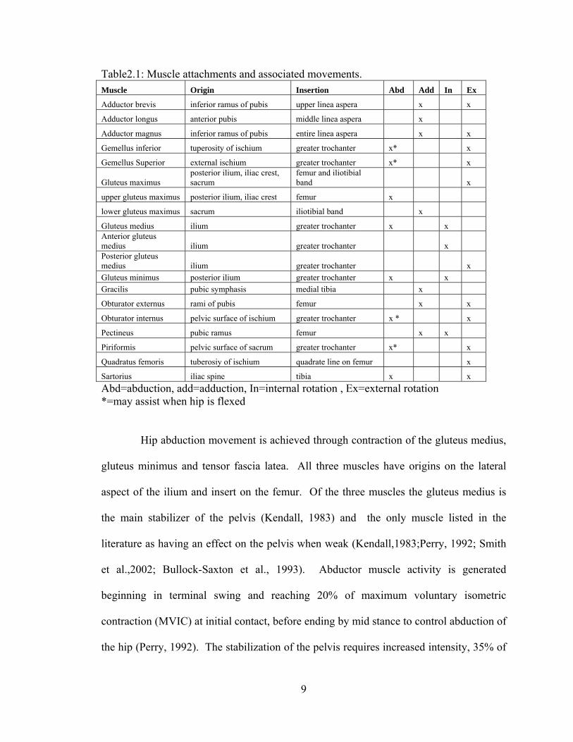
9
Table2.1: Muscle attachments and associated movements. Muscle Origin Insertion Abd Add In Ex
Adductor brevis inferior ramus of pubis upper linea aspera x x
Adductor longus anterior pubis middle linea aspera x
Adductor magnus inferior ramus of pubis entire linea aspera x x
Gemellus inferior tuperosity of ischium greater trochanter x* x
Gemellus Superior external ischium greater trochanter x* x
Gluteus maximus posterior ilium, iliac crest, sacrum
femur and iliotibial band x
upper gluteus maximus posterior ilium, iliac crest femur x
lower gluteus maximus sacrum iliotibial band x
Gluteus medius ilium greater trochanter x x Anterior gluteus medius ilium greater trochanter x Posterior gluteus medius ilium greater trochanter x
Gluteus minimus posterior ilium greater trochanter x x
Gracilis pubic symphasis medial tibia x
Obturator externus rami of pubis femur x x
Obturator internus pelvic surface of ischium greater trochanter x * x
Pectineus pubic ramus femur x x
Piriformis pelvic surface of sacrum greater trochanter x* x
Quadratus femoris tuberosiy of ischium quadrate line on femur x
Sartorius iliac spine tibia x x
Abd=abduction, add=adduction, In=internal rotation , Ex=external rotation *=may assist when hip is flexed
Hip abduction movement is achieved through contraction of the gluteus medius,
gluteus minimus and tensor fascia latea. All three muscles have origins on the lateral
aspect of the ilium and insert on the femur. Of the three muscles the gluteus medius is
the main stabilizer of the pelvis (Kendall, 1983) and the only muscle listed in the
literature as having an effect on the pelvis when weak (Kendall,1983;Perry, 1992; Smith
et al.,2002; Bullock-Saxton et al., 1993). Abductor muscle activity is generated
beginning in terminal swing and reaching 20% of maximum voluntary isometric
contraction (MVIC) at initial contact, before ending by mid stance to control abduction of
the hip (Perry, 1992). The stabilization of the pelvis requires increased intensity, 35% of

10
MVIC, during loading response and single leg support during swing. The amount of
muscle activity generated depends on the speed of the walk and the high mechanical
demand (Perry, 1992).
Hip adduction is achieved with five muscles that have origins on the ramus of
pubis or symphysis pubis and insert on the medial aspect of the femur and tibia. The
pectineus, adductor magnus, gracilis, adductor brevis and adductor longus are historically
weaker than the hip abductor muscles and also assist in hip flexion, and knee medial
rotation (Kendall, 1986, Hall, 2002). Each muscle assists in maintaining hip stability
during different stages of the gait cycle. For instance the lower gluteus maximus and
adductor magnus provide stability at the hip during loading response while the gluteus
maximus and adductor magnus increase their activity to 30% MVIC during early loading.
The adductor muscles as a whole are vital in pre swing to decelerate the passive
abduction of the hip caused by weight transfer to the other foot and the gracilis assists the
passive momentum of the hip during initial swing (Perry, 1992).
Internal or medial hip rotation is controlled by the same muscles as hip abduction
but with the tensor fascia latea and gluteus minimus as the main rotators with assistance
from the anterior fibers of the gluteus medius (Kendall, 1983, Hall, 2005). The internal
rotators are activated during loading response as a result of subtalar joint reaction to heel
loading and rotation of ipsilateral pelvis from the trailing limb (Perry, 124).
The external or lateral rotators of the hip are made up of six to nine muscles with origins
on the pelvis and insertions on the greater trochanter of the femur. Kendall lists the
piriformis, quadratus femoris, obturotor internus, obturator externus, gemellus superior,
and gemullus inferior as external rotators while Hall, 2005 included the gluteus maximus,

11
adductor magnus, and the adductor brevis (Hall, 2005). The external rotators are the
shortest and weakest muscle group of the hip.
Pattern and variation of movement of the pelvis and hip during gait
For the purpose of this paper, the focus will be on the kinematics of the frontal
plane of the pelvis and hip during walking. Although the total excursions of these
movements are small during walking, they are no less important in maintaining an
efficient gait pattern and control of the trunk. At initial contact, the pelvis is level but
quickly moves into the loading response where the pelvis looses the support of the
contralateral leg, causing the pelvis of the supported limb to raise to counter balance the
contralateral pelvis. This pattern continues throughout the stance phase. During pre-
swing the unloading of the limb results in an ipsilateral pelvic drop due to the loss of
support of the limb (Perry, 1992). The total excursion of the pelvis or the sum of the
degrees of the rise and fall of the pelvis is that is reported most in the literature when
talking about pelvic obliquity. The sacroiliac joint initiates pelvic movement when the
trunk mass is eccentric to the center of the supporting hip joint and is stimulated by the
height of the center of gravity and the change in momentum induced by foot initial
contact (Perry, 1992). The movement of the hip follows the pelvis in the frontal plane so
that when one side of the pelvis is high the hip is adducted in relation to the pelvis
(Kendall, 170).
Only a hand full of studies has researched pelvic obliquity in a healthy population.
The majority of studies that investigate pelvic obliquity involve subjects with lower
extremity amputations, arthritis in the hip, stroke and cerebral palsy. Our knowledge of
pelvic obliquity in a healthy population has come from three main authors (Blanke &

12
Hageman, 1989; Hageman & Blanke, 1986; and Smith et al., 2002). In 1986, Hageman
and Blanke reported the difference in pelvic obliquity between young and elderly women.
Thirteen 20-35 year olds and thirteen 60+ aged women demonstrated a significant
difference pelvic obliquity. The younger women demonstrated 9.86 ± 2.38º of pelvic
obliquity while the older group only demonstrated 6.77± 2.05º of pelvic obliquity
(Hageman, 1986). This was followed up in 1989 by Blanke and Hageman studying the
pelvic obliquity of twenty four males. Twelve men aged 20-32 were reported to have
7.42± 2.11º of pelvic obliquity while the second group of twelve men aged 60-74 had
6.08± 2.5º pelvic obliquity (Blank & Hageman, 1989). While the first two studies
focused on age differences, in 2002, Smith et al. examined the affect of gender on pelvic
obliquity. Four groups of thirty subjects made up of younger and older men and women
of similar ages as the prior two studies were used. Comparisons were made between
younger men and women, older men and women, and all men and women from both age
groups. All women had significantly greater pelvic obliquity then all men with 9.4± 3.5º
for all women and 7.4± 3.4º for men at a p=.0024. Young women were close to a
significant difference in pelvic obliquity compared to young men with 11.1±3.2º and
9.6±3.2º at p=.07, respectively. Older women demonstrated significantly greater pelvic
obliquity then older men at 7.7±3.1º compared to 5.3±1.9º with p=.0005 (Smith et al.,
2002). In summary the three papers have established that younger women have greater
pelvic obliquity then older women and in general women have more pelvic obliquity then
men.
13
Kinematics of Stair Descent Task
Stair climbing is a common activity of daily living, especially in the college
population. Sagittal plane kinematics and kinetics have been well documented in the
elderly and those with knee injuries including patellofemoral pain syndrome (PFPS) and
ACL tears. The scarce amount of research that has been conducted on stair kinematics
and kinetics in the frontal plane has been primarily confined to the knee and ankle in a
PFPS population. The results reported in these investigations have been inconsistent.
Similar to level walking, stair descent is made up of stance and swing phase. The Stance
phase during stair descent makes up 68±2% of the gait cycle (GC) comprised of three
subphases; weight acceptance (0-14% of GC), forward continuance (14-34% of GC) and
controlled lowering (34-68% of GC). The swing phase makes up 32 ±2% of the gait
cycle with two subphases of leg pull through (68-84% of GC) and foot placement (84-
100% of GC) (Zachazewski et al., 1993). Brindle et al. reported 5.1± 3.0ºof pelvic
obliquity during stair descent in subjects with symptomatic PFPS and 4.4± 4.7º in
asymptomatic PFPS. The study used twelve females and four males in the PFP group
and seven females and five males in the control group. Measurements were collapsed
across genders and although not reported, it is assumed that these values are of pelvic
drop as compared to pelvic obliquity. Mian et al., 2005, reported 6.6 ± 2.5 degrees pelvic
obliquity ROM and 10.1 ± 2.2 degrees hip ab/adduction ROM during stair descent in
healthy young men. It is assumed that the same relationship between pelvic obliquity and
gender during gait would exist during stair ascent and descent as well.

14
Muscle recruitment patterns during stair descent
The gluteus medius is important in the progression of the gait cycle during the
stair descent task because the muscle facilitates the shift of weight to the support limb
(McFadyen & Winter, 1988). The gluteus medius activity begins at terminal swing and
continues into stance at 20% MVIC while the upper portion of the gluteus maximus is
activated at the same time with 15% MVIC (Lyons et al., 1983; Mcfadyen & Winter,
1987). Hollman et al recently found similar results when testing 20 healthy women
during stair descent. Hollman et al reported the %MVIC of the gluteus medius and
maximus at 21.9± 13.1 % and 9.2±4.1 %, respectively. The study also tested and
correlated hip adduction and knee valgus angles, and abductor and external rotator
strength. There were significant correlations for gluteus maximus %MVIC and knee
valgus, and hip abductor strength and knee valgus. The study concluded that the gluteus
maximus can eccentrically control femoral internal rotation and adduction during
unilateral tasks and that gluteus medius strength may contribute to femoral internal
rotation during single-limb weight bearing (Hollman et al., 2009).
Strength Testing
Strength testing is important in clinical and research setting to understanding
more about the influence of the muscle on the accompanied movement. Isokinetic
strength testing is considered by most the gold standard for strength measurements (Lund
et al., 2005) and can be performed in concentric or eccentric contractions. Isokinetic
measures are popular due to the ability to measure muscle performance including peak
torque and endurance during a specified limb movement (Rothstein et al, 1987).
15
Mechanical isokinetic dynamometer’s such as Cybex and Biodex brands are considered
the gold standard by the industry. They are more reliable then a hand-held dynamometer
because the test does not rely on tester strength or tester response to the subject.
Furthermore, the Biodex has been found to be highly reliable with no learning effect for
the subject with proper instruction (Lund et al., 2005). The Biodex and Cybex
dynamometers also have an advantage to clinicians and researchers by supplying a wide
variety of parameters relating to force, power, work and arithmetic calculations (Sapega,
1990).
One previous study has reported abduction strength testing in a supine position,
but the author also built a body stabilization frame to assist with stability (Cahalan et al.,
1988). Due to the isokinetic testing, the recommendations of the manufacturer, and the
similarity to the other dynamic activities being tested, the subjects in this study were
standing during the hip adduction and abduction testing. While no study has directly
compared standing and side-lying positions, one study found no difference in strength
measures between side-lying and side-lying with restraints of the pelvis (Laheru et al.,
2007). This leads the author to believe that testing in a standing position can increase
pelvic instability, but will not significantly alter strength testing results.
Although strength testing with dynamometers has been popular since the 1970’s the
development of population specific data with sensitivity to muscle group, age, gender,
and activity level is a formidable task that has not been competed to date (Sapega, 1990).
Furthermore, small muscles like hip internal and external rotators have long been
overlooked for larger muscle groups. Other confounding factors of comparing strength
testing between studies include subject population, equipment used, and specification of
16
test including type of contraction, speed, and position of the body during testing.
Cahalan et al reported strength measurements in closest relation to the current study
testing isokinetic ab/adduction and hip internal and external rotation in women. The
abduction and adduction torque at a speed of 90 degrees/sec were 54 ± 20 nm and 6 2±
32 nm, respectively. Hip internal and external rotation torque at 30 degrees/sec were 47
± 13 nm and 43± 13 nm, respectively. Cahalan also showed that strength will decrease as
speed increases.
It has been established that muscle imbalances exist in a normal population. At
the same time, “in non-athletes the assumption that symmetry is the norm for muscular
performance is reasonable for all major muscle groups in the lower extremities” (Sapega,
1990). Caution must be taken when describing a muscle difference as an imbalance
because a difference of 10 percent qualifies by definition as a real difference in the
capacity of performance between two extremities, but is not necessarily considered an
abnormal difference. Since the degree of imbalance that impairs functional performance
and or predisposes the area to injury has not been clearly established, the literature
remains vague as to categorizing muscle imbalances (Sapega, 1990). The following
guidelines have been established to name muscle imbalances in a healthy population; less
then ten percent is considered normal; ten to twenty percent difference is considered a
possible abnormality; and differences greater than 20 percent are probably abnormal
(Sapega, 1990).
Pelvic stabilization and gluteus medius weakness
Kendall described two stages of gluteus medius weakness as marked weakness
and slight weakness. Marked weakness consists of “displacement of the body weight
17
laterally toward the side of the weak muscle in such a way that the hip joint is thrust in
the position of hip abduction in relation to the pelvis.” (Kendall, 1983). Slight weakness
is described as only a postural deviation when standing caused by a high pelvis and
adducted femur on the side with the abductor weakness. Other authors only make one
classification of abductor weakness as the point when the gluteus medius is unable to
stabilize the pelvis in single leg stance (Whittle &Levine,1999) due to an abductor
muscle strength less than grade 3+ out of 6 for manual muscle testing (Perry, 1992).
Lower Extremity injuries associated with gluteus medius weakness
In the last decade however, researchers have consistently found significant
weakness of the hip abductor and external rotator strength in subjects with PFPS (Ireland
et al., 2003;Cichanowski et al., 2007; Leetun et al., 2004; Bolgla et al., 2007, Robinson &
Nee, 2007). Niemuth et al attributed weaker abduction strength in recreational runners to
overuse lower extremity injuries such as plantar fascitis, Achilles tendonitis, tibial stress
fractures, illiotibial band syndrome and patellar tendonitis (Niemuth et al., 2005).
Although strength testing among those with PFPS has consistently found statistically
significant weaknesses in the hip abductors and external rotators, the kinematic affects of
the weakness during stair descent has not yielded consistent results. Mascal et al.
reported the changes in stair descent in two females participating in physical therapy for
PFPS. Kinematic analysis of stair descent before and after treatment revealed an
improvement in the hip rotator angle from 1.4º internal rotation to 2.6º of external
rotation. Hip adduction decreased from 8.7º to 2.3º and the contralateral pelvic drop was
reduced from 3.9º to 1.1º (Mascal et al., 2003). Bolgla et al. however compared PFPS
patients with healthy females and found similar strength measures as Mascal et al. in the

18
PFPS group but did not find any significant differences in kinematics compared to the
control group during stair descent.
Low back pain associated with gluteus medius weakness
Nadler et al. has investigated the affect of hip strength on core strength and the
development of low back pain in an athletic population. Injured females were found to
have greater differences in side to side strength then injured males (Nadler et al., 2000).
Interestingly, female athletes with weaker left abductors were significantly more likely to
develop LBP (Nadler et al., 2001) while the difference in side to side abductor strength of
the same population did not lead to the development of LBP (Nadler et al., 2002). It is
unknown if the same relationships exist in a non-athletic female population. Fifteen to
twenty percent of persistent low back pain is estimated to be caused by sacroiliac joint
dysfunction (Rathmell, 2008). The cause and therefore treatment of SI joint dysfunction
is still unknown. Nine muscles of the hips and abdomen support the lower back and
specifically the sacroiliac (SI) joint. Of this nine, three, the gluteus maximus, gluteus
medius and piriformis are also involved in movement of the hip in the frontal plane. All
nine muscles are used to create stability for effective transfer of forces across the sacrum
and pelvis and successful compression of the SI joint to prevent shearing. Several studies
have already shown significant weakness in these stabilizing muscles that could
potentially disrupt the stability of the SI joint (Harrison et al., 1997).
Conclusion
In summary, gluteus medius weakness has been associated with low back pain
and lower extremity injuries. The gluteus medius is associated with pelvic obliquity but
the association has not been directly tested. Furthermore, the complexity of the
19
musculature that stabilizes the hip makes it unlikely that the weakness of one muscle
would lead to increased pelvic obliquity. Testing the strength of hip muscle groups and
comparing them to the movement of the pelvis in a healthy population will give a better
idea of muscle interactions and the importance of the strength of different muscle groups.
CHAPTER THREE: METHODS
Based on the literature there was a lack of research about pelvic obliquity that was
gender and age specific. Secondly, muscle strength and the kinematics of the distal
segments have been studied (Bolgla et al., 2008; Hollman et al., 2009) but muscle
strength and the proximal segments have not been studied to date. Lastly, pelvic
obliquity has not been documented during activities of daily living in a specific age and
gender group. The following describes the methods used to test the hip muscle strength
and pelvic obliquity during activities of daily living in college females.
Subjects
Female subjects between the ages of 18 to 25 were recruited from flyers in the
University of Kentucky Kinesiology and Health Promotion department and the
Rehabilitation Sciences department. Testing was held in the Biodynamics Laboratory at
the University of Kentucky. Based on prior testing by Cichanowski et al., strength
differences can be detected when using thirteen subjects with an effect size of 1 and a
70% power to detect significant differences between groups with a two-sample t-test
(Cichanowski et al., 2007). Thirteen females were recruited (mean age=23±2 yrs, mean
height= 167 ± 14cm and weight 61.8 ± 16.9 kgs).
20
Procedures
All subjects read and signed an informed consent form approved by the
University of Kentucky Institutional Review Board before testing began. A full version
of the consent form is in Appendix 1. All procedures followed were within ethical
standards of the Institutional Review Board at the University of Kentucky. A short
questionnaire, Appendix 2, was completed by the subject as to medical history of lower
extremity injuries to confirm eligibility.
Subjects underwent familiarization of isokinetic strength testing on the Biodex
System 3 dynamometer (Biodex Medical Systems, Shirley, NY). Hip abduction and
adduction were tested in a standing position, facing the Biodex lever arm. The pad was
positioned on the lower third of the thigh on the medial and lateral aspects of the leg

21
Figure 3.1: Setup of subject on Biodex for abduction and adduction strength testing
The range of motion was set by setting hip abduction at the point prior to pelvic
rise and placing feet together for adduction. Subjects were instructed to perform the full
range of motion as fast and as hard as possible for three repetitions of isokinetic
concentric contractions at 120 º/sec. Subjects went through the same procedure on both
legs and performed three complete repetitions on each leg. Next the subjects sat in the
Biodex bench and straps were applied across each shoulder and over the uninvolved leg.
The lever arm with pad was changed and repositioned on the proximal third of the shank.
22
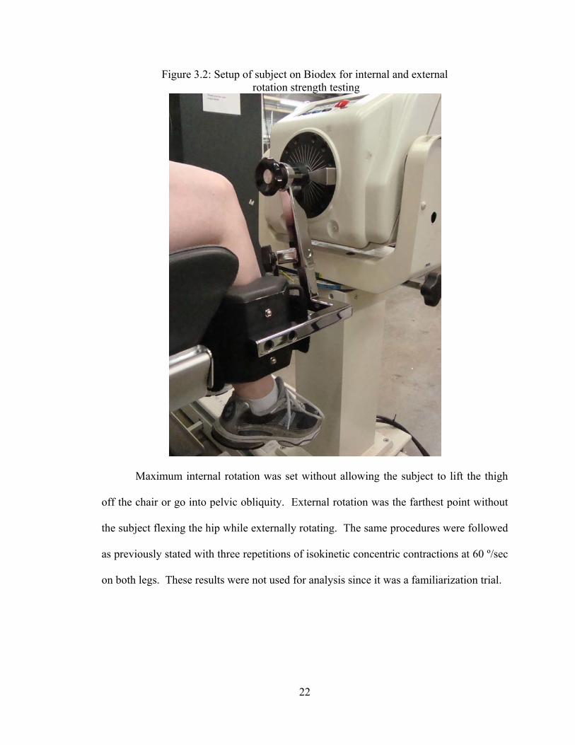
Figure 3.2: Setup of subject on Biodex for internal and external rotation strength testing
Maximum internal rotation was set without allowing the subject to lift the thigh
off the chair or go into pelvic obliquity. External rotation was the farthest point without
the subject flexing the hip while externally rotating. The same procedures were followed
as previously stated with three repetitions of isokinetic concentric contractions at 60 º/sec
on both legs. These results were not used for analysis since it was a familiarization trial.
23
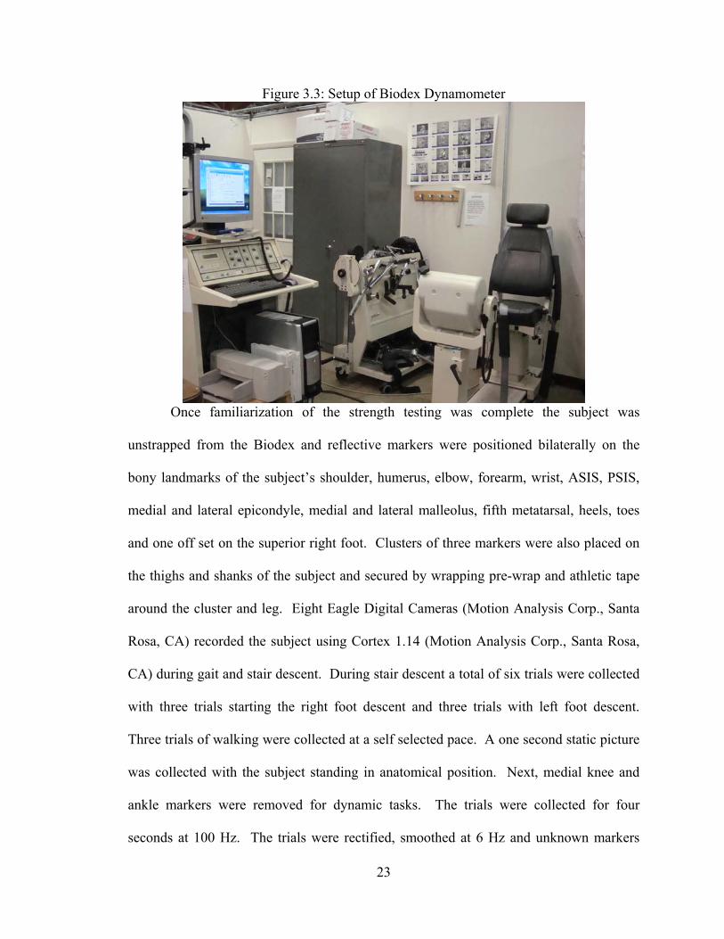
Figure 3.3: Setup of Biodex Dynamometer
Once familiarization of the strength testing was complete the subject was
unstrapped from the Biodex and reflective markers were positioned bilaterally on the
bony landmarks of the subject’s shoulder, humerus, elbow, forearm, wrist, ASIS, PSIS,
medial and lateral epicondyle, medial and lateral malleolus, fifth metatarsal, heels, toes
and one off set on the superior right foot. Clusters of three markers were also placed on
the thighs and shanks of the subject and secured by wrapping pre-wrap and athletic tape
around the cluster and leg. Eight Eagle Digital Cameras (Motion Analysis Corp., Santa
Rosa, CA) recorded the subject using Cortex 1.14 (Motion Analysis Corp., Santa Rosa,
CA) during gait and stair descent. During stair descent a total of six trials were collected
with three trials starting the right foot descent and three trials with left foot descent.
Three trials of walking were collected at a self selected pace. A one second static picture
was collected with the subject standing in anatomical position. Next, medial knee and
ankle markers were removed for dynamic tasks. The trials were collected for four
seconds at 100 Hz. The trials were rectified, smoothed at 6 Hz and unknown markers

24
were deleted. Trials were loaded into Visual 3-D v. 4.00.17 (C-Motion, Germantown,
MD) post processing software for further analysis. Stair trials were analyzed from toe off
at the highest step to heel strike of the ipsilateral leg on the ground level. Walking trials
were analyzed by full strides, defined as heel strike to ipsilateral heel strike. The three
trials of each leg were averaged and graphed.
Figure 3.4: Subject performing stair descent task.
25
Once finished with the stair descent and gait trials, all markers were removed and
subjects were instructed to sit on the Biodex chair and were again strapped to the chair
for safety, and were aligned with the lever arm for hip internal and external rotation
starting on the right leg. Range of motion and all instructions were reviewed with the
subject as previously stated before the test was administered.
Once the subject had performed three repetitions of internal and external rotation
through the full range of motion on the right leg, the leg stabilization strap was moved to
the right leg and the same procedure was administered on the left leg. Once the subject
had completed the internal and external hip rotation strength, the subject was unstraped
from the chair and the computer and dynamometer were set up for hip abduction and
adduction strength testing. Once set up, placement of the subject and range of motion
was complete as previously stated, the subject performed to test on the right leg and then
the left leg. Patients were given a print out with two graphs, one with right leg and left
leg during abduction and adduction and one with right and left leg during internal and
external rotation. For analysis, the numerical data from the Biodex was exported into
Excel 2003 (Farmington, CT). The peak force produced for each movement was found
for all three repetitions and averaged together.
Statistical Analysis
SPSS version15.0 (Chicago, IL) was used to perform all statistical analysis.
Descriptive statistics were run on all variables including mean, and standard deviation.
Strength measures were calculated by averaging the peak torque of three repetitions for
abduction, adduction, internal rotation and external rotation. Joint angles that were
analyzed were expressed as the total excursion of the pelvis and hip in the frontal plane in
26
degrees. Pelvic obliquity during stair descent (POS), hip ab/adduction during stair
descent (HAS), pelvic obliquity during walking (POW) and hip ab/adduction during
walking (HAW) were compared to the strength measures. The percent difference between
mean peak torque for abduction and adduction (%Δ ab/add) muscle groups were
calculated within each leg. The imbalance was calculated as the difference between
abduction torque and adduction torque, divided by abduction torque and multiplied by
100. Percent difference between mean peak torque for internal and external rotation (%Δ
in/ex) strength were calculated within each leg. The imbalance was calculated as the
difference between internal and external rotation torque that was divided by the internal
rotation and multiplied by 100. A paired sample t-test was run to determine significant
differences between the right and left sides. The mean value of each strength
measurement was used to divide all strength tests into two groups. The group of subjects
that tested above the mean was named the “stronger” group while the subjects that tested
below the mean fell into the category of “weaker”. An independent t-test using Levene’s
test for equality of variance was used to determine differences between the weaker and
stronger group in the amount of pelvic and hip motion seen in the subjects during
walking and stair descent.
CHAPTER FOUR: RESULTS
The purpose of the study was to examine the influence of hip muscle strength on
the range of motion of the hip and pelvis in the frontal plane during a walking task and
stair descent task. This was achieved by testing the strength of the hip abductors,
adductors, internal rotators and external rotators and tracking the pelvic obliquity and hip
27
ab/adduction ROM during self selected pace walking and self selected pace stair descent
walking. This chapter describes the results of the strength testing and kinematic analysis
of the pelvis.
Descriptive Statistics
The descriptive statistics for strength testing, imbalances, and range of motion of
the pelvis and hip of the combined right and left legs for all subjects are shown in Table
4.1. The minimum value, maximum value, mean and standard deviation and paired
differences t-test between the right and left leg are displayed.
Table4.1: Descriptive statistics of combined right and left limbs with paired differences p-value for the right and left limbs
Minimum Maximum Mean& Std. Deviation Paired Differences
Abductors(Nm) 10.83 70.7 37.89± 14.89 0.941 Adductors(Nm) 10.33 87.37 42.28± 20.45 0.69 internal rotators(Nm) 16.2 66.2 38.14± 12.72 0.963 external rotators(Nm) 19.83 37.5 29.35± 5.44 0.263 %Δ ab/add(%) 1.83 59.21 27.25±16.79 0.855 %Δ in/ex(%) 1.6 55.59 21.58±14.56 0.943 POS(º) 6.76 15.46 10.06± 2.24 0.596 HAS(º) 6.84 22.9 14.27± 3.61 0.656 POW(º) 7.11 17.96 11.47± 2.84 0.881 HAW(º) 6.67 22 15.03± 3.65 0.357
POS=pelvic obliquity during stair descent HAS=hip angle in the frontal plane during stair descent POW=pelvic obliquity during walking HAW=hip angle in the frontal plane during walking %Δab/add=percent difference between abductors and adductors in both right and left legs %Δin/ex=percent difference between internal and external rotators in both right and left legs
The hip adductors were the strongest muscle group tested and had the greatest
variability at 42.28± 20.45 Nm. The external rotators were the weakest muscle group
tested at 29.35± 5.44 Nm. Abductors and internal rotators were almost identical in
strength measurement. There was no difference in the ROM of the pelvis and hip
between stair descent and level walking. There was also no significant difference (p=.05)
28
between the measurements of the right and left leg on any test parameter. Since the right
and left side variables are not significantly different the two sides were combined to
lower the risk of a type I or type II error due to the smaller population size.
Independent T-tests
Using a t-test to compare means and Levene’s test for equality of variance, the
mean value of the muscle group strength measures and imbalances (percent difference of
a pair of muscles on the same leg) was used to determine the affect of strength on the
range of motion of the pelvis and hip. These results are reported in Table 4.2.
Table 4.2: Results from 2-tail independent t-test of strength testing
and affect on pelvic and hip ROM with p-values
POS p-value HAS p-value POW p-value HAW p-value
abductors> 40 nm 11.22±2.22 0.013*
15.33±3.90 0.175
11.41±2.10 0.923
14.25±3.59 0.324
abductors< 40nm 9.06±1.76 13.35±3.21 11.52±3.42 15.70±3.69
adductors> 40nm 10.98±2.54 0.033*
14.81±4.42 0.454
11.83±2.56 0.526
14.43±3.93 0.413
adductors< 40nm 9.14±1.46 13.72±2.65 11.11±3.15 15.63±3.37
internal> 40nm 11.46±2.56 0.043*
15.48±4.60 0.293
11.63±1.95 0.814
13.77±3.86 0.207
internal< 40nm 9.31±1.68 13.62±2.92 11.38±3.26 15.69±3.46
external> 30 nm 10.71±2.29 0.105
14.64±3.12 0.587
10.30±1.62 0.02*
14.01±2.39 0.129
external< 30 nm 9.29±2.00 13.82±4.21 12.83±3.38 16.21±4.53
%Δ ab/add> 30% 10.25±1.87 0.671
14.90±3.56 0.376
12.15±2.77 0.229
15.57±3.88 0.457
%Δ ab/add< 30% 9.86±2.61 13.62±3.69 10.79±2.83 14.48±3.45
%Δrotators >20% 11.12±2.22 0.012*
15.07±3.96 0.261
11.60±2.22 0.819
14.75±3.87 0.708
%Δrotators <20% 8.99±1.73 13.45±3.17 11.34±3.43 15.30±3.53 %Δab/add=percent difference between abductors and adductors %Δrotators=percent difference between internal and external rotators
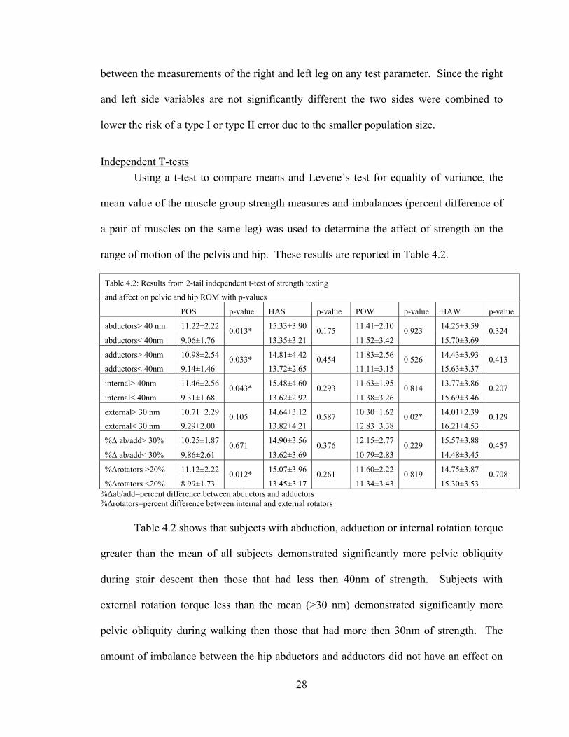
Table 4.2 shows that subjects with abduction, adduction or internal rotation torque
greater than the mean of all subjects demonstrated significantly more pelvic obliquity
during stair descent then those that had less then 40nm of strength. Subjects with
external rotation torque less than the mean (>30 nm) demonstrated significantly more
pelvic obliquity during walking then those that had more then 30nm of strength. The
amount of imbalance between the hip abductors and adductors did not have an effect on

29
the pelvis or hip during stair descent or walking while the greater imbalance found in the
rotators lead to an increase in pelvic obliquity during stair descent.
Muscle imbalances were calculated as a percent difference between agonist and
antagonist muscle groups within each leg for the left and the right legs. The amount of
imbalance between the abductor and adductor group did not significantly change the
ROM of the pelvis during stair descent or walking. On the other hand, the more
imbalances that were seen between the internal and external rotators, the more motion
that was seen in the pelvis during stair descent.
Representative Kinematics during Stair Descent
Based on the peak mean torque of the hip abductors, a representative weaker and stronger
subject was randomly selected and graphed to demonstrate the kinematic affects of hip
abductor strength on the pattern of movement. Figures 4.1 to 4.4 are graphed from right
heel strike to right heel strike with the stance phase and swing phase labeled on each
graph. In Figure 4.1 the mean and standard deviation pelvic obliquity pattern of three
strides on the right side of the two representative subjects during stair descent is graphed
from right heel strike on the stair to right heel strike on the ground.
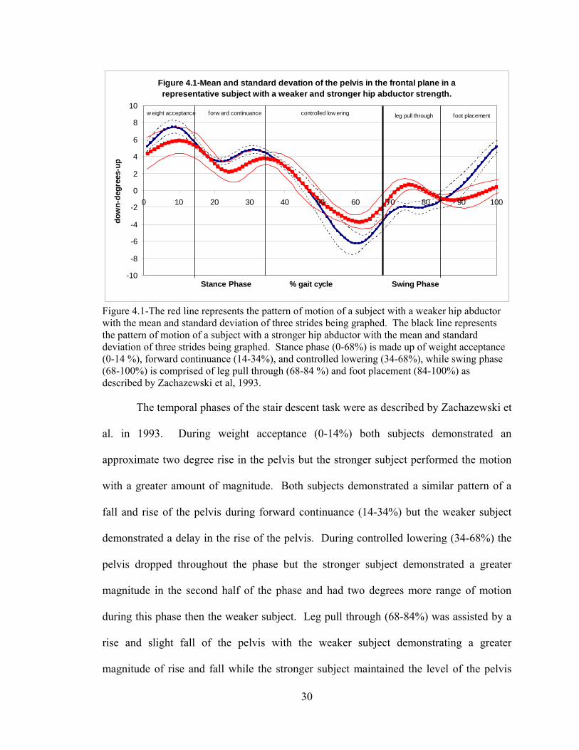
30
Figure 4.1-Mean and standard devation of the pelvis in the frontal plane in a representative subject with a weaker and stronger hip abductor strength.
-10
-8
-6
-4
-2
0
2
4
6
8
10
0 10 20 30 40 50 60 70 80 90 100
Stance Phase % gait cycle Swing Phase
do
wn
-deg
rees
-up
w eight acceptance forw ard continuance controlled low ering leg pull through foot placement
Figure 4.1-The red line represents the pattern of motion of a subject with a weaker hip abductor with the mean and standard deviation of three strides being graphed. The black line represents the pattern of motion of a subject with a stronger hip abductor with the mean and standard deviation of three strides being graphed. Stance phase (0-68%) is made up of weight acceptance (0-14 %), forward continuance (14-34%), and controlled lowering (34-68%), while swing phase (68-100%) is comprised of leg pull through (68-84 %) and foot placement (84-100%) as described by Zachazewski et al, 1993. The temporal phases of the stair descent task were as described by Zachazewski et
al. in 1993. During weight acceptance (0-14%) both subjects demonstrated an
approximate two degree rise in the pelvis but the stronger subject performed the motion
with a greater amount of magnitude. Both subjects demonstrated a similar pattern of a
fall and rise of the pelvis during forward continuance (14-34%) but the weaker subject
demonstrated a delay in the rise of the pelvis. During controlled lowering (34-68%) the
pelvis dropped throughout the phase but the stronger subject demonstrated a greater
magnitude in the second half of the phase and had two degrees more range of motion
during this phase then the weaker subject. Leg pull through (68-84%) was assisted by a
rise and slight fall of the pelvis with the weaker subject demonstrating a greater
magnitude of rise and fall while the stronger subject maintained the level of the pelvis
31
through most of the phase. Lastly, the foot placement phase (84-100%) demonstrated a
steady rise of approximately six degrees in the pelvis of the stronger subject while the
weaker subjected only demonstrated an approximate one degree rise and did not reach the
same degree of pelvic rise that was seen at the beginning of the gait cycle.
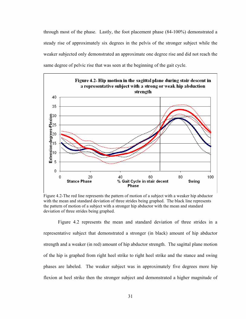
Figure 4.2-The red line represents the pattern of motion of a subject with a weaker hip abductor with the mean and standard deviation of three strides being graphed. The black line represents the pattern of motion of a subject with a stronger hip abductor with the mean and standard deviation of three strides being graphed. Figure 4.2 represents the mean and standard deviation of three strides in a
representative subject that demonstrated a stronger (in black) amount of hip abductor
strength and a weaker (in red) amount of hip abductor strength. The sagittal plane motion
of the hip is graphed from right heel strike to right heel strike and the stance and swing
phases are labeled. The weaker subject was in approximately five degrees more hip
flexion at heel strike then the stronger subject and demonstrated a higher magnitude of
32
extension till 20 % of the gait cycle. From 20 to 70% of the gait cycle the two subjects
demonstrated similar magnitude and range of motion of the hip. During the swing phase,
the weaker subject maintained approximately five degrees greater hip flexion throughout
the swing phase then the stronger subject.
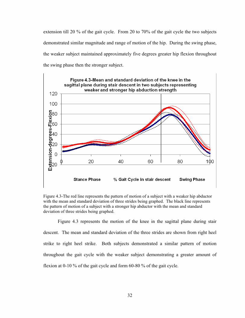
Figure 4.3-The red line represents the pattern of motion of a subject with a weaker hip abductor with the mean and standard deviation of three strides being graphed. The black line represents the pattern of motion of a subject with a stronger hip abductor with the mean and standard deviation of three strides being graphed. Figure 4.3 represents the motion of the knee in the sagittal plane during stair
descent. The mean and standard deviation of the three strides are shown from right heel
strike to right heel strike. Both subjects demonstrated a similar pattern of motion
throughout the gait cycle with the weaker subject demonstrating a greater amount of
flexion at 0-10 % of the gait cycle and form 60-80 % of the gait cycle.
33
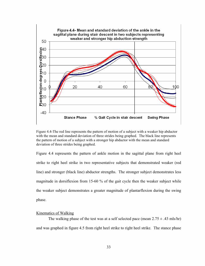
Figure 4.4-The red line represents the pattern of motion of a subject with a weaker hip abductor with the mean and standard deviation of three strides being graphed. The black line represents the pattern of motion of a subject with a stronger hip abductor with the mean and standard deviation of three strides being graphed. Figure 4.4 represents the pattern of ankle motion in the sagittal plane from right heel
strike to right heel strike in two representative subjects that demonstrated weaker (red
line) and stronger (black line) abductor strengths. The stronger subject demonstrates less
magnitude in dorsiflexion from 15-60 % of the gait cycle then the weaker subject while
the weaker subject demonstrates a greater magnitude of plantarflexion during the swing
phase.
Kinematics of Walking
The walking phase of the test was at a self selected pace (mean 2.75 ± .43 mls/hr)
and was graphed in figure 4.5 from right heel strike to right heel strike. The stance phase

34
and swing phase of the gait cycle are labeled with the mean and standard deviation of
pelvic obliquity in all subjects.
During the first half of the gait cycle the hip is going into adduction and the pelvis is moving in an upward direction. They descend and level off between 40 to 50 % of the gait cycle before the pelvis continues to drop and the hip goes into abduction till 70 % of gait cycle. From 70 to 100% of the gait cycle the pelvis and hip rise and adduct to neutral.
Conclusion
Over all hip abduction, adduction and internal rotation significantly influence the amount
of pelvic obliquity seen during stair descent. A greater amount of pelvic obliquity was
seen in patients that were considered to have stronger hip muscle torque production.
Differences in sagittal plane motions were seen between a representative weaker and
stronger hip abductor subject. Hip abduction weakness was not a contributing factor in
increased pelvic obliquity during gait, but subjects with weaker external rotators did
experience an increase in pelvic obliquity during walking.

35
CHAPTER FIVE: DISCUSSION
The purpose of this paper was to investigate the relationship between hip
musculature strength and pelvic obliquity in females during walking and stair descent.
To accomplish this goal, thirteen females were recruited to participate in the study. The
average peak torque of four muscle groups were compared to the amount of pelvic
obliquity the subjects demonstrated during stair descent self selected pace walking. The
following chapter will provide a discussion of the finding of the current study in
comparison to current literature.
Strength testing
Compared to the literature, our strength measures were lower than expected.
Cahalan et al. tested 18 females aged 20 to 40 using isokinetic concentric motion at 30º,
90º, 150º, and 210 º per second. As the testing procedures in this investigation were
similar to Cahalan except for the type of contraction, it was expected our hip abduction
and adduction strength measures, measured at 120 º per sec, to be less than Cahalan’s
measures at 90 º per second but greater that at 150 º per second. The measures that we
recorded were similar to the measures that Cahalan reported at 210 º per second. Internal
and external hip rotators were closer to expected values due to the decrease in range of
hip strength that the muscle can produce. The lower values of strength can be attributed
to the smaller age range of predominately advanced degree seeking college students.
Furthermore, every dynamometer measures differently and the fact that our standard
deviations were smaller then Cahalan et al, shows that the values are still relevant and
accurate.

36
Muscle Imbalances
Muscle imbalances between agonist and antagonist muscles have not been
reported in a healthy population. Several studies have reported significant imbalances of
the same muscle between legs in a healthy population (Jacobs et al., 2005) and an injured
athletic population (Nadler et al., 2002). Our study did not support either of these
findings since the current study did not find any significant differences between muscle
groups on the right and left sides. Imbalances were calculated between abduction and
adduction and internal and external rotation of the same leg. Both sets of groups
exhibited a mean that was greater than 20 percent which is considered “probably
abnormal” for a normal population according to Sapega, 1990. Since none of the subjects
had ever experience a lower extremity injury it is likely that injuries are caused by more
than one mechanism besides just a muscle imbalance.
Kinematics of the pelvis and hip
The results of this investigation for the range of motion of the pelvis during
walking(11.47± 2.84º ) were higher then Hageman et al., who reported 9.86 ± 2.38º of
pelvic obliquity in thirteen females aged 20 to 35, but were in agreement with Smith et al
who reported 30 females aged 22 to 40 with a pelvic obliquity of 11.1 ± 3.2º. Our hip
ab/adduction ROM is in agreement with Perry who stated that during walking the hip will
go through 15 degrees of ab/adduction.
Although no study has directly compared pelvic obliquity during stair descent and
level walking, it was hypothesized in this study that stair descent would require more
eccentric force, and therefore produces higher torque within the muscle and would
increase the amount of pelvic obliquity. One study of young men, aged 24-30 years
37
reported pelvic obliquity during stair descent as 6.6±2.5º (Mian et al., 2007), while
Blanke et al reported pelvic obliquity at 7.42±2.11º during walking in men of the same
age range. This study was in agreement with the later two studies and found no
difference in pelvic obliquity between level walking and stair descent.
Relationship between muscle strength and pelvic kinematics
Based on the literature, the gluteus medius is the main abductor muscle of the hip
and the main stabilizer of the pelvis (Kendall, 1983; Perry, 1992). Weakness of the
gluteus medius is thought to cause an increase in pelvic obliquity (Perry, 1992). This
theory leads to the hypothesis that weaker musculature would equate to more range of
motion in stairs and walking. However, in this investigation this theory was not found to
be the case for walking or stair descent. The only hip musculature that significantly
increased pelvic obliquity when the muscle was weaker was the external rotators during
the walking task. Although this study cannot distinguish which of the external rotator
muscles has the most influence on pelvic movement, it can be theorized which muscles
have the most ability to influence the pelvis. Given the size, location, and muscle
activation patterns, it is most likely that the upper portions of the gluteus maximus have
the most influence of all the external rotators. External hip rotation strength was testing
in a sitting position which would increase the use of the gluteus maximus as a
simultaneous external rotator and abductor due to the hips being flexed. Furthermore,
several books and articles have commented on the similar activation timing and relative
intensity of the gluteus maximus and gluteus medius during gait (Perry, 1992; Bullock-
Saxton et al., 1993; Lyons et al., 1983). Also, PFPS patients have been shown to have
both abductor and external rotator weakness when compared to normal subjects (Bolgla

38
et al., 2008; Ireland et al., 2003). This leads to the conclusion that hip external rotators
may play as much or more of a role in pelvis stability then hip abductors in young
females during self selected pace walking. More research is needed to determine the
exact relationship between hip abductors, external rotators and pelvic obliquity during
gait.
Stair Descent
Walking down stairs is an activity of daily living and is achieved through
eccentric contractions that control the gravitational forces acting on the body (McFadyen
and Winters, 1987). In the current study, the hip musculature had an opposite affect on
the kinematics of the pelvis as what was hypothesized during stair descent. The stronger
the hip abductors, adductors and internal rotators were, the more range of motion the
pelvis demonstrated. Stair descent is a dynamic process that requires more balance
compensation based on a greater magnitude of separation between the center of mass and
center of pressure (Zachazewski et al., 1993). Theoretically, if a muscle was stronger,
then it would be able to lengthen more during an eccentric contraction, allowing the joint
that it was inserted into to move through a greater range of motion during the contraction.
Furthermore, Smith et al., 2002 stated that greater pelvic obliquity reduces vertical center
of mass displacement and conserves energy. It may be that during the greater demands of
stair descent that only the stronger women are able to take advantage of decreasing the
amount of movement of their center of mass.
Based on the analysis of the sagittal plane kinematics of two subjects representing
the weaker and stronger abductor group, subjects with greater range of motion in the
pelvis may compensate by a decreased range of motion in the sagittal plane in the hip,
39
knee, or ankle. On the other hand, subjects with weaker hip abduction strength that
demonstrated less pelvic obliquity during stairs may compensate by increasing the
sagittal plane range of motion of the hip, knee, or ankle during stair descent. More
research is needed to determine if a consistent pattern of joint compensation is used to
negotiate stair descent with hip abductor weakness.
Conclusion
Up to this point the influence of muscle groups on pelvic obliquity has not been
researched. Surface EMG cannot reach inferior muscles and can not distinguish while
fine wire EMG cannot accurately test the small inferior muscles of the pelvis such as the
external rotators. The pelvis absorbs forces from the lower and upper extremity and more
and more studies are reporting the influence of hip musculature weakness on the
development of lower extremity and low back injuries. For this reason, it is important to
have a better understanding of the interactions of the hip musculature on pelvic
movement during activities of daily living. Our findings indicate that the external
rotators may play an important role in pelvic obliquity in females during gait.
Furthermore, this study shows that the pelvis may have different compensatory actions
during stair descent compared to gait that needs to be researched further.
CHAPTER SIX: SUMMARY AND RECOMMENDATIONS
Summary
To date, the movements of the hip and pelvis have largely been over looked,
especially in the frontal plane. Weakness in the muscles that control pelvic obliquity
have been associated with low back pain (Bullock-Saxton et al., 1993; Nadler et al.,
2002; Hollman et al., 2009) and over use injuries in the lower extremities (Bolgla et al.,
40
2008; Cichanowski et al., 2007; Ireland et al., 2003; Leetun et al., 2004; Niemuth et al.,
2005), but none of these studies have examined the affect of this weakness on the pelvis.
Pelvic obliquity is important in the transfer of forces from the lower extremities to the
spine, yet the study of the movement and the muscles that control this movement have
been sparsely studied. For these reasons the purpose of this study was to investigate the
influence of muscle strength on pelvic obliquity during activities of daily living in a
healthy population of female college students. This will serve as a baseline for
comparison of those with hip musculature weakness to better understand the affects on
pelvic obliquity.
To achieve this goal, healthy females from the University of Kentucky were
recruited to participate. Subjects underwent isokinetic strength testing of the hip
abductors, adductors, internal rotators and external rotators on a Biodex System 3.
Subjects also underwent 3-dimensional gait analysis during self selected pace walking
and stair descent. An independent t-test was run to determine the strength of a particular
hip muscle group resulted in a significant difference in pelvic obliquity or hip
ab/adduction. Muscle groups and % difference imbalances were divided into two groups
(above and below the mean) and compared to the total excursion of the pelvis and hip in
the frontal plane during stair descent and walking.
The subjects demonstrated a wide range of strengths and imbalances from 10.33
nm to 87.37nm of torque in the adductors to 1.83 % to 59.21 % difference between
abductors and adductors. No differences were found between the right and left legs for
all variables and no kinematic differences were found between the stairs descent and
walking tasks. Kinematic differences were found in relation to strength in several muscle

41
groups for stair descent and walking tasks. Hip abductors, adductors, and internal
rotators that were stronger than the mean of the group demonstrated more pelvic
obliquity during stair descent then the subjects that produced less torque then the mean of
the group. On the other hand, the subjects with stronger external rotators produced less
pelvic obliquity during walking.
In this study, healthy young women demonstrated a different relationship between
muscle group torque production and the range of motion of the pelvis during walking and
stair descent. During stair descent, subjects with higher torque production demonstrated
more pelvic obliquity which may be a mechanism for controlling the center of mass
during controlled lowering. The finding of this paper that the external rotators were the
only muscle group to have a significant affect on pelvic obliquity during walking
supports the theory of several other authors that the external rotators, mainly gluteus
maximus also works as a pelvic stabilizer (Powers, 2003; Bullock-Saxton et al., 1993;
Cichanowski et al, 2007; Cohen, 2005;Pool et al., 1998). A better understanding of the
affects of the hip musculature on the stability of the pelvis may lead to better care for low
back and lower extremity injures.
Conclusion
Based on the results of the study the following conclusions are warranted:
The hypothesis that weaker hip abductor strength was associated with increased
pelvic obliquity during stair descent or level walking in a population of healthy females
was rejected.
The hypothesis that pelvic obliquity would be greater during stair descent then
level walking was rejected.
42
Clinical implications
Although the clinical relevance of 2 or 3 degrees differences in pelvic obliquity is
questionable, the importance of a base line measure of normals is helpful in
understanding the pathology of patients with hip muscle weakness. The pelvis is an
important structure and its function has implications for both the lower extremities and
the trunk. A better understanding of muscular influences during tasks will help
physicians and physical therapist provide better preventative care. This data is a baseline
for future research that could help establish clinical parameters for treatment.
Recommendations for future research
Future research should focus on establishing the role of both the hip abductors
and external rotators on pelvic obliquity in males and the elderly. Future research of
PFPS and low back pain is needed to determine if the abductor and external rotator
weakness these populations exhibit will influence the kinematics of the pelvis in the
frontal plane. Lastly, a study of kinetics should be performed to determine if more pelvic
obliquity has an effect on power absorption or generation in the lower extremities.

43
APPENDICES
Appendix A
________________________________________________________________________
Consent to Participate in a Research Study WHY ARE YOU BEING INVITED TO TAKE PART IN THIS RESEARCH? This study is about understanding the relationship between hip strength and pelvic movement in females with and without anterior knee pain. You are being invited to take part in this research study because you have anterior knee pain and have not had surgery on the affected joint or use crutches, or because you have no history of knee injuries. If you volunteer to take part in this study, you will be one of about thirty people to do so. WHO IS DOING THE STUDY? The person in charge of this study is Kelly Rodriguez, a Master's student at the University of Kentucky Department of Kinesiology and Health Promotion. She is being guided in this research by Dr. Robert Shapiro. There may be other people on the research team assisting at different times during the study. WHAT IS THE PURPOSE OF THIS STUDY? By doing this study, we hope to learn if a relationship exists between hip weakness and pelvic movement in females with and without anterior knee pain. ARE THERE REASONS WHY YOU SHOULD NOT TAKE PART IN THIS STUDY? You should not take part in this study if you are under 18, or over 25 and if you are or could be pregnant and if you have other lower extremity injuries not related to the knee. WHERE IS THE STUDY GOING TO TAKE PLACE AND HOW LONG WILL IT LAST? The research procedures will be conducted at the University of Kentucky Biodynamics Laboratory. You will need to come to Wenner-Gren Center for Biomedical Engineering, room 50 once during the study. The visit will take about one and a half hours. The total amount of time you will be asked to volunteer for this study is once for one and a half hours. WHAT WILL YOU BE ASKED TO DO? You will be asked to complete the consent form and answer a questionnaire about past knee injuries and participation in sports activities. After changing into shorts and sneakers you will have a warm up period that will help you become familiar with the Biodex machine which measures muscle strength. Reflective markers will be placed on your body and you will be asked to walk in a straight line for approximately 10 yards for about three trials while eight digital cameras record the movement of the markers. Next you will be asked to walk down a set of three

44
steps while still wearing the markers for a total of about three times. In the ground a plate will measure forces your body exerts on the ground during the walking and stair stepping. The markers will be removed and you will be asked to warm up on a stationary bicycle with out resistance, and instructed on specific stretch for five minutes. Next you will be asked to perform maximum isometric contractions on the Biodex machine. While standing your leg will be aligned with the machine and strapped to the pad on the lever arm. While moving your leg away from or toward the middle of your body you will be asked to resist the direction that the machine is pushing your leg to the best of your ability. The movements from the machine are controlled and the only resistance you experience is the amount of resistance you place on the machine. You will then sit down and repeat the same procedures at least three times on each leg and each muscle group that is being tested. WHAT ARE THE POSSIBLE RISKS AND DISCOMFORTS? To the best of our knowledge, the things you will be doing have no more risk of harm than you would experience in everyday life. Possible risks include irritation to the affected knee during the Biodex strength test and step down task for injured participants. You may experience slight discomfort when removing the reflective markers, but no more than that of removing a band aid. In addition to the risks listed above, you may experience a previously unknown risk or side effect. WILL YOU BENEFIT FROM TAKING PART IN THIS STUDY? You will not get any personal benefit from taking part in this study. Benefits to society include a better understanding of the relationship between hip strength and pelvic movement in women with anterior knee pain. This understanding will provide the foundation for future protocol changes for the reduction of pain and prevention of subsequent injuries. DO YOU HAVE TO TAKE PART IN THE STUDY? If you decide to take part in the study, it should be because you want to volunteer. You will not lose any benefits or rights you would normally have if you choose not to volunteer. You can stop at any time during the study and still keep the benefits and rights you had before volunteering. IF YOU DON’T WANT TO TAKE PART IN THE STUDY, ARE THERE OTHER CHOICES? If you do not want to be in the study, there are no other choices except not to take part in the study. WHAT WILL IT COST YOU TO PARTICIPATE? There are no costs associated with taking part in the study. WILL YOU RECEIVE ANY REWARDS FOR TAKING PART IN THIS STUDY? You will not receive any rewards or payment for taking part in the study. You will receive a print out of your strength testing on the Biodex that shows the strength of your muscles tested in each leg.

45
WHO WILL SEE THE INFORMATION THAT YOU GIVE? Your information will be combined with information from other people taking part in the study. When we write about the study to share it with other researchers, we will write about the combined information we have gathered. You will not be personally identified in these written materials. We may publish the results of this study; however, we will keep your name and other identifying information private. We will make every effort to prevent anyone who is not on the research team from knowing that you gave us information, or what that information is. We will keep your personal information under lock and key in the Biodynamics laboratory. We will keep private all research records that identify you to the extent allowed by law. However, there are some circumstances in which we may have to show your information to other people. For example, the law may require us to show your information to a court. Also, officials from the University of Kentucky may look at or copy pertinent portions of records that identify you. CAN YOUR TAKING PART IN THE STUDY END EARLY? If you decide to take part in the study you still have the right to decide at any time that you no longer want to continue. You will not be treated differently if you decide to stop taking part in the study. The individuals conducting the study may need to withdraw you from the study. This may occur if you are not able to follow the directions they give you, if they find that your being in the study is more risk than benefit to you, or if the agency funding the study decides to stop the study early for a variety of scientific reasons ARE YOU PARTICIPATING OR CAN YOU PARTICIPATE IN ANOTHER RESEARCH STUDY AT THE SAME TIME AS PARTICIPATING IN THIS ONE? You may take part in this study if you are currently involved in another research study. It is important to let the investigator know if you are in another research study. You should also discuss with the investigator before you agree to participate in another research study while you are enrolled in this study. WHAT HAPPENS IF YOU GET HURT OR SICK DURING THE STUDY? If you believe you are hurt or if you get sick because of something that is due to the study, you should call Kelly Rodriguez at 859-268-5677 immediately. It is important for you to understand that the University of Kentucky does not have funds set aside to pay for the cost of any care or treatment that might be necessary because you get hurt or sick while taking part in this study. Also, the University of Kentucky will not pay for any wages you may lose if you are harmed by this study. Medical costs that result from research related harm can not be included as regular medical costs. Therefore, the medical costs related to your care and treatment because of research related harm will be your responsibility. Your insurer may agree to pay those costs (you should ask your insurer if you have any questions regarding your insurer’s willingness to pay under these circumstances). You do not give up your legal rights by signing this form.
46
WHAT IF YOU HAVE QUESTIONS, SUGGESTIONS, CONCERNS, OR COMPLAINTS? Before you decide whether to accept this invitation to take part in the study, please ask any questions that might come to mind now. Later, if you have questions, suggestions, concerns, or complaints about the study, you can contact the investigator, Kelly Rodriguez at (859)268-5677. If you have any questions about your rights as a volunteer in this research, contact the staff in the Office of Research Integrity at the University of Kentucky at 859-257-9428 or toll free at 1-866-400-9428. We will give you a signed copy of this consent form to take with you. WHAT IF NEW INFORMATION IS LEARNED DURING THE STUDY THAT MIGHT AFFECT MY DECISION TO PARTICIPATE? If the researcher learns of new information in regards to this study, and it might change your willingness to stay in this study, the information will be provided to you. You may be asked to sign a new informed consent form if the information is provided to you after you have joined the study. _________________________________________ ____________ Signature of person agreeing to take part in the study Date _________________________________________ Printed name of person agreeing to take part in the study _________________________________________ ____________ Name of [authorized] person obtaining informed consent Date ________________________________________ _____________ Signature of Principal Investigator Date
47
Appendix B
Participation Qualification Form
In which knee(s) do you experience pain during activities or prolonged sitting? ___Right ____Left ____Both____ Neither How long have you been experiencing pain in your knee(s) during activities or prolonged sitting? _____ Months How did you injure your knee(s)? ________________________________________________________________________________________________________________________________________________________________________________________________________________________ Have you seen a doctor for the injury(s) and if so what was the diagnosis? ________________________________________________________________________ Please list any injuries or surgeries on either leg that you have experienced in the last year. ________________________________________________________________________________________________________________________________________________

48
REFERENCES
Brindle, T.J., Mattacola, C., & McCrory, J. (2003). Electromyographic Changes in the Gluteus Medius during Stair Ascent and Descent in Subjects with Anterior Knee Pain. Knee Surg Sports Traumatol Arthrosc, 11, 244-251. Bolgla, L., Malone, T., Umberger, B., & Uhl, T. (2008). Hip strength and Hip and Knee Kinematics During Stair Descent in Females With and Without Patellofemoral Pain Syndrome. Journal of Orthopaedic & Sports Physical Therapy, 38(1),12-17. Bullock-Saxton, JE., Janda, V., & Bullock, MI. (1993). Reflex Activation of Gluteal Muscles in Walking. Spine, 18(6):704-708. Cahalan, T.D., Johnson, M.E., Liu, S., & Chao, E.S. (1988). Quantitative Measures of Hip Strength in Different Age Groups. Clinical Orthopaedics and Related Research, 246, 136-145. Cichanowski, H.R., Schmitt, J.S., Johnson, R.J., & Niemuth, P.E. (2007). Hip Strength in Collegiate Female Athletes with Patellofemoral Pain. Medicine & Science & Sports Exercise, 39(8), 1227-1232. Cohen, S.P. (2005). Sacroiliac Joint Pain: a Comprehensive Review of Anatomy, Diagnosis, and Treatment. Anesth Analg, 101(1), 1440-53. Hageman, P.A., & Blanke, D.J. (1986). Comparison of Gait of Young Women and Elderly Women. Physical Therapy, 66(9), 1382-1387. Hall, S.J. (2003). Basic Biomechanics, New York: McGraw-Hill. Harrison, D.E., Harrison, D.D., & Troyanovich, S.J. (1997). The Sacroiliac Joint: a Review of Anatomy and Biomechanics with Clinical Implications. Journal of Manipulative & Physiological Therapeutics, 20(9), 607-617. Hollman, J.H., Ginos, B.E., Kozuchowski, J., Vauhgn, A.S., Krause, D.A., & Youdas, J.W. (2009). Relationships Between Knee Valgus, Hip-Muscle Strength, and Hip- Muscle Recruitment During a Single-Limb Step-Down. Journal of Sport Rehabilitation, 18, 104-117. Ireland, M.L., Willson, J.D., Ballantyne, B.T., & Davis, I.M. (2003). Hip strength in Females with and without patellofemoral pain. Journal of Orthopaedic Sports Physical Therapy, 33(11),671-676. Jacobs, C., Uhl, T.L., Seeley, M., Sterling, W., & Goodrich, L. (2005). Strenght and Fatigability of the Dominant and Nondominant Hip Abductors. Journal of Athletic Training, 40(3), 203-206.

49
Kendall, F.P., & McCreary, E.K. (1983). Muscles Testing and Function. Baltimore: Williams & Wilkins. Laheru, D., Kerr, J.C., & McGregor, A. H. (2007). Assessing Hip Abduction and Adduction Strength: Can Greater Segmental Fixation Enhance the Reproducibility. Archives of Physical Medicine & Rehabilitation, 88, 1147-53. Leetun, D.T., Ireland, M.L., Willson, J.D., Ballantyne, B.T., & Davis, I.M. (2004). Core Stability Measures as Risk Factors for Lower Extremity Injury in Athletes. Medicine & Science & Sports Exercise, 36(6), 926-934. Lund, H., Sondergaard, T., Zachariassen, R., Chistensen, P., et al. (2005). Learning effect Of Isokinetic Measurements in Healthy Subjects, and Reliablity and Comparability of Biodex and Lido Dynamometers. Clinical Physiology & Functional Imaging, 25, 75-82. Lyons, K., Perry, J., Gronley, J.K., Barnes, L., & Antonelli, D. (1983). Timing and Relative Intensity of Hip Extensor and Abductor Muscle Action During Level and Stair Ambulation. Physical Therapy, 63(10), 1597-1605. Mascal C.L., Landel, R., & Powers, C. (2003). Management of PFP Targeting Hip, Pelvis, and Trunk Muscle Function: 2 case reports. Journal of Orthopaedic & Sports Physical Therapy, 33(11), 648-659. McFadyen, B.J., Winter, D.A. (1988). An Integrated Biomechanical Analysis of Normal Stair Ascent and Descent. Journal of Biomechanics, 21(9), 733-744. Mian, O.S., Thom, J.M., Narici, M.V., & Baltzopoulos, V. (2007). Kinematics of Stair Descent in Young and Older Adults and the Impact of Exercise Training. Gait &Posture, 25, 9-17. Nadler, S.F., Malanga, G.A., Bartoli, L.A., Feinberg, J.H.,. Prybicien, M., Deprince, M. (2002). Hip Muscle Imbalance and Low Back Pain in Athletes: Influence of Core Strengthening. Medicine & Science in Sports & Exercise, 34, 9-16. Nadler, S.F., Malanga, G.A., DePrince, M., Stitik, T.P., & Feinberg, J.H. (2000). The Relationship Between Lower Extremity Injury, Low Back Pain, and Hip Muscle Strength in Male and Female Collegiate Athletes. Clinical Journal of Sport Medicine, 10, 89-97. Nadler, S.F., Malanga, G.A., Feinberg, J.H., Prybicien, M., Stitik, T.P., DePrince, M.
(2001). Relationship Between Hip Muscle Imbanace and Occurrence of Low Back Pain in Collegiate Athletes: A Prospective Study. American Journal of Physical Medicine, 80(8),572-577.

50
Niemuth, P.E., Johnson, R.J., Myers, M.J., Thieman, T.J. (2005). Hip Muscle Weakness And Overuse Injuries in Recreational Runners. Clinical Journal & Sport Medicine, 15(1):14-21.
Perry, J. (1992). Gait Analysis: Normal and Pathological Gait. New York: McGraw Hill. Pool-Goudzwaard, A.L., Vleeming, A., Stoeckart, R., Snijders, J.M., Mens, A. (1998). Insufficient Lumbopelvic Stability: A Clinical, Anatomical and Biomechanical Approach to ‘a-specific’ Low Back Pain. Manual Therapy, 3(1), 12-20. Powers, C.M. (2003). The Influence of altered Lower-Extremity Kinematics on
Patellofemoral Joint Dysfunction: A Theoretical Perspective. Journal of Orthopaedic & Sports Physical Therapy, 33, 639-646.
Rathmell, J. (2008). The Promise of an effective Treatment for Sacroiliac-related Low
Back Pain. Anesthesiology, 109(2), 167-8. Robinson, R.L., & Nee, R.J. (2007). Analysis of Hip Strength in Females Seeking
Physical Therapy Treatment for Unilateral Patelloemoral Pain Syndrome. Journal of Orthopaedic & Sports Physical Therapy, 37(5), 232-237.
Rothstein, J.M., Lamb, R.L., Mayhew, T.P. (1987). Clinical uses of isokinetic measurements. Physical Therapy, 16, 1840-1844. Sapega, A.A. (1990). Muscle Performance Evaluation in Orthopaedic Practice. The
Journal of Bone & Joint Surgery, 72(10), 1567-1570. Smith L.K., Lelas J.L., & Kerrigan D.C. (2002). Gender Differences in Pelvic Motions
and Center of Mass Displacement during Walking: Stereotypes Quantified. Journal of Women’s Health & Gender-Based Medicine ,11 (5), 453-458.
Whittle, M.W., &Levine, D. (1999). Three-Dimensional Relationships between the
Movements of the Pelvis and Lumbar Spine during Normal Gait. Human Movement Science 18,681-692.
Zachazewski, J.E., & Riley, P.O. (1993). Biomechanical Analysis of Body Mass Transfer
During Stair Ascent and Descent of Healthy Subjects. Journal of Rehabilitation Research & Development, 30(4),1-11.
51
VITA
Kelly Marie Rodriguez, BS Date of birth: November 25, 1983 Place of birth: Corpus Christi, TX Educational Institutions Schreiner University, B.S. Exercise Science awarded Dec 2005 University of Kentucky, M. S. Kinesiology and Health Promotion with an emphasis in Biomechanics, yet to be awarded Scholastic and professional honors Graduated Cum Laude with Honors, Schreiner University Who’s Who Among American Colleges and Universities Alpha Lambda Delta Honors Society Professional positions Kinesiologist, Motion Analysis Lab, Shriners Hospital of Lexington (February 2008 to present) Poster Presentation 2009 Gait & Clinical Movement Analysis Society Poster Presentation White, H., Rodriguez, K., Augsburger, S., Tylkowski, C. “Changes in Upper Extremity Weight Bearing Overtime for Children Diagnosed with Cerebral Palsy.” (Presented by K. M. Rodriguez) 2009 American College of Sports Medicine Poster Presentation Abel, M. G., Peritore, N., Shapiro, R., Mullineaux, D., Rodriguez, K., “Effects of Leg-Length, Pedometer Tilt, Sex, and Walking Speed on Pedometer Accuracy” (Presented by M. G. Able.) Publications Abel, M. G., Peritore, N., Shapiro, R., Mullineaux, D., Rodriguez, K., Hannon, J., & Pettitt, R. (In progress). “The Effect of Leg-length on Pedometer Accuracy.”
Copyright © 2022 FDOKUMEN