Update on the epidemiology and genetics of myopic refractive ...
Upload
khangminh22Category
view
4download
0
ORIGINAL RESEARCH ARTICLE
High myopic patients with and without foveoschisis:morphological and functional characteristics
Marcella Nebbioso . Alessandro Lambiase . Magda Gharbiya .
Alice Bruscolini . Ludovico Alisi . Vincenza Bonfiglio
Received: 6 September 2019 / Accepted: 10 April 2020
� Springer-Verlag GmbH Germany, part of Springer Nature 2020
Abstract
Purpose Myopic foveoschisis (MF) is characterized
by the splitting of the retinal layers in the fovea of
patients with high myopia (HM). MF may progress
into foveal detachment or macular hole formation with
consequent loss of central vision. The aim of this study
is to investigate morphological and functional changes
of the macular region in myopic subjects with and
without foveoschisis.
Design Observational, cross-sectional, comparative
study.
Methods Forty-eight patients with HM and 24
healthy controls were evaluated by spectral domain-
optical coherence tomography (SD-OCT), multifocal
electroretinography (mfERG) and microperimetry
(MP-1) tests to assess macular thickness, functionality
and sensitivity values, respectively. The results of the
diagnostic examinations were compared between
three groups: HM patients with MF (N = 24), HM
patients without MF (N = 24) and control group (CG)
(N = 24). All statistical analyses were performed with
STATA 14.0 (Collage Station, Texas, USA). One-way
analysis of variance (ANOVA) followed by Tukey’s
post hoc test was used to analyze differences between
groups unless specified; p values\ 0.05 were consid-
ered as statistically significant. Gender distribution
was compared by the Chi square test.
Results The statistical analysis with one-way
ANOVA followed by Tukey’s post hoc test showed
a significant increase in macular thickness in HM
patients with MF when compared to both HM patients
without MF and CG. Morphological changes were
associated with functional impairment as demon-
strated by the significant decrease in amplitude of
the P1 wave and MP-1 sensitivity (p\ 0.05), accord-
ing to the anatomical landmarks.
Conclusions This study showed that the morpholog-
ical changes observed in the central retina of HM
patients with MF are associated with functional
alterations. High-tech diagnostic tests such as SD-
OCT, mfERG and MP-1 could be useful for manage-
ment in complications of MF.
Keywords High myopia � Microperimetry (MP-1) �Multifocal electroretinography (mfERG) � Myopic
foveoschisis � Pathological myopia � Retinoschisis �Spectral domain-optical coherence tomography (SD-
OCT)
M. Nebbioso � A. Lambiase (&) � M. Gharbiya �A. Bruscolini � L. Alisi
Department of Sense Organs, Sapienza University of
Rome, p. le A. Moro 5, 00185 Rome, Italy
e-mail: [email protected]
V. Bonfiglio
Department of Ophthalmology, University of Catania, Via
S. Sofia 76, 95100 Catania, Italy
123
Doc Ophthalmol
https://doi.org/10.1007/s10633-020-09767-y(0123456789().,-volV)( 0123456789().,-volV)

Introduction
The prevalence of myopia is increasing throughout the
world, becoming a prominent public health issue,
particularly in Taiwan, Japan, Hong Kong, Singapore
and the USA [1–3]. To date, myopia is one of the
ocular conditions classified as priorities by the World
Health Organization’s Global Initiative for the Elim-
ination of Avoidable Blindness [4]. High myopia
(HM), in particular, is characterized by a refractive
error greater than - 6.00 diopters (DS) and/or by an
axial length exceeding 26.5 mm [3–5]. This condition
is a major risk factor for the development of cataract,
glaucoma, retinal detachment (RD) and myopic
retinopathy [3–5]. In addition, patients with HM may
develop pathological changes of the fundus, namely
pathological myopia (PM), caused by the progressive
stretching and thinning occurring in the posterior
section of the eye [3–5]. These changes include
peripapillary intrachoroidal cavitations, choroidal
neovascular membranes, paravascular inner retinal
cysts, tractional internal limiting membrane detach-
ment, macular holes, posterior retinal detachment and
dehiscence of retinal layers (retinoschisis) [5–7].
Moreover, patients with HM and posterior staphyloma
are prone to develop myopic foveoschisis (MF), which
is characterized by the splitting of the inner foveal
layers [3–5]. In 1958, Phillips was the first to notice a
lifting of the neurosensory retina in the absence of
macular holes or RD in HM patients [8]. In 1999,
Takano and Kishi, using optical coherence tomogra-
phy (OCT) technology, objectively evaluated the
retinal status of patients with PM and confirmed
Phillips’s findings, coining the term MF [9]. Currently,
the use of spectral domain-OCT (SD-OCT) scans
allows the evaluation of morphological changes
associated with MF. It has been described that the
progressive confluence of intraretinal cysts leads to the
thickening of the MF and the accumulation of sub-
retinal fluid, which may be complicated by the
development of a macular hole, with or without RD,
and loss of central vision [10, 11]. The natural course
and progression of MF are unpredictable, mainly
because the disease is initially asymptomatic and
progresses slowly. Hence, MF is often underdiag-
nosed, and its incidence is considerably underesti-
mated. Epidemiological studies show a prevalence of
MF ranging from 9 to 34% in PM eyes [3–5, 10, 11].
Early diagnosis and monitoring of MF are crucial to
identify complications such as the development of
vitreoretinal interface traction, epiretinal membrane,
macular hole, limiting membrane detachment, par-
avascular microholes and RD [5, 10, 11]. In fact, up to
50% of highly myopic eyes with MF have been
reported to develop RD or macular hole within a few
years of diagnosis [6, 12–16].
Although many studies have investigated structural
and functional changes in myopia, few have focused
on the correlation between structural and functional
changes, especially in healthy high myopia and with
macular foveoschisis. Further on, we have also
analyzed retinal function using objective (mfERG)
and psychophysical (MP-1) tests in patients with high
myopia and macular schisis.
Therefore, the aim of this study was to evaluate
macular morphological and functional changes in
patients affected by HM. In particular, data obtained
using SD-OCT, multifocal electroretinography
(mfERG) and microperimetry (MP-1) in HM patients
with and without MF were compared with healthy
control subjects.
Materials and methods
In this study, we recruited 48 HM subjects (24 with
MF and 24 without MF) from the Retina and
Electrophysiology Unit, Department of Sense Organs,
Umberto I Policlinico of Rome. We included 24
healthy subjects as control group (CG). The research
was carried out over three months. A comprehensive
eye examination, including best-corrected visual acu-
ity (BCVA) measured using the early treatment
diabetic retinopathy study (ETDRS) charts at 4 m,
slit lamp biomicroscopy, intraocular pressure mea-
surement with Goldmann applanation tonometry,
ocular biometric measurement and dilated fundus
examination, was performed in all the subjects partic-
ipating in the study. The indications for the study and
inclusion criteria of 48 HM patients were: refractive
defect[-8.00 DS and ocular axial length[ 26 mm;
visual acuity with BCVA [ 20/50 Snellen (? 0.4
logMAR); age from 47 to 55 years; and macular
retinoschisis shown by OCT in MF group. The
exclusion criteria were: patients with B -8.00 DS;
lamellar or complete macular holes; chorioretinal
neovascularization; history of macular photocoagula-
tion; paravascular retinal cysts; cystoid macular
123
Doc Ophthalmol

edema; RD; amblyopia; glaucoma; ocular media
opacities; uveitis; chorioretinitis; neuritis; and patients
who underwent any previous ocular surgery. Were
also excluded from the study those affected by
systemic diseases that could invalidate our tests, such
as diabetes mellitus, cardiovascular disorders, cancer,
connective disease and dysthyroidism. Inclusion cri-
teria of the healthy group were: age from 48 to
55 years, BCVA [ 20/25 Snellen (? 0.1 logMAR),
refractive condition of prevalent emmetropia, no
abnormality in SD-OCT, mfERG and MP-1 exams,
and no vitreoretinal diseases, or neuritis, glaucoma,
cataract, or previous ocular surgery, etc.
One eye from each healthy subject was randomly
chosen for instrumental evaluation of the macular area
between 0 and 20 degrees, according to the anatomical
landmarks, and to the chronological order of arrival of
the participants starting from the right eye. Further-
more, 24 of the 48 patients with HM were chosen in
the absence of macular foveoschisis and 24 with
macular foveoschisis.
In accordance with the Helsinki Declaration and
Good Clinical Practice guidelines, all the patients and
controls were informed about the use of their data and
signed an informed consent. The study protocol was
approved by the Ethics Committee of the Sapienza
University of Rome.
All patients and controls were evaluated by SD-
OCT, a non-contact imaging technology that provides
a detailed cross-sectional view of the retinal profile
using an infrared laser probe and confocal scanning
laser ophthalmoscopy, mfERG, a technique that
provides an objective assessment of macular func-
tionality, MP-1 tests, a psychophysical examination
that quantifies central retinal sensitivity in different
areas and evaluates the fixation characteristics, allow-
ing the correlation of this information with the fundus.
We evaluated all three tests in one setting: retinal
sensitivity in decibel (dB) by MP-1 microperimetry;
macular thickness in micrometers (lm) by SD-OCT,
retinal function with amplitude in microvolts/squared
degree (lV/deg2) and the implicit time in milliseconds
(ms) of the P1 wave of mfERG.
OCT measurement
SD-OCT data were obtained using the Spectralis OCT
(Spectralis� HRA/OCT Heidelberg Engineering, Hei-
delberg Germany). A macular thickness map was
obtained for each eye, using the raster 25� 9 15�,25-line horizontal raster scan protocol, centered on the
fovea, with up to 10 frames averaged for each scan.
Retinal thickness and macular volume (Fig. 1) within
1 mm (central area), 3 mm (2�–5� ring) and 6 mm
(5�–10� ring) diameter area centered on the fovea -
were automatically calculated by the software
(Fig. 2). Retinoschisis on the SD-OCT images was
classified according to the extent of the outer schisis:
no macular retinoschisis, partial and/or entire macular
retinoschisis.
Electrophysiological testing
MfERG data were obtained using the Optoelectronic
Stimulator Vision Monitor MonPak 120 by Metrovi-
sion, (Perenchies, France) according to the standard
guidelines of the International Society for Clinical
Electrophysiology of Vision (ISCEV) [17]. Before
examination, pupils were dilated with tropicamide 1%
eye drops. All subjects were left to adapt to ordinary
room light for 30 min before testing. MfERG record-
ing was performed using an ERG-Jet corneal contact
lens active electrode; the cornea was previously
anaesthetized with proparacaine hydrochloride 0.5%
eye drops. A skin reference or inactive electrode was
Fig. 1 Grid used for evaluation of retinal thickness, function-
ality and sensitivity values of the macular areas using spectral
domain-optical coherence tomography (SD-OCT), multifocal
electroretinography (mfERG) and microperimetry (MP-1) tests
123
Doc Ophthalmol
placed at the outer canthus of the corresponding eye. A
ground electrode was placed on the patient’s earlobe.
The active, inactive and ground electrodes were
connected with a junctional box, from which signals
were delivered to additional recording components for
amplification and display. MfERG was evaluated on
the computerized Optoelectronic Stimulator (Table 1)
[17]. The standard display was a hexagonal stimulus
pattern scaled in size to produce mfERG responses of
approximately equal amplitude across the healthy
retina. Roughly half of the elements were illuminated
at any one time. Thus, the central hexagons were
smaller than the more peripheral ones. The stimulus
field consisted of a hexagonal array with the fixation
point at the center. The field contained either 61
hexagons within a diameter of 20�–25� radius from the
fixation point to edge of display. Amplitudes and
implicit times of three peaks named N1, P1 and N2
were extrapolated using the first-order Kernel. The
average responses were over a group of up to three
rings from zero to 10 degrees of eccentricity relative to
fixation (Fig. 3a). The P1 peak includes the activity of
the cells contributing to the light-adapted b-wave and
oscillatory potentials of the full-field ERG [17].
Perimetric examinations with MP-1
MP-1 was performed in a dedicated psychophysics
dark room. All subjects had a pupil diameter wider
than 4 mm, required for the MP-1 Nidek measurement
(Nidek Technologies Padua, Italy). A Goldmann III
size white stimulus with a luminance of 1.0 cd/m2 and
duration of 200 ms was projected onto a white
background. The stimulus intensity ranged from 0 to
34 dB, and the light threshold was determined by a
4–2 staircase strategy. The MP-1 test is based on a 4–2
full threshold staircase strategy using a Goldmann III
stimulus size. The measured test points in the MP-1
are shown in Fig. 3b. The fixation target is a 1�diameter red circle, and the background luminance is
set at 31.4 asb. The maximum luminance of the MP-1
is 10,000 asb, and the stimulus dynamic range is
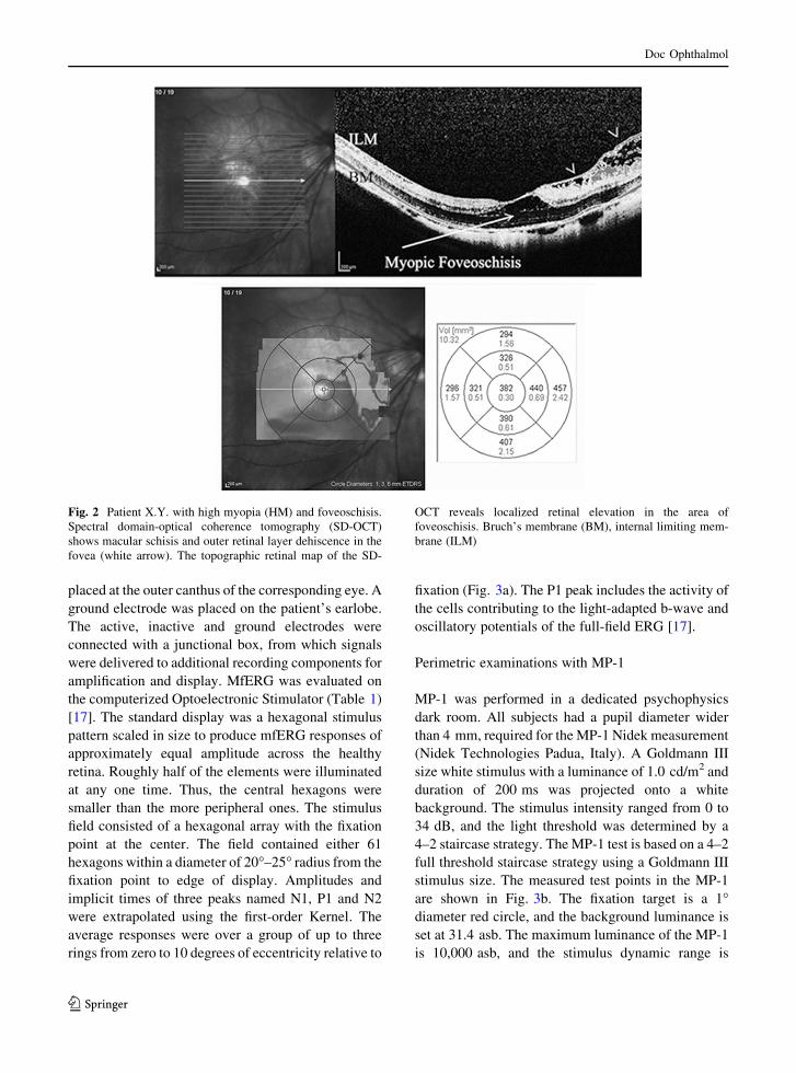
Fig. 2 Patient X.Y. with high myopia (HM) and foveoschisis.
Spectral domain-optical coherence tomography (SD-OCT)
shows macular schisis and outer retinal layer dehiscence in the
fovea (white arrow). The topographic retinal map of the SD-
OCT reveals localized retinal elevation in the area of
foveoschisis. Bruch’s membrane (BM), internal limiting mem-
brane (ILM)
123
Doc Ophthalmol

between 0 and 34 dB. Only reliable visual fields were
used in the analyses, defined as a fixation loss (FL) rate
\ 20% and a false-positive (FP) rate \ 15%. Using
the retinal sensitivities obtained, the mean sensitivity
in the fovea, within 2 degrees, 2–5 degrees and 5–10
degrees were calculated. To assess fixation stability,
movements of the fundus were tracked during the
examination while the patient stared at the fixation
target.
Statistical analysis
Statistical analysis was performed using the STATA
14.0 (Collage Station, Texas, USA). Results were
presented as mean value ± SD. One-way analysis of
variance (ANOVA) followed by Tukey’s post hoc test
was used to analyze differences between groups unless
specified. The ANOVA test was also used to compare
mean age between study groups. Gender distribution
was compared by the Chi square test; p values\ 0.05
were considered as statistically significant.
Results
In the present study, we enrolled 48 eyes of 48 HM
patients with a mean age of 54.1 ± 2.3 years (ranging
from 47 to 55 years). Twenty-four HM eyes with MF
had a mean refractive error of - 14.23 DS and a mean
BCVA of 20/40 Snellen or ? 0.3 logarithm of the
minimum angle of resolution (logMAR). An addi-
tional group of 24 HM patients without foveoschisis
was included. They had a mean refractive error of
- 12.09 DS, and a mean BCVA of 20/20 (0.0
logMAR). Twenty-four healthy, emmetropic subjects
were also included as the control group (CG), they had
a mean age of 53.2 ± 1.9 years SD (ranging from 48
to 54 years of age) and a mean BCVA of 20/20 (0.0
logMAR). We checked for the normality of our data
and ANOVA assumptions were tested and met. The
mean age and gender distribution between the three
groups was not statistically significant (p values
\ 0.05).
We examined a 10� macular area with SD-OCT,
mfERG and MP-1 to assess macular thickness,
amplitude and implicit time of the P1 wave and retinal
sensitivity. No abnormality in SD-OCT, mfERG and
Table 1 Type of analysis of multifocal electroretinography (mfERG)
Six minutes per analysis MfERG photopic response 61B
Optical correction As required at 30 cm
Stimulated fields Thirty degrees horizontally and 23 degrees vertically
Modes of stimulation and
zones
Areas covering the central 25� of the retina and scaled eccentrically to simulate an array of 61
hexagons. Zones of size 3.4� centrally
Hexagons modulated Between a high luminance of stimulations set at 200 cd/mq for the bright flashes and 1 cd/m2 for the
dark flashes according to a binary pseudo-random m-sequence
Standard stimulation Black/white monochrome cathode ray tube monitor with blue
Background: rod and cone responses
Frame frequency 120 Hz to provide higher temporal resolution
Band-pass filtering High pass cutoff 10 Hz; low pass cutoff 300 Hz; amplified with a gain of 100,000
Stimulus screen Surrounded by uniformly illuminated background cover with a luminance set at 30 cd/m2 to
eliminate the rod responses
Stimulus frequency Set at 17 Hz to optimize the amplitude of responses
Fixation stability Monitored with an infrared refractor camera
Cutoff of amplitude P1-N1 in
lV/deg2
Ring 1: 104; Ring 2: 78; Ring 3: 57.6; Ring 4: 42.9; Ring 5: 41.7
Parameters used on the computerized Optoelectronic Stimulator Vision Monitor MonPack 120 Metrovision (Perenchies, France). The
analysis generates a histogram for each of the extended zones indicating the average amplitude of the peaks, and of the root mean
square (RMS) in microvolts/deg2 (lV/deg2). The RMS characterizes the energy content of each response. Several visualization
modes were obtained with 2-D and 3-D maps. Different types of ring display allow the measurement of P1–N1 amplitude and implicit
time, which can be compared to normative data to evaluate the general size and timing of signals in a given patient
123
Doc Ophthalmol

MP-1 examinations was recorded in the CG. In HM
patients with MF, SD-OCT showed an increase in
macular thickness (Fig. 2) associated with a reduction
in the amplitude and a delay in the implicit time of the
P1 wave recorded by mfERG. Lower values of retinal
sensitivity, as assessed by MP-1, were also found in
these subjects (Fig. 3).
Patients with MF showed an increased macular
thickness, assessed by SD-OCT, when compared with
both the HM patients without MF and the CG
(ANOVA, p\ 0.001) (Table 2). The morphological
changes observed in patients with MF were associated
with a functional impairment as demonstrated by the
decreased P1 wave amplitude evaluated by mfERG
(p\ 0.001) (Table 2), and the decreased macular
sensitivity in the three areas assessed with MP-1
(ANOVA, p\ 0.001) (Table 2). The values of
implicit time in the mfERG were statistically signif-
icant only between 5� and 10� (Table 2). Furthermore,
HM and CG did not show significant difference, in the
variables analyzed, except for the third ring of SD-
Fig. 3 Patient X.Y. the figure shows lower values of retinal
functionality with delay in the implicit time and reduction in the
amplitude of the mfERG P1 waves registered in the area affected
by myopic foveoschisis (MF) (a). P1 average waveform and
standard deviation (±) of amplitude (A) and implicit time
(T) from the three rings (1, 2 and 3). These values indicate that
the patient responses are reduced. The microperimetry (MP-1)
shows lower values of retinal sensitivity in the same area as well
(b)
123
Doc Ophthalmol

OCT and the mfERG amplitude in all the rings
(Table 2).
Discussion
The purpose of our study was to evaluate the
morphological and functional retinal changes in
patients affected by HM with and without MF using
SD-OCT, mfERG and MP-1. We demonstrated that
significant retinal functional changes occur in MF
subjects with SD-OCT anatomical alterations. To the
best of our knowledge, we evaluated two groups of
patients with HM, those affected and not affected by
macular foveoschisis [18]. Moreover, the decreased
sensitivity to MP-1 tests, probably related to the
stretching of the photoreceptor outer segments, is also
useful in the evaluation of myopia-related retinal
foveoschisis. Several studies have been carried out to
describe MF in HM patients; however, functional
changes associated with the morphological character-
istics of these subjects have not yet been reported. In
1938, Rochon-Duvigneaud observed a histological
description of foveoschisis in a highly myopic eye that
appears to be very similar to our SD-OCT results [19].
Using OCT scans, it has been clearly demonstrated
that MF in PM patients is characterized by a detach-
ment of the neuroretina at the level of the plexiform
layer [19, 20]. It has been hypothesized that the main
causes of retinal delamination in myopic macular
retinoschisis could be the posterior myopic staphy-
loma, and incomplete vitreous detachment that exerts
excessive traction on a previously stretched posterior
pole [12, 14, 21]. It has been also demonstrated that the
presence of vitreous pre-retinal membranes represents
a risk factor for MF progression, while the role of axial
Table 2 Data obtained using spectral domain-optical coher-
ence tomography (SD-OCT), multifocal electroretinography
(mfERG), and microperimetry (MP-1) in high myopia (HM)
patients with and without myopic foveoschisis (MF) compared
to the control group (CG)
Area MF SD-OCT
mean ± SD
HM SD-OCT
mean ± SD
CG SD-OCT
mean ± SD
p
ANOVA MF versus
HM*
MF versus
CG*
HM versus
CG*
2� 406.29 ± 25.59 276.08 ± 8.33 271.32 ± 9.61 \ 0.001 \ 0.001 \ 0.001 NS
2�–5� 420.19 ± 41.01 342.70 ± 19.92 300.87 ± 10.01 \ 0.001 \ 0.001 \ 0.001 NS
5�–10� 382.10 ± 12.09 298.11 ± 6.04 283.48 ± 8.68 \ 0.001 \ 0.001 \ 0.001 \ 0.001
MF mfERG A HM mfERG A CG mfERG A
2� 51.24 ± 15.01 90.33 ± 19.61 155.21 ± 8.08 \ 0.001 \ 0.001 \ 0.001 <0.001
2�–5� 31.00 ± 12.71 59.32 ± 8.00 89.91 ± 2.81 \ 0.001 \ 0.001 \ 0.001 <0.001
5�–10� 29.91 ± 10.00 40.30 ± 4.51 61.29 ± 2.99 \ 0.001 \ 0.001 \ 0.001 \ 0.001
MF mfERG T HM mfERG T CG mfERG T
2� 44.21 ± 2.98 44.05 ± 3.02 44.39 ± 3.09 NS NS NS NS
2�–5� 46.08 ± 7.31 39.89 ± 2.09 41.89 ± 2.01 NS NS NS NS
5�–10� 45.68 ± 4.01 42.05 ± 1.97 42.11 ± 3.09 \ 0.05 \ 0.05 \ 0.05 NS
MF MP-1 HM MP-1 CG MP-1
2� 11.90 ± 3.15 19.11 ± 2.03 20.00 ± 0.00 \ 0.001 \ 0.001 \ 0.001 NS
2�–5� 12.09 ± 2.90 19.09 ± 1.09 19.04 ± 1.20 \ 0.001 \ 0.001 \ 0.001 NS
5�–10� 12.31 ± 2.99 18.08 ± 1.06 19.00 ± 1.30 \ 0.001 \ 0.001 \ 0.001 NS
A one-way analysis of variance (ANOVA) followed by Tukey’s post hoc test was applied for analysis of differences between groups.
Macular area thickness (in lm) through SD-OCT. Retinal functional amplitude (A) expressed as microvolts/deg2 (lV/deg2) and
implicit time (T) in milliseconds (ms) of the P1 wave with mfERG. Retinal functionality expressed as sensitivity values in dB
measured by MP-1
Values are expressed as mean ± standard deviation (SD). The statistical significance was attested at a probability of p\ 0.05. Value
significantly higher by one-way ANOVA and Tukey’s post hoc test (in bold)
NS not significant
*After Tukey correction
123
Doc Ophthalmol

length and chorioretinal atrophy in the progression of
the disease has not been clearly established yet
[9, 22–26].
Polito et al. and Lai et al. reported some cases of
foveoschisis with incomplete vitreous detachment that
recovered after vitreo-retinal adhesion release with
increased visual function and normalization of OCT
scans. This observation supports the hypothesis that an
incomplete vitreous detachment, with less severe
traction on the retina, may represent a cause of MF
in highly myopic eyes, which could resolve sponta-
neously [26, 27]. Shimada et al. investigated morpho-
logical changes occurred during the development of
early RD in HM patients with MF performing a series
of OCT scans in five patients. This longitudinal
research highlighted that the progression from MF to
RD goes through four stages, and the formation of an
outer lamellar hole predisposes to RD [9]. In addition,
Lai et al. found that foveal detachment can also
spontaneously resolve in patients with high myopic
traction maculopathy without evidence of posterior
vitreous detachment [27].
In our study, we evaluated retinal function and
morphological changes in patients with HM and MF
through three hi-tech examinations, SD-OCT, mfERG
and MP-1. We demonstrated that the macular central
area in highly myopic eyes affected by MF showed
functional alterations associated with anatomical
changes. These findings are in line with previous
studies showing retinal thickening at the posterior pole
and separation between the less reflective external
retina and the more reflective internal one, in HM
patients with MF [9, 14, 20, 22–26].
Notably, in our research, MF was mainly confined
to the 10� scanning area, and no disruption of any
detached internal limiting membrane was noted.
Nevertheless, the macular sensitivity analysis per-
formed by mfERG and MP-1 detected a significant
alteration of the retinal function in patients with HM
and MF. Previous studies demonstrated that
microperimetry shows an adequate reproducibility,
greater than conventional perimetry, to evaluate mean
retinal sensitivity in patients with macular diseases
[28]. Sugiura et al. showed that retinal sensitivities,
measured by microperimetry, declined with the
increase in retinal thickness, assessed by SD-OCT
[28]. In addition, Park and colleagues showed that,
especially in patients with HM, the correlation
between structural (thickness) and functional
(amplitude/implicit time) changes should be consid-
ered when interpreting retinal structure and function
using SD-OCT and mfERG [10].
In our study, in order to minimize the impact of age
on the results, only subjects aged between 47 and
55 years were included. Moreover, it is well known
that mfERG can be used for the quantitative and
objective detection of abnormal macular areas [29]. In
fact, the first-order kernel mainly reflects the function
of the outer retina, especially of the cones [29]. The
differences of P1 implicit time among the three groups
of patients were statistically significant in ring 5�–10�,but only for HM patients with MF versus HM patients
without MF, and HM patients with MF versus CG. We
can therefore assert that amplitude of the mfERG is
more sensitive than implicit time. The reasons for the
increased macular thickness caused by the schisis
associated with the thinning in the retinal morpholog-
ical profile (SD-OCT) and the decreased retinal
sensitivity (mfERG/MP-1) are probably related to
the stretching of the photoreceptor outer segments.
The phenomenon of abnormal axial elongation could
explain the enlargement of the subretinal space with
consequent decreased foveal cellular density caused
by ischemia and retinal circulation disturbances [10].
In our study, we described a positive correlation
between TROL in retinal macular areas and mfERG
and MP-1 tests. Consequently, a thicker retina outer
layer may imply an improved photoreceptor metabo-
lism and better visual function, as also demonstrated in
another study using mfERG and SD-OCT [30]. There
are two key pathological factors that are deemed to be
responsible for this retinal dysfunction. One is the
mechanical stretching related to posterior staphyloma
and vitreous traction of the inner retina associated with
the elongation of the globe in myopic eyes. The other
is the metabolic distress of the photoreceptor layer
caused by a decrease in blood supply of the thinned
HM choroid [30]. Ultimately, both the mechanical and
vascular factors may contribute to the derangements of
the retinal functional responses [15].
It has also been suggested that other factors, such as
the dysfunction of the dopaminergic system, may play
a role in the pathogenesis of the functional decline in
myopic eyes [30, 31]. Experimental studies in animal
models have demonstrated that an increase in retinal
levels of dopamine activates D1 and D2 dopaminergic
receptors present throughout the retina generating a
123
Doc Ophthalmol

signal that inhibits axial growth once the eye has
reached a normal length [31–34].
Currently, OCT scans and BCVA are the most
commonly used tools to define MF progression,
especially because of the lack, in many clinics, of
more sophisticated techniques such as MP-1 and
mfERG [9, 14]. Nevertheless, the clinical work-up and
follow-up of HM patients with MF should include hi-
tech diagnostic tests such as SD-OCT, mfERG and
MP-1, which could be useful not only for the detection
of macular modifications in MF, but also to assess the
relationship between functional variations and struc-
tural degeneration. These methods may allow predict-
ing the disease’s progression providing, especially in
the presence of complications, an objective basis for
the management of surgical treatment and the pre-
vention of loss of sight in MF patients. In order to fully
understand the processes involved in retinal damage, a
further study aiming to longitudinally evaluate the
results of these examinations in a larger population of
HM patients with MF will be carried out at our clinic.
Funding No funding was received for this research.
Compliance with ethical standards
Conflict of interest All authors certify that they have no
affiliations with or involvement in any organization or entity
with any financial interest (such as honoraria; educational
grants; participation in speakers’ bureaus; membership,
employment, consultancies, stock ownership or other equity
interest; and expert testimony or patent-licensing arrangements)
or non-financial interest (such as personal or professional rela-
tionships, affiliations, knowledge or beliefs) in the subject
matter or materials discussed in this manuscript.
Statement of human rights All procedures performed in
studies involving human participants were in accordance with
the ethical standards of the institutional and/or national research
committee and with the 1964 Helsinki Declaration and its later
amendments or comparable ethical standards.
Statement on the welfare of animals No animals were used
in this research.
Informed consent Informed consent was obtained from all
individual participants included in the study.
References
1. Holden BA, Fricke TR, Wilson DA, Jong M, Naidoo KS,
Sankaridurg P, Wong TY, Naduvilath TJ, Resnikoff S
(2016) Global Prevalence of Myopia and
High Myopia and Temporal Trends from 2000 through
2050. Ophthalmology 123:1036–1042
2. Rudnicka AR, Kapetanakis VV, Wathern AK, Logan NS,
Gilmartin B, Whincup PH, Cook DG, Owen CG (2016)
Global variations and time trends in the prevalence of
childhood myopia, a systematic review and quantitative
meta-analysis: implications for aetiology and early pre-
vention. Br J Ophthalmol 100:882–890
3. Vongphanit J, Mitchell P, Wang JJ (2002) Prevalence and
progression of myopic retinopathy in an older population.
Ophthalmology 109(4):704–711
4. Pararajasegaram R (1999) VISION 2020-the right to sight:
from strategies to action. Am J Ophthalmol 128:359–360
5. Eye Disease Case-Control Study Group (1993) Risk factors
for idiopathic rhegmatogenous retinal detachment. Am J
Epidemiol 137:749–757
6. Marcus MW, de Vries MM, Junoy Montolio FG, Jansonius
NM (2011) Myopia as a risk factor for open-angle glau-
coma: a systematic review and meta-analysis. Ophthal-
mology 118(1989–1994):e2
7. Lim R, Mitchell P, Cumming RG (1999) Refractive asso-
ciations with cataract: the Blue Mountains Eye Study.
Invest Ophthalmol Vis Sci 40:3021–3026
8. Phillips C (1958) Retinal detachment at the posterior pole.
Br J Ophtalmol 42:749–753
9. Takano M, Kishi S (1999) Foveal retinoschisis and retinal
detachment in severely myopic eyes with posterior
staphyloma. Am J Ophthalmol 128:472–476
10. Faghihi H, Hajizadeh F, Riazi-Esfahani M (2010) Optical
Coherence Tomographic Findings in Highly Myopic Eyes.
J Ophthalmic Vis Res 5:110–121
11. Park S, Kim SH, Park TK, Ohn YH (2013) Evaluation of
structural and functional changes in non-pathologic myopic
fundus using multifocal electroretinogram and optical
coherence tomography. Doc Ophthalmol 126(3):199–210
12. Wu PC, Chen YJ, Chen YH, Chen CH, Shin SJ, Tsai CL,
Kuo HK (2009) Factors associated with foveoschisis and
foveal detachment without macular hole in high myopia.
Eye 23:356–361
13. Shimada N, Ohno-Matsui K, Baba T, Futagami S, Tokoro T,
Mochizuki M (2006) Natural course of macular
retinoschisis in highly myopic eyes without macular hole or
retinal detachment. Am J Ophthalmol 142:497–500
14. Gohil R, Sivaprasad S, Han LT, Mathew R, Kiousis G, Yang
Y (2015) Myopic foveoschisis: a clinical review. Eye
29:593–601
15. Kamal-Salah R, Morillo-Sanchez MJ, Rius-Diaz F, Garcia-
Campos JM (2015) Relationship between paravascular
abnormalities and foveoschisis in highly myopic patients.
Eye (London) 29(2):280–285
16. Ortisi E, Avitabile T, Bonfiglio V (2012) Surgical man-
agement of retinal detachment because of macular hole in
highly myopic eyes. Retina 32(9):1704–1718
17. Hood DC, Bach M, Brigell M, Keating D, Kondo M, Lyons
JS, Marmor MF, McCulloch DL, Palmowski-Wolfe AM,
International Society For Clinical Electrophysiology of
Vision (2012) ISCEV standard for clinical multifocal
electroretinography (mfERG) (2011 edition). Doc Oph-
thalmol 2012(124):1–13
18. Sachidanandam R, Ravi P, Sen P (2017) Effect of axial
length on full-field and multifocal electroretinograms. Clin
123
Doc Ophthalmol

Exp Optom 100:668–675. https://doi.org/10.1111/cxo.
12529
19. Rochon-Duvigneaud M (1938) Deformation et lesions de
l’oeil myope: in Mawas J, introduction a‘ l’etude de la
myopie et des chorioretinites myopiques. Bull Soc Ophtal-
mol Paris 1:1–10
20. Panozzo GMA (2004) Optical coherence tomography
findings in myopic traction maculopathy. Arch Ophthalmol
122:1455–1460
21. Steidl SM, Pruet RC (1997) Macular complications asso-
ciated with posterior staphyloma. Am J Ophthalmol
123:181–187
22. Fujimoto M, Hangai M, Suda K, Yoshimura N (2016)
Features associated with foveal retinal detachment in
myopia macular retinoschisis. Am J Ophthalmol
150:863–870
23. Coppe AM, Ripandelli G (2003) Optical coherence
tomography in the evaluation of vitreoretinal disorders of
the macula in highly myopic eyes. Semin Ophthalmol
18:85–88
24. Chen YP, Chen TL, Yang KR, Lee WH, Kuo YH, Chao AN,
Wu WC, Chen KJ, Lai CC (2006) Treatment of retinal
detachment resulting from posterior staphyloma-associated
macular hole in highly myopic eyes. Retina 26(1):25–31
25. Vingolo EM, Salvatore S, Domanico D, Spadea L, Neb-
bioso M (2013) Visual rehabilitation in patients with myo-
pic maculopathy: our experience. Can J Ophthalmol
48:438–442
26. Polito A, Lanzetta P, Del Borrello M, Bandello F (2003)
Spontaneous resolution of a shallow detachment of the
macula in a highly myopic eye. Am J Ophthalmol
135:546–547
27. Lai TT, Ho TC, Yang CM (2016) Spontaneous resolution
of foveal detachment in traction maculopathy in high
myopia unrelated to posterior vitreous detachment. BMC
Ophthalmol 16:18
28. Sugiura A, Fujino R, Takemiya N, Shimizu K, Matsuura M,
Murata H, Inoue T, Obata R, Asaoka R (2017) The asso-
ciation between visual function and retinal structure
in chronic central serous chorioretinopathy. Sci Rep
7:16288
29. Song AP, Yu T, Wang JR, Liu W, Sun Y, Ma SX (2016)
Multifocal electroretinogram in non-pathological myopic
subjects: correlation with optical coherence tomography. Int
J Ophthalmol 9:286–291
30. Flores-Moreno I, Arias-Barquet L, Rubio-Caso MJ, Munoz-
Blanco A, Vidal-Martı M, Catala-Mora J, Ruiz-Moreno JM,
Duker JS, Caminal JM (2017) Structure versus func-
tion: correlation between outer retinal and choroidal
thicknesses measured by swept-source OCT with multifo-
cal electroretinography and visual acuity. Int J Retina
Vitreous 3:29
31. Cavallotti C, Artico M, Pescosolido N, Tranquilli Leali FM,
Feher J (2004) Age-related changes in the human retina.
Can J Ophthalmol 39(1):61–68
32. Wrzesinska D, Nowomiejska K, Nowakowska D, Brzo-
zowska A, Avitabile T, Reibaldi M, Rejdak R, Toro M
(2019) Vertical and horizontal m-charts and microperime-
try for assessment of the visual function in patients after
vitrectomy with ILM peeling due to stage 4 macular hole.
J Ophthalmol 6(2019):4975973
33. Pescosolido N, Parisi F, Russo P, Buomprisco G, Nebbioso
M (2013) Role of dopaminergic receptors in glaucomatous
disease modulation. Biomed Res Int 2013:193048
34. Witkovsky P (2004) Dopamine and retinal function. Doc
Ophthalmol 108:17–40
Publisher’s Note Springer Nature remains neutral with
regard to jurisdictional claims in published maps and
institutional affiliations.
123
Doc Ophthalmol
Copyright © 2022 FDOKUMEN