High Affinity Human Antibody Fragments to Dengue Virus Non-Structural Protein 3
10
High Affinity Human Antibody Fragments to Dengue Virus Non-Structural Protein 3 Nicole J. Moreland 1. , Moon Y. F. Tay 1. , Elfin Lim 1 , Prasad N. Paradkar 1 , Danny N. P. Doan 1 , Yin Hoe Yau 2 , Susana Geifman Shochat 2 , Subhash G. Vasudevan 1 * 1 Program in Emerging Infectious Diseases, DUKE-NUS Graduate Medical School, Singapore, Singapore, 2 School of Biological Sciences, Nanyang Technical University, Singapore, Singapore Abstract Background: The enzyme activities catalysed by flavivirus non-structural protein 3 (NS3) are essential for virus replication. They are distributed between the N-terminal protease domain in the first one-third and the C-terminal ATPase/helicase and nucleoside 59 triphosphatase domain which forms the remainder of the 618-aa long protein. Methodology/Principal Findings: In this study, dengue full-length NS3 protein with residues 49 to 66 of NS2B covalently attached via a flexible linker, was used as bait in biopanning with a naı ¨ve human Fab phage-display library. Using a range of truncated constructs spanning the NS2B cofactor region and the full-length NS3, 10 unique Fab were identified and characterized. Of these, monoclonal Fab 3F8 was shown to bind a30 (residues 526 through 531) within subdomain III of the helicase domain. The antibody inhibits the ATPase and helicase activites of NS3 in biochemical assays and reduces DENV replication in HEK293 cells that were previously transfected with Fab 3F8 compared with mock transfected cells. Conclusions/Significance: Antibodies such as 3F8 are valuable tools for studying the molecular mechanisms of flaviviral replication and for the monospecific detection of replicating dengue virus in vivo. Citation: Moreland NJ, Tay MYF, Lim E, Paradkar PN, Doan DNP, et al. (2010) High Affinity Human Antibody Fragments to Dengue Virus Non-Structural Protein 3. PLoS Negl Trop Dis 4(11): e881. doi:10.1371/journal.pntd.0000881 Editor: Maria G. Guzman, Tropical Medicine Institute Pedro Kourı ´ (IPK), Cuba Received April 21, 2010; Accepted October 13, 2010; Published November 9, 2010 Copyright: ß 2010 Moreland et al. This is an open-access article distributed under the terms of the Creative Commons Attribution License, which permits unrestricted use, distribution, and reproduction in any medium, provided the original author and source are credited. Funding: This work is supported by the DUKE NUS Signature Research Program (funded by the Agency for Science, Technology and Research, Singapore and the Ministry of Health, Singapore) and the National Medical Research Council, Singapore (www.nmrc.gov.sg) under grant NMRC/GMS/1186/2008. SGV and NJM acknowledge the Singapore National Research Foundation (www.nrf.gov.sg) proof-of-concept grant NRF2009-POC001-045. The funders had no role in study design, data collection and analysis, decision to publish, or preparation of the manuscript. Competing Interests: The authors have declared that no competing interests exist. * E-mail: [email protected] . These authors contributed equally to this work. Introduction Dengue virus belongs to the Flaviviridae family and is the etiological agent of dengue fever, dengue hemorrhagic fever and dengue shock syndrome. It is the most prevalent arthropod transmitted infectious disease in humans and has four antigenically distinct viral serotypes (DENV 1–4) [1]. The genome of dengue viruses comprises a positive single stranded RNA of 11kb. Post-translational processing of the polyprotein gives rise to three strucural proteins (C, prM and E) and seven non-structural proteins (NS1, NS2A, NS2B, NS3, NS4A, NS4B and NS5). The processing of the amino terminal region of the polyprotein is carried out by host signal peptidases, while processing of the 2A-2B, 2B-3, 3-4A and 4B-5 sites is catalysed by the two- component viral protease NS2B/NS3 [2,3]. DENV NS3 is a multifunctional enzyme with three known catalytic activities segregated into two distinct domains (Figure 1). The serine protease lies within the N-terminal 180 amino acid residues of the 618 amino acid protein. The central hydrophillic portion of the intergral membrane protein NS2B (residues 49–96) is required for protease activity [4–6]. The ATPase/helicase and nucleoside 59-triphosphate activities are localised in the remaining C-terminal domain. There appears to be cross-talk between the two domains; the helicase activity is approximately 30-fold higher in the full-length NS3 protein than in the domain and the affinity of the full-length protein for ATP is 10 fold lower than that of the helicase domain alone [7,8]. Recent crystal structures of full-length NS3 from DENV and the related flavivirus Murray Valley encephalitis virus, reveal that the protease and helicase domains are linked by an interdomain linker (residues 169–179 in DENV) as illustrated in Figure 1 [8,9]. Infection with one DENV serotype results in immunity to that serotype only; this protection is thought to be due to neutralizing antibodies, DENV-specific memory T cells, or a combination of the two. While the T-cell response is directed against several DENV proteins, NS3 appears to be the dominant target for CD4+ and CD8+ T cells, and multiple human T cell epitopes have been mapped onto NS3 (reviewed in [10]). Interestingly DENV NS3 also elicits a specific antibody response in humans. A study of acute sera from patients infected with DENV-2 or DENV-4 showed that although anti-E (envelope) antibodies were the most abundant, anti-NS3 antibodies were widely detected, particularly in those with secondary infections [11]. Given the vital role NS3 plays in viral replication, and the specific T- and B-cell responses observed towards NS3 in www.plosntds.org 1 November 2010 | Volume 4 | Issue 11 | e881
Transcript of High Affinity Human Antibody Fragments to Dengue Virus Non-Structural Protein 3

High Affinity Human Antibody Fragments to DengueVirus Non-Structural Protein 3Nicole J. Moreland1., Moon Y. F. Tay1., Elfin Lim1, Prasad N. Paradkar1, Danny N. P. Doan1, Yin Hoe
Yau2, Susana Geifman Shochat2, Subhash G. Vasudevan1*
1 Program in Emerging Infectious Diseases, DUKE-NUS Graduate Medical School, Singapore, Singapore, 2 School of Biological Sciences, Nanyang Technical University,
Singapore, Singapore
Abstract
Background: The enzyme activities catalysed by flavivirus non-structural protein 3 (NS3) are essential for virus replication.They are distributed between the N-terminal protease domain in the first one-third and the C-terminal ATPase/helicase andnucleoside 59 triphosphatase domain which forms the remainder of the 618-aa long protein.
Methodology/Principal Findings: In this study, dengue full-length NS3 protein with residues 49 to 66 of NS2B covalentlyattached via a flexible linker, was used as bait in biopanning with a naıve human Fab phage-display library. Using a range oftruncated constructs spanning the NS2B cofactor region and the full-length NS3, 10 unique Fab were identified andcharacterized. Of these, monoclonal Fab 3F8 was shown to bind a30 (residues 526 through 531) within subdomain III of thehelicase domain. The antibody inhibits the ATPase and helicase activites of NS3 in biochemical assays and reduces DENVreplication in HEK293 cells that were previously transfected with Fab 3F8 compared with mock transfected cells.
Conclusions/Significance: Antibodies such as 3F8 are valuable tools for studying the molecular mechanisms of flaviviralreplication and for the monospecific detection of replicating dengue virus in vivo.
Citation: Moreland NJ, Tay MYF, Lim E, Paradkar PN, Doan DNP, et al. (2010) High Affinity Human Antibody Fragments to Dengue Virus Non-Structural Protein3. PLoS Negl Trop Dis 4(11): e881. doi:10.1371/journal.pntd.0000881
Editor: Maria G. Guzman, Tropical Medicine Institute Pedro Kourı (IPK), Cuba
Received April 21, 2010; Accepted October 13, 2010; Published November 9, 2010
Copyright: � 2010 Moreland et al. This is an open-access article distributed under the terms of the Creative Commons Attribution License, which permitsunrestricted use, distribution, and reproduction in any medium, provided the original author and source are credited.
Funding: This work is supported by the DUKE NUS Signature Research Program (funded by the Agency for Science, Technology and Research, Singapore and theMinistry of Health, Singapore) and the National Medical Research Council, Singapore (www.nmrc.gov.sg) under grant NMRC/GMS/1186/2008. SGV and NJMacknowledge the Singapore National Research Foundation (www.nrf.gov.sg) proof-of-concept grant NRF2009-POC001-045. The funders had no role in studydesign, data collection and analysis, decision to publish, or preparation of the manuscript.
Competing Interests: The authors have declared that no competing interests exist.
* E-mail: [email protected]
. These authors contributed equally to this work.
Introduction
Dengue virus belongs to the Flaviviridae family and is the etiological
agent of dengue fever, dengue hemorrhagic fever and dengue shock
syndrome. It is the most prevalent arthropod transmitted infectious
disease in humans and has four antigenically distinct viral serotypes
(DENV 1–4) [1]. The genome of dengue viruses comprises a positive
single stranded RNA of 11kb. Post-translational processing of the
polyprotein gives rise to three strucural proteins (C, prM and E) and
seven non-structural proteins (NS1, NS2A, NS2B, NS3, NS4A,
NS4B and NS5). The processing of the amino terminal region of the
polyprotein is carried out by host signal peptidases, while processing
of the 2A-2B, 2B-3, 3-4A and 4B-5 sites is catalysed by the two-
component viral protease NS2B/NS3 [2,3].
DENV NS3 is a multifunctional enzyme with three known
catalytic activities segregated into two distinct domains (Figure 1).
The serine protease lies within the N-terminal 180 amino acid
residues of the 618 amino acid protein. The central hydrophillic
portion of the intergral membrane protein NS2B (residues 49–96)
is required for protease activity [4–6]. The ATPase/helicase and
nucleoside 59-triphosphate activities are localised in the remaining
C-terminal domain. There appears to be cross-talk between the
two domains; the helicase activity is approximately 30-fold higher
in the full-length NS3 protein than in the domain and the affinity
of the full-length protein for ATP is 10 fold lower than that of the
helicase domain alone [7,8]. Recent crystal structures of full-length
NS3 from DENV and the related flavivirus Murray Valley
encephalitis virus, reveal that the protease and helicase domains
are linked by an interdomain linker (residues 169–179 in DENV)
as illustrated in Figure 1 [8,9].
Infection with one DENV serotype results in immunity to that
serotype only; this protection is thought to be due to neutralizing
antibodies, DENV-specific memory T cells, or a combination of
the two. While the T-cell response is directed against several
DENV proteins, NS3 appears to be the dominant target for CD4+and CD8+ T cells, and multiple human T cell epitopes have been
mapped onto NS3 (reviewed in [10]). Interestingly DENV NS3
also elicits a specific antibody response in humans. A study of acute
sera from patients infected with DENV-2 or DENV-4 showed that
although anti-E (envelope) antibodies were the most abundant,
anti-NS3 antibodies were widely detected, particularly in those
with secondary infections [11].
Given the vital role NS3 plays in viral replication, and the
specific T- and B-cell responses observed towards NS3 in
www.plosntds.org 1 November 2010 | Volume 4 | Issue 11 | e881
DENV infected patients, well characterised anti-NS3 antibodies
would be vaulable tools for studying viral replication in detail
and detecting DENV infected cells. There are very few reports
describing the production of monoclonal antibodies specific for
NS3, and those that have used hybridoma technology in mice
[12–15]. In three of these studies anti-NS3 antibodies were
isolated following immunization with recombinant NS3 [12–14]
but the fourth study innoculated mice with DENV-1 virus
(purified from suckling mouse brain) and selected hybridomas
that produced anti-NS3 antibodies [15]. These antibodies were
then used to immunize mice that were subsequently challenged
with DENV-1. Intriguingly an increase in survival, albeit
equivocal, was noted with four of the monoclonal antibodies
tested.
Recombinant antibody technology (phage display) is a powerful
alternative to conventional antibody techniques that permits the
selection of high-affinity antibodies specific for the target protein
[16,17]. Antibody fragments are expressed on the surface of
filamentous phage linking the antibody protein with its encoding
DNA sequence within the phage. Phage displaying antibodies that
bind the target protein are enriched by several rounds of selection
and amplification (bio-panning), and the resulting antibody
fragments can be produced recombinantly in E. coli without the
need for immunization. This study describes the identification and
characterisation of human Fab antibody fragments that bind
DENV NS3 using phage display. The panel of antibodies have
different specificity patterns towards the NS3 protease and helicase
domains, and NS3 proteins from DENV 1–4. We have evaluated
the ability of the antibodies to inhibit the protease, helicase and
ATPase activities catalysed by NS3 in vitro and DENV replication
using cell based assays, and have identified one Fab, designated
3F8, that recognises a conserved epitope on subdomain III of the
NS3 helicase domain. This antibody is cross-reactive with all four
serotypes, and binds NS3 with high affinity. It can be used as a tool
to study the DENV replication complex or could potentially be
developed as a therapeutic.
Materials and Methods
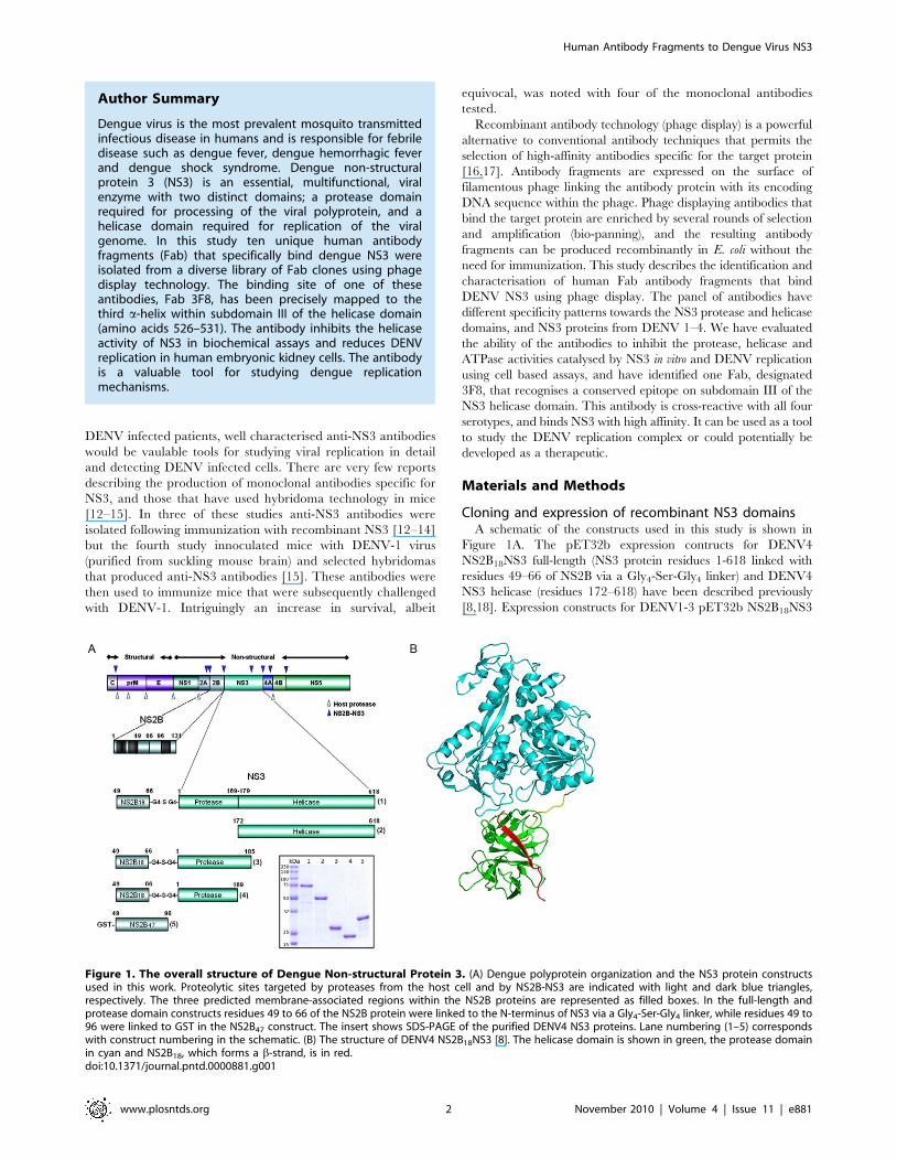
Cloning and expression of recombinant NS3 domainsA schematic of the constructs used in this study is shown in
Figure 1A. The pET32b expression contructs for DENV4
NS2B18NS3 full-length (NS3 protein residues 1-618 linked with
residues 49–66 of NS2B via a Gly4-Ser-Gly4 linker) and DENV4
NS3 helicase (residues 172–618) have been described previously
[8,18]. Expression constructs for DENV1-3 pET32b NS2B18NS3
Figure 1. The overall structure of Dengue Non-structural Protein 3. (A) Dengue polyprotein organization and the NS3 protein constructsused in this work. Proteolytic sites targeted by proteases from the host cell and by NS2B-NS3 are indicated with light and dark blue triangles,respectively. The three predicted membrane-associated regions within the NS2B proteins are represented as filled boxes. In the full-length andprotease domain constructs residues 49 to 66 of the NS2B protein were linked to the N-terminus of NS3 via a Gly4-Ser-Gly4 linker, while residues 49 to96 were linked to GST in the NS2B47 construct. The insert shows SDS-PAGE of the purified DENV4 NS3 proteins. Lane numbering (1–5) correspondswith construct numbering in the schematic. (B) The structure of DENV4 NS2B18NS3 [8]. The helicase domain is shown in green, the protease domainin cyan and NS2B18, which forms a b-strand, is in red.doi:10.1371/journal.pntd.0000881.g001
Author Summary
Dengue virus is the most prevalent mosquito transmittedinfectious disease in humans and is responsible for febriledisease such as dengue fever, dengue hemorrhagic feverand dengue shock syndrome. Dengue non-structuralprotein 3 (NS3) is an essential, multifunctional, viralenzyme with two distinct domains; a protease domainrequired for processing of the viral polyprotein, and ahelicase domain required for replication of the viralgenome. In this study ten unique human antibodyfragments (Fab) that specifically bind dengue NS3 wereisolated from a diverse library of Fab clones using phagedisplay technology. The binding site of one of theseantibodies, Fab 3F8, has been precisely mapped to thethird a-helix within subdomain III of the helicase domain(amino acids 526–531). The antibody inhibits the helicaseactivity of NS3 in biochemical assays and reduces DENVreplication in human embryonic kidney cells. The antibodyis a valuable tool for studying dengue replicationmechanisms.
Human Antibody Fragments to Dengue Virus NS3
www.plosntds.org 2 November 2010 | Volume 4 | Issue 11 | e881

full-length proteins were kind gifts from the Novartis Intitute for
Tropical Diseases, Singapore, and comprise the same correspond-
ing residues as the DENV4 full length construct.
The DENV4 protease domain constructs were amplified from
the DENV4 NS2B18NS3 full-length construct. For DENV4
NS2B18NS3pro169 (NS3 protein residues 1–169 linked with residues
49–66 of NS2B via a Gly4-Ser-Gly4 linker) the forward primer 59-
CCACGCGGTTCTCATATGGCAGACTTGTCACTA-39 and
the reverse primer 59-TTCATAATCTGGATCCCCAATTCAT-
TCAGCTTGCGT-39 were used. The underlined sequence
corresponds with the NdeI and BamHI sites, respectively. DENV4
NS2B18NS3pro185 (NS3 protein residues 1–185 linked with residues
49–66 of NS2B via a Gly4-Ser-Gly4 linker) was amplified using the
same forward primer as above and the reverse primer 59-GT-
AAGTCCATTATGGATCCTCTTTACTTTCGAAAAATG-39.
The PCR fragments were digested with NdeI and BamHI and cloned
into pET14b (Novagen). Escherichia coli BL21 Codon Plus (DE3)-
RIL cells (Stratagene) transformed with pET32b or pET14b
expression constructs were grown in ZYM5052 autoinduction
medium [19] for 4 hours at 37uC followed by 20 hours at 18uC.
The PCR fragment for the DENV4 GST-NS2B47 expression
construct (encoding residues 49–96 of NS2B fused at the N-
terminus with Glutathione S-transferase) was generated with the
forward primer 59-GTGGTGGATCCGCAGATCTGTCACTA-
GAG-39 and reverse primer 59-CAGTGAATTCAAAAGTCA-
TATCATATTGGTTTCCTC-39 (BamHI and EcoRI sites are
underlined) and the previously constructed DENV4 pET15b
CF47-NS3 protease vector [6] as template. The PCR product was
digested with BamHI and EcoRI and cloned into pGEX-4T-1 (GE
Healthcare). E. coli BL21 Codon Plus (DE3)-RIL cells transformed
with the DENV4 GST-NS2B47 construct were grown in in
ZYM5052 autoinduction medium at 37uC for 4 hours followed by
16uC for 20 hours.
Protein purificationThe DENV1-4 NS2B18NS3 full-length and DENV4 NS3hel
proteins were purified by immobilised metal-ion affinity chroma-
tography (IMAC) and size exclusion chromatography (SEC) as
described previously [8,18]. SEC was performed in 20 mM Tris
pH 7.5, 150 mM NaCl, 3 mM b-mercaptoethanol and 5%
glycerol. The pET14b DENV4 NS3 protease proteins were
purified using the same protocol except the SEC buffer was
20 mM Hepes pH 7.5, 250 mM NaCl, 3 mM b-mercaptoethanol
and 5% glycerol.
DENV4 GST-NS2BCF47 cell pellets were lysed in 20 mM Tris
pH 7.5, 200 mM NaCl and the clarified lysate was incubated with
Glutathione Sepharose 4B (GE Healthcare) for 2 hours at 4uC.
Beads were washed extensively in lysis buffer and the protein was
eluted in a buffer containing 20–50 mM reduced glutathione
followed by dialysis into 20 mM Tris pH 7.5, 200 mM NaCl and
1 mM b-mercaptoethanol for storage.
Biotinylation of DENV4 NS2B18NS3 full lengthPurified DENV4 NS2B18NS3 full length was dialysed into
phosphate buffered saline (PBS) pH 7.5 prior to biotinylation. The
protein was incubated with a 20-fold molar excess of the biotin
reagent on ice for 2 hours according to the manufacturers
instructions (Thermo Fisher Scientific). The reaction was stopped
with 100 mM glycine and excess biotin was removed by SEC in
PBS pH 7.5.
Phage display Fab library screeingLibrary screening was performed with a naıve human fab phage
display library HX02 (Humanyx Pte Ltd, Singapore) displayed in
a modified pCES1 vector [20]. The amber stop codon prior to
bacteriophage gene III in pCES1 has been removed and replaced
with a SalI site. An additional SalI site has been placed at the C-
terminus of gene III such that following SalI digestion and
religation (and the concurrent formation of a TAA stop codon)
soluble Fab can be expressed in both suppressor and non-
suppresor strains of E. coli.
Library panning was essentially performed as decribed previ-
ously [21] but streptavidin megnetic beads (Invitrogen) were used
to immobilise the antigen (biotinylated DENV4 NS2B18NS3). The
concentration of DENV4 NS2B18NS3 was 200 nM in the first
round and reduced to 40 nM and 10 nM in rounds two and three,
respectively. The number of input phage in each round was
constant at 161012 pfu while washing was increased from six times
with PBS-T (0.1% Tween-20) in round one to 14 times with PBS-
T in rounds two and three. Bound phage were eluted with
100 mM triethylamine and used to infect E. coli TG1 cells. Phage
were resuced with M13K07 helper phage and amplified on 2xTY
(tryptone-yeast) agar plates supplemented with 100 mg/mL
ampicillin and 25 mg/mL kanamycin. Plates were scraped with
Tris-buffered saline (TBS) and phage was concentrated from the
supernatant by polyethylene glycol-NaCl precipitation.
Following the third round of selection individual TG1 clones
were rescued with M13K07 and screened by enzyme-linked
immunosorbent assay (ELISA) for reactivity against DENV4
NS2B18NS3 full-length (coated at 5 mg/mL in PBS pH 7.5). An
anti-M13-horse radish peroxidase (HRP) conjugate (GE Health-
care) was used for detection and clones with an absorbance value
two times higher than background levels were considered positive.
To assess clone uniqueness a BstN1 restriction digest was
performed following PCR amplification of the Fab coding region
of the phagemid. Clones with unique DNA fingerprints were
subject to automated sequencing.
Expression, purification and Elisa of Fab fusion proteinsPhagemids from unique Fab-phage clones were digested with
SalI to remove the gene III coding sequence and re-ligated with T4
DNA ligase. The resulting plasmids were transformed into E. coli
Top10 F’ cells (Invitrogen) for expression and periplasmic
extraction. Cell pellets were resuspended in chilled lysis buffer
(120 mM Tris pH 8.0, 0.3 mM EDTA and 300 mM surose) and
incubated on ice for 30 minutes for periplasmic extraction.
Magnesium chloride (2.5 mM) was added to the clarified extract
prior to IMAC purification. Fab were further purified by SEC
(S200 10/300 column) if required.
For ELISA Maxisorb Immunoplates (Nunc) were coated with
the relevant NS3 antigen (0.25 mM) in PBS pH 7.5 and blocked
with 5% skim milk in PBS-T. Blocked wells were incubated with
purified Fab (100 nM unless otherwise stated) at room tempera-
ture for 1 hour. Plates were washed with PBS-T and incubated
with an anti-c-myc HRP conjugate (Roche) for detection.
Measurement of binding affinitiesBinding affinities of the Fab for DENV4 NS2B18NS3 were
determined by surface plasmon resonance (SPR) using a Biacore
3000 instrument (GE Healthcare). All experiments were conduct-
ed at 25uC in HBS-EP (10 mM Hepes, 150 mM NaCl, 3.4 mM
EDTA, 0.0005% P-20, pH 7.4). Biotinylated full-length NS3
protein was captured on a streptavidin (SA) sensorchip at a flow
rate of 10 ml/min. For screening, the 10 Fab (100 nM) were
injected across the flowcells, in replicates, at 10 ml/min for 1 min
and allowed to dissociate for 2.5 min. Regeneration of the surface
was achieved by a 30 second pulse with 15 mM HCl. The Fab
that showed the best apparent KD in screening were selected for
Human Antibody Fragments to Dengue Virus NS3
www.plosntds.org 3 November 2010 | Volume 4 | Issue 11 | e881

kinetic analysis. Kinetic parameters were measured by varying the
molar concentration of each Fab (3.9–500 nM) and injecting these
across the flowcells, in duplicates, with the same conditions used in
the screening. Raw sensorgram data were aligned, solvent-
corrected and double-referenced using the Scrubber II software
(BioLogic Software, Campbell, Australia). A simple 1:1 model,
with or without the mass transport coefficient, was used for global
kinetic analysis as appropriate.
Enzyme assaysNTPase assays were conducted as previously decribed [7].
DENV4 NS2B18NS3 full-length (4.8 nM) was preincubated with
1 mM Fab for 30 minutes at room temperature in 90 ml of reaction
buffer (50 mM Tris pH 7.4, 2 mM MgCl2, 1.5 mM DTT, 0.05%
Tween 20, 0.25 ng/ml bovine serum albumin). Poly U (1 mg,
average length 200–250 bases) was added and a further 5 minute
incubation at 37uC was performed before initiating the reaction
with 10 ml of ATP. The reaction was carried out at 37uC for 30
minutes after which the malachite green reagent (Sigma) was
added and absorbance (630 nm) was measured. The amount of
phosphate released was determined with a standard curve and all
assays were carried out in triplicate.
Protease activity was determined for NS2B47NS3pro185 (NS3
protein residues 1–169 linked with residues 49–96 of NS2B via a
Gly4-Ser-Gly4 linker) based on protocols published by Li et al. [6]
as detailed in the supporting information (Figure S1 in Supporting
Information S1).
Helicase activity assays were performed as published [7,22].
The substrate was prepared by annealing an 18-mer DNA oligo
(59-GCCTCGCTGCCGTCGCCA-39) with a 38-mer RNA oligo
(59-UGGCGACGGCAGCGAGGCUUUUUUUUUUUUUUU-
UUUUU-39). The 59 end of the DNA was labelled using T4
polynucleotide kinase and [c232P]ATP. Reactions (10 ml) con-
tained 50 mM Tris-HCl pH 7.4 supplemented with 5 nM of the
DNA:RNA duplex, 500 nM DENV4 NS2B18NS3 full-length,
1.75 mM Fab (NS3:Fab ratio 1:3.5), 4 units of RNAsin, 2 mM
MgCl2, 1 mM DTT, 0.5% Tween and 0.25 mg/mL BSA. Fab and
NS3 were preincubated in assay buffer for 10 minutes prior to
initiating the reaction with 5 mM ATP. Assays were performed at
37uC for 30 minutes and were resolved on a 10% native
polyacrylamide gel and autoradiographed using a Pharos FX
system. Signal intensity was quantified with Quantity One
software (Biorad).
Statistical analysis of all assay data was performed using paired
t-tests. The results were considered statistically significant if
p,0.05.
Cell based assaysHEK293 cells were maintained at 37uC in a CO2 incubator in
Dulbeco’s modified eagle’s medium (DMEM) containing 10%
fetal calf serum and 1% penicillin-streptomycin. DENV2 (Eden
3295) was propagated in C6/36 cells prior to infection. For Fab
transfection, 56104 cells per well were transfected with 1 mg Fab
(3F8, or the non-NS3 binding control Fab 3F6) using the
TurboFect protein transfection reagent (Fermentas) and control
cells were mock-transfected with TurboFect according to the
manufactures instructions. Cells were infected four hours post
transfection with DENV2 (Eden 3295) at an MOI of 1.0 in fresh
media. For immunofluorescence cells were fixed 48 hours post
infection using methanol, and incubated with 4G2 mouse
monoclonal antibody for two hours at room temperature followed
by a goat-anti-mouse secondary antibody conjugated with Alexa-
488. Coverslips were mounted using ProLong Gold antifade
reagent with DAPI (Invitrogen). Cells were visualized by
fluorescence microscope using the 20X objective.
For plaque assay, media supernatants were collected 48 hours
post-infection and virus titers (plaque forming unit per ml, PFU/
ml) were determined by a plaque assay on BHK-21 cells as
previously described [23]. Western bots were performed using the
4G2 mouse monoclonal antibody and an anti-His-tag antibody.
Anti-GAPDH antibody was used as a loading control.
Epitope mapping of 3F8 using peptide phage displayThe Ph.D-12 random dodecapeptide library was purchased
from New England Biolabs. Panning was performed as described
in the New England Biolab Instruction Manual. Purified 3F8
(240 pmol) was mixed with 161011 pfu phage for 20 minutes at
room temperature. Phage that bound 3F8 were isolated using anti-
c-myc resin (Thermoscientific). Resin was washed 10 times and
bound phage were eluted with 200 mM Glycine-HCl pH 2.2. The
amplified eluate was enriched by two further rounds of selection.
To minimize target unrelated peptides, phage that bound 3F8
were isolated using magnetic Ni-NTA agarose beads (Qiagen) in
the second round. In the third round phage were pre-incubated
with anti-c-myc resin and Ni-NTA magnetic beads in a ‘negative’
selection step prior to incubation with 3F8. Individual phage
clones were purified following the third round of biopanning and
tested for reactivity in an ELISA with an anti-M13-HRP
conjugate. Single stranded DNA was isolated from positive clones
using an iodine buffer (10 mM Tris pH 8.0, 1 mM EDTA, 4 M
NaI) and sequenced using the M13 (-96gIII) primer provided in
the library kit.
The epitope identified by peptide phage display was verified by
competition ELISA using an array of overlapping 15-mer peptides
purchased from Mimotopes that span subdomain III of the NS3
helicase domain (DENV-2 strain 16681). 3F8 at a concentration of
0.6 nM was preincubated with 3 nM of peptide for 30 minutes at
room temprature before being transferred to an immunoplate
previously coated with DENV2 NS2B18NS3 (5 mg/mL in PBS
pH 7.5). For control, full-length DENV2 NS2B18NS3 was used as
a competing reagent. Bound Fab was detected with an anti-c-myc-
HRP conjugate and all measurements were repeated in duplicate
and the mean value taken.
Results
Identification of anti-NS3 FabThe NS3 full-length proteins (DENV 1–4) and the DENV4 NS3
domain constructs were purified at yields of between 5–10 mg/
litre of culture (Figure 1A). DENV4 NS2B18NS3 was biotinylated
using a 20 fold excess of the biotin reagent, bound onto
streptavidin magnetic beads, and used to screen for binding
against the naıve human Fab phage library. After three rounds of
selection 480 TG1 clones were screened for their ability to bind
DENV4 NS2B18NS3 by ELISA. A BstN1 digest of the 150 positive
clones enabled grouping of clones with similar DNA fingerprints.
Sequencing confirmed the identification of 10 unique clones.
Sequence analysis with IMGT/V-QUEST showed that all the
variable heavy chain (VH) sequences belong to the VH3 or VH4
gene families while the variable light (VL) sequences selected are
derived from a larger number of gene families (Vk2, Vk3, Vl1,
Vl2, Vl6) (Table S1 in Supporting Information S1). The heavy
chain complementary determining region 3 (CDR3) has been
shown to have the most influence over antibody binding specificity
[24]. CDR3 heavy chain sequences of the anti-NS3 clones selected
are diverse in length and composition.
Human Antibody Fragments to Dengue Virus NS3
www.plosntds.org 4 November 2010 | Volume 4 | Issue 11 | e881

Domain specificity and cross-reactivity of the unique FabThe phagemids of the 10 unique clones were digested with SalI
to remove the gene III sequence and enable expression of the
soluble Fab in E. coli Top 10F’ cells. The expressed Fab have a
hexa-histidine and c-myc tag at the C-terminus of the CH domain
and were purified from the periplasm of Top10 F’ E. coli cells by
IMAC. Binding of the Fab to the antigen used in panning
(DENV4 NS2B18NS3 full-length) and the DENV4 NS3 domain
constructs was confirmed in an ELISA incorporating 100 nM Fab
and an anti-c-myc-HRP conjugate (Figure 2A). Three Fab clearly
bind the helicase domain of NS3 (3F4, 7 and 8), while a further
two Fab (3F3 and 16) also appeared to bind the helicase domain,
although the signal at 100 nM was low. An ELISA using 1000 nM
Fab confirmed the helicase specificity of 3F3 and 16 (data not
shown). Two Fab (3F10 and 11) gave positive signals with DENV4
NS2B18NS3pro169, DENV4 NS2B18NS3pro185, and DENV4
GST-NS2B47 indicating they bind to the 18 residues of NS2B
(49 through 66) required for maintaining stability of the protease
domain as these are the only residues present in all three domain
constructs. 3F9 binds both DENV4 NS2B18NS3pro169 and
DENV4 NS2B18NS3pro185 but has a higher signal with the
construct ending at residue 185. This suggests the epitope for 3F9
spans the protease domain (up to residue 169) and residues 170
through 185. X-ray crystallography data shows that residues 169–
179 form the 10-residue linker located between the protease and
helicase domain in the DENV4 NS3 full-length structure and 3F9
may bind this linker region (Figure 1B). The two remaining Fab
(3F12 and 14) bind only the the full-length NS3 protein. The
epitopes for these Fab maybe structural epitopes located at the
interface of the protease and helicase domains, that are only
present in the full-length protein.
An ELISA performed with NS2B18NS3 full-length from all four
DENV serotypes and 100 nM Fab (Figure 2B) showed that three
of the helicase specific Fab (3F4, 7 and 8) are cross-reactive with all
the serotypes tested. 3F10 binds DENV-2, DENV-3 and DENV-4,
while 3F3 and 3F9 bind DENV-2 and DENV-4. Three Fab (3F11,
12 and 14) were DENV-4 specific. The signals for 3F16 are low
but indicate it cross-reacts with DENV-3 and DENV-4.
Binding affinities of Fab to DENV4 NS3An ELISA using serial dilutions of the purified Fab showed that
all Fab gave concentration-dependent binding curves with 3F4, 7,
8, 10 and 11 recognising NS3 at concentrations of less than
100 nM (Figure 3A). 3F8 in particular has a high ELISA signal at
15 nM which suggests a high binding affinity. 3F3, 9, 12, 14 and
16 appear to bind to NS3 with lower affinity.
To further probe the affinity of the Fab for NS3, kinetic rates
and affinity constants were measured in real-time using SPR.
Biotinylated DENV4 NS2B18NS3 was immobilised on a SA sensor
chip and, for initial screening, all 10 Fab were injected at a
concentration of 100 nM. Binding responses were observed for
five of the Fab at 100 nM (3F4, 7, 8, 10 and 11), while the
remaining Fab showed no binding at 100 nM (3F3, 9, 12, 14, 16).
The five Fab that showed a binding response also gave the highest
signals in the affinity ELISA against NS3 (Figure 3A). Kinetic
studies were performed on the binding Fab over a concentration
range of 3.9–500 nM (Figure 3B). The Fab ranged 70-fold in their
affinity for DENV4 NS3, with the highest affinity observed for 3F8
(KD 10.5 nM). This was followed by 3F7 (94.9 nM), 3F11
(95.6 nM) and 3F4 (98 nM). 3F10 had the lowest affinity of those
measured with a KD of 670 nM (Table 1). This contrasts with the
ELISA binding curve where 3F10 reaches maximum signal at a
lower concentration than 3F11. An ELISA measures endpoint
binding while SPR is in real time. The differences in ka (on rate)
and kd (off rate) of the Fab may explain this discrepancy.
Effect of the Fab on NS3 enzyme activityDENV NS3 utilizes ATP to drive the unwinding of the RNA
duplex during replication. To determine the inhibitory character-
istics of the Fab, DENV4 NS2B18NS3 full-length was pre-
incubated with 1 mM of each Fab for 30 minutes at room
temperature and ATP hydrolysis was monitored in a colorimetric
assay [7]. Of the 10 Fab tested, 3F8 was the only antibody that
significantly (p,0.05) reduced the amount of phosphate released
(60.661.7 mM) compared with NS3 alone (105.667.6 mM) as seen
in Figure 4A.
Protease activity was examined with a DENV2 NS2B47NS3pro185
construct which contains the 47 amino acids from NS2B sufficient
for activating the protease domain. 3F10 inhibits DENV2 protease
activity in a dose-dependent manner with activity significantly
reduced to 49.5% of control at 1.2 mM (Figure S1 in Supporting
Information S1). The epitope for 3F10 is contained within the 18
residues of NS2B that form an N-terminal b-strand distal from the
NS3 protease active site (Figure 2A) suggesting allosteric inhibition of
the NS2B-NS3 protease.
Helicase assays were performed with a 32P-labelled DNA:RNA
duplex prepared by annealing an 18-mer DNA oligonucleotide
with a 32-mer RNA oligonucleotide. As seen in Figure 4B, with an
ATP concentration of 5 mM and a 100-fold molar excess of NS3
relative to nucleotide duplex a significant level of unwinding was
observed, in keeping with results published previously [7,22]. The
Figure 2. Binding specificity of the Fab (100 nM) as measuredby ELISA. (A) Comparison of Fab binding to DENV4 NS2B18NS3 full-length (black), DENV4 NS3 helicase (cross-hatch), DENV4NS2B18NS3pro169 (grey), DENV4 NS2B18NS3pro185 (dots) and DENV4GST-NS2B47 (white). (B) Comparison of Fab binding to NS3 full-lengthproteins from all four dengue virus serotypes; DENV-1 (Hawaii, black),DENV-2 (TSV01, grey), DENV-3 (S22103, cross-hatch) and DENV-4(MY22713, white). An unrelated Fab is included as control.doi:10.1371/journal.pntd.0000881.g002
Human Antibody Fragments to Dengue Virus NS3
www.plosntds.org 5 November 2010 | Volume 4 | Issue 11 | e881

effect of 3F4, 7 and 8 (the three cross-reactive, helicase specific
Fab) on unwinding was assessed at a NS3:Fab molar ratio of 1:3.5.
Both 3F4 and 3F8 significantly (p,0.05) reduced NS3 helicase
activity (60 and 53% of control, respectively), while 3F7 had no
effect on unwinding.
3F8 and DENV infected cellsThe helicase specific Fab 3F8 was chosen for further
characterisation. This Fab has superior affinity to the other
helicase specific Fab, and inhibits both the ATPase and unwinding
activities of NS3. The cross-reactive ELISA in Figure 2b
demonstrates 3F8 reacts with NS3 from DENV1–4. To determine
whether the affinity of the cross-reactions is comparable between
serotypes an ELISA using serial dilutions of 3F8 was performed
(Figure S2 in Supporting Information S1). The binding curves for
DENV1, 2 and 4 overlay while the curve for DENV3 is shifted to
the right suggesting lower reactivity with this serotype. A similar
trend was seen by SPR where 3F8 was immobilised using amine
coupling and kinetic rates and affinity constants were measured for
NS3 from the four serotypes (Table S2 in Supporting Information
S1). The affinity of DENV3 NS3 was lower (KD 38.0 nM) than for
NS3 from the other serotypes (DENV1 KD 6.6 nM, DENV2
11.0 nM, DENV4 16.7 nM). Nevertheless, 3F8 binding is in the
nanamolar range across serotypes. Western blots were performed
Figure 3. Binding affinity of the Fab to DENV4 NS2B18NS3. (A) An ELISA with serial dilutions of Fab starting from 1000 nM. (B) Kinetic analysisof the Fab at 3.9–500 nM using surface plasmon resonance (black). Aligned, solvent-corrected and double-referenced responses were globally fit to asimple 1:1 binding model (red).doi:10.1371/journal.pntd.0000881.g003
Table 1. Kinetic constants and binding affinities of the Fabfor DENV4 NS2B18NS3 full-length.
Fab ka* (M21s21) kd (s21) KD (nM)
3F4 4.036104 3.9561023 98.0
3F7 1.756105 1.6561022 94.9
3F8 3.186105 3.3461023 10.5
3F10 7.216104 4.8361022 670.0
3F11 8.486105 8.0261022 95.6
*The constants were derived from the sensorgrams in Figure 3B using a simple1:1 binding model.doi:10.1371/journal.pntd.0000881.t001
Figure 4. Effect of the Fab on NS3 enzyme activity. Statisticalsignificance (p,0.05) is denoted with an asterix. (A) Comparison ofinhibition of DENV4 NS2B18NS3 full-length ATPase activity by the Fab.The amount of inorganic phosphate released was measured with themalachite green reagent. (B) Unwinding activity of DENV4 NS2B18NS3full-length using a radio-labelled substrate. The first lane is the negativecontrol in the absence of ATP. Results are expressed as a percentage ofthe positive control and represent an average of three experiments.doi:10.1371/journal.pntd.0000881.g004
Human Antibody Fragments to Dengue Virus NS3
www.plosntds.org 6 November 2010 | Volume 4 | Issue 11 | e881

to demonstrate 3F8 specifity. The Fab recognises a band of the
expected molecular weight of NS3 (70 kDa) in DENV2-infected
C6/36 cells but not in uninfected cells demonstrating the detection
is robust and specific (Figure S2 in Supporting Information S1).
As NS3 is an intracellular viral enzyme, to observe the effects
3F8 has on DENV replication it must first be delivered across the
cell membrane. A protein transfection reagent was used to ensure
sufficient 3F8 cell penetration is achieved in HEK293 cells. As
shown in Figure 5A, immunofluorescence with the dengue
antibody 4G2 shows punctate staining in HEK293 cells infected
with DENV2, indicating that 48 hour post-infection most cells
were infected with dengue virus where as control cells show no
staining. Cells that were transfected with 3F8 prior to infection
with DENV2 (Infection +3F8) show less staining for dengue
suggesting reduced viral replication in these cells compared with
mock transfected cells, and cells transfected with a non-binding
Fab control (Infection +3F6). To confirm this plaque assays were
performed with supernatants from infected cultures. Cells
transfected with 3F8 prior to DENV2 infection showed a two-
log decrease in released virus compared with DENV2 infected
cells and cells infected with control Fab. Uninfected control cells
show no viral replication (Figure 5B). Western blot shows bands in
cells transfected with 3F8 using an anti-His antibody indicating
successful transfection of His-tagged Fab. A western blot with the
anti-dengue 4G2 antibody shows cells transfected with 3F8 have
decreased band density verifying the immunofluorescence and
plaque assay results (Figure 5C).
Epitope mapping of 3F8Peptide inserts obtained after three rounds of panning with the
Ph.D-12 random dodecapeptide library and 3F8 were sequenced.
Sequences were obtained for twenty phage clones and compared
to the sequence for DENV2 NS3 helicase. The DENV2 serotype
sequence was chosen for comparison as the peptide array used in
the competetion ELISA (described below) was DENV2. Despite
using solution panning methods to minimise plastic binding phage,
and alternating the affinity resin (between anti-c-myc and Ni-NTA
resin) to minimise resin binding phage, a large proportion of
target-unrelated peptides were obtained [25]. Eighteen of the
twenty sequences were highly hydrophobic and the phage clones
had no affinity for 3F8 in an ELISA. However, one clone showed
sequence identity to DENV2 NS3 helicase. The 12-mer peptide
sequence was DETPMRGETRKV, the residues with identity to
NS3 helicase are underlined. A second clone also displayed
sequence identity with DENV2 NS3 helicase although there were
fewer matching residues coompared with the first clone
(LSPVQRNNVAII). Both clones had affinity for 3F8 in a phage
ELISA and represent possible 3F8 epitopes (data not shown).
To determine which peptide-phage sequence (phagotype)
contained the true epitope a competition ELISA was perfomed
using an array of overlapping 15-mer peptides from DENV2 NS3
helicase subdomain III. The first phagotype was contained within
peptide 105 and the second phagotype within peptide 113. As
shown in Figure 6A, peptides 105 and 106 strongly compete with
3F8 for binding to DENV2 NS2B18NS3, whereas peptide 113, nor
any other peptide from subdomain III, do not compete. The
sequences for peptides 104, 105 and 106 suggest the lysine (K531)
in the RGExRK motif identified by peptide phage display is
essential for 3F8 binding as peptide 104 terminates at R530 and
does not compete in the ELISA.
The 3F8 epitope identified maps to residues 526–531 in the
third a-helix (a399) in subdomain III of NS3 helicase. An
Figure 5. Effect of 3F8 on DENV replication in HEK293 cells. (A) Immunofluorescence using 4G2 mouse monoclonal antibody which stains theDENV envelope protein. The upper panel shows 4G2 images, while lower panel shows DAPI images. (B) Plaque assay performed on culturesupernatants 48 hours post infection. (C) Western blot of cells infected with DENV2 probed with anti-His, anti-DENV (4G2) or anti-GAPDH antibody.The figure shows representative blots from two separate experiments.doi:10.1371/journal.pntd.0000881.g005
Human Antibody Fragments to Dengue Virus NS3
www.plosntds.org 7 November 2010 | Volume 4 | Issue 11 | e881

alignment of subdomain III from the four Dengue serotypes
(Figure 6B) shows the epitope residues are strictly conserved with
the exception of R526 which is replaced with a similar, positively
charged residue (K526) in DENV3. Interestingly, this conservative
substitution appears to have some effect on 3F8 reactivity. Signals
in western blot and ELISA are lower for NS3 from DENV3
(Figure S3 in Supporting Information S1), and the affinity of 3F8
for DENV3 NS3 was reduced compared with the other serotypes
(Table S2 in Supporting Information S1).
Surface accessibity derived from an apo DENV2 NS3 helicase
crystal structure [7] is shown underneath the alignment in
Figure 6B. Of the residues in the RGExRK motif R526, G527,
E528 and K531 are highly accessible (blue) while R530 is buried
(white) suggesting it may not interact with 3F8 in the structural
epitope. The position of the epitope (green) on the surface of
DENV2 NS3 helicase is shown in Figure 6C. It is distal from the
ATP binding site (blue) but is in close proximity to residues
involved in RNA binding (red). The ATP and RNA binding sites
were previously proposed and/or observed in the DENV2 and
DENV4 crystal structures [7]. The distance between the a-carbons
of K531 in the 3F8 epitope and R538 and T264 in the RNA binding
tunnel is 10.5 and 14.9 A, respectively.
Discussion
A naıve human Fab-phage library has been successfully
employed to isolate antibody fragments with specificity for
DENV4 NS2B-NS3. They can be broadly grouped based on
their domain specifity; those that bind the NS3 helicase domain
(3F3, 4, 7, 8 and 16), those that bind the 18 residues of NS2B
required for correct folding and stability of the protease domain
(3F10 and 11), and a final Fab that binds the 10 residue linker
between the protease and helicase domains of NS3 (3F9). The
helicase domain appears to have dominated the selection process
with the majority of Fab binding this domain. This is most likely
due to the size of the helicase domain (50 kDa) relative to that of
the protease domain (20 kDa), rather than a lack of epitopes on
the protease domain since a recent study in which the NS3
protease domain from West Nile Virus was subject to phage
display identified several protease specific Fab [26], albiet with a
synthetically expanded library.
From the 10 antibodies originally isolated, three of the helicase
specific Fab (3F4, 7 and 8) cross-react with NS3 from the four
DENV serotypes (Figure 2B). Of these, 3F8 was the most
promising for detailed characterisation as it binds NS3 with 10-
fold higher affinity (KD 10.5 nM) compared with 3F4 (KD
98.0 nM) and 3F7 (KD 94.9 nM). The epitope of 3F8 has been
mapped to residues 526–531 in subdomain III of the helicase
domain (Figure 6C). Subdomains I and II are observed across the
SF2 superfamily of helicases. However subdomain III is unique to
the flaviviruses [7] and it has been shown to influence DENV NS3
activity, with mutation of a single arginine (Arg513) to alanine in
a20 producing a defective helicase [27]. An alignment of flavivirus
NS3 sequences shows the 3F8 epitope (RGExRK) is essentially
conserved across several members of the flaviviridae including
Japanese encephalitis virus, West Nile virus, Tick Borne
Figure 6. Epitope mapping of 3F8. (A) Competition ELISA results with 3F8 and an array of overlapping 15-mer peptides from subdomain III ofDENV2 NS3 helicase. (B) An alignment of NS3 helicase domain III for DENV 1–4. Residues comprising the 3F8 phagotope are highlighted (*) and theaccessibility of each residue, derived from the DENV2 NS3 helicase structure (PDB accession code 2BMF), is indicated; blue is highly accessible, cyan isintermediate, white is buried and grey is not predicted. The aligment was performed using ClustalW [32]. (C) Structure of DENV2 NS3 helicase (2BMF)rendered in surface representation. Residues involved in RNA binding are shown in red and the ATP binding site is in blue. Residues corresponding tothe 3F8 epitope are shown in green.doi:10.1371/journal.pntd.0000881.g006
Human Antibody Fragments to Dengue Virus NS3
www.plosntds.org 8 November 2010 | Volume 4 | Issue 11 | e881

encephalitis virus and Yellow fever virus suggesting the antibody
will cross react with NS3 from these species (Figure S3 in
Supporting Information S1). However, the epitope is not observed
in hepatitis C virus, and the largest differences in both sequence
and structure between DENV and hepatitis C virus NS3 have
been observed in subdomain III [7].
The 3F8 epitope is distal from the ATP binding pocket, and 10–
15 A from the subdomain I end of the RNA tunnel. The impact
3F8 has on NS3 enzymatic activities was assessed using
biochemical assays. 3F8 reduces NS3 catalysed unwinding of a
double stranded DNA:RNA substrate (Figure 4C). When 3F8 is
bound, accessibility to the RNA binding site may be reduced,
especially when the size of a Fab molecule (50 kDa) is considered.
Allosteric effects induced by 3F8 binding may also contribute to
the reduced activity observed. Large quaternary changes have
been observed in NS3 upon RNA binding [18] and these may be
hindered in the presence of a strongly binding subdomain III
antibodies such as 3F8. The helicase and ATPase activities of NS3
are linked with ATP hydrolysis being induced by RNA binding.
This drives translocation of NS3 in a 39 to 59 direction along an
RNA substrate. Interestingly, 3F8 was the only antibody to reduce
ATP turnover by NS3 (Figure 4A). It is possible 3F8 binding
induces a conformational change in the distal ATP binding pocket,
or alternatively it may restrict RNA binding and, in turn, reduce
ATP turnover. The ATPase assay includes poly(U) to stimulate
NS3 ATPase activity [28].
The reduced enzyme activity observed in biochemical assays
translated to a cell-based system with 3F8 reducing DENV
replication in HEK293 cells (Figure 5B). While the direct effect
3F8 has on NS3 activity will contribute to this reduced
replication, protein-protein interactions must also be consid-
ered. As part of the viral replicative complex, NS3 interacts with
the RNA-dependent RNA polymerase NS5. The region for the
NS5 interaction has been mapped to subdomains II and III of
NS3 [29], and 3F8, by binding subdomain III may interfere
with this interaction. NS3 co-localises with the integral
membrane protein NS2B in infected cells [30] and Fab binding
may also have steric effects in this context, inhibiting protein
rearrangments that occur in close proximity with the endoplas-
mic reticulum membrane during the replication cycle. More
studies are required to understand how the various DENV
proteins assemble to form the molecular machinery necessary
for RNA replication, and a panel of epitope mapped, NS3 and
NS5 specific antibodies such as 3F8 may prove powerful tools in
such studies.
The high specificity and affinity of 3F8, together with its ability
to inhibit DENV replication may be further exploited in
therapeutics. However as NS3 activity is intracellular, transport
through the cell membrane is a major hurdle in developing anti-
NS3 antibodies for clinical use. Experimental approaches such as
coupling the antibody with a transport protein [31], or expressing
the antibody fragment as an intracellular protein (intrabody) using
gene therapy vectors maybe considered to overcome the
challenges associated with delivery of biomolecules into cells.
Supporting Information
Supporting Information S1 Figure S1. Inhibition of DENV2
NS2B47NS3pro185 protease activity by 3F10. Figure S2. Cross-
reactivity of 3F8 with NS3 from DENV1-4 as measured by ELISA
and western blot. Figure S3. Sequence alignment of amino acids
spanning the 3F8 epitope in flavivirus NS3 proteins. Table S1.
Sequence analysis and variable gene usage of anti-NS3 Fab. Table
S2. Kinetic constants and binding affinities of 3F8 with NS3 from
DENV1-4 determined by surface plasmon resonance.
Found at: doi:10.1371/journal.pntd.0000881.s001 (0.17 MB
DOC)
Acknowledgments
We thank Professor Casey Chan from Humanyx Pte Ltd in collaboration
with the NanoBioanalytics Lab at the National University of Singapore for
providing the phage display library. The Singapore Cord Blood Bank at
the NUH-NUS Tissue Repository is acknowledged for providing the
sample materials used to generate the library. We thank Dr Brendon
Hanson and Mr Conrad Chan from DSO laboratories, Singapore and Dr
Vikram Sharma from Panorama Research Inc, USA for helpful advice
with phage display. Ms Wei Na and Mr Lai Kok Soon are thanked for their
assistance with enzyme assays as is Mr Sven Malik for his assistance with
Biacore.
Author Contributions
Conceived and designed the experiments: NJM PNP SGS SGV.
Performed the experiments: NJM MYFT EL PNP DNPD YHY. Analyzed
the data: NJM MYFT EL PNP DNPD YHY SGS SGV. Wrote the paper:
NJM SGV.
References
1. Gubler DJ (1998) Dengue and dengue hemorrhagic fever. Clin Microbiol Rev
11: 480–496.
2. Gorbalenya AE, Donchenko AP, Koonin EV, Blinov VM (1989) N-terminal
domains of putative helicases of flavi- and pestiviruses may be serine proteases.
Nucleic Acids Res 17: 3889–3897.
3. Arias CF, Preugschat F, Strauss JH (1993) Dengue 2 virus NS2B and NS3 form
a stable complex that can cleave NS3 within the helicase domain. Virology 193:
888–899.
4. Yusof R, Clum S, Wetzel M, Murthy HM, Padmanabhan R (2000) Purified
NS2B/NS3 serine protease of dengue virus type 2 exhibits cofactor NS2B
dependence for cleavage of substrates with dibasic amino acids in vitro. J Biol
Chem 275: 9963–9969.
5. Leung D, Schroder K, White H, Fang NX, Stoermer MJ, et al. (2001) Activity of
recombinant dengue 2 virus NS3 protease in the presence of a truncated NS2B co-
factor, small peptide substrates, and inhibitors. J Biol Chem 276: 45762–45771.
6. Li J, Lim SP, Beer D, Patel V, Wen D, et al. (2005) Functional profiling of
recombinant NS3 proteases from all four serotypes of dengue virus using
tetrapeptide and octapeptide substrate libraries. J Biol Chem 280: 28766–28774.
7. Xu T, Sampath A, Chao A, Wen D, Nanao M, et al. (2005) Structure of the
Dengue virus helicase/nucleoside triphosphatase catalytic domain at a resolution
of 2.4 A. Journal of Virology 79: 10278–10288.
8. Luo D, Xu T, Hunke C, Gruber G, Vasudevan SG, et al. (2008) Crystal structure
of the NS3 protease-helicase from dengue virus. Journal of Virology 82: 173–183.
9. Assenberg R, Mastrangelo E, Walter TS, Verma A, Milani M, et al. (2009)
Crystal structure of a novel conformational state of the flavivirus NS3 protein:
implications for polyprotein processing and viral replication. Journal of Virology
83: 12895–12906.
10. Clyde K, Kyle JL, Harris E (2006) Recent advances in deciphering viral and host
determinants of dengue virus replication and pathogenesis. Journal of Virology
80: 11418–11431.
11. Valdes K, Alvarez M, Pupo M, Vazquez S, Rodrıguez R, et al. (2000) Human
Dengue antibodies against structural and nonstructural proteins. Clin Diagn Lab
Immunol 7: 856–857.
12. Balsitis SJ, Coloma J, Castro G, Alava A, Flores D, et al. (2009) Tropism of
dengue virus in mice and humans defined by viral nonstructural protein 3-
specific immunostaining. Am J Trop Med Hyg 80: 416–424.
13. Chen Z, Tian Y, Liu L, An J (2008) Production of a monoclonal antibody
against non-structural protein 3 of dengue-2 virus by intrasplenic injection.
Hybridoma (Larchmt) 27: 467–471.
14. Garcıa-Cordero J, Ramirez HR, Vazquez-Ochoa M, Gutierrez-Castaneda B,
Santos-Argumedo L, et al. (2005) Production and characterization of a
monoclonal antibody specific for NS3 protease and the ATPase region of
Dengue-2 virus. Hybridoma (Larchmt) 24: 160–164.
15. Tan CH, Yap EH, Singh M, Deubel V, Chan YC (1990) Passive protection
studies in mice with monoclonal antibodies directed against the non-structural
protein NS3 of dengue 1 virus. J Gen Virol 71(Pt 3): 745–749.
Human Antibody Fragments to Dengue Virus NS3
www.plosntds.org 9 November 2010 | Volume 4 | Issue 11 | e881

16. Barbas CF, Kang AS, Lerner RA, Benkovic SJ (1991) Assembly of combinatorial
antibody libraries on phage surfaces: the gene III site. Proc Natl Acad Sci USA88: 7978–7982.
17. Hoogenboom HR, Griffiths AD, Johnson KS, Chiswell DJ, Hudson P, et al.
(1991) Multi-subunit proteins on the surface of filamentous phage: methodol-ogies for displaying antibody (Fab) heavy and light chains. Nucleic Acids Res 19:
4133–4137.18. Luo D, Xu T, Watson RP, Scherer-Becker D, Sampath A, et al. (2008) Insights
into RNA unwinding and ATP hydrolysis by the flavivirus NS3 protein. EMBO J
27: 3209–3219.19. Studier FW (2005) Protein production by auto-induction in high density shaking
cultures. Protein Expr Purif 41: 207–234.20. de Haard HJ, van Neer N, Reurs A, Hufton SE, Roovers RC, et al. (1999) A
large non-immunized human Fab fragment phage library that permits rapidisolation and kinetic analysis of high affinity antibodies. J Biol Chem 274:
18218–18230.
21. Lim APC, Chan CEZ, Wong SKK, Chan AHY, Ooi EE, et al. (2008)Neutralizing human monoclonal antibody against H5N1 influenza HA selected
from a Fab-phage display library. Virol J 5: 130.22. Chernov AV, Shiryaev SA, Aleshin AE, Ratnikov BI, Smith JW, et al. (2008)
The two-component NS2B-NS3 proteinase represses DNA unwinding activity of
the West Nile virus NS3 helicase. J Biol Chem 283: 17270–17278.23. Rajamanonmani R, Nkenfou C, Clancy P, Yau YH, Shochat SG, et al. (2009)
On a mouse monoclonal antibody that neutralizes all four dengue virusserotypes. J Gen Virol 90: 799–809.
24. Xu JL, Davis MM (2000) Diversity in the CDR3 Region of VH Is Sufficient forMost Antibody Specificities. Immunity 13: 37–45.
25. Menendez A, Scott JK (2005) The nature of target-unrelated peptides recovered
in the screening of phage-displayed random peptide libraries with antibodies.Anal Biochem 336: 145–157.
26. Shiryaev SA, Radichev IA, Ratnikov BI, Aleshin AE, Gawlik K, et al. (2010)
Isolation and characterization of selective and potent human Fab inhibitors
directed to the active site region of the two-component NS2B-NS3 proteinase of
West Nile virus. Biochem J 427: 369–376.
27. Benarroch D, Selisko B, Locatelli GA, Maga G, Romette J-L, et al. (2004) The
RNA helicase, nucleotide 59-triphosphatase, and RNA 59-triphosphatase
activities of Dengue virus protein NS3 are Mg2+-dependent and require a
functional Walker B motif in the helicase catalytic core. Virology 328: 208–218.
28. Li H, Clum S, You S, Ebner KE, Padmanabhan R (1999) The serine protease
and RNA-stimulated nucleoside triphosphatase and RNA helicase functional
domains of dengue virus type 2 NS3 converge within a region of 20 amino acids.
Journal of Virology 73: 3108–3116.
29. Brooks AJ, Johansson M, John AV, Xu Y, Jans DA, et al. (2002) The
interdomain region of dengue NS5 protein that binds to the viral helicase NS3
contains independently functional importin beta 1 and importin alpha/beta-
recognized nuclear localization signals. J Biol Chem 277: 36399–36407.
30. Luo D, Wei N, Doan DN, Paradkar PN, Chong Y, et al. (2010) Flexibility
between the protease and helicase domains of the dengue virus NS3 protein
conferred by the linker region and its functional implications. J Biol Chem 285:
18817–188127.
31. Eisele K, Gropeanu RA, Zehendner CM, Rouhanipour A, Ramanathan A,
et al. (2010) Fine-tuning DNA/albumin polyelectrolyte interactions to produce
the efficient transfection agent cBSA-147. Biomaterials, Sep 2. [Epub ahead of
print].
32. Thompson JD, Higgins DG, Gibson TJ (1994) CLUSTAL W: improving the
sensitivity of progressive multiple sequence alignment through sequence
weighting, position-specific gap penalties and weight matrix choice. Nucleic
Acids Res 22: 4673–4680.
Human Antibody Fragments to Dengue Virus NS3
www.plosntds.org 10 November 2010 | Volume 4 | Issue 11 | e881