Hidradenitis suppurativa
157
University of Groningen Hidradenitis suppurativa Rondags, Angelique DOI: 10.33612/diss.119123035 IMPORTANT NOTE: You are advised to consult the publisher's version (publisher's PDF) if you wish to cite from it. Please check the document version below. Document Version Publisher's PDF, also known as Version of record Publication date: 2020 Link to publication in University of Groningen/UMCG research database Citation for published version (APA): Rondags, A. (2020). Hidradenitis suppurativa: Rheumatologic comorbidities, classification, categorization, and mechanical stress. Rijksuniversiteit Groningen. https://doi.org/10.33612/diss.119123035 Copyright Other than for strictly personal use, it is not permitted to download or to forward/distribute the text or part of it without the consent of the author(s) and/or copyright holder(s), unless the work is under an open content license (like Creative Commons). The publication may also be distributed here under the terms of Article 25fa of the Dutch Copyright Act, indicated by the “Taverne” license. More information can be found on the University of Groningen website: https://www.rug.nl/library/open-access/self-archiving-pure/taverne- amendment. Take-down policy If you believe that this document breaches copyright please contact us providing details, and we will remove access to the work immediately and investigate your claim. Downloaded from the University of Groningen/UMCG research database (Pure): http://www.rug.nl/research/portal. For technical reasons the number of authors shown on this cover page is limited to 10 maximum. Download date: 14-09-2022
-
Upload
khangminh22 -
Category
Documents
-
view
1 -
download
0
Transcript of Hidradenitis suppurativa

University of Groningen
Hidradenitis suppurativaRondags, Angelique
DOI:10.33612/diss.119123035
IMPORTANT NOTE: You are advised to consult the publisher's version (publisher's PDF) if you wish to cite fromit. Please check the document version below.
Document VersionPublisher's PDF, also known as Version of record
Publication date:2020
Link to publication in University of Groningen/UMCG research database
Citation for published version (APA):Rondags, A. (2020). Hidradenitis suppurativa: Rheumatologic comorbidities, classification, categorization,and mechanical stress. Rijksuniversiteit Groningen. https://doi.org/10.33612/diss.119123035
CopyrightOther than for strictly personal use, it is not permitted to download or to forward/distribute the text or part of it without the consent of theauthor(s) and/or copyright holder(s), unless the work is under an open content license (like Creative Commons).
The publication may also be distributed here under the terms of Article 25fa of the Dutch Copyright Act, indicated by the “Taverne” license.More information can be found on the University of Groningen website: https://www.rug.nl/library/open-access/self-archiving-pure/taverne-amendment.
Take-down policyIf you believe that this document breaches copyright please contact us providing details, and we will remove access to the work immediatelyand investigate your claim.
Downloaded from the University of Groningen/UMCG research database (Pure): http://www.rug.nl/research/portal. For technical reasons thenumber of authors shown on this cover page is limited to 10 maximum.
Download date: 14-09-2022

HIDRADENITIS SUPPURATIVA
Rheumatologic comorbidities,
classification, categorization, and mechanical stress
Angelique Rondags

Angelique Rondags
Hidradenitis suppurativa Rheumatologic comorbidities, classification, categorization, and mechanical stress ISBN: 978-94-034-2392-0 ISBN: 978-94-034-2393-7 (eBook) © Angelique Rondags, the Netherlands, 2020 All rights reserved. No parts of this book may be reproduced or transmitted in any form or by any means without prior permission of the author. The copyright of previously published chapters of this thesis remains with the publisher or journal. Cover design: Unsplash, Angelique Rondags Interior layout: Angelique Rondags Printing: Ipskamp printing Financial support for the publication of this thesis was provided by: Studiefonds Dermatologie Universitair Medisch Centrum Groningen, Rijksuniversiteit Groningen, and Universitair Medisch Centrum Groningen

Hidradenitis suppurativa
Rheumatologic comorbidities,
classification, categorization, and mechanical stress
Proefschrift
ter verkrijging van de graad van doctor aan de Rijksuniversiteit Groningen
op gezag van de rector magnificus prof. dr. C. Wijmenga
en volgens besluit van het College voor Promoties.
De openbare verdediging zal plaatsvinden op
dinsdag 15 september 2020 om 11:00 uur
door
Angelica Leonardus Veronica Rondags
geboren op 4 januari 1988 te Maastricht

Promotores Dr. B. Horváth Prof. dr. H. Bootsma Prof. dr. M.F. Jonkman † Copromotor Dr. J.P.L. Spoorenberg Beoordelingscommissie Prof. dr. G.B.E. Jemec Prof. dr. T.E.C. Nijsten Prof. dr. G. Dijkstra

Paranimfen N.M. Pirozzi M.A. Lamberts

Aan mijn familie

CONTENTS
Chapter 1 General introduction and outline of this thesis 9
Chapter 2 High prevalence of hidradenitis suppurativa symptoms in 37
axial spondyloarthritis patients: a possible new extra-articular
manifestation
Chapter 3 High prevalence of clinical spondyloarthritis features in patients 53
with hidradenitis suppurativa
Chapter 4 Correlation of the refined Hurley classification for hidradenitis 71
suppurativa with patients’ reported quality of life and objective
disease severity assessment
Chapter 5 The refined Hurley classification: the inter-rater and intrarater 85
reliability and face validity
Chapter 6 The refined Hurley questionnaire: an accurate self-assessment 97
instrument for deriving the correct refined Hurley stage in
hidradenitis suppurativa
Chapter 7 Identification of clinical categories in hidradenitis suppurativa 111
based on patient characteristics: results from a cluster analysis
Chapter 8 Ectopic hidradenitis suppurativa on the dorsal foot of a road maker 125
Chapter 9 Summary, general discussion, and future perspectives 131
Appendices Samenvatting 149
Publicatielijst
Curriculum vitae


1
GENERAL INTRODUCTION AND
OUTLINE AND AIMS OF THIS THESIS
Angelique Rondags
Department of Dermatology,
University of Groningen,
University Medical Center Groningen,
Groningen, the Netherlands.

10
GENERAL INTRODUCTION
Hidradenitis suppurativa (HS) is a common, chronic, debilitating inflammatory skin
disease of the terminal hair follicle affecting mostly the intertriginous body areas.1
Hidradenitis suppurativa is considered to be a complex and heterogeneous disease that is
difficult to treat. The exact pathogenesis and aetiology of HS is still unknown. Despite HS
being a common disease, the number of studies devoted to HS is still limited compared to
other common skin diseases. Fortunately, HS is losing its status as an orphan disease. In
the last two decades, scientific as well as public interest in HS is increasing; more than half
of all publications in HS research appeared in the past five years.
A disease without a proper name, alias hidradenitis suppurativa
About 180 years ago (1839), HS was first described by Velpeau.2 He reported a case of a
patient with abscesses in the axillary, mammary, and perianal regions, but did not give HS
its current name yet. In 1854, a French surgeon called Verneuil described HS as an
apocrinitis, and designated it ‘hidrosadénite phlegmoneuse’.3 This is also why HS is
sometimes referred to as ‘Verneuil’s disease’. He proposed that inflammation of the sweat
glands was the first important step in the pathogenesis of HS. In 1892, the term
‘hidradenitis destruens suppurativa’ was suggested by Pollitzer.4 In 1939, Brunsting
hypothesized that the apocrine, and to a lesser extent the eccrine, sweat glands where the
focus of HS.5 In 1952, Brunsting also reported commonalities of HS to acne vulgaris.6
Scientific evidence for inflammation of the apocrine sweat glands in HS was provided first
by Shelly and Cahn, and they suggested that this was caused by an infection by normal
microflora from the axilla due to a hyperkeratotic obstructing plug in the apocrine duct.7
In 1956, HS was first named ‘hidradenitis suppurativa’ by Pillsbury, who described HS as
part of the ‘follicular occlusion triad’ together with acne conglobata and dissecting
cellulitis of the scalp, and hypothesized that all three diseases were caused by occlusion of
the hair follicle by follicular hyperkeratinisation and secondary bacterial infection.8 In
1975, Plewig and Kligman added sinus pilonidalis to the triad, resulting in the ‘follicular
occlusion tetrad’.9 In 1989, Plewig and Steger proposed to rename HS into ‘acne inversa’,
due to the resemblance to acne and the preferred body sites of disease presentation.10
Some still refer to HS as acne inversa, although HS seems to have a significantly different
pathogenesis than acne. In some (mostly Dutch) literature, HS is referred to as ‘acne
ectopica’.11 Recently, in 2017, Chen and Plewig proposed to change the name HS to
‘dissecting terminal hair folliculitis’, based on current histopathological knowledge of HS.12
Although HS appears to be a misnomer, this is still the most frequently used term in
clinical and scientific practice to describe the disease.

11
Epidemiology
Hidradenitis suppurativa is thought to be a common disease. However, the true prevalence
of HS is unknown. Reported prevalence varies between 0.053% and 4.1%.13–19 These
contradictory differences are due to the different methodologies used, and might be
geographically determined. The most frequently used methods determining HS diagnosis
are: use of pre-existing registries, physical examination, and patient self-reported diagnosis
in certain groups/populations. These different methodologies all have strengths and
weaknesses. The lowest prevalence estimate was found in a North American study that
used billing codes data from insurance databases.15 The highest prevalence rate of 4.1% was
found in a Danish study using physical examination as a diagnostic tool in a population
that was screened for sexually transmitted diseases.13 Few studies performed in the United
States suggest that HS is more common among patients of African-American descent
compared to Caucasians.20 The average, widely accepted, prevalence of HS is often set at
1%, but remains an estimate. Females are said to be affected more often than males (about
3:1).1
Clinical signs and symptoms
The two core elements of HS are inflammatory nodules and sinus tracts (subcutaneous
tunnel formations).1,21 Other frequently seen lesions are abscesses, bridged scars, and post-
inflammatory “tombstone” double-ended pseudocomedones.21 Hidradenitis suppurativa
typically starts after puberty with deep seated painful nodules that can progress to
abscesses, and usually heal with scars. In a later stage, sinus tracts can develop. However,
sinus tracts may develop rapidly after onset of HS and can even be the first noticeable
symptoms, or will never develop at all. The preferred body areas for HS are the body folds:
axillae, groin, gluteal, inter- and inframammary regions.1,21 Hidradenitis suppurativa can
also occur ectopically at other body sites where terminal hair follicles are present, such as
the abdomen, face and neck. Hidradenitis suppurativa is known to have a chronic course
with relapses and remissions. During an active disease phase inflammation is the main
problem, causing symptoms such as pain and also pruritus is reported.1,21,22 Pus can drain
from abscesses and sinus tracts, often leading to an unpleasant smell and increasing
discomfort for the patient. Due to the destructive nature of the disease, architectural loss
at the involved body sites can occur. Additionally, patients describe symptoms of systemic
malaise during flares.23
Diagnosis
Hidradenitis suppurativa has a clear, distinct clinical presentation. Hidradenitis
suppurativa is a clinical diagnosis and there is no pathognomonic test. However, average

12
time for confirming the diagnosis can take up to seven years (patient’s and doctor’s
delay).24,25 The diagnosis of HS is made on the basis of the following three criteria
according to the modified Dessau definition21,26,27:
1. Typical lesions: deep seated painful nodules, either suppurative or not. Other
lesions frequently described are: abscesses, bridged scars, (draining) sinuses, and
pseudocomedones.
2. Typical body sites: occurrence in one or more predilected areas: axillae,
submammary, intermammary, inguinal areas, perineal region or buttocks.
3. Chronicity and recurrences: two recurrences in six months has (arbitrarily) been
used as a diagnostic criterion. The diagnostic delay may therefore not be longer
than six months.
Self-reported diagnosis of HS, based on questions comprising these three criteria, has a
high sensitivity (90-97%) and specificity (82-97%). These questions were tested in cohorts
of patients known with a clinical diagnosis of HS.28,29 This makes epidemiologic research
regarding HS diagnosis based on patient questionnaires feasible.
The differential diagnosis of HS includes common folliculitis, common abscess, carbuncles
and furunculosis, infected Bartholin's gland, infected or inflamed epidermal cysts,
lymphogranuloma venereum, scrofuloderma, actinomycosis, developmental fistulae,
nodular acne and pilonidal cyst, and cutaneous presentation of Crohn's disease.30
Frequently, HS is staged with the use of the Hurley classification (Table 1).31 However, the
Hurley classification cannot be used to globally stage HS in a patient and determine
disease severity. Importantly, the Hurley classification was designed to describe HS lesions
at one affected body region and also to guide surgical treatment options. Therefore, a
classification system to validly and accurately stage HS patients globally is needed in daily
clinical practise and research.
Several HS severity instruments have been developed in the past years, such as the
International HS Severity Scoring System (IHS4), Modified Sartorius Score (MSS), HS
Clinical Response (HiSCR), Acne Inversa Severity Index (AISI), and the HS Severity Index
(HSSI). Until now, none of these instruments can be used as the universal standard to
globally assess HS disease severity since validation is often incomplete and/or of limited
methodological quality.32
Table 1. Hurley classification for hidradenitis suppurativa31
Hurley stage Description
I Abscess formation, single or multiple without sinus tracts and scarring
II Recurrent abscesses with sinus tracts and scarring. Single or multiple widely separated lesions
III Diffuse or almost diffuse involvement or multiple interconnected tracts and abscess throughout an entire area

13
Aetiology and pathophysiology
Hitherto, the aetiology and pathophysiology of HS is still poorly understood. The exact
chronology of pathogenic events in HS is uncertain. Currently, one of the most plausible
hypotheses is that HS is an (auto-)inflammatory disease that occurs in a genetically
susceptible individual exposed to certain environmental risk factors.33–37 Follicular
occlusion is assumed to be one of the first or even the central event in the pathogenesis of
HS and an underlying aberrant immune system is suggested to play a key role.38 The cause
of occlusion in combination with an underlying aberrant inflammatory state is probably
multifactorial. Tobacco smoking and obesity are epidemiologically highly linked to HS.
Furthermore, bacteria, endocrine/metabolic alterations, and mechanical stress have been
proposed to contribute to development or worsening of HS.38
Genetics
A family history of HS is reported by approximately one-third of patients. The pattern of
inheritance suggests an autosomal dominant trait.39–41 However, environmental factors
that prevail within families such as dietary and smoking habits should not be ruled out.
Linkage of HS to chromosome 1p21.1 – 1q25.3 was found in a study including a large Han
Chinese family of four generations with autosomal dominant HS inheritance.42
Heterozygous ϒ-secretase gene mutations in HS have been found, although only in a
minority of HS patients, and carriage does not necessarily lead to the HS phenotype.43–45
Mutations in either PSENEN, NCSTN or PSEN1 genes were found in a study including six
Han Chinese families with autosomal dominant HS inheritance pattern. PSENEN, NCSTN
and PSEN1 encodes subunits of the ϒ-secretase protease.46 ϒ-secretase is a protease,
composed of four protein subunits including nicastrin, and is involved in the Notch
signalling pathway. In mouse models, ϒ-secretase deficiency leads to epidermal cysts and
absence of sebaceous glands.47 Nicastrin-deficient mice showed follicular and cystic
hyperkeratosis, especially in sebaceous gland-bearing areas of the skin.48
Histological findings
It is known that the first structural changes in HS occur in the hair follicle, or better
described as the folliculopilosebaceous unit (FPSU) (Figure 1).49,50 Histological findings in
early HS lesions are: orthohyperkeratosis of the infundibular epidermis, hyperplasia of
follicular epithelium, psoriatiform hyperplasia of the interfollicular epidermis, and
perifolliculitis with a lymphocytic mixed infiltrate (infundibulitis). Subsequently, the hair
follicle ruptures, releasing interfollicular debris and elements (corneocytes, hair shaft
fragments/keratin, and bacteria) into the dermis, triggering a neutrophilic foreign body
inflammatory response. In a later stage sinus tracts can be formed (can also occur rapidly

14
after disease onset). The instigating pathomechanism causing follicular occlusion is still
controversial.
Other histological findings are a reduced number of sebaceous glands in clinically
unaffected skin in HS and an absence of periodic acid-Schiff positivity of the basement
membrane zone at the sebofollicular junction in clinically unaffected skin.51,52 The latter is
hypothesized to contribute to fragility of the sebofollicular junction, however
contradictory results have been found.53 Also, an increased expression of cytokeratine 16 in
interfollicular and infundibular epidermis in lesional HS skin has been reported.54
Figure 1. Folliculopilosebaceous unit

15
Pathophysiology of inflammation
As mentioned earlier, the exact inflammatory pathophysiology of HS is unclear.35 Auto-
inflammation, also termed innate immune-mediated inflammation, is suggested to play a
key role in the pathophysiology of HS. A perturbed innate immunity is thought to
contribute to HS disease; abnormal levels of innate immune effectors such as antimicrobial
peptides (AMPs), complement proteins, and cytokines have been found.
Cutaneous AMPs are expressed by keratinocytes and have a role in the defence against
cutaneous infections. Several studies showed aberrant increased and decreased levels of
AMPs in HS skin.55,56 Local decrease of AMPs is suggested to support the susceptibility to
secondary infections.56
Recently, the complement system, a principal part of the innate immune system providing
a host defence against various pathogenic microbes, is suggested to have a role in HS.57–60
This system also has regulating abilities in inflammatory and immune responses.59
Commensal follicular skin microbes could function as pathogen-associated molecular
patterns (PAMPs) and cellular fragments after follicular rupture as danger (or damage)-
associated molecular patterns (DAMPs), which can both activate the complement system.
A systematic review recently integrated the data about cytokine profiles in HS in different
compartments: skin serum, blood or wound exudate.36 The cytokines interleukin (IL)-1β,
IL-6, IL-8, IL-17A, and tumor necrosis factor (TNF)-α were analysed by five or more
studies. Of these cytokines, IL-17A seemed to have the strongest and most significant
role.36
Tumor necrosis factor-α, a pro-inflammatory cytokine produced by innate and adaptive
immune cells including T helper (Th) cells, seems to have an important role in HS, which
is also demonstrated by the favourable treatment effects of TNF-α inhibitors.21 IL-1β, a
potent pro-inflammatory cytokine of the innate immune response produced by a subset of
CD14+ dermal dendritic cells is reported to be relevant in HS disease by several studies.36,61
Interleukin-1β is among IL-6, transforming growth factor β, and IL-23 one of the cytokines
that drives the differentiation of Th cells.62 Interleukin-17A, also a pro-inflammatory
cytokine, is produced by Th17 cells in response to stimulation by IL-23. Interleukin-17A is
also reported to be produced by innate lymphoid cells, gamma-delta T cells, mast cells and
neutrophils. Enhanced expression of IL-23 by macrophages in HS was found.55 Based on
these findings it is suggested that the IL-1β-IL-23/Th17/IL-17 pathway is important in the
pathogenesis of HS.35
Messenger-RNA microarray studies support aberrant inflammatory responses in HS.57,60
Significant differences in gene expression in lesional skin compared to healthy skin of HS
patients were found.57 Pathway analyses of the modulated genes were mostly related to
inflammation, including cell adhesion, diapedesis and extravasation as well as immune cell
signalling and communication pathways. Further in depth analysis showed abundant

16
immunoglobulin transcripts, AMPs, an interferon signature, and plasma cells in HS skin
lesions. Dysregulation of the complement system in HS blood and skin was also found.57,60
Messenger-RNA microarray analysis of whole blood of HS patients versus healthy controls
did not show significant differences.57 Despite all these data, there is no specific biomarker
found in HS yet.
Other laboratory findings suggesting systemic inflammation are elevated levels of e.g. C-
reactive protein, erythrocyte sedimentation rate, neutrophils, monocytes, and serum
amyloid A.61,63
Microbiology
Currently, HS is not considered to be a primarily infectious disease because bacterial
cultures from early lesions show mainly negative results.1 However, the intertriginous body
areas where HS lesions preferably occur, are favourable for bacterial growth due to the
high humidity level, presence of sebaceous glands, sweat, and (terminal) hair follicles
which might suggest a possible (secondary) role for bacteria in HS.1
Several studies have isolated bacteria like coagulase-negative staphylococci (such as
Staphylococcus epidermidis) and Staphylococcus aureus from (active and/or acute) HS
lesions.64–66 Even though these bacteria are known as part of the commensal skin flora,
they are also able to cause severe infections in immunocompromised patients.67
Interestingly, the cutaneous microbiome of HS was found to be significantly different from
healthy controls in both lesional and nonlesional skin recently.68 In lesional skin, Coryne
bacterium species and Porphyromonas and Peptoniphilis species were found to be the
predominant microbiome types. In nonlesional HS skin, a significantly increased diversity
of the microbiota compared to HS skin and healthy controls was found, which might
indicate an altered/imbalanced microbiome preceding development of HS lesions.
Furthermore, Propionibacterium was found significantly less in HS skin than in healthy
controls.68
Although antibiotics are often prescribed for HS, not all are effective, making the role of
bacteria in the pathogenesis of HS still questionable. It is thought that the antibiotics that
are (partially) effective in HS are those with anti-inflammatory and immunomodulatory
properties, such as tetracyclines and clindamycin-rifampicin. They are usually prescribed
for a period of ≥10 weeks.1
Endocrinology
There are several facts pointing out the importance of hormonal influences on HS. First,
HS is the disease of young adults; it occurs predominantly (shortly) after puberty and
onset of HS in elderly is highly uncommon.69 Hidradenitis suppurativa affects much more
females than males. These data suggest the role of sex hormones. Endocrine diseases like

17
polycystic ovarian syndrome, diabetes, and thyroid disease are reported to be more
common in HS patients than in controls.16,38 Therefore, endocrine factors are suggested to
play a role in HS, however, the exact mechanism in HS remains unclear. Contradictory
results have been reported regarding the effect of male and female sex hormones on HS.
The majority of HS patients exhibit normal androgen levels. However, HS frequently
improves during pregnancy and during the use of (oral) contraceptives when oestrogen
levels are high, and worsens postpartum and just before menstruation when oestrogen
levels decrease. Onset of HS after menopause is uncommon. It has been proposed that
hormones execute a focal hormonal dysregulation, i.e. at the site of the FPSU, however this
still needs to be investigated.69,70
Risk factors
Besides a genetic predisposition, two major identified risk factors for HS are obesity and
smoking. Most HS patients are overweight or obese.71 Adiposity leads to a low-grade pro-
inflammatory state systemically, which may contribute to inflammatory reactions in HS.
Additionally, obesity also leads to increased skin-skin contact, friction, and follicular
microtrauma.72 It is hypothesized is that mechanical stress or friction induces
hyperkeratosis.72 Nicotine and tobacco smoke components are thought to influence HS by
for instance promoting epidermal hyperplasia and keratinization leading to infundibular
occlusion, altering the skin immune response, increasing pathogenic effects of microbes
and decreasing skin AMPs.35 Other risk factors mentioned to maintain or aggravate HS are
stress, heat, exercise, sweating, tight clothing, deodorants, and shaving, although reports
are limited.38,73
Integrated pathophysiological theories
One proposed integrated pathophysiological hypothesis is that in a genetically susceptible
individual that is exposed to environmental factors, an underlying aberrant inflammatory
state exists.35 Certain events, such as an aberrant AMP production and deficient Notch
signalling, contribute to intra-follicular changes: epidermal hyperplasia and infundibular
keratosis with subsequent follicular occlusion and cyst formation. Subsequently, follicular
rupture causes expulsion of free keratin, corneocytes, hair shaft fragments, sebum, and
commensal bacteria in the dermis. These act as DAMPs and PAMPs, and are thus
recognised by the immune system as foreign bodies. This activates the NLRP3
inflammasome with caspase-1 mediated cleavage of pro-IL-1β into IL-1β leading to pro-
inflammatory effects.33,35 Inflammation is maintained by various immune cells, such as T
cells (mainly CD4+, but also CD8+), B lymphocytes, macrophages and neutrophils, and
their products such as IL-17, TNF-α, and IL-23.35
18
Another hypothesis suggests an imbalance (i.e. increase) in the ratio of Th17 and T
regulator (Treg) cells in lesional HS skin, leading to an impairment of the follicular stem
cells’ homeostasis. This is presumed to affect the integrity of the infundibulum of terminal
hair follicles, leading to dissection and subsequently perifollicular inflammation.12,74,75
Whether these disturbances occur primarily or secondarily is unknown. Smoking, obesity
and decreased Notch signalling also seem to contribute negatively to the Th17/Treg ratio.75
One publication reported that all drugs with beneficial effects in HS have normalizing
effects on the Th17/Treg ratio.75
Interestingly, abnormal levels of TNF-α, IL-17A, IL-1β, and IL-23 and imbalanced Th17/Treg
ratio were also reported for HS associated auto-inflammatory diseases, such as
spondyloarthritis (SpA) and inflammatory bowel disease (IBD).75–78 Many findings support
the hypothesis that HS may be a systemic disease.
Comorbidities
Hidradenitis suppurativa is associated with a range of co-morbidities including auto-
inflammatory diseases such as IBD, SpA, pyoderma gangrenosum (PG), the metabolic
syndrome, the follicular occlusion tetrad, and acne. Furthermore, HS is associated with a
significant psychosocial morbidity. The findings of these associations have contributed to
the understanding of the pathophysiology of HS and the hypothesis of HS being a systemic
(auto-)inflammatory disease. It is important to recognize and identify symptoms of co-
morbidities as these can influence treatment choices and outcomes.
Auto-inflammatory diseases
Multiple publications report an association between HS and IBD. Questionnaire based
studies found an HS prevalence of 6.8-10.6% to as high as 23% in IBD patients, especially in
patients with Crohn’s disease.79–81 An electronic health record database study performed in
the United States identified that Crohn’s disease was significantly more prevalent in HS
patients than in patients without HS (2.0 vs. 0.6%).82 Another cross-sectional study from
Israel also found a significant association between HS and Crohn’s disease, but not for
ulcerative colitis.83 In a Danish study, using nationwide registers, Crohn’s disease and
ulcerative colitis were both more prevalent in patients with HS than in the general
population (Crohn’s disease 0.8% vs. 0.3% and ulcerative colitis 1.3% vs. 0.7%).84 HS and
IBD, particularly Crohn’s disease, share similarities. Similar to HS, cutaneous presentation
of Crohn’s disease can present with sterile abscesses and sinus tracts in the perineal and
inguinal areas and both are associated with arthritis. An aberrant immune response is
thought to play an important role in both of these chronic inflammatory diseases.85
Cutaneous Crohn’s disease can be mistaken for HS and vice versa.86 In contrary to Crohn’s
disease, HS does not form rectal fistulas. Sometimes it is necessary to perform a magnetic

19
resonance image to distinguish between both diseases.87 Furthermore, both diseases
respond well to anti-inflammatory therapy with TNF-α inhibitors.21,88
Hidradenitis suppurativa is also reported to be associated with SpA, previously known as
seronegative spondyloarthritides. Spondyloarthritis is a heterogeneous group of
interrelated chronic inflammatory articular rheumatic conditions, which include
ankylosing spondylitis (AS), psoriatic arthritis, arthritis related to IBD, reactive arthritis,
and undifferentiated SpA. These conditions share clinical symptoms and are associated
with the HLA-B27 gene. Two recent publications showed a higher prevalence of SpA in the
HS population than in the general population (±1%), but the reported prevalence rates
have a wide range of 2.3%-28.2%.89–93 However, it is not known what the prevalence of HS
in SpA is. Interestingly, commonalities between HS and SpA include the association with
IBD and the shared treatment option of TNF-α inhibitors.21,92
Hidradenitis suppurativa is also associated with PG, an inflammatory (neutrophilic)
dermatosis. Pyoderma gangrenosum is also associated with IBD and rheumatoid arthritis,
psoriatic arthritis, AS, and inflammatory arthritis.94 Several syndromes have been
described with HS and PG: PASH (PG, acne conglobata, and HS), PAPASH (pyogenic
arthritis, PG, acne, and HS), and PsAPASH (psoriatic arthritis, PG, acne, and HS). In PASH
and PAPASH, underlying genetic mutations that lead to an increased IL-1β mediated
inflammation have been described.95
Metabolic syndrome
Metabolic factors are suggested to play a role in HS. Metabolic syndrome criteria,
hypertriglyceridemia, central obesity, reduced levels of high-density lipoprotein
cholesterol, and hyperglycaemia, were all found to be more prevalent in HS patients than
in controls.96–98 About one third to half of HS patients appear to suffer from metabolic
syndrome, and this is significantly more than the control population.96–98 It is suggested
that a high systemic burden, which may occur in severe HS, leads to insulin resistance.
Also, lifestyle aspects (overeating, lack of exercise) may contribute to development of
metabolic syndrome and therefore indirectly to the development of HS. Metformin as
therapeutic option in refractory HS disease has shown effectiveness in HS, which further
supports the association between HS and metabolic disorders.99,100
Follicular occlusion tetrad and acne
Similar to HS, follicular occlusion is also an etiologic event in acne conglobata and
dissecting cellulitis of the scalp. In 1951, these three diseases were described as the
follicular occlusion triad and in 1975 another disease with follicular occlusion was added to
this triad, namely pilonidal sinus, forming the ‘follicular occlusion tetrad’.9,101 Acne vulgaris

20
has also been epidemiologically associated with HS; about a fourth to one third of HS
patients have or have had acne.102,103
Other physical co-morbidities and complications
Other somatic co-morbidities or complications suggested to occur more frequently in HS
patients than in controls without HS include anaemia, kidney disease, and squamous cell
carcinoma in patients with long standing HS.38,104 Also, congenital disorders such as Down
syndrome, Dowling Degos, and keratitis-ichthyosis-deafness syndrome have been reported
to co-occur with HS.105–108
Psychological comorbidities and burden
Hidradenitis suppurativa is known to be a debilitating skin disease, impairing the patient’s
quality of life (QoL) significantly on a private and professional level.109,110 Symptoms of
pain, suppuration, unpleasant smell, pruritus, and involved body locations contribute
profoundly to the HS patient’s diminished wellbeing.110 Compared to other skin diseases,
patients with HS score among the worst on QoL measurements.29,111,112 A higher incidence
of depression and anxiety was found in HS patients compared to controls.113 Up to 39% of
HS patients have been reported to suffer from depression. Similar inflammatory
pathomechanisms between HS and depression has been suggested, indicating not only
subjective feelings increase the risk of depression in HS.75 Suicide risk is also said to be
increased.114 Furthermore, the sexual health of HS patients is reported to be significantly
impaired, especially in females and females with late onset HS.115
Treatment
Hidradenitis suppurativa is a heterogeneous disease, for which still no phenotypes are
defined and validated. Therefore, tailored treatment of HS can be challenging and there is
no cure. This is largely due to the facts that the pathogenesis of HS is not fully understood;
the primary event is not eliminable and a large number of (high-quality) randomized,
control trials are still missing. Moreover, it is difficult to compare different studies due to
variations in methodology/outcome parameters, as core outcomes are not defined yet.116
However, there is an on-going project to achieve consensus about the core outcomes in HS
(HIdradenitis SuppuraTiva cORe outcomes set International Collaboration,
HISTORIC).117,118 Therapeutic options thus far includes patients’ education about the
disease itself, life style interventions, medical and surgical interventions and pain
management (Table 2). Often, a holistic, multimodal, treatment approach combining anti-
inflammatory and surgical treatment is recommended and due to its chronicity long-term
maintenance treatment is necessary. The main objectives are to gain rapid control of

21
Table 2. Standard treatment options for hidradenitis suppurativa
inflammation and relieve symptoms, prevent relapses, prevent tissue damage, prevent
superinfection, achieve long-term disease remission, and to improve QoL.21
Education and lifestyle interventions
Because HS is difficult to treat and to prevent disappointment and frustration, a
cornerstone of treatment of HS is educating the patient about HS disease, treatment, and
treatment goals.
Life style interventions such as cessation of smoking and weight loss should be
recommended and guided. Unfortunately, scientific evidence is still lacking to support the
effectiveness of life style interventions in HS.21
Topical treatment
Medical options for HS can be divided into topical and systemic treatment. Topical
treatment is suitable for mild HS, such as the keratolytic resorcinol 15% cream with anti-
inflammatory and itch reducing properties and topical clindamycin 1% that has anti-
inflammatory and anti-septic properties. Antibacterial soap can be prescribed as
maintenance therapy to prevent secondary bacterial involvement, however evidence
Education About disease pathophysiology, chronic recurrent character, treatment options, and treatment goals
Lifestyle intervention Smoking cessation Weight reduction Avoid trigger factors known to patient Topical treatment Resorcinol 15% cream once daily Clindamycin 1% lotion Anti-bacterial soap Medical treatment Tetracyclines Clindamycin-rifampicin Rifampicin-moxifloxacin-metronidazole TNF-α-inhibitors (adalimumab) Acitretin Surgery Deroofing Limited excision (skin-tissue-saving excision with electrosurgical peeling) Wide excision Acute flares Incision and drainage Intralesional, topical or (short duration) systemic corticosteroids Resorcinol 15% cream twice daily Pain management Acetaminophen Nonsteroidal anti-inflammatory drugs Opiates Anticonvulsants Selective serotonin reuptake inhibitors / serotonin-norepinephrine reuptake

22
regarding topical antiseptic treatments are lacking.21 Specific wound care (foam) dressings
to prevent leakage can contribute to the comfort of the patient.
Systemic treatment
Systemic treatment is indicated in moderate to severe inflammatory HS. Antibiotics with
anti-inflammatory properties like tetracyclines are frequently prescribed, and if this is
ineffective, combination therapy of clindamycin and rifampicin or rifampicin,
moxifloxacin, and metronidazole can be given. In more severe and recalcitrant
inflammatory HS, TNF-α inhibitors (biologics) are indicated. Currently, adalimumab is the
only biologic approved for HS.21 Acitretin, a systemic retinoid, can be prescribed in men
and non-reproductive women, and seems most suitable in migratory nodules and
abscesses and presences of evident follicular plugging (pseudocomedones). It inhibits
excessive cell growth and keratinisation.70,119
Surgery
Surgery is indicated for recurrent or persistent/chronic HS lesions. More permanent
lesions can be treated with deroofing or limited excision, with ‘skin-tissue-saving excision
with electrosurgical peeling’ (STEEP) or wide excision techniques.21,120,121 However, there is
no consensus about surgical definitions and outcomes in HS.
Acute exacerbations
An acute abscess can be treated with incision and drainage to relieve pain.21,120 Acute
exacerbations can be managed with intralesional, topical or (short duration) systemic
corticosteroids.21,122 Also, topical resorcinol 15% twice daily can be recommended.123,124
Pain management
Adequate pain control is mandatory in HS, as pain is one of the main symptoms in HS.
Although anti-inflammatory medicines and surgery can reduce pain, adjuvant pain
medication is often required.125 General pain guidelines (such as the World Health
Organisation pain ladder) can be followed. First-line options are topical analgesics, oral
acetaminophen, and oral nonsteroidal anti-inflammatory drugs. When these are
insufficient, oral opiates can be necessary. Furthermore, anticonvulsants and selective
serotonin reuptake inhibitors / serotonin-norepinephrine reuptake inhibitors can alleviate
HS associated neuropathic pain, as well as reduce feelings of itch and depression.125

23
Other options
Other treatment modalities, such as dapsone, ciclosporin A, antiandrogens, metformin,
laser therapy, and zinc gluconate have been investigated, although studies are sparse.21
Dapsone has antibacterial and anti-inflammatory properties and ciclosporin A
immunosuppressive activity.21 Both therapies should only be considered when HS is
refractory to standard treatment. Antiandrogens such as cyproterone acetate-ethinyl
estradiol should be considered in women with evident pre-menstrual flares or irregular
menses and clinical or aberrant blood hormone levels suggesting hyperandrogenism.21
Metformin is suggested to be beneficial in (refractory) HS, by reducing hyperandrogenism
through reducing ovarian overproduction of androgens.99,100 Laser therapy, like Nd:YAG,
alexandrite, diode, and intense pulsed light laser, aiming to reduce the number of hair
follicles in HS areas are suggested to improve HS after treatment, although long-term
follow up is lacking.126 Zinc gluconate might be effective in HS, by inducing an alteration of
innate immunity in HS skin.127
Disease course and prognosis
There is limited data about the prognosis of HS. A questionnaire based study reported that
the average duration of a boil was 6.9 days and HS patients reached their maximum
disease activity after a mean disease duration of 6.4 years.73 However, disease severity was
not specified. One cross-sectional study with a mean follow-up time of 22 years found a
patient self-reported remission of HS of 39% (defined as no inflammatory boils within the
last six months), improvement by 32%, unchanged severity by 21%, and worsening by
9%.128 Tobacco smoking and obesity were more common in the group without remission,
suggesting lifestyle factors play a role in the prognosis of HS.128 A retrospective patient
questionnaire based study found that patients with Hurley stage III had a quicker and
more aggressive disease course compared with patients with Hurley stage II HS.129 This
study also suggests that a relatively rapid disease progression from Hurley I to Hurley II is
a predictive factor to develop Hurley III HS, and could therefore be a sign of a poor
prognosis.129

24
OUTLINE AND AIMS OF THIS THESIS
In this thesis, seven studies covering four topics about hidradenitis suppurativa (HS) are
presented. The first topic is about the prevalence of HS in patients with the rheumatologic
disease spondyloarthritis (SpA) and vice versa (Chapter 2 and Chapter 3). In the second
topic, the validation process of the refined Hurley classification for HS and symptom self-
assessment of HS based on the refined Hurley classification are presented (Chapter 4,
Chapter 5, and Chapter 6). A study to define patient categories in HS is presented in the
third topic (Chapter 7) and in the last topic, mechanical stress as an exogenous risk factor
for HS is discussed (Chapter 8). Background information on each topic is described below.
1. Hidradenitis suppurativa and spondyloarthritis
Spondyloarthritis, previously termed seronegative SpA, is an umbrella term for an
interrelated group of chronic auto-inflammatory rheumatic diseases that includes
ankylosing spondylitis (AS), psoriatic arthritis, reactive arthritis, arthritis associated with
inflammatory bowel disease (IBD), and undifferentiated SpA.92 These disease subtypes
share clinical and immunological features, including inflammation of joints and entheses
at peripheral and/or axial sites. Furthermore, extra-articular manifestations (EAMs) can be
found in SpA such as IBD, uveitis, and psoriasis. There is absence of diagnostic
autoantibodies (seronegative). Prevalence rates of SpA as a whole vary globally: from 0.2%
in South East Asia to 1.6% in Northern Arctic communities.130 In Northern Arctic
communities, SpA prevalence is similar to prevalence of rheumatoid arthritis. The overall
average prevalence of SpA is approximately 1%.93 A strong association between SpA and
human leukocyte antigen (HLA)-B27 (a major histocompatibility complex class I molecule)
exists, which is considered to be a major genetic risk factor in SpA.92
Recently, the Assessment of SpondyloArthritis international Society (ASAS) developed the
currently most used classification for SpA.131–133 However, importantly, SpA is a clinical
diagnosis made by a rheumatologist. The SpA patient can be classified into predominantly
axial SpA or peripheral SpA, although they may have overlapping features. The ASAS
classification criteria include clinical, laboratory (e.g. HLA-B27 positivity), and imaging
SpA features (e.g. sacroiliitis on imaging) (Figure 2).133
In axial SpA, sacroiliitis and spondylitis (i.e. inflammation of the sacro-iliac (SI) joints and
spine, respectively) are the hallmark of the disease. Axial SpA consist of AS (in Dutch also
named Bechterew’s disease) and non-radiographic axial SpA. In AS, radiographic damage
of SI-joints can be detected on an X-ray of the pelvis. In non-radiographic axial SpA
structural damage of the SI-joints cannot (yet) be detected on X-ray, however
inflammation defined as bone marrow oedema at the SI-joints can be seen on magnetic
resonance imaging with fat-suppression technique. It is assumed that non-radiographic

25
axial SpA is most often an early stage of AS.92 However, not all non-radiographic axial SpA
patient will develop into AS.
Patients with peripheral SpA present predominantly with arthritis, enthesitis, and/or
dactylitis.92 Peripheral SpA patients can be diagnosed as psoriatic arthritis, arthritis
associated with IBD, reactive arthritis, and undifferentiated peripheral SpA.
Similar to HS, the exact pathophysiology and aetiology of SpA are not fully understood. An
increasing body of evidence suggests that SpA and HS, but also IBD (one of the diseases
epidemiologically linked to HS and SpA) share a similar pathogenesis. Elevated levels of
tumor necrosis factor-α, interleukin (IL) 17A, IL-1β, and IL-23 are reported for HS as well as
SpA and IBD, suggesting a similar aberrant immune mediated response.76–78
In Chapter 2, the prevalence of HS symptoms is investigated in patients with axial SpA
from the Groningen Leeuwarden Axial Spondyloarthritis (GLAS) cohort. These patients
were requested to fill in a symptom self-assessment questionnaire based on validated
diagnostic questions with high sensitivity and specificity to identify HS symptoms.
Conversely, in Chapter 3, the prevalence of self-reported axial and peripheral SpA
symptoms in HS patients is studied through a symptom self-assessment questionnaire.
Figure 2. The Assessment of SpondyloArthritis international Society criteria for axial spondyloarthritis and peripheral spondyloarthritis133
SpA, spondyloarthritis; HLA, human leukocyte antigen; NSAIDs, non-steroidal anti-inflammatory drugs.
26
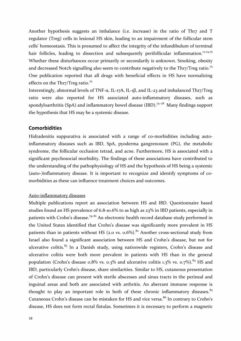
2. Refined Hurley classification for hidradenitis suppurativa
After a patient is diagnosed with HS, the patient is usually classified to indicate current
disease symptoms. Originally, the Hurley classification was developed in 1989 by the
dermatologist H.J. Hurley to identify HS symptoms in one body region for surgical
treatment purposes (Table 1, General introduction).31 Although simple in use, and
therefore popular, the Hurley classification was not developed to stage a whole HS patient
and to define disease severity (i.e. stage I being mild, II moderate, and III severe) as it does
not take into account inflammatory signs and extensiveness of the disease. Furthermore, it
has never been validated.116 As such, it is not suitable to guide holistic treatment plans and
does not reflect current disease activity.134,135 Therefore, a modification of the Hurley
classification was proposed recently by a Dutch panel of HS experts.136
When breaking HS down to its core symptoms and signs, the developers of the refined
Hurley classification agreed on three main items that are important to classify the entire
HS patient:
the presence of inflammation, to guide anti-inflammatory treatments;
the presence of operable sinus tracts, to guide surgical treatment approaches;
the extensiveness of the disease, i.e. number and size of the involved body areas.
The refined Hurley classification contains a three-step algorithm after which the HS
patient is classified into one of the seven stages (Figure 3). Hurley I and II are subdivided
in three sub stages each, i.e. IA, IB, and IC and IIA, IIB, and IIC. The letters A, B, and C are
suggested to represent HS disease severity, in which A stands for mild, B for moderate, and
C for severe disease. Refined Hurley stage III is not sub-staged, but is redefined: at least 1%
of the body surface area in an involved body area is affected with interconnected sinus
tracts with the presence of inflammatory sinus tracts. Regardless of the number of affected
body regions, refined Hurley stage III is always considered as severe HS disease as well.136
Before a new (or refined) classification can be implemented, it is important to assess its
validity. However, there are no clear-cut existing guidelines on validation of classification
systems in medicine. Literature about the methodology for validation of measurement
systems in medicine is more substantial.137 In this thesis, the first steps are made to
investigate whether the refined Hurley classification is a sound system to classify HS
patients. At a minimum, the interrater and intrarater reliability should be analysed. Due to
the underlying severity scale in the refined Hurley stage I and II, these sub-scales can be
regarded as (ordinal) measurements scales. Therefore, the construct validity can be
investigated, which indicates the degree to which the refined Hurley severity sub-scales
imply what it is measuring/indicating.
In Chapter 4, the construct validity of the refined Hurley classification is investigated by
correlating it to the dermatology life quality index (DLQI, a patient reported quality of life
measurement) and to the International HS Severity Score System (IHS4, objective disease

27
severity assessment), and in Chapter 5 the interrater and intrarater reliability are assessed.
Also, the face validity is explored, which is a test that indicates how well the refined Hurley
classification is subjectively viewed as covering the concept it aims to measure. In Chapter
6, a patient self-assessment questionnaire based on the refined Hurley classification is
developed and tested in a patient cohort.
Figure 3. Refined Hurley classification flowchart136
3. Clinical categories of hidradenitis suppurativa
Besides heterogeneous У-secretase gene mutations in a minority of HS patients, other
possibly relevant underlying genetic mutations in HS have not been uncovered yet.138 It is
reported that HS displays a certain phenotypic heterogeneity.138,139 Identification and
accurate description of the phenotype(s) of HS patients can assist in understanding many
aspects of HS disease such as aetiology, pathophysiology, and treatment, and can help to
enhance phenotype-genotype correlations. So far, scientifically identified and validated
clinical phenotypes for HS do not exist yet.33,139–143 Definitions for ‘clinical phenotype’ vary
from a single, few, multiple or the sum of all observable characteristics (disease/patient)
that describes differences between individuals with a certain disease as they relate to
clinically meaningful outcomes. In order to contribute to the sound description of clinical
phenotypes in HS, cluster analysis was performed on a multi-centre patient cohort of adult
patients with HS to identify distinct patient categories in HS in Chapter 7.

28
4. Mechanical stress as a risk factor in hidradenitis suppurativa
One of the frequently proposed risk factors for HS is the exogenous influence of
mechanical stress or friction.144 Currently, it is still unknown why HS preferably presents in
the intertriginous body sites. One theory is that the body folds are more predisposed to
mechanical friction or stress than non-intertriginous body areas.144 Unfortunately, no
experimental evidence exists to support the hypothesis that mechanical stress is a cause or
aggravating factor of HS. In Chapter 8, a case is reported of a male patient known with HS
and working as a road maker, who developed an ectopic HS lesion on his dorsal foot area.

29
References 1. Jemec GBE. Hidradenitis Suppurativa. N Engl J Med. 2012;366(2):158-164. 2. Velpeau A. Dictionnaire de Medecine Ou, Repertoire General Des Sciences Medicales Considerees Sous Les
Rapports Theorique et Pratique. Paris: Bechet Jeune; 1839. 3. Verneuil A. Etudes sur les tumeurs de la peau et quelques maladies de glandes sudoripares [Studies on
skin tumors; some diseases of the sweat glands]. Arch Gen Med. 1854;(94):693–705. 4. Pollitzer S. Hydradenitis destruens suppurativa. J cutan Dis. 1892;(10):9-24. 5. Brunsting HA. Hidradenitis suppurativa; abscess of the apocrine sweat glands: A study of the clinical and
pathologic features, with a report of twenty-two cases and a review of the literature. Arch Derm Syphilol. 1939;39(1):108-120.
6. Brunsting HA. Hidradenitis and other variants of acne. AMA Arch Derm Syphilol. 1952;65(3):303-315. 7. Shelly WB, Cahn MM. The pathogenesis of hidradenitis suppurativa in man; experimental and histologic
observations. AMA Arch Derm. 1955;72(6):562-565. 8. Pillsbury DM, Shelley WB, Kligman AM. Bacterial infections of the skin. Dermatology. 1956:482-489. 9. Plewig G, Kligman AM. Acne, Morphogenesis and Treatment. Berlin Heidelberg New York: Springer; 1975. 10. Plewig G, Steger M. Acne inversa (alias acne triad, acne tetrad or hidradenitis suppurativa). In: Marks
R, Plewig G (eds) Acne and related disorders. Martin Dunitz, London; 1989. 11. Jansen I, Altmeyer P, Piewig G. Acne inversa (alias hidradenitis suppurativa). J Eur Acad Dermatol
Venereol. 2001;15(6):532-540. 12. Chen W, Plewig G. Should hidradenitis suppurativa/acne inversa best be renamed as “dissecting terminal
hair folliculitis”?. Exp Dermatol. 2017;26(6):544-547. 13. Jemec GB, Heidenheim M, Nielsen NH. The prevalence of hidradenitis suppurativa and its potential
precursor lesions. J Am Acad Dermatol. 1996;35(2 Pt 1):191-194. 14. Revuz JE, Canoui-Poitrine F, Wolkenstein P, et al. Prevalence and factors associated with hidradenitis
suppurativa: results from two case-control studies. J Am Acad Dermatol. 2008;59(4):596-601. 15. Cosmatos I, Matcho A, Weinstein R, Montgomery MO, Stang P. Analysis of patient claims data to
determine the prevalence of hidradenitis suppurativa in the United States. J Am Acad Dermatol. 2013;68(3):412-419.
16. Shlyankevich J, Chen AJ, Kim GE, Kimball AB. Hidradenitis suppurativa is a systemic disease with substantial comorbidity burden: a chart-verified case-control analysis. J Am Acad Dermatol. 2014;71(6):1144-1150.
17. Shahi V, Alikhan A, Vazquez BG, Weaver AL, Davis MD. Prevalence of hidradenitis suppurativa: a population-based study in Olmsted County, Minnesota. Dermatology. 2014;229(2):154-158.
18. Garg A, Lavian J, Lin G, Strunk A, Alloo A. Incidence of hidradenitis suppurativa in the United States: A sex- and age-adjusted population analysis. J Am Acad Dermatol. 2017;77(1):118-122.
19. Lee JH, Kwon HS, Jung HM, Kim GM, Bae JM. Prevalence and comorbidities associated with hidradenitis suppurativa in Korea: a nationwide population-based study. J Eur Acad Dermatology Venereol. 2018;32(10):1784-1790.
20. Vaidya T, Vangipuram R, Alikhan A. Examining the race-specific prevalence of hidradenitis suppurativa at a large academic center; results from a retrospective chart review. Dermatol Online J. 2017;23(6).
21. Zouboulis CC, Desai N, Emtestam L, et al. European S1 guideline for the treatment of hidradenitis suppurativa/acne inversa. J Eur Acad Dermatology Venereol. 2015;29(4):619-644.
22. Vossen ARJ V., Schoenmakers A, van Straalen KR, Prens EP, van der Zee HH. Assessing pruritus in hidradenitis suppurativa: a cross-sectional study. Am J Clin Dermatol. 2017;18(5):687-695.
23. Ring HC, Theut Riis P, Zarchi K, Miller IM, Saunte DM, Jemec GB. Prodromal symptoms in hidradenitis suppurativa. Clin Exp Dermatol. 2017;42(3):261-265.
24. Blok JL, Boersma M, Terra JB, et al. Surgery under general anaesthesia in severe hidradenitis suppurativa: a study of 363 primary operations in 113 patients. J Eur Acad Dermatology Venereol. 2015;29(8):1590-1597.
25. Saunte DM, Boer J, Stratigos A, et al. Diagnostic delay in hidradenitis suppurativa is a global problem. Br J Dermatol. 2015;173(6):1546-1549.
26. Fimmel S, Zouboulis CC. Comorbidities of hidradenitis suppurativa (acne inversa). Dermatoendocrinol. 2010;2(1):9-16.
27. Kurzen H, Kurokawa I, Jemec GBE, et al. What causes hidradenitis suppurativa? Exp Dermatol. 2008;17(5):455-456; discussion 457-72.

30
28. Esmann S, Dufour DN, Jemec GBE. Questionnaire-based diagnosis of hidradenitis suppurativa: specificity, sensitivity and positive predictive value of specific diagnostic questions. Br J Dermatol. 2010;163(1):102-106.
29. Vinding GR, Miller IM, Zarchi K, Ibler KS, Ellervik C, Jemec GBE. The prevalence of inverse recurrent suppuration: a population-based study of possible hidradenitis suppurativa. Br J Dermatol. 2014;170(4):884-889.
30. Revuz J. Hidradenitis suppurativa. J Eur Acad Dermatology Venereol. 2009;23(9):985-998. 31. Hurley HJ. Axillary hyperhidrosis, apocrine bromhidrosis, hidradenitis suppurativa, and familial benign
pemphigus: surgical approach. Dermatologic surgery. New York: Marcel Dekker. 1989:729–739. 32. Thorlacius L, Garg A, Riis PT, et al. Interrater agreement and reliability of outcome measurement
instruments and staging systems used in hidradenitis suppurativa. Br J Dermatol. 2019;181(3):483-491 33. van der Zee HH, Laman JD, Boer J, Prens EP. Hidradenitis suppurativa: viewpoint on clinical phenotyping,
pathogenesis and novel treatments. Exp Dermatol. 2012;21(10):735-739. 34. Prens E, Deckers I. Pathophysiology of hidradenitis suppurativa: an update. J Am Acad Dermatol.
2015;73(5):S8-S11. 35. Kelly G, Prens EP. Inflammatory mechanisms in hidradenitis suppurativa. Dermatol Clin. 2016;34(1):51-
58. 36. Frew JW, Hawkes JE, Krueger JG. A systematic review and critical evaluation of inflammatory cytokine
associations in hidradenitis suppurativa. F1000Research. 2018;7:1930. 37. Vossen ARJ V., van der Zee HH, Prens EP. Hidradenitis suppurativa: a systematic review integrating
inflammatory pathways into a cohesive pathogenic model. Front Immunol. 2018;9. 38. Miller IM, McAndrew RJ, Hamzavi I. Prevalence, risk Factors, and comorbidities of hidradenitis
suppurativa. Dermatol Clin. 2016;34(1):7-16. 39. Fitzsimmons JS, Fitzsimmons EM, Gilbert G. Familial hidradenitis suppurativa: evidence in favour of single
gene transmission. J Med Genet. 1984;21(4):281-285. 40. Fitzsimmons JS, Guilbert PR, Fitzsimmons EM. Evidence of genetic factors in hidradenitis suppurativa. Br J
Dermatol. 1985;113(1):1-8. 41. Von Der Werth JM, Williams HC, Raeburn JA. The clinical genetics of hidradenitis suppurativa revisited. Br
J Dermatol. 2000;142(5):947-953. 42. Gao M, Wang P-G, Cui Y, et al. Inversa acne (hidradenitis suppurativa): a case report and identification of
the locus at chromosome 1p21.1–1q25.3. J Invest Dermatol. 2006;126(6):1302-1306. 43. Ingram JR, Wood M, John B, Butler R, Anstey A V. Absence of pathogenic γ-secretase mutations in a
South Wales cohort of familial and sporadic hidradenitis suppurativa (acne inversa). Br J Dermatol. 2013;168(4):874-876.
44. Nomura Y, Nomura T, Suzuki S, et al. A novel NCSTN mutation alone may be insufficient for the development of familial hidradenitis suppurativa. J Dermatol Sci. 2014;74(2):180-182.
45. Pink AE, Simpson MA, Desai N, Trembath RC, Barker JNW. γ-Secretase mutations in hidradenitis suppurativa: new insights into disease pathogenesis. J Invest Dermatol. 2013;133(3):601-607.
46. Wang B, Yang W, Wen W, et al. Gamma-secretase gene mutations in familial acne inversa. Science. 2010;330(6007):1065.
47. Pan Y, Lin M-H, Tian X, et al. gamma-secretase functions through Notch signaling to maintain skin appendages but is not required for their patterning or initial morphogenesis. Dev Cell. 2004;7(5):731-743.
48. Li T, Wen H, Brayton C, et al. Epidermal growth factor receptor and notch pathways participate in the tumor suppressor function of gamma-secretase. J Biol Chem. 2007;282(44):32264-32273.
49. Jemec GB, Hansen U. Histology of hidradenitis suppurativa. J Am Acad Dermatol. 1996;34(6):994-999. 50. Boer J, Weltevreden EF. Hidradenitis suppurativa or acne inversa. A clinicopathological study of early
lesions. Br J Dermatol. 1996;135(5):721-725. 51. Danby FW, Jemec GBE, Marsch WC, von Laffert M. Preliminary findings suggest hidradenitis suppurativa
may be due to defective follicular support. Br J Dermatol. 2013;168(5):1034-1039. 52. Kamp S, Fiehn AM, Stenderup K, et al. Hidradenitis suppurativa: a disease of the absent sebaceous gland?
Sebaceous gland number and volume are significantly reduced in uninvolved hair follicles from patients with hidradenitis suppurativa. Br J Dermatol. 2011;164(5):1017-1022.
53. Blok J, Janse I, Horvath B, Jonkman M. Increased expression of integrin α6β4 in the basement membrane zone lining the sebaceous glands in hidradenitis suppurativa. Acta Derm Venereol. 2015;95(8):994-996.
54. Janse IC, Blok JL, Diercks GFH, Horvath B, Jonkman MF. Hidradenitis suppurativa: a disease of infundibular epidermis rather than pilosebaceous units? Br J Dermatol. 2017;176(6):1659-1661.
31
55. Schlapbach C, Hanni T, Yawalkar N, Hunger RE. Expression of the IL-23/Th17 pathway in lesions of hidradenitis suppurativa. J Am Acad Dermatol. 2011;65(4):790-798.
56. Wolk K, Warszawska K, Hoeflich C, et al. Deficiency of IL-22 contributes to a chronic inflammatory disease: pathogenetic mechanisms in acne inversa. J Immunol. 2011;186(2):1228-39
57. Blok JL, Li K, Brodmerkel C, Jonkman MF, Horváth B. Gene expression profiling of skin and blood in hidradenitis suppurativa. Br J Dermatol. 2016;174(6):1392-4
58. Kanni T, Zenker O, Habel M, Riedemann N, Giamarellos-Bourboulis EJ. Complement activation in hidradenitis suppurativa: a new pathway of pathogenesis? Br J Dermatol. 2018;179(2):413-419.
59. Giang J, Seelen MAJ, van Doorn MBA, Rissmann R, Prens EP, Damman J. Complement activation in inflammatory skin diseases. Front Immunol. 2018;9:639
60. Hoffman LK, Tomalin LE, Schultz G, et al. Integrating the skin and blood transcriptomes and serum proteome in hidradenitis suppurativa reveals complement dysregulation and a plasma cell signature. PLoS One. 2018;13(9):e0203672.
61. Witte-Handel E, Wolk K, Tsaousi A, et al. The IL-1 pathway is hyperactive in hidradenitis suppurativa and contributes to skin infiltration and destruction. J Invest Dermatol. 2019;139(6):1294-1305.
62. Kelly G, Sweeney CM, Tobin A-M, Kirby B. Hidradenitis suppurativa: the role of immune dysregulation. Int J Dermatol. 2014;53(10):1186-1196.
63. Jimenez-Gallo D, de la Varga-Martinez R, Ossorio-Garcia L, Albarran-Planelles C, Rodriguez C, Linares-Barrios M. The clinical significance of increased serum proinflammatory cytokines, C-reactive protein, and erythrocyte sedimentation rate in patients with hidradenitis suppurativa. Mediators Inflamm. 2017;2017:1-8.
64. Ring HC, Emtestam L. The microbiology of hidradenitis suppurativa. Dermatol Clin. 2016;34(1):29-35. 65. Sartorius K, Killasli H, Oprica C, Sullivan A, Lapins J. Bacteriology of hidradenitis suppurativa exacerbations
and deep tissue cultures obtained during carbon dioxide laser treatment. Br J Dermatol. 2012;166(4):879-883.
66. Lapins J, Jarstrand C, Emtestam L. Coagulase-negative staphylococci are the most common bacteria found in cultures from the deep portions of hidradenitis suppurativa lesions, as obtained by carbon dioxide laser surgery. Br J Dermatol. 1999;140(1):90-95.
67. von Eiff C, Peters G, Heilmann C. Pathogenesis of infections due to coagulase-negative staphylococci. Lancet Infect Dis. 2002;2(11):677-685.
68. Ring HC, Thorsen J, Saunte DM, et al. The follicular skin microbiome in patients with hidradenitis suppurativa and healthy controls. JAMA dermatology. 2017;153(9):897-905.
69. Karagiannidis I, Nikolakis G, Zouboulis CC. Endocrinologic aspects of hidradenitis suppurativa. Dermatol Clin. 2016;34(1):45-49.
70. Boer J, Nazary M. Long-term results of acitretin therapy for hidradenitis suppurativa. Is acne inversa also a misnomer? Br J Dermatol. 2011;164(1):170-175.
71. Schrader AMR, Deckers IE, van der Zee HH, Boer J, Prens EP. Hidradenitis suppurativa: a retrospective study of 846 Dutch patients to identify factors associated with disease severity. J Am Acad Dermatol. 2014;71(3):460- 467.
72. Alikhan A, Lynch PJ, Eisen DB. Hidradenitis suppurativa: A comprehensive review. J Am Acad Dermatol. 2009;60(4):539-561.
73. von der Werth JM, Williams HC. The natural history of hidradenitis suppurativa. J Eur Acad Dermatol Venereol. 2000;14(5):389-392.
74. Schneider MR, Paus R. Deciphering the functions of the hair follicle infundibulum in skin physiology and disease. Cell Tissue Res. 2014;358(3):697-704.
75. Melnik BC, John SM, Chen W, Plewig G. T helper 17 cell/regulatory T-cell imbalance in hidradenitis suppurativa/acne inversa: the link to hair follicle dissection, obesity, smoking and autoimmune comorbidities. Br J Dermatol. 2018;179(2):260-272
76. O’Shea J, Ma A, Lipsky P. Cytokines and autoimmunity. Nat Rev Immunol. 2002;2(1):37-45. 77. van der Heijde D, Cheng-Chung Wei J, Dougados M, et al. Ixekizumab, an interleukin-17A antagonist in
the treatment of ankylosing spondylitis or radiographic axial spondyloarthritis in patients previously untreated with biological disease-modifying anti-rheumatic drugs (COAST-V): 16 week results of a phase 3 randomised, double- blind, active-controlled and placebo-controlled trial. Lancet. 2018;392(10163):2441-2451.
78. Vanaki N, Aslani S, Jamshidi A, Mahmoudi M. Role of innate immune system in the pathogenesis of ankylosing spondylitis. Biomed Pharmacother. 2018;105:130-143.

32
79. van der Zee HH, van der Woude CJ, Florencia EF, Prens EP. Hidradenitis suppurativa and inflammatory bowel disease: are they associated? Results of a pilot study. Br J Dermatol. 2010;162(1):195-197.
80. van der Zee HH, de Winter K, van der Woude CJ, Prens EP. The prevalence of hidradenitis suppurativa in 1093 patients with inflammatory bowel disease. Br J Dermatol. 2014;171(3):673-675.
81. Janse IC, Koldijk MJ, Spekhorst LM, et al. Identification of clinical and genetic parameters associated with hidradenitis suppurativa in inflammatory bowel disease. Inflamm Bowel Dis. 2016;22(1):106-113.
82. Garg A, Hundal J, Strunk A. Overall and subgroup prevalence of Crohn disease among patients with hidradenitis suppurativa. JAMA Dermatology. 2018;154(7):814.
83. Shalom G, Freud T, Ben Yakov G, et al. Hidradenitis suppurativa and inflammatory bowel disease: a cross-sectional study of 3,207 patients. J Invest Dermatol. 2016;136(8):1716-1718.
84. Egeberg A, Jemec GBE, Kimball AB, et al. Prevalence and risk of inflammatory bowel disease in patients with hidradenitis suppurativa. J Invest Dermatol. 2017;137(5):1060-1064.
85. van der Zee HH, Horvath B, Jemec GBE, Prens EP. The association between hidradenitis suppurativa and Crohn’s disease: in search of the missing pathogenic link. J Invest Dermatol. 2016;136(9):1747-1748.
86. Bassas-Vila J, Gonzalez Lama Y. Hidradenitis suppurativa and perianal Crohn disease: differential diagnosis. Actas Dermosifiliogr. 2016;107 Suppl 2:27-31.
87. Monnier L, Dohan A, Amara N, et al. Anoperineal disease in hidradenitis suppurativa: MR imaging distinction from perianal Crohn’s disease. Eur Radiol. 2017;27(10):4100-4109.
88. Mowat C, Cole A, Windsor A, et al. Guidelines for the management of inflammatory bowel disease in adults. Gut. 2011;60(5):571-607.
89. Richette P, Molto A, Viguier M, et al. Hidradenitis suppurativa associated with spondyloarthritis -- results from a multicenter national prospective study. J Rheumatol. 2014;41(3):490-494.
90. Schneider-Burrus S, Witte-Haendel E, Christou D, Rigoni B, Sabat R, Diederichs G. High prevalence of back pain and axial spondyloarthropathy in patients with hidradenitis suppurativa. Dermatology. 2016;232(5):606-612.
91. Fauconier M, Reguiai Z, Barbe C, et al. Association between hidradenitis suppurativa and spondyloarthritis. Joint Bone Spine. 2018;85(5):593-597.
92. Dougados M, Baeten D. Spondyloarthritis. Lancet. 2011;377(9783):2127-2137. 93. van Tubergen A. The changing clinical picture and epidemiology of spondyloarthritis. Nat Rev Rheumatol.
2015;11(2):110-118. 94. Ashchyan HJ, Butler DC, Nelson CA, et al. The association of age with clinical presentation and
comorbidities of pyoderma gangrenosum. JAMA Dermatology. 2018;154(4):409. 95. Marzano AV, Ceccherini I, Gattorno M, et al. Association of pyoderma gangrenosum, acne, and
suppurative hidradenitis (PASH) shares genetic and cytokine profiles with other autoinflammatory diseases. Medicine (Baltimore). 2014;93(27):e187.
96. Sabat R, Chanwangpong A, Schneider-Burrus S, et al. Increased prevalence of metabolic syndrome in patients with acne inversa. PLoS One. 2012;7(2):e31810.
97. Gold DA, Reeder VJ, Mahan MG, Hamzavi IH. The prevalence of metabolic syndrome in patients with hidradenitis suppurativa. J Am Acad Dermatol. 2014;70(4):699-703.
98. Miller IM, Ellervik C, Vinding GR, et al. Association of metabolic syndrome and hidradenitis suppurativa. JAMA dermatology. 2014;150(12):1273-1280.
99. Arun B, Loffeld A. Long-standing hidradenitis suppurativa treated effectively with metformin. Clin Exp Dermatol. 2009;34(8):920-921.
100. Verdolini R, Clayton N, Smith A, Alwash N, Mannello B. Metformin for the treatment of hidradenitis suppurativa: a little help along the way. J Eur Acad Dermatol Venereol. 2013;27(9):1101-1108.
101. Kierland R. Unusual pyodermas (hidrosadenitis suppurativa, acne conglobata, dissecting cellulitis of the scalp). Minn Med. 1951;34(4):319-325.
102. Vazquez BG, Alikhan A, Weaver AL, Wetter DA, Davis MD. Incidence of hidradenitis suppurativa and associated factors: a population-based study of Olmsted County, Minnesota. J Invest Dermatol. 2013;133(1):97-103.
103. Canoui-Poitrine F, Revuz JE, Wolkenstein P, et al. Clinical characteristics of a series of 302 French patients with hidradenitis suppurativa, with an analysis of factors associated with disease severity. J Am Acad Dermatol. 2009;61(1):51-57.
104. Lavogiez C, Delaporte E, Darras-Vercambre S, et al. Clinicopathological study of 13 cases of squamous cell carcinoma complicating hidradenitis suppurativa. Dermatology. 2010;220(2):147-153.

33
105. Blok J, Jonkman M, Horvath B. The possible association of hidradenitis suppurativa and Down syndrome: is increased amyloid precursor protein expression resulting in impaired Notch signalling the missing link? Br J Dermatol. 2014;170(6):1375-1377.
106. Maintz L, Betz RC, Allam J-P, et al. Keratitis-ichthyosis-deafness syndrome in association with follicular occlusion triad. Eur J Dermatol. 15(5):347-352.
107. Loo WJ, Rytina E, Todd PM. Hidradenitis suppurativa, Dowling-Degos and multiple epidermal cysts: a new follicular occlusion triad. Clin Exp Dermatol. 2004;29(6):622-624.
108. Agut-Busquet E, Gonzalez-Villanueva I, Romani de Gabriel J, Pascual J, Ribera Pibernat M, Luelmo J. Dowling-Degos 24 disease and hidradenitis suppurativa. Epidemiological and clinical study of 15 patients and review of the literature. Acta Derm Venereol. 2019 Sep 1;99(10):917-918
109. von der Werth JM, Jemec GB. Morbidity in patients with hidradenitis suppurativa. Br J Dermatol. 2001;144(4):809-813.
110. Matusiak Ł. Profound consequences of hidradenitis suppurativa: a review. Br J Dermatol. May 2018 [Epub ahead of print].
111. Basra MKA, Fenech R, Gatt RM, Salek MS, Finlay AY. The Dermatology Life Quality Index 1994-2007: a comprehensive review of validation data and clinical results. Br J Dermatol. 2008;159(5):997-1035.
112. Onderdijk AJ, van der Zee HH, Esmann S, et al. Depression in patients with hidradenitis suppurativa. J Eur Acad Dermatol Venereol. 2013;27(4):473-478.
113. Shavit E, Dreiher J, Freud T, Halevy S, Vinker S, Cohen AD. Psychiatric comorbidities in 3207 patients with hidradenitis suppurativa. J Eur Acad Dermatology Venereol. 2015;29(2):371-376.
114. Thorlacius L, Cohen AD, Gislason GH, Jemec GBE, Egeberg A. Increased suicide risk in patients with hidradenitis suppurativa. J Invest Dermatol. 2018;138(1):52-57.
115. Janse IC, Deckers IE, van der Maten AD, et al. Sexual health and quality of life are impaired in hidradenitis suppurativa: a multicentre cross-sectional study. Br J Dermatol. 2017;176(4):1042-1047.
116. Ingram JR, Hadjieconomou S, Piguet V. Development of core outcome sets in hidradenitis suppurativa: systematic review of outcome measure instruments to inform the process. Br J Dermatol. 2016;175(2):263-272.
117. Thorlacius L, Ingram JR, Villumsen B, et al. A core domain set for hidradenitis suppurativa trial outcomes: an international Delphi process. Br J Dermatol. 2018;179(3):642-650.
118. Thorlacius L, Garg A, Ingram JR, et al. Towards global consensus on core outcomes for hidradenitis suppurativa research: an update from the HISTORIC consensus meetings I and II. Br J Dermatol. 2018;178(3):715-721.
119. Matusiak L, Bieniek A, Szepietowski JC. Acitretin treatment for hidradenitis suppurativa: a prospective series of 17 patients. Br J Dermatol. 2014;171(1):170-174.
120. Janse I, Bieniek A, Horvath B, Matusiak Ł. Surgical procedures in hidradenitis suppurativa. Dermatol Clin. 2016;34(1):97-109.
121. Blok JL, Spoo JR, Leeman FWJ, Jonkman MF, Horvath B. Skin-Tissue-sparing Excision with Electrosurgical Peeling (STEEP): a surgical treatment option for severe hidradenitis suppurativa Hurley stage II/III. J Eur Acad Dermatology Venereol. 2015;29(2):379-382.
122. Riis PT, Boer J, Prens EP, et al. Intralesional triamcinolone for flares of hidradenitis suppurativa (HS): A case series. J Am Acad Dermatol. 2016;75(6):1151-1155.
123. Boer J, Jemec GBE. Resorcinol peels as a possible self-treatment of painful nodules in hidradenitis suppurativa. Clin Exp Dermatol. 2010;35(1):36-40.
124. Pascual JC, Encabo B, Ruiz de Apodaca RF, Romero D, Selva J, Jemec GB. Topical 15% resorcinol for hidradenitis suppurativa: an uncontrolled prospective trial with clinical and ultrasonographic follow-up. J Am Acad Dermatol. 2017;77(6):1175-1178.
125. Horvath B, Janse IC, Sibbald GR. Pain management in patients with hidradenitis suppurativa. J Am Acad Dermatol. 2015;73(5 Suppl 1):S47-51.
126. Saunte DM, Lapins J. Lasers and intense pulsed light hidradenitis suppurativa. Dermatol Clin. 2016;34(1):111-119.
127. Brocard A, Knol A-C, Khammari A, Dreno B. Hidradenitis suppurativa and zinc: a new therapeutic approach. A pilot study. Dermatology. 2007;214(4):325-327.
128. Kromann CB, Deckers IE, Esmann S, Boer J, Prens EP, Jemec GBE. Risk factors, clinical course and long-term prognosis in hidradenitis suppurativa: a cross-sectional study. Br J Dermatol. 2014;171(4):819-824.
129. Vanlaerhoven AMJD, Ardon CB, van Straalen KR, Vossen ARJ V, Prens EP, van der Zee HH. Hurley III Hidradenitis suppurativa has an aggressive disease course. Dermatology. 2018;234(5-6):232-233.

34
130. Stolwijk C, van Onna M, Boonen A, van Tubergen A. Global prevalence of spondyloarthritis: a systematic review and meta-regression analysis. Arthritis Care Res (Hoboken). 2016;68(9):1320-1331.
131. Rudwaleit M, Landewe R, van der Heijde D, et al. The development of Assessment of SpondyloArthritis international Society classification criteria for axial spondyloarthritis (part I): classification of paper patients by expert opinion including uncertainty appraisal. Ann Rheum Dis. 2009;68(6):770-776.
132. Rudwaleit M, van der Heijde D, Landewe R, et al. The development of Assessment of SpondyloArthritis international Society classification criteria for axial spondyloarthritis (part II): validation and final selection. Ann Rheum Dis. 2009;68(6):777-783.
133. Rudwaleit M, van der Heijde D, Landewe R, et al. The Assessment of SpondyloArthritis International Society classification criteria for peripheral spondyloarthritis and for spondyloarthritis in general. Ann Rheum Dis. 2011;70(1):25-31.
134. Zouboulis CC, del Marmol V, Mrowietz U, Prens EP, Tzellos T, Jemec GBE. Hidradenitis suppurativa/acne inversa: criteria for diagnosis, severity assessment, classification and disease evaluation. Dermatology. 2015;231(2):184-190.
135. Revuz JE, Jemec GBE. Diagnosing hidradenitis suppurativa. Dermatol Clin. 2016;34(1):1-5. 136. Horvath B, Janse IC, Blok JL, et al. Hurley staging Refined: a proposal by the Dutch hidradenitis
suppurativa expert group. Acta Derm Venereol. 2017;97(3):412-413. 137. de Vet HCW, Terwee CB, Mokkink LB, Knol DL. Measurement in Medicine - A Practical Guide. Cambridge
University Press; 2011. 138. Ingram JR. The Genetics of Hidradenitis Suppurativa. Dermatol Clin. 2016;34(1):23-28. 139. Ingram JR, Piguet V. Phenotypic heterogeneity in hidradenitis suppurativa (acne inversa): classification is
an essential step toward personalized therapy. J Invest Dermatol. 2013;133(6):1453-1456. 140. Canoui-Poitrine F, Le Thuaut A, Revuz JE, et al. Identification of Three Hidradenitis Suppurativa
Phenotypes: Latent Class Analysis of a Cross-Sectional Study. J Invest Dermatol. 2013;133(6):1506-1511. 141. van der Zee HH, Jemec GBE. New insights into the diagnosis of hidradenitis suppurativa: Clinical
presentations and phenotypes. J Am Acad Dermatol. 2015;73(5 Suppl 1):S23-6. 142. Frew JW, Vekic DA, Woods J, Cains GD. Phenotypic heterogeneity implies heterogeneous pathogenic
pathways in hidradenitis suppurativa. Exp Dermatol. 2015;24(5):338-339. 143. van Straalen KR, Verhagen T, Horvath B, et al. Poor interrater reliability of hidradenitis suppurativa
phenotypes. J Am Acad Dermatol. 2018;79(3):577-578. 144. Boer J, Nazary M, Riis PT. The Role of mechanical stress in hidradenitis suppurativa. Dermatol Clin.
2016;34(1):37-43

35

36

37
2
HIGH PREVALENCE OF HIDRADENITIS SUPPURATIVA
SYMPTOMS IN AXIAL SPONDYLOARTHRITIS PATIENTS:
A POSSIBLE NEW EXTRA-ARTICULAR MANIFESTATION
Published in adapted form in
Seminars in Arthritis and Rheumatism. 2019 Feb; 48 (4): 611-17. doi: 10.1016/j.semarthrit.2018.03.010
Angelique Rondags1
Suzanne Arends2,3
Freke R.Wink3
Barbara Horváth1
Anneke Spoorenberg2,3
1. Department of Dermatology, University of Groningen, University Medical Center Groningen, Groningen, the Netherlands. 2. Department of Rheumatology and Clinical Immunology, University Medical Center Groningen, Groningen, the Netherlands. 3. Department of Rheumatology, Medical Center Leeuwarden, Leeuwarden, the Netherlands.
Leeuwarden, the Netherlands

38
ABSTRACT
Background: Spondyloarthritis (SpA), a chronic inflammatory, rheumatic disease, and
hidradenitis suppurativa (HS), a chronic, debilitating, inflammatory skin disease, share
several clinical and pathophysiological features, such as the association with inflammatory
bowel disease and elevated cytokine levels IL-17 and TNF-α. Recently, SpA was reported to
be more prevalent (2.3–28.2%) in patients with HS than in the general population.
Conversely, the prevalence of HS in SpA is not exactly known.
Objective: To determine the prevalence of HS in patients with axial SpA, a subtype of SpA
primarily of the axial skeleton. Secondly, to identify patient characteristics associated with
the presence of HS in axial SpA.
Methods: In this cross-sectional study, a self-screening questionnaire based on validated
diagnostic HS questions was sent to all participating axial SpA patients from the
Groningen Leeuwarden Axial Spondyloarthritis (GLAS) cohort fulfilling the ASAS axial
SpA criteria. Self-reported HS symptoms were confirmed by previous medical diagnosis or
verification by phone using highly specific validated questions.
Results: In total, 75.6% (449/592) questionnaires were eligible for analyses. Included
patients had a mean age of 50 ± 13 years, 64% was male, mean symptom duration was 23 ±
13 years, and 79% was HLA-B27 positive. HS diagnosis could be confirmed in 41 patients,
resulting in an estimated prevalence of 9.1%. In comparison to patients without a positive
history of HS, these patients were more often female (54% vs. 35%, P = 0.02), showed
higher axial SpA disease activity (mean BASDAI 4.5 vs. 3.6, p = 0.01 and ASDASCRP 2.6 vs.
2.2, P = 0.003) and worse quality of life (QoL) (median ASQoL 9.0 vs. 4.0, P < 0.001). Also,
a history of heel enthesitis and dactylitis was more prevalent (34% vs. 19%, P = 0.03 and
15% vs. 6%, P = 0.05, respectively). Multivariable analysis showed that a higher score on
ASDAS was independently associated with HS (OR: 1.639, 95% CI: 1.176–2.284).
Conclusion: In our cohort of axial SpA patients, HS is more prevalent than in the general
population (9.1% versus 0.053–4.1%). HS is associated with female gender, lower QoL, and
especially higher axial SpA disease activity.

39
INTRODUCTION
Axial spondyloarthritis (SpA) is a chronic rheumatic inflammatory disease of primarily the
axial skeleton and is associated with several extra-articular manifestations (EAMs).1,2
Uveitis is the most common EAM, followed by psoriasis and inflammatory bowel disease
(IBD); pooled data showed prevalence rates of approximately 26%, 9% and 7%,
respectively.2 The presence of EAMs may help to diagnose axial SpA.3 Furthermore, the
presence of one or more EAMs may affect axial SpA treatment decisions and disease
prognosis, quality of life (QoL), and other health-related outcomes and resource usages.2
Interestingly, recently SpA was reported to be more common in patients with hidradenitis
suppurativa (HS) than in the general population, with a prevalence rate between 2.3% and
28.2%.4-6
Hidradenitis suppurativa is a chronic, recurrent, often disfiguring and debilitating,
inflammatory skin disease affecting the terminal hair follicles in the apocrine gland
bearing inverse body regions.7 There are no pathognomonic tests for diagnosing HS;
diagnosis is made clinically and based on three primary criteria: 1. Presence of typical
deep-seated painful nodules. Other frequently reported lesions are abscesses, sinus tracts,
scarring, and open “tombstone” double ended comedones; 2. Involvement of HS in at least
one of the predilection areas: axillae, genitofemoral area, buttocks, and inframammary
area in women; and 3. Chronicity and recurrences (i.e., 2 recurrences in 6 months) of
lesions (Figure 1).8
Figure 1. Hurley stages of hidradenitis suppurativa
Hidradenitis suppurativa in an axilla.
A. Hurley stage I: abscess formation, single or multiple, without sinus tracts and scarring.
B. Hurley stage II: recurrent abscesses with sinus tract formation and scarring, single or multiple, widely
separated lesions.
C. Hurley stage III: diffuse or nearly diffuse involvement, with multiple interconnected sinus tracts and
abscesses across the entire area.
With permission reproduced from www.huidziekten.nl.
40
The reported prevalence of HS varies between 0.053% and 4.1%, and is overall estimated at
1%.9-14 Hidradenitis suppurativa affects more females than males (3:1), usually initiates after
puberty and the prevalence declines after 50 years of age.10,14 Cigarette smoking and being
overweight are important risk factors for both the development and severity of HS.15-18 Due
to its chronicity, the location of the affected body areas and symptoms such as pain, itch
and foul smell of discharge of pus, the professional and private quality of life (QoL) of HS
patients is significantly impaired.19,20
The cause and exact pathogenesis of HS are unclear. An increasing amount of literature
postulates that an aberrant innate and adaptive immune response plays a key role in HS.
Very interestingly, the tumor necrosis factor-alpha (TNF-α) pathway and interleukin (IL)-
23/IL-17 axis seem to be involved in the pathogenesis of both axial SpA and HS.1,21-24
Treatment overlap for HS and axial SpA also exists; a substantial part of the patients of
both diseases respond to TNF-α inhibitors.1,8 Moreover, similar to axial SpA, it is known
that IBD, particularly Crohn’s disease, occurs more frequently in HS and vice versa than in
the general population, further strengthening the concept of (partially) shared
pathogenesis.1,2,25,26
Although axial SpA and HS seem to share common denominators in the pathogenesis and
treatment, the prevalence of HS in axial SpA is not exactly known. Therefore, the objective
of this study was to determine the prevalence of HS in a large cohort of axial SpA patients
and to identify patient characteristics associated with the presence of HS in axial SpA.
METHODS
Patients
All patients participating in the Groningen Leeuwarden Axial Spondyloartritis (GLAS)
cohort, included before June 2016, were asked to participate in this study. The GLAS cohort
is an on-going, longitudinal, prospective observational cohort study which started in 2004
by including all consecutive ankylosing spondylitis (AS) patients 18 years or older, fulfilling
the modified New-York criteria and starting TNF-α inhibitors.27 Since the development of
the Assessment in Spondylo-Arthrits international Society (ASAS) axial SpA criteria in
2009, all consecutive axial SpA patients fulfilling these criteria, irrespective of treatment,
are included.28
Patients are clinically evaluated according to a fixed protocol every six months. Patient
characteristics included gender, age, symptom duration, HLA-B27 status, body mass index
(BMI), and smoking history and status. During follow-up, the occurrence/episodes of
peripheral SpA features (peripheral arthritis, heel enthesitis, dactylitis) and EAMs (uveitis,
psoriasis, IBD) are recorded.

41
Furthermore, disease activity is assessed with Bath AS Disease Activity Index (BASDAI,
range 0–10), AS Disease Activity Score (ASDASCRP), C reactive protein (CRP), and
erythrocyte sedimentation rate (ESR). Also, health-related QoL was assessed with the
Ankylosing Spondylitis Quality of Life questionnaire (ASQoL, range 0–18). Structural
spinal damage was quantified by the modified Stoke Ankylosing Spondylitis Spine Score
(mSASSS, range 0–72) at baseline.
HS questionnaire and verification of diagnosis
The applied diagnostic HS question “Do you repeatedly have outbreaks of painful
abscesses or inflammations in the armpits, groin or buttocks or other body areas?” is based
on previously validated questions with a sensitivity between 92% and 97% and specificity
of 82–86%.29 Patients could reply with “no” or “yes, in the past” or “currently”. In addition,
to enable patients to self-assess the presence of HS more accurately, prototypical colour
pictures of HS lesions of the Hurley stages of the axilla and a brief informative description
of HS were added, as suggested by Esmann et al. (Figure 1).29 Patients were asked to fill in
the questionnaire and return it by post using a stamped return envelope. After three
weeks, a reminder was send to the non-respondents. Anonymously returns and largely
incomplete answered questionnaires were excluded.
Two sources of verification of HS diagnoses were used. First, a medical record check was
done in patients who indicated having a diagnosis of HS made by a physician. The
diagnosis of HS was considered valid if it was made by a dermatologist. Secondly,
verification by phone took place by an HS expert physician (AR), using a second validated
diagnostic HS question “Have you had outbreaks of boils during the last six months, with a
minimum of two boils?”. This question yielded a sensitivity of 90% and specificity of 97%
in previous research.30 The history of recurrent boils was also questioned. In addition, in
case of a positive answer, the involved body locations were asked. The diagnosis of HS was
considered verified if the body locations of occurrence of the lesions were specific for HS.
Statistical analysis
First, the prevalence rate of a positive history of HS was calculated. Descriptive statistics
were applied for all relevant variables; results were expressed as number of patients (%),
mean ± standard deviation (SD) or median (interquartile range (IQR)) for categorical,
normally distributed and non-normally distributed data, respectively. Group comparison
for patients with and without HS was done using the independent-sample t-tests or Mann-
Whitney U tests for continuous variables and chi square tests or Fisher exact tests for
dichotomous variables. Univariable and multivariable logistic regression analysis with a
backward likelihood ratio method were performed to investigate independent predictors
for having a history of HS. The maximum number of independent variables allowed to be

42
included in the multivariable analysis was 10% of the number of patients diagnosed with
HS in this study, as explained by a method by Peduzzi et al.31 Data are presented as odds
ratios (OR) with a 95% confidence interval (CI). P-values ≤ 0.05 were considered as
statistically significant. Statistics were performed using IBM SPSS 23.0 software for
Windows (SPSS, Chicago, IL, USA).
Ethical considerations
No formal informed consent is necessary for this type of questionnaire study in the
Netherlands, verified at the local ethics committee of the University Medical Center
Groningen (UMCG). The GLAS cohort was approved by the local ethics committees of the
UMCG and the Medical Center Leeuwarden. All patients provided written informed
consent according to the Declaration of Helsinki, in which they also gave written
permission to be approached (non-committal) for future additional/new SpA related
research.
RESULTS
Patients characteristics and questionnaires sent and returned
All 592 axial SpA patients currently participating in the GLAS cohort were sent a
questionnaire, of which 471 questionnaires (79.6%) questionnaires were returned.
Excluded were 22 (3.8%) questionnaires due to anonymously returns (n = 4) and missing
data (n = 18), resulting in 449 (75.8%) questionnaires eligible for analysis. Baseline
characteristics of the 449 included respondents showed a mean age of 50.1 ± 12.7 years,
63.7% were male, mean symptom duration was 23.0 ± 13.1 years and 78.5% was HLA-B27
positive. The mean scores of BASDAI and ASDASCRP, both assessed at the closest visit to
the questionnaire response date, were 3.7 ± 2.2 and 2.2 ± 1.0, respectively (Table 1). The
remaining 143 axial SpA patients not included in the analysis were significantly younger
(43.3 ± 13.6 years, P < 0.001). The male–female ratio between included and excluded
patients was similar.
Reported prevalence of HS in axial SpA
Of the in total 449 respondents, 50 (11.1%) had a positive self-screening diagnosis of HS, of
whom three had a previously confirmed diagnosis made by a dermatologist. Verification of
the diagnosis of HS took place by phone in the 47 remaining respondents, of which in 38
the diagnoses could be confirmed. Overall, the diagnosis of HS could be made in 41 of the
449 respondents, resulting in an estimated positive history of HS prevalence rate of 9.1% in
the GLAS cohort. Assuming that all non-responders and patients from excluded
questionnaires never had HS, the minimal HS prevalence rate would be 6.9% (41/592).

43
Table 1. Axial SpA patient characteristics with and without HS
Clinical values
Total axial SpA patients
(n = 449)
Axial SpA patients with HS (n = 41)
Axial SpA patients without HS
(n = 408)
Comparison of groups with HS vs. without
HS, P-value
Age, years 50.1 ± 12.7 46.7 ± 12.7 50.5 ± 12.7 .07 Male gender 286 (63.7) 19 (46.3) 267 (65.4) .02 Age of onset axial SpA, years
26.9 ± 10.5 28.3 ± 10.7 26.8 ± 10.5 .41
Symptom duration axial SpA, years
23.0 ± 13.1 19.3 ± 12.5 23.3 ± 13.1 .07
HLA-B27+ 339 (78.5) 31 (81.6) 308 (78.2) .69 BMI at baseline, kg/m
2
26.7 ± 4.6 27.8 ± 4.8 26.6 ± 4.5 .12
BMI ≥ 25 kg/m2 277 (62.4) 31 (75.6) 251 (61.0) .09
History of smoking 290 (67.4) 31 (77.5) 259 (66.4) .16 History of peripheral arthritis
161 (38.5) 16 (41.0) 145 (38.3) .86
History of enthesitis (heel)
90 (20.2) 14 (34.1) 76 (18.8) .03
History of dactylitis 30 (6.7) 6 (14.6) 24 (5.9) .05 History of EAMs
a 209 (46.5) 19 (46.3) 190 (46.6) .00
History of uveitis 143 (35.2) 10 (26.3) 133 (36.1) .29 History of psoriasis 55 (14.0) 8 (21.6) 47 (13.2) .21 History of IBD 50 (12.5) 4 (9.8) 46 (12.7) .80 Crohn’s disease 24 (5.3) 1 (2.4) 23 (5.6) .71 Colitis ulcerosa 23 (5.0) 3 (7.3) 20 (4.9) .46 History of TNF-α inhibitor use
252 (56.4) 21 (51.2) 231 (56.9) .51
BASDAI at last visit, range 0–10
3.6 ± 2.2 4.5 ± 2.6 3.6 ± 2.2 .01
ASDASCRP at last visit 2.2 ± 1.0 2.6 ± 1.1 2.2 ± 1.0 .003 CRP at last visit, mg/l
3 (2–6) 4 (2–9) 3 (2–6) .14
ESR at last visit, mm/hour
9 (4–18) 13 (3–19) 9 (4–18) .52
ASQoL at last visit, range 0–18
4.5 (1.0–9.0) 9.0 (5.0–11.9) 4.0 (1.0–9.0) <.001
mSASSS at baseline, range 0-72
4.6 (1–15.5)b 2.0 (0.0–8.5) 5.0 (1.0–15.7) .07
Values are presented as number of patients (%), mean ± standard deviation, or median (interquartile range). Axial SpA, axial spondyloarthritis; HS, hidradenitis suppurativa; BMI, body mass index; EAMs, extra articular manifestations; IBD, inflammatory bowel disease; TNF-α, tumor necrosis factor-alpha; BASDAI, Bath Ankylosing Spondylitis Disease Activity Index; ASDAS, Ankylosing Spondylitis Disease Activity Score; CRP, C-reactive protein; ESR, erytrocyte sedimentation rate; ASQoL, Ankylosing Spondylitis Quality of Life; mSASSS, modified Stoke Ankylosing Spondylitis Spine Score. a. Uveitis, psoriasis and inflammatory bowel disease. b. MSASSS at baseline is missing in 196 out of 449 (43.7%) respondents.

44
Comparison of axial SpA patients with HS versus without HS
The 41 axial SpA patients with HS were significantly more often female than the 408
patients without HS (53.7% vs. 34.6%, P = 0.02) (Table 1). A history of heel enthesitis and
dactylitis were significantly more prevalent in axial SpA patients with HS, 34.1% vs. 18.8%
(P = 0.03) and 14.7% vs. 5.9% (P = 0.05), respectively. Axial SpA patients with HS scored
significantly worse on self-reported axial SpA-related disease activity (BASDAI) and QoL
(ASQoL) measurements, 4.5 vs. 3.6 (P = 0.01) and 9.0 vs. 4.0 (P < 0.001), respectively. Also,
the composite index of subjective and objective axial SpA-related disease activity
(ASDASCRP) was higher in these patients (2.6 vs. 2.2 respectively, P = 0.003). Axial SpA
patients with HS had a trend to higher BMI and a more often positive smoking history
(27.8 vs. 26.6, P = 0.12 and 77.5% vs. 66.4%, P = 0.16, respectively). No other differences
between these two groups were found for the other patient characteristics (Table 1).
Patients characteristics associated with HS in axial SpA
Univariable logistic regression resulted in six out of 20 tested parameters significantly
associated HS in axial SpA, as also mentioned above (Table A.1, Appendix). HS was
confirmed in 41 axial SpA patients, therefore a maximum of four parameters (10% of 41)
were allowed as to use as independent variables in the multivariable logistic regression
analysis. After careful consideration, the four most clinically relevant from these six
significant parameters were selected, resulting in gender, history of heel enthesitis, history
of dactylitis, and ASDASCRP. BASDAI was not selected since BASDAI and ASDASCRP contain
overlap in patient-reported disease activity and CRP is also included in the ASDAS,
resulting in a more objective disease activity measure. ASQoL was not included, because
QoL reflects disease outcome. Multivariable analysis showed that a higher ASDASCRP,
indicating higher axial SpA disease activity, was the only independent predictor associated
with HS (OR 1.639, 95% CI: 1.176–2.284, P = 0.004).
DISCUSSION
To our knowledge, this is the first study that investigated the prevalence of HS in the axial
SpA population. In our cohort derived from daily clinical practice, we found a prevalence
rate of 9.1% after thorough standardized verification of the HS diagnosis, and minimal
6.9% when correcting for non-response. This is at least 1.7 times more frequent than in the
general population (0.053–4.1%).9-14
Interestingly, compared to the acknowledged EAMs in axial SpA, the prevalence rate of HS
found in our study is more or less similar. The reported pooled prevalence of psoriasis and
IBD in axial SpA is 9.3% and 6.8% respectively, compared to approximately 2% and 0.4% in
the general population.2,32,33

45
This high prevalence of HS in axial SpA is not surprisingly; a link between HS and SpA has
been described previously. Cross-sectional studies showed that SpA is more prevalent in
HS than in the general population. A large multi-centre study reported a crude prevalence
between 2.3% and 3.7% of combined axial and peripheral SpA in 640 HS patients defined
by ASAS and the European Spondyloarthropathy Study Group criteria, respectively, which
is higher than the prevalence of axial SpA in the general population of 0.32–1.4%.1,4 Very
recently, a smaller single-centre study reported a prevalence of SpA as high as 28.2% (11/39
HS patients), in which approximately 80% were defined as axial SpA and 20% peripheral
SpA based on the ASAS criteria, compared to 2.6% (1/39) in the control group), resulting in
a OR of 11 (95% CI: 4.1–83.3).5 However, both studies report limitations such as the
incompleteness of clinical and diagnostic examinations and selection bias.4,6 Furthermore,
another study in 46 HS patients found in 39.1% signs of active inflammation and 32.6%
signs of chronic SpA changes at the sacroiliac joints in retrospectively evaluated pelvic
magnetic resonance images (MRIs).5 Noteworthy, the authors reported that 15.2% of these
HS patients simultaneously presented clinical signs and symptoms of acute or chronic
SpA.5 The used diagnostic MRI criteria for sacroiliitis in this study were not exactly the
same as defined by the ASAS-working group.34-36 Further data suggesting (a partially)
shared pathogenesis exists, comes from multiple case series, in which HS patients had
presented with peripheral and/or axial SpA features.37-39 A similar dysfunctional immune
response is theorized to be involved in both axial SpA and HS, reflected by the elevated
involved cytokines IL-1, IL-23, IL-17 and TNF-α, and the good response to TNF-α inhibitors
in both conditions.1,7,8,21,24 With this in mind, HS might fall under the concept of immune-
mediated inflammatory diseases (IMID), in which a group of seemingly unrelated
conditions are described that share common inflammatory pathways.40 Axial SpA and also
IBD, a comorbidity that is associated with both axial SpA and HS, have already been
described as one of the IMID conditions.40
Group comparison analysis between SpA patients with HS and without HS in our study
showed that HS is more prevalent in females than in males with axial SpA. This is not
surprising, because HS occurs more frequently in females (ratio 3:1).10 Therefore, the
clinicians treating axial SpA patients should be aware of the possible co-occurrence of HS,
particularly in females. However, this might be challenging due to the hidden nature of HS
lesions (inverse body regions) and that HS symptoms, such as malodorous inflammations
and affected intimate body locations, can cause HS patients to feel embarrassed about
talking and showing their lesions.41
Furthermore, two major risk factors identified for disease severity in HS are tobacco
smoking and overweight; most HS patients are overweight (45–80%) and active smokers
(60–90%) or ex-smokers (5–15%).15-18 Also in axial SpA, smoking and obesity were found to
be associated with higher incidence of axial SpA and higher disease activity.42-44 Although

46
it was a trend, also in our study a larger percentage of axial SpA patients with HS had a
positive smoking history and obesity at baseline than patients without HS. The main
possibility for not reaching statistical significance seems a lack of power. In all respondents
(n = 449) both the percentage of smoking (68%) and obesity (62%) were already relatively
high.
Further, other peripheral SpA features such as heel enthesitis and dactylitis were
significantly more prevalent in axial SpA with HS than without HS symptoms.
Interestingly, the occurrence of a positive history of heel enthesitis and dactylitis in HS
patients was also reported previously.4,45 It can be hypothesised that patients with (a
history of) more SpA features have a more active aberrant immune system, making them
more susceptible for having other (auto-)inflammatory diseases, such as HS. This remains
to be investigated.
Another notable finding from our study is that axial SpA patients with HS scored
significantly worse on the measurements BASDAI and ASDASCRP. Resulting from our
multivariable analysis, a higher ASDASCRP, meaning a higher axial SpA disease activity, was
independently associated with a positive history of HS. Furthermore, the outcome measure
ASQoL was significantly higher in axial SpA patients with HS, indicating these patients
experience a worse QoL. HS is a skin condition with one of the highest impact on QoL
among other chronic skin conditions, including eczema and psoriasis.46,47 Therefore, it is
likely that having HS symptoms contributes to a higher (negative) impact on disease
activity and QoL of axial SpA patients.
A strength of the present study is the high response rate to the questionnaire, nearly 80%.
In total, 75.8% of the patients could be included in the final analysis, decreasing the
nonresponse bias to a great extent. Nevertheless, both underestimation and
overestimation of the HS prevalence is still possible, since a postal questionnaire does not
result in the same validity as physical examination by a physician. We have tried to
overcome this by using questions with very high diagnostic power. The HS question used
in our patient-self-administered questionnaire was based on previously validated questions
and showed a very high sensitivity of 92–97% and a specificity of 82–86%.29 To yield an
even higher diagnostic power, we added a brief description and prototypical photos of HS
disease.29 Subsequently, we thoroughly verified the positive self-reported HS symptoms
through a detailed phone call that included another validated question with a high
specificity (97%).30 Due to recurrent nature of HS symptoms in which periods of remission
are possible, we also asked patients by phone if they had a history of such HS symptoms.
Therefore, recall bias is possible. However, in case there was any uncertainty if these
symptoms belonged to the diagnosis of HS, we decided to classify these patients as HS
negative. Another limitation from our study is that there were no longitudinal data,

47
because of the cross-sectional study design. A causal association between axial SpA
characteristics and HS could therefore not be determined.
CONCLUSIONS
Despite these limitations, our results show meaningful information with respect to the
association of two auto-inflammatory diseases: axial SpA and HS. The high prevalence and
shared denominators in the pathogenesis and treatment may cautiously suggest that HS
could be another EAM in (axial) SpA. Clinicians should be aware of undiagnosed HS
symptoms, especially in female axial SpA patients with high disease activity and who
experience poor QoL. Additionally, a history of peripheral SpA features such as heel
enthesitis and dactylitis seem to be associated with the presence of HS. This awareness, in
combination with possible shared treatment options, may benefit and improve disease
understanding and treatment as well as QoL outcomes for both axial SpA and HS patients.
Besides research exploring the possible shared pathophysiology, further studies are needed
to prospectively determine the prevalence and incidence of HS in axial SpA patients
diagnosed by a dermatologist by physical examination.
Acknowledgments
The authors thank the patients and research nurses from the GLAS cohort.

48
APPENDIX
Table A.1. Univariable and multivariable logistic regression analysis of parameters associated
with the presence of hidradenitis suppurativa in 449 patients with axial spondyloarthritis
Clinical values Univariable logistic regression Multivariable logistic regression
OR 95% CI P value OR 95% CI P value
Age, years 0.977 0.952–1.002 .08
a
Female gender 2.193 1.148–4.187 .02
b
Age of onset axial SpA, years 1.013 0.982–1.045 .41
a
Symptom duration axial SpA, years 0.975 0.948–1.003 .08
a
HLA-B27+ 1.237 0.526–2.906 .63
a
BMI at baseline, kg/m2 1.053 0.986–1.125 .12
a
History of smoking 1.742 0.806–3.768 .16
a
History of peripheral arthritis 1.123 0.574–2.192 .74
a
History of heel enthesitis 2.238 1.120–4.471 .02
b
History of dactylitis 2.714 1.040–7.084 .04
b
History of uveitis 0.631 0.297–1.340 .23
a
History of psoriasis 1.814 0.782–4.204 .17
a
History of IBD 0.808 0.274–2.383 .70
a
History of TNF-α inhibitor use 0.795 0.418–1.513 .49
a
BASDAI at last visit 1.199 1.040–1.382 .01
a
ASDASCRP at last visit 1.620 1.167–2.250 .004 1.639 1.176-2.284 .004
CRP at last visit, mg/L 1.017 0.989–1.047 .24
a
ESR at last visit, mm/hour 1.007 0.981–1.003 .62
a
ASQoL at last visit 1.128 1.055–1.205 <.001
a
mSASSS at baseline 0.976 0.940–1.013 .20
a
Values are presented as odds ratios (OR) with a 95% confidence interval (CI).
Axial SpA, axial spondyloarthritis; HS, hidradenitis suppurativa; BMI, body mass index; IBD, inflammatory bowel disease (Crohn’s disease and colitis ulcerosa); TNF-α, tumor necrosis factor-alpha; BASDAI, Bath Ankylosing Spondylitis Disease Activity Index; ASDAS, Ankylosing Spondylitis Disease Activity Score; CRP, C-reactive protein; ESR, erytrocyte sedimentation rate; ASQoL, Ankylosing Spondylitis Quality of Life; mSASSS, modified Stoke Ankylosing Spondylitis Spine Score. a. The variable was not tested in multivariable regression analysis based on the p-value in univariable regression analysis. b. The variable was not selected during multivariable regression analysis.
49
References 1. Sieper J, Poddubnyy D. Axial spondyloarthritis. Lancet. 2017;390:73-84 2. Stolwijk C, Tubergen A, Castillo-Ortiz JD, Boonen A. Prevalence of extra-articular manifestations in patients
with ankylosing spondylitis: a systematic review and meta-analysis. Ann Rheum Dis.2015;74:65-73 3. Rudwaleit M, Heijde D, Khan MA, Braun J, Sieper J. How to diagnose axial spondyloarthritis early. Ann
Rheum Dis. 2004;63:535-543 4. Richette P, Molto A, Viguier M, et al. Hidradenitis suppurativa associated with spondyloarthritis—results
from a multicenter national prospective study. J Rheumatol.2014;41:490-49 5. Schneider-Burrus S, Witte-Haendel E, Christou D, Rigoni B,. Sabat R, Diederichs G. High prevalence of back
pain and axial spondyloarthropathy in patients with hidradenitis suppurativa. Dermatology. 2016;232:606-6012
6. Fauconier M, Reguiai Z, Barbe C, et al. Association between hidradenitis suppurativa and spondyloarthritis Joint Bone Spine. 2018;85(5):593-597
7. Jemec GBH. Hidradenitis suppurativa. N Engl J Med. 2012;366:158-164 8. Zouboulis C, Desai N, Emtestam L, et al. European S1 guideline for the treatment of hidradenitis
suppurativa/acne inversa. J Eur Acad Dermatol Venereol. 2015;29:619-644 9. Jemec GB, Heidenheim M, Nielsen NH. The prevalence of hidradenitis suppurativa and its potential
precursor lesions. J Am Acad Dermatol. 1996;35(2,1):191-194 10. Revuz JE, Canoui-Poitrine F, Wolkenstein P, et al.Prevalence and factors associated with hidradenitis
suppurativa: results from two case–control studies. J Am Acad Dermato. 2008;59:596-601 11. Cosmatos I, Matcho A, Weinstein R, Montgomery MO, Stang P. Analysis of patient claims data to
determine the prevalence of hidradenitis suppurativa in the United States. J Am Acad Dermatol. 2013;68:412-419
12. Shlyankevich J, Chen AJ, Kim GE, Kimball AB. Hidradenitis suppurativa is a systemic disease with substantial comorbidity burden: a chart-verified case–control analysis. J Am Acad Dermatol. 2014;71:1144-1150
13. Shahi V, Alikhan A, Vazquez BG, Weaver AL, Davis MD. Prevalence of hidradenitis suppurativa: a population-based study in Olmsted County, Minnesota. Dermatology. 2014;229: 154-158
14. Garg A, Lavian J, Lin G, Strunk A, Alloo A. Incidence of hidradenitis suppurativa in the United States: a sex- and age- adjusted population analysis. J Am Acad Dermatol. 2017;17:118-122
15. König A, Lehmann C, Rompel R, Happle R. Cigarette smoking as a triggering factor of hidradenitis suppurativa.Dermatology. 1999;198:261-264
16. Sartorius K, Emtestam L, Jemec GB, Lapins J. Objective scoring of hidradenitis suppurativa reflecting the role of tobacco smoking and obesity. Br J Dermatol. 2009;161:831-839
17. Vazquez BG, Alikhan A, Weaver AL, Wetter DA, Davis MD. Incidence of hidradenitis suppurativa and associated factors: a population-based study of Olmsted county, Minnesota. J Invest Dermatol. 2013;133: 97-103
18. Kromann CB, Deckers JE, Esmann S, Boer J, Prens EP, Jemec GB. Risk factors, clinical course and long-term prognosis in hidradenitis suppurativa: a cross-sectional study. Br J Dermatol. 2014;171:819-824
19. Onderdijk AJ, Van der Zee HH, Esmann S, et al. Depression in patients with hidradenitis suppurativa. J Eur Acad Dermatol Venereol, 2013;27:473-478
20. Janse IC, Deckers IE, Maten AD, et al.Sexual health and quality of life are impaired in hidradenitis suppurativa: a multicentre cross-sectional study. Br J Dermato. 2017;176:1042-1047
21. Schlapbach C, Hanni T, Yawalkar N, Hunger RE. Expression of the IL-23/Th17 pathway in lesions of hidradenitis suppurativa. J Am Acad Dermatol. 2011;65:790-798
22. Van der Zee HH, Ruiter L, Broecke DG, Dik WA, Laman JD, Prens EP. Elevated levels of tumour necrosis factor (TNF)-alpha, interleukin (IL)-1beta and IL-10 in hidradenitis suppurativa skin: a rationale for targeting TNF-alpha and IL-1beta. Br J Dermatol. 2011;164:1292-1298
23. Hreggvidsdottir HS, Noordenbos T, Baeten DL. Inflammatory pathways in spondyloarthritis. Mol Immunol. 2014;57:28-37
24. Kelly G, Prens EP. Inflammatory mechanisms in hidradenitis suppurativa. Dermatol Clin. 2016;34:51-58 25. Deckers IE, Benhadou F, Koldijk MJ, et al. Inflammatory bowel disease is associated with hidradenitis
suppurativa: results from a multicenter cross-sectional study. J Am Acad Dermatol. 2017;76:49-53 26. Janse IC, Koldijk MJ, Spekhorst LM, et al.Identification of clinical and genetic parameters associated with
hidradenitis suppurativa in inflammatory bowel disease. Inflamm Bowel Dis. 2016;22:106-113

50
27. Linden S, Valkenburg HA, Cats A. Evaluation of diagnostic criteria for ankylosing spondylitis. A proposal for modification of the New York criteria. Arthritis Rheum. 1984;27:361-368
28. Rudwaleit M, Heijde D, Landewé R, et al.The development of assessment of SpondyloArthritis international society classification criteria for axial spondyloarthritis (part II): validation and final selection. Ann Rheum Dis. 2009;68:777-783
29. Esmann S, Dufour DN, Jemec GB. Questionnaire-based diagnosis of hidradenitis suppurativa: specificity, sensitivity and positive predictive value of specific diagnostic questions. Br J Dermatol. 2010;163:102-106
30. Vinding GR, Miller IM, Zarchi K, Ibler KS, Ellervik C, Jemec GB. The prevalence of inverse recurrent suppuration: a population-based study of possible hidradenitis suppurativa. Br J Dermatol. 2014;170:884-889
31. Peduzzi P, Concato J, Kemper E, Holford TR, Feinstein AR. A simulation study of the number of events per variable in logistic regression analysis. J Clin Epidemiol. 1996;49:1373-1379
32. Christophers E. Psoriasis—epidemiology and clinical spectrum. Clin Exp Dermatol. 2001;26:314-320 33. Loftus EV Jr. Clinical epidemiology of inflammatory bowel disease: incidence, prevalence, and
environmental influences. Gastroenterology. 2004;126:1504-1517 34. Hermann KG, Bollow M. Magnetic resonance imaging of the axial skeleton in rheumatoid disease. Best
Pract Res Clin Rheumatol, 2004;18:881-907 35. Hermann KG, Braun J, Fischer T, Reisshauer H, Bollow M. Magnetic resonance tomography of sacroiliitis:
anatomy, histological pathology, MR-morphology, and grading. Radiologe. 2004;44:217-228 36. Rudwaleit M, Heijde D, Landewe R, et al.The assessment of SpondyloArthritis international society
classification criteria for peripheral spondyloarthritis and for spondyloarthritis in general. Ann Rheum Dis. 2011;70:25-31
37. Rosner IA, Richter DE, Huettner TL, Kuffner GH, Wisnieski JJ, Burg CG. Spondyloarthropathy associated with hidradenitis suppurative and acne conglobate. Ann Intern Med. 1982;97:520-525
38. Bhalla R, Sequeira W. Arthritis associated with hidradenitis suppurativa. Ann Rheum Dis. 1994;53:64-66 39. Lim DT, James NM, Hassan S, Khan MA. Spondyloarthritis associated with acne conglobata, hidradenitis
suppurativa and dissecting cellulitis of the scalp: a review with illustrative cases. Curr Rheumatol Rep, 2013;15
40. Kuek A, Hazleman BL, Ostor AJ. Immune-mediated inflammatory diseases (IMIDs) and biologic therapy: a medical revolution. Postgrad Med J. 2007;83:251-260
41. von der Werth J, Jemec GB. Morbidity in patients with hidradenitis suppurativa. Br J Dermatol. 2001;144:809-813
42. Chung HY, Machado P, Heijde D, D'Agostino MA, Dougados M. Smokers in early axial spondyloarthritis have earlier disease onset, more disease activity, inflammation and damage, and poorer function and health-related quality of life: results from the DESIR cohort. Ann Rheum Dis. 2012;71:809-816
43. Maas F, Arends S, Veer E, et al. Obesity is common in axial spondyloarthritis and is associated with poor clinical outcome. J Rheumatol. 2016;43:383-387
44. Videm V, Cortes A, Thomas R, Brown MA. Current smoking is associated with incident ankylosing spondylitis—the HUNT population-based norwegian health study. J Rheumatol. 2014;41:2041-2048
45. Fioravanti A, Lauraflori M, Guidelli GM, Giordano N. Dactylitis as a first manifestation of arthritis associated with hidradenitis suppurativa. Indian J Dermatol Venereol Leprol. 2011;77:74-76
46. Janse IC, Deckers IE, Maten AD, et al. Sexual health and quality of life are impaired in hidradenitis suppurativa: a multicentre cross-sectional study. Br J Dermatol. 2017;176:1042-1047
47. Onderdijk AJ, Van der Zee HH, Esmann S, et al. Depression in patients with hidradenitis suppurativa. J Eur Acad Dermatol Venereol. 2013;27:473-478

51

52

53
3
HIGH PREVALENCE OF CLINICAL SPONDYLOARTHRITIS
FEATURES IN PATIENTS WITH HIDRADENITIS SUPPURATIVA
Published in adapted form in
Journal of the American Academy of Dermatology. 2019 Feb;80(2):551-554.e1. doi: 10.1016/j.jaad.2018.06.028
Angelique Rondags1
Kelsey R. van Straalen2
Suzanne Arends3
Hessel H. van der Zee2
Errol P. Prens2
Anneke Spoorenberg3
Barbara Horváth1
1. Department of Dermatology, University of Groningen, University Medical Center Groningen, Groningen, the Netherlands. 2. Department of Dermatology, Erasmus Medical Center, Rotterdam, the Netherlands. 3. Department of Rheumatology and Clinical Immunology, University Medical Center Groningen, Groningen, the Netherlands.
54
ABSTRACT
Background: Spondyloarthritis (SpA) and hidradenitis suppurativa (HS) share several
pathophysiological features.
Objective: To investigate the prevalence of self-reported clinical SpA features in HS
patients and to identify patient characteristics associated with these features.
Methods: Cross-sectional study. A questionnaire concerning the presence of ASAS axial
and peripheral SpA entry classification criteria and other SpA features was sent to all HS
patients in two Dutch tertiary HS referral centers.
Results: Overall, 47.2% (620/1313) of questionnaires were eligible for analysis. Of these, 416
(67.1%) patients fulfilled ≥1 of the four ASAS entry criteria. Entry criteria for axial and
peripheral SpA were reported by 72.8% and 27.2%, respectively. The large majority (87%)
reported ≥1 additional clinical SpA features: one feature by 32.9%, two by 29.1%, three by
16.1%, and ≥4 by 8.9%. In comparison to patients without self-reported entry criteria
(n=204), patients with self-reported fulfillment were significantly more frequently female,
had higher BMI, were more often ex- or current smokers, had longer HS disease duration,
and more active HS symptoms at the survey response time.
Limitations: Non-responder bias and self-reporting design.
Conclusion: Self-reported clinical SpA features are common in HS patients, especially in
the ‘classic’ HS patient, with active HS symptoms, and longer HS disease duration.

55
INTRODUCTION
Hidradenitis suppurativa (HS) is a chronic, recurrent, and debilitating inflammatory skin
disease of the inverse body regions.1 Hidradenitis suppurativa is associated with a range of
somatic and psychological comorbidities including inflammatory bowel disease (IBD),
particularly Crohn’s disease, metabolic syndrome, depression, and probably rheumatologic
conditions such as spondyloarthritis (SpA).1,2
Spondyloarthritis, previously termed seronegative spondyloarthritides, is a heterogeneous
group of interrelated chronic inflammatory articular rheumatic conditions, which include
ankylosing spondylitis (AS), psoriatic arthritis, arthritis related to IBD, reactive arthritis,
and undifferentiated SpA.3 These conditions share clinical symptoms and are associated
with the HLA-B27 gene. In most patients, the first symptoms develop in the third or fourth
decade of life, and males are more frequently affected.3 The diagnosis of SpA is made by a
rheumatologist and is mainly based on the presence of “SpA features”, which includes
spinal (axial) features such as sacroiliitis and spondylitis; peripheral features such as
peripheral arthritis, enthesitis, and dactylitis; and the so called extra-articular
manifestations uveitis, psoriasis, and IBD.3 Spondyloarthritis features may develop during
the course of the disease and may come and go and often do not occur simultaneously.
The probability of SpA increases when more of these features have occurred in a patient.4,5
Spondyloarthritis can be categorized as predominantly axial or peripheral depending on
the main involved articular features.3 The latest and currently most used classification
criteria were developed by the Assessment in SpondyloArthrtis international Society
(ASAS) (Figure 1).6-8 These classification criteria comprise clinical features of SpA,
laboratory (e.g. HLA-B27 positivity) and imaging features (e.g. sacroiilitis on radiography
and/or magnetic resonance imagining (MRI)).6-9 According to ASAS, four “entry criteria”
for the classification of SpA into (predominantly) axial or peripheral SpA exist. The entry
criteria for axial SpA begins with “back pain ≥3 months with age of onset before 45 years
(with or without the presence of peripheral manifestations)”.7 The three entry criteria for
peripheral SpA are “peripheral arthritis, enthesitis or dactylitis, without axial involvement”
(Figure 1).8
Until recently, the association between SpA and HS had only been described in limited
publications, mostly concerning case reports and small retrospective case series.10-16 Two
recent studies on the prevalence of SpA in HS patients showed large differences in results.
The first study suggested a prevalence of SpA of 2.3 to 3.7%, depending on the clinical and
imaging criteria used, whereas the other suggested a prevalence of 28.2%.

56
Axial spondyloarthritis Peripheral spondyloarthritis
Entry criterion:
Back pain ≥3 months, onset <45 years old
(with or without peripheral manifestations)
Entry criteria:
In patients with peripheral manifestations ONLY:
Peripheral arthritis or enthesitis (any) or dactylitis
plus:
Imaging group
Sacroiliitis* plus
≥1 other SpA features
or Clinical group
HLA-B27-positive plus
≥2 other SpA features
Additional SpA features
≥1 additional
SpA features or
≥2 other additional
SpA features
▪ IBP
▪ Arthritis
▪ Heel enthesitis
▪ Uveitis
▪ Dactylitis
▪ Psoriasis
▪ IBD
▪ Good response of back pain
to NSAIDs
▪ Family history of SpA
▪ HLA-B27 positive
▪ Increased CRP†
▪ Uveitis
▪ Psoriasis
▪ IBD
▪ Preceding gastro-
intestinal or
genitourinary
infection
▪ HLA-B27 positive
▪ Sacroiliitis*
▪ IBP (ever)
▪ Arthritis
▪ Enthesitis (any)
▪ Dactylitis
▪ Family history of
SpA
Sensitivity and specificity of ASAS classification criteria
Axial SpA criteria: sensitivity 82.9%; specificity 84.4%; n=649.7
Peripheral SpA criteria: sensitivity 77.8%; specificity 82.2%; n=266.8
Combination of axial and peripheral SpA criteria: sensitivity 79.5%; specificity 83.3%; n= 975.8
Figure 1. Classification criteria of axial and peripheral spondyloarthritis according to the
Assessment in SpondyloArthritis international Society (ASAS)
ASAS, Assessment of SpondyloArthritis international Society; SpA, spondyloarthritis; IBP, inflammatory back
pain; CRP, C-reactive protein; IBD, inflammatory bowel disease (Crohn’s disease / colitis ulcerosa).
* Either active inflammation detected by MRI highly suggestive of sacroiliitis associated with SpA, or definite
radiographic sacroiliitis according to the modified New York criteria.
† Increased CRP is considered a SpA feature in the context of chronic back pain.
Adapted from: Rudwaleit M et al., Ann Rheum Dis. 2009;68(6):777-7837; Rudwaleit et al., Ann Rheum Dis.
2011;70(1):25-31.8

57
HS patients.17,18 Both prevalence rates are higher than the prevalence of SpA in the general
population, which is approximately 1%.3,4
Because of these discrepancies in prevalence rates and to obtain insight in which HS
patients report SpA features and possibly have a higher chance of SpA, we undertook this
cross-sectional study in two large HS cohorts in the Netherlands. Our objective was to
investigate the prevalence of self-reported clinical SpA features in HS patients and to
identify HS patient characteristics associated with the presence of these features.
METHODS
Subjects and design
In this multicenter cross-sectional study, adult patients with a billing code for HS
(between 2010 and 2016) in the dermatology departments of the University Medical Center
Groningen (UMCG) and Erasmus Medical Center (EMC) received a postal questionnaire
and were requested to return the completed questionnaire by post using a pre-stamped
return envelope. A reminder was sent to the non-respondents after four weeks. Unopened
returned questionnaires and insufficiently answered surveys (no information on SpA entry
criteria) were excluded. According to Dutch law, no formal informed consent was needed
for this type of study as verified by the UMCG ethical committee.
Questionnaire about clinical SpA features
The questionnaire comprised questions about clinical SpA features that are easily self-
identifiable by patients. These questions were formulated based on the ASAS definitions
for axial and peripheral SpA entry criteria and additional clinical SpA features in the past
or present.6-9 The questions included: a history of back pain existing ≥3 months with an
age of onset <45 years; inflammatory back pain (IBP); response of back pain to non-steroid
anti-inflammatory drugs (NSAIDs); peripheral arthritis (swollen joints); enthesitis of the
Achilles tendon; dactylitis; ophthalmologist diagnosed uveitis; physician diagnosed
psoriasis including pustulosis palmoplantaris; gastroenterologist diagnosed IBD (Crohn’s
disease or ulcerative colitis); and a positive family history of SpA and SpA related
conditions (psoriasis, uveitis, and IBD) in first or second degree relatives. When
applicable, the SpA questions were illustrated with colored prototypical pictures. In
addition, data on age, gender, length, weight, smoking habits, and HS characteristics such
as age of symptom onset and current HS symptoms to derive the Hurley and refined
Hurley stage, were collected.

58
Statistical analysis
Descriptive statistics are reported as mean ± standard deviation (SD) or median
(interquartile range (IQR)) for normally distributed and non-normally distributed data,
respectively. Determined was how many HS patients reported the ASAS “entry criteria” for
classification of SpA. Subsequently, the prevalence of “additional” self-reported SpA
features to the entry criteria were calculated. Comparison between HS patients with and
without self-reported SpA entry criteria was done using the independent-sample t or
Mann-Whitney U tests for continuous and chi square or Fisher’s exact tests for
dichotomous variables. It was reported when ≥5% of data was missing per variable. P-
values ≤ 0.05 were considered as statistically significant. Statistics were performed using
IBM SPSS 23.0 software for Windows (SPSS, Chicago, IL, USA).
RESULTS
Inclusion and exclusion
In total, 1313 HS patients received a questionnaire of which 730 (55.6%) were returned
(Figure 2). Of these 730 questionnaires, 110 (15.1%) were excluded due to unopened returns
(n=70), insufficiently answered surveys (n=32) and other reasons (n=8). Subsequently, 620
(47.2%) questionnaires were eligible for analysis (Figure 2).
The excluded patients (n=693) were more frequently male (36.7 vs. 29.8%, p=0.010) and
were younger (40.8 ± 17.5 vs. 43.4 ± 13.9, p<0.001) compared with the included patients.
Subjects
Overall, 70.2% (434/620) of respondents were female, with a mean age of 43.4 ± 13.9 years,
and mean BMI of 28.0 ± 5.8 kg/m2. Furthermore, 83.5% (518/620) patients were ex- or
current smokers (Table 1). One fourth (24.8%) of patients had no HS symptoms at the time
of the survey, 20.6% were categorized into Hurley stage I, 46.1% into stage II and 8.5% into
stage III. An overview of the distribution within the refined Hurley classification is shown
in Table 1. The patient characteristics were similar between UMCG and EMC cohorts (data
not shown).
Prevalence of clinical SpA features
In total, 416 (67.1%) HS patients fulfilled ≥1 of the four axial or peripheral ASAS entry
criteria (Figure 3). Moreover, 87% of these patients reported ≥1 additional clinical SpA
features: one additional feature was reported by 137 (32.9%) patients, two features by
121 (29.1%), three features by 67 (16.1%), and ≥4 features by 37 (8.9%) patients. Table 2
gives an overview of the self-reported clinical SpA features.

59
Figure 2. Flow chart of exclusion of questionnaires
HS, hidradenitis suppurativa; UMCG, University Medical Center Groningen; EMC, Erasmus Medical Center.
Of the 416 patients who fulfilled ≥1 of the four ASAS entry criteria, 72.8% (303/416)
reported to fulfill the ASAS entry criteria for axial SpA and IBP was reported by 27.7%.
Of the 303 patients, 82.2% reported ≥1 additional clinical SpA feature. The other 27.2%
(113/416) reported to fulfill the ASAS entry criteria for peripheral SpA. Of these
patients, 85.0% reported an episode of peripheral arthritis, 22.1% enthesitis of the
Achilles tendon and 22.1% dactylitis. One peripheral SpA feature was reported by
72.6% (n=82), two by 25.7% (n=29), and 1.8% (n=2) reported all three peripheral SpA
features. All these 113 patients also reported ≥1additional clinical SpA features as well
(Figure 3).
The remaining 32.9% (204/620) patients did not fulfill any of the ASAS entry criteria.
Of these patients, 71.6% did not report the presence of additional SpA features, 23.0%
reported one feature, and 5.4% two features (Figure 3). Family history of SpA
conditions was reported most frequently (21.1%), followed by IBD (6.9%), psoriasis
(6.4%), and uveitis (1.0%).
1313 (UMCG: 654; EMC: 659)
Questionnaires sent to all HS
patients with billing code HS at the UMCG and EMC between
2010-2016
730 (55.6%) (UMCG: 400; EMC: 330)
Questionnaires returned
110 (15.1%) (UMCG: 50; EMC: 60)
Excluded
Reasons:
70 unopened returns to sender; 32 insufficiently answered questions;
5 unwilling to participate; 2 recently deceased;
1 temporarily lives abroad.
620 (47.2%) (UMCG: 350; EMC: 270)
Filled out questionnaires included in final analysis

60
Table 1. Patient characteristics included hidradenitis suppurativa patients (n=620)
Data are presented as number of patients (%) or mean ± standard deviation.
Missing data ≥5%: BMI (6.8%), age of onset HS (6.6%), disease duration of HS (6.5%), Hurley classification stage
(5.2%), refined Hurley classification stage (6.0%).
HS, hidradenitis suppurativa; BMI, Body Mass Index.
Patient characteristics associated with SpA features in HS
In comparison to patients without self-reported entry criteria (204/620), patients
fulfilling the ASAS entry criteria (416/620) were more frequently female (74.8% vs.
60.9%, p<0.001), had a higher BMI (28.6 ± 5.9 vs. 26.7 ± 5.3, p<0.001), were more often
ex- or current smokers (87.9% vs. 76.2%, p=0.001), had a longer HS disease duration
(19.9 ± 12.8 vs. 17.0 ± 12.4, p=0.012), and reported more active HS at time of the survey
response reflected by the Hurley and refined Hurley classification distribution (both
p<0.001) (Table 3).
Patient characteristics
Age, years 43.4 ± 13.9 Female gender 434 (70.2) Age of onset HS, years 24.0 ± 12.5 Disease duration of HS, years 18.9 ± 12.7 BMI, kg/m
2 28.0 ± 5.8
Smoking status Non-smoker Ex-smoker Current smoker
98 (15.9) 199 (32.3) 319 (51.8)
Hurley classification stage Not active Hurley I Hurley II Hurley III
146 (24.8) 121 (20.6) 271 (46.1) 50 (8.5)
Refined Hurley classification stage Not active Refined Hurley IA Refined Hurley IB Refined Hurley IC Refined Hurley IIA Refined Hurley IIB Refined Hurley IIC Refined Hurley III
146 (25.0) 33 (5.7) 14 (2.4) 74 (12.7) 45 (7.7) 68 (11.7) 153 (26.2) 50 (8.6)

61
Figure 3. Flowchart of self-reported ASAS classification entry criteria for spondyloarthirits in
patients with hidradenitis suppurativa
HS, hidradenitis suppurativa; SpA, spondyloarthritis; ASAS, Assessment of SpondyloArthritis international
Society.
620 Included HS patients
416 (67.1%) With fulfillment of ≥1 out of 4
ASAS entry criteria for SpA
204 (32.9%) Without fulfillment of any ASAS entry criteria for SpA
303 (72.8%) Axial SpA entry criterion Back pain ≥3 months, onset
<45 years old (without or with peripheral manifestations)
113 (27.2%) Peripheral SpA entry criteria
Peripheral arthritis (swollen joint) and/or enthesitis of Achilles
tendon and/or dactylitis
249 (82.2%) ≥1 additional self-reported
clinical SpA features
113 (100%) ≥1 additional self-reported
clinical SpA features
58 (28.4%) ≥1 self-reported
clinical SpA features
249 (82.2%) ≥1 additional self-reported
clinical SpA features
113 (100%) ≥1 additional self-reported
clinical SpA features

62
Table 2. Overview of the prevalence of self-reported spondyloarthritis entry criteria and
features in patients with hidradenitis suppurativa
Fulfillment of ≥1 out of 4 ASAS entry criteria
†
(n=416)
Total number of self-reported SpA features in addition to the entry criteria*
0 features 54 (13.0) 1 feature 137 (32.9) 2 features 121 (29.1) 3 features 67 (16.1) ≥4 features 37 (8.9) SpA-features (self-reported) Inflammatory back pain
‡ 84 (20.2)
Effect of NSAIDs on back pain Good response (>50% response) 63 (22.0) No or under 50% response 93 (32.5) No NSAIDs used 30 (45.5) Peripheral arthritis (swollen joints) 248 (59.6) Enthesitis (Achilles tendon) 66 (15.9) Dactylitis 83 (20.0) Uveitis
§ 13 (3.2)
Psoriasis (incl. pustulosis palmoplantaris)§ 39 (9.4)
Inflammatory bowel disease§ 31 (7.6)
Crohn’s disease 19 (4.7) Ulcerative colitis 12 (2.9) Family history for SpA in 1
st/2
nd degree relatives
# 125 (30.0)
SpA, spondyloarthritis; ASAS, Assessment of SpondyloArthritis international Society; NSAIDs, nonsteroidal anti-inflammatory drugs. * Additional self-reported SpA-features: inflammatory back pain, inflammatory bowel syndrome, uveitis, psoriasis, good effect of non-steroidal drugs on back pain, family history of spondyloarthritis, dactylitis, enthesitis of the Achilles tendon, and peripheral arthritis (swollen joint). † ASAS entry criteria for axial and peripheral SpA: “back pain for ≥3 months with age of onset <45 years” and “peripheral arthritis, enthesitis or dactylitis”, respectively. ‡ Inflammatory back pain: at least four out of five ASAS criteria for inflammatory back pain need to be fulfilled: insidious onset, pain at night (with improvement upon getting up), age at onset <40 years, improvement with exercise, and no improvement with rest.
10
§ Physician diagnosed. # Family history for SpA in 1st/2nd degree relatives: ankylosing spondylitis, psoriatic arthritis, psoriasis, uveitis, and inflammatory bowel disease.

63
Table 3. Patient characteristics between hidradenitis suppurativa patients with versus without
fulfillment of spondyloarthritis entry criteria
HS, hidradenitis suppurativa; BMI, Body Mass Index. Missing data ≥5%: Hurley classification stage (5.2%), refined Hurley classification stage (6.0%).
DISCUSSION
This study shows that self-reported clinical SpA features are common in HS patients: 67%
reported ≥1 ASAS entry criteria and the large majority of these patients (87%) reported
additional clinical SpA features. Approximately 75% of these patients reported long-term
back pain as entry criteria for axial SpA of which approximately 28% reported
inflammatory back pain.
In this study, we used the ASAS criteria for axial and peripheral SpA, with a previously
reported sensitivity and specificity of 79.5% and 83.3% respectively according to the
diagnosis made by the rheumatologist.7,8 Important to mention is that the ASAS criteria
were developed as classification criteria for clinical research and not as diagnostic criteria.8
However, for the diagnosis of SpA, the presence and number of clinical SpA features seem
important since the probability of a SpA diagnosis increases if more of these SpA features
are present.4,5,19-21 The relevance of these clinical SpA features for the diagnosis can be
Fulfillment of ≥1 of 4 ASAS entry criteria
No fulfillment of ASAS entry criteria
p-value
(n=416) (n=204)
Age, years 43.8 ± 13.2 42.5 ± 15.2 0.29 Female gender 311 (74.8) 123 (60.9) <0.001 Age of onset HS, years 23.4 ± 12.2 25.0 ± 13.1 0.15 Disease duration of HS, years 19.9 ± 12.8 17.0 ± 12.4 0.012 BMI, kg/m
2 28.6 ± 5.9 26.7 ± 5.3 <0.001
Smoking status 0.001 Non-smoker Ex-smoker Current smoker
50 (12.1) 48(23.8) 138(33.3) 61(30.2) 226(54.6) 93(46.0)
Hurley classification stage <0.001 Not active Hurley I Hurley II Hurley III
78 (20.1) 68 (34) 72 (18.6) 47 (12.1) 198 (51.0) 73 (36.5) 38 (9.8) 12 (6.1)
Refined Hurley classification stage <0.001 Not active Refined Hurley IA Refined Hurley IB Refined Hurley IC Refined Hurley IIA Refined Hurley IIB Refined Hurley IIC Refined Hurley III
78 (20.2) 68 (34.5) 18 (4.7) 15 (7.6) 7 (1.8) 7 (3.6) 49 (12.7) 25 (12.7) 29 (7.5) 16(8.1) 45 (11.7) 23 (11.7) 122 (31.6) 31 (15.7) 38 (9.8) 12 (6.1)

64
expressed in likelihood ratios (LR), i.e. ranging from 2.5 for psoriasis to 7.3 for uveitis.21 In
our study, of the HS patients who reported ≥1 of the ASAS entry criteria for SpA, almost all
(87%) reported the presence of ≥1 other additional (clinical) SpA features. More than half
of the patients reported multiple additional SpA features of which 29% reported two
features, 16% three features, and nearly 9% reported ≥4 features, this further increases the
diagnostic probability (by calculating the LR product).
Since over 50% of the HS patients who returned their questionnaire reported multiple SpA
features, we argue that HS and SpA seem associated which is in line with previous studies.
In 2014, a prospective study investigated the history of clinical SpA features (arthritis,
enthesitis, and IBP) in 640 HS patients in France.17 All HS patients with articular
complaints (n=184) (not further defined), were referred to a rheumatologist and an episode
of arthritis, enthesitis and/or IPB (determined with the Calin or Berlin criteria) was
reported in 43 (6.7%) of these patients. Additional diagnostics (HLA-B27, radiography,
MRI) were done, although not in all patients. Finally, 15 patients fulfilled the ASAS criteria
for SpA, leading to a prevalence of SpA of 2.3% in 640 HS patients.17 Another French study,
published in 2017, compared the prevalence of SpA between 39 HS patients and an age and
gender matched control group without dermatological diseases.18 They reported a
prevalence as high as 28.2% in the HS group, versus 2.6% in the control group. In this
study, the rheumatologist used the ASAS classification criteria to diagnose SpA. In both
studies axial involvement was most common.17,18 The results of both studies are higher
than the average reported prevalence of SpA of around 1-1.5% in the general population,
but differ considerably.3,4,17,18 However, both studies reported limitations, such as selection
bias probablity and incompleteness of additional diagnostic test.17,18
An association between HS and SpA could be explained by a similar pathogenic
mechanism, even though the exact pathogenesis of both HS and SpA remains to be
elucidated. A perturbed immune response is assumed to be involved in both conditions,
sharing dysregulation of tumor necrosis factor-alpha (TNF-α), interleukin-1, interleukin-12,
interleukin-23 and interleukin-17 pathways.3,22-24 Moreover, both conditions are also
associated with IBD.25-27 Both HS and SpA show common dysregulated inflammatory
pathways with IBD, but seem unrelated at first sight due to the different affected end-
organs.3,28 The relationship between all these conditions might be explained by the
immune‐mediated inflammatory disease (IMID) concept, a theory that is used to
collectively describe a group of seemingly unrelated conditions that in fact share common
inflammatory pathways.29 The first hints for common pathogenic pathways within IMID
conditions were based on the therapeutic effect of TNF-α inhibitors, indicating that an
imbalance in TNF-α plays an important role in the pathogenesis of all of these IMID
conditions.29 The IMID concept is already described for IBD and SpA, and based on
previous publications and our study, HS might be added to the list of IMID conditions.29

65
The risk factors smoking and a high BMI are epidemiologically linked to both HS and SpA;
high prevalence rates are found in both diseases and are associated with a higher disease
activity.30-34 Interestingly, our results show a significantly higher prevalence of ex- or
current smokers and a higher mean BMI in the group positive for at least one SpA entry
criterion, compared to the patients without self-reported SpA entry criteria. Several
theories are formed about the influence of these risk factors on HS and SpA disease. One
plausible hypothesis is, that smoking and a higher BMI might alter immunological
(inflammatory) responses, contributing to HS and SpA disease activity.29,35
Another notable, significantly more prevalent characteristic in HS patients positive for ≥1
SpA entry criteria were active HS symptoms at time of the questionnaire response. In line
with the above, this might suggest that HS disease activity might influence the
immunological inflammatory responses, possibly increasing chances for other
inflammatory disease such as SpA. However, one might also suggest that patients with
active symptoms of one disease, are more likely to respond to a survey as this one.
Our study has limitations. There is the risk of non-responder bias. Furthermore, this study
focused on clinical SpA features assumed to be easily self-identified by patients, therefore,
the developed questionnaire does not cover all clinical SpA features (e.g. the question
regarding enthesitis was restricted to enthesitis of the Achilles tendon and not enthesitis
of other sites). A notable finding was that a large number (approximately 50%) HS patients
reported a positive history of swollen joints (to identify a history of peripheral arthritis).
This number must be taken with caution since a possible discrepancy between physicians’
and patients’ swollen joint count has been shown previously in a systematic review for
rheumatoid arthritis.36 In order to limit the false positive answers, we added prototypical
pictures of patients with swollen joints in the self-administered questionnaire. Altogether,
both underestimation and overestimation of clinical SpA features is possible. We asked
patients for present and past SpA features and no additional knowledge was available of
laboratory tests such as the presence of HLA-B27 and imaging to detect sacroiliitis. A
conformation of a possible SpA diagnosis by a rheumatologist was not part of this study.
In conclusion, this study demonstrated high prevalence of self-reported ASAS
classification entry criteria for either axial or peripheral SpA, and additional SpA features
in HS patients. This strongly suggests a relevant association with SpA. Early recognition of
possible SpA in HS patients is important for management and outcome in both SpA and
HS. In this study, the prevalence of SpA features was associated with ‘classic’ HS patient
(female, overweight, smoker), with longer HS disease duration and symptoms of active HS.
Further evaluation of these patients, by referral to a rheumatologist, is therefore
considered. The developed questionnaire in this study might be a useful instrument for the
initial screening of HS patients for features of SpA.

66
Acknowledgements
The authors thank miss W. Baas and miss A.M. Vanlaerhoven for their contribution of
collecting data and the HS patients for their participation.
67
References 1. Jemec GB. Clinical practice. Hidradenitis suppurativa. N Engl J Med. 2012;366(2):158-164. 2. Miller IM, McAndrew RJ, Hamzavi I. Prevalence, risk factors, and comorbidities of hidradenitis suppurativa.
Dermatol clinics. 2016;34(1), 7-16. 3. Dougados M, Baeten D. Spondyloarthritis. Lancet. 2011;377(9783):2127-2137. 4. van Tubergen A. The changing clinical picture and epidemiology of spondyloarthritis. Nat Rev Rheumatol.
2015;11(2):110-118. 5. Rudwaleit M, van der Heijde D, Khan MA, Braun J, Sieper J. How to diagnose axial spondyloarthritis early.
Ann Rheum Dis. 2004;63(5):535-543. 6. Rudwaleit M, Landewe R, van der Heijde D, et al. The development of Assessment of SpondyloArthritis
international Society classification criteria for axial spondyloarthritis (part I): Classification of paper patients by expert opinion including uncertainty appraisal. Ann Rheum Dis. 2009;68(6):770-776.
7. Rudwaleit M, van der Heijde D, Landewe R, et al. The development of Assessment of SpondyloArthritis international Society classification criteria for axial spondyloarthritis (part II): Validation and final selection. Ann Rheum Dis. 2009;68(6):777-783.
8. Rudwaleit M, van der Heijde D, Landewe R, et al. The Assessment of SpondyloArthritis international Society classification criteria for peripheral spondyloarthritis and for spondyloarthritis in general. Ann Rheum Dis. 2011;70(1):25-31.
9. Sieper J, van der Heijde D, Landewe R, et al. New criteria for inflammatory back pain in patients with chronic back pain: A real patient exercise by experts from the Assessment of SpondyloArthritis international Society (ASAS). Ann Rheum Dis. 2009;68(6):784-788.
10. Bhalla R, Sequeira W. Arthritis associated with hidradenitis suppurativa. Ann Rheum Dis. 1994;53(1):64-66. 11. Rosner IA, Richter DE, Huettner TL, Kuffner GH, Wisnieski JJ, Burg CG. Spondyloarthropathy associated with
hidradenitis suppurative and acne conglobata. Ann Intern Med. 1982;97(4):520-525. 12. Rosner IA, Burg CG, Wisnieski JJ, Schacter BZ, Richter DE. The clinical spectrum of the arthropathy
associated with hidradenitis suppurativa and acne conglobata. J Rheumatol. 1993;20(4):684-687. 13. Leybishkis B, Fasseas P, Ryan KF, Roy R. Hidradenitis suppurativa and acne conglobata associated with
spondyloarthropathy. Am J Med Sci. 2001;321(3):195-197. 14. Marquardt AL, Hackshaw KV. Reactive arthritis associated with hidradenitis suppurativa. J Natl Med Assoc.
2009;101(4):367-369. 15. Fioravanti A, Lauraflori M, Guidelli GM, Giordano N. Dactylitis as a first manifestation of arthritis associated
with hidradenitis suppurativa. Indian J Dermatol Venereol Leprol. 2011;77(1):74-76. 16. Shlyankevich J, Chen AJ, Kim GE, Kimball AB. Hidradenitis suppurativa is a systemic disease with substantial
comorbidity burden: A chart-verified case-control analysis. J Am Acad Dermatol. 2014;71(6):1144-1150. 17. Richette P, Molto A, Viguier M, et al. Hidradenitis suppurativa associated with spondyloarthritis -- results
from a multicenter national prospective study. J Rheumatol. 2014;41(3):490-494. 18. Fauconier M, Reguiai Z, Barbe C, et al. Association between hidradenitis suppurativa and spondyloarthritis.
Joint Bone Spine. 2018;85(5):593-597. 19. Rudwaleit M, Khan MA, Sieper J. The challenge of diagnosis and classification in early ankylosing
spondylitis: do we need new criteria? Arthritis Rheum. 2005 Apr;52(4):1000-8. 20. Rudwaleit M, Feldtkeller E, Sieper J. Easy assessment of axial spondyloarthritis (early ankylosing
spondylitis) at the bedside. Ann Rheum Dis. 2006 Sep;65(9):1251-2. 21. Feldtkeller E, Rudwaleit M, Zeidler H. Easy probability estimation of the diagnosis of early axial
spondyloarthritis by summing up scores. Rheumatology (Oxford). 2013 Sep;52(9):1648-50. 22. van der Zee HH, de Ruiter L, van den Broecke DG, Dik WA, Laman JD, Prens EP. Elevated levels of tumour
necrosis factor (TNF)-alpha, interleukin (IL)-1beta and IL-10 in hidradenitis suppurativa skin: A rationale for targeting TNF- alpha and IL-1beta. Br J Dermatol. 2011;164(6):1292-1298.
23. Hreggvidsdottir HS, Noordenbos T, Baeten DL. Inflammatory pathways in spondyloarthritis. Mol Immunol. 2014;57(1):28-37.
24. Kelly G, Prens EP. Inflammatory mechanisms in hidradenitis suppurativa. Dermatol Clin. 2016;34(1):51-58. 25. van der Zee HH, Winter K, Woude CJ, Prens, EP. The prevalence of hidradenitis suppurativa in 1093
patients with inflammatory bowel disease. Br J Dermatol. 2014;171(3):673-5 26. Janse IC, Koldijk MJ, Spekhorst LM, et al. Identification of clinical and genetic parameters associated with
hidradenitis suppurativa in inflammatory bowel disease. Inflamm Bowel Dis. 2016;22(1):106-13

68
27. Stolwijk C, van Tubergen A, Castillo-Ortiz JD, Boonen A. Prevalence of extra-articular manifestations in patients with ankylosing spondylitis: a systematic review and meta-analysis. Ann Rheum Dis. 2015;74(1):65-
73 28. Khor B, Gardet A, Xavier RJ. Genetics and pathogenesis of inflammatory bowel disease. Nature.
2011;474(7351):307-317. 29. Blandizzi C, Gionchetti P, Armuzzi A, et al. The role of tumour necrosis factor in the pathogenesis of
immune- mediated diseases. Int J Immunopathol Pharmacol. 2014;27(1_suppl):1-10. 30. Chung HY, Machado P, van der Heijde D, D'Agostino MA, Dougados M. Smokers in early axial
spondyloarthritis have earlier disease onset, more disease activity, inflammation and damage, and poorer function and health- related quality of life: Results from the DESIR cohort. Ann Rheum Dis. 2012;71(6):809-816.
31. Kromann CB, Ibler KS, Kristiansen VB, Jemec GB. The influence of body weight on the prevalence and severity of hidradenitis suppurativa. Acta Derm Venereol. 2014;94(5):553-557.
32. Maas F, Arends S, van der Veer E, et al. Obesity is common in axial spondyloarthritis and is associated with poor clinical outcome. J Rheumatol. 2016;43(2):383-387.
33. Sartorius K, Emtestam L, Jemec GB, Lapins J. Objective scoring of hidradenitis suppurativa reflecting the role of tobacco smoking and obesity. Br J Dermatol. 2009;161(4):831-839.
34. Ward MM, Hendrey MR, Malley JD, et al. Clinical and immunogenetic prognostic factors for radiographic severity in ankylosing spondylitis. Arthritis Care & Research. 2009;61(7):859-866.
35. Nazary M, Van Der Zee H, Prens E, Folkerts G, Boer J. Pathogenesis and pharmacotherapy of hidradenitis suppurativa. Eur J Pharmacol. 2011;672(1):1-8.
36. Barton JL, Criswell LA, Kaiser R, Chen YH, Schillinger D. Systematic review and meta-analysis of patient self-report versus trained assessor joint counts in rheumatoid arthritis. J Rheumatol. 2009;36(12):2635-2641.

69

70

71
4
CORRELATION OF THE REFINED HURLEY CLASSIFICATION
FOR HIDRADENITIS SUPPURATIVA WITH
PATIENTS’ REPORTED QUALITY OF LIFE AND
OBJECTIVE DISEASE SEVERITY ASSESSMENT
British Journal of Dermatology. 2019 May;180(5):1214-1220. doi: 10.1111/bjd.17508
Angelique Rondags1
Kelsey R. van Straalen2
Jelmer R. van Hasselt1
Ineke C. Janse1,3
Christine B. Ardon2
Allard R.J.V. Vossen2
Errol P. Prens2
Hessel H. van der Zee2
Barbara Horváth1
1. Department of Dermatology, University of Groningen, University Medical Center Groningen, Groningen, the Netherlands. 2. Department of Dermatology, Erasmus Medical Center, Rotterdam, the Netherlands. 3. Department of Dermatology, Meander Medical Center, Amersfoort, the Netherlands.

72
ABSTRACT
Background: Hidradenitis suppurativa (HS) is a chronic, debilitating, heterogeneous
disease requiring different treatment approaches. Recently, we refined the classic Hurley
classification into a seven‐stage classification in order to guide these treatment choices.
This new classification subdivides Hurley stage I and II into three substages, namely mild
(A), moderate (B) and severe (C) HS disease. Hurley stage III is not subcategorized and is
always severe.
Objectives: To investigate the correlation between the given severity grades of Hurley I
and Hurley II in the refined Hurley classification, and the patient‐reported quality of life
and physician‐assessed objective severity score.
Methods: In this cross‐sectional study, patients with HS participating in the observational
cohorts of two Dutch tertiary referral centres were included before June 2017. The patient‐
reported Dermatology Life Quality Index (DLQI) and physician‐assessed International HS
Severity Score System (IHS4) scores were compared between the refined Hurley stages.
Results: In total, 433 patients were analysed. DLQI and IHS4 scores increased within
Hurley stage I and II from A through C. There was a significant positive correlation of
DLQI and IHS4 with increasing refined Hurley substages [refined Hurley stage I (A, B and
C) to DLQI: rs = 0·259, P < 0·001 and refined Hurley stage II (A, B and C) to DLQI: rs =
0·185, P = 0·010; refined Hurley stage I (A, B and C) to IHS4: rs = 0·603, P < 0·001 and
refined Hurley stage II (A, B and C) to IHS4: rs = 0·532, P < 0·001].
Conclusions: The refined Hurley classification accurately correlates with HS severity
assessed by both patients and clinicians. Therefore, the refined Hurley classification is a
useful tool for the quick assessment of severity in HS.

73
INTRODUCTION
Hidradenitis suppurativa (HS) is a chronic, inflammatory, recurrent, debilitating skin
disease (of the terminal hair follicle) that usually presents after puberty with painful, deep‐
seated, inflamed lesions, most commonly in the axillary, inguinal and anogenital regions
(modified, Dessau definition, first International Conference on Hidradenitis Suppurativa,
30 March to 1 April 2006, Dessau, Germany).1-4
HS can have a tremendous negative influence on quality of life (QoL), owing to its chronic,
recurrent nature and painful inflammatory nodules and abscesses in intimate body
regions, which can lead to scarring and disfigurement.5 The exact pathogenesis of HS
remains to be elucidated and a cure does not yet exist.2-4, 6
Hidradenitis suppurativa is a heterogeneous disease, and there are different stages within
the HS disease spectrum that require different therapeutic approaches. However, an
accurate classification and severity assessment to define these different stages is still
lacking. As the original Hurley classification was conceived to describe HS severity in a
single affected body area, for surgical purposes only, it does not take into account the
extent of the disease and the degree of inflammation in the entire patient.7 However, it is
still frequently improperly used as a global severity assessment tool. In order to better
classify and match the therapeutic approach, a modification of the Hurley classification
was proposed in 2016, named the refined Hurley classification.8
The refined Hurley classification aims to incorporate all fundamental aspects of the disease
within a patient: the presence of sinus tracts, the number of affected body regions, and the
degree of inflammation (Figure 1).8 Based on the extent and inflammatory component,
three subcategories can be distinguished within refined Hurley stage I and II (A, B and C),
that represent mild, moderate and severe HS disease. Hurley III was redefined but not
subcategorized and is always severe.
Accordingly, the refined Hurley makes it possible for patients with Hurley stage I to have
severe disease, based on the wide extent and high number of, especially migratory,
inflammatory lesions. The refined Hurley classification enables the physician to quickly
assess the severity of HS across different stages and helps to guide treatment, in particular
whether surgery and/or anti‐inflammatory treatment is indicated.8
In this study, we aim to investigate whether the refined Hurley classification accurately
distinguishes three different severities of HS by correlating them with the patient‐reported
Dermatology Life Quality Index (DLQI) and physician‐assessed International HS Severity
Score System (IHS4).

74
Figure 1. The refined Hurley classification HS, hidradenitis suppurativa; BSA, body surface area. Previously published by Horváth et al. in Acta Derm Venereol 2017.
8 Adapted with permission.
METHODS
Study population
In this multicentre study adult patients with a baseline visit before June 2017 within the
Hidradenitis Suppurativa Registry (HiSURE) cohort of the University Medical Center
Groningen (UMCG) and HiScreen cohort of the Erasmus Medical Center (EMC), both in
the Netherlands, were included. Both cohorts are parallel longitudinal observational
databases. Since 2015, adult patients who have adequate knowledge of the Dutch language,
who visited the outpatient clinic of the dermatology department in the UMCG or EMC and
have been diagnosed with HS by a dermatologist and are willing to participate, have been
included and followed using a standardized protocol. The following patient characteristics
were collected: sex, age, age of HS symptom onset, smoking status, body mass index
(BMI), total DLQI score (range 0–30), and the refined Hurley stage. The IHS4 score was
calculated for all patients using the raw data of presence of lesion types and counts.

75
Exclusion criteria were missing refined Hurley stage, missing components to derive the
IHS4, and missing DLQI scores. For this type of study, a sample size calculation is not
applicable. The HiSURE and HiScreen cohorts were approved by the local ethics
committees of the UMCG and EMC, respectively. Medical ethical committee approval is
not required for this type of analysis under Dutch law.
Data collection and comparison
The average DLQI scores and IHS4 scores of each refined Hurley stage were calculated.
The refined Hurley classification is a seven‐stage, discriminative classification system for
patients with HS.8 In refined Hurley stage I and II, the letters A, B and C represent the
severity grades mild, moderate and severe HS disease, respectively, and are based on the
extent and the degree of inflammation of HS in the entire patient (Figure 1).8
The DLQI, a validated patient‐reported dermatology‐specific QoL questionnaire, consists
of 10 questions covering six domains: symptoms and feelings, daily activities, leisure, work
and school, personal relationships and treatment.8 A total score of 0–1 indicates no effect
on QoL, 2–5 a small effect, 6–10 a moderate effect, 11–20 a very large effect and 21–30 an
extremely large effect.9
The IHS4 is a physician‐assessed dynamic HS severity tool, developed and validated by the
European Hidradenitis Suppurativa Foundation Investigator group in 2017.10 The IHS4
score is the sum of the number of inflammatory nodules multiplied by 1; number of
abscesses multiplied by 2; and number of draining tunnels (fistulae/sinuses) multiplied by
4. A score of ≤ 3 is considered mild, 4–10 moderate and ≥ 11 severe HS.10
The following comparisons were made between the refined Hurley stages regarding DLQI
and IHS4 scores: (i) differences in scores between Hurley stages, e.g. IA vs. IC and IIA vs.
IIC; (ii) differences in scores between Hurley stages of the same severity category, e.g. IA
vs. IIA and IC vs. IIC; (iii) correlation with the refined Hurley stage I (A, B and C) and
refined Hurley stage II (A, B and C).
Statistical analysis
Statistical analysis was performed using IBM SPSS Statistics version 23·0 for Windows
(SPSS Inc., Chicago, IL, U.S.A.). Results are presented as number of patients, n (%), mean ±
SD for normally distributed data, and median (interquartile range) for non‐normally
distributed data. A Mann–Whitney U‐test was performed to analyse differences between
refined Hurley stages regarding DLQI and IHS4 scores. A Spearman correlation coefficient
test was performed to analyse the correlation of the refined Hurley stage I (A, B and C) and
stage II (A, B and C) to the DLQI and IHS4. A two‐sided P‐value < 0·05 was considered
significant.

76
RESULTS
Patient characteristics
A total of 492 patients with HS were included from the combined cohorts. Fifty‐nine
patients were excluded because of missing data regarding refined Hurley stage, DLQI
scores or data to calculate the IHS4 score, yielding 433 patients eligible for analysis:
HiSURE 244 (56·4%) and HiScreen 189 (43·6%) patients. There was a female predominance
(72·3%). Overall, 79·7% patients were current or former smokers, and the mean BMI was
28.5 ± 6·0 kg m−2 (Table 1). The median DLQI score was 10 (5·0–16·0) and IHS4 score 4·0
(1·0–11·0). The distribution of the refined Hurley classification showed that the majority of
patients had stage IA (29·3%), followed by IIB (20·1%), IIC (18·0%), IC (11·3%), III (8·8%), IB
(6·2%) and IIA (6·2%) disease (Table 2).
Correlation of refined Hurley categories to Dermatology Life Quality Index
Overall, the refined Hurley classification correlated well to the patient‐reported DLQI per
stage (Table 2, Figure 2).
Comparison of Dermatology Life Quality Index between refined Hurley stages
(I and II)
Within refined Hurley stage I, the median DLQI scores increased from IA through IB to IC.
There were significant differences in DLQI scores between stage IA and IC (P < 0·001) and
between IB and IC (P = 0·047). Similar to this, within refined Hurley stage II median DLQI
scores increased from IIA through IIB to IIC and a significant difference was found
between stage IIA and IIC (P = 0·022) (Table 3, Figure 2).
Comparison of Dermatology Life Quality Index to the refined Hurley stages of the same
severity category (A, B and C)
Comparison of the refined Hurley stages of the same severity grades mild (IA vs. IIA),
moderate (IB vs. IIB) and severe (IC vs. IIC) showed no differences in median DLQI scores
(P = 0·784; P = 0·582; and P = 0·956, respectively), indicating similar DLQI scores between
the refined Hurley stages of the same severity category. Refined Hurley stage III showed
the highest median DLQI score. However, this was not significantly different from stage IC
and IIC (P = 0·095 and P = 0·104, respectively), indicating similarities between stage IC, IIC
and III regarding patient‐reported QoL (Figure 2).

77
Table 1. Patient characteristics of included patients with hidradenitis suppurativa (n = 433)
Characteristics Values
Female sex 313 (72·3) Age, years 39·0 ± 12·4 Age of symptom onset, years 22·3 ± 10·2 Smoking status
*
Current 231 (54·6) Former 106 (25·1) Never 86 (20·3) BMI
*, kg m
−2 28·5 ± 6·0
DLQI, score (range 0–30) 10·0 (5·0–16·0) IHS4, score 4·0 (1·0–11·0) IHS4, severity Mild 205 (47.3) Moderate 114 (26·3) Severe 114 (26·3)
Values are presented as n (%) patients, mean ± standard deviation, or median (interquartile range).
BMI, body mass index; DLQI, Dermatology Life Quality Index; IHS4, International hidradenitis suppurativa severity score system.
* Missing data for BMI in n = 51 (11·8%), smoking status n = 10 (2.3%).
Table 2. Patient distribution (n = 433); DLQI and IHS4 per refined Hurley classification stage
Refined Hurley classification, stage
Patients Female sex
Age, years Age of symptom onset, years
DLQI score IHS4 score
Stage IA 127 (29·3) 105 (82·7) 37·3 ± 12·5 22·3 ± 10·5 7·0 (3·0–13·0)
1·0 (0·0–2·0)
Stage IB 27 (6·2) 19 (70·4) 39·7 ± 11·9 27·2 ± 12·5 9·0 (7·0–13·0)
2·0 (1·0–4·0)
Stage IC 49 (11·3) 38 (77·6) 38·8 ± 11·9 19·0 ± 7·3 13·0 (6·5–18·5)
5·0 (3·0–10·0)
Stage IIA 27 (6·2) 21 (77·8) 37·7 ± 11·9 23·2 ± 10·1 9·0 (2·0–16·0)
0·0 (0·0–4·0)
Stage IIB 87 (20·1) 56 (64·4) 38·8 ± 11·6 22·2 ± 9·3 10·0 (6·0–15·0)
7·0 (4·0–12·0)
Stage IIC 78 (18·0) 54 (69·2) 39·8 ± 12·2 22·3 ± 10·0 13·0 (6·75–19·0)
12·5 (7·0–22·0)
Stage III 38 (8·8) 20 (52·6) 44·2 ± 14·9 22·9 ± 12·2 16·5 (12·0–21·0)
20·0 (9·0–44·75)
Values are presented as n (%) patients, mean ± standard deviation, or median (interquartile range).
DLQI, Dermatology Life Quality Index; IHS4, International Hidradenitis Suppurativa Severity Score System.

78
Figure 2. Distribution of the DLQI scores per refined Hurley stage
There are significant differences in DLQI scores between stage IA and IC (P < 0·001), IB and IC (P = 0·047) and stage IIA and IIC (P = 0·022). There are no significant differences in DLQI scores between IA vs. IIA (P = 0·784), IB vs. IIB (P = 0·582) and IC vs. IIC (P = 0·956). The dotted arrows illustrate the positive correlation between the DLQI and refined Hurley stage I (A, B and C combined; P < 0·001) and refined Hurley stage II (A, B and C combined; P = 0·010).
DLQI, Dermatology Life Quality Index.
Table 3. Comparison of DLQI and IHS4 scores between the refined Hurley classification stages and severity grades
Refined Hurley classification, stages P‐value DLQI IHS4
Stages Stage IA vs. IB (mild vs. moderate) 0·160 < 0·001 Stage IB vs. IC (moderate vs. severe) 0·047 < 0·001 Stage IA vs. IC (mild vs. severe) < 0·001 < 0·001 Stage IIA vs. IIB (mild vs. moderate) 0·289 < 0·001 Stage IIB vs. IIC (moderate vs. severe) 0·063 < 0·001 Stage IIA vs. IIC (mild vs. severe) 0·022 < 0·001 Severity Stage IA vs. IIA (mild vs. mild) 0·784 0·375 Stage IB vs. IIB (moderate vs. moderate) 0·582 < 0·001 Stage IC vs. IIC (severe vs. severe) 0·956 < 0·001 Stage IC vs. III (severe vs. III) 0·095 < 0·001 Stage IIC vs. III (severe vs. III) 0·104 < 0·001
DLQI, Dermatology Life Quality Index; IHS4, International Hidradenitis Suppurativa Severity Score System.

79
Correlation of the Dermatology Life Quality Index to the refined Hurley classification
There was a significant positive correlation between the DLQI and refined Hurley stage I
(A, B and C combined), and refined Hurley stage II (A, B and C combined) (rs = 0·259, P <
0·001 and rs = 0·185, P = 0·010, respectively) (Figure 2).
Correlation of refined Hurley classification to physician‐assessed severity measurement
International Hidradenitis Suppurativa Severity Score System
Overall, the refined Hurley classification correlated well to the median IHS4 scores per
refined Hurley stage as shown in Table 2.
Comparison of International Hidradenitis Suppurativa Severity Score System between
refined Hurley stages
Similar to the DLQI scores, the median IHS4 scores increased from stage IA through IB to
IC and from stage IIA through IIB to IIC. Refined Hurley stage III showed the highest IHS4
score. There were significant differences in IHS4 scores between all seven refined Hurley
stages (Table 3, Figure 3).
Comparison of the International Hidradenitis Suppurativa Severity Score System to the
refined Hurley stages of the same severity category (A, B and C)
Regarding the severity grades, there was no difference in median IHS4 scores between the
mild refined Hurley stages (IA vs. IIA, P = 0·375). For moderate (IB vs. IIB) and severe (IC
vs. IIC) refined Hurley stages there were significant differences in median IHS4 scores
(both P < 0·001) (Table 3, Figure 3).
Correlation of the International Hidradenitis Suppurativa Severity Score System to refined
Hurley classification
A significant positive correlation was found between the IHS4 and refined Hurley stage I
(A, B and C) and refined Hurley stage II (A, B and C) (rs = 0·603, P < 0·001 and rs = 0·532, P
< 0·001, respectively) (Figure 3).
DISCUSSION
In this study, we investigated whether the three distinguished severity grades within
Hurley stage I and II correlate with patient‐reported outcome DLQI and the objective
IHS4 scores. Our results show that there are increasing DLQI and IHS4 scores within
refined Hurley stage I and II: both scores increased from A through B to C, and most of the

80
Figure 3. Distribution of the IHS4 scores per refined Hurley stage There is no significant difference in median IHS4 scores between IA vs. IIA (P = 0·375). Between all other stages there are significant differences (all P < 0·001). The dotted arrows illustrate the positive correlation between the IHS4 and refined Hurley stage I (A, B and C combined; P < 0·001) and refined Hurley stage II (A, B and C combined; P < 0·001). IHS4, International Hidradenitis Suppurativa Severity Score System. *P < 0·001
scores were significantly different between the stages. Moreover, the DLQI scores between
stage IA and IIA (both considered mild HS), IB and IIB (moderate disease) and IC, IIC and
III (severe HS) did not differ statistically, indicating a comparable QoL impairment for
severity categories between the stages. A significant positive correlation with the DLQI
and IHS4 was observed in refined Hurley stage I (A, B and C combined) and Hurley stage
II (A, B and C combined). Results from this study confirmed the construct validity of the
severity subcategories of the refined Hurley classification. These results clearly
demonstrate that the refined Hurley classification is able to define specific severity
subtypes within Hurley I such as moderate (IB) or severe (IC), which were previously
considered as mild cases. Moreover, within Hurley stage II also, a mild (IIA), moderate
(IIB) and severe (IIC) patient population can be classified.

81
The DLQI is a commonly used questionnaire to measure the impact of HS on QoL.11-14 One
study showed that patients with HS have the highest DLQI scores of almost all
dermatological diseases.13 This impact is due to the painful and draining lesions, which can
soil clothing, can be malodorous, and can be very itchy.3, 15 Furthermore, lesions are often
located in intimate body areas such as the inguino‐genital area and buttocks and may be
unsightly and disfiguring, increasing feelings of embarrassment.3,4 Thus, it is not surprising
that HS can have a profound negative impact on a patient's professional and private life
and depression is a not uncommon comorbidity.4, 16
However, previous data have shown that the DLQI score increases with the ‘classic’ Hurley
stages.13, 14, 17 We hypothesized that the impact of HS on QoL is influenced by the number
of lesions, the number (and area) of affected body regions, and the presence of
inflammation, rather than by sinus tracts alone. The results from our study confirm this
hypothesis. Therefore, the classic Hurley classification lacks important items to validly
assess HS severity.
Although the IHS4 scores increased within refined Hurley stage I and II from severity
subgroups A through C, IHS4 scores for moderate (IB and IIB) and severe (IC and IIC)
were different. This difference is a result of the construction of the IHS4 score. A single
draining tunnel contributes four times more than an inflammatory nodule, and two times
more than an abscess, resulting in higher scores for patients at refined Hurley stage IIB,
IIC and III.10 Hurley stage IA and IIA did show comparable IHS4 scores, because in both
stages the IHS4 score is determined by the number of inflammatory nodules and abscesses
only; draining (inflammatory) sinus tracts are not present in both stage IA and IIA (only
nondraining in IIA).
Summarizing, the refined Hurley classification allows the identification of specific severity
subtypes of HS. For example, there is a distinct severe HS subtype (Hurley stage IC) within
the classical Hurley stage I, previously defined as mild. These patients experience a high
burden of disease, which is reflected by the high DLQI scores that are comparable with
refined Hurley stage IIC and III as seen in this study. Regarding therapy, surgery is not an
appropriate option for these migratory lesions and antibiotics might be inefficient.
Therefore, by recognizing these patients as having severe HS, they are eligible for
treatment with biologics (e.g. tumour necrosis α inhibitors). We hypothesize that
acknowledgement of these subpopulations within HS contributes to a better
understanding of the disease and more appropriate treatment decisions and outcomes in
HS.
Our study does have some limitations. The number of patients in each refined Hurley
stage differed: stage IA had the highest number of patients (n = 127) and stage IB and IIA
the lowest (both n = 27). Furthermore, this study was performed with patients visiting
tertiary referral centres for HS; thus, it is possible that the median DLQI and IHS4 are

82
higher than in the general HS population. However, the median DLQI score in this study
was similar to other publications.11-14, 16 In addition, the patient characteristics in this study
are comparable with the general HS population, meaning our results could be extrapolated
to the general HS population. Another point of discussion is that there is currently no
validated HS disease‐specific QoL measurement tool.
We have shown that in the refined Hurley classification stage I and II, three different
subclasses of severity are distinguishable in both patient‐reported outcomes and
physician‐reported objective levels. Therefore, we conclude that the refined Hurley
classification accurately indicates the severity of HS and thus seems valid.
Acknowledgments
We thank all the patients with HS who are participating in the HiSURE and HiScreen
cohorts.

83
References 1. Fimmel S, Zouboulis C. Comorbidities of hidradenitis suppurativa (acne inversa). Dermato-Endocrinol.
2010;2:9–16 2. Kurzen H, Kurokawa I, Jemec GB et al. What causes hidradenitis suppurativa? Exp Dermatol. 2008;17:455–6 3. Jemec GB. Hidradenitis suppurativa. N Engl J Med. 2012;366:158–64 4. Prens E, Deckers I. Pathophysiology of hidradenitis suppurativa: an update. J Am Acad Dermatol.
2015;73:S8–11 5. Deckers I, Kimball A. The handicap of hidradenitis suppurativa. Dermatol Clin. 2016;34:17–22 6. Zouboulis C, Desai N, Emtestam L et al. European S1 guideline for the treatment of hidradenitis
suppurativa/acne inversa. J Eur Acad Dermatol Venereol. 2015;29:619–44 7. Hurley HJ. Axillary hyperhidrosis, apocrine bromhidrosis, hidradenitis suppurativa, and familial benign
pemphigus: surgical approach. Dermatologic surgery. New York: Marcel Dekker. 1989:729–739. 8. Horváth B, Janse I, Blok J et al. Hurley staging refined: a proposal by the Dutch Hidradenitis Suppurativa
Expert Group. Acta Dermato Venereologica. 2017;97:412–13 9. Finlay A, Khan G. Dermatology Life Quality Index (DLQI) – a simple practical measure for routine clinical
use. Clin Exp Dermatol. 1994;19:210–16 10. Zouboulis C, Tzellos T, Kyrgidis A et al. Development and validation of the International Hidradenitis
Suppurativa Severity Score System (IHS4), a novel dynamic scoring system to assess HS severity. Br J Dermatol. 2017;177:1401–9
11. Von der Werth J, Jemec GB. Morbidity in patients with hidradenitis suppurativa. Br J Dermatol. 2001;144:809–13
12. Sartorius K, Emtestam L, Jemec G, Lapins J. Objective scoring of hidradenitis suppurativa reflecting the role of tobacco smoking and obesity. Br J Dermatol. 2009; 161:831–9
13. Onderdijk A, van der Zee H, Esmann S et al. Depression in patients with hidradenitis suppurativa. J Eur Acad Dermatol Venereol. 2012;27:473–8
14. Alavi A, Anooshirvani N, Kim W et al. Quality-of-life impairment in patients with hidradenitis suppurativa: a Canadian study. Am J Clin Dermatol. 2014;16:61–5
15. Vossen A, Schoenmakers A, van Straalen K et al. Assessing pruritus in hidradenitis suppurativa: a cross-sectional study. Am J Clin Dermatol. 2017;18:687–95
16. Janse I, Deckers I, van der Maten A et al. Sexual health and quality of life are impaired in hidradenitis suppurativa: a multicenter cross-sectional study. Br J Dermatol. 2017;176:1042–7
17. Matusiak Ł, Bieniek A, Szepietowski J. Psychophysical aspects of hidradenitis suppurativa. Acta Dermato Venereol. 2010;90:264

84

85
5
THE REFINED HURLEY CLASSIFICATION:
THE INTERRATER AND INTRARATER RELIABILITY
AND FACE VALIDITY
Published in adapated form in
British Journal of Dermatology. 2019 Dec; 181(6): 1335–1337. doi: 10.1111/bjd.18235
Angelique Rondags1,*
Lisette M. Prens1,*
Rob J. Volkering1
Ineke C. Janse2
Klaziena Politiek1
Yolinde S. Zuidema1
Iana Turcan1
Jelmer R. van Hasselt1
Hessel H. van der Zee3
Barbara Horváth1
*both authors contributed equally
1. Department of Dermatology, University of Groningen University Medical Center Groningen Groningen, the Netherlands. 2. Department of Dermatology, Meander Medical Center, Amersfoort, the Netherlands. 3. Department of Dermatology, Erasmus Medical Center, Rotterdam, the Netherlands.

86
ABSTRACT
Background: Hidradenitis suppurativa (HS) is a heterogeneous, chronic, inflammatory
skin disease, usually staged according to the Hurley classification. This classification has
some limitations; it can only describe one body region and not classify the whole patient.
Therefore, a modification was proposed: the “refined Hurley classification”.
Objectives: To determine the interrater and intrarater reliability and face validity of the
refined Hurley classification.
Methods: In this observational study two sub-studies were conducted at the dermatology
department of the University Medical Center Groningen. Adult patients with active HS
were either clinically assessed in real-life or photographed systematically for digital
assessment. The real-life assessment included two groups, each with two independent
raters. The digital assessment included one group with ten independent raters.
Results: Real-life assessment: 25 patients were assessed: 13 in group 1 and 12 in group 2.
The interrater agreement varied from 46.2 to 83.3%, and the interrater reliability ranged
from Krippendorff’s α = 0.68 (95% CI 0.32-0.95) to α = 0.92 (95% CI 0.78-1.00).
Digital assessment: 15 digital cases were assessed. The interrater reliability demonstrated α
= 0.74 (95% CI 0.71-0.78) for the first round and α = 0.80 (95% CI 0.77-0.82) for the second
round. The intrarater reliability demonstrated a mean α of 0.83 (95% CI 0.78-0.89). The
face validity showed scores of 78.7 ± 10.3 and 76.5 ± 9.7, on a scale of 0–100.
Conclusions: The refined Hurley classification might be a reliable, useful tool for
classifying HS patients.

87
INTRODUCTION
Hidradenitis suppurativa (HS) is a common, debilitating, chronic inflammatory skin
disease with recurrent painful abscesses and nodules in the intertriginous regions of the
body such as the axilla and groin.1,2 In a later stage, formation of sinus tracts and
hypertrophic scars can occur.2 The three stage Hurley classification is used in daily practice
to assess severity of HS patients. Although easy to use, it was only intended to describe the
symptoms in one anatomical region and guide surgical treatment options, and therefore it
is not a valid instrument to classify HS in a whole patient.3 As HS is a heterogeneous
disease, the Dutch HS expert group recently proposed a modification of the Hurley
classification aiming to classify the whole HS patient and guide holistic treatment
options).4 In this simple three-step algorithm the presence of sinus tracts, inflammation
and number affected body areas are assessed (Figure 1). The refined Hurley consists of
seven stages, subdividing each of the first two stages into A (mild), B (moderate) and C
(severe) based on the degree of inflammation and extend of the disease. This classification
aims for more adequately staging of HS patients in daily clinical practice as well as for
scientific purposes, ultimately in order to improve treatment outcomes. Evaluation of the
reliability of this modified classification is essential. Recently, we showed that the patient
reported quality of life (Dermatology Life Quality Index, DLQI) and physician-assessed
disease severity (International HS Severity Score System, IHS4) correlated with the mild,
moderate and severe categories of the refined Hurley classification indicating the
construct validity.5
The aim of this study is to assess the interrater, intrarater reliability and face validity of the
refined Hurley classification in daily practice.
METHODS
Study design
In order to assess the reliability and validity of the refined Hurley classification two sub-
studies were designed, both carried out at the dermatology department of the University
Medical Center Groningen (UMCG), a tertiary referral centre for HS. For this study, no
medical ethical committee approval is required under Dutch law. The study design follows
the proposed “Guidelines for Reporting Reliability an Agreement Studies” (GRASS)
guidelines were followed.6

88
Real-life assessment for interrater reliability
Consecutive patients with active HS visiting the UMCG dermatology clinic, who gave
written informed consent, were included. Two groups of two raters were formed.
Additionally, another rater (BH, a dermatologist), who was involved in developing the
refined Hurley classification, assessed all patients as well. This assessment served as the
control (i.e. reference standard). The other independent raters were residents in
dermatology who regularly see HS patients, and are trained to assess them. First, each
rater received a brief training on how to use the refined Hurley classification. For every
assessment, the raters filled out a standardized form. Raters were allowed to use the
refined Hurley classification flowchart, as is possible in daily practice. The raters were not
allowed to discuss their findings with one another during the study. Assessments were
performed between May and November 2017.
Figure 1. Flowchart refined Hurley classification
Adapted with permission of the Dutch hidradenitis suppurativa expert group.4
Available as an app in the Apple App Store and Android play store, search term ‘hidradenitis suppurativa app’.

89
Digital photo assessment for interrater and intrarater analysis
In addition, a digital photo assessment study was designed since HS is a dynamic disease,
which makes a real-life assessment for intrarater analysis unsuitable.
All adult HS patients were eligible to participate. After obtaining informed consent,
patients were photographed according to a standardized protocol. All photographs were
assessed by two independent researchers (LP and AR) for eligibility. At least two patients
per refined Hurley stadium were included. A web-based survey was created using Qualtrics
(Qualtrics 2018, Provo, Utah). All raters received a brief training, similar to the training for
the real-life assessment and use of the flowchart was again permitted (Figure 1). Raters
were requested to fill out the same survey twice, with four weeks in between. Each case in
the survey started with pictures of the axilla/chest and inguinal area, followed by three
questions, according to the refined Hurley flowchart: ‘are there sinus tracts present?’, ‘is
active inflammation present?’ and ‘what refined Hurley stage would you give this patient?’.
Twenty dermatologists and dermatology residents at the UMCG Dermatology department
were invited (non-committal) to participate. All participating raters filled out the
assessment independently. Assessments were performed between April and July 2018.
In order to assess face validity, which is part of the content validity, raters were asked to
indicate the usefulness of the refined Hurley classification. The question ‘How would you
rate the usefulness of the refined Hurley classification on a scale between 0 and 100?’, in
which a score of 0 (not useful at all) and 100 (very useful) was asked prior to filling in the
first digital assessment and after completing the assessment for the second time. Raters
were also requested to clarify their choice of scoring by a written response. This was
intentionally an open question to avoid bias via prefabricated answers by the authors. This
subjective information indicates whether clinicians find this classification a practical tool
for staging patients with HS.
Statistical analyses
Continuous variables are presented as mean ± standard deviation (SD). The interrater
agreement and reliability were analysed. Interrater agreement determines the percent
agreement between raters. For the digital assessment, the interrater agreement was
calculated manually for questions with binary outcomes. The Krippendorff’s alpha (α) test
was used to determine the interrater and intrarater reliability. According to Zapf et al., this
test is suitable for interrater reliability analysis in case of nominal data.7 In this statistical
test, both the number of raters and number of refined Hurley stages are taken into
account. Furthermore, it is able to deal with missing data.8 Statistics were performed with
IBM SPSS Statistics version 23.0 for Windows (SPSS Inc., Chicago, IL, USA). A specifically
designed syntax for the Krippendorff’s α test in SPSS was used, as referred to by Zapf et al..7
Bootstrap level for the confidence intervals was set at 10.000.

90
The results are reported as α with 95% confidence interval (CI). As reported by
Krippendorff, an α >0.8 indicates high reliability and 0.67< α <0.8 suggests moderate
reliability.8
RESULTS
Real-life assessment
In total, 13 patients were included in the first group of raters and 12 in the second group.
An overview of patient characteristics per group is shown in Table 1.
Group 1 (n = 13) consisted of 7 males and 6 females, with an average age of 36.3 ± 13.4 years.
Results of the interrater agreement and reliability assessment showed an agreement of
46.2% and α = 0.68 (95% CI 0.32-0.95). Group 2 (n = 12) consisted of 10 females and 2
males, with an average age of 43.1 ± 11.7 years. For this group, the assessments were
performed on six different time points in the same month. Results showed an interrater
agreement of 83.3% and α = 0.92 (95% CI 0.78-1.00). Next, every individual rater (n=4) was
compared to the “reference standard”. One rater from the first group displayed low
interrater reliability (α = 0.60; 95% CI 0.25-0.90), while the other three raters showed high
interrater reliability, with α-values ranging from 0.88 (95% CI 0.65-1.00) to 0.98 (95% CI
0.93-1.00).
Table 1. Patient characteristics real-life assessment
Group 1 n = 13
Group 2 n = 12
Sex (n) Female Male
6 7
10 2
Age (years, mean ± SD) 36.3 ± 13.4 43.1 ± 11.7 Ethnicity (n) Caucasian Hindustani
13 0
11 1
Refined Hurley classification stage (n) Refined Hurley IA Refined Hurley IB Refined Hurley IC Refined Hurley IIA Refined Hurley IIB Refined Hurley IIC Refined Hurley III
2 1 3 0 3 4 0
3 0 3 1 0 5 0
Digital photo assessment
In total, 23 patients were screened and photographed for the digital assessment, of which
15 cases were selected for inclusion. The majority was Caucasian with skin type I or II in
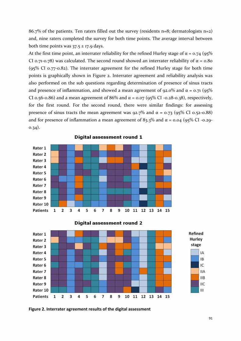
91
86.7% of the patients. Ten raters filled out the survey (residents n=8; dermatologists n=2)
and, nine raters completed the survey for both time points. The average interval between
both time points was 37.5 ± 17.9 days.
At the first time point, an interrater reliability for the refined Hurley stage of α = 0.74 (95%
CI 0.71-0.78) was calculated. The second round showed an interrater reliability of α = 0.80
(95% CI 0.77-0.82). The interrater agreement for the refined Hurley stage for both time
points is graphically shown in Figure 2. Interrater agreement and reliability analysis was
also performed on the sub questions regarding determination of presence of sinus tracts
and presence of inflammation, and showed a mean agreement of 92.0% and α = 0.71 (95%
CI 0.56-0.86) and a mean agreement of 86% and α = 0.07 (95% CI -0.28-0.38), respectively,
for the first round. For the second round, there were similar findings: for assessing
presence of sinus tracts the mean agreement was 92.7% and α = 0.73 (95% CI 0.52-0.88)
and for presence of inflammation a mean agreement of 83.3% and α = 0.04 (95% CI -0.29-
0.34).
Figure 2. Interrater agreement results of the digital assessment

92
Intrarater agreement and reliability outcomes showed a mean agreement of 64.4% (range
40-93.3%) and a mean α of 0.82 (95% CI 0.76-0.88) (range 0.68 (95% CI 0.44-0.88) – 0.96
(95% CI 0.90-1.00)), respectively.
The analyses of intrarater agreement and reliability for assessing sinus tracts demonstrated
a mean agreement of 91.9% (range 86.7-100%) and a mean α = 0.81 (95% CI 0.72-0.90) and
for presence of inflammation a mean agreement of 85.9% (range 73.3-100%) and mean α =
0.14 (95% CI -0.01-0.32).
Regarding face validity, raters scored the usefulness of the refined Hurley classification
with a mean of 78.7 ± 10.3 after completing the survey of the first round and a mean score
of 76.5 ± 9.7 after the second round. Nine out of 10 raters commented on their choice of
score. Before the first round, seven raters commented the classification to be ‘clear’ or
‘useful’, two raters found the classification helpful for choosing a treatment strategy. Two
raters reported the use of the classification to be time consuming. After the second round,
two raters additionally commented on the difficulty to judge presence of erythema on
photographs.
DISCUSSION
In this study, we assessed the interrater and intrarater reliability and face validity of the
refined Hurley classification. In the real-life assessment, the first group demonstrated low
agreement and moderate interrater reliability, while the second group showed high
agreement and interrater reliability. The lower outcomes in the first group might be due to
the fact that one of the raters showed a low agreement and reliability when compared to
the reference standard. The other rater from group one and both raters from group two
demonstrated to be in high agreement with the reference standard. In the digital
assessment, we found a moderate interrater reliability for the first round. For the second
round, the ten raters showed a high agreement for assigning the refined Hurley stadium,
possibly indicating the positive learning curve for using the refined Hurley classification.
Additionally, high intrarater reliability for the refined Hurley classification was found. The
intrarater reliability cannot be determined easily in real life due to the dynamic nature of
HS. Hence, the choice for a digital assessment in which the exact same cases were
presented. The intrarater reliability demonstrated high percentages for these assessments.
However, scoring HS disease activity on photographs is challenging, which reflects in the
low to moderate reliability between the raters for assessing inflammation and presence of
sinus tracts. This difference was particularly evident for the assessment of inflammation. A
possible explanation for this is, that the variation in answers was very low, over 80% of the
answers in both rounds was ‘yes’. This lack in variation of answers makes the outcome of

93
the Krippendorff’s alpha test less informative, because the test cannot adequately cope
with this.8 The percent agreement results are therefore said to be more meaningful.6,7
The original Hurley classification was first introduced in 1989 and created for surgical
purposes. The inflammatory component of the disease and the number of involved
anatomic areas are not taken into account. Even though this static classification serves as a
standard for severity assessment of HS, only one recent publication studied its interrater
and intrarater reliability. A moderate interrater reliability and substantial intrarater
reliability was found.9 This study only used digital photo assessments (n=30), therefore
extrapolation of the results to real-life assessments should perhaps be done with care. The
refined Hurley classification was very recently tested in a study in which 9 instruments for
HS were assessed in 24 live patients by 12 HS experts in one session. A fair interrater
reliability was found. One may question if the used study design reliably reflects real-life
situations as well, for which the refined Hurley classification is predominantly intended.10
Two other classification systems for HS, based on phenotypes, were previously proposed.
In 2013 Canoui-Poitrine identified three HS phenotypes by latent class analysis: axillary-
mammary type, follicular type and gluteal type.11 Recently, the interrater reliability of these
three phenotypes have been assessed in a digital setting (n=30). The classification
demonstrated low interrater reliability with a Fleiss’ kappa of only 0.37 (95% CI 0.32-0.42),
and therefore may only be of limited use in daily practice.12 The other phenotype
classification was outlined by Van Der Zee et al. based on expert opinion, describing six
possible subtypes of HS: the regular type; frictional furuncle type; scarring folliculitis type;
conglobata type; syndromic type and ectopic type.13 This classification has not been
validated yet.
Multiple instruments assessing the severity of HS have also been developed over the last
decades. These include the Modified Sartorius Score (MSS), the Hidradenitis Suppurativa
Clinical Response (HiSCR), and the Hidradenitis Suppurativa Severity Score System
(IHS4). However, validation of these instruments is sometimes incomplete or of mainly
low methodological quality and perhaps more valuable in research, than for use in daily
practice.10,14–19
For assessing face validity of the refined Hurley classification, a specific part of the
validation process, there are no standards on how it should be assessed. However, ‘lack of
face validity’ is a very strong argument for not using an instrument.20 In our study, we
found a high face validity outcome for the refined Hurley classification, which most likely
indicates that the assessors found it useful. However, this finding might be biased for only
half of the invited raters participated. We asked our raters to comment on the usefulness
of this classification. A few raters found it somewhat time consuming. The overall opinion
was that the refined Hurley classification was clear and easy to use. The different
treatment strategies linked to each stage, were also appreciated as helpful in daily practice.

94
Digital assistance, for example through an application on a mobile device, could help to
assess the patient.
Possible limitations of our study could be the relatively small number of raters and
patients. However, standardized protocols or guidelines on methodology about reliability
assessments of classification systems are lacking.6 Therefore, our sample size was based on
studies in the same field and topic and expert advice. A recent study investigating several
measurement and classification systems in HS also used similar number of raters and
subjects.10
In conclusion, our results show an overall moderate to high interrater and intrarater
agreement and reliability of the refined Hurley classification in real life as well as in digital
assessments. Face validity results were also positively high. Therefore, the refined Hurley
classification might be a useful and practical tool to stage HS patients. We recommend
further research investigating the validity of the refined Hurley classification.
Acknowledgments
The authors thank all the patients who participated.

95
References
1. Revuz J. Hidradenitis suppurativa. J Eur Acad Dermatology Venereol. 2009 Sep;23(9):985–98. 2. Jemec GBE, Kimball AB. Hidradenitis suppurativa: Epidemiology and scope of the problem. J Am Acad
Dermatol. 2015 Nov 1;73(5):S4–7. 3. Hurley HJ. Axillary hyperhidrosis, apocrine bromhidrosis, hidradenitis suppurativa, and familial benign
pemphigus: surgical approach. Dermatologic surgery. New York: Marcel Dekker. 1989:729–739. 4. Horváth B, Janse IC, Blok JL, et al. Hurley staging refined: A proposal by the dutch hidradenitis suppurativa
expert group. Acta Derm Venereol. 2017;97(3):412–3. 5. Rondags A, van Straalen KR, van Hasselt JR, et al. Correlation of the Refined Hurley Classification for
Hidradenitis suppurativa with Patient Reported Quality of Life and Objective Disease Severity Assessment. Br J Dermatol. 2018 Dec 4;
6. Kottner J, Audigé L, Brorson S, et al. Guidelines for Reporting Reliability and Agreement Studies (GRRAS) were proposed. J Clin Epidemiol. 2011 Jan 1;64(1):96–106.
7. Zapf A, Castell S, Morawietz L, Karch A. Measuring inter-rater reliability for nominal data – which coefficients and confidence intervals are appropriate? BMC Med Res Methodol. 2016 Dec 5;16(1):93.
8. Krippendorff K. Content Analysis: An Introduction to Its Methodology. Vol. 79, Education. 2004. 440 p. 9. Ovadja ZN, Schuit MM, van der Horst CMAM, Lapid O. Inter- and Intrarater Reliability of the Hurley
Staging for Hidradenitis Suppurativa. Br J Dermatol. 2018 Dec 26; 10. Thorlacius L, Garg A, Riis PT, et al. Interrater agreement and reliability of outcome measurement
instruments and staging systems used in hidradenitis suppurativa. Br J Dermatol. 2019;181(3):483-491 11. Canoui-Poitrine F, Le Thuaut A, Revuz JE, et al. Identification of Three Hidradenitis Suppurativa
Phenotypes: Latent Class Analysis of a Cross-Sectional Study. J Invest Dermatol. 2013 Jun 1;133(6):1506–11. 12. van Straalen KR, Verhagen T, Horváth B, et al. Poor interrater reliability of hidradenitis suppurativa
phenotypes. J Am Acad Dermatol. 2018 Sep 1;79(3):577–8. 13. van der Zee HH, Jemec GBE. New insights into the diagnosis of hidradenitis suppurativa: Clinical
presentations and phenotypes. J Am Acad Dermatol. 2015 Nov 1;73(5):S23–6. 14. Sartorius K, Emtestam L, Jemec GBE, Lapins J. Objective scoring of hidradenitis suppurativa reflecting the
role of tobacco smoking and obesity. Br J Dermatol. 2009 Oct 1;161(4):831–9. 15. Sartorius K, Killasli H, Heilborn J, Jemec GBE, Lapins J, Emtestam L. Interobserver variability of clinical
scores in hidradenitis suppurativa is low. Br J Dermatol. 2010 Feb 22;162(6):1261–8. 16. Kimball AB, Jemec GBE, Yang M, et al. Assessing the validity, responsiveness and meaningfulness of the
Hidradenitis Suppurativa Clinical Response (HiSCR) as the clinical endpoint for hidradenitis suppurativa treatment. Br J Dermatol. 2014 Dec 1;171(6):1434–42.
17. Zouboulis CC, Tzellos T, Kyrgidis A, et al. Development and validation of the International Hidradenitis Suppurativa Severity Score System (IHS4), a novel dynamic scoring system to assess HS severity. Br J Dermatol. 2017 Nov;177(5):1401–9.
18. Kimball A, Tzellos T, Calimlim B, Teixeira H, Geng Z, Okun M. Achieving Hidradenitis Suppurativa Response Score (HiSCR) Is Associated With Significant Improvement in Clinical and Patient-Reported Outcomes: Post Hoc Analysis of Pooled Data From PIONEER I and II. Acta Derm Venereol. 2018;0.
19. Ingram JR, Hadjieconomou S, Piguet V. Development of core outcome sets in hidradenitis suppurativa: systematic review of outcome measure instruments to inform the process. Br J Dermatol. 2016 Aug 1;175(2):263–72.
20. De Vet HCW, Terwee CB, Mokkink LB, Knol DL. Measurement in Medicine - A Practical Guide. Cambridge University Press; 2011.

96

97
6
THE REFINED HURLEY QUESTIONNAIRE:
AN ACCURATE SELF-ASSESSMENT INSTRUMENT FOR
DERIVING THE CORRECT REFINED HURLEY STAGE IN
HIDRADENITIS SUPPURATIVA
Acta Dermato-Venereologica. 2019 Jun 1;99(7):703-704. doi: 10.2340/00015555-3126
Angelique Rondags1
Rob J. Volkering1
Iana Turcan1
Yolinde S. Zuidema1
Ineke C. Janse1,2
Barbara Horváth1
1. Department of Dermatology, University of Groningen, University Medical Center Groningen, Groningen, the Netherlands. 2. Department of Dermatology, Meander Medical Center, Amersfoort, the Netherlands.

98
INTRODUCTION
Hidradenitis suppurativa (HS) is a chronic, debilitating, inflammatory skin disease that
mainly affects body folds (e.g. axillae and groin).1 Primary lesions include deep-seated
inflammatory nodules, abscesses, and sinus tracts.1 The diagnosis of HS can be made easily
due to its clear, distinct clinical presentation.
Deckers et al. have reported that patients with HS can score their own disease severity
according to the original Hurley classification.2 However, the purpose of the Hurley
classification was to assess HS in a single affected body region in order to guide surgical
intervention.3 It was not intended to classify HS disease activity and severity in the whole
patient and to assist in extensive treatment plans including, for example, anti-
inflammatory options.3,4
Therefore, a modification of the Hurley classification was proposed by a Dutch HS expert
group in 2017: the “refined Hurley classification”.5 In contrast to the original Hurley
classification, the 7-stage refined Hurley classification assesses not only the presence of
sinus tracts, but also inflammatory symptoms and the extensiveness of the disease.3,5
Refined Hurley stages I and II are subdivided into A, B and C, corresponding to mild,
moderate and severe disease. Stage III is not subdivided and corresponds to severe HS
disease. This was recently confirmed by a construct validation study.6 Furthermore, a
comprehensive treatment ladder is added to the flow chart.5
The aim of this study was to develop and investigate the reliability of a patient self-
assessment questionnaire corresponding to the items addressed in the refined Hurley
classification, in order to derive the refined Hurley stage.
METHODS
Subjects and study design
Consecutive patients with HS were recruited at the dermatology outpatient clinic of the
University Medical Centre Groningen (UMCG), a tertiary referral centre for HS. Subjects
were eligible if they were diagnosed with HS by a dermatologist, were older than 18 years,
and were capable of completing the questionnaire in Dutch.
A patient symptom self-assessment questionnaire was developed by HS experts based on
the refined Hurley classification flow chart. Following a pilot study performed with 16
patients, several modifications were applied to the concept questionnaire. Patients with
HS were requested to complete the definitive questionnaire (the refined Hurley
classification questionnaire for patients with HS (Appendix)) before their regular
consultation at the dermatologist. The refined Hurley stage was derived following the

99
flowchart by an investigator (RV).5 The dermatologists were requested to report a detailed
dermatological examination and report the refined Hurley classification, as they also do in
daily clinical practice. The percent agreement of data entry between 2 investigators (RV
and AR), based on a sample of 10 out of 75 (13.3%) randomly chosen subjects, was 97.2%.
No formal sample size calculation can be performed for this type of study. Based on
literature on methodology and similar studies in the same field, the aim was to include 75
subjects in the final cohort testing.2,7 For this type of study, medical ethics committee
approval is not required under Dutch law.
Statistical analysis
Descriptive statistics were used to describe the study population. The inter-rater
agreement and reliability between the HS patient’s derived refined Hurley stages and
physician’s reported refined Hurley stages were calculated. Next, the inter-rater agreement
and reliability of the presence of sinus tracts and HS disease severity, defined by the
refined Hurley classification, was calculated. For the inter-rater agreement, percentages of
agreement between physicians and patients were calculated manually. Because the refined
Hurley classification is a nominal scale, a Krippendorff’s alpha (α) is suitable to determine
the inter-rater reliability.8 Statistical analysis was performed using IBM SPSS Statistics 23.0
for Windows (SPSS, Chicago, USA). p-values ≤ 0.05 were considered statistically
significant.
RESULTS
A total of 75 subjects participated in this study. An overview of the patients’ characteristics
is shown in Table I. Approximately one-third were patients visiting the dermatology
outpatients clinic (UMCG) for the first time.
Inter-rater agreement and reliability
The derived refined Hurley stages and disease severity (based on the refined Hurley
classification) from the patient’s answers to the questionnaire vs. the physician’s
dermatological examination report are shown in Table SI and Table SII. The inter-rater
agreement between patient’s derived and physician’s reported refined Hurley stages was
78.7% (59/75). The inter-rater reliability resulted in an α of 0.737 (95% confidence interval
[CI] 0.622–0.852) (Table SIII). Similar results were found for inter-rater agreement and
reliability regarding HS disease severity (82.7%, α=0.733 (95% CI 0.589–0.856) (Table SIII).
Concerning the assessment of sinus tracts, inter-rater agreement was 89.2% and reliability
of α=0.785 (95% CI 0.650–0.919).

100
Table 1. Patients’ characteristics (n = 75)
Characteristics
Age, years, mean ± standard deviation 40.5 ± 12.7 Female sex, % 72.0 Body Mass Index, kg/m
2, mean ± standard deviation 29.4 ± 6.0
Smoking status, n (%) Non-smoker 13 (17.3) Ex-smoker 19 (25.3) Current smoker 43 (57.3) New (first visit) or control patient, n (%) New 26 (34.7) Control 49 (65.3) Refined Hurley classification stage, according to physician, n (%) Refined Hurley IA 22 (29) Refined Hurley IB 5 (7) Refined Hurley IC 8 (11) Refined Hurley IIA 8 (11) Refined Hurley IIB 11 (15) Refined Hurley IIC 16 (21) Refined Hurley III 5 (7)
DISCUSSION
In this study, we developed a patient symptom self-assessment questionnaire based on the
refined Hurley classification algorithm for HS. We investigated whether the derived
refined Hurley stages from the patient questionnaire correspond to the physician’s
dermatological examination and given refined Hurley stage. It was found that a substantial
inter-rater agreement and reliability, indicating that, in most cases, the same refined
Hurley stage could be extracted from the patients’ answers to our questionnaire as
assigned by the physician.
Notably, in contrast to the flow chart of the refined Hurley classification, we found in the
current study that it is important to first ask patients with HS about the presence of
abscesses/inflammatory nodules, prior to the presence of sinus tracts. This might be due to
the chronological order in which HS mostly develops: the first signs of HS are usually
recurrent inflammatory nodules and/or abscesses, and in a later stage sinus tracts might
develop. Furthermore, the reliability of the questionnaire is enhanced by educating the
patient about the main HS lesions, by providing a concise description with prototypical
pictures of these lesions.
One of the main items in the original as well as in the refined Hurley classification that has
to be determined is the presence of sinus tracts. We have shown that the inter-rater
agreement and reliability regarding the presence of sinus tracts is especially high.
However, as stated previously, the original Hurley classification lacks valuable information
to assess symptoms and severity in an entire individual.4 Recently, we have shown that the

101
sub-stages of the refined Hurley classification correlated significantly with patient-
reported quality of life and physician-assessed disease severity.6 In the current study we
showed that patients and physicians also agree on the level of disease severity.
Furthermore, compared with the study by Deckers et al. and another study regarding self-
assessment of disease severity of other skin diseases (acne, psoriasis, and atopic eczema),
our results are the highest.2,9
A limitation of the current study is that it was conducted in a single university hospital
with HS expertise. This might have biased the results. Patients with HS seen at our
department might have a longer duration of disease and are usually extensively informed
about their disease. This could indicate that these patients are more familiar with the
symptoms of HS than are patients treated in primary and secondary healthcare centres.
However, besides inclusion of patients coming for follow-up consultation, new referrals
were also included.
In conclusion, the symptom self-assessment questionnaire described here is an accurate
instrument for deriving the correct refined Hurley stage within patients with HS and
might be useful for daily clinical practice, as well as for future epidemiological and clinical
studies in HS. We recommend investigating the usefulness of this questionnaire further in
other/multiple treatment centers, including sub-analyses, such as the results of new vs.
follow-up patients, presence of inflammatory nodules/abscesses, and involved anatomical
region.
Acknowledgements
The authors are grateful for the participation of all the patients with HS in this study.

102
SUPPLEMENT
Table SI. Refined Hurley classification derived from patient’s answers to the questionnaire vs. physician’s report
By physician IA IB IC IIA IIB IIC III
By
pat
ien
t
IA 21 0 1 1 0 2 0 IB 0 4 0 0 0 0 0 IC 0 0 5 0 0 0 0 IIA 0 0 0 5 1 2 0 IIB 1 0 0 1 8 2 0 IIC 0 1 2 1 2 10 0 III 0 0 0 0 0 0 5
Table SII. Disease severity based on refined Hurley classification derived from patient’s answers to the questionnaire vs. physician’s report
By physician Mild (IA+IIA) Moderate (IB+IIB) Severe (IC+IIC+III)
By
pat
ien
t Mild (IA+IIA) 27 2 1 Moderate (IB+IIB) 1 12 3 Severe (IC+IIC+III) 4 2 23
Table SIII. Results definitive questionnaire (n=75)
Inter-rater agreement, %
Inter-rater reliability, α
95% confidence interval
Derived refined Hurley stage 78.7 0.737 0.622-0.852 Presence of sinus tracts 89.2 0.785 0.650-0.919 Derived disease severity based on refined Hurley classification
82.7 0.733 0.589-0.856

103
APPENDIX
Definitive questionnaire: “The refined Hurley classification questionnaire for patients with HS”
Vragenlijst Dermatologie
Invuldatum (dd/mm/jjjj) .. .. / .. .. / .. .. .. ..
Uw achternaam …………………………………...............................................................
Uw geboortedatum (dd/mm/jjjj) .. .. / .. .. / .. .. .. ..
Uw geslacht Man Vrouw
Uw lengte …………………..meter
Uw gewicht …………………..kg
Rookt u? Ja, circa ………… sigaretten per dag sinds het jaartal …………..
Gestopt, circa ……….. jaar ………. sigaretten per dag gerookt.
Nee, ik heb nooit gerookt.
Deze vragenlijst gaat over Uw huidklachten van hidradenitis suppurativa op dit moment.
Het invullen van de vragenlijst duurt enkele minuten.
Alvast heel hartelijk dank voor Uw medewerking!
Pagina 1 van 6
104
Hidradenitis suppurativa Korte beschrijving van mogelijke klachten die gepaard kunnen gaan met hidradenitis suppurativa
Hidradenitis suppurativa, soms ook HS, acne inversa of acne ectopica genoemd, is een chronische huidziekte. Kenmerkend voor hidradenitis suppurativa zijn terugkerende, pijnlijke ontstekingen in lichaamsplooien, zoals de liezen en/of de oksels. Ook kan men ontstekingen hebben onder de borsten, op de billen, in de schaamstreek/geslachtsdeel en gezicht/oren/behaarde hoofdhuid. De type ontstekingsverschijnselen kunnen onderverdeeld worden in 2 groepen:
1) De losse bulten/ontstekingen liggen meestal diep onder de huid maar kunnen ook lijken op
puisten. Ook kunnen abcessen (holtes met pus) ontstaan.
2) Op langere termijn kunnen er onderhuidse tunnels ontstaan (ook wel sinussen genoemd).
Deze tunnels kunnen in lengte variëren, van ongeveer 1 cm tot meer dan 10 cm.
Pagina 2 van 6
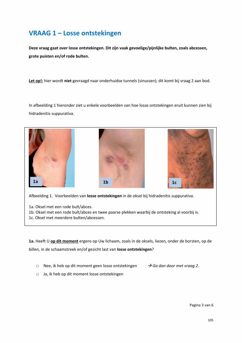
105
VRAAG 1 – Losse ontstekingen
Deze vraag gaat over losse ontstekingen. Dit zijn vaak gevoelige/pijnlijke bulten, zoals abcessen,
grote puisten en/of rode bulten.
Let op!: hier wordt niet gevraagd naar onderhuidse tunnels (sinussen); dit komt bij vraag 2 aan bod.
In afbeelding 1 hieronder ziet u enkele voorbeelden van hoe losse ontstekingen eruit kunnen zien bij
hidradenitis suppurativa.
Afbeelding 1. Voorbeelden van losse ontstekingen in de oksel bij hidradenitis suppurativa. 1a. Oksel met een rode bult/abces. 1b. Oksel met een rode bult/abces en twee paarse plekken waarbij de ontsteking al voorbij is. 1c. Oksel met meerdere bulten/abcessen.
1a. Heeft U op dit moment ergens op Uw lichaam, zoals in de oksels, liezen, onder de borsten, op de
billen, in de schaamstreek en/of gezicht last van losse ontstekingen?
□ Nee, ik heb op dit moment geen losse ontstekingen Ga dan door met vraag 2.
□ Ja, ik heb op dit moment losse ontstekingen
Pagina 3 van 6
1a 1b 1c

106
1b. In welke lichaamslocaties heeft U op dit moment losse ontstekingen?
U kunt hieronder in de figuur aankruisen in welke lichaamslocaties de losse ontstekingen voorkomen:
1c. Hoeveel losse ontstekingen telt U op dit moment in totaal?
□ Minder dan 5
□ 5 of meer
1d. Ontstaan de losse ontstekingen voornamelijk op precies dezelfde plaats(en) binnen dezelfde
locatie(s), of elke keer op nieuwe/wisselende plaatsen/locaties?
□ Voornamelijk op dezelfde plaatsen
□ Voornamelijk op nieuwe/wisselende plaatsen
Pagina 4 van 6

107
VRAAG 2 – Onderhuidse tunnels
Deze vraag gaat over onderhuidse tunnels (ook wel sinussen genoemd). Onderhuidse tunnels zijn
meestal duidelijk met het blote oog te zien aan het oppervlakte van de huid. De tunnels kunnen
ontstoken zijn, dan lekt er vaak pus. Onderhuidse tunnels zullen nooit uit zichzelf verdwijnen.
Daarom wordt vaak voorgesteld om deze tunnels chirurgisch weg te snijden.
In afbeelding 2 hieronder ziet u enkele voorbeelden van hoe onderhuidse tunnels eruit kunnen zien
bij hidradenitis suppurativa.
Afbeelding 2. Voorbeelden van tunnels bij hidradenitis suppurativa in een oksel. Er is sprake van onderhuidse tunnelvorming, dit is te zien aan de plooien die zich in de huid vormen. 2a. Oksel met beginnende tunnelvorming, zonder ontstekingsverschijnselen. 2b. Oksel met iets uitgebreidere en duidelijkere tunnelvorming, zonder ontstekingsverschijnselen. 2c. Oksel met meerdere tunnels en ontstekingsverschijnselen.
2a. Heeft U op dit moment ergens op Uw lichaam, zoals in de oksels, liezen, onder de borsten, op de
billen, in de schaamstreek en/of gezicht last van onderhuidse tunnels?
□ Nee, ik heb op dit moment geen onderhuidse tunnels Einde vragenlijst
□ Ja, ik heb op dit moment onderhuidse tunnels
Pagina 5 van 6
2a 2b 2c

108
2b. In welke lichaamslocaties komen de onderhuidse tunnels bij U op dit moment voor?
U kunt hieronder in de figuur aankruisen in welke lichaamslocaties onderhuidse tunnels voorkomen:
2c. Is het aangedane gebied in één lichaamslocatie waar onderhuidse tunnels voorkomen kleiner óf groter dan het oppervlakte van Uw hand (handpalm plus vingers, zie de afbeelding hiernaast)?
□ Kleiner
□ Groter
2d. Zijn één of meerdere van deze onderhuidse tunnels op dit moment gevoelig/pijnlijk én rood?
□ Ja
□ Nee
2e. Lekt er bij één of meerdere van deze onderhuidse tunnels op dit moment pus?
□ Ja
□ Nee
Hartelijk dank voor het invullen van de vragenlijst!
Pagina 6 van 6
109
References 1. Jemec GB. Hidradenitis suppurativa. N Engl J Med. 2012;366: 158–164. 2. Deckers I, Mihajlovic D, Prens E, Boer J. Hidradenitis suppurativa: a pilot study to determine the capability
of patients to self-assess their Hurley stage. Br J Dermatol. 2015; 172: 1418–1419. 3. Hurley HJ. Axillary hyperhidrosis, apocrine bromhidrosis, hidradenitis suppurativa, and familial benign
pemphigus: surgical approach. Dermatologic surgery. New York: Marcel Dekker. 1989:729–739. 4. Revuz JE, Jemec GB. Diagnosing hidradenitis suppurativa. Dermatol Clin. 2016; 34:1–5. 5. Horváth B, Janse IC, Blok JL, Driessen RJ, Boer J, Mekkes JR, et al. Hurley staging refined: a proposal by the
dutch hidradenitis suppurativa expert group. Acta Derm Venereol. 2017; 97:412–413. 6. Rondags A, van Straalen KR, van Hasselt JR, et al. Correlation of the refined Hurley classification for
hidradenitis suppurativa with patient reported quality of life and objective disease severity assessment. Br J Dermatol. 2019;180(5):1214-1220.
7. Zapf A, Castell S, Morawietz L, Karch A. Measuring inter-rater reliability for nominal data – which coefficients and confidence intervals are appropriate? BMC Med Res Methodol. 2016;16:93.
8. Krippendorff K. Agreement and information in the reliability of coding. Communic Methods Meas. 2011;5:93–112.
9. Magin PJ, Pond CD, Smith WT, Watson AB, Goode SM. Correlation and agreement of self-assessed and objective skin disease severity in a cross-sectional study of patients with acne, psoriasis, and atopic eczema. Int J Dermatol. 2011;50:1486–1490.

110
111
7
IDENTIFICATION OF CLINICAL CATEGORIES IN HIDRADENITIS
SUPPURATIVA BASED ON PATIENT CHARACTERISTICS:
RESULTS FROM A CLUSTER ANALYSIS
In preparation
Angelique Rondags1
Suzanne Arends2
Rob J. Volkering1
Ineke C. Janse3
Janine L. Dickinson-Blok4
Eldrid Schoonhoven1
Hendrika Bootsma2
Anneke Spoorenberg2
Barbara Horváth1
1. Department of Dermatology, University of Groningen, University Medical Center Groningen, Groningen, the Netherlands. 2. Department of Rheumatology and Clinical Immunology, University Medical Center Groningen, Groningen, the Netherlands. 3. Department of Dermatology, Meander Medical Center, Amersfoort, the Netherlands. 4. Department of Dermatology, Hospital Nij Smellinghe, Drachten, the Netherlands.

112
ABSTRACT
Background: It is suggested that hidradenitis suppurativa (HS) is a skin disease with a
certain phenotypic heterogeneity, which possibly indicates different etiologic,
pathophysiologic and genetic backgrounds that require tailored treatment approaches.
Robust description of HS phenotypes does not yet exist.
Objectives: To identify distinct clinical categories of HS patients based on associated
clinical patient characteristics.
Methods: Cross-sectional study. Cluster analysis was performed on two prospective,
longitudinal and observational cohorts including adult HS patients seen in three centres in
the Netherlands between 2015 and 2017. Clinical variables included in the analysis were
sex, smoking history, body mass index (BMI), and follicular occlusion comorbidity.
Results: Included were 345 HS patients: 72.8% female, mean age 38.3 ± 12.2 years, mean
symptom duration 15.4 ± 11.7 years, mean BMI 29.0 ± 6.3 kg/m², and 82.3% was ever a
smoker. Five distinct clinical sub-categories of HS were revealed: cluster 1. “females with
stereotypical HS” (40.0%) are characterized by female smokers with overweight; 2.
“females with a single exogenous risk factor for HS” (22.6%) is marked by females that
either have a positive smoking history or are overweight; 3. “male HS” (22.0%) represents
male patients who have a positive smoking history and/or are overweight; 4. “HS plus
follicular occlusion comorbidity” (n=32, 9.2%) is defined by HS patients who are also
known with acne conglobata, dissecting cellulitis of the scalp and/or pilonidal sinus; and 5.
“limited HS” (6.1%) typifies HS patients without associated risk factors smoking, high BMI
and follicular occlusion tetrad comorbidities.
Conclusion: These clinical sub-categories of HS may help to define sound phenotypes of
HS.

113
INTRODUCTION
Hidradenitis suppurativa (HS) is a chronic, painful, immune mediated auto-inflammatory
skin disease affecting approximately 1% of the general population.1,2 Hidradenitis
suppurativa is located typically in the inverse body regions like axillae, inframammary,
inguinofemoral region, perineal regions, and buttocks. Ectopic sites such as the face, neck,
and abdomen can also be involved.1,2 The exact pathophysiology and possibly underlying
genetic cause remain uncertain. Current scientific insight shows that the first pivotal
histological event in HS is hyperplasia and hyperkeratosis of the follicular infundibulum
that leads to follicular plugging. This eventually results in rupture and incitement of a
perifollicular aberrant inflammatory response causing typical deep-seated inflammatory
nodules.3 Other (frequently) seen elements are abscesses, sinus tracts, ‘tombstone’ double-
ended or giant comedones (pseudocomedones) and hypertrophic ‘bridged’ and ‘crater
holes’ scars.4 Identified risk factors for HS are female sex, high body mass index (BMI), and
smoking.2,5 Hidradenitis suppurativa has been described as part of syndromes, particularly
the follicular occlusion tetrad (HS, acne conglobata, dissecting cellulitis of the scalp and
pilonidal sinus). It is thought that these comorbidities share a similar
(histo)pathophysiology.6 Furthermore, HS is associated with other (auto-)inflammatory
diseases, such as Crohn’s disease and spondyloarthropathy.6–9 The clinical spectrum of HS
disease is wide, ranging from subclinical and mild to very severe symptoms. Symptoms can
relapse, be continues for a long period, and go in remission. It is therefore suggested that
HS is a disease with a phenotypic heterogeneity possibly indicating different etiologic,
pathophysiologic and genetic backgrounds that require tailored treatment approaches.10,11
Propositions for categorization of HS into clinical phenotypes have been published
previously (Table 1).12–15 Furthermore, several authors have described distinct
morphological presentations of HS in some patients, such as “plaque form” and “ectopic”
or “atypical” HS.16–20 However, all of these categorizations have not been internationally
accepted thus far and the genetic and pathophysiological background was not
investigated. This necessitates further investigation to understand many aspects of HS
disease such as aetiology and pathophysiology, to ultimately enhance treatment outcomes.
Towards valid identification of HS phenotypes, categorization of HS patients based on
associated patient characteristics will be valuable.11 Therefore, in this study we sought to
investigate distinct clinical categories of HS based on clinical patient characteristics.

114
Table 1. Previously proposed clinical phenotypes in hidradenitis suppurativa
Author, year of publication, and study design
Subgroup/Phenotype Description
Canoui-Poitrine et al., 201312 Cross-sectional, latent class analysis, n=618
Axillary–mammary (48%)
Predominant locations are the axillae and breast. Predominant lesions are hypertrophic scars. More likely female sex, less likely smokers, less likely to have family history.
Follicular (26%) Predominant locations are the axillae or breast, ears, chest, back, and legs. Predominant lesions are follicular lesions such as comedones, epidermal cysts, and pilonidal sinus. Other characteristics are a history of severe acne, family history of HS, higher proportion of men and current/former smokers, greater disease severity (Sartorius and Hurley), earlier disease onset, and longer disease duration.
Gluteal (26%) Predominant location is the gluteal area. Predominant lesions are papules and folliculitis. Other characteristics are a family history of HS, higher proportion of smokers, lower BMI values, less severe disease, longer duration.
Van der Zee and Jemec, 201513 Expert opinion, descriptive, n=0
Regular Fulfilment of all diagnostic criteria for HS, most common type, all patients who lack other specific characteristics belong in this group.
Frictional furuncle Predominant affected locations are sites that are exposed to enhanced friction, such as abdomen, thighs and buttocks. Predominant lesions are multiple deep nodules and abscesses. Sinus tract formation is unusual. Other characteristics are being overweight.
Scarring folliculitis Buttocks, inguinal area, and pubic area are predominantly affected. Predominant lesions are pustules, cysts, superficial nodules, depressed cribriform scarring, double-ended comedones. Sinus tract formation is unusual. Other characteristics are being overweight and smoker.
Conglobata
Back and face are predominantly affected. Predominant lesions are cysts and sinus tracts. Other characteristics are acne conglobata, family history of HS, overweight, and male sex.
Syndromic Patients are characterized by concomitant diseases, such as pyoderma gangrenosum and arthritis, such as the pyoderma gangrenosum, acne, and suppurative hidradenitis (PASH) syndrome or pyogenic arthritis, pyoderma gangrenosum, acne, and suppurative hidradenitis (PAPASH) syndrome.
Ectopic HS involving the face.
Theut Riss et al., 201714
Explorative, cross‐sectional, descriptive study, n=451
Low BMI (<25 kg/m2) Less severe disease (based on Hurley classification, physician global assessment, number of areas affected and patient-reported severity)
High BMI (>25 kg/m2) More severe disease (based on Hurley classification, physician global assessment, number of areas affected and patient-reported severity). No difference in smoking or family history of HS.
Frew et al., 201915
Explorative, descriptive, n=0
Typical Similar to the previously described “axillary–mammary” type by Canoui Poitrine et al. and “regular” type by Van der Zee and Jemec.
Atypical This includes two subtypes: - Scarring follicular subtype. Similar to the previously described “follicular” and “gluteal” type by Canoui Poitrine et al. and “scarring folliculitis by Van der Zee and Jemec. - Conglobata subtype. Similar to the previously described “follicular” and “gluteal” type by Canoui Poitrine et al. and “conglobata” type by Van der Zee and Jemec.
Syndromic Not strictly defined, but might include PASH (pyoderma gangrenosum, acne and HS) and PAPASH (pyogenic arthritis, pyoderma gangrenosum, acne and HS) syndromes, and maybe autoimmune, inherited and autoinflammatory syndromes (such as familial Mediterranean fever, Crohn’s disease, and Dowling-Degos disease).

115
METHODS
Patients
In this cross-sectional study, baseline data of all patients with HS from in the Hidradenitis
Suppurativa Registry (HiSURE) cohort of the University Medical Center Groningen
(UMCG) between April 2015 and July 2017 and the Hidradenitis suppurativa CARE
(HiCARE) cohort of the UMCG, Meander Medical Hospital (Amersfoort) and Hospital Nij
Smellinghe (Drachten) in the Netherlands between March 2018 and June 2019 were
included. Both cohorts are prospective, longitudinal and observational cohorts in which all
consecutive adult HS patients clinically diagnosed by a dermatologist (based on the
modified Dessau definition of HS), who are cognitively able and willing to participate, are
included and data is collected in a standardized manner.
In both cohorts, variables are recorded in a standardized manner, which include sex,
smoking history, body mass index (BMI), age at symptom onset, family history of HS,
comorbidities such as the history or present occurrence of severe acne/acne conglobata,
pilonidal sinus and dissecting cellulitis of the scalp, and symptoms of HS at baseline.
Presence of comedones and refined Hurley stage was standardly recorded in the HiCARE
study. In the HiSURE cohort, presence of comedones and refined Hurley stage was
retrieved retrospectively by reviewing written medical records and photo documentation.
Statistical analysis
Descriptive statistics were used to describe the study population. Results are presented as
number of patients (%), mean ± standard deviation, or median (interquartile range) for
categorical, normally distributed and non-normally distributed data, respectively.
Hierarchical cluster analysis with a Ward minimum-variance and agglomerative approach
is performed to explore whether different clusters, i.e. phenotypes, within the HS disease
spectrum can be defined.
Four variables, all made binary, were critically chosen for the analysis because they are
considered as significant determinants of the HS clinical phenotypes: sex categorized in
female and male, smoking history categorized in never smoked and current/former
smoker, BMI categorized in underweight plus normal (<25 kg/m²) and overweight plus
obese (≥25 kg/m²), and presence of the follicular tetrad comorbidities: acne conglobata,
pilonidal sinus and dissecting cellulitis of the scalp categorized in none and ≥1 present in
the patient. Symptoms of HS, such as presence of inflammatory nodules, abscesses, sinus
tracts, comedones, scarring, inflammation, and number of involved body regions were not
included as variables, as these symptoms can be considerably inconsistent over time and
can be quite difficult to assess.

116
The output of the cluster analysis is a dendrogram: a diagram that represents a tree in
which each subject starts as a single cluster and then joins subjects who are closest to each
other based on similarities or dissimilarities and these initial clusters are then merged
according to the similarities and so on until all subjects form one cluster. Cut-off points in
the dendrogram were determined based on the formation of the clinically most relevant
phenotypes.
Group comparisons between all clusters were performed using one-way ANOVA for
normally distributed continuous variables, Kruskal-Wallis test for non-normally
distributed continuous variables or Chi-square test for categorical variables. In case the
overall group comparison was statistically significant, independent sample t-test, Mann-
Whitney U test, and Chi-square test or Fisher’s exact test were performed as appropriate to
compare between the individual clusters.
Statistical analysis was performed using IBM SPSS Statistics 23.0 for Windows (SPSS,
Chicago, IL, USA). P-values ≤0.05 were considered statistically significant.
RESULTS
In total, 345 HS patients were included (HiSURE 65.8%; HiCARE 34.2%). Of these, 72.8%
were female, mean age was 38.3 ± 12.2 years, mean symptom duration was 15.4 ± 11.7 years,
mean BMI was 29.0 ± 6.3 kg/m², and 82.3% was ever a smoker (Table 2). These patient
characteristics were similar in both cohorts (HiSURE and HiCARE).
Cluster analysis
Interpretation of the cluster analysis results revealed five categories (Table 2 and 3).
The largest group, cluster 1 (n=138, 40.0%) was designated as “females with stereotypical
HS”. This category included solely females who all present with a positive smoking history
(former or current) and obesity (mean BMI 32.6 ± 5.9 kg/m2). All refined Hurley stages
were seen, similar to the entire cohort. About half of these patients presented with sinus
tracts and/or comedones. Conjunction of follicular occlusion tetrad comorbidities was not
seen in this cluster. Cluster 2 (n=78, 22.6%) was named “females with a single exogenous
risk factor for HS” and represents females that are either former/current smokers (73.1%)
or are overweight/obese (26.9%)). Like in cluster 1, follicular occlusion tetrad
comorbidities were not present. Cluster 3 (n=76, 22.0%), was defined as the “male HS” type
and represents a cluster formed by only males, that had similar characteristics as the entire
HS study population. In this group, 75% had sinus tracts (thus presented with either
refined Hurley stages IIA, IIB and IIC or III). Comedones were seen in half of the patients.
Similar to cluster 1 and 2, follicular occlusion tetrad comorbidities were not present.
Cluster 4 (n=32, 9.2%) represents the “HS plus follicular occlusion comorbidity” category.

117
Table 2. Demographic characteristics of the overall cohort and of the separate categories
Results are presented as number of patients (%), mean ± standard deviation or median (interquartile range).
HS, hidradenitis suppurativa; BMI, body mass index.
a. p<0.05 when comparing cluster 1 to cluster 2; b. p<0.05 when comparing cluster 1 to cluster 3; c. p<0.05 when comparing cluster 1 to cluster 4; d. p<0.05 when comparing cluster 1 to cluster 5; e. p<0.05 when comparing cluster 2 to cluster 3; f. p<0.05 when comparing cluster 2 to cluster 4; g. p<0.05 when comparing cluster 2 to cluster 5; h. p<0.05 when comparing cluster 3 to cluster 4; i. p<0.05 when comparing cluster 3 to cluster 5; j. p<0.05 when comparing cluster 4 to cluster 5.
* Acne conglobata, dissecting cellulitis of the scalp, and/or pilonidal sinus.
Overall population, n=345
Cluster 1, n=138 (40.0) “Females with stereotypical HS”
Cluster 2, n=78 (22.6) “Females with a single exogenous risk factor for HS”
Cluster 3, n=76 (22.0) “Male HS”
Cluster 4, n=32 (9.3)
“HS plus 1 other follicular occlusion disease”
Cluster 5, n=21 (6.1) “Limited HS”
Overall comparison between all clusters, p-value
Sex <0.001b-j
Male 94 (27.2) 0 (0) 0 (0) 76 (100) 15 (46.9) 3 (14.3) Female 251 (72.8) 138 (100) 78 (100) 0 (0) 17 (53.1) 18 (85.7) Smoking <0.001a-e,g,i,j
Never 61 (17.7) 0 (0) 21 (26.9) 10 (13.2) 9 (28.1) 21 (100) Former or current 284 (82.3) 138 (100) 57 (73.1) 66 (86.8) 23 (71.9) 0 (0) BMI <0.001a-g,i,j
< 25 kg/m2 101 (29.3) 0 (0) 57 (73.1) 16 (21.1) 7 (21.9) 21 (100) ≥ 25 kg/m2 244 (70.7) 138 (100) 21 (26.9) 60 (78.9) 25 (78.1) 0 (0) Follicular occlusion tetrad comorbidities*
<0.001c,f,h,j
No 313 (90.7) 138 (100) 78 (100) 76 (100) 0 (0) 21 (100) Yes 32 (9.3) 0 (0) 0 (0) 0 (0) 32 (100) 0 (0) Age, years 36.0
(28.0-48.0) 39.0 (32.0-47.0)
33.5 (26.0-48.0)
36.0 (29.0-51.0)
32.0 (28.0-41.0)
27.0 (21.8-45.0)
0.026c,d,i
Symptom duration, years
12.0 (6.0-22.0)
15.5 (8.0-24.3)
11.0 (6.0-20.0)
11.0 (6.0-22.0)
12.0 (6.3-20.8)
5.0 (2.0-18.3)
0.001d,i,j
Age at onset of HS symptoms, years
20.0 (15.0-28.0)
20.0 (15.0-28.0)
20.0 (15.0-27.0)
23.0 (17.0-31.0)
18.5 (15.0-25.8)
19.0 (16.5-24.8)
0.660
BMI exact, kg/m2 27.6 (24.5-33.0)
32.0 (27.4-35.7)
23.8 (21.2-26.4)
27.6 (25.3-30.4)
29.5 (25.2-32.3)
23.3 (22.0-24.2)
<0.001a-f,i,j
Family history HS in 1st degree
0.186
No 231 (67.0) 88 (63.8) 55 (70.5) 52 (68.4) 18 (56.3) 18 (85.7) Yes 114 (33.0) 50 (36.2) 23 (29.5) 24 (31.6) 14 (43.8) 3 (14.3) Refined Hurley 0.002a,d,i
Stage IA 75 (21.7) 22 (15.9) 24 (30.8) 10 (13.2) 7 (21.9) 12 (57.1) Stage IB 27 (7.8) 15 (10.9) 3 (3.8) 5 (6.6) 3 (9.4) 1 (4.8) Stage IC 32 (9.3) 20 (14.5) 5 (6.4) 4 (5.3) 2 (6.3) 1 (4.8) Stage IIA 34 (9.9) 11 (8.0) 12 (15.4) 7 (9.2) 2 (6.3) 2 (9.5) Stage IIB 64 (18.6) 24 (17.4) 16 (20.5) 15 (19.7) 8 (25.0) 1 (4.8) Stage IIC 87 (25.2) 40 (29.0) 14 (17.9) 25 (32.0) 6 (18.8) 2 (9.5) Stage III 26 (7.5) 6 (4.3) 5.1 (5.1) 10 (13.2) 4 (12.5) 2 (9.5) Sinus tract formation
0.009b,d,e,g,i,j
No 134 (38.8) 57 (41.3) 32 (41.0) 19 (25.0) 12 (37.5) 14 (66.7) Yes 211 (61.2) 81 (58.7) 46 (59.0) 57 (75.0) 20 (62.5) 7 (33.3) Comedones 0.005a,d,e,i
No 174 (50.4) 58 (42.0) 48 (61.5) 34 (44.7) 18 (56.3) 16 (76.2) Yes 171 (49.6) 80 (58.0) 30 (38.5) 42 (55.3) 14 (43.8) 5 (23.8)

118
Table 3. Description of categories according to this cluster analysis study
HS, Hidradenitis suppurativa; BMI, Body mass index.
Besides having HS, all of these patients also had a history of pilonidal sinus, acne
conglobata or dissecting cellulitis of the scalp. The final cluster is number 5 (n=21, 6.1%).
This category does not present with HS risk factors, smoking, overweight/obesity, or
follicular occlusion comorbidities. Therefore, this cluster is termed the “limited HS”
category.
Clinical characteristics
Comparison of patient characteristics of the first category “females with stereotypical HS”
to the other 4 categories particularly showed a higher BMI (median 31.97 (27.43-35.73) vs.
23.26 (21.99-24.22) to 29.47 (25.17-32.29), p<0.05). Presence of comedones was seen
significantly less in “females with a single exogenous risk factor for HS” (38.5%), compared
to “females with stereotypical HS” and “male HS” (58.0% and 55.3%, respectively). Patients
belonging to the “limited HS” category presented at a significantly younger age compared
to category 1 and 4 and have significantly shorter symptom duration compared to all other
phenotypes except category 2 (“female with a single exogenous risk factor for HS”)
(p<0.005). Furthermore, a large proportion of refined Hurley stage IA stage (57%),
especially compared to the “female stereotypical HS” category and “male HS” category, was
seen in this category (15.9% and 13.2%, respectively, both p<0.05). Sinus tract formation is
seen significantly less in this category compared to all other categories. Presence of
comedones were also seen less in this category, again particularly compared to the “female
stereotypical HS” phenotype and the “male HS” type (p<0.05). For age at onset of HS
symptoms and family history of HS no statistical significant difference were found when
comparing all clusters at once. To avoid bias of multiple testing, these variables were not
further tested between single clusters.
Cluster Category Sex Smoking BMI
Follicular occlusion tetrad
Sinus tracts
Comedones Symptoms duration
1 Female stereotypical HS
♀ + + -
2 Females with single exogenous risk factor for HS
♀ +/- +/- -
3 Male HS ♂ +/- +/- -
4 HS plus 1 other follicular occlusion disease
♀/♂ +/- +/- +
5 Limited HS ♀/♂ - - -

119
DISCUSSION
In the present study using data from two prospective observational HS patient cohorts, we
identified five distinct clusters, i.e. clinical categories, within the HS disease spectrum
(Table 3). Cluster analysis was performed with the clinically known relevant variables sex,
smoking history, BMI and follicular occlusion tetrad comorbidities. These variables were
carefully chosen by HS experts based on a sizeable body of evidence from former studies
supporting the importance of these risk factors and comorbidities.6
The largest category that we identified is the “female stereotypical HS” cluster which
describes the classic HS patient: a smoking, obese, female patient. As the name implies,
the second largest category “females with single exogenous risk factor for HS”, also
concerns females, but in contrary to the first category these patients are only exposed to
one exogenous risk factor (either current/former smoker or BMI >25 kg/m2). In this
second category, comedones are seen less frequently than in the first (and third)
phenotype, which might indicate the importance of exogenous risk factors in contributing
to comedone formation. In contrary to the first two categories, which include solely
females, the third category consists exclusively of males. Sinus tracts are frequently seen in
this group. Other patient characteristics are similar to the overall HS population.
In all the first three clusters, sex and exogenous risk factors smoking and BMI define the
category. Epidemiologically, female sex is an important risk factor for HS as HS affects
three times as many women as men.1 Observational data shows that onset of HS usually
occurs after puberty, pre-menstrual flares are reported, and disease activity might become
less during pregnancy and worsens post-partum.21 In general, as learned from previous
research, sex should be considered as an important biological variable in fundamental,
preclinical, and clinical research of HS.
Cigarette smoking (former/current) and being overweight or obese have shown to be the
two most important recognized exogenous risk factors for developing HS and worse
disease outcome, because they induce alteration of inflammatory responses and
histological changes.3,22,23 It seems almost self-evident that treatment strategies targeting
life style intervention, i.e. behavioural changes to achieve normal body weight and
cessation of smoking, need to be applied for to achieve better HS disease outcome.
Nearly one out of ten patients in this study were grouped into the fourth category “HS plus
1 other follicular occlusion disease”, which concerns HS patients with acne conglobata,
pilonidal sinus and/or dissecting cellulitis of the scalp. Patients of this group seem to be
somewhat younger at initial HS symptom onset, although not confirmed statistically. All
conditions in this tetrad (including HS) share the contributing aetiological factor of
follicular occlusion. However, much more than this shared pathogenesis remains
unknown. Importantly, treatment modalities for patients in this group might differ from

120
the other categories. For example, treatment with systemic retinoid might be more
effective and therefore should be higher up the treatment ladder. However, this needs
further investigation.
Strikingly, this analysis also revealed a group of patients who are not exposed to any
typical exogenous risk factors (i.e. smoking and overweight) for HS. We designated this
category as “limited HS”. More than half of these patients presented with a Hurley stage
IA. Accordingly, sinus tracts are less frequently seen in this category when comparing to
the other categories. These patients have a shorter duration of HS symptoms. It would be
very interesting to further study patients within this category to try to uncover why and
how they developed HS and what their clinical course will be. This might generate more
data about risk factors and the pathophysiology of HS for instance.
This study is not the first in a quest to identify HS categories, subtypes or phenotypes
within the HS disease spectrum (Table 1). However, this is the first attempt to identify
distinct categories based on clinical data using cluster analysis. This statistical technique
can be used to make relatively homogenous groups out of heterogeneous variables. It is
said to be an unsupervised and unbiased method, however, the choice of variables
included in the analysis may influence the results. Moreover, cluster analysis cannot
distinguish between possibly important or unimportant variables. As said before, the
variables used in this study were critically chosen based on scientific evidence and clinical
relevance in HS.6 This is the task of the researcher, which also includes defining the
relevant cut-off points in the dendrogram which is the outcome of the cluster analysis.
Previously, Canoui-Poitrine et al. have identified three groups through latent class analysis
without an a priori hypothesis in a cohort of 618 HS patients, by including ten variables
chosen by the researchers (three body locations, five lesion types, association with acne,
and family history of HS).12 Latent class analysis is also a statistical method to find
homogenous groups/subtypes of patients in a multivariate categorical and/or continuous
dataset and uses a probabilistic model for clustering, whereas in cluster analysis a distance
function is used to assess the similarity between cases. Although the latent class analysis
seems methodically sound, the found latent classes have not been used widely (yet),
possibly due to the difficulties in assigning HS patients to solely one of the classes
resulting in poor inter-rater reliability scores.24 In 2015, Van der Zee and Jemec proposed
another set of possible subtypes (six types), based on their (clinical) expert opinion.13 Frew
et al. proposed three subtypes, after studying and combining the results of several previous
publications regarding HS phenotypes including that of Canoui-Poitrine et al. and Van der
Zee and Jemec.15 Theut Riis et al. proposed that patients with a low and high BMI could
represent two clinically different subtypes within the whole HS population.14 We have also
shown that BMI is an important variable to distinguish subtypes of HS, but according to
our analyses it is not the only relevant variable. Notably, until now all proposed subtypes

121
above have not been validated. Besides these proposed subtypes, multiple
explorative/descriptive reports (e.g. case reports, case series) suggest that HS can present
distinctly from classic HS (with nodules, abscesses and/or sinus tracts in the body folds),
such as “plaque form” HS involving purple inflamed plaques on different parts of the body
with typical cribriform scarring and HS involving ectopic sites such as the nape, the latter
sometimes referred to as “atypical” HS.16–18,20
In our analysis we decided not to focus on presence of (certain) HS symptoms (lesion
types, degree of inflammation) and affected body regions, since not a single symptom or
feature of HS defines a robust subtype or phenotype. Thereby, HS often displays a dynamic
disease course. For example, patients can go from Hurley stage IA to IC from IIC to III in
days or progress from a Hurley stage I (without sinus tracts) to a stage with sinus tracts (II
and III). As seen in this study, all (refined) Hurley stages can be seen in each category,
however, some refined Hurley stages seem to be more common in certain categories than
in others. Hence, in this study only four variables were chosen, which are thought to be
significant and constant contributors to the HS phenotype. Furthermore, these can be
assessed in any patient presenting with HS. We suggest that the identified clinical
categories in this study should be used complementary to the refined Hurley staging, to
have an integral depiction of the entire HS patient. This can be used for research or to
make individual treatment plans in daily clinical practice (e.g. a category 4 patient “HS
plus 1 other follicular occlusion disease” presenting with Hurley stage IC for who
treatment options such as systemic retinoids or biologics are reasonable).
The current study is not without limitations. One limitation might be the relatively small
sample size. However, the overall study population characteristics are similar to the
average HS population. Other more frequent comorbidities (e.g. metabolic syndrome,
spondyloarthritis, and Crohn’s disease) and specific syndromes (e.g. PASH, PAPASH) in
HS were not included as variables.6 However, the occurrence of (multiple) associated
diseases can influence therapeutic approaches and outcomes.
In summary, we identified and defined five distinct clinical categories within the HS
disease spectrum through cluster analysis. This categorization of HS patients seems
clinically relevant, is easily applicable, and may influence treatment strategies and
outcomes. Additionally, it might contribute to future genotype-phenotype correlations
and enhanced understanding of de pathophysiology of HS and therefore improvement of
clinical care. We recommend further investigating and testing these clinical phenotypes in
a larger cohort of HS patients.
Acknowledgments
We thank all the patients with HS who are participating in the HiSURE and HiCARE
cohorts.

122
References
1. Jemec GBE. Hidradenitis Suppurativa. N Engl J Med. 2012;366(2):158-164. 2. Zouboulis CC, Desai N, Emtestam L, et al. European S1 guideline for the treatment of hidradenitis suppurativa/acne
inversa. J Eur Acad Dermatology Venereol. 2015;29(4):619-644. 3. Kelly G, Prens EP. Inflammatory Mechanisms in Hidradenitis Suppurativa. Dermatol Clin. 2016;34(1):51-58. 4. Revuz JE, Jemec GBE. Diagnosing Hidradenitis Suppurativa. Dermatol Clin. 2016;34(1):1-5. 5. Sartorius K, Emtestam L, Jemec GBE, Lapins J. Objective scoring of hidradenitis suppurativa reflecting the role of
tobacco smoking and obesity. Br J Dermatol. 2009;161(4):831-839. 6. Miller IM, McAndrew RJ, Hamzavi I. Prevalence, Risk Factors, and Comorbidities of Hidradenitis Suppurativa. Dermatol
Clin. 2016;34(1):7-16. 7. Janse IC, Koldijk MJ, Spekhorst LM, et al. Identification of Clinical and Genetic Parameters Associated with Hidradenitis
Suppurativa in Inflammatory Bowel Disease. Inflamm Bowel Dis. 2016;22(1):106-113. 8. Rondags A, Arends S, Wink FR, Horváth B, Spoorenberg A. High prevalence of hidradenitis suppurativa symptoms in
axial spondyloarthritis patients: A possible new extra-articular manifestation. Semin Arthritis Rheum. 2019;48(4):611-617.
9. Rondags A, van Straalen KR, Arends S, et al. High prevalence of clinical spondyloarthritis features in patients with hidradenitis suppurativa. J Am Acad Dermatol. 2019;80(2):551-554.e1.
10. van der Zee HH, Laman JD, Boer J, Prens EP. Hidradenitis suppurativa: viewpoint on clinical phenotyping, pathogenesis and novel treatments. Exp Dermatol. 2012;21(10):735-739.
11. Ingram JR, Piguet V. Phenotypic heterogeneity in hidradenitis suppurativa (acne inversa): classification is an essential step toward personalized therapy. J Invest Dermatol. 2013;133(6):1453-1456.
12. Canoui-Poitrine F, Le Thuaut A, Revuz JE, et al. Identification of Three Hidradenitis Suppurativa Phenotypes: Latent Class Analysis of a Cross-Sectional Study. J Invest Dermatol. 2013;133(6):1506-1511.
13. van der Zee HH, Jemec GBE. New insights into the diagnosis of hidradenitis suppurativa: Clinical presentations and phenotypes. J Am Acad Dermatol. 2015;73(5 Suppl 1):S23-6.
14. Theut Riis P, Saunte DM, Benhadou F, et al. Low and high body mass index in hidradenitis suppurativa patients—different subtypes? J Eur Acad Dermatology Venereol. 2018;32(2):307-312.
15. Frew JW, Hawkes JE, Sullivan‐Whalen M, Gilleaudeau P, Krueger JG. Inter‐rater reliability of phenotypes and exploratory genotype–phenotype analysis in inherited hidradenitis suppurativa. Br J Dermatol. April 2019:bjd.17695.
16. Syed ZU, Hamzavi IH. Atypical hidradenitis suppurativa involving the posterior neck and occiput. Arch Dermatol. 2011;147(11):1343-1344.
17. Naasan H, Affleck A. Atypical hidradenitis suppurativa. Clin Exp Dermatol. 2015;40(8):891-893. 18. van Rappard DC, Starink M V., van der Wal AC, de Rie MA, Mekkes JR. Four cases of plaque form hidradenitis
suppurativa. J Eur Acad Dermatology Venereol. 2016;30(10):e104-e106. 19. Agut-Busquet E, Romaní J, Ribera M, Luelmo J. Hidradenitis suppurativa of the nape: Description of an atypical
phenotype related to severe early-onset disease in men. J Dermatol. 2019;46(2):149-153. 20. Poli F, Wolkenstein P, Revuz J. Back and face involvement in hidradenitis suppurativa. Dermatology. 2010;221(2):137-
141. 21. Karagiannidis I, Nikolakis G, Zouboulis CC. Endocrinologic Aspects of Hidradenitis Suppurativa. Dermatol Clin.
2016;34(1):45-49. 22. Schrader AMR, Deckers IE, van der Zee HH, Boer J, Prens EP. Hidradenitis suppurativa: A retrospective study
of 846 Dutch patients to identify factors associated with disease severity. J Am Acad Dermatol. 2014;71(3):460-467. 23. Alikhan A, Lynch PJ, Eisen DB. Hidradenitis suppurativa: A comprehensive review. J Am Acad Dermatol.
2009;60(4):539-561. 24. van Straalen KR, Verhagen T, Horváth B, et al. Poor interrater reliability of hidradenitis suppurativa phenotypes. J Am
Acad Dermatol. 2018;79(3):577-578.

123

124

125
8
ECTOPIC HIDRADENITIS SUPPURATIVA ON THE
DORSAL FOOT OF A ROAD MAKER
JAAD Case Reports. 2017 Sep; 3(5): 429–431 doi: 10.1016/j.jdcr.2017.06.002
Angelique Rondags1
Gilles F. Diercks2
Paul M.N. Werker3
Marcel F. Jonkman1
Barbara Horváth1
1. Department of Dermatology, University of Groningen, University Medical Center Groningen, Groningen, The Netherlands. 2. Department of Pathology, University Medical Center Groningen, Groningen, The Netherlands. 3. Department of Plastic Surgery, University Medical Center Groningen, Groningen, The Netherlands.

126
INTRODUCTION
Hidradenitis suppurativa (HS) is a chronic, often debilitating, inflammatory skin condition
that affects approximately 1% of the general population.1,2 HS is characterized by painful
subcutaneous nodules or abscesses, and in a later stage tunnels (sinus tracts) are formed in
the dermis and subcutaneous fat and eventually scarring can occur.1 These lesions are
mainly seen in the inverse body areas, like the axillary, inguinal, anogenital, and
inframammary regions.1 All these locations contain apocrine glands, are humid, and have a
predisposition to mechanical stress.3,4
The diagnosis of HS, based on the modified Dessau definition, is in general a clinical one;
it is based on the patient's history, chronicity, and recurrence of typical lesions in
prototypical body areas, with the possibility of typical HS lesions in atypical (ectopic)
locations.2 Therapy of HS is still challenging; a combination therapy that involves both
anti-inflammatory medicine and surgery is often needed to induce (partial) remission.2
Here, we report the case of a male patient who had an HS lesion on his dorsal foot possibly
caused by a predisposition of HS in combination with mechanical pressure and friction
from occupational-related factors.
CASE PRESENTATION
A 28-year-old man presented with a bleeding 1.5-cm ulcer with raised edges and laterally a
dark subcutaneous, blanchable area of 1 × 2 cm on his right dorsal foot (Figure 1, A). His
foot was erythematous, swollen, and painful. Skin symptoms started 5 months earlier as a
purple, slowly progressive papule. There were no systemic symptoms, such as fever or
malaise. The patient had a normal body weight and was a smoker. He had a history of
pilonidal sinus (excision 3 years ago), acne conglobata, and HS. A deroofing of an HS
lesion in the axilla took place previously with good treatment outcome.
At the moment of first presentation of the ulcer, the HS was active in femoral and perianal
regions (Hurley stage I) and was being treated with clindamycin, 300 mg twice daily, and
rifampicin, 300 mg twice daily. Detailed occupational history found that the patient is a
road maker, who paves streets with bricks or stones, and mostly worked in a right-legged
kneeling position causing bending of his right working shoe. He had just received a new,
rigid pair of tight-fitting work shoes.
Differential diagnosis included atypical (ectopic) presentation of HS, infectious wound,
traumatic ulcer, hypertensive leg ulcer (Martorell ulcer), infected hematoma, pyoderma
gangrenosum in association with PASH (pyoderma gangrenosum, acne and suppurative
hidradenitis) syndrome, and actinomycosis. It was decided to stop the rifampicin and
change the clindamycin scheme from 300 mg twice daily to 300 mg 3 times a day, and
ciprofloxacin was started additionally at 500 mg twice daily.

127
Despite antibiotic treatment, symptoms progressed. Small ulcers with brown/yellow
discharge and sinus tract formation developed into a dark erythematous plaque (Figure 1,
B). Further diagnostic tests found a normal blood glucose level (ruling out underlying
diabetes) and a negative wound bacterial culture by swab of the exudate. Based on the
clinical presentation, diagnostics, and anamneses, the diagnosis of ectopic HS was made.
Because of the progression, surgical excision (a skin tissue–sparing excision with
electrosurgical peeling procedure) was performed with split-thickness skin graft.
Successful secondary wound healing had occurred after initial split-thickness skin graft
failure (Figure 1, C). Furthermore, the patient was strongly advised to wear comfortable
nonfrictional shoes and clothing in general. Histologic investigation of the removed tissue
found follicular plugging, sinus tract formation, and dermal fibrosis with chronic active
inflammation, consistent with a diagnosis of HS (Figure 1, D).
The subject gave his written permission to use his personal information and images.
Figure 1. Ectopic hidradenitis suppurativa on the dorsal foot of a road maker
A, Initial clinical presentation of wound on dorsal right foot; B, Four months after initial presentation, during
surgery. Probe shows interconnected sinus tracts between lesions; C, Eight months postsurgery. Nearly
complete wound healing; D, Histologic findings, hematoxylin-eosin stain. Sinus tract formation in a background
with extensive fibrosis and a chronic infiltrate. Right upper corner follicular occlusion.

128
DISCUSSION
The pathogenesis of HS remains to be elucidated. Most likely, HS originates in the
follicular pilosebaceous unit and the surrounding tissue. The first event is the infundibular
hyperkeratinization with subsequent dilatation of the follicle that leads to rupture, causing
inflammation in the skin. The role of mechanical stress is proposed as a (contributive)
factor in the pathogenesis of HS, by stimulating interfollicular hyperplasia; however,
evidence is still limited.4
Several case reports support the contribution of mechanical stress in the development of
HS. For instance, an HS-like lesion on the stump was diagnosed in a case report of a limb
amputee after wearing a leg prosthesis. In this report, the leg prosthesis increased
mechanical friction at the stump area and created a warm, moist, occlusive environment,
similar to the predisposed intertriginous areas in classic presentations of HS.5
Another report presented the case of an obese woman with a history of classic HS, in
which HS was significantly reduced after weight loss. The HS particularly improved
around the waistband, which was mentioned to be an area highly exposed to friction.6
Recently, another similar case was described of an obese woman with classic HS, who had
HS lesions exactly where her bra strap was exerting mechanical pressure and friction on
the skin.4
The theory of mechanical stress as a causative factor in HS development in subjects with
aberrant hair follicles is also hypothesized in the case of a young child known with a nevus
comedonicus in the groin area, with histologically dilated hair follicles, who had HS there
just after she became mobile.7
Furthermore, the role of mechanical stress as a trigger factor for HS is supported by a
histologic study showing that the basement membrane zone of the sebofollicular junction
at the follicle pilosebaceous unit in perilesional HS skin seems to be aberrant, indicating
fragility of the hair follicle.8 One can speculate that increased epidermal mechanical stress
more easily leads to follicle rupture. However, there is contradictory evidence regarding
the integrity of the basement membrane zone.9
We report a case of ectopic HS in a predisposed male subject that is consistent with the
theory of mechanical stress as a contributive cause of HS. We propose that the HS lesion
on the patient's dorsal foot resulted from repetitive pressure and mechanical friction on
the skin because of his too-tight-fitting working shoe, which created a warm and humid
environment as well.
Acknowledgements
The authors thank Piet Toonder for providing the clinical presentation images.

129
References 1. Jemec GB. Hidradenitis suppurativa. N Engl J Med. 2012;366(2):158-164 2. Zouboulis CC, Desai N, Emtestam L, et al. European S1 guideline for the treatment of hidradenitis
suppurativa/acne inversa. J Eur Acad Dermatol Venereol. 2015;29(4):619-644 3. Revuz J. Hidradenitis suppurativa. J Eur Acad Dermatol Venereol. 2009;23(9):985-998 4. Boer J, Nazary M, Riis PT. The role of mechanical stress in hidradenitis suppurativa. Dermatol Clin.
2016;34(1):37-43. 5. De Winter K, van der Zee HH, Prens EP. Is mechanical stress an important pathogenic factor in hidradenitis
suppurativa? Exp Dermatol. 2012;21(3):176-177 6. Boer J. Resolution of hidradenitis suppurativa after weight loss by dietary measures, especially on frictional
locations. J Eur Acad Dermatol Venereol. 2016;30(5):895-896 7. Dufour DN, Bryld LE, Jemec, GB. Hidradenitis suppurativa complicating naevus comedonicus: the possible
influence of mechanical stress on the development of hidradenitis suppurativa. Dermatology. 2010;220(4):323-325
8. Danby FW, Jemec GB, Marsch WC, Von Laffert M. Preliminary findings suggest hidradenitis suppurativa may be due to defective follicular support. Br J Dermatol. 2013;168(5):1034-1039
9. Blok JL, Janse IC, Horváth B, Jonkman MF. Increased expression of integrin α6β4 in the basement membrane zone lining the sebaceous glands in hidradenitis suppurativa. Acta Derm Venereol. 2015;95(8):994-996

130

131
9
SUMMARY, GENERAL DISCUSSION,
AND FUTURE PERSPECTIVES
Angelique Rondags
Department of Dermatology, University of Groningen, University Medical Center Groningen, Groningen, the Netherlands.

132
SUMMARY, GENERAL DISCUSSION, AND FUTURE PERSPECTIVES
Hidradenitis suppurativa (HS) is a common, debilitating auto-inflammatory skin disease,
characterized by inflamed nodules, abscesses and/or sinus tracts in the body folds. The
exact aetiology and pathophysiologic sequence of events is unknown and a cure does not
exist yet. Although there is a rapid rise in the number of publications about the multiple
facets of HS there is still a lot to be learned about this unpleasant skin disease that was
considered an orphan disease until recently. This thesis contributes to the epidemiological
and clinical knowledge of HS.
Four main topics are covered in this thesis. First, the prevalence of HS in axial
spondyloarthritis (SpA) and the prevalence of SpA features in HS patients were
investigated and associated patient characteristics were determined. Interestingly, these
two auto-inflammatory diseases share common denominators in pathogenesis and
treatment. In the second part, the validation process of the refined Hurley classification for
staging HS patients is described. In the third part, the identification of clinical patient
categories within the HS disease spectrum is presented. Finally, the possible influence of
mechanical stress as a risk factor for the development and maintenance of HS is discussed.
1. Hidradenitis suppurativa, beyond the surface of the skin
Chapter 2 and 3 contribute to the epidemiologic data on the concurrence of HS and SpA.
SpA is an umbrella term for a group of inter-related chronic auto-inflammatory rheumatic
conditions. Overall, SpA can be classified into predominantly axial or peripheral SpA
according to the SpA classification criteria, defined by the ‘Assessment of
SpondyloArthritis international Society (ASAS) in 2009. The most well-known phenotype
for axial SpA is ankylosing spondylitis (AS) and for peripheral SpA, psoriatic arthritis.
These ASAS criteria comprise a combination of clinical, laboratory and imaging SpA
features (Chapter 1, Figure 2).1–3
A link between HS and SpA has been described previously since both auto-inflammatory
disease share the same inflammatory pathways and therefore also treatment options. Few
case reports and studies have described that SpA and HS occur at the same time in a
patient. Epidemiological studies suggest that SpA is more prevalent in HS than in the
general population. Variable prevalence rates between 2.3% to 28.2% vs. ±1% are
reported.4–15 Vice versa, the prevalence of HS in SpA is unknown.
Therefore, in Chapter 2 we investigated the prevalence of HS in axial SpA patients from a
large northern Dutch cohort “the Groningen-Leeuwarden axial SpA (GLAS) cohort”. A
patient questionnaire based study containing validated diagnostic HS questions with a
high sensitivity and specificity for HS (92-97% and 82–86%, respectively), including
prototypical pictures of HS, was used.16 The self-reported HS symptoms were verified by

133
checking medical records for a previously reported HS diagnosis and the remaining cases
were verified through a detailed standardized telephone call including a diagnostic
question with high specificity (97%) for the diagnosis of HS.17 There was a very high
patient questionnaire response rate of 76%. The estimated prevalence of HS was 9.1%,
6.9% when corrected for non-responders, which is much higher than in the general
population (±1%).18–24 Female sex, a higher axial SpA patient reported disease activity and a
combined axial SpA disease activity score, and worse quality of life (QoL) were
significantly more prevalent among axial SpA patients with HS than without HS.
Interestingly, the combined axial SpA disease activity score was independently associated
with prevalence of HS.
Conversely, in Chapter 3 we investigated the prevalence of axial and peripheral SpA
features in HS. In this two centre study, performed at the outpatient clinic of two Dutch
tertiary HS referral centres (University Medical Center Groningen located in Groningen
and Erasmus Medical Center in Rotterdam), a self-reported questionnaire concerning SpA
features that are part of the ASAS classification criteria supported with prototypical
pictures of the features was developed and presented to HS patients. Results show that
approximately two-third of the respondents reported to fulfil one or more of the four ASAS
entry criteria for axial or peripheral SpA, approximately 73% and 27%, respectively.
Interestingly, the vast majority (87%) of these patients also reported one or more
additional clinical SpA features. The clinical SpA features were mostly found in the ‘classic’
HS patient: female, overweight and smoker, with a longer HS disease duration and
symptoms of active HS. These results are concordant with the results of Chapter 2, in
which female patients with high SpA disease activity were also identified.
A body of evidence suggests that HS is an ‘immune mediated inflammatory disease’
(IMID). IMID is a term to describe a concept of multiple inflammatory diseases that share
common dysregulated immune mediated inflammatory pathways, concerning mostly
overproduction of (pro-)inflammatory cytokines. Clinically, these diseases seem unrelated
because different organs are affected.25,26 Diseases that can be included into this concept,
are among others: Crohn’s disease, pyoderma gangrenosum, and axial and peripheral
SpA.25,26 Importantly, all these diseases are linked to HS.25–27 However, the IMID spectrum
is broader, encompassing also rheumatoid arthritis, multiple sclerosis, diabetes mellitus
type I, and systemic lupus erythematosus.25,26 In these diseases other inflammatory
pathways may play a role.
The exact aetiology of IMIDs is uncertain and is thought to be multifactorial: a prerequisite
is a genetically susceptible individual that is further exposed to certain endogenous and
exogenous risk factors.25,26 Frequently, IMIDs run in families. IMIDs are known to
negatively affect morbidity, QoL, and may lead to premature death.

134
Commonalities between HS and SpA are found on multiple levels. Regarding the involved
immune mediated pathways, elevated pro-inflammatory cytokine levels of both the innate
(e.g. interleukin (IL)-1β and tumor necrosis factor (TNF)-α) and the adaptive immune
system (e.g. IL-17A and IL-23) are found in both diseases. The TNF-α pathway and the IL-
23/IL-17 axis seem to be important in the pathogenesis of both axial SpA and HS.27–31 There
is evidence for an unbalanced T-helper 17/T-regulator ratio.32 Moreover, both HS and SpA
are epidemiologically associated with IBD, in HS particularly Crohn’s disease. In IBD, the
above described cytokine pathways are also perturbed.33–35 Therefore, it is not surprising
that these IMIDs respond to anti-cytokine therapy, i.e. biologics.25 Strong evidence is
available showing effectiveness of anti-TNF-α therapy in HS, AS, and IBD.31,36,37
Furthermore, a body of evidence indicates that the environmental factors smoking and
obesity are (highly) significant exogenous risk factor in HS and (axial) SpA: a higher
incidence and disease activity are reported for both conditions.38–44 Interestingly, obesity is
also reported as an important risk factor for IBD and smoking is an important risk factor in
Crohn’s disease.45,46 Smoking and obesity can both lead to pro-inflammatory effects.
Adipose tissue represents a metabolically and hormonally active organ, producing
adipokines that causes a pro-inflammatory effect that drives disease activity in patients
with IMIDs.47 Cigarette smoking can augment the production of pro- and anti-
inflammatory cytokins as well.48 Interestingly, in Chapter 2 we found a trend for axial SpA
patients with HS for both a higher (body mass index) BMI and a positive smoking history.
The results of our studies presented in Chapter 2 and 3 strengthen the soundness of HS as
an IMID and the IMID concept as a whole, and contribute to knowledge about the
immunopathological basis of HS and SpA. Although we did not confirm a diagnosis of SpA
in Chapter 3, we did find a high prevalence of more than one self-reported clinical SpA
feature in HS patients, contributing to the existing literature that SpA is likely to be more
prevalent in HS than in the general population. Moreover, with a prevalence of ±9% of HS
in SpA, HS could perhaps be considered as a novel extra-articular manifestation of SpA,
similar to psoriasis (9.3%) and IBD (6.8%).49 Clinically, it is also important to identify
comorbidities, since they influence health outcomes such as QoL and treatment
decisions.49
Additionally, also Blandizzi et al. reported the occurrence of two or more IMIDs in one
patient.26 Combination of certain diseases that are more likely than others to present in
the same patient are commonly named 'disease associations'. In the context of IMIDs,
combinations are also designated as 'immune-mediated inflammatory syndromes.26
Currently, it is only hypothesized why some IMIDs are likely to co-occur and others not.
Suggested is a genetic susceptibility, based on genome wide association studies and a
family history of IMIDs that is frequently present.26 Similar inflammatory pathways and
shared external risk factors might also play a role. Another hypothesis could be that the

135
pro-inflammatory state of an individual caused by one auto-inflammatory disease is likely
to lower the bar for another auto-inflammatory condition. This is supported by results
from both Chapter 2 and 3, in which we have found that in both axial SpA patients with a
high disease activity and HS patients with active HS symptoms the occurrence of HS
symptoms or SpA symptoms was more prevalent. Furthermore, a positive smoking history
and higher BMI were also more prevalent in both studies in patients with both HS and SpA
symptoms. These two risk factors are both epidemiologically linked to HS and (axial) SpA,
and associated with a higher disease activity.38,39,42–44,50,51 The inflammatory
pathophysiologic commonalities between HS and SpA include dysregulation of
inflammatory cytokines TNF-α, IL-1, IL-12, IL-23 and IL-17A.15,27,30,32,52 The IL-12/IL 23
pathway including IL-17A and TNF-α are known to play an important role in the
pathogenesis of HS and SpA. This is supported by the fact that both diseases respond well
to anti-TNFα therapy.15,36
Therefore, we state that HS is not solely an auto-inflammatory skin disease, but an IMID.
We suggest not to restricting the diagnostic and treatment approach to HS alone; the
treating physician must keep in mind the possibility of another IMID in a patient
diagnosed with HS as well as in the past, present or future. If beside HS one or more other
IMIDs are present in a patient, a multidisciplinary approach towards diagnosis and a
comprehensive treatment of HS and accompanying IMID(s) is warranted.
Future perspectives
Many challenges continue to exist and the prevalence of HS in SpA and vice versa remains
to be investigated more profoundly to determine associations more accurately. The
developed questionnaire concerning axial and peripheral clinical SpA features (Chapter 3)
needs to be validated in a prospective cohort, by referring those patients with positive
answers to a rheumatologist for further analysis and diagnosis. Patient self-reported
symptoms about HS and SpA should be prospectively verified. Furthermore, it would be
interesting to further investigate genetic, local and systemic dysregulations in patients who
present with both diseases in an active state at the same time. This will provide more
insight into the pathogenesis of both SpA and HS.
2. Refined classification for hidradenitis suppurativa
To date, no globally accepted and properly validated disease staging system, severity
measurement, and phenotype classification exists for HS.53 HS is considered to be a
dynamic disease with a diverse clinical picture. The clinical course of HS is highly variable,
ranging from mild self-limiting disease to rapidly progressive inflammation with
destruction of normal skin architecture and severe physical and psychological disability.
Hitherto, it is still an enigma what causes HS disease manifestation and what drives

136
inflammation in HS. In order to gain more insight in the pathophysiology of HS and
ultimately its cure, it is important to accurately stage and subtype patients within the HS
disease spectrum. This provides a basis for disease definition and activity and to
standardize enrolment and outcome criteria for clinical trials and the ability to compare
the results of these (randomized controlled) trials.
Until now, the most frequently used staging system for HS is the Hurley classification
(Chapter 1, Table 1).53,54 Hurley stage I represents single or multiple abscesses without sinus
tract formation or scarring, stage II recurrent abscesses with one or more sinus tracts and
scarring widely separated by normal skin, and stage III concerns diffuse involvement with
multiple sinus tracts and no intervening normal skin. The Hurley classification was
proposed 30 years ago (1989) to determine symptoms of HS in one particular affected body
region in order to make treatment plans for this specific region.54 However, to date, the
Hurley classification is also used to stage patients globally, both in clinical practise and in
research. However, although straightforward in use, data regarding its validation to stage
patients globally is very limited.53 Although knowledge about the pathophysiology of HS
and treatment options have expanded, especially the last two decades, the Hurley
classification was never adapted and validated to use in the whole patient. Therefore,
recently, the Hurley classification was revisited by the Dutch Hidradenitis Suppurativa
Expert Group (members of the European Hidradenitis Suppurativa Foundation e.V.).55 The
refined Hurley classification is a modification, i.e. refinement, of the three staged Hurley
classification, and is designed to describe current HS symptoms in the whole patient that
is diagnosed with HS and to guide comprehensive treatment modalities per stage. It
includes seven stages: refined Hurley stage IA, IB, IC, IIA, IIB, IIC, and III (Chapter 1,
Figure 3). Refined Hurley stages I (A, B, and C) are characterized by the presence of
inflammatory nodules and abscesses and no sinus tracts. Refined Hurley stages II (A, B,
and C) are characterized by the presence of sinus tracts and scarring is not assessed
anymore (in contrary to the original Hurley classification). The letters A, B, and C are said
to represent mild, moderate, and severe HS disease. Refined Hurley stage III is redefined as
the involvement of ≥ 1% body surface area of a body site with interconnected
inflammatory sinus tracts, and is regarded as severe per definition.55
In Chapter 4, we first analysed whether the refined Hurley classification accurately
distinguishes mild, moderate and severe HS disease by determining its correlation to the
validated patient’s reported QoL questionnaire (Dermatology Life Quality Index, DLQI)
and a (largely) validated physician-assessed disease severity score
(International Hidradenitis Suppurativa Severity Score System, IHS4).56,57 In this
multicentre observational study in 433 patients, we found a significant positive correlation
of DLQI and IHS4 within refined Hurley stage I and II, from A through C (for DLQI rs =

137
0,259 and rs = 0,185, respectively; for IHS4 rs = 0,603 and rs = 0.532, respectively). This
indicates the soundness of the construct validity of the refined Hurley classification.
Subsequently, in Chapter 5 we determined the interrater and intrarater reliability of the
refined Hurley classification in two sub studies. Two groups of each two trained raters
(dermatology residents) classified 13 and 12 HS patients respectively at the regular HS
outpatient clinic and we found a moderate to high interrater reliability (Krippendorff’s α =
0.68, 95% confidence interval (CI) 0.32-0.95 to α = 0.92, 95% CI 0.78-1.00). Through a
photographic survey, ten trained raters (dermatology residents and dermatologists)
assessed at two time points with approximately one month in between, 15 de-identified HS
cases with standardized pictures showing HS symptoms. A moderate to high interrater
reliability was found as well, and the intrarater reliability demonstrated a high reliability
(interrater reliability α = 0.74, 95% CI 0.71-0.78 at first time and α = 0.80, 95% CI 0.77-0.82)
at second time point, and a mean intrarater reliability of α = 0.83, 95% CI 0.78-0.89).
Furthermore, the face validity assessment in Chapter 5 which indicates how well the
raters found that the refined Hurley classification was covering the concept it aims to
measure, showed good results (78.7 ± 10.3 prior the first assessment and 76.5 ± 9.7 after the
second assessment, on a scale of 0-100).
In the past few years important efforts have been made to develop and define core
outcome sets for HS.53,58,59 One review from 2016 pointed out that 30 outcome
measurement instruments, including the Hurley classification, were used in 12 randomized
controlled trials for HS.53 Surprisingly, 27 (90%) of these outcome measurements were
never validated, and the methodological quality of the ones with any validation data were
mostly graded fair or poor. This indicates the urgency for validation of classification and
outcome measurements in HS. For outcome measurements, specific guidelines are
developed, such as the COSMIN study (COnsensus-based Standards for the selection of
health Measurement INstruments), for classification systems specific guidelines are less
defined, but the COSMIN study guidelines can be applied.60,61 According to these
standards, the validation process includes the domains validity, reliability, interpretability,
and responsiveness.60 Feasibility is also an important aspect to assess.53
In 2015, one study of fair methodological quality, found a statistically significant Pearson’s
correlation coefficient of β = 0.59 between the Hurley classification and DLQI.62 This
shows a higher correlation compared to our study, although the sample size in this study
was very small (n = 55). In 2018 and 2019, the interrater and intrarater reliability of the
original Hurley and/or refined Hurley classification were assessed.63–65 The first study
included fifteen raters (five dermatologists, five plastic surgeons, and five general
surgeons, all with at least two years of HS experience). The original Hurley classification
was assessed in 30 photographic cases and demonstrated a moderate interrater and
substantial intrarater reliability (Cohen’s Ƙ = 0.59, 95% CI 0.48-0.70 and Ƙ = 0.65, 95% CI

138
0.58-0.72, respectively). Authors concluded that photographic assessment, especially of
Hurley stage III, is reliable and time efficient.64 The second study included 12 raters (all
considered HS experts, with at least ten year of HS experience). In this study, nine
instruments for HS, including the Hurley and refined Hurley classification, were each
assessed consecutively in 24 HS patients.63 Outcomes showed good interrater results for
Hurley classification when determined in axillae and gluteal regions, and fair for the groin
(intra class correlation (ICC) of 0.72, 95% CI 0.63-0.81; 0.72, 95% CI 0.62-0.80; and 0.55,
95% CI 0.44-0.67, respectively), and for the refined Hurley classification fair interrater
reliability (ICC = 0.51, 95% CI 0.35-0.68). In this study, intrarater reliability was not
assessed. Of note, at the time this study was conducted, the refined Hurley classification
had only just been developed, so raters were not acquainted with this classification prior to
participation in this study. In the last study, 32 physicians (24 dermatology residents and
eight specialists) assessed four scoring systems in five HS patients.65 Fair interrater
reliability and good intrarater reliability results were reported for both the Hurley and
refined Hurley classification (only the lower CI results were showed: Hurley interrater of Ƙ
= 0.58, intrarater of ICC 0.76; refined Hurley interrater of Ƙ = 0.47, intrarater of ICC =
0.74). The second time patients were scored in this study revealed better results, indicating
a positive learning curve. This was comparable to our results presented in Chapter 5. All
these studies contribute to the validation data for classification systems in HS, and both
the Hurley and the refined Hurley classification show similar reliability. Importantly, the
refined Hurley classification adds a significant extra aspect since it reflects the extent and
inflammatory activity of HS in the whole patient, and the original Hurley classification was
not designed to do so.66
In Chapter 6, we developed a patient symptom self-assessment questionnaire based on
the refined Hurley classification algorithm. We investigated whether the derived refined
Hurley stages from the patient questionnaire correspond to the physician’s dermatological
examination and given refined Hurley stage. We found a substantial interrater agreement
and reliability, indicating that in most cases the same refined Hurley stage could be
extracted from the patients’ answers to our questionnaire, as assigned by the physician (α
= 0.74, 95% CI 0.62–0.85). This questionnaire could be valuable to use for research, but
also for clinical purposes at the outpatient clinic or as part of “e-health” (healthcare
services provided electronically via the Internet).
The refined Hurley classification allows recognizing severe HS throughout the entire
original Hurley classification. We propose that using the refined Hurley classification will
likely lead to more accurate stratification of HS patients for treatment in daily practice as
well as for enrolment in epidemiologic and clinical studies. For example, within refined
Hurley stage I, the IC patients who are characterized by numerous widespread
inflammatory nodules/abscesses, are acknowledged similar to Hurley IIC and III patients,

139
as having severe HS. This opens possibilities for including these patients in certain clinical
trials, e.g. trials testing biologics or other anti-inflammatory medicines.
Future perspectives
Although a few steps in the validation process still need to be taken which is depicted in
Figure 1, current data indicates that the refined Hurley classification is a sound system for
staging HS patients. However, after completing the whole validation process re-refinement
of the refined Hurley classification could be in order (Figure 1). An additional suggestion
could be to also include a refined Hurley stage ‘zero’ (0), since HS is a dynamic disease and
lesions can resolve. For example, in refined Hurley stage I in calm periods or after
successful surgery of refined Hurley stage II and III.
Figure 1. Suggested validation process of the refined Hurley classification
Phase 2 Construct validity of the refined Hurley classification:
correlation to patient’s quality of life and objective severity assessment this thesis, Chapter 4
Phase 1 Development of the refined Hurley classification
by Horváth et al. 201755
Phase 3 Reliability of the refined Hurley classification:
interrater and intrarater reliability and face validity assessment this thesis, Chapter 5
Phase 4 Complete initial validation of the refined Hurley classification
(interpretability, responsiveness, and feasibility) future perspectives
Phase 5 Propose modifications based on the results of Phase 2,3 and 4
to further enhance the accuracy of the refined Hurley classification this thesis, Chapter 9 and future perspectives
Phase 6 Final testing: validation assessments in a multicenter and multinational
large patient cohort and multiple raters future persepectives

140
3. Towards phenotyping of the heterogenic disease hidradenitis suppurativa
In addition to staging patients by assessing current symptoms, other patient
characteristics are important for the global clinical picture of an HS patient as well.
Although HS is a clinical diagnosis, it is suggested that HS shows a marked heterogeneity
in clinical manifestations and outcomes.67,68 Although about a third of the patients report
a positive family history of HS, suggesting an autosomal dominant inheritance pattern, the
genetic background of HS has not been uncovered yet.69
Determination of sound phenotypes of HS can contribute to our understanding of
multiple aspects of this unpleasant skin disease, such as the aetiology, genetic profile,
pathomechanism, and management of HS. Definitions for ‘clinical phenotype’ vary from a
single to multiple or the sum of all observable (disease/patient) characteristics that
describes differences between individuals with a certain disease as they relate to clinically
meaningful outcomes. Ideally, phenotyping of a (heterogeneous) disease should be done
based on the observational clinical characteristics, such as patient characteristics,
morphology, and disease characteristics that are clinically meaningful.
In Chapter 7, we have explored in a multi-centre HS cohort through a cluster analysis
technique whether distinct clinical patient categories within the HS disease spectrum
exist. Four clinical variables were used: sex, smoking history, BMI and the presence of ≥1
follicular occlusion disease (acne conglobata, pilonidal sinus, and dissecting cellulitis of
the scalp). We have critically chosen these four variables, partly based on the fact that a
substantial body of evidence exists for a strong association with HS.52 Furthermore, these
four variables can be evaluated in every HS patient.
Five categories were identified: 1. female stereotypical HS (40%), 2. females with single
exogenous risk factor for HS (22.6%), 3. male HS (22.0%), 4. HS plus 1 other follicular
occlusion disease (9.3%), and 5. limited HS (6.1%). Interestingly, the latter category
(“limited HS”) that was revealed by the cluster analysis describes a group of HS patients
that are not subjected to typical HS risk factors smoking and/or a high BMI, and they we
are also not known with other follicular occlusion diseases. Two-third of the patients
presented with mild HS disease (refined Hurley stage IA and IIA, 57% and 10%
respectively). It would be very interesting to investigate what possibly triggered HS disease
onset in these patients and how they respond to treatment. This could unravel certain
aetiologies for instance.
Currently, no consensus exists on which variables are required for accurate phenotyping of
HS patients. Previously, multiple publications have appeared regarding subtyping
(subgroups/phenotypes/categories) of the HS disease spectrum, describing case-series
with distinct clinical presentations, or as part of syndroms.70–79 Only one of these
publications was a study based on a statistical method: latent class analysis without a
priori hypothesis was performed with ten variables (three body locations, five lesion types,

141
association with acne, and family history of HS).72 However, none of these publications
have reached international acceptance (yet), possibly due to lack of further investigating
such as testing its validation. However, they all do contribute to knowledge of HS, and the
viewpoint of HS being a heterogeneous disease.
Future perspectives
It should be considered that other characteristics such as morphological features and
presences of one or multiple comorbidities that seem to be considerably prevalent
amongst HS patients, could also significantly define the HS phenotype. Therefore, it is
recommended to perform cluster analysis in other HS cohorts, to evaluate whether results
from Chapter 7 can be duplicated. This will provide information about the robustness of
our findings. It is also recommended to perform cluster analysis with other or more
variables, in order to see whether other relevant categories can be found. Subsequently,
(longitudinal) validation of the found categories is necessary to finally define robust
phenotypes in HS. These outcomes might ultimately result in the comprehension of the
HS disease spectrum.
4. Mechanical stress in hidradenitis suppurativa
The exact cause of HS is still unknown. As far as we know, the first histopathological event
of HS takes place in the infundibulum of the terminal hair follicle. Hyperkeratosis of the
infundibular epithelium leads to follicular plugging and subsequently to an infundibulitis.
It is not clarified yet why HS has a strong preference for the body folds. In Chapter 8, we
reported a case of a 28-year-old male patient known with HS and acne conglobata who
developed an ectopic HS lesion on the dorsum of his foot. The lesion was removed
surgically with the STEEP (skin-tissue-sparing excision with electrosurgical peeling)
procedure for HS. Histologic findings showed follicular plugging, a chronic infiltrate and
sinus tract development, which are in conformity with the histopathological findings for
HS. Mechanical stress in combination with a similar local environment for typical
(intertiginous) HS, seemed to be the triggering situation for the development of the HS
lesion on his dorsal foot (Chapter 8, Figure 1).
Mechanical stress has been proposed by other authors and patients as a provoking or
aggravating factor for HS.52,80 Mechanical stress can be caused by pressure,
friction/rubbing, tension, pulling, and pinching. It is proposed that mechanical stress
stimulates interfollicular hyperplasia, however evidence is still limited.80 There are some
reported histological findings indicating aberrant or fragility of the structure of hair
follicles in HS patients, which may suggest HS patients are at risk for ectopic HS.
Contradictory evidence exists about the integrity of the basement membrane zone around
the sebofollicular junction in perilesional HS skin. One group has found that it was

142
aberrant suggesting it could more easily rupture when exposed to e.g. friction, however,
another group could not confirm these results.81,82 A recent publication found loss of key
adherence junction proteins (E-cadherin and p120 catenin) and reduced desmosome
length in HS lesional skin compared to healthy controls (both samples taken from the
axillae), and suggested that HS skin exhibits substantial inability to resist mechanical
stress.83
Several other case reports have proposed that in certain circumstances HS(-like) lesions
can develop after the skin is exposed to mechanical stress, both in patients known with
typical HS as patients without a history of HS.84–87 Commonalities in these cases are a
favourable environment (warm, moist, and occlusive) and exposure to enhanced
mechanical stress. For example, the reports about limb amputees without a pre-existing
diagnosis of HS, who present with HS(-like) lesion development at the stump after wearing
leg prosthesis.85
One author (Boer) has proposed the hypothesis that development of HS after mechanical
stress shows similarities to the Koebner phenomenon that is described in skin diseases
such as vitiligo, psoriasis, and lichen planus.88 However, usually repetitive trauma, i.e.
mechanical stress, is needed for the development of HS rather than acute trauma.
Furthermore, the HS lesions usually do not develop as the typical described Koebnered
lesions which exactly follow the traumatized location.
Future perspectives
We support the theory that mechanical stress, in combination with a favourable local
environment and/or a predisposed patient, can induce HS(-like) lesions at for HS atypical
body sites. However, basic experimental evidence to substantiate this hypothesis and
clinical observations are warranted.

143
References
1. Rudwaleit M, Landewe R, van der Heijde D, et al. The development of Assessment of SpondyloArthritis international Society classification criteria for axial spondyloarthritis (part I): classification of paper patients by expert opinion including uncertainty appraisal. Ann Rheum Dis. 2009;68(6):770-776.
2. Rudwaleit M, van der Heijde D, Landewe R, et al. The development of Assessment of SpondyloArthritis international Society classification criteria for axial spondyloarthritis (part II): validation and final selection. Ann Rheum Dis. 2009;68(6):777-783.
3. Rudwaleit M, van der Heijde D, Landewe R, et al. The Assessment of SpondyloArthritis International Society classification criteria for peripheral spondyloarthritis and for spondyloarthritis in general. Ann Rheum Dis. 2011;70(1):25-31.
4. Bhalla R, Sequeira W. Arthritis associated with hidradenitis suppurativa. Ann Rheum Dis. 1994;53(1):64-66. 5. Rosner IA, Burg CG, Wisnieski JJ, Schacter BZ, Richter DE. The clinical spectrum of the arthropathy
associated with hidradenitis suppurativa and acne conglobata. J Rheumatol. 1993;20(4):684-687. 6. van Tubergen A. The changing clinical picture and epidemiology of spondyloarthritis. Nat Rev Rheumatol.
2015;11(2):110-118. 7. Stolwijk C, van Onna M, Boonen A, van Tubergen A. Global prevalence of spondyloarthritis: a systematic
review and meta-regression analysis. Arthritis Care Res (Hoboken). 2016;68(9):1320-1331. 8. Leybishkis B, Fasseas P, Ryan KF, Roy R. Hidradenitis suppurativa and acne conglobata associated with
spondyloarthropathy. Am J Med Sci. 2001;321(3):195-197. 9. Marquardt AL, Hackshaw K V. Reactive arthritis associated with hidradenitis suppurativa. J Natl Med Assoc.
2009;101(4):367-369. 10. Fioravanti A, Lauraflori M, Guidelli GM, Giordano N. Dactylitis as a first manifestation of arthritis associated
with hidradenitis suppurativa. Indian J Dermatol Venereol Leprol. 2011;77(1):74-76. 11. Lim DT, James NM, Hassan S, Khan MA. Spondyloarthritis associated with acne conglobata, hidradenitis
suppurativa and dissecting cellulitis of the scalp: a review with illustrative cases. Curr Rheumatol Rep. 2013;15(8):346.
12. Richette P, Molto A, Viguier M, et al. Hidradenitis suppurativa associated with spondyloarthritis -- results from a multicenter national prospective study. J Rheumatol. 2014;41(3):490-494.
13. Schneider-Burrus S, Witte-Haendel E, Christou D, Rigoni B, Sabat R, Diederichs G. High prevalence of back pain and axial spondyloarthropathy in patients with hidradenitis suppurativa. Dermatology. 2016;232(5):606-612.
14. Fauconier M, Reguiai Z, Barbe C, et al. Association between hidradenitis suppurativa and spondyloarthritis. Joint Bone Spine. 2018;85(5):593-597.
15. Dougados M, Baeten D. Spondyloarthritis. Lancet. 2011;377(9783):2127-2137. 16. Esmann S, Dufour DN, Jemec GBE. Questionnaire-based diagnosis of hidradenitis suppurativa: specificity,
sensitivity and positive predictive value of specific diagnostic questions. Br J Dermatol. 2010;163(1):102-106.
17. Vinding GR, Miller IM, Zarchi K, Ibler KS, Ellervik C, Jemec GBE. The prevalence of inverse recurrent suppuration: a population-based study of possible hidradenitis suppurativa. Br J Dermatol. 2014;170(4):884-889.
18. Jemec GB, Heidenheim M, Nielsen NH. The prevalence of hidradenitis suppurativa and its potential precursor lesions. J Am Acad Dermatol. 1996;35(2 Pt 1):191-194.
19. Revuz JE, Canoui-Poitrine F, Wolkenstein P, et al. Prevalence and factors associated with hidradenitis suppurativa: results from two case-control studies. J Am Acad Dermatol. 2008;59(4):596-601.
20. Cosmatos I, Matcho A, Weinstein R, Montgomery MO, Stang P. Analysis of patient claims data to determine the prevalence of hidradenitis suppurativa in the United States. J Am Acad Dermatol. 2013;68(3):412-419.
21. Shlyankevich J, Chen AJ, Kim GE, Kimball AB. Hidradenitis suppurativa is a systemic disease with substantial comorbidity burden: a chart-verified case-control analysis. J Am Acad Dermatol. 2014;71(6):1144-1150.
22. Shahi V, Alikhan A, Vazquez BG, Weaver AL, Davis MD. Prevalence of hidradenitis suppurativa: a population-based study in Olmsted County, Minnesota. Dermatology. 2014;229(2):154-158.
23. Garg A, Lavian J, Lin G, Strunk A, Alloo A. Incidence of hidradenitis suppurativa in the United States: A sex- and age- adjusted population analysis. J Am Acad Dermatol. 2017;77(1):118-122.

144
24. Lee JH, Kwon HS, Jung HM, Kim GM, Bae JM. Prevalence and comorbidities associated with hidradenitis suppurativa in Korea: a nationwide population-based study. J Eur Acad Dermatology Venereol. 2018;32(10):1784- 1790.
25. Kuek A, Hazleman BL, Ostor AJK. Immune-mediated inflammatory diseases (IMIDs) and biologic therapy: a medical revolution. Postgrad Med J. 2007;83(978):251-260.
26. Blandizzi C, Gionchetti P, Armuzzi A, et al. The role of tumour necrosis factor in the pathogenesis of immune-mediated diseases. Int J Immunopathol Pharmacol. Int J Immunopathol Pharmacol. 2014;27(1
Suppl):1-10. 27. Kelly G, Prens EP. Inflammatory Mechanisms in Hidradenitis Suppurativa. Dermatol Clin. 2016;34(1):51-58. 28. van der Zee HH, de Ruiter L, van den Broecke DG, Dik WA, Laman JD, Prens EP. Elevated levels of tumour
necrosis factor (TNF)-α, interleukin (IL)-1β and IL-10 in hidradenitis suppurativa skin: a rationale for targeting TNF-α and IL- 1β. Br J Dermatol. 2011;164(6):1292-1298.
29. Schlapbach C, Hanni T, Yawalkar N, Hunger RE. Expression of the IL-23/Th17 pathway in lesions of hidradenitis suppurativa. J Am Acad Dermatol. 2011;65(4):790-798.
30. Hreggvidsdottir HS, Noordenbos T, Baeten DL. Inflammatory pathways in spondyloarthritis. Mol Immunol. 2014;57(1):28-37.
31. Sieper J, Poddubnyy D. Axial spondyloarthritis. Lancet. 2017;390(10089):73-84 32. Melnik BC, John SM, Chen W, Plewig G. T helper 17 cell/regulatory T-cell imbalance in hidradenitis
suppurativa/acne inversa: the link to hair follicle dissection, obesity, smoking and autoimmune comorbidities. Br J. Dermatol. 2018;179(2):260-272
33. O’Shea J, Ma A, Lipsky P. Cytokines and autoimmunity. Nat Rev Immunol. 2002;2(1):37-45. 34. van der Heijde D, Cheng-Chung Wei J, Dougados M, et al. Ixekizumab, an interleukin-17A antagonist in the
treatment of ankylosing spondylitis or radiographic axial spondyloarthritis in patients previously untreated with biological disease-modifying anti-rheumatic drugs (COAST-V): 16 week results of a phase 3 randomised, double-blind, active-controlled and placebo-controlled trial. Lancet. 2018;392(10163):2441-2451.
35. Vanaki N, Aslani S, Jamshidi A, Mahmoudi M. Role of innate immune system in the pathogenesis of ankylosing spondylitis. Biomed Pharmacother. 2018;105:130-143.
36. Zouboulis CC, Desai N, Emtestam L, et al. European S1 guideline for the treatment of hidradenitis suppurativa/acne inversa. J Eur Acad Dermatology Venereol. 2015;29(4):619-644.
37. Mowat C, Cole A, Windsor A, et al. Guidelines for the management of inflammatory bowel disease in adults. Gut. 2011;60(5):571-607.
38. Konig A, Lehmann C, Rompel R, Happle R. Cigarette smoking as a triggering factor of hidradenitis suppurativa. Dermatology. 1999;198(3):261-264.
39. Sartorius K, Emtestam L, Jemec GBE, Lapins J. Objective scoring of hidradenitis suppurativa reflecting the role of tobacco smoking and obesity. Br J Dermatol. 2009;161(4):831-839.
40. Vazquez BG, Alikhan A, Weaver AL, Wetter DA, Davis MD. Incidence of hidradenitis suppurativa and associated factors: a population-based study of Olmsted County, Minnesota. J Invest Dermatol. 2013;133(1):97-103.
41. Kromann CB, Deckers IE, Esmann S, Boer J, Prens EP, Jemec GBE. Risk factors, clinical course and long-term prognosis in hidradenitis suppurativa: a cross-sectional study. Br J Dermatol. 2014;171(4):819-824.
42. Chung HY, Machado P, van der Heijde D, D’Agostino MA, Dougados M. Smokers in early axial spondyloarthritis have earlier disease onset, more disease activity, inflammation and damage, and poorer function and health-related quality of life: results from the DESIR cohort. Ann Rheum Dis. 2012;71(6):809-816.
43. Videm V, Cortes A, Thomas R, Brown MA. Current smoking is associated with incident ankylosing spondylitis — The HUNT population-based Norwegian health study. J Rheumatol. 2014;41(10):2041-2048.
44. Maas F, Arends S, van der Veer E, et al. Obesity Is Common in Axial Spondyloarthritis and Is Associated with Poor Clinical Outcome. J Rheumatol. 2016;43(2):383-387.
45. Harper JW, Zisman TL. Interaction of obesity and inflammatory bowel disease. World J Gastroenterol. 2016;21;22(35):7868-81.
46. Van Der Sloot KWJ, Amini M, Peters V, Dijkstra G, Alizadeh BZ. Inflammatory bowel diseases: review of known environmental protective and risk factors involved. Inflamm Bowel Dis. 2017 Sep;23(9):1499-150
47. Fantuzzi G. Adipose tissue, adipokines, and inflammation. J Allergy Clin Immunol. 2005;115(5):911-919; quiz 920.
48. Arnson Y, Shoenfeld Y, Amital H. Effects of tobacco smoke on immunity, inflammation and autoimmunity. J Autoimmun. 2010;34(3):J258-65

145
49. Stolwijk C, van Tubergen A, Castillo-Ortiz JD, Boonen A. Prevalence of extra-articular manifestations in patients with ankylosing spondylitis: a systematic review and meta-analysis. Ann Rheum Dis. 2015;74(1):65-73.
50. Kromann CB, Deckers IE, Esmann S, Boer J, Prens EP, Jemec GBE. Risk factors, clinical course and long-term 107 prognosis in hidradenitis suppurativa: a cross-sectional study. Br J Dermatol. 2014;171(4):819-824.
51. Ward MM, Hendrey MR, Malley JD, et al. Clinical and immunogenetic prognostic factors for radiographic severity in ankylosing spondylitis. Arthritis Rheum. 2009;61(7):859-866.
52. Miller IM, McAndrew RJ, Hamzavi I. Prevalence, risk factors, and comorbidities of hidradenitis suppurativa. Dermatol Clin. 2016;34(1):7-16.
53. Ingram JR, Hadjieconomou S, Piguet V. Development of core outcome sets in hidradenitis suppurativa: systematic review of outcome measure instruments to inform the process. Br J Dermatol. 2016;175(2):263-272.
54. Hurley HJ. Dermatologic Surgery. Axillary Hyperhidrosis, Apocrine Bromhidrosis, Hidradenitis Suppurativa, and Familial Benign Pemphigus: Surgical Approach. New York: Marcel Dekker. 1989.
55. Horvath B, Janse IC, Blok JL, et al. Hurley staging refined: A proposal by the Dutch hidradenitis suppurativa expert group. Acta Derm Venereol. 2017;97(3):412-413.
56. Basra MKA, Fenech R, Gatt RM, Salek MS, Finlay AY. The Dermatology Life Quality Index 1994-2007: a comprehensive review of validation data and clinical results. Br J Dermatol. 2008;159(5):997-1035.
57. Zouboulis CC, Tzellos T, Kyrgidis A, et al. Development and validation of the International Hidradenitis Suppurativa Severity Score System (IHS4), a novel dynamic scoring system to assess HS severity. Br J Dermatol. 2017;177(5):1401-1409.
58. Thorlacius L, Garg A, Ingram JR, et al. Towards global consensus on core outcomes for hidradenitis suppurativa research: an update from the HISTORIC consensus meetings I and II. Br J Dermatol. 2018;178(3):715-721.
59. Thorlacius L, Ingram JR, Villumsen B, et al. A core domain set for hidradenitis suppurativa trial outcomes: an international Delphi process. Br J Dermatol. 2018;179(3):642-650.
60. Mokkink LB, Terwee CB, Patrick DL, et al. The COSMIN checklist for assessing the methodological quality of studies on measurement properties of health status measurement instruments: an international Delphi study. Qual Life Res. 2010;19(4):539-549.
61. de Vet HCW, Terwee CB, Mokkink LB, Knol DL. Measurement in Medicine - A Practical Guide. Cambridge University Press; 2011.
62. Alavi A, Anooshirvani N, Kim WB, Coutts P, Sibbald RG. Quality-of-Life Impairment in Patients with Hidradenitis Suppurativa: A Canadian Study. Am J Clin Dermatol. 2015;16(1):61-65.
63. Thorlacius L, Garg A, Riis PT, et al. Interrater agreement and reliability of outcome measurement instruments and staging systems used in hidradenitis suppurativa. Br J Dermatol. 2019;181(3):483-491
64. Ovadja ZN, Schuit MM, van der Horst CMAM, Lapid O. Inter- and intrarater reliability of Hurley staging for hidradenitis suppurativa. Br J Dermatol. 2019;181(2):344-349
65. Zouboulis CC, Matusiak L, Jemec GBE, et al. Interrater and intrarater agreement and reliability in clinical staging of hidradenitis suppurativa/acne inversa. Br J Dermatol. 2019;181(4):852-854
66. Revuz JE, Jemec GBE. Diagnosing Hidradenitis Suppurativa. Dermatol Clin. 2016;34(1):1-5. 67. van der Zee HH, Laman JD, Boer J, Prens EP. Hidradenitis suppurativa: viewpoint on clinical phenotyping,
pathogenesis and novel treatments. Exp Dermatol. 2012;21(10):735-739. 68. Ingram JR, Piguet V. Phenotypic heterogeneity in hidradenitis suppurativa (acne inversa): classification is
an essential step toward personalized therapy. J Invest Dermatol. 2013;133(6):1453-1456. 69. Ingram JR. The Genetics of Hidradenitis Suppurativa. Dermatol Clin. 2016;34(1):23-28. 70. Poli F, Wolkenstein P, Revuz J. Back and face involvement in hidradenitis suppurativa. Dermatology.
2010;221(2):137-141. 71. Syed ZU, Hamzavi IH. Atypical hidradenitis suppurativa involving the posterior neck and occiput. Arch
Dermatol. 2011;147(11):1343-1344. 72. Canoui-Poitrine F, Le Thuaut A, Revuz JE, et al. Identification of Three Hidradenitis Suppurativa
Phenotypes: Latent Class Analysis of a Cross-Sectional Study. J Invest Dermatol. 2013;133(6):1506-1511. 73. Naasan H, Affleck A. Atypical hidradenitis suppurativa. Clin Exp Dermatol. 2015;40(8):891-893. 74. van der Zee HH, Jemec GBE. New insights into the diagnosis of hidradenitis suppurativa: Clinical
presentations and phenotypes. J Am Acad Dermatol. 2015;73(5 Suppl 1):S23-6. 75. van Rappard DC, Starink M V., van der Wal AC, de Rie MA, Mekkes JR. Four cases of plaque form
hidradenitis suppurativa. J Eur Acad Dermatology Venereol. 2016;30(10):e104-e106.

146
76. Frew JW, Hawkes JE, Sullivan‐Whalen M, Gilleaudeau P, Krueger JG. Inter‐rater reliability of phenotypes and exploratory genotype–phenotype analysis in inherited hidradenitis suppurativa. Br J Dermatol. 2019;181(3):566-571
77. Agut-Busquet E, Romani J, Ribera M, Luelmo J. Hidradenitis suppurativa of the nape: Description of an atypical phenotype related to severe early-onset disease in men. J Dermatol. 2019;46(2):149-153.
78. Kohorst JJ, Kimball AB, Davis MDP. Systemic associations of hidradenitis suppurativa. J Am Acad Dermatol. 2015;73(5):S27-S35.
79. Gasparic J, Theut Riis P, Jemec GB. Recognizing syndromic hidradenitis suppurativa: a review of the literature. J Eur Acad Dermatol Venereol. 2017;31(11):1809-1816.
80. Boer J, Nazary M, Riis PT. The role of mechanical stress in hidradenitis suppurativa. Dermatol Clin. 2016;34(1):37-43.
81. Danby FW, Jemec GBE, Marsch WC, von Laffert M. Preliminary findings suggest hidradenitis suppurativa may be due to defective follicular support. Br J Dermatol. 2013;168(5):1034-1039.
82. Blok J, Janse I, Horvath B, Jonkman M. Increased expression of integrin α6β4 in the basement membrane zone lining the sebaceous glands in hidradenitis suppurativa. Acta Derm Venereol. 2015;95(8):994-996.
83. Nelson AM, Cong Z, Gettle SL, et al. E-cadherin and p120ctn protein expression are lost in hidradenitis suppurativa lesions. Exp Dermatol. 2019;28(7):867-871.
84. Dufour DN, Bryld LE, Jemec GBE. Hidradenitis Suppurativa Complicating Naevus Comedonicus: The Possible Influence of Mechanical Stress on the Development of Hidradenitis Suppurativa. Dermatology. 2010;220(4):323-325.
85. de Winter K, van der Zee HH, Prens EP. Is mechanical stress an important pathogenic factor in hidradenitis suppurativa? Exp Dermatol. 2012;21(3):176-177.
86. Boer J, Mihajlovic D. Boils at Frictional Locations in a Patient with Hidradenitis Suppurativa. Acta Dermatovenerol Croat. 2016;24(4):303-304.
87. De Vita V, Fabbrocini G. Mechanical Stress as a Cause of Hidradenitis Suppurativa: A Lesson from a Patient with a Monster Hernia. Acta Dermatovenerol Croat. 2018;26(3):260-261.
88. Boer J. Should hidradenitis suppurativa be included in dermatoses showing Koebnerization? Is it friction or fiction? Dermatology. 2017;233(1):47-52.

147

148

149
APPENDICES
SAMENVATTING
PUBLICATIELIJST
DANKWOORD
CURRICULUM VITAE
Angelique Rondags

150
NEDERLANDSE SAMENVATTING Hidradenitis suppurativa (HS) is een chronische auto-inflammatoire aandoening. Pijnlijke
terugkerende ontstekingen zoals abcessen en/of sinussen (tunnels) presenteren zich in de
lichaamsplooien, zoals in de oksels, liezen, onder de borsten, het genitaal gebied en in de
bilnaad. Hidradenitis suppurativa wordt gezien als een dynamische ziekte met een
wisselend verloop en een veelzijdig klinisch beeld. De ziekte openbaart zich vaak na de
puberteit, en vrouwen zijn vaker aangedaan dan mannen. Hidradenitis suppurativa komt
bij ongeveer 1% van de bevolking voor. De precieze oorzaak en het pathofysiologische
proces zijn onbekend. Hidradenitis suppurativa wordt gerekend tot de ‘folliculaire occlusie
tetrade’, waartoe acne conglobata, perifolliciulitis capitis abscedens et suffodiens en sinus
pilonidalis (haarnestcyste) ook behoren. Er wordt verondersteld dat HS begint met een
blokkade in het bovenste gedeelte van de haarfollikel. Dit leidt vervolgens tot een
ophoping van debris in de haarfollikel en uiteindelijk barst deze en ontstaat er een
afwijkende ontstekingsreactie. Roken en overgewicht zijn risicofactoren voor HS. Er zijn
diverse behandelopties, variërend van lokale therapie, systemische therapie tot
chirurgische ingrepen, genezing bestaat echter nog niet. De invloed van HS op de kwaliteit
van leven kan enorm zijn. Lange tijd is er weinig wetenschappelijke aandacht geweest voor
HS. Dit is de laatste jaren gelukkig veranderd. We komen steeds meer te weten over deze
onaangename ziekte en de behandelopties. Dit proefschrift bestaat uit vier te
onderscheiden delen.
In het eerste deel van dit proefschrift hebben wij epidemiologisch onderzoek gedaan naar
het voorkomen van HS bij patiënten met spondyloarthritis (SpA) en vice versa. In de eerste
studie (Hoofdstuk 2) onderzochten wij het voorkomen van HS bij patiënten met axiale
spondyloarthritis (SpA). Axiale SpA is een auto-inflammatoire reumatische ziekte en wordt
onderverdeeld in non-radiografische en radiografische axiale SpA. Radiografische axiale
SpA staat ook wel bekend als ankyloserende spondylitis en in Nederland wordt het vaak de
ziekte van Bechterew genoemd. In deze studie hebben wij gebruik gemaakt van een zelf-
diagnostische vragenlijst met gevalideerde vragen over HS. Deze vragenlijst werd
verzonden naar alle deelnemende axiale SpA patiënten van het Groningen Leeuwarden
Axiale Spondyloarthtis (GLAS) cohort geïncludeerd voor juni 2016. Positieve antwoorden
werden vervolgens telefonisch geverifieerd. Resultaten van ons onderzoek heeft
aangetoond dat HS ongeveer zes tot negen keer zo vaak voor komt bij axiale SpA patiënten
als in de algemene bevolking, en lijkt geassocieerd te zijn met het vrouwelijk geslacht, een
lagere kwaliteit van leven en met name een hogere axiale SpA ziekte activiteit. Vervolgens
hebben we in een tweede studie (Hoofdstuk 3) onderzocht hoe vaak HS-patiënten bij
zichzelf klinische SpA kenmerken herkennen. Hiervoor werd een vragenlijst ontwikkeld,

151
gebaseerd op de klinische axiale en perifere SpA classificatie criteria gedefinieerd door de
ASAS (Assessment in SpondyloArthritis International Society). Deze vragenlijst werd
verzonden naar alle HS patiënten gezien in het Universitair Medisch Centrum Groningen
en Erasmus Medisch Centrum tussen 2010 en 2016. Van alle patiënten die de vragenlijst
beantwoorden, bleek dat ongeveer twee-derde voldeed aan ten minste een van de vier
kenmerken die als ASAS toegangscriteria voor axiale en perifere SpA gelden, vooral aan die
voor axiale SpA. Daarnaast rapporteerden bijna alle patiënten nog een of meer andere SpA
kenmerken te hebben. In vergelijking met de patiënten die geen ASAS criteria
rapporteerden waren de HS patiënten met zelf-gerapporteerde kenmerken vaker van het
stereotype HS (vrouwelijke geslacht, hogere body mass index (BMI), vaker voormalige of
huidige rokers), hadden een langere HS ziekteduur en hadden meer actieve HS
symptomen ten tijde van beantwoording van de vragenlijst. In eerder gepubliceerd
onderzoek werd ook al aangegeven dat HS patiënten vaker klachten en/of een diagnose
hebben van SpA. Echter, er is voorheen nog nooit gepubliceerd over het voorkomen van
HS bij (axiale) SpA patiënten. Wanneer alle resultaten samen worden gewogen, wordt een
relevante associatie tussen HS en SpA gesuggereerd. Zowel HS als SpA worden beschouwd
als auto-inflammatoire ziektes, waarvan de exacte oorzaak nog niet is opgehelderd.
Genetische aanleg lijkt evenwel een rol te spelen. Bij beide aandoeningen is er sprake van
een te sterk afgesteld immuunsysteem. Er zijn bepaalde overeenkomsten in het
pathofysiologisch mechanisme (o.a. cytokines) en daarnaast delen de beide aandoening
anti-inflammatoire behandelopties (zoals anti-tumor necrosis alfa medicatie). Daarnaast
zijn beide aandoeningen geassocieerd met andere auto-inflammatoire ziekten, specifiek de
ziekte van Crohn komt bij beide vaker voor dan in de algemene populatie. In de ASAS
classificatie voor SpA patiënten worden bepaalde geassocieerde aandoeningen buiten de
gewrichten beschreven, te weten de ziekte van Crohn, psoriasis en uveitis
(regenboogvliesontsteking), dit worden de extra-articulaire manifestaties genoemd.
Daarom stellen wij dat HS niet alleen als een huidziekte moet worden beschouwd, maar
als een zogenaamde immuun gemedieerde inflammatoire ziekte, net als SpA en de ziekte
van Crohn. Verder zou HS in de toekomst wellicht tot de extra-articulaire manifestaties
van SpA kunnen worden gerekend.
Vervolgens beschrijven wij in het tweede deel meerdere stappen van het validatieproces
van de refined Hurley classificatie. Deze classificatie werd in 2017 gepubliceerd, en is een
hervormde versie van de originele Hurley classificatie (1989). In deze nieuwe classificatie
worden Hurley stadium I en II onderverdeeld in drie subklassen, namelijk milde (A),
matige (B) en ernstige (C) HS. Refined Hurley III is niet onderverdeeld en wordt altijd
gezien als ernstige HS. Allereerst werd de constructvaliditeit getest (Hoofdstuk 4). Hierbij
werd een sterke samenhangen (correlatie) aangetoond tussen de ernst van HS zoals

152
gedefinieerd in de refined Hurley classificatie en een patiënt gerapporteerde kwaliteit van
leven score (Dermatology Life Quality Index, DLQI) en een door de arts afgenomen
klinische score van de ernst van HS (HS Severity Score System, IHS4). Vervolgens werden
de inter- en intrabeoordelaar betrouwbaarheid bekeken, in een klinische en digitale setting
(Hoofdstuk 5). De resultaten waren matig tot goed, waarbij ook een positieve leercurve
werd gesuggereerd. De indruksvaliditeit liet een goed resultaat zien, waarmee wordt
aangegeven dat beoordelaars de refined Hurley classificatie als bruikbaar beschouwen.
Hoewel het validatieproces nog niet compleet is, suggereren deze resultaten dat de refined
Hurley classificatie een adequaat classificatiesysteem is voor HS. Na finaliseren van het
validatieproces is het mogelijk om details van de classificatie eventueel nog scherp te
stellen, waarna het validatieproces opnieuw moet worden uitgevoerd. Daarnaast hebben
we in dit tweede deel een vragenlijst voor HS patiënten ontworpen, waaruit de refined
Hurley classificatie kan worden afgeleid (Hoofdstuk 6). Hierbij hebben wij ook de
interbeoordelaar overeenkomst en betrouwbaarheid getest, welke goede resultaten lieten
zien. Wij stellen daarom dat deze vragenlijst potentie heeft om te worden gebruikt in
wetenschappelijke studies en klinische settingen, bijvoorbeeld in “eHealth” (elektronische
gezondheidzorg).
In het derde deel van dit proefschrift onderzochten wij of er binnen de diversiteit van de
HS patiëntenpopulatie specifieke klinische subgroepen bestaan, dit om uiteindelijk bij te
dragen aan een solide beschrijving van de HS fenotypen (Hoofdstuk 7). In deze studie
maakten wij gebruik van clusteranalyse, uitgevoerd in een Nederlands multi-centrum HS
patiënten cohort (patiënten werden gezien tussen april 2015 en juni 2019 in het
Universitair Medisch Centrum Groningen, Meander Medisch Centrum te Amersfoort en
Ziekenhuis Nij Smellinge te Drachten). Op basis van vier klinische variabelen, te weten
geslacht, BMI, rookgeschiedenis en voorkomen van geassocieerde folliculaire occlusie
ziekten uit de ‘tetrade’, identificeerden wij vijf specifieke HS patiëntcategorieën:
1. “vrouwen met stereotypische HS” (40%), met een positieve rookgeschiedenis en
overgewicht/obesitas; 2. “vrouwen met één specifieke risicofactor voor HS” (23%) (met een
positieve rookgeschiedenis of overgewicht/obesitas); 3. “mannen met HS” (22%), met een
rookgeschiedenis en/of overgewicht/obesitas); 4. “HS plus andere folliculaire occlusie
aandoeningen” (9%), waarbij HS patiënten ook bekend zijn met acne conglobata,
perifolliciulitis capitis abscedens et suffodiens en/of sinus pilonidalis (haarnestcyste); en
5. “gelimiteerde HS” (6%), HS patiënten zonder geassocieerde risicofactoren
rookgeschiedenis en overgewicht en zonder folliculaire occlusie aandoeningen. Van deze
laatste categorie is het interessant om na te gaan waarom HS zich heeft ontwikkeld en hoe
deze patiënten reageren op therapie.

153
Zowel het tweede als derde deel van dit proefschrift dragen bij aan het verkrijgen van meer
inzicht in de pathofysiologie en behandeling van HS, hierbij is het namelijk belangrijk om
HS patiënten adequaat te classificeren en categoriseren.
Ten slotte presenteerden wij de casus van een HS patiënt (die rookt en acne conglobata
heeft) met HS op de wreef van zijn voet, een atypische locatie (Hoofdstuk 8). Hij werkt
als stratenmaker, waarbij hij stevige en strakke werkschoenen draagt en vaak geknield aan
het werk is waarbij zijn voet in een gebogen stand verkeert. De wreef van de voet bevat
ook (terminale) haarfollikels, net als de lichaamsplooien. Door het dragen van de
werkschoenen wordt er een warme, vochtige en occlusieve omgeving gecreëerd, gelijk aan
de lichaamsplooien waarin HS zich typisch presenteert. Zijn werkhouding suggereert dat
er ook sprake is van regelmatige wrijving van zijn schoen over zijn wreef. Deze factoren
samengenomen wordt de theorie ondersteund dat mechanische stress (druk en wrijving op
de huid) een risicofactor kan zijn voor het ontstaan of in stand houden van HS(-achtige)
laesies.
Met dit proefschrift hopen wij een bijdrage geleverd te hebben aan zowel
epidemiologische als klinische kennis betreffende HS.

154
PUBLICATIELIJST
Rondags A, Arends S, Volkering RJ, Janse IC, Dickinson-Blok JL, Schoonhoven E, Bootsma
H, Spoorenberg A, Horvath B. Identification of clinical categories in hidradenitis
suppurativa based on patient characteristics: results from a cluster analysis. In preparation
Prens LM, Rondags A, Volkering RJ, Janse IC, Politiek K, Zuidema YS, Turcan I, van Hasselt
J, van der Zee HH, Horvath B. The refined Hurley classification: the interrater and
intrarater reliability and face validity. Br J Dermatol. 2019 Dec;181(6):1335-1337
Rondags A, Volkering RJ, Turcan I, Zuidema YS, Janse IC, Horvath B. The refined Hurley
patient questionnaire: An accurate self-assessment instrument for deriving the correct
refined Hurley stage in hidradenitis suppurativa. Acta Derm Venereol. 2019 Jun;99(7):703- 704.
Rondags A, van Straalen KR, van Hasselt JR, Janse IC, Ardon CB, Vossen ARJV, Prens EP,
van der Zee HH, Horvath B. Correlation of the refined Hurley classification for
hidradenitis suppurativa with patient-reported quality of life and objective disease severity
assessment. Br J Dermatol. 2019 May;180(5):1214-1220.
Rondags A, van Straalen KR, Arends S, van der Zee HH, Prens EP, Spoorenberg A, Horvath
B. High prevalence of clinical spondyloarthritis features in patients with hidradenitis
suppurativa. J Am Acad Dermatol. 2019 Feb;80(2):551-554.e1.
Rondags A, Arends S, Wink FR, Horvath B, Spoorenberg A. High prevalence of
hidradenitis suppurativa symptoms in axial spondyloarthritis patients: A possible new
extra-articular manifestation. Semin Arthritis Rheum. 2019 Feb;48(4):611-617.
van Straalen KR, Verhagen T, Horvath B, Ardon C, Vossen ARJV, Driessen R, Boer J,
Rondags A, Prens EP, van der Zee HH. Poor interrater reliability of hidradenitis
suppurativa phenotypes. J Am Acad Dermatol. 2018 Sep;79(3):577-578.
Krotzek-Seah J, Himawan A, Rondags A, Metsemakers J, Nur Kristina T. Insufficient
implementation of tuberculosis screening and prophylaxis in child contacts: a situational
analysis. Pak J Med Health Sci. 2018 Sep; 12.3: 1343-1349.
Rondags A, Diercks GF, Werker PMN, Jonkman MF, Horvath B. Ectopic hidradenitis
suppurativa on the dorsal foot of a road maker. JAAD Case Rep. 2017 Sep;3(5):429-431.

155
Rondags A, Yuen WY, Jonkman MF, Horvath B. Fullerene C60 with cytoprotective and
cytotoxic potential: prospects as a novel treatment agent in Dermatology? Exp Dermatol.
2017 Mar;26(3):220-224.
Farida H, Rondags A, Gasem MH, Leong K, Adityana A, van den Broek PJ, Keuter M,
Natsch S. Development of quality indicators to evaluate antibiotic treatment of patients
with community-acquired pneumonia in Indonesia. Trop Med Int Health. 2015
Apr;20(4):501-9.
Rondags A, Himawan AB, Metsemakers JF, Kristina TN. Factors influencing non-
adherence to tuberculosis treatment in Jepara, central Java, Indonesia. Southeast Asian J
Trop Med Public Health. 2014 Jul;45(4):859-68.
160
CURRICULUM VITAE
Angelique Rondags werd geboren op 4 januari 1988 te Maastricht. Zij slaagde voor haar
vwo-examen aan het Porta Mosana College te Maastricht, waarna zij aansluitend de studie
geneeskunde startte aan de Universiteit Maastricht. Hierbij volgde zij enkele coschappen
en stages in het buitenland, te weten in Ferrara (Italië), Syracuse (Verenigde Staten),
Samarinda (Indonesië) en Semarang (Indonesië). Met goed gevolg behaalde zij in 2012 haar
masterdiploma. Hierna nam zij een tussenjaar om in Semarang, de geboortestad van haar
moeder, meer te leren over de Indonesische taal, over de cultuur en bepaalde traditionele
kunstvormen zoals batik en dans waarvoor zij een beurs kreeg. Tijdens dit jaar deed zij ook
mee aan een onderzoek binnen het CAPSIN project, waarbij kwaliteitsindicatoren
betreffende het antibioticabeleid voor thuis-opgelopen pneumonie in Indonesië werden
ontwikkeld. In 2014 ging zij aan de slag als basisarts (niet in opleiding) bij een
verpleeghuisorganisatie in midden Limburg. Vanaf 2015 is zij werkzaam bij de afdeling
dermatologie in het Universitair Medisch Centrum Groningen. Ze startte met een
onderzoek naar de toepasbaarheid van een fullereenproduct voor de behandeling van
rosacea, onder begeleiding van dr. B. Horváth en wijlen prof. dr. M.F. Jonkman. Vanaf 2016
richtte zij zich op wetenschappelijk onderzoek naar hidradenitis suppurativa onder
begeleiding van haar promotores dr. B. Horváth, prof. dr. H. Bootsma en wijlen prof. dr.
M.F. Jonkman en co-promotor dr. J.P.L. Spoorenberg. Sinds mei 2018 is zij in opleiding tot
dermatoloog in het Universitair Medisch Centrum Groningen, met als opleiders dr. B.
Horváth en dr. J.M. Oldhoff.