


Here - IndiaBioscience
140
-
Upload
khangminh22 -
Category
Documents
-
view
0 -
download
0
Transcript of Here - IndiaBioscience


Sponsors
About PDF Meeting 2021
The annual Young Investigators’ Meeting (YIM) brings together exceptional young and senior scientists, heads of institutes, and representatives from funding agencies for discussions and interactions focusing on science and careers in a broad range of disciplines of biology. Since its inception in 2009, the YIM has established its brand in the life-science fraternity. The meeting has created a vibrant atmosphere for exchanging ideas for improving science in India and catalysing friendships and collaborations between young Indian scientists.
Keeping the current pandemic situation in mind, the two major components of a physical meeting, the YIM, followed by the PDF Satellite Meeting, have been split into two chapters- YIM 2021 and PDF Meeting 2021. YIM 2021, scheduled from 17-19 March 2021, provided an opportunity for young investigators from India and postdoctoral fellows from across
the globe (looking to start independent research careers in India) to experience the flavours and components that a traditional YIM offers. This year, a dedicated Postdoctoral Fellows (PDF) Meeting 2021 has been set up spread across four days (18-21 May 2021), for postdoctoral fellows from India and abroad.
Although it is not an in-person meeting, PDF Meeting 2021 has been designed in such a way that it will enable the participants to network and forge connections. It will also give them the opportunity to share their experiences and become a part of the larger scientific community, which the YIMs are already known to cater to.
PDF Meeting 2021 is supported by funding from the Department of Biotechnology (DBT), Govt. of India.
PDF MEETING 2021 | 3
Although The PDF Meeting 2021 makes an effort to facilitate job searches for postdoctoral fellows, it is not a job fair.
PDF MEETING 2021 | 4
Acknowledgements
IndiaBioscience and the organisers of PDF Meeting 2021 are thankful for the support they received from:
• the sponsors of PDF Meeting 2021: - Department of Biotechnology, Govt. of India
• the staff of the administration and purchase departments of NCBS and inStem, and • the board members of IndiaBioscience
They also thank the Indian life science community for their engagement!

Table of Contents
About PDF Meeting 2021
Acknowledgements
Table of Contents
Organisers
About IndiaBioscience
IndiaBioscience Board Members
Programme Schedule
Institutional Heads and Representatives
Panel Discussions
PDF Abstracts
Code of Conduct
PDF MEETING 2021 | 5
03
04
05
07
10
12
14
18
25
28
138


PDF Meeting 2021Organisers
IMROZE KHAN
Ashoka University, Sonepat https://myevoecoimmunol.com/Email: [email protected]
Imroze is a DBT/Wellcome Trust Intermediate Fellow and Assistant Professor of Biology at Ashoka University. He is an evolutionary geneticist with major interests in understanding how organisms adaptively evolve against/with infection and disease, using experimental evolution, genetics and genomics as primary tools. Among his other interests is to study the parallelism between evolutionary concepts and the cultural history of the world and integrating the two within the framework of liberal education.
He did his doctoral studies in Evolutionary Biology at the IISER Kolkata. Before joining Ashoka, Imroze was a postdoctoral fellow at the National Centre for Biological Sciences (NCBS), Bengaluru and a visiting researcher at the Free University of Berlin.
KARISHMA S KAUSHIK
Savitribai Phule Pune University, Pune https://www.karishmakaushiklab.com/Email: [email protected]
Karishma is a physician-scientist at Savitribai Phule Pune University, who returned to India in 2018 to start her independent research group. Her group focuses on the study of complex infection microenvironments, with a focus on biofilms, and aims to develop human-relevant infection models that provide alternatives to animal studies and enable the development of composite infection therapeutics. As a young investigator herself, she is looking to build a dynamic and committed yet open and transparent team environment that reflects the change we want to see in academic science. In addition, her group is actively engaged with science communication and outreach initiatives (for children) that interface with the larger science ecosystem and wider community in India.
PDF MEETING 2021 | 7
PDF Meeting 2021Organisers
SHANTALA HARI DASS
IndiaBioscience, Bengaluruhttps://indiabioscience.org/Email: [email protected]
Shantala completed her PhD in behavioural neuroscience from Singapore, following which she moved to Canada for her postdoctoral studies. Across the continents and research questions, her interest in communicating science and facilitating the evolution of the scientific community has stayed strong. At IndiaBioscience, she is keen to see their network grow, expand their activities, bringing greater national and international visibility to the Indian life science community and think of creative and bilateral modes of engagement with the community.
SMITA JAIN
IndiaBioscience, Bengaluruhttps://indiabioscience.org/Email: [email protected]
Smita has a PhD from the Indian Institute of Science, Bengaluru, in the field of Cancer Biology. After exploring the industry for a couple of years, she moved into the field of scientific management. With her keen interest in management, ability to communicate, she played a key role in establishing the business and processes at C-CAMP, Bangalore. She also has experience working as a research analyst with a digital content organisation. She is deeply motivated to take the activities of IndiaBioscience to all possible corners of the country and make a strong knit network of Indian life science researchers and professionals. She is passionate about ‘Careers in Science’, ‘Mentoring’, and developing courses for ‘Professional development’.
Smita is a recipient of #IRMI Research Management Grant of India Alliance. She is also a nominated member of the review group for the programmes and activities of NSTC, DST, GoI and an invited member of the committee set up by DST, Govt of Rajasthan for the welfare of Women in Science.
PDF MEETING 2021 | 8
PDF MEETING 2021 | 9
PDF Meeting 2021Organisers
VASUDHARANI DEVANATHAN
IISER Tirupatihttp://www.iisertirupati.ac.in/faculty/vasudharani/vasudha.phpEmail: [email protected]
Vasudharani is a neurobiologist at IISER Tirupati, focussing on understanding neuronal response to altered glucose conditions. Along with her team, she focuses on how the neurons try to cope with glucose insults. At IISER T, she teaches graduate students both foundation and advanced level courses. Passionate about research, she reaches out to the local community via Unnati, the student outreach team at IISER Tirupati, which she leads. She is also the chair of student affairs at IISER Tirupati. She graduated from ZMNH in Hamburg, and her ties with Germany continue via collaborative science, and she is a DAAD Research Ambassador.
PDF MEETING 2021 | 10
IndiaBioscience is an organisation that fills a unique niche in the ecosystem of the life sciences in India by being a catalyst to promote changes that affect the culture and practice of the field through engagement with academia, government and industry at various levels. IndiaBioscience aims to increase the visibility of science in society by being a hub for policy discussions, science communication, and as an aggregator of information.
IndiaBioscience plays an administrative and advisory role in each year’s YIM, but its engagement with the participants extends beyond the meeting. IndiaBioscience sets out to forge a long-standing bond with the YIM alumni to promote their career development and aid the flourishing of their research groups. Through this sustained ripple effect, it hopes to create a meaningful and lasting impact on the research ecosystem in the life sciences in India.
MANJULA HARIKRISHNASenior Program Associate
Team Members
About IndiaBioscience
SHANTALA HARI DASSAssociate Director
SHREYA GHOSHProgram Manager - Science Communication
SHWETHA C Office Administrator
SMITA JAINExecutive Director
SUCHIBRATA BORAHProgram Manager - Digital Initiatives

PDF MEETING 2021 | 11
Engage with us
• Subscribe to our Newsletter • Listen to our podcast • Attend our webinars • Follow us on social media • Join our online discussion at discuss.indiabioscience.org • Write for us • Advertise jobs, events and grants on our website
VIJETA RAGHURAMProgram Manager - Science Education
ZILL-E-ANAMProgram Associate for International Grants Awareness Program (iGAP)
About IndiaBioscience
SUSHEELA SRINIVASConsultant - Science Communication

IndiaBioscience Board Members
RASHNA BHANDARI
Centre for DNA Fingerprinting and Diagnostics, HyderabadEmail: [email protected]://www.cdfd.org.in/labpages/dr_rashna_bhandari.html
RON VALE
Janelia Research Campus, Howard Hughes Medical Institute (HHMI), USAEmail: [email protected]://www.janelia.org/people/ronald-vale
ROOP MALLIK
Indian Institute of Technology Bombay, MumbaiEmail: [email protected]://www.bio.iitb.ac.in/~roop/
SATYAJIT MAYOR
National Centre for Biological Sciences, BengaluruEmail: [email protected]://www.ncbs.res.in/faculty/mayor
L S SHASHIDHARA
Ashoka University, SonepatEmail: [email protected]://www.ashoka.edu.in/faculty#!/l-s-shashidhara-617
PDF MEETING 2021 | 12


Programme Schedule
17:30 - 17:35
17:35 - 17:40
17:40 - 17:45
17:45 - 18:05
18:05 - 18:25
18:25 - 18:45
18:45 - 19:05
19:05 - 19:25
19:25 - 19:30
19.30 - 20.30
Note: The schedule may change. You can find the updated schedule on the PDF Meeting 2021 webpage.
18 MayDay 1: PDF Meeting 2021
Welcome by YIM Organiser (along with housekeeping instructions)
Introduction to IndiaBioscience: Engaging Communities, Enabling Change
Opening remarks
NCBS-TIFR, Bengaluru by Satyajit Mayor (Director, NCBS-TIFR) or Upinder S Bhalla (Professor & Dean, NCBS-TIFR)
IISER Kolkata by Sourav Pal (Director, IISER Kolkata)
IIT Gandhinagar by Sudhir K Jain (Director, IIT Gandhinagar)
IISc, Bengaluru by Usha Vijayraghavan (Dean of Biological Sciences, IISc)
JNU, New Delhi by Suneel Kateriya (Professor and Director, Research & Development, JNU)
Break
Starting Out as a Young Investigator in India [Panellist: Deepak Modi (NIRRH), Gitanjali Yadav (NIPGR), Nishad Matange (IISER Pune), Soumen Basak (NII) | Moderator: Karishma Kaushik (Savitribai Phule Pune University) & Vasudharani Devanathan (IISER Tirupati)]
PDF MEETING 2021 | 14

Programme Schedule
16:10 - 17:20
17:30 - 17:35
17:35 - 17:55
17:55 - 18:15
18:15 - 18:35
18:35 - 18:55
18:55 - 19:15
19:15 - 19:20
19:20 - 19:50
Note: The schedule may change. You can find the updated schedule on the PDF Meeting 2021 webpage.
19 MayDay 2: PDF Meeting 2021
Screening of YIM2021 Panel Discussion on Indian & International Funding Scenarios & Opportunities [(Panellist: Meenakshi Munshi (DBT), Vasan Sambandamurthy (DBT/Wellcome Trust India Alliance), Sanjay Mishra (DST), Balachandar Venkatesan (SERB) | Moderator: Vasudharani Devanathan (IISER Tir-upati)]Note: this is a recording not a live session
Welcome by YIM Organiser (along with housekeeping instructions)
RCB, Faridabad by Sudhanshu Vrati (Executive Director, RCB)
inStem, Bengaluru by Apurva Sarin (Director, inStem)
TBC
IISER Berhampur by KVR Chary (Director, IISER Berhampur)
Shiv Nadar University by Rupamanjari Ghosh (Vice Chancellor, Shiv Nadar University)
Break
Networking Session 1 [Participating Institutional Representatives: Anindita Bhadra (IISER Kolkata), Colin Jamora (inStem), Deepak Nair (RCB), Dhiraj Bhatia (IIT GN), Mukesh Jain (JNU), Sanjeev Galande (SNU), Subba Rao Gangi Setty (IISc), Amrita Sadarangani/ TBC (GBU), TBC (IISER Berhampur), TBC (NCBS-TIFR)]
PDF MEETING 2021 | 15

Programme Schedule
16:20 - 17:20
17:30 - 17:35
17:35 - 17:55
17:55 - 18:15
18:15 - 18:35
18:35 - 18:55
18:55 - 19:15
19:15 - 19:20
19:20 - 20:20
Note: The schedule may change. You can find the updated schedule on the PDF Meeting 2021 webpage.
20 MayDay 3: PDF Meeting 2021
Lounge
Welcome by YIM Organiser (along with housekeeping instructions)
Ashoka University by Shahid Jameel (Director, Trivedi School of Biosciences)
TBC
NCCS, Pune by Manoj Kumar Bhat (Director, NCCS)
IIT Mandi by Venkat Krishnan (Dean (SRIC) and Associate Professor, IIT Mandi)
NIBMG, Kalyani by Saumitra Das (Director, NIBMG)
Break
Navigating the Academic Job Market: How, What, and When? [Panellist: BJ Rao (IISER Tirupati), Jyotsna Dhawan (CCMB), LS Shashidhara (Ashoka University), Rashna Bhandari (CDFD) | Moderator: Imroze Khan (Ashoka University)]
PDF MEETING 2021 | 16

Programme Schedule
16:20 - 17:20
17:30 - 17:35
17:35 - 17:55
17:55 - 18:15
18:15 - 18:35
18:35 - 18:55
18:55 - 19:15
19:15 - 19:20
19:25 - 19:55
Note: The schedule may change. You can find the updated schedule on the PDF Meeting 2021 webpage.
21 MayDay 4: PDF Meeting 2021
Lounge
Welcome by YIM Organiser (along with housekeeping instructions)
CDFD, Hyderabad by K Thangaraj (Director, CDFD)
TIFR- Hyderabad by Ullas Kolthur (Professor, TIFR-H)
Amity University by W. Selvamurthy (President, Amity Science, Technology & Innovation Foundation & Chancellor, Amity University, Chhattisgarh)
IIT Jodhpur by Santanu Chaudhury (Director, IIT Jodhpur)
Krea University by S Sivakumar (Divisional Chair Sciences, Krea University)
Concluding remarks
Networking Session 2 [Participating Institutional Representatives: 1. Anup Padmanabhan (Ashoka University), Deepa Subramanyam (NCCS), Prosenjit Mondal (IIT Mandi), Rashna Bhandari (CDFD ), Shivani Jadeja +Lakshman Varanasi (Krea University), Sushmita Jha (IITJ), Aprotim Mazumder (TIFR H), W. Selvamurthy+ Rajiv Sharma (Amity University), TBC (CCMB), TBC (NIBMG)]
PDF MEETING 2021 | 17

Institutional Heads and Representatives
Amity Universityhttps://www.amity.edu/
PDF MEETING 2021 | 18
W. SELVAMURTHY
President, Amity Science, Technology &
Innovation Foundation & Chancellor, Amity
University, Chhattisgarh
RAJIV SHARMA
Director General, Amity Foundation for Science,
Technology & Innovation Alliances (AFSTIA) &
Scientific Advisor to Founder President, Amity
Education [email protected]
Ashoka Universityhttps://www.ashoka.edu.in/
SHAHID JAMEEL
Director, Trivedi School of Biosciences
ANUP PADMANABHAN
Assistant Professor of Biology, Ashoka University
Centre for DNA Fingerprinting and Diagnostics (CDFD)http://www.cdfd.org.in/
K THANGARAJ
Director
RASHNA BHANDARI
Group Head
Institutional Heads and Representatives
PDF MEETING 2021 | 19
CSIR - Centre for Cellular and Molecular Biology (CSIR-CCMB)https://www.ccmb.res.in/
RAKESH MISHRA
Director
Gujarat Biotechnology University (GBU)https://gbu.gujarat.gov.in/
AMRITA SADARANGANI
Executive Director
Indian Institute of Science (IISc)https://iisc.ac.in/
USHA VIJAYARAGHAVAN
Dean of Biological Sciences
SUBBA RAO GANGI SETTY
Assistant Professor
DAVID GRAY
Head of School of Biological Sciences

Institutional Heads and Representatives
Indian Institute of Science Education and Research Berhampur (IISER Berhampur)https://www.iiserbpr.ac.in/
PDF MEETING 2021 | 20
K. V. R. CHARY
Director
Indian Institute of Science Education and Research Kolkata (IISER Kolkata)https://www.iiserkol.ac.in/
SOURAV PAL
Director
ANINDITA BHADRA
Assistant Professor
Indian Institute of Technology Gandhinagar (IIT Gandhinagar)https://iitgn.ac.in/
SUDHIR K. JAIN
Director
DHIRAJ BHATIA
Assistant Professor

Institutional Heads and Representatives
PDF MEETING 2021 | 21
Indian Institute of Technology Mandi (IIT Mandi)https://www.iitmandi.ac.in/
VENKAT KRISHNAN
Dean (SRIC) and Associate Professor
Indian Institute of Technology Jodhpur (IIT Jodhpur)https://iitj.ac.in/
SANTANU CHAUDHURY
Director
Jawaharlal Nehru University (JNU) https://www.jnu.ac.in/
SUNEEL KATERIYA
Professor & Director, Research & Development [email protected]
MUKESH JAIN
PROSENJIT MONDAL
Associate Professor
SUSHMITA JHA
Associate Professor

Institutional Heads and Representatives
Krea Universityhttps://krea.edu.in/
PDF MEETING 2021 | 22
S SIVAKUMAR
Divisional Chair Sciences
National Center for Biological Sciences (NCBS - TIFR)https://www.ncbs.res.in/
SATYAJIT MAYOR
Centre Director
National Centre for Cell Science (NCCS) https://www.nccs.res.in/
MANOJ KUMAR BHAT
Director
DEEPA SUBRAMANYAM
Faculty
SHIVANI JADEJA AND LAKSHMAN VARANASI
Assistant Professors
Institutional Heads and Representatives
PDF MEETING 2021 | 23
Regional Centre for Biotechnology (RCB)https://www.rcb.res.in/
SUDHANSHU VRATI
Executive Director
National Institute of Biomedical Genomics (NIBMG)https://www.nibmg.ac.in/
SAUMITRA DAS
Director
Shiv Nadar University https://snu.edu.in/
RUPAMANJARI GHOSH
Vice [email protected]
SANJEEV GALANDE
DEEPAK T NAIR
Professor
Institutional Heads and Representatives
PDF MEETING 2021 | 24
Tata Institute of Fundamental Research (TIFR Hyderabad)https://www.tifrh.res.in/
ULLAS S KOLTHUR
Professor
The Institute for Stem Cell Science and Regenerative Medicine (inStem)https://instem.res.in/
APURVA SARIN
COLIN JAMORA
Professor
APROTIM MAZUMDER
Professor

Starting out as a Young Investigator in India
PDF MEETING 2021 | 25
DEEPAK MODI
ICMR-National Institute for Research in Reproductive Health (NIRRH), Mumbai Email: [email protected]
GITANJALI YADAV
National Institute of Plant Genome research (NIPGR)Email: [email protected]
NISHAD MATANGE
IISER PuneEmail: [email protected]
SOUMEN BASAK
National Institute of Immunology, New DelhiEmail: [email protected]
Panel Discussion 118 May 2021 | 19:30 - 20:30
ModeratorVASUDHARANI DEVANATHAN, IISER Tirupati & KARISHMA KAUSHIK, Savitribai Phule Pune University

Navigating the Academic Job Market: How, What and When?
PDF MEETING 2021 | 26
BJ RAO
IISER TirupatiEmail: [email protected]
JYOTSNA DHAWAN
CSIR- Centre for Cellular & Molecular Biology, HyderabadEmail: [email protected]
LS SHASHIDHARA
Ashoka University, SonepatEmail: [email protected]
RASHNA BHANDARI
Centre for DNA Fingerprinting and Diagnostics, HyderabadEmail: [email protected]
Panel Discussion 220 May 2021 | 19:20 - 20:20
ModeratorIMROZE KHAN, Ashoka University

PDF Abstracts
PDF MEETING 2021 | 28
ABHIJIT AMBEGAONKAR
B cell receptor signalling; Immunological synapse;
Memory B cells; Chronic infectious diseases; Anti-
gen affinity discrimination
ABHISHEK MISHRA
Evo-devo; Nervous system; Nematostella vecten-
sis; Neuroanatomy; Gal4/UAS system
ABHISHEK SUBRAMANIAN
Computational systems biology; Network biology;
Bioinformatics; Mathematical and statistical mod-
els; Machine learning
ADITYA KUMAR PADHI
High-throughput protein design; Computational
biophysics; Biomolecular structure-function rela-
tionship; Genetic variations and disease mecha-
nisms; Structural bioinformatics
AJAY TIJORE
Cancer mechanobiology; Cell biology; Biomateri-
als; Mechanical forces; Microfluidic devices
ANGIKA BASANT
Cytoskeleton; Viral spread; Live imaging; Tyrosine
kinase signalling; Rho GTPase signalling
ANIL ANNAMNEEDI
Bassoon; Presynapse; Autism spectrum disorders;
Conditional knockout mice; Learning and memory
PDF 01
PDF 02
PDF 03
PDF 04
PDF 05
PDF 06
PDF 07
ANUBAMA RAJAN
Organoids, RSV, SARS-CoV-2, COPD, GI diseases
ANUSHA SHANKAR
Physiology; Energetics; Transcriptomics; Ecology;
Conservation
ANUSHILA CHATTERJEE
Infectious diseases, Bacteriophage, Antimicrobial
resistance, Metagenomics, Host-microbe interac-
tions
ASHA VELAYUDHAN NAIR
Antibiotic resistance; Multidrug resistance trans-
porters; Bacterial biofilm; Efflux pump inhibitors;
Targeted drug delivery
AVIJIT BANIK
Alzheimer’s disease; Neurodegeneration; Neuroin-
flammation; Neurocognition; Stem cell therapeu-
tics
AVINASH PADHI
Keratinocyte biology; Skin Inflammation; Innate
Immune responses; Immune-skin interaction;
Transciptomics
BHAVUK GARG
SCN-severe congenital neutropenia; AML-acute
myeloid leukemia; MDS-Myelodysplastic syn-
drome; ELANE-Neutrophil elastase
PDF 08
PDF 09
PDF 10
PDF 11
PDF 12
PDF 13
PDF 14
PDF Abstracts
PDF MEETING 2021 | 29
CHETANCHANDRA JOSHI
Gametogenesis; Preterm birth and placental physi-
ology; Metabolomics of host-pathogen interaction;
Autophagy; aging; Oxidative stress
CHINGAKHAM CHINGLENTHOIBA
Microplastic; Water conservation; Plastic pollution;
Food chain; Sustainablity
DHANANJAY CHATURVEDI
Stem cells; Drosophila; Disease models; Muscle re-
pair
DILIP KUMAR
Structural biology; Virology; Biochemistry; Cryo-
EM; X-ray crystallography
DIYA BINOY JOSEPH
Urology; Infection; Pathology; Single cell RNA-se-
quencing; Tissue repair
HIMANSHU GOGOI
Vaccine; Innate immunity; Lung immunology; Ad-
juvants; Ageing
INDRAJIT DEB
Computational biophysics; Multiscale molecular
modeling; Computer-aided drug development; Mo-
lecular dynamics simulations; Quantum mechani-
cal calculations
PDF 15
PDF 16
PDF 17
PDF 18
PDF 19
PDF 20
PDF 21
JAGADISH SANKARAN
Fluorescence microscopy; Quantitative imaging;
Assay development; Biofilm; Pseudomonas aerug-
inosa
KANIKA KHANNA
Microbiology; Microbiome; Bacterial cell biology;
Cryo-electron tomography; Cryo-focused ion-
beam milling
KARTHIK CHANDIRAN
T cells; Infectious diseases; Cell differentiation; In-
tracellular signaling; Next-generation sequencing
KARTHIK KRISHNAMURTHY
Neurodegeneration; Cell stress; Stem cells; Brain
organoids; Synapses
KAVITA YADAV
Synthetic biology; Metabolic engineering; Microbi-
al biotechnology; Sustainable bio-based products;
High value chemicals; Systems biology
KSHIPRA NAIK
Nanobiotechnology; Bioengineering; Point of care
devices; Smart materials; Microfluidics
LAASYA SAMHITA
Non-genetic variation; Protein synthesis; Transla-
tion errors; Adaptation; Antibiotic resistance
PDF 22
PDF 23
PDF 24
PDF 25
PDF 26
PDF 27
PDF 28

PDF Abstracts
PDF MEETING 2021 | 30
LIJO CHERIAN OZHATHIL
Cardiac physiology; Pharmacology; Ion channel;
Drug discovery; Cardiac arrhythmia
MADHURA RAGHAVAN
Infectious diseases; Malaria; Immunology; Phage
display; Vaccine
MASUM SAINI
Cancer & therapy; Molecular biology & genetics;
Cell signalling; Cellular & molecular mechanisms;
Developmental biology
MEETALI SINGH
Epigenetics; Small RNAs; RNA biology; Proteomics
high-throughput sequencing;
NEERAJ SHARMA
Auditory neuroscience; Brain imaging; Electrical
sciences; Machine learning; Healthcare
NEHA NAGPAL
Non-coding RNAs; Cancer; Stem cells; Telomerase;
RNA biogenesis
NISHIT SRIVASTAVA
Mechanobiology; Cell mechanics; Cell growth; Cell
proliferation; Biophysics
NITHYA RAMAKRISHNAN
Information theory in biological inheritance; Prob-
abilistic modelling of biological phenomena; Com-
putational data analysis; Bioinformatics
PDF 29
PDF 30
PDF 31
PDF 32
PDF 33
PDF 34
PDF 35
PDF 36
PARIKSHIT BAGCHI
Virology; Cell biology; Host-virus interaction;
Membrane contact sites; Endoplasmic reticulum
PRATIK KUMAR
Chemigentic fluorescent dyes; Single-molecule
imaging; Cell-type pharmacology; Photopharma-
cology; Protein engineering
RAGHAVENDRAN LAKSHMINARAYANAN
Precision oncology; Machine learning; Cancer sys-
tems biology; Bioinformatics; Computational biol-
ogy
RAJIV KUMAR KAR
Quantum mechanics; Computational spectrosco-
py; Photoreceptor; Structural bioinformatics; Ma-
chine learning
RAMA NAGESH VENKATA KRISHNA DEEPAK
Biomacromolecular dynamics; Computer-aided
drug design; Snakebite anti-venom Immunothera-
peutics; Small molecule therapeutics
ROHIT KONGARI
Bacteriophage biology; Phage therapy; Antimicro-
bial resistance; Phage genomics and transcriptom-
ics; Microbiome and Virome
SANDEEP AMETA
Self-sustaining reaction networks; Droplet-micro-
fluidics; Phase-separated droplets; Single-droplet
sequencing; RNA catalysis
PDF 37
PDF 38
PDF 39
PDF 40
PDF 41
PDF 42
PDF 43

PDF Abstracts
PDF MEETING 2021 | 31
SANDHYA GANESAN
Host-pathogen interaction; Cell biology of infec-
tious diseases; Intracellular bacterial pathogens;
Innate immunity; Vesicle traffic
SARMISTHA MAHANTY
Specialised organelles; lysosomes; cell differentia-
tion; tissue homeostasis; and diseases
SHWETA RAMDAS
Genomics; Human genetics; Sequencing; Epigenet-
ics; Bioinformatics
SNEHA SHAH
Fragile X syndrome; Autism; Biomarkers; Neurosci-
ence; Alternative splicing
SNEHAL KARPE
Bioinformatics; Genomics; Computational tools;
Structural biology; Biodiversity
SONISILPA MOHAPATRA
Protein engineering; Superresolution imaging; Sin-
gle molecule fluorescence microscopy; Synthetic
biology; Antimicrobial peptides
SOUMITRA MOHANTY
Innate immunity; E. coli; Urinary tract infections;
Antimicrobial peptides; Bladder infection
SRIJIT DAS
Proteostasis; Aging; Epigenetics; Neurodegenera-
tive Diseases; Cellular stress response
PDF 44
PDF 45
PDF 46
PDF 47
PDF 48
PDF 49
PDF 50
PDF 51
SRINATH KRISHNAMURTHY
Structural dynamics; Molecular machines; Struc-
tural mass spectrometry; Biophysics; Membrane
protein complexes
SUDARSHAN GADADHAR
Cilia and Flagella; Tubulin post translational mod-
ifications; Ciliopathies; Cell Signalling; Microtu-
bules
SUNDAR NAGANATHAN
Left-right symmetry; Tissue mechanics; Cell and
tissue flow; Scoliosis; Cleft palate
SWAPNIL SHINDE
Primary cilia; Vesicular trafficking; Ciliopathies;
Ubiquitination; GPCRs
TANUMOY MONDOL
Protein biochemistry and biophysics; DNA repli-
cation and transcription; Protein homeostasis and
chaperone biology; Ensemble and single molecule
fluorescence spectroscopy; Structural biology
VEERENDRA KALYAN JAGANNADH
Opto-fluidic imaging; Microfluidic imaging flow
cytometry; Quantitative cell cytometry; Organ on
chips; Microphysiological systems
VINAY KUMAR
Environmental engineering; Environmental reme-
diation; Bioprocess engineering; Analytical chem-
istry
PDF 52
PDF 53
PDF 54
PDF 55
PDF 56
PDF 57
PDF 58

PDF Abstracts
PDF MEETING 2021 | 32
WASIM SAYYAD
Actin cytoskeleton; Super-resolution; Optical
tweezers; Biophysics; Nanomaterials
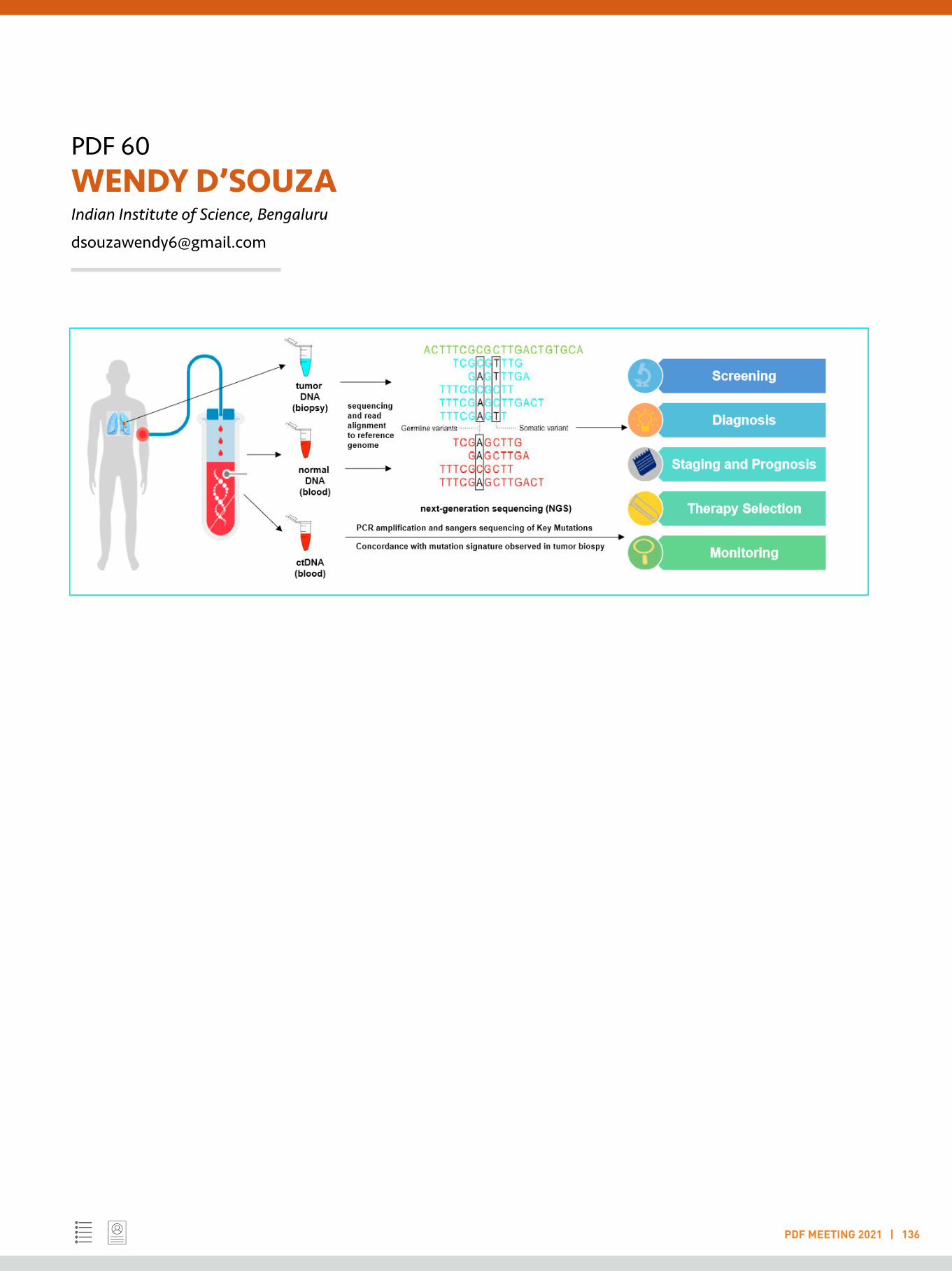
WENDY D’SOUZA
Molecular oncology; Cancer cell biology; Transcrip-
tome; Translatome; Clinical oncology
PDF 59
PDF 60

PDF 01 ABHIJIT AMBEGAONKAR National Institutes of Health, Rockville
PDF MEETING 2021 | 33
LIGHTNING TALK
B cell receptor signaling; Immunological synapse; Memory B cells; Chronic infectious diseases; Antigen affinity discrimination
Immunological memory in human chronic infectious diseases
The successful generation of antibody immunological memory after infection depends on acquisition of long-lived plasma cells that produce high affinity protective antibodies, and memory B cells (MBCs) that can respond to reinfection with the pathogen and its variants. However, immunity is not readily established to many chronic human infectious diseases such as HIV-AIDS, malaria and TB. These diseases are associated with a large expansion of a phenotypically and transcriptionally distinct subpopulation of B cells termed atypical MBCs[1]. Atypical MBCs are distinguished by their high expression of a variety of inhibitory receptors and by their inability to respond to antigens in solution, suggesting that atypical MBCs contribute to the poor acquisition of immunity in chronic infections[2]. Clearly, the development of vaccines for chronic infectious diseases would benefit from a better understanding of the function of atypical MBCs. I have investigated the mechanisms by which atypical MBCs are expanded during chronic infections and the function of atypical MBCs. I demonstrated that naïve B cells are the progenitors of atypical MBCs in infectious diseases such as malaria in which antigen may be persistently presented in the presence of IFN-gamma secreted by follicular helper T cells and toll-like receptor ligands from pathogen[3]. I also demonstrated that malaria-associated atypical MBCs respond robustly to antigens associated with cell surfaces due to the ability of atypical MBCs to segregate the potent inhibitory receptor FcγRIIB from the B cell receptor (BCR) immune synapse[4]. Additionally, I showed that a subpopulation of atypical MBCs that express high levels of IgD BCRs acquired high affinity thresholds for antigen-driven activation. I speculate that during chronic infections, atypical MBC expand to allow responses to foreign antigens that associate with cell surfaces, such as antigens in immune complexes, yet limit responses to fully soluble antigens, such as self-antigens. In future,
I plan to investigate the role of BCR affinity for antigens in activation and differentiation of MBCs. These studies extend our understanding of the function of B cell subpopulations in chronic infectious diseases and provide crucial insights about antigen design strategies for vaccination that can efficiently generate broadly neutralizing antibodies against infectious pathogens.
References:
1. Portugal, S., Obeng-Adjei, N., Moir, S., Crompton, P.D., and Pierce, S.K. (2017). Atypical memory B cells in human chronic infectious diseases: An interim report. Cell Immunol.
2. Portugal, S., Tipton, C.M., Sohn, H., Kone, Y., Wang, J., Li, S., Skinner, J., Virtaneva, K., Sturdevant, D.E., Porcella, S.F., et al. (2015). Malaria-associated atypical memory B cells exhibit markedly reduced B cell receptor signaling and effector function. Elife 4.
3. Ambegaonkar, A.A., Nagata, S., Pierce, S.K., and Sohn, H. (2019). The Differentiation in vitro of Human Tonsil B Cells With the Phenotypic and Functional Characteristics of T-bet+ Atypical Memory B Cells in Malaria. Front Immunol 10, 852.
4. Ambegaonkar, A.A., Kwak, K., Sohn, H., Manzella-Lapeira, J., Brzostowski, J., and Pierce, S.K. (2020). Expression of inhibitory receptors by B cells in chronic human infectious diseases restricts responses to membrane-associated antigens. Sci Adv 6, eaba6493.

PDF 01ABHIJIT AMBEGAONKARNational Institutes of Health, Rockville
PDF MEETING 2021 | 34
PDF 02 ABHISHEK MISHRA University of Fribourg
PDF MEETING 2021 | 35
LIGHTNING TALK
Keywords: Evo-devo; Nervous system; Nematostella vectensis; Neuroanatomy; Gal4/UAS system
Understanding evolution of the nervous system in the cnidarian Nematostella Vectensis
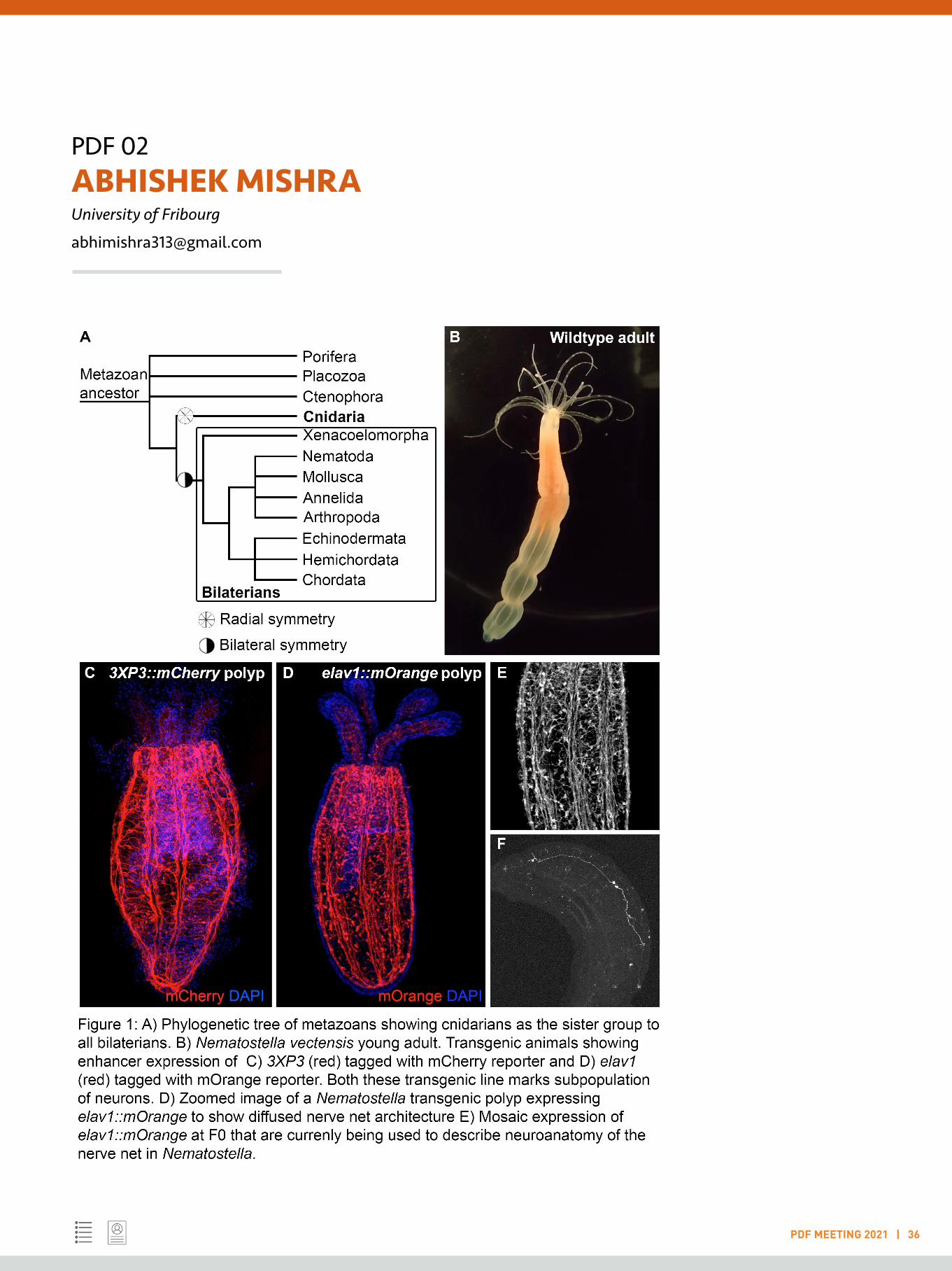
Nervous system consists of billions of neurons that are classified based on their diverse morphological, functional and anatomical properties. Despite recent advancements in identifying and characterising different neuronal subtypes in genetic model systems (such as in C. elegans, Drosophila, zebrafish or mouse), the knowledge about its evolutionary origin is still in its infancy. While these models have contributed immensely to understand molecular mechanisms underlying core developmental processes, their phylogenetic position makes it difficult to understand the ancestral origin and architecture of the ancient nervous system.
Cnidarians are placed in a phylogenetic tree as a sister group to all bilaterians (including vertebrates) and they are one of the earliest branching animal taxa that have a nervous system consisting of ‘simple interconnected nerve nets and no centralized brain’. Therefore, they serve as an excellent way to understand evolution of the nervous system helping in getting fundamental insights of the cnidarian nervous system and then comparing it with other bilaterians (Kelava et al., 2015). Cnidarian nervous system shares many fundamental properties with the vertebrate nervous system and these animals display a diverse array of coordinated behaviors including prey recognition, locomotion, body contraction, feeding etc. (Han et al., 2018). However, it is unknown how a decentralised nervous system in cnidarians having only interconnected neurons can sense external stimuli, process sensory inputs and in response, perform specific behaviors.
My current research work is focused to understand nervous system of cnidarian Nematostella vectensis and its function in a cellular, molecular and behavioral level. I am currently decoding the wiring pattern of its diffused nerve net, neuron-to-neuron, at a higher resolution and reconstructing neuronal
projections and its spatial distribution in Nematostella. By applying state-of-the-art labelling and imaging, I am systematically characterizing neuronal morphology and cell types which would be helpful in providing a cellular resolution description of its nerve net-like nervous system architecture.
The advent of genetic techniques to activate or inactivate specific neurons in a spatiotemporal manner allows us to identify neuronal functions with high precision. In the fruit fly, Gal4/UAS binary expression system provides valuable insights to understand neuronal functions linked with specific behavioral responses (Brand and Perrimon, 1993). I am establishing Gal4/UAS system in Nematostella that would be extremely helpful to understand the development and functions of the “decentralised nervous system” and how they are wired to perform specific behaviours. Establishing Gal4/UAS system would not only provide significant details about neuronal mechanisms controlling specific behaviour in cnidarians but would also shed evolutionary insights into how neural circuits corresponding to those behaviours have been evolved over time.
References:
Brand, A.H., Perrimon, N., 1993. Targeted gene expression as a means of altering cellfates and generating dominant phenotypes. Development 118, 401-415.
Han, S., Taralova, E., Dupre, C., Yuste, R., 2018. Comprehensive machine learninganalysis of Hydra behaviour reveals a stable basal behavioral repertoire. Elife 7.
Kelava, I., Rentzsch, F., Technau, U., 2015. Evolution of eumetazoan nervoussystems: insights from cnidarians. Philos Trans R Soc Lond B Biol Sci 370.
PDF 02ABHISHEK MISHRAUniversity of Fribourg
PDF MEETING 2021 | 36
PDF 03 ABHISHEK SUBRAMANIAN VIB-KU Leuven
PDF MEETING 2021 | 37
LIGHTNING TALK
Keywords: Computational systems biology; Network biology; Bioinformatics; Mathematical and statistical models; Machine learning
Unraveling condition-specific metabolic adaptations of eukaryotic cells using genome-scale models
Biological function is mediated by a complex interplay of discrete components forming an integrated system. Additionally, biological function is multi-dimensional and varies with time, space, environmental perturbations, cellular requirements, species, etc. My research work encompasses statistical or mathematical modeling of bio-systems to uncover design principles governing biological function.
My Ph.D research focused on characterizing stage and species-specific factors that enable the survival of eukaryotic parasite Leishmania (causes leishmaniasis) in the human and sandfly hosts. These parasites switch between the sandfly midgut and human macrophage phagolysosome environments. As these environments contain a distinct metabolic repertoire [1], the parasite has to flexibly adapt its metabolism for proliferation. Hence, we manually curated an energy metabolic and a novel, genome-scale metabolic reconstruction of L. infantum for large-scale flux predictions [2]. Applying flux balance (FBA), robustness, reaction essentiality and flux-coupling analyses on the two reconstructions, the previously known metabolic observations could be captured, substantiating a high confidence in the reconstruction quality. We discovered that a) subcellular compartmentalization constrains the choice of pathways for environmental adaptations and, b) succinate fermentation is essential to satisfy glutamate demand for energy maintenance [2].
In another work, we identified that flux-coupling constraints are major predictors of sequence-based evolutionary rates of metabolic genes across Leishmania species [3].
During my postdoctoral research, I chose to work on a more complex system, human endothelial cells(EC), where metabolic behavior is largely regulated by environmental
cues. When triggered by growth factor stimuli, ECs rapidly proliferate to form blood vessels. The aim of my project was to develop a new genome-scale metabolic model for ECs (EC-GEM) by tailoring human metabolic reconstructions with high-throughput, proliferation-specific RNA-sequencing data to predict fluxes that can maximize biomass production [4]. The EC-GEM predictions could successfully recapture active metabolic pathways with high precision. The EC-GEM was also integrated withsingle-celll RNA sequencing-characterized transcriptome of proliferating choroidal endothelial cells (CEC), to predict targets involved in leaky blood vessel formation during age-related macular degeneration (AMD) [4]. Cholesterol and collagen biosynthesis were identified as new anti-angiogenic targets. Rate-limiting enzymes of these pathways were demonstrated to inhibit EC angiogenesis in an in vitro EC model and in vivo angiogenic mouse models [4]. The EC-GEM is now being used to predict other novel pathways (ongoing). Additionally, I wrote a research proposal with my supervisor (recently granted) on prediction of metabolic targets in pathological angiogenesis by using scRNA sequencing data and experimental validation.
As a long-term goal, I plan to develop a large-scale, computational resource that automatically enables cross-species comparison of metabolic networks across eukaryotes.
References:
1. Subramanian A, Sarkar RR, 2018. Perspectives on Leishmania Species and Stage-specific Adaptive Mechanisms. Trends Parasitol. 34(12): 1068-81
2. Subramanian A, Sarkar RR, 2017. Revealing the mystery of metabolic adaptations using a genome-scale model of

PDF 03ABHISHEK SUBRAMANIANVIB-KU Leuven
PDF MEETING 2021 | 38
Leishmania infantum. Sci. Reports 7: 10262
3. Subramanian A, Sarkar RR, 2018. Evolutionary Perspectives of Genotype-Phenotype Factors in Leishmania Metabolism. J. Mol. Evol 86(7):443-456
4. Rohlenova K#, Goveia J#, García-Caballero M#, Subramanian A# et al., 2020. Single-cell RNA sequencing maps endothelial metabolic plasticity in pathological angiogenesis, Cell Metabolism 31(4): 862-877.e14

PDF 04 ADITYA KUMAR PADHI RIKEN Yokohama
PDF MEETING 2021 | 39
LIGHTNING TALK
Keywords: High-throughput protein design; Computational biophysics; Biomolecular structure-function relationship; Genetic varia-tions and disease mechanisms; Structural bioinformatics
Seven amino acid types suffice to create the interlaced core fold of RNA Polymerase
One of the greatest mysteries in life science is how the central dogma of molecular biology was established on ancient Earth. Modern proteins with large and complex structures are generally thought to have evolved from small and simple ancient proteins with “prototype folds” (e.g. Rossmann fold, ferredoxin fold, and (β/α)8-barrel) 1. These prototype folds must have played essential roles in the early evolution of life, as they are often conserved in fundamental biochemical pathways such as metabolism, replication, transcription, and translation. However, it remains elusive how such prototype folds emerged on the ancient earth, where the primitive translation system likely performed imprecise syntheses of short peptides composed of fewer amino acids as compared to modern proteins. Especially the components of the earliest genetic code are still an open question, as the 9–13 amino acid types used in previous ancestral protein reconstructions are at scattered positions in the modern codon table 2. The double-psi beta-barrel (DPBB) is one of the oldest protein folds conserved in various fundamental enzymes, such as the core domain of RNA polymerase. In this work, by employing high-throughput protein designing, we reverse engineered a modern DPBB domain and reconstructed its evolutionary pathway started by “interlacing homo-dimerization” of a half-size peptide, followed by gene duplication and fusion 3,4. Furthermore, by simplifying the amino acid repertoire of the peptide, we successfully created the DPBB fold with only seven amino acid types (Ala, Asp, Glu, Gly, Lys, Arg, and Val), which can be coded by only GNN and ARR (R=A or G) codons in the modern translation system. This is the smallest amino acid repertoire used for ancient protein reconstructions to our knowledge. These observations describe the evolutionary origin of a fundamental protein
materialised by the early genetic code and also demonstrate that such a complicated (interlaced) protein fold can be a straightforward target for protein engineering, in spite of its entangled appearance. Furthermore, our discovery also provides a fascinating indication about biology not only on Earth but also on the other planets, demonstrating folded proteins can emerge much more easily than what protein scientists and astrobiologists have imagined.
References:
1. Romero R, Rabin ML, & Tawfik DS. 2016. Functional Proteins from Short Peptides: Dayhoff’s Hypothesis Turns 50. Angew. Chemie - Int. Ed. 55: 15966–15971.
2. Koga, R. et al. 2020. Robust folding of a de novo designed ideal protein even with most of the core mutated to valine. Proc. Natl. Acad. Sci.U. S. A. 117: 31149-31156.
3. Yagi S, Padhi AK, Vucinic J, Barbe S, Schiex T, Nakagawa R, Simoncini D, Zhang KYJ & Tagami S. Seven amino acid types suffice to reconstruct the core fold of RNA polymerase. 2021. bioRxiv doi: 10.1101/2021.02.22.432383; (Under review, NatureChemistry).
4. Voet ARD. et al. 2014. Computational design of a self-assembling symmetrical β-propeller protein. Proc. Natl. Acad. Sci. U. S. A. 111: 15102–15107.

PDF 05 AJAY TIJORE Mechanobiology Institute, National University of Singapore
PDF MEETING 2021 | 41
LIGHTNING TALK
Keywords: Cancer mechanobiology; Cell biology; Biomaterials; Mechanical forces; Microfluidic devices
Mechanical Force-Induced Selective Killing of Cancer Cells
Historically, cancer incidence correlates with repeated injury, indicating that the repetitive activation of growth of adult cells can result in rigidity-independent (transformed) cancer growth. Recently, we observed that many cancer cells lack rigidity sensing because of depletion of mechanosensory cytoskeletal proteins e.g. tropomyosin 2.1 (Tpm2.1), which promotes rigidity-independent transformed growth. Surprisingly, we find that cancer cells that lack rigidity sensing are mechanosensitive and undergo mechanical force-mediated apoptosis.
In particular, we find that mechanical stretching reduces cancer cell growth and promotes apoptosis. In contrast, when rigidity sensing is restored in cancer cells by Tpm2.1 expression,Tpm2.1 expressed-cancer cells show rigidity-dependent growth upon stretching similar to the normal cells. The mechanism of cancer cell apoptosis involves stretch-mediated calcium uptake through mechanosensitive Piezo1 channels, which then activates a calpain protease to initiate a mitochondrial apoptotic pathway. To enable clinical mechanical therapy, we have developed a non-invasive ultrasound-based technology, in which ultrasound generated mechanical forces induce cancer cell apoptosis. Not only does ultrasound treatment promote cancer cell apoptosis in cell-based assays, but also it kills tumors grown in chick embryos and mice as well as patient-derived tumor organoids without damaging normal cells/tissues.
Mechanistically, the ultrasound mediated-apoptotic pathway mimics the mechanical stretch-mediated apoptosis. Thus, ultrasound treatment selectivity kills cancer cells in all environments tested without harming healthy cells, making it an effective tool for cancer therapy.
References:
1. Tijore A., Mingxi Y., et al., Biomaterials, in revision
2. Yao M, Tijore A et. al., Nature Materials, in revision
3. Tijore A, Margadant Felix, et al., Biomaterials, in revision1. Tijore A et al., ACS Applied Materials & Interfaces, 2014, 6, 15686 (IF 8.7)
4. Tijore A, et al., Advanced Healthcare Materials, 2015, 4, 1399 (IF 7.4)
5. Tijore A*, et al., Biofabrication, 2021, (IF 8.2)
6. Tijore A, et al., Biofabrication, 2018, 10, 025003
PDF 06 ANGIKA BASANT The Francis Crick Institute, London
PDF MEETING 2021 | 42
LIGHTNING TALK
Keywords: Cytoskeleton; Viral spread; Live imaging; Tyrosine kinase signalling; Rho GTPase signalling
Cytoskeletal regulation: in dividing cells and moving viruses
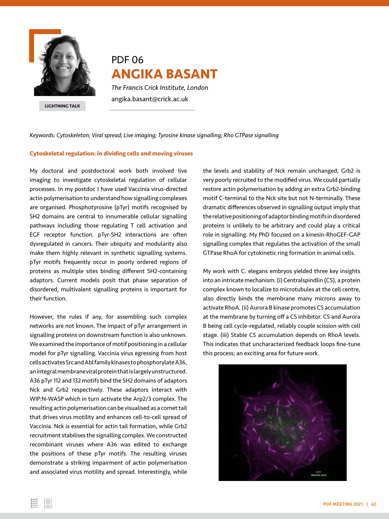
My doctoral and postdoctoral work both involved live imaging to investigate cytoskeletal regulation of cellular processes. In my postdoc I have used Vaccinia virus-directed actin polymerisation to understand how signalling complexes are organised. Phosphotyrosine (pTyr) motifs recognised by SH2 domains are central to innumerable cellular signalling pathways including those regulating T cell activation and EGF receptor function. pTyr-SH2 interactions are often dysregulated in cancers. Their ubiquity and modularity also make them highly relevant in synthetic signalling systems. pTyr motifs frequently occur in poorly ordered regions of proteins as multiple sites binding different SH2-containing adaptors. Current models posit that phase separation of disordered, multivalent signalling proteins is important for their function.
However, the rules if any, for assembling such complex networks are not known. The impact of pTyr arrangement in signalling proteins on downstream function is also unknown. We examined the importance of motif positioning in a cellular model for pTyr signalling. Vaccinia virus egressing from host cells activates Src and Abl family kinases to phosphorylate A36, an integral membrane viral protein that is largely unstructured. A36 pTyr 112 and 132 motifs bind the SH2 domains of adaptors Nck and Grb2 respectively. These adaptors interact with WIP:N-WASP which in turn activate the Arp2/3 complex. The resulting actin polymerisation can be visualised as a comet tail that drives virus motility and enhances cell-to-cell spread of Vaccinia. Nck is essential for actin tail formation, while Grb2 recruitment stabilises the signalling complex. We constructed recombinant viruses where A36 was edited to exchange the positions of these pTyr motifs. The resulting viruses demonstrate a striking impairment of actin polymerisation and associated virus motility and spread. Interestingly, while
the levels and stability of Nck remain unchanged, Grb2 is very poorly recruited to the modified virus. We could partially restore actin polymerisation by adding an extra Grb2-binding motif C-terminal to the Nck site but not N-terminally. These dramatic differences observed in signalling output imply that the relative positioning of adaptor binding motifs in disordered proteins is unlikely to be arbitrary and could play a critical role in signalling. My PhD focused on a kinesin-RhoGEF-GAP signalling complex that regulates the activation of the small GTPase RhoA for cytokinetic ring formation in animal cells.
My work with C. elegans embryos yielded three key insights into an intricate mechanism. (i) Centralspindlin (CS), a protein complex known to localize to microtubules at the cell centre, also directly binds the membrane many microns away to activate RhoA. (ii) Aurora B kinase promotes CS accumulation at the membrane by turning off a CS inhibitor. CS and Aurora B being cell cycle-regulated, reliably couple scission with cell stage. (iii) Stable CS accumulation depends on RhoA levels. This indicates that uncharacterized feedback loops fine-tune this process; an exciting area for future work.

PDF 07 ANIL ANNAMNEEDI Institute of Biology, Otto-von-Guericke University Magdeburg
PDF MEETING 2021 | 43
LIGHTNING TALK
Keywords: Bassoon; Presynapse; Autism spectrum disorders; Conditional knockout mice; Learning and memory
Presynapse in neuronal health and disease
Functional and structural changes at the chemical synapses, synaptic plasticity, play a major role in normal cognitive abilities of the healthy brain. Abnormal plasticity including the dysregulation of synaptic proteins function is observed during different pathological situations, often called synaptopathies. Till date, the presynaptic protein’s dysfunction for disease etiology and progression is poorly understood. Bassoon (gene: BSN), a large scaffolding protein at cytomatrix of the presynaptic active zone (neurotransmitter release site) and is exhibiting a variety of important presynaptic functions. Bassoon plays a crucial role in homeostatic synaptic plasticity, localization of voltage-gated calcium channels, presynaptic autophagy, proteasomal degradation and proper development of hippocampal CA3 moss fiber synapses 1,2,3. Recent findings in human studies and mouse models have revealed a connecting link between Bassoon and different neurological diseases. Human BSN gene mutations have been identified in Landau-Kleffner syndrome (an early childhood epilepsy), intellectual disability, deafness and progressive supranuclear palsy (PSP) like syndrome.
Bassoon proteinopathy is indeed shown to drive neurodegeneration in patients and EAE mouse models of multiple sclerosis. Dysregulated Bassoon expression have been reported in glutamatergic neurons (Bsn2lx/lx-Emx1Cre- B2E cKO), leads to immature dentate gyrus (DG) phenotype, a hallmark in neuropsychiatry, and DG-dependent learning changes 4. Interestingly, mice lacking Bassoon specifically in GABAergic interneurons (Bsn2lx/lx-Dlx5/6Cre- B2I cKO), display pronounced emotional and social behavioral changes reminiscent to autism and enhanced expression of several mitochondrial proteins. To conclude, Bassoon is an important protein at presynaptic release sites involved in essential functions starting from neurotransmission, presynaptic
protein clearance/maintenance to learning and memory and linked to neurodevelopmental/neuropsychiatric to neurodegenerative disorders. Hence, different Bassoon mutant mice can be served in elucidating the critical synaptic, network activity changes and circuit dysfunctions underlying pathological states.
References:
1. Gundelfinger, E. D., Reissner, C., & Garner, C. C. 2016. Role of Bassoon and Piccolo in Assembly and Molecular Organization of the Active Zone. Frontiers in synapticneuroscience. 7, 19.
2. Hoffmann-Conaway, S., Brockmann, M. M., Schneider, K., Annamneedi, A., Rahman, K. A., Bruns, C., Textoris-Taube, K., Trimbuch, T., Smalla, K. H., Rosenmund, C., Gundelfinger, E. D., Garner, C. C., & Montenegro-Venegas, C. 2020. Parkin contributes to synaptic vesicle autophagy in Bassoon-deficient mice. eLife, 9, e56590.
3. Montenegro-Venegas C, Fienko S, Anni D, Pina-Fernández E, Frischknecht R, Fejtova A. 2021. Bassoon inhibits proteasome activity via interaction with PSMB4. Cell Mol LifeSci. 78(4):1545-1563.
4. Annamneedi, A., Caliskan, G., Müller, S., Montag, D., Budinger, E., Angenstein, F., Fejtova, A., Tischmeyer, W., Gundelfinger, E. D., & Stork, O. 2018. Ablation of the presynaptic organizer Bassoon in excitatory neurons retards dentate gyrus maturation and enhances learning performance. Brain structure & function, 223(7), 3423–3445.

PDF 07ANIL ANNAMNEEDIInstitute of Biology, Otto-von-Guericke University Magdeburg
PDF MEETING 2021 | 44

PDF 08 ANUBAMA RAJAN Baylor College of Medicine, Houston
PDF MEETING 2021 | 45
LIGHTNING TALK
Keywords: Organoids, RSV, SARS-CoV-2, COPD, GI diseases
Human nose organoid model to study SARS-CoV-2 and RSV pathogenesis and evaluate therapeutics
My research work is aimed on development of lung and nose organoid models to understand respiratory viral pathogenesis with a focus on severe Acute Respiratory Syndrome Coronavirus 2 (SARS-CoV-2) and Respiratory Syncytial Virus (RSV), and to evaluate vaccine candidates and therapeutics for the same.
Background: There is a significant and unmet need for disease and pre-clinical models to understand the pathogenesis of SARS-CoV-2 and RSV; and predict responsiveness of immunotherapies to both these viruses. Organoid technology has the potential to address this need in basic science and clinical science research. Airway organoids can be utilised to study pathogenesis, can serve as ex-vivo human challenge model, and also overcome many of the limitations of current small animal and human challenge models. However, the existing methods for generating lung organoids rely on invasive or biopsy derived samples from patients to make organoids. This is often a roadblock to basic science researchers who do not have access to clinical samples. We addressed this major technical gap by developing a non-invasive technique to generate human airway organoids using nasal wash solution as an alternative to biopsy derived organoids. These human nose organoids (HNOs) consist of pseudostratified airway epithelium and can be used to model airway diseases.
Objectives: (1) To establish HNOs as a model to study SARS-CoV-2 and RSV pathogenesis, and (2) to study the effect of neutralizing antibodies and monoclonal antibodies against these major viral infections.
Results: We developed a non-invasive method to establish HNOs using stem cells isolated from nasal-wash samples and we established techniques to make differentiated air liquid interface (ALI) cultures from HNOs. Our results
showed that HNO-ALI cultures consist of well-differentiated, pseudostratified, ciliated, and mucosal respiratory epithelial cells. Next, we used HNO-ALI system to develop infection model to study viral infections and we assessed SARS-CoV-2 and RSV replication using real time-polymerase chain reaction, plaque assays and immunofluorescence techniques in HNO-ALI infected with infectious virions. Our results showed that HNO-ALI are susceptible to SARS-CoV-2, RSV A and B infection. The infected HNO-ALI recapitulated aspects of SARS-CoV-2 and RSV pathology, including viral shedding, ciliary damage, and mucus hyper-secretion. As proof of concept, we then evaluated the feasibility of HNO-ALI model system to test the efficacy of palivizumab monoclonal antibodies to prevent infection using palivizumab sensitive and resistant RSV strains. The model also effectively showed protection to infection in the presence of monoclonal antibodies. Conclusion: We established a non-invasive method to generate HNO-ALI model as an authentic and an alternative to transformed cell lines and small animal models. Our ex-vivo HNO-ALI infection model provides a novel approach to study respiratory viruses and for testing therapeutic interventions.
Research in-progress: My current ongoing research focuses on building an immune co-culture system to determine the contribution of the innate and cellular immune responses to the prevention, clearance, and pathology of RSV and SARS-CoV-2 infection. This will help us dissect the complex host-pathogen interaction for these two major respiratory viral pathogens.

PDF 08ANUBAMA RAJANBaylor College of Medicine, Houston
PDF MEETING 2021 | 46
PDF 09 ANUSHA SHANKAR Cornell University, Ithaca
PDF MEETING 2021 | 47
LIGHTNING TALK
Keywords: Physiology; Energetics; Transcriptomics; Ecology; Conservation
Integrative organismal physiology, ecology, and -omics in the tropics
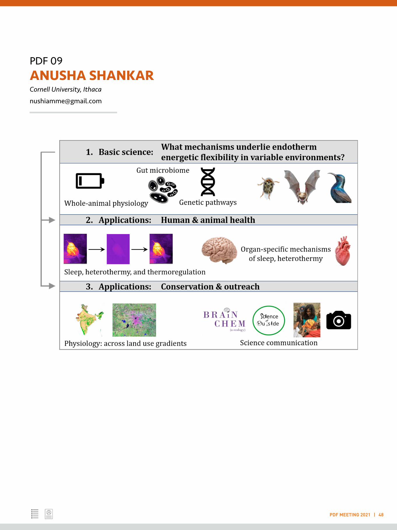
I use an integrative and multi-disciplinary approach to study how animals adapt and respond to challenging environmental conditions, while deriving lessons for wildlife conservation and human health. Rapid global climate change motivates me to identify the physiological and behavioural strategies that could allow diverse taxa to adapt to current and future environmental stressors. I am especially interested in the energetic strategies that tropical animals employ, given that the tropics are the seat of highest biodiversity, and that they will continue to experience the largest declines in biodiversity now and in the future. However, there is a dearth of whole-animal physiological data from the tropics, especially from India and the Paleotropics. I have gathered experience with a variety of methods and experimental approaches with Indian, North American temperate, and South American tropical species over the past 14 years. I am compelled to return to India to address fundamental and applied questions about Indian animals’ physiological ecology, while training upcoming Indian integrative biologists to expand our capacity in physiological ecology.
I am especially interested in the physiology and evolution of heterothermy (variable body temperatures) in vertebrates as a strategy to deal with variable environmental conditions. Animals from insects to birds and mammals use varying degrees of heterothermy to save energy by lowering their body temperatures, and correspondingly decreasing their energy expenditure. Heterothermy usually occurs under conditions where either the energetic demands are high (e.g., cold conditions), or energy supply is low (e.g., limited food availability). Insects such as beetles and caterpillars use diapause or aestivation to enter a suspended metabolic state for weeks or months. Some bird and mammal species, including hummingbirds, nightjars, and bats, use ‘torpor’ to
lower their body temperatures and metabolic needs at night. Hibernation is a well-known form of torpor: while daily torpor can occur every night for a few hours, hibernation is a torpor bout extended over several days, weeks, or months. Over 200 bird and mammal species so far are known to use some form of torpor, but much remains to be uncovered about the mechanisms underlying torpor. I will assess how animals respond to variable environments across three axes. 1. Basic science: animal physiology, -omics, and evolution. My primary questions are: How prevalent is heterothermy among Indian endotherms (birds and mammals)? What genetic pathways underlie heterothermy in birds vs mammals? And what evolutionary pathways for heterothermy converge/diverge in Indian species vs. New World species? Heterothermy is an evolutionarily widespread, but understudied, energy-saving strategy across many animal taxa, and there is great potential to study its prevalence and use in Indian animals. 2. Applications in human health. At its most applied, this work on heterothermy could inform medical research on induced hypothermia for surgeries in stroke and cardiac patients. 3. Applications in conservation science. I will aim to answer this question: How does behaviour, physiology, and genetics change across humaland-usese gradients, in free-living endotherms? And then to work with conservation practitioners to implement the results of these research in conservation action.
PDF 09ANUSHA SHANKAR Cornell University, Ithaca
PDF MEETING 2021 | 48
PDF 10 ANUSHILA CHATTERJEE Arizona State University, Tempe
PDF MEETING 2021 | 49
LIGHTNING TALK
Keywords: Infectious diseases, Bacteriophage, Antimicrobial resistance, Metagenomics, Host-microbe interactions
Exploiting insights from microbial interactions to combat antibiotic resistant bacteria
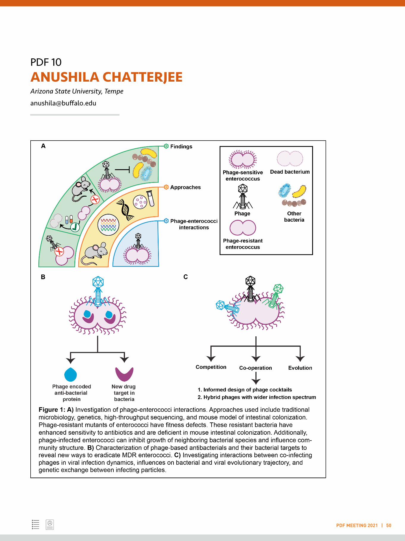
The frequency of multidrug resistant (MDR) bacterial infections is soaring. This is especially pertinent for the Gram-positive bacterium Enterococcus faecalis, a leading cause of hospital-acquired bloodstream infections world-wide and an opportunistic pathogen of high-priority according to the Indian Priority Pathogen List developed by WHO (India) and DBT. The pathogenic feat of MDR E. faecalis is further compounded by their ability to disseminate these drug-resistance traits to other MDR bacterial pathogens. Therefore, my research focuses on the use of bacteriophages, a.k.a. phages (viruses that infect bacteria), to eradicate drug resistant enterococci. Recently, phages are being considered for the elimination of systemic and biofilm-associated enterococcal infections. Although challenging, understanding the molecular interactions between phage and its target bacteria is critical prior to phage application to treat bacterial infections. Using culture-based approaches, animal models, and high-throughput sequencing techniques in my postdoctoral research, I identified a) enterococcal factors that are essential for productive phage infection and how mutation in genes encoding these factors prevent phage infection (1, 2), and c) subsequently demonstrated that phage resistance in enterococci imposes fitness costs which can be clinically exploited to limit bacterial infections (1) (fig. 1A).
Additionally, I elucidated how phage infection can modulate interactions of enterococci with different neighboring bacteria (3) (fig. 1A), and laid the groundwork to investigate if phage administration during enterococcal infection may disrupt host-associated beneficial microbial communities. Together, these insights into phage-enterococcal interactions paves the way to apply phage therapy in improved and innovative ways. My training has laid the groundwork for my future goals. First, I am interested in understanding how phages hijack
bacterial machinery to prevent bacterial growth, and how we can exploit this knowledge to interfere with MDR enterococci. Through proteomic profiling of phage infected enterococci, I have identified 5 phage-encoded proteins that could have growth-inhibitory effects. Characterization of these phage-derived inhibitors and identification of their bacterial targets will reveal novel ways to tackle MDR enterococci (Fig. 1B). Second, I want to adapt a social lens towards the study of phages and their implications on bacterial physiology. Although co-infections of bacteria by two or more phages are pervasive in nature and in the context of clinically relevant phage cocktails, the community behavior of phages is a highly neglected area of research. Leveraging my training in microbial genetics and genomics, I will explore different aspects of phage co-infection in enterococci, including how phage-phage interactions shape viral pathogenesis, and how the bacterial physiology modulates social life and evolutionary course of phages during co-infection (Fig. 1C).
References:
1. A. Chatterjee et al., Bacteriophage resistance alters antibiotic-mediated intestinal expansion of enterococci. Infect Immun 87, (2019).
2. A. Chatterjee et al., Parallel Genomics Uncover Novel Enterococcal-Bacteriophage Interactions. mBio 11, (2020).
3. A. Chatterjee, J. L. E. Willett, G. M. Dunny, B. A. Duerkop, Phage infection and sub-lethal antibiotic exposure mediate Enterococcus faecalis type VII secretion system dependent inhibition of bystander bacteria. PLoS Genet 17, e1009204 (2021).
PDF 10ANUSHILA CHATTERJEE Arizona State University, Tempe
PDF MEETING 2021 | 50
PDF 11 ASHA VELAYUDHAN NAIR Indian Institute of Technology, Kharagpur
PDF MEETING 2021 | 51
LIGHTNING TALK
Keywords: Antibiotic resistance; Multidrug resistance transporters; Bacterial biofilm; Efflux pump inhibitors; Targeted drug delivery
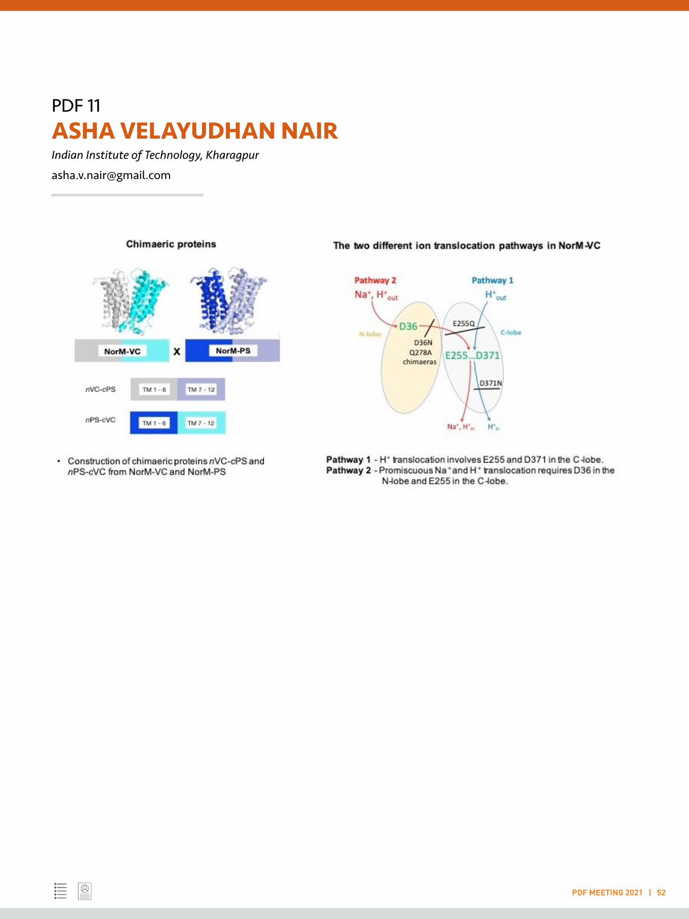
Chimaeric and mutant MATE multidrug transporters identify two functionally distinct ion-coupling pathways in NorM from Vibrio cholerae
Although the introduction of antibiotics revolutionised human and veterinary medicine, the rise of antibiotic resistance among pathogenic bacteria and drug resistance in cancers is reversing this progress. One important mechanism of drug resistance is based on the activity of multidrug transporters that mediate the active extrusion of structurally-dissimilar drugs from the cell. As the extrusion process prevents the binding of antibiotics and other cytotoxic agents to their intracellular targets, drug toxicity is overcome. The bacterial MATE proteins transport amphiphilic cationic drugs, such as norfloxacin and ethidium, from the cellular interior and mediate this reaction in exchange for H+ or Na+. Plant MATE transporters have been implicated in the H+/substrate antiport and have physiological roles in herbicide resistance, sequestration of plant-derived organic compounds in vacuoles, leaf senescence, aluminium tolerance, iron homeostasis, and synthesis of auxins.
In mammals, MATE transporters are localised in the proximal convoluted tubule and proximal straight tubule in the kidney as well as the canalicular membrane in hepatocytes, where they mediate H+-coupled transport of organic cations in the final steps of drug elimination from the body. Owing to their involvement in a wide range of physiological processes, MATE transporters are attractive pharmaceutical targets. Here, to study the molecular mechanism of MATE transporters, we engineered mutant transporters and chimeric proteins based on Na+ and H+-coupled NorM-VC from Vibrio cholerae and H+-coupled NorM-PS from Pseudomonas stutzeri, in which the N-lobe of one transporter is fused to the C-lobe of the other. Using biochemical assays like transport studies, substrate binding studies with ethidium as a substrate, we observe that the C-lobes mediate ethidium-H+ antiport
whereas the N-lobe of NorM-VC provides a promiscuous pathway for Na+ or H+ depending on ion availability. The mutant transporters we produced helped in the identification of the catalytic carboxylates that are important in the ion coupling mechanism. Thus, our findings demonstrate that the N- and C-lobes in NorM-type MATE transporters contribute distinct catalytic activities to the overall transport reaction. This conclusion is fundamental for our understanding of how these fascinating transport proteins operate, hence a step near to use this knowledge to combat the resistance mechanisms brought in to the organisms by these pumps.
References:
1. Raturi, S*., Nair, A.V*., Shinoda, K., Singh, H., Bai , B., Murakami, S., Fujitani, H., and Veen, H.W., (2021) Engineered chimeric and mutant NorM-type MATE multidrug transporters reveal two distinct ion-coupling pathways in the two halves. Communications Biology (Nature) (accepted) (* - equal contribution)
2. Yoonhee, J., Nair, A., and van Veen, H.W. (2014), Multidrug Transport Protein NorM from Vibrio cholerae Simultaneously Couples to Sodium- and Proton-Motive Force, J. Biol. Chem, 289:14624-14632
PDF 11ASHA VELAYUDHAN NAIR Indian Institute of Technology, Kharagpur
PDF MEETING 2021 | 52
PDF 12 AVIJIT BANIK Emory University, Atlanta
PDF MEETING 2021 | 53
LIGHTNING TALK
Keywords: Alzheimer’s disease; Neurodegeneration; Neuroinflammation; Neurocognition; Stem cell therapeutics
Anti-inflammatory effect of Prostaglandin receptor EP2 antagonist in treating pathology of Alzheimer’s disease
Cyclooxygenase-2 (COX-2), a key enzyme responsible for prostaglandin synthesis, in turn activates neuroinflammatory pathways in Alzheimer’s disease (AD) brain. Because of adverse cardiovascular events reported in use of COX-2 inhibitors, we examined downstream prostanoid receptor signaling to ameliorate the COX-2 mediated neuroinflammation in AD brains. In this study, we examined the effect of chronic treatment with a potent and selective EP2 antagonist, in the 5xFAD transgenic mouse model of AD. First, we characterized the potency, selectivity and anti-inflammatory properties of the EP2 antagonist in glial cells in culture. Then, 5xFAD mice and their non-transgenic littermates were treated for two months with the EP2 antagonist 100mg/kg/daily in drinking water. Mice were administered 0.5 mg/kg lipopolysaccharide (LPS) by intraperitoneal injection once a week to induce an additional level of brain inflammation. The EP2 antagonist
had no adverse effect on body weight and other organs. Complete blood count (CBC) analysis revealed an inflammatory effect of LPS in WBC, RBC and platelet distribution, which was not altered by the EP2 antagonist. The brain tissue analysis revealed that in female mice the mRNA level of proinflammatory mediators (IL-1β, TNFα, IL-6, CCl2, CXCL10) and glial markers (IBA1, GFAP, CD11b, S110B) were significantly reduced by the EP2 antagonist, whereas in male brains this effect was not found. There was no effect on the overall number of amyloid plaques or area covered by them in different regions of the brain. Taken together our findings suggest a therapeutic effect of EP2 antagonism in ameliorating chronic neuroinflammation in the AD brain.
Investigations are underway to elucidate the underlying mechanisms involving in this anti-inflammatory effect.

PDF 13 AVINASH PADHI Karolinska Institutet, Stockholm
PDF MEETING 2021 | 54
LIGHTNING TALK
Keywords: Keratinocyte biology; Skin Inflammation; Innate Immune responses; Immune-skin interaction; Transciptomics
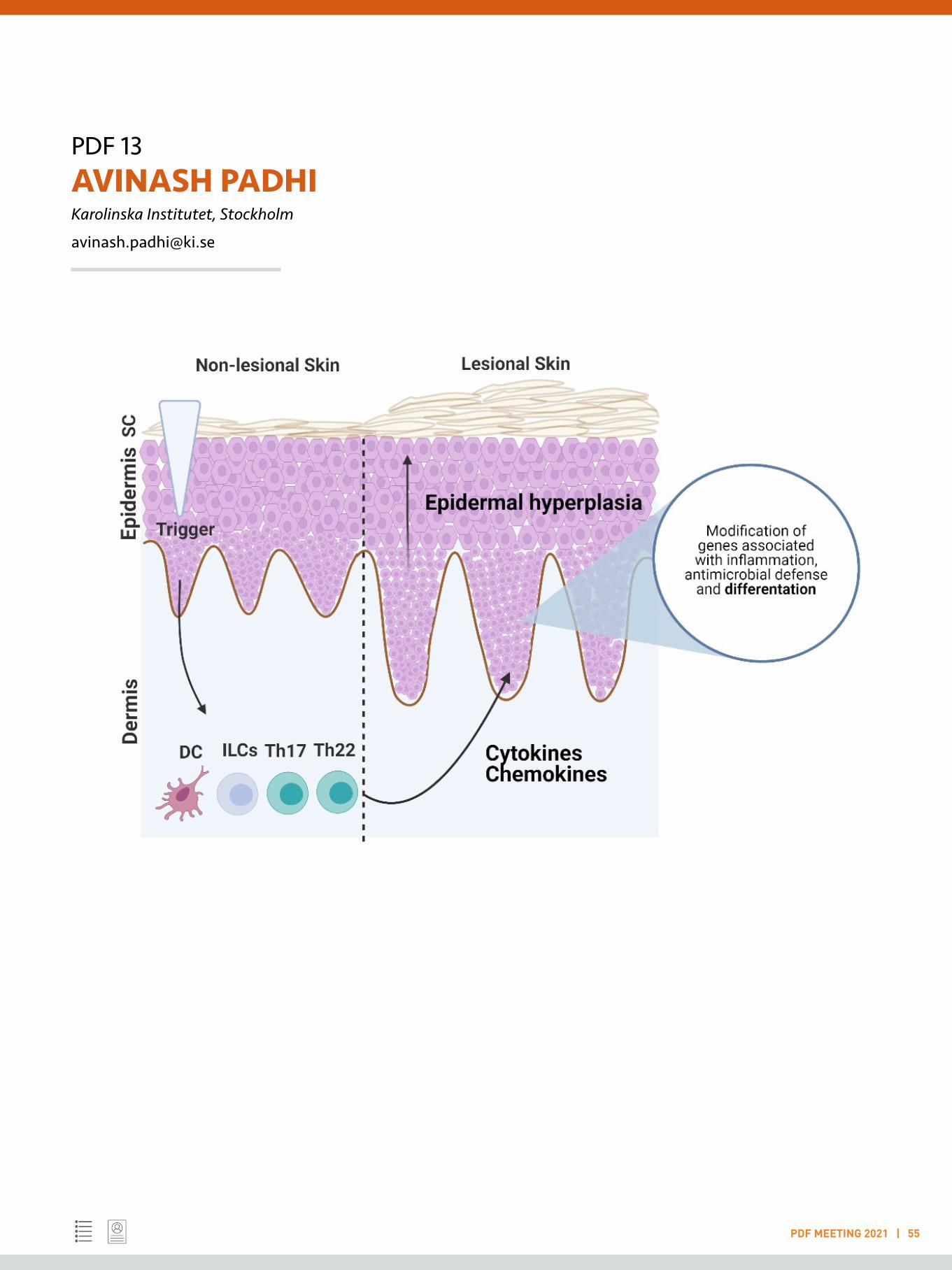
Cross talk between immune system and keratinocytes in Psoriasis
Skin barrier dysfunction is one of the most pre-disposing factor for development of chronic inflammatory skin diseases. An impaired epidermal barrier could instigate skin inflammation, while a persistent inflammation can additionally debilitate the skin barrier, suggesting an existing loop. However, elements of this loop are still poorly understood. Protein post-translational modifications form a vital component of inflammatory microenvironment and are critical in the pathogenesis of multiple skin disorders. One among them is protein deimination or citrullination, catalyzed by peptidyl-arginine deiminases (PADs) (Mechin et al, 2020). Initial studies hint at dysregulation of the enzymes in skin diseases. Citrullination plays an important role in several physiological processes including immune responses, regulation of gene expression, cellular events such as apoptosis and autophagy and epidermal barrier function. The modification has recently gained much attention since establishment of its pathological relevance in cancer, rheumatoid arthritis, and psoriasis to name a few (Alghamdi et al, 2019). Expression of these enzymes in tightly controlled during differentiation of keratinocytes, however the regulatory elements are still unknown. Here we show that the cytokines infiltrating skin with barrier defects, regulate PADs expressed by keratinocytes. Transcription profiling by qRT-PCR in 3D skin equivalents and differentiated epidermal keratinocytes revealed that pro-inflammatory cytokines suppress the expression of PADs at RNA (p<0.0001) level. Western blotting and antibody-based PAD activity assay (p<0.05) in primary keratinocytes revealed decrease in enzymatic activity of PADs in presence of high concentrations of cytokines. Considering lesional psoriatic skin is enriched with immune cells, we hypothesized that PAD expression would be lower in patients compared to healthy controls. To test this, we checked the expression of PAD isotypes in biopsies isolated from paired lesional and non-lesional skin of patients suffering
with plaque psoriasis and from healthy controls. We saw a decreased expression of PADs in lesional epidermis (p<0.037) as compared to healthy controls. However, we failed to correlate our findings with severity of the disease. Further, decrease in PAD expression in keratinocytes had a functional impact on overall deimination of proteins, especially filaggrin (p<0.05) and keratin (p<0.05), known to be crucial for epidermal differentiation. Drugs used to treat skin disorders involving keratinization defects could turn around this deleterious effect, by boosting overall deimination. These data not only support the important role played by deimination in skin barrier function, but they also identify a potential effect of cytokine environment on epidermal keratinocytes. Taken together, these findings could pave the way for PAD inducers as alternate therapy for treating skin disorders.
References: 1. Mechin MC, Takahara H, Simon M. Deimination and Peptidylarginine Deiminases in Skin Physiology and Diseases. 2020. Int J Mol Sci. 15;21(2):566.
2. Alghamdi M, Al Ghamdi KA, Khan RH, Uversky VN, Redwan EM. An interplay of structure and intrinsic disorder in the functionality of peptidylarginine deiminases, a family of key autoimmunity-related enzymes. 2019. Cell Mol Life Sci, 76(23):4635-4662.
PDF 13AVINASH PADHIKarolinska Institutet, Stockholm
PDF MEETING 2021 | 55

PDF 14 BHAVUK GARG Lerner Research Institute, Cleveland
PDF MEETING 2021 | 56
LIGHTNING TALK
Keywords: SCN-severe congenital neutropenia; AML-acute myeloid leukemia; MDS-Myelodysplastic syndrome; ELANE-Neutrophil elastase
Identifying key steps in granulopoiesis through inherited mutations
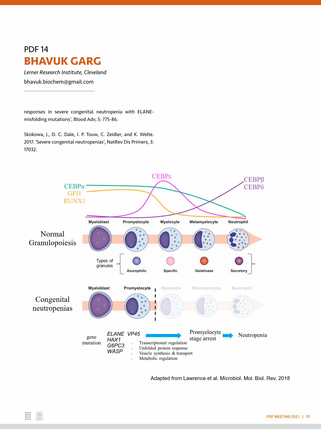
Granulocytic development is a prodigious process (1011 cells/day) divided into multiple stages from progenitors/ myeloblast to terminally differentiated neutrophils (Graphical Summary). This is accompanied by a profound change in cellular architecture which includes nuclear morphology, granular composition, metabolic programming, and surface marker expression. It is regulated by a small set of ligand-receptor interactions and transcription factors (Fiedler and Brunner 2012).
Severe congenital neutropenia (SCN) is a paediatric bone marrow failure syndrome characterized by acute paucity of circulating neutrophils in the peripheral blood. This leads to recurrent life threatening bacterial infections in children. It is attributed to the maturation arrest of granulocytic precursors at the promyelocytic stage. Mutations in a diverse set of genes such as ELANE, HAX1, G6PC3, SRP54 and CSF3R, have been implicated in SCN pathogenesis. Granulocyte colony stimulating factor (GCSF) administration restores the blood neutrophil counts and alleviates the SCN related infections (Skokowa et al. 2017). The underlying mechanisms of these gene mutations for SCN pathogenesis and their leukemic progression remain elusive.
There is a lack of experimental models which replicate the disease. Other challenges include the acquisition of rare patient samples from infants and imperfect mouse models. Patient-derived iPSCs require precise culture condition requirements, while stable transfectant cell line models have a selection bias of studying those cells that survive expression of the mutation. Using doxycycline-inducible expression, we reported an impaired granulocytic differentiation upon expression of disease causing ELANE mutations
(ELANE G185R), which was due to a suppression of transcriptional regulation of terminal granulocytic differentiation. This challenged the popular hypothesis of an unfolded protein response (UPR), for which we found no evidence (Garg et al. 2020). A subsequent independent report also indicated a UPR independent mechanism of disease associated ELANE mutations (Olofsen et al. 2021).
Investigating the molecular mechanisms SCN pathogenesis and its leukemic progression will further our understanding of normal and neoplastic granulopoiesis, which can be translated to better therapeutic alternatives.
References:
Fiedler, K., and C. Brunner. 2012. ‘The role of transcription factors in the guidance of granulopoiesis’, AmJ Blood Res, 2: 57-65.
Garg, B., H. M. Mehta, B. Wang, R. Kamel, M. S. Horwitz, and S. J. Corey. 2020. ‘Inducible expression of a disease-associated ELANE mutation impairs granulocytic differentiation, without eliciting an unfolded protein response’, J Biol Chem, 295: 7492-500.
Lawrence, S. M., R. Corriden, and V. Nizet. 2018. ‘The Ontogeny of a Neutrophil: Mechanisms of Granulopoiesis and Homeostasis’, Microbiol Mol Biol Rev, 82.
Olofsen, P. A., D. A. Bosch, O. Roovers, P. M. H. van Strien, H. W. J. de Looper, R. M. Hoogenboezem, S. Barnhoorn, P. G. Mastroberardino, M. Ghazvini, V. H. J. van der Velden, E. M. J. Bindels, E. M. dePater, and I. P. Touw. 2021. ‘PML-controlled
PDF 14BHAVUK GARGLerner Research Institute, Cleveland
PDF MEETING 2021 | 57
responses in severe congenital neutropenia with ELANE-misfolding mutations’, Blood Adv, 5: 775-86.
Skokowa, J., D. C. Dale, I. P. Touw, C. Zeidler, and K. Welte. 2017. ‘Severe congenital neutropenias’, NatRev Dis Primers, 3: 17032.
PDF 15 CHETANCHANDRA JOSHI Washington University in St. Louis
PDF MEETING 2021 | 58
LIGHTNING TALK
Keywords: Gametogenesis; Preterm birth and placental physiology; Metabolomics of host-pathogen interaction; Autophagy; aging; Oxidative stress
The NRF2/Keap1/p62 Pathway Governs the Host Response to Urinary Tract infections
Urinary tract infections (UTIs) are the most common recurring infections. More than 85% of UTIs are caused by uropathogenic Escherichia coli (UPEC) (Dielubanza and Schaeffer, 2011) that are able to use Type-1 fimbria to attach to uroplakin proteins on the surface of superficial cells of the bladder epithelial cells. Toll like receptor 4 (TLR4) on urothelial cells recognizes UPEC and activates pro-inflammatory cytokines (Schilling, et al., 2001). After bacterial invasion, the urothelial cells exocytose the bacteria via Rab GTPase (Khandelwal, et al., 2013). UPEC display a number of genes involved in oxidative stress response (Himpsl, et al., 2020). However, Reactive oxygen species (ROS) have not been directly quantitated in UPEC-infected urothelial cells. If urothelial cells do have increased ROS during UPEC infection, then the cells must have a mechanism to maintain redox homeostasis, as ROS build-up is detrimental for vesicle transport (Ravi, et al., 2016) and thus may impair UPEC expulsion. Also, the orchestration between bacterial recognition and bacterial exocytosis is unknown. We show that urothelial cells activate TLR4/TRAF6 dependent ROS production upon UPEC infection. Our immunoprecipitation data shows, in early hours of post infection, p62 accumulates and continues to associate with its binding partner KEAP1. Whereas, KEAP1 quickly dissociates from NF-E2-related factor 2 (NRF2) post infection. The p62 dependent lysosomal degradation of KEAP1 leads to translocation of NRF2 to nucleus and activation of ROS-buffering targets. Consequently, we developed KEAP1-deficient cells using CRISPR-Cas9 displayed over activated NRF2 resulting in reduced oxidative stress, inflammation and, ultimately showed heightened UPEC expulsion. In contrast, NRF2-deficient cells and mice exhibited accumulation of ROS, cell death, and reduced UPEC expulsion. Finally, we addressed the reason for heightened UPEC expulsion in KEAP1 deficient cells, we observed that NRF2
transcriptionally upregulated RAB27B expression, which has paramount importance in exocytosis of UPEC. Based on our findings, we tested whether pharmacological activation of NRF2 pathway would promote UPEC expulsion. The treatment of urothelial cells with FDA approved drug activated NRF2 pathway. The treatment reduces inflammatory response, increases RAB27B expression, and lowered bacterial burden in urothelial cells and in a mouse with UTI. Our findings elucidate the mechanism underlying the host response to UPEC invasion and provide a new strategy to combat UTIs.
References:
1. Dielubanza, E.J., and Schaeffer, A.J. 2011. Urinary tract infections in women. Med Clin North Am 95, 27-41.
2. Schilling, J.D., Mulvey, M.A., Vincent, C.D., Lorenz, R.G., and Hultgren, S.J. 2001. Bacterial invasion augments epithelial cytokine responses to Escherichia coli through a lipopolysaccharide-dependent mechanism. J Immunol 166, 1148-55.
3. Khandelwal, P., Ruiz, W.G., and Apodaca, G. 2010. Compensatory endocytosis in bladder umbrella cells occurs through an integrin-regulated and RhoA- and dynamin-dependent pathway. EMBO J 29, 1961-75.
4. Himpsl, S.D., Shea, A.E., Zora, J., Stocki, J.A., Foreman, D., Alteri, C.J., and Mobley, H.L.T. 2020. The oxidative fumarase FumC is a key contributor for E. coli fitness under iron-limitation and during UTI. PLoS Pathog 16, e1008382.

PDF 15CHETANCHANDRA JOSHIWashington University St. Louis
PDF MEETING 2021 | 59
5. Ravi, S., Pena, K.A., Chu, C.T., and Kiselyov, K. 2016. Biphasic regulation of lysosomal exocytosis by oxidative stress. Cell Calcium 60, 356-362.
PDF 16 CHINGAKHAM CHINGLENTHOIBA National University of Singapore
PDF MEETING 2021 | 60
LIGHTNING TALK
Keywords: Microplastic; Water conservation; Plastic pollution; Food chain; Sustainablity
Are you a Plasticterian? - A preliminary study of Microplastic identification in Sardine fish using Nile red dye.
Microplastics (MPs), which are generally defined as plastic particles between 1 μm to 5 mm in size, have been widely detected in both aquatic and terrestrial environments worldwide [1].This has raised concerns about MPs’ potential effects on aquatic organisms, particularly for smaller micro-sized (1-10 μm) plastics. The low degradation rate and small sizes make MPs persistent and easily accessible to various aquatic organisms (from zooplankton to mammals), disturbing the food chain [2]. Studies on MPs in freshwater environments have been predominantly conducted in lakes, rivers, and estuaries. Although the number of studies in freshwater environments have increased over the last few years, information about MPs in constructed wetlands is still largely limited. Constructed wetlands have been used worldwide to treat wastewater or stormwater and have significant values in urban freshwater environments [3]. They also provide habitats for organisms and essential services, such as reducing nutrients and pollutants entering receiving waters and are considered a ‘sink’ for many micro-pollutants. Storm water runoff and wastewater discharge can transport pollutants to aquatic environments and have been considered significant pathways for entry of MPs in freshwater bodies.
The Post-doctoral research works on classifications, sampling and characterization of MP in the marine ecosystem. On further, we also work on identification of the transportations of MP in marine and its effects of MP in the food chain. The classification and identification of MP were done using FTIR, Raman spectroscopy; and imaging of MPs were done using an optical microscope with a green and blue emission filter at 450-560 nm excitation. Nile red dye was used to stain and excite the polymers. The given data (unpublished) is taken from the intestinal guts of Sardine fish and treated with Nile red dye for observing under an optical microscope with a green and
blue emission filter at 450-560 nm excitation. Nile red dye was used to stain and excite the polymers. The given data (unpublished) is taken from the intestinal guts of Sardine fish and treated with Nile red dye for observing under an optical microscope with a green and blue emission filter at 450-560 nm excitation. The bright red dots and fibers were MPs from various macro plastics such as plastic bags, food packages and synthetic clothing. The data give us the awareness of MPs in sea food and many more.
References:
1. M. Cole, P. Lindeque, C. Halsband, and T. S. Galloway, “Microplastics as contaminants in the marine environment: A review,” Mar. Pollut. Bull., vol. 62, no. 12, pp. 2588–2597, 2011.
2. E. Guzzetti, A. Sureda, S. Tejada, and C. Faggio, “Microplastic in marine organism: Environmental and toxicological effects,” Environ. Toxicol. Pharmacol., vol. 64, no. September, pp. 164–171, 2018.
3. F. Gallo et al., “Marine litter plastics and microplastics and their toxic chemicals components: the need for urgent preventive measures,” Environ. Sci. Eur., vol. 30, no. 1, 2018.
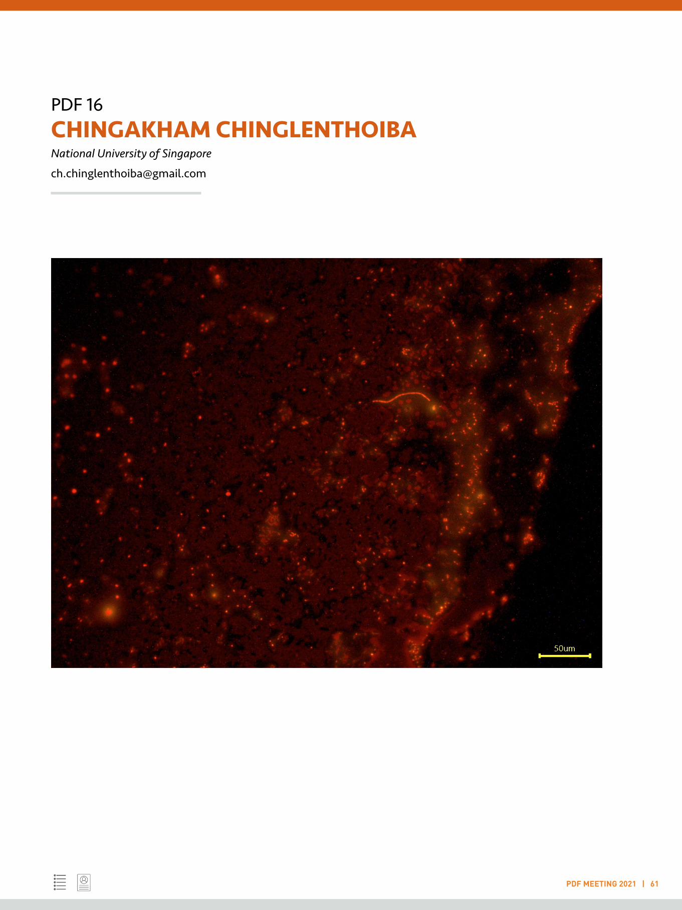
PDF 16CHINGAKHAM CHINGLENTHOIBANational University of Singapore
PDF MEETING 2021 | 61
PDF 17 DHANANJAY CHATURVEDI National Centre for Biological Sciences, Bengaluru
PDF MEETING 2021 | 62
LIGHTNING TALK
Keywords: Stem cells; Drosophila; Disease models; Muscle repair
Understanding Muscle Repair and Maintenance in vivo through Drosophila
Growth and repair of adult skeletal muscles critically depend on a population of quiescent stem cells known as satellite-cells. Injury triggers their proliferation to regenerate damaged muscle. The loss of satellite-cells correlates with debilitating muscle function that occurs during ageing and as a result of several genetic disorders (1). Given the profound importance of satellite cells in muscle repair, we are surprisingly far from a comprehensive understanding of the molecular and cellular biology of repair. Uncovering underlying fundamental principles must rely on understanding inextricable active molecular, cellular and tissue dynamics, inside a living organism. Intuitively, change in the innate tension of muscles would be the first parameter sensed by damaged muscles and satellite-cells upon injury with cascading effects. I aim to tease apart molecular details of the influence of mechanical tension on muscle autonomous and satellite-cell specific function. Though ex vivo systems offer technical ease, these studies decouple fundamental stimuli such as mechanical tension, metabolic state and inflammatory signals to satellite cells in vivo. Missing fundamental insights may come from other models, particularly those with a rich heritage of revealing conserved principles behind development and regeneration: Drosophila. They are a versatile, accessible, simple yet powerful model that allow intricate in vivo manipulation. Through my Post- Doctoral work, we were the first to show the existence of adult muscle stem cells, satellite cells in adult Drosophila flight muscles, marked by the transcription factor Zfh1 (3,4) subsequently discovered in mammals (5). My MicroCT scanning method measures in situ form at sub-micron resolution to complement confocal microscopy (6). Our findings, reagents, tools and methods position Drosophila as the system to generate an integrated view of in vivo muscle repair. Improved repair outcomes may hinge on a comprehensive answer to these questions: What role does mechanical
tension of substrate muscle play in satellite cell function? How does mechanical tension influence muscle autonomous homeostasis? What specific molecular responses are triggered in stem cells and muscles to injury, and how are they coupled molecularly to bring about repair in vivo? Further, does molecular and metabolic heterogeneity within muscles impact maintenance and repair? My expertise in Development and fly genetics, combined with bioinformatics tools, microscopy and microCT analysis identifying candidates on which the recovery process pivots. Conservation of these mechanisms can be tested in mammalian muscles. These studies will furnish molecular details of the muscle repair process, the molecular transmission of mechanical tension to regulate muscle homeostasis, possibly uncovering signatures of ageing. These studies in the short term serve as the foundation for understanding mature muscle function in vivo(9). Comparisons with clinical data from myodystrophy patients may construct, combine or fine tune pharmacological and physiotherapy regimen. In all, these findings may coherently bridge the fundamentals of conserved muscle repair mechanisms with clinical outcomes.
References:
1. Sambasivan R, Yao R, Kissenpfennig A, Van Wittenberghe L, Paldi A, Gayraud-Morel B, et al. Pax7- expressing satellite cells are indispensable for adult skeletal muscle regeneration. Development. 2011 Sep 6;138(19):4333–3.
2. Chang NC, Rudnicki MA, Rudnicki MA. Satellite Cells. In: Stem Cells in Development and Disease. Elsevier; 2014. pp. 161–81. (Current Topics in Developmental Biology; vol. 107). Available from: http://dx.doi.org/10.1016/B978-0-12-416022-4.00006-8

PDF 17DHANANJAY CHATURVEDINational Centre for Biological Sciences, Bengaluru
PDF MEETING 2021 | 63
3. Chaturvedi D, Reichert H, Gunage RD, VijayRaghavan K. Identification and functional characterization of muscle satellite cells in Drosophila. eLife. eLife Sciences Publications, Ltd; 2017;6:641.
4. Boukhatmi H, Bray S. A population of adult satellite-like cells in Drosophila is maintained through a switch in RNA-isoforms. eLife. eLife Sciences Publications Limited; 2018 Apr 9;7:878.
5. Siles L, Ninfali C, Cortés M, Darling DS, Postigo A. ZEB1 protects skeletal muscle from damage and is required for its regeneration. Nat Comms. Springer US; 2019 Mar 18;10(1):1–18.
6. Chaturvedi D, Prabhakar S, Aggarwal A, Atreya KB, VijayRaghavan K. Adult Drosophilamuscle morphometry through microCT reveals dynamics during ageing. Open Biol. 2019 Jun 26;9(6):190087–9.
7. Fletcher GC, Diaz-de-la-Loza M-D-C, Borreguero-Muñoz N, Holder M, Aguilar-Aragon M, Thompson BJ. Mechanical strain regulates the Hippo pathway in Drosophila. Development. Oxford University Press for The Company of Biologists Limited; 2018 Mar 1;145(5):dev159467.
8. Judson RN, Gray SR, Walker C, Carroll AM, Itzstein C, Lionikas A, et al. Constitutive Expression of Yes- Associated Protein (Yap) in Adult Skeletal Muscle Fibres Induces Muscle Atrophy and Myopathy. Mouly V, editor. PLoS ONE. Public Library of Science; 2013 Mar 27;8(3):e59622.
9. Andriy S. Yatsenko , Mariya M. Kucherenko , Yuanbin Xie , Dina Aweida , Henning Urlaub, Renate J. Scheibe , Shenhav Cohen and Halyna R. Shcherba et al. Profiling of the muscle-specific dystroglycan interactome reveals the role of Hippo signaling in muscular dystrophy and age-dependent muscle atrophy. Vol. 18, BMC Medicine. BMC Medicine; 2020. pp. 1–26.

PDF 18 DILIP KUMAR Baylor College of Medicine, Houston
PDF MEETING 2021 | 64
LIGHTNING TALK
Keywords: Structural biology; Virology; Biochemistry; Cryo-EM; X-ray crystallography
The structural insights into rotavirus capping assembly polls
My postdoctoral project is focused on structural and functional study replication and transcription complex (RTC) of gastroenteric pathogen rotavirus to delineate the viral replication, and particle assembly. Rotavirus is a major cause of infant gastroenteritis and hundreds of thousands child deaths (~200,000 annually) worldwide. The structure of virus particle and viral structural proteins have been determined except for the capping enzyme, VP3.We have determined the first full-length high resolution (2.7 Å) cryo-EM structure of rotavirusVP3 and have also observed some unique features associated with it (Kumar et al 2020). VP3 forms a stable tetrameric assembly of antiparallel dimers with each subunit having a modular domain organization to facilitate all three enzymatic steps of mRNA capping. Biochemical analysis showed VP3 exhibits RTPase activity through the phosphodiesterase (PDE) domain of VP3. Additionally, we have also observed ATP modulated helicase activity in full-lengthVP3. Based on these studies, we propose a novel mechanism in which each VP3 oligomer associated with a pair of VP1 inside the core caps the nascent transcripts emerging from VP1 during transcription (Figure). Currently, we are exploring the VP3-RNA complex structures to understand the capping process in mechanistic details. We have also determined the conformational flexibility of Influenza virus NS1 protein using X-ray crystallography and SM-FRET (Mitra et al, 2019). I also participated in the EMDB model challenge 2019 and performed de novo modelling in an assigned EM map with high accuracy (Lawson et al, 2021).
My doctoral research involved structural and functional studies of nucleic acid modulating enzymes from mycobacteria. We have determined the first crystal structure of mycobacterial nanoRNase, MSMEG_2630 to ~2.2 Å resolution which forms
a dimer with each subunit folding into a distinct N-terminal DHH and a C-terminal DHHA1 domain connected through a flexible linker region. The novel mode of subunit packing and variations in the linker region that enlarge the domain interface, are responsible for alternate substrates recognition.
References:
Kumar et al. 2.7 Å Cryo-EM structure of rotavirus core protein VP3, a unique capping machine with a helicase activity. Science Advances 15 Apr 2020: Vol. 6, no. 16, eaay6410.
Mitra S et al. Influenza A virus protein NS1 exhibits strain-independent conformational plasticity. J Virol. 2019 Aug 2. 917-919. doi: 10.1128/JVI.00917-19.
Lawson et al. Outcomes of the 2019 EMDataResource model challenge: validation of cryo-EM models at near-atomic resolution. https://doi.org/10.1101/2020.06.12.147033. NatureMethods 18, pages 156–164(2021).
Srivastav and Kumar et al. Unique subunit packing in mycobacterial nanoRNase leads toalternate substrate recognitions in DHH phosphodiesterases. Nucleic Acids Res. 2014 Jul;42(12):7894-910.
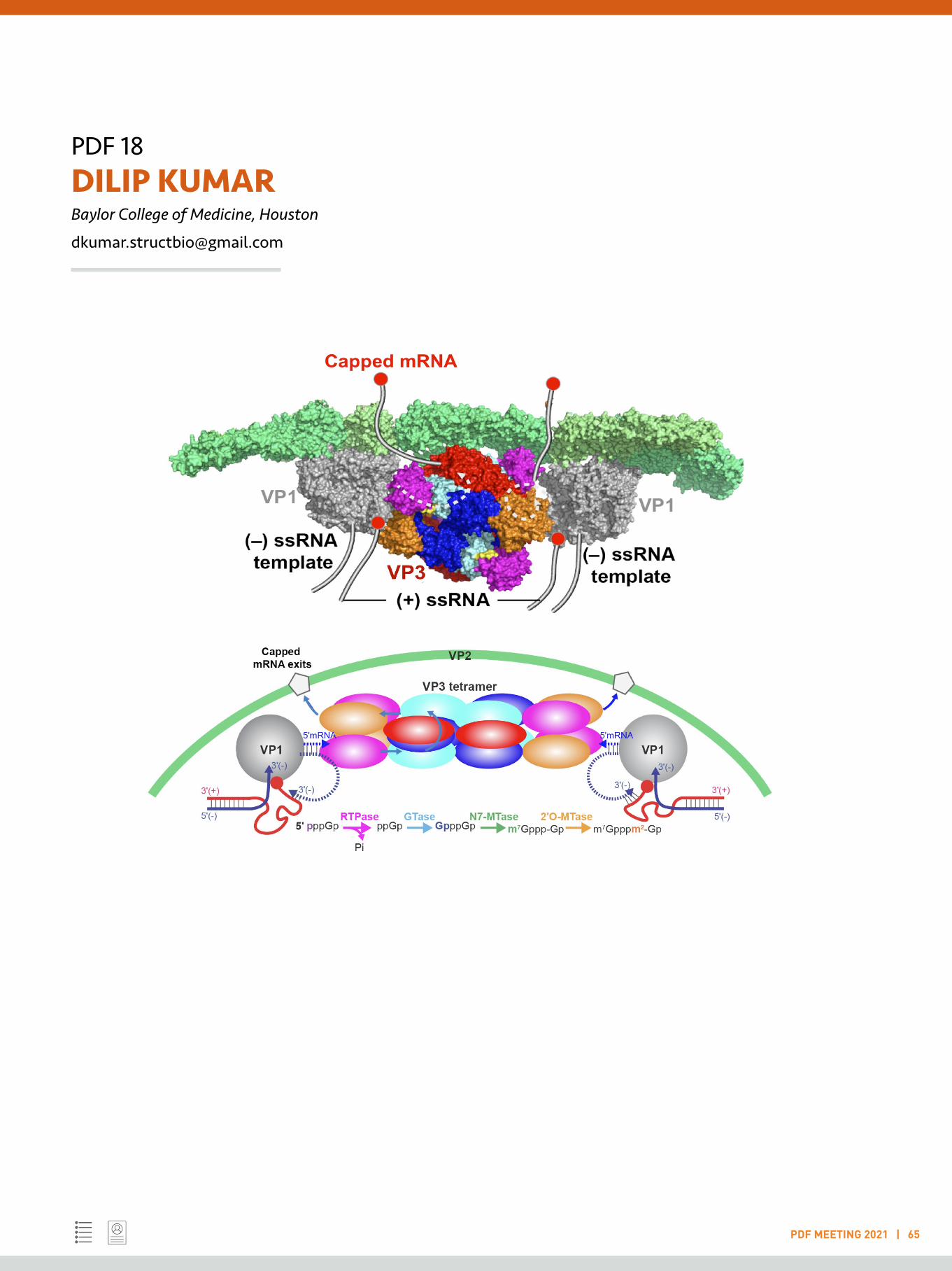
PDF 18DILIP KUMARBaylor College of Medicine, Houston
PDF MEETING 2021 | 65
PDF 19 DIYA BINOY JOSEPH UT Southwestern Medical Center, Dallas
PDF MEETING 2021 | 66
LIGHTNING TALK
Keywords: Urology; Infection; Pathology; Single cell RNA-sequencing; Tissue repair
Spatial transcriptomics approach to pinpoint cellular heterogeneity in 5-alpha reductase treatment resistance
Introduction: Benign prostatic hyperplasia (BPH) is an enlargement of the prostate that occurs with age. The increased prostate mass impinges on the urethral lumen and restricts urinary outflow. 5-alpha reductase inhibitors (5ARIs) are prescribed to reduce prostate volume and alleviate lower urinary tract symptoms. In some patients, prostate shrinkage is focal and does not result in improved urinary symptoms. Patients resistant or partially resistant to 5ARIs often return to the clinic and are prescribed surgical resection of the prostate. Although the broad pathological changes accompanying 5ARI treatment resistance have been described 1, cellular heterogeneity in the resistant regions have not been defined at the single cell level.
Methods: We collected simple prostatectomy tissues from a cohort of BPH patients which we divided into patients who had been prescribed 5ARI treatment and another group who had never been prescribed 5ARIs (untreated). 5ARI patients with detectable levels of drug and reduced dihydrotestosterone levels by mass spectroscopy were used for subsequent spatial transcriptomic studies. Visium spatial transcriptomics was performed on histologically-responsive and non-responsive regions from 5ARI patients.
Results: In 5ARI treated patients, regions of normal prostate glandular architecture were often seen adjacent to regions of smaller acini with multiple cell layers, defined as histologically-resistant and -responsive, respectively. Clustering using k-means revealed two distinct epithelial populations in histologically-responsive regions. One of these clusters was identified as normal prostate secretory luminal cells with a characteristic androgen driven gene expression signature.
The second epithelial cell cluster shared the gene expression profile of specialized urethral epithelial club cells that we
had previously described2. Club-like cells in 5ARI treated patients display low expression of AR, NKX3.1 and AR dependent genes like ACPP and KLK3. Club cell markers were upregulated in regions of 5ARI histologic response including SCGB1A1, LCN2, PIGR and OLFM4. We observed an increase in TNF-alpha expression and nuclear localization of the NF-KB component phospho-p65 in regions of 5ARI histologic response. To test whether NF-KB pathway activation drives the altered gene expression profile in these regions, we treated primary prostate epithelial cells with TNF-alpha and Lipopolysaccharide, which are known to activate the NF-KB pathway. LCN2, PIGR and OLFM4 were upregulated 24 hours after treatment, recapitulating the in vivo expression of these genes in regions of 5ARI histologic response.
Conclusions: We conclude that 5ARI treatment results in regions of altered epithelial cell composition that resemble androgen-independent club cells. A switch from AR dependent gene signaling to NF-KB pathway activation drives the altered gene expression profile observed in 5ARI histologic response. Further studies are required to determine how adjacent ‘normal’ prostate glands maintain AR dependent signaling despite low levels of tissue DHT.
References:
1. Vis AN, Schröder FH. 2009. Key targets of hormonal treatment of prostate cancer. Part 2: the androgen receptor and 5alpha-reductase. BJU Int 140;9: 1191-1197
2. Henry GH, Malewska A, Joseph DB et al. 2018. A Cellular Anatomy of the Normal Adult Human Prostate and Prostatic Urethra. Cell Reports 25;12: 3530-3542
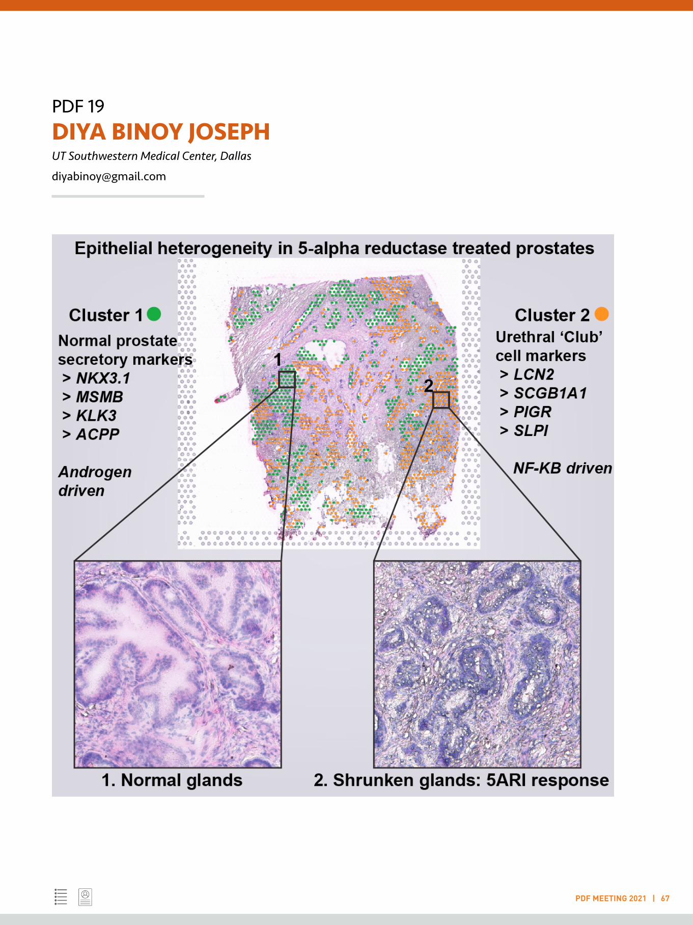
PDF 19DIYA BINOY JOSEPHUT Southwestern Medical Center, Dallas
PDF MEETING 2021 | 67

PDF 20 HIMANSHU GOGOI University of Florida, Gainesville
PDF MEETING 2021 | 68
LIGHTNING TALK
Keywords: Vaccine; Innate immunity; Lung immunology; Adjuvants; Ageing
Lung moDC targeting TNF-fc(IgG2a) can restore CDG mucosal adjuvant activity in aged mice
My current research focuses on targeted vaccine delivery. Briefly, it can be summarized as (i) to identify the in vivo mode of action of cyclic di- GMP (CDG) adjuvant in the adult mice; (ii) to define the defects in the aged mice that compromises CDG adjuvanticity in vivo; (iii) to design new biologics to circumvent the defective component in CDG mode of action and restore CDG adjuvanticity in the aged mice.
Cyclic di- nucleotides (CDNs) have been recognized as potent immunostimulators in the mammalian system [1, 2]. Mechanistically, CDG adjuvanticity depends on the stimulator of interferon genes (STING)-induced tumor necrosis factor (TNF) production in vivo [3]. We have demonstrated that intranasal immunization of CDNs; specifically, cyclic di-GMP (CDG) is phagocytosed by specific lung dendritic cell (DC) populations; conventional DC1 (cDC1) and cDC2 to orchestrate the immune response. While cDC1 is dispensable for CDG mediated response, cDC2 plays the pivotal role in directly inducing cellular response as well as activating another DC population monocyte derived DC (moDC) to propagate the humoral response [4].
As an extension of this project, we demonstrated that although CDG is a very potent mucosal adjuvant, it fails to stimulate the immunoscent immune system and aged mice had defective CDG-induced memory T helper (Th)1 and Th17 responses and high-affinity serum immunoglobulin (Ig)G, mucosal IgA production. This was primarily attributed to a depleted lung cDC2 in the aged mice. To address this shortcoming, we generated two novel tumor necrosis factor (TNF) fusion proteins; soluble TNF (solTNF) and transmembrane TNF (tmTNF) to target moDCs to enhance CDG vaccine efficacy in aged mice. The moDC-targeting TNF fusion proteins restored CDG-induced memory Th1, Th17, and high-affinity IgG, IgA
responses in the aged old mice. Together, our data suggested that aging negatively impacts CDG vaccine adjuvanticity and moDC-targeting TNF fusion proteins restored CDG adjuvanticity in the aged mice [5].
References:
1. Ebensen, T., et al., The bacterial second messenger cyclic diGMP exhibits potent adjuvant properties. Vaccine, 2007. 25(8): p. 1464-1469.
2. Gray, P.M., et al., Evidence for cyclic diguanylate as a vaccine adjuvant with novel immunostimulatory activities. Cellular immunology, 2012. 278(1-2): p. 113-119.
3. Blaauboer, S.M., V.D. Gabrielle, and L. Jin, MPYS/STING-mediated TNF-α, not type I IFN, is essential for the mucosal adjuvant activity of (3’–5’)-cyclic-di-guanosine-monophosphate in vivo. The Journal of Immunology, 2014. 192(1): p. 492-502.
4. Mansouri, S., et al., Immature lung TNFR2− conventional DC 2 subpopulation activates moDCs to promote cyclic di-GMP mucosal adjuvant responses in vivo. Mucosal immunology, 2019. 12(1): p. 277-289.
5. Gogoi, H., et al., New modc-targeting tnf fusion proteins enhance cyclic di-gmp vaccine adjuvanticity in middle-aged and aged mice. Frontiers in immunology, 2020. 11: p. 1674.
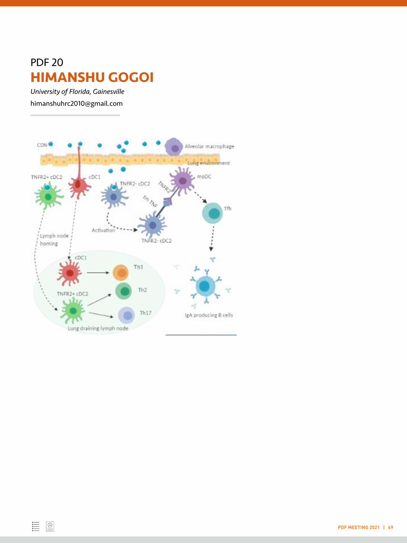
PDF 20HIMANSHU GOGOIUniversity of Florida, Gainesville
PDF MEETING 2021 | 69
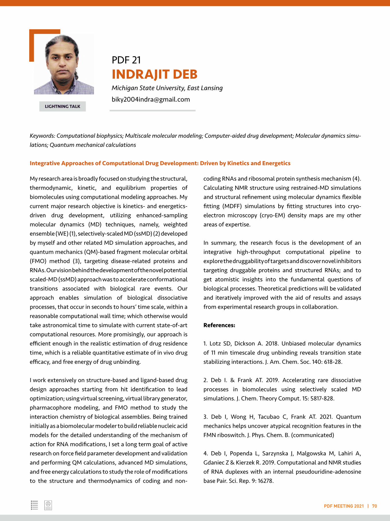
PDF 21 INDRAJIT DEB Michigan State University, East Lansing
PDF MEETING 2021 | 70
LIGHTNING TALK
Keywords: Computational biophysics; Multiscale molecular modeling; Computer-aided drug development; Molecular dynamics simu-lations; Quantum mechanical calculations
Integrative Approaches of Computational Drug Development: Driven by Kinetics and Energetics
My research area is broadly focused on studying the structural, thermodynamic, kinetic, and equilibrium properties of biomolecules using computational modeling approaches. My current major research objective is kinetics- and energetics-driven drug development, utilizing enhanced-sampling molecular dynamics (MD) techniques, namely, weighted ensemble (WE) (1), selectively-scaled MD (ssMD) (2) developed by myself and other related MD simulation approaches, and quantum mechanics (QM)-based fragment molecular orbital (FMO) method (3), targeting disease-related proteins and RNAs. Our vision behind the development of the novel potential scaled-MD (ssMD) approach was to accelerate conformational transitions associated with biological rare events. Our approach enables simulation of biological dissociative processes, that occur in seconds to hours’ time scale, within a reasonable computational wall time; which otherwise would take astronomical time to simulate with current state-of-art computational resources. More promisingly, our approach is efficient enough in the realistic estimation of drug residence time, which is a reliable quantitative estimate of in vivo drug efficacy, and free energy of drug unbinding.
I work extensively on structure-based and ligand-based drug design approaches starting from hit identification to lead optimization; using virtual screening, virtual library generator, pharmacophore modeling, and FMO method to study the interaction chemistry of biological assemblies. Being trained initially as a biomolecular modeler to build reliable nucleic acid models for the detailed understanding of the mechanism of action for RNA modifications, I set a long term goal of active research on force field parameter development and validation and performing QM calculations, advanced MD simulations, and free energy calculations to study the role of modifications to the structure and thermodynamics of coding and non-
coding RNAs and ribosomal protein synthesis mechanism (4). Calculating NMR structure using restrained-MD simulations and structural refinement using molecular dynamics flexible fitting (MDFF) simulations by fitting structures into cryo-electron microscopy (cryo-EM) density maps are my other areas of expertise.
In summary, the research focus is the development of an integrative high-throughput computational pipeline to explore the druggability of targets and discover novel inhibitors targeting druggable proteins and structured RNAs; and to get atomistic insights into the fundamental questions of biological processes. Theoretical predictions will be validated and iteratively improved with the aid of results and assays from experimental research groups in collaboration.
References:
1. Lotz SD, Dickson A. 2018. Unbiased molecular dynamics of 11 min timescale drug unbinding reveals transition state stabilizing interactions. J. Am. Chem. Soc. 140: 618-28.
2. Deb I. & Frank AT. 2019. Accelerating rare dissociative processes in biomolecules using selectively scaled MD simulations. J. Chem. Theory Comput. 15: 5817-828.
3. Deb I, Wong H, Tacubao C, Frank AT. 2021. Quantum mechanics helps uncover atypical recognition features in the FMN riboswitch. J. Phys. Chem. B. (communicated)
4. Deb I, Popenda L, Sarzynska J, Malgowska M, Lahiri A, Gdaniec Z & Kierzek R. 2019. Computational and NMR studies of RNA duplexes with an internal pseudouridine-adenosine base Pair. Sci. Rep. 9: 16278.
PDF 21INDRAJIT DEBMichigan State University, East Lansing
PDF MEETING 2021 | 71
PDF 22 JAGADISH SANKARAN Genome Institute of Singapore
PDF MEETING 2021 | 72
LIGHTNING TALK
Keywords: Fluorescence microscopy; Quantitative imaging; Assay development; Biofilm; Pseudomonas aeruginosa
Development and application of quantitative fluorescence microscopy tools for investigating bacterial biofilms
An engineer by training, I am a biophysicist with experience across both dry and wet components of research. My specialization is in developing quantitative fluorescence imaging assays in general and to utilize these assays to study pathophysiology of bacterial biofilms in particular.
In the dry lab, my work is primarily focused on developing data analysis algorithms and tools to extract information about dynamics and organization of biomolecules from large fluorescence imaging stacks (Refs 1, 2, 3). Typically, fluorescence imaging techniques to monitor dynamics and microscopic structure of biomolecules often have mutually exclusive experimental strategies with a need to choose one over the other. We identified a solution by devising a measurement approach and a post-processing strategy wherein the original data is repurposed for various different analytic techniques reporting on structure and dynamics (Ref 4).
In the wet lab, my work is primarily focused on imaging and investigating communities of bacteria adherent to surfaces called biofilms. One of the issues of biofilms relevant in the context of human health is their increased tolerance to antibiotics. The pathogen which we investigated is Pseudomonas aeruginosa which has been listed as a critical priority pathogen in “Indian priority pathogen list”. It is believed that the tolerance is due to the fact that biofilms act as diffusion barriers for antibiotics. We then asked the question, do Pseudomonas aeruginosa biofilms act as diffusion barriers to antibiotics. Our results suggest that diffusion limitations do not contribute to the observed tolerance in clinically relevant biofilms for antimicrobials (Ref 5). Since this study demonstrates that antibiotics penetrate into and diffuse in biofilms, one of the ways to improve the action of antibiotics on biofilms is to administer it in combination with a molecule
which disperses the biofilms.
I plan to start a laboratory with two themes: developing quantitative fluorescence microscopy-based biological assays in general and utilizing those assays to address questions pertinent to biofilms composed of Pseudomonas aeruginosa. I find myself uniquely and strategically positioned at the confluence of engineering, biology, and computation enabling me to effectively communicate and act as a liaison between these fields facilitating cross-disciplinary collaboration and discussion by acting as a catalyst for mutual exchange of ideas.
References:
1. Sankaran J, Manna M, Guo L, Kraut RS & Wohland, T. Diffusion, transport, and cell membrane organization investigated by imaging fluorescence cross-correlation spectroscopy. Biophysical Journal 97, 2630-2639 (2009)
2. Sankaran J, Shi X, Ho LY, Stelzer EH & Wohland T. ImFCS: a software for imaging FCS data analysis and visualization. Optics Express 18, 25468-25481 (2010)
3. Sankaran J, Bag N, Kraut RS & Wohland, T. Accuracy and Precision in Camera- Based Fluorescence Correlation Spectroscopy Measurements. Analytical Chemistry 85, 3948-3954 (2013)
4. Sankaran J, Balasubramanian H, Tang WH, Ng XW, Rollin A, Wohland T. Simultaneous spatiotemporal super-resolution and multi-parametric fluorescence microscopy. Nature Communications 12, 1748 (2021)

PDF 22JAGADISH SANKARANGenome Institute of Singapore
PDF MEETING 2021 | 73
5. Sankaran J, Tan NJHJ, But KP, Cohen Y, Rice SA, Wohland T. Single microcolony diffusion analysis in Pseudomonas aeruginosa biofilms. npj Biofilms and Microbiomes 5, 35 (2019).

PDF 23 KANIKA KHANNA Stanford University, Palo Alto
PDF MEETING 2021 | 74
LIGHTNING TALK
Keywords: Microbiology; Microbiome; Bacterial cell biology; Cryo-electron tomography; Cryo-focused ion-beam milling
Opening windows into the microbial word using cell biology and synthetic ecology approaches
PhD work: During my PhD, I uncovered the roles of tiny molecular machines in regulating bacterial cellular processes at a high spatial resolution using cryo-electron tomography (cryo-ET) coupled to cryo-focused ion beam milling, setting it up as a method of choice for conducting bacterial cell biology.
1. Sporulation in Bacillus subtilis: B. subtilis undergoes sporulation during nutrient starvation wherein an asymmetrically positioned septum generates a larger mother cell and a smaller forespore. Subsequently, the mother cell engulfs the forespore in a process resembling phagocytosis. I investigated several outstanding questions during cell division and cell wall remodeling. During vegetative growth, the division septum is formed at the mid cell and is almost twice as thick as the polar septum formed during sporulation. The organizational details of cell division proteins during these two conditions have remained elusive due to inability to resolve them using current microscopy techniques. Using cryo-ET, I demonstrated that FtsZ filaments, the major orchestrators of bacterial cell division have different spatial organization and abundance during sporulation and vegetative growth, giving rise to different septal thickness and cell fate. These results reveal a novel mechanism by which bacteria regulate protein localization in space and time to carry out cytokinesis, paving way for future studies in prokaryotic cell division. My work also provided new insights into the engulfment process, including the role of coordinated degradation and synthesis of cell wall and the role of forespore chromosome translocation in shaping the forespore by generating turgor pressure.
2. Jumbo phage (genome size >250 kb) replication in bacteria: I also investigated the mechanism of replication of jumbo phages (genome size >250 kb) in Pseudomonas hosts. Our studies on the assembly of a nucleus-like structure and
spindle during phage replication unveil potential ways in which viruses avoid their host’s defense mechanism and hint at the evolutionary origins of nucleus formation. Moreover, the spatio-temporal mechanism of phage replication in situ can be harnessed as an effective tool to genetically engineer phages that can fight notorious antibiotic-resistance bacterial infections.
Postdoc work: The microbiome field is rapidly generating exciting data but many of these early studies are correlative and there is a need to advance to molecular and mechanistic understanding of host-microbe and microbe-microbe interactions in the gut. Hydrogenotrophs or H2-consuming microbes comprise a low abundance population but play a significant role in regulating the fermentation potential of the gut microbiota by reoxidizing the hydrogen produced by primary fermenters to drive forward the metabolism. This in turn also influences the host physiology and metabolism. I am currently using a 120-member synthetic gut bacterial community which can be stably engrafted in germ-free mice to elucidate the relative effects and contributions of three major groups of hydrogen consumers (acetogens, methanogens and sulfate-reducers) on the community structure and function. Mechanistic understanding gleaned from these experiments will allow us to engineer synthetic communities that can be endowed with specific functions in real-world settings for therapies related to cancer and metabolic disorders like obesity and malnutrition.

PDF 24 KARTHIK CHANDIRAN University of Connecticut Health Center, Hamden
PDF MEETING 2021 | 75
LIGHTNING TALK
Keywords: T cells; Infectious diseases; Cell differentiation; Intracellular signaling; Next-generation sequencing
Smad signaling determines the fate of activated CTLs via multiple intersecting signaling pathways
Immune responses to an infection are controlled by various factors including antigens, co-stimulatory molecules and cytokines present in the tissue microenvironment. TGFb is a pleiotropic cytokine that modulates immune response and cellular differentiation usually through SMAD (Smad 2,3 & 4)-mediated signaling cascade. Interestingly, TGFb and Smad4 have independent and opposing roles in determining the fate of activated CTLs. While TGFb-receptor mediated signaling is required for resident memory (TRM) cells, Smad4 is required for effector (TEff) and central memory subsets (TCM). However, the mechanism behind TGFb as well as Smad4 mediated differentiation is yet to be unraveled. Using transgenic animal models and pharmacological inhibitors, we investigate the role of TGFb and different SMAD proteins in determining the fate of activated CTLs. We identified that while canonical TGFb signaling, through Smad2 and Smad3, induces CD103 expression (TRM), Smad4 inhibits CD103 expression thus preventing TRM differentiation. In addition, targeted-ablation of Smad4 alter the expression levels of
multiple transcription factors that participate in fate determination, including Eomes and KLF2. Although Eomes has previously been implicated in negative regulation of CD103, over-expression of Eomes does not alter CD103 expression in the absence of Smad4. This suggests that Smad4 is required for negative regulation of CD103. These results reveal a novel central role for Smad4 in guiding the fate decisions of activated CTLs and tissue localization via multiple intersecting signaling pathways.
References:
1. Hu YH et al., Smad4 promotes differentiation of effector and circulating memory CD8 T cells but is dispensable for tissue resident memory CD8 T cells. J Immunol 2015.
2. Mackay LK et al., T-box transcription factors combine with the cytokines TGFb and IL-15 to control tissue-resident memory T cell fate. Immunity 2015.
PDF 25 KARTHIK KRISHNAMURTHY Thomas Jefferson University, Philadelphia
PDF MEETING 2021 | 76
LIGHTNING TALK
Keywords: Neurodegeneration; Cell stress; Stem cells; Brain organoids; Synapses
Aberrant protein aggregation and cellular stress drive neurodegeneration in amyotrophic lateral sclerosis and frontotemporal dementia
My fundamental research interest is to understand the molecular and genetic basis of neurodegeneration using human and rodent models of disease. I earned my Ph.D. in Biophysics at the National Institute of Mental Health and Neurosciences (NIMHANS), Bangalore, India after receiving the Indian Council of Medical Research Fellowship. My Ph.D. work identified the role of AMPA subtype of glutamate receptor interacting proteins in the selective vulnerability of motor neurons to excitotoxicity (Krishnamurthy et al., 2013 https://www.sciencedirect.com/science/article/pii/S0006899313009256?via%3Dihub). Following my Ph.D., I moved to Johns Hopkins University for a brief fellowship in Neuroscience with Dr. Paul Worley. At Hopkins, I studied the involvement of metabotropic glutamate receptors in the degradation of critical synaptic proteins implicated in Autism. Currently, I am a Postdoctoral fellow at the Jefferson Weinberg ALS Centre (Jefferson University, Philadelphia) under the mentorship of Dr. Piera Pasinelli. For my postdoctoral research, I studies the role of cellular stress and pathobiology of stress granules in neurodegeneration. The central question of my current research is to understand how neurons respond to the cellular stress mediated by aberrant aggregation of RNA binding proteins (TDP-43, FUS) observed in neurodegenerative diseases such as Amyotrophic lateral sclerosis (ALS) and fronto-temporal dementia (FTD). How does the interaction of RNA binding proteins with RNA in the context of disease regulate the biophysical properties of protein aggregation? This interaction results in formation of membrane-less organelles called stress granules. Although an adaptive response chronic stress granule persistence is shown to be a driver for neurodegeneration. I utilise a multi-disciplinary approach including high-resolution time lapse microscopy, cell biology, RNA sequencing, proteomics and biochemistry
to address these questions in human and rodent models of neurodegeneration.
My research has uncovered how the dynamics of these stress granules are altered during disease resulting in protein translational defects (Krishnamurthy et al., in submission). We further discovered genetic modifiers of this stress granule pathophysiology using a combination of Drosophila, mammalian and human iPSC neuronal models (Casci & Krishnamurthy* et al., Nature Communications, 2019 https://www.nature.com/articles/s41467-019-13383-z and Daigle & Krishnamurthy* et al; Acta Neuropathologica, 2016 https://www.ncbi.nlm.nih.gov/pmc/articles/PMC4791193/).
I have also utilized my previous research experience on AMPA receptor mediated excitotoxicity (Krishnamurthy et al., Brain Research, 2013) towards elucidating mechanisms underlying astrocyte mediated motor neuron toxicity in ALS (Kia, McAvoy*, Krishnamurthy et al., Glia 2018 https://www.ncbi.nlm.nih.gov/pmc/articles/PMC5873384/).
I have actively established collaborations with in my own group (Wen et al; Neuron 2014, https://www.ncbi.nlm.nih.gov/pmc/articles/PMC4632245/, Jensen et al., EMBO Molecular medicine, 2020 https://www.embopress.org/doi/full/10.15252/emmm.201910722) and other groups (Charsar et al., The Faseb Journal, 2019 https://faseb.onlinelibrary.wiley.com/doi/full/10.1096/fj.201901730R, Ettickan et al., under review) demonstrating my ability to carry out collaborative research.
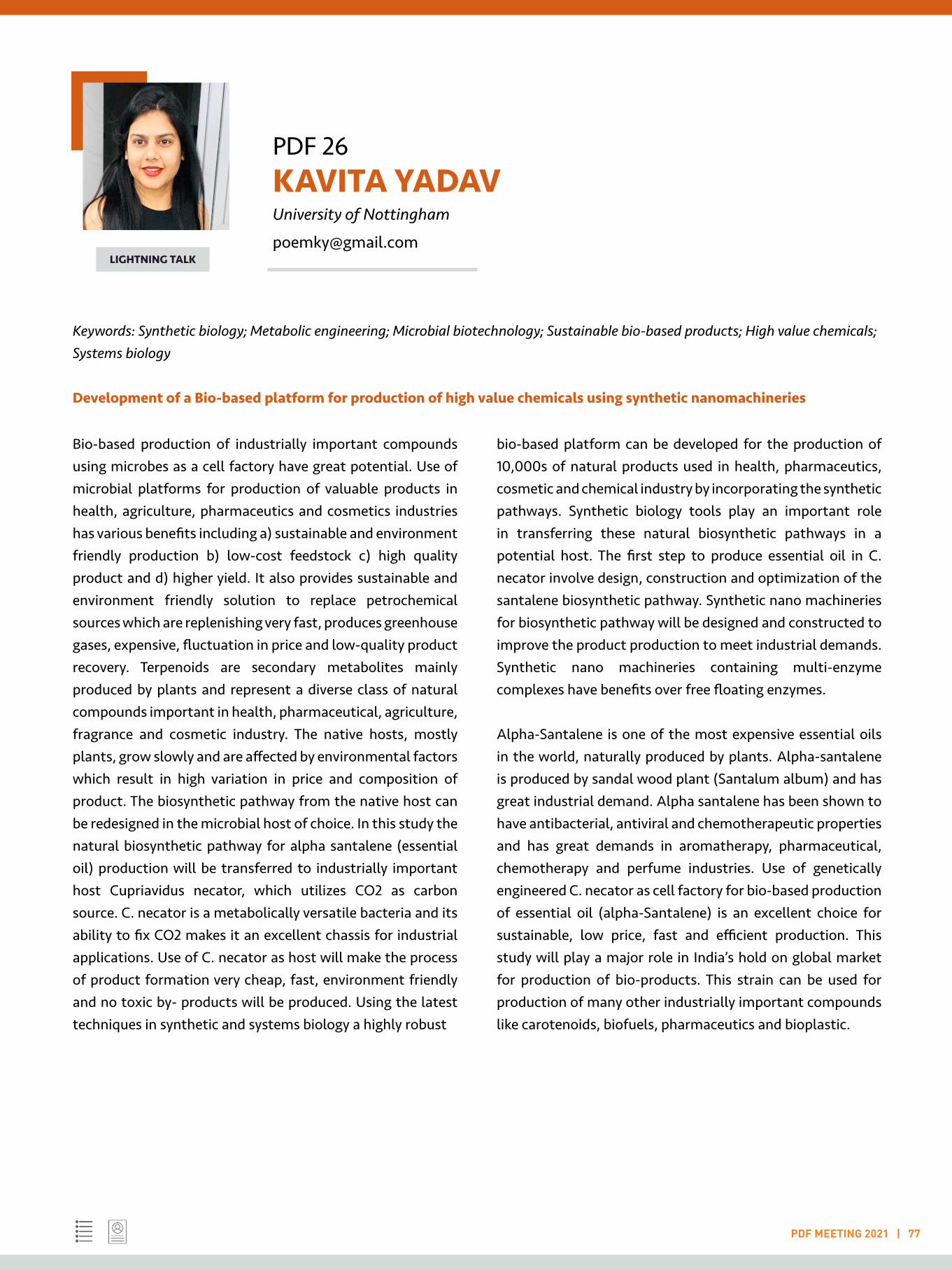
PDF 26 KAVITA YADAV University of Nottingham
PDF MEETING 2021 | 77
LIGHTNING TALK
Keywords: Synthetic biology; Metabolic engineering; Microbial biotechnology; Sustainable bio-based products; High value chemicals; Systems biology
Development of a Bio-based platform for production of high value chemicals using synthetic nanomachineries
Bio-based production of industrially important compounds using microbes as a cell factory have great potential. Use of microbial platforms for production of valuable products in health, agriculture, pharmaceutics and cosmetics industries has various benefits including a) sustainable and environment friendly production b) low-cost feedstock c) high quality product and d) higher yield. It also provides sustainable and environment friendly solution to replace petrochemical sources which are replenishing very fast, produces greenhouse gases, expensive, fluctuation in price and low-quality product recovery. Terpenoids are secondary metabolites mainly produced by plants and represent a diverse class of natural compounds important in health, pharmaceutical, agriculture, fragrance and cosmetic industry. The native hosts, mostly plants, grow slowly and are affected by environmental factors which result in high variation in price and composition of product. The biosynthetic pathway from the native host can be redesigned in the microbial host of choice. In this study the natural biosynthetic pathway for alpha santalene (essential oil) production will be transferred to industrially important host Cupriavidus necator, which utilizes CO2 as carbon source. C. necator is a metabolically versatile bacteria and its ability to fix CO2 makes it an excellent chassis for industrial applications. Use of C. necator as host will make the process of product formation very cheap, fast, environment friendly and no toxic by- products will be produced. Using the latest techniques in synthetic and systems biology a highly robust
bio-based platform can be developed for the production of 10,000s of natural products used in health, pharmaceutics, cosmetic and chemical industry by incorporating the synthetic pathways. Synthetic biology tools play an important role in transferring these natural biosynthetic pathways in a potential host. The first step to produce essential oil in C. necator involve design, construction and optimization of the santalene biosynthetic pathway. Synthetic nano machineries for biosynthetic pathway will be designed and constructed to improve the product production to meet industrial demands. Synthetic nano machineries containing multi-enzyme complexes have benefits over free floating enzymes.
Alpha-Santalene is one of the most expensive essential oils in the world, naturally produced by plants. Alpha-santalene is produced by sandal wood plant (Santalum album) and has great industrial demand. Alpha santalene has been shown to have antibacterial, antiviral and chemotherapeutic properties and has great demands in aromatherapy, pharmaceutical, chemotherapy and perfume industries. Use of genetically engineered C. necator as cell factory for bio-based production of essential oil (alpha-Santalene) is an excellent choice for sustainable, low price, fast and efficient production. This study will play a major role in India’s hold on global market for production of bio-products. This strain can be used for production of many other industrially important compounds like carotenoids, biofuels, pharmaceutics and bioplastic.
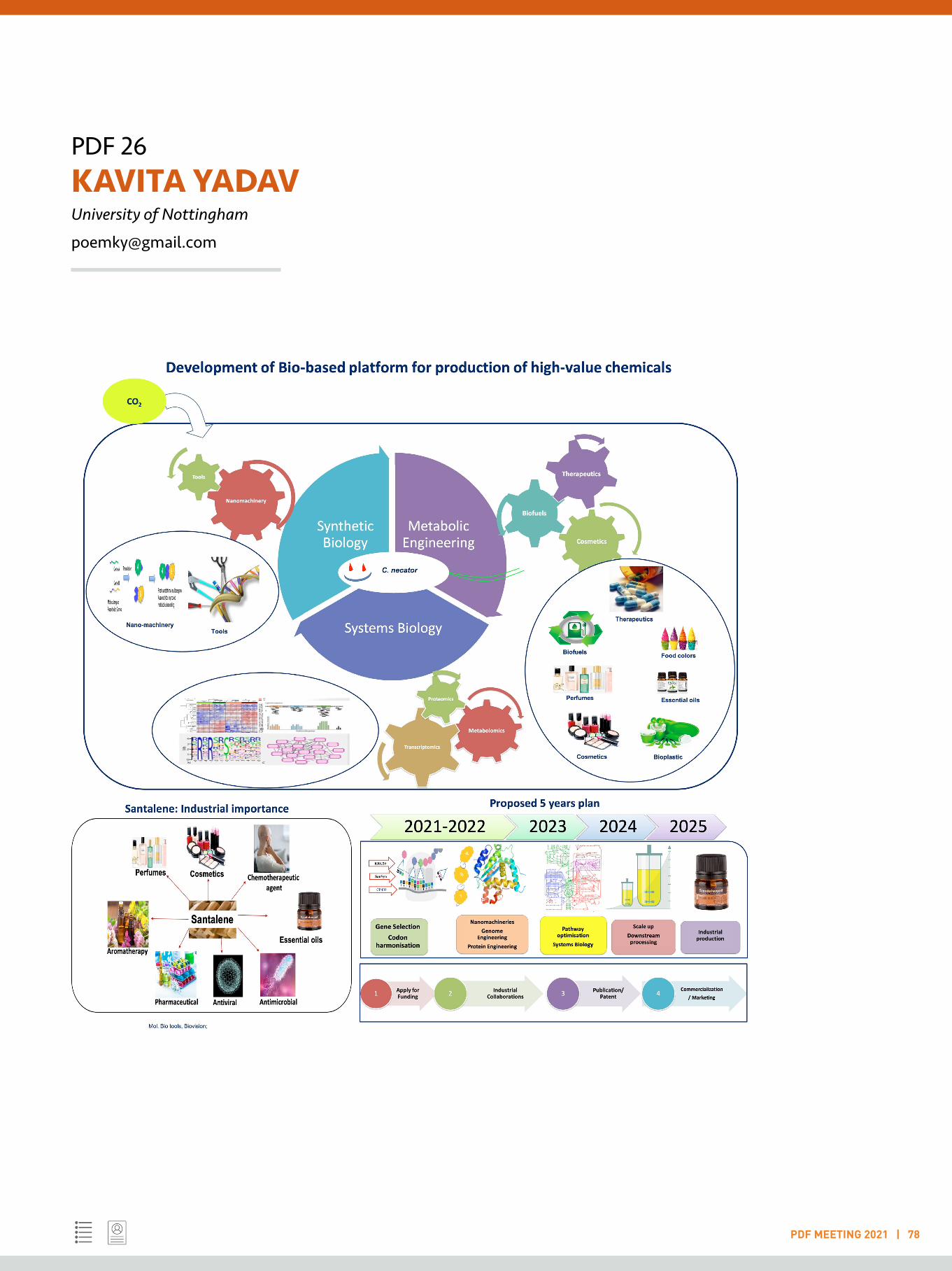
PDF 26KAVITA YADAVUniversity of Nottingham
PDF MEETING 2021 | 78

PDF 27 KSHIPRA NAIK Indian Institute of Technology Madras
PDF MEETING 2021 | 79
LIGHTNING TALK
Keywords: Nanobiotechnology; Bioengineering; Point of care devices; Smart materials; Microfluidics
Fabrication of piezoelectric contact lens based pressure sensor for early detection and management of Glaucoma
Glaucoma is a group of eye conditions that leads to irreversible blindness if not detected and managed at an early stage. It is caused due to excess accumulation of fluid in the anterior chamber of the eye which leads to an increase in the intraocular pressure (IOP). Consequently, high IOP of more than 22 mm Hg damages the nerve connecting the eye to the brain leading to permanent vision loss. Glaucoma is estimated to affect about 80 million people worldwide by 2020. As there is no cure available and vision lost from the disease cannot be restored, accurate measurement of IOP is crucial for glaucoma detection and management. Nonetheless, IOP has a dynamic nature and it keeps fluctuating based on many factors. Therefore there is a need to continuously monitor the changes in the IOP by means of a point-of-care device. To fabricate a piezoelectric contact lens type of pressure sensor, polyvinylidene fluoride (PVDF) was subjected to electrospinning to obtain nanofiber films with high beta phase content that exhibit superior piezoelectric property. A solution of 12 wt% concentration of PVDF in dimethylformamide and acetone was prepared and subjected to electrospinning at a voltage of 10 kV, using needle tip to collector distance of 10 cm and feeding rate of 0.3 mL/hr. The as prepared PVDF nanofiber film was characterized using HR-SEM, X-ray diffraction and FTIR studies. The average nanofiber diameter obtained from HR-SEM was 163 nm. XRD and FTIR spectra exhibited featured peaks corresponding to polar beta phase and the amount of beta phase content was calculated to be 84 %. Next, the amount of voltage produced by these piezoelectric PVDF films on application of the mechanical force was measured. It was observed that a voltage of 1.739 V was produced at a frequency of 22.287 Hz on subjecting the films to mechanical vibration. The advantages of using such a sensor are that it is easily wearable and non-invasive, does not require any surgical procedure, poses minimal risk and is passive i.e. completely self-powered which means it does not
require any external power supply like batteries which can make the sensor bulky.
Prior to this, during my doctoral training, I primarily worked on the development of novel biocompatible nanomaterials derived from Silver, titanium dioxide and hydroxyapatite with a special focus on bioengineering applications such as antifouling, food preservation and bone tissue engineering. In future, I see myself working at the intersection of various research areas such as Bioengineering, Smart Nanomaterials and Microbiology. My research interest encompasses the innovative design, synthesis and characterization of stimulus responsive nanomaterials and their composites for bioengineering and environmental applications including novel point-of-care devices, sensing, diagnostics and treatment of diseases.
References:
1. Leonardi M, Pitchon EM, Bertsch A, Renaud P & Mermoud A. 2009. Wireless contact lens sensor for intraocular pressure monitoring: assessment on enucleated pig eyes. Acta Ophthalmol. 87: 433-7.
2. Araci IE, Su B, Quake SR & Mandel Y. 2014. An implantable microfluidic device for self-monitoring of intraocular pressure. Nature Medicine 20: 1074-8.
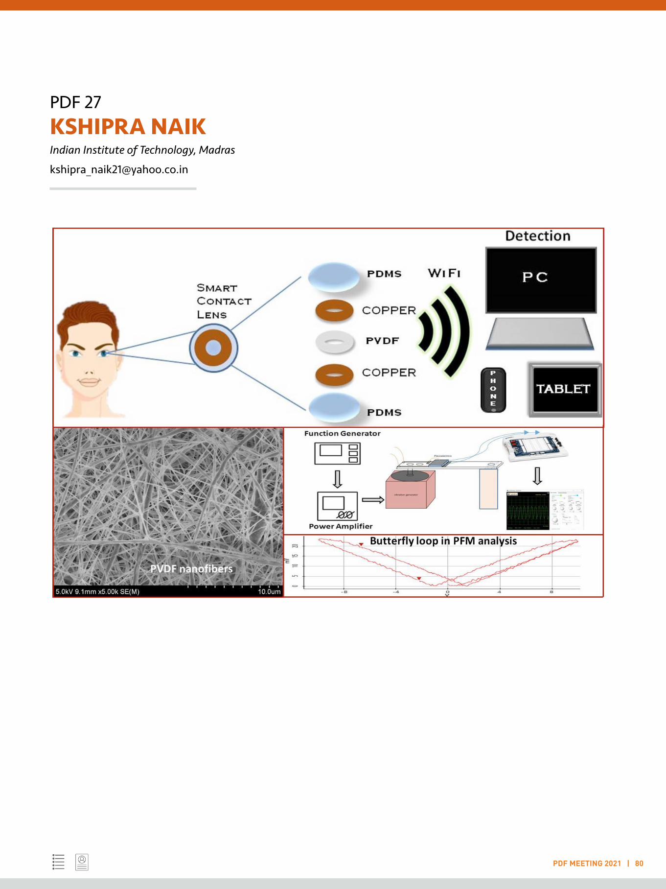
PDF 27KSHIPRA NAIKIndian Institute of Technology, Madras
PDF MEETING 2021 | 80

PDF 28 LAASYA SAMHITA National Centre for Biological Sciences, Bengaluru
PDF MEETING 2021 | 81
LIGHTNING TALK
Keywords: Non-genetic variation; Protein synthesis; Translation errors; Adaptation; Antibiotic resistance
The evolutionary potential of translation errors
Mistranslation is typically deleterious for cells, since it alters existing protein structure and function [1]. Although specific mistranslated proteins can confer a short-term benefit in a particular environment, the prevalence of high global mistranslation rates has remained puzzling. Altering basal mistranslation levels in several ways, I have shown that generalized (non-specific) mistranslation enhances early E. coli survival under DNA damage, by rapidly activating DNA repair via the SOS response [2]. Mistranslating cells show greater tolerance when exposed to DNA damaging antibiotics such as ciprofloxacin, and therefore have a higher probability of sampling critical mutations that confer antibiotic resistance. Interestingly, the wild type E. coli cell relies on its basal translation error rate to acquire even a basal resistance to antibiotics like ciprofloxacin, suggesting that hyper-accurate ribosomes [3] may provide a means for a new drug target against tolerant bacteria. This stress resistance relies on Lon protease, which acts as a key effector that induces the SOS response in addition to alleviating proteotoxic stress [4]. In addition, using experimental evolution, I find that mistranslation alters the genetic basis of adaptation. That is, the kinds of mutations that appear under selection for resistance to various classes of antibiotics changes when translation accuracy is altered. The new links between error-prone protein synthesis, DNA damage, and generalised stress resistance indicate surprising coordination between intracellular stress responses and suggest a novel hypothesis to explain high global mistranslation rates. Also, the fact that mistranslation, the most ubiquitous form of non-genetic variation, can influence mutational identity [5], raises several fundamental questions about the links between genetic and non-genetic change and their role in evolution.
References:
1. Ribas de Pouplana, L., et al., Protein mistranslation: friend or foe? Trends Biochem Sci, 2014. 39(8): p. 355-62.
2. Samhita, L., P.K. Raval, and D. Agashe, Global mistranslation increases cell survival under stress in Escherichia coli. PLoS Genet, 2020. 16(3): p. e1008654.
3. Zaher, H.S. and R. Green, Hyperaccurate and error-prone ribosomes exploit distinct mechanisms during tRNA selection. Mol Cell, 2010. 39(1): p. 110-20.
4. Breidenstein, E.B., M. Bains, and R.E. Hancock, Involvement of the lon protease in the SOS response triggered by ciprofloxacin in Pseudomonas aeruginosa PAO1. Antimicrob Agents Chemother, 2012. 56(6): p. 2879-87.
5. Samhita, L., The Boggarts of biology: how non-genetic changes influence the genotype. Curr Genet, 2020. 10.1007/s00294-020-01108-5.
PDF 28LAASYA SAMHITANational Centre for Biological Sciences, Bengaluru
PDF MEETING 2021 | 82
PDF 29 LIJO CHERIAN OZHATHIL University of Copenhagen
PDF MEETING 2021 | 83
LIGHTNING TALK
Keywords: Cardiac physiology; Pharmacology; Ion channel; Drug discovery; Cardiac arrhythmia
Deciphering the molecular interaction and regulation of cardiac ion channels in arrhythmia: From subcellular process to whole heart dynamics
Sudden Cardiac Death (SCD) and arrhythmia represents one of the major worldwide health problem and accounts for 15-20% of all deaths. Since SCD is spread across all the age groups it accounts for personal loss and also has an adverse effect on the global economy. Early detection and intervention remains the key to survival. Although the epidemiology of SCD in the Indian population is understudied, a recent report has highlighted 10.3% of SCD deaths in a population from Southern India. If such a population study is further extended across different states of India the numbers are sure to increase several-fold. Thus it is extremely needed now to better understand such SCDs. Unfortunately, the molecular mechanism of cardiac arrhythmia leading to SCD is complex and incompletely understood. For the past decade, several genes encoding for membrane proteins called ion channels are found to have a major role in the development and progression of cardiac arrhythmias. Still how these ion channel malfunction either due to genetic or epigenetic factor could trigger lethal cardiac arrhythmia remains largely unknown.A recent set of studies including my postdoctoral work1-3 has discovered cardiac sodium channel (NaV1.5) physically interacts with other cardiac potassium channels (Kir2.1, hERG, TRPM4) and form a unique multi-protein complex called “channelosome”. These observations of existence of channelosome in cardiac tissue were rather unexpected, it is now extremely important and interesting to understand the molecular details of such channelosome and how they function in physiology and pathophysiology.
To achieve this, I would like to apply a multiscale approach of studying arrhythmia from a single ion channel gene to whole heart function:
1) Identifying the molecular partners facilitating the interaction of SCN5a with other pool of cardiac ion channels in heterologous expression system.2) Deciphering the trafficking mechanism of ion channel complexes from nucleus to membrane in physiology and pathophysiology using rodent model.3) Studying functional consequences of ion channel complexes on cardiac cellular excitability and conduction using cardiac stem cell model.4) Extrapolating the studies from rodent and stem cell-based model to a human scale using in silico human heart model for studying the role of ion channel complexes in cardiac arrhythmogenesis.
Studying channelosome in cardiac function by focusing on interaction between ion channels and their localization is important to identify and explore novel targets with pharmacological interest for developing advanced therapeutics for SCDs.
References:
1. Catherine A Eichel , Erick B Ríos-Pérez , Fang Liu , Margaret B Jameson , David K Jones , Jennifer J Knickelbine , Gail A Robertson (2019) A microtranslatome coordinately regulates sodium and potassium currents in the human heart. eLife 8, e52654.
2. Ozhathil, L.C.; Rougier, J.-S.; Arullampalam, P.; Essers, M.C.; Ross-Kaschitza, D.; Abriel, H (2021). Deletion of Trpm4 Alters the Function of the Nav1.5 Channel in Murine Cardiac Myocytes. Int. J. Mol. Sci., 22, 3401.
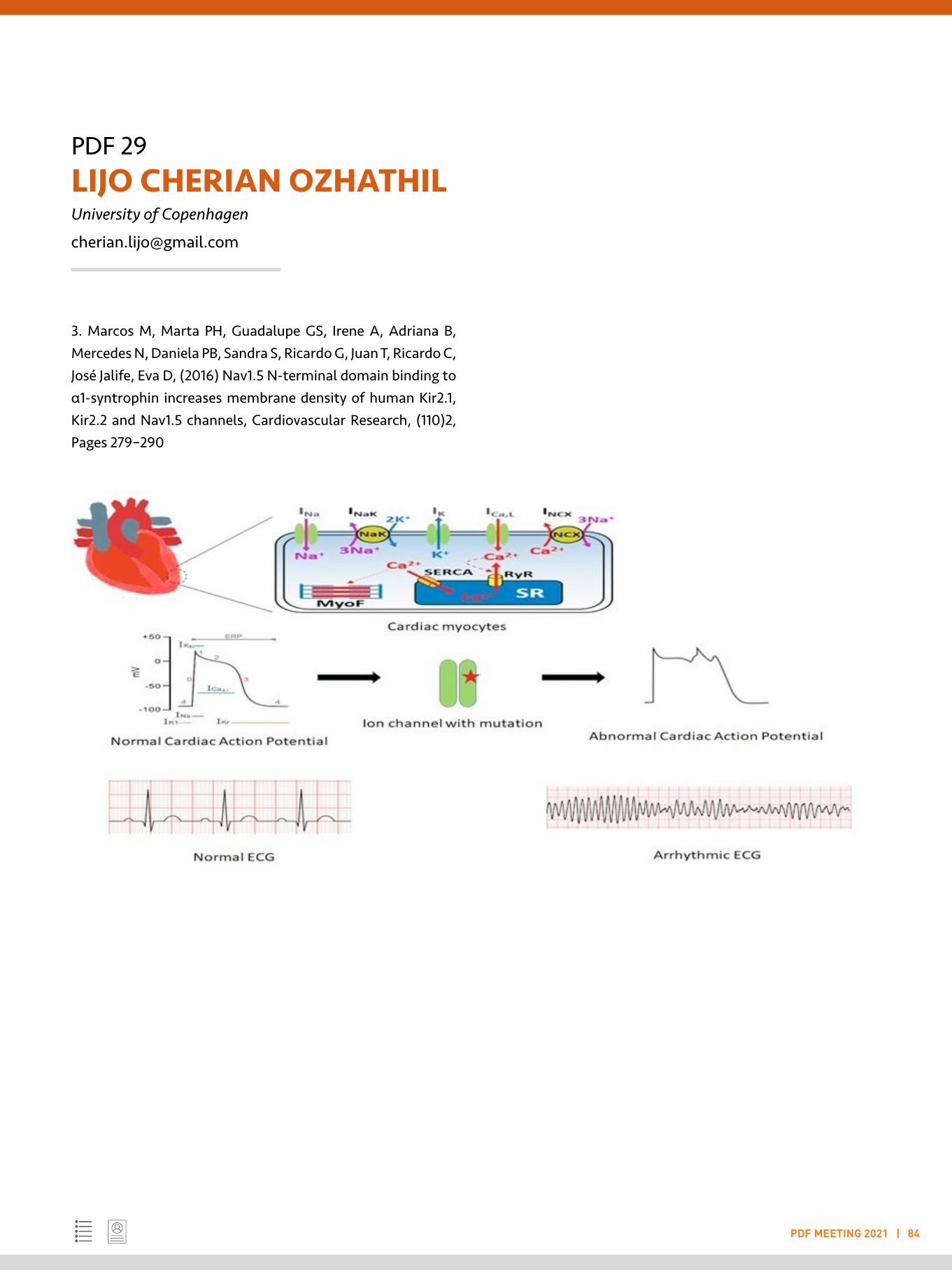
PDF 29LIJO CHERIAN OZHATHILUniversity of Copenhagen
PDF MEETING 2021 | 84
3. Marcos M, Marta PH, Guadalupe GS, Irene A, Adriana B, Mercedes N, Daniela PB, Sandra S, Ricardo G, Juan T, Ricardo C, José Jalife, Eva D, (2016) Nav1.5 N-terminal domain binding to α1-syntrophin increases membrane density of human Kir2.1, Kir2.2 and Nav1.5 channels, Cardiovascular Research, (110)2, Pages 279–290
PDF 30 MADHURA RAGHAVAN University of California, San Francisco
PDF MEETING 2021 | 85
LIGHTNING TALK
Keywords: Infectious diseases; Malaria; Immunology; Phage display; Vaccine
Massively parallel immunological profiling of antibody responses to Plasmodium falciparum using phage display
Malaria is a disease that affects around 200 million people annually. Protection from the disease develops with age and repeated exposure, wherein adults are more protected than children. It is known that antibodies play a crucial role in this immunity, and the response is targeted against multiple antigens. A systematic, unbiased investigation of targets of antibodies in Malaria at high-resolution across age groups is much needed not only to identify targets of protective responses, but also to understand the basis of delayed acquisition of immunity. Here, we present such an approach using PhIP-seq2 in falciparum Malaria using a Plasmodium falciparum (Pf)-specific phage display library, displaying falciparum proteins in tiled 62-aa sections, on an age- and exposure-stratified cohort from Uganda and detect several antigenic regions at high resolution, including many previously well-characterized epitopes as well as novel regions, in proteins across all the life stages of Pf. The antigenic regions are enriched for polar residues and low complexity repeat elements, and immunodominant regions have a higher representation of repeat elements, with even children in low exposure settings displaying sero-reactivity against these regions. Further, sequence analysis of these regions shows widespread sharing of 7-aa or longer motifs across 1/3 of the antigenic proteins, with the most extensive sharing displayed by the highly variant PfEMP1 antigenic gene family. The high degree of similarity suggests extensive potential for cross-reactivity of antibody responses. Further, as expected, the overall breadth of antibody reactivity increases with exposure in children while being similar across exposure in adults. Interestingly, children facing high exposure develop as much breadth with similar antigenic profiles as adults and we identify a few regions with differential seroprevalence in these children. This study provides the first ever unbiased deep dive into the antigenic regions in Pf and offers insights to better understand immunity to Malaria.
References:
1. Doolan, D.L., Dobaño, C. and Baird, J.K., 2009. Acquired immunity to malaria. Clinical microbiology reviews, 22(1), pp.13-36.
2. Larman HB, Zhao Z, Laserson U, et al. 2011. Autoantigen discovery with a synthetic human peptidome. Nat Biotechnol. 2011;29(6):535-541.

PDF 31 MASUM SAINI Regional Centre for Biotechnology, Faridabad
PDF MEETING 2021 | 86
LIGHTNING TALK
Keywords: Cancer & therapy; Molecular biology & genetics; Cell signalling; Cellular & molecular mechanisms; Developmental biology
Understanding Cancer in the Light of Development
I am currently working as an India Alliance (IA) Early Career Fellow (ECF) at the Regional Centre for Biotechnology (RCB), Faridabad. My current and past work have been progressive steps towards my long-term goal of understanding dysregulation of molecular mechanisms in cancer, deriving cues from how such mechanisms are spatio-temporally regulated during embryonic development, and using the insights obtained for therapeutic intervention.
Early in my academic training I became keen to understand the molecular basis of human diseases, specifically cancer, and pursued this for my doctoral research. For the first time, we reported KIT (a receptor tyrosine kinase–RTK) is aberrantly expressed in brain tumours (meningiomas, Saini et al., 2012, BMC Cancer, 12:212). Since KIT is a known therapeutic target of Imatinib in other cancers, it is worth exploring if such meningiomas would benefit from Imatinib. We reported a KIT mutation in brain tumours (gliomas and meningiomas, Saini et al., 2012, Gene, 497:155-63; Saini et al., 2012, BMC Cancer, 12:212) that was recently shown to be associated with aggressive gliomas. Further, we found that levels of MN1 (a transcriptional co-regulator), and its molecular interplay with other genes, IGFBP5 and IGF1 (known to have a role in gliomas), help predict patient survival. Thus, MN1 may be a promising prognostic marker (Saini et al., 2020, Human Molecular Genetics 29:3532-45).
Interestingly, I noted that these genes are also involved in embryonic development, and exploring how they are tightly regulated during development may hold a key to understanding their malfunctioning in cancer.
Therefore, as a Young Investigator Awardee, I began working on Rhabdomyosarcoma (RMS) to understand the regulation of another oncogenic RTK (MET), simultaneously
developing skills/techniques to answer these questions from a developmental perspective (Sharma et al.,2018, bioRxiv, doi: 10.1101/261685; Agarwal et al,. 2020, Development, 147:dev184507). I showed that MET is prevented from degradation owing to its interaction with SPRY2, a modulator of RTK signalling. Abrogating SPRY2 function significantly reduced MET levels, thereby decreasing metastatic and clonogenic potential, and induced RMS cells to differentiate (Saini et. al., 2018, Cell Death and Disease, 9:237). This provided a new perspective to the mechanism by which dysregulated RTKs mediate constitutive oncogenic signals and may open a new therapeutic avenue in RMS and in other cancers where MET is dysregulated. As an IA fellow, I am now investigating the regulation of MET in development, regeneration and disease to obtain molecular insights on these processes.
In the medium and long term, I intend to further explore, the regulation of molecular mechanisms/signalling pathways, in cancer and development using animal models. My lab will aim to understand how oncogenic and developmentally crucial signalling pathways cooperate in a cancer-specific manner to promote chemoresistance, a persistent problem in the field. I am also keen to establish collaborations to set up organoid cultures from patient samples and explore the contribution of cellular heterogeneity and tumour niche to deranged cell signalling. These will also serve as humanized ex vivo models for screening/ identifying chemotherapeutic candidate molecules.

PDF 31MASUM SAINI Regional Centre for Biotechnology, Faridabad
PDF MEETING 2021 | 87
PDF 32 MEETALI SINGH Institut Pasteur, Paris
PDF MEETING 2021 | 88
LIGHTNING TALK
Keywords: Epigenetics; Small RNAs; RNA biology; Proteomics high-throughput sequencing;
Translation and codon usage regulate Argonaute slicer activity to trigger small RNA biogenesis
In the Caenorhabditis elegans germline, thousands of mRNAs are concomitantly expressed with antisense 22G-RNAs, which are loaded into the Argonaute CSR-1(Cecere et al., 2014; Claycomb et al., 2009). Despite their essential functions for animal fertility and embryonic development (Claycomb et al., 2009; Quarato et al., 2021), how CSR-1 22G-RNAs are produced remains unknown. In the current project, I show that CSR-1 slicer activity is primarily involved in triggering the synthesis of small RNAs on the coding sequences of germline mRNAs and post-transcriptionally regulates a fraction of targets. CSR-1-cleaved mRNAs prime the RNA-dependent RNA polymerase, EGO-1, to synthesize 22G-RNAs in phase with ribosome translation in the cytoplasm, in contrast to other 22G-RNAs mostly synthesized in germ granules. Moreover, codon optimality and efficient translation antagonize CSR-1 slicing and 22G-RNAs biogenesis (Singh et al. 2020). We propose that codon usage differences encoded into mRNA sequences might be a conserved strategy in eukaryotes to regulate small RNA biogenesis and Argonaute targeting. In addition, I was a co-author of the publication on transgenerational silencing of histones by small RNAs in piwi mutants resulting in impaired fertility (Barucci et al., 2020). I contributed to elucidate the mechanism of histone silencing in absence of PIWI by showing in absence of piRNAs, downstream components of the piRNA pathway relocalize from germ granules and piRNA targets to histone mRNAs to synthesize antisense small RNAs (sRNAs) using biochemical and proteomics approaches.
I have initiated two new projects- first, to investigate soma to germline transmission of epigenetic information especially sRNAs as a response to environmental stimuli. Second, I am the lead investigator in a collaborative project to study the
role of SARS-CoV2 encoded small RNAs in viral pathogenesis using cell lines, hamster animal model and patient samples.
References:
Barucci, G., E. Cornes, M. Singh, B. Li, M. Ugolini, A. Samolygo, C. Didier, F. Dingli, D. Loew, P. Quarato, and G. Cecere. 2020. Small-RNA-mediated transgenerational silencing of histone genes impairs fertility in piRNA mutants. Nat Cell Biol. 22:1–11. doi:10.1038/s41556-020-0462-7.
Cecere, G., S. Hoersch, S. O’Keeffe, R. Sachidanandam, and A. Grishok. 2014. Global effects of the CSR-1 RNA interference pathway on the transcriptional landscape. Nature Structural and Molecular Biology. 21:nsmb.2801. doi:10.1038/nsmb.2801.
Claycomb, J.M., P.J. Batista, K.M. Pang, W. Gu, J.J. Vasale, J.C. van Wolfswinkel, D.A. Chaves, M. Shirayama, S. Mitani, R.F. Ketting, D. Conte, and C.C. Mello. 2009. The Argonaute CSR-1 and Its 22G-RNA Cofactors Are Required for Holocentric Chromosome Segregation. Cell. 139:123–34. doi:10.1016/j.cell.2009.09.014.
Quarato, P., M. Singh, E. Cornes, B. Li, L. Bourdon, F. Mueller, C. Didier, and G. Cecere. 2021. Germline inherited small RNAs facilitate the clearance of untranslated maternal mRNAs in C. elegans embryos. Nat Commun. 12:1441. doi:10.1038/s41467-021-21691-6.
Singh, M., E. Cornes, B. Li, P. Quarato, L. Bourdon, F. Dingli, D. Loew, S. Proccacia, and G. Cecere. 2020, Translation and codon usage regulate Argonaute slicer activity to trigger small RNA biogenesis. BioRxiv. doi:10.1101/2020.09.04.282863.
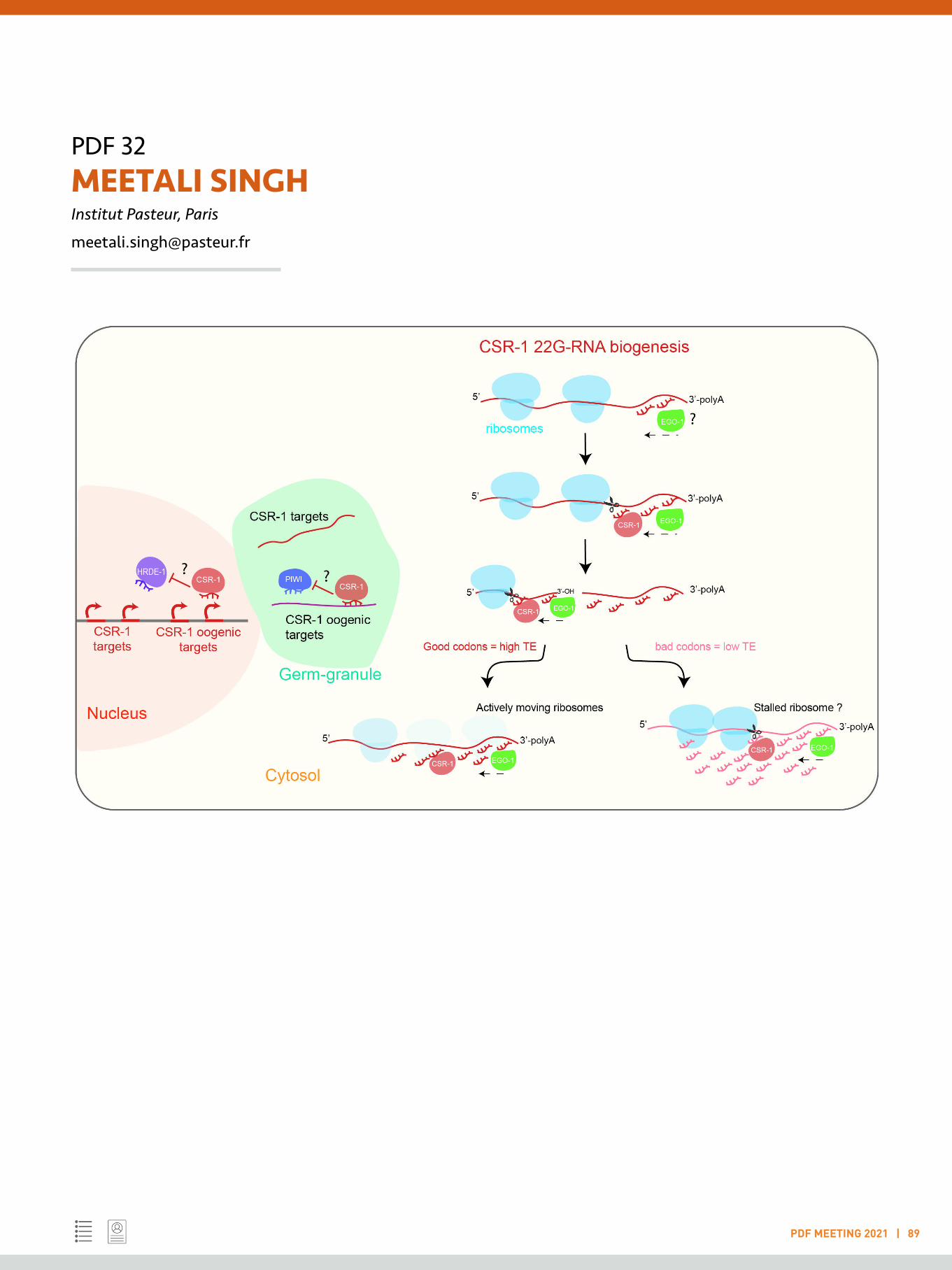
PDF 32MEETALI SINGH Institut Pasteur, Paris
PDF MEETING 2021 | 89
PDF 33 NEERAJ SHARMA Indian Institute of Science, Bengaluru
PDF MEETING 2021 | 90
LIGHTNING TALK
Keywords: Auditory neuroscience; Brain imaging; Electrical sciences; Machine learning; Healthcare
Understanding what, why, and how of human speech and audio processing
The air pressure released from an individual’s mouth goes through the ambient surrounding, makes its way into our ear canals, and is decoded by our brain, in tens of milli-secs, to gather a lot of information. For instance, from hearing someone speak, we immediately understand their gender (and maybe, identity too), the message, and sometimes also the health of the individual. The robustness of this means of information transfer has made spoken conversations a prevalent form of social communication over thousands of years. The scientific insights into the underlying brain processes are still in their infancy. My research is aimed at contributing to further this understanding.
My work is founded on three aspects. Firstly, I use signal processing to build mathematical models of speech and audio signals. These signals feature a non-stationary spectrum and pose interesting problems for mathematical analysis. In my PhD, I proposed techniques for this, and some of these have been useful in re-thinking the traditional approaches to analyze speech and audio signals [1], resulting in improved engineering applications. I also use this analysis to understand how information is encoded into sound signals. For instance, can we quantify how the voices of different individuals differ? Secondly, as we move towards a world with increased human-machine interaction, understanding how the human brain processes sensory signals, such as speech, will help in the improved design of human-machine interaction systems. Towards this, I use behavioural and brain imaging experiments (using electroencephalography) to understand the brain processes that are triggered when we hear speech signals. Using this, I have worked on figuring out how fast humans detect talker changes in speech utterances [2], and how an unfamiliar language impacts talker change perception. Finally, when we process sound signals using machines there might
be a lot more information that we can extract than what we are able to hear using our ears. Drawn by this, recently, I have been involved in understanding if paralinguistic sound signals, such as coughs, can be used to detect the health status of an individual (with the main emphasis on COVID-19 detection).
References:
[1] Neeraj Sharma, and T. V. Sreenivas, “Time-varying Sinusoidal Demodulation for Non-stationary Modeling of Speech”, Elsevier Speech Communication, 2018
[2] Neeraj Sharma, Shobhana Ganesh, Sriram Ganapathy, Lori L.Holt, “Talker Change Detection: A comparison of human and machine performance”, Journal of Acoustic Society of America (JASA), 2019
PDF 34 NEHA NAGPAL Boston Children’s Hospital, Boston
PDF MEETING 2021 | 91
LIGHTNING TALK
Keywords: Non-coding RNAs; Cancer; Stem cells; Telomerase; RNA biogenesis
Targeting of human telomerase via non-coding RNA pathways
Telomerase reverse transcriptase, TERT, is the on/off switch for telomerase activity while, human genetic data indicate that the non-coding RNA component of telomerase, TERC, is limiting and determines telomerase activity in human stem cells. Mendelian disorders associated with compromised telomere maintenance cause a spectrum of telomere biology disorders ranging from severe, early-onset such as dyskeratosis congenita (DC) to more common mid-life presentations such as pulmonary fibrosis (PF). Mutations impacting TERT and TERC often underlie these diseases where there are no curative therapies, only bone marrow or organ transplantation. We hypothesized that manipulating non-coding RNA pathways could be therapeutically useful to augment TERC in patients with telomere diseases. Earlier in our lab, we demonstrated that PARN mutations cause DC via destabilization of nascent TERC RNA. Specifically, PARN is an RNA exonuclease that is required to remove post-transcriptionally added, non-genomically encoded adenosine residues, that target TERC for destruction by the nuclear exosome. We subsequently identified the non-canonical poly(A) polymerase PAPD5 as the enzyme responsible for TERC oligo-adenylation and destabilization and demonstrated that RNA interference of PAPD5 restored telomere length in DC patient cells. To further address this, we disrupted PAPD5 by CRISPR/Cas9-mediated genome editing in DC patient induced pluripotent stem cells (iPSCs) and found that PAPD5-null clones could be isolated and propagated indefinitely. These results provide genetic evidence of an unanticipated therapeutic window for PAPD5 inhibition, as a potential strategy to modulate TERC RNA levels.
Pharmacological inhibitors of PAPD5 have not been identified. We, therefore, developed and executed a high-throughput
functional screen to discover novel small molecule PAPD5 inhibitors. Of 100,055 compounds tested, we identified several hits that inhibited recombinant PAPD5 (rPAPD5) and triaged based on potency, specificity, and activity in orthogonal in vitro assays. BCH001, one of these novel compounds, when used in multiple PARN-mutant DC patient-derived iPSCs, results in reduced TERC RNA oligo-adenylation and increased steady-state TERC RNA levels. Strikingly and consistently, treatment with BCH001 led to telomere elongation across all patient samples and clones tested, establishing a normalized telomere length set-point over weeks that was reversible with drug removal. Further, in immunodeficient mice xenotransplanted with human hematopoietic stem and progenitor cells (HSPCs) that carry PARN-mutations, oral treatment with PAPD5 inhibitors rescues TERC 3′-end maturation and telomere length, with no adverse effect on engraftment and lineage-determination. Taken together, these data provide genetic and pharmacological evidence that TERC RNA and telomere length can be controlled by modulation of the PARN/PAPD5 post-transcriptional axis in human cells. This has important therapeutic implications for hematologic and non-hematologic manifestations of DC and related genetic disorders, and possibly a broad range of human degenerative diseases.
References:
Nagpal N, Wang J, Zeng J, Lo E, Moon DH, Luk K, Braun RO, Burroughs LM, Keel SB, Reilly C, Lindsley RC, Wolfe SA, Tai AK, Cahan P, Bauer DE, Fong YW, Agarwal S 2020. Small-Molecule PAPD5 Inhibitors Restore Telomerase Activity in Patient Stem Cells. Cell Stem Cell 26, 896-909 e898.
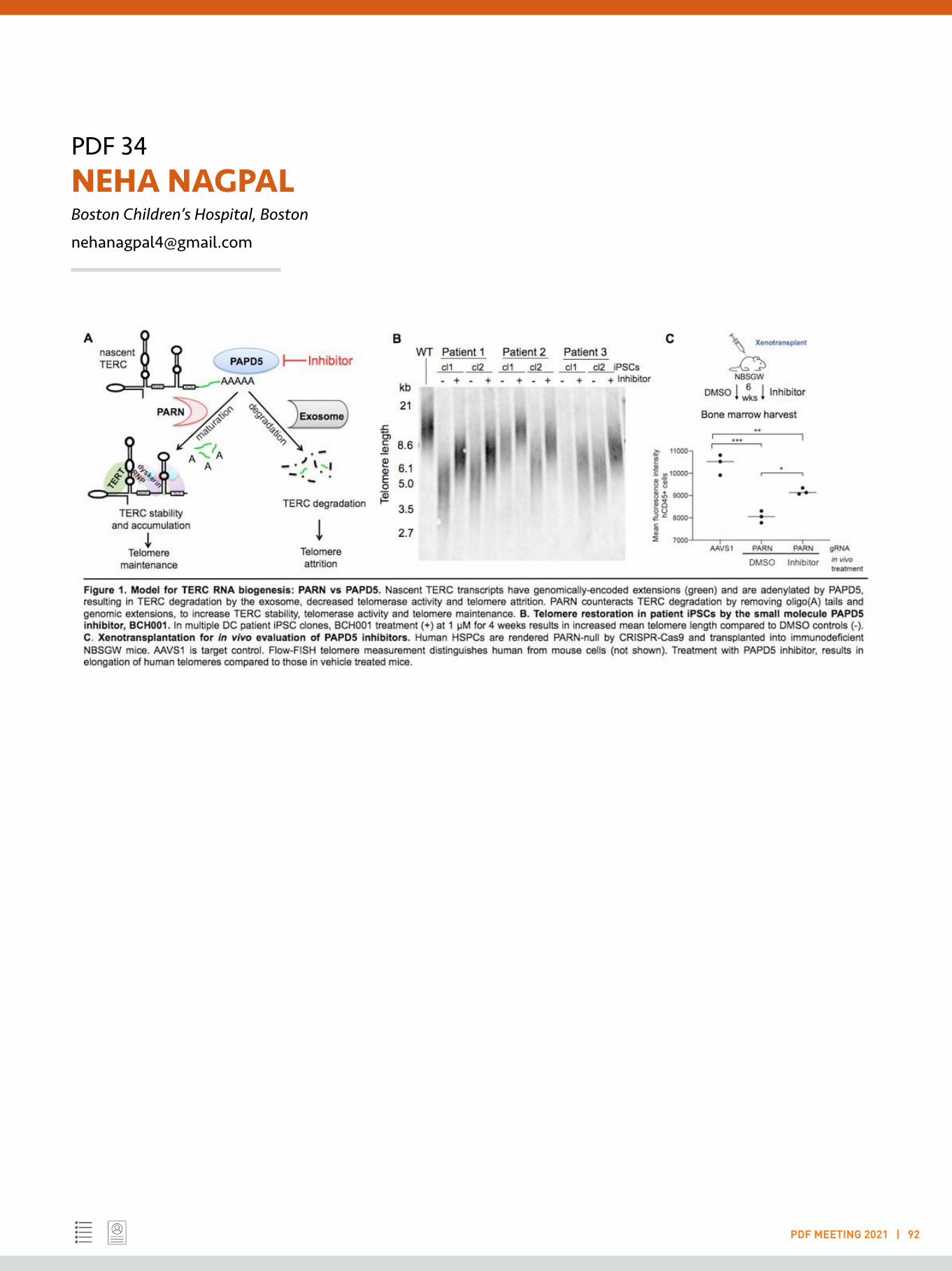
PDF 34NEHA NAGPALBoston Children’s Hospital, Boston
PDF MEETING 2021 | 92

PDF 35 NISHIT SRIVASTAVA Institut Curie, Paris
PDF MEETING 2021 | 93
LIGHTNING TALK
Keywords: Mechanobiology; Cell mechanics; Cell growth; Cell proliferation; Biophysics
Density homeostasis and cell size control
Cells that make up the same tissue or organ tend to have a stereotypic physical size, while variability in cell size is a marker of abnormal cell growth. A well-defined cell size with low variance is crucial for the intimate coordination between cell growth and division, which drives physiological functions like cell proliferation, differentiation, and embryonic development. But, how size homeostasis is achieved and what size parameter is measured is still an open question for mammalian cells.
In this work, we combine two independent methods for live single-cell size measurement: quantitative phase microscopy to measure cell dry mass and fluorescence exclusion, to measure cell volume, on a number of cell types over a complete cell cycle. Specifically, we aim to:1) Understand fluctuations in mass and volume growth during the cell cycle and,2) Study mechanisms that couple mass and volume thus modulating cell density.
We show that cells grow exponentially in both mass and volume, over the complete cell cycle. We also show that the density of cells is not constant, and instead, a density jump is observed as mass increase starts immediately at mitotic exit whereas volume increase is delayed in newborn cells. Our results also show that the mass growth rate is anti-correlated with the density while the volume growth rate remains independent of it. The spreading area shows a biphasic growth with a very high growth rate for several hours at the beginning of the cell cycle, which decreases to a lower value thereafter. The rate of change in volume is inversely correlated to the rate of change in the spreading area during this period of high growth rate in the spreading area. Density perturbations are also seen during cell spreading on adhesive substrates. The cells tend to get diluted when their spreading is blocked on
PEG treated surfaces. We aim to understand the factors which modulate the density and mass/volume coupling which might also correspond to dynamic cell shape changes due to migration/spreading in adhesive cultured cells. This work aims at a fundamental understanding of how cells grow and regulate their size. It will also elucidate the functional consequences of modifications in mechanisms of mass/volume coupling on cellular functions.
Additionally, we hypothesised that the nucleus could act as a size sensor or a ruler. We show that nuclear deformation leads to an increase in the NE tension, affecting G1/S progression. Upon deformation of cells, the time spent in the G1 phase decreased appreciably from about 8-9hours in 20um confined cells to about 6 hours in cells under 8um and 4um confinement. We further show that this decrease in time spent in G1 was contractility dependent and that this phenotype could be rescued by confining the cells. Furthermore, we used drugs to inhibit cPLA2 activation upon confinement of cells and found that this severely impairs the progression of cells from G1 to S phase of the cell cycle. This size sensing mechanism could be an important regulator of growth in a highly crowded environment.
References:
1. C. Cadart, et al., Size control in mammalian cells involves modulation of both growth rate and cell cycle duration. Nat. Commun. 9, 3275 (2018).
2. E. Zlotek-Zlotkiewicz, S. Monnier, G. Cappello, M. Le Berre, M. Piel, Optical volume and mass measurements show that mammalian cells swell during mitosis. J. Cell Biol. 211, 765–774 (2015).

PDF 35NISHIT SRIVASTAVAInstitut Curie, Paris
PDF MEETING 2021 | 94
3. A. J. Lomakin, et al., The nucleus acts as a ruler tailoring cell responses to spatial constraints. Science 370 (2020).
4. N. Srivastava, et al., Nuclear fragility, blaming the blebs. Curr. Opin. Cell Biol. 70, 100–108 (2021).
5. N. Srivastava, D. Traynor, M. Piel, A. J. Kabla, R. R. Kay, Pressure sensing through Piezo channels controls whether cells migrate with blebs or pseudopods. Proc. Natl. Acad. Sci. U. S. A. 117, 2506–2512 (2020).

PDF 36 NITHYA RAMAKRISHNAN Indian Institute of Technology Bombay, Mumbai
PDF MEETING 2021 | 95
LIGHTNING TALK
Keywords: Information theory in biological inheritance; Probabilistic modelling of biological phenomena; Computational data analy-sis; Bioinformatics
Computational Analysis of Biological Phenomena and Data for Inheritance and Diseases using Information Theory
The areas of my research have been largely in the field of Bioinformatics, Probabilistic Modeling of epigenetic phenomena and Prediction of diseases. They are explained in more detail in the paragraphs below.
PhD Research:During my PhD, I focussed on the classical problem of Intron-Exon segmentation in DNA. We employed a physio-chemical property of the nucleotides (viz) the dipole moment [1] and used its angular orientations to compute the dipole angular entropy for a given segment of the DNA. We surmised that the dipole angular entropies are significantly different in the coding and non-coding regions.
Another part of my research was to analyse the DNA methylation distributions [2] of healthy and tumorous samples and predict the phenotype of a sample, based on measures like “methylation entropy” (a measure that we introduced) and Kullback-Leibler divergence of a given sample’s methylation distribution from the “average healthy methylation distribution”. We analysed the CpG islands of tumour suppressor genes, oncogenes as different sets of analyses.
Post-Doctoral Research:We analysed the gene-wise patterns of the histone modification values in yeast data obtained from experiments [3]. We used Principal Component Analysis and obtained clusters of genes that have similar functionalities, just based on their modification patterns. Additionally, we cluster the modification patterns that are redundant, based on their correlation/anti-correlation values and check for the minimum set of histone modification data that may be required for defining the ‘histone-code” [4].
Currently, I also work on a mathematical model for the inheritance of the histone post-translational marks post replication. We model the replication event (where a mother chromatin’s PTMs are to be split amongst its 2 daughter strands [5]) as a noisy communication channel and predict, based on Information theory, that enzymes that reproduce the mother chromatin in the daughter strands, modify blocks of nucleosomes. The size of the block depends on the statistical nature of the mother chromatin.
Future Research:In the future, I would like to employ tools from Information theory for DNA Methylation inheritance and to study the inheritance of genetic mutations and to predict their impact on the phenotype - whether it would lead to diseases or evolution.
References:
[1] J. Slater and N. Frank, Electromagnetism. New York: McGraw-Hill Book Co., 1947.
[2] Laird, P.W., 2003. The power and the promise of DNA methylation markers. Nature Reviews Cancer, 3(4), pp.253-266.
[3] Weiner, A., Hsieh, T.H.S., Appleboim, A., Chen, H.V., Rahat, A., Amit, I., Rando, O.J. and Friedman, N., 2015. High-resolution chromatin dynamics during a yeast stress response. Molecular cell, 58(2), pp.371-386.

PDF 36NITHYA RAMAKRISHNANIndian Institute of Technology Bombay, Mumbai
PDF MEETING 2021 | 96
[4]Strahl, B.D. and Allis, C.D., 2000. The language of covalent histone modifications. Nature, 403(6765), pp.41-45.
[5] Probst AV, Dunleavy E, Almouzni G. Epigenetic inheritance during the cell cycle. Nature reviews Molecular cell biology. 2009 Mar;10(3):192-206.
PDF 37 PARIKSHIT BAGCHI University of Michigan, Ann Arbor
PDF MEETING 2021 | 97
LIGHTNING TALK
Keywords: Virology; Cell biology; Host-virus interaction; Membrane contact sites; Endoplasmic reticulum
Exploitation of endoplasmic reticulum during virus infection
My post-doctoral research in the lab of Prof. Billy Tsai at the University of Michigan, USA is focused on the role of endoplasmic reticulum (ER) during virus infection. Viruses use cellular compartments for their trafficking inside host cells to get access to replication and translation machinery. In this route, endoplasmic reticulum plays an important role for many viruses. My study is centred on 3 different viruses from 3 different families. First one is SV40. It is a non-enveloped tumour causing virus. SV40 is an archetype member of the Polyomaviridae family, which is widely used as a model virus in polyomavirus (Py) research1-2. Human Pys like Merkel Cell Py, BKPy and JCPy are causative agents of numerous human diseases3. It crosses ER en-route to the nucleus where it replicates4. My focus is on polyomavirus entry and exit pathways to and from ER and I have identified diverse roles of different subunits of an ER-localized multiprotein complex called ER membrane protein complex or EMC in polyomavirus trafficking (Fig 1, 2)5-7.
The other two viruses I presently study are dengue virus (DENV) and SARS-CoV2. DENV belongs to the Flaviviridae family while SARS CoV-2 belongs to Coronaviridae family, both are enveloped viruses and with positive-sense single-stranded RNA as genome8-9. I am currently working on the role of different ER membrane proteins in dengue and SARS-CoV2 infection in BSL2 and BSL3 lab environments. As both two viruses use ER membrane-derived structures for virus replication, studying ER factors that are exploited by viruses can be helpful to find antiviral targets.
References:
1. Stehle, T., Gamblin, S. J., Yan, Y. & Harrison, S. C. The structure of simian virus 40 refined at 3.1 A resolution. Structure 4, 165–182 (1996).
2. Chen, X. S., Stehle, T. & Harrison, S. C. Interaction of polyomavirus internal protein VP2 with the major capsid protein VP1 and implications for participation of VP2 in viral entry. EMBO J. 17, 3233–3240 (1998).
3. Jiang, M., Abend, J. R., Johnson, S. F. & Imperiale, M. J. The role of polyomaviruses in human disease. Virology 384, 266–273 (2009).
4. Chen, Y.-J.; Liu, X.; Tsai, B. SV40 Hijacks Cellular Transport, Membrane Penetration, and Disassembly Machineries to Promote Infection. Viruses 2019, 11, 917.
5. Bagchi P, Walczak CP, Tsai B. 2015. The endoplasmic reticulum membrane J protein C18 executes a distinct role in promoting simian virus 40 membrane penetration. J Virol. Apr;89(8):4058-4068.
6. Bagchi P, Inoue T, Tsai B. 2016. EMC1-dependent stabilization drives membrane penetration of a partially destabilized non-enveloped virus. eLife. Dec 24;5. pii: e21470.
7. Bagchi P, Torres M., Qi L., Tsai B. 2020. Selective EMC subunits act as molecular tethers of intracellular organelles exploited during viral entry. Nat Commun. Feb 28;11(1):1127. DOI: 10.1038/s41467-020-14967-w.
8. Diamond MS, Pierson TC. Molecular Insight into Dengue Virus Pathogenesis and Its Implications for Disease Control. Cell. 2015 Jul 30;162(3):488-92. doi: 10.1016/j.cell.2015.07.005.
9. Haque SM, Ashwaq O, Sarief A, Azad John Mohamed AK. A comprehensive review about SARS-CoV-2. Future Virol. 2020 Sep;15(9):625-648. doi: 10.2217/fvl-2020-0124.
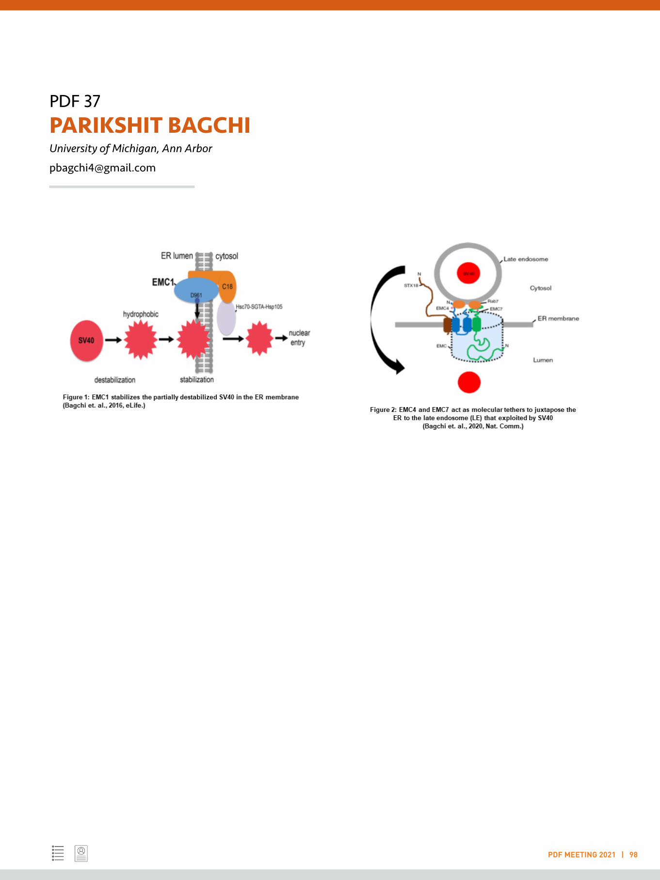
PDF 37PARIKSHIT BAGCHIUniversity of Michigan, Ann Arbor
PDF MEETING 2021 | 98
PDF 38 PRATIK KUMAR HHMI Janelia Research Institute, Ashburn
PDF MEETING 2021 | 99
LIGHTNING TALK
Keywords: Chemigentic fluorescent dyes; Single-molecule imaging; Cell-type pharmacology; Photopharmacology; Protein engineering
Chemigenetic multifunctional fluorophores: A fluorescent toolbox for labelling, imaging, and manipulation of proteins
Advances in our understanding of cellular processes are often limited by our ability to visualise and manipulate biomolecules. Innovations in fluorescence microscopy have led to imaging and tracking individual fluorophore-tagged proteins with superresolution. Many modern imaging methods are photon-intensive, however, requiring bright and photostable fluorescent labels. The demand for more photons is driving a renaissance in small-molecule fluorescent dyes, which show superior properties to fluorescent proteins and can be deployed with genetic specificity. Although improvements in the photophysics and bioavailability of fluorescent dyes have enabled new imaging experiments in living systems, the development of multifunctional fluorophores that are activatable, amplifiable, or allow manipulation of a protein target is still in its infancy. Through rational design, I have developed four categories of dye-based tools that leverage the tunable cell permeability and brightness of organic dyes to image and manipulate proteins with genetic specificity.
Single-molecule imaging: I have developed two strategies to obtain fluorogenic (nonfluorescent to fluorescent) dyes for single-molecule imaging and tracking of intracellular and membrane proteins. The first type covalently binds to the target protein via genetically encoded self-labelling tags and becomes highly fluorescent upon photoactivation; enabling tracking of target proteins even in densely labelled samples. Unfortunately, many proteins, especially membrane proteins, cannot be labelled via self-labelling tags or fluorescent proteins due to perturbation in protein function and organization. To overcome this, the second type of label becomes fluorescent upon bioorthogonal reaction with a genetically encoded non-canonical amino acid. These chemigenetic fluorogenic dyes will overcome the current limitations of activatable labels for live-cell single-molecule localization microscopy of proteins
and can be extended to other biomolecules such as DNA or RNA.
Multifunctional dyes for chemigenetic delivery of biological cargo: I have designed multifunctional dyes, moving the utility of small-molecule fluorophores from solely imaging proteins to delivering dye-linked cargo to cells, thereby enabling tools for proteomics, expansion microscopy, and protein degradation. Our lab has improved and fine-tuned rhodamines to obtain bright and photostable JaneliaFlour dyes. I have built upon this to develop multifunctional dyes that have several desirable properties including efficiently labelling of target proteins via genetically encoded self-labelling tags; providing a direct fluorescence readout of protein labelling; and, improving the membrane permeability of dye-linked cargo (e.g., affinity tags, protein degraders) across the plasma membrane for further manipulation of target proteins.
Cell-type and optical pharmacology: Most pharmacophores are studied in a population of mixed cell types due to a lack of tools to unravel their activity on their protein targets in defined cell types. I have combined dyes with pharmacologics for targeting them in genetically defined cell types, and for photo-pharmacological manipulation of endogenous receptors without any genetic manipulation. These probes allow simultaneous imaging of their binding to receptors while also exhibiting acute pharmacology. The photocontrolled probes allow optical control over the fast and efficient release of pharmacologics for studying their endogenous receptors.Engineered protein tags: Through directed evolution and protein engineering, I am developing far-red fluorogen binding protein tags that are smaller than fluorescent proteins or self-labelling tags for multiplexed live-cell imaging.
PDF 39 RAGHAVENDRAN LAKSHMINARAYANAN Jawaharlal Nehru University, New Delhi
PDF MEETING 2021 | 101
LIGHTNING TALK
Keywords: Precision oncology; Machine learning; Cancer systems biology; Bioinformatics; Computational biology
Integrating Machine Learning with Feature Selection to Build Interpretable Models for Precision Oncology
It is now well-established that the efficacy of cancer drugs is strongly. patient-dependent. On average only 25% of oncological patients actually respond to cancer drugs (1). Consequently, there is a great need to find accurate ways to predict which cancer patients will respond to a given drug. The predominant approach to date has been to identify a specific tumour mutation to act as a single-gene biomarker discriminating between responders and non-responders to the drug (2). This has only been found to be predictive in a few drugs. A promising alternative to such single-gene markers is the application of Machine Learning (ML) to learn which combinations of gene alterations are most predictive of tumour response to a given drug. In this way, ML can provide predictive multi-gene models for some of the many drugs for which a single-gene biomarker is simply not enough to predicttumour response (6, 7). Due to practical and ethical constraints on data availability, systematic studies across drugs are not possible. Instead, the vast majority of drugs only count with less relevant, yet very useful, preclinical data. Here, we apply ML models to predict drug sensitivity to the most advanced type of preclinical data: in vivo mice models known as Patient-Derived Xenografts (PDXs). We considered data from 1075 PDXs (8), with 40% of these molecularly-profiled at three levels: whole-exome single-nucleotide variants (SNVs), copy-number alterations (CNAs) and gene expression (GEX). We also employed a new strategy, Optimal Model Complexity (OMC), to enhance the ability of Random Forest (RF) to reduce the dimensionality of tumour molecular profiles. The latter is essential to improve the accuracy and beneficial in reducing the cost and time factors in eventual clinical application.
References:
1. Spear, B. B., Heath-Chiozzi, M., & Huff, J. (2001). Clinical application of pharmacogenetics. Trends in Molecular Medicine, 7(5), 201–204.
2. Rodríguez-Antona, C., & Taron, M. (2015). Pharmacogenomic biomarkers for personalized cancer treatment. Journal of Internal Medicine, 277(2), 201–217.
3. Prasad, V. (2016). Perspective: The precision-oncology illusion. Nature, 537(7619), S63–S63.4. Huang, M., Shen, A., Ding, J., & Geng, M. (2014). Molecularly targeted cancer therapy: Some lessons from the past decade. Trends in Pharmacological Sciences, 35(1), 41–50.
5. Libbrecht, M. W., & Noble, W. S. (2015). Machine learning applications in genetics and genomics. Nature Reviews. Genetics, 16(6), 321–332.
6. Geeleher, P., Loboda, A., Lenkala, D., Wang, F., LaCroix, B., Karovic, S., et al. (2015). Predicting response to histone deacetylase inhibitors using high-throughput genomics. Journal of the National Cancer Institute, 107(11).
7. Naulaerts, S., Dang, C. C., & Ballester, P. J. (2017). Precision and recall oncology: combining multiple gene mutations for improved identification of drug-sensitive tumours. Oncotarget, 8(57), 97025–97040.
8. Gao, H., Korn, J. M., Ferretti, S., Monahan, J. E., Wang, Y., Singh, M., et al. (2015). High-throughput screening using patient-derived tumor xenografts to predict clinical trial drug response. Nature Medicine, 21(11), 1318–1325.
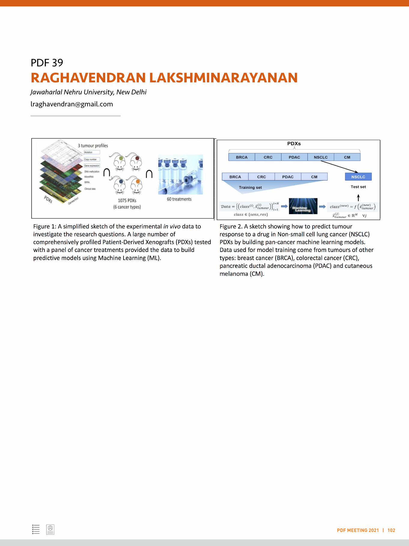
PDF 39RAGHAVENDRAN LAKSHMINARAYANANJawaharlal Nehru University, New Delhi
PDF MEETING 2021 | 102
PDF 40 RAJIV KUMAR KAR Technical University Berlin
PDF MEETING 2021 | 103
LIGHTNING TALK
Keywords: Quantum mechanics; Computational spectroscopy; Photoreceptor; Structural bioinformatics; Machine learning
Understanding spectral properties of photoreceptor proteins
My research work focuses on understanding the spectroscopic properties of photoreceptor proteins using multiscale modelling techniques. Photoreceptor proteins contain chromophores like retinal, flavin, and phytochrome. They absorb light at a specific wavelength, ranging from 310 - 600 nm. This property of light absorbance is controlled or tuned by the amino acids in their binding site. Understanding the relationship between amino acids and chromophore absorption can help designing mutants with desired spectroscopic property, that is, absorbance at long-wavelength (red-light, greater than 650 nm). The use of long-wavelength for absorption is valuable as an optogenetic tool to control neuronal activity. It attributes their property to deeper penetration of light coupled with minimal tissue-scattering and absorption by hemoglobin. Other applications include biosensor and fluorescent probe. A review of advanced methods to fill this knowledge gap through computational biology is present in [1].
We investigate the conformational intermediates of a light-driven sodium pump - Krokinobacter eikastus rhodopsin-2 (KR2) [2]. This photoreceptor contains a retinal chromophore. Using the hybrid QM/MM method, we find that the retinal configuration in the dark state (without light illumination) gives an absorption maximum of 452 nm. The light-activated state containing Na+ in the retinal binding site gives 507 nm. The difference of absorption of these photo-intermediates is 55 nm (red-shift), which is in good agreement with the experimental difference of 66 nm. We also tested our calculations on the protein model, replacing Na+ with a water molecule. However, this gives an absorbance shift of -11 nm (blue-shift). The work is an independent validation to the time-resolved X-ray crystallography in differentiating the electron density of water and Na+ ion and confirming the putative binding site of Na+ ion in KR2.
The work on flavin chromophore includes benchmarking quantum mechanical methods to calculate the spectroscopic properties [3]. We found that wavefunction based methods are more accurate compared to density-functional theory (DFT). The extension of this work includes establishing mathematical modelling of flavin hyperfine structure in the protein environment [4]. An advanced review of the electronic structure and spectra of flavin is reported in [5].
Bacterial phytochrome is another chromophore - a potential candidate for the optogenetic tool. It can expand the spectral range to the near-infrared region. In recent work, we have shown that bacterial phytochrome in Deinococcus deserti has two-photon absorption property [6]. Here, we use structural bioinformatics and quantum biology to understand the spectroscopic (one-photon and two-photon) properties.
References:
[1] Mroginski MA, Adam S, …, Kar RK, ... & Schapiro I. 2020. Frontiers in Multiscale Modelling of Photoreceptor Proteins. Photochemistry and Photobiology, 97: 243-269. (Authors in alphabetical order)
[2] Skopintsev P, Ehrenberg D, Weinert T, James D, Kar RK,... & Standfuss J. 2020. Femtosecond-to-millisecond structural changes in a light-driven sodium pump. Nature, 583: 314-8.
[3] Kar RK, Borin VA, … & Schapiro I. 2019. Spectroscopic properties of lumiflavin: a quantum chemical study. Photochemistry and photobiology, 95: 662-674.
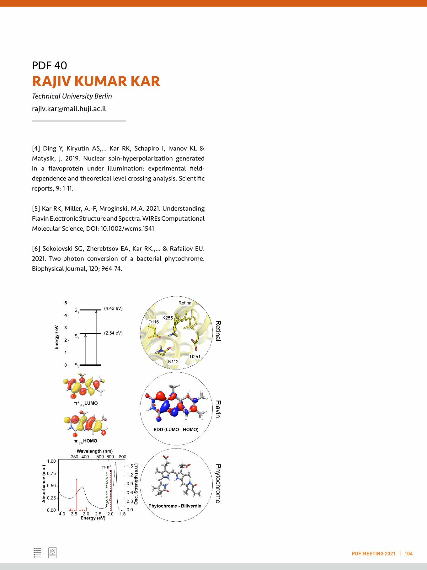
PDF 40RAJIV KUMAR KARTechnical University Berlin
PDF MEETING 2021 | 104
[4] Ding Y, Kiryutin AS,... Kar RK, Schapiro I, Ivanov KL & Matysik, J. 2019. Nuclear spin-hyperpolarization generated in a flavoprotein under illumination: experimental field-dependence and theoretical level crossing analysis. Scientific reports, 9: 1-11.
[5] Kar RK, Miller, A.-F, Mroginski, M.A. 2021. Understanding Flavin Electronic Structure and Spectra. WIREs Computational Molecular Science, DOI: 10.1002/wcms.1541
[6] Sokolovski SG, Zherebtsov EA, Kar RK.,... & Rafailov EU. 2021. Two-photon conversion of a bacterial phytochrome. Biophysical Journal, 120; 964-74.
PDF 41 RAMA NAGESH VENKATA KRISHNA DEEPAK Agency for Science, Technology and Research , Singapore
PDF MEETING 2021 | 105
LIGHTNING TALK
Keywords: Biomacromolecular dynamics; Computer-aided drug design; Snakebite anti-venom Immunotherapeutics; Small molecule therapeutics
Non-covalent interactions - The bricks and mortar of molecular architecture
Non-covalent interactions are weak, often fleeting, physical forces that are foundational to life and the study of the role of these interactions in their diverse manifestations in the biological context has been the central theme of my research activity. My efforts have been focused on understanding the fundamental principles underlying protein structure, function and dynamics, biomacromolecular recognition, and their application towards drug design and discovery.
As a postdoctoral researcher, I have collaborated extensively with experimental researchers focussing my efforts in two distinct directions, 1) drug design and discovery and 2) structural basis of membrane protein function. With respect to drug discovery, we successfully repurposed FDA-approved drugs that could also act as inhibitors of human executioner caspases [1], and established the structural basis of drug resistance in a newly discovered oncogenic mutant BRAF kinase [2] in collaboration with Cancer Biologists from the National Cancer Center Singapore (NCCS). Currently, I am working on ligand discovery efforts to develop anti-inflammatory drug candidates targeting the human C5a receptor in a collaborative effort with GPCR structural biologists from the University of Pittsburgh. With respect to membrane proteins, my research is centred around structural modelling of transmembrane transporters such as the bacterial lysophospholipid transporter [3], human riboflavin transporter and human H+/sugar transporter, and molecular dynamics simulation studies on human GPCRs, namely the receptors for C5a [4] and prostaglandin D2. The broad objective of my work with membrane proteins is to establish the structure-function relationships and mechanisms involved in substrate/inhibitor binding to drive forward drug discovery and design efforts.
During my PhD, I worked on characterizing unconventional non-covalent interactions and their myriad roles in biomacromolecules. We identified several types of NH…N hydrogen bonds in protein structures and demonstrated the ability of the backbone and sidechain N-atoms in proline [5] and histidine [6], respectively, to participate in hydrogen bonding and established the critical importance of the interactions in modulating protein structure and function.
References:
[1] Deepak RNVK†, Abdullah A†,..., Fan H, Ravanan P. 2018. Identification of FDA-approved drugs as novel allosteric inhibitors of human executioner caspases. Proteins: Structure, Function, and Bioinformatics 86(11), 1202-1210.
[2] Yap J†, Deepak RNVK†,..., Fan H, Hu J. In Press. The stability of R-spine defines RAF inhibitor resistance: A comprehensive analysis of oncogenic BRAF mutants with in-frame insertion of αC-β4 loop. Science Advances.
[3] Lin Y†, Deepak RNVK†,..., Fan H, Zheng L. 2018. A dual substrate-accessing mechanism of a major facilitator superfamily protein facilitates lysophospholipid flipping across the cell membrane. Journal of Biological Chemistry 293(51), 19919-19931.
[4] Dumitru AC†, Deepak RNVK†, Liu H†,..., Fan H, Alsteens D. 2020. Submolecular probing of the complement C5a receptor-ligand binding reveals a cooperative two-site binding mechanism. Communications Biology 3(1), 1-13.

PDF 41RAMA NAGESH VENKATA KRISHNA DEEPAKAgency for Science, Technology and Research , Singapore
PDF MEETING 2021 | 106
[5] Deepak RNVK, Sankararamakrishnan R. 2016. Unconventional NH…N hydrogen bonds involving proline backbone nitrogen in protein structures. Biophysical Journal 110(9), 1967-1979.
[6] Deepak RNVK, Sankararamakrishnan R. 2016. N–H···N hydrogen bonds involving histidine imidazole nitrogen atoms: A new structural role for histidine residues in proteins. Biochemistry 55(27), 3774-3783.
PDF 42 ROHIT KONGARI United States Food and Drug Administration, Silver Spring
PDF MEETING 2021 | 107
LIGHTNING TALK
Keywords: Bacteriophage biology; Phage therapy; Antimicrobial resistance; Phage genomics and transcriptomics; Microbiome and Virome
Understanding host-phage interactions to facilitate combat strategies against multidrug-resistant bacteria
Staphylococcus aureus is a Gram-positive commensal bacterium that also causes a variety of clinical infections. It is one of the leading causative agents of bacteremia, infective endocarditis, osteomyelitis, pleuropulmonary infections, and other device-related infections (1). Emergence of antibiotic-resistant strains such as methicillin-resistant S. aureus (MRSA) has complicated the treatment of such infections and rendered most existing treatment options ineffective (2). Bacteriophage therapy is quickly gaining attention as an alternative approach to counter multidrug-resistant bacterial infections (3). Our lab is presently investigating the genetic aspects and utility of bacteriophage therapy targeting MRSA using phage K as a model, with a focus on phage-host interaction dynamics and resistance mechanisms. Bacteriophage K belongs to the Twort-like group of phages, known for their virulent lifestyle and broad host range, thus making it an attractive potential therapeutic agent against S. aureus. Eighteen spontaneous host mutants resistant to phage K at 30 °C were previously isolated using a clinically relevant S. aureus strain NRS384. Whole-genome sequencing revealed missense mutations in the host RNA polymerase beta prime subunit, wall teichoic acid biosynthesis operon, ligase, and tRNA-Gln. An RNA-seq based approach is currently being used to characterize the mode of resistance in the RNA polymerase mutants, comparing the transcription profiles of the phage at different stages of infection in the mutant to the wild-type host. Preliminary data indicates that phage late gene transcription is negatively impacted in the polymerase mutant, resulting in a failed infection. Moreover, two long non-coding RNA (lncRNA) regions of ~500 bp size in the phage genome have been identified to be consistently expressed at exceptionally high levels compared to the rest of the genome, but the physiological relevance of this transcription is yet to
be tested. To further explore phage-host interactions, 22 spontaneous phage K mutants that overcome the resistance conferred by the abovementioned mutations have been isolated. In addition, 4 spontaneous phage K mutants (BPK1-4) that plate more effectively at 37 °C compared to wild-type K have been isolated. Further characterization and whole-genome sequencing of these phages revealed mutations in phage genes that encode potential players involved in different stages of phage growth such as the tail morphogenic protein (adsorption), phage-encoded anti-sigma factor (transcription), helicase (replication) etc. All the sequenced temperature-adapted BPK phages carried mutations abolishing the expression of the same gene, gp102 which is highly conserved in Twort-like phages. Interestingly, one of the BPK mutants had increased effectiveness at clearing out bacterial populations when tested in a mouse decolonization model. In addition, an ORF library expressing all annotated phage K genes was used to identify 30 phage K genes that showed growth inhibition effects upon induction. (All data mentioned here is yet to be published, presented in multiple talks and posters at recent conferences, manuscripts in preparation). Overall, the results from this work should provide insight into antibacterial strategies, clues to make phage therapy more effective, such as genetically modifying phages to overcome the resistance barrier or formulating cocktails with phages that use different host factors or follow diverse lifestyles.
References:
1. Lin DM, K. B. (2017). Phage therapy: An alternative to antibiotics in the age of multi-drug resistance. World J Gastrointest Pharmacol Ther., 8(3):162-173.
PDF 42ROHIT KONGARIUnited States Food and Drug Administration, Silver Spring
PDF MEETING 2021 | 108
2. Steven Y. C. Tong, J. S. (2015). Staphylococcus aureus Infections: Epidemiology, Pathophysiology, Clinical Manifestations, and Management. Clinical Microbiology Reviews, 28 (3) 603-661.
3. Turner, N. S.-K. (2019). Methicillin-resistant Staphylococcus aureus: an overview of basic and clinical research. Nat Rev Microbiol , 17, 203–218 .

PDF 43 SANDEEP AMETA National Center for Biological Sciences, Bengaluru
PDF MEETING 2021 | 109
LIGHTNING TALK
Keywords: Self-sustaining reaction networks; Droplet-microfluidics; Phase-separated droplets; Single-droplet sequencing; RNA catalysis
Darwinian evolution using a minimal chemical system
Life might have started on early Earth as a gradual process from elementary interactions between abiotic molecules. Deciphering such interactions is critical in understanding the origins of life or building a ‘minimal synthetic life’ in the lab.
I am interested in creating a ‘minimal chemical system’ capable of undergoing Darwinian evolution. As a self-replicating model, I use autocatalytic reaction networks (ACSs) of catalytic RNAs where ribozymes self-assemble from smaller substrate fragments. ACSs have been implicated as very important to kick-start early life and might have preceded the contemporary template-based replication [1,2]. However, building evolving ACSs is not trivial and requires both conceptual and experimental developments. Some of the unanswered questions that need to be addressed are-emergence, network identity and heritability in ACSs. Additionally, how variation appears leading to differential fitness of some networks over others such that they can be selected in a Darwinian manner. Though theoretical formalizations are available, experiments are lagging behind or missing altogether [3].
To address these questions, I am developing experimental systems based on ACSs of RNAs derived from group I intron of Azoarcus bacterium [4,5]. Modulating the specificity in these ribozymes, cross-catalytic self-sustaining networks can be constructed [6]. Using barcoded sequencing and droplet-based microfluidics (similar to single-cell transcriptomics), I have established a landscape of RNA ACSs for deriving relationships between network topology, growth, and robustness to perturbations [7].
A crucial component for evolution using such ACSs is a robust and dynamic compartmentalized life cycle. Currently, I am focusing on developing protocell models based on liquid-liquid phase separation (coacervate droplets). We observe that
these droplets confer transient robustness to the networks against perturbation by other competing RNA catalysts [8].
As next steps, I am integrating phase-separated droplets with microfluidics and barcoded single-cell sequencing to develop; a) a strategy to observe the growth of compartments (coacervate droplets) as a function of the growth of ACSs; b) to demonstrate the propagation of heritable network across generations of compartments; c) to develop selection protocols where survival of ‘fittest’ ACSs can be demonstrated. Combining all these elements together will pave the way to create a ‘minimal synthetic system’ possessing life-like features.
References:
1. Vasas, V., Fernando, C., Santos, M., Kauffman, S. & Szathmáry, E. 2012. Biol. Direct 7: discussion.
2. Adamski P., Eleveld M., Sood A., Kun A., Szilágyi A., Czárán T., Szathmáry E. & Otto S. 2020. Nat. Rev. Chem. 4: 386–403.
3. Ameta S., Matsubara Y.J., Chakraborty N., Krishna S. & Thutupalli S. 2021 Life 308.
4. Hayden E.J. & Lehman N. 2006. Chem. Biol. 13: 909-918.
5. Arsène, S., Ameta, S., Lehman, N., Griffiths, A.D. & Nghe, P. Nucleic Acids Res. 46: 9660–9666.
6. Vaidya, N., Manapat, M.L., Chen, I.A., Xulvi-Brunet, R., Hayden, E.J., & Lehman, N. 2012. Nature 491: 72–77.
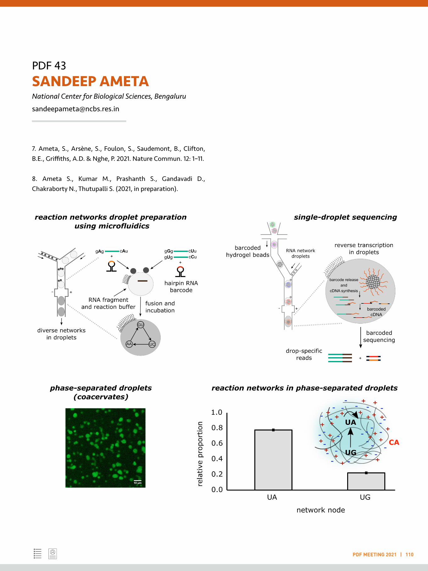
PDF 43SANDEEP AMETANational Center for Biological Sciences, Bengaluru
PDF MEETING 2021 | 110
7. Ameta, S., Arsène, S., Foulon, S., Saudemont, B., Clifton, B.E., Griffiths, A.D. & Nghe, P. 2021. Nature Commun. 12: 1–11.
8. Ameta S., Kumar M., Prashanth S., Gandavadi D., Chakraborty N., Thutupalli S. (2021, in preparation).
PDF 44 SANDHYA GANESAN Yale University, Hamden
PDF MEETING 2021 | 111
LIGHTNING TALK
Keywords: Host-pathogen interaction; Cell biology of infectious diseases; Intracellular bacterial pathogens; Innate immunity; Vesicle traffic
Identification of novel cellular defense mechanisms against bacterial pathogens:Lessons learnt from Coxiella burnetii
Many bacterial pathogens successfully manipulate host cell biology and establish a secure intracellular niche for their replication (Example: Mycobacteria, Salmonella, Legionella, Coxiella, Chlamydia). I am deeply interested in how the human cell-intrinsic defense system detects intracellular pathogens and controls their replication inside host cells. My graduate work focused on the sensing of fungal and bacterial pathogens by innate immune cells and activation of the multi-protein complexes called inflammasomes (Ganesan, S. et al. J Immunol. 2014 and others).
Most pathogens that replicate inside vacuolar organelles inhibit the fusion of their compartment with lysosomes to avoid the highly acidic and proteolytic environment. Coxiella burnetii, a zoonotic pathogen that causes the disease Q-fever, has uniquely evolved to replicate in lysosome-derived compartments called the Coxiella-containing vacuoles (CCV). Mammalian Interferon-gamma (IFNγ) signalling pathway restricts the replication of intracellular C. burnetii. However, the specific mechanisms that underlie the restriction of a lysosome-adapted pathogen inside a membrane-bound compartment are not well-defined. Using a siRNA-based screen, my postdoctoral work demonstrated that IFNγ-induced genes, indoleamine dioxygenase 1 (IDO1) and syntaxin 11 (STX11) were required for C. burnetii restriction. IDO1 is an enzyme that depletes cellular tryptophan and restricts C. burnetii replication since Coxiella is a tryptophan auxotroph (Ganesan, S. and Roy, C. R. PLoS Pathog. 2019). On the other hand, STX11 is a SNARE (Soluble NSF Attachment Protein Receptor) that mediates vesicle fusion processes, lysosomal exocytosis and granule secretion (Tang, B. L. et al. Mol Membr Biol. 2015). STX11-deficient cells generated by CRISPR-Cas9
editing exhibit significantly higher C. burnetii replication and expansion of the CCV, whereas ectopic expression of STX11 reverses these phenotypes. Interestingly, STX11 is enriched in the acidic lumen of the CCV (manuscript in preparation). Thus, STX11 negatively impacts intracellular C. burnetii replication and CCV expansion.
Understanding the mechanistic basis of C. burnetii restriction by STX11 is an exciting immediate goal. In addition, identification of IDO1 and STX11 indicate that there are many human genes whose antimicrobial functions against vacuolar pathogens have not yet been fully explored. With the pathogen-containing compartment as the focal point, my overall aim is to discover cell-intrinsic defence mechanisms and organelle biogenesis factors that make a pathogen-containing compartment unsustainable for pathogen survival. In addition, my independent research program will also investigate how effector proteins secreted by bacterial pathogens evade various facets of immune response. With an initial interest in C. burnetii, I aspire to expand the scope of these studies to Salmonella and Mycobacteria to understand the principles of cell-autonomous defense against bacterial pathogens that subvert vacuolar organelles for their intracellular replication.
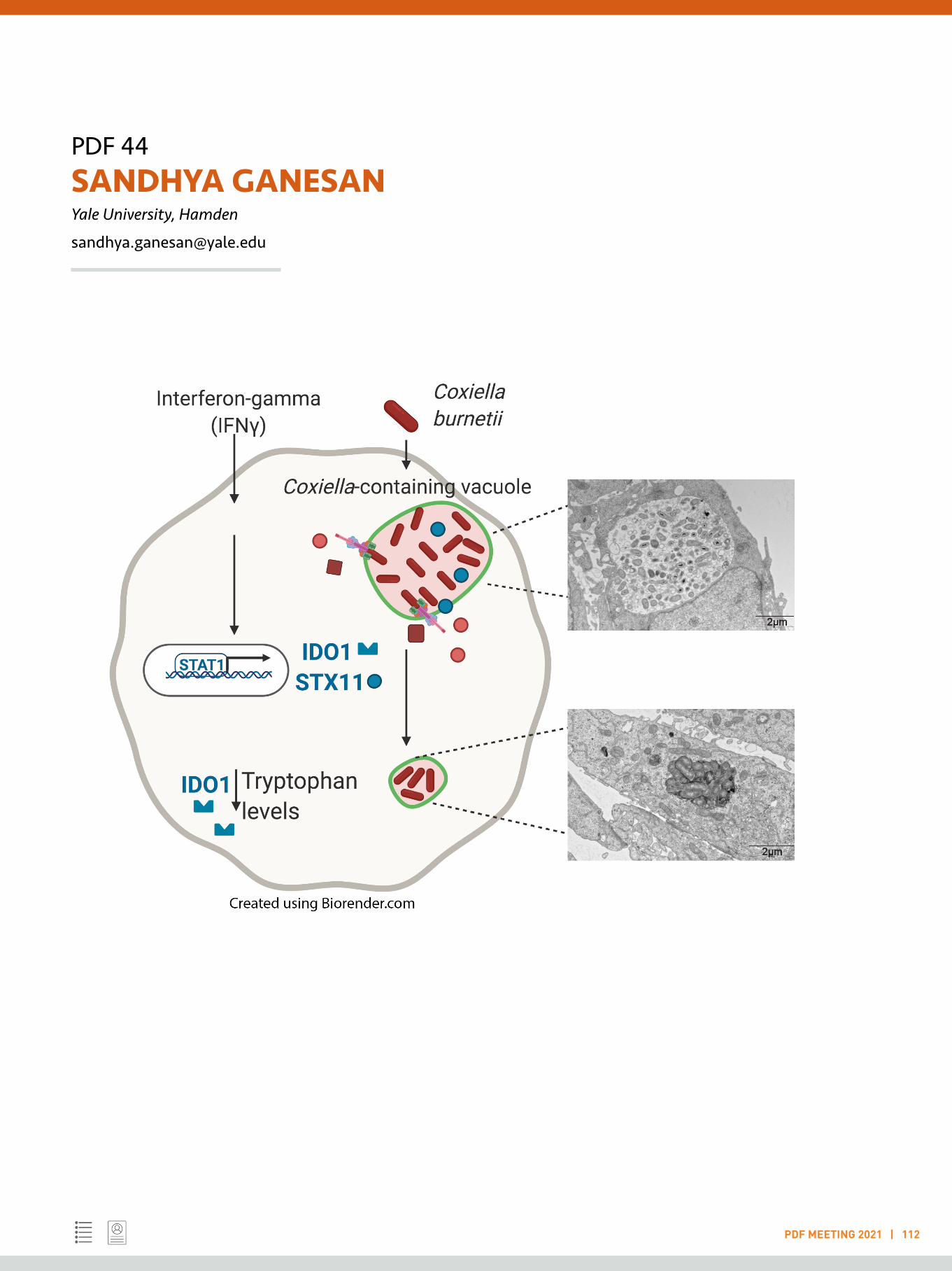
PDF 44SANDHYA GANESANYale University, Hamden
PDF MEETING 2021 | 112
PDF 45 SARMISTHA MAHANTY Indian Institute of Science , Bengaluru
PDF MEETING 2021 | 113
LIGHTNING TALK
Keywords: Specialised organelles; lysosomes; cell differentiation; tissue homeostasis; and diseases
Elucidating the role of epidermal lysosomes in skin homeostasis and diseases
Background: To support the functional speciality of specialized cells, either unique intracellular organelles are produced or universal organelles acquire special characteristics. Lysosomes are the most dynamic organelles that alter the morphology and distribution to justify the cellular need. Piling up evidence has established the diverse role of lysosomes in cellular physiology (reviewed in Ballabio, A. and Bonifacino, J.S., 2020). Interestingly, lysosomes may also acquire special machinery to generate Lysosome Related Organelles in specialized cell types; for example, lytic granules, platelet-dense granules etc. (Marks et al.,2013). Hence, to understand LROs biogenesis and functioning mechanisms, they should be studied in parallel to the lysosomes of a particular cell type. Inline, specific alterations in morphology and function of lysosomes /LROs are reported in specific diseased conditions. For example, severe skin diseases such as atopic dermatitis, Herlequin type Ichthyosis, and psoriasis are represented by the malformed skin lamellar bodies and lysosomes respectively (Feingold 2012; Elias et al., 2014).
Research question and outcome: With this idea, my current research focuses on understanding the biogenesis and secretion mechanism of skin lamellar bodies, which remain completely unknown in the decades of keratinocyte research. Keratinocytes, the major constituent cell type of epidermis undergo differentiation in response to high extracellular calcium (Bikale et al., 2012). Here, we employ high-end live-cell microscopy, super-resolution microscopy, and extensive electron microscopy techniques besides regular cell biology and biochemical assays to identify the molecular machinery involved in the biogenesis of skin lamellar bodies in human primary keratinocytes and organoid model system. Altogether, our experimental findings have established unconventional
features of these lysosomes that strongly support the idea that these lysosomes are the precursor of LBs. For example, these lysosomes show a unique association with the Golgi proteins and open up new avenues to study inter-organelle contact of lysosomes and Golgi, which otherwise was not known.
References:
Ballabio A. and Bonifacino JS.2020. Lysosomes as dynamic regulators of cell and organismal homeostasis. Nature Reviews Molecular Cell Biology, 21(2):101-118.
Bikle DD, Z Xie, and CL Tu. 2012. Calcium regulation of keratinocyte differentiation. Expert Rev Endocrinol Metab 7:461-472.
Elias PM, and JS Wakefield. 2014. Mechanisms of abnormal lamellar body secretion and the dysfunctional skin barrier in patients with atopic dermatitis. J Allergy Clin Immunol. 134:781-791.
Feingold KR, 2012. Lamellar bodies: the key to cutaneous barrier function. Journal of Investigative Dermatology 132(8): 1951-1953.
Marks MS, Heijnen HF, and Raposo G. 2013. lysosome-related organelles: unusual compartments become mainstream. Curr Opin Cell Biol 25(4):495-505.

PDF 45SARMISTHA MAHANTYIndian Institute of Science , Bengaluru
PDF MEETING 2021 | 114

PDF 46 SHWETA RAMDAS University of Pennsylvania, Philadelphia
PDF MEETING 2021 | 115
LIGHTNING TALK
Keywords: Genomics; Human genetics; Sequencing; Epigenetics; Bioinformatics
Studying regulatory variation in founder populations to identify functional rare variants
With increasing sample sizes, GWAS and sequencing studies are now able to identify trait-associated variants with low allele frequencies. However, interpreting the function of these rare variants remains challenging. The functional interpretation of common disease-associated variation has been aided by the identification of variants regulating gene expression. However, expression quantitative locus (eQTL) studies are currently limited by their sample sizes, which make the functional interpretation of rare non-coding variants from these studies a challenge. In this study, we attempt an alternate approach for the functional interpretation of rare variants using a genetically isolated (founder) population. Founder populations harbor an increased burden of functional variants rare in more heterogeneous populations, making it possible to characterize their functional impact with smaller sample sizes. In this experimental design, we study genes with extremely high or low levels of expression (called ‘expression outliers’). We posit that identifying expression outliers in a founder population will allow us to determine regulatory variants of large effect otherwise not detectable in commonly-studied populations.
The Amish represent a genetic isolate whose European ancestors settled in the Americas starting in the 17th century. We analyze RNA-seq data from lymphoblastoid cell lines (LCL) obtained from 97 genotyped samples of a large multi-generational pedigree; this represents the first transcriptomic study on the Amish population. We identify 1,209 genes with eQTLs in this cohort. While we see an eQTL replication rate of 72% in larger cohorts of LCLs, we find 206 eQTL genes unique to the Amish population at loci harboring genetic variation that is rare in large European cohorts. The unique pedigree structure of our cohort enables us to identify genomic segments shared identical by descent and proximal to gene
expression outliers. We identify more than 6,000 outliers for gene expression, splicing, or allele-specific expression; more than 100 of which are associated with specific cis-regulatory haplotypes. We then added orthogonal epigenomic information to prioritize truly causal regulatory variants within these regions. These methods and results allow us to prioritize disease variants by identifying variants of large-effect, and lead to a more comprehensive annotation of the regulatory genome.

PDF 47 SNEHA SHAH Umass Medical School, Worcester
PDF MEETING 2021 | 116
LIGHTNING TALK
Keywords: Fragile X syndrome; Autism; Biomarkers; Neuroscience; Alternative splicing
Understanding the molecular signatures of Fragile X Syndrome and Autism
Autism spectrum disorders are prevalent worldwide. Current estimates show one in every 270 people lie on the Autism spectrum disorder (ASD). Fragile X Syndrome (FXS) is the most common known monogenetic cause of ASD. FXS occurs due to a genetic mutation that results in FMR1 gene silencing and loss of its protein product FMRP. FMRP protein is an RNA binding protein that impedes ribosome movement (ribosome stalling) along RNAs [1]–[4]. Thus, upon loss of FMRP, protein synthesis of the target RNAs is increased. We devised a method to assess which RNAs had increased ribosome movement in mouse brain tissue slices upon loss of FMRP. Ribosome profiling over a time course, demonstrates a wide range of ribosome translocation rates on specific mRNAs, ranging from rapid to near complete stalling. Several mRNAs retain 4-6 ribosomes after runoff in WT, but not in FMRP-deficient slices. One such mRNA encodes SETD2, a lysine methyltransferase that catalyzes H3K36me3. ChIP-Seq demonstrates that loss of FMRP alters the deployment of this epigenetic mark on chromatin. H3K36me3 is assoSciated with alternative pre-RNA processing, which I find occurs in an FMRP-dependent manner on transcripts linked to neural function and autism spectrum disorders [5]. Previous studies have identified aberrant splicing in post-mortem samples from autism patients [6], [7].
Furthermore, I find widespread aberrations in alternative splicing in post-mortem brain tissues and blood samples obtained from patients (unpublished data). We also find highly statistically significant changes in total RNA levels for many important mRNAs that could also help us understand FXS pathology. This discovery can potentially lead to the development of biomarkers that are much needed in the field of intellectual disability. The correlation of aberrant splicing in FXS and those identified previously in autism patients can identify possible diagnostic and therapeutic targets.
Identifying these aberrant splicing changes, their mechanism of action and potential use in therapeutics is of great interest to the field of intellectual disability.
References:
[1] J. C. Darnell et al., “FMRP stalls ribosomal translocation on mRNAs linked to synaptic function and autism,” Cell, 2011.
[2] J. D. Richter and X. Zhao, “The molecular biology of FMRP: new insights into fragile X syndrome,” Nature Reviews Neuroscience. 2021.
[3] T. Udagawa et al., “Genetic and acute CPEB1 depletion ameliorate fragile X pathophysiology,” Nat. Med., vol. 19, p. 1473, Oct. 2013.
[4] B. Liu et al., “Regulatory discrimination of mRNAs by FMRP controls mouse adult neural stem cell differentiation,” Proc. Natl. Acad. Sci. U. S. A., 2018.
[5] S. Shah, G. Molinaro, B. Liu, R. Wang, K. M. Huber, and J. D. Richter, “FMRP Control of Ribosome Translocation Promotes Chromatin Modifications and Alternative Splicing of Neuronal Genes Linked to Autism,” Cell Rep., 2020.
[6] M. Quesnel-Vallières et al., “Misregulation of an Activity-Dependent Splicing Network as a Common Mechanism Underlying Autism Spectrum Disorders,” Mol. Cell, 2016.
[7] M. Irimia et al., “A highly conserved program of neuronal microexons is misregulated in autistic brains,” Cell, 2014.
PDF 48 SNEHAL KARPE GIGA, Université de Liège
PDF MEETING 2021 | 118
LIGHTNING TALK
Keywords: Bioinformatics; Genomics; Computational tools; Structural biology; Biodiversity
Exploring biological diversity through eukaryotic genome annotations
My doctoral research focused on the de-orphanisation (identifying cognate ligand) of olfactory receptors (OR) in various organisms using computational tools. First, to address the unavailability of the experimentally solved protein structures, we predicted structures of 100 representative olfactory receptors from 5 model organisms that were shared in the form of a Database of Olfactory Receptors. Next, I focused on annotating olfactory receptor genes in unexplored insect genomes. The insect OR gene repertoires have adapted rapidly to changing requirements, ultimately leaving unique signatures of their evolution in the genome. This work involved semi-automated manual curation of ORs from the dwarf Asian honey bee Apis florea (AfOrs)1 and two solitary bee genomes2. Thereby, I discovered around 150 new OR genes. Through phylogenetic reconstruction and antennal transcriptome sequencing, we were able to identify the equivalent of the Apis mellifera queen mandibular pheromone (9-ODA) receptor in the A. florea genome (AfOr11). Worker bee-enriched expression of some OR subfamilies helped to hypothesize their functions by comparison with other species - putative cuticular hydrocarbon receptors, putative floral scent receptors and a set of bee expanded receptors. We challenged a previous hypothesis correlating expansion of OR repertoires with eusociality in bees by discovering almost equal number of OR genes in a non-social species. Through homology modeling, docking and molecular dynamics simulations of AmOr11, we attempted to understand the principles behind the specificity of pheromone perception. I developed ‘insectOR’, a gene-annotation web server for predicting any divergent family of proteins from a genome of interest3. Scientists from all over the world are using this tool (website visits from over 39 countries).
Separately, I collaborated on a project analysing volatile organic compounds from Blackbuck fecal samples that might
help in their mating. I also contributed to the genome and transcriptome sequencing of the Tulsi and Drumstick plants in the lab.
My fascination in understanding new genomes led me to shift gears during my postdoctoral research in ULiege, Belgium, where I am currently working on retrovirus-induced leukemia in patient cohorts. Here, I analyse clonal architecture of the proviral integration sites of Human T-Lymphotropic Virus-1 (HTLV-1) and Bovine Leukemia Virus (BLV) in the human and sheep genomes respectively, using a newly developed NGS technique in our lab. It has diagnostic advantages over other methods and it also aids in the fundamental understanding of the oncogenic switch. Currently we are working to show prognostic advantages of the method to distinguish leukemia progressors from non-progressors from a longitudinal cohort of HTLV-1 carriers.
References:
1. Karpe SD, Jain R, Brockmann A, Sowdhamini R. 2016. Identification of Complete Repertoire of Apis florea Odorant Receptors Reveals Complex Orthologous Relationships with Apis mellifera. Genome Biology and Evolution 8: 2879–2895.
2. Karpe SD, Dhingra S, Brockmann A, Sowdhamini R. 2017. Computational genome-wide survey of odorant receptors from two solitary bees Dufourea novaeangliae (Hymenoptera: Halictidae) and Habropoda laboriosa (Hymenoptera: Apidae). Scientific Reports 7: 10823.
3. Karpe SD, Tiwari V, Ramanathan S. 2021. InsectOR-Webserver for sensitive identification of insect olfactory receptor genes from non-model genomes. PloS one 16(1): e0245324.Link to Abstract Image
PDF 49 SONISILPA MOHAPATRA Johns Hopkins University, Baltimore
PDF MEETING 2021 | 119
LIGHTNING TALK
Keywords: Protein engineering; Superresolution imaging; Single molecule fluorescence microscopy; Synthetic biology; Antimicrobial peptides
Engineering photo-controlled helicases
Cellular processes are a result of the highly coordinated functioning of a network of proteins. One way to decipher the role of a protein in this complex system is by switching it on and off on demand. Light has been utilized to activate and inactivate proteins in a spatiotemporally defined manner while minimally perturbing the cells. In my research project, we are interested in designing photoswitchable DNA and RNA helicases to learn about the mechanistic details behind their function. Since a variety of proteins display a strong structure-function relationship, we relied on altering protein structure using light to generate light-controlled functionalities. A photo-sensitive protein domain, LOV whose N to C terminus distance changes in response to light, can be inserted into different regions of the helicase of interest to introduce light controlled structural modulation of the helicase. As our first prototype helicase, we are working on engineering a photoswitchable Rep, an E. coli DNA helicase. Using in silico DNA library design and a high-throughput directed golden gate assembly approach, we have generated a 700-member library of LOV inserted Rep. In each member of this library, the insertion position of LOV is sequentially shifted by one residue so that this library samples all possible insertion positions along the original Rep peptide sequence. We developed functional assays for high-throughput in vivo screening of Rep in a light-dependent manner. The viability of ΔrepΔuvrD cells exogenously expressing Rep mutant library is studied with and without light. The Rep mutants that complement cell viability in rich growth medium in light on/off conditions can be identified through next-generation sequencing. As a proof of principle experiment, we generated a prototype 50-membered library of DNA helicases, where the light-sensitive domain is inserted in a specific location of the DNA helicase. Out of this small-scale library, we have
been able to successfully identify Rep mutants, which display light-sensitive in vivo function. In the near future, we will be expanding on this work to isolate photoswitchable Rep mutants from the 700 member library and the mutants that display stronger contrast in survival efficiency in a light-dependent manner will be expressed and purified. The in vitro unwinding activity of these light modulated Rep mutants will be characterized and optimized for usage in various biotechnological applications. The optogenetics platform that we develop for tuning the activity of Rep by light can be applied to understand the roles of a diverse range of helicases in humans, many of which have uncharacterized functions, but are frequently dysregulated in cancers.
References:
Coyote-Maestas W, Nedrud D, Okorafor S, He Y, Schmidt D. 2020. Targeted insertional mutagenesis libraries for deep domain insertion profiling. Nucleic Acids Research 48:2 e11.

PDF 49SONISILPA MOHAPATRAJohns Hopkins University, Baltimore
PDF MEETING 2021 | 120
PDF 50 SOUMITRA MOHANTY Karolinska Institutet, Stockholm
PDF MEETING 2021 | 121
LIGHTNING TALK
Keywords: Innate immunity; E. coli; Urinary tract infections; Antimicrobial peptides; Bladder infection
Impact of diabetes on urinary tract infection: A mechanistic approach to understand recurrent infection
Urinary tract infection (UTI) is one of the most common bacterial infections with a high risk of recurrence. Patients with diabetes have an increased risk of developing complicated UTI. The underlying mechanisms are still not fully understood. We for the first time observed an increased level of glucose leading to suppression of host defence antimicrobial peptide (AMP), AMP-1 in diabetic patients and mice with hereditary diabetes. To confirm the effect, a hyperglycemic clamp was performed in prediabetic patients. Glucose infusion in prediabetic patients increased the serum glucose level from the median value 4.65 mM to 11mM in 2 h. An increase in serum glucose leads to an increase in serum insulin levels. However, AMP-1 found to be downregulated confirming the glucose-mediated downregulation of AMP-1. Antimicrobial peptides regulate the epithelial barrier protein to prevent the invasion of bacteria into the host. Reduction in AMP-1 level was associated with downregulation of several epithelial tight junction proteins, occludin, JAM, claudin-1 and claudin-4 in the bladders of uninfected and E. coli infected diabetic mice. Apart from antimicrobial activity, AMP-1 also regulates the proinflammatory cytokines like IL-1β, IL-6 and IL-8. Compromised IL-8 or mice KC levels resulted in poor
neutrophil migration in diabetic mice transurethrally infected with E. coli. This favoured more bacterial load and several intracellular bacterial communities in the bladders of infected mice with impaired bacterial clearance in comparison to non-diabetic littermates. High glucose also modulated cell surface markers like UP1A, TLR4 and ITGB1 which allowed more attachment of bacteria into the umbrella cells of the urinary bladder. Diabetes also altered the cytoskeletal protein to facilitate more bacterial survival. Several transcriptional activator analyses revealed the mechanism of glucose-mediated downregulation of AMP-1. Apart from this, my research also focuses on understanding the role different antimicrobial agents like novel compounds from commonly used plant extracts, silver nanoparticles, hydrogels loaded with antibiotics, and their effective delivery to clear the infection.
PDF 51 SRIJIT DAS University of Iowa, Iowa City
PDF MEETING 2021 | 122
LIGHTNING TALK
Keywords: Proteostasis; Aging; Epigenetics; Neurodegenerative Diseases; Cellular stress response
Understanding the mechanisms of neuronal regulation of stress response and inheritance of epigenetic memory of stress in C. elegans
When exposed to unfavorable conditions, cells maintain protein homeostasis by robust synthesis of heat shock proteins due to stress-induced activation of the conserved transcription factor heat shock factor 1 (HSF1) – a process known as heat shock response (HSR). HSR is regulated cell autonomously in eukaryotic cells, however; in metazoans, HSR is also regulated non-autonomously by the nervous system. Pioneering work from our laboratory demonstrated that in Caenorhabditis elegans, neuronal regulation of HSR occurs through activation of thermosensory neurons and subsequent release of the neurotransmitter serotonin (5-HT), however; the mechanism remained unknown. My work shows that 5-HT released by maternal neurons upon the perception of stress activates HSF1 and initiates a conserved transcriptional program in the germline, which ensures the viability and stress resilience of future offspring. The intracellular signal transduction pathway by which 5-HT release activates HSF1 is conserved in C. elegans and mammalian neurons and occurs through activation of Protein Kinase A which enables HSF1 to alter chromatin in soon-to-be fertilized germ cells by recruiting histone chaperone FACT (FAcilitates Chromatin Transcription), displacing histones and initiating protective gene expression. Without 5-HT release, FACT is not recruited by HSF1 in germ cells, transcription occurs but is delayed, and progeny of stressed C. elegans mothers fail to complete development1. This study uncovers a novel mechanism by which stress sensing by neurons is coupled to transcription response times of germ cells to ensure survival of future offspring.
In many organisms including C. elegans, the memory of early-life exposure to environmental stress is ‘preserved’ through unknown mechanisms and later-life cellular programs and survival mechanisms. My study uncovers
a novel mechanism whereby in response to stress, the germline activity of the conserved transcription factor HSF1 recruits the repressive histone H3K9 SET domain methyltransferase MET-2 (SETDB1 in human) to insulin receptor daf-2 and other HSF1 target genes. The increased H3K9me2 levels persist in adults, decreasing their stress responsiveness but at the same time, enhancing their stress resilience by activating the C. elegans FOXO ortholog DAF-16. This mechanism represents a novel and consequential function of HSF1 in the establishment of a heritable epigenetic memory of stress2.
My broad research interest lies in understanding (a) how the mechanism underlying serotonergic activation of HSF1 and subsequent expression of protective genes can be manipulated to find new avenues to combat against late-onset protein misfolding diseases and (b) the mechanism(s) of transgenerational inheritance of epigenetic marks and its contribution in aging.
References:
1. Das S, Ooi FK, Corchado JC, Fuller LC, Weiner JA, Prahlad V (2020) Serotonin signaling by maternal neurons upon stress ensures progeny survival. eLife 9, e55246
2. Das S, Min S, Prahlad V (2021) Germline activity of the heat shock factor HSF-1 programs the insulin-receptor daf-2 in C. elegans. bioRxiv (submitted to Nat Cell Biol)
PDF 52 SRINATH KRISHNAMURTHY KU Leuven
PDF MEETING 2021 | 123
LIGHTNING TALK
Keywords: Structural dynamics; Molecular machines; Structural mass spectrometry; Biophysics; Membrane protein complexes
Structural dynamics of Molecular Machines
Protein interactions are essential regulators of cellular and biochemical processes. Understanding how proteins interact to form large multi-protein complexes is a primary focus of modern structural biology. The paradigm of structural biology is ever changing with the advent of cutting-edge biophysical tools that provide protein structural and dynamics information, with the ultimate goal being to determine structure-dynamics-function relationships. My research focuses on understanding how intrinsic structural dynamics regulate the function of membrane associated protein complexes. I have combined cutting-edge structural mass spectrometry methods with biochemical assays, MD simulations and single molecular techniques to understand how proteins interact and what are the structural dynamics consequences to these interactions. In the first example, I looked at the termination phase of the cAMP signaling pathway. The cAMP pathway is a fundamental regulatory pathway for regulating cellular signaling in response to hormonal stimuli. Protein Kinase A (PKA) is activated by high affinity interactions with cAMP that dissociates its regulatory and catalytic subunits. If the regulatory subunit is bound to cAMP, reassociation and hence termination of the cAMP pathway is restricted. By combining structural mass spectrometry, fluorescence polarization assays and computational docking, we determined that cyclic nucleotide Phosphodiesterases bind directly to the cAMP binding sites of PKA, induce the release of the nucleotide and hydrolyze cAMP to AMP through active site coupling and substrate channeling in signaling pathways (1). In the second example, I focus on the membrane associate bacterial Sec translocase. The Sec translocase is a membrane localized multi-subunit protein complex consisting of the SecYEG membrane channel and the peripheral ATPase, SecA. SecA interacts with cytosolic preproteins and translocates them across the SecYEG channel by coupling ATP hydrolysis
to translocation work. We used structural MS, single molecule FRET and fully atomistic MD simulations to monitor the conformational dynamics of SecA as part of a fully functional membrane associated Sec translocase. We reveal a nexus of allosteric checkpoints that regulate communication within SecA and keep cytosolic SecA conformationally and catalytically auto inhibited. Activation is achieved through a tightly regulated allosteric relay that spans multiple domains of SecA. A core checkpoint lies at the interface of the ATPase motor and the preprotein binding region and is strategically located to control cross-communication with the SecYEG channel and couple the release of auto-inhibition elements to result in translocase channel activation. We now show using HDXMS, how interactions with SecYEG, signal peptides and associated mature domains sequentially activate the Sec translocase. Importantly, we have reconstituted preprotein translocation through the Sec translocase in HDXMS conditions and have monitored the dynamics of a fully activated Sec translocase (2).
References:
1) Krishnamurthy S, Moorthy BS, Lim XX, Lim XS, Bharatham K, N. Tulsian NK, Mihalek I, and Anand GS. 2014. Active site cou-pling in PDE-PKA Complexes Promotes cAMP Signal Termination. Biophysical Journal. 107(6)
2) Krishnamurthy S, Eleftheriadis N, Karathanou K, Smit JH, Portaliou A, Chatzi KE, Sardis MF, Karamanou S, Bondar AN, Gouridis G, Economou A. 2021. A nexus of intrinsic dynamics underlies protein translocase activation. Structure (in Press)
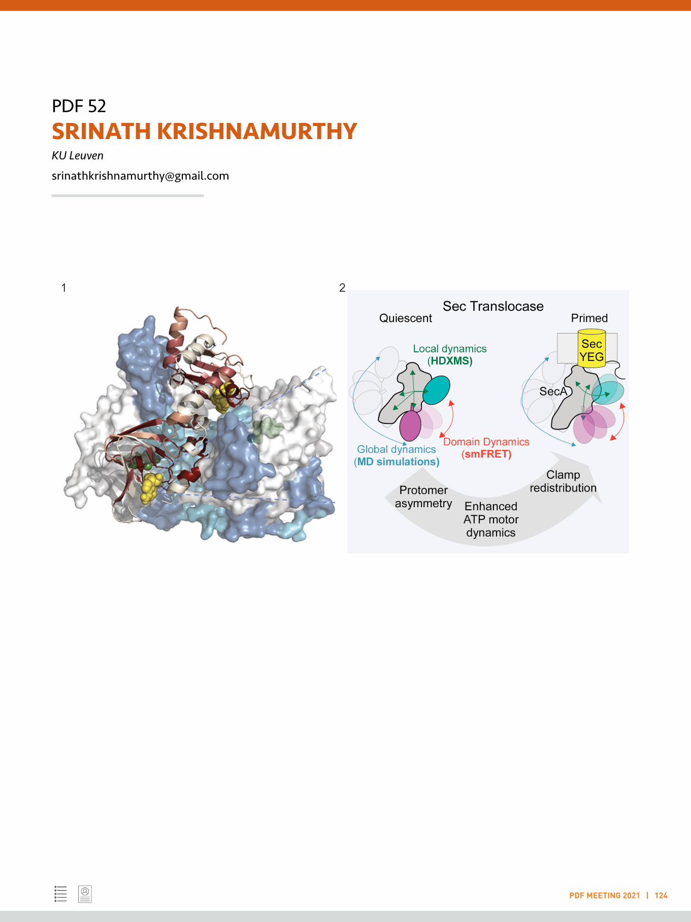
PDF 52SRINATH KRISHNAMURTHYKU Leuven
PDF MEETING 2021 | 124
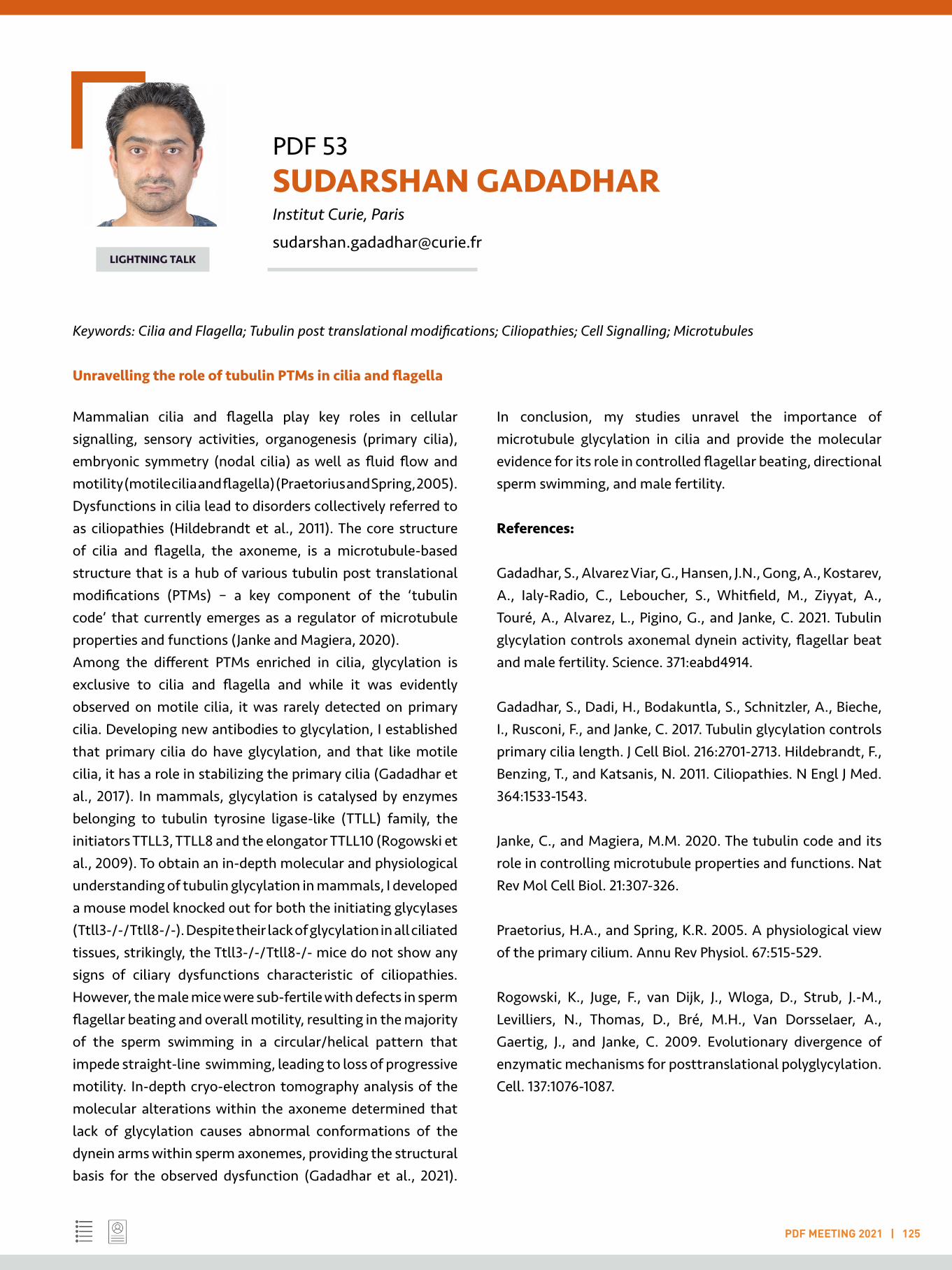
PDF 53 SUDARSHAN GADADHAR Institut Curie, Paris
PDF MEETING 2021 | 125
LIGHTNING TALK
Keywords: Cilia and Flagella; Tubulin post translational modifications; Ciliopathies; Cell Signalling; Microtubules
Unravelling the role of tubulin PTMs in cilia and flagella
Mammalian cilia and flagella play key roles in cellular signalling, sensory activities, organogenesis (primary cilia), embryonic symmetry (nodal cilia) as well as fluid flow and motility (motile cilia and flagella) (Praetorius and Spring, 2005). Dysfunctions in cilia lead to disorders collectively referred to as ciliopathies (Hildebrandt et al., 2011). The core structure of cilia and flagella, the axoneme, is a microtubule-based structure that is a hub of various tubulin post translational modifications (PTMs) – a key component of the ‘tubulin code’ that currently emerges as a regulator of microtubule properties and functions (Janke and Magiera, 2020).Among the different PTMs enriched in cilia, glycylation is exclusive to cilia and flagella and while it was evidently observed on motile cilia, it was rarely detected on primary cilia. Developing new antibodies to glycylation, I established that primary cilia do have glycylation, and that like motile cilia, it has a role in stabilizing the primary cilia (Gadadhar et al., 2017). In mammals, glycylation is catalysed by enzymes belonging to tubulin tyrosine ligase-like (TTLL) family, the initiators TTLL3, TTLL8 and the elongator TTLL10 (Rogowski et al., 2009). To obtain an in-depth molecular and physiological understanding of tubulin glycylation in mammals, I developed a mouse model knocked out for both the initiating glycylases (Ttll3-/-/Ttll8-/-). Despite their lack of glycylation in all ciliated tissues, strikingly, the Ttll3-/-/Ttll8-/- mice do not show any signs of ciliary dysfunctions characteristic of ciliopathies. However, the male mice were sub-fertile with defects in sperm flagellar beating and overall motility, resulting in the majority of the sperm swimming in a circular/helical pattern that impede straight-line swimming, leading to loss of progressive motility. In-depth cryo-electron tomography analysis of the molecular alterations within the axoneme determined that lack of glycylation causes abnormal conformations of the dynein arms within sperm axonemes, providing the structural basis for the observed dysfunction (Gadadhar et al., 2021).
In conclusion, my studies unravel the importance of microtubule glycylation in cilia and provide the molecular evidence for its role in controlled flagellar beating, directional sperm swimming, and male fertility.
References:
Gadadhar, S., Alvarez Viar, G., Hansen, J.N., Gong, A., Kostarev, A., Ialy-Radio, C., Leboucher, S., Whitfield, M., Ziyyat, A., Touré, A., Alvarez, L., Pigino, G., and Janke, C. 2021. Tubulin glycylation controls axonemal dynein activity, flagellar beat and male fertility. Science. 371:eabd4914.
Gadadhar, S., Dadi, H., Bodakuntla, S., Schnitzler, A., Bieche, I., Rusconi, F., and Janke, C. 2017. Tubulin glycylation controls primary cilia length. J Cell Biol. 216:2701-2713. Hildebrandt, F., Benzing, T., and Katsanis, N. 2011. Ciliopathies. N Engl J Med. 364:1533-1543.
Janke, C., and Magiera, M.M. 2020. The tubulin code and its role in controlling microtubule properties and functions. Nat Rev Mol Cell Biol. 21:307-326.
Praetorius, H.A., and Spring, K.R. 2005. A physiological view of the primary cilium. Annu Rev Physiol. 67:515-529.
Rogowski, K., Juge, F., van Dijk, J., Wloga, D., Strub, J.-M., Levilliers, N., Thomas, D., Bré, M.H., Van Dorsselaer, A., Gaertig, J., and Janke, C. 2009. Evolutionary divergence of enzymatic mechanisms for posttranslational polyglycylation. Cell. 137:1076-1087.

PDF 53SUDARSHAN GADADHARInstitut Curie, Paris
PDF MEETING 2021 | 126
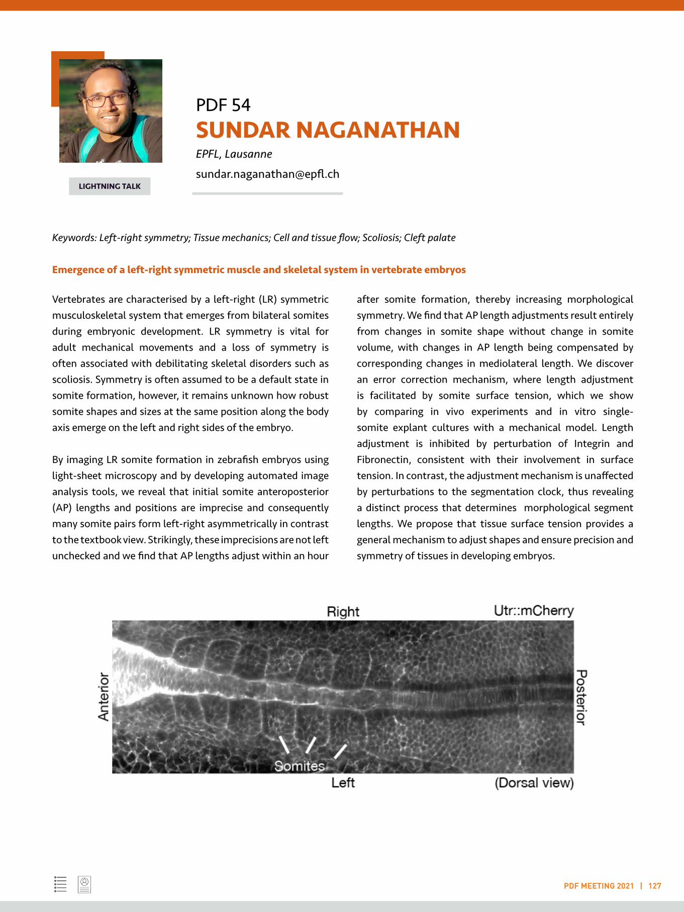
PDF 54 SUNDAR NAGANATHAN EPFL, Lausanne
PDF MEETING 2021 | 127
LIGHTNING TALK
Keywords: Left-right symmetry; Tissue mechanics; Cell and tissue flow; Scoliosis; Cleft palate
Emergence of a left-right symmetric muscle and skeletal system in vertebrate embryos
Vertebrates are characterised by a left-right (LR) symmetric musculoskeletal system that emerges from bilateral somites during embryonic development. LR symmetry is vital for adult mechanical movements and a loss of symmetry is often associated with debilitating skeletal disorders such as scoliosis. Symmetry is often assumed to be a default state in somite formation, however, it remains unknown how robust somite shapes and sizes at the same position along the body axis emerge on the left and right sides of the embryo.
By imaging LR somite formation in zebrafish embryos using light-sheet microscopy and by developing automated image analysis tools, we reveal that initial somite anteroposterior (AP) lengths and positions are imprecise and consequently many somite pairs form left-right asymmetrically in contrast to the textbook view. Strikingly, these imprecisions are not leftunchecked and we find that AP lengths adjust within an hour
after somite formation, thereby increasing morphological symmetry. We find that AP length adjustments result entirely from changes in somite shape without change in somite volume, with changes in AP length being compensated by corresponding changes in mediolateral length. We discover an error correction mechanism, where length adjustment is facilitated by somite surface tension, which we show by comparing in vivo experiments and in vitro single-somite explant cultures with a mechanical model. Length adjustment is inhibited by perturbation of Integrin and Fibronectin, consistent with their involvement in surface tension. In contrast, the adjustment mechanism is unaffected by perturbations to the segmentation clock, thus revealing a distinct process that determines morphological segment lengths. We propose that tissue surface tension provides a general mechanism to adjust shapes and ensure precision and symmetry of tissues in developing embryos.

PDF 55 SWAPNIL SHINDE University of California, San Francisco
PDF MEETING 2021 | 128
LIGHTNING TALK
Keywords: Primary cilia; Vesicular trafficking; Ciliopathies; Ubiquitination; GPCRs
Quality control of the cilia proteome
The cilium is an ancestral microtubule-based organelle present on nearly every cell in the human body. Primary cilia function in phototransduction, olfaction, and developmental signaling such as Hedgehog signaling 1. Abnormalities in primary cilia function are linked to numerous human diseases that includes Bardet-Biedl syndrome (BBS), Joubert syndrome, and nephronophthisis; collectively termed as ciliopathies (1). Cilia lacks protein synthesis machinery and hence relies on the importing process governed by intraflagellar transport (IFT), to assemble signaling cascades. BBSome, is an obligate coat like complex of eight BBS proteins, which regulates the trafficking of ciliary cargoes (2). Several studies in mammalian cells have established the regulatory role of BBSome in signal-dependent exit of G Protein-Coupled Receptors such as Somatostatin Receptor 3 (SSTR3) from cilia (2). Moreover, BBSome governs exit of the membrane-associated protein phospholipase D (PLD) from Chlamydomonas flagella. Given the role of BBSome in the retrieval of ciliary proteins, cilia of BBS mutants display anomalous accumulation of proteins. Quantitative proteomic profiling of primary cilia from Bbs19/Ift27/- kidney cells revealed accumulation of numerous signaling proteins (2).The photoreceptor outer segment (POS) is hyper specialised primary cilia, which senses light. Proteomic profiling of POS from Bbs17-/- mice revealed over 100 proteins accumulating in POS of Bbs mutant mice compared to WT mice (3).
Currently, the key question is how the BBSome can selectively recognize over 100 distinct cargoes/proteins? Ubiquitin– proteasome system, functions as a surveillance machinery to remove the unwanted proteins from the cellular system. In Chlamydomonas reinhardtii flagella, global ubiquitination of the ciliary proteome is elevated during cilium disassembly. Since the proteasome is absent from cilia, and the BBSome was suggested to associate with ubiquitinated proteins
in trypanosome, ubiquitination likely plays regulatory roles inside cilia, possibly regulating trafficking (3).
Recently, we demonstrated upon activation, ciliary GPCRs are ubiquitinated in a β-arrestin– dependent manner before BBSome-mediated exit. Removal of ubiquitin acceptor lysine residues from the SSTR3 and GPR161 demonstrated that ubiquitination of ciliary GPCRs is required for their regulated exit from cilia. Furthermore, targeting a UbK63-specific deubiquitinase to cilia blocks the exit of GPR161, SSTR3, and Smoothened (SMO) from cilia. Finally, ubiquitinated proteins accumulate in cilia of mammalian photoreceptors and Chlamydomonas cells when BBSome function is compromised. We conclude that Ub chains mark GPCRs and other unwanted ciliary proteins for recognition by the ciliary exit machinery (3). Currently, we are characterizing enzymatic machinery required for ubiquitination of proteins in cilia and recognition of ubiquitinated cargoes by the BBSome.
References:
1. Reiter, J., Leroux, M. 2017. Genes and molecular pathways underpinning ciliopathies. Nat Rev Mol Cell Biol 18, 533–547.
2. Nachury, M.V., Mick, D.U. 2019. Establishing and regulating the composition of cilia for signal transduction. Nat Rev Mol Cell Biol 20, 389–405.
3. Shinde SR, Nager AR, Nachury MV. 2020. Ubiquitin chains earmark GPCRs for BBSome-mediated removal from cilia. J Cell Biol. 219(12).
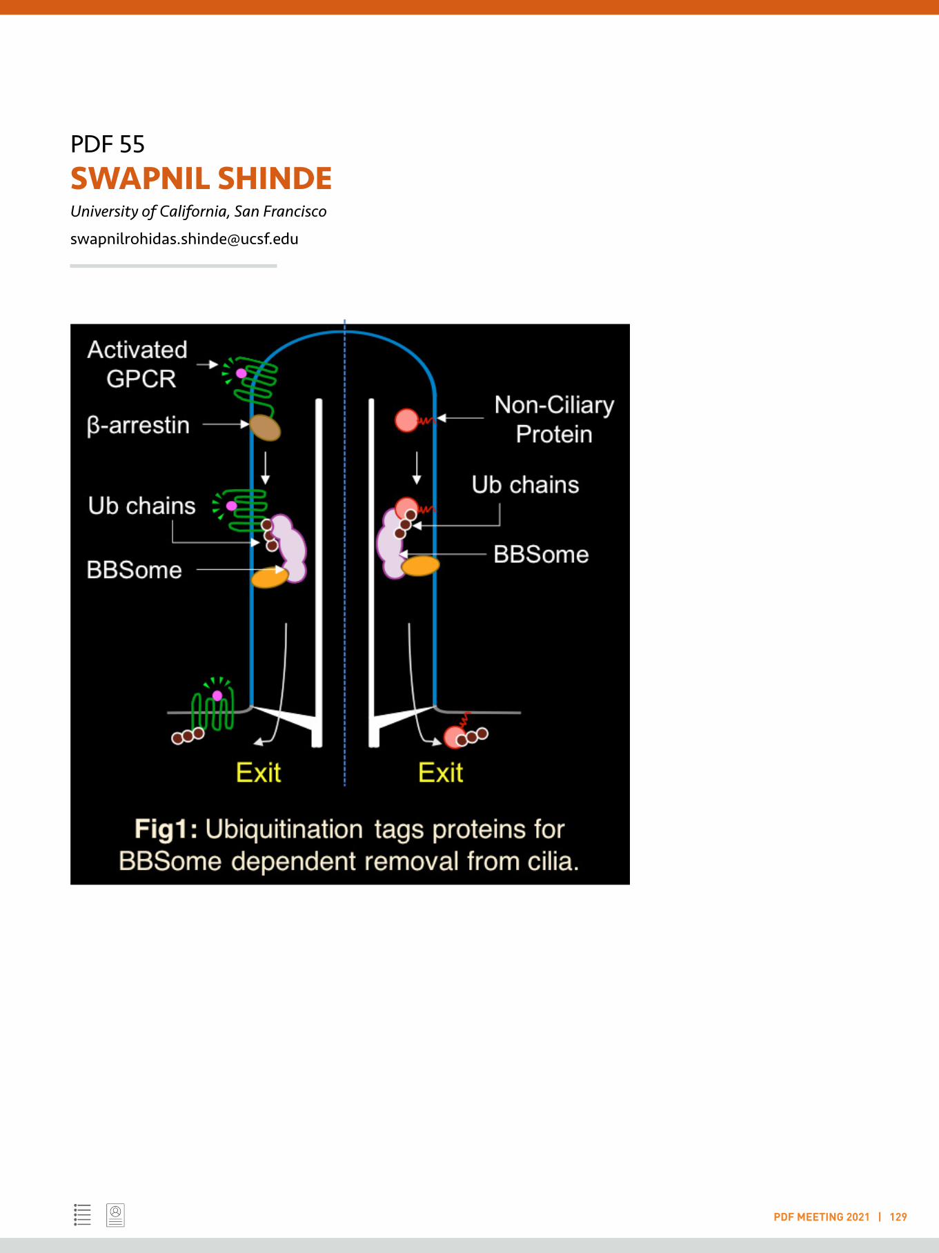
PDF 55SWAPNIL SHINDEUniversity of California, San Francisco
PDF MEETING 2021 | 129

PDF 56 TANUMOY MONDOL University of Freiburg
PDF MEETING 2021 | 130
LIGHTNING TALK
Keywords: Protein biochemistry and biophysics; DNA replication and transcription; Protein homeostasis and chaperone biology; Ensemble and single molecule fluorescence spectroscopy; Structural biology
Investigation of DNA Replication and Protein Homeostasis using Biochemical and Biophysical Approaches
My research interests are directed towards understanding the specific DNA-protein and protein-protein interactions involved in protein homeostasis and gene regulation (DNA replication and transcription). I am specifically interested in understanding the correlation between protein structure, dynamics and functions of essential eukaryotic proteins that control gene regulation (e.g. DNA polymerase) and protein homeostasis (e.g. Hsp90 chaperone).
The interdisciplinary research training during my PhD and postdoctoral tenures has broadened my research field from biochemistry, biophysics, to molecular and structural biology. My PhD research work was dedicated to the understanding of DNA sequence dependent allostery in the conformation and dynamics of transcription factor- lambda repressor, using biochemical and biophysical techniques. My research demonstrates that binding of transcription factors to specific DNA sequences alters the dynamical properties of the bound protein in a DNA-sequence dependent manner 1. Furthermore, during my research in Umea University, I have investigated the structure and function of Human copper chaperone Atox1 and Wilson’s disease protein using NMR spectroscopy. I have demonstrated that copper binding triggers a structural rearrangement in Wilson’s disease protein, triggering copper transport 2.
During my postdoctoral research in Prof. Peter Burgers’ laboratory at Washington University in St. Louis, I have extensively studied the role of DNA polymerase delta in DNA replication and DNA repair in Yeast. My research provided evidence that Proliferating Cell Nuclear Antigen (PCNA) accelerates the nucleotide incorporation rate of DNA polymerase delta. I have identified a growth-defective yeast PCNA mutant that is partially defective in accelerating the catalytic rate of Polymerase delta. This indicates that the face
of PCNA is important for the acceleration of catalysis. My observation led to the intriguing conclusion that PCNA affects and accelerates the conformational change of the ternary Polymerase-dNTP-DNA complex from the open to closed conformation and thus enhances catalytic rate. My research also provided evidence that DNA Polymerase delta forms higher ordered complexes upon binding to DNA as observed by electrophoretic mobility shift assays, fluorescence intensity changes and fluorescence anisotropy binding titrations 3.
Currently in ‘Hugel’ laboratory at the University of Freiburg, Germany, I am studying the role of Hsp90 chaperone machinery in protein homeostasis using single molecule fluorescence microscopy techniques. I am specifically exploring Hsp90-client-cochaperone multi-protein interactions, using single molecule FRET. Additionally, I am studying conformation and dynamics of Hsp90 and its complexes with cochaperone and client proteins.
In future, I aim to study the structure, dynamics, and function of major eukaryotic and prokaryotic DNA replicative enzymes in DNA replication. I would investigate how Hsp90 assists in DNA replication and repair apart from protein homeostasis. I will also study how the conformation and dynamics of Hsp90 regulates its interaction with the client proteins. I will employ biochemical, biophysical and structural biology methods to address these scientific questions.
References:
1 Mondol, T. et al, FEBS Lett 586(3):258-62, 20122 Mondol, T. et al, Biochem Biophys Res Commun 470(3):663-669,20163 Mondol, T. et al, Nucleic Acids Res 47(4):1977-1986, 2019

PDF 57 VEERENDRA KALYAN JAGANNADH Elvesys Microfluidic Innovation Center, Hyderabad
PDF MEETING 2021 | 131
LIGHTNING TALK
Keywords: Opto-fluidic imaging; Microfluidic imaging flow cytometry; Quantitative cell cytometry; Organ on chips; Microphysiological systems
Discretized Organ-on-Chip Modules for Drug Development
As part of my PhD at the Indian Institute of Science (IISc), I have developed novel optofluidic imaging technologies, with potential applications in point-of-care clinical diagnostics. From a detailed literature survey, it was identified that the diagnostic testing was one of the key bottlenecks for the current health-care paradigm of India. To tackle this bottleneck, the need for the development of an automated diagnostic instrument, capable of performing quantitative cellular morphometry was identified. A novel opto-fluidics based architecture was proposed for the instrument. The proposed architecture leveraged advances from different fields of optics, microfluidics and image processing to automate the complete workflow of conventional diagnostic microscopy. The developed system consists of a portable microscopy reader, designed to image cells flowing across microfluidic conduits. The design of the microfluidic chip ensured mixing of the micro-scale volumes of sample with reagents specific to a given diagnostic test/sample. Subsequently, cells flow through a portion, which is aligned with the optical axis of the portable microscopy reader.
Optical and microfluidic subsystems were integrated accordingly to implement the required functionality. The function of the optical subsystem is to enable imaging of cells (in flow), with sufficient resolution, even while employing off-the-shelf optoelectronic components. At first, this functionality was enabled by re-purposing ubiquitous electronic devices like cell phones. These portable prototypes were found to be capable of resolving features as small as 0.78 μm bars (spaced 0.78 μm apart). In flow conditions, these prototypes offered a maximum throughput of about 450 cells imaged per second. Further improvements were made to the prototype by employing a standalone image sensor. The improved stand-alone OFM acquired images with sufficient fidelity, so as to enable visualization features as small as 1 μm
parasites and also very clearly distinguish the cells present in different stages of malaria infection (Ring, Schizont, Trophozoite etc.,) [1]. Further work involved development of approaches which enable high-throughput imaging and 3D fluorescence imaging of cells in flow [2].
Following which, I joined Elvesys Innovation center as a Marie-Curie Individual Fellow to work on Novel architectures for Organ-on-Chip devices. Organ-on-chip systems recapitulate organ-level physiological function, without actually having to grow the entire tissue/organ. The devices consist of microfluidic chambers, which are designed to impart key physiological cues (chemical, biochemical and also physical) on the cells cultured within these devices. Among the required physiological cues, mechanical strain occurs most often and is required for emulation of cell/tissue microenvironments of several different body organs. The focus of the work is on development of a plug-in tool, which would facilitate mechanical stretching of cells.
References:
1. Veerendra Kalyan Jagannadh, Rashmi Sreeramachandra Murthy, Rajesh Srinivasan, Sai Siva Gorthi, 2016. Automated quantitative cytological analysis using portable microfluidic microscopy. Journal of Biophotonics, 9 (6): 586-595
2. Veerendra Kalyan Jagannadh, Mark D Mackenzie, Parama Pal, Ajoy K Kar, Sai Siva Gorthi, 2016, Slanted channel microfluidic chip for 3D fluorescence imaging of cells in flow. Optics Express, 24(19) - 22144-58

PDF 58 VINAY KUMAR Indian Institute of Technology Roorkee
PDF MEETING 2021 | 132
LIGHTNING TALK
Keywords: Environmental engineering; Environmental remediation; Bioprocess engineering; Analytical chemistry
1) PROBLEM STATEMENT:Synthetic plastics demand has increased very sharply in a few decades. The demand has led to overproduction and disposal to the environment. Synthetic plastics are known carcinogens causing adverse effects and death of marine animals. Therefore, alternative solutions are required. One of the solutions is the use of bioplastics. However, bioplastics such as polyhydroxyalkanoates (PHA) are much popular due to their biodegradability and green production procedures. But they are not competitive enough to substitute synthetic plastics. The major hurdles are the economic cost and the durability of the polymer. Therefore, research is underway to minimise the cost of carbon feedstocks using alternative cheaper substrates.
As we concern, raw palm oil wastes residues cannot be used directly as another by-product. They are produced as palm oil sludge, palm oil biomass and palm oil mill effluents. Therefore, pretreatment of the substrates is a significant concern. The next primary consideration will be optimising the production parameters in terms of the C/N ratio to maximise the PHA yield. Durability and strength of the produced PHA are other significant concerns that need to be addressed as well as the economic and environmental performances of the produced PHA that are rarely investigated for produced PHA products using a comprehensive and holistic environmental tool like LCA.
2) HYPOTHESIS:Palm oil wastes residues are produced in enormous amounts in industries. The vast waste generated from the industries has no major economic value and therefore remains underutilised. Being a higher source of various sugars and fatty acids, these residues can serve as a cheaper source for bacterial feedstock. This will minimise the waste management problem and will
produce useful bioplastic on the other hand. Therefore, the study is focused on the utilisation of palm oil wastes residues to produce economical and durable bioplastics in terms of PHAs.
3) RESEARCH QUESTIONS:There are three research questions to be addressed. First question will be pre-treatment of palm oil wastes to produce PHA. Second question will be to maximise the PHA yield utilising the available substrates. Third question will be determining the LCA of the produced PHA.
PDF 59 WASIM SAYYAD Yale University, New Haven
PDF MEETING 2021 | 134
LIGHTNING TALK
Keywords: Actin cytoskeleton; Super-resolution; Optical tweezers; Biophysics; Nanomaterials
Deciphering the molecular mechanism underlying the cell motility, polarity, endocytosis and cytokinesis using Biophysical techniques.
My research focuses on investigating the molecular mechanism underlying the cell motility, polarity, endocytosis and cytokinesis. I have a broad background in Physical and Biological sciences, starting from nanoparticle synthesis and characterization, micro/spectroscopy to cell biology, microbiology, biochemistry, and molecular genetics.
As a master’s student in the Physics Department of Pune University India, I learned to synthesize TiO2 nanoparticles, characterize them to study doping effects using different techniques and studied the size dependent bactericidal efficiency of TiO2 nanoparticles1. During the same time, I learned the culturing and biophysical characterization of Magnetotactic bacteria which produces magnetite nanoparticles. I studied the uniform magnetic field effect on the growth and size of the magnetite nanoparticles which led to my M. Phil thesis. A short term project at AU-KBC, Chennai, India, introduced me to the intriguing technique of optical tweezers. We reported a new method to characterize optical traps for metallic particles2.
The expertise in optical trapping directed me to my Ph.D. in Neuroscience in Prof. V. Torres lab at SISSA/ISAS in Italy. During my Ph.D., I learned to isolate, culture, and maintain dorsal root ganglion neurons. I studied the force generation mechanism in proliferating neurons using optical tweezers. We studied the important roles of myosin-II and Rac1 in making lamellipodia and filopodia, structures that sense the external environment by exerting pico-Newton forces3,4. The cytoskeleton structure and function in the neuronal growth cone directed my research in the field of cellular processes related to actin dynamics.
As a postdoc in Prof. Tom Pollard’s lab at Yale University I have used the fascinating super resolution imaging technique Fluorescence Photoactivation localization microscopy (FPALM) and developed expertise in Fluorescence Correlation spectroscopy (FCS), Fluorescence recovery after photobleaching (FRAP) as well as in Airyscan imaging. We used high speed FPALM to study endocytosis in S. pombe and obtained definitive evidence that actin filaments assemble in two zones at sites of clathrin-mediated endocytosis5. I used FCS to show that single Myosin 2 molecules with two heads and a long tail diffuse in the cytoplasm of interphase and mitotic S. pombe cells 6. Currently, I am finishing a manuscript on my discovery that cells make a number of nodes, the precursors of the contractile ring that divides cells, proportional to cell size. My next project is to measure the turnover of the proteins in nodes of live cells at high spatiotemporal resolution using FPALM.
References:
1. Panda, A. B. et al. Surf. Coatings Technol. 205, 1611–1617 (2010).2. Jadhav, V. S., Sayyad, W.A, Kulkarni, G. R. & Jaffar Ali, B. M. Complex Light Opt. Forces V 7950, 79500Y (2011).3. Sayyad, W. A.*, Amin, L. *, Fabris, P., Ercolini, E. & Torre, V. Sci. Rep. 5, 7842 (2015). (*Equal contribution)4. Sayyad, W. A., Fabris, P. & Torre, V. PLoS One 11, 1–22 (2016).5. Arasada, R., Sayyad, W. A., Berro, J. & Pollard, T. D. Mol. Biol. Cell 29, 295–303 (2018).6. Friend, J. E*, Sayyad W.A. * et al. Cytoskeleton 75, 164–173 (2018). (*Equal contribution)
PDF 60 WENDY D’SOUZA Indian Institute of Science, Bengaluru
PDF MEETING 2021 | 135
LIGHTNING TALK
Keywords: Molecular oncology; Cancer cell biology; Transcriptome; Translatome; Clinical oncology
NGS-based genomic profiling of circulating tumor DNA in advanced non-small cell lung cancer
Next-generation sequencing (NGS)-based circulating tumor DNA (ctDNA) assays have provided a new noninvasive monitoring method of identifying tumor-driving gene alterations in patients with advanced non-small cell lung carcinoma (NSCLC). The study of concordance of the mutation status on biopsy tissue and ctDNA may establish the reliability of ctDNA as a viable substitute for tumor biopsy. Current National Comprehensive Cancer Network® (NCCN®) Clinical Practice Guidelines in Oncology for lung cancer recommends molecular diagnostic workup for EGFR, ALK, ROS1, BRAF and PD-L1 testing and subsequent administration of targeted therapy as standard of care. Because the incidence of NSCLC is increasing not only in India, but also worldwide, compounded with high mortality, it is imperative to have accurate evaluation of the disease status in order to avoid complications and to expect longer survival. Through the study of 28 advanced NSCLC patients, we have identified several somatic missense, small indels, frameshifts and stop gain alterations in 22 tumor-related genes. In 92.85% (26/28) of the patients, we also identified genomic alterations that could be targeted by agents that are available from ongoing trials or are being used as off-label FDA approved drugs. Actionable genetic alterations included 10 MUC16 mutations, 8 TP53 mutations, 5 EGFR mutations, 5 MSH3 mutations, 5 DRD4 mutations, 3 VEGFC mutations, 2 mutations each in ARID1A, CDKN2A and ERBB2, 1 mutation each in STK11, ALK, ATM, FBXW7 and KRAS. Of the 28 patients with advanced NSCLC, 17.85% (5/28) were suitable for treatment with National Comprehensive Cancer Network (NCCN) guideline-approved targeted drugs. The EGFR alterations in these 5 patients with NSCLC were located in the following exons: exon 19 (E746_A750del, n=2), and exons 20 (T790M and S768_D770dup, n=3) and 21 (L858R, n=2). Additionally, the EGFR exon 20 point mutations and exon 20 insertions (T790M and S768_D770dup) was found in 2 of the patients with EGFR-TKI-
resistant NSCLC, suggesting that the NGS-based ctDNA assay might be an optional method to monitor EGFR-TKI resistance and to discover mechanisms of drug resistance (Chen et al., 2019; Demuth et al., 2018; Vollbrecht et al., 2018; Newman et al., 2014). Interestingly, another competing resistance mutation KRAS Q61K was identified in our cohort. Notably, mutations in KRAS are mutually exclusive with EGFR mutations and are associated with severe prognosis and resistance to chemotherapy or EGFR inhibitors (Oxnard et al., 2018; Yang et al., 2018; Eberhard et al., 2005). NGS-based ctDNA assays have an advantage of tailoring a personalised treatment plan and as marker for prognosis and clinical follow-up given the increasing number of therapeutic targets owing to wider exome coverage and robustness of blood based detection. The NGS-based ctDNA assay might be a comprehensive and efficient strategy that complements tissue analysis and expands the scope of personalised targeted therapies for advanced NSCLC.

All IndiaBioscience meetings, workshops and conferences are subject to a Code of Conduct. All participants at PDF Meeting 2021 are required to follow this code of conduct.
IndiaBioscience is dedicated to providing a harassment-free experience for everyone, regardless of gender, gender identity and expression, sexual orientation, disability, physical appearance, body size, race, or religion. We do not tolerate harassment of event participants in any form. All communication should be appropriate for a professional audience including people of many different backgrounds. We expect participants to follow these rules at all event venues (virtual or physical) and event-related social interactions.
Speakers and Presenters: Sexual language and imagery are not appropriate for the event venue, including talks and posters. Sexist, racist or exclusionary jokes are not acceptable.
Participants: Harassment includes offensive verbal comments related to gender, sexual orientation, disability, physical appearance, body size, race, religion, sexual images in public spaces, deliberate intimidation, stalking, following, harassing photography or recording, sustained disruption of talks or other events, inappropriate physical contact, and unwelcome sexual attention.
Sponsors: Sponsors are also subject to the anti-harassment policy.
Virtual events: Attendees, Speakers, Trainers and Event Organisers are required to keep all e-communications - emails, online presentations, webinar chat/ Q&A sessions etc - professional and respectful. As with physical events, sexual language/imagery and harassment are not appropriate and will not be tolerated at online seminars/workshops/courses and associated correspondence.
Action: Attendees asked to stop any harassing behaviour are expected to comply immediately. If an attendee engages in behaviour that violates this code of conduct, the conference organisers may take any action they deem appropriate, including warning the offender, expelling him or her from the virtual event.
Social Media: We will be sharing snippets as the meeting progresses via the IndiaBioscience Twitter handle.You are welcome to share snapshots and updates of the meeting, but kindly adhere to the guidelines in this Code of Conduct when doing so.
Contact: If you are being harassed, have noticed that someone else is being harassed, or have any other concerns, please contact: yim2021[at]indiabioscience[dot]org
License: This Conference Code of Conduct was modified from the Code of Conduct for JSFOO 2014 and Code of Conduct for YIM 2015, available under a Creative Commons Attribution 3.0 Unported License, and the Geek Feminism Wiki, created by the Ada Initiative and volunteers, available under a Creative Commons Zero license.
The Conference Code of Conduct for IndiaBioscience events is licensed under a Creative Commons Attribution 3.0 Unported License.
PDF MEETING 2021 | 138
Conference Code of Conduct

DESIGN: MANOJ RANGANCONTACT: +91 9902685031


