Hepatitis B virus–induced lipid alterations contribute to natural killer T cell–dependent...
23
HEPATITIS B VIRUS-INDUCED ALTERATIONS IN HEPATOCYTE CD1D LIPID ANTIGENS ACTIVATE NATURAL KILLER T CELLS AND CONTRIBUTE TO PROTECTIVE IMMUNITY Sebastian Zeissig 1,2 , Kazumoto Murata 3 , Lindsay Sweet 4 , Jean Publicover 5 , Zongyi Hu 3 , Arthur Kaser 1 , Esther Bosse 2 , Jahangir Iqbal 6 , M. Mahmood Hussain 6 , Katharina Balschun 7 , Christoph Röcken 7 , Alexander Arlt 2 , Rainer Günther 2 , Jochen Hampe 2 , Stefan Schreiber 2 , Jody L. Baron 5 , D. Branch Moody 4 , T. Jake Liang 3,8 , and Richard S. Blumberg 1,8 1 Division of Gastroenterology, Hepatology, and Endoscopy, Brigham and Women’s Hospital, Harvard Medical School, Boston, MA, USA 2 Department of Internal Medicine I, University Medical Center Schleswig-Holstein, Christian- Albrechts-University, Kiel, Germany 3 Liver Diseases Branch, NIDDK, National Institutes of Health, Bethesda, MD, USA 4 Division of Rheumatology, Immunology and Allergy, Brigham and Women's Hospital and Harvard Medical School, Boston, MA USA 5 Department of Medicine, Liver Center, University of California, San Francisco, CA, USA 6 Departments of Cell Biology and Pediatrics, SUNY Downstate Medical Center, Brooklyn, NY, USA 7 Department of Pathology, University Medical Center Schleswig-Holstein, Campus Kiel, Christian- Albrechts-University, Kiel, Germany Abstract In most adult patients, hepatitis B is a self-limiting disease leading to life-long protective immunity, which is the consequence of a robust adaptive immune response occurring weeks after HBV infection. Intriguingly, HBV-specific T cells can be detected shortly after infection but the mechanisms underlying this early immune priming and its consequences for subsequent control of viral replication are poorly understood. Using primary human and murine hepatocytes and mouse models of transgenic and adenoviral HBV expression, we show that HBV-expressing hepatocytes produce endoplasmic reticulum (ER)-associated endogenous antigenic lipids including lysophospholipids that are generated by HBV-induced secretory phospholipases and lead to activation of natural killer T (NKT) cells. The absence of NKT cells, CD1d or a defect in ER- associated transfer of lipids onto CD1d results in diminished HBV-specific T and B cell responses Correspondence and requests for materials should be addressed to [email protected] or [email protected]. 8 These authors share senior authorship. AUTHOR CONTRIBUTIONS S.Z. designed, performed, and analyzed experiments and prepared the manuscript with R.S.B. and T.J.L; K.M., Z.H. generated adenoviruses and adenoviral mutants and contributed to Ad-HBV studies; L.S. and D.B.M. designed, performed, and analyzed LC-MS experiments together with S.Z.; J.P. and J.L.B. designed, performed and analyzed studies with HBV-Env mice; A.K. generated H- Mttp −/− mice and contributed to their characterization; K.B. and C.R. performed histopathological analyses; M.M.H. obtained purified MTP; E.B. performed PLA 2 inhibitor and siRNA studies; R.G. obtained primary HBV isolates; A.A. and J.H. contributed to human hepatocyte studies; S.S. contributed to supervision of the studies; T.J.L. and R.S.B. supervised the studies. There are no competing financial interests. NIH Public Access Author Manuscript Nat Med. Author manuscript; available in PMC 2012 October 22. Published in final edited form as: Nat Med. 2012 July ; 18(7): 1060–1068. doi:10.1038/nm.2811. NIH-PA Author Manuscript NIH-PA Author Manuscript NIH-PA Author Manuscript
-
Upload
independent -
Category
Documents
-
view
3 -
download
0
Transcript of Hepatitis B virus–induced lipid alterations contribute to natural killer T cell–dependent...

HEPATITIS B VIRUS-INDUCED ALTERATIONS IN HEPATOCYTECD1D LIPID ANTIGENS ACTIVATE NATURAL KILLER T CELLSAND CONTRIBUTE TO PROTECTIVE IMMUNITY
Sebastian Zeissig1,2, Kazumoto Murata3, Lindsay Sweet4, Jean Publicover5, Zongyi Hu3,Arthur Kaser1, Esther Bosse2, Jahangir Iqbal6, M. Mahmood Hussain6, KatharinaBalschun7, Christoph Röcken7, Alexander Arlt2, Rainer Günther2, Jochen Hampe2, StefanSchreiber2, Jody L. Baron5, D. Branch Moody4, T. Jake Liang3,8, and Richard S.Blumberg1,8
1Division of Gastroenterology, Hepatology, and Endoscopy, Brigham and Women’s Hospital,Harvard Medical School, Boston, MA, USA2Department of Internal Medicine I, University Medical Center Schleswig-Holstein, Christian-Albrechts-University, Kiel, Germany3Liver Diseases Branch, NIDDK, National Institutes of Health, Bethesda, MD, USA4Division of Rheumatology, Immunology and Allergy, Brigham and Women's Hospital andHarvard Medical School, Boston, MA USA5Department of Medicine, Liver Center, University of California, San Francisco, CA, USA6Departments of Cell Biology and Pediatrics, SUNY Downstate Medical Center, Brooklyn, NY,USA7Department of Pathology, University Medical Center Schleswig-Holstein, Campus Kiel, Christian-Albrechts-University, Kiel, Germany
AbstractIn most adult patients, hepatitis B is a self-limiting disease leading to life-long protectiveimmunity, which is the consequence of a robust adaptive immune response occurring weeks afterHBV infection. Intriguingly, HBV-specific T cells can be detected shortly after infection but themechanisms underlying this early immune priming and its consequences for subsequent control ofviral replication are poorly understood. Using primary human and murine hepatocytes and mousemodels of transgenic and adenoviral HBV expression, we show that HBV-expressing hepatocytesproduce endoplasmic reticulum (ER)-associated endogenous antigenic lipids includinglysophospholipids that are generated by HBV-induced secretory phospholipases and lead toactivation of natural killer T (NKT) cells. The absence of NKT cells, CD1d or a defect in ER-associated transfer of lipids onto CD1d results in diminished HBV-specific T and B cell responses
Correspondence and requests for materials should be addressed to [email protected] or [email protected] authors share senior authorship.
AUTHOR CONTRIBUTIONSS.Z. designed, performed, and analyzed experiments and prepared the manuscript with R.S.B. and T.J.L; K.M., Z.H. generatedadenoviruses and adenoviral mutants and contributed to Ad-HBV studies; L.S. and D.B.M. designed, performed, and analyzed LC-MSexperiments together with S.Z.; J.P. and J.L.B. designed, performed and analyzed studies with HBV-Env mice; A.K. generated H-Mttp−/− mice and contributed to their characterization; K.B. and C.R. performed histopathological analyses; M.M.H. obtainedpurified MTP; E.B. performed PLA2 inhibitor and siRNA studies; R.G. obtained primary HBV isolates; A.A. and J.H. contributed tohuman hepatocyte studies; S.S. contributed to supervision of the studies; T.J.L. and R.S.B. supervised the studies.
There are no competing financial interests.
NIH Public AccessAuthor ManuscriptNat Med. Author manuscript; available in PMC 2012 October 22.
Published in final edited form as:Nat Med. 2012 July ; 18(7): 1060–1068. doi:10.1038/nm.2811.
NIH
-PA Author Manuscript
NIH
-PA Author Manuscript
NIH
-PA Author Manuscript

and delayed viral control. NKT cells may therefore contribute to control of HBV infection throughsensing of HBV-induced modified self-lipids.
Conventional T and B cells play a crucial role in HBV infection.1–5 In contrast, thecontribution of cells at the interface between innate and adaptive immunity such as NKTcells remains controversial.6 NKT cells respond in a T cell receptor (TCR)-restricted mannerto lipid antigens presented by CD1d and exhibit pronounced cytokine secretion within hoursof cognate antigen recognition, which enables broad effects on activation of other innate(NK) and adaptive immune cells (T and B cells).7,8
The role that NKT cells play in HBV infection is unclear. Analysis of liver gene expressionin chimpanzees two weeks after HBV infection has shown evidence for a lack of inductionof immune-related genes suggesting that HBV acts a stealth virus that escapes innateimmune responses during early infection.9 On the other hand, studies in HBV-infectedhumans and chimpanzees have demonstrated the presence of HBV-specific T- and B cellswithin weeks of infection consistent with successful priming of an adaptive immuneresponse.1,10,11 These observations suggest that HBV might be susceptible to recognition bythe immune system directly following infection. Accordingly, recent studies in animalmodels of Hepadnaviridae infection and HBV patients have demonstrated activation of NKTcells at very early time points following infection. Thus, infection with woodchuck hepatitisvirus led to hepatic NKT cell infiltration within 48 hours, which correlated with IFN-γsecretion and temporary suppression of viral replication.12 These findings are in accordancewith the fact that pharmacological stimulation of invariant (i) NKT cells by administrationof the iNKT cell antigen α-galactosylceramide (αGalCer) led to rapid IFN-γ-dependentinhibition of viral replication in HBV transgenic mice.13 Similarly, a study of humansduring the incubation phase of HBV infection demonstrated increased levels of peripheralNK cells in accordance with innate immune activation early after HBV infection.11 Mostnotably, a recent report on two HBV patients demonstrated very early activation ofperipheral natural T cells, a population of cells that phenotypically resembles classical NKTcells. Natural T cell activation preceded activation of NK and conventional T cells and wasassociated with subsequent control of HBV infection.10 These studies demonstrate acorrelation between viral control and NKT cell activation. To investigate whether NKT cellsare an important checkpoint that contributes to control of HBV infection, we studied variousin vitro and in vivo HBV models.
RESULTSEarly activation of NKT cells in response to Ad-HBV
To study NKT cell responses we investigated a mouse model that overcomes non-permissiveness of murine hepatocytes to HBV through adenoviral delivery of a replication-competent HBV genome under the control of its endogenous promoters.14–16 Injection of1×109 HBV-expressing adenoviral particles (Ad-HBV), a dose shown by us (Suppl. Fig. 1a)and others16 to induce an immune response against HBV but not the adenoviral carrier, ledto HBV replication (Suppl. Fig. 1b) followed by a rapid drop in hepatic HBV DNA andserum HBsAg that preceded hepatitis (Suppl. Fig. 1b–c). A control adenovirus expressing β-galactosidase (Ad-LacZ) did not lead to hepatitis, confirming HBV-dependent inflammation(Suppl. Fig. 1a). 14–16 Since Ad-HBV infection in mice resembles the course of naturalHBV infection in humans in several aspects17, we further studied NKT cell responses in thismodel.
Interestingly, the entire population of liver but not splenic iNKT cells exhibited activationand IFN-γ secretion as early as one day after Ad-HBV but not Ad-LacZ administration and
Zeissig et al. Page 2
Nat Med. Author manuscript; available in PMC 2012 October 22.
NIH
-PA Author Manuscript
NIH
-PA Author Manuscript
NIH
-PA Author Manuscript

thus before histological inflammation and serum ALT elevation (Fig. 1a–c, Suppl. Fig. 2).Similar to iNKT cells, hepatic but not splenic non-invariant NKT cells, an NKT cellpopulation expressing a more diverse set of TCRs that were detected by a novel reportermodel (see Suppl. Results and Suppl. Fig. 3), displayed pronounced activation and IFN-γsecretion in response to Ad-HBV (Fig. 1d–f, Suppl. Fig. 4). Thus, invariant and non-invariant NKT cells exhibited broad and rapid activation upon Ad-HBV exposure anddirectly contributed to secretion of IFN-γ. In contrast, activation of NK and T cells was notobserved until three days after Ad-HBV challenge (Figure 1g–i and data not shown) andthus followed activation of NKT cells, similar to observations in HBV patients.10
NKT activation contributes to innate and adaptive immune responses against HBVTo investigate whether NKT cells contribute to innate and adaptive immune responsesagainst HBV, we analyzed activation of liver mononuclear cells (LMNCs) in response toAd-HBV in mice lacking invariant (Jα18−/−) or all NKT cells (CD1d−/−). In wild-type mice,hepatic NK, CD4+, and CD8+ T cells exhibited strong activation and IFN-γ secretion inresponse to Ad-HBV, but not Ad-LacZ. In contrast, CD1d−/− mice exhibited significantlydiminished NK, CD4+, and CD8+ T cell activation, whereas Jα18-deficiency predominantlyaffected CD4+ T cell activation (Fig. 1g–i). Viral transduction of hepatocytes was similar inall mouse strains (Suppl. Fig. 5).
NKT-dependent activation was also observed for HBV-specific CD8+ T cell responsesagainst envelope- (S190–197) and core-antigens (C93–100) but not in response tophytohemagglutinin (Fig. 2a, Suppl. Fig. 6a–b). Analysis of LMNC responses to pools ofpeptides spanning the entire HBV envelope18 revealed pronounced defects in the magnitudebut not diversity of HBV-specific CD8+ T cell responses in HBV envelope transgenic (Tg) ×Rag1−/− × CD1d−/− (HBVEnv Rag1−/− CD1d−/−) mice that received, by adoptive transfer,wild-type splenocytes (Fig. 2b, Suppl. Fig. 6c). Adaptive immune defects extended to Bcells since levels of anti-HBsAg antibodies were lower in Ad-HBV-challenged CD1d−/− andJα18−/− mice (Fig. 2c) and in HBVEnv Rag1−/− CD1d−/− mice (Fig. 2d), resulting in lack ofsustained HBsAg clearance from serum (Fig. 2e). These data show that NKT cells contributeto activation of NK, T and B cells in different HBV mouse models.
NKT cells contribute to early antiviral action in response to Ad-HBVIn accordance with a central role of NKT cells in HBV-induced hepatitis19,20, CD1d−/− andJα18−/− mice exhibited reduced peak levels of serum ALT (Fig. 2f), liver IFN-α (Fig. 2g),and hepatic immune cell infiltration (data not shown). However, CD1d−/− and Jα18−/− micedeveloped chronic low-grade inflammation that persisted at least to day 30 (Fig. 2f, h), theend of the observation period. This was associated with delayed clearance of serum HBsAg(Fig. 2i) and serum and liver HBV DNA (Fig. 2j, Suppl. Fig. 7d–e), consistent with defectsin viral control leading to chronic inflammation. In accordance with a critical role of CD8+
T cells in the antiviral response against HBV1, NKT cells were necessary but not sufficientfor viral control since Tap1−/− mice lacking CD8+ T cells also exhibited severe defects inHBV clearance (Suppl. Fig. 7). These results show that NKT cells contribute to the initiationof antiviral immune responses against HBV and to viral control and prevention of chronicviral replication in the Ad-HBV model.
HBV- and Ad-HBV-infected human and murine hepatocytes activate NKT cellsTo identify the cell type responsible for NKT cell activation upon Ad-HBV challenge, westudied activation of NKT cell hybridomas in response to liver and spleen mononuclear cellsand primary hepatocytes obtained from Ad-HBV-infected mice. Only Ad-HBVtransducedhepatocytes but not T cell-depleted mononuclear cells induced activation of NKT cells (Fig.3a, Suppl. Fig. 8a and data not shown). Similar observations were made upon in vitro
Zeissig et al. Page 3
Nat Med. Author manuscript; available in PMC 2012 October 22.
NIH
-PA Author Manuscript
NIH
-PA Author Manuscript
NIH
-PA Author Manuscript

exposure of primary hepatocytes to Ad-HBV (Suppl. Fig. 8b; for purity and transduction ofprimary hepatocytes see Suppl. Fig. 9). NKT activation was CD1drestricted (Fig. 3b) andlimited to NKT cells, as demonstrated by unaffected MHC class I presentation (Fig. 3c).Activation was specific for a subset of invariant (24.7 cell line) and non-invariant (14S.6 cellline) murine NKT cell hybridomas (Fig. 3a) and could also be demonstrated with humanprimary hepatocytes infected with Ad-HBV or a primary (non-adenoviral) HBV isolate (Fig.3d–e). These data reveal an unanticipated role of hepatocytes in HBV-dependent NKT cellactivation.
HBV-induced NKT cell activation is dependent on hepatocyte microsomal triglyceridetransfer protein (MTP)
MTP is an ER-resident protein that transfers endogenous phospholipids onto CD1d and iscritical for CD1d function.21–25 To delineate the role of hepatocyte MTP and CD1d in Ad-HBV infection, we generated mice with hepatocyte-specific deletion of Mttp encoding forMTP (H-Mttp−/− 26, Suppl. Fig. 10a). MTP-deficiency led to severe defects in thepresentation of endogenous and exogenous lipid antigens that were specific for hepatocytesand limited to CD1d (Suppl. Fig. 10b–e).
Consistent with a critical role of MTP in HBV-induced and CD1d-restricted antigenpresentation, primary hepatocytes from H-Mttp−/− mice exhibited impaired activation ofNKT cells in response to Ad-HBV in vitro (Fig. 3f–g, Suppl. Fig. 10). Similar observationswere made upon chemical inhibition of MTP (Suppl. Fig. 11) and using human hepatocytesinfected with a primary HBV isolate (Fig. 3e). These results were confirmed by in vivoexperiments, in which NKT cells from Ad-HBV-challenged H-Mttp−/− mice exhibitedimpaired activation and IFN-γ secretion (Fig. 1a–c). Accordingly, NKT cell-dependentactivation of NK cells and HBV-specific T and B cells was impaired in HMttp−/− mice (Fig.1g–i, Fig. 2a, c, Suppl. Fig. 6a). H-Mttp−/− mice exhibited diminished acute hepatitis uponAd-HBV challenge, impaired viral control and chronic low-grade inflammation (Fig. 2,Supp. Fig. 7). These data confirm that hepatocytes are the antigen presenting cellsresponsible for NKT cell activation and show that this effect is dependent upon hepatocyteexpression of MTP and CD1d.
Ad-HBV expression is associated with alterations in endogenous hepatocyte lipidsAd-HBV-induced NKT cell activation was not the consequence of altered CD1d expressionor trafficking (Suppl. Fig. 12a, b). Accordingly, iNKT cell activation by hepatocytes loadedwith the exogenous model lipid was unaltered by Ad-HBV (Suppl. Fig. 12c).
Since HBV buds from the ER and selectively recruits ER lipids for its envelope27, weinvestigated whether HBV-induced NKT activation is the consequence of alterations inendogenous ER-acquired CD1d lipids and studied activation of NKT hybridomas inresponse to plate-bound CD1d loaded with lipids from Ad-HBV-infected primary murinehepatocytes. Even uninfected primary hepatocytes contained antigenic CD1d lipids thatwere enriched in microsomal ER preparations (Suppl. Fig. 13a–e). Importantly, microsomesfrom Ad-HBV- but not Ad-LacZ-transduced hepatocytes exhibited significantly increasedactivation of invariant and non-invariant NKT cell hybridomas (Fig. 4a–b). Similaralterations in microsomal lipids were found in CD1d−/− and H-Mttp−/− hepatocytes (Suppl.Fig. 13f and data not shown), demonstrating that these antigenic lipids were unable to beloaded (H-Mttp−/−) or presented by CD1d (CD1d−/−). Accordingly, purified MTP enhancedpresentation of immunogenic Ad-HBV-dependent lipids (Fig. 4c).
Consistently, a significant population of sorted primary non-invariant liver NKT cellsrecognized Ad-HBV-induced alterations in microsomal lipids as determined by upregulation
Zeissig et al. Page 4
Nat Med. Author manuscript; available in PMC 2012 October 22.
NIH
-PA Author Manuscript
NIH
-PA Author Manuscript
NIH
-PA Author Manuscript

of CD69 expression (Fig. 4e). CD69 upregulation was less pronounced for sorted iNKT cellssuggesting that iNKT cell responses to Ad-HBV are either restricted to a subpopulation ofthese cells or that iNKT cells are broadly but less strongly activated by Ad-HBV (Fig. 4d).In conclusion, exposure to Ad-HBV is associated with increased antigenicity of ER lipids,which are recognized by NKT cells.
Ad-HBV leads to increased abundance of antigenic lysophosphatidylethanolamineTo characterize Ad-HBV-dependent lipid alterations, microsomal lipids were separatedaccording to solubility.28 Ad-HBV-induced stimulatory fractions for non-invariant andinvariant NKT cells were both recovered in phospholipid-enriched methanol eluents, butsubfractionation indicated that the two NKT cell subtypes were activated by different lipids(Fig. 4f–g). To characterize the particular lipids induced by Ad-HBV and stimulatory forNKT cells, we screened methanol fractions by high performance liquid chromatographymass-spectrometry (HPLC-MS). Previous reports demonstrated selective recruitment of ERlipids by HBV and underrepresentation of PE species in the HBV envelope.27 Comparisonof chromatograms corresponding to the mass and collisional spectra of diacylphosphatidylethanolamine (PE) showed strong augmentation of PE andlysophosphatidylethanolamine (lysoPE) after Ad-HBV exposure (Suppl. Fig. 14).Lysophospholipids have recently been shown to be antigenic endogenous CD1d ligands inhumans in vitro.29,30 Therefore, we rescreened stimulatory fractions at massescorresponding to monoacyl PE species with C16, C18:2, C18:1 and C18 monacyl forms, andfound in all cases that these lipids were upregulated after Ad-HBV administration in vivo(Fig. 5a). There is thus an overlap among the types of lipids upregulated in response to Ad-HBV and lysophosopholipids that can stimulate NKT cells.
To further test initial conclusions derived from complex cellular lipid mixtures generated invivo, we tested activation of murine NKT hybridomas in response to various purifiedphospholipids in vitro. 14S.6, a non-invariant NKT cell hybridoma that responded to Ad-HBV (Fig. 3a), also responded to 16:0/0 and 18:0/0 lysoPE and lysoPC (Fig. 5b). SincelysoPE but not lysoPC was increased in response to Ad-HBV (Suppl. Fig. 14), only lysoPEis implicated in the response to Ad-HBV. NKT cell activation by lysoPE was, amonghybridomas, specific for 14S.6 and not observed with other iNKT (24.7, 24.8, 24.9,DN32.D3) or non-invariant NKT hybridomas (14S.7, 14S.10, 14S.15; Fig. 5b and data notshown). Lyso-PE-induced NKT activation required plate-bound CD1d (data not shown) andpurified MTP facilitated lysoPE presentation (Fig. 5c). These results extended to primaryNKT cells since more than fifty percent of sorted non-invariant 4Get X Jα18−/− liver NKTcells exhibited upregulation of CD69 in response to lysoPE but not PE (Fig. 5d). Non-invariant NKT cell activation was not limited to cells expressing Vβ14, the TCRβ chainshared by the non-invariant NKT hybridoma (14S.6) that recognized lysoPE (Fig. 5d)suggesting that lysoPE broadly activates non-invariant NKT cells in the liver. In contrast,iNKT cells did not exhibit activation in response to lysoPE (Fig. 5d).
HBV-induced activation of non-invariant NKT cells is dependent on secretoryphospholipases (sPLA2)
sPLA2 enzymes contribute to the generation of lysophospholipids recognized by NKT cellsand are active within secretory compartments.30,31 We therefore investigated the role ofsPLA2 in Ad-HBV-dependent NKT cell activation. Chemical inhibitors of sPLA2 but notcytoplasmic PLA2 (cPLA2) prevented HBV-dependent activation of non-invariant NKTcells without affecting MHC class I presentation (Figure 5e, Suppl. Fig 15a–g). sPLA2inhibitors also did not affect iNKT cells in accordance with the lack of lysoPE-dependentiNKT activation (Fig. 5f).
Zeissig et al. Page 5
Nat Med. Author manuscript; available in PMC 2012 October 22.
NIH
-PA Author Manuscript
NIH
-PA Author Manuscript
NIH
-PA Author Manuscript

Since Ad-HBV-transduced hepatocytes exhibited increased abundance of PE (Suppl. Fig.14b), the substrate for PLA2-mediated production of lysoPE, we investigated hepatocyteexpression of sPLA2, cPLA2 (Pla2g4a), and calcium-independent PLA2 (iPLA2, Pla2g6a).Ad-HBV led to rapid and selective upregulation of sPLA2 Pla2g2c and Pla2g5 but notcPLA2, iPLA2, and other sPLA2 genes in vivo (Fig. 5g, Suppl. Fig. 16a). These results are inaccordance with increased hepatic PLA2 group II and V expression in patients with viralhepatitis.32,33 Primary murine hepatocytes also exhibited Ad-HBV-induced Pla2g2cupregulation confirming hepatocyte-specific effects and demonstrating that sPLA2regulation is not secondary to hepatic inflammation but a direct consequence of exposure toAd-HBV (Suppl. Fig. 16b). Primary human hepatocytes exposed to Ad-HBV or a primaryHBV isolate also exhibited upregulation of sPLA2, specifically of PLA2G2A (Fig. 5h).Since lysoPE activates transcription of PLA2G2A34, it likely supports its production througha positive feedback loop involving sPLA2. Thus, HBV leads to upregulation of sPLA2 groupII in murine and human hepatocytes. Specific subgroups of sPLA2 differ between humansand mice in accordance with differences in genomic organization of sPLA2 loci (PLA2G2Cis a pseudogene in humans35, Pla2g2a is inactivated by a frameshift in C57BL/6J mice).
Consistent with a critical role of sPLA2 in HBV-dependent generation of lysophospholipidsas major antigens recognized by non-invariant NKT cells, small interfering RNA (siRNA)directed against Pla2g2c and Pla2g5 (Suppl. Fig. 17a) but not other sPLA2, cPLA2, andiPLA2 enzymes, prevented Ad-HBV-induced activation of non-invariant NKT but notinvariant NKT cells or MHC class I-restricted T cells (Fig. 5i–j, Suppl. Fig. 17b, and datanot shown).
The small HBsAg contributes to the activation of non-invariant NKT cellsTo investigate the requisite HBV element for NKT cell activation, we inactivated individualHBV open reading frames and studied the effects on NKT activation. While all viral mutantsshowed similar transduction (Suppl. Fig. 5, 9), only deletion of the small HBsAg (S-), whichis required for viral secretion, led to impaired activation of non-invariant NKT cells (Fig.6a). A similar but non-significant trend was observed for iNKT cells (Fig. 6b). Consistently,Ad-HBV S- led to impaired hepatocyte sPLA2 upregulation and activation of non-invariantNKT cells by microsomal hepatocyte lipids (Fig. 6c, Suppl. Fig. 16c). To extend thesefindings, we examined mice with transgenic HBsAg expression.19 HBsAg-transgenichepatocytes led to CD1d-dependent activation of noninvariant and invariant NKT but notMHC class I-restricted T cell hybridomas (Fig. 6d–f). In addition, microsomal lipids ofHBsAg-transgenic hepatocytes activated NKT cells (Fig. 6g–h). Consistently, in vivoadministration of Ad-HBV S- resulted in prolonged chronic necroinflammation and delayedviral control in association with sustained viral replication and reduced hepatic IFN-γ levels(Suppl. Fig. 18). These studies show that the small HBsAg contributes to Ad-HBV-mediatedactivation of NKT cells, which is in accordance with a temporal association between HBsAgexpression and early NKT cell activation in human HBV.10
Ad-HBV-induced activation iNKT cells is cytokine-dependent in vivoPrimary liver iNKT cells were not activated by lysoPE (Fig. 5d) and exhibited less robustactivation to Ad-HBV microsomal lipids (Fig. 4d). We therefore investigated whethercytokine-mediated indirect activation contributes to Ad-HBV-induced iNKT cell activationin vivo. Indeed, neutralization of IL-12, a cytokine critical for indirect NKT cellactivation36–38, largely prevented CD69 upregulation and IFN-γ secretion by iNKT cells buthad negligible effects on non-invariant NKT cells (Fig. 6i–j, Suppl. Fig. 19). These resultssuggest that non-invariant NKT cells are directly activated upon Ad-HBV exposure byCD1d-restricted presentation of lysoPE and other uncharacterized hepatocyte antigens. Thisin turn leads to cytokine-dependent activation iNKT cells, presumably through activation of
Zeissig et al. Page 6
Nat Med. Author manuscript; available in PMC 2012 October 22.
NIH
-PA Author Manuscript
NIH
-PA Author Manuscript
NIH
-PA Author Manuscript

liver dendritic cells and macrophages.36,37 Importantly, IL-12-mediated indirect activationof liver iNKT cells requires hepatocyte MTP (Fig. 1a–c), suggesting that cytokine-dependent iNKT activation is downstream of CD1d-restricted activation of non-invariantNKT cells and NKT-dependent DC maturation.39,40
DISCUSSIONThe findings presented here show that immune responses provided by NKT cells not onlycontribute to HBV-induced hepatitis19 but also to viral control. Although HBV-inducedNKT cell activation is associated with acute hepatitis, NKT cells contribute significantly tothe emergence of anti-viral B and T cell immunity. In the absence of this NKT cell response,not only is adaptive immunity and viral control diminished but chronic, low-grade hepatitisensues. We show that the cell type responsible for orchestrating these HBV-inducedresponses is unanticipated and importantly the infected hepatocyte itself. Within thehepatocyte, HBV induces ER lipids including lysophospholipids derived from PE throughthe action of HBV-induced secretory phospholipases. CD1d-restricted presentation oflysophospholipids leads to direct activation of non-invariant NKT cells and subsequentIL-12-mediated indirect iNKT cell activation, confirming that both direct and indirectmechanisms contribute to NKT cell activation in vivo.36,37
The generation of antigenic lipids is partially dependent on the presence of the HBV surfaceantigen, the principal HBV structural component responsible for assembling PC-richparticles in the ER, and possibly results from an imbalanced lipid milieu within the ER.27 Assuch, the hepatocyte utilizes secretory phospholipases, MTP and CD1d to sense the presenceof HBV and alert NKT cells through the display of modified self-lipids on the cell surface ofthe hepatocyte to trigger an adaptive immune response. Our findings of an NKT cellresponse soon after HBV exposure are in accordance with other recent observations inhumans and animal models of HBV10–12 and suggest that NKT cells are part of an early,important sensing system that activates the immune response leading to effective priming ofHBV-specific adaptive immune cells that are required for viral clearance. Our data suggestthat although HBV acts as a stealth virus during a prolonged phase preceding viral control9,it is susceptible to a distinct type of immune recognition directly following infection whichis important for subsequent immune clearance. Further studies in HBV patients are requiredto confirm a central role of NKT cells in human HBV infection.
METHODSMice
CD1d−/−, Jα18−/−, and Tap1−/− mice have been described before.42–44 H-Mttp−/− miceexhibiting an hepatocyte-specific knockout of the gene (Mttp) encoding for microsomaltriglyceride transfer protein (MTP) were generated by crossing Alb-Cre (B6.Cg-Tg(Alb-cre)21Mgn/J) mice45 that express Cre recombinase under control of the hepatocyte-specificalbumin promoter with Mttpfl/fl mice that contain loxP sites flanking Mttp exon 1.46 Togenerate mice that allow for specific detection of invariant and noninvariant NKT cells,4Get mice expressing GFP via an internal ribosome entry site (IRES) in the IL-4 transcript(IL-4/GFP-enhanced transcript (4Get) mice)47,48 were crossed with CD1d−/− and Jα18−/−
mice (see Suppl. Results for further information). All mice were maintained on C57BL/6Jbackground and were backcrossed for at least eight generations. HBV-Env mice expressingthe entire HBV envelope (subtype ayw) under control of the albumin promoter49 werecrossed with Rag1−/− and CD1d−/− mice as described previously.19
Zeissig et al. Page 7
Nat Med. Author manuscript; available in PMC 2012 October 22.
NIH
-PA Author Manuscript
NIH
-PA Author Manuscript
NIH
-PA Author Manuscript

Primary cells and cell linesPrimary human hepatocytes were obtained from Invitrogen (Carlsbad, CA). Primary murinehepatocytes were extracted as described before.50 Purity and transduction rates of primaryhepatocytes are shown in Suppl. Fig. 9. For a description of all cell lines please refer to thesupplementary material.
Lipids and chemical inhibitorsPlease refer to the supplementary material.
Viral constructs and virus administrationThe parental plasmid for HBV constructs was pGEM7-HBV1.3 containing a 1.3-fold-overlength genome of HBV, subtype ayw, with a 5′ terminal redundancy encompassingenhancers I and II, the origin of replication (direct repeats DR1 and DR2), the X- andpregenomic/core promoter regions, the transcription initiation site of the pregenomic RNA,the unique polyadenylation site, and the entire X open reading frame. To generate HBVmutants, point mutations were introduced using the QuickChange II site-directedmutagenesis kit and protocol (Agilent Technologies, Santa Clara, CA) at the 19th codon ofpreS1 gene from TTG to TAG (L-) or at the initiation codon of pre-S2 and S from ATG toGTG (M- and S-). The PreS1 myristylation-defective (myr-) mutation was generated bychanging the second codon of the preS1 gene from GGG (glycine) to GCG (alanine).
Please refer to supplementary material for additional information on virus generation andapplication. Viral transduction rates in vivo and in vitro are shown in Suppl. Fig. 5 and 9.
Determination of ALT, HBsAg, HBV DNA levels and liver histologyPlease refer to the supplementary material.
Flow cytometryFlow cytometry was performed as described before.25 For a detailed description please referto the supplementary material.
Preparation and separation of cellular and microsomal lipidsFor extraction of microsomal lipids, primary hepatocytes were obtained as described above,microsomes were extracted according to Ernster et al.51 and lipids were extracted followingthe Folch protocol.52 Total hepatocyte lipids were extracted in a similar manner. Separationof lipids according to solubility was done as described in 28. For a detailed descriptionplease refer to the supplementary material.
Liquid chromatography-mass spectrometryLipid extracts were dried down, weighed and then re-suspended in 95% 60:40hexanes:isopropanol and 5% methanol (V: V) prior to mass spectrometry analyses. Twentyµg of lipid from each fraction was injected onto a 150 mm × 2.0 mm monochrom 3 diolcolumn (Varian), and eluted with a gradient program in which solvent A consisted ofmethanol (0.1% formic acid and 0.05% ammonium hydroxide, M:V) and solvent Bconsisted of 60% hexanes and 40% isopropanol (V: V) (0.1% formic acid and 0.05%ammonium hydroxide M:V). The gradient was run from 95% to 85% solvent B from 0 to6.6 minutes, to 0% solvent B until 16.2 minutes, then increased back to 95% solvent B from22.8 to 26 minutes. The HPLC system was an Agilent 1200 series with a 6520 quadrupoleaccurate mass time of flight (Q-TOF) mass spectrometer (Agilent Technologies, SantaClara, CA), with the voltage 3.5 kV, source temperature 325 °C, drying gas 5L/min,
Zeissig et al. Page 8
Nat Med. Author manuscript; available in PMC 2012 October 22.
NIH
-PA Author Manuscript
NIH
-PA Author Manuscript
NIH
-PA Author Manuscript

nebulizer pressure 30 psi, running in the negative ion mode. Collision experiments werecarried out by subjecting target ions to 25eV in the collision cell. Data analysis wasperformed using Agilent Qualitative Analysis Mass Hunter Software Version B.03.01.
Antigen presentationAntigen presentation assays were performed in 96 well flat bottom plates using 2×10⁴
hepatocytes, 5×10⁴ RMA-S/d cells, 1×10⁵ splenocytes or hepatic mononuclear cells and, asresponders, 1×10⁵ NKT cells or NKT cell hybridoma cells. Cytokine secretion wasdetermined by ELISA after 16 hours of coculture (BD Biosciences). In some experiments,MTP inhibitors were added at a final concentration of 10 µM (BMS212122, BMS200150)and 100 nM (BMS197636), respectively. Alternatively, purified monoclonal CD1d blockingantibodies (1B1 clone, 19G11 clone) were used at a final concentration of 10 µg/ml.
For cell-free antigen presentation assays, monomeric mouse CD1d (NIH Tetramer CoreFacility) was loaded onto 96 well flat bottom plates (0.25 µg/well), unbound CD1d waswashed off, and lipids were added at a molar ratio of 40 :1. Unbound lipids were thenwashed off, NKT cells were added and cytokine secretion was determined by ELISA asdescribed above. In some experiments, MTP purified from bovine liver (M.M.H.) was addedat a final concentration of 500 ng/ml. In addition, in some experiments, GFP+ αGalCer/CD1d-tetramer+ 4Get invariant NKT cells and GFP+ 4Get X Jα18−/− noninvariant NKTcells were sorted from liver mononuclear cells using a BD Biosciences FACSAria II cellsorter and were used as responders in antigen presentation assays. Flow cytometricdetermination of cell surface CD69 and intracellular IFN-γ expression on GFP+CD3+ NKTcells was studied as readout. For in vitro neutralization of IL-12, a monoclonal anti-IL-12p40 antibody (clone C17.8, eBioscience) was used at a final concentration of 10 µg/ml.
For ELISpot assays please refer to supplementary material.
Protein extraction, western blottingProtein extraction and western blotting were performed as described previously.25 For adetailed description please refer to the supplementary material.
Real-time PCRReal-time PCR was performed as described before.25 For a detailed description please referto the supplementary material.
RNA interferenceInhibition of phospholipase expression was achieved by FlexiTube siRNA (Qiagen Inc.,Hilden, Germany). siRNA was transfected using Lipofectamine 2000 (Invitrogen) accordingto the manufacturer’s instructions. 48 and 72 hours after transfection, downregulation ofPLA2 expression was investigated by SYBR green qPCR. For antigen presentation assays,primary hepatocytes were infected with Ad-HBV and control viruses and were transfectedwith siRNA 24 hours after infection. 48 hours after siRNA transfection, NKT cells wereadded and cytokine secretion was determined after 16 hours of coculture.
Statistical analysisData are expressed as means ± s.e.m. Statistical testing was done using the unpairedStudent’s t-test. For comparisons of more than two groups, one-way analysis of variancewas performed and was followed by Dunnett’s correction. Statistical analyses were doneusing GraphPad Prism (GraphPad Software, Inc.).
Zeissig et al. Page 9
Nat Med. Author manuscript; available in PMC 2012 October 22.
NIH
-PA Author Manuscript
NIH
-PA Author Manuscript
NIH
-PA Author Manuscript
Supplementary MaterialRefer to Web version on PubMed Central for supplementary material.
AcknowledgmentsThis work was supported by NIH grants DK51362, DK44319, DK53056, DK88199, the Harvard DigestiveDiseases Center DK034854 (to R.S.B.); the Deutsche Forschungsgemeinschaft (Ze 814/1-1, Ze 814/4-1), the FP7-PEOPLE program of the European Commission (Marie Curie International Reintegration Grant 256363), and theCrohn’s and Colitis Foundation of America (to S.Z.); the Crohn’s and Colitis Foundation of America, AustrianScience Fund, and Max Kade Foundation (to A.K.); NIH AR048632, AI049313, and the Burroughs WellcomeFund for Translational Research (to D. B. M.); DK46900 (to M.M.H.); the NIH Intramural Research Program(K.M, Z.H, T.J.L); NIH grants AI068090, DK026743, and the Burroughs Wellcome Fund (to J.L.B.); and the A.P.Gianinni Foundation (to J.P.) We thank David E. Cohen and Stephanie K. Dougan for insightful discussions.
REFERENCES1. Thimme R, et al. CD8(+) T cells mediate viral clearance and disease pathogenesis during acute
hepatitis B virus infection. J Virol. 2003; 77:68–76. [PubMed: 12477811]
2. Yeo W, et al. Hepatitis B virus reactivation in lymphoma patients with prior resolved hepatitis Bundergoing anticancer therapy with or without rituximab. J Clin Oncol. 2009; 27:605–611.[PubMed: 19075267]
3. Esteve M, et al. Chronic hepatitis B reactivation following infliximab therapy in Crohn's diseasepatients: need for primary prophylaxis. Gut. 2004; 53:1363–1365. [PubMed: 15306601]
4. Calabrese LH, Zein NN, Vassilopoulos D. Hepatitis B virus (HBV) reactivation withimmunosuppressive therapy in rheumatic diseases: assessment and preventive strategies. AnnRheum Dis. 2006; 65:983–989. [PubMed: 16627542]
5. Thio CL, et al. HIV-1, hepatitis B virus, and risk of liver-related mortality in the Multicenter CohortStudy (MACS). Lancet. 2002; 360:1921–1926. [PubMed: 12493258]
6. Guidotti LG, Chisari FV. Immunobiology and pathogenesis of viral hepatitis. Annu Rev Pathol.2006; 1:23–61. [PubMed: 18039107]
7. Bendelac A, Savage PB, Teyton L. The biology of NKT cells. Annu Rev Immunol. 2007; 25:297–336. [PubMed: 17150027]
8. Tupin E, Kinjo Y, Kronenberg M. The unique role of natural killer T cells in the response tomicroorganisms. Nat Rev Microbiol. 2007; 5:405–417. [PubMed: 17487145]
9. Wieland S, Thimme R, Purcell RH, Chisari FV. Genomic analysis of the host response to hepatitis Bvirus infection. Proc Natl Acad Sci U S A. 2004; 101:6669–6674. [PubMed: 15100412]
10. Fisicaro P, et al. Early kinetics of innate and adaptive immune responses during hepatitis B virusinfection. Gut. 2009; 58:974–982. [PubMed: 19201769]
11. Webster GJ, et al. Incubation phase of acute hepatitis B in man: dynamic of cellular immunemechanisms. Hepatology. 2000; 32:1117–1124. [PubMed: 11050064]
12. Guy CS, Mulrooney-Cousins PM, Churchill ND, Michalak TI. Intrahepatic expression of genesaffiliated with innate and adaptive immune responses immediately after invasion and during acuteinfection with woodchuck hepadnavirus. J Virol. 2008; 82:8579–8591. [PubMed: 18596101]
13. Kakimi K, Guidotti LG, Koezuka Y, Chisari FV. Natural killer T cell activation inhibits hepatitis Bvirus replication in vivo. J Exp Med. 2000; 192:921–930. [PubMed: 11015434]
14. Isogawa M, Kakimi K, Kamamoto H, Protzer U, Chisari FV. Differential dynamics of theperipheral and intrahepatic cytotoxic T lymphocyte response to hepatitis B surface antigen.Virology. 2005; 333:293–300. [PubMed: 15721363]
15. Sprinzl MF, Oberwinkler H, Schaller H, Protzer U. Transfer of hepatitis B virus genome byadenovirus vectors into cultured cells and mice: crossing the species barrier. J Virol. 2001;75:5108–5118. [PubMed: 11333892]
16. von Freyend MJ, et al. Sequential control of hepatitis B virus in a mouse model of acute, self-resolving hepatitis B. J Viral Hepat. 2011; 18:216–226. [PubMed: 20367794]
Zeissig et al. Page 10
Nat Med. Author manuscript; available in PMC 2012 October 22.
NIH
-PA Author Manuscript
NIH
-PA Author Manuscript
NIH
-PA Author Manuscript
17. Guidotti LG, et al. Viral clearance without destruction of infected cells during acute HBVinfection. Science. 1999; 284:825–829. [PubMed: 10221919]
18. Publicover J, et al. IL-21 is pivotal in determining age-dependent effectiveness of immuneresponses in a mouse model of human hepatitis B. J Clin Invest. 2011; 121:1154–1162. [PubMed:21393863]
19. Baron JL, et al. Activation of a nonclassical NKT cell subset in a transgenic mouse model ofhepatitis B virus infection. Immunity. 2002; 16:583–594. [PubMed: 11970881]
20. Vilarinho S, Ogasawara K, Nishimura S, Lanier LL, Baron JL. Blockade of NKG2D on NKT cellsprevents hepatitis and the acute immune response to hepatitis B virus. Proc Natl Acad Sci U S A.2007; 104:18187–18192. [PubMed: 17991774]
21. Brozovic S, et al. CD1d function is regulated by microsomal triglyceride transfer protein. Nat Med.2004; 10:535–539. [PubMed: 15107843]
22. Dougan SK, Rava P, Hussain MM, Blumberg RS. MTP regulated by an alternate promoter isessential for NKT cell development. J Exp Med. 2007; 204:533–545. [PubMed: 17312007]
23. Dougan SK, et al. Microsomal triglyceride transfer protein lipidation and control of CD1d onantigen-presenting cells. J Exp Med. 2005; 202:529–539. [PubMed: 16087713]
24. Kaser A, et al. Microsomal triglyceride transfer protein regulates endogenous and exogenousantigen presentation by group 1 CD1 molecules. Eur J Immunol. 2008; 38:2351–2359. [PubMed:18624350]
25. Zeissig S, et al. Primary deficiency of microsomal triglyceride transfer protein in humanabetalipoproteinemia is associated with loss of CD1 function. J Clin Invest. 2010; 120:2889–2899.[PubMed: 20592474]
26. Khatun I, et al. Phospholipid transfer activity of microsomal triglyceride transfer protein producesapolipoprotein B and reduces hepatosteatosis while maintaining low plasma lipids in mice.Hepatology. 2011
27. Satoh O, Umeda M, Imai H, Tunoo H, Inoue K. Lipid composition of hepatitis B virus surfaceantigen particles and the particle-producing human hepatoma cell lines. J Lipid Res. 1990;31:1293–1300. [PubMed: 2169517]
28. Gumperz JE, et al. Murine CD1d-restricted T cell recognition of cellular lipids. Immunity. 2000;12:211–221. [PubMed: 10714687]
29. Cox D, et al. Determination of cellular lipids bound to human CD1d molecules. PLoS One. 2009;4:e5325. [PubMed: 19415116]
30. Fox LM, et al. Recognition of lyso-phospholipids by human natural killer T lymphocytes. PLoSBiol. 2009; 7 e1000228.
31. Ni Z, Okeley NM, Smart BP, Gelb MH. Intracellular actions of group IIA secreted phospholipaseA2 and group IVA cytosolic phospholipase A2 contribute to arachidonic acid release andprostaglandin production in rat gastric mucosal cells and transfected human embryonic kidneycells. J Biol Chem. 2006; 281:16245–16255. [PubMed: 16603549]
32. Ito M, et al. Distribution of type V secretory phospholipase A2 expression in human hepatocytesdamaged by liver disease. J Gastroenterol Hepatol. 2004; 19:1140–1149. [PubMed: 15377291]
33. Masuda S, Murakami M, Ishikawa Y, Ishii T, Kudo I. Diverse cellular localizations of secretoryphospholipase A2 enzymes in several human tissues. Biochim Biophys Acta. 2005; 1736:200–210.[PubMed: 16188494]
34. Kuwata H, Yamamoto S, Takekura A, Murakami M, Kudo I. Group IIA secretory phospholipaseA2 is a unique 12/15-lipoxygenase-regulated gene in cytokine-stimulated rat fibroblastic 3Y1cells. Biochim Biophys Acta. 2004; 1686:15–23. [PubMed: 15522818]
35. Tischfield JA, et al. Low-molecular-weight, calcium-dependent phospholipase A2 genes are linkedand map to homologous chromosome regions in mouse and human. Genomics. 1996; 32:328–333.[PubMed: 8838795]
36. Brigl M, Bry L, Kent SC, Gumperz JE, Brenner MB. Mechanism of CD1d-restricted natural killerT cell activation during microbial infection. Nat Immunol. 2003; 4:1230–1237. [PubMed:14578883]
Zeissig et al. Page 11
Nat Med. Author manuscript; available in PMC 2012 October 22.
NIH
-PA Author Manuscript
NIH
-PA Author Manuscript
NIH
-PA Author Manuscript

37. Brigl M, et al. Innate and cytokine-driven signals, rather than microbial antigens, dominate innatural killer T cell activation during microbial infection. J Exp Med. 2011; 208:1163–1177.[PubMed: 21555485]
38. Nagarajan NA, Kronenberg M. Invariant NKT cells amplify the innate immune response tolipopolysaccharide. J Immunol. 2007; 178:2706–2713. [PubMed: 17312112]
39. Fujii S, Liu K, Smith C, Bonito AJ, Steinman RM. The linkage of innate to adaptive immunity viamaturing dendritic cells in vivo requires CD40 ligation in addition to antigen presentation andCD80/86 costimulation. J Exp Med. 2004; 199:1607–1618. [PubMed: 15197224]
40. Fujii S, Shimizu K, Smith C, Bonifaz L, Steinman RM. Activation of natural killer T cells byalpha-galactosylceramide rapidly induces the full maturation of dendritic cells in vivo and therebyacts as an adjuvant for combined CD4 and CD8 T cell immunity to a coadministered protein. JExp Med. 2003; 198:267–279. [PubMed: 12874260]
41. Ishak K, et al. Histological grading and staging of chronic hepatitis. J Hepatol. 1995; 22:696–699.[PubMed: 7560864]
42. Smiley ST, Kaplan MH, Grusby MJ. Immunoglobulin E production in the absence ofinterleukin-4-secreting CD1-dependent cells. Science. 1997; 275:977–979. [PubMed: 9020080]
43. Cui J, et al. Requirement for Valpha14 NKT cells in IL-12-mediated rejection of tumors. Science.1997; 278:1623–1626. [PubMed: 9374462]
44. Van Kaer L, Ashton-Rickardt PG, Ploegh HL, Tonegawa S. TAP1 mutant mice are deficient inantigen presentation, surface class I molecules, and CD4–8+ T cells. Cell. 1992; 71:1205–1214.[PubMed: 1473153]
45. Postic C, et al. Dual roles for glucokinase in glucose homeostasis as determined by liver andpancreatic beta cell-specific gene knock-outs using Cre recombinase. J Biol Chem. 1999;274:305–315. [PubMed: 9867845]
46. Raabe M, et al. Analysis of the role of microsomal triglyceride transfer protein in the liver oftissue-specific knockout mice. J Clin Invest. 1999; 103:1287–1298. [PubMed: 10225972]
47. Mohrs M, Shinkai K, Mohrs K, Locksley RM. Analysis of type 2 immunity in vivo with abicistronic IL-4 reporter. Immunity. 2001; 15:303–311. [PubMed: 11520464]
48. Stetson DB, et al. Constitutive cytokine mRNAs mark natural killer (NK) and NK T cells poisedfor rapid effector function. J Exp Med. 2003; 198:1069–1076. [PubMed: 14530376]
49. Chisari FV, et al. Structural and pathological effects of synthesis of hepatitis B virus largeenvelope polypeptide in transgenic mice. Proc Natl Acad Sci U S A. 1987; 84:6909–6913.[PubMed: 3477814]
50. Scapa EF, et al. Regulation of energy substrate utilization and hepatic insulin sensitivity byphosphatidylcholine transfer protein/StarD2. FASEB J. 2008; 22:2579–2590. [PubMed:18347010]
51. Ernster L, Siekevitz P, Palade GE. ENZYME-STRUCTURE RELATIONSHIPS IN THEENDOPLASMIC RETICULUM OF RAT LIVER : A Morphological and Biochemical Study. JCell Biol. 1962; 15:541–562. [PubMed: 19866614]
52. Folch J, Lees M, Sloane Stanley GH. A simple method for the isolation and purification of totallipides from animal tissues. J Biol Chem. 1957; 226:497–509. [PubMed: 13428781]
Zeissig et al. Page 12
Nat Med. Author manuscript; available in PMC 2012 October 22.
NIH
-PA Author Manuscript
NIH
-PA Author Manuscript
NIH
-PA Author Manuscript

Figure 1.NKT cells become activated in response to Ad-HBV and contribute to adaptive immuneresponses. Expression of the indicated markers on hepatic iNKT cells (a–c, αGalCer/CD1d+CD3+) and non-invariant NKT cells (d–f, 4Get X Jα18-KO) two days after i.v.administration of PBS (no virus), Ad-HBV or Ad-LacZ as determined by flow cytometry. Ina–c, H-Mttp-KO and wildtype mice are shown. (g–i) NK (CD3− NK1.1+) and conventional(αGalCer/CD1d−) CD8+ and CD4+ T cell activation five days after virus injection asdescribed above. Shown is the mean fluorescence intensity (MFI). Shown are mean ± s.e.mof 4–6 mice per group. Results are representative of three independent experiments.
Zeissig et al. Page 13
Nat Med. Author manuscript; available in PMC 2012 October 22.
NIH
-PA Author Manuscript
NIH
-PA Author Manuscript
NIH
-PA Author Manuscript
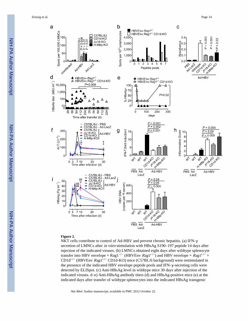
Figure 2.NKT cells contribute to control of Ad-HBV and prevent chronic hepatitis. (a) IFN-γsecretion of LMNCs after in vitro stimulation with HBsAg S190–197 peptide 14 days afterinjection of the indicated viruses. (b) LMNCs obtained eight days after wildtype splenocytetransfer into HBV envelope × Rag1−/− (HBVEnv Rag1−/−) and HBV envelope × Rag1−/− ×CD1d−/− (HBVEnv Rag1−/− CD1d-KO) mice (C57BL/6 background) were restimulated inthe presence of the indicated HBV envelope peptide pools and IFN-γ-secreting cells weredetected by ELISpot. (c) Anti-HBsAg level in wildtype mice 30 days after injection of theindicated viruses. d–e) Anti-HBsAg antibody titers (d) and HBsAg-positive mice (e) at theindicated days after transfer of wildtype splenocytes into the indicated HBsAg transgenic
Zeissig et al. Page 14
Nat Med. Author manuscript; available in PMC 2012 October 22.
NIH
-PA Author Manuscript
NIH
-PA Author Manuscript
NIH
-PA Author Manuscript

mouse strains. Serum alanine aminotransferase (ALT) (f), IFN-γ mRNA in liver tissue asdetermined by qPCR (g, day 5 after virus injection), histological grading of liverinflammation (Ishak et al.41) (h, day 30 after virus injection), serum HBsAg (i) and serumHBV DNA (j, day 7 after virus injection). In f and i significance levels as indicated byasterisks reflect from top to bottom Jα18-KO, CD1d-KO, H-Mttp-KO vs. control. Mean ±s.e.m. is shown (6–8 mice per group). Results are representative of two independentexperiments.
Zeissig et al. Page 15
Nat Med. Author manuscript; available in PMC 2012 October 22.
NIH
-PA Author Manuscript
NIH
-PA Author Manuscript
NIH
-PA Author Manuscript

Figure 3.NKT activation in response to HBV is mediated by hepatocytes and dependent onhepatocyte CD1d and MTP. (a) Activation of invariant (24.7, 24.8, 24.9, DN32.D3) andnon-invariant (14S.6, 14S.7, 14S.10, 14S.15) NKT hybridomas in response to coculture withwildtype primary mouse hepatocytes 48h after Ad-HBV transduction. (b) Activation ofinvariant 24.7 and non-invariant 14S.6 in response to primary hepatocytes of wildtype andCD1d-KO mice 48h after virus administration. Viral transduction was similar in hepatocytesfrom both strains (see Suppl. Fig. 9) (c) Activation of the MHC class I-restricted T cellhybridoma RF33.70 in response to SIINFEKL (1 µg/ml) presented by primary hepatocytes48h post infection. (d) Activation of human iNKT cell clones J24L.17, J3N.5, andJα24Vβ11-positive human iNKT cells expanded from peripheral blood in response tohuman hepatocytes infected with Ad-LacZ, Ad-HBV, and a primary HBV isolate. (e)Primary human hepatocytes were infected with a primary HBV isolate and were treated with
Zeissig et al. Page 16
Nat Med. Author manuscript; available in PMC 2012 October 22.
NIH
-PA Author Manuscript
NIH
-PA Author Manuscript
NIH
-PA Author Manuscript

blocking CD1d antibodies and MTP-inhibitors (BMS212122, BMS197636) or a structurallyrelated negative compound. IFN-γ secretion of J24L.17 NKT cells was determined 72h afterinfection and 48h after MTP inhibitor treatment. (f–g) Activation of non-invariant 14S.6 (f)and invariant 24.7 (g) by primary hepatocytes of the indicated mouse strains 48h afterinfection with the indicated viruses or vehicle. Cytokine secretion in (a–g) was determinedby ELISA 24h after coculture. Mean ± s.e.m. of triplicate cultures are shown. Results arerepresentative of three independent experiments.
Zeissig et al. Page 17
Nat Med. Author manuscript; available in PMC 2012 October 22.
NIH
-PA Author Manuscript
NIH
-PA Author Manuscript
NIH
-PA Author Manuscript
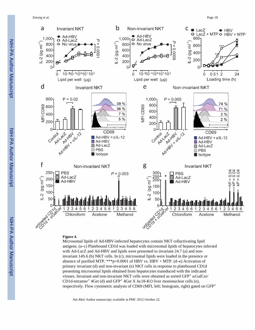
Figure 4.Microsomal lipids of Ad-HBV-infected hepatocytes contain NKT cellactivating lipidantigens. (a–c) Platebound CD1d was loaded with microsomal lipids of hepatocytes infectedwith Ad-LacZ and Ad-HBV and lipids were presented to invariant 24.7 (a) and non-invariant 14S.6 (b) NKT cells. In (c), microsomal lipids were loaded in the presence orabsence of purified MTP. ***p=0.0001 of HBV vs. HBV + MTP. (d–e) Activation ofprimary invariant (d) and non-invariant (e) NKT cells in response to platebound CD1dpresenting microsomal lipids obtained from hepatocytes transduced with the indicatedviruses. Invariant and non-invariant NKT cells were obtained as sorted GFP+ αGalCer/CD1d-tetramer+ 4Get (d) and GFP+ 4Get X Jα18-KO liver mononuclear cells (e),respectively. Flow cytometric analysis of CD69 (MFI, left; histogram, right) gated on GFP+
Zeissig et al. Page 18
Nat Med. Author manuscript; available in PMC 2012 October 22.
NIH
-PA Author Manuscript
NIH
-PA Author Manuscript
NIH
-PA Author Manuscript
CD3+ cells is shown. Numbers in histograms indicate the percentage of cells that shift inexpression of CD69 compared to microsomal lipids of uninfected cells. IL-12 neutralizationdemonstrates absence of indirect cytokine-mediated NKT cell activation as expected in anAPC-free assay. (f–g) Activation of non-invariant 14S.6 (f) and invariant 24.7 (g) inresponse to plate-bound CD1d presenting chloroform-, acetone-, and methanol-soluble lipidsobtained two days after infection with Ad-LacZ and Ad-HBV. IL-2 secretion wasdetermined by ELISA. Mean ± s.e.m. of triplicate cultures is shown. Results arerepresentative of two (d–e) or three (a–c, f–g) independent experiments.
Zeissig et al. Page 19
Nat Med. Author manuscript; available in PMC 2012 October 22.
NIH
-PA Author Manuscript
NIH
-PA Author Manuscript
NIH
-PA Author Manuscript
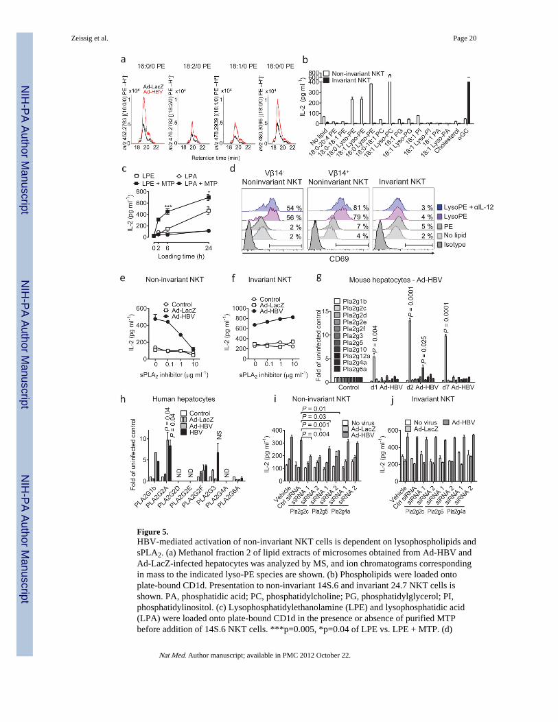
Figure 5.HBV-mediated activation of non-invariant NKT cells is dependent on lysophospholipids andsPLA2. (a) Methanol fraction 2 of lipid extracts of microsomes obtained from Ad-HBV andAd-LacZ-infected hepatocytes was analyzed by MS, and ion chromatograms correspondingin mass to the indicated lyso-PE species are shown. (b) Phospholipids were loaded ontoplate-bound CD1d. Presentation to non-invariant 14S.6 and invariant 24.7 NKT cells isshown. PA, phosphatidic acid; PC, phosphatidylcholine; PG, phosphatidylglycerol; PI,phosphatidylinositol. (c) Lysophosphatidylethanolamine (LPE) and lysophosphatidic acid(LPA) were loaded onto plate-bound CD1d in the presence or absence of purified MTPbefore addition of 14S.6 NKT cells. ***p=0.005, *p=0.04 of LPE vs. LPE + MTP. (d)
Zeissig et al. Page 20
Nat Med. Author manuscript; available in PMC 2012 October 22.
NIH
-PA Author Manuscript
NIH
-PA Author Manuscript
NIH
-PA Author Manuscript

Activation of primary non-invariant (left, middle) and iNKT (right) cells in response toplate-bound CD1d presenting the indicated lipids. NKT cells were obtained as sortedinvariant GFP+ αGalCer/CD1d-tetramer+ 4Get (right) and non-invariant GFP+ 4Get XJα18-KO liver NKT cells (left, middle). Flow cytometric analysis of CD69 gated on GFP+
CD3+ cells is shown. (e–f) Primary hepatocytes were infected as indicated and treated withthe sPLA2 inhibitor c(2NapA)LS(2NapA)R before addition of NKT cells. (g) Expression ofPLA2 transcripts in murine livers following in vivo exposure to Ad-HBV. (h) Analysis ofPLA2 RNA expression in primary human hepatocytes 24h after in vitro infection. Mean ±s.e.m. of triplicate cultures are shown. (i–j) Primary hepatocytes were infected with theindicated viruses before transfection with siRNAs as indicated and coculture with NKTcells. Results are representative of two (a, d) or three (b–c, e–j) independent experiments.
Zeissig et al. Page 21
Nat Med. Author manuscript; available in PMC 2012 October 22.
NIH
-PA Author Manuscript
NIH
-PA Author Manuscript
NIH
-PA Author Manuscript

Figure 6.Expression of HBsAg and cytokines contribute to NKT cell activation. (a–b) Primaryhepatocytes were infected in vitro with the indicated Ad-HBV mutants and were cocultured24h after infection with noninvariant (14S.6, a) and invariant (24.7, b) NKT cellhybridomas. (c) Microsomal lipids of primary hepatocytes obtained 48h after infection withthe indicated viruses. Presentation by plate-bound CD1d to the non-invariant NKT cellhybridoma 14S.6 is shown. (d–f) Primary hepatocytes of the indicated mouse strains weretreated with MTP inhibitors or 9-fluorenyl carboxylic acid as negative compound for 48h.Hepatocytes were then cocultured with non-invariant 14S.6 NKT cells, invariant 24.7 NKTcells, and MHC-class I-restricted RF33.70 T cells. (g–h) Microsomal lipids of the indicated
Zeissig et al. Page 22
Nat Med. Author manuscript; available in PMC 2012 October 22.
NIH
-PA Author Manuscript
NIH
-PA Author Manuscript
NIH
-PA Author Manuscript

mouse strains were loaded onto plate-bound CD1d. Presentation to invariant 24.7 and non-invariant 14S.6 is shown. IL-2 secretion by T cell hybridomas in a–h was determined byELISA. (i–j) 4Get mice were injected with the indicated viruses, LPS (indirect NKT cellactivation) and αGalCer (direct iNKT cell activation) in the presence or absence ofantibody-mediated IL-12 neutralization. Liver mononuclear cells were obtained 6 hours(αGalCer) and 24 hours (viruses, LPS) after stimulation and expression of CD69 wasdetermined by flow cytometric analysis of GFP+ αGalCer/CD1d-tetramer+ CD3+ invariantNKT cells (i) and GFP+ αGalCer/CD1d21 tetramer− CD3+ non-invariant NKT cells (j).Representative histograms and dot plots are shown. Bar graphs show mean ± s.e.m. Resultsare representative of two (d–j) or three (a–c) independent experiments.
Zeissig et al. Page 23
Nat Med. Author manuscript; available in PMC 2012 October 22.
NIH
-PA Author Manuscript
NIH
-PA Author Manuscript
NIH
-PA Author Manuscript