HEK293 cell response to static magnetic fields via the ... - PLOS
11
RESEARCH ARTICLE HEK293 cell response to static magnetic fields via the radical pair mechanism may explain therapeutic effects of pulsed electromagnetic fields Marootpong Pooam 1,2 , Nathalie Jourdan 1 , Mohamed El Esawi 1,3 , Rachel M. Sherrard 1 , Margaret Ahmad ID 1,4 * 1 Sorbonne Universite ´ – CNRS, UMR8256 - IBPS, Paris, France, 2 Department of Biology, Faculty of Science, Naresuan University, Phitsanulok, Thailand, 3 Botany Department, Faculty of Science, Tanta University, Tanta, Egypt, 4 Xavier University, Cincinnati, Ohio, United States of America * [email protected] Abstract PEMF (Pulsed Electromagnetic Field) stimulation has been used for therapeutic purposes for over 50 years including in the treatment of memory loss, depression, alleviation of pain, bone and wound healing, and treatment of certain cancers. However, the underlying cellular mechanisms mediating these effects have remained poorly understood. In particular, because magnetic field pulses will induce electric currents in the stimulated tissue, it is unclear whether the observed effects are due to the magnetic or electric component of the stimulation. Recently, it has been shown that PEMFs stimulate the formation of ROS (reac- tive oxygen species) in human cell cultures by a mechanism that requires cryptochrome, a putative magnetosensor. Here we show by qPCR analysis of ROS-regulated gene expres- sion that simply removing cell cultures from the Earth’s geomagnetic field by placing them in a Low-Level Field condition induces similar effects on ROS signaling as does exposure of cells to PEMF. This effect can be explained by the so-called Radical Pair mechanism, which provides a quantum physical means by which the rates and product yields (e.g. ROS) of bio- chemical redox reactions may be modulated by magnetic fields. Since transient cancelling of the Earth’s magnetic field can in principle be achieved by PEMF exposure, we propose that the therapeutic effects of PEMFs may be explained by the ensuing modulation of ROS synthesis. Our results could lead to significant improvements in the design and therapeutic applications of PEMF devices. Introduction All life on Earth is exposed to a magnetic field generated through the motion of molten metal at the Earth’s core. This so-called geomagnetic field is a directional force that moves outward from near the South Pole, then curves upwards, and re-enters the Earth near the North Pole [1]. The greatest benefit to living organisms of the geomagnetic field is to deflect high-energy PLOS ONE PLOS ONE | https://doi.org/10.1371/journal.pone.0243038 December 3, 2020 1 / 11 a1111111111 a1111111111 a1111111111 a1111111111 a1111111111 OPEN ACCESS Citation: Pooam M, Jourdan N, El Esawi M, Sherrard RM, Ahmad M (2020) HEK293 cell response to static magnetic fields via the radical pair mechanism may explain therapeutic effects of pulsed electromagnetic fields. PLoS ONE 15(12): e0243038. https://doi.org/10.1371/journal. pone.0243038 Editor: Yi Cao, Xiangtan University, CHINA Received: August 10, 2020 Accepted: November 15, 2020 Published: December 3, 2020 Copyright: © 2020 Pooam et al. This is an open access article distributed under the terms of the Creative Commons Attribution License, which permits unrestricted use, distribution, and reproduction in any medium, provided the original author and source are credited. Data Availability Statement: All relevant data are within the manuscript and its Supporting information files. Funding: National Science Foundation USA (#1658640) to MA Air Force Office of Scientific Research USA (FA9550-14-0-0409) to MA Novo Nordisk Foundation (Denmark) to MA Agence Nationale de Recherche, France grant no: ANR-19- CE37-0021 to RS. Competing interests: NO competing interests.
-
Upload
khangminh22 -
Category
Documents
-
view
5 -
download
0
Transcript of HEK293 cell response to static magnetic fields via the ... - PLOS

RESEARCH ARTICLE
HEK293 cell response to static magnetic fields
via the radical pair mechanism may explain
therapeutic effects of pulsed electromagnetic
fields
Marootpong Pooam1,2, Nathalie Jourdan1, Mohamed El Esawi1,3, Rachel M. Sherrard1,
Margaret AhmadID1,4*
1 Sorbonne Universite – CNRS, UMR8256 - IBPS, Paris, France, 2 Department of Biology, Faculty of
Science, Naresuan University, Phitsanulok, Thailand, 3 Botany Department, Faculty of Science, Tanta
University, Tanta, Egypt, 4 Xavier University, Cincinnati, Ohio, United States of America
Abstract
PEMF (Pulsed Electromagnetic Field) stimulation has been used for therapeutic purposes
for over 50 years including in the treatment of memory loss, depression, alleviation of pain,
bone and wound healing, and treatment of certain cancers. However, the underlying cellular
mechanisms mediating these effects have remained poorly understood. In particular,
because magnetic field pulses will induce electric currents in the stimulated tissue, it is
unclear whether the observed effects are due to the magnetic or electric component of the
stimulation. Recently, it has been shown that PEMFs stimulate the formation of ROS (reac-
tive oxygen species) in human cell cultures by a mechanism that requires cryptochrome, a
putative magnetosensor. Here we show by qPCR analysis of ROS-regulated gene expres-
sion that simply removing cell cultures from the Earth’s geomagnetic field by placing them in
a Low-Level Field condition induces similar effects on ROS signaling as does exposure of
cells to PEMF. This effect can be explained by the so-called Radical Pair mechanism, which
provides a quantum physical means by which the rates and product yields (e.g. ROS) of bio-
chemical redox reactions may be modulated by magnetic fields. Since transient cancelling
of the Earth’s magnetic field can in principle be achieved by PEMF exposure, we propose
that the therapeutic effects of PEMFs may be explained by the ensuing modulation of ROS
synthesis. Our results could lead to significant improvements in the design and therapeutic
applications of PEMF devices.
Introduction
All life on Earth is exposed to a magnetic field generated through the motion of molten metal
at the Earth’s core. This so-called geomagnetic field is a directional force that moves outward
from near the South Pole, then curves upwards, and re-enters the Earth near the North Pole
[1]. The greatest benefit to living organisms of the geomagnetic field is to deflect high-energy
PLOS ONE
PLOS ONE | https://doi.org/10.1371/journal.pone.0243038 December 3, 2020 1 / 11
a1111111111
a1111111111
a1111111111
a1111111111
a1111111111
OPEN ACCESS
Citation: Pooam M, Jourdan N, El Esawi M,
Sherrard RM, Ahmad M (2020) HEK293 cell
response to static magnetic fields via the radical
pair mechanism may explain therapeutic effects of
pulsed electromagnetic fields. PLoS ONE 15(12):
e0243038. https://doi.org/10.1371/journal.
pone.0243038
Editor: Yi Cao, Xiangtan University, CHINA
Received: August 10, 2020
Accepted: November 15, 2020
Published: December 3, 2020
Copyright: © 2020 Pooam et al. This is an open
access article distributed under the terms of the
Creative Commons Attribution License, which
permits unrestricted use, distribution, and
reproduction in any medium, provided the original
author and source are credited.
Data Availability Statement: All relevant data are
within the manuscript and its Supporting
information files.
Funding: National Science Foundation USA
(#1658640) to MA Air Force Office of Scientific
Research USA (FA9550-14-0-0409) to MA Novo
Nordisk Foundation (Denmark) to MA Agence
Nationale de Recherche, France grant no: ANR-19-
CE37-0021 to RS.
Competing interests: NO competing interests.

cosmic rays emitted by solar wind, which would otherwise make life on earth impossible.
However, the geomagnetic field in and of itself also has effects on living systems. For example,
studies in numerous organisms indicate that the magnetic field can be used as a source of
directional information, as well as inducing cellular and physiological consequences [1–3].
Because these biological effects are generally weak, often of unknown mechanism, and all
requiring carefully controlled conditions to reproducibly demonstrate, there has been consid-
erable lack of clarity in the field.
However, in spite of the many unknowns, the application of magnetic field pulses has been
empirically and reliably proven an effective therapeutic tool in the treatment of human disease.
In particular, Pulsed Electromagnetic Field (PEMF) therapy has been used. PEMF therapy
involves the non-invasive exposure of parts of the human body to low intensity pulsed electro-
magnetic fields in the 10–300 Hz range, typically for a few minutes a day over a period of sev-
eral days to several weeks [3]. Clinical studies have shown the effectiveness of PEMF in
treating such conditions as arthritis [4, 5], chronic pain [6, 7], bone injury [8–10], wound heal-
ing [11–15], and lupus erythematosus [16]. All of these conditions furthermore involve resolu-
tion of chronic or acute inflammation, which is sensitive to intracellular concentrations of
ROS (reactive oxygen species). Although many changes in cellular and biochemical markers
induced by PEMF exposure have been documented [e.g. in 17, 18], the molecular mechanisms
underlying therapeutic effects of PEMF stimulation remains unknown.
PEMF signals comprise both magnetic and electrical field components, with considerable
variability imposed by signal shapes, intensities, and frequencies [19, 20]. Although many stud-
ies have suggested that PEMF alters cellular function through the action of induced electrical
currents, others have suggested that the magnetic field component of PEMF stimulation is
what induces the physiological effects [21, 22]. This latter possibility has received support from
a recent study on the response characteristics of human cell cultures to 10Hz PEMF exposure,
in which a putative magnetic field receptor, known as cryptochrome, has been implicated [23].
A rapid increase in cellular concentration of ROS, with accompanying changes in gene expres-
sion, were observed in response to the PEMF signal [23]. These experiments do not exclude
that the induced electric fields may trigger the biological response. However, they nonetheless
raise the intriguing possibility that therapeutic PEMF effects could be mediated through the
same underlying mechanisms as are responsible for biological responses to static magnetic
fields, which do not generate electric currents.
Currently, there are two well-characterized biological magnetosensing mechanisms. One of
these involves ferromagnetite, an iron-based compound, which can be attached to cellular
structures or proteins to enable organisms to orient in the direction of the magnetic north pole
[24, 25]. In addition, there has been accumulating evidence of biological magnetosensing
through the so-called ‘Radical Pair Mechanism’ [26]. This mechanism involves quantum phys-
ical effects of electromagnetic fields on electron spin of reaction intermediates formed during
susceptible enzymatic reactions. Flavoenzyme-based redox reactions have been proposed as
particularly likely candidates for these quantum forces. In summary, the effect of a weak mag-
netic field would be to alter rates of enzyme-catalyzed chemical reactions within the cell. These
could be of metabolic enzymes, resulting in altered metabolism or altered rates of biosynthesis
of ROS (a byproduct of redox chemistry). Intriguingly, an evolutionarily conserved flavopro-
tein receptor known as cryptochrome [27], also undergoes redox chemistry thought compati-
ble with the Radical Pair Mechanism for magnetic sensitivity [28]. Cryptochrome has been
linked to static field magnetosensing in many biological systems [28–30], and has also been
implicated in sensitivity to PEMF exposure in human HEK cells [23] and mouse organotypic
brain culture [31].
PLOS ONE Static magnetic field stimulation of cells in culture
PLOS ONE | https://doi.org/10.1371/journal.pone.0243038 December 3, 2020 2 / 11

These observations thereby directly pose the question of whether the effects of PEMF stim-
ulation result from one of the known biological magnetosensing mechanisms, instead of being
an effect of induced electrical currents. A clear prediction would then be that a simple static
magnetic field should provide some of the same effects as the PEMF signal on stimulation of
cellular ROS.
In the present work we address this question by determining the effect on HEK293 cells in
culture of exposure to a number of static magnetic field conditions. Firstly we assayed cells at 2
mT, which was the peak intensity of magnetic field output by the PEMF device used in our
original study [23] showing induction of ROS. We also chose 500 μT since it induces physio-
logical effects and modulation of cryptochrome function in a number of experimental systems
including Drosophila behavioral assays and in plants [28, 30]. We chose a low-level field (LLF)
to provide a condition where the Earth’s magnetic field is essentially removed (less than 200
nT intensity).–this condition had furthermore been previously shown to induce biosynthesis
of ROS in a variety of mammalian cell cultures [32], similarly to our PEMF device [23] Finally,
we chose the ambient Earth’s geomagnetic field as the control condition, which was 40 μT
inside the incubator. For our biological assay, we have monitored expression of ROS-respon-
sive genes that were up-regulated as a result of PEMF stimulation to use as markers for cellular
ROS induction [23].
In summary, this study explores whether the effects of PEMF stimulation on cell cultures
can be mimicked by using only static magnetic fields. A positive outcome would provide for
the first time a molecular and biophysical explanation for the therapeutic effects of PEMF by
known biochemical and quantum physical mechanisms.
Materials and methods
Cell culture and growth conditions
Methods in this study are essentially identical to those previously described [23]. HEK 293
(ATCC-CRL-1573) cells were acquired from the ATCC cell bank Briefly, human embryonic
kidney (HEK) 293 cells were cultured in a CO2 incubator (MCO-18AC, Panasonic Biomedical,
Leicestershire, UK), at 37˚C and 5% CO2. Cells were grown in 75 ml culture flasks containing
10 ml Modified Eagle medium (MEM; Sigma, Sigma, St Louis, MO) and sub-cultured every 4
days.
Static magnetic field and low-level field exposure conditions
The static magnetic field exposure conditions are essentially as previously described [28, 29],
with the exception that all experiments were conducted in the dark, and in the CO2 incubator.
The Control condition was the culture of HEK293 exposed to the local magnetic fields inside
the CO2 incubator (40 μT).
The higher intensity static magnetic fields (500 μT and 2 mT) were generated by a square
Helmholtz coil (20 cm per side) placed inside the incubator as described previously [23, 28,
29]. Each coil consisted of 20 turns of copper wire with the two coils separated by 11.5 cm.
Current from a DC power source was used to generate parallel current to achieve the desired
(500 μT or 2 mT) static MF treatment condition. The sham condition was generated by run-
ning the current through the two wire coils in antiparallel directions to cancel the magnetic
fields without affecting other environmental components (e.g. temperature or vibration)–
described in [23, 28].
To generate the low-level static field exposure (LLF), the samples were placed within two
concentric μ-metal cylinders with walls 0.2 mm thick, as described previously [29]. The inner
cylinder was 11.5 cm diameter, and the outer one was 16 cm diameter, and both were 30 cm in
PLOS ONE Static magnetic field stimulation of cells in culture
PLOS ONE | https://doi.org/10.1371/journal.pone.0243038 December 3, 2020 3 / 11

height. The culture dish was placed at the center of the μ-metal cylinder where the intensity of
local magnetic fields was lower than 200 nT (LLF). To generate the sham condition for LLF
exposure, a small round Helmholtz coil was placed inside the inner μ-metal cylinder. Each coil
consisted of 20 windings of 1 mm-diameter copper wire around a plastic circular frame (10
cm diameter, at a separation of 10 cm between coils). The current provided to the coils gener-
ated a 40 μT static MF, which was local magnetic field inside the incubator. This set-up pro-
vided a control condition directly within the mu-metal cylinder.
For all Magnetic Field exposure experiments, HEK293 cells were seeded into 3.5 cm2 round
culture-dishes and grown for 48 h. Subsequently, culture dishes were exposed to LLF, Control
(40 μT), 500 μT or 2000 μT static fields for either 10 minutes or 3h, which we have previously
shown increase ROS and modify gene expression [23]. In all cases cells were harvested 3 hours
after initial onset of exposure. Control and sham conditions using the same cell cultures were
always performed in parallel for comparative purposes.
Quantitative RT-PCR analysis of altered gene expression
The qPCR analysis was performed as described [23]. After exposure to each treatment condi-
tion, total RNA was extracted from HEK293 cells by Total RNA Miniprep Kit (New England
Biolabs). cDNA was prepared from 1 μg total RNA using SuperScript first-strand synthesis sys-
tem (Thermo Fisher Scientific). Quantitative RT-PCR was performed using Luna qPCR mas-
ter mix (New England Biolabs). We selected three ROS-regulated genes, KIAA1211, RPS16P5
and TAS2R19 that had been previously reported to be up-regulated after exposure to 10 Hz
PEMF at 2 mT [21]. The GADPH gene was used as the reference gene. Quantitative RT-PCR
was performed by Mastercycler1 RealPlex2 (Eppendorf). Three biological replicates were per-
formed for each gene (N = 3). Data analysis to represent the relative expression level of genes
of interest was performed as previously described [23]. Primers used for gene expression anal-
ysis are described in Table 1.
Statistical analysis
All data were analyzed by using GraphPad Prism version 7.4.2 for Mac (GraphPad Software,
La Jolla California, USA). Data were analyzed for normality with the Shapiro-Wilk test. Results
will be expressed as mean ± standard error of the mean (SEM). The difference between treated
and control conditions for each gene were compared using One-way ANOVA followed by
Tukey’s multiple comparisons test. Comparisons were of the Exposed (to a given static field
condition) or Sham-exposed (to a cancelled magnetic field condition) sample relative to the
Control (unexposed) sample grown at the same time from the same cell stock. Differences
were considered statistically significant with a p-value< 0.05 (�),< 0.01 (��),<0.01 (���).
Table 1. List of primers used in the current study.
Gene Primer sequence
KIAA1211 Forward AGCTGGCTGTTAAGCCAAAA
Reverse CCTCCAGTTCTCGCCAGTAG
RPS16P5 Forward TGCTAATGGCTGTGTGAAGC
Reverse GCCACAACAGGAAAAGGTGT
TAS2R19 Forward GCAAACTGTGACCTCCTTCC
Reverse CGTGTCATCTGCCACAAAAC
GADPH Forward TGCACCACCAACTGCTTAGC
Reverse GGCATGGACTGTGGTCATGAG
https://doi.org/10.1371/journal.pone.0243038.t001
PLOS ONE Static magnetic field stimulation of cells in culture
PLOS ONE | https://doi.org/10.1371/journal.pone.0243038 December 3, 2020 4 / 11
Results
The present study seeks to establish whether exposure to a static magnetic field could promote
comparable effects on cultured cells as exposure to PEMF (pulsed electromagnetic fields) and
therefore clarify whether cellular effects were primarily due to the magnetic or induced-electric
fields. Our assay was based on a prior study which identified marker genes that are regulated by
ROS and are induced immediately following PEMF stimulation by a mechanism that implicates
a known static magnetic field sensor [See supplement in ref. 23]. We monitored expression of a
representative number of these genes (KIAA1211, RPS16P5, TAS2R19) as a simple and rapid
assay for a biological response to static magnetic field stimulation in HEK cell cultures.
We tested a range of static magnetic field conditions: the Control condition was the local
geomagnetic field measured within the incubator (40 μT); 500 μT; 2 mT and LLF (Low Level
Field). LLF was at an intensity of less than 200nT, a static field which has been shown previ-
ously to induce physiological effects on biosynthesis of ROS in mammalian cell cultures [22]
and so is a likely candidate for investigating PEMF induced effects.
We observed different effects on gene expression according to the intensity of the static
magnetic field, but not with the different durations of exposure. Significant change in gene
expression was obtained from HEK cells in response to exposure to the LLF condition. Even a
10 min exposure to LLF was effective in stimulating increased expression in our selected
genes. The level of induction was comparable to that obtained from these cultured cells subse-
quent to exposure to the 10 Hz PEMF signal [23]. Intriguingly, two of the three genes
responded similarly after a 3-hour exposure period (RPS16P5, TAS2R19). However, one of the
genes (KIAA1211) proved insensitive to 3 hours of continuous exposure to LLF. This suggests
some degree of adaptation over the longer term, a common feature of cellular response to
altered concentration of ROS [33]. The ‘Control’ and ‘Sham Exposure’ experimental condi-
tions showed no statistical variation, indicating that the effect was indeed due to the applied
static magnetic field (Fig 1).
Also, some studies on biological magnetosensing have used exposure conditions of 500 μT
static field (roughly 10 X that of the earth’s geomagnetic field). We accordingly performed
HEK cell gene expression analysis after exposure to 500 μT magnetic field intensity (Fig 2). In
this case, there was no significant change in expression of any of the three tested genes as com-
pared to Sham (mock-exposed) and Control (40 μT local field) conditions (Fig 2).
Most experiments on static magnetic field effects on biological organisms have been
observed at magnetic field strengths relatively close to that of the local geomagnetic field. In
contrast, PEMF is often applied at higher intensities in the milliTesla range. However, these
higher magnetic field strengths in their static form are predicted to have limited effects on bio-
logical responses by any of the known magnetosensing mechanisms [24–26]. We therefore
exposed HEK293 cell cultures to a 2 mT static magnetic field (Fig 3). There was no change in
gene expression to either a 10 min or 3-hour exposure at this intensity.
These results can be summarized as follows: when cells were exposed to the LLF condition,
they were effectively deprived of the Earth’s magnetic field. This led to a marked increase in
expression of ROS-related genes (Fig 1), indicating that physiological ROS synthesis is much
lower in the presence of the local (Earth’s) geomagnetic field. Higher magnetic field strengths
showed no measurable further effect on ROS-related gene expression. This indicates that regu-
lation of cellular ROS is optimal in the presence of the geomagnetic field.
Discussion
The goal of this study was to determine whether an underlying biological response to static
magnetic fields could account for the effectiveness of PEMF as a therapeutic tool [4–13]. This
PLOS ONE Static magnetic field stimulation of cells in culture
PLOS ONE | https://doi.org/10.1371/journal.pone.0243038 December 3, 2020 5 / 11

Fig 2. Effect of a 500 μT static magnetic field on HEK293 gene expression. The results are presented as the expression level of genes KIAA1211,
RPS16P5 and TAS2R19 after exposure to 500 μT condition for 10 min (dark-grey bar) or 3h (light-grey bay) in comparison to the Control (black bar) or
sham-exposed (white bar) condition. The ‘Control’ condition is the local geomagnetic field in the incubator. The ‘Sham’ condition uses a Helmholtz
coil with current running in antiparallel directions to cancel the induced magnetic field (see Methods). Data are shown as mean ± SE of three
independent experiments (N = 3). The asterisks indicate significance level of the differences: �p-value< 0.1; �� p-value< 0.01; ��� p-value< 0.01.
https://doi.org/10.1371/journal.pone.0243038.g002
Fig 1. Effect of low level field (LLF) on HEK293 gene expression. The expression level of genes KIAA1211, RPS16P5 and TAS2R19 after exposure to
LLF condition for 10 min (dark-grey bar) or 3h (light-grey bay) in comparison to the Control (black bar) or sham-exposed (white bar) condition. The
‘Control’ condition represents growth in the incubator at the local geomagnetic field without added applied static field. The ‘Sham’ condition was a
40 μT field produced by Helmholtz coils placed inside the Mu-metal cylinder used to generate the LLF to mimick the geomagnetic field (see methods).
Data are shown as mean ± SE of three independent experiments (N = 3). The asterisks indicate significance level of the differences: �p-value< 0.1; �� p-
value< 0.01; ��� p-value< 0.01.
https://doi.org/10.1371/journal.pone.0243038.g001
PLOS ONE Static magnetic field stimulation of cells in culture
PLOS ONE | https://doi.org/10.1371/journal.pone.0243038 December 3, 2020 6 / 11

was suggested by the fact that cryptochrome, a putative static magnetic field receptor, had
been implicated in PEMF responses [23, 31], and by the fact that the static magnetic fields
emitted transiently by PEMF devices occur within a range of intensities known to have physio-
logical consequences [28–30]. Our initial expectation was that exposure to the higher magnetic
fields (0.5 and 2 mT) might result in an enhanced biological response as compared to exposure
to the earth’s geomagnetic field (40 μT). What we in fact found was that ROS–regulated gene
expression was identical at all three tested static magnetic field conditions—40 μT, 0.5 mT and
2 mT (Figs 2 and 3), whereas only the absence of the magnetic field stimulated a significant
increase in ROS-regulated gene expression (Fig 1). These results indicate that levels of ROS in
cellular cultures are indeed regulated by the presence of a static magnetic field. Furthermore,
our data suggest that ROS synthesis has an optimal biological minimum at the intensity of the
Earth’s geomagnetic field, otherwise higher magnetic field strengths would have biological
consequences.
To understand how a PEMF signal could create such an LLF effect, it must be considered
that the magnetic field component of PEMF signals are generated by electrical currents pulsed
through a coil at a defined frequency [19, 20]. During such treatments, the magnetic field is
continuously increasing and decreasing in non-homogeneous directions, thereby covering a
range of directional vectors and intensities (rising and falling) at rates determined by the
PEMF pulse frequency and signal shape. It must also be considered that magnetic fields are
additive and therefore can cancel each other out when coming from different directions at
comparable intensities. To generate a Low-Level Field effect, the magnetic field emitted by the
PEMF coil must at certain instants be in opposing orientation and of suitable intensity to can-
cel the static geomagnetic field in the incubator (around 40 μT). Over a prolonged PEMF
exposure period, all of the susceptible receptor molecules throughout the sample would be
intermittently exposed to the LLF.
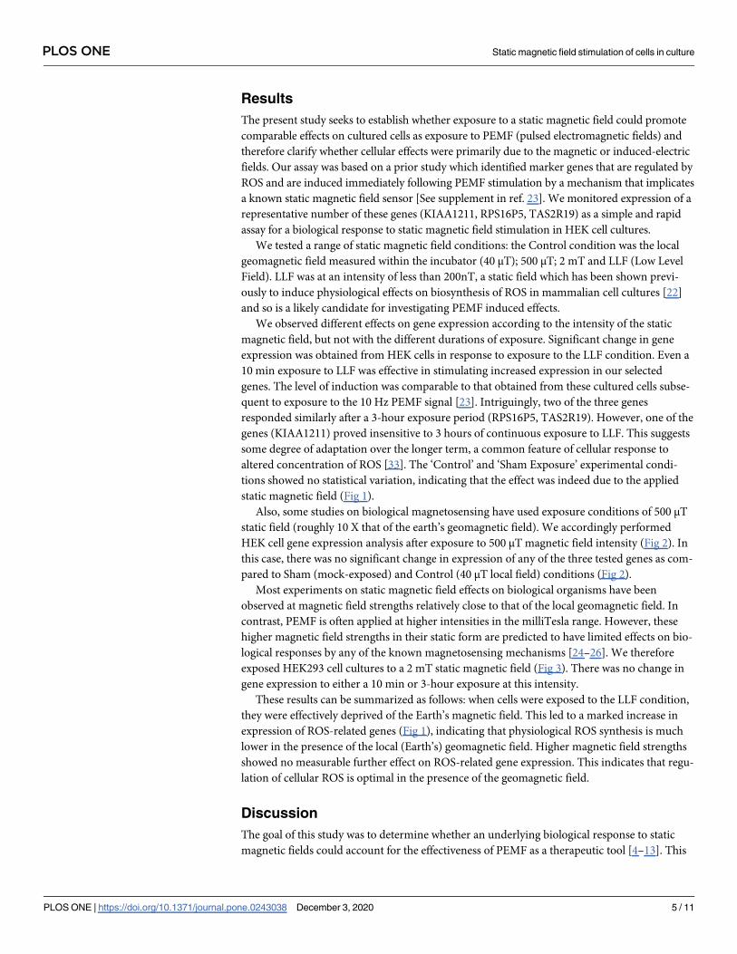
Fig 3. Effect of a 2 mT static magnetic field on HEK293 gene expression. The results are presented as the Relative expression of the ROS-related
genes KIAA1211, RPS16P5 and TAS2R19 after 2 mT (grey bar), Control (black bar) or Sham (white bar) exposure conditions. Control cell cultures
were exposed to the local magnetic field. The Sham condition was provided by the Helmholtz coil that generated the 2mT magnetic field but with
antiparallel currents. Data are shown as mean ±SE of three independent experiments (N = 3).
https://doi.org/10.1371/journal.pone.0243038.g003
PLOS ONE Static magnetic field stimulation of cells in culture
PLOS ONE | https://doi.org/10.1371/journal.pone.0243038 December 3, 2020 7 / 11
Given the assumption that PEMF exposure can achieve a transient decline in the Earth’s
magnetic field, then the PEMF effect on cellular ROS stimulation can now be explained by the
‘Radical Pair Mechanism’ of biological magnetosensing. In this mechanism, magnetic fields
act on radical pairs formed by biological receptors as extremely short-lived reaction intermedi-
ates [26]. The magnetic field will then either accelerate or decrease the ultimate reaction rate
and reaction product formation. Indeed, the Radical Pair mechanism has been implicated in a
number of studies probing the modulation of H202 subsequent to LLF exposure in cellular cul-
tures [34–36]. Transient static LLFs formed in the course of PEMF stimulation could thereby
significantly impact on rates of ROS formation in cell culture. For instance, if each flavoprotein
receptor molecule received a single transient LLF stimulus at least once during the (10 min or
3 hr) PEMF exposure period, the triggered change in biological activity would cause a measur-
able physiological change in ROS.
In this context, an important feature of the Radical Pair mechanism is that quantum physi-
cal effects of the magnetic field on electron spin chemistry are predicted to operate in a narrow
range and reach a plateau beyond which further increase in magnetic field intensity could have
no further effect on product yields [26]. In agreement with such a mechanism, our experi-
ments shown no difference in biological response at Earth magnetic field (40 μT) as compared
to higher magnetic field strengths (0.5 and 2 mT)–see Figs 2 and 3. Only a decrease in the mag-
netic field intensity (ie the LLF condition in our experiments) was able to induce measurable
effects on the biological response, Our results are therefore consistent with the cellular reaction
rates generating ROS being tuned to reach an optimal minimum (the physiologically most
desirable steady state) near the intensity of the Earth’s magnetic field.
From a mechanistic perspective, our findings are consistent with a mode of action whereby
modest, transient, changes in cellular ROS such as induced by PEMF devices [23] provide an
explanation for their beneficial effects and healing functions [23]. These concentrations of
ROS are much lower than those that induce damage known as oxidative stress [33], and
instead involve a phenomenon known as ‘redox biology’ wherein cellular ROS plays a benefi-
cial role by stimulating anti-oxidant defense and repair pathways [37]. In support of this mech-
anism, many pathologies on which therapeutic effects of PEMF have been documented [4–15]
involve cellular mechanisms (eg. underlying anti-inflammatory effects) responsive to ROS sig-
naling pathways [eg. 17, 18, 37–39]. This is also true for resolution of chronic and acute pain,
for which PEMF therapy has been proven effective, and which has the great advantage that
there are no potential negative side effects as caused by pharmaceutical remedies [40]. Since
the Radical Pair Mechanism predicts only modest changes in concentrations of ROS on theo-
retical grounds, a means of inducing transient small increases in cellular ROS at will by appro-
priately altering the geomagnetic field provides a powerful paradigm for the effectiveness of
PEMF devices in therapy.
Our findings indicate that effects of PEMF stimulation on gene expression can be induced
through static magnetic field exposure. However, they do not exclude the possibility that the
induced electrical fields generated by PEMF may also play a part. Our findings also do not
exclude that cellular ROS stimulation which results either as a consequence of LLF or of PEMF
exposure could occur by different underlying mechanisms. Ultimately, further research using
defined and simplified PEMF signals, analysed by studying a variety of cellular signaling path-
ways, will be required to fully resolve these questions.
Conclusion
In this study, we show that exposing human cell cultures to a reduced (Low Level) magnetic
field replicates the effect of PEMF exposure on ROS-regulated gene expression. These
PLOS ONE Static magnetic field stimulation of cells in culture
PLOS ONE | https://doi.org/10.1371/journal.pone.0243038 December 3, 2020 8 / 11
observations are consistent with the Radical Pair Mechanism, which provides a quantum bio-
logical explanation for how the redox chemistry of susceptible flavoproteins can be manipu-
lated by magnetic fields including that of the Earth. Because PEMF output produces magnetic
fields with changing amplitudes and directions, we propose that PEMF exposure may tran-
siently change the static magnetic field exposure conditions to alter ROS synthesis in cell
cultures.
Supporting information
S1 Appendix. Files of raw data, means and S.E. used to build graphs in Figs 1–3.
(XLSX)
Acknowledgments
We are indebted to Alain d’Harlingue and Jacques Witczak for expert technical assistance.
Author Contributions
Conceptualization: Marootpong Pooam, Nathalie Jourdan, Margaret Ahmad.
Data curation: Marootpong Pooam.
Formal analysis: Marootpong Pooam.
Funding acquisition: Rachel M. Sherrard, Margaret Ahmad.
Investigation: Marootpong Pooam, Mohamed El Esawi, Margaret Ahmad.
Methodology: Nathalie Jourdan, Mohamed El Esawi, Margaret Ahmad.
Project administration: Rachel M. Sherrard, Margaret Ahmad.
Resources: Rachel M. Sherrard, Margaret Ahmad.
Supervision: Nathalie Jourdan, Mohamed El Esawi, Rachel M. Sherrard, Margaret Ahmad.
Writing – original draft: Marootpong Pooam, Margaret Ahmad.
Writing – review & editing: Mohamed El Esawi, Rachel M. Sherrard, Margaret Ahmad.
References1. Wiltschko R, Wiltschko W (1995) Magnetic Orientation in Animals. Zoophysiology 33: 1–297.
2. Hong FT (1995) Magnetic field effects on biomolecules, cells, and living organisms. BioSystems 36:
187–229. https://doi.org/10.1016/0303-2647(95)01555-y PMID: 8573700
3. Markov M (2015) XXIst century magnetotherapy. Electromagn. Biol. Med. 34:190–196. https://doi.org/
10.3109/15368378.2015.1077338 PMID: 26444192
4. Ganesan K, Gengadharan AC, Balachandran C, Manohar BM, Puvanakrishnan R (2009) Low fre-
quency pulsed electromagnetic field—a viable alternative therapy for arthritis. Indian J Exp Biol. 47:
939–48. PMID: 20329696
5. Ross CL, Ang DC, Almeida-Porada G (2019) Targeting Mesenchymal Stromal Cells/Pericytes (MSCs)
With Pulsed Electromagnetic Field (PEMF) Has the Potential to Treat Rheumatoid Arthritis. Front.
Immunol | https://doi.org/10.3389/fimmu.2019.00266
6. Guo L, Kubat NJ, Isenberg RA (2011) Pulsed radio frequency energy (PRFE) use in human medical
applications. Electromagn Biol Med. 30: 21–45. https://doi.org/10.3109/15368378.2011.566775 PMID:
21554100
7. Varcaccio-Garofalo G, Carriero C, Loizzo MR, Amoruso S, Loizzi P (1995) Analgesic properties of elec-
tromagnetic field therapy in patients with chronic pelvic pain. Clin Exp Obstet Gynecol. 22: 350–4.
PMID: 8777794
PLOS ONE Static magnetic field stimulation of cells in culture
PLOS ONE | https://doi.org/10.1371/journal.pone.0243038 December 3, 2020 9 / 11

8. Yuan J, Xin F, Jiang W (2018) Underlying Signaling Pathways and Therapeutic Applications of Pulsed
Electromagnetic Fields in Bone Repair. Cell Physiol Biochem. 46: 1581–1594. https://doi.org/10.1159/
000489206 PMID: 29694967
9. Bassett CAL (1994) Therapeutic uses of electric and magnetic fields in orthopedics. In: Karpenter D.,
Ayrapetyan S. Biological Effects of Electric and Magnetic Fields. San Diego: Academic Press. pp. 13–
18.
10. Cook JJ, Summers NJ, Cook EA (2015). Healing in the new millennium: bone stimulators: an overview
of where we’ve been and where we may be heading. Clin. Podiatr. Med. Surg. 32: 45–59. https://doi.
org/10.1016/j.cpm.2014.09.003 PMID: 25440417
11. Strauch B, Herman CH, Dabb R, Ignarro LJ, Pilla AA (2009) Evidence-Based Use of Pulsed Electro-
magnetic Field Therapy in Clinical Plastic Surgery. Aesthetic Surgery Journal 29: 135–143. https://doi.
org/10.1016/j.asj.2009.02.001 PMID: 19371845
12. Saliev T, Mustapova Z, Kulsharova G, Bulanin D, Mikhalovsky S (2014). Therapeutic potential of elec-
tromagnetic fields for tissue engineering and wound healing. Cell Prolif. 47: 485–493. https://doi.org/
10.1111/cpr.12142 PMID: 25319486
13. Bloise N, Petechhia L, Ceccarelli G, Fassina L, Usai C et al. (2018) The effect of pulsed electromagnetic
field exposure on osteoinduction of human mesenchymal stem cells cultured on nano-TiO2 surfaces.
PLOS ONE https://doi.org/10.1371/journal.pone.0199046 PMID: 29902240
14. Ceccarelli G, Bloise N, Mantelli M, Gastaldi G, Fassina L, De Angelis MGC, et al (2013) A comparative
analysis of the in vitro effects of pulsed electromagnetic field treatment on osteogenic differentiation of
two different mesenchymal cell lineages. Biores Open Access. 2: 283–94. https://doi.org/10.1089/
biores.2013.0016 PMID: 23914335
15. Maziarz A, Kocan B, Bester M, Budzik S, Cholewa M, Ochiya T, et al. (2016) How electromagnetic fields
can influence adult stem cells: positive and negative impacts. Stem Cell Res Ther. 7: 54. https://doi.
org/10.1186/s13287-016-0312-5 PMID: 27086866
16. Khamaganova IV, Berlin IuV, Volkov VE, Voinich ZV, Arutiunova ES (1995) The use of a pulsed mag-
netic field in the treatment of lupus erythematosus. Ter. Arkh. 67: 84–7. PMID: 8779120
17. Ross CL, Harrison BL (2013) Effect of pulsed electromagnetic field on inflammatory pathway markers in
RAW 264.7 murine macrophages J Inflamm Res. 6: 45–51. https://doi.org/10.2147/JIR.S40269 PMID:
23576877
18. Kubat NJ, Moffett J, Fray LM (2015) Effect of pulsed electromagnetic field treatment on programmed
resolution of inflammation pathway markers in human cells in culture. Journal of Inflammation
Research, 8: 59–69. https://doi.org/10.2147/JIR.S78631 PMID: 25759595
19. Bassett CAL (1989). Fundamental and practical aspects of therapeutical uses of pulse electromagnetic
fields (PEMFs). Crit. Rev. Biomed. Eng. 17:451,529 PMID: 2686932
20. Liboff AR (2004). Signal shapes in electromagnetic therapy. In: Rosch P.J., Markov M. Bioelectromag-
netic Medicine. NY: Marcel Dekker. pp. 17–37
21. Rodger J, Mo C, Wilkes T, Dunlop SA, Sherrard RM. Transcranial pulsed magnetic field stimulation facili-
tates reorganisation of abnormal neural circuits and corrects behavioural deficits without disrupting normal
connectivity. FASEB J. 2012; 26:1593–606. https://doi.org/10.1096/fj.11-194878 PMID: 22223750
22. Martino CF, Perea H, Hopfner U, Ferguson VL, Wintermantel E (2010) Effects of weak static magnetic
fields on endothelial cells. Bioelectromagnetics 31: 296–301 https://doi.org/10.1002/bem.20565 PMID:
20119972
23. Sherrard RM, Morellini N, Jourdan N, El-Esawi M, Arthaut L-D et al (2018) Low-intensity electromag-
netic fields induce human cryptochrome to modulate intracellular reactive oxygen species. PloS Biol
16, e2006229. https://doi.org/10.1371/journal.pbio.2006229 PMID: 30278045
24. Johnsen S, Lohmann K (2005) The Physics and Neurobiology of Magnetoreception. Nat Rev Neurosci.
6: 703–12. https://doi.org/10.1038/nrn1745 PMID: 16100517
25. Lefèvre CT, Bazylinski DA (2013) Ecology, Diversity, and Evolution of Magnetotactic Bacteria. Microbi-
ology and Molecular Biology Reviews 77: 497–526 https://doi.org/10.1128/MMBR.00021-13 PMID:
24006473
26. Hore PJ, Mouritsen H (2016) The radical pair mechanism of magnetoreception. Ann. Rev. Biophys. 45:
299–344.
27. Chaves I, Pokorny R, Byrdin M, Hoang N, Ritz T, Brettel K, et al. (2011) The cryptochromes: blue light
photoreceptors in plants and animals. Ann. Rev. Plant Biol. 62: 335–364. https://doi.org/10.1146/
annurev-arplant-042110-103759 PMID: 21526969
28. Pooam M, Arthaut LD, Burdick D, Link J, Martino CF, Ahmad M (2019) Magnetic sensitivity mediated by
the Arabidopsis blue-light receptor cryptochrome occurs during flavin reoxidation in the dark. Planta
249: 319–332. https://doi.org/10.1007/s00425-018-3002-y PMID: 30194534
PLOS ONE Static magnetic field stimulation of cells in culture
PLOS ONE | https://doi.org/10.1371/journal.pone.0243038 December 3, 2020 10 / 11
29. Hammad M, Albaqami M, Pooam M, Kernevez E, Witczak J et al. (2020) Cryptochrome mediated mag-
netic sensitivity in Arabidopsis occurs independently of light-induced electron transfer to the flavin.
Photochem & Photobiol Sci. https://doi.org/10.1039/c9pp00469f PMID: 32065192
30. Foley LE, Gegear RJ, Reppert SM (2011). Human cryptochrome exhibits light-dependent magnetosen-
sitivity. Nat Commun. 2: 356. https://doi.org/10.1038/ncomms1364 PMID: 21694704
31. Dufor T, Grehl S, Tang AD, Doulazmi M, Traore M, Debray N, et al. (2019) Neural circuit repair by low-
intensity magnetic stimulation requires cellular magneto-receptors and specific stimulation patterns. Sci
Adv 5:eaav9847. https://doi.org/10.1126/sciadv.aav9847 PMID: 31692960
32. Martino C.F.; Castello P.R. Modulation of hydrogen peroxide production in cellular systems by low level
magnetic fields. PLoS ONE 2011, 6, e22753. https://doi.org/10.1371/journal.pone.0022753 PMID:
21887222
33. Sies H (2015) Oxidative stress: a concept in redox biology and medicine. Redox Biol. 4: 180–183.
https://doi.org/10.1016/j.redox.2015.01.002 PMID: 25588755
34. Castello PR, Hill I, Sivo F, Portelli L, Barnes F, Usselman R, et al. (2014) Inhibition of cellular prolifera-
tion and enhancement of hydrogen peroxide production in fibrosarcoma cell line by weak radio fre-
quency magnetic fields. Bioelectromagnetics 35: 598–602. https://doi.org/10.1002/bem.21858 PMID:
25251337
35. Usselman RJ, Hill I, Singel DJ, Martino CF (2014) Spin biochemistry modulates reactive oxygen species
(ROS) production by radio frequency magnetic fields. Plos One. 9: e93065. https://doi.org/10.1371/
journal.pone.0093065 PMID: 24681944
36. Usselman RJ, Chavarriaga C, Castello PR, Procopio M, Ritz T et al (2016) The Quantum Biology of
Reactive Oxygen Species Partitioning Impacts Cellular Bioenergetics. Sci Rep. 6: 38543. https://doi.
org/10.1038/srep38543 PMID: 27995996
37. Schieber M, Chandel NS (2014) ROS Function in Redox Signaling and Oxidative Stress. Current Biol
24: R453–62. https://doi.org/10.1016/j.cub.2014.03.034 PMID: 24845678
38. Ehnert et al. Extremely low frequency pulsed electromagnetic fields cause antioxidative defense mech-
anisms in human osteoblasts via induction of •O2 and H2O2. 2017. Sci Reports 7, Article number:
14544.
39. Lu C, Fan Z, Xing D. Photo-enhancement of macrophage phagocytic activity via Rac1-mediated signal-
ing pathway: Implications for bacterial infection Int J Biochem & Cell Biol 2016: 206–216. https://doi.org/
10.1016/j.biocel.2016.06.010 PMID: 27345261
40. Paolucci T, Pezzi L, Centra AM, Giannandrea N, Bellomo RG, Saggini R. (2020). Electromagnetic Field
Therapy: A Rehabilitative Perspective in the Management of Musculoskeletal Pain–A Systematic
Review. J. Pain Res. 13, 1385–1400. https://doi.org/10.2147/JPR.S231778 PMID: 32606905
PLOS ONE Static magnetic field stimulation of cells in culture
PLOS ONE | https://doi.org/10.1371/journal.pone.0243038 December 3, 2020 11 / 11