Heavy charged particles in radiation biology and biophysics
29
This content has been downloaded from IOPscience. Please scroll down to see the full text. Download details: IP Address: 89.179.244.102 This content was downloaded on 17/10/2013 at 17:21 Please note that terms and conditions apply. Heavy charged particles in radiation biology and biophysics View the table of contents for this issue, or go to the journal homepage for more 2008 New J. Phys. 10 075006 (http://iopscience.iop.org/1367-2630/10/7/075006) Home Search Collections Journals About Contact us My IOPscience
-
Upload
independent -
Category
Documents
-
view
1 -
download
0
Transcript of Heavy charged particles in radiation biology and biophysics
This content has been downloaded from IOPscience Please scroll down to see the full text
Download details
IP Address 89179244102
This content was downloaded on 17102013 at 1721
Please note that terms and conditions apply
Heavy charged particles in radiation biology and biophysics
View the table of contents for this issue or go to the journal homepage for more
2008 New J Phys 10 075006
(httpiopscienceioporg1367-2630107075006)
Home Search Collections Journals About Contact us My IOPscience
T h e o p e n ndash a c c e s s j o u r n a l f o r p h y s i c s
New Journal of Physics
Heavy charged particles in radiationbiology and biophysics
H Nikjoo15 S Uehara2 D Emfietzoglou3 and A Brahme4
1 Radiation Biophysics Medical Radiation Physics DepartmentKarolinska Institute Box 260 SE-171 76 Stockholm Sweden2 School of Health Sciences Kyushu University Fukuoka Japan3 Medical Physics Laboratory University of Ioannina Medical School451 10 Ioannina Greece4 Medical Radiation Physics Karolinska Institute Stockholm SwedenE-mail hooshangnikjookise
New Journal of Physics 10 (2008) 075006 (28pp)Received 5 February 2008Published 28 July 2008Online at httpwwwnjporgdoi1010881367-2630107075006
Abstract Ionizing radiations induce a variety of molecular and cellular typesof damage in mammalian cells as a result of energy deposition by the radiationtrack In general tracks are divided into two classes of sparsely ionizing onessuch as electron tracks and densely ionizing tracks such as heavy ions The paperdiscusses various aspects and differences between the two types of radiations andtheir efficacies in radiation therapy Biophysical studies of radiation tracks haveprovided much of the insight in mechanistic understanding of the relationshipbetween the initial physical events and observed biological responses Thereforedevelopment of Monte Carlo track-structure techniques and codes are paramountfor the progress of the field In this paper we report for the first time the latestdevelopment for the simulation of proton tracks up to 200 MeV similar to beamenergies in proton radiotherapy and space radiation Vital to the developmentof the models for ion tracks is the accurate simulation of electron tracks crosssections in liquid water In this paper we report the development of electrontrack cross sections in liquid water using a new dielectric model of low-energyelectrons accurate to nearly 10 down to 100 eV
5 Author to whom any correspondence should be addressed
New Journal of Physics 10 (2008) 0750061367-263008075006+28$3000 copy IOP Publishing Ltd and Deutsche Physikalische Gesellschaft
2
Contents
1 Introduction 22 General features of cell response to ionizing radiation 33 Frequencies of energy deposition by heavy ions 44 The clinical efficiency of heavy ions 45 Cross sections for high-energy protons 1ndash200 MeV 7
51 Ionization 852 Excitation 1353 Elastic scattering 1354 Depthndashdose distribution 1555 Radial dose distribution 1556 Microdosimetry 16
6 Electron track simulation in liquid water 1661 Elastic scattering 1862 Inelastic scattering 20
7 Conclusions 26References 26
1 Introduction
The relationship between the quality of ionizing radiation and cell response has been the subjectof much debate over the past half century In this paper we discuss a few selected aspects ofbiological significance and relative importance of heavy ions of low atomic number (z) versuselectrons In the context of biophysics we present development of two new tools for simulationof energetic protons from 1 keVndash200 MeV and a new significant breakthrough in modelling ofelectron track structure in liquid water as a surrogate for tissue In the context of radiotherapywe discuss the merits of therapy with heavy ions It is noteworthy to state by definition byheavy ions we mean protons and ions of higher z values
Two major achievements in the 20th century were mapping of the human genome andhuman space exploration In the former we seek to understand the underlying genetics of humandisease in particular cancer and to create new therapy regimes and drug delivery systems Inspace research we seek to understand and overcome many of the human frailties for long-termspace travel and seek biomedical and technological advances to reduce the risks In both thescenarios ionizing radiation acts like a double-edged sword In one we have used radiation asa curing agent and in the other it is a major carcinogenic agent limiting the scope and durationof deep space human exploration In this paper by definition the term heavy ion refers toprotons and higher atomic number ions Considerable resources have been utilized either tomitigate the risk associated with the exposure or devise methods with more efficient deliveryof the radiation beam to eradicate the tumour In parallel with these activities understandingthe mechanism of radiation action in human cells and tissue has been an ongoing process sincethe discovery of x-rays Scientific interest in understanding the mechanism of radiation actionwas generally formalized with the publication of the book by Douglas Lea in 1947 and the birthof radiobiology Since then many fundamental questions have been elucidated many more
New Journal of Physics 10 (2008) 075006 (httpwwwnjporg)
3
remain What is the target for radiationmdashis it a single cell or a tissue Is the response dose-dose-rate- and particle-dependent Are observed biological lesions a function of physicalproperties of the radiation track or entirely dictated by physiological biological and geneticresponses to damaged cell and tissue Is there a lower level threshold to radiation responseIonizing radiation causes DNA damage and may lead to chromosome damage mutation andcell transformation What is the risk from exposures to chronic and acute radiation Is cancera chromosomal or mutational disease How safe is radiation therapy using photons versusprotons carbon and other heavy ions These are a few questions but there remain manymore on the role of genetic instability and bystander effects For example to date 291 cancergenes have been discovered (1 of all genes httpwwwsangeracuk) There have been morethan 241 000 experiments on the causes of cancer more than 1800 experiments on varioustumours gt24 000 experiments on somatic mutations and greater than 3200 experiments oninvolvement of various genes in different cancers Despite such efforts there still remains agap in understanding observed biological lesions chromosomal aberrations mutations and celltransformations with cancer induction
In line with the experimental approach to radiobiological problems theoretical andmodelling approaches have played a significant role in the development of mechanistic modelsin radiobiology to guide experiments Biophysical models have provided useful predictions onbiological phenomena for example the role of kinase pathways in human diseases differencesbetween the low and the high linear energy transfer (LET) radiations and predictions onthe frequencies of the spectrum of DNA damage In general theoretical and computationalapproaches are known as microdosimetry and radiation biophysics The principle aim ofradiation biophysics is to characterize the primary properties of the radiation track whichdetermines the effect of radiation in mammalian cells and tissue The theoretical approachthen tries to relate the physical properties to the observed biological responses to radiationsof different qualities and their implications in radiation therapy and radiation protection
2 General features of cell response to ionizing radiation
Interaction of ionizing radiation with mammalian cells induces a variety of damage in DNA asthe result of energy deposition in the cell nucleus [1] DNA damage could lead to senescencenecrosis and apoptosis mutation chromosome damage mitotic catastrophe genetic instabilityand most importantly to a fully repaired surviving cell Damage to the cell or the consequenceof the damage can also cause release of certain cytokines which could affect neighbouring cellsThe latter process is known as the bystander effect [2 3] The process of energy deposition startswith ionizations and excitations of DNA and the surroundings For example a 1 Gy irradiationof the cell with low and high LET could cause on average a large number of events But notevery physical or chemical event in the form of ionization and excitation hydrated electronsand other molecular products leads to recognizable molecular damage in the form of strandbreakage and base damage In principle there is no simple relationship between the DNAdamage and cell survival Assuming a single hit mechanism and Poisson statistics the 37survival level corresponds on average to one lethal hit per cell This value is cell type andcell-cycle dependent For mammalian cell lines it varies between 20 and 80 double strandbreaks (DSB) per lethal event for HF19 a more sensitive tumour cell line it is about 20A popular hypothesis in the field is the concept of clustered DNA damage The hypothesisputs forward the concept that the more complex the DNA damage the more difficult it is to
New Journal of Physics 10 (2008) 075006 (httpwwwnjporg)
4
repair it thereby causing more lethality Therefore a greater percentage of cells exposed to lowLET irradiation survive because they are repaired more readily than those exposed to high LETradiation as the latter produces more complex forms of DNA damage per unit dose absorbed andper genome Experiments and model calculations show that the relative biological effectivenessfor induction of double-strand breaks as a function of the LET of the particle is unity for allradiations [4]ndash[9] Modelling and calculations of frequencies of the spectrum of DNA damagehas been carried out by a number of authors [10]ndash[12] Monte Carlo modelling of the yield ofinitial complex double-strand breaks increases with the LET of the radiation up to a few hundredkilo-electron-volts per micrometre [6 10] Experimental work to confirm such findings supportsthe model and calculations that complexity of DNA damage may be a very important parameterin repair and survival [13]ndash[16]
3 Frequencies of energy deposition by heavy ions
The central parameter of radiation dosimetry is the absorbed dose in the volume of the targetAbsorbed dose defined as the quotient of the local energy deposited in the volume of interest bythe radiation track(s) and the mass of the volume In line with this the differences in quality ofradiations in terms of spatial and temporal distributions of radiation interactions in the volume ofinterest can be obtained as the absolute frequency of energy deposition Such data in particularfor protons and heavier ions is a rich source of data for biophysical analysis Such differencescan only become meaningful when the dimensions of target volumes become sufficiently small1ndash100 nm diameter of a sphere or a cylinder Spheres and cylinders in general mimic mostbiological targets such as cells and linear segments of DNA or its macromolecular structuresBy energy-deposition events we mean inelastic events such as ionizations and excitationsFigure 1 presents an example of absolute frequencies of energy depositions in a segment ofchromatin fibre 30 nm diameter by 30 nm length cylinders for selected energies of protonsα-particles carbon and iron ions The abscissa is the energy deposited or greater in the targetvolume in electron volts and the ordinate gives the absolute frequency of energy depositions perGy per target A number of monographs of frequencies of energy depositions for target sizes1ndash100 nm and aspect ratios 05ndash8 for electrons (10 eVndash100 keV) [17] ultrasoft x-rays (278 eVC 15 keV Al 45 keV Ti and 8 keV Cu) [18] protons (1ndash26 keV) [19] protons and alpha-particles (03ndash4 MeV uminus1) [20] alpha-particles (1 keV uminus1ndash2 MeV uminus1) [21] and low-energyheavy ions (H Li Be C and O) [22] have been published A complete set of microdosimetryparameters can be derived from these distributions [23]
4 The clinical efficiency of heavy ions
The major problem with classical low LET radiation therapy using electrons and photonsis the presence of hypoxic and otherwise radiation-resistant tumours where the dose to thetumour cannot be sufficiently increased without severely damaging the surrounding normaltissues During the last 15 years the treatment results have been improved substantially byusing physically optimized intensity-modulated photon therapy (IMRT) and to some extent alsoproton therapy with 3D spot scanning to maximize the tumour dose and minimize the sideeffects in normal tissues Further improvements can be achieved using low LET radiations bymore accurate determination of the optimal dose delivery based on radiobiological treatmentobjectives such as maximizing the probability of achieving complication-free tumour cure To
New Journal of Physics 10 (2008) 075006 (httpwwwnjporg)
5
Figure 1 Frequency distributions of energy deposition in a target volumecorresponding approximately to a segment of chromatin fibre 30 nm in lengthand 30 nm in diameter in water irradiated with alpha-particles protons and ironions The ordinate gives the absolute frequency ( f gt E) of deposition eventsgreater than the energy E (eV) in the target when randomly placed in wateruniformly irradiated with 1 Gy of the given radiation
get further with severely hypoxic tumours such as lung tumours prostate cancer and bone- andsoft-tissue sarcoma a higher therapeutic effect on the gross tumour growth is essential [24] Thiscan be achieved using ions heavier than protons and to some extent with ions beyond carbonHowever an increased ionization density on its own is not sufficient to increase the treatmentoutcome as was seen with neutron therapy The increased LET of neutrons and ions heavier thanprotons will certainly increase the cell kill in the tumour But exposure of normal tissue to a highLET radiation is a major disadvantage as the cellular damage will be largely irreparable and thehealthy tissue will not recover overnight as it largely does with low LET radiation damage
The optimal radiation modality for radiation therapy should therefore have the highestpossible LET in the Bragg peak and be primarily placed in the tumour area and mainly havea low LET component in the entrance and plateau regions which essentially passes throughshallow normal tissues And the same should preferably be the case in the fragmentation tail
New Journal of Physics 10 (2008) 075006 (httpwwwnjporg)
6
Table 1 Biochemical relapse-free survival at 5 years for photon (conformal andIMRT) proton and carbon ion therapy of prostate cancer
PSA Photon Proton Carbonsubgroup Memorial 2005a Loma Linda 2004b NIRS 2004c
Survival Number of Survival Number of Survival Number of Survival3D patients IMRT () patients () patients ()
conformal () IMRT
lt41 ngml 77 403 90 106 90 3 10041ndash10 606 84 60 92101ndash200 50 476 70 339 65 73 88201ndash 21 292 48 133 45 112 87
aZelefsky [26]bSlater et al [27]cTsujii [28]
beyond the Bragg peak since this too often reaches intact normal tissues From this point ofview ions from helium to carbon are most interesting since protons are essentially low LET-type whereas heavier ions beyond carbon contain increasingly high LET-type in the plateauregion and fragmentation tail In fact as was shown recently the most ideal ion for smalltumours is probably lithium ions since they have low LET component in practically the entireplateau and a small fragmentation tail region [25] For the largest tumours carbon and oxygenions may even be of interest since the high-LET region can reach as far as 4ndash6 cm in front ofand behind the Bragg peak However if radiation-sensitive normal tissues of essentially parallelintrinsic organization such as the spinal cord are located close to the tumour it is essential thatno irradiation from the Bragg peaks or the high LET component falls there to avoid paralysis
To illustrate the improvement in clinical efficiency by the abovementioned developmentssome recent phase-1 and -2 clinical trial data for hundreds of prostate cancer patients aresummarized in table 1 It is seen that for generally large prostate cancers with a prostate-specific antigen (PSA) level of more than 20 ng mlminus1 the probability for biochemical localcontrol at 5 years was about 21 with classical conformal radiation therapy The results morethan doubled to 48 by introducing physically optimized IMRT as shown by Zelewski andco-workers at Memorial Sloan-Kettering Cancer Center in New York With protons the groupat Loma Linda reached a similar level (45) as expected since protons mainly have a lowLET (RBE sim 110ndash115) However as these prostate tumours are largely hypoxic a significantfurther improvement was seen with carbon ions at the National Institute of RadiologicalSciences NIRS in Japan with an 87 local relapse-free control at 5 years This is almost asecond doubling over the result of conformal and IMRT and proton treatments For the smallertumours (PSA lt 20) hypoxic and low LET radiation resistance is a minor problem makingthe difference between the four treatment techniques less pronounced These and many similarresults indicate a breakthrough in clinical outcome for advanced cancers that are difficult to curewith normal low LET radiation therapy
Recently it has been shown that these highly improved outcomes may partly be caused byincreased probability for apoptotic cell-kill due to medium LET particles (sim50 keV micromminus1) [29]
New Journal of Physics 10 (2008) 075006 (httpwwwnjporg)
7
which have a large probability to eliminate even severely hypoxic tumour cells About half ofall the tumours were mutated in the tumour suppressor gene p53 often called lsquothe guardianof the genomersquo In the tumours where it is intact low LET damage often induces serine 10and 15 phosphorylation leading to cell-cycle arrest and DNA-damage repair through p21 andGADD45 (growth arrest DNA damage) signalling [29 30] With low z ions this largely happensin the normal tissues whereas in the tumour the ionization density is much higher due to the highLET Bragg peak damage that more effectively phosphorylate the serine 46 site In the latter casep53 selects the apoptotic (programmed cell death) pathway since this damage is more severeand difficult to repair In tumours where p53 is mutated there are a number of other apoptoticpathways that are active at high LET and much less so at low LET such as the ceramidepathway which will be induced by severe membrane damage at high LET Interestingly thep53-dependent response is highest at medium LET there are many more tracks through the cellnuclei per unit dose delivered [25 29] For many small- to medium-sized tumours for examplein paediatric tumours the optimal radiation modality should thus be lithium to beryllium ionsand as far as possible Bragg peaks reaching normal tissues outside the tumour should be avoidedat all costs The promising results from NIRS with carbon ions on larger tumours are veryencouraging and point to an increased future use of ions with low z particularly for prostatelung liver head and neck cancers as well as bone- and soft-tissue sarcoma Therefore it is ofgreat importance to understand better the physical and biological properties of ions with low zincluding proton to carbon
The shape of the biologically effective dose-delivery with a hypoxic tumour between 10and 15 cm is shown in figure 2 indicating that the effective dose is substantially reduced in thehypoxic volume both with photons and protons The carbon ions have a smaller reduction dueto an oxygen enhancement ratio (OER) of around 15 instead of 2ndash25 for photons and protonsFigure 3 shows the three field techniques used at NIRS for prostate cancer It is interesting that asmaller number of beams are sufficient with ions due to the sharp dose-distributionmdashoften 4ndash6beams are needed with photons Together these four examples explain the positive developmentgoing from photons to protons and to carbon ions Therefore it is important to understand betterthe optimal use of available ions for radiation therapy
5 Cross sections for high-energy protons 1ndash200 MeV
In the preceding sections we described the importance of protons and heavier ions in radiationtherapy and in space travel and reasoned the need for a better understanding of their biophysicaland biological aspects of their tracks To progress on this front a necessary tool is requiredto simulate tracks of ions at the molecular level In this section we describe for the firsttime simulation of energetic protons between 1 and 200 MeV similar to protons in spaceand radiotherapy beams Simulation of proton tracks is obtained for full slowing down ofprotons including consideration of charge exchange at energies below the Bragg peak Thecross-sections for the Monte Carlo simulation of proton tracks for low-energy protons between1 keV and 1 MeV have been published in a previous paper [31] The contribution of the chargetransfer processes can be neglected at energies above 1 MeV Therefore relevant cross-sectionsare limited to those for ionization excitation and elastic scattering for the projectile H+
New Journal of Physics 10 (2008) 075006 (httpwwwnjporg)
8
Figure 2 The biologically effective dose with a highly hypoxic tumour at a depthof 10ndash15 cm Due to the large OER for photons and protons the effective doseis only 40ndash50 with low LET radiations whereas for high LET carbon ionsthe reduction is generally much less (70ndash80) This partly explains the higherefficiency of carbon ions for hypoxic prostate cancer (cf table 1)
51 Ionization
511 Total ionization cross sections (TICSs) TICSs for high-energy protons were obtainedby energy scaling of the electron ionization cross sections The relationship between the kineticenergy of a proton Tp and that of an electron Te with the same speed v is given by
Tp =1
2mpv
2= λTe (1)
where λ is the mass ratio mpme = 1836 For example the electron kinetic energy of 109 keVcorresponds to the proton kinetic energy of 200 MeV The proton cross-section at Tp wasobtained by such scaling of the electron cross section at Te Therefore we explain the formulafor electron cross sections used in our electron track code KURBUC [32]
New Journal of Physics 10 (2008) 075006 (httpwwwnjporg)
9
Figure 3 A typical isodose distribution for a 3-field carbon ion prostate treatmentat NIRS in Japan Twenty fractions over four weeks are delivered with a totalphoton-equivalent dose of 66 Gy Eq The low dose to normal tissues surroundingthe tumour is clearly seen The increased therapeutic effect in hypoxic tumourcells is shown more clearly in figure 2
The calculation of electron ionization cross sections was carried out using Seltzerrsquosformula [33] For the jth orbital of a water molecule the cross-section differential in kineticenergy ε of the ejected electron is written as the sum of two contributions the close collisionand the distant collision
dσ ( j)
dε=
dσ ( j)c
dε+
dσ( j)d
dε (2)
The first term is described in terms of a collision between two electrons
dσ ( j)c
dε=
2πr 2e mec2n j
β2
T
T + B j + U j
times
1
E2+
1
(T minus ε)2+
1
T 2
( τ
τ + 1
)2minus
1
E(T minus ε)
2τ + 1
(τ + 1)2+ G j
(3)
G j =8U j
3π
[1
E3+
1
(T minus ε)3
] [tanminus1 radic
y +radic
y(y minus 1)
(y + 1)2
] (4)
in which re = 281794 times 10minus13 cm mec2= 511 0034 eV is the electron rest mass τ = Tmec2
the kinetic energy in units of the rest mass β2= 1 minus 1(τ + 1)2 n j the number of electrons
in the orbital B j the orbital binding energy U j the mean kinetic energy of the target electron inthe orbital E the energy transfer (= ε + B j ) and y = εU j
The second term is described in terms of the interaction of the equivalent radiation fieldwith the orbital electrons
dσ( j)d
dε= n j I (E)σ
( j)PE (E) (5)
New Journal of Physics 10 (2008) 075006 (httpwwwnjporg)
10
where σ( j)PE is the photoelectric cross section for the jth orbital (per orbital electron) for an
incident photon of energy E = ε + B j The virtual-photon spectrum integrated over impactparameters bmin lt b lt bmax is given by
I (E) =2
137πβ2 E[G(xmin) minus H(xmax)]
G(xmin) = xminK0(xmin)K1(xmin) minusx2
min
2
K 2
1 (xmin) minus K 20 (xmin)
(6)
H(xmax) = xmaxK0(xmax)K1(xmax) minusx2
max
2
K 2
1 (xmax) minus K 20 (xmax)
where
xmin =Ebmin
hc
radic1 minus β2
β (7)
xmax =Ebmax
hc
radic1 minus β2
β (8)
bmin and bmax are impact parameters bmin = 〈r〉 j is the expectation value of the electron radiusfor the orbital of interest and bmax is given by
bmax =1123hcβ
B j
radic1 minus β2
(9)
where K0 and K1 are the Bessel functions of the order of 0 and 1 It should also be noted thatI (E) xmin and bmin are different for each orbital
The partial ionization cross-section for a molecular orbital is given by
σ ( j)=
int (T minusB j )2
0
dσ ( j)
dεdε (10)
The TICS is given by
σion =
5sumj=1
σ ( j) (11)
Various input data requisite for numerical calculations for a water molecule were takenfrom the published literature These include atomic data for each shell of hydrogen and oxygenthe atomic electron radii 〈r〉 and the molecular radii 〈r〉 j the photoelectric cross-sections σ
( j)PE for
molecular orbitals values of B j and U j of the orbital electron and so on Detailed descriptionsand the data are given in the previous paper [32] Partial and total ionization cross sectionswere calculated from 10 eV to the mega-electron-volt region Both the cross sections for theenergy range of electrons between 545 eV and 109 keV were used for cross sections for thecorresponding proton energy range between 1 and 200 MeV A minor adjustment was made forthe energies higher than 150 MeV in order to reproduce the proper continuous slowing-downranges and the stopping powers Figure 4 shows the TICSs including those for energies lowerthan 1 MeV
New Journal of Physics 10 (2008) 075006 (httpwwwnjporg)
11
10ndash20
10ndash19
10ndash18
10ndash17
10ndash16
10ndash15
10ndash14
10ndash3 10ndash2 10ndash1 100 101 102
Elastic
Ionization
Excitation
Tot
alcr
oss
sect
ion
(cm
2 )
Proton energy (MeV)
Figure 4 Total cross-sections due to proton impact on water Those for energieslower than 1 MeV were the same as the previous paper [31] TICS for high-energy protons were obtained by energy scaling of the electron ionization crosssections as described in section 511 Excitation cross sections were calculatedusing equation (15) Elastic scattering cross sections were calculated usingequation (26)
512 Single differential cross sections (SDCSs) The collision of a charged particle withanother at rest is described by the Rutherford scattering formula [34]
dσR
dε=
4πa20
T
(R
E
)2
(12)
where ε is the kinetic energy of the secondary electron after the collision T the kinetic energyof an electron with the same speed as the incident proton ie T = Tpλ a0 the Bohr radiusR the Rydberg energy (136 eV) and E = ε + B the energy transfer in which B is the bindingenergy of a molecular orbital
Based on the Rutherford formula the binary-encounter approximation treats the collisionas a classical one between the projectile and a single electron in the target A simpler form ofthe binary-encounter theory leads to a singly differential cross section of the form [34]dσBE
dε=
(dσR
dε
) (1 +
4U
3E
) for Emin 6 E 6 Eminus (13a)
dσBE
dε=
(dσR
dε
) (U
6E
) (
4T
U
)32
+
[1 minus
radic1 +
E
U
]3 for Eminus 6 E 6 E+ (13b)
New Journal of Physics 10 (2008) 075006 (httpwwwnjporg)
12
10ndash21
10ndash20
10ndash19
10ndash18
10ndash17
10ndash16
10ndash15
10ndash14
1 10 100 1000
SD
CS
(cm
2 e
Vndash1
)
Electron energy (eV)
x 100
x 10
Binary-encounter approximation15 MeV
30 MeV
42 MeV
Lines this workSymbols expt (Wilson et al)
Figure 5 Randomly sampled energy spectra for secondary electrons ejectedby proton impact in comparison with the experimental data taken from Wilsonet al [35]
anddσBE
dε= 0 for E gt E+ (13c)
where U is the kinetic energy of the target electron and
Eplusmn = 4T plusmn 4(T U )12 (13d)
Figure 5 shows the randomly sampled energy spectra of secondary electrons for the protonenergies 15 3 and 42 MeV in comparison with the experimental data by Wilson et al [35] Thesampled spectra were scaled to fit to the absolute experimental data
513 Angular distributions for secondary electrons For low-energy proton impact exper-imental angular distributions of electrons ejected from water by proton impact were usedExperimental data are unavailable for high-energy protons gt1 MeV The initial emission anglefor secondary electrons was approximated by the kinematical relationship
cos θ =
radicλε
4Tp (14)
This equation suggests almost all of the secondary electrons are ejected vertically from theprojectile path The tracks of secondary electrons were generated by the electron code KURBUCwhich is effective for the electron energy of 10 MeV
New Journal of Physics 10 (2008) 075006 (httpwwwnjporg)
13
52 Excitation
Excitation cross sections and mean excitation energy loss by proton impact on water moleculeswere treated based on the formula given by Miller and Green [36] The formula was used forthe low-energy protons [31] They assumed an analytical function for each excited level of theform
σexc(T ) =σ0(Za)(T minus W )ν
I +ν + T +ν (15)
where σ0 = 10minus16 cm2 and Z = 10 W a I and ν for 28 excitation states are given in table IIIof [36] The calculated total cross sections are shown in figure 4 They noted that their excitationcross sections were only rough estimates proposed in the absence of detailed theoretical orexperimental data They further noted that the position and height of the maximum in the crosssection could easily be in error by a factor of two
The mean excitation energy Wav was calculated using the relationship 6W jσe j(T )
6σe j(T ) where W j is the excitation threshold energy and σe j(T ) is the excitation cross sectionof equation (15) above for state j
53 Elastic scattering
531 Total cross sections The classical mechanics trajectory calculations (CMTCs) wereapplied to the calculations of total elastic cross sections for high-energy protons This methodis just the same as for the previous work on low-energy protons lower than 1 MeV [31] Theinteraction between the projectile and the target atom can be described in terms of a potentialfunction
V (r) = (zZe2r)Fs(rrs) (16)
The first factor in equation (16) is the Coulomb potential for two bare nuclei with charges zeand Ze The factor Fs(rrs) takes into account the screening by atomic electrons The extentof screening is characterized by the screening length rs A commonly used prescription for thescreening length is
rs = 088534rB(z23 + Z 23)minus12 (17)
where rB is the Bohr radius and 088534 is a numerical constant from the ThomasndashFermi modelof the atom For protons a screening function Fs(rrs) is given by the International Commissionof Radiation Units (ICRU) [37]
Fs(rrs) = 010eminus6rrs + 055eminus12rrs + 035eminus03rrs (18)
For a particle scattered in a central potential V (r) the deflection angle θ is obtained in a CMTCas a function of the impact parameter p [37]
θ = π minus 2int
infin
rmin
p
r 2radic
1 minus V (r)Tcm minus p2r 2dr (19)
where rmin is the distance of the closest approach This value is given by the positive root of thefollowing
r 2minus 2zZre
mec2
βPcr Fs(rrs) minus p2
= 0 (20)
New Journal of Physics 10 (2008) 075006 (httpwwwnjporg)
14
in which
β =
radic1 minus
(mpc2
T + mpc2
)2
Pc =
radic(T + mpc2)2 minus (mpc2)2 (21)
mpc2= proton rest mass and P is the momentum of the proton Tcm is the particle energy in the
centre-of-mass system given by
Tcm =T
1 + mpMt (22)
where T is the kinetic energy in the laboratory system and mp and Mt are the masses ofthe proton and target atom Equation (19) was solved numerically using procedures given byEverhart et al [38] and Wilson and Haggmark [39] Thereby the deflection angle θ was obtainedas a function of the impact parameter p The boundary of lsquolarge-angle calculationsrsquo and lsquosmall-angle calculationsrsquo described by Everhart et al was set at θ = 01π where smooth transitionsare given
The differential elastic scattering cross section can be obtained by numerical differentiationof the curve of impact parameter versus deflection angle using a B-spline interpolation
dσel
d= minus
p
sin θ
dp
dθ (23)
where θ and dσeld are functions of the impact parameter and the particle energy The energydeposition 1E in elastic scattering depends on both the scattering angle and the proton energyThis is given by the formula W (θ T ) according to ICRU 49 [37]
1E = W (θ T ) = 4Tmp Mt
(mp + Mt)2sin2 θ
2 (24)
In order to reduce the divergence of the total cross section the cut-off angles θcut were setsuch as to limit the increase in the scattering probability at low scattering angles [40]
θcut asymp1
137Z 13 mec2
Pc (25)
Total elastic scattering cross section for the proton energy of T is calculated by
σel(T ) = 2π
int π
θcut
dσel
dsin θdθ (26)
Figure 4 shows the calculated total cross section (equation (26)) of proton elastic scatteringin water as a function of T Reproducibility of the nuclear stopping power was confirmed incomparison with ICRU data [37]
532 Angular distributions In the early stages of the development of data compilationangular distributions of elastic scattering calculated by the CMTC were examined The MonteCarlo full slowing-down tracks showed unrealistic tortuous tracks for energies greater than10 MeV Therefore we adopted an alternative formula called the Mott-scattering formulaeffective for high-energy protons [40]
dσel(θ)
d=
1
4NA
Z 2
Az2r 2
e
(mec
Pβ
)2 1 minus β2 sin2(θ2)
sin4(θ2) (27)
where NA = the Avogadro constant and A = molar mass of the scattering medium The Mottformula was derived assuming the repulsive Coulomb force not taking into account the
New Journal of Physics 10 (2008) 075006 (httpwwwnjporg)
15
0
1times10ndash9
2times10ndash9
3times10ndash9
4times10ndash9
5times10ndash9
6times10ndash9
7times10ndash9
0 3 6 9 12 15 18
proton
2nd-e
sum
Depth (cm)
Depthndashdose distribution150 MeV protons
Dos
e(G
ycm
2 )CSDA range = 158 cm
Figure 6 Calculated depthndashdose curves for protons and secondary electrons andtheir sum for a broad parallel beam of protons with 150 MeV These distributionswere obtained from the full slowing down proton tracks
screening by atomic electrons for the relativistic energy of projectiles At the non-relativisticlimit this formula is reduced to the classical Rutherford scattering formula Both formulaeprovide less-deflective tracks than the CMTC results because the screening effect is neglected
54 Depthndashdose distribution
The reliability of cross sections and angular distributions for protons can be tested by severalmacroscopic quantities such as stopping powers continuous slowing down approximation(CSDA) ranges and depthndashdose distributions We confirmed that the calculated stopping powersand ranges agree well with the published data for liquid water in the energy range up to200 MeV [37] Depthndashdose distribution is a severe criterion on the track structure code becauseit is affected by not only stopping powers but also proton deflections
Figure 6 shows the depthndashdose curve in water phantom for a broad parallel beam of protonswith 150 MeV Absorbed dose is represented in Gy cm2 units In this case the HEPHIST codewas executed under the lsquofull slowing downrsquo mode The depth of the Bragg peak is in agreementwith the CSDA range of 158 cm for liquid water This supports an adoption of the Mott-scattering formula for angular distributions for high-energy protons The calculated distributionimplies the capability of the microscopic track structure to obtain such a macroscopic quantityin the simcm region The contribution of secondary electrons to the total dose amounts to 70over the whole depth
55 Radial dose distribution
The radial dose distribution which is the energy absorbed locally at a certain radial distancefrom the path of a primary particle has been used to check the reliability of track structurecalculations Figure 7 shows the radial dose profiles around the path of protons with variousenergies According to the definition of radial dose distribution the energy depositions along the
New Journal of Physics 10 (2008) 075006 (httpwwwnjporg)
16
10ndash2
10ndash1
100
101
102
103
104
105
0 10 20 30 40 50 60 7 0
1 MeV
10 MeV
50 MeV
200 MeV
Radial distance (nm)
Abs
orbe
d do
se (
Gy)
Radial dose profile for protons
Figure 7 Calculated radial dose profiles at 1 10 50 and 200 MeV proton paths
proton track itself are excluded in dose calculations Energies deposited only due to secondaryelectrons were accumulated within a coaxial shell with a radial interval of 1 nm along the protontrack with a short length
56 Microdosimetry
A tissue equivalent proportional counter (TEPC) is the principal instrument for measuring thedistribution of energy deposited in a specific volume The instrument is commonly designed asa spherical or a cylindrical volume with varying sensitive volume The standard representationof a microdosimetric spectrum is the dose distribution yd(y) in the lineal energy y in whichthe area between any two values of y indicates the dose delivered in that range Distributionsof lineal energy were simulated for various proton energies through a wall-less counter 1 micromdiameter using proton tracks generated by the HEPHIST code
Figure 8 shows a comparison of the calculated spectrum for 50 MeV protons betweenthe present work and the published data by Nikjoo et al [41] The theory of microdosimetrydefines several average parameters the frequency average lineal energy yF and specific energyzF and the dose average yD and zD Table 2 shows such average parameters for various protonenergies in comparison with the published data which were obtained using a track segmentcode PITS [41] The parameters calculated by the present code showed a good agreementwith the PITS data The reliability of the HEPHIST code was confirmed within the statisticaluncertainties
6 Electron track simulation in liquid water
Electron track simulation in matter depends on the availability of realistic interaction crosssections over the complete slowing down energy spectrum The interaction of electrons with
New Journal of Physics 10 (2008) 075006 (httpwwwnjporg)
17
00
02
04
06
08
10
01 1 10
this workNikjoo et al
)y(d y
y (keVmicrom)
50 MeV protons1 microm wall-less TEPC
yF = 093 keVmicrom
yD = 205 keVmicrom
zF = 019 Gy
zD = 042 Gy
Figure 8 Calculated lineal energy distributions y d(y) of 50 MeV protons insimulated wall-less TEPC for a site diameter of 1 micron
Table 2 Microdosimetric quantities for a wall-less TEPC with a site diameter of1 microm calculated using the HEPHIST code
Proton energy yF yD zF zD
(MeV) (keV micromminus1) (keV micromminus1) (Gy) (Gy)
1 256 295 523 6025 636 809 130 16510 381 515 078 10520 206 328 042 06740 109 208 022 04250 093 205 019 042100 050 132 010 027150 033 105 0068 021200 026 074 0053 015
water vapour is relatively well understood and scattering cross sections are generally available toa good accuracy (sim10ndash20) by either theory or experiment [42] This is in sharp contrast to thecondensed phase (ie liquids and solids) where experimental data are scarce and the underlyingtheory is computationally intractable [43] Therefore a large number of codes simulateelectron tracks in a unit-density water-vapour medium [44] However due to the long-range
New Journal of Physics 10 (2008) 075006 (httpwwwnjporg)
18
polarization-screening effects in the condensed phase scattering cross sections are expected todiffer from those in the gas phase [45]
The most important interaction coefficients for electron track simulation are the differentialcross sections (DCSs) for elastic (dσeld) and inelastic (dσineldE) scattering which determinerespectively the scattering angle () and energy loss (E) in a single collision The elastic meanfree path (EMFP λel) and inelastic mean free path (IMFP λinel) scattering are then readilyobtained from the corresponding DCSs For use in track simulation studies the DCSs are mostconveniently calculated within the relativistic plane-wave Born-approximation (PWBA) whichprovides a near-exact framework at sufficiently high projectile energies [46]
61 Elastic scattering
For elastic electron scattering by atomic nuclei the PWBA with a Wentzel-like potentialV (r) = minus(Zr)eminusrR where R = 0885 α0 Zminus13 leads to particular useful analytic forms forboth the DCS and EMFP which are often referred to as the screened Rutherford formulae [47]
dσel
d= Z 2r 2
e
(1 minus β2
β4
)1
(1 minus cos θ + 2ηW)2 (28)
λminus1el = π Z 2r 2
e N
(1 minus β2
β4
)1
ηW(ηW + 1) (29)
where θ is the scattering angle between the emitted and the incident electrons N is the densityof scattering centres Z the atomic number of the material re the classical electron radiusβ(equiv υc) the ratio of the incident electron velocity (υ) to the velocity of light (c) and ηW ascreening parameter which reduces the cross section at small scattering angles (associated withlarge impact parameters) Within the Wentzel model ηW takes the form
ηW =17 times 10minus5 Z 23
τ(τ + 2) (30)
where τ = Tm0c2 (T and m0 are the electron kinetic energy and rest mass respectively)Improved values for the screening parameter have been evaluated by Moliere [6]
ηM =
[113 + 376
(Z
137β
)2]
ηW (31)
and by Nigam et al [48]
ηN = (112)2ηW (32)
For electron track simulations in water it is customary to use empirically derived screeningparameters by taking into account relevant experimental data One such very popular screeningparameter is that of Grosswendt and Waibel [49] based on nitrogen data
ηG = (164 minus 00825 ln T )ηW (33)
A screening parameter specifically derived from water-vapour data is that of Uehara et al [31]
ηU =
1198ηW T lt 50 keV
ηM T gt 50 keV(34)
A value for the screening parameter between 06ηW and 1ηW has also been suggested in [50]from an analysis of measurements on various low-Z molecules
New Journal of Physics 10 (2008) 075006 (httpwwwnjporg)
19
At high energies the PWBA overestimates the DCS at large scattering angles due to theneglect of relativistic spin effects A simple scheme has been suggested [50] whereby the DCSis expressed as the product of (28) and the ratio of the Mott to the Rutherford cross sectionwhich reads [51]
Krel(θ) = 1 minus β2 sin2 θ
2+ πβ
Z
137
(1 minus sin
θ
2
)sin
θ
2 (35)
In many simulation codes the (less-important) angular deflections in inelastic collisions areaccounted for in an approximate way by replacing Z 2 by Z(Z + 1) in (28) and (29) In [9]an additional suggestion is made to also replace Z by an effective atomic number Zeff whichtentatively equals the corresponding photoelectric value (ie Zeff asymp 74ndash75 for water)
The above discussion neglects condensed phase effects and therefore is strictly applicableto electron track simulation in water vapour A more consistent approach than the PWBA is therelativistic partial wave expansion method (PWEM) [52] where condensed phase effects maybe accounted for through an appropriate interaction potential [53] Unfortunately calculationsby the PWEM are much more cumbersome compared to the PWBA and importantly they donot lead to a closed analytic form The NIST electron elastic-scattering cross-section database(SRD 64) [54] provides PWEM data within the static-exchange approximation (equivalentto the first-order perturbation theory) for electron energies between 50 eV and 300 keV andelements with atomic number (Z ) from 1 to 96 using the algorithm of Salvat and Mayol [55]A detailed discussion of various aspects of these calculations is provided in [56] A comparisonof elastic-DCSs and EMFPs for water vapour is presented in figures 9 and 10 respectivelyThe NIST values are obtained from a linear combination of the corresponding quantities ofthe composite atoms ie dσNISTd = 2dσHd + dσOd and λminus1
NIST = 2λminus1H + λminus1
O They arecompared with calculations based on the screened Rutherford formulae (29) and (30) usingdifferent screening parameters (31)ndash(35) as well as the Mott spin-correction (36) With respectto the DCS (figure 9) the agreement at 100 keV is satisfactory at small scattering angles wherethe DCS is largest Differences increase with scattering angle where the use of the Mott spin-correction substantially improves the agreement with NIST At 100 eV the NIST data are mostuncertain due to the neglect of polarization and absorption effects (equivalent to second-orderperturbation terms) As expected the choice of the screening parameter becomes importantat low energies and affects the DCS mostly at small scattering angles The Moliere screeningleads to unrealistic DCSs whereas the magnitude of the Mott spin-correction is negligibleWith respect to the EMFP (figure 10) it seems that by an appropriate selection of the screeningparameter very good agreement with NISTrsquos more elaborate PWEM may be obtained above afew keV In the sub-keV region where both the static-exchange approximation (used by NIST)and the assumptions of the PWBA (underlying the screened Rutherford formulae) deterioratediscrepancies increase
The use of a solid-state potential in the PWEM is known to reduce the DCS at smallscattering angles and electron energies below a few tens of keV [53] The magnitude ofcondensed-phase effects on elastic electron scattering in water is not yet clear For examplethe experimental data of Michaud et al [43] for very low-energy electrons (lt100 eV) suggestthat the EMFP in amorphous ice is larger compared to the vapour phase by a factor of 2ndash10 Onthe other hand the recent PWEM calculations of Aouchiche et al [57] for gaseous and liquidwater reveal a very small phase effect (sim15) above 100 eV which further diminishes at lowerenergies
New Journal of Physics 10 (2008) 075006 (httpwwwnjporg)
20
Figure 9 The differential cross section for elastic scattering for (a) 100 eVand (b) 100 keV electrons in water Comparison of the NIST database SRD64 [51] with the screened Rutherford formula (1) using the screening parametersof (3)ndash(7) and the Mott spin-correction (8)
62 Inelastic scattering
The main challenge in the study of inelastic electron scattering from atomic electrons is toproperly account for binding effects This is the main reason why binary collision models failunder various circumstances The PWBA provides a consistent framework for accounting forbinding effects through the use of material-specific structure functions An in-depth discussionof relativistic inelastic electron-scattering in condensed matter within the PWBA has recentlybeen presented by Fernaacutendez-Varea et al [58] The inelastic DCS may be expressed as the sum
New Journal of Physics 10 (2008) 075006 (httpwwwnjporg)
21
Figure 10 The mean free path for elastic scattering of electrons in water overthe 10 eV to 1 MeV energy range Comparison of the NIST database SRD64 [54] with the screened Rutherford formula (2) using the screening parametersof (3)ndash(7) and the Mott spin-correction (8)
of two terms
dσPWBA
dE=
dσ(L)
PWBA
dE+
dσ(T)
PWBA
dE (36)
where the superscripts lsquoLrsquo and lsquoTrsquo denote the longitudinal and transverse contributionsrespectively The longitudinal term is obtained from
dσ(L)
PWBA
dE=
1
πα0 Nmc2β2
QmaxintQmin
1 + Qmc2
Q(1 + Q2mc2)Im
[minus1
ε(E q)
]dQ (37)
where E and q are the energy and momentum transfers respectively Q is the free-recoil energydefined by Q(Q + 2mc2) = (cq)2 and ε(E Q) = ε1(E Q) + iε2(E Q) is the complex-valuedielectric response function of the material The Im[minus1ε(E q)] = ε2(E q)|ε(E q)|2 termis the so-called loss function The limits of integration in (37) are
Qmaxmin =
[radicT (T + 2mc2) plusmn
radic(T minus E)(T minus E + 2mc2)
]2+ (mc2)2
12
minus mc2 (38)
The transverse term in (36) is obtained from
dσ(T)
PWBA
dE=
1
πα0 Nβ2mc2Im
[minus1
ε(E q = 0)
] [ln
(1
1 minus β2
)minus β2
] (39)
Note that the transverse term vanishes at the non-relativistic limit (β 1) and that onlyinteractions with zero momentum transfer (q = 0) contribute to its magnitude [59] The
New Journal of Physics 10 (2008) 075006 (httpwwwnjporg)
22
connection between condensed and gas phase inelastic cross-sections is obtained from [58]
d f (E q)
dE= E(1 + Qmc2)
1
8Nπ 2α30 R2
Im[
minus1
ε(E q)
] (40)
where d f (E q)dE is the generalized oscillator strength In the gas phase or for high-energytransfers in the condensed phase ε2(E q) ε1(E q) asymp 1 so Im[minus1ε(E q)] asymp ε2(E q)
The main advantage of the PWBA is that essentially the inelastic scattering problem isreduced to that of finding an appropriate description of either the atomicmolecular oscillatorstrength or the solid-state dielectric response function over the complete range of energy-and momentum-transfer (ie the so-called Bethe surface) Ab initio calculations due totheir complexity are generally limited to atomic systems [60] For use in track simulationssemi-empirical models based on optical data and momentum-extension schemes have provedto be most suitable [61] and have been widely applied to condensed water [62]ndash[66] Inparticular the use of experimental optical data automatically accounts for many-body effects(eg polarization correlation collective excitations) that are still computationally intractablePerhaps the only limitation is the scarcity of experimental data at finite momentum transferThe versatility of the extended-optical-data methodology allows the implementation of differentdispersion approximations for the momentum-dependence of the dielectric response functionThe application of the most popular dispersion schemes such as those of Lindhard MerminRitchie Penn Ashley and Liljequist to condensed water have been investigated by Emfietzoglouet al [67] In particular the convenient analytic properties of Ritchiersquos extended-Drude modelhave been exploited by Emfietzoglou et al [68] who developed an improved dielectric responsefunction for liquid water consistent with the experimental data at both zero and finite momentumtransfers The new model is based on the parametrization of the experimental Im(ε) = ε2 databy a linear combination of Drude-type dielectric functions
ε2(E q) = E2p
ionizsumi
D2(E q fi Ei γi) +excitsum
j
D2(E q f j E j γ j)
(41)
where Ep = h(4πne2m)12 is the plasma energy (sim2146 eV for liquid water) and the notationD2 and D2 stands for the lsquonormalrsquo and lsquoderivativersquo Drude functions
D2(E q fi Ei γi) =fi(q)γi(q)E
((Ei + (q22m)g(q))2 minus E2)2 + (γi(q)E)2 (42a)
D2(E q f j E j γ j) =2 f j(q)γ 3
j (q)E3
((E2j minus E2)2 + (γ j(q)E)2)2
(42b)
The subscripts i and j denote respectively the ionization and excitation channels usedin the deconvolution of the experimental spectrum Appropriate ionization cut-offs and aband-gap energy (as an approximate excitation threshold) are applied to truncate the non-physical extension of the Drude functions to sub-threshold values The corresponding analyticexpressions of the Drude functions used to represent Re ε = ε1 (ie D1 and D1) are obtainedfrom the KramersndashKronig relations The details of the above procedure and the values of theDrude coefficients along with their q-dependence are given in [68]
New Journal of Physics 10 (2008) 075006 (httpwwwnjporg)
23
Even in the case where an accurate dielectric response function is available inelasticcalculations within the PWBA are known to lead to sizeable errors at sub-keV energies andin particular at the region of the maximum Alternative methodologies such as for examplethe distorted-wave Born approximation are notoriously more difficult to use and thereforeso far have been applied only to a limited number of atomic systems [60] However it hasbeen recognized that for track simulations PWBA calculations may be safely extended downto low electron energies if supplemented with simple correction terms that account in anapproximate way for exchange and higher order perturbation effects [69] The former maybe conveniently accounted for by either the Ochkur approximation or by the use of Mott-like Born terms whereas the latter by either a simple Coulomb-field velocity transformation(similar to the lsquoBurgessrsquo denominator in the binary-encounter approximation) or a second-orderperturbation term analogous to the Barkas Z 3-term in the stopping of heavy charged particlesThe effect of various model approximations within the extended-optical-data methodology hasbeen investigated in [70] The following scheme has been found to effectively incorporate Borncorrections (BC) in the DCS of low-energy electrons (lt10 keV) [71]
dσBC
dE=
dσPWBA
dE+
dσ2nd
dE+
dσex
dE (43)
where
dσ2nd
dE= minus
2
παoT NL(E ξ)Im
[minus1
ε(E q = 0)
] (44)
dσex(E T )
dE=
ionizsumi
dσi(T minus E + Bi T )
dEminus
[dσi(E T )
dEtimes
dσi(T minus E + Bi T )
dE
]12
(45)
The function L(E ξ) in (17) depends on the parameter ξ associated with the cut-off distanceof soft (q = 0) collisions [72] whereas in the right-hand side of (18) σi denotes the sumσ
(i)PWBA + σ
(i)2nd In figure 11 the inverse-IMFP (ie macroscopic inelastic cross section) of electrons
in liquid water is presented over the 10 eVndash1 MeV energy range Relativistic corrections becomeimportant above a few tens of keV with the main contribution coming from the kinematics sincethe transverse term contributes little (lt10) up to 1 MeV The BC terms are progressivelyimportant below 1 keV In figure 12 the IMFP of electrons in liquid water is presented overthe 10 eVndash10 keV energy range The PWBA results with and without BCs are compared withthose obtained from the TPP-2M formula of NIST [73] The differences from the NIST valuesincrease with decreasing electron energy and are generally between 20 and 40 over the 10 keVto 100 eV range The effect of BCs upon the IMFP remains small (up to 10) down to 300 eVit increases drastically though at lower energies reaching sim50 at 100 eV
Radial distributions of absorbed energy in the vapour and liquid phases of water (both atunit density) calculated from track simulations are presented in figure 13 for point-isotropicmonoenergetic electron sources of 01 1 and 10 keV The energy deposited (average over 105
tracks) is scored in concentric spherical shells of thickness 05 nm For both phases we usethe same set of elastic cross sections calculated from the screened Rutherford formulae Theinelastic cross sections for the vapour have been described in [74] whereas those of the liquidare based on the dielectric response function presented in [68] The electron cut-off energy forboth the primary and all the subsequent generations of electrons was set at 1 Ry It is evident
New Journal of Physics 10 (2008) 075006 (httpwwwnjporg)
24
Figure 11 The inelastic scattering cross section (or inverse inelastic mean freepath) of electrons in liquid water over the 10 eV to 1 MeV energy range Thedielectric response function (14) and (15) of [68] has been used as an input
Figure 12 The inelastic mean free path of electrons in liquid water overthe 10 eV to 10 keV energy range The results from the TPP-2M formula ofNIST [73] are compared with those obtained from the PWBA with and withoutBC The dielectric response function (14) and (15) of [68] was used in the PWBAand BC calculations
New Journal of Physics 10 (2008) 075006 (httpwwwnjporg)
25
Figure 13 Comparison of radial distribution of energy deposition for (a) 01(b) 1 and (c) 10 keV electrons in the vapour and liquid phases of water (both atunit densities)
New Journal of Physics 10 (2008) 075006 (httpwwwnjporg)
26
that polarization and screening effects in the liquid phase as reflected in its dielectric responsefunction result in a higher electron penetration and a more diffuse energy-deposition patternThe effect is more pronounced as the electron energy decreases where cross section differencesbetween the two phases become largest [75]
7 Conclusions
This paper has presented a brief discussion on biological and therapeutic efficacies of heavyions with low atomic number (z) Biophysical aspects of radiation tracks are best explored withthe context of microdosimetry as seen in figures 1 and 8 The paper argues that the use of heavyions such as lithium and carbon will bring much desired improvement in radiation therapyThe paper has presented the first example of a full event by event Monte Carlo track structuregenerating the full slowing down track of a typical radiotherapy proton beam The new toolwill bring the ability to examine in much more detail the biophysical features of the protontrack in the Bragg peak region And lastly the paper presented cross sections for electron tracksimulations in liquid water The electron cross sections in liquid water were calculated usingthe new dielectric model of low-energy electrons which is accurate to better than sim10 downto 100 eV On Earth all radiobiological experiments and calculations are compared against thegold standard data for x-rays and gamma photons Similarly in space and in radiation therapywith heavy ions all data need to be compared with protons as the gold standard It is for thisreason that good and reliable proton data need to be established
References
[1] Nikjoo H and Uehara S 2004 Charged Particle and Photon Interactions with Matter ChemicalPhysiochemical and Biological Consequences with Applications ed A Mozuumder and Y Hatano (NewYork Marcel Dekker)
[2] Nikjoo H and Khvostunov I K 2003 Int J Radiat Biol 79 43[3] Nikjoo H and Khvostunov I K 2006 Int J Low Radiat 3 143[4] Nikjoo H OrsquoNeill P Wilson W E and Goodhead D T 2001 Radiat Res 156 577[5] Nikjoo H OrsquoNeill P Goodhead D T and Terrissol M 1997 Int J Radiat Biol 71 467[6] Charlton D E Nikjoo H and Humm J L 1989 Int J Radiat Biol 56 1[7] Prise K M et al 1998 Int J Radiat Biol 74 173[8] Frankenberg D Brede H J Schrewe U J Steinmetz C Frankenberg-Schwager M Kasten G and Pralle E 2000
Adv Space Res 25 2085[9] Rydberg B Cooper B Cooper P K Holley W R and Chatterjee A 2005 Radiat Res 163 526
[10] Ottolenghi A Ballarini F and Merzagora M 1999 Radiat Environ Biophys 38 1[11] Campa A et al 2005 Int J Radiat Biol 81 841[12] Hsiao Y and Stewart R D 2008 Phys Med Biol 53 233ndash44[13] Harrison L Hatahet Z and Wallace S S 1999 J Mol Biol 290 667[14] David-Cordonnier M H Cunniffe S M Hickson I D and OrsquoNeill P 2002 Biochemistry 41 634[15] Sutherland B M Bennett P V Sidorkina O and Laval J 2000 Proc Natl Acad Sci USA 97 103[16] Chaudhry M A and Weinfeld M 1997 J Biol Chem 272 15650[17] Nikjoo H Goodhead D T Charlton D E and Paretzke H G 1994 Energy deposition by monoenergetic electrons
in cylindrical targets MRC Monograph 1994 MRC Radiobiology Unit Harwell UK[18] Nikjoo H Goodhead D T and Charlton D E 1988 Energy deposition in cylindrical volumes in water irradiated
with ultrasoft x-rays of energy 028 15 45 and 81 keV MRC Radiobiology Unit Monograph 88 MRCRadiobiology Unit Harwell UK
New Journal of Physics 10 (2008) 075006 (httpwwwnjporg)
27
[19] Nikjoo H Boyden-Pratt N Uehara S and Goodhead D T 2001 Energy deposition by monoenergetic protonsin cylindrical targets MRC Monograph 2001 MRC Radiation and Genome Stability Unit Harwell UK
[20] Charlton D E Goodhead D T Wilson W E and Paretzke H G 1985 Energy deposition by monoenergeticprotons and alpha-particles in cylindrical targets MRC Monograph 1985 MRC Radiobiology Unit HarwellUK
[21] Nikjoo H Boyden-Pratt N Girard P Uehara S and Goodhead D T 2002 Energy deposition by alpha-particlesin cylindrical targets MRC Monograph 2002 MRC Radiation and Genome Stability Unit Harwell UK
[22] Kylonen J E Girard P Uehara S Lindborg L and Nikjoo H 2003 Energy deposition by heavy ionsin cylindrical targets MRC Monograph 2003 MRC Radiation and Genome Stability Unit Harwell UK
[23] Grindborg J-E Lillhoumlk J E Lindborg L Gudowska I Soumlderberg J Alm Carlsson G and Nikjoo H 2007Radiat Prot Dosim 126 463ndash6
[24] Brahme A 2004 Int J Rad Oncol Biol Phys 58 603[25] Kempe J Gudowska I and Brahme A 2007 Med Phys 34 183[26] Zelefsky M 2007 private communication Memorial Sloan Kettering Cancer Center New York NY USA[27] Slater J Rossi C Yonemoto L Bush D Jabola B Levy R Grove R Preston W and Slater J 2004 Int J
Radiat Oncol Biol Phys 59 348ndash52[28] Tsujii H 2007 private communication National Institute for Radiological Sciences Chiba Japan[29] Svensson H Ringborg U Naumlslund J and Brahme A 2004 Radiother Oncol 73 206[30] Nakamura Y 2004 Cancer Sci 95 7[31] Uehara S Toburen L H and Nikjoo H 2001 Int J Radiat Biol 77 139[32] Uehara S Nikjoo H and Goodhead D T 1993 Phys Med Biol 38 1841[33] Seltzer S M 1988 Transport of Electrons and Photons ed T M Jenkins W R Nelson and A Rindi (New York
Plenum)[34] ICRU 1996 Secondary electron spectra from charged particle interactions ICRU Report 55[35] Wilson W E Miller J H Toburen L H and Manson S T 1984 J Chem Phys 80 5631[36] Miller J H and Green A E S 1973 Radiat Res 54 343[37] ICRU 1993 Stopping powers and ranges for protons and alpha particles ICRU Report 49[38] Everhart E Stone G and Carbone R J 1955 Phys Rev 99 1287[39] Wilson W D and Haggmark L G 1977 Calculations of nuclear stopping ranges and straggling in the low-
energy region Phys Rev B 15 2458[40] ICRU 1978 Basic aspects of high energy particle interactions and radiation dosimetry ICRU Report 28[41] Nikjoo H Uehara S Pinsky L and Cucinotta F A 2007 Radiat Prot Dosim 126 512[42] Itikawa Y and Mason N 2005 J Phys Chem Ref Data 34 1[43] Michaud M Wen A and Sanche L 2003 Radiat Res 159 3[44] Nikjoo H Uehara S Emfietzoglou D and Cucinotta F A 2006 Radiat Meas 41 1052[45] Inokuti M 1991 Radiat Eff Defects Solids 117 143[46] Inokuti M 1971 Rev Mod Phys 43 297[47] Fernandez-Varea J M Mayol R Baro J and Salvat F 1993 Nucl Instrum Methods B 73 447[48] Nigam B P Sundaresan M K and Wu T-Y 1959 Phys Rev 115 491[49] Grosswendt B and Waibel E 1978 Nucl Instrum Methods 155 145[50] Berger M J Seltzer S M and Maeda K 1970 J Atmos Terr Phys 32 1015[51] Yalcin S Gurler O Gultekin A and Gundoglu O 2006 Phys Lett A 356 138[52] Walker D W 1971 Adv Phys 20 257[53] Fernandez-Varea J M Llovet X and Salvat F 2005 Surf Interface Anal 37 824[54] Powell C J Jablonski A and Salvat F 2005 Surf Interface Anal 37 1068[55] Salvat F and Mayol R 1993 Comput Phys Commun 74 358[56] Jablonski A Salvat F and Powell C J 2004 J Phys Chem Ref Data 33 409[57] Aouchiche H Champion C and Oubaziz D 2008 Radiat Phys Chem 77 107[58] Fernandez-Varea J M Salvat F Dingfelder M and Liljequist D 2005 Nucl Instrum Methods B 229 187
New Journal of Physics 10 (2008) 075006 (httpwwwnjporg)
28
[59] Soh D S Cho B H and Kim Y-K 1982 Phys Rev A 26 1357[60] Segui S Dingfelder M Fernandez-Varea J M and Salvat F 2002 J Phys B At Mol Opt Phys 35 33[61] Powell C J and Jablonski A 1999 J Phys Chem Ref Data 28 19[62] Dingfelder M Hantke D Inokuti M and Paretzke H G 1998 Radiat Phys Chem 53 1[63] Pimblott S M and Siebbeles D A 2002 Nucl Instrum Methods B 194 237[64] Emfietzoglou D 2003 Radiat Phys Chem 66 373[65] Tan Z Xia Y Liu X Zhao M Ji Y Li F and Huang B 2004 Radiat Environ Biophys 43 173ndash82[66] Tung C J Chan W T Chao T C Tu Y H and Kwei C M 2007 Nucl Instrum Methods A 580 598[67] Emfietzoglou D Abril I Garcia-Molina R Petsalakis I D Nikjoo H Kyriakou I and Pathak A 2008 Nucl
Instrum Methods B 266 1154ndash61[68] Emfietzoglou D Cucinotta F A and Nikjoo H 2005 Radiat Res 164 202[69] Fernandez-Varea J M 1998 Radiat Phys Chem 53 235[70] Emfietzoglou D and Nikjoo H 2005 Radiat Res 163 98[71] Emfietzoglou D and Nikjoo H 2007 Radiat Res 167 110[72] Ashley J C 1991 J Phys Condens Matter 3 2741[73] Tanuma S Powell C J and Penn D R 1993 Surf Interface Anal 21 165[74] Emfietzoglou D Papamichael G Kostarelos K and Moscovitch M 2000 Phys Med Biol 45 3171[75] Uehara S Nikjoo H and Goodhead D T 1999 Radiat Res 152 202
New Journal of Physics 10 (2008) 075006 (httpwwwnjporg)

2
Contents
1 Introduction 22 General features of cell response to ionizing radiation 33 Frequencies of energy deposition by heavy ions 44 The clinical efficiency of heavy ions 45 Cross sections for high-energy protons 1ndash200 MeV 7
51 Ionization 852 Excitation 1353 Elastic scattering 1354 Depthndashdose distribution 1555 Radial dose distribution 1556 Microdosimetry 16
6 Electron track simulation in liquid water 1661 Elastic scattering 1862 Inelastic scattering 20
7 Conclusions 26References 26
1 Introduction
The relationship between the quality of ionizing radiation and cell response has been the subjectof much debate over the past half century In this paper we discuss a few selected aspects ofbiological significance and relative importance of heavy ions of low atomic number (z) versuselectrons In the context of biophysics we present development of two new tools for simulationof energetic protons from 1 keVndash200 MeV and a new significant breakthrough in modelling ofelectron track structure in liquid water as a surrogate for tissue In the context of radiotherapywe discuss the merits of therapy with heavy ions It is noteworthy to state by definition byheavy ions we mean protons and ions of higher z values
Two major achievements in the 20th century were mapping of the human genome andhuman space exploration In the former we seek to understand the underlying genetics of humandisease in particular cancer and to create new therapy regimes and drug delivery systems Inspace research we seek to understand and overcome many of the human frailties for long-termspace travel and seek biomedical and technological advances to reduce the risks In both thescenarios ionizing radiation acts like a double-edged sword In one we have used radiation asa curing agent and in the other it is a major carcinogenic agent limiting the scope and durationof deep space human exploration In this paper by definition the term heavy ion refers toprotons and higher atomic number ions Considerable resources have been utilized either tomitigate the risk associated with the exposure or devise methods with more efficient deliveryof the radiation beam to eradicate the tumour In parallel with these activities understandingthe mechanism of radiation action in human cells and tissue has been an ongoing process sincethe discovery of x-rays Scientific interest in understanding the mechanism of radiation actionwas generally formalized with the publication of the book by Douglas Lea in 1947 and the birthof radiobiology Since then many fundamental questions have been elucidated many more
New Journal of Physics 10 (2008) 075006 (httpwwwnjporg)
3
remain What is the target for radiationmdashis it a single cell or a tissue Is the response dose-dose-rate- and particle-dependent Are observed biological lesions a function of physicalproperties of the radiation track or entirely dictated by physiological biological and geneticresponses to damaged cell and tissue Is there a lower level threshold to radiation responseIonizing radiation causes DNA damage and may lead to chromosome damage mutation andcell transformation What is the risk from exposures to chronic and acute radiation Is cancera chromosomal or mutational disease How safe is radiation therapy using photons versusprotons carbon and other heavy ions These are a few questions but there remain manymore on the role of genetic instability and bystander effects For example to date 291 cancergenes have been discovered (1 of all genes httpwwwsangeracuk) There have been morethan 241 000 experiments on the causes of cancer more than 1800 experiments on varioustumours gt24 000 experiments on somatic mutations and greater than 3200 experiments oninvolvement of various genes in different cancers Despite such efforts there still remains agap in understanding observed biological lesions chromosomal aberrations mutations and celltransformations with cancer induction
In line with the experimental approach to radiobiological problems theoretical andmodelling approaches have played a significant role in the development of mechanistic modelsin radiobiology to guide experiments Biophysical models have provided useful predictions onbiological phenomena for example the role of kinase pathways in human diseases differencesbetween the low and the high linear energy transfer (LET) radiations and predictions onthe frequencies of the spectrum of DNA damage In general theoretical and computationalapproaches are known as microdosimetry and radiation biophysics The principle aim ofradiation biophysics is to characterize the primary properties of the radiation track whichdetermines the effect of radiation in mammalian cells and tissue The theoretical approachthen tries to relate the physical properties to the observed biological responses to radiationsof different qualities and their implications in radiation therapy and radiation protection
2 General features of cell response to ionizing radiation
Interaction of ionizing radiation with mammalian cells induces a variety of damage in DNA asthe result of energy deposition in the cell nucleus [1] DNA damage could lead to senescencenecrosis and apoptosis mutation chromosome damage mitotic catastrophe genetic instabilityand most importantly to a fully repaired surviving cell Damage to the cell or the consequenceof the damage can also cause release of certain cytokines which could affect neighbouring cellsThe latter process is known as the bystander effect [2 3] The process of energy deposition startswith ionizations and excitations of DNA and the surroundings For example a 1 Gy irradiationof the cell with low and high LET could cause on average a large number of events But notevery physical or chemical event in the form of ionization and excitation hydrated electronsand other molecular products leads to recognizable molecular damage in the form of strandbreakage and base damage In principle there is no simple relationship between the DNAdamage and cell survival Assuming a single hit mechanism and Poisson statistics the 37survival level corresponds on average to one lethal hit per cell This value is cell type andcell-cycle dependent For mammalian cell lines it varies between 20 and 80 double strandbreaks (DSB) per lethal event for HF19 a more sensitive tumour cell line it is about 20A popular hypothesis in the field is the concept of clustered DNA damage The hypothesisputs forward the concept that the more complex the DNA damage the more difficult it is to
New Journal of Physics 10 (2008) 075006 (httpwwwnjporg)
4
repair it thereby causing more lethality Therefore a greater percentage of cells exposed to lowLET irradiation survive because they are repaired more readily than those exposed to high LETradiation as the latter produces more complex forms of DNA damage per unit dose absorbed andper genome Experiments and model calculations show that the relative biological effectivenessfor induction of double-strand breaks as a function of the LET of the particle is unity for allradiations [4]ndash[9] Modelling and calculations of frequencies of the spectrum of DNA damagehas been carried out by a number of authors [10]ndash[12] Monte Carlo modelling of the yield ofinitial complex double-strand breaks increases with the LET of the radiation up to a few hundredkilo-electron-volts per micrometre [6 10] Experimental work to confirm such findings supportsthe model and calculations that complexity of DNA damage may be a very important parameterin repair and survival [13]ndash[16]
3 Frequencies of energy deposition by heavy ions
The central parameter of radiation dosimetry is the absorbed dose in the volume of the targetAbsorbed dose defined as the quotient of the local energy deposited in the volume of interest bythe radiation track(s) and the mass of the volume In line with this the differences in quality ofradiations in terms of spatial and temporal distributions of radiation interactions in the volume ofinterest can be obtained as the absolute frequency of energy deposition Such data in particularfor protons and heavier ions is a rich source of data for biophysical analysis Such differencescan only become meaningful when the dimensions of target volumes become sufficiently small1ndash100 nm diameter of a sphere or a cylinder Spheres and cylinders in general mimic mostbiological targets such as cells and linear segments of DNA or its macromolecular structuresBy energy-deposition events we mean inelastic events such as ionizations and excitationsFigure 1 presents an example of absolute frequencies of energy depositions in a segment ofchromatin fibre 30 nm diameter by 30 nm length cylinders for selected energies of protonsα-particles carbon and iron ions The abscissa is the energy deposited or greater in the targetvolume in electron volts and the ordinate gives the absolute frequency of energy depositions perGy per target A number of monographs of frequencies of energy depositions for target sizes1ndash100 nm and aspect ratios 05ndash8 for electrons (10 eVndash100 keV) [17] ultrasoft x-rays (278 eVC 15 keV Al 45 keV Ti and 8 keV Cu) [18] protons (1ndash26 keV) [19] protons and alpha-particles (03ndash4 MeV uminus1) [20] alpha-particles (1 keV uminus1ndash2 MeV uminus1) [21] and low-energyheavy ions (H Li Be C and O) [22] have been published A complete set of microdosimetryparameters can be derived from these distributions [23]
4 The clinical efficiency of heavy ions
The major problem with classical low LET radiation therapy using electrons and photonsis the presence of hypoxic and otherwise radiation-resistant tumours where the dose to thetumour cannot be sufficiently increased without severely damaging the surrounding normaltissues During the last 15 years the treatment results have been improved substantially byusing physically optimized intensity-modulated photon therapy (IMRT) and to some extent alsoproton therapy with 3D spot scanning to maximize the tumour dose and minimize the sideeffects in normal tissues Further improvements can be achieved using low LET radiations bymore accurate determination of the optimal dose delivery based on radiobiological treatmentobjectives such as maximizing the probability of achieving complication-free tumour cure To
New Journal of Physics 10 (2008) 075006 (httpwwwnjporg)
5
Figure 1 Frequency distributions of energy deposition in a target volumecorresponding approximately to a segment of chromatin fibre 30 nm in lengthand 30 nm in diameter in water irradiated with alpha-particles protons and ironions The ordinate gives the absolute frequency ( f gt E) of deposition eventsgreater than the energy E (eV) in the target when randomly placed in wateruniformly irradiated with 1 Gy of the given radiation
get further with severely hypoxic tumours such as lung tumours prostate cancer and bone- andsoft-tissue sarcoma a higher therapeutic effect on the gross tumour growth is essential [24] Thiscan be achieved using ions heavier than protons and to some extent with ions beyond carbonHowever an increased ionization density on its own is not sufficient to increase the treatmentoutcome as was seen with neutron therapy The increased LET of neutrons and ions heavier thanprotons will certainly increase the cell kill in the tumour But exposure of normal tissue to a highLET radiation is a major disadvantage as the cellular damage will be largely irreparable and thehealthy tissue will not recover overnight as it largely does with low LET radiation damage
The optimal radiation modality for radiation therapy should therefore have the highestpossible LET in the Bragg peak and be primarily placed in the tumour area and mainly havea low LET component in the entrance and plateau regions which essentially passes throughshallow normal tissues And the same should preferably be the case in the fragmentation tail
New Journal of Physics 10 (2008) 075006 (httpwwwnjporg)
6
Table 1 Biochemical relapse-free survival at 5 years for photon (conformal andIMRT) proton and carbon ion therapy of prostate cancer
PSA Photon Proton Carbonsubgroup Memorial 2005a Loma Linda 2004b NIRS 2004c
Survival Number of Survival Number of Survival Number of Survival3D patients IMRT () patients () patients ()
conformal () IMRT
lt41 ngml 77 403 90 106 90 3 10041ndash10 606 84 60 92101ndash200 50 476 70 339 65 73 88201ndash 21 292 48 133 45 112 87
aZelefsky [26]bSlater et al [27]cTsujii [28]
beyond the Bragg peak since this too often reaches intact normal tissues From this point ofview ions from helium to carbon are most interesting since protons are essentially low LET-type whereas heavier ions beyond carbon contain increasingly high LET-type in the plateauregion and fragmentation tail In fact as was shown recently the most ideal ion for smalltumours is probably lithium ions since they have low LET component in practically the entireplateau and a small fragmentation tail region [25] For the largest tumours carbon and oxygenions may even be of interest since the high-LET region can reach as far as 4ndash6 cm in front ofand behind the Bragg peak However if radiation-sensitive normal tissues of essentially parallelintrinsic organization such as the spinal cord are located close to the tumour it is essential thatno irradiation from the Bragg peaks or the high LET component falls there to avoid paralysis
To illustrate the improvement in clinical efficiency by the abovementioned developmentssome recent phase-1 and -2 clinical trial data for hundreds of prostate cancer patients aresummarized in table 1 It is seen that for generally large prostate cancers with a prostate-specific antigen (PSA) level of more than 20 ng mlminus1 the probability for biochemical localcontrol at 5 years was about 21 with classical conformal radiation therapy The results morethan doubled to 48 by introducing physically optimized IMRT as shown by Zelewski andco-workers at Memorial Sloan-Kettering Cancer Center in New York With protons the groupat Loma Linda reached a similar level (45) as expected since protons mainly have a lowLET (RBE sim 110ndash115) However as these prostate tumours are largely hypoxic a significantfurther improvement was seen with carbon ions at the National Institute of RadiologicalSciences NIRS in Japan with an 87 local relapse-free control at 5 years This is almost asecond doubling over the result of conformal and IMRT and proton treatments For the smallertumours (PSA lt 20) hypoxic and low LET radiation resistance is a minor problem makingthe difference between the four treatment techniques less pronounced These and many similarresults indicate a breakthrough in clinical outcome for advanced cancers that are difficult to curewith normal low LET radiation therapy
Recently it has been shown that these highly improved outcomes may partly be caused byincreased probability for apoptotic cell-kill due to medium LET particles (sim50 keV micromminus1) [29]
New Journal of Physics 10 (2008) 075006 (httpwwwnjporg)
7
which have a large probability to eliminate even severely hypoxic tumour cells About half ofall the tumours were mutated in the tumour suppressor gene p53 often called lsquothe guardianof the genomersquo In the tumours where it is intact low LET damage often induces serine 10and 15 phosphorylation leading to cell-cycle arrest and DNA-damage repair through p21 andGADD45 (growth arrest DNA damage) signalling [29 30] With low z ions this largely happensin the normal tissues whereas in the tumour the ionization density is much higher due to the highLET Bragg peak damage that more effectively phosphorylate the serine 46 site In the latter casep53 selects the apoptotic (programmed cell death) pathway since this damage is more severeand difficult to repair In tumours where p53 is mutated there are a number of other apoptoticpathways that are active at high LET and much less so at low LET such as the ceramidepathway which will be induced by severe membrane damage at high LET Interestingly thep53-dependent response is highest at medium LET there are many more tracks through the cellnuclei per unit dose delivered [25 29] For many small- to medium-sized tumours for examplein paediatric tumours the optimal radiation modality should thus be lithium to beryllium ionsand as far as possible Bragg peaks reaching normal tissues outside the tumour should be avoidedat all costs The promising results from NIRS with carbon ions on larger tumours are veryencouraging and point to an increased future use of ions with low z particularly for prostatelung liver head and neck cancers as well as bone- and soft-tissue sarcoma Therefore it is ofgreat importance to understand better the physical and biological properties of ions with low zincluding proton to carbon
The shape of the biologically effective dose-delivery with a hypoxic tumour between 10and 15 cm is shown in figure 2 indicating that the effective dose is substantially reduced in thehypoxic volume both with photons and protons The carbon ions have a smaller reduction dueto an oxygen enhancement ratio (OER) of around 15 instead of 2ndash25 for photons and protonsFigure 3 shows the three field techniques used at NIRS for prostate cancer It is interesting that asmaller number of beams are sufficient with ions due to the sharp dose-distributionmdashoften 4ndash6beams are needed with photons Together these four examples explain the positive developmentgoing from photons to protons and to carbon ions Therefore it is important to understand betterthe optimal use of available ions for radiation therapy
5 Cross sections for high-energy protons 1ndash200 MeV
In the preceding sections we described the importance of protons and heavier ions in radiationtherapy and in space travel and reasoned the need for a better understanding of their biophysicaland biological aspects of their tracks To progress on this front a necessary tool is requiredto simulate tracks of ions at the molecular level In this section we describe for the firsttime simulation of energetic protons between 1 and 200 MeV similar to protons in spaceand radiotherapy beams Simulation of proton tracks is obtained for full slowing down ofprotons including consideration of charge exchange at energies below the Bragg peak Thecross-sections for the Monte Carlo simulation of proton tracks for low-energy protons between1 keV and 1 MeV have been published in a previous paper [31] The contribution of the chargetransfer processes can be neglected at energies above 1 MeV Therefore relevant cross-sectionsare limited to those for ionization excitation and elastic scattering for the projectile H+
New Journal of Physics 10 (2008) 075006 (httpwwwnjporg)
8
Figure 2 The biologically effective dose with a highly hypoxic tumour at a depthof 10ndash15 cm Due to the large OER for photons and protons the effective doseis only 40ndash50 with low LET radiations whereas for high LET carbon ionsthe reduction is generally much less (70ndash80) This partly explains the higherefficiency of carbon ions for hypoxic prostate cancer (cf table 1)
51 Ionization
511 Total ionization cross sections (TICSs) TICSs for high-energy protons were obtainedby energy scaling of the electron ionization cross sections The relationship between the kineticenergy of a proton Tp and that of an electron Te with the same speed v is given by
Tp =1
2mpv
2= λTe (1)
where λ is the mass ratio mpme = 1836 For example the electron kinetic energy of 109 keVcorresponds to the proton kinetic energy of 200 MeV The proton cross-section at Tp wasobtained by such scaling of the electron cross section at Te Therefore we explain the formulafor electron cross sections used in our electron track code KURBUC [32]
New Journal of Physics 10 (2008) 075006 (httpwwwnjporg)
9
Figure 3 A typical isodose distribution for a 3-field carbon ion prostate treatmentat NIRS in Japan Twenty fractions over four weeks are delivered with a totalphoton-equivalent dose of 66 Gy Eq The low dose to normal tissues surroundingthe tumour is clearly seen The increased therapeutic effect in hypoxic tumourcells is shown more clearly in figure 2
The calculation of electron ionization cross sections was carried out using Seltzerrsquosformula [33] For the jth orbital of a water molecule the cross-section differential in kineticenergy ε of the ejected electron is written as the sum of two contributions the close collisionand the distant collision
dσ ( j)
dε=
dσ ( j)c
dε+
dσ( j)d
dε (2)
The first term is described in terms of a collision between two electrons
dσ ( j)c
dε=
2πr 2e mec2n j
β2
T
T + B j + U j
times
1
E2+
1
(T minus ε)2+
1
T 2
( τ
τ + 1
)2minus
1
E(T minus ε)
2τ + 1
(τ + 1)2+ G j
(3)
G j =8U j
3π
[1
E3+
1
(T minus ε)3
] [tanminus1 radic
y +radic
y(y minus 1)
(y + 1)2
] (4)
in which re = 281794 times 10minus13 cm mec2= 511 0034 eV is the electron rest mass τ = Tmec2
the kinetic energy in units of the rest mass β2= 1 minus 1(τ + 1)2 n j the number of electrons
in the orbital B j the orbital binding energy U j the mean kinetic energy of the target electron inthe orbital E the energy transfer (= ε + B j ) and y = εU j
The second term is described in terms of the interaction of the equivalent radiation fieldwith the orbital electrons
dσ( j)d
dε= n j I (E)σ
( j)PE (E) (5)
New Journal of Physics 10 (2008) 075006 (httpwwwnjporg)
10
where σ( j)PE is the photoelectric cross section for the jth orbital (per orbital electron) for an
incident photon of energy E = ε + B j The virtual-photon spectrum integrated over impactparameters bmin lt b lt bmax is given by
I (E) =2
137πβ2 E[G(xmin) minus H(xmax)]
G(xmin) = xminK0(xmin)K1(xmin) minusx2
min
2
K 2
1 (xmin) minus K 20 (xmin)
(6)
H(xmax) = xmaxK0(xmax)K1(xmax) minusx2
max
2
K 2
1 (xmax) minus K 20 (xmax)
where
xmin =Ebmin
hc
radic1 minus β2
β (7)
xmax =Ebmax
hc
radic1 minus β2
β (8)
bmin and bmax are impact parameters bmin = 〈r〉 j is the expectation value of the electron radiusfor the orbital of interest and bmax is given by
bmax =1123hcβ
B j
radic1 minus β2
(9)
where K0 and K1 are the Bessel functions of the order of 0 and 1 It should also be noted thatI (E) xmin and bmin are different for each orbital
The partial ionization cross-section for a molecular orbital is given by
σ ( j)=
int (T minusB j )2
0
dσ ( j)
dεdε (10)
The TICS is given by
σion =
5sumj=1
σ ( j) (11)
Various input data requisite for numerical calculations for a water molecule were takenfrom the published literature These include atomic data for each shell of hydrogen and oxygenthe atomic electron radii 〈r〉 and the molecular radii 〈r〉 j the photoelectric cross-sections σ
( j)PE for
molecular orbitals values of B j and U j of the orbital electron and so on Detailed descriptionsand the data are given in the previous paper [32] Partial and total ionization cross sectionswere calculated from 10 eV to the mega-electron-volt region Both the cross sections for theenergy range of electrons between 545 eV and 109 keV were used for cross sections for thecorresponding proton energy range between 1 and 200 MeV A minor adjustment was made forthe energies higher than 150 MeV in order to reproduce the proper continuous slowing-downranges and the stopping powers Figure 4 shows the TICSs including those for energies lowerthan 1 MeV
New Journal of Physics 10 (2008) 075006 (httpwwwnjporg)
11
10ndash20
10ndash19
10ndash18
10ndash17
10ndash16
10ndash15
10ndash14
10ndash3 10ndash2 10ndash1 100 101 102
Elastic
Ionization
Excitation
Tot
alcr
oss
sect
ion
(cm
2 )
Proton energy (MeV)
Figure 4 Total cross-sections due to proton impact on water Those for energieslower than 1 MeV were the same as the previous paper [31] TICS for high-energy protons were obtained by energy scaling of the electron ionization crosssections as described in section 511 Excitation cross sections were calculatedusing equation (15) Elastic scattering cross sections were calculated usingequation (26)
512 Single differential cross sections (SDCSs) The collision of a charged particle withanother at rest is described by the Rutherford scattering formula [34]
dσR
dε=
4πa20
T
(R
E
)2
(12)
where ε is the kinetic energy of the secondary electron after the collision T the kinetic energyof an electron with the same speed as the incident proton ie T = Tpλ a0 the Bohr radiusR the Rydberg energy (136 eV) and E = ε + B the energy transfer in which B is the bindingenergy of a molecular orbital
Based on the Rutherford formula the binary-encounter approximation treats the collisionas a classical one between the projectile and a single electron in the target A simpler form ofthe binary-encounter theory leads to a singly differential cross section of the form [34]dσBE
dε=
(dσR
dε
) (1 +
4U
3E
) for Emin 6 E 6 Eminus (13a)
dσBE
dε=
(dσR
dε
) (U
6E
) (
4T
U
)32
+
[1 minus
radic1 +
E
U
]3 for Eminus 6 E 6 E+ (13b)
New Journal of Physics 10 (2008) 075006 (httpwwwnjporg)
12
10ndash21
10ndash20
10ndash19
10ndash18
10ndash17
10ndash16
10ndash15
10ndash14
1 10 100 1000
SD
CS
(cm
2 e
Vndash1
)
Electron energy (eV)
x 100
x 10
Binary-encounter approximation15 MeV
30 MeV
42 MeV
Lines this workSymbols expt (Wilson et al)
Figure 5 Randomly sampled energy spectra for secondary electrons ejectedby proton impact in comparison with the experimental data taken from Wilsonet al [35]
anddσBE
dε= 0 for E gt E+ (13c)
where U is the kinetic energy of the target electron and
Eplusmn = 4T plusmn 4(T U )12 (13d)
Figure 5 shows the randomly sampled energy spectra of secondary electrons for the protonenergies 15 3 and 42 MeV in comparison with the experimental data by Wilson et al [35] Thesampled spectra were scaled to fit to the absolute experimental data
513 Angular distributions for secondary electrons For low-energy proton impact exper-imental angular distributions of electrons ejected from water by proton impact were usedExperimental data are unavailable for high-energy protons gt1 MeV The initial emission anglefor secondary electrons was approximated by the kinematical relationship
cos θ =
radicλε
4Tp (14)
This equation suggests almost all of the secondary electrons are ejected vertically from theprojectile path The tracks of secondary electrons were generated by the electron code KURBUCwhich is effective for the electron energy of 10 MeV
New Journal of Physics 10 (2008) 075006 (httpwwwnjporg)
13
52 Excitation
Excitation cross sections and mean excitation energy loss by proton impact on water moleculeswere treated based on the formula given by Miller and Green [36] The formula was used forthe low-energy protons [31] They assumed an analytical function for each excited level of theform
σexc(T ) =σ0(Za)(T minus W )ν
I +ν + T +ν (15)
where σ0 = 10minus16 cm2 and Z = 10 W a I and ν for 28 excitation states are given in table IIIof [36] The calculated total cross sections are shown in figure 4 They noted that their excitationcross sections were only rough estimates proposed in the absence of detailed theoretical orexperimental data They further noted that the position and height of the maximum in the crosssection could easily be in error by a factor of two
The mean excitation energy Wav was calculated using the relationship 6W jσe j(T )
6σe j(T ) where W j is the excitation threshold energy and σe j(T ) is the excitation cross sectionof equation (15) above for state j
53 Elastic scattering
531 Total cross sections The classical mechanics trajectory calculations (CMTCs) wereapplied to the calculations of total elastic cross sections for high-energy protons This methodis just the same as for the previous work on low-energy protons lower than 1 MeV [31] Theinteraction between the projectile and the target atom can be described in terms of a potentialfunction
V (r) = (zZe2r)Fs(rrs) (16)
The first factor in equation (16) is the Coulomb potential for two bare nuclei with charges zeand Ze The factor Fs(rrs) takes into account the screening by atomic electrons The extentof screening is characterized by the screening length rs A commonly used prescription for thescreening length is
rs = 088534rB(z23 + Z 23)minus12 (17)
where rB is the Bohr radius and 088534 is a numerical constant from the ThomasndashFermi modelof the atom For protons a screening function Fs(rrs) is given by the International Commissionof Radiation Units (ICRU) [37]
Fs(rrs) = 010eminus6rrs + 055eminus12rrs + 035eminus03rrs (18)
For a particle scattered in a central potential V (r) the deflection angle θ is obtained in a CMTCas a function of the impact parameter p [37]
θ = π minus 2int
infin
rmin
p
r 2radic
1 minus V (r)Tcm minus p2r 2dr (19)
where rmin is the distance of the closest approach This value is given by the positive root of thefollowing
r 2minus 2zZre
mec2
βPcr Fs(rrs) minus p2
= 0 (20)
New Journal of Physics 10 (2008) 075006 (httpwwwnjporg)
14
in which
β =
radic1 minus
(mpc2
T + mpc2
)2
Pc =
radic(T + mpc2)2 minus (mpc2)2 (21)
mpc2= proton rest mass and P is the momentum of the proton Tcm is the particle energy in the
centre-of-mass system given by
Tcm =T
1 + mpMt (22)
where T is the kinetic energy in the laboratory system and mp and Mt are the masses ofthe proton and target atom Equation (19) was solved numerically using procedures given byEverhart et al [38] and Wilson and Haggmark [39] Thereby the deflection angle θ was obtainedas a function of the impact parameter p The boundary of lsquolarge-angle calculationsrsquo and lsquosmall-angle calculationsrsquo described by Everhart et al was set at θ = 01π where smooth transitionsare given
The differential elastic scattering cross section can be obtained by numerical differentiationof the curve of impact parameter versus deflection angle using a B-spline interpolation
dσel
d= minus
p
sin θ
dp
dθ (23)
where θ and dσeld are functions of the impact parameter and the particle energy The energydeposition 1E in elastic scattering depends on both the scattering angle and the proton energyThis is given by the formula W (θ T ) according to ICRU 49 [37]
1E = W (θ T ) = 4Tmp Mt
(mp + Mt)2sin2 θ
2 (24)
In order to reduce the divergence of the total cross section the cut-off angles θcut were setsuch as to limit the increase in the scattering probability at low scattering angles [40]
θcut asymp1
137Z 13 mec2
Pc (25)
Total elastic scattering cross section for the proton energy of T is calculated by
σel(T ) = 2π
int π
θcut
dσel
dsin θdθ (26)
Figure 4 shows the calculated total cross section (equation (26)) of proton elastic scatteringin water as a function of T Reproducibility of the nuclear stopping power was confirmed incomparison with ICRU data [37]
532 Angular distributions In the early stages of the development of data compilationangular distributions of elastic scattering calculated by the CMTC were examined The MonteCarlo full slowing-down tracks showed unrealistic tortuous tracks for energies greater than10 MeV Therefore we adopted an alternative formula called the Mott-scattering formulaeffective for high-energy protons [40]
dσel(θ)
d=
1
4NA
Z 2
Az2r 2
e
(mec
Pβ
)2 1 minus β2 sin2(θ2)
sin4(θ2) (27)
where NA = the Avogadro constant and A = molar mass of the scattering medium The Mottformula was derived assuming the repulsive Coulomb force not taking into account the
New Journal of Physics 10 (2008) 075006 (httpwwwnjporg)
15
0
1times10ndash9
2times10ndash9
3times10ndash9
4times10ndash9
5times10ndash9
6times10ndash9
7times10ndash9
0 3 6 9 12 15 18
proton
2nd-e
sum
Depth (cm)
Depthndashdose distribution150 MeV protons
Dos
e(G
ycm
2 )CSDA range = 158 cm
Figure 6 Calculated depthndashdose curves for protons and secondary electrons andtheir sum for a broad parallel beam of protons with 150 MeV These distributionswere obtained from the full slowing down proton tracks
screening by atomic electrons for the relativistic energy of projectiles At the non-relativisticlimit this formula is reduced to the classical Rutherford scattering formula Both formulaeprovide less-deflective tracks than the CMTC results because the screening effect is neglected
54 Depthndashdose distribution
The reliability of cross sections and angular distributions for protons can be tested by severalmacroscopic quantities such as stopping powers continuous slowing down approximation(CSDA) ranges and depthndashdose distributions We confirmed that the calculated stopping powersand ranges agree well with the published data for liquid water in the energy range up to200 MeV [37] Depthndashdose distribution is a severe criterion on the track structure code becauseit is affected by not only stopping powers but also proton deflections
Figure 6 shows the depthndashdose curve in water phantom for a broad parallel beam of protonswith 150 MeV Absorbed dose is represented in Gy cm2 units In this case the HEPHIST codewas executed under the lsquofull slowing downrsquo mode The depth of the Bragg peak is in agreementwith the CSDA range of 158 cm for liquid water This supports an adoption of the Mott-scattering formula for angular distributions for high-energy protons The calculated distributionimplies the capability of the microscopic track structure to obtain such a macroscopic quantityin the simcm region The contribution of secondary electrons to the total dose amounts to 70over the whole depth
55 Radial dose distribution
The radial dose distribution which is the energy absorbed locally at a certain radial distancefrom the path of a primary particle has been used to check the reliability of track structurecalculations Figure 7 shows the radial dose profiles around the path of protons with variousenergies According to the definition of radial dose distribution the energy depositions along the
New Journal of Physics 10 (2008) 075006 (httpwwwnjporg)
16
10ndash2
10ndash1
100
101
102
103
104
105
0 10 20 30 40 50 60 7 0
1 MeV
10 MeV
50 MeV
200 MeV
Radial distance (nm)
Abs
orbe
d do
se (
Gy)
Radial dose profile for protons
Figure 7 Calculated radial dose profiles at 1 10 50 and 200 MeV proton paths
proton track itself are excluded in dose calculations Energies deposited only due to secondaryelectrons were accumulated within a coaxial shell with a radial interval of 1 nm along the protontrack with a short length
56 Microdosimetry
A tissue equivalent proportional counter (TEPC) is the principal instrument for measuring thedistribution of energy deposited in a specific volume The instrument is commonly designed asa spherical or a cylindrical volume with varying sensitive volume The standard representationof a microdosimetric spectrum is the dose distribution yd(y) in the lineal energy y in whichthe area between any two values of y indicates the dose delivered in that range Distributionsof lineal energy were simulated for various proton energies through a wall-less counter 1 micromdiameter using proton tracks generated by the HEPHIST code
Figure 8 shows a comparison of the calculated spectrum for 50 MeV protons betweenthe present work and the published data by Nikjoo et al [41] The theory of microdosimetrydefines several average parameters the frequency average lineal energy yF and specific energyzF and the dose average yD and zD Table 2 shows such average parameters for various protonenergies in comparison with the published data which were obtained using a track segmentcode PITS [41] The parameters calculated by the present code showed a good agreementwith the PITS data The reliability of the HEPHIST code was confirmed within the statisticaluncertainties
6 Electron track simulation in liquid water
Electron track simulation in matter depends on the availability of realistic interaction crosssections over the complete slowing down energy spectrum The interaction of electrons with
New Journal of Physics 10 (2008) 075006 (httpwwwnjporg)
17
00
02
04
06
08
10
01 1 10
this workNikjoo et al
)y(d y
y (keVmicrom)
50 MeV protons1 microm wall-less TEPC
yF = 093 keVmicrom
yD = 205 keVmicrom
zF = 019 Gy
zD = 042 Gy
Figure 8 Calculated lineal energy distributions y d(y) of 50 MeV protons insimulated wall-less TEPC for a site diameter of 1 micron
Table 2 Microdosimetric quantities for a wall-less TEPC with a site diameter of1 microm calculated using the HEPHIST code
Proton energy yF yD zF zD
(MeV) (keV micromminus1) (keV micromminus1) (Gy) (Gy)
1 256 295 523 6025 636 809 130 16510 381 515 078 10520 206 328 042 06740 109 208 022 04250 093 205 019 042100 050 132 010 027150 033 105 0068 021200 026 074 0053 015
water vapour is relatively well understood and scattering cross sections are generally available toa good accuracy (sim10ndash20) by either theory or experiment [42] This is in sharp contrast to thecondensed phase (ie liquids and solids) where experimental data are scarce and the underlyingtheory is computationally intractable [43] Therefore a large number of codes simulateelectron tracks in a unit-density water-vapour medium [44] However due to the long-range
New Journal of Physics 10 (2008) 075006 (httpwwwnjporg)
18
polarization-screening effects in the condensed phase scattering cross sections are expected todiffer from those in the gas phase [45]
The most important interaction coefficients for electron track simulation are the differentialcross sections (DCSs) for elastic (dσeld) and inelastic (dσineldE) scattering which determinerespectively the scattering angle () and energy loss (E) in a single collision The elastic meanfree path (EMFP λel) and inelastic mean free path (IMFP λinel) scattering are then readilyobtained from the corresponding DCSs For use in track simulation studies the DCSs are mostconveniently calculated within the relativistic plane-wave Born-approximation (PWBA) whichprovides a near-exact framework at sufficiently high projectile energies [46]
61 Elastic scattering
For elastic electron scattering by atomic nuclei the PWBA with a Wentzel-like potentialV (r) = minus(Zr)eminusrR where R = 0885 α0 Zminus13 leads to particular useful analytic forms forboth the DCS and EMFP which are often referred to as the screened Rutherford formulae [47]
dσel
d= Z 2r 2
e
(1 minus β2
β4
)1
(1 minus cos θ + 2ηW)2 (28)
λminus1el = π Z 2r 2
e N
(1 minus β2
β4
)1
ηW(ηW + 1) (29)
where θ is the scattering angle between the emitted and the incident electrons N is the densityof scattering centres Z the atomic number of the material re the classical electron radiusβ(equiv υc) the ratio of the incident electron velocity (υ) to the velocity of light (c) and ηW ascreening parameter which reduces the cross section at small scattering angles (associated withlarge impact parameters) Within the Wentzel model ηW takes the form
ηW =17 times 10minus5 Z 23
τ(τ + 2) (30)
where τ = Tm0c2 (T and m0 are the electron kinetic energy and rest mass respectively)Improved values for the screening parameter have been evaluated by Moliere [6]
ηM =
[113 + 376
(Z
137β
)2]
ηW (31)
and by Nigam et al [48]
ηN = (112)2ηW (32)
For electron track simulations in water it is customary to use empirically derived screeningparameters by taking into account relevant experimental data One such very popular screeningparameter is that of Grosswendt and Waibel [49] based on nitrogen data
ηG = (164 minus 00825 ln T )ηW (33)
A screening parameter specifically derived from water-vapour data is that of Uehara et al [31]
ηU =
1198ηW T lt 50 keV
ηM T gt 50 keV(34)
A value for the screening parameter between 06ηW and 1ηW has also been suggested in [50]from an analysis of measurements on various low-Z molecules
New Journal of Physics 10 (2008) 075006 (httpwwwnjporg)
19
At high energies the PWBA overestimates the DCS at large scattering angles due to theneglect of relativistic spin effects A simple scheme has been suggested [50] whereby the DCSis expressed as the product of (28) and the ratio of the Mott to the Rutherford cross sectionwhich reads [51]
Krel(θ) = 1 minus β2 sin2 θ
2+ πβ
Z
137
(1 minus sin
θ
2
)sin
θ
2 (35)
In many simulation codes the (less-important) angular deflections in inelastic collisions areaccounted for in an approximate way by replacing Z 2 by Z(Z + 1) in (28) and (29) In [9]an additional suggestion is made to also replace Z by an effective atomic number Zeff whichtentatively equals the corresponding photoelectric value (ie Zeff asymp 74ndash75 for water)
The above discussion neglects condensed phase effects and therefore is strictly applicableto electron track simulation in water vapour A more consistent approach than the PWBA is therelativistic partial wave expansion method (PWEM) [52] where condensed phase effects maybe accounted for through an appropriate interaction potential [53] Unfortunately calculationsby the PWEM are much more cumbersome compared to the PWBA and importantly they donot lead to a closed analytic form The NIST electron elastic-scattering cross-section database(SRD 64) [54] provides PWEM data within the static-exchange approximation (equivalentto the first-order perturbation theory) for electron energies between 50 eV and 300 keV andelements with atomic number (Z ) from 1 to 96 using the algorithm of Salvat and Mayol [55]A detailed discussion of various aspects of these calculations is provided in [56] A comparisonof elastic-DCSs and EMFPs for water vapour is presented in figures 9 and 10 respectivelyThe NIST values are obtained from a linear combination of the corresponding quantities ofthe composite atoms ie dσNISTd = 2dσHd + dσOd and λminus1
NIST = 2λminus1H + λminus1
O They arecompared with calculations based on the screened Rutherford formulae (29) and (30) usingdifferent screening parameters (31)ndash(35) as well as the Mott spin-correction (36) With respectto the DCS (figure 9) the agreement at 100 keV is satisfactory at small scattering angles wherethe DCS is largest Differences increase with scattering angle where the use of the Mott spin-correction substantially improves the agreement with NIST At 100 eV the NIST data are mostuncertain due to the neglect of polarization and absorption effects (equivalent to second-orderperturbation terms) As expected the choice of the screening parameter becomes importantat low energies and affects the DCS mostly at small scattering angles The Moliere screeningleads to unrealistic DCSs whereas the magnitude of the Mott spin-correction is negligibleWith respect to the EMFP (figure 10) it seems that by an appropriate selection of the screeningparameter very good agreement with NISTrsquos more elaborate PWEM may be obtained above afew keV In the sub-keV region where both the static-exchange approximation (used by NIST)and the assumptions of the PWBA (underlying the screened Rutherford formulae) deterioratediscrepancies increase
The use of a solid-state potential in the PWEM is known to reduce the DCS at smallscattering angles and electron energies below a few tens of keV [53] The magnitude ofcondensed-phase effects on elastic electron scattering in water is not yet clear For examplethe experimental data of Michaud et al [43] for very low-energy electrons (lt100 eV) suggestthat the EMFP in amorphous ice is larger compared to the vapour phase by a factor of 2ndash10 Onthe other hand the recent PWEM calculations of Aouchiche et al [57] for gaseous and liquidwater reveal a very small phase effect (sim15) above 100 eV which further diminishes at lowerenergies
New Journal of Physics 10 (2008) 075006 (httpwwwnjporg)
20
Figure 9 The differential cross section for elastic scattering for (a) 100 eVand (b) 100 keV electrons in water Comparison of the NIST database SRD64 [51] with the screened Rutherford formula (1) using the screening parametersof (3)ndash(7) and the Mott spin-correction (8)
62 Inelastic scattering
The main challenge in the study of inelastic electron scattering from atomic electrons is toproperly account for binding effects This is the main reason why binary collision models failunder various circumstances The PWBA provides a consistent framework for accounting forbinding effects through the use of material-specific structure functions An in-depth discussionof relativistic inelastic electron-scattering in condensed matter within the PWBA has recentlybeen presented by Fernaacutendez-Varea et al [58] The inelastic DCS may be expressed as the sum
New Journal of Physics 10 (2008) 075006 (httpwwwnjporg)
21
Figure 10 The mean free path for elastic scattering of electrons in water overthe 10 eV to 1 MeV energy range Comparison of the NIST database SRD64 [54] with the screened Rutherford formula (2) using the screening parametersof (3)ndash(7) and the Mott spin-correction (8)
of two terms
dσPWBA
dE=
dσ(L)
PWBA
dE+
dσ(T)
PWBA
dE (36)
where the superscripts lsquoLrsquo and lsquoTrsquo denote the longitudinal and transverse contributionsrespectively The longitudinal term is obtained from
dσ(L)
PWBA
dE=
1
πα0 Nmc2β2
QmaxintQmin
1 + Qmc2
Q(1 + Q2mc2)Im
[minus1
ε(E q)
]dQ (37)
where E and q are the energy and momentum transfers respectively Q is the free-recoil energydefined by Q(Q + 2mc2) = (cq)2 and ε(E Q) = ε1(E Q) + iε2(E Q) is the complex-valuedielectric response function of the material The Im[minus1ε(E q)] = ε2(E q)|ε(E q)|2 termis the so-called loss function The limits of integration in (37) are
Qmaxmin =
[radicT (T + 2mc2) plusmn
radic(T minus E)(T minus E + 2mc2)
]2+ (mc2)2
12
minus mc2 (38)
The transverse term in (36) is obtained from
dσ(T)
PWBA
dE=
1
πα0 Nβ2mc2Im
[minus1
ε(E q = 0)
] [ln
(1
1 minus β2
)minus β2
] (39)
Note that the transverse term vanishes at the non-relativistic limit (β 1) and that onlyinteractions with zero momentum transfer (q = 0) contribute to its magnitude [59] The
New Journal of Physics 10 (2008) 075006 (httpwwwnjporg)
22
connection between condensed and gas phase inelastic cross-sections is obtained from [58]
d f (E q)
dE= E(1 + Qmc2)
1
8Nπ 2α30 R2
Im[
minus1
ε(E q)
] (40)
where d f (E q)dE is the generalized oscillator strength In the gas phase or for high-energytransfers in the condensed phase ε2(E q) ε1(E q) asymp 1 so Im[minus1ε(E q)] asymp ε2(E q)
The main advantage of the PWBA is that essentially the inelastic scattering problem isreduced to that of finding an appropriate description of either the atomicmolecular oscillatorstrength or the solid-state dielectric response function over the complete range of energy-and momentum-transfer (ie the so-called Bethe surface) Ab initio calculations due totheir complexity are generally limited to atomic systems [60] For use in track simulationssemi-empirical models based on optical data and momentum-extension schemes have provedto be most suitable [61] and have been widely applied to condensed water [62]ndash[66] Inparticular the use of experimental optical data automatically accounts for many-body effects(eg polarization correlation collective excitations) that are still computationally intractablePerhaps the only limitation is the scarcity of experimental data at finite momentum transferThe versatility of the extended-optical-data methodology allows the implementation of differentdispersion approximations for the momentum-dependence of the dielectric response functionThe application of the most popular dispersion schemes such as those of Lindhard MerminRitchie Penn Ashley and Liljequist to condensed water have been investigated by Emfietzoglouet al [67] In particular the convenient analytic properties of Ritchiersquos extended-Drude modelhave been exploited by Emfietzoglou et al [68] who developed an improved dielectric responsefunction for liquid water consistent with the experimental data at both zero and finite momentumtransfers The new model is based on the parametrization of the experimental Im(ε) = ε2 databy a linear combination of Drude-type dielectric functions
ε2(E q) = E2p
ionizsumi
D2(E q fi Ei γi) +excitsum
j
D2(E q f j E j γ j)
(41)
where Ep = h(4πne2m)12 is the plasma energy (sim2146 eV for liquid water) and the notationD2 and D2 stands for the lsquonormalrsquo and lsquoderivativersquo Drude functions
D2(E q fi Ei γi) =fi(q)γi(q)E
((Ei + (q22m)g(q))2 minus E2)2 + (γi(q)E)2 (42a)
D2(E q f j E j γ j) =2 f j(q)γ 3
j (q)E3
((E2j minus E2)2 + (γ j(q)E)2)2
(42b)
The subscripts i and j denote respectively the ionization and excitation channels usedin the deconvolution of the experimental spectrum Appropriate ionization cut-offs and aband-gap energy (as an approximate excitation threshold) are applied to truncate the non-physical extension of the Drude functions to sub-threshold values The corresponding analyticexpressions of the Drude functions used to represent Re ε = ε1 (ie D1 and D1) are obtainedfrom the KramersndashKronig relations The details of the above procedure and the values of theDrude coefficients along with their q-dependence are given in [68]
New Journal of Physics 10 (2008) 075006 (httpwwwnjporg)
23
Even in the case where an accurate dielectric response function is available inelasticcalculations within the PWBA are known to lead to sizeable errors at sub-keV energies andin particular at the region of the maximum Alternative methodologies such as for examplethe distorted-wave Born approximation are notoriously more difficult to use and thereforeso far have been applied only to a limited number of atomic systems [60] However it hasbeen recognized that for track simulations PWBA calculations may be safely extended downto low electron energies if supplemented with simple correction terms that account in anapproximate way for exchange and higher order perturbation effects [69] The former maybe conveniently accounted for by either the Ochkur approximation or by the use of Mott-like Born terms whereas the latter by either a simple Coulomb-field velocity transformation(similar to the lsquoBurgessrsquo denominator in the binary-encounter approximation) or a second-orderperturbation term analogous to the Barkas Z 3-term in the stopping of heavy charged particlesThe effect of various model approximations within the extended-optical-data methodology hasbeen investigated in [70] The following scheme has been found to effectively incorporate Borncorrections (BC) in the DCS of low-energy electrons (lt10 keV) [71]
dσBC
dE=
dσPWBA
dE+
dσ2nd
dE+
dσex
dE (43)
where
dσ2nd
dE= minus
2
παoT NL(E ξ)Im
[minus1
ε(E q = 0)
] (44)
dσex(E T )
dE=
ionizsumi
dσi(T minus E + Bi T )
dEminus
[dσi(E T )
dEtimes
dσi(T minus E + Bi T )
dE
]12
(45)
The function L(E ξ) in (17) depends on the parameter ξ associated with the cut-off distanceof soft (q = 0) collisions [72] whereas in the right-hand side of (18) σi denotes the sumσ
(i)PWBA + σ
(i)2nd In figure 11 the inverse-IMFP (ie macroscopic inelastic cross section) of electrons
in liquid water is presented over the 10 eVndash1 MeV energy range Relativistic corrections becomeimportant above a few tens of keV with the main contribution coming from the kinematics sincethe transverse term contributes little (lt10) up to 1 MeV The BC terms are progressivelyimportant below 1 keV In figure 12 the IMFP of electrons in liquid water is presented overthe 10 eVndash10 keV energy range The PWBA results with and without BCs are compared withthose obtained from the TPP-2M formula of NIST [73] The differences from the NIST valuesincrease with decreasing electron energy and are generally between 20 and 40 over the 10 keVto 100 eV range The effect of BCs upon the IMFP remains small (up to 10) down to 300 eVit increases drastically though at lower energies reaching sim50 at 100 eV
Radial distributions of absorbed energy in the vapour and liquid phases of water (both atunit density) calculated from track simulations are presented in figure 13 for point-isotropicmonoenergetic electron sources of 01 1 and 10 keV The energy deposited (average over 105
tracks) is scored in concentric spherical shells of thickness 05 nm For both phases we usethe same set of elastic cross sections calculated from the screened Rutherford formulae Theinelastic cross sections for the vapour have been described in [74] whereas those of the liquidare based on the dielectric response function presented in [68] The electron cut-off energy forboth the primary and all the subsequent generations of electrons was set at 1 Ry It is evident
New Journal of Physics 10 (2008) 075006 (httpwwwnjporg)
24
Figure 11 The inelastic scattering cross section (or inverse inelastic mean freepath) of electrons in liquid water over the 10 eV to 1 MeV energy range Thedielectric response function (14) and (15) of [68] has been used as an input
Figure 12 The inelastic mean free path of electrons in liquid water overthe 10 eV to 10 keV energy range The results from the TPP-2M formula ofNIST [73] are compared with those obtained from the PWBA with and withoutBC The dielectric response function (14) and (15) of [68] was used in the PWBAand BC calculations
New Journal of Physics 10 (2008) 075006 (httpwwwnjporg)
25
Figure 13 Comparison of radial distribution of energy deposition for (a) 01(b) 1 and (c) 10 keV electrons in the vapour and liquid phases of water (both atunit densities)
New Journal of Physics 10 (2008) 075006 (httpwwwnjporg)
26
that polarization and screening effects in the liquid phase as reflected in its dielectric responsefunction result in a higher electron penetration and a more diffuse energy-deposition patternThe effect is more pronounced as the electron energy decreases where cross section differencesbetween the two phases become largest [75]
7 Conclusions
This paper has presented a brief discussion on biological and therapeutic efficacies of heavyions with low atomic number (z) Biophysical aspects of radiation tracks are best explored withthe context of microdosimetry as seen in figures 1 and 8 The paper argues that the use of heavyions such as lithium and carbon will bring much desired improvement in radiation therapyThe paper has presented the first example of a full event by event Monte Carlo track structuregenerating the full slowing down track of a typical radiotherapy proton beam The new toolwill bring the ability to examine in much more detail the biophysical features of the protontrack in the Bragg peak region And lastly the paper presented cross sections for electron tracksimulations in liquid water The electron cross sections in liquid water were calculated usingthe new dielectric model of low-energy electrons which is accurate to better than sim10 downto 100 eV On Earth all radiobiological experiments and calculations are compared against thegold standard data for x-rays and gamma photons Similarly in space and in radiation therapywith heavy ions all data need to be compared with protons as the gold standard It is for thisreason that good and reliable proton data need to be established
References
[1] Nikjoo H and Uehara S 2004 Charged Particle and Photon Interactions with Matter ChemicalPhysiochemical and Biological Consequences with Applications ed A Mozuumder and Y Hatano (NewYork Marcel Dekker)
[2] Nikjoo H and Khvostunov I K 2003 Int J Radiat Biol 79 43[3] Nikjoo H and Khvostunov I K 2006 Int J Low Radiat 3 143[4] Nikjoo H OrsquoNeill P Wilson W E and Goodhead D T 2001 Radiat Res 156 577[5] Nikjoo H OrsquoNeill P Goodhead D T and Terrissol M 1997 Int J Radiat Biol 71 467[6] Charlton D E Nikjoo H and Humm J L 1989 Int J Radiat Biol 56 1[7] Prise K M et al 1998 Int J Radiat Biol 74 173[8] Frankenberg D Brede H J Schrewe U J Steinmetz C Frankenberg-Schwager M Kasten G and Pralle E 2000
Adv Space Res 25 2085[9] Rydberg B Cooper B Cooper P K Holley W R and Chatterjee A 2005 Radiat Res 163 526
[10] Ottolenghi A Ballarini F and Merzagora M 1999 Radiat Environ Biophys 38 1[11] Campa A et al 2005 Int J Radiat Biol 81 841[12] Hsiao Y and Stewart R D 2008 Phys Med Biol 53 233ndash44[13] Harrison L Hatahet Z and Wallace S S 1999 J Mol Biol 290 667[14] David-Cordonnier M H Cunniffe S M Hickson I D and OrsquoNeill P 2002 Biochemistry 41 634[15] Sutherland B M Bennett P V Sidorkina O and Laval J 2000 Proc Natl Acad Sci USA 97 103[16] Chaudhry M A and Weinfeld M 1997 J Biol Chem 272 15650[17] Nikjoo H Goodhead D T Charlton D E and Paretzke H G 1994 Energy deposition by monoenergetic electrons
in cylindrical targets MRC Monograph 1994 MRC Radiobiology Unit Harwell UK[18] Nikjoo H Goodhead D T and Charlton D E 1988 Energy deposition in cylindrical volumes in water irradiated
with ultrasoft x-rays of energy 028 15 45 and 81 keV MRC Radiobiology Unit Monograph 88 MRCRadiobiology Unit Harwell UK
New Journal of Physics 10 (2008) 075006 (httpwwwnjporg)
27
[19] Nikjoo H Boyden-Pratt N Uehara S and Goodhead D T 2001 Energy deposition by monoenergetic protonsin cylindrical targets MRC Monograph 2001 MRC Radiation and Genome Stability Unit Harwell UK
[20] Charlton D E Goodhead D T Wilson W E and Paretzke H G 1985 Energy deposition by monoenergeticprotons and alpha-particles in cylindrical targets MRC Monograph 1985 MRC Radiobiology Unit HarwellUK
[21] Nikjoo H Boyden-Pratt N Girard P Uehara S and Goodhead D T 2002 Energy deposition by alpha-particlesin cylindrical targets MRC Monograph 2002 MRC Radiation and Genome Stability Unit Harwell UK
[22] Kylonen J E Girard P Uehara S Lindborg L and Nikjoo H 2003 Energy deposition by heavy ionsin cylindrical targets MRC Monograph 2003 MRC Radiation and Genome Stability Unit Harwell UK
[23] Grindborg J-E Lillhoumlk J E Lindborg L Gudowska I Soumlderberg J Alm Carlsson G and Nikjoo H 2007Radiat Prot Dosim 126 463ndash6
[24] Brahme A 2004 Int J Rad Oncol Biol Phys 58 603[25] Kempe J Gudowska I and Brahme A 2007 Med Phys 34 183[26] Zelefsky M 2007 private communication Memorial Sloan Kettering Cancer Center New York NY USA[27] Slater J Rossi C Yonemoto L Bush D Jabola B Levy R Grove R Preston W and Slater J 2004 Int J
Radiat Oncol Biol Phys 59 348ndash52[28] Tsujii H 2007 private communication National Institute for Radiological Sciences Chiba Japan[29] Svensson H Ringborg U Naumlslund J and Brahme A 2004 Radiother Oncol 73 206[30] Nakamura Y 2004 Cancer Sci 95 7[31] Uehara S Toburen L H and Nikjoo H 2001 Int J Radiat Biol 77 139[32] Uehara S Nikjoo H and Goodhead D T 1993 Phys Med Biol 38 1841[33] Seltzer S M 1988 Transport of Electrons and Photons ed T M Jenkins W R Nelson and A Rindi (New York
Plenum)[34] ICRU 1996 Secondary electron spectra from charged particle interactions ICRU Report 55[35] Wilson W E Miller J H Toburen L H and Manson S T 1984 J Chem Phys 80 5631[36] Miller J H and Green A E S 1973 Radiat Res 54 343[37] ICRU 1993 Stopping powers and ranges for protons and alpha particles ICRU Report 49[38] Everhart E Stone G and Carbone R J 1955 Phys Rev 99 1287[39] Wilson W D and Haggmark L G 1977 Calculations of nuclear stopping ranges and straggling in the low-
energy region Phys Rev B 15 2458[40] ICRU 1978 Basic aspects of high energy particle interactions and radiation dosimetry ICRU Report 28[41] Nikjoo H Uehara S Pinsky L and Cucinotta F A 2007 Radiat Prot Dosim 126 512[42] Itikawa Y and Mason N 2005 J Phys Chem Ref Data 34 1[43] Michaud M Wen A and Sanche L 2003 Radiat Res 159 3[44] Nikjoo H Uehara S Emfietzoglou D and Cucinotta F A 2006 Radiat Meas 41 1052[45] Inokuti M 1991 Radiat Eff Defects Solids 117 143[46] Inokuti M 1971 Rev Mod Phys 43 297[47] Fernandez-Varea J M Mayol R Baro J and Salvat F 1993 Nucl Instrum Methods B 73 447[48] Nigam B P Sundaresan M K and Wu T-Y 1959 Phys Rev 115 491[49] Grosswendt B and Waibel E 1978 Nucl Instrum Methods 155 145[50] Berger M J Seltzer S M and Maeda K 1970 J Atmos Terr Phys 32 1015[51] Yalcin S Gurler O Gultekin A and Gundoglu O 2006 Phys Lett A 356 138[52] Walker D W 1971 Adv Phys 20 257[53] Fernandez-Varea J M Llovet X and Salvat F 2005 Surf Interface Anal 37 824[54] Powell C J Jablonski A and Salvat F 2005 Surf Interface Anal 37 1068[55] Salvat F and Mayol R 1993 Comput Phys Commun 74 358[56] Jablonski A Salvat F and Powell C J 2004 J Phys Chem Ref Data 33 409[57] Aouchiche H Champion C and Oubaziz D 2008 Radiat Phys Chem 77 107[58] Fernandez-Varea J M Salvat F Dingfelder M and Liljequist D 2005 Nucl Instrum Methods B 229 187
New Journal of Physics 10 (2008) 075006 (httpwwwnjporg)
28
[59] Soh D S Cho B H and Kim Y-K 1982 Phys Rev A 26 1357[60] Segui S Dingfelder M Fernandez-Varea J M and Salvat F 2002 J Phys B At Mol Opt Phys 35 33[61] Powell C J and Jablonski A 1999 J Phys Chem Ref Data 28 19[62] Dingfelder M Hantke D Inokuti M and Paretzke H G 1998 Radiat Phys Chem 53 1[63] Pimblott S M and Siebbeles D A 2002 Nucl Instrum Methods B 194 237[64] Emfietzoglou D 2003 Radiat Phys Chem 66 373[65] Tan Z Xia Y Liu X Zhao M Ji Y Li F and Huang B 2004 Radiat Environ Biophys 43 173ndash82[66] Tung C J Chan W T Chao T C Tu Y H and Kwei C M 2007 Nucl Instrum Methods A 580 598[67] Emfietzoglou D Abril I Garcia-Molina R Petsalakis I D Nikjoo H Kyriakou I and Pathak A 2008 Nucl
Instrum Methods B 266 1154ndash61[68] Emfietzoglou D Cucinotta F A and Nikjoo H 2005 Radiat Res 164 202[69] Fernandez-Varea J M 1998 Radiat Phys Chem 53 235[70] Emfietzoglou D and Nikjoo H 2005 Radiat Res 163 98[71] Emfietzoglou D and Nikjoo H 2007 Radiat Res 167 110[72] Ashley J C 1991 J Phys Condens Matter 3 2741[73] Tanuma S Powell C J and Penn D R 1993 Surf Interface Anal 21 165[74] Emfietzoglou D Papamichael G Kostarelos K and Moscovitch M 2000 Phys Med Biol 45 3171[75] Uehara S Nikjoo H and Goodhead D T 1999 Radiat Res 152 202
New Journal of Physics 10 (2008) 075006 (httpwwwnjporg)
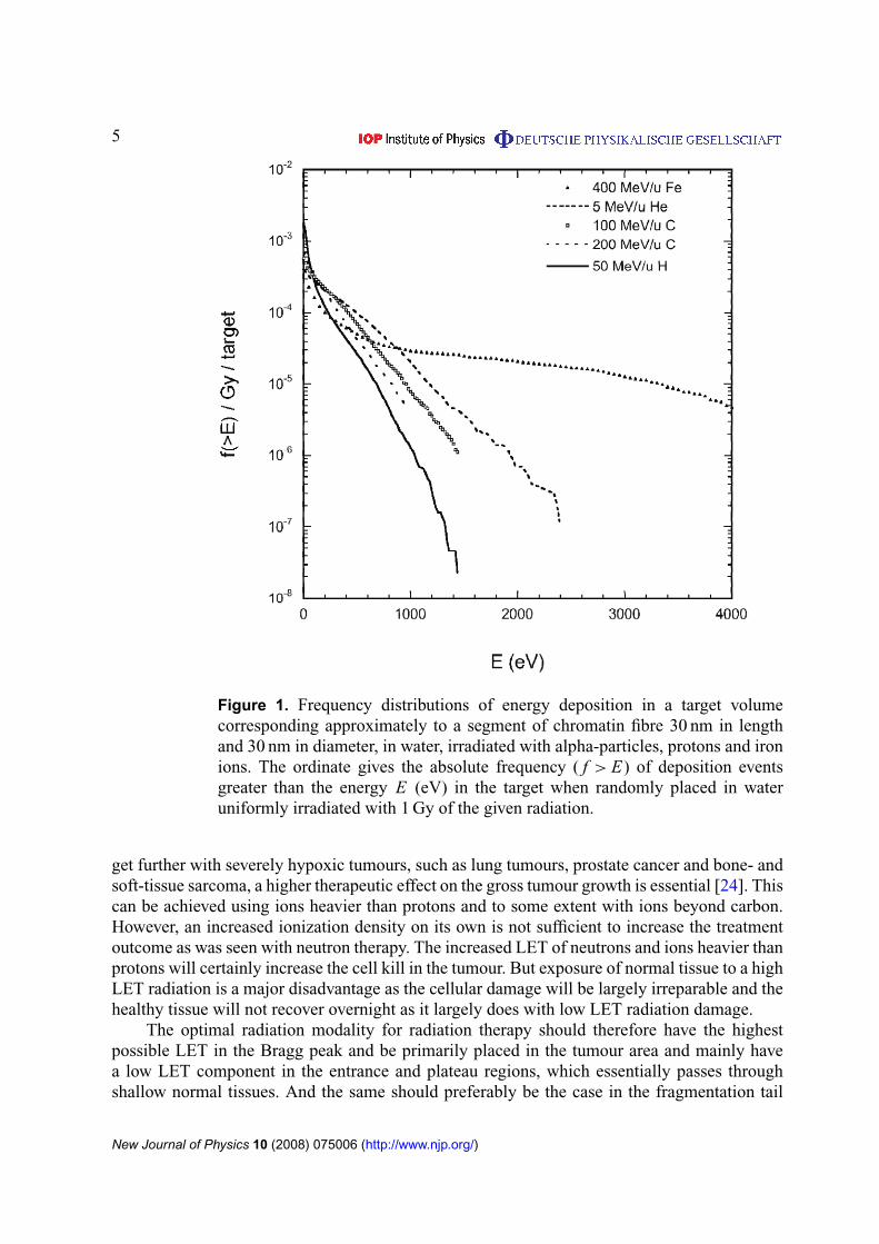
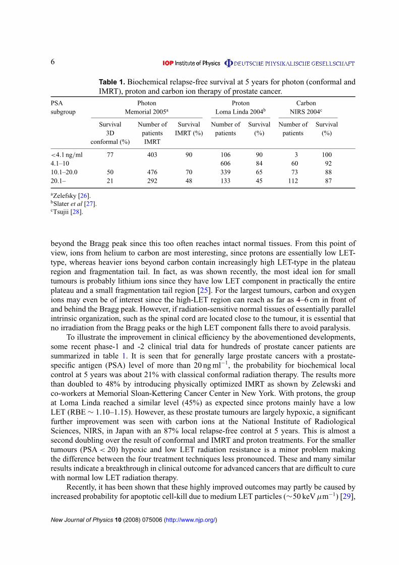
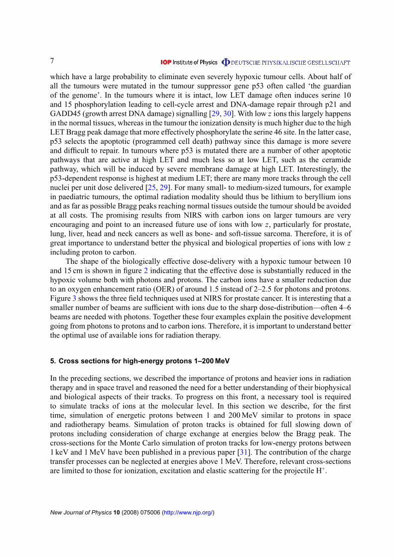
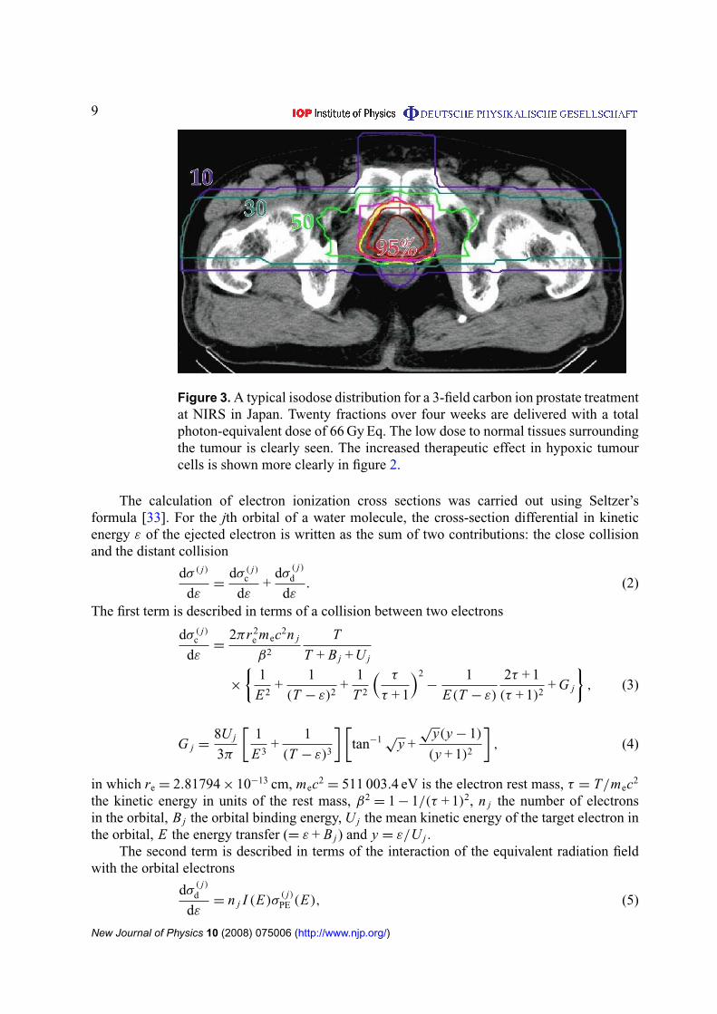
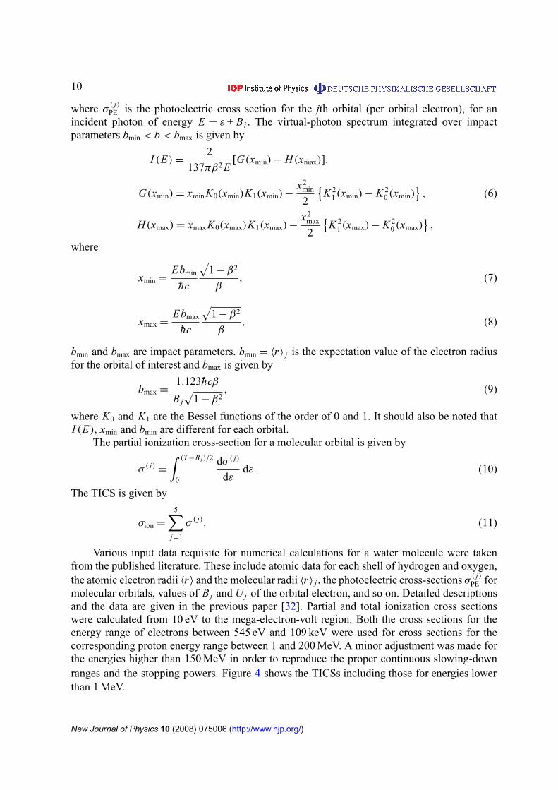
10
where σ( j)PE is the photoelectric cross section for the jth orbital (per orbital electron) for an
incident photon of energy E = ε + B j The virtual-photon spectrum integrated over impactparameters bmin lt b lt bmax is given by
I (E) =2
137πβ2 E[G(xmin) minus H(xmax)]
G(xmin) = xminK0(xmin)K1(xmin) minusx2
min
2
K 2
1 (xmin) minus K 20 (xmin)
(6)
H(xmax) = xmaxK0(xmax)K1(xmax) minusx2
max
2
K 2
1 (xmax) minus K 20 (xmax)
where
xmin =Ebmin
hc
radic1 minus β2
β (7)
xmax =Ebmax
hc
radic1 minus β2
β (8)
bmin and bmax are impact parameters bmin = 〈r〉 j is the expectation value of the electron radiusfor the orbital of interest and bmax is given by
bmax =1123hcβ
B j
radic1 minus β2
(9)
where K0 and K1 are the Bessel functions of the order of 0 and 1 It should also be noted thatI (E) xmin and bmin are different for each orbital
The partial ionization cross-section for a molecular orbital is given by
σ ( j)=
int (T minusB j )2
0
dσ ( j)
dεdε (10)
The TICS is given by
σion =
5sumj=1
σ ( j) (11)
Various input data requisite for numerical calculations for a water molecule were takenfrom the published literature These include atomic data for each shell of hydrogen and oxygenthe atomic electron radii 〈r〉 and the molecular radii 〈r〉 j the photoelectric cross-sections σ
( j)PE for
molecular orbitals values of B j and U j of the orbital electron and so on Detailed descriptionsand the data are given in the previous paper [32] Partial and total ionization cross sectionswere calculated from 10 eV to the mega-electron-volt region Both the cross sections for theenergy range of electrons between 545 eV and 109 keV were used for cross sections for thecorresponding proton energy range between 1 and 200 MeV A minor adjustment was made forthe energies higher than 150 MeV in order to reproduce the proper continuous slowing-downranges and the stopping powers Figure 4 shows the TICSs including those for energies lowerthan 1 MeV
New Journal of Physics 10 (2008) 075006 (httpwwwnjporg)
11
10ndash20
10ndash19
10ndash18
10ndash17
10ndash16
10ndash15
10ndash14
10ndash3 10ndash2 10ndash1 100 101 102
Elastic
Ionization
Excitation
Tot
alcr
oss
sect
ion
(cm
2 )
Proton energy (MeV)
Figure 4 Total cross-sections due to proton impact on water Those for energieslower than 1 MeV were the same as the previous paper [31] TICS for high-energy protons were obtained by energy scaling of the electron ionization crosssections as described in section 511 Excitation cross sections were calculatedusing equation (15) Elastic scattering cross sections were calculated usingequation (26)
512 Single differential cross sections (SDCSs) The collision of a charged particle withanother at rest is described by the Rutherford scattering formula [34]
dσR
dε=
4πa20
T
(R
E
)2
(12)
where ε is the kinetic energy of the secondary electron after the collision T the kinetic energyof an electron with the same speed as the incident proton ie T = Tpλ a0 the Bohr radiusR the Rydberg energy (136 eV) and E = ε + B the energy transfer in which B is the bindingenergy of a molecular orbital
Based on the Rutherford formula the binary-encounter approximation treats the collisionas a classical one between the projectile and a single electron in the target A simpler form ofthe binary-encounter theory leads to a singly differential cross section of the form [34]dσBE
dε=
(dσR
dε
) (1 +
4U
3E
) for Emin 6 E 6 Eminus (13a)
dσBE
dε=
(dσR
dε
) (U
6E
) (
4T
U
)32
+
[1 minus
radic1 +
E
U
]3 for Eminus 6 E 6 E+ (13b)
New Journal of Physics 10 (2008) 075006 (httpwwwnjporg)
12
10ndash21
10ndash20
10ndash19
10ndash18
10ndash17
10ndash16
10ndash15
10ndash14
1 10 100 1000
SD
CS
(cm
2 e
Vndash1
)
Electron energy (eV)
x 100
x 10
Binary-encounter approximation15 MeV
30 MeV
42 MeV
Lines this workSymbols expt (Wilson et al)
Figure 5 Randomly sampled energy spectra for secondary electrons ejectedby proton impact in comparison with the experimental data taken from Wilsonet al [35]
anddσBE
dε= 0 for E gt E+ (13c)
where U is the kinetic energy of the target electron and
Eplusmn = 4T plusmn 4(T U )12 (13d)
Figure 5 shows the randomly sampled energy spectra of secondary electrons for the protonenergies 15 3 and 42 MeV in comparison with the experimental data by Wilson et al [35] Thesampled spectra were scaled to fit to the absolute experimental data
513 Angular distributions for secondary electrons For low-energy proton impact exper-imental angular distributions of electrons ejected from water by proton impact were usedExperimental data are unavailable for high-energy protons gt1 MeV The initial emission anglefor secondary electrons was approximated by the kinematical relationship
cos θ =
radicλε
4Tp (14)
This equation suggests almost all of the secondary electrons are ejected vertically from theprojectile path The tracks of secondary electrons were generated by the electron code KURBUCwhich is effective for the electron energy of 10 MeV
New Journal of Physics 10 (2008) 075006 (httpwwwnjporg)
13
52 Excitation
Excitation cross sections and mean excitation energy loss by proton impact on water moleculeswere treated based on the formula given by Miller and Green [36] The formula was used forthe low-energy protons [31] They assumed an analytical function for each excited level of theform
σexc(T ) =σ0(Za)(T minus W )ν
I +ν + T +ν (15)
where σ0 = 10minus16 cm2 and Z = 10 W a I and ν for 28 excitation states are given in table IIIof [36] The calculated total cross sections are shown in figure 4 They noted that their excitationcross sections were only rough estimates proposed in the absence of detailed theoretical orexperimental data They further noted that the position and height of the maximum in the crosssection could easily be in error by a factor of two
The mean excitation energy Wav was calculated using the relationship 6W jσe j(T )
6σe j(T ) where W j is the excitation threshold energy and σe j(T ) is the excitation cross sectionof equation (15) above for state j
53 Elastic scattering
531 Total cross sections The classical mechanics trajectory calculations (CMTCs) wereapplied to the calculations of total elastic cross sections for high-energy protons This methodis just the same as for the previous work on low-energy protons lower than 1 MeV [31] Theinteraction between the projectile and the target atom can be described in terms of a potentialfunction
V (r) = (zZe2r)Fs(rrs) (16)
The first factor in equation (16) is the Coulomb potential for two bare nuclei with charges zeand Ze The factor Fs(rrs) takes into account the screening by atomic electrons The extentof screening is characterized by the screening length rs A commonly used prescription for thescreening length is
rs = 088534rB(z23 + Z 23)minus12 (17)
where rB is the Bohr radius and 088534 is a numerical constant from the ThomasndashFermi modelof the atom For protons a screening function Fs(rrs) is given by the International Commissionof Radiation Units (ICRU) [37]
Fs(rrs) = 010eminus6rrs + 055eminus12rrs + 035eminus03rrs (18)
For a particle scattered in a central potential V (r) the deflection angle θ is obtained in a CMTCas a function of the impact parameter p [37]
θ = π minus 2int
infin
rmin
p
r 2radic
1 minus V (r)Tcm minus p2r 2dr (19)
where rmin is the distance of the closest approach This value is given by the positive root of thefollowing
r 2minus 2zZre
mec2
βPcr Fs(rrs) minus p2
= 0 (20)
New Journal of Physics 10 (2008) 075006 (httpwwwnjporg)
14
in which
β =
radic1 minus
(mpc2
T + mpc2
)2
Pc =
radic(T + mpc2)2 minus (mpc2)2 (21)
mpc2= proton rest mass and P is the momentum of the proton Tcm is the particle energy in the
centre-of-mass system given by
Tcm =T
1 + mpMt (22)
where T is the kinetic energy in the laboratory system and mp and Mt are the masses ofthe proton and target atom Equation (19) was solved numerically using procedures given byEverhart et al [38] and Wilson and Haggmark [39] Thereby the deflection angle θ was obtainedas a function of the impact parameter p The boundary of lsquolarge-angle calculationsrsquo and lsquosmall-angle calculationsrsquo described by Everhart et al was set at θ = 01π where smooth transitionsare given
The differential elastic scattering cross section can be obtained by numerical differentiationof the curve of impact parameter versus deflection angle using a B-spline interpolation
dσel
d= minus
p
sin θ
dp
dθ (23)
where θ and dσeld are functions of the impact parameter and the particle energy The energydeposition 1E in elastic scattering depends on both the scattering angle and the proton energyThis is given by the formula W (θ T ) according to ICRU 49 [37]
1E = W (θ T ) = 4Tmp Mt
(mp + Mt)2sin2 θ
2 (24)
In order to reduce the divergence of the total cross section the cut-off angles θcut were setsuch as to limit the increase in the scattering probability at low scattering angles [40]
θcut asymp1
137Z 13 mec2
Pc (25)
Total elastic scattering cross section for the proton energy of T is calculated by
σel(T ) = 2π
int π
θcut
dσel
dsin θdθ (26)
Figure 4 shows the calculated total cross section (equation (26)) of proton elastic scatteringin water as a function of T Reproducibility of the nuclear stopping power was confirmed incomparison with ICRU data [37]
532 Angular distributions In the early stages of the development of data compilationangular distributions of elastic scattering calculated by the CMTC were examined The MonteCarlo full slowing-down tracks showed unrealistic tortuous tracks for energies greater than10 MeV Therefore we adopted an alternative formula called the Mott-scattering formulaeffective for high-energy protons [40]
dσel(θ)
d=
1
4NA
Z 2
Az2r 2
e
(mec
Pβ
)2 1 minus β2 sin2(θ2)
sin4(θ2) (27)
where NA = the Avogadro constant and A = molar mass of the scattering medium The Mottformula was derived assuming the repulsive Coulomb force not taking into account the
New Journal of Physics 10 (2008) 075006 (httpwwwnjporg)
15
0
1times10ndash9
2times10ndash9
3times10ndash9
4times10ndash9
5times10ndash9
6times10ndash9
7times10ndash9
0 3 6 9 12 15 18
proton
2nd-e
sum
Depth (cm)
Depthndashdose distribution150 MeV protons
Dos
e(G
ycm
2 )CSDA range = 158 cm
Figure 6 Calculated depthndashdose curves for protons and secondary electrons andtheir sum for a broad parallel beam of protons with 150 MeV These distributionswere obtained from the full slowing down proton tracks
screening by atomic electrons for the relativistic energy of projectiles At the non-relativisticlimit this formula is reduced to the classical Rutherford scattering formula Both formulaeprovide less-deflective tracks than the CMTC results because the screening effect is neglected
54 Depthndashdose distribution
The reliability of cross sections and angular distributions for protons can be tested by severalmacroscopic quantities such as stopping powers continuous slowing down approximation(CSDA) ranges and depthndashdose distributions We confirmed that the calculated stopping powersand ranges agree well with the published data for liquid water in the energy range up to200 MeV [37] Depthndashdose distribution is a severe criterion on the track structure code becauseit is affected by not only stopping powers but also proton deflections
Figure 6 shows the depthndashdose curve in water phantom for a broad parallel beam of protonswith 150 MeV Absorbed dose is represented in Gy cm2 units In this case the HEPHIST codewas executed under the lsquofull slowing downrsquo mode The depth of the Bragg peak is in agreementwith the CSDA range of 158 cm for liquid water This supports an adoption of the Mott-scattering formula for angular distributions for high-energy protons The calculated distributionimplies the capability of the microscopic track structure to obtain such a macroscopic quantityin the simcm region The contribution of secondary electrons to the total dose amounts to 70over the whole depth
55 Radial dose distribution
The radial dose distribution which is the energy absorbed locally at a certain radial distancefrom the path of a primary particle has been used to check the reliability of track structurecalculations Figure 7 shows the radial dose profiles around the path of protons with variousenergies According to the definition of radial dose distribution the energy depositions along the
New Journal of Physics 10 (2008) 075006 (httpwwwnjporg)
16
10ndash2
10ndash1
100
101
102
103
104
105
0 10 20 30 40 50 60 7 0
1 MeV
10 MeV
50 MeV
200 MeV
Radial distance (nm)
Abs
orbe
d do
se (
Gy)
Radial dose profile for protons
Figure 7 Calculated radial dose profiles at 1 10 50 and 200 MeV proton paths
proton track itself are excluded in dose calculations Energies deposited only due to secondaryelectrons were accumulated within a coaxial shell with a radial interval of 1 nm along the protontrack with a short length
56 Microdosimetry
A tissue equivalent proportional counter (TEPC) is the principal instrument for measuring thedistribution of energy deposited in a specific volume The instrument is commonly designed asa spherical or a cylindrical volume with varying sensitive volume The standard representationof a microdosimetric spectrum is the dose distribution yd(y) in the lineal energy y in whichthe area between any two values of y indicates the dose delivered in that range Distributionsof lineal energy were simulated for various proton energies through a wall-less counter 1 micromdiameter using proton tracks generated by the HEPHIST code
Figure 8 shows a comparison of the calculated spectrum for 50 MeV protons betweenthe present work and the published data by Nikjoo et al [41] The theory of microdosimetrydefines several average parameters the frequency average lineal energy yF and specific energyzF and the dose average yD and zD Table 2 shows such average parameters for various protonenergies in comparison with the published data which were obtained using a track segmentcode PITS [41] The parameters calculated by the present code showed a good agreementwith the PITS data The reliability of the HEPHIST code was confirmed within the statisticaluncertainties
6 Electron track simulation in liquid water
Electron track simulation in matter depends on the availability of realistic interaction crosssections over the complete slowing down energy spectrum The interaction of electrons with
New Journal of Physics 10 (2008) 075006 (httpwwwnjporg)
17
00
02
04
06
08
10
01 1 10
this workNikjoo et al
)y(d y
y (keVmicrom)
50 MeV protons1 microm wall-less TEPC
yF = 093 keVmicrom
yD = 205 keVmicrom
zF = 019 Gy
zD = 042 Gy
Figure 8 Calculated lineal energy distributions y d(y) of 50 MeV protons insimulated wall-less TEPC for a site diameter of 1 micron
Table 2 Microdosimetric quantities for a wall-less TEPC with a site diameter of1 microm calculated using the HEPHIST code
Proton energy yF yD zF zD
(MeV) (keV micromminus1) (keV micromminus1) (Gy) (Gy)
1 256 295 523 6025 636 809 130 16510 381 515 078 10520 206 328 042 06740 109 208 022 04250 093 205 019 042100 050 132 010 027150 033 105 0068 021200 026 074 0053 015
water vapour is relatively well understood and scattering cross sections are generally available toa good accuracy (sim10ndash20) by either theory or experiment [42] This is in sharp contrast to thecondensed phase (ie liquids and solids) where experimental data are scarce and the underlyingtheory is computationally intractable [43] Therefore a large number of codes simulateelectron tracks in a unit-density water-vapour medium [44] However due to the long-range
New Journal of Physics 10 (2008) 075006 (httpwwwnjporg)
18
polarization-screening effects in the condensed phase scattering cross sections are expected todiffer from those in the gas phase [45]
The most important interaction coefficients for electron track simulation are the differentialcross sections (DCSs) for elastic (dσeld) and inelastic (dσineldE) scattering which determinerespectively the scattering angle () and energy loss (E) in a single collision The elastic meanfree path (EMFP λel) and inelastic mean free path (IMFP λinel) scattering are then readilyobtained from the corresponding DCSs For use in track simulation studies the DCSs are mostconveniently calculated within the relativistic plane-wave Born-approximation (PWBA) whichprovides a near-exact framework at sufficiently high projectile energies [46]
61 Elastic scattering
For elastic electron scattering by atomic nuclei the PWBA with a Wentzel-like potentialV (r) = minus(Zr)eminusrR where R = 0885 α0 Zminus13 leads to particular useful analytic forms forboth the DCS and EMFP which are often referred to as the screened Rutherford formulae [47]
dσel
d= Z 2r 2
e
(1 minus β2
β4
)1
(1 minus cos θ + 2ηW)2 (28)
λminus1el = π Z 2r 2
e N
(1 minus β2
β4
)1
ηW(ηW + 1) (29)
where θ is the scattering angle between the emitted and the incident electrons N is the densityof scattering centres Z the atomic number of the material re the classical electron radiusβ(equiv υc) the ratio of the incident electron velocity (υ) to the velocity of light (c) and ηW ascreening parameter which reduces the cross section at small scattering angles (associated withlarge impact parameters) Within the Wentzel model ηW takes the form
ηW =17 times 10minus5 Z 23
τ(τ + 2) (30)
where τ = Tm0c2 (T and m0 are the electron kinetic energy and rest mass respectively)Improved values for the screening parameter have been evaluated by Moliere [6]
ηM =
[113 + 376
(Z
137β
)2]
ηW (31)
and by Nigam et al [48]
ηN = (112)2ηW (32)
For electron track simulations in water it is customary to use empirically derived screeningparameters by taking into account relevant experimental data One such very popular screeningparameter is that of Grosswendt and Waibel [49] based on nitrogen data
ηG = (164 minus 00825 ln T )ηW (33)
A screening parameter specifically derived from water-vapour data is that of Uehara et al [31]
ηU =
1198ηW T lt 50 keV
ηM T gt 50 keV(34)
A value for the screening parameter between 06ηW and 1ηW has also been suggested in [50]from an analysis of measurements on various low-Z molecules
New Journal of Physics 10 (2008) 075006 (httpwwwnjporg)
19
At high energies the PWBA overestimates the DCS at large scattering angles due to theneglect of relativistic spin effects A simple scheme has been suggested [50] whereby the DCSis expressed as the product of (28) and the ratio of the Mott to the Rutherford cross sectionwhich reads [51]
Krel(θ) = 1 minus β2 sin2 θ
2+ πβ
Z
137
(1 minus sin
θ
2
)sin
θ
2 (35)
In many simulation codes the (less-important) angular deflections in inelastic collisions areaccounted for in an approximate way by replacing Z 2 by Z(Z + 1) in (28) and (29) In [9]an additional suggestion is made to also replace Z by an effective atomic number Zeff whichtentatively equals the corresponding photoelectric value (ie Zeff asymp 74ndash75 for water)
The above discussion neglects condensed phase effects and therefore is strictly applicableto electron track simulation in water vapour A more consistent approach than the PWBA is therelativistic partial wave expansion method (PWEM) [52] where condensed phase effects maybe accounted for through an appropriate interaction potential [53] Unfortunately calculationsby the PWEM are much more cumbersome compared to the PWBA and importantly they donot lead to a closed analytic form The NIST electron elastic-scattering cross-section database(SRD 64) [54] provides PWEM data within the static-exchange approximation (equivalentto the first-order perturbation theory) for electron energies between 50 eV and 300 keV andelements with atomic number (Z ) from 1 to 96 using the algorithm of Salvat and Mayol [55]A detailed discussion of various aspects of these calculations is provided in [56] A comparisonof elastic-DCSs and EMFPs for water vapour is presented in figures 9 and 10 respectivelyThe NIST values are obtained from a linear combination of the corresponding quantities ofthe composite atoms ie dσNISTd = 2dσHd + dσOd and λminus1
NIST = 2λminus1H + λminus1
O They arecompared with calculations based on the screened Rutherford formulae (29) and (30) usingdifferent screening parameters (31)ndash(35) as well as the Mott spin-correction (36) With respectto the DCS (figure 9) the agreement at 100 keV is satisfactory at small scattering angles wherethe DCS is largest Differences increase with scattering angle where the use of the Mott spin-correction substantially improves the agreement with NIST At 100 eV the NIST data are mostuncertain due to the neglect of polarization and absorption effects (equivalent to second-orderperturbation terms) As expected the choice of the screening parameter becomes importantat low energies and affects the DCS mostly at small scattering angles The Moliere screeningleads to unrealistic DCSs whereas the magnitude of the Mott spin-correction is negligibleWith respect to the EMFP (figure 10) it seems that by an appropriate selection of the screeningparameter very good agreement with NISTrsquos more elaborate PWEM may be obtained above afew keV In the sub-keV region where both the static-exchange approximation (used by NIST)and the assumptions of the PWBA (underlying the screened Rutherford formulae) deterioratediscrepancies increase
The use of a solid-state potential in the PWEM is known to reduce the DCS at smallscattering angles and electron energies below a few tens of keV [53] The magnitude ofcondensed-phase effects on elastic electron scattering in water is not yet clear For examplethe experimental data of Michaud et al [43] for very low-energy electrons (lt100 eV) suggestthat the EMFP in amorphous ice is larger compared to the vapour phase by a factor of 2ndash10 Onthe other hand the recent PWEM calculations of Aouchiche et al [57] for gaseous and liquidwater reveal a very small phase effect (sim15) above 100 eV which further diminishes at lowerenergies
New Journal of Physics 10 (2008) 075006 (httpwwwnjporg)
20
Figure 9 The differential cross section for elastic scattering for (a) 100 eVand (b) 100 keV electrons in water Comparison of the NIST database SRD64 [51] with the screened Rutherford formula (1) using the screening parametersof (3)ndash(7) and the Mott spin-correction (8)
62 Inelastic scattering
The main challenge in the study of inelastic electron scattering from atomic electrons is toproperly account for binding effects This is the main reason why binary collision models failunder various circumstances The PWBA provides a consistent framework for accounting forbinding effects through the use of material-specific structure functions An in-depth discussionof relativistic inelastic electron-scattering in condensed matter within the PWBA has recentlybeen presented by Fernaacutendez-Varea et al [58] The inelastic DCS may be expressed as the sum
New Journal of Physics 10 (2008) 075006 (httpwwwnjporg)
21
Figure 10 The mean free path for elastic scattering of electrons in water overthe 10 eV to 1 MeV energy range Comparison of the NIST database SRD64 [54] with the screened Rutherford formula (2) using the screening parametersof (3)ndash(7) and the Mott spin-correction (8)
of two terms
dσPWBA
dE=
dσ(L)
PWBA
dE+
dσ(T)
PWBA
dE (36)
where the superscripts lsquoLrsquo and lsquoTrsquo denote the longitudinal and transverse contributionsrespectively The longitudinal term is obtained from
dσ(L)
PWBA
dE=
1
πα0 Nmc2β2
QmaxintQmin
1 + Qmc2
Q(1 + Q2mc2)Im
[minus1
ε(E q)
]dQ (37)
where E and q are the energy and momentum transfers respectively Q is the free-recoil energydefined by Q(Q + 2mc2) = (cq)2 and ε(E Q) = ε1(E Q) + iε2(E Q) is the complex-valuedielectric response function of the material The Im[minus1ε(E q)] = ε2(E q)|ε(E q)|2 termis the so-called loss function The limits of integration in (37) are
Qmaxmin =
[radicT (T + 2mc2) plusmn
radic(T minus E)(T minus E + 2mc2)
]2+ (mc2)2
12
minus mc2 (38)
The transverse term in (36) is obtained from
dσ(T)
PWBA
dE=
1
πα0 Nβ2mc2Im
[minus1
ε(E q = 0)
] [ln
(1
1 minus β2
)minus β2
] (39)
Note that the transverse term vanishes at the non-relativistic limit (β 1) and that onlyinteractions with zero momentum transfer (q = 0) contribute to its magnitude [59] The
New Journal of Physics 10 (2008) 075006 (httpwwwnjporg)
22
connection between condensed and gas phase inelastic cross-sections is obtained from [58]
d f (E q)
dE= E(1 + Qmc2)
1
8Nπ 2α30 R2
Im[
minus1
ε(E q)
] (40)
where d f (E q)dE is the generalized oscillator strength In the gas phase or for high-energytransfers in the condensed phase ε2(E q) ε1(E q) asymp 1 so Im[minus1ε(E q)] asymp ε2(E q)
The main advantage of the PWBA is that essentially the inelastic scattering problem isreduced to that of finding an appropriate description of either the atomicmolecular oscillatorstrength or the solid-state dielectric response function over the complete range of energy-and momentum-transfer (ie the so-called Bethe surface) Ab initio calculations due totheir complexity are generally limited to atomic systems [60] For use in track simulationssemi-empirical models based on optical data and momentum-extension schemes have provedto be most suitable [61] and have been widely applied to condensed water [62]ndash[66] Inparticular the use of experimental optical data automatically accounts for many-body effects(eg polarization correlation collective excitations) that are still computationally intractablePerhaps the only limitation is the scarcity of experimental data at finite momentum transferThe versatility of the extended-optical-data methodology allows the implementation of differentdispersion approximations for the momentum-dependence of the dielectric response functionThe application of the most popular dispersion schemes such as those of Lindhard MerminRitchie Penn Ashley and Liljequist to condensed water have been investigated by Emfietzoglouet al [67] In particular the convenient analytic properties of Ritchiersquos extended-Drude modelhave been exploited by Emfietzoglou et al [68] who developed an improved dielectric responsefunction for liquid water consistent with the experimental data at both zero and finite momentumtransfers The new model is based on the parametrization of the experimental Im(ε) = ε2 databy a linear combination of Drude-type dielectric functions
ε2(E q) = E2p
ionizsumi
D2(E q fi Ei γi) +excitsum
j
D2(E q f j E j γ j)
(41)
where Ep = h(4πne2m)12 is the plasma energy (sim2146 eV for liquid water) and the notationD2 and D2 stands for the lsquonormalrsquo and lsquoderivativersquo Drude functions
D2(E q fi Ei γi) =fi(q)γi(q)E
((Ei + (q22m)g(q))2 minus E2)2 + (γi(q)E)2 (42a)
D2(E q f j E j γ j) =2 f j(q)γ 3
j (q)E3
((E2j minus E2)2 + (γ j(q)E)2)2
(42b)
The subscripts i and j denote respectively the ionization and excitation channels usedin the deconvolution of the experimental spectrum Appropriate ionization cut-offs and aband-gap energy (as an approximate excitation threshold) are applied to truncate the non-physical extension of the Drude functions to sub-threshold values The corresponding analyticexpressions of the Drude functions used to represent Re ε = ε1 (ie D1 and D1) are obtainedfrom the KramersndashKronig relations The details of the above procedure and the values of theDrude coefficients along with their q-dependence are given in [68]
New Journal of Physics 10 (2008) 075006 (httpwwwnjporg)
23
Even in the case where an accurate dielectric response function is available inelasticcalculations within the PWBA are known to lead to sizeable errors at sub-keV energies andin particular at the region of the maximum Alternative methodologies such as for examplethe distorted-wave Born approximation are notoriously more difficult to use and thereforeso far have been applied only to a limited number of atomic systems [60] However it hasbeen recognized that for track simulations PWBA calculations may be safely extended downto low electron energies if supplemented with simple correction terms that account in anapproximate way for exchange and higher order perturbation effects [69] The former maybe conveniently accounted for by either the Ochkur approximation or by the use of Mott-like Born terms whereas the latter by either a simple Coulomb-field velocity transformation(similar to the lsquoBurgessrsquo denominator in the binary-encounter approximation) or a second-orderperturbation term analogous to the Barkas Z 3-term in the stopping of heavy charged particlesThe effect of various model approximations within the extended-optical-data methodology hasbeen investigated in [70] The following scheme has been found to effectively incorporate Borncorrections (BC) in the DCS of low-energy electrons (lt10 keV) [71]
dσBC
dE=
dσPWBA
dE+
dσ2nd
dE+
dσex
dE (43)
where
dσ2nd
dE= minus
2
παoT NL(E ξ)Im
[minus1
ε(E q = 0)
] (44)
dσex(E T )
dE=
ionizsumi
dσi(T minus E + Bi T )
dEminus
[dσi(E T )
dEtimes
dσi(T minus E + Bi T )
dE
]12
(45)
The function L(E ξ) in (17) depends on the parameter ξ associated with the cut-off distanceof soft (q = 0) collisions [72] whereas in the right-hand side of (18) σi denotes the sumσ
(i)PWBA + σ
(i)2nd In figure 11 the inverse-IMFP (ie macroscopic inelastic cross section) of electrons
in liquid water is presented over the 10 eVndash1 MeV energy range Relativistic corrections becomeimportant above a few tens of keV with the main contribution coming from the kinematics sincethe transverse term contributes little (lt10) up to 1 MeV The BC terms are progressivelyimportant below 1 keV In figure 12 the IMFP of electrons in liquid water is presented overthe 10 eVndash10 keV energy range The PWBA results with and without BCs are compared withthose obtained from the TPP-2M formula of NIST [73] The differences from the NIST valuesincrease with decreasing electron energy and are generally between 20 and 40 over the 10 keVto 100 eV range The effect of BCs upon the IMFP remains small (up to 10) down to 300 eVit increases drastically though at lower energies reaching sim50 at 100 eV
Radial distributions of absorbed energy in the vapour and liquid phases of water (both atunit density) calculated from track simulations are presented in figure 13 for point-isotropicmonoenergetic electron sources of 01 1 and 10 keV The energy deposited (average over 105
tracks) is scored in concentric spherical shells of thickness 05 nm For both phases we usethe same set of elastic cross sections calculated from the screened Rutherford formulae Theinelastic cross sections for the vapour have been described in [74] whereas those of the liquidare based on the dielectric response function presented in [68] The electron cut-off energy forboth the primary and all the subsequent generations of electrons was set at 1 Ry It is evident
New Journal of Physics 10 (2008) 075006 (httpwwwnjporg)
24
Figure 11 The inelastic scattering cross section (or inverse inelastic mean freepath) of electrons in liquid water over the 10 eV to 1 MeV energy range Thedielectric response function (14) and (15) of [68] has been used as an input
Figure 12 The inelastic mean free path of electrons in liquid water overthe 10 eV to 10 keV energy range The results from the TPP-2M formula ofNIST [73] are compared with those obtained from the PWBA with and withoutBC The dielectric response function (14) and (15) of [68] was used in the PWBAand BC calculations
New Journal of Physics 10 (2008) 075006 (httpwwwnjporg)
25
Figure 13 Comparison of radial distribution of energy deposition for (a) 01(b) 1 and (c) 10 keV electrons in the vapour and liquid phases of water (both atunit densities)
New Journal of Physics 10 (2008) 075006 (httpwwwnjporg)
26
that polarization and screening effects in the liquid phase as reflected in its dielectric responsefunction result in a higher electron penetration and a more diffuse energy-deposition patternThe effect is more pronounced as the electron energy decreases where cross section differencesbetween the two phases become largest [75]
7 Conclusions
This paper has presented a brief discussion on biological and therapeutic efficacies of heavyions with low atomic number (z) Biophysical aspects of radiation tracks are best explored withthe context of microdosimetry as seen in figures 1 and 8 The paper argues that the use of heavyions such as lithium and carbon will bring much desired improvement in radiation therapyThe paper has presented the first example of a full event by event Monte Carlo track structuregenerating the full slowing down track of a typical radiotherapy proton beam The new toolwill bring the ability to examine in much more detail the biophysical features of the protontrack in the Bragg peak region And lastly the paper presented cross sections for electron tracksimulations in liquid water The electron cross sections in liquid water were calculated usingthe new dielectric model of low-energy electrons which is accurate to better than sim10 downto 100 eV On Earth all radiobiological experiments and calculations are compared against thegold standard data for x-rays and gamma photons Similarly in space and in radiation therapywith heavy ions all data need to be compared with protons as the gold standard It is for thisreason that good and reliable proton data need to be established
References
[1] Nikjoo H and Uehara S 2004 Charged Particle and Photon Interactions with Matter ChemicalPhysiochemical and Biological Consequences with Applications ed A Mozuumder and Y Hatano (NewYork Marcel Dekker)
[2] Nikjoo H and Khvostunov I K 2003 Int J Radiat Biol 79 43[3] Nikjoo H and Khvostunov I K 2006 Int J Low Radiat 3 143[4] Nikjoo H OrsquoNeill P Wilson W E and Goodhead D T 2001 Radiat Res 156 577[5] Nikjoo H OrsquoNeill P Goodhead D T and Terrissol M 1997 Int J Radiat Biol 71 467[6] Charlton D E Nikjoo H and Humm J L 1989 Int J Radiat Biol 56 1[7] Prise K M et al 1998 Int J Radiat Biol 74 173[8] Frankenberg D Brede H J Schrewe U J Steinmetz C Frankenberg-Schwager M Kasten G and Pralle E 2000
Adv Space Res 25 2085[9] Rydberg B Cooper B Cooper P K Holley W R and Chatterjee A 2005 Radiat Res 163 526
[10] Ottolenghi A Ballarini F and Merzagora M 1999 Radiat Environ Biophys 38 1[11] Campa A et al 2005 Int J Radiat Biol 81 841[12] Hsiao Y and Stewart R D 2008 Phys Med Biol 53 233ndash44[13] Harrison L Hatahet Z and Wallace S S 1999 J Mol Biol 290 667[14] David-Cordonnier M H Cunniffe S M Hickson I D and OrsquoNeill P 2002 Biochemistry 41 634[15] Sutherland B M Bennett P V Sidorkina O and Laval J 2000 Proc Natl Acad Sci USA 97 103[16] Chaudhry M A and Weinfeld M 1997 J Biol Chem 272 15650[17] Nikjoo H Goodhead D T Charlton D E and Paretzke H G 1994 Energy deposition by monoenergetic electrons
in cylindrical targets MRC Monograph 1994 MRC Radiobiology Unit Harwell UK[18] Nikjoo H Goodhead D T and Charlton D E 1988 Energy deposition in cylindrical volumes in water irradiated
with ultrasoft x-rays of energy 028 15 45 and 81 keV MRC Radiobiology Unit Monograph 88 MRCRadiobiology Unit Harwell UK
New Journal of Physics 10 (2008) 075006 (httpwwwnjporg)
27
[19] Nikjoo H Boyden-Pratt N Uehara S and Goodhead D T 2001 Energy deposition by monoenergetic protonsin cylindrical targets MRC Monograph 2001 MRC Radiation and Genome Stability Unit Harwell UK
[20] Charlton D E Goodhead D T Wilson W E and Paretzke H G 1985 Energy deposition by monoenergeticprotons and alpha-particles in cylindrical targets MRC Monograph 1985 MRC Radiobiology Unit HarwellUK
[21] Nikjoo H Boyden-Pratt N Girard P Uehara S and Goodhead D T 2002 Energy deposition by alpha-particlesin cylindrical targets MRC Monograph 2002 MRC Radiation and Genome Stability Unit Harwell UK
[22] Kylonen J E Girard P Uehara S Lindborg L and Nikjoo H 2003 Energy deposition by heavy ionsin cylindrical targets MRC Monograph 2003 MRC Radiation and Genome Stability Unit Harwell UK
[23] Grindborg J-E Lillhoumlk J E Lindborg L Gudowska I Soumlderberg J Alm Carlsson G and Nikjoo H 2007Radiat Prot Dosim 126 463ndash6
[24] Brahme A 2004 Int J Rad Oncol Biol Phys 58 603[25] Kempe J Gudowska I and Brahme A 2007 Med Phys 34 183[26] Zelefsky M 2007 private communication Memorial Sloan Kettering Cancer Center New York NY USA[27] Slater J Rossi C Yonemoto L Bush D Jabola B Levy R Grove R Preston W and Slater J 2004 Int J
Radiat Oncol Biol Phys 59 348ndash52[28] Tsujii H 2007 private communication National Institute for Radiological Sciences Chiba Japan[29] Svensson H Ringborg U Naumlslund J and Brahme A 2004 Radiother Oncol 73 206[30] Nakamura Y 2004 Cancer Sci 95 7[31] Uehara S Toburen L H and Nikjoo H 2001 Int J Radiat Biol 77 139[32] Uehara S Nikjoo H and Goodhead D T 1993 Phys Med Biol 38 1841[33] Seltzer S M 1988 Transport of Electrons and Photons ed T M Jenkins W R Nelson and A Rindi (New York
Plenum)[34] ICRU 1996 Secondary electron spectra from charged particle interactions ICRU Report 55[35] Wilson W E Miller J H Toburen L H and Manson S T 1984 J Chem Phys 80 5631[36] Miller J H and Green A E S 1973 Radiat Res 54 343[37] ICRU 1993 Stopping powers and ranges for protons and alpha particles ICRU Report 49[38] Everhart E Stone G and Carbone R J 1955 Phys Rev 99 1287[39] Wilson W D and Haggmark L G 1977 Calculations of nuclear stopping ranges and straggling in the low-
energy region Phys Rev B 15 2458[40] ICRU 1978 Basic aspects of high energy particle interactions and radiation dosimetry ICRU Report 28[41] Nikjoo H Uehara S Pinsky L and Cucinotta F A 2007 Radiat Prot Dosim 126 512[42] Itikawa Y and Mason N 2005 J Phys Chem Ref Data 34 1[43] Michaud M Wen A and Sanche L 2003 Radiat Res 159 3[44] Nikjoo H Uehara S Emfietzoglou D and Cucinotta F A 2006 Radiat Meas 41 1052[45] Inokuti M 1991 Radiat Eff Defects Solids 117 143[46] Inokuti M 1971 Rev Mod Phys 43 297[47] Fernandez-Varea J M Mayol R Baro J and Salvat F 1993 Nucl Instrum Methods B 73 447[48] Nigam B P Sundaresan M K and Wu T-Y 1959 Phys Rev 115 491[49] Grosswendt B and Waibel E 1978 Nucl Instrum Methods 155 145[50] Berger M J Seltzer S M and Maeda K 1970 J Atmos Terr Phys 32 1015[51] Yalcin S Gurler O Gultekin A and Gundoglu O 2006 Phys Lett A 356 138[52] Walker D W 1971 Adv Phys 20 257[53] Fernandez-Varea J M Llovet X and Salvat F 2005 Surf Interface Anal 37 824[54] Powell C J Jablonski A and Salvat F 2005 Surf Interface Anal 37 1068[55] Salvat F and Mayol R 1993 Comput Phys Commun 74 358[56] Jablonski A Salvat F and Powell C J 2004 J Phys Chem Ref Data 33 409[57] Aouchiche H Champion C and Oubaziz D 2008 Radiat Phys Chem 77 107[58] Fernandez-Varea J M Salvat F Dingfelder M and Liljequist D 2005 Nucl Instrum Methods B 229 187
New Journal of Physics 10 (2008) 075006 (httpwwwnjporg)
28
[59] Soh D S Cho B H and Kim Y-K 1982 Phys Rev A 26 1357[60] Segui S Dingfelder M Fernandez-Varea J M and Salvat F 2002 J Phys B At Mol Opt Phys 35 33[61] Powell C J and Jablonski A 1999 J Phys Chem Ref Data 28 19[62] Dingfelder M Hantke D Inokuti M and Paretzke H G 1998 Radiat Phys Chem 53 1[63] Pimblott S M and Siebbeles D A 2002 Nucl Instrum Methods B 194 237[64] Emfietzoglou D 2003 Radiat Phys Chem 66 373[65] Tan Z Xia Y Liu X Zhao M Ji Y Li F and Huang B 2004 Radiat Environ Biophys 43 173ndash82[66] Tung C J Chan W T Chao T C Tu Y H and Kwei C M 2007 Nucl Instrum Methods A 580 598[67] Emfietzoglou D Abril I Garcia-Molina R Petsalakis I D Nikjoo H Kyriakou I and Pathak A 2008 Nucl
Instrum Methods B 266 1154ndash61[68] Emfietzoglou D Cucinotta F A and Nikjoo H 2005 Radiat Res 164 202[69] Fernandez-Varea J M 1998 Radiat Phys Chem 53 235[70] Emfietzoglou D and Nikjoo H 2005 Radiat Res 163 98[71] Emfietzoglou D and Nikjoo H 2007 Radiat Res 167 110[72] Ashley J C 1991 J Phys Condens Matter 3 2741[73] Tanuma S Powell C J and Penn D R 1993 Surf Interface Anal 21 165[74] Emfietzoglou D Papamichael G Kostarelos K and Moscovitch M 2000 Phys Med Biol 45 3171[75] Uehara S Nikjoo H and Goodhead D T 1999 Radiat Res 152 202
New Journal of Physics 10 (2008) 075006 (httpwwwnjporg)
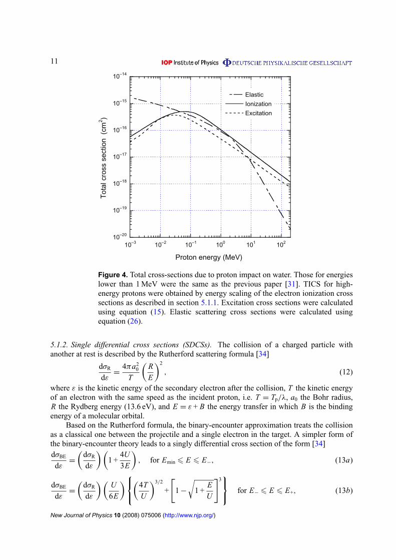
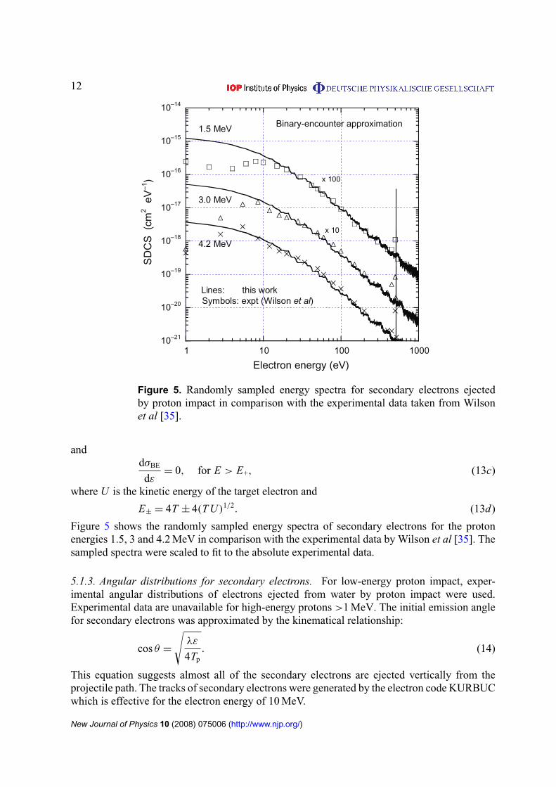
12
10ndash21
10ndash20
10ndash19
10ndash18
10ndash17
10ndash16
10ndash15
10ndash14
1 10 100 1000
SD
CS
(cm
2 e
Vndash1
)
Electron energy (eV)
x 100
x 10
Binary-encounter approximation15 MeV
30 MeV
42 MeV
Lines this workSymbols expt (Wilson et al)
Figure 5 Randomly sampled energy spectra for secondary electrons ejectedby proton impact in comparison with the experimental data taken from Wilsonet al [35]
anddσBE
dε= 0 for E gt E+ (13c)
where U is the kinetic energy of the target electron and
Eplusmn = 4T plusmn 4(T U )12 (13d)
Figure 5 shows the randomly sampled energy spectra of secondary electrons for the protonenergies 15 3 and 42 MeV in comparison with the experimental data by Wilson et al [35] Thesampled spectra were scaled to fit to the absolute experimental data
513 Angular distributions for secondary electrons For low-energy proton impact exper-imental angular distributions of electrons ejected from water by proton impact were usedExperimental data are unavailable for high-energy protons gt1 MeV The initial emission anglefor secondary electrons was approximated by the kinematical relationship
cos θ =
radicλε
4Tp (14)
This equation suggests almost all of the secondary electrons are ejected vertically from theprojectile path The tracks of secondary electrons were generated by the electron code KURBUCwhich is effective for the electron energy of 10 MeV
New Journal of Physics 10 (2008) 075006 (httpwwwnjporg)
13
52 Excitation
Excitation cross sections and mean excitation energy loss by proton impact on water moleculeswere treated based on the formula given by Miller and Green [36] The formula was used forthe low-energy protons [31] They assumed an analytical function for each excited level of theform
σexc(T ) =σ0(Za)(T minus W )ν
I +ν + T +ν (15)
where σ0 = 10minus16 cm2 and Z = 10 W a I and ν for 28 excitation states are given in table IIIof [36] The calculated total cross sections are shown in figure 4 They noted that their excitationcross sections were only rough estimates proposed in the absence of detailed theoretical orexperimental data They further noted that the position and height of the maximum in the crosssection could easily be in error by a factor of two
The mean excitation energy Wav was calculated using the relationship 6W jσe j(T )
6σe j(T ) where W j is the excitation threshold energy and σe j(T ) is the excitation cross sectionof equation (15) above for state j
53 Elastic scattering
531 Total cross sections The classical mechanics trajectory calculations (CMTCs) wereapplied to the calculations of total elastic cross sections for high-energy protons This methodis just the same as for the previous work on low-energy protons lower than 1 MeV [31] Theinteraction between the projectile and the target atom can be described in terms of a potentialfunction
V (r) = (zZe2r)Fs(rrs) (16)
The first factor in equation (16) is the Coulomb potential for two bare nuclei with charges zeand Ze The factor Fs(rrs) takes into account the screening by atomic electrons The extentof screening is characterized by the screening length rs A commonly used prescription for thescreening length is
rs = 088534rB(z23 + Z 23)minus12 (17)
where rB is the Bohr radius and 088534 is a numerical constant from the ThomasndashFermi modelof the atom For protons a screening function Fs(rrs) is given by the International Commissionof Radiation Units (ICRU) [37]
Fs(rrs) = 010eminus6rrs + 055eminus12rrs + 035eminus03rrs (18)
For a particle scattered in a central potential V (r) the deflection angle θ is obtained in a CMTCas a function of the impact parameter p [37]
θ = π minus 2int
infin
rmin
p
r 2radic
1 minus V (r)Tcm minus p2r 2dr (19)
where rmin is the distance of the closest approach This value is given by the positive root of thefollowing
r 2minus 2zZre
mec2
βPcr Fs(rrs) minus p2
= 0 (20)
New Journal of Physics 10 (2008) 075006 (httpwwwnjporg)
14
in which
β =
radic1 minus
(mpc2
T + mpc2
)2
Pc =
radic(T + mpc2)2 minus (mpc2)2 (21)
mpc2= proton rest mass and P is the momentum of the proton Tcm is the particle energy in the
centre-of-mass system given by
Tcm =T
1 + mpMt (22)
where T is the kinetic energy in the laboratory system and mp and Mt are the masses ofthe proton and target atom Equation (19) was solved numerically using procedures given byEverhart et al [38] and Wilson and Haggmark [39] Thereby the deflection angle θ was obtainedas a function of the impact parameter p The boundary of lsquolarge-angle calculationsrsquo and lsquosmall-angle calculationsrsquo described by Everhart et al was set at θ = 01π where smooth transitionsare given
The differential elastic scattering cross section can be obtained by numerical differentiationof the curve of impact parameter versus deflection angle using a B-spline interpolation
dσel
d= minus
p
sin θ
dp
dθ (23)
where θ and dσeld are functions of the impact parameter and the particle energy The energydeposition 1E in elastic scattering depends on both the scattering angle and the proton energyThis is given by the formula W (θ T ) according to ICRU 49 [37]
1E = W (θ T ) = 4Tmp Mt
(mp + Mt)2sin2 θ
2 (24)
In order to reduce the divergence of the total cross section the cut-off angles θcut were setsuch as to limit the increase in the scattering probability at low scattering angles [40]
θcut asymp1
137Z 13 mec2
Pc (25)
Total elastic scattering cross section for the proton energy of T is calculated by
σel(T ) = 2π
int π
θcut
dσel
dsin θdθ (26)
Figure 4 shows the calculated total cross section (equation (26)) of proton elastic scatteringin water as a function of T Reproducibility of the nuclear stopping power was confirmed incomparison with ICRU data [37]
532 Angular distributions In the early stages of the development of data compilationangular distributions of elastic scattering calculated by the CMTC were examined The MonteCarlo full slowing-down tracks showed unrealistic tortuous tracks for energies greater than10 MeV Therefore we adopted an alternative formula called the Mott-scattering formulaeffective for high-energy protons [40]
dσel(θ)
d=
1
4NA
Z 2
Az2r 2
e
(mec
Pβ
)2 1 minus β2 sin2(θ2)
sin4(θ2) (27)
where NA = the Avogadro constant and A = molar mass of the scattering medium The Mottformula was derived assuming the repulsive Coulomb force not taking into account the
New Journal of Physics 10 (2008) 075006 (httpwwwnjporg)
15
0
1times10ndash9
2times10ndash9
3times10ndash9
4times10ndash9
5times10ndash9
6times10ndash9
7times10ndash9
0 3 6 9 12 15 18
proton
2nd-e
sum
Depth (cm)
Depthndashdose distribution150 MeV protons
Dos
e(G
ycm
2 )CSDA range = 158 cm
Figure 6 Calculated depthndashdose curves for protons and secondary electrons andtheir sum for a broad parallel beam of protons with 150 MeV These distributionswere obtained from the full slowing down proton tracks
screening by atomic electrons for the relativistic energy of projectiles At the non-relativisticlimit this formula is reduced to the classical Rutherford scattering formula Both formulaeprovide less-deflective tracks than the CMTC results because the screening effect is neglected
54 Depthndashdose distribution
The reliability of cross sections and angular distributions for protons can be tested by severalmacroscopic quantities such as stopping powers continuous slowing down approximation(CSDA) ranges and depthndashdose distributions We confirmed that the calculated stopping powersand ranges agree well with the published data for liquid water in the energy range up to200 MeV [37] Depthndashdose distribution is a severe criterion on the track structure code becauseit is affected by not only stopping powers but also proton deflections
Figure 6 shows the depthndashdose curve in water phantom for a broad parallel beam of protonswith 150 MeV Absorbed dose is represented in Gy cm2 units In this case the HEPHIST codewas executed under the lsquofull slowing downrsquo mode The depth of the Bragg peak is in agreementwith the CSDA range of 158 cm for liquid water This supports an adoption of the Mott-scattering formula for angular distributions for high-energy protons The calculated distributionimplies the capability of the microscopic track structure to obtain such a macroscopic quantityin the simcm region The contribution of secondary electrons to the total dose amounts to 70over the whole depth
55 Radial dose distribution
The radial dose distribution which is the energy absorbed locally at a certain radial distancefrom the path of a primary particle has been used to check the reliability of track structurecalculations Figure 7 shows the radial dose profiles around the path of protons with variousenergies According to the definition of radial dose distribution the energy depositions along the
New Journal of Physics 10 (2008) 075006 (httpwwwnjporg)
16
10ndash2
10ndash1
100
101
102
103
104
105
0 10 20 30 40 50 60 7 0
1 MeV
10 MeV
50 MeV
200 MeV
Radial distance (nm)
Abs
orbe
d do
se (
Gy)
Radial dose profile for protons
Figure 7 Calculated radial dose profiles at 1 10 50 and 200 MeV proton paths
proton track itself are excluded in dose calculations Energies deposited only due to secondaryelectrons were accumulated within a coaxial shell with a radial interval of 1 nm along the protontrack with a short length
56 Microdosimetry
A tissue equivalent proportional counter (TEPC) is the principal instrument for measuring thedistribution of energy deposited in a specific volume The instrument is commonly designed asa spherical or a cylindrical volume with varying sensitive volume The standard representationof a microdosimetric spectrum is the dose distribution yd(y) in the lineal energy y in whichthe area between any two values of y indicates the dose delivered in that range Distributionsof lineal energy were simulated for various proton energies through a wall-less counter 1 micromdiameter using proton tracks generated by the HEPHIST code
Figure 8 shows a comparison of the calculated spectrum for 50 MeV protons betweenthe present work and the published data by Nikjoo et al [41] The theory of microdosimetrydefines several average parameters the frequency average lineal energy yF and specific energyzF and the dose average yD and zD Table 2 shows such average parameters for various protonenergies in comparison with the published data which were obtained using a track segmentcode PITS [41] The parameters calculated by the present code showed a good agreementwith the PITS data The reliability of the HEPHIST code was confirmed within the statisticaluncertainties
6 Electron track simulation in liquid water
Electron track simulation in matter depends on the availability of realistic interaction crosssections over the complete slowing down energy spectrum The interaction of electrons with
New Journal of Physics 10 (2008) 075006 (httpwwwnjporg)
17
00
02
04
06
08
10
01 1 10
this workNikjoo et al
)y(d y
y (keVmicrom)
50 MeV protons1 microm wall-less TEPC
yF = 093 keVmicrom
yD = 205 keVmicrom
zF = 019 Gy
zD = 042 Gy
Figure 8 Calculated lineal energy distributions y d(y) of 50 MeV protons insimulated wall-less TEPC for a site diameter of 1 micron
Table 2 Microdosimetric quantities for a wall-less TEPC with a site diameter of1 microm calculated using the HEPHIST code
Proton energy yF yD zF zD
(MeV) (keV micromminus1) (keV micromminus1) (Gy) (Gy)
1 256 295 523 6025 636 809 130 16510 381 515 078 10520 206 328 042 06740 109 208 022 04250 093 205 019 042100 050 132 010 027150 033 105 0068 021200 026 074 0053 015
water vapour is relatively well understood and scattering cross sections are generally available toa good accuracy (sim10ndash20) by either theory or experiment [42] This is in sharp contrast to thecondensed phase (ie liquids and solids) where experimental data are scarce and the underlyingtheory is computationally intractable [43] Therefore a large number of codes simulateelectron tracks in a unit-density water-vapour medium [44] However due to the long-range
New Journal of Physics 10 (2008) 075006 (httpwwwnjporg)
18
polarization-screening effects in the condensed phase scattering cross sections are expected todiffer from those in the gas phase [45]
The most important interaction coefficients for electron track simulation are the differentialcross sections (DCSs) for elastic (dσeld) and inelastic (dσineldE) scattering which determinerespectively the scattering angle () and energy loss (E) in a single collision The elastic meanfree path (EMFP λel) and inelastic mean free path (IMFP λinel) scattering are then readilyobtained from the corresponding DCSs For use in track simulation studies the DCSs are mostconveniently calculated within the relativistic plane-wave Born-approximation (PWBA) whichprovides a near-exact framework at sufficiently high projectile energies [46]
61 Elastic scattering
For elastic electron scattering by atomic nuclei the PWBA with a Wentzel-like potentialV (r) = minus(Zr)eminusrR where R = 0885 α0 Zminus13 leads to particular useful analytic forms forboth the DCS and EMFP which are often referred to as the screened Rutherford formulae [47]
dσel
d= Z 2r 2
e
(1 minus β2
β4
)1
(1 minus cos θ + 2ηW)2 (28)
λminus1el = π Z 2r 2
e N
(1 minus β2
β4
)1
ηW(ηW + 1) (29)
where θ is the scattering angle between the emitted and the incident electrons N is the densityof scattering centres Z the atomic number of the material re the classical electron radiusβ(equiv υc) the ratio of the incident electron velocity (υ) to the velocity of light (c) and ηW ascreening parameter which reduces the cross section at small scattering angles (associated withlarge impact parameters) Within the Wentzel model ηW takes the form
ηW =17 times 10minus5 Z 23
τ(τ + 2) (30)
where τ = Tm0c2 (T and m0 are the electron kinetic energy and rest mass respectively)Improved values for the screening parameter have been evaluated by Moliere [6]
ηM =
[113 + 376
(Z
137β
)2]
ηW (31)
and by Nigam et al [48]
ηN = (112)2ηW (32)
For electron track simulations in water it is customary to use empirically derived screeningparameters by taking into account relevant experimental data One such very popular screeningparameter is that of Grosswendt and Waibel [49] based on nitrogen data
ηG = (164 minus 00825 ln T )ηW (33)
A screening parameter specifically derived from water-vapour data is that of Uehara et al [31]
ηU =
1198ηW T lt 50 keV
ηM T gt 50 keV(34)
A value for the screening parameter between 06ηW and 1ηW has also been suggested in [50]from an analysis of measurements on various low-Z molecules
New Journal of Physics 10 (2008) 075006 (httpwwwnjporg)
19
At high energies the PWBA overestimates the DCS at large scattering angles due to theneglect of relativistic spin effects A simple scheme has been suggested [50] whereby the DCSis expressed as the product of (28) and the ratio of the Mott to the Rutherford cross sectionwhich reads [51]
Krel(θ) = 1 minus β2 sin2 θ
2+ πβ
Z
137
(1 minus sin
θ
2
)sin
θ
2 (35)
In many simulation codes the (less-important) angular deflections in inelastic collisions areaccounted for in an approximate way by replacing Z 2 by Z(Z + 1) in (28) and (29) In [9]an additional suggestion is made to also replace Z by an effective atomic number Zeff whichtentatively equals the corresponding photoelectric value (ie Zeff asymp 74ndash75 for water)
The above discussion neglects condensed phase effects and therefore is strictly applicableto electron track simulation in water vapour A more consistent approach than the PWBA is therelativistic partial wave expansion method (PWEM) [52] where condensed phase effects maybe accounted for through an appropriate interaction potential [53] Unfortunately calculationsby the PWEM are much more cumbersome compared to the PWBA and importantly they donot lead to a closed analytic form The NIST electron elastic-scattering cross-section database(SRD 64) [54] provides PWEM data within the static-exchange approximation (equivalentto the first-order perturbation theory) for electron energies between 50 eV and 300 keV andelements with atomic number (Z ) from 1 to 96 using the algorithm of Salvat and Mayol [55]A detailed discussion of various aspects of these calculations is provided in [56] A comparisonof elastic-DCSs and EMFPs for water vapour is presented in figures 9 and 10 respectivelyThe NIST values are obtained from a linear combination of the corresponding quantities ofthe composite atoms ie dσNISTd = 2dσHd + dσOd and λminus1
NIST = 2λminus1H + λminus1
O They arecompared with calculations based on the screened Rutherford formulae (29) and (30) usingdifferent screening parameters (31)ndash(35) as well as the Mott spin-correction (36) With respectto the DCS (figure 9) the agreement at 100 keV is satisfactory at small scattering angles wherethe DCS is largest Differences increase with scattering angle where the use of the Mott spin-correction substantially improves the agreement with NIST At 100 eV the NIST data are mostuncertain due to the neglect of polarization and absorption effects (equivalent to second-orderperturbation terms) As expected the choice of the screening parameter becomes importantat low energies and affects the DCS mostly at small scattering angles The Moliere screeningleads to unrealistic DCSs whereas the magnitude of the Mott spin-correction is negligibleWith respect to the EMFP (figure 10) it seems that by an appropriate selection of the screeningparameter very good agreement with NISTrsquos more elaborate PWEM may be obtained above afew keV In the sub-keV region where both the static-exchange approximation (used by NIST)and the assumptions of the PWBA (underlying the screened Rutherford formulae) deterioratediscrepancies increase
The use of a solid-state potential in the PWEM is known to reduce the DCS at smallscattering angles and electron energies below a few tens of keV [53] The magnitude ofcondensed-phase effects on elastic electron scattering in water is not yet clear For examplethe experimental data of Michaud et al [43] for very low-energy electrons (lt100 eV) suggestthat the EMFP in amorphous ice is larger compared to the vapour phase by a factor of 2ndash10 Onthe other hand the recent PWEM calculations of Aouchiche et al [57] for gaseous and liquidwater reveal a very small phase effect (sim15) above 100 eV which further diminishes at lowerenergies
New Journal of Physics 10 (2008) 075006 (httpwwwnjporg)
20
Figure 9 The differential cross section for elastic scattering for (a) 100 eVand (b) 100 keV electrons in water Comparison of the NIST database SRD64 [51] with the screened Rutherford formula (1) using the screening parametersof (3)ndash(7) and the Mott spin-correction (8)
62 Inelastic scattering
The main challenge in the study of inelastic electron scattering from atomic electrons is toproperly account for binding effects This is the main reason why binary collision models failunder various circumstances The PWBA provides a consistent framework for accounting forbinding effects through the use of material-specific structure functions An in-depth discussionof relativistic inelastic electron-scattering in condensed matter within the PWBA has recentlybeen presented by Fernaacutendez-Varea et al [58] The inelastic DCS may be expressed as the sum
New Journal of Physics 10 (2008) 075006 (httpwwwnjporg)
21
Figure 10 The mean free path for elastic scattering of electrons in water overthe 10 eV to 1 MeV energy range Comparison of the NIST database SRD64 [54] with the screened Rutherford formula (2) using the screening parametersof (3)ndash(7) and the Mott spin-correction (8)
of two terms
dσPWBA
dE=
dσ(L)
PWBA
dE+
dσ(T)
PWBA
dE (36)
where the superscripts lsquoLrsquo and lsquoTrsquo denote the longitudinal and transverse contributionsrespectively The longitudinal term is obtained from
dσ(L)
PWBA
dE=
1
πα0 Nmc2β2
QmaxintQmin
1 + Qmc2
Q(1 + Q2mc2)Im
[minus1
ε(E q)
]dQ (37)
where E and q are the energy and momentum transfers respectively Q is the free-recoil energydefined by Q(Q + 2mc2) = (cq)2 and ε(E Q) = ε1(E Q) + iε2(E Q) is the complex-valuedielectric response function of the material The Im[minus1ε(E q)] = ε2(E q)|ε(E q)|2 termis the so-called loss function The limits of integration in (37) are
Qmaxmin =
[radicT (T + 2mc2) plusmn
radic(T minus E)(T minus E + 2mc2)
]2+ (mc2)2
12
minus mc2 (38)
The transverse term in (36) is obtained from
dσ(T)
PWBA
dE=
1
πα0 Nβ2mc2Im
[minus1
ε(E q = 0)
] [ln
(1
1 minus β2
)minus β2
] (39)
Note that the transverse term vanishes at the non-relativistic limit (β 1) and that onlyinteractions with zero momentum transfer (q = 0) contribute to its magnitude [59] The
New Journal of Physics 10 (2008) 075006 (httpwwwnjporg)
22
connection between condensed and gas phase inelastic cross-sections is obtained from [58]
d f (E q)
dE= E(1 + Qmc2)
1
8Nπ 2α30 R2
Im[
minus1
ε(E q)
] (40)
where d f (E q)dE is the generalized oscillator strength In the gas phase or for high-energytransfers in the condensed phase ε2(E q) ε1(E q) asymp 1 so Im[minus1ε(E q)] asymp ε2(E q)
The main advantage of the PWBA is that essentially the inelastic scattering problem isreduced to that of finding an appropriate description of either the atomicmolecular oscillatorstrength or the solid-state dielectric response function over the complete range of energy-and momentum-transfer (ie the so-called Bethe surface) Ab initio calculations due totheir complexity are generally limited to atomic systems [60] For use in track simulationssemi-empirical models based on optical data and momentum-extension schemes have provedto be most suitable [61] and have been widely applied to condensed water [62]ndash[66] Inparticular the use of experimental optical data automatically accounts for many-body effects(eg polarization correlation collective excitations) that are still computationally intractablePerhaps the only limitation is the scarcity of experimental data at finite momentum transferThe versatility of the extended-optical-data methodology allows the implementation of differentdispersion approximations for the momentum-dependence of the dielectric response functionThe application of the most popular dispersion schemes such as those of Lindhard MerminRitchie Penn Ashley and Liljequist to condensed water have been investigated by Emfietzoglouet al [67] In particular the convenient analytic properties of Ritchiersquos extended-Drude modelhave been exploited by Emfietzoglou et al [68] who developed an improved dielectric responsefunction for liquid water consistent with the experimental data at both zero and finite momentumtransfers The new model is based on the parametrization of the experimental Im(ε) = ε2 databy a linear combination of Drude-type dielectric functions
ε2(E q) = E2p
ionizsumi
D2(E q fi Ei γi) +excitsum
j
D2(E q f j E j γ j)
(41)
where Ep = h(4πne2m)12 is the plasma energy (sim2146 eV for liquid water) and the notationD2 and D2 stands for the lsquonormalrsquo and lsquoderivativersquo Drude functions
D2(E q fi Ei γi) =fi(q)γi(q)E
((Ei + (q22m)g(q))2 minus E2)2 + (γi(q)E)2 (42a)
D2(E q f j E j γ j) =2 f j(q)γ 3
j (q)E3
((E2j minus E2)2 + (γ j(q)E)2)2
(42b)
The subscripts i and j denote respectively the ionization and excitation channels usedin the deconvolution of the experimental spectrum Appropriate ionization cut-offs and aband-gap energy (as an approximate excitation threshold) are applied to truncate the non-physical extension of the Drude functions to sub-threshold values The corresponding analyticexpressions of the Drude functions used to represent Re ε = ε1 (ie D1 and D1) are obtainedfrom the KramersndashKronig relations The details of the above procedure and the values of theDrude coefficients along with their q-dependence are given in [68]
New Journal of Physics 10 (2008) 075006 (httpwwwnjporg)
23
Even in the case where an accurate dielectric response function is available inelasticcalculations within the PWBA are known to lead to sizeable errors at sub-keV energies andin particular at the region of the maximum Alternative methodologies such as for examplethe distorted-wave Born approximation are notoriously more difficult to use and thereforeso far have been applied only to a limited number of atomic systems [60] However it hasbeen recognized that for track simulations PWBA calculations may be safely extended downto low electron energies if supplemented with simple correction terms that account in anapproximate way for exchange and higher order perturbation effects [69] The former maybe conveniently accounted for by either the Ochkur approximation or by the use of Mott-like Born terms whereas the latter by either a simple Coulomb-field velocity transformation(similar to the lsquoBurgessrsquo denominator in the binary-encounter approximation) or a second-orderperturbation term analogous to the Barkas Z 3-term in the stopping of heavy charged particlesThe effect of various model approximations within the extended-optical-data methodology hasbeen investigated in [70] The following scheme has been found to effectively incorporate Borncorrections (BC) in the DCS of low-energy electrons (lt10 keV) [71]
dσBC
dE=
dσPWBA
dE+
dσ2nd
dE+
dσex
dE (43)
where
dσ2nd
dE= minus
2
παoT NL(E ξ)Im
[minus1
ε(E q = 0)
] (44)
dσex(E T )
dE=
ionizsumi
dσi(T minus E + Bi T )
dEminus
[dσi(E T )
dEtimes
dσi(T minus E + Bi T )
dE
]12
(45)
The function L(E ξ) in (17) depends on the parameter ξ associated with the cut-off distanceof soft (q = 0) collisions [72] whereas in the right-hand side of (18) σi denotes the sumσ
(i)PWBA + σ
(i)2nd In figure 11 the inverse-IMFP (ie macroscopic inelastic cross section) of electrons
in liquid water is presented over the 10 eVndash1 MeV energy range Relativistic corrections becomeimportant above a few tens of keV with the main contribution coming from the kinematics sincethe transverse term contributes little (lt10) up to 1 MeV The BC terms are progressivelyimportant below 1 keV In figure 12 the IMFP of electrons in liquid water is presented overthe 10 eVndash10 keV energy range The PWBA results with and without BCs are compared withthose obtained from the TPP-2M formula of NIST [73] The differences from the NIST valuesincrease with decreasing electron energy and are generally between 20 and 40 over the 10 keVto 100 eV range The effect of BCs upon the IMFP remains small (up to 10) down to 300 eVit increases drastically though at lower energies reaching sim50 at 100 eV
Radial distributions of absorbed energy in the vapour and liquid phases of water (both atunit density) calculated from track simulations are presented in figure 13 for point-isotropicmonoenergetic electron sources of 01 1 and 10 keV The energy deposited (average over 105
tracks) is scored in concentric spherical shells of thickness 05 nm For both phases we usethe same set of elastic cross sections calculated from the screened Rutherford formulae Theinelastic cross sections for the vapour have been described in [74] whereas those of the liquidare based on the dielectric response function presented in [68] The electron cut-off energy forboth the primary and all the subsequent generations of electrons was set at 1 Ry It is evident
New Journal of Physics 10 (2008) 075006 (httpwwwnjporg)
24
Figure 11 The inelastic scattering cross section (or inverse inelastic mean freepath) of electrons in liquid water over the 10 eV to 1 MeV energy range Thedielectric response function (14) and (15) of [68] has been used as an input
Figure 12 The inelastic mean free path of electrons in liquid water overthe 10 eV to 10 keV energy range The results from the TPP-2M formula ofNIST [73] are compared with those obtained from the PWBA with and withoutBC The dielectric response function (14) and (15) of [68] was used in the PWBAand BC calculations
New Journal of Physics 10 (2008) 075006 (httpwwwnjporg)
25
Figure 13 Comparison of radial distribution of energy deposition for (a) 01(b) 1 and (c) 10 keV electrons in the vapour and liquid phases of water (both atunit densities)
New Journal of Physics 10 (2008) 075006 (httpwwwnjporg)
26
that polarization and screening effects in the liquid phase as reflected in its dielectric responsefunction result in a higher electron penetration and a more diffuse energy-deposition patternThe effect is more pronounced as the electron energy decreases where cross section differencesbetween the two phases become largest [75]
7 Conclusions
This paper has presented a brief discussion on biological and therapeutic efficacies of heavyions with low atomic number (z) Biophysical aspects of radiation tracks are best explored withthe context of microdosimetry as seen in figures 1 and 8 The paper argues that the use of heavyions such as lithium and carbon will bring much desired improvement in radiation therapyThe paper has presented the first example of a full event by event Monte Carlo track structuregenerating the full slowing down track of a typical radiotherapy proton beam The new toolwill bring the ability to examine in much more detail the biophysical features of the protontrack in the Bragg peak region And lastly the paper presented cross sections for electron tracksimulations in liquid water The electron cross sections in liquid water were calculated usingthe new dielectric model of low-energy electrons which is accurate to better than sim10 downto 100 eV On Earth all radiobiological experiments and calculations are compared against thegold standard data for x-rays and gamma photons Similarly in space and in radiation therapywith heavy ions all data need to be compared with protons as the gold standard It is for thisreason that good and reliable proton data need to be established
References
[1] Nikjoo H and Uehara S 2004 Charged Particle and Photon Interactions with Matter ChemicalPhysiochemical and Biological Consequences with Applications ed A Mozuumder and Y Hatano (NewYork Marcel Dekker)
[2] Nikjoo H and Khvostunov I K 2003 Int J Radiat Biol 79 43[3] Nikjoo H and Khvostunov I K 2006 Int J Low Radiat 3 143[4] Nikjoo H OrsquoNeill P Wilson W E and Goodhead D T 2001 Radiat Res 156 577[5] Nikjoo H OrsquoNeill P Goodhead D T and Terrissol M 1997 Int J Radiat Biol 71 467[6] Charlton D E Nikjoo H and Humm J L 1989 Int J Radiat Biol 56 1[7] Prise K M et al 1998 Int J Radiat Biol 74 173[8] Frankenberg D Brede H J Schrewe U J Steinmetz C Frankenberg-Schwager M Kasten G and Pralle E 2000
Adv Space Res 25 2085[9] Rydberg B Cooper B Cooper P K Holley W R and Chatterjee A 2005 Radiat Res 163 526
[10] Ottolenghi A Ballarini F and Merzagora M 1999 Radiat Environ Biophys 38 1[11] Campa A et al 2005 Int J Radiat Biol 81 841[12] Hsiao Y and Stewart R D 2008 Phys Med Biol 53 233ndash44[13] Harrison L Hatahet Z and Wallace S S 1999 J Mol Biol 290 667[14] David-Cordonnier M H Cunniffe S M Hickson I D and OrsquoNeill P 2002 Biochemistry 41 634[15] Sutherland B M Bennett P V Sidorkina O and Laval J 2000 Proc Natl Acad Sci USA 97 103[16] Chaudhry M A and Weinfeld M 1997 J Biol Chem 272 15650[17] Nikjoo H Goodhead D T Charlton D E and Paretzke H G 1994 Energy deposition by monoenergetic electrons
in cylindrical targets MRC Monograph 1994 MRC Radiobiology Unit Harwell UK[18] Nikjoo H Goodhead D T and Charlton D E 1988 Energy deposition in cylindrical volumes in water irradiated
with ultrasoft x-rays of energy 028 15 45 and 81 keV MRC Radiobiology Unit Monograph 88 MRCRadiobiology Unit Harwell UK
New Journal of Physics 10 (2008) 075006 (httpwwwnjporg)
27
[19] Nikjoo H Boyden-Pratt N Uehara S and Goodhead D T 2001 Energy deposition by monoenergetic protonsin cylindrical targets MRC Monograph 2001 MRC Radiation and Genome Stability Unit Harwell UK
[20] Charlton D E Goodhead D T Wilson W E and Paretzke H G 1985 Energy deposition by monoenergeticprotons and alpha-particles in cylindrical targets MRC Monograph 1985 MRC Radiobiology Unit HarwellUK
[21] Nikjoo H Boyden-Pratt N Girard P Uehara S and Goodhead D T 2002 Energy deposition by alpha-particlesin cylindrical targets MRC Monograph 2002 MRC Radiation and Genome Stability Unit Harwell UK
[22] Kylonen J E Girard P Uehara S Lindborg L and Nikjoo H 2003 Energy deposition by heavy ionsin cylindrical targets MRC Monograph 2003 MRC Radiation and Genome Stability Unit Harwell UK
[23] Grindborg J-E Lillhoumlk J E Lindborg L Gudowska I Soumlderberg J Alm Carlsson G and Nikjoo H 2007Radiat Prot Dosim 126 463ndash6
[24] Brahme A 2004 Int J Rad Oncol Biol Phys 58 603[25] Kempe J Gudowska I and Brahme A 2007 Med Phys 34 183[26] Zelefsky M 2007 private communication Memorial Sloan Kettering Cancer Center New York NY USA[27] Slater J Rossi C Yonemoto L Bush D Jabola B Levy R Grove R Preston W and Slater J 2004 Int J
Radiat Oncol Biol Phys 59 348ndash52[28] Tsujii H 2007 private communication National Institute for Radiological Sciences Chiba Japan[29] Svensson H Ringborg U Naumlslund J and Brahme A 2004 Radiother Oncol 73 206[30] Nakamura Y 2004 Cancer Sci 95 7[31] Uehara S Toburen L H and Nikjoo H 2001 Int J Radiat Biol 77 139[32] Uehara S Nikjoo H and Goodhead D T 1993 Phys Med Biol 38 1841[33] Seltzer S M 1988 Transport of Electrons and Photons ed T M Jenkins W R Nelson and A Rindi (New York
Plenum)[34] ICRU 1996 Secondary electron spectra from charged particle interactions ICRU Report 55[35] Wilson W E Miller J H Toburen L H and Manson S T 1984 J Chem Phys 80 5631[36] Miller J H and Green A E S 1973 Radiat Res 54 343[37] ICRU 1993 Stopping powers and ranges for protons and alpha particles ICRU Report 49[38] Everhart E Stone G and Carbone R J 1955 Phys Rev 99 1287[39] Wilson W D and Haggmark L G 1977 Calculations of nuclear stopping ranges and straggling in the low-
energy region Phys Rev B 15 2458[40] ICRU 1978 Basic aspects of high energy particle interactions and radiation dosimetry ICRU Report 28[41] Nikjoo H Uehara S Pinsky L and Cucinotta F A 2007 Radiat Prot Dosim 126 512[42] Itikawa Y and Mason N 2005 J Phys Chem Ref Data 34 1[43] Michaud M Wen A and Sanche L 2003 Radiat Res 159 3[44] Nikjoo H Uehara S Emfietzoglou D and Cucinotta F A 2006 Radiat Meas 41 1052[45] Inokuti M 1991 Radiat Eff Defects Solids 117 143[46] Inokuti M 1971 Rev Mod Phys 43 297[47] Fernandez-Varea J M Mayol R Baro J and Salvat F 1993 Nucl Instrum Methods B 73 447[48] Nigam B P Sundaresan M K and Wu T-Y 1959 Phys Rev 115 491[49] Grosswendt B and Waibel E 1978 Nucl Instrum Methods 155 145[50] Berger M J Seltzer S M and Maeda K 1970 J Atmos Terr Phys 32 1015[51] Yalcin S Gurler O Gultekin A and Gundoglu O 2006 Phys Lett A 356 138[52] Walker D W 1971 Adv Phys 20 257[53] Fernandez-Varea J M Llovet X and Salvat F 2005 Surf Interface Anal 37 824[54] Powell C J Jablonski A and Salvat F 2005 Surf Interface Anal 37 1068[55] Salvat F and Mayol R 1993 Comput Phys Commun 74 358[56] Jablonski A Salvat F and Powell C J 2004 J Phys Chem Ref Data 33 409[57] Aouchiche H Champion C and Oubaziz D 2008 Radiat Phys Chem 77 107[58] Fernandez-Varea J M Salvat F Dingfelder M and Liljequist D 2005 Nucl Instrum Methods B 229 187
New Journal of Physics 10 (2008) 075006 (httpwwwnjporg)
28
[59] Soh D S Cho B H and Kim Y-K 1982 Phys Rev A 26 1357[60] Segui S Dingfelder M Fernandez-Varea J M and Salvat F 2002 J Phys B At Mol Opt Phys 35 33[61] Powell C J and Jablonski A 1999 J Phys Chem Ref Data 28 19[62] Dingfelder M Hantke D Inokuti M and Paretzke H G 1998 Radiat Phys Chem 53 1[63] Pimblott S M and Siebbeles D A 2002 Nucl Instrum Methods B 194 237[64] Emfietzoglou D 2003 Radiat Phys Chem 66 373[65] Tan Z Xia Y Liu X Zhao M Ji Y Li F and Huang B 2004 Radiat Environ Biophys 43 173ndash82[66] Tung C J Chan W T Chao T C Tu Y H and Kwei C M 2007 Nucl Instrum Methods A 580 598[67] Emfietzoglou D Abril I Garcia-Molina R Petsalakis I D Nikjoo H Kyriakou I and Pathak A 2008 Nucl
Instrum Methods B 266 1154ndash61[68] Emfietzoglou D Cucinotta F A and Nikjoo H 2005 Radiat Res 164 202[69] Fernandez-Varea J M 1998 Radiat Phys Chem 53 235[70] Emfietzoglou D and Nikjoo H 2005 Radiat Res 163 98[71] Emfietzoglou D and Nikjoo H 2007 Radiat Res 167 110[72] Ashley J C 1991 J Phys Condens Matter 3 2741[73] Tanuma S Powell C J and Penn D R 1993 Surf Interface Anal 21 165[74] Emfietzoglou D Papamichael G Kostarelos K and Moscovitch M 2000 Phys Med Biol 45 3171[75] Uehara S Nikjoo H and Goodhead D T 1999 Radiat Res 152 202
New Journal of Physics 10 (2008) 075006 (httpwwwnjporg)
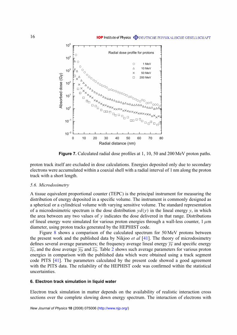
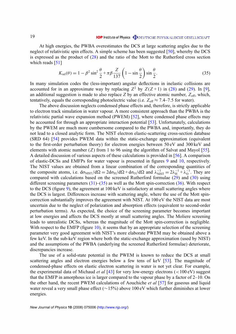
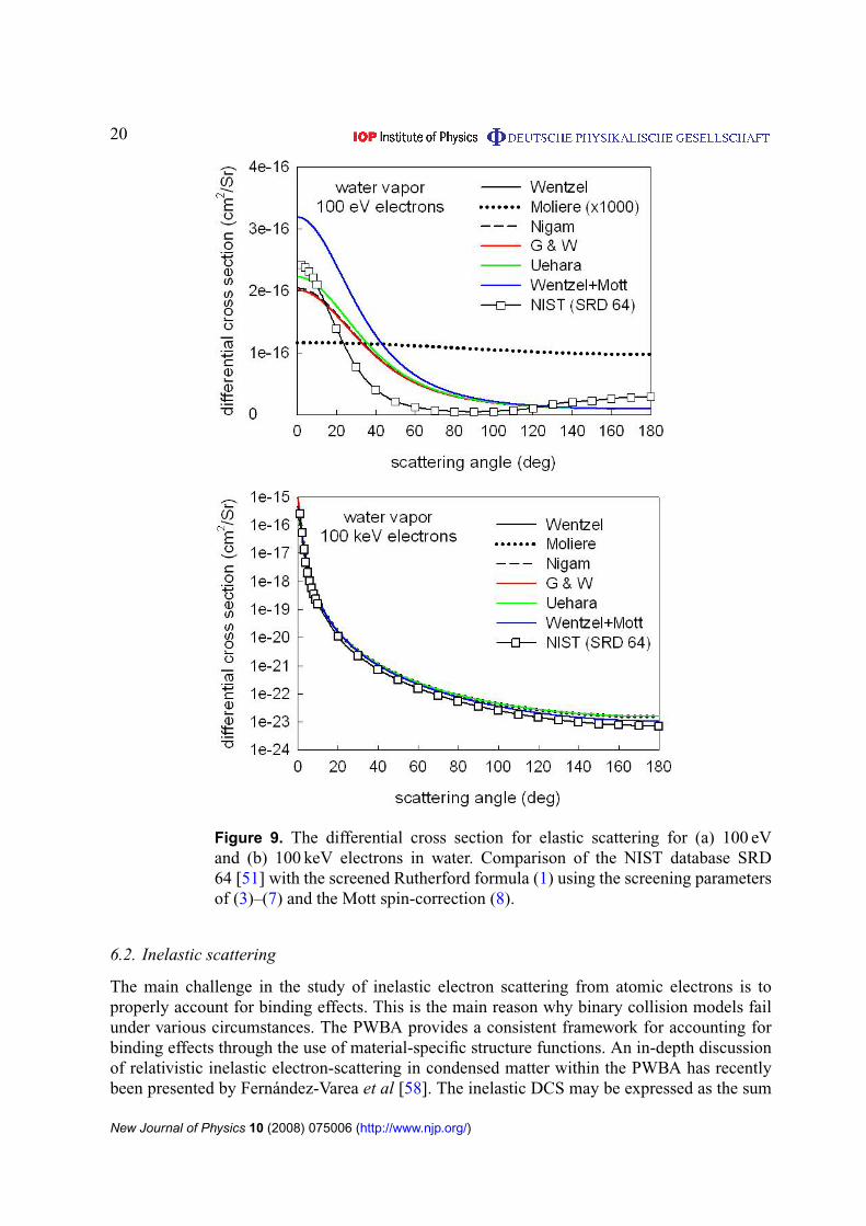
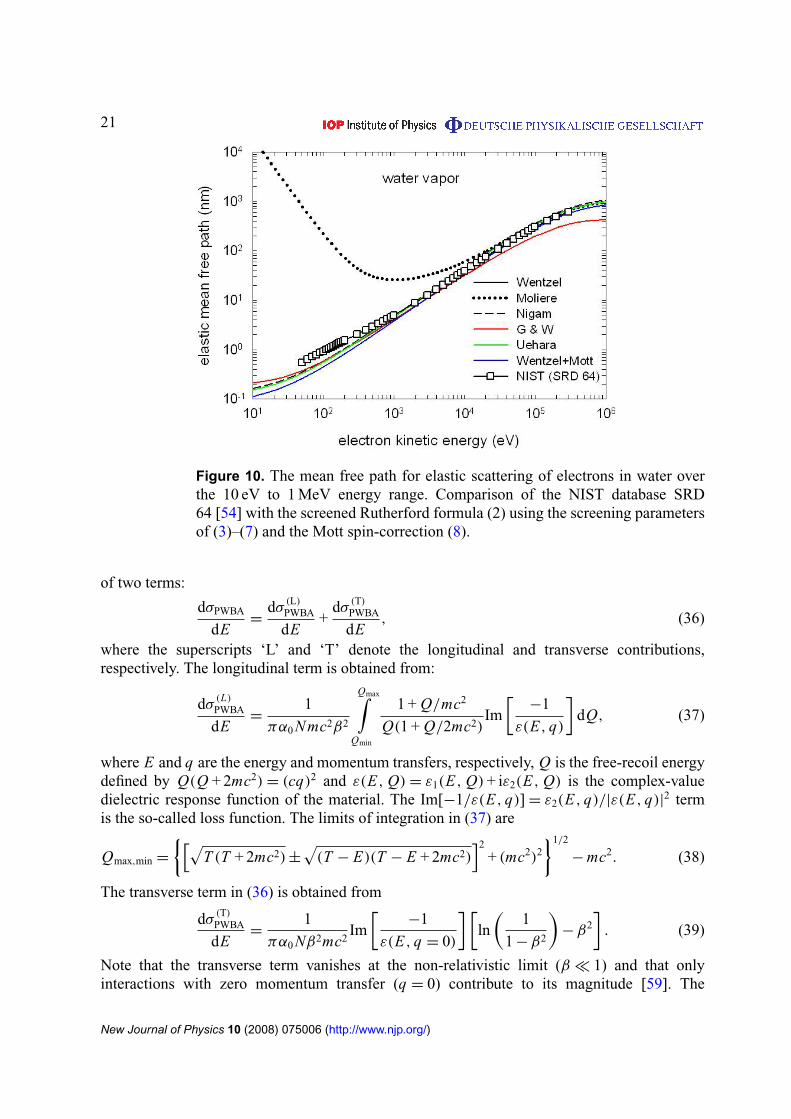
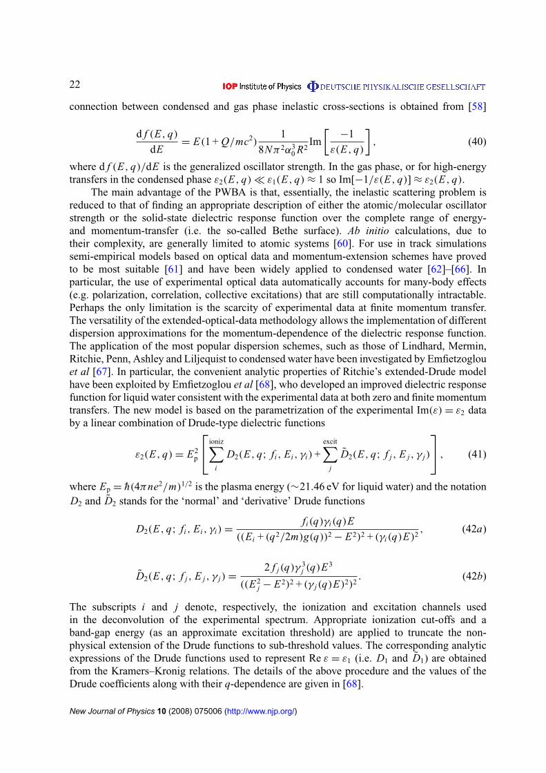
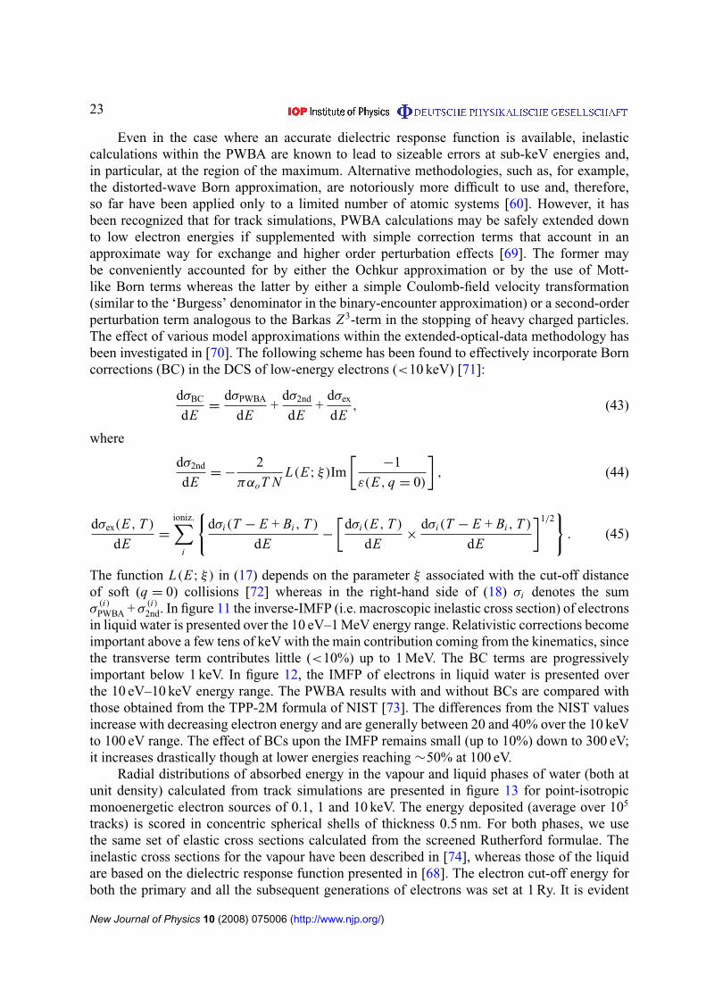
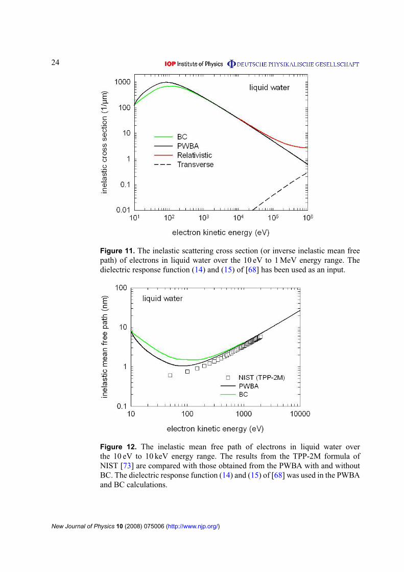
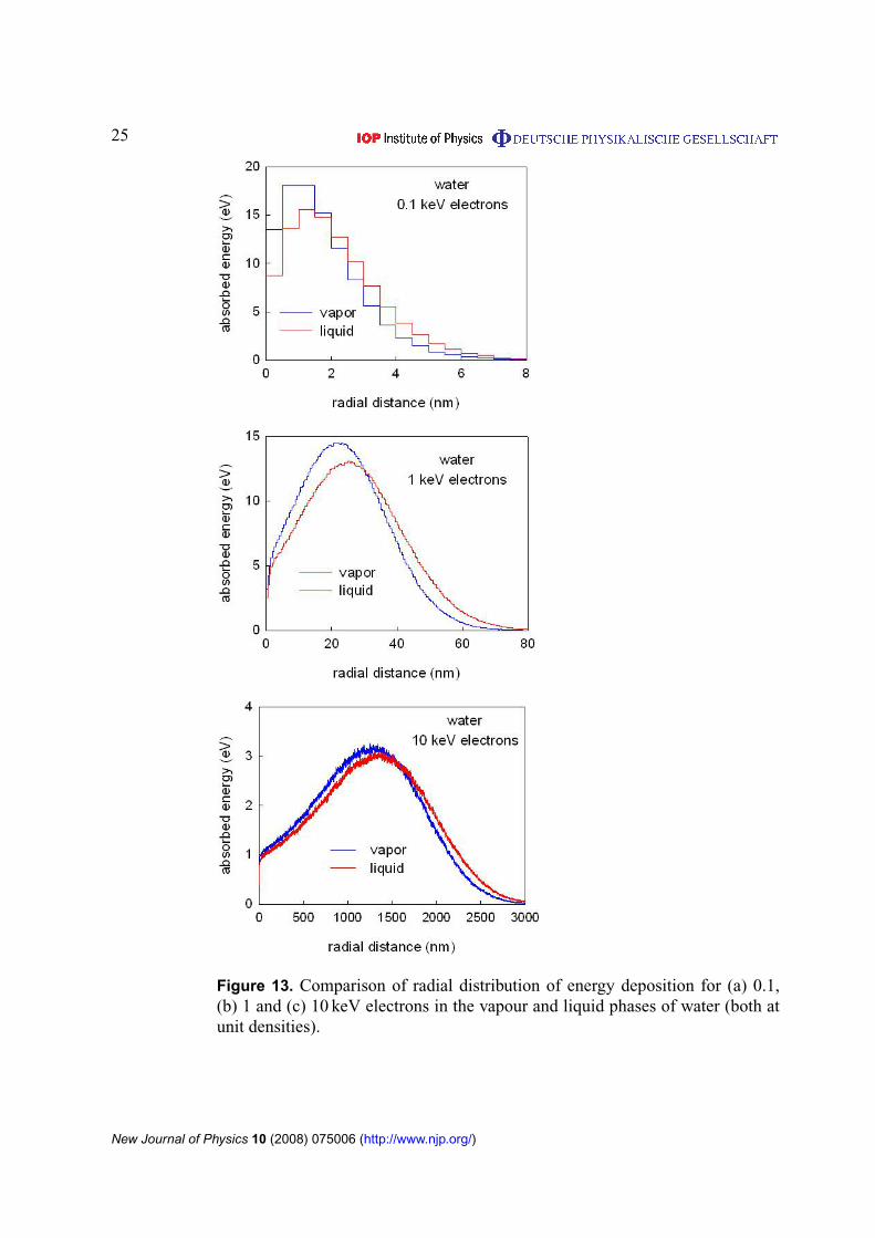