Head-to head comparison of mGlu1 and mGlu5 receptor activation in chronic treatment of absence...
13
Head-to head comparison of mGlu1 and mGlu5 receptor activation in chronic treatment of absence epilepsy in WAG/Rij rats V. D'Amore a, 1 , I. Santolini a, 1 , R. Celli a , L. Lionetto b , A. De Fusco a , M. Simmaco b , C.M. van Rijn c , E. Vieira d , S.R. Stauffer e, f , P.J. Conn e, f , P. Bosco g , F. Nicoletti a, h , G. van Luijtelaar c , R.T. Ngomba a, * a I.R.C.C.S., NEUROMED, Neuropharmacology Unit, Parco Tecnologico, Localit a Camerelle 86077 Pozzilli, Isernia, Italy b Department of Neuroscience and Mental Health, St. Andrea Hospital, Rome, Italy c Donders Centre for Cognition, Donders Institute for Brain, Cognition and Behaviour, Radboud University Nijmegen, Nijmegen, The Netherlands d pRED Discovery Chemistry F. Hoffmann-La Roche Ltd, Pharmaceutical Division, Basel, Switzerland e Department of Pharmacology, Vanderbilt University, Nashville, TN 37232, USA f Vanderbilt Center for Neuroscience Drug Discovery, Nashville, TN 37232, USA g IRCCS Oasi Maria SS Institute for Research on Mental Retardation and Brain Aging, Italy h Department of Physiology and Pharmacology, University “Sapienza”, Rome, Italy article info Article history: Received 26 August 2013 Received in revised form 10 April 2014 Accepted 4 May 2014 Available online 20 May 2014 Keywords: Absence epilepsy Spikeewave discharges WAG/Rij rats VU0360172 RO0711401 Tolerance Chronic treatment Group I mGlu receptors abstract Acute treatment with positive allosteric modulators (PAMs) of mGlu1 and mGlu5 metabotropic gluta- mate receptors (RO0711401 and VU0360172, respectively) reduces the incidence of spike-and wave discharges in the WAG/Rij rat model of absence epilepsy. However, from the therapeutic standpoint, it was important to establish whether tolerance developed to the action of these drugs. We administered either VU0360172 (3 mg/kg, s.c.) or RO0711401 (10 mg/kg, s.c.) to WAG/Rij rats twice daily for ten days. VU0360172 maintained its activity during the treatment, whereas rats developed tolerance to RO0711401 since the 3rd day of treatment and were still refractory to the drug two days after treatment withdrawal. In response to VU0360172, expression of mGlu5 receptors increased in the thalamus of WAG/Rij rats after 1 day of treatment, and remained elevated afterwards. VU0360172 also enhanced mGlu5 receptor expression in the cortex after 8 days of treatment without changing the expression of mGlu1a receptors. Treatment with RO0711401 enhanced the expression of both mGlu1a and mGlu5 receptors in the thal- amus and cortex of WAG/Rij rats after 3e8 days of treatment. These data were different from those obtained in non-epileptic rats, in which repeated injections of RO0711401 and VU0360172 down-regu- lated the expression of mGlu1a and mGlu5 receptors. Levels of VU0360172 in the thalamus and cortex remained unaltered during the treatment, whereas levels of RO0711401 were reduced in the cortex at day 8 of treatment. These findings suggest that mGlu5 receptor PAMs are potential candidates for the treatment of absence epilepsy in humans © 2014 Elsevier Ltd. All rights reserved. 1. Introduction About 20% of patients with absence epilepsy, particularly those with atypical absence seizures, are refractory to the currently used anti-absence drugs, such as ethosuximide, valproate, and clonazepam (Panayiotopoulos, 1999; Glauser et al., 2010). More- over, in patients that are not refractory, antiepileptic drugs may cause class-related adverse effects, such as sedation, dizziness, amnesia and ataxia. Unfortunately, newer anti-absence drugs, such as lamotrigine and lacosemide, loose efficacy during chronic treatment (Glauser et al., 2010) or may have only marginal effects or paradoxically enhance absences seizures in animal models (van Rijn et al., 1994; van Luijtelaar et al., submitted for publication). Therefore, more effective antiepilepsy drugs are still needed. Metabotropic glutamate (mGlu) receptors are potential targets for novel anti-absence drugs. mGlu receptors form a family of eight, * Corresponding author. I.N.M. Neuromed, Localit a Camerelle, 86077 Pozzilli, Isernia, Italy. Tel.: þ39 0865915201; fax: þ39 0865927575. E-mail addresses: [email protected], [email protected] (R. Ngomba). 1 Co-first authors. Contents lists available at ScienceDirect Neuropharmacology journal homepage: www.elsevier.com/locate/neuropharm http://dx.doi.org/10.1016/j.neuropharm.2014.05.005 0028-3908/© 2014 Elsevier Ltd. All rights reserved. Neuropharmacology 85 (2014) 91e103
-
Upload
independent -
Category
Documents
-
view
3 -
download
0
Transcript of Head-to head comparison of mGlu1 and mGlu5 receptor activation in chronic treatment of absence...

lable at ScienceDirect
Neuropharmacology 85 (2014) 91e103
Contents lists avai
Neuropharmacology
journal homepage: www.elsevier .com/locate/neuropharm
Head-to head comparison of mGlu1 and mGlu5 receptor activation inchronic treatment of absence epilepsy in WAG/Rij rats
V. D'Amore a, 1, I. Santolini a, 1, R. Celli a, L. Lionetto b, A. De Fusco a, M. Simmaco b,C.M. van Rijn c, E. Vieira d, S.R. Stauffer e, f, P.J. Conn e, f, P. Bosco g, F. Nicoletti a, h,G. van Luijtelaar c, R.T. Ngomba a, *
a I.R.C.C.S., NEUROMED, Neuropharmacology Unit, Parco Tecnologico, Localit�a Camerelle 86077 Pozzilli, Isernia, Italyb Department of Neuroscience and Mental Health, St. Andrea Hospital, Rome, Italyc Donders Centre for Cognition, Donders Institute for Brain, Cognition and Behaviour, Radboud University Nijmegen, Nijmegen, The Netherlandsd pRED Discovery Chemistry F. Hoffmann-La Roche Ltd, Pharmaceutical Division, Basel, Switzerlande Department of Pharmacology, Vanderbilt University, Nashville, TN 37232, USAf Vanderbilt Center for Neuroscience Drug Discovery, Nashville, TN 37232, USAg IRCCS Oasi Maria SS Institute for Research on Mental Retardation and Brain Aging, Italyh Department of Physiology and Pharmacology, University “Sapienza”, Rome, Italy
a r t i c l e i n f o
Article history:Received 26 August 2013Received in revised form10 April 2014Accepted 4 May 2014Available online 20 May 2014
Keywords:Absence epilepsySpikeewave dischargesWAG/Rij ratsVU0360172RO0711401ToleranceChronic treatmentGroup I mGlu receptors
* Corresponding author. I.N.M. Neuromed, LocalitIsernia, Italy. Tel.: þ39 0865915201; fax: þ39 086592
E-mail addresses: [email protected],(R. Ngomba).
1 Co-first authors.
http://dx.doi.org/10.1016/j.neuropharm.2014.05.0050028-3908/© 2014 Elsevier Ltd. All rights reserved.
a b s t r a c t
Acute treatment with positive allosteric modulators (PAMs) of mGlu1 and mGlu5 metabotropic gluta-mate receptors (RO0711401 and VU0360172, respectively) reduces the incidence of spike-and wavedischarges in the WAG/Rij rat model of absence epilepsy. However, from the therapeutic standpoint, itwas important to establish whether tolerance developed to the action of these drugs. We administeredeither VU0360172 (3 mg/kg, s.c.) or RO0711401 (10 mg/kg, s.c.) to WAG/Rij rats twice daily for ten days.VU0360172 maintained its activity during the treatment, whereas rats developed tolerance to RO0711401since the 3rd day of treatment and were still refractory to the drug two days after treatment withdrawal.In response to VU0360172, expression of mGlu5 receptors increased in the thalamus of WAG/Rij rats after1 day of treatment, and remained elevated afterwards. VU0360172 also enhanced mGlu5 receptorexpression in the cortex after 8 days of treatment without changing the expression of mGlu1a receptors.Treatment with RO0711401 enhanced the expression of both mGlu1a and mGlu5 receptors in the thal-amus and cortex of WAG/Rij rats after 3e8 days of treatment. These data were different from thoseobtained in non-epileptic rats, in which repeated injections of RO0711401 and VU0360172 down-regu-lated the expression of mGlu1a and mGlu5 receptors. Levels of VU0360172 in the thalamus and cortexremained unaltered during the treatment, whereas levels of RO0711401 were reduced in the cortex atday 8 of treatment. These findings suggest that mGlu5 receptor PAMs are potential candidates for thetreatment of absence epilepsy in humans
© 2014 Elsevier Ltd. All rights reserved.
1. Introduction
About 20% of patients with absence epilepsy, particularly thosewith atypical absence seizures, are refractory to the currently usedanti-absence drugs, such as ethosuximide, valproate, and
�a Camerelle, 86077 Pozzilli,[email protected]
clonazepam (Panayiotopoulos, 1999; Glauser et al., 2010). More-over, in patients that are not refractory, antiepileptic drugs maycause class-related adverse effects, such as sedation, dizziness,amnesia and ataxia. Unfortunately, newer anti-absence drugs, suchas lamotrigine and lacosemide, loose efficacy during chronictreatment (Glauser et al., 2010) or may have only marginal effectsor paradoxically enhance absences seizures in animal models (vanRijn et al., 1994; van Luijtelaar et al., submitted for publication).Therefore, more effective antiepilepsy drugs are still needed.
Metabotropic glutamate (mGlu) receptors are potential targetsfor novel anti-absence drugs. mGlu receptors form a family of eight,

Fig. 1. Representative EEG recording from symptomatic WAG/Rij rats and age-matchedWistar rats. A typical SWDs in the EEG recording of WAG/Rij rats is shown in (A). SWDshad a frequency of 7e10 Hz, an amplitude of at least twice the background, and aminimal duration of 1 s. Note the absence of SWDs in control Wistar rats (B).
V. D'Amore et al. / Neuropharmacology 85 (2014) 91e10392
G-protein-coupled subtypes (mGlu1-8). mGlu1 and mGlu5 re-ceptors are coupled to Gq/G11, whereas the rest are coupled to Gi/Go. These receptors are strategically distributed at synapses of thecortico-thalamo-cortical network, which is the anatomical site oforigin of spikeewave discharges (SWDs) underlying absence epi-lepsy (Ngomba et al., 2011a; van Luijtelaar et al., 2011). For example,mGlu1 and mGlu5 receptors are localized postsynaptically onneurons of ventrobasal thalamic nuclei (VB) (Romano et al., 1995;Liu et al., 1998), whereas mGlu4 receptors are localized presynap-tically on cortical glutamatergic afferents onto thalamic reticularneurons (Ngomba et al., 2008). In the cerebral cortex, mGlu1 re-ceptors are found on GABAergic interneurons (Stinehelfer et al.,2000), whereas mGlu5 receptors are expressed by pyramidalneurons postsynaptic to thalamo-cortical projections (Wijetungeet al., 2008), as well as by interneurons. The study of mGlu re-ceptors in models of absence epilepsy has been facilitated by therecent availability of potent and systemically active subtype-selective ligands. Noteworthily, some of these drugs are now un-der clinical development for neurological and psychiatric disordersshowing an overall good profile of safety and tolerability (reviewedby Nicoletti et al., 2011).
Ligands that bind to the glutamate recognition site of mGlureceptors behave as orthosteric agonists or antagonists. On theother hand, ligands that bind to an allosteric site typically localizedon the seven-transmembrane domain, can act as either positive ornegative allosteric modulators (PAMs and NAMs, respectively). Bydefinition, PAMs amplify receptor function only in the presence ofan orthosteric agonist, and therefore recruit only mGlu receptorsactivated by endogenous glutamate (reviewed Niswender andConn, 2010). Acute systemic treatment with mGlu1 and mGlu5receptor PAMs (RO0711401 and VU030172, respectively) reducesthe incidence of SWDs in the WAG/Rij rat model (Ngomba et al.,2011b; D'Amore et al., 2013), a genetic model of absence epilepsywith excellent predictive validity (Coenen and van Luijtelaar, 2003;van Luijtelaar and Sitnikova, 2006). Because RO0711401 andVU030172 do not affect motor behaviour, there are interesting forthe development as anti-absence drugs for humans. However, froma therapeutic standpoint it is important to establish whether theanti-absence activity of mGlu1 and mGlu5 receptors PAMs isretained or not in response to repeated drug administrations. Infact, the development of tolerance seriously limits the therapeuticuse of clonazepam and other benzodiazepines currently used in thetreatment of epilepsy (Peeters et al., 1990).
2. Materials and methods
2.1. Drugs
VU0360172 (N-clyclobutyl-6-[2-3(fluorophenyl) ethynyl] pyridine-3-carboxamine), a selective mGlu5 receptor PAM, was obtained from VanderbiltUniversity Medical Center (Williams et al., 2011). RO0711401 (9H-xanthene-9-carboxylic acid (4-trifluoromethyl-oxazol-2-yl) amide, a selective mGlu1 receptorPAM, was kindly provided by F. Hoffmann-La Roche (Basel, Switzerland). VU0360172was dissolved in 10% Tween 80 and injected subcutaneously (s.c.). RO0711401 wasdissolved in arachis oil (SigmaeAldrich, Italy) and also injected s.c. All drug solutionswere prepared freshly daily.
Control animals received equal volumes of 10% Tween 80 or arachis oil, s.c.RO0711401 was administered at 10 mg/kg because we previously found that thisdose was effective at suppressing the number and mean duration of SWDs (Ngombaet al., 2011b). VU0360172 was administered at the centrally active dose of 3 mg/kg(Rodriguez et al., 2010), whichwas also effective in reducing SWD number andmeanduration (D'Amore et al., 2013), taken into consideration that very high doses(30 mg/kg and more) of mGlu5 PAMs [e.g. 5PAM523 ¼ (4-Fluorophenyl){(2R,5S)-5-[5-(5-fluoropyridin-2-yl)-1,2,4-oxadiazol-3-yl]-2-methylpiperidin-1-yl}meth-anone), has been reported to show neurotoxic and seizure side effects in FemaleWistar Hannover rats (Parmentier-Batteur et al., 2013).
2.2. Animals
Thirty-six male WAG/Rij and six male ACI (Agouti Copenhagen Irish) rats wereused for combined EEGebehavioural studies. These rats were born and raised at
Radboud University Nijmegen, The Netherlands, and had a mean body weight of350 g at 9 months of age. They were defined as being “symptomatic” at 6 months ofage, when they develop about 16e20 SWDs per hour, or more than200 SWDs per day (van Luijtelaar and Coenen, 1988). The animals were individuallyhoused inMacrolon cages, kept under controlled conditions (20 �C, 60% humidity) ina roomwith a reversed lightedark cycle (white light on from 9 p.m. to 9 a.m.), withfood and drinking water always available. Animals were handled regularly a fewdays before starting EEG registrations.
In addition, twelve 3-month-old male Wistar rats (mean body weight,200e240 g; Charles River, Italy) and five 3 to 4-month-old male Wistar rats (meanbody weight, 380e420 g; Charles River, Italy) were used for Western blot analysisand EEG recording respectively. These rats were housed under similar conditions ofWAG/Rij and ACI rats in the animal facility at Neuromed Pozzilli, Italy.
Male adult cervelet-4 (crv4) homozygous mutant mice lacking mGlu1 receptors(Conti et al., 2006; kindly provided by Prof. Alda Maria Puliti, University of Genoa,Italy) and male adult mGlu5�/� mice (breeded at Neuromed Institute) were used totest the specificity of the antibodies used for the detection of mGlu1a and mGlu5receptors in Western blot experiments.
The study was performed in accordance with the guidelines of the EuropeanCommunity for the use of experimental animals and was approved by local ethicscommittee for animal studies (RU-DEC). All efforts were made to reduce discomfortexperienced by the animals and to keep the number of animals as low as acceptable.
2.3. Drug injection regimens
For EEG recordings and assessment of spontaneous motor activity, four separategroups of 8e9WAG/Rij ratswere treated twice daily (at 9 a.m. and 9 p.m.) for 10 dayswith RO0711401 (10 mg/kg, s.c.), VU0360172 (3 mg/kg, s.c.), or their respective ve-hicles (see above, s.c.). Drugs and vehicles were injected once more at 9 a.m. 2 daysafter withdrawal (day 13). Additional groups of WAG/Rij rats (n ¼ 16, 4 rats pergroup) or non-epileptic Wistar rats (n ¼ 12, 3 rats per group) were treated twicedaily for 8 days with drugs or vehicles, and used for immunoblot analysis of mGlu1aand mGlu5 receptors in the thalamus and cerebral cortex. The same WAG/Rij ratstreated with RO0711401 or VU0360172 were also used for measurements of druglevels in the thalamus, and cerebral cortex. These Wistar and WAG/Rij rats werekilled 1 h after the morning injection (i.e. at 10 a.m.). Additional groups of nonepileptic rats (Wistar or ACI rats) were injected with VU0360172, 3 mg/kg, s.c. or itsvehicles.
2.4. In vivo recordings
2.4.1. EEGA cortical tripolar electrode set was implanted via stereotactic surgery under
isoflurane anaesthesia supplemented with pre- and postoperative Rimadyl asanalgesic and lidocaine as local anaesthetic. One active electrode was implanted inthe frontal region (coordinates with the skull surface flat and from bregma zer-oezero, AP þ2, 0: L �3, 5) with a second one in the parietal region (A �6, 0: L �4, 0)(Paxinos and Watson, 2005). The ground electrode was placed over the cerebellum.

Fig. 2. Differential effects of RO0711401 and VU0360172 on the incidence of SWDs on days 1 and 10 of treatment in WAG/Rij rats. Rats were treated s.c. twice daily with RO0711401(10 mg/kg), VU0360172 (3 mg/kg) or their respective vehicles. SWD incidence measured at baseline (time 0) and in the first 5 h post injection on day 1 and 10 of treatment areshown in (A) and (B), respectively. Mean duration of SWDs is shown in (C) and (D); spontaneous motor activity is shown in (E) and (F). Values are means þ S.E.M. P < 0.05 vs. therespective vehicles (*) or vs. VU0360172 (#). Statistical analysis is detailed in the Results session.
V. D'Amore et al. / Neuropharmacology 85 (2014) 91e103 93
After surgery the rats had two weeks to recover, after which, they were moved intotransparent EEG recording cages supplied sawdust and cage enrichment and withwater and food ad libitum. WAG/Rij and ACI rats were connected to an EEG cablewith a preamplifier and a swivel, which allowed free movement. Before recordingthe rats were habituated to the leads for at least 12 h. The differential recorded EEGwas filtered (only frequencies between 1 and 100 Hzwere allowed to pass) andweredigitalized with a sample frequency of 512 Hz, and saved for an off-line analysisusing Windaq system (DATAQ, Instruments, Akron, OH, USA).
Wistar rats were implanted with stainless-steel wire recording electrodes epi-durally on the left and right parietal cortex under isofluorane anaesthesia
supplemented with lidocaine local anaesthetic. EEG was recorded by means ofGrass-Telefactor system and visually analysed to evaluate the occurrence of SWDs.
BaselineEEG recordings (Day0)were carriedout atday0during thefirst twohoursof the dark period (i.e. 9 a.m. 11 a.m.). EEG and behavioural recordings were takenduring thedark-phase,fivehourspost injection,because this iswhenWAG/Rij ratshavethe greatest incidence of SWDs (van Luijtelaar and Coenen, 1988; Smyk et al., 2012).
SWDs were labelled visually using common criteria, regular trains of sharpspikes and slow waves lasting from of 1e10 s, spikeewave frequency of 7e10 Hz, aspikes amplitude at least twice the background signal and asymmetric appearanceof the SWDs (van Luijtelaar and Coenen, 1986; Ovchinnikov et al., 2010).

Fig. 3. Development of tolerance to the anti-absence activity of RO0711401 after thefirst 3 days of treatment. WAG/Rij rats were treated s.c. twice daily for 10 days withRO0711401 (10 mg/kg), VU0360172 (3 mg/kg), or the respective vehicles. A re-challenge with each of the two drugs was performed after 3 days of withdrawal. Theincidence of SWDs at 1 h after the morning injection is shown. Values aremeans þ S.E.M. P < 0.05 vs. the respective vehicles (*) or vs. VU0360172 (#).
V. D'Amore et al. / Neuropharmacology 85 (2014) 91e10394
2.4.2. Spontaneous motor activitySpontaneous motor activity was recorded as previously reported (van Rijn et al.,
2010); an analogic passive infrared detector (PIR) (Luna PR, Rokonet Electronics LTD,Rishon Le Tzion, Israel) was fixed to a semi-open lid on top of the each rat's EEGrecording cage. The analogue signal was digitalized simultaneously with the EEG
Fig. 4. Drug levels in the thalamus and cortex during 8-day treatment with RO0711401 or V(10 mg/kg) or VU0360172 (3 mg/kg). Values are means þ S.E.M. #P < 0.05 vs. the respectiv
signal. Movements were quantified by calculating the mean of the absolute value ofthe PIR signal per hour. The values of each individual rat were analysed to investigateif there were any differences in motor activity between baseline- and post injectionperiods to see if there were any drug effects.
2.5. Detection of RO0711401 and VU0360172 in cortex and thalamus of WAG/Rij rats
2.5.1. Sample preparationWAG/Rij rats treated with RO0711401 or VU0360172 were killed by decapitation
on days 1, 3 or 8 as described above. The brains were rapidly removed, and the leftportion of the thalamus and cerebral cortex were dissected and immediately frozenat�80 �C. Tissue samples were homogenized with 1ml of 0,1% aqueous formic acid.The weight of each sample was recorded. 30 ml of tissue homogenate was added to150 ml of internal standard working solution (1 mM Dansilnorvaline in 100%acetonitrile). After extensive vortex (60 s), samples were centrifuged at 14,000 rpmfor 5 min. 40 ml of supernatant was then mixed with 160 ml of 0.1% aqueous formicacid and transferred to an autosampler vial for injection into the chromatographicsystem.
2.5.2. Liquid Chromatographyetandem mass spectrometry analysisHPLC analysis was performed with an Agilent Liquid Chromatography System
series 1100 (Agilent Technologies, USA), with a binary pump, an autosampler, asolvent degasser and a column oven. Chromatographic separation was performedwith a 50� 2.0mm, Luna C18, 5 mm,100Å pore size column (Phenomenex, Torrance,CA, USA), equipped with a Security guard precolumn (Phenomenex, Torrance, CA,USA), containing the same packing material. The column was maintained at roomtemperature. Themobile phase was a solution of 0.1% aqueous formic acid (eluent A)and 100% acetonitrile (eluent B). The injection volume was 20 ml; elution was per-formed at a flow rate of 300 ml/min, using 10% solvent B for 1 min, linear gradient to90% solvent B for 3min, 90% solvent B for 2min and afterwards re-equilibrating with90% solvent A for 4 min. The total analysis run time was 10 min.
U0360172 in WAG/Rij rats. Rats were treated s.c. twice daily for 8 days with RO0711401e values on day 1 or 3 of treatment.

mGlu5-/-
mouse
mGlu5 >
-actin >
Wistar rat
< mGlu1
< -actin
crv4mouse
Wistar rat
140kDa >
44kDa >
< 130kDa
< 44kDa
Fig. 5. Specificity of the antibodies used for immunoblot analysis of mGlu1a andmGlu5 receptors. Western blot analysis was performed in protein extracts from thecerebellum of crv4 mutant mice (left) and the cerebral cortex of mGlu5�/� mice (right)to verify the identity of the bands corresponding to mGlu1a and mGlu5 receptors,respectively. mGlu1a and mGlu5 receptor labelling are also shown in the cortex andthalamus of Wistar rats, respectively, for comparison. The upper band at 140 and130 kDa corresponds to mGlu1a and mGlu5 receptor monomers, respectively. Thelower band was non-specific because it was still present in crv4 and mGlu5�/� mice.
V. D'Amore et al. / Neuropharmacology 85 (2014) 91e103 95
Mass spectrometry was performed with a 3200 Triple Quadrupole system,equipped with a Turbo Ion Spray source (Applied Biosystems, Foster City, CA). Datawere acquired and processedwith Analyst 1.4.2 software. The detector was set in thepositive ion mode. The ion spray voltage was set at 5000 V and the source tem-perature was 300 �C. The collision activation dissociation (CAD) gas was set atmedium value and nitrogen was used as the collision gas. The Q1 and Q3 quadru-poles were tuned for the unit mass resolution. The transitions of the precursor ionsto the product ions were monitored with a dwell time of 100 ms for each analyte.The instrument was set in the multiple reactionmonitoring (MRM) modes andmassspectrometer parameters were optimized to maximize sensitivity for all transitions.
Data analysis were carried out using a calibration curve with established knownconcentrations (0, 6.25, 12.5, 25, 50, 100, 250, 500, and 1000 ng/ml) of RO0711401and VU0360172 dissolved in 0.1% aqueous formic acid and processed in the sameway as of tissue samples. The equation of linear regression obtained for this valuerange was y ¼ 0.000204x þ 0.0033 (r ¼ 0.9982). Mass spectrometer parameterswere optimized to maximize sensitivity for each transition of both drugs (internalstandard) and the instrument was set in the multiple-reaction monitoring mode.
2.6. Western blot analysis of mGlu1a and mGlu5 receptors
This analysis was carried out in the right portions of the thalamus and cerebralcortex dissected from WAG/Rij and Wistar rats treated for 1, 3 or 8 days withRO0711401, VU0360172, or their respective vehicles. Samples from crv4mutantmice(cerebellum) andmGlu5�/�mice (cerebral cortex) were used to verify the identity ofthe mGlu1a and mGlu5 receptor bands, respectively. Tissue samples were homog-enized at 4 �C in 50 mM TriseHCl buffer, pH 7.4, containing 1 mM EDTA, 1% Triton X-100, 1 mM PMSF, 1 mg/ml aprotinin, 1 mg/ml pepstatin, and 1 mg/ml leupeptin. Pro-teins were resuspended in SDS-bromophenol blue reducing buffer with 40 mMDTT.Western blot analyses were carried out using 8% SDS polyacrylamide gels, whichwere electroblotted onto immunoblot PVDF membranes (BioRad, Milano, Italy);filters were blocked overnight in TTBS buffer containing 5% non-fatty milk.
Specific rabbit polyclonal antibodies for mGlu1a or mGlu5 receptor were used(1:500 and 1:1,000, respectively, Upstate Biotechnology, Lake Placid, NY, USA). Blotswere incubated for 1 h with primary polyclonal antibodies or a mouse monoclonalantibody to label b-actin (1:100,000, Sigma, St.Louis, MO, USA) and then incubatedfor 1 h with secondary antibodies (peroxidase-coupled anti-rabbit or anti-mouse,Amersham, Piscataway, NJ, USA) diluted 1:7000 with TTBS. Immunostaining wasrevealed by enhanced ECL (Amersham, Piscataway, NJ, USA).
2.7. Statistical analysis
It was first tested whether the two vehicles differed from each other or wereassociated with hourly or daily EEG or behavioural parameters. Since this was notthe case, it was decided to pool the data of the two control groups. The effects ofchronic administration of VU0360172 or RO0711401 on incidence and mean dura-tion of SWDs as a percentage of the base-line data as well as the behavioural activityof animals were tested in three separate repeated-measures mixed-design ANOVAwith incidence and mean duration of SWDs or amplitude of the PIR as dependentvariables. For all three analyses, the time of EEG recording (5 h) and day of experi-ment (day 1, day 10, day 13) were used as the within-subjects factors, the admin-istered compound (vehicle(s), VU0360172 and RO0711401) was used as thebetween-subjects factors. Bonferroni's test was used to isolate the differences be-tween drugs, days and hours. Two-way mixed-design ANOVA was used for theanalyses of the day effect across all injection days (1e10 and 13) in order to establishmore clearly the development of tolerance and the effects of interruption of treat-ment. The incidence in the first hour post-drug was the dependent variable.
Statistical analysis of HPLC and Western blot data for thalamus and cortexseparately were evaluated using a one-way ANOVA with drug treatment between
days (day 1, day 3, day 8) as within-subjects factor, followed by the post-hoc analysis(Bonferroni's multiple comparison test). Data are expressed as means ± SEM. Wherevalues are means ± S.E.M. of 3 determinations per group with *P < 0.05 vs. Controland #P < 0.05 vs. day 1 or x P < 0.05 vs. day 3 treatment.
3. Results
3.1. Acute and chronic administration of mGlu1 and mGlu5 receptorPAMs in WAG/Rij rats: effects on SWDs
Symptomatic 6e7 months old WAG/Rij rats were treated twicedaily for 10 days with RO0711401 (10 mg/kg, s.c.), VU0360172(3 mg/kg, s.c.) or their respective vehicles. SWDs were analysedevery day during the treatment and then after 3 days of withdrawalin response to drug rechallenge. Sample recordings of EEG activitiesfrom a representativeWAG/Rij rat and a non epilepticWistar rat areshown in Fig. 1A, B.
The analysis of the incidence of SWDs during the first 5 h postinjection (data on incidence and duration for day 1 and 10 areshown in Fig. 2) showed a drug effect (F ¼ 14.12, df 1.24, P < 0.001,h2 ¼ .54; VU0360172 and RO0711401 groups had less SWDs thanvehicle), an “hour” effect (F ¼ 22.40, df 4.96, P < 0.001, h2 ¼ 0.48;hour 1 < hours 2e5), and interactions between hour � drug(F ¼ 7.13, df 8.96, P < 0.001; h2 ¼ 0.38) and between day � drug(F ¼ 10.20, df 4.48, P < 0.001, h2 ¼ 0.46). Post hoc analysis on day 1showed that RO0711401 and VU0360172 reduced the incidence ofSWDs in the 1st post injection hour (Fig. 2A), with VU0360172being more effective than RO0711401. WAG/Rij rats treated withVU0360172 (but not with RO0711401) also showed a trend to areduction in the incidence of SWDs at the 2nd hour post injection(P > 0.05). Post-hoc analyses on the incidence of SWDs on day 10showed that VU0360172 retained its anti-absence activity at the 1sthour post injection whereas RO0711401 became inactive. Fig. 2Bshows the incidence of SWDs on day 10 of treatment. Post hocanalyses on day 13 showed exactly the same group differences ason day 10, with VU0360172, but not RO0711401, being effective asan anti-absence drug in the 1st hour post-injection. However,RO0711401 significantly reduced the incidence of SWDs in thesecond hour post injection on day 13 as compared to VU0360172and vehicle (P < 0.05; not shown), suggesting that RO0711401 hadregained some of its effects after 3 days of withdrawal.
Analysis of mean duration of SWDs at days 1, 10, and 13 showeda day effect (F ¼ 9.39, df ¼ 2.46, P < 0.000, h2 ¼ 0.290) and a sig-nificant second-order interaction between day � hour � drug(F¼ 3.54, df¼ 16.184, P < 0.000, h2 ¼ 0.235). Post hoc tests on day 1showed that mean duration of SWDswas reduced by VU0360172 atthe 2nd hour post injection (see Fig. 2C) compared to all othergroups. VU0360172 had lost its effect on the mean duration of SWDat day 10 and 13. Mean durationwas not affected by treatment withRO0711401 (Fig. 2C, D).
To exclude that SWDs data could be biased by a potentialbehavioural effect of the two drugs, motor behaviour wasmeasuredwith PIR at day 1, 10 and 13 during the first 5 h post injection.Statistical analysis showed neither drug effect nor interactions ef-fects between drug and any other independent variable (Fig. 2E, F).
The incidence of SWDs and their mean duration were exclu-sively analysed at 1 h post injection on all treatment days in orderto inspect the changes in drug efficacy over time. Two-way ANOVAanalysis showed a drug effect (F ¼ 591.90, df 2.15, P < 0.001,h2 ¼ 0.99; VU0360172 < RO0711401 < vehicle), a day effect(F¼ 7.63, df 10.150, P < 0.001, h2¼ 0.34) and an interaction betweenday � drug (F ¼ 9.80, df 20.150, P < 0.001, h2 ¼ 0.57). Post hocanalysis showed that VU0360172 retained its anti-absence actionacross all 10 days of treatment, and kept also its anti-absence actionat the rechallenge on day 2 after withdrawal. In contrast,
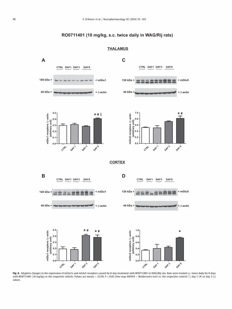
Fig. 6. Adaptive changes in the expression of mGlu1a and mGlu5 receptors caused by 8-day treatment with RO0711401 in WAG/Rij rats. Rats were treated s.c. twice daily for 8 dayswith RO0711401 (10 mg/kg) or the respective vehicle. Values are means þ S.E.M. P < 0.05 (One-way ANOVA þ Bonferroni's test) vs. the respective control (*), day 1 (#) or day 3 (x)values.
V. D'Amore et al. / Neuropharmacology 85 (2014) 91e10396

Fig. 7. Adaptive changes in the expression of mGlu5 receptors caused by 8-day treatment with VU0360172 in WAG/Rij rats. Rats were treated s.c. twice daily for 8 days withVU0360172 (3 mg/kg) or the respective vehicle. Values are means þ S.E.M. P < 0.05 (One-way ANOVA þ Bonferroni's test) vs. the respective control (*) or day 1 (#) values.
V. D'Amore et al. / Neuropharmacology 85 (2014) 91e103 97

Fig. 8. Adaptive changes in the expression of mGlu1a and mGlu5 receptors caused by 8-day treatment with RO0711401 in non-epileptic Wistar rats. Rats were treated s.c. twicedaily for 8 days with RO0711401 (10 mg/kg) or the respective vehicle. Values are means þ S.E.M. P < 0.05 (One-way ANOVA þ Bonferroni's test) vs. the respective control (*) or day 1(#) values.
V. D'Amore et al. / Neuropharmacology 85 (2014) 91e10398

Fig. 9. Adaptive changes in the expression of mGlu5 receptors caused by 8-day treatment with VU0360172 in non-epileptic Wistar rats. Rats were treated s.c. twice daily for 8 dayswith VU0360172 (3 mg/kg) or the respective vehicle. Values are means þ S.E.M. P < 0.05 (One-way ANOVA þ Bonferroni's test) vs. the respective control (*) or day 1 (#) values.
V. D'Amore et al. / Neuropharmacology 85 (2014) 91e103 99

Fig. 10. Schematic diagram illustrating mGlu1/mGlu5 receptor responses in symptomatic WAG/Rij and non-epileptic control Wistar rats following chronic treatment with eithermGlu1 or mGlu5 receptor PAMs. Detailed results of western blot analysis of mGlu1 and mGlu5 receptors are provided in Figs. 6e9.
V. D'Amore et al. / Neuropharmacology 85 (2014) 91e103100
RO0711401 showed its anti-absence effect only during the first 2days of treatment, and failed to reduce the incidence of SWDs af-terwards. The anti-absence activity of RO0711401 was not restoredafter 2 days of drug withdrawal (P > 0.05; Fig. 3). There was noeffect of the two drugs on the mean duration of SWDs at 1 h postinjection at any day of the treatment (not shown).
3.2. Measurements of thalamic and cortical drug levels duringrepeated administrations of RO0711401 or VU0360172
To examine whether the development of tolerance toRO0711401 was associated with time-dependent changes in centralbioavailability of the drug, we measured RO0711401 levels in thethalamus and cortex in three separate groups of WAG/Rij ratstreated with the drug as mentioned above and killed at day 1, 3 or 8at one hour after the morning injection. One-way ANOVA did notreveal any significant effect of day of treatment in the thalamus(Fig. 4B, P > 0.05) while a significant day effect was observed in thecortex (Fig. 4A, F ¼ 1,501, df ¼ 1.4, P < 0.05, h2 ¼ 0.273), with levelsof RO0711401 being reduced on day 8 of treatment. However,RO0711401 levels were unchanged on day 3 of treatment, whentolerance to the anti-absence effect begun to develop (Fig. 4A). Wealso measured VU0360172 levels and found no significant dayeffect, neither in cortex nor in thalamus (Fig. 4C, D, P > 0.05).
3.3. Measurements of mGlu1a and mGlu5 expression in thethalamus and somatosensory cortex after treatment withRO0711401 or VU0360172
Next it was examined whether repeated administrations ofRO0711401 or VU0360172 induced any adaptive changes in theexpression of mGlu1a and mGlu5 receptors in WAG/Rij rats that
could correlate with the temporal profile of drug response on SWDincidence.
Immunoblot analysis revealed bands at 140 kDa and 130 kDa,which, that correspond to the deduced molecular sizes of the re-ceptor's monomers (arrowheads in the representative immuno-blots). A faint higher molecular weight band corresponding todimers of mGlu1a and mGlu5 receptors was also visible in some ofthe immunoblots (not shown) but was not included in our densi-tometric analysis. The identity of the bands was verified using thecerebellum of crv4 mutant mice lacking mGlu1 receptors (Contiet al., 2006) or the cerebral cortex of mGlu5 receptor knockoutmice as negative controls. The lower band present in the repre-sentative mGlu1a and mGlu5 immunoblots was non-specificbecause it was still visible in samples from crv4 and mGlu5�/�
mice, respectively (Fig. 5).Treatment ofWAG/Rij rats with vehicle twice daily for 8 days did
not change the expression of mGlu1a or mGlu5 receptors in thethalamus or cortex (only values at day 8 are shown and indicated as“controls”). Treatment with RO0711401 caused an increase in bothmGlu1a and mGlu5 receptor protein levels on day 8. The drug alsoenhancedmGlu1a receptor levels on day 3 in the cortex [Fig. 6AeD;mGlu1a (F ¼ 10.36, P ¼ 0.0039 and F ¼ 15.50, P ¼ 0.011; for thethalamus and cortex respectively) and mGlu5 (F ¼ 7.33, P ¼ 0.011and F ¼ 6.15, P ¼ 0.018; for the thalamus and cortex respectively)].In contrast, treatment with VU0360172 in WAG/Rij rats caused anearly and persistent increase in mGlu5 receptor expression in thethalamus (Fig. 7A: F ¼ 7.56, P ¼ 0.0042) a late increase of mGlu5receptor expression in the cortex (Fig. 7B: F ¼ 9.17, P ¼ 0.0057) andno changes in mGlu1a receptor levels in any of the two brain re-gions (Fig. 7C, D).
To examine whether these adaptive changes in receptorexpression represented a general response to mGlu1 and mGlu5

Fig. 11. EEG records showing no SWDs in Wistar or ACI rats treated chronically withVU0360172. Wistar or ACI rats (n ¼ 3e6 per group) were treated with VU0360172(3 mg/kg s.c.) or vehicle twice daily for 8e10 days, and recorded at baseline (day 0) andat the indicated days of treatment. These treatments did not cause the appearance ofSWDs in the two strains of rats. Representative EEG recordings of rats treated withVU0360172 are shown.
V. D'Amore et al. / Neuropharmacology 85 (2014) 91e103 101
receptor PAMs or were peculiar to epileptic WAG/Rij rats, theanalysis was extended to non-epileptic age-matched Wistar ratstreated with vehicles, RO0711401 or VU0360172 for 1, 3 and 8 days.It was confirmed in these rats that treatment with RO0711401caused changes in both mGlu1a and mGlu5 receptors whereastreatment with VU0360172 caused selective changes in mGlu5receptors. However, the direction of these changes was opposite tothat observed in WAG/Rij rats. Wistar rats treated with RO0711401showed an early and persistent down-regulation of mGlu1a andmGlu5 receptors in the thalamus and cortex [Fig. 8AeD; mGlu1a(F ¼ 18.51, P ¼ 0.0020 and F ¼ 46.19, P ¼ 0.0001; for the thalamusand cortex respectively) and mGlu5 (F ¼ 10.76, P ¼ 0.0035 andF ¼ 21.00, P¼ 0.0026; for the thalamus and cortex respectively)]. Incontrast, Wistar rats treated with VU0360172 showed a latereduction in mGlu5 receptor expression in the thalamus (F ¼ 10.75,P ¼ 0.0035) and an early and persistent reduction of mGlu5 re-ceptor expression in the cortex (F ¼ 14.82, P ¼ 0.0012) with nochanges in the expression of mGlu1a receptors (Fig. 9AeD). Thus,interestingly, epileptic WAG/Rij rats and non-epileptic Wistar ratsshowed opposite manifestations of receptor adaptation in responseto prolonged mGlu1 and mGlu5 receptor enhancement. A synopsison the effect of chronic treatment with RO0711401 or VU0360172on the thalamic/cortical expression of mGlu1a and mGlu5 re-ceptors is shown in Fig. 10.
Finally, we examined whether chronic treatment withVU0360172 in Wistar rats could induce the appearance of SWDs asa result of the down-regulation of mGlu5 receptors in the thalamus(see D'Amore et al., 2013). No SWDs were recorded in Wistar ratstreated for 8 days with VU0360172 (3 mg/kg, s.c. twice daily).Similarly, no SWDs were induced by repeated VU0360172 in-jections (3 mg/kg, s.c., twice daily for 10 days) in non-epileptic ACIrats, used as additional controls (Fig. 11).
4. Discussion
The main objective of the study was to establish whethertolerance develops to the SWD-suppressing activity of mGlu1 andmGlu5 receptor PAMs and to unravel the molecular nature oftolerance (if any). This is a necessary step for the preclinical andclinical development of mGlu receptor PAMs for the treatment ofabsence epilepsy in humans.
As expected (Ngomba et al., 2011b; D'Amore et al., 2013), bothmGlu1 and mGlu5 receptor PAMs were able to suppress SWDs inWAG/Rij rats in the early phases of the treatment. However, a largedifference between the two drugs was apparent when the responsewas monitored on a daily basis during chronic administration. Theanti-absence effect of the mGlu5 receptor PAM, VU0360172, on theincidence of SWDs largely persisted over time, with only smallsigns of tolerance. More precisely, treatment with VU0360172caused a significant reduction in the incidence of SWDs at the 1sthour and a reduction trend at the 2nd hour on day 1. The effect atthe 1st hour was fully maintained on day 10. On the other hand, themean duration of SWDs was reduced at the 2nd hour post injectionon day 1 but not on day 10. In contrast, the mGlu1 receptor PAM,RO0711401, lost its anti-absence effects on the incidence of SWDson day 10.
In order to disclose when precisely tolerance to RO0711401began to develop, we analysed the incidence of SWDs in the firsthour of injection of all ten consecutive days of treatment. Theoutcomes showed that the anti-absence action of RO0711401remained unchanged from day 1 to day 2, diminished on day 3, andcompletely disappeared on day 4, remaining absent throughout therest of the experiment. Therefore, tolerance to RO0711401 began todevelop after relatively short time, i.e., after only four injections andwas complete after two additional injections. The small and
incomplete tolerance to the mGlu5 receptor PAM, VU0360172, issomehow consistent with data obtained with CDPPB anothermGlu5 receptor PAM, in a mouse model of audiogenic seizures(Pacey et al., 2011).
Therefore, it seems that the two drugs differ with respect to thedevelopment of tolerance, with VU0360172 retaining most of the“therapeutic” effect when repeatedly administered toWAG/Rij rats,and RO0711401 inducing fast and complete tolerance to the anti-absence effect.
We have collected only few pharmacokinetic data on the twoPAMs in this study. VU0360172 shows high plasma protein-bindingand little or no first-pass metabolism (Rodriguez et al., 2010).R00711401 has a short elimination half-life (<2 h), but no data onliver metabolism are available (Vieira et al., 2009). Whether or notthe two drugs may inhibit or accelerate their ownmetabolismwithtime is unknown. We found that thalamic levels of the two drugsremained stable during eight consecutive days of treatment inWAG/Rij rats, while a decrease over the days (day 3 vs. day 8) foundin the cortex might indicate a contribution of pharmacokinetictolerance to the loss of effects of RO0711401 on day 8 (but not onday 3).
We next measured the expression of mGlu1a and mGlu5 re-ceptors in the thalamus and cortex ofWAG/Rij rats on different daysof drug treatment, as compared to treatments with the respectivevehicles. This analysis was also carried out on non epileptic Wistarrats treated with the two drugs. We were surprised to find thatadaptive changes in receptor expression induced by the two PAMs

V. D'Amore et al. / Neuropharmacology 85 (2014) 91e103102
were highly divergent between WAG/Rij and Wistar rats. In Wistarrats, both drugs substantially reduced the expression of therespective mGlu receptor subtype particularly in the cerebral cor-tex. Interestingly, repeated administrations of RO0711401 alsoreduced the expression of the mGlu5 receptors whereasVU0360172 has no effect on the expression of the mGlu1a re-ceptors. This particular type of receptor regulation was completelydisrupted inWAG/Rij rats, where the two PAMs rather enhanced theexpression of the respective subtype, although with differenttemporal and regional profile. However, also in this case,RO0711401 changed the expression of both the mGlu1a and themGlu5 receptors, whereas VU0360172 selectively changed theexpression of the mGlu5 receptors only.
In an attempt to explain the different receptor selectivity in theadaptive changes induced by the two PAMs, we wish to highlightthat mGlu receptor subtypes can form both homodimers (e.g.,either mGlu1-mGlu1 or mGlu5-mGlu5 dimers) and intra-groupheterodimers (e.g., mGlu1-mGlu5 heterodimers), at least in heter-ologous expression systems (Doumazane et al., 2011). In addition,only one PAM molecule per dimer is sufficient to fully amplify re-ceptor activity (Kniazeff et al., 2004). We speculate that in thethalamus and cortex of both WAG/Rij and Wistar rats there is aprevalence of mGlu1-mGlu1 homodimers and mGlu1-mGlu5 het-erodimers over mGlu5-mGlu5 homodimers. This explains why themGlu1 receptor PAM, RO0711401, induced adaptive changes in bothmGlu1 and mGlu5 receptors, whereas the mGlu5 receptor PAM,VU0360172, selectively induced changes in mGlu5 receptors. Thereasonwhy receptor regulation caused by chronic PAM treatment isqualitatively different in WAG/Rij rats is unknown. Under normalconditions, prolonged agonist exposure causes mGlu receptordesensitization (i.e., uncoupling from G proteins), followed by re-ceptor internalization that may lead to reducing receptor expres-sion (i.e., receptor down-regulation) if de novo synthesis of receptorprotein does not compensate for receptor degradation (for reviews,see Reiter and Lefkowitz, 2006; Whalen et al., 2011). It is possiblethat mechanisms that lie at the core of receptor desensitization anddown-regulation are distorted in WAG/Rij rats because of thehypersynchronous activity of the cortico-thalamic-corticalnetwork. This encourages the study of molecules involved indesensitization and internalization of mGlu1 and mGlu5 receptors,such as G-protein coupled receptor kinases and b-arrestin(reviewed by Iacovelli et al., 2013) inWAG/Rij rats and other animalmodels of absence epilepsy.
In conclusion, our data support the development of mGlu5 re-ceptor PAMs as potential symptomatic drugs for the chronictreatment of absence epilepsy. These drugs may not cause phar-macodynamic tolerance perhaps because they fail to induceadaptive changes in the cognate mGlu1 receptor. We wish tohighlight that VU0360172 (and also the mGlu1 PAM, RO0711401)did not affect motor behaviour and did not cause gross signs oftoxicity in either WAG/Rij or Wistar rats. mGlu5 receptor PAMs arealready under development for the treatment of schizophreniawith the only concern of neurotoxicity and convulsive seizuresinduced by very high doses of these compounds (Parmentier-Batteur et al., 2013). It remains to be established whether mGlu5receptor PAMs can be safely associated with other drugs used in thetreatment of absence epilepsy in patients that are resistant toconventional antiepileptic medication.
Acknowledgements
We wish to thank Elly Willems-van Bree, Hans Krijnen, SaskiaHermeling, Gerard van Oijen and Michelle Huismans for technicalsupport and Silvia Gatti (F. Hoffmann-La Roche) for providingRO0711401.
References
Coenen, A.M., van Luijtelaar, E.L., 2003. Genetic animal models for absence epilepsy:a review of the WAG/Rij strain of rats. Behav. Genet. 33, 635e655.
Conti, V., Aghaie, A., Cilli, M., Martin, N., Caridi, G., Musante, L., Candiano, G.,Castagna, M., Fairen, A., Ravazzolo, R., Guenet, J.L., Puliti, A., 2006. crv4, a mousemodel for human ataxia associated with kyphoscoliosis caused by an mRNAsplicing mutation of the metabotropic glutamate receptor 1 (Grm1). Int. J. Mol.Med. 18, 593e600.
D'Amore, V., Santolini, I., van Rijn, C.M., Biagioni, F., Molinaro, G., Prete, A., Conn, P.J.,Lindsley, C.W., Zhou, Y., Vinson, P.N., Rodriguez, A.L., Jones, C.K., Stauffer, S.R.,Nicoletti, F., van Luijtelaar, G., Ngomba, R.T., 2013. Potentiation of mGlu5 re-ceptors with the novel enhancer, VU0360172, reduces spontaneous absenceseizures in WAG/Rij rats. Neuropharmacology 66, 330e338.
Doumazane, E., Scholler, P., Zwier, J.M., Trinquet, E., Rondard, P., Pin, J.P., 2011. A newapproach to analyze cell surface protein complexes reveals specific hetero-dimeric metabotropic glutamate receptors. FASEB J. 25, 66e77.
Glauser, T.A., Cnaan, A., Shinnar, S., Hirtz, D.G., Dlugos, D., Masur, D., Clark, P.O.,Capparelli, E.V., Adamson, P.C., 2010. Childhood absence epilepsy study group.Ethosuximide, valproic acid, and lamotrigine in childhood absence epilepsy.N. Engl. J. Med. 362, 790e799.
Iacovelli, L., Nicoletti, F., De Blasi, A., 2013. Molecular mechanisms that desensitizemetabotropic glutamate receptor signaling: an overview. Neuropharmacology66, 24e30.
Kniazeff, J., Bessis, A.S., Maurel, D., Ansanay, H., Pr�ezeau, L., Pin, J.P., 2004. Closedstate of both binding domains of homodimeric mGlu receptors is required forfull activity. Nat. Struct. Mol. Biol. 11, 706e713.
Liu, X.B., Mu~noz, A., Jones, E.G., 1998. Changes in subcellular localization ofmetabotropic glutamate receptor subtypes during postnatal development ofmouse thalamus. J. Comp. Neurol. 395, 450e465.
Ngomba, R.T., Ferraguti, F., Badura, A., Citraro, R., Santolini, I., Battaglia, G., Bruno, V.,De Sarro, G., Simonyi, A., van Luijtelaar, G., Nicoletti, F., 2008. Positive allostericmodulation of metabotropic glutamate 4 (mGlu4) receptors enhances sponta-neous and evoked absence seizures. Neuropharmacology 54, 344e354.
Ngomba, R.T., Santolini, I., Salt, T.E., Ferraguti, F., Battaglia, G., Nicoletti, F., van Luijtelaar, G.,2011a. Metabotropic glutamate receptors in the thalamocortical network: strategictargets for the treatment of absence epilepsy. Epilepsia 52, 1211e1222.
Ngomba, R.T., Santolini, I., Biagioni, F., Molinaro, G., Simonyi, A., van Rijn, C.M.,D'Amore, V., Mastroiacovo, F., Olivieri, G., Gradini, R., Ferraguti, F., Battaglia, G.,Bruno, V., Puliti, A., van Luijtelaar, G., Nicoletti, F., 2011b. Protective role fortype-1 metabotropic glutamate receptors against spike and wave discharges inthe WAG/Rij rat model of absence epilepsy. Neuropharmacology 60, 1281e1291.
Nicoletti, F., Bockaert, J., Collingridge, G.L., Conn, P.J., Ferraguti, F., Schoepp, D.D.,Wroblewski, J.T., Pin, J.P., 2011. Metabotropic glutamate receptors: from theworkbench to the bedside. Neuropharmacology 60, 1017e1041.
Niswender, C.M., Conn, P.J., 2010. Metabotropic glutamate receptors: physiology,pharmacology, and disease. Annu. Rev. Pharmacol. Toxicol. 50, 295e322.
Ovchinnikov, A., Lüttjohann, A., Hramov, A., van Luijtelaar, G., 2010. An algorithm forreal-time detection of spikeewave discharges in rodents. J. Neurosci. Methods194, 172e178.
Pacey, L.K., Tharmalingam, S., Hampson, D.R., 2011. Subchronic administration andcombination metabotropic glutamate and GABAB receptor drug therapy infragile X syndrome. J. Pharmacol. Exp. Ther. 338, 897e905.
Panayiotopoulos, C.P., 1999. Typical absence seizures and their treatment. Arch. Dis.Child. 81, 351e355.
Parmentier-Batteur, S., Hutson, P.H., Menzel, K., Uslaner, J.M., Mattson, B.A.,O'Brien, J.A., Magliaro, B.C., Forest, T., Stump, C.A., Tynebor, R.M., Anthony, N.J.,Tucker, T.J., Zhang, X.F., Gomez, R., Huszar, S.L., Lambeng, N., Faur�e, H., LePoul, E., Poli, S., Rosahl, T.W., Rocher, J.P., Hargreaves, R., Williams, T.M., 2013.Mechanism based neurotoxicity of mGlu5 positive allosteric modulators edevelopment challenges for a promising novel antipsychotic target. Neuro-pharmacology, 1e13. http://dx.doi.org/10.1016/j.neuropharm.2012.12.003.
Paxinos, G., Watson, C., 2005. The Rat Brain in the Stereotaxic Coordinates. Aca-demic Press Ltd., London.
Peeters, B.W., van Rijn, C.M., Nutt, D.J., Titulaer, M.N., Vossen, J.M., Coenen, A.M.,1990. Diazepam and Ro 15-1788 increase absence epilepsy in WAG/Rij ratschronically exposed to diazepam. Eur. J. Pharmacol. 178, 111e114.
Reiter, E., Lefkowitz, R.J., 2006. GRKs and beta-arrestins: roles in receptor silencing,trafficking and signaling. Trends Endocrinol. Metab. 17, 159e165.
Rodriguez, A.L., Grier, M.D., Jones, C.K., Herman, E.J., Kane, A.S., Smith, R.L.,Williams, R., Zhou, Y., Marlo, J.E., Days, E.L., Blatt, T.N., Jadhav, S., Menon, U.N.,Vinson, P.N., Rook, J.M., Stauffer, S.R., Niswender, C.M., Lindsley, C.W.,Weaver, C.D., Conn, P.J., 2010. Discovery of novel allosteric modulators ofmetabotropic glutamate receptor subtype 5 reveals chemical and functionaldiversity and in vivo activity in rat behavioral models of anxiolytic and anti-psychotic activity. Mol. Pharmacol. 78, 1105e1123.
Romano, C., Sesma, M.A., McDonald, C.T., O'Malley, K., Van den Pol, A.N., Olney, J.W.,1995. Distribution of metabotropic glutamate receptor mGluR5 immunoreac-tivity in rat brain. J. Comp. Neurol. 355, 455e469.
Smyk, M.K., Coenen, A., Lewandowski, M.H., van Luijtelaar, G., 2012. Internaldesynchronization facilitates seizures. Epilepsia 53, 1511e1518.
Stinehelfer, S., Vruwink, M., Burette, A., 2000. Immunolocalization of mGluR1alphain specific populations of local circuit neurons in the cerebral cortex. Brain Res.861, 37e44.

V. D'Amore et al. / Neuropharmacology 85 (2014) 91e103 103
van Luijtelaar, E.L., Coenen, A.M., 1986. Two types of electrocortical paroxysms in aninbred strain of rats. Neurosci. Lett. 70, 393e397.
van Luijtelaar, G., Sitnikova, E., 2006. Global and focal aspects of absence epilepsy:the contribution of genetic models. Neurosci. Biobehav. Rev. 30, 983e1003.
van Luijtelaar, E.L., Coenen, A.M., 1988. Circadian rhythmicity in absence epilepsy inrats. Epilepsy Res. 2, 331e336.
van Luijtelaar, G., Sitnikova, E., Littjohann, A., 2011. On the origin and suddenness ofabsences in genetic absence models. Clin. EEG Neurosci. 42, 83e97.
van Luijtelaar, G., Bikbaev, A., LeClerck, K., Mantagne, A., Kaminski, R. The effects oflacosamide on absence seizures in the WAG/Rij and GAERS models. Epilepsia(submitted for publication).
van Rijn, C.M., Weyn Banningh, E.W., Coenen, A.M., 1994. Effects of lamotrigine onabsence seizures in rats. Pol. J. Pharmacol. 46, 467e470.
van Rijn, C.M., Gaetani, S., Santolini, I., Badura, A., Gabova, A., Fu, J., Watanabe, M.,Cuomo, V., van Luijtelaar, G., Nicoletti, F., Ngomba, R.T., 2010. WAG/Rij rats show
a reduced expression of CB₁ receptors in thalamic nuclei and respond to the CB₁receptor agonist, R(þ)WIN55,212-2, with a reduced incidence of spikeewavedischarges. Epilepsia 51, 1511e1521.
Vieira, E., Huwyler, J., Jolidon, S., Knoflach, F., Mutel, V., Wichmann, J., 2009. Fluo-rinated 9H-xanthene-9-carboxylic acid oxazol-2-yl-amides as potent, orallyavailable mGlu1 receptor enhancers. Bioorg. Med. Chem. Lett. 19, 1666e1669.
Whalen, E.J., Rajagopal, S., Lefkowitz, R.J., 2011. Therapeutic potential of b-arrestin-and G protein-biased agonists. Trends Mol. Med. 17, 126e139.
Wijetunge, L.S., Till, S.M., Gillingwater, T.H., Ingham, C.A., Kind, P.C., 2008. mGluR5regulates glutamate-dependent development of the mouse somatosensorycortex. J. Neurosci. 28, 13028e13037.
Williams, R., Manka, J.T., Rodriguez, A.L., Vinson, P.N., Niswender, C.M., Weaver, C.D.,Jones, C.K., Conn, P.J., Lindsley, C.W., Stauffer, S.R., 2011. Synthesis and SAR ofcentrally active mGlu5 positive allosteric modulators based on an aryl acety-lenic bicyclic lactam scaffold. Bioorg. Med. Chem. Lett. 21, 1350e1353.