Membrane Trafficking in the Yeast Saccharomyces cerevisiae Model
Upload
independentCategory
view
0download
0
Genome Biology 2002, 4:R3
com
ment
reviews
reports
deposited research
interactions
inform
ation
refereed research
ResearchHap4p overexpression in glucose-grown Saccharomyces cerevisiaeinduces cells to enter a novel metabolic stateRomeo Lascaris*†, Harmen J Bussemaker*‡, André Boorsma*, Matt Piper§,Hans van der Spek*, Les Grivell*¶ and Jolanda Blom*
Addresses: *Swammerdam Institute for Life Sciences, University of Amsterdam, 1098 SM Amsterdam, The Netherlands. §Kluyver Laboratoryof Biotechnology, TU Delft, Julianalaan 67, 2628 BC Delft, The Netherlands. Current addresses: †Netherlands Cancer Institute, Plesmanlaan121, 1066 CX Amsterdam, The Netherlands. ‡Department of Biological Sciences and Columbia Genome Center, Columbia University, 1212Amsterdam Avenue MC 2441, New York, NY 10027, USA. ¶EMBO, Meyerhofstrasse 1, D-69117 Heidelberg, Germany.
Correspondence: Romeo Lascaris. E-mail: [email protected]
Abstract
Background: Metabolic and regulatory gene networks generally tend to be stable. However, wehave recently shown that overexpression of the transcriptional activator Hap4p in yeast causescells to move to a state characterized by increased respiratory activity. To understand whyoverexpression of HAP4 is able to override the signals that normally result in glucose repressionof mitochondrial function, we analyzed in detail the changes that occur in these cells.
Results: Whole-genome expression profiling and fingerprinting of the regulatory activitynetwork show that HAP4 overexpression provokes changes that also occur during the diauxicshift. Overexpression of HAP4, however, primarily acts on mitochondrial function andbiogenesis. In fact, a number of nuclear genes encoding mitochondrial proteins are induced to agreater extent than in cells that have passed through a normal diauxic shift: in addition to genesrequired for mitochondrial energy conservation they include genes encoding mitochondrialribosomal proteins.
Conclusions: We show that overproduction of a single nuclear transcription factor enables cellsto move to a novel state that displays features typical of, but clearly not identical to, otherderepressed states.
Published: 17 December 2002
Genome Biology 2002, 4:R3
The electronic version of this article is the complete one and can befound online at http://genomebiology.com/2002/4/1/R3
Received: 8 August 2002Revised: 23 September 2002Accepted: 29 October 2002
Background Unraveling and understanding the complex regulatory net-
works in a cell is an important step in genomic research,
since it should enable us to understand how the cell modifies
its behavior in response to both internal and external signals.
A diauxic shift occurs when yeast cells have consumed all
glucose fermentatively and convert their metabolism to
oxidative catabolism of the ethanol remaining in the
medium. This shift has been extensively studied [1] and is a
good example of how cells move from one ‘equilibrium’ state,
for example, fermentative physiology, to another ‘equilib-
rium’ state, for example, respiratory physiology. The transi-
tion from fermentative physiology to respiratory physiology,
in the wild or in the laboratory, involves many changes in
gene expression that contribute to the final phenotype. The
Open Access
© 2002 Lascaris et al.; licensee BioMed Central Ltd. This is an Open Access article: verbatim copying and redistribution of this article are permitted in allmedia for any purpose, provided this notice is preserved along with the article's original URL.
HAP4 overexpression system on the other hand is relatively
simple: one factor that is known to be required for induction
of respiratory activity can indeed induce a physiological
change that resembles that of the diauxic shift [2].
Metabolic and regulatory gene networks have generally
evolved to allow physiological and developmental processes
to compensate for the effects of potentially deleterious muta-
tions, termed robustness [3]. Important questions are how
equilibrium states are maintained and how the transition
between states is implemented by the cell. Depending on
where a particular regulatory protein is located in the hierar-
chical global regulatory network of the cell, the response to a
change in its activity may be either localized or pleiotropic.
To identify the various components of the pleiotropic
response to HAP4 overexpression, that is, to ‘fingerprint’ the
changes in the regulatory network, we use two different
methods. First, we use the algorithm REDUCE [4] to infer
the regulatory activity of transcription factors from the
mRNA expression of their target genes. Second, we use a
new and related algorithm, named Quontology, to identify
classes of genes with similar function that are significant
induced or repressed. Both methods have the property that
they can detect small-amplitude but coordinated changes in
the average expression level of a set of genes, even if the
expression of individual genes is not changing significantly.
In this paper we report detailed analysis of the changes that
occur as a result of overexpression of HAP4. Using a number
of techniques it could be shown that overexpression of HAP4
enhances transcription of a large set of mitochondrial
protein genes, leading to increased mitochondrial biogenesis
and enabling cells to move to a distinct and novel state.
Results Cells overexpressing HAP4 upregulate mitochondrialbiogenesis and activity even in the presence of glucose To determine the effect of overexpression of HAP4 on mito-
chondria, several different experimental approaches were
used to quantify mitochondrial components and mitochon-
drial structures in the cell.
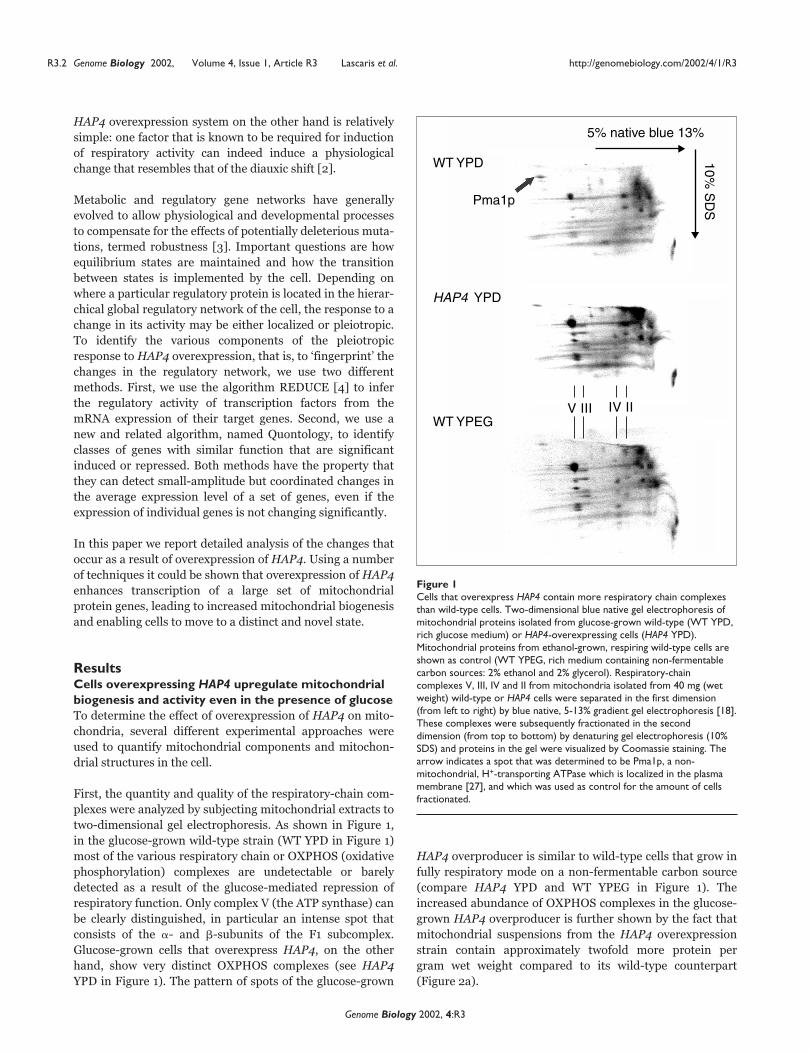
First, the quantity and quality of the respiratory-chain com-
plexes were analyzed by subjecting mitochondrial extracts to
two-dimensional gel electrophoresis. As shown in Figure 1,
in the glucose-grown wild-type strain (WT YPD in Figure 1)
most of the various respiratory chain or OXPHOS (oxidative
phosphorylation) complexes are undetectable or barely
detected as a result of the glucose-mediated repression of
respiratory function. Only complex V (the ATP synthase) can
be clearly distinguished, in particular an intense spot that
consists of the �- and �-subunits of the F1 subcomplex.
Glucose-grown cells that overexpress HAP4, on the other
hand, show very distinct OXPHOS complexes (see HAP4
YPD in Figure 1). The pattern of spots of the glucose-grown
HAP4 overproducer is similar to wild-type cells that grow in
fully respiratory mode on a non-fermentable carbon source
(compare HAP4 YPD and WT YPEG in Figure 1). The
increased abundance of OXPHOS complexes in the glucose-
grown HAP4 overproducer is further shown by the fact that
mitochondrial suspensions from the HAP4 overexpression
strain contain approximately twofold more protein per
gram wet weight compared to its wild-type counterpart
(Figure 2a).
R3.2 Genome Biology 2002, Volume 4, Issue 1, Article R3 Lascaris et al. http://genomebiology.com/2002/4/1/R3
Genome Biology 2002, 4:R3
Figure 1 Cells that overexpress HAP4 contain more respiratory chain complexesthan wild-type cells. Two-dimensional blue native gel electrophoresis ofmitochondrial proteins isolated from glucose-grown wild-type (WT YPD,rich glucose medium) or HAP4-overexpressing cells (HAP4 YPD).Mitochondrial proteins from ethanol-grown, respiring wild-type cells areshown as control (WT YPEG, rich medium containing non-fermentablecarbon sources: 2% ethanol and 2% glycerol). Respiratory-chaincomplexes V, III, IV and II from mitochondria isolated from 40 mg (wetweight) wild-type or HAP4 cells were separated in the first dimension(from left to right) by blue native, 5-13% gradient gel electrophoresis [18].These complexes were subsequently fractionated in the seconddimension (from top to bottom) by denaturing gel electrophoresis (10%SDS) and proteins in the gel were visualized by Coomassie staining. Thearrow indicates a spot that was determined to be Pma1p, a non-mitochondrial, H+-transporting ATPase which is localized in the plasmamembrane [27], and which was used as control for the amount of cellsfractionated.
Pma1p
5% native blue 13%
10% S
DS
WT YPD
WT YPEG
HAP4 YPD
V III IV II

The cellular content of cytochromes c + c1 and b is also
markedly increased, 50% and 65% respectively, when the
HAP4 gene is overexpressed in glucose-grown cells
(Figure 2b). The cytochrome aa3 content is also increased in
the HAP4 strain, although the corresponding part of the dif-
ference spectra does not allow proper quantification.
The mitochondrial content of cells was further analyzed by
fluorescence microscopy using the fluorescent dye DASPMI,
which specifically stains mitochondria [5]. To compare mito-
chondrial DASPMI fluorescence between strains directly,
glucose-grown wild-type and HAP4-overexpressing cells
were mixed. Wild-type and HAP4 cells were identified in situ
by also staining one of the strains with the cell-wall dye Cal-
coFluorWhite (CFW [6]) before mixing the cells. As shown in
Figure 3, HAP4-overexpressing cells contain more mitochon-
drial structures per cell (HAP4 cells are identified by CFW
fluorescence in the left panels and by the absence of CFW flu-
orescence in the right panels). This is consistent with the fact
that more membranous vesicles are present in mitochondrial
suspensions extracted from HAP4-overexpressing cells (data
not shown). Interestingly, HAP4-overexpressing cells also
exhibit increased DASPMI fluorescence. Although this may
suggest an increased membrane potential over the mito-
chondrial inner membrane [7], we favor the idea that the
inner membrane has a more compact structure in these
mitochondria [5].
Nuclear DNA encodes the majority of mitochondrial pro-
teins. Several mitochondrial components, such as a number
of subunits of enzymes of the respiratory chain, are encoded
by the mitochondrial genome. Figure 4 shows that overex-
pression of HAP4 in glucose-growth conditions not only
results in increased transcription of nuclear-encoded mito-
chondrial protein genes ([2], and see below) but also leads to
enhanced transcription of all mitochondrial genes tested. In
particular, COX2 mRNA was induced to high levels. It
should be noted that the increase in transcription in the
mitochondrion is not accompanied by strong alterations in
the copy number of mitochondrial DNA (mtDNA) (data not
shown), indicating that the increase in mitochondrial tran-
scription is mainly due to increased activity of the mitochon-
drial transcription machinery.
The strong increase in OXPHOS complex abundance and in
mitochondrial protein and cytochrome content per cell,
together with the evidence that shows that HAP4 cells
contain more mitochondrial structures per cell, conclusively
shows that overexpression of HAP4 in glucose-growth con-
ditions results in increased mitochondrial biogenesis.
Fingerprinting the regulatory network of HAP4overexpressing cells Genome-wide expression was monitored with Affymetrix
GeneChips using RNA from wild-type and HAP4 cells grown
aerobically on minimal glucose medium (YNB) in a batch
com
ment
reviews
reports
deposited research
interactions
inform
ation
refereed research
http://genomebiology.com/2002/4/1/R3 Genome Biology 2002, Volume 4, Issue 1, Article R3 Lascaris et al. R3.3
Genome Biology 2002, 4:R3
Figure 2HAP4-overexpressing cells contain more protein and more cytochromescompared to wild-type cells. (a) Five independent mitochondrial extractswere isolated from logarithmic-phase, glucose-grown wild-type and HAP4strains. The amount of mitochondrial protein per gram cellular wetweight was determined. (b) Absorption difference spectra of whole-cellsuspensions from respiro-fermentative, glucose-repressed wild-type andHAP4-overexpressing cells (YPD) and respiratory grown wild-type cells(YPEG) were obtained using a split-beam spectrophotometer. The cellularcontent of cytochromes c + c1, b and aa3 was calculated from thedifference in absorption between reduced versus oxidized forms of thecytochromes.
A =
0.0
05
Cyt
c +
c1
Cyt
aa 3
Cyt
b
WT
HAP4
WT
YPD
500 550 600Wavelength (nm)
III
II
I
III
II
I
YPD
YPEG
100
166
193
100
% Cyt c + c1 Cyt b
151
211
0
0.25
WT
Pro
tein
/wet
wei
ght (
mg/
g)
0.5
0.75
1
1.25
1.5
1.75
2(a)
(b)
HAP4

fermenter. For each strain, two separate hybridizations were
carried out and the log2-expression ratios or fold-changes
were calculated for HAP4 overexpression versus wild type.
Because the HAP4-overexpression system can induce a
physiological change that resembles that of the diauxic shift
[2], we first analyzed whether the state of the regulatory
activity network in the HAP4 overexpressor is similar to one
or more of the transient states of the diauxic shift [1]. For
this, the regulatory activity network was fingerprinted using
the Quontology algorithm (see Materials and methods).
This algorithm enables the identification of functional
groups of genes that have changed in expression as a result
of increased expression of HAP4. A Z-score is calculated for
the functional categories from the MIPS database [8],
which is a measure of the changes that occur in the regula-
tory activity network. Categories with a Z-score larger than
4 can be considered to be induced or repressed signifi-
cantly. Figure 5 shows all functional categories that were
induced or repressed significantly in the HAP4-overex-
pressing strain or in one of the expression profiles from the
diauxic shift.
From Figure 5 it is evident that overexpression of HAP4
results in alterations in the regulatory network that resem-
bles the time points of the diauxic shift with respect to
several important functional categories (see categories
marked with a filled circle).
The alterations in the regulatory activity network of the
diauxic shift mainly occur in three phases, as indicated in
Figure 5: high glucose concentrations; strongly decreasing
glucose concentration; and glucose depletion where cells
grow in respiratory mode on the remaining ethanol. The
HAP4 overexpression profile is similar in a number of
respects to the profiles of cells growing in decreasing glucose
concentrations (13.5 and 15.5 hours; see Figure 5). However,
in this case also, the changes that occur in the diauxic shift
are much more extensive, in particular with respect to the
categories ‘metabolism of energy reserves’ and ‘rRNA tran-
scription’. Hence, the regulatory activity network of the
HAP4-overexpressing strain has moved to a state that
resembles one of the transition states of the diauxic shift, but
at the same time differs in a number of important aspects.
Hap4p is a global regulator that coordinately andspecifically upregulates mitochondrial function andmitochondrial biogenesis All functional categories that are significantly increased
(Z-score > 4) in the HAP4-overexpressing strain are related
to mitochondrial function (see Figure 5), including the main
category ‘protein synthesis’ which consists largely of mito-
chondrial protein genes. Consistently, the vast majority of
the 139 nuclear genes that are induced more than twofold in
the HAP4-overexpressing strain (see genes listed in Figure 6
and Materials and methods) encode proteins that localize to
mitochondria. It is striking that if the expression of these
genes is compared to the expression in respiratory-state cells
(columns 2-8 in Figure 6) or in glucose-repressed cells
(column 9), it can be observed in the right part of Figure 6
R3.4 Genome Biology 2002, Volume 4, Issue 1, Article R3 Lascaris et al. http://genomebiology.com/2002/4/1/R3
Genome Biology 2002, 4:R3
Figure 3HAP4-overexpressing cells contain more mitochondrial structures withgreater DASPMI fluorescence than wild-type cells. DASPMI fluorescenceof glucose-grown wild-type and HAP4-overproducing cells was comparedin microscopic preparations of mixed cells. To identify HAP4-overexpressing cells or wild-type cells in situ, microscopic preparationswere analyzed in which only one of the strains was additionally stainedwith CalcoFluorWhite (CFW), a dye that stains the cell wall, in particularthe bud-neck and bud scars [6]. (a) Top panels show a mixture of HAP4-expressing cells stained with CFW + DASPMI and wild-type cells stainedwith DASPMI only. Left, CFW fluorescence; right, phase contrast. (b)Top panels show a mixture of wild-type cells stained with CFW +DASPMI and HAP4-overexpressing cells stained with DASPMI only. Left,CFW fluorescence; right, phase contrast. In both (a) and (b) the largebottom panels show a mixture of wild-type and HAP4-overexpressingcells stained with DASPMI only.
(a) (b)
Figure 4Transcription in mitochondria is increased as a result of overexpressionof the nuclear HAP4 gene. Northern analysis of wild-type and HAP4-overexpressing cells grown logarithmically in rich glucose medium.Transcripts from COX1, COX2 and SCEI are expressed from themitochondrial genome. PDA1 mRNA is used as internal control.Transcriptional changes were quantified and indicated as a percentage ofthe wild-type transcript level.
COX2WT HAP4 WT HAP4 WT HAP4
PDA1
COX2
PDA1145% 230% 127%
SCEI
PDA1

that there are a large number of genes that are strongly
upregulated in the HAP4 overproducer but not upregulated
in respiring cells. As these genes also encode mitochondrial
proteins, this suggests that overexpression of HAP4 leads to
a novel state in which the expression of mitochondrial pro-
teins goes beyond levels that are required in normal physio-
logical conditions.
The two clusters of genes in Figure 6 fall into different func-
tional categories. The group of genes that is responsive to
changes in the respiro-fermentative growth mode consists
mainly of genes encoding proteins that are directly involved
in respiration (see genes listed in the left part of Figure 6).
The second, larger and non-responsive group mainly con-
sists of genes that encode proteins involved in mitochondrial
protein synthesis (see genes listed in the right part of
Figure 6). Apparently, overexpression of HAP4 not only
results in increased expression of genes encoding proteins
required for mitochondrial energy conservation (or genera-
tion) but also of genes that are required for the synthesis of
functional mitochondria.
The upstream activation sequences (UAS) of the genes of the
non-responsive group - but not those of the responsive
group - generally all lack the typical Hap2/3/4/5p-binding
element CCAATCA. This is interesting, as this particular
element strongly correlates with the changes in expression
that occur as a result of overexpression of HAP4 ([4],
REDUCE: p < 10-6). Furthermore, with a more in-depth
computational search for potential Hap2/3/4/5p sites using
a nucleotide-distribution matrix, no difference could be
observed between promoters of the non-responsive group
and random-sequence DNA (Figure 7a). In contrast, in the
gene promoters of the responsive gene cluster a large
number of potential Hap2/3/4/5p sites can be detected that
are distributed over the promoter according to a typical
yeast UAS (100-400 bp upstream of the translational start
codon ATG). These data strongly suggest that the
Hap2/3/4/5p complex does not directly regulate the vast
majority of genes of the non-responsive gene cluster, namely
genes important for mitochondrial proliferation.
Overexpression of HAP4 negatively affectstranscription of a small set of genes involved in zincmetabolism In addition to the above-mentioned genes, which are upregu-
lated and lead to increased mitochondrial biogenesis, it
should be noted that the expression of 23 genes is reduced by
overexpression of HAP4. These genes are RPR1 (-2.3), EUG1
(-2.3), ZRT1 (-17.0), ADH4 (-7.9), YGL258W (-3.9),
YHR048W (-2.5), HXT4 (-2.7), TDH1 (-3.0), ZAP1 (-6.8),
CWP1 (-3.0), ZRT3 (-3.4), ZRT2 (-2.9), TFS1 (-2.4), THI7
(-2.9), CAR2 (-2.7), SNO1 (-3.8), SNZ1 (-2.9), FET4 (-2.7),
YNL254C (-5.3), FRE4 (-3.0), COS10 (-2.4), YOL154W
(-27.3), YOR387C (-18.4). Interestingly, a large number of
these genes are involved in zinc metabolism. Genes indicated
com
ment
reviews
reports
deposited research
interactions
inform
ation
refereed research
http://genomebiology.com/2002/4/1/R3 Genome Biology 2002, Volume 4, Issue 1, Article R3 Lascaris et al. R3.5
Genome Biology 2002, 4:R3
Figure 5Fingerprinting of the yeast regulatory activity network using Quontology(H.J.B. and R.L., unpublished work, see Materials and methods). Functionalcategories are induced (red) or repressed (green) by overexpression ofHAP4 or in the diauxic shift [1]. The whole-genome expression data of thediauxic shift are divided into three datasets that represent: glucose excess;a strongly decreasing glucose concentration; and glucose depletion. Usingthe quontology algorithm, Z-scores were calculated for the functionalcategories defined by MIPS [8]. Generally, all categories that have at leastone Z-score larger than 4 or smaller than -4 are listed (p < 0.1). Thehierarchy of the categories is preserved: the main categories in capitals,the subcategories indented and in lower case. A filled circle marks MIPScategories that show the same pattern of expression in more respiratoryconditions (the five columns on the right). Interestingly, overexpression ofHAP4 results in specific induction of MIPS categories that are involved inmitochondrial function, causing the regulatory activity network to move toa state that is different from the diauxic shift.
METABOLISM Amino-acids biosynthesis Nucleotides purines C-compound and carbohydrates utilization transport
ENERGY Glycolysis and gluconeogenesis Pentose-phosphate pathway Tricarboxylic-acid pathway Respiration Fermentation Metabolism of energy reserves Glyoxylate cycle
TRANSCRIPTION rRNA transcription rRNA synthesis rRNA processing tRNA transcription tRNA synthesis mRNA transcription mRNA synthesis transcriptional control
PROTEIN SYNTHESIS Ribosomal proteins Translation tRNA-synthetases
CELLULAR ORGANIZATION Cytoplasm ER Golgi Nucleus Mitochondrion Peroxisome Vacuoles and lysosomes
Diauxic shift (h)
Hig
h [g
luc]
Dec
reas
ing
[glu
c]
Glu
cose
dep
lete
d
9.5
11.5
13.5
15.5
18.5
20.5
HA
P4

in bold are most strongly affected by overexpression of
HAP4 (<-3) and have been proposed to be targeted by zinc-
specific transcription factor Zap1p [9]. In fact, the expres-
sion of ZAP1 itself is strongly repressed by overexpression of
HAP4 and, furthermore, the repressive motif ACC-5-GGT,
the DNA-binding site of Zap1p [10], was identified by the
REDUCE algorithm with high significance, p < 10-12. Hence,
we conclude that the main repressive effect of overexpres-
sion of HAP4 is mediated through decreased expression of
the Zap1p transcription factor. Genes that are typically not
altered in expression on zinc depletion or zap1-deletion [9]
are given in non-italic type above, and are suggested to be
repressed through a different mechanism, possibly through
the same mechanism that initially represses the ZAP1 gene.
Discussion In this report we show that overexpression of the gene
encoding the activation moiety of the Hap2/3/4/5p tran-
scription complex causes glucose-grown cells to move to a
novel state that is characterized by a shift to higher respira-
tory activity. HAP4-overexpressing cells contain more mito-
chondrial structures, increased amounts of mitochondrial
proteins/cytochromes and enhanced transcription of the
R3.6 Genome Biology 2002, Volume 4, Issue 1, Article R3 Lascaris et al. http://genomebiology.com/2002/4/1/R3
Genome Biology 2002, 4:R3
Figure 6Genes that show more than twofold induction in the HAP4 overexpression dataset can be divided in two distinct groups using k-means clustering analysis(Cluster software [22]). The gene-expression profile of HAP4-overexpressing cells was compared to datasets derived from cells that have a particularand/or shifted balance between respiration and fermentation. These were: the 18.5 h and 20.5 h time points of the diauxic shift at which glucose has beenexhausted [1]; the ‘adaptive evolution’ datasets, which are derived from strains selected for growth in aerobic, glucose-limited chemostats for more than250 generations (denoted as pare1, pare2 and pare3 [28]); stationary phase cells (2-day stationary phase); and expression profiles from cells grown eitheron ethanol or glucose [11]. It should be emphasized that mitochondrial transcripts are not represented in this analysis as these transcripts do not containa 3’ poly(A) tail and hence are not labeled properly.
ACH1 (3.7), DAL2 (3), YKR016W (3.3),SOD2 (3), ODC1 (3.3), HAP4 (5.3),YJL103C (4.1), PBI2 (3.1)
MSS116 (2.8), MSU1 (2.3), IMG2 (2.9),NUC1 (2.7), SPR3 (8.6), SNZ2 (6.4), ECM19(2.3), MAS2 (2.2), MAS1 (2.2), FRE2 (2.6),FRE3 (4.1), AQY2 (3), SNO2 (2.2)
Unknown
YBR230C (2.5), YFL030W (7.8), YLR168C(3.3), AAD14 (2.3), YNR002C (3.8),YPR151C (3.0)
YBL059W (3.5), YDR316W (3), YCL036W(3), QRI7 (2.7), YDR430C (2.7), YDR493W(2.8), YDR494W (3.4), YER182W (2.5),YFR011C (3.4), YGL107C (2.7), YGL146C(3.5), YHR059W (3.7), YKL066W (2.2),YML087C (12.3), YML089C (3.3), YNL122C(2.8), YNL184C (2.6), YNR022C (2.9),YOL071W (2.9), YOR354C (2.3), YPR083W (3.4)
Responsive gene cluster
k-means first cluster:Responsive gene cluster
k-means second cluster:Non-responsive gene cluster
Non-responsive gene cluster
Mitochondrial energy metabolism
INH1 (5.6), ATP7 (2.5), ATP14 (2.6),YPR020W (3.4), MCR1 (2.8), CYB2 (5),COX9 (2.3), QCR6 (3.2), COX4 (2.4), QCR9(2.2), COX6 (2.2), CYC1 (2.3), NDI1 (4.4),CYT1 (2.3), QCR2 (3.1), MBA1 (2.4), RIP1(2.4), KGD1 (2.3), KGD2 (4.7), SDH4 (3.2),SDH3 (2.3), SDH1 (4.8), SDH2 (3.9)
RPP1B (2.4), ATP12 (3.9), ATP11 (2.4),CYT2 (2.9), QCR10 (3.4), PET117 (4.2),OXA1 (3.8), MAM33 (3.8), COX17 (4.7),COX10 (2.6)
Protein synthesis in mitochondrion
YMR31 (2.7), TUF1 (2.4), RPM2 (4.4) MRP21 (2.6), MRPS9 (2.9), MRPL37 (2.4),MRPL27 (2.5), MRPL32 (2.7), MRPL11(2.6), RSM10 (2.4), MRPL7 (3.4), MRPL35(3.6), RML2 (2.4), MRF1 (2.8), MRPL25(2.7), MRP13 (2.7), MRPL9 (2.5), MRP4(3.0), MSR1 (2.5), MRPL6 (2.4), MRPL49(2.9), MRPL31 (2.9), MRPL38 (3.0),MRPL20 (2.7), MEF1 (2.7), MRPL4 (5.4),YML025C (2.8), MRPL3 (2.1), MRPL24(3.0), MRPL44 (2.9), MRPL2 (2.5), NAM9(2.2), MRPL19 (2.5), YNL306W (2.5), MSE1(2.6), MRPL23 (2.5), PET123 (2.5), ISM1(2.9), MSY1 (3.1), MSD1 (2.5), MRPL40(2.9), CBP6 (3), YDR116C (3.7), RSM24 (3),RSM18 (2.5), YGL068W (2.4), RSM23 (3.0),MTO1 (3.2), YGR021W (3.7), YGR165W(2.7), RSM27 (2.5), KIM4 (5.1), RSM25(5.0), YHR116W (3.1), YMR157C (2.6),YMR158W (2.7), YMR188C (3.0),YNL177C (2.8)
Miscellaneous
1 HAP42 Diauxic shift 18.5 h3 Diauxic shift 20.5 h4 ParE15 ParE26 ParE37 Stationary phase8 Ethanol growth9 Glucose growth
1 2 3 4 5 6 7 8 9 1 2 3 4 5 6 7 8 9 1 2 3 4 5 6 7 8 9

com
ment
reviews
reports
deposited research
interactions
inform
ation
refereed research
http://genomebiology.com/2002/4/1/R3 Genome Biology 2002, Volume 4, Issue 1, Article R3 Lascaris et al. R3.7
Genome Biology 2002, 4:R3
Figure 7Non-responsive gene promoters rarely contain Hap2/3/4/5p regulatory elements and respond to signals that depend on growth rate. (a) Frequencydistribution of potential Hap2/3/4/5p sites in the promoters of genes from the responsive gene cluster (red) and the non-responsive gene cluster (blue)from Figure 6. The frequency of occurrence of potential Hap2/3/4/5p sites as detected by a Hap2/3/4/5/p nucleotide-distribution matrix was calculated in100 bp intervals (threshold = 0.85 [24]). The distribution of Hap2/3/4/5p sites in the promoters of genes from the responsive gene cluster localizebetween -400 and -100 bp from the ATG, a common distance for yeast UAS. Putative Hap2/3/4/5p sites extracted from the non-responsive gene clustershow a random distribution similar to that found for random-sequence DNA (A/T 31%, C/G 19%), and are therefore likely to be non-functional matchesto the matrix. (b) The correlation between Z-scores and growth rate of the constitutive (left) and regulated (right) clusters calculated with theQuontology algorithm (see also Figure 5) using experimental data from a large number of deletion mutants [15]. Z-scores larger than 4 or smaller than -4have significantly altered expression (p < 0.1). The running mean of the absolute Z-scores was calculated to determine to what extent regulation iscorrelated to the growth rate.
Non-responsive cluster
Hap
2/3/
4/5p
site
s pe
r ge
ne p
er 1
00 b
p
Responsive clusterRandom
Non-responsive cluster
0.6 0.7 0.8 0.9 1 1.1 1.2
Responsive cluster
0.6
Mean |Z-score|
Relative growth rate
Z-s
core
Relative growth rate
0.7 0.8 0.9 1 1.1 1.2
4
−4
4
−4
0−800/−700
−700/−600
−600/−500
−500/−400
−400/−300
−300/−200
−200/−100
−100/0
0.1
0.2
0.3
0.4
0.5(a)
(b)

mitochondrial genome. This suggests that overexpression of
HAP4 results in a general increase in mitochondrial biogene-
sis. Consistently, whole-genome expression profiling and
subsequent computational analyses do indeed show that
overexpression of HAP4 specifically alters transcription of a
very large set of mitochondrial protein genes. HAP4-overex-
pressing cells resemble cells that undergo a physiological
diauxic shift, but at the same time differ from these cells in a
number of important respects. First, in the HAP4 overex-
pressor no increase occurs in the functional category ‘metab-
olism of energy reserves’ and in functional categories that
are involved in transcription, mainly ‘rRNA transcription’.
Second, genes involved in zinc metabolism are downregu-
lated, and third, genes involved in protein synthesis in mito-
chondria are strongly upregulated.
With respect to the latter it should be noted that the effects
we show in Figure 6 might to some extent be affected by dif-
ferences in the set-up of the experiments, that is, differences
in the genetic background of the strains and/or of the array
platforms used. However, other previously published
expression profiles show induction of this non-responsive
group of genes, which contains mainly mitochondrial riboso-
mal protein genes (MRP genes). These studies looked at the
effects of oxidative stress [11], addition of rapamycin [12],
mitochondrial dysfunction [13,14], a ‘pulse’-like induction at
the diauxic shift (see lanes 2 in Figure 6), and at the diauxic
shift that precedes the stationary phase [11]. Furthermore, if
the Z-scores are calculated for the genes of the non-respon-
sive and the responsive group using the expression profiles
of 300 null mutants from Hughes et al. [15], we find that the
expression of the non-responsive group alters more strongly
in mutants that have a decreased growth rate (Figure 7b). As
the growth rate of the HAP4 overproducer is not affected, we
suggest that the transcriptional response of MRP genes is
not only dependent on the growth rate itself, but also on
whether cells respire more and/or ferment less.
The fact that genes involved in zinc metabolism are primarily
downregulated by overexpression of HAP4 is surprising.
Complex (auto)regulation of the zinc regulator Zap1p and the
central role of mitochondria in metal metabolism are likely to
be important in this downregulation. However, as zinc is an
essential component of more than 300 enzymes - including,
paradoxically, cytochrome oxidase - particular zinc-depen-
dent factors may be involved. Candidate factors that require
zinc are: the mitochondrial metalloproteases Afg3p, Rca1p
and Yem1p; the transcription factors Cat8, Mig1p, Mig2p,
Mal63 and Hap1p; and alcohol dehydrogenases.
We have shown here that increased expression of a single
transcription factor enables the alteration of a particular
branch of the yeast regulatory activity network, leading to
remarkably specific alterations in expression of specific
functional sets of genes and revealing only minor cross-talk
with other parts of the regulatory activity network. Hence,
the regulatory activity network that determines mitochondr-
ial function appears to be highly modular. This is an impor-
tant notion, as the regulatory activity network may consist of
similar more-or-less isolated modules, which may enable the
construction of a simple but realistic computational model of
the in silico cell.
Materials and methods Strains and growth conditions The wild-type strain CEN.PK113-7D and its HAP4-overpro-
ducing counterpart 436 GH [16], in which the HAP4 gene is
overexpressed more than fivefold in glucose-growth condi-
tions, were used. The latter contains a genomically inte-
grated HAP4-overproduction cassette in which the TDH3
promoter regulates the HAP4 gene. Cells were grown aerobi-
cally in YPD (1% yeast extract, 1% Bacto-peptone and 2 or
3% D-glucose) or YPEG (YP containing 2% ethanol and 2%
glycerol). In all experiments cells were harvested at early
logarithmic phase.
Isolation of mitochondria and blue native PAGE Yeast cells were grown in 100 ml YPD or YPEG medium until
OD600 of 0.7-1.0 and mitochondria were isolated according
to [17]. Protein concentration was determined using Brad-
ford. Two-dimensional gel electrophoresis was carried out
according to [18]: the first dimension consisted of blue
native, 5-13% gradient polyacrylamide gel electrophoresis
(PAGE), and the second of denaturing PAGE on gels con-
taining 10% SDS. Protein was visualized by Coomassie stain-
ing of the gels. The identity of a constitutive spot at the
upper-left part of the gels was determined using mass spec-
trometry [17].
Spectral analysis and fluorescence microscopy Cells were grown to OD600 = 1.0-1.2, frozen in liquid nitro-
gen and stored in -20°C. The cell pellet was resuspended in a
final volume of 2 ml using a buffer containing 100 mM KPO4
pH 7.3, 250 mM sucrose and 0.5% Na-cholate. Difference
spectra were derived from dithionite-reduced cells minus
ferricyanide-oxidized cells. Spectral measurements were
carried out at room temperature in a DW2000 dual-beam
spectrophotometer. Concentrations of cytochromes were
determined essentially as in [19].
Mitochondrial biogenesis in living cells was analyzed by
DASPMI staining of cells grown logarithmically in liquid
medium (YPD). YPD culture samples of 5 ml were incubated
at 28°C in the presence of 10 �l of 1 mg/ml CalcoFluorWhite
[6], centrifuged, resuspended in 20 �l YPD. CFW stained
cells (2 �l) and 2 �l unstained cells were mixed, 1 �l of
1 mg/ml DASPMI and 15 �l 0.1 M Tris-Cl pH 8.0 were
added. After 5 min, cells were washed in 0.1 M Tris-Cl pH
8.0, centrifuged and resuspended in 0.1 M Tris-Cl pH 8.0.
After addition of 1 volume YPD, cells were aerated by pipet-
ing and analyzed microscopically.
R3.8 Genome Biology 2002, Volume 4, Issue 1, Article R3 Lascaris et al. http://genomebiology.com/2002/4/1/R3
Genome Biology 2002, 4:R3

Northern analysis and microarray experiments RNA isolation, northern blotting and hybridizations were
carried out essentially as in [20]. Probes for mitochondrial
transcripts COX1, COX2 and SCEI and the loading control
PDA1 were obtained by [32P]ATP labeling of PCR-generated
DNA fragments. For microarray analyses, a pre-culture was
grown overnight in YNB medium (0.67% yeast nitrogen
base) containing 2% raffinose. The main culture was inocu-
lated at OD600 = 0.1 in a batch fermenter containing 500 ml
YNB medium buffered at pH 5.0 with 100 mM sodium
phthalate and supplemented with 2% glucose, stirred at
500 rpm and aerated at 1 vvm (vessel volume per minute).
Both strains grew equally well, having growth rates of � =
0.36/h and 0.34/h for wild-type and HAP4, respectively. The
respiratory quotient (RQ), decreased from RQ = 3.5 in wild-
type cells to 2.6 in the HAP4-overexpressing strain. This
shows that the latter strain grows in a more respiratory mode,
similar to [2], which is accompanied by a decrease in the
glucose consumption and ethanol production rates: From
qglucose = 15.6 mmol/g/hand qethanol = 19.8 mmol/g/h for the
wild-type strain, to qglucose = 11.1 mmol/g/hand qethanol =
15.6 mmol/g/h for the HAP4-overexpressing strain. The cul-
tures were grown until OD600 = 1.0 (approximately 8 h), cells
were chilled rapidly by addition of ice, centrifuged and stored
at -70°C. RNA was extracted using hot phenol according to
[21] and poly(A)+ mRNA was isolated using Oligotex
(Qiagen). Labeling and hybridization of GeneChip Yeast
Genome S98 Arrays were carried out according to Affymetrix.
Computational analysis The average ratio was calculated from log2 expression ratios
that derive from two independent experiments of the HAP4
overexpressor relative to two independent wild-type experi-
ments. ORFs for which the expression ratios diverged
twofold in duplicate experiments were excluded from the
analysis. Using interpolated variance analysis, 110 genes
were calculated to be regulated significantly (p � 0.01; data
not shown). In this analysis genes are selected that show a
twofold change. k-means clustering was carried out using
Cluster [22], corrected for a minor bug that inappropriately
displays the gene of the second cluster as the final gene of
the first cluster. Matrix searches were carried out using the
Hap2/3/4/5/p nucleotide-distribution matrix from the
TransFac database [23] and the MatInspector program [24].
Matches to the Hap2/3/4/5/p matrix were analyzed in
random-sequence DNA (A/T 31%, C/G 19% [25]) that con-
tained the same number of promoters of the non-responsive
gene cluster.
Quontology algorithm Several publications have used genome-wide expression pat-
terns to score functional gene categories, by considering the
overlap between each category and the set of genes induced or
repressed above a certain threshold. We take a very different
approach that is essentially identical to the REDUCE algo-
rithm for scoring promoter elements [4] but with manually
defined gene categories from an ontology replacing the set of
genes whose promoter contains a particular DNA motif.
Again, a Z-score can be calculated for each category that
measures the deviation of the average log-ratio for genes in
the category from the genome-wide average, in units of the
standard deviation. A similar method has recently been dis-
cussed by Pavlidis et al. [26].
Additional data files The original data used to carry out this analysis is available
as an Excel file with the online version of this article. This
contains the following columns: Affymetrix identifier, ORF
names, four columns containing the Affymetrix cross-
experiment comparison (log2) of HAP4 versus wild type,
and one column containing the average log ratio of four
data points (log2).
Acknowledgements We thank Leo Nijtmans, Marta Artal, Monique van Galen, Jan Berden,Conrad Woldringh, Matthew Piper, Jack Pronk, Betsie Voetdijk, Elzo deWit and Joost Teixeira de Mattos for help, advice and technical support.This work was financially supported by the Dutch Ministry of EconomicAffairs (EET program EETK-99020).
References 1. DeRisi JL, Iyer VR, Brown PO: Exploring the metabolic and
genetic control of gene expression on a genomic scale.Science 1997, 278:680-686.
2. Blom J, De Mattos MJ, Grivell LA: Redirection of the respiro-fermentative flux distribution in Saccharomyces cerevisiae byoverexpression of the transcription factor Hap4p. ApplEnviron Microbiol 2000, 66:1970-1973.
3. Wagner A: Robustness against mutations in genetic networksof yeast. Nat Genet 2000, 24:355-361.
4. Bussemaker HJ, Li H, Siggia ED: Regulatory element detectionusing correlation with expression. Nat Genet 2001, 27:167-171.
5. Bereiter-Hahn J: Dimethylaminostyrylmethylpyridiniumiodine(DASPMI) as a fluorescent probe for mitochondria in situ.Biochim Biophys Acta 1976, 423:1-14.
6. Pringle JR: Staining of bud scars and other cell wall chitin withcalcofluor. Methods Enzymol 1991, 194:732-735.
7. Mewes HW, Rafael J: The 2-(dimethylaminostyryl)-1-methylpyridinium cation as indicator of the mitochondrialmembrane potential. FEBS Lett 1981, 131:7-10.
8. Mewes HW, Albermann K, Heumann K, Liebl S, Pfeiffer F: MIPS: adatabase for protein sequences, homology data and yeastgenome information. Nucleic Acids Res 1997, 25:28-30.
9. Lyons TJ, Gasch AP, Gaither LA, Botstein D, Brown PO, Eide DJ:Genome-wide characterization of the Zap1p zinc-respon-sive regulon in yeast. Proc Natl Acad Sci USA 2000, 97:7957-7962.
10. Zhao H, Butler E, Rodgers J, Spizzo T, Duesterhoeft S, Eide D: Reg-ulation of zinc homeostasis in yeast by binding of the ZAP1transcriptional activator to zinc-responsive promoter ele-ments. J Biol Chem 1998, 273:28713-28720.
11. Gasch AP, Spellman PT, Kao CM, Carmel-Harel O, Eisen MB, StorzG, Botstein D, Brown PO: Genomic expression programs inthe response of yeast cells to environmental changes. MolBiol Cell 2000, 11:4241-4257.
12. Shamji AF, Kuruvilla FG, Schreiber SL: Partitioning the transcrip-tional program induced by rapamycin among the effectorsof the Tor proteins. Curr Biol 2000, 10:1574-1581.
13. Kang W, Matsushita Y, Grohmann L, Graack HR, Kitakawa M, IsonoK: Cloning and analysis of the nuclear gene for YmL33, aprotein of the large subunit of the mitochondrial ribosomein Saccharomyces cerevisiae. J Bacteriol 1991, 173:4013-4020.
com
ment
reviews
reports
deposited research
interactions
inform
ation
refereed research
http://genomebiology.com/2002/4/1/R3 Genome Biology 2002, Volume 4, Issue 1, Article R3 Lascaris et al. R3.9
Genome Biology 2002, 4:R3

14. Partaledis JA, Mason TL: Structure and regulation of a nucleargene in Saccharomyces cerevisiae that specifies MRP13, aprotein of the small subunit of the mitochondrial ribosome.Mol Cell Biol 1988, 8:3647-3660.
15. Hughes TR, Marton MJ, Jones AR, Roberts CJ, Stoughton R, ArmourCD, Bennett HA, Coffey E, Dai H, He YD, et al.: Functional dis-covery via a compendium of expression profiles. Cell 2000,102:109-126.
16. van Maris AJA, Bakker BM, Brandt M, Boorsma A, De Mattos MJ,Grivell LA, Pronk JT, Blom J: Modulating the distribution offluxes among respiration and fermentation by overexpres-sion of HAP4 in Saccharomyces cerevisiae. FEMS Yeast Res 2001,1:139-149.
17. Nijtmans LG, de Jong L, Artal Sanz M, Coates PJ, Berden JA, BackJW, Muijsers AO, van der Spek H, Grivell LA: Prohibitins act as amembrane-bound chaperone for the stabilization of mito-chondrial proteins. EMBO J 2000, 19:2444-2451.
18. Schagger H, von Jagow G: Blue native electrophoresis for isola-tion of membrane protein complexes in enzymaticallyactive form. Anal Biochem 1991, 199:223-231.
19. Hemrika W, Berden JA, Grivell LA: A region of the C-terminalpart of the 11-kDa subunit of ubiquinol-cytochrome-c oxi-doreductase of the yeast Saccharomyces cerevisiae con-tributes to the structure of the Qout reaction domain. Eur JBiochem 1993, 215:601-609.
20. Lascaris RF, Groot E, Hoen PB, Mager WH, Planta RJ: Differentroles for abf1p and a T-rich promoter element in nucleo-some organization of the yeast RPS28A gene. Nucleic Acids Res2000, 28:1390-1396.
21. Stanford: functional genomics facility[http://www.microarray.org]
22. Eisen MB, Spellman PT, Brown PO, Botstein D: Cluster analysisand display of genome-wide expression patterns. Proc NatlAcad Sci USA 1998, 95:14863-14868.
23. Wingender E, Chen X, Hehl R, Karas H, Liebich I, Matys V, Mein-hardt T, Pruss M, Reuter I, Schacherer F: TRANSFAC: an inte-grated system for gene expression regulation. Nucleic AcidsRes 2000, 28:316-319.
24. Quandt K, Frech K, Karas H, Wingender E, Werner T: MatInd andMatInspector: new fast and versatile tools for detection ofconsensus matches in nucleotide sequence data. Nucleic AcidsRes 1995, 23:4878-4884.
25. van Helden J, Andre B, Collado-Vides J: A web site for the com-putational analysis of yeast regulatory sequences. Yeast 2000,16:177-187.
26. Pavlidis P, Lewis DP, Noble WS: Exploring gene expression datawith class scores. Pac Symp Biocomput 2002, 474-485.
27. Serrano R, Kielland-Brandt MC, Fink GR: Yeast plasma membraneATPase is essential for growth and has homology with (Na+ +K+), K+- and Ca2+-ATPases. Nature 1986, 319:689-693.
28. Ferea TL, Botstein D, Brown PO, Rosenzweig RF: Systematicchanges in gene expression patterns following adaptive evo-lution in yeast. Proc Natl Acad Sci USA 1999, 96:9721-9726.
R3.10 Genome Biology 2002, Volume 4, Issue 1, Article R3 Lascaris et al. http://genomebiology.com/2002/4/1/R3
Genome Biology 2002, 4:R3
Copyright © 2022 FDOKUMEN