Haemato-biochemical and pathological studies on aflatoxicosis and treatment of broiler chicks in...
15
Veterinaria Italiana, 45 (2), 323‐337 © IZS A&M 2009 www.izs.it/vet_italiana Vol. 45 (2), Vet Ital 323 Haemato‐biochemical and pathological studies on aflatoxicosis and treatment of broiler chicks in Egypt Mohamed A. Hashem (1) & Mohamed H. Mohamed (2) Summary The aim of this study was to evaluate the prophylactic efficacy of protexin (probiotic), inulin (prebiotic) and both (synbiotics), when included in a diet containing aflatoxins and fed to growing broiler chicks (from 1 to 21 days old). The criteria of the evaluation included body weight gain, haematological profile and biochemistry, in addition to associated lesions in chicks. A total of 160 Hubbard male day‐old broiler chicks were separated into eight groups that all received different diets (additional aflatoxins, protexin, inulin and symbiotic). The birds were weighed and sacrificed at the age of 21 days. Compared to the controls, aflatoxins alone significantly (p<0.05) decreased body weight gain in one group. No difference in body weight gain was found in three groups, indicating apparent protection against the deleterious effects caused by aflatoxins. The weight gain of chicks fed on the diet containing synbiotics alone or synbiotics and aflatoxins was significantly (p<0.05) greater than that of chicks on a diet containing the other treatments. The birds the second group showed significant (p<0.05) reduction in the haematological parameters in comparison with the controls. The biochemical analysis showed a considerable (p<0.05) increase in the serum alanine aminotransferase (ALT), aspartate aminotransferase (AST), gamma ‐glutamyl transferase (GGT), alkaline phosphatase (ALP), uric acid and creatinine levels, with a reduction in the serum total proteins, albumin and globulins. The addition of protexin, inulin, or both, diminished the adverse effects of aflatoxins. Finally, it was concluded that the protexin, inulin and synergism of both are effective in the amelioration of the toxic effects of aflatoxins that may be present in poultry rations at levels up to 4 mg/kg diet. Synbiotics (protexin and inulin) are more effective than the protexin and the inulin alone which are variable in the alleviation of toxic effects caused by aflatoxins. Keywords Aflatoxin, Chick, Egypt, Enzyme, Protexin, Inulin, Symbiotic, Probiotic. Studi ematobiochimici e patologici sull’aflatossicosi e trattamento dei pulcini di broiler in Egitto Riassunto Scopo di questo studio era valutare l’efficacia profilattica della protexina (probiotica), dell’inulina (prebiotica) e di entrambe (simbiotica), incluse in un regime contenente aflatossine (AF) destinato a pulcini di broiler in crescita (età: 1‐21 giorni). I criteri di valutazione erano: incremento ponderale, profilo ematologico e parametri biochimici, oltre alle lesioni associate riscontrate nei pulcini. In totale, 160 pulcini maschi Hubbard di un giorno suddivisi in otto gruppi, hanno ricevuto regimi differenti (aggiunta di AF, protexina, inulina e alimento simbiotico). I pulcini sono stati pesati e sacrificati a 21 giorni dalla nascita. Rispetto ai controlli, la sola AF ha prodotto una riduzione significativa (1) Department of Clinical Pathology, Faculty of Veterinary Medicine, Zagazig University, 44519 Zagazig City, Sharkia Province, Egypt [email protected] (2) Department of Pathology, Faculty of Veterinary Medicine, Zagazig University, 44519 Abo-Kbeir City, Sharkia Province, Egypt
Transcript of Haemato-biochemical and pathological studies on aflatoxicosis and treatment of broiler chicks in...

Veterinaria Italiana, 45 (2), 323‐337
© IZS A&M 2009 www.izs.it/vet_italiana Vol. 45 (2), Vet Ital 323
Haemato‐biochemical and pathological studies on
aflatoxicosis and treatment of broiler chicks in Egypt
Mohamed A. Hashem(1) & Mohamed H. Mohamed(2)
Summary The aim of this study was to evaluate the prophylactic efficacy of protexin (probiotic), inulin (prebiotic) and both (synbiotics), when included in a diet containing aflatoxins and fed to growing broiler chicks (from 1 to 21 days old). The criteria of the evaluation included body weight gain, haematological profile and biochemistry, in addition to associated lesions in chicks. A total of 160 Hubbard male day‐old broiler chicks were separated into eight groups that all received different diets (additional aflatoxins, protexin, inulin and symbiotic). The birds were weighed and sacrificed at the age of 21 days. Compared to the controls, aflatoxins alone significantly (p<0.05) decreased body weight gain in one group. No difference in body weight gain was found in three groups, indicating apparent protection against the deleterious effects caused by aflatoxins. The weight gain of chicks fed on the diet containing synbiotics alone or synbiotics and aflatoxins was significantly (p<0.05) greater than that of chicks on a diet containing the other treatments. The birds the second group showed significant (p<0.05) reduction in the haematological parameters in comparison with the controls. The biochemical analysis showed a considerable (p<0.05) increase in the serum alanine aminotransferase (ALT), aspartate aminotransferase (AST), gamma ‐glutamyl transferase (GGT), alkaline phosphatase (ALP), uric acid and creatinine levels, with a reduction in the serum total proteins, albumin and globulins. The addition of protexin, inulin,
or both, diminished the adverse effects of aflatoxins. Finally, it was concluded that the protexin, inulin and synergism of both are effective in the amelioration of the toxic effects of aflatoxins that may be present in poultry rations at levels up to 4 mg/kg diet. Synbiotics (protexin and inulin) are more effective than the protexin and the inulin alone which are variable in the alleviation of toxic effects caused by aflatoxins.
Keywords Aflatoxin, Chick, Egypt, Enzyme, Protexin, Inulin, Symbiotic, Probiotic.
Studi ematobiochimici e patologici sull’aflatossicosi e trattamento dei pulcini di broiler in Egitto Riassunto Scopo di questo studio era valutare l’efficacia profilattica della protexina (probiotica), dell’inulina (prebiotica) e di entrambe (simbiotica), incluse in un regime contenente aflatossine (AF) destinato a pulcini di broiler in crescita (età: 1‐21 giorni). I criteri di valutazione erano: incremento ponderale, profilo ematologico e parametri biochimici, oltre alle lesioni associate riscontrate nei pulcini. In totale, 160 pulcini maschi Hubbard di un giorno suddivisi in otto gruppi, hanno ricevuto regimi differenti (aggiunta di AF, protexina, inulina e alimento simbiotico). I pulcini sono stati pesati e sacrificati a 21 giorni dalla nascita. Rispetto ai controlli, la sola AF ha prodotto una riduzione significativa
(1) Department of Clinical Pathology, Faculty of Veterinary Medicine, Zagazig University, 44519 Zagazig City, Sharkia
Province, Egypt [email protected]
(2) Department of Pathology, Faculty of Veterinary Medicine, Zagazig University, 44519 Abo-Kbeir City, Sharkia Province, Egypt

Haemato‐biochemical and pathological studies on aflatoxicosis Mohamed A. Hashem & Mohamed H. Mohamed and treatment of broiler chicks in Egypt
324 Vol. 45 (2), Vet Ital www.izs.it/vet_italiana © IZS A&M 2009
(p<0,05) dell’incremento ponderale in un gruppo. Tre gruppi non hanno evidenziato differenze in termini di incremento ponderale, indicando un’apparente protezione contro gli effetti deleteri dell’AF. L’incremento ponderale dei pulcini che avevano ricevuto la dieta contenente il solo alimento simbiotico o l’alimento simbiotico più l’AF è risultato significativamente (p<0,05) superiore a quello dei pulcini che avevano ricevuto gli altri trattamenti. Il secondo gruppo ha mostrato una riduzione significativa (p<0,05) dei parametri ematologici rispetto ai controlli. L’analisi biochimica ha evidenziato un aumento considerevole (p<0,05) dei livelli sierici di alanine aminotransferase (ALT), aspartate amino‐transferase (AST), gamma ‐glutamyl transferase (GGT), alkaline phosphatase (ALP), acido urico e creatinina, con una riduzione delle proteine totali, dell’albumina e delle globuline sieriche. L’aggiunta di protexina, inulina o entrambi gli agenti ha ridotto gli effetti avversi dell’AF. Si è concluso infine che la protexina, l’inulina e la combinazione di entrambe migliorano efficacemente gli effetti tossici dell’AF che può essere presente nelle razioni di pollo a livelli fino a 4 mg/kg di dieta. L’alimento simbiotico (protexina + inulina) è più efficace delle sole protexina e inulina che hanno un effetto variabile nell’attenuare gli effetti tossici dell’AF.
Parole chiave Aflatossina, Egitto, Enzima, Inulina, Probiotico, Protexina, Pulcino, Simbiotico.
Introduction Aflatoxins, a class of mycotoxins that are produced by fungal species of the genus Aspergillus (A. flavus and A. parasiticus), are natural contaminants of poultry feeds (3, 47, 57). The occurrence of aflatoxins in poultry and animal feedstuffs is quite common in many countries. It causes significant economic losses (50). Aflatoxins cause severe hepatic necrosis, biliary hyperplasia and fatty changes (3, 13, 41, 60) in addition to renal damage (8, 28) and immunosuppression (36, 43). An increase in serum enzyme activities of transferases, alkaline phosphatase with a reduction in total serum protein, albumin, globulins, cholesterol, glucose, uric acid, calcium and phosphorus
were observed in cases of aflatoxicosis (1, 18, 40, 48). Microcytic normochromic anaemia, associated with leukocytosis, heterophilia, lymphopenia and monocytopaenia have been observed by many authors (15, 26, 61). Measures used by livestock farmers to protect animals from the toxic effects of aflatoxins, includes grain testing, use of mould inhibitors, fermentation, microbial inactivation, physical separation, thermal inactivation, irradiation, ammoniation (11), ozone degradation (29) and adsorbents (39). Unfortunately, most of these methods are impractical or potentially unsafe because of the formation of toxic residues or alteration of nutrient content, flavour and odour of the product. Furthermore these methods are both time consuming and costly. Thus, new practical and effective methods to eliminate aflatoxicosis are being sought. One of the more promising approaches is the use of adsorbents in the diet, which adsorb aflatoxins in the gastro‐intestinal tract of animals and reduce bioavailability and toxicity. At present, the use of probiotics is the method of choice. These are added to feed and drinking water contaminated with aflatoxins, to remove aflatoxins during the digestive process, allowing the mycotoxin to pass harmlessly through the animal (10, 31, 35, 37, 53). The proposed modes of action of probiotics in poultry include the following: maintaining a beneficial microbial population in the alimentary tract (21)
improving feed intake and digestion (33) altering bacterial metabolism (13). However, the effectiveness of probiotics is related to their ability to survive in the acid stomach environment and in the alkaline conditions of the duodenum, as well as to their ability to adhere to the intestinal mucosa of the large intestine and to colonise the colon. After passage through the stomach and the small intestine, those probiotics that do survive become transiently established in the colon (12, 55). Inulins are a group of naturally occurring oligosaccharides (several simple sugars linked together) produced by many types of plants. They belong to a class of carbohydrates known
Mohamed A. Hashem & Mohamed H. Mohamed Haemato‐biochemical and pathological studies on aflatoxicosis and treatment of broiler chicks in Egypt
© IZS A&M 2009 www.izs.it/vet_italiana Vol. 45 (2), Vet Ital 325
as fructans. Inulin is used increasingly in foods because it has excellent nutritional and functional characteristics. Inulin is also a highly effective prebiotic that stimulates the growth of beneficial probiotic bacteria in the gut. Following ingestion, the prebiotic oligosaccharides reach the colon with few of them being digested in the upper gastro‐intestinal tract. The oligosaccharides are fermented by bifidobacteria, lactobacilli and other bacteria, in the colon to produce the short‐chain fatty acids acetate, propionate and butyrate besides hydrogen, hydrogen sulphide, carbon dioxide and methane gases. Moreover, lactate, pyruvate, succinate and formate are formed (17, 22, 23). The acetate, propionate and butyrate that are not metabolised in colonocytes are absorbed from the colon and transported via the portal circulation to the liver. These short‐chain fatty acids are extensively metabolised in the hepatocytes. Acetate, propionate and butyrate that are not metabolised in the hepatocytes are transported by the blood to various tissues where they undergo further metabolism. The butyrate is an important respiratory fuel for colonocytes (37, 59).
Protexin is one of the probiotics used in poultry feed. Protexin is multi‐strain probiotic that contains live microbes which establish, enhance or re‐establish essential microflora in the gut. Protexin is a highly concentrated pre‐mix containing seven strains of bacteria and two yeasts. All the micro‐organisms in the protexin occur naturally and have been isolated from a wide range of feed, plant, animal, bird and human sources (5, 32). Protexin, a commercial probiotic compound, significantly increased weight gain (p<0.05) not only during the growth but also during the entire production period. Feeding protexin at 120% of the recommended level caused a significant improvement in the feed conversion ratio compared to the control group (30). Administration of protexin in drinking water of broiler chickens caused a significant increase in weight gain at 4, 5 and 6 weeks of age (24) and increased body weight, feed intake, feed conversion efficiency and reduction in mortality (6, 42).
Synbiotics refer to the combination of the nutritional supplements, probiotics and prebiotics. Synbiotic supplements that are currently available include combinations of bifidobacteria and fructo‐oligosaccharides (FOS), Lactobacillus GG and inulins and bifidobacteria and lactobacilli and FOS or inulins (9, 51, 56). The objective of our work was to evaluate the prophylactic efficacy of protexin (P) (probiotic), inulin (I) (prebiotic) and both (synbiotics) (P+I), when included in a diet containing aflatoxins and fed to growing broiler chicks (from 1 to 21 days of age). The criteria of the evaluation included body weight gain, haematological profile and biochemistry, in addition to the associated lesions in chicks.
Materials and methods
Animals and experimental design A total of 160 Hubbard male broiler chicks (all one day old) were purchased from a commercial hatchery and housed in well‐isolated floor pens under continuous fluorescent lighting. They all received the specific vaccines and prophylactic doses of vitamins, antibiotics and anticoccidial drugs that do not affect the organisms evaluated. The chicks were fed a commercial starter ration formulated to meet or exceed the recommended nutrient requirements, as shown in Table I, according to the National Research Council (NRC) (34). All the birds were weighed individually at the start of the experiment and at weekly intervals thereafter (Table II). They were randomly assigned to eight equal groups, as follows: group 1: the control diet group 2: 4 mg aflatoxins/kg of diet group 3: protexin (0.5 g/l of drinking water) group 4: 5% inulin (oligofructose) in the diet group 5: simultaneously given the protexin and inulin mixture (synbiotics) in diet
group 6: 4 mg aflatoxins/kg of diet + protexin (0.5 g/l of drinking water)
group 7: 4 mg aflatoxins/kg of diet + inulin in diet
group 8: 4 mg aflatoxins/kg of diet + synbiotic (P+I) in diet.

Haemato‐biochemical and pathological studies on aflatoxicosis Mohamed A. Hashem & Mohamed H. Mohamed and treatment of broiler chicks in Egypt
326 Vol. 45 (2), Vet Ital www.izs.it/vet_italiana © IZS A&M 2009
Table I Ingredients and chemical composition of the diet
Percentage Nutrient content g/kg Ingredients
88.22 2 605.11
17.00 4.70 6.00
13.00 0.76 0.61 0.66 0.62 1.35 0.80 0.79 3.50 0.48 0.15 0.15
Dry matter Metabolisable energy (Kcal/kg) Crude protein Ether extract Crude fibre Crude ash Lysine Threonine Methionine + cystine Isoleucine Leucine Valine Phenylalanine Calcium Available P Sodium Potassium
300.00 198.82 109.54 80.73 71.52 60.00 43.88 41.29 43.43 30.08 10.00 2.51 2.00 2.00 2.00 1.50 0.70
Corn Barley Wheat bran Sunflower Limestone Poultry offal meal Full fat Meat-and bone-meal Wheat middlings Soybean Corn gluten meal Soda Vitamin premix(a) Bentonite Toksisep Mineral premix(b) Alimet®(c) (a) each kg of vitamin premix contains 6 000 000 IU vitamin A; 800 000 IU vitamin D3; 14 000 mg vitamin E; 1 600 mg vitamin K3;
1 250 mg vitamin B1; 2 800 mg vitamin B2; 8 000 mg niacin; 4 000 mg Ca-D-pantothenate; 2 000 mg vitamin B6; 6 mg vitamin B12; 400 mg folic acid; 18 mg D-biotin; 20 000 mg vitamin C; 50 000 mg choline chloride
(b) each kg of mineral premix contains 80 000 mg manganese; 60 000 mg iron; 60 000 mg zinc; 5 000 mg copper; 200 mg cobalt; 1 000 mg iodine; 150 mg selenium
(c) feed supplement (an 88% aqueous solution of 2-hydroxy-4-(methylthio) butanoic acid [HMTBA]) The bis-chelate complex of Zn2+ with 2-hydroxy-(4-methylthio) butanoate (MHA-H the anion derived from the so-called methionine hydroxy-analogue [MHA]) is an effective, bioavailable mineral supplement for animal feeding MHA in water (88%) is available as Alimet® feed supplement from Novus International Inc., St Charles, Missouri
Table II Treatments and body weight gain of broilers (mean value ± standard error)
Dietary treatment Body weight gain ± standard error (g/chick) Treatment Aflatoxins/4 mg/k
g diet Protexin (0.5 g/l) Inulin 5% 1-day age 1st week 2nd week 3rd week
1 – – – 52.5(a) ±1.1
201.00(b) ±11.30
287.00(b) ±14.20
563.00(b) ±30.08
2 + – – 51.7(c) ±1.11
187.00(c) ±9.75
246.00(c) ± 15.96
511.00(c) ± 31.26
3 – + – 52.4(a) ±0.92
229.00(b) ±13.92
290.00(b) ± 11.45
582.00(b) ± 31.77
4 – – + 52.3(b) ±1.04
230.00(b) ±13.72
301.00(b) ± 17.13
600.00(a, b) ± 37.08
5 – + + 52.4(a) ±0.66
238.00(a( ±14.14
322.00 a ± 18.36
633.00(a) ± 31.22
6 + + – 52.4(a)
±1.05 212.00(b) ±10.75
295.00(b) ± 15.29
575.00(b) ± 39.12
7 + – + 52.00(a)
±1.12 195.00(b, c) ±13.44
278.00(b) ± 18.14
545.00(b) ± 34.02
8 + + + 51.6(a) ±1.0
235.00(a, b) ±12.88
306.00(b) ± 18.11
602.00(a, b) ± 37.05
Means in the same column with different superscript letters (a, b and c) are significantly different (p<0.05)
Mohamed A. Hashem & Mohamed H. Mohamed Haemato‐biochemical and pathological studies on aflatoxicosis and treatment of broiler chicks in Egypt
© IZS A&M 2009 www.izs.it/vet_italiana Vol. 45 (2), Vet Ital 327
The experimental diet and water were available ad libitum. At the age of 21 days, the study was terminated to examine the blood and haematological profile and to perform the pathological examination on those sacrificed by exsanguination after 21 days. Aflatoxin was produced on enriched long‐grain rice medium by A. flavus NRRL 3357 as described previously (44, 45). The aflatoxins were extracted by chloroform and qualitatively determined by thin layer chromatography (4) and quantitatively by gas liquid chromatography (58). The dose of aflatoxin (4 mg/kg of diet) was prepared and adjusted (15). Protexin is a probiotic containing seven bacteria (Lactobacillus acidophilus, L. casei, L. plantarum, L. bulgaricus, Bifidobacterium bifidum, Streptococcus thermophilus and S. fecium) and two fungi (Torulopsis sp. and Aspergillus oryzae). This product was applied in accordance with the instructions of the manufacturer, i.e. 0.5 g/l of drinking water (43). Protexin was obtained from the Novartis Egypt Company in Cairo. Inulin was obtained from the El‐Gomhoria Company, also in Cairo.
Haematological and biochemical examination Two blood samples were collected from each bird in all groups via wing vein puncture at 21 days of age. The first portion (2 ml) was collected in heparinised vacutainer tubes for haematological studies, in accordance with standard techniques (19). The second portion was collected in plain centrifuge tubes and the serum was separated for measuring the activities of the serum aspartate aminotrans‐ferase (AST) and alanine amino‐transferase (ALT) (46), gamma glutamyl transferase (GGT) (54) and alkaline phosphatase (ALP) (27), in addition to total serum proteins (38) and albumin (14). Serum globulins were calculated as the difference between total proteins and albumin. Creatinine (49) and uric acid (20) were also measured.
Pathological examination Specimens were collected from the liver, kidneys and lymphoid organs. They were
fixed in 10% buffered neutral formalin. Paraffin sections (thickness: 5 microns) were prepared and were stained by haematoxylin and eosin (H&E) (7) and examined microscopically.
Statistical analysis The data of the present investigation were statistically analysed (52) using one‐way analysis of variance (ANOVA). The means, showing significant differences in ANOVA, were compared using the Duncan multiple range test at p<0.05. The comparison between groups for major microscopic observations in the liver and kidney of the 8 broiler treatments was performed using the Chi‐square (χ2) test.
Results
Clinicopathological findings The statistical analysis of body weight, haematological and biochemical parameters are illustrated in Tables II, III and IV. In comparison to the control group, body weight gain was significantly reduced (p<0.05) in birds of group 2. Body weight gain in birds in groups 3, 4, 6, 7 and 8 were not significantly different from group 1. The body weight gain of birds in group 5 increased significantly. The toxic effects of aflatoxins were apparently shown in the significant alterations in the haematological values (Table III). The birds in group 2 showed a significant reduction in the total erythrocytic count, haemoglobin concen‐tration, packed cell volume and mean corpuscular volume, associated with leuko‐paenia, heteropaenia, lymphopaenia and insignificant changes in the eosinophil and monocyte counts, when compared to the controls. The biochemical analysis showed a significant increase in the activities of ALT, AST, GGT and ALP enzymes and an elevation in the values of uric acid and creatinine with a reduction in the total serum protein, albumin and globulins (Table IV). On the other hand, the albumin/globulin (A/G) ratio showed insignificant change except in group 2 birds which increased significantly (p<0.05). There were no significant differences in these parameters in the other groups, except in
Haemato‐biochemical and pathological studies on aflatoxicosis Mohamed A. Hashem & Mohamed H. Mohamed and treatment of broiler chicks in Egypt
328 Vol. 45 (2), Vet Ital www.izs.it/vet_italiana © IZS A&M 2009
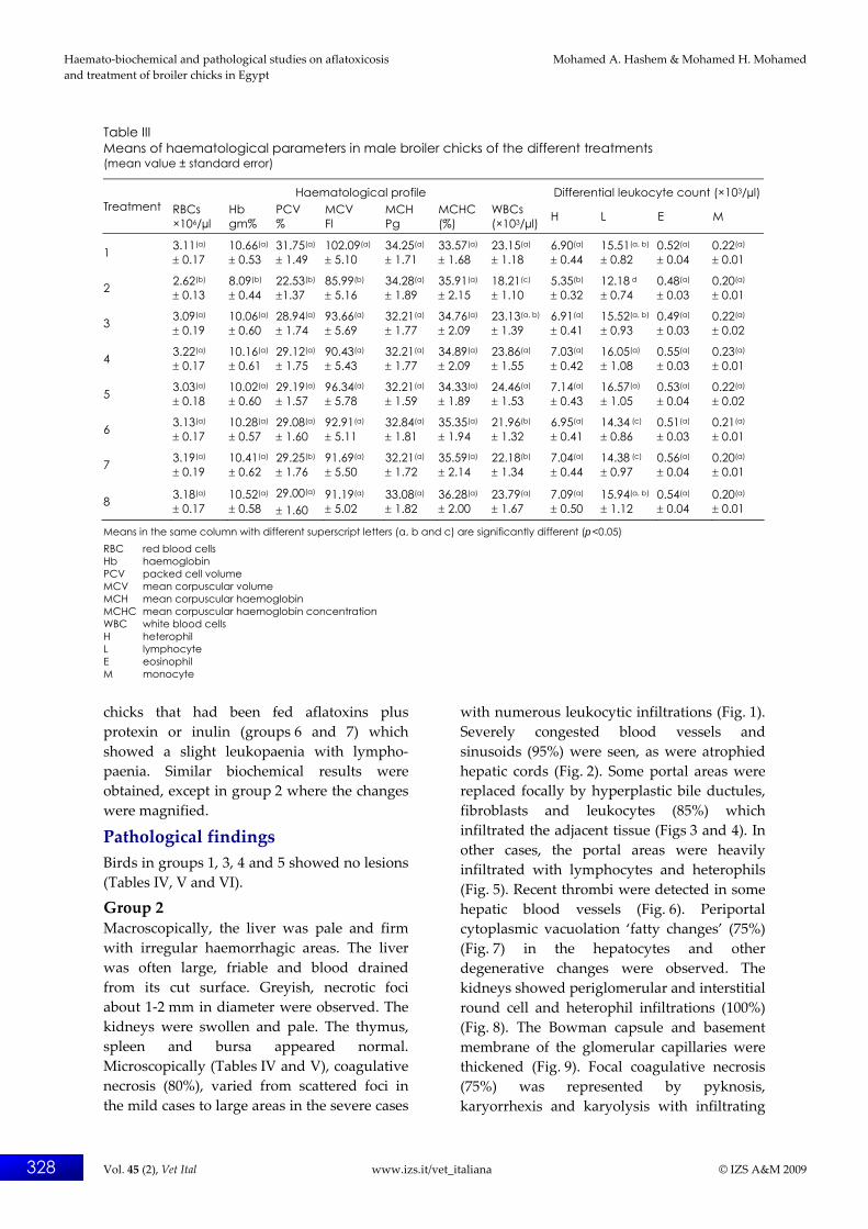
Table III Means of haematological parameters in male broiler chicks of the different treatments (mean value ± standard error)
Haematological profile Differential leukocyte count (×103/µl) Treatment RBCs
×106/µl Hb gm%
PCV %
MCV Fl
MCH Pg
MCHC (%)
WBCs (×103/µl) H L E M
1 3.11(a) ± 0.17
10.66(a) ± 0.53
31.75(a) ± 1.49
102.09(a) ± 5.10
34.25(a) ± 1.71
33.57(a) ± 1.68
23.15(a) ± 1.18
6.90(a) ± 0.44
15.51(a, b) ± 0.82
0.52(a) ± 0.04
0.22(a) ± 0.01
2 2.62(b) ± 0.13
8.09(b) ± 0.44
22.53(b) ±1.37
85.99(b) ± 5.16
34.28(a) ± 1.89
35.91(a) ± 2.15
18.21(c) ± 1.10
5.35(b) ± 0.32
12.18 d ± 0.74
0.48(a) ± 0.03
0.20(a) ± 0.01
3 3.09(a) ± 0.19
10.06(a) ± 0.60
28.94(a) ± 1.74
93.66(a) ± 5.69
32.21(a) ± 1.77
34.76(a) ± 2.09
23.13(a, b) ± 1.39
6.91(a) ± 0.41
15.52(a, b) ± 0.93
0.49(a) ± 0.03
0.22(a) ± 0.02
4 3.22(a) ± 0.17
10.16(a) ± 0.61
29.12(a) ± 1.75
90.43(a) ± 5.43
32.21(a) ± 1.77
34.89(a) ± 2.09
23.86(a) ± 1.55
7.03(a) ± 0.42
16.05(a) ± 1.08
0.55(a) ± 0.03
0.23(a)
± 0.01
5 3.03(a) ± 0.18
10.02(a)
± 0.60 29.19(a) ± 1.57
96.34(a) ± 5.78
32.21(a) ± 1.59
34.33(a) ± 1.89
24.46(a) ± 1.53
7.14(a) ± 0.43
16.57(a) ± 1.05
0.53(a) ± 0.04
0.22(a) ± 0.02
6 3.13(a) ± 0.17
10.28(a)
± 0.57 29.08(a)
± 1.60 92.91(a) ± 5.11
32.84(a) ± 1.81
35.35(a) ± 1.94
21.96(b) ± 1.32
6.95(a) ± 0.41
14.34 (c) ± 0.86
0.51(a) ± 0.03
0.21(a) ± 0.01
7 3.19(a) ± 0.19
10.41(a) ± 0.62
29.25(b) ± 1.76
91.69(a) ± 5.50
32.21(a) ± 1.72
35.59(a) ± 2.14
22.18(b) ± 1.34
7.04(a) ± 0.44
14.38 (c) ± 0.97
0.56(a) ± 0.04
0.20(a) ± 0.01
8 3.18(a) ± 0.17
10.52(a) ± 0.58
29.00(a) ± 1.60
91.19(a) ± 5.02
33.08(a) ± 1.82
36.28(a) ± 2.00
23.79(a) ± 1.67
7.09(a) ± 0.50
15.94(a, b) ± 1.12
0.54(a) ± 0.04
0.20(a) ± 0.01
Means in the same column with different superscript letters (a, b and c) are significantly different (p<0.05) RBC red blood cells Hb haemoglobin PCV packed cell volume MCV mean corpuscular volume MCH mean corpuscular haemoglobin MCHC mean corpuscular haemoglobin concentration WBC white blood cells H heterophil L lymphocyte E eosinophil M monocyte
chicks that had been fed aflatoxins plus protexin or inulin (groups 6 and 7) which showed a slight leukopaenia with lympho‐paenia. Similar biochemical results were obtained, except in group 2 where the changes were magnified.
Pathological findings Birds in groups 1, 3, 4 and 5 showed no lesions (Tables IV, V and VI).
Group 2 Macroscopically, the liver was pale and firm with irregular haemorrhagic areas. The liver was often large, friable and blood drained from its cut surface. Greyish, necrotic foci about 1‐2 mm in diameter were observed. The kidneys were swollen and pale. The thymus, spleen and bursa appeared normal. Microscopically (Tables IV and V), coagulative necrosis (80%), varied from scattered foci in the mild cases to large areas in the severe cases
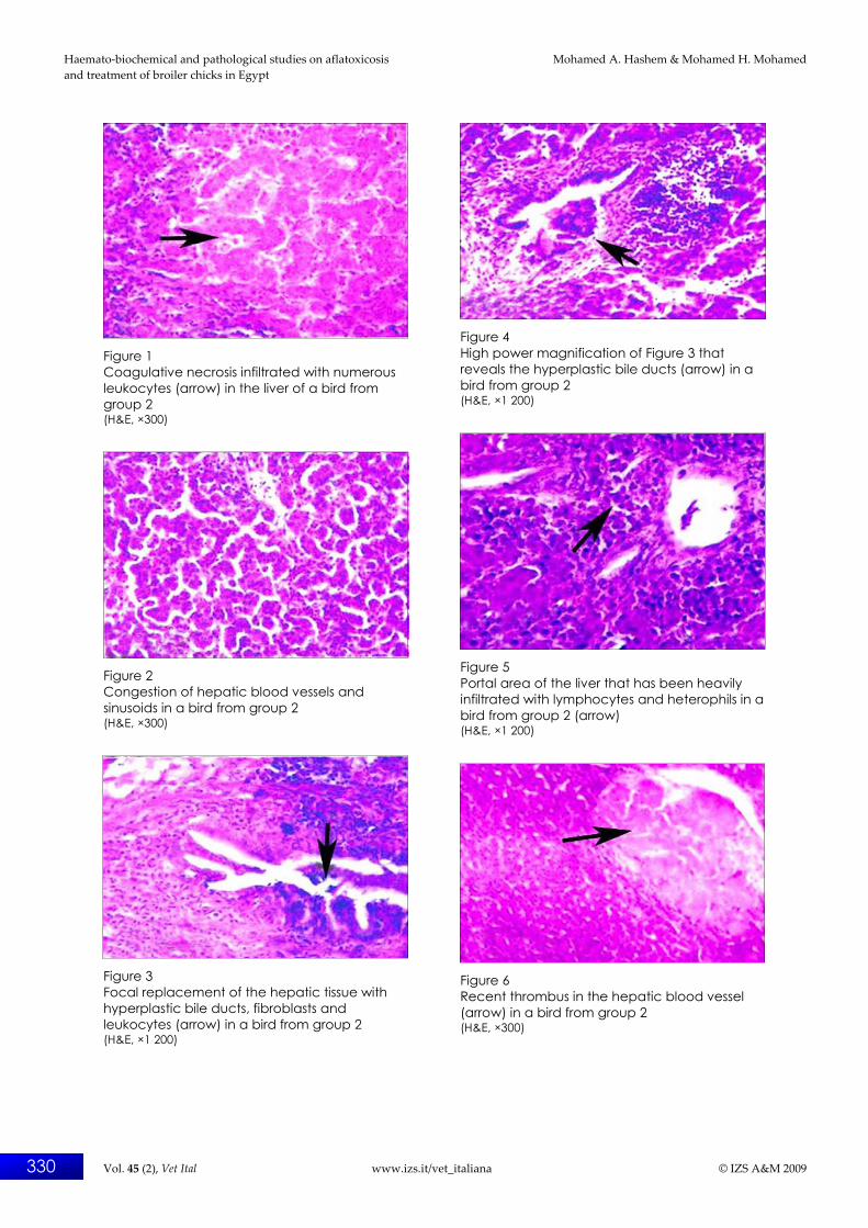
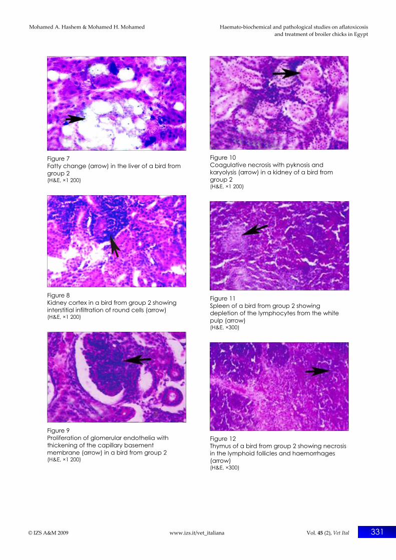
with numerous leukocytic infiltrations (Fig. 1). Severely congested blood vessels and sinusoids (95%) were seen, as were atrophied hepatic cords (Fig. 2). Some portal areas were replaced focally by hyperplastic bile ductules, fibroblasts and leukocytes (85%) which infiltrated the adjacent tissue (Figs 3 and 4). In other cases, the portal areas were heavily infiltrated with lymphocytes and heterophils (Fig. 5). Recent thrombi were detected in some hepatic blood vessels (Fig. 6). Periportal cytoplasmic vacuolation ‘fatty changes’ (75%) (Fig. 7) in the hepatocytes and other degenerative changes were observed. The kidneys showed periglomerular and interstitial round cell and heterophil infiltrations (100%) (Fig. 8). The Bowman capsule and basement membrane of the glomerular capillaries were thickened (Fig. 9). Focal coagulative necrosis (75%) was represented by pyknosis, karyorrhexis and karyolysis with infiltrating

Mohamed A. Hashem & Mohamed H. Mohamed Haemato‐biochemical and pathological studies on aflatoxicosis and treatment of broiler chicks in Egypt
© IZS A&M 2009 www.izs.it/vet_italiana Vol. 45 (2), Vet Ital 329
round cells (Fig. 10). The lymphoid organs (spleen, bursa and thymus) showed moderate depletion and necrosis of the lymphocytes (Figs 11 and 12).
Groups 6, 7 and 8 Macroscopically, the liver appeared to be normal except in group 7 where the liver was slightly large and severely congested. The
Table IV Means of biochemical parameters in male broiler chicks of the different treatments (mean value ± standard error)
Enzymes Treatment
ALT (U/l) AST (U/l) GGT (U/l) ALP (U/l)
Total protein (g/dl)
Albumin (g/dl)
Globulins (g/dl)
A/G (ratio)
Uric acid (mg/dl)
Creatinine (mg/dl)
1 95.14(b) ± 3.80
169.84(b) ± 6.79
41.56(b) ± 1.66
61.56(b) ± 1.66
3.76(a) ± 0.15
1.99(a) ± 0.08
1.77(a) ± 0.07
1.12(a) ± 0.04
3.91(b) ± 0.16
1.48(b) ± 0.06
2 155.23(a) ± 10.09
238.66(a) ± 14.32
68.91(a) ±4.13
88.31(a) ± 6.31
2.84(c) ± 0.17
1.56(b) ± 0.09
1.28(c) ± 0.08
1.22(b) ± 0.08
4.88(a) ± 0.24
1.88(a) ± 0.11
3 111.20(b) ± 8.29
178.08(b) ±13.32
51.99(b) ± 3.36
65.13(b) ± 4.36
3.22(a, b) ± 0.19
1.66(a) ± 0.10
1.56(a) ± 0.09
1.14(a) ± 0.07
4.12(b) ± 0.23
1.51(b) ± 0.11
4 112.19(b) ± 7.29
180.93(b) ±11.14
48.15(b) ± 3.13
63.15(b) ± 4.13
3.65(a) ± 0.18
1.84(a) ± 0.13
1.81(a) ± 0.12
1.02(a) ± 0.07
4.16(b) ± 0.27
1.50(b) ± 0.12
5 110.19(b) ± 7.09
181.19(b) ± 12.07
51.88(b) ± 3.30
63.88(b) ± 2.30
3.35(a) ± 0.18
1.68(a) ± 0.10
1.67(a) ± 0.09
1.14(a) ± 0.07
4.19(b) ± 0.27
1.48(b) ± 0.11
6 118.57(b) ± 9.44
192.54(c) ± 15.68
59.95(c) ± 3.76
69.82(b, c) ± 2.41
2.91(b) ± 0.189
1.55(b) ± 0.1
1.36(b) ± 0.09
1.14(a) ± 0.07
4.18(b) ± 0.20
1.53(b) ± 0.15
7 119.83(b) ± 7.16
189.92(c) ± 12.32
48.23(b) ± 3.13
66.89(b, c) ± 2.08
3.05(b) ± 0.23
1.59(b) ± 0.13
1.46(b) ± 0.11
1.08(a) ±0.06
3.98(b) ± 0.24
1.52(b) ± 0.10
8 114.25(b) ± 9.28
179.55(b) ± 13.16
50.09(b) ± 3.71
68.12(b) ± 2.52
3.49(a) ± 0.22
1.73(a) ± 0.12
1.76(a) ± 0.10
1.14(a) ± 0.08
4.16(b) ± 0.30
1.51(b) ± 0.13
Means in the same column with different superscript letters (a, b and c) are significantly different (p<0.05) U unit ALT alanine aminotransferase AST aspartate aminotransferase GGT gamma -glutamyl transferase ALP alkaline phosphatase A/G albumin/globulin ratio
Table V Frequency of major microscopic observations in the liver of male broilers
Liver Inflammation and
congestion Hyperplasia Necrosis Fatty changes Treatment No. of chicks
+ – Lesions (%) + – % of
lesions + – Lesions (%) + – Lesions
(%)
1 20 0 20 0.0(e) 0 20 0.0(d) 0 20 0.0(e) 0 20 0.0(c)
2 20 19 1 95.0(a) 17 3 85.0(a) 16 4 80.0(a) 15 5 75.0(a)
3 20 0 20 0.0(e) 0 20 0.0(d) 0 20 0.0(e) 0 20 0.0(c)
4 20 0 20 0.0(e) 0 20 0.0(d) 0 20 0.0(e) 0 20 0.0(c)
5 20 0 20 0.0(e) 0 20 0.0(d) 0 20 0.0(e) 0 20 0.0(c)
6 20 5 15 25.0(c) 2 18 10.0(b) 4 16 20.0(b) 2 18 10.0(b)
7 20 7 13 35.0(b) 1 19 5.0(c) 3 17 15.0(c) 2 18 10.0(b)
8 20 2 18 10.0(d) 0 20 0.0(d) 1 19 5.0(d) 0 20 0.0(c)
Chi-square (χ2) 92.503* 124.945* 82.353* 89.765*
Probability 0.00001 0.00001 0.00001 0.00001
Means within the same column with different superscript letters (a, b, c, d and e) are significantly different (p<0.05) + positive – negative * highly significant difference
Haemato‐biochemical and pathological studies on aflatoxicosis Mohamed A. Hashem & Mohamed H. Mohamed and treatment of broiler chicks in Egypt
330 Vol. 45 (2), Vet Ital www.izs.it/vet_italiana © IZS A&M 2009
Figure 1 Coagulative necrosis infiltrated with numerous leukocytes (arrow) in the liver of a bird from group 2 (H&E, ×300)
Figure 2 Congestion of hepatic blood vessels and sinusoids in a bird from group 2 (H&E, ×300)
Figure 3 Focal replacement of the hepatic tissue with hyperplastic bile ducts, fibroblasts and leukocytes (arrow) in a bird from group 2 (H&E, ×1 200)
Figure 4 High power magnification of Figure 3 that reveals the hyperplastic bile ducts (arrow) in a bird from group 2 (H&E, ×1 200)
Figure 5 Portal area of the liver that has been heavily infiltrated with lymphocytes and heterophils in a bird from group 2 (arrow) (H&E, ×1 200)
Figure 6 Recent thrombus in the hepatic blood vessel (arrow) in a bird from group 2 (H&E, ×300)
Mohamed A. Hashem & Mohamed H. Mohamed Haemato‐biochemical and pathological studies on aflatoxicosis and treatment of broiler chicks in Egypt
© IZS A&M 2009 www.izs.it/vet_italiana Vol. 45 (2), Vet Ital 331
Figure 7 Fatty change (arrow) in the liver of a bird from group 2 (H&E, ×1 200)
Figure 8 Kidney cortex in a bird from group 2 showing interstitial infiltration of round cells (arrow) (H&E, ×1 200)
Figure 9 Proliferation of glomerular endothelia with thickening of the capillary basement membrane (arrow) in a bird from group 2 (H&E, ×1 200)
Figure 10 Coagulative necrosis with pyknosis and karyolysis (arrow) in a kidney of a bird from group 2 (H&E, ×1 200)
Figure 11 Spleen of a bird from group 2 showing depletion of the lymphocytes from the white pulp (arrow) (H&E, ×300)
Figure 12 Thymus of a bird from group 2 showing necrosis in the lymphoid follicles and haemorrhages (arrow) (H&E, ×300)
Haemato‐biochemical and pathological studies on aflatoxicosis Mohamed A. Hashem & Mohamed H. Mohamed and treatment of broiler chicks in Egypt
332 Vol. 45 (2), Vet Ital www.izs.it/vet_italiana © IZS A&M 2009
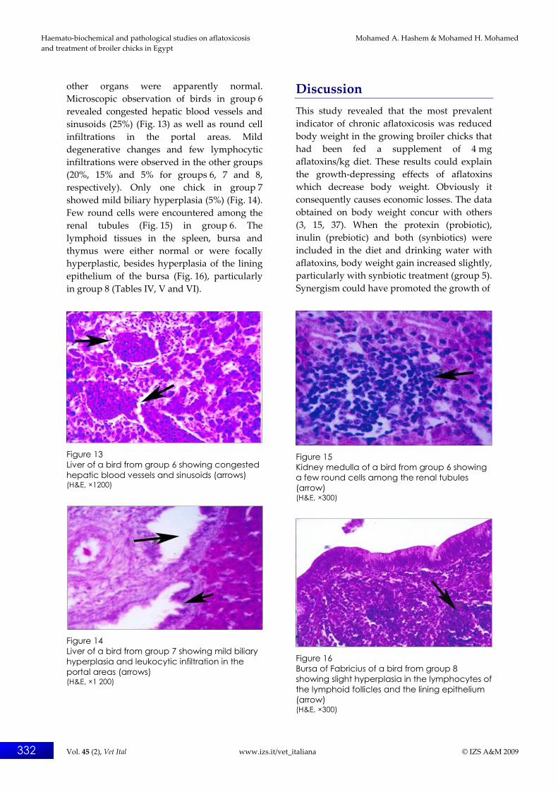
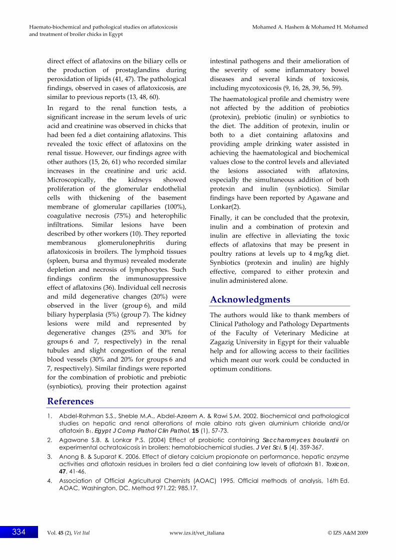
other organs were apparently normal. Microscopic observation of birds in group 6 revealed congested hepatic blood vessels and sinusoids (25%) (Fig. 13) as well as round cell infiltrations in the portal areas. Mild degenerative changes and few lymphocytic infiltrations were observed in the other groups (20%, 15% and 5% for groups 6, 7 and 8, respectively). Only one chick in group 7 showed mild biliary hyperplasia (5%) (Fig. 14). Few round cells were encountered among the renal tubules (Fig. 15) in group 6. The lymphoid tissues in the spleen, bursa and thymus were either normal or were focally hyperplastic, besides hyperplasia of the lining epithelium of the bursa (Fig. 16), particularly in group 8 (Tables IV, V and VI).
Figure 13 Liver of a bird from group 6 showing congested hepatic blood vessels and sinusoids (arrows) (H&E, ×1200)
Figure 14 Liver of a bird from group 7 showing mild biliary hyperplasia and leukocytic infiltration in the portal areas (arrows) (H&E, ×1 200)
Discussion This study revealed that the most prevalent indicator of chronic aflatoxicosis was reduced body weight in the growing broiler chicks that had been fed a supplement of 4 mg aflatoxins/kg diet. These results could explain the growth‐depressing effects of aflatoxins which decrease body weight. Obviously it consequently causes economic losses. The data obtained on body weight concur with others (3, 15, 37). When the protexin (probiotic), inulin (prebiotic) and both (synbiotics) were included in the diet and drinking water with aflatoxins, body weight gain increased slightly, particularly with synbiotic treatment (group 5). Synergism could have promoted the growth of
Figure 15 Kidney medulla of a bird from group 6 showing a few round cells among the renal tubules (arrow) (H&E, ×300)
Figure 16 Bursa of Fabricius of a bird from group 8 showing slight hyperplasia in the lymphocytes of the lymphoid follicles and the lining epithelium (arrow) (H&E, ×300)

Mohamed A. Hashem & Mohamed H. Mohamed Haemato‐biochemical and pathological studies on aflatoxicosis and treatment of broiler chicks in Egypt
© IZS A&M 2009 www.izs.it/vet_italiana Vol. 45 (2), Vet Ital 333
Table VI Frequency of major microscopic observations in the kidneys of male broilers
Kidney Inflammation and
congestion Hyperplasia Necrosis Fatty changes Treatments No. of chicks
+ – Lesions (%) + – Lesions
(%) + – Lesions (%) + – Lesions
(%)
1 20 0 20 0.0(e) 0 20 0.0(e) 0 20 0.0(e) 0 20 0.0(d)
2 20 20 0 100.0(a) 18 2 90.0(a) 15 5 75.0(a) 10 10 50.0(a)
3 20 0 20 0.0(e) 0 20 0.0(e) 0 20 0.0(e) 0 20 0.0(d)
4 20 0 20 0.0(e) 0 20 0.0(e) 0 20 0.0(e) 0 20 0.0(d)
5 20 0 20 0.0(e) 0 20 0.0(e) 0 20 0.0(e) 0 20 0.0(d)
6 20 6 14 30.0(b) 4 16 20.0(b) 5 15 25.0(c) 2 18 10.0(c)
7 20 4 16 20.0(c) 3 17 15.0(c) 6 14 30.0(b) 3 17 15.0(b)
8 20 2 18 10.0(d) 1 19 5.0(d) 1 19 5.0(d) 0 20 0.0(d)
Chi-square (χ2) 102.50* 97.543* 69.819* 49.949*
Probability 0.00001 0.00001 0.00001 0.00001
Means within the same column with different superscript letters (a, b, c, d and e) are significantly different (p<0.05) * highly significant difference
the beneficial microflora and consequently enhanced the immunity. Both probiotics and prebiotics have been shown to bind aflatoxins in the digestive tract, allowing the mycotoxins to pass harmlessly through the digestive tract (23, 39). This generally improves the overall health condition and increases body weight (31). The toxicity of aflatoxins was expressed as significant alterations in the haematological profile and biochemical parameters of serum. Microcytic normochromic anaemia with a drastic decrease in the total leukocytic count, heterophils and lymphocytes were observed during cases of aflatoxicosis. Previous findings are in accordance with the results obtained by other authors (15, 61) and in contrast with another (26). The decrease in the total leukocytic count, heterophils and lymphocytes may be due to the toxic effect of mycotoxins on the circulating cells, sequestration of cells in tissues and/or effect of aflatoxins on the bone marrow and lymphoid tissues. The insignificant changes in the eosinophil and monocyte counts were similar to other findings (25). An elevation in the hepatic enzyme activities (AST, ALT, ALP, GGT), in group 2 could reflect the hepatic damage and leakage of enzymes in the blood stream. Similar data have been reported previously (18, 40, 48).
The proteinogram showed a significant decline. The reduction of the total serum protein, associated with hypoalbuminaemia and hypoglobulinaemia, may be due to the interaction of aflatoxins with DNA, RNA and intracellular proteins of chicken‐hepatocytes, leading to impaired protein synthesis and leakage of albumin due to nephrotoxicity induced by aflatoxins (1, 40, 43). The data on hepatic damage agree with those reported elsewhere (3, 26, 41, 60). The above results coincide with our pathological findings which revealed that the liver is considered to be the primary target organ in cases of aflatoxicosis. Microscopically, the liver showed coagulative necrosis (80%), hyperplasia of the bile ducts in the portal areas (85%), fatty changes (75%) and extensive haemorrhaging. These degenerative and necrotic changes (80%) observed in cases of aflatoxicosis could be attributed to the damage of critical cellular macromolecules (lipids, DNA and proteins) through the oxidative stress of aflatoxins, which may result in the peroxidation of lipids and oxidative damage of DNA (31, 47). Moreover, the accumulation of intracellular calcium in cases of aflatoxicosis, causes mitochondrial dysfunction and reduces the adenosine triphosphate (ATP) generation (18, 40). The hyperplasia of the bile ducts is a characteristic or pathognomic lesion and may be due to the
Haemato‐biochemical and pathological studies on aflatoxicosis Mohamed A. Hashem & Mohamed H. Mohamed and treatment of broiler chicks in Egypt
334 Vol. 45 (2), Vet Ital www.izs.it/vet_italiana © IZS A&M 2009
direct effect of aflatoxins on the biliary cells or the production of prostaglandins during peroxidation of lipids (41, 47). The pathological findings, observed in cases of aflatoxicosis, are similar to previous reports (13, 48, 60). In regard to the renal function tests, a significant increase in the serum levels of uric acid and creatinine was observed in chicks that had been fed a diet containing aflatoxins. This revealed the toxic effect of aflatoxins on the renal tissue. However, our findings agree with other authors (15, 26, 61) who recorded similar increases in the creatinine and uric acid. Microscopically, the kidneys showed proliferation of the glomerular endothelial cells with thickening of the basement membrane of glomerular capillaries (100%), coagulative necrosis (75%) and heterophilic infiltrations. Similar lesions have been described by other workers (10). They reported membranous glomerulonephritis during aflatoxicosis in broilers. The lymphoid tissues (spleen, bursa and thymus) revealed moderate depletion and necrosis of lymphocytes. Such findings confirm the immunosuppressive effect of aflatoxins (36). Individual cell necrosis and mild degenerative changes (20%) were observed in the liver (group 6), and mild biliary hyperplasia (5%) (group 7). The kidney lesions were mild and represented by degenerative changes (25% and 30% for groups 6 and 7, respectively) in the renal tubules and slight congestion of the renal blood vessels (30% and 20% for groups 6 and 7, respectively). Similar findings were reported for the combination of probiotic and prebiotic (synbiotics), proving their protection against
intestinal pathogens and their amelioration of the severity of some inflammatory bowel diseases and several kinds of toxicosis, including mycotoxicosis (9, 16, 28, 39, 56, 59). The haematological profile and chemistry were not affected by the addition of probiotics (protexin), prebiotic (inulin) or synbiotics to the diet. The addition of protexin, inulin or both to a diet containing aflatoxins and providing ample drinking water assisted in achieving the haematological and biochemical values close to the control levels and alleviated the lesions associated with aflatoxins, especially the simultaneous addition of both protexin and inulin (synbiotics). Similar findings have been reported by Agawane and Lonkar(2). Finally, it can be concluded that the protexin, inulin and a combination of protexin and inulin are effective in alleviating the toxic effects of aflatoxins that may be present in poultry rations at levels up to 4 mg/kg diet. Synbiotics (protexin and inulin) are highly effective, compared to either protexin and inulin administered alone.
Acknowledgments The authors would like to thank members of Clinical Pathology and Pathology Departments of the Faculty of Veterinary Medicine at Zagazig University in Egypt for their valuable help and for allowing access to their facilities which meant our work could be conducted in optimum conditions.
References 1. Abdel-Rahman S.S., Sheble M.A., Abdel-Azeem A. & Rawi S.M. 2002. Biochemical and pathological
studies on hepatic and renal alterations of male albino rats given aluminium chloride and/or aflatoxin B1. Egypt J Comp Pathol Clin Pathol, 15 (1), 57-73.
2. Agawane S.B. & Lonkar P.S. (2004) Effect of probiotic containing Saccharomyces boulardii on experimental ochratoxicosis in broilers: hematobiochemical studies. J Vet Sci, 5 (4), 359-367.
3. Anong B. & Suparat K. 2006. Effect of dietary calcium propionate on performance, hepatic enzyme activities and aflatoxin residues in broilers fed a diet containing low levels of aflatoxin B1. Toxicon, 47, 41-46.
4. Association of Official Agricultural Chemists (AOAC) 1995. Official methods of analysis, 16th Ed. AOAC, Washington, DC, Method 971.22; 985.17.

Mohamed A. Hashem & Mohamed H. Mohamed Haemato‐biochemical and pathological studies on aflatoxicosis and treatment of broiler chicks in Egypt
© IZS A&M 2009 www.izs.it/vet_italiana Vol. 45 (2), Vet Ital 335
5. Ayasan T., Ozcan B. D., Baylan M. & Canogullari S. 2006. The effects of dietary inclusion of probiotic protexin on egg yield parameters of Japanese quails (Coturnix coturnix japonica). Int J Poult Sci, 5 (8), 776-779.
6. Balevi T., Ucan U.S., Coskun B., Kurtoglu V. & Cetingul S. 2001. Effect of dietary probiotic on performance and humoral immune response in layer hens. Br Poult. Sci., 42, 456-461.
7. Bancroft J.D. & Stevens A. 1996. Theory and practice of histological technique, 4th Ed. Churchill, Livingstone, New York, London, San Francisco, Tokyo, 766 pp.
8. Beg M.U., Al-Mutairi M., Beg K.R., Al-Mazeedi H.M., Ali L.N. & Saeed T. 2006. Mycotoxins in poultry feed in Kuwait. Arch Environ Contam Toxicol, 50 (4), 594-602.
9. Bengmark S. & Gil A. 2006. Bioecological and nutritional control of disease: prebiotics, probiotics and synbiotics. Nutr Hosp, 2, 73-86.
10. Chen C. & Walker W. 2005. Probiotics and prebiotics: role in clinical disease states. Adv Pediatriatics, 52, 77-113.
11. Council for Agricultural Science and Technology (CAST) 1989. CAST Task Force Report 16. Mycotoxins. Economic and Health Risks. CAST, Ames, Iowa, 1-91.
12. Corcoran B.M., Ross R.P., Fitzgerald G.F. & Stanton C. 2004. Comparative survival of probiotic lactobacilli spray-dried in the presence of prebiotic substances. J Appl Microbiol, 96 (5), 1024-1039.
13. Del Bianchi M., Oliveira C.A., Albuquerque R., Guerra J.L. & Correa B. 2005. Effects of prolonged oral administration of aflatoxin B1 and fumonisin B1 in broiler chickens. Poultry Sci, 84 (12), 1835-1840.
14. Drupt F. 1974. Colorimetric method for determination of albumin. Pharm Bio, 9, 777. 15. Edrington T.S., Harvey R.B. & Rottinghaus G.E. 1997. Environmental health influence of a
superactivated charcoal of the toxic effects of aflatoxin or T2 toxin in growing broilers. Poultry Sci, 76, 1205-1211.
16. El-Nezami H.S., Polychronaki N.N., Ma J, Zhu H., Ling W., Salminen E.K., Juvonen R.O., Salminen S.J., Poussa T. & Mykkanen H.M. 2006. Probiotic supplementation reduces a biomarker for increased risk of liver cancer in young men from southern China. Am J Clin Nutr, 83 (5), 1199-1203.
17. Ezz El-Arab A.M., Girgis S.M., Hegazy EM & Abd El-Khalek A.B. 2006. Effect of dietary honey on intestinal microflora and toxicity of mycotoxins in mice. BMC Complement Altern Med, 6, 6-22.
18. Fatemi F., Allameh A., Dadkhah A., Forouzandeh M., Kazemnejad S. & Sharifi R. 2006. Changes in hepatic cytosolic glutathione S-transferase activity and expression of its class-P during prenatal and postnatal periods in rats treated with aflatoxin B1. Arch Toxicol, 80 (9), 572 -579.
19. Feldman B.F., Zinkl J.G. & Jain N.C. 2000. Schalm’s veterinary hematology. 5th Ed. Lippincott Williams and Wilkins, A Wolters Company, New York, 1085-1088.
20. Fossati P., Prencipe L. & Berti G. 1980. Enzymatic determination of uric acid. Clin Chem, 26 (2), 227-231.
21. Fuller R. 1989. Probiotics in man and animals. A review. J Appl Bacteriol, 66 (5), 365-378. 22. Furrie E., Macfarlane S., Kennedy A., Cummings JH, Walsh S.V., O’Neil D.A. & Macfarlane G.T. 2005.
Synbiotic therapy (Bifidobacterium longum/synergy 1) initiates resolution of inflammation in patients with active ulcerative colitis: a randomized controlled pilot trial. Gut, 54 (9), 242-249.
23. Haskey N. & Dahl W.J. 2008. Synbiotic therapy: a promising new adjunctive therapy for ulcerative colitis. Nutr Rev, 64 (3), 132-138.
24. Kabir M.L., Rahman M.M., Rahman M.B., Rahman M.M. & Ahmed S.U. 2004. The dynamics of probiotics on growth performance and immune response in broilers. Int J Poult Sci, 3 (5), 361-364.
25. Kaufhold J., Hammon H.M. & Blum J.W. 2001. Fructo-oligosaccharide supplementation: effects on metabolic, endocrine and hematological traits in veal calves. J Vet Med Series A, 47 (1), 17-29.
26. Kececi T., Oguz H., Kurtoglu V. & Demet O. 1998. Effect of polyvinyl-polypyrrolidone, synthetic zeolite and bentonite on serum biochemical hematological characters of broiler chickens during aflatoxicosis. Br Poult Sci, 39 (3), 452-458.
27. Kind P.R. & King E.G. 1954. Colorimetric determination of alkaline phosphatase activity. J Clin Pathol, 7, 322.
28. Ledoux D.R., Rottinghaus G.E., Bermudez A.J. & Alonso Debolt M. 1999. Efficacy of hydrated sodium calcium aluminosilicate to ameliorate the toxic effects of aflatoxin in broiler chicks. Poult Sci, 77 (2), 204-210.
Haemato‐biochemical and pathological studies on aflatoxicosis Mohamed A. Hashem & Mohamed H. Mohamed and treatment of broiler chicks in Egypt
336 Vol. 45 (2), Vet Ital www.izs.it/vet_italiana © IZS A&M 2009
29. McKenzie K.S., Sarr A.B., Mayura K., Bailey R.H., Miller D.R., Rogers T.D., Norred W.P., Voss K.A., Plattner R.D., Kubena L.F. & Phillips T.D. 1997. Oxidative degradation and detoxification of mycotoxins using a novel source of ozone. Food Chem Toxicol, 35 (8), 807-820.
30. Mehr A.M., Shargh S. M., Dastar B., Hassani S. & Akbar M.R. 2007. Effect of different levels of protein and protexin on broiler performance. Int J Poultry Sci, 6 (8), 573-577.
31. Mohamed M.H. & Selim A.G. 2003. Effects of some detoxifying agents on aflatoxicosis in broiler chicks. Egypt J Comp Pathol Clin Pathol, 16 (2), 270-286.
32. Monoura P., Rahman M., Khan M.F.R., Rahman M.B. & Rahman M.M. 2008. Effect of vitamins, minerals and probiotics on production of antibody and live weight gain following vaccination with BCRDV in broiler birds. Bangl J Vet Med, 6 (1), 31-36.
33. Nahaston S.N., Nakaue H.S. & Mirosh L.W. 1993. Effect of directly fed microbials on nutrient retention and production parameters of single comb white Leghorn pullets. Poult Sci, 72, 87.
34. National Research Council (NRC) 1994. Nutrient requirements of poultry, 9th Revised Ed. National Academy Press, Washington, DC, 157 pp (www.nap.edu/openbook.php?isbn=0309048923 accessed on 2 June 2009).
35. Nomoto K. 2005. Prevention of infections by probiotics. J Biosci Bioeng, 100 (6), 583-592. 36. Ogawa T., Asai Y., Tamai R., Makimura Y., Sakamoto H., Hashikawa S. & Yasuda K. 2006. Natural killer
cell activities of synbiotic Lactobacillus casei ssp. casei in conjunction with dextran. Clin Exp Immunol, 143 (1), 103-109.
37. Patterson J.A. & Burkholder K.M. 2003. Application of prebiotics and probiotics in poultry production. Poult Sci, 82 (4), 627-631.
38. Peters T. 1968. Colorimetric method for determination of total serum proteins. Clin Chem, 14, 1147. 39. Phillips T.D., Clement B.A., Kubena L.F. & Harvey R.B. 1990. Detection of detoxification of aflatoxins.
Prevention of aflatoxicosis and aflatoxin residues with hydrated sodium calcium aluminosilicate. Vet Human Toxicol, 32, 15-19.
40. Quezada T., Cuellar H., Valdivia A.G. & Reys J.I. 2002. Effects of aflatoxin B1 on the liver and kidneys of broiler chickens during development. Comp Biochem Physiol C Toxicol Pharmacol, 125 (3), 265-272.
41. Quist C.F., Bounous D.I., Kilburn T.V., nettles V.F. & Wyatt R.D. 2002. The effect of dietary aflatoxin on wild turkey poultry. J Wildl Dis, 36 (3), 436-444.
42. Rajmane B.V. & Sonawane N.S. 1998. Efficacy of probiotics on performance of broilers in hot climate. World’s Poultry Science Association (WPSA)-Israel Branch. In Proc. 10th European Poultry Conference, 21-26 June, Jerusalem. WSPA, Beekbergen, 559-562.
43. Raju M.V., Rama Rao S.V., Radhika K. & Panda A.K. 2005. Effect of amount and source of supplemental dietary vegetable oil on broiler chickens exposed to aflatoxicosis. Br Poult Sci, 46 (5), 587-594.
44. Raper K.B. & Fennel D.I. 1977. The genus Aspergillus. Robert E. Krieger publishing Co. Inc., Washington, DC, 686 pp.
45. Reedy T.V., Viswananthan L. & Venkitasubramanian T.A. 1971. High aflatoxin production on chemical defined medium. Appl Microbiol, 22, 393-396.
46. Reitman S. and Frankel S. 1957. Colorimetric determination of transaminase activity. Am J Clin Pathol, 28, 56.
47. Saif Y.M., Barnes H.J., Glissons J.R., McDougald L.R. & Swayne D.E. 2003. Diseases of poultry, 11th Ed. Editorial Board for the American Association of Avian Pathologists. Mosby-Wolfe, Iowa State University Press, Ames, Iowa, 320-326.
48. Scholl P.F., McCoy L., Kensler T.W. & Groopman J.D. 2006. Quantitative analysis and chronic dosimetry of the aflatoxin B1 plasma albumin adduct Lys-AFB1 in rats by isotope dilution mass spectrometry. Chem Res Toxicol, 19 (1), 44-49.
49. Seeling H.P. & Wust H. 1969. Colorimetric method for determination of creatinine. Arztl Lab, 15, 34. 50. Shi Y.H., Xub Z.R., Feng J.L. & Wang C.Z. 2006. Efficacy of modified montmorillonite nanocomposite
to reduce the toxicity of aflatoxin in broiler chicks. Animal Feed Sci Technol, 129 (1-2), 138-148. 51. Snelling A.M. 2005. Effects of probiotics on the gastrointestinal tract. Curr Opin Infect Dis, 18 (5),
420-426. 52. Statistical Analysis System (SAS) Institute 2002. Statistical analysis system user’s guide statistics.
Version 8 Ed. SAS Institute, Cary, North Carolina, 275-331.
Mohamed A. Hashem & Mohamed H. Mohamed Haemato‐biochemical and pathological studies on aflatoxicosis and treatment of broiler chicks in Egypt
© IZS A&M 2009 www.izs.it/vet_italiana Vol. 45 (2), Vet Ital 337
53. Swennen K., Courtin C.M. & Delcour J.A. 2006. Non-digestible oligosaccharides with prebiotic properties. Crit Rev Food Sci Nutr, 46 (6), 459-471.
54. Szasz G. & Persij J.P. 1974. Colorimetric determination of gamma glutamyl transpeptidase activity. Z Klin Chem uklin Biochem, 12, 228.
55. Szilagyi A. 2005. Use of prebiotics for inflammatory bowel disease. Can J Gastroenterol, 19 (8), 505-510.
56. Thitaram S.N., Siragusa G.R. & Hinton A.J. 2005. Bifidobacterium-selective isolation and enumeration from chicken caeca by a modified oligosaccharide antibiotic-selective agar medium. Lett Appl Microbiol, 41 (4), 355-360.
57. Trucksess M.W. 2006. Committee on natural toxins and food allergens; mycotoxins. J AOAC Int, 89 (1), 270-284.
58. Trucksess M.W., Brumley W.C. & Nesheim S. 1984. Rapid quantitation and confirmation of aflatoxins in corn and peanut butter, using a disposable silica gel column, thin-layer gas and chromatography/mass spectrometry. J Assoc Analytical Chem, 67 (5), 973-975.
59. Wu W. 1997. Counteracting Fusarium proliferatum toxicity in broiler chicks by supplementing drinking water with poultry aid plus. Poultry Sci, 76 (3), 463-468.
60. Yates M.S., Kwak M.K., Egner P.A., Groopman J.D., Bodreddigari S., Sutter T.R., Baumgartner K.J., Roebuck B.D., Liby K.T., Yore M.M., Honda T., Gribble G.W., Sporn M.B. & Kensler T.W. 2006. Potent protection against aflatoxin-induced tumorigenesis through induction of Nrf2-regulated pathways by the triterpenoid 1-[2-cyano-3-, 12-dioxooleana-1, 9(11)-dien-28-oyl] imidazole. Cancer Res, 66 (4), 2488-2494.
61. Yiannikouris A., Andre G., Poughon L., François J., Dussap C.G., Jeminet G., Bertin G. & Jouany J.P. 2006. Chemical and conformational study of the interactions involved in mycotoxin complexation with beta-D-glucans. Biomacromolecules, 7 (4), 1147-1155.