H-NS Oligomerization Domain Structure Reveals the Mechanism for High Order Self-association of the...
10
H-NS Oligomerization Domain Structure Reveals the Mechanism for High Order Self-association of the Intact Protein Diego Esposito 1 , Arsen Petrovic 1 , Richard Harris 1 , Shusuke Ono 1 John F. Eccleston 2 , Amina Mbabaali 2 , Ihtshamul Haq 1 Christopher F. Higgins 3 , Jay C. D. Hinton 4 , Paul C. Driscoll 1,5 and John E. Ladbury 1 * 1 Department of Biochemistry and Molecular Biology University College London Darwin Building, Gower Street London WC1E 6BT, UK 2 National Institute for Medical Research, The Ridgeway, Mill Hill, London NW7 1AA, UK 3 Faculty of Medicine, MRC Clinical Sciences Centre Imperial College, Hammersmith Hospital Campus, DuCane Road, London W12 0NN, UK 4 Institute for Food Research Norwich Research Park, Colney Norwich NR4 7UA, UK 5 Ludwig Institute for Cancer Research, 91 Riding House Street, London W1W 7BS, UK H-NS plays a role in condensing DNA in the bacterial nucleoid. This 136 amino acid protein comprises two functional domains separated by a flexible linker. High order structures formed by the N-terminal oligomeri- zation domain (residues 1–89) constitute the basis of a protein scaffold that binds DNA via the C-terminal domain. Deletion of residues 57–89 or 64–89 of the oligomerization domain precludes high order structure for- mation, yielding a discrete dimer. This dimerization event represents the initial event in the formation of high order structure. The dimers thus constitute the basic building block of the protein scaffold. The three- dimensional solution structure of one of these units (residues 1–57) has been determined. Activity of these structural units is demonstrated by a dominant negative effect on high order structure formation on addition to the full length protein. Truncated and site-directed mutant forms of the N-terminal domain of H-NS reveal how the dimeric unit self-associ- ates in a head-to-tail manner and demonstrate the importance of second- ary structure in this interaction to form high order structures. A model is presented for the structural basis for DNA packaging in bacterial cells. q 2002 Elsevier Science Ltd. All rights reserved Keywords: chromatin; coiled-coil; DNA packaging; nucleoid assembly; histone-like *Corresponding author Introduction Several proteins interact with DNA in the bacterial nucleoid to facilitate chromosome con- densation and modulate gene expression. H-NS is one of the most abundant components of bacterial chromatin. 1–6 H-NS is a 15.6 kDa, 136 amino acid protein that is present at high copy number (up to 10 5 molecules) in cells of all Enterobacteriacea. Several studies have investigated the role of H-NS in the modulation of DNA super-coiling and the con- trol of gene expression, 5,7 but a structural model of the intact H-NS molecule has yet to evolve. Full length H-NS forms concentration-dependent, self-associated, high order, complexes. 8,9 The protein is composed of two distinct domains: the N-terminal domain mediates protein oligomerization, and the C-terminal domain is required for non-sequence specific DNA binding. 7,8,10 – 12 These domains are sep- arated by a flexible linker that provides the C-term- inal domain with a freedom of movement relative to the oligomeric N-terminal domain. 9 Residues 1–89 are necessary and sufficient for higher-order oligomerization. 9 In contrast, a truncated version of this domain (residues 1–64; H-NS 1–64 ) was shown to 0022-2836/02/$ - see front matter q 2002 Elsevier Science Ltd. All rights reserved E-mail address of the corresponding author: [email protected] Abbreviations used: H-NS, wild-type from Salmonella typhimurium with the C at position 20 replaced by a S; H- NS 1–57 , H-NS 1–64 , the first 57 or 64 residues of H-NS but also including the GSHM-tag derived from the cloning vector; NOE, nuclear Overhauser effect; HSQC, heteronuclear single quantum correlation. doi:10.1016/S0022-2836(02)01141-5 available online at http://www.idealibrary.com on B w J. Mol. Biol. (2002) 324, 841–850
-
Upload
mpi-dortmund-mpg -
Category
Documents
-
view
3 -
download
0
Transcript of H-NS Oligomerization Domain Structure Reveals the Mechanism for High Order Self-association of the...

H-NS Oligomerization Domain Structure Reveals theMechanism for High Order Self-association of theIntact Protein
Diego Esposito1, Arsen Petrovic1, Richard Harris1, Shusuke Ono1
John F. Eccleston2, Amina Mbabaali2, Ihtshamul Haq1
Christopher F. Higgins3, Jay C. D. Hinton4, Paul C. Driscoll1,5 andJohn E. Ladbury1*
1Department of Biochemistryand Molecular BiologyUniversity College LondonDarwin Building, Gower StreetLondon WC1E 6BT, UK
2National Institute for MedicalResearch, The Ridgeway, MillHill, London NW7 1AA, UK
3Faculty of Medicine, MRCClinical Sciences CentreImperial College, HammersmithHospital Campus, DuCaneRoad, London W12 0NN, UK
4Institute for Food ResearchNorwich Research Park, ColneyNorwich NR4 7UA, UK
5Ludwig Institute for CancerResearch, 91 Riding HouseStreet, London W1W 7BS, UK
H-NS plays a role in condensing DNA in the bacterial nucleoid. This 136amino acid protein comprises two functional domains separated by aflexible linker. High order structures formed by the N-terminal oligomeri-zation domain (residues 1–89) constitute the basis of a protein scaffoldthat binds DNA via the C-terminal domain. Deletion of residues 57–89 or64–89 of the oligomerization domain precludes high order structure for-mation, yielding a discrete dimer. This dimerization event represents theinitial event in the formation of high order structure. The dimers thusconstitute the basic building block of the protein scaffold. The three-dimensional solution structure of one of these units (residues 1–57) hasbeen determined. Activity of these structural units is demonstrated by adominant negative effect on high order structure formation on additionto the full length protein. Truncated and site-directed mutant forms ofthe N-terminal domain of H-NS reveal how the dimeric unit self-associ-ates in a head-to-tail manner and demonstrate the importance of second-ary structure in this interaction to form high order structures. A model ispresented for the structural basis for DNA packaging in bacterial cells.
q 2002 Elsevier Science Ltd. All rights reserved
Keywords: chromatin; coiled-coil; DNA packaging; nucleoid assembly;histone-like*Corresponding author
Introduction
Several proteins interact with DNA in thebacterial nucleoid to facilitate chromosome con-densation and modulate gene expression. H-NS isone of the most abundant components of bacterialchromatin.1 – 6 H-NS is a 15.6 kDa, 136 amino acidprotein that is present at high copy number (up to
105 molecules) in cells of all Enterobacteriacea.Several studies have investigated the role of H-NSin the modulation of DNA super-coiling and the con-trol of gene expression,5,7 but a structural model ofthe intact H-NS molecule has yet to evolve.
Full length H-NS forms concentration-dependent,self-associated, high order, complexes.8,9 The proteinis composed of two distinct domains: the N-terminaldomain mediates protein oligomerization, and theC-terminal domain is required for non-sequencespecific DNA binding.7,8,10–12 These domains are sep-arated by a flexible linker that provides the C-term-inal domain with a freedom of movement relative tothe oligomeric N-terminal domain.9 Residues 1–89are necessary and sufficient for higher-orderoligomerization.9 In contrast, a truncated version ofthis domain (residues 1–64; H-NS1–64) was shown to
0022-2836/02/$ - see front matter q 2002 Elsevier Science Ltd. All rights reserved
E-mail address of the corresponding author:[email protected]
Abbreviations used: H-NS, wild-type from Salmonellatyphimurium with the C at position 20 replaced by a S; H-NS1 – 57, H-NS1 – 64, the first 57 or 64 residues of H-NS butalso including the GSHM-tag derived from the cloningvector; NOE, nuclear Overhauser effect; HSQC,heteronuclear single quantum correlation.
doi:10.1016/S0022-2836(02)01141-5 available online at http://www.idealibrary.com onBw
J. Mol. Biol. (2002) 324, 841–850
have a defined oligomeric state that is largely a-heli-cal and was likely to self-associate via a coiled-coilinteraction. Thus, residues 1–64 can be consideredto form a basic building block, while residues from65 to 89 appear to be responsible, at least in part, forthe formation of the higher-order structures.9
A H-NS1 – 64 protomer, within the context of itsdefined oligomeric state, was previously shownby NMR to consist of three helices separated byshort linkers.13 Residues 54–64 form a largelyunstructured, dynamic region. Here we report thethree dimensional structure of residues 1–57which in their dimeric state represent part of thebasic building block for the high order self-assembly of H-NS. The largest helix (H3) forms aparallel coiled-coil interface whilst the N-terminalhelices H1 and H2 lie approximately anti-parallelwith H3 and make both intra- and inter-molecularinteractions to the coiled-coil core. Investigation ofthe formation of the high-order, heterodispersedcomplexes by self-assembly of the N-terminaldomain suggest a model whereby the high orderprotein scaffold is formed via a head-to-tail (i.e. Nterminus to C terminus) interaction of the com-ponent dimeric units. This result provides the basisfor understanding how H-NS can facilitate thepackaging of chromosomal DNA in the bacterial cell.
Results
The solution structure of residues 1–57 of theH-NS oligomerization domain
We have previously shown by heteronuclearNMR spectroscopy that the residues towards the
C terminus of H-NS1 – 64 exhibited characteristics ofhigh internal mobility, suggesting a dynamicallydisordered conformation with no well-defined sec-ondary structure.13 Furthermore, these residuesyielded NMR resonances with strong overlap inthe NMR spectrum. As a result we expressed anH-NS polypeptide comprising just the N-terminalresidues 1–57, H-NS1 – 57, for further NMR studiesand solution structure determination. Sedimen-tation equilibrium analytical ultracentrifugation(AUC) experiments established that H-NS1 – 57 is ahomodimer (as with the 1–64 construct, seebelow). The 2D [15N,1H]-heteronuclear single quan-tum coherence (HSQC) spectrum of isotopelabelled H-NS1– 57 retains the main features pre-viously observed for H-NS1 – 64,
13 but with fewercross-peaks, enhanced spectral resolution, andreduced cross-peak overlap. 15N nuclear relaxationdata were recorded to obtain residue-by-residueR1, R2 relaxation rates and {1H}15N heteronuclearNOEs. The model-free formalism-derived estimateof the molecular rotational correlation timetc , 11 ns is consistent with a molecular entitythat reorientates more rapidly than H-NS1 – 64.
14
A double 15N/13C-isotope labelled H-NS1 – 57 samplewas used in a standard set of triple resonanceexperiments to obtain sequence-specific resonanceassignments using a strategy similar that reportedpreviously for H-NS1 – 64,
13 except that perdeutera-tion of the protein was not required. The higha-helical content of H-NS1 – 57, combined with theabsence of aromatic side-chains, gives rise to rela-tively poor dispersion of the Ca and Ha chemicalshifts making the side-chain resonance assign-ments difficult to obtain. Nevertheless theseassignments were determined by analysis of
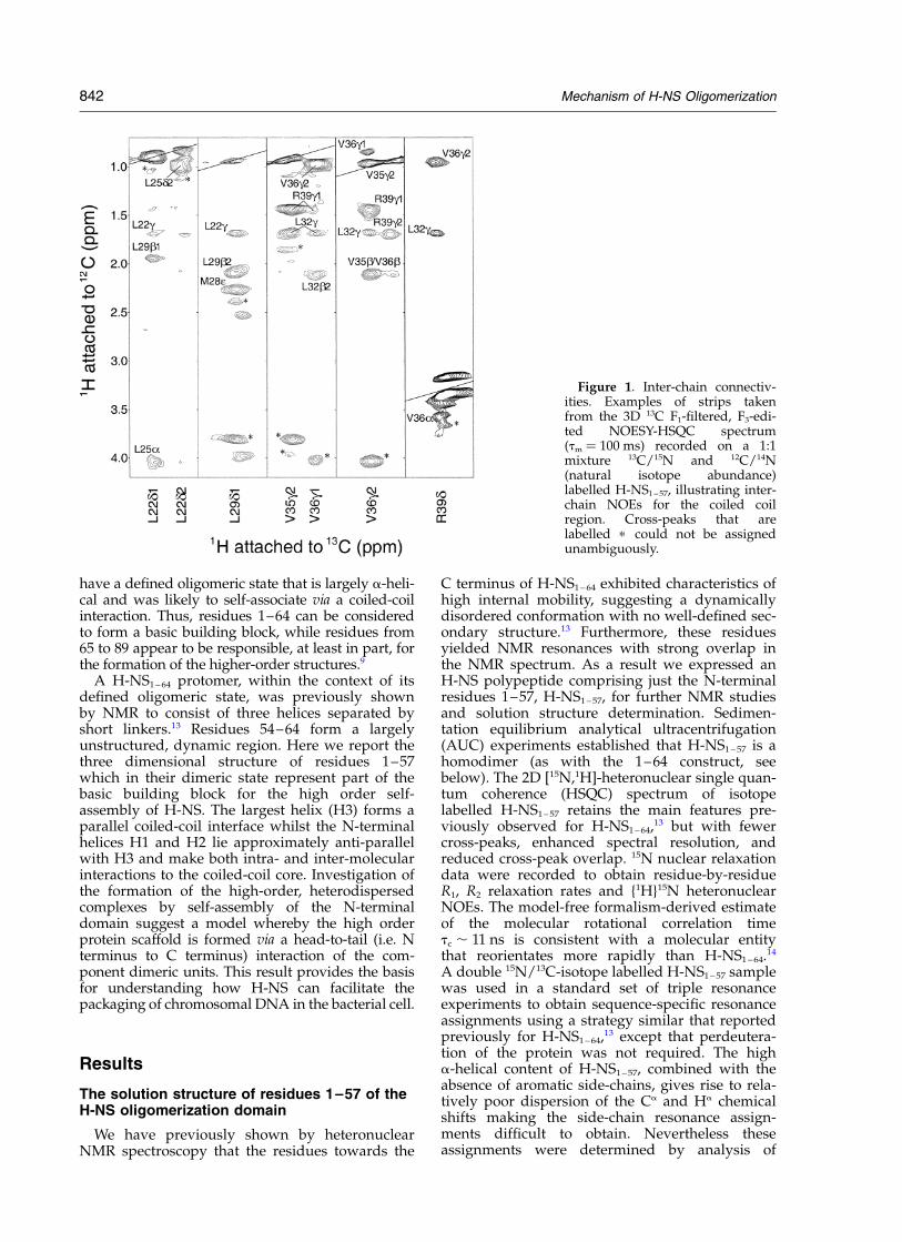
Figure 1. Inter-chain connectiv-ities. Examples of strips takenfrom the 3D 13C F1-filtered, F3-edi-ted NOESY-HSQC spectrum(tm ¼ 100 ms) recorded on a 1:1mixture 13C/15N and 12C/14N(natural isotope abundance)labelled H-NS1– 57, illustrating inter-chain NOEs for the coiled coilregion. Cross-peaks that arelabelled p could not be assignedunambiguously.
842 Mechanism of H-NS Oligomerization
1H– 13C– 13C– 1H correlations in a 17 ms mixingtime 3D HCCH-TOCSY experiment combinedwith 2D 13C,1H-HSQC and 3D 13C-NOESY-HSQCspectra. In contrast to H-NS1 – 64, the majority of1H– 1H correlations between NH protons and theside-chains could be detected in 3D 15N-TOCSY-HSQC and 15N-NOESY-HSQC spectra (mixingtimes 60 ms and 80 ms, respectively), which greatlyassisted the confidence in the accuracy of theassignments. A complete listing of the resonanceassignments for H-NS1 – 57 has been deposited atthe BioMagResBank (Number RCSB016203).
The observed pattern of residue-by-residuesecondary 13C chemical shifts and 15N nuclearrelaxation data is essentially identical for residues1–48 of H-NS1 – 57 and H-NS1 – 64, indicating thatthere are no significant differences in the com-ponent secondary structural elements. The poly-peptide comprises two short a-helices (H1,residues 2–7; and H2, residues 10–16) followedby a longer third helix (H3, residues 22–49). Aswith H-NS1 – 64, we noted that incubation ofH-NS1 – 57 at room temperature over the course ofseveral weeks yielded evidence, clearly observedin the NMR spectra, of a gradual proteolytic degra-dation of the sample. Proteolysis occurs primarilyat the C-terminal end of the protein, indicatinglack of a rigid secondary structure in this region.This phenomenon is consistent with 15N relaxationand heteronuclear NOEs data that reveal a higherdegree of flexibility of residues 50–57, which inH-NS1 – 64 appear to be arranged as part of thecoiled-coil core helices but showed faster trans-verse relaxation time-constants compared to thepreceding residues in the helix segment.13 ForH-NS1 – 57 the chemical shift and heteronuclearrelaxation data indicate that ordered helical struc-ture is present up to residue 49.
The 3D solution structure of the H-NS1 – 57 homo-dimer was determined on the basis of inter-protondistance and dihedral angle restraints derivedfrom the NMR data recorded using isotope-labelled samples of the protein. Unambiguousassignment of 101 NOE connectivities betweenprotomers was obtained by analysis of a 3D 13CF1-filtered, F3-edited NOESY-HSQC experiment15
recorded using a sample containing a 1:1 ratio ofunlabelled (12C/14N) and double 13C/15N-isotopelabelled chains (Figure 1). NOE cross-peaksobserved in 3D 13C-NOESY-HSQC experiments onfully labelled samples but absent in the 3D 13C F1-filtered, F3-edited NOESY HSQC experiment withmixed labelled and unlabelled chains, wereattributed exclusively to intra-chain connectivities.
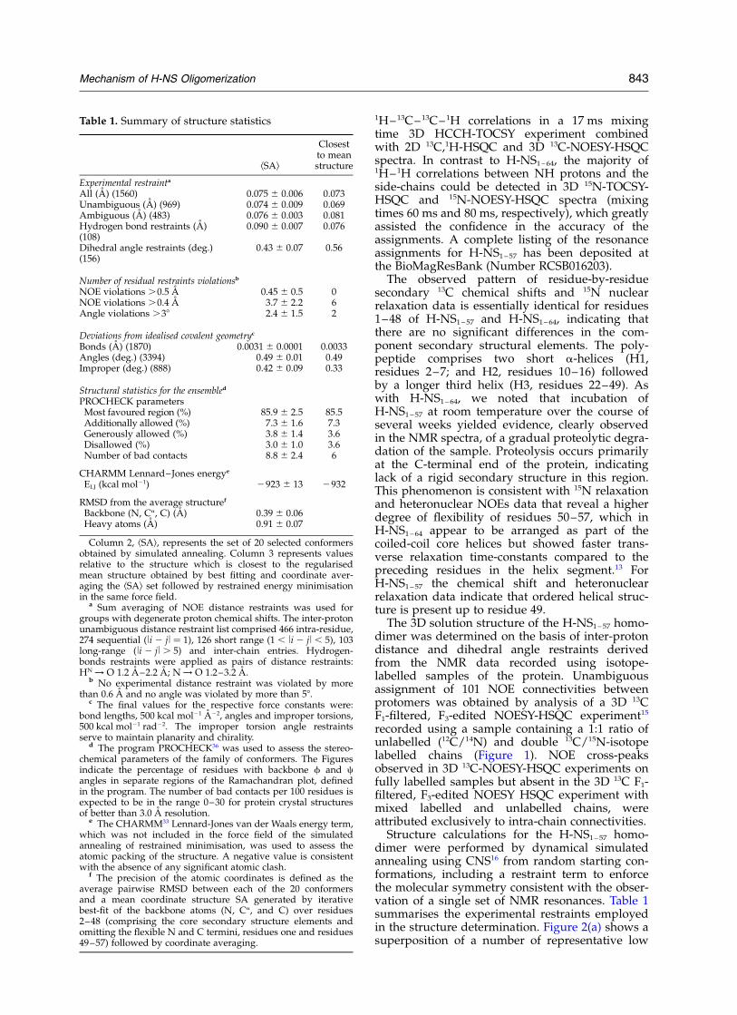
Structure calculations for the H-NS1 – 57 homo-dimer were performed by dynamical simulatedannealing using CNS16 from random starting con-formations, including a restraint term to enforcethe molecular symmetry consistent with the obser-vation of a single set of NMR resonances. Table 1summarises the experimental restraints employedin the structure determination. Figure 2(a) shows asuperposition of a number of representative low
Table 1. Summary of structure statistics
kSAl
Closestto meanstructure
Experimental restrainta
All (A) (1560) 0.075 ^ 0.006 0.073Unambiguous (A) (969) 0.074 ^ 0.009 0.069Ambiguous (A) (483) 0.076 ^ 0.003 0.081Hydrogen bond restraints (A)(108)
0.090 ^ 0.007 0.076
Dihedral angle restraints (deg.)(156)
0.43 ^ 0.07 0.56
Number of residual restraints violationsb
NOE violations .0.5 A 0.45 ^ 0.5 0NOE violations .0.4 A 3.7 ^ 2.2 6Angle violations .38 2.4 ^ 1.5 2
Deviations from idealised covalent geometryc
Bonds (A) (1870) 0.0031 ^ 0.0001 0.0033Angles (deg.) (3394) 0.49 ^ 0.01 0.49Improper (deg.) (888) 0.42 ^ 0.09 0.33
Structural statistics for the ensembled
PROCHECK parametersMost favoured region (%) 85.9 ^ 2.5 85.5Additionally allowed (%) 7.3 ^ 1.6 7.3Generously allowed (%) 3.8 ^ 1.4 3.6Disallowed (%) 3.0 ^ 1.0 3.6Number of bad contacts 8.8 ^ 2.4 6
CHARMM Lennard–Jones energye
ELJ (kcal mol21) 2923 ^ 13 2932
RMSD from the average structuref
Backbone (N, Ca, C) (A) 0.39 ^ 0.06Heavy atoms (A) 0.91 ^ 0.07
Column 2, kSAl, represents the set of 20 selected conformersobtained by simulated annealing. Column 3 represents valuesrelative to the structure which is closest to the regularisedmean structure obtained by best fitting and coordinate aver-aging the kSAl set followed by restrained energy minimisationin the same force field.
a Sum averaging of NOE distance restraints was used forgroups with degenerate proton chemical shifts. The inter-protonunambiguous distance restraint list comprised 466 intra-residue,274 sequential (li 2 jl ¼ 1), 126 short range (1 , li 2 jl , 5), 103long-range (li 2 jl . 5) and inter-chain entries. Hydrogen-bonds restraints were applied as pairs of distance restraints:HN ! O 1.2 A–2.2 A; N ! O 1.2–3.2 A.
b No experimental distance restraint was violated by morethan 0.6 A and no angle was violated by more than 58.
c The final values for the respective force constants were:bond lengths, 500 kcal mol21 A22, angles and improper torsions,500 kcal mol21 rad22. The improper torsion angle restraintsserve to maintain planarity and chirality.
d The program PROCHECK36 was used to assess the stereo-chemical parameters of the family of conformers. The Figuresindicate the percentage of residues with backbone f and cangles in separate regions of the Ramachandran plot, definedin the program. The number of bad contacts per 100 residues isexpected to be in the range 0–30 for protein crystal structuresof better than 3.0 A resolution.
e The CHARMM33 Lennard-Jones van der Waals energy term,which was not included in the force field of the simulatedannealing of restrained minimisation, was used to assess theatomic packing of the structure. A negative value is consistentwith the absence of any significant atomic clash.
f The precision of the atomic coordinates is defined as theaverage pairwise RMSD between each of the 20 conformersand a mean coordinate structure SA generated by iterativebest-fit of the backbone atoms (N, Ca, and C) over residues2–48 (comprising the core secondary structure elements andomitting the flexible N and C termini, residues one and residues49–57) followed by coordinate averaging.
Mechanism of H-NS Oligomerization 843

energy conformers that emerged from the structurecalculations. The refinement proceeds with lowvalues of the restraint penalty functions whilstclearly retaining helices H1–H3 with excellentstereochemical properties. The structure is well-defined by the input restraints, and the RMSDvalues for the converged family of conformers iswithin the range anticipated for accurate solutionstructures (see Table 1). The overall shape is globu-lar but non-spherical, with a longest dimension ofapproximately 50 A. The overall fold of theH-NS1 – 57 homodimer constitutes a left-handedparallel coiled-coil (Helix H3) buttressed by thetwo smaller helices H1 and H2 at the N terminusthat fold back on to the H3 helix from the sameprotomer (Figure 2(b)). The positions of residues50–57 are not well defined by the input restraintsand appear disordered in the conformer bundle,
consistent with the NMR characteristics for thisregion described above. Superposition of the back-bone coordinates with the solution structure of thec-Myc-Max heterodimer leucine zipper (PDB code1a93) for the central coiled-coil region (residues5–31 of the leucine zipper aligned with H-NS1– 57
residues 24–50) yields an RMSD of only about2.0 A. The a positions of the canonical coiled-coilheptad repeats are occupied by residues Leu22,Leu29, Val36 and Glu43; the d positions by residuesLeu25, Leu32, Arg39 and Ala46. The guanidiniumgroup of Arg39 is directed to the solvent exposedsurface of the protein, and makes an interactionwith the more buried carboxyl group of Glu43.
Ile6, Leu7, Ile10 and Leu13 from helices H1 andH2, which “bind back” onto the coiled-coil struc-ture, form a contiguous hydrophobic ridge thatfits into the groove between the two H3 helices,
Figure 2. (a) Bundle of the 20 lowest energy structures of H-NS1 –57 after least-squares alignment of the backboneand/or heavy atoms. The two protomers are shown in red and blue, respectively. The N and C termini are shown forone of the protomers in the homodimer. (b) Stereo view ribbon diagram of the lowest energy, three dimensional struc-ture of H-NS1 – 57. The two protomers are shown in red and blue, respectively. Helices H1–H3 are shown for one proto-mer in the homodimer. (c) View of the hydrophobic interactions between the coiled coil (Helix 3) and Helices 1 and 2on H-NS1 – 57. One protomer is shown with a van der Waals surface representation (grey) whilst the other is displayedas a ribbon structure (green). The hydrophobic residues on the van der Waals surface are shown in yellow, whilst theside-chains of the other protomer extend from the ribbon (orange) and are labelled. (d) The electrostatic surface ofH-NS1– 57 shown on van der Waals surface representation of the structure. The structure has a largely negativelycharged exterior (red) with few solvent exposed basic residues (blue). Key acidic and basic residues are labelled inyellow and black, respectively. The first 57 residues of H-NS are shown at the top of the page with the secondary struc-tural units highlighted above. The heptad repeat elements are shown in grey.
844 Mechanism of H-NS Oligomerization

the bottom of which is lined by the side-chains ofLeu22, Leu25, Leu29 and Leu32 (Figure 2(c)). TheArg14 side-chain of each protomer forms saltbridge interactions with Glu23 and Glu26 of theother chain in an inter-molecular interaction. Thiscontact is consistent with the strong downfieldchemical shift observed for the Arg14 H1 reson-ance (d ¼ 9.10 ppm). The orientation of H1 and H2on the coiled-coil structure exposes a number ofacidic and basic residues at the N terminus of thestructure (e.g. Lys5, Arg11, Arg14, Arg18, Glu19).However, the exposed surface of the coiled-coil isdominated by negatively charged residues, whichcombine to form a contiguous negatively chargedpatch that runs the length of the molecule (seeFigure 2(d)). This region of negative charge wouldtend to oppose interaction with DNA and thusdirect it to the C terminus.
The truncated N-terminal domain dimer bindsfull length H-NS
If the truncated N-terminal polypeptide dimer iscorrectly folded and represents a functional unit itshould be able to interact with full length H-NS.Since truncated forms of the protein (i.e. H-NS1 – 57
and H-NS1 –64) are unable to form high order struc-ture, addition of these polypeptides to full lengthH-NS would be expected to inhibit the formationof high order oligomers, i.e. acting to “cap” theprogressive self-assembly. Full length H-NS wasincubated for different periods of time and at arange of temperatures with an equimolar concen-tration of H-NS1 – 64, and the distribution of oligo-meric forms measured by analytical size exclusionchromatography (SEC). Typical data for samplesincubated at 45 8C (Figure 3) show that the SECprofile changes after addition of H-NS1 – 64. Thesize of the highest molecular mass species declined(i.e. the elution volume increased) dramaticallyafter one hour of incubation. As the incubationtime was increased the maximum size of the highorder oligomer (peak a) became progressivelylower (peak b). Concomitantly a discrete peak
appeared at a lower molecular mass (initiallyobserved as a shoulder on the high molecularmass peak after one hour, but as the major peakafter 24 hours incubation (peak c). On the basis ofcomparison with globular protein standards thespecies at peak c has a molecular mass of approxi-mately 49 kDa. Since the H-NS1 – 64 polypeptideforms a stable dimer (as confirmed by AUCstudies, Figure 4) this lower molecular massspecies has a molecular mass close to that expectedfrom the formation of a discrete tetramer formedbetween a homodimer of full length H-NS and ahomodimer of H-NS1 – 64 (with total predicted mol-ecular mass of approximately 47.8 kDa). Thus,assuming that the dimerization constant for theH3 coiled-coil is high, resulting in stable homo-dimers, a tetramer represents the minimum com-plex size formed between full length andtruncated H-NS. These data are consistent withdimeric H-NS1 – 64 inhibiting the high order self-assembly of the full length protein, and thereforeimplying its properties have physiological relevance.
Dimeric oligomerization domains of H-NSassemble to high order structures by head-to-tail interactions
The dimeric H-NS1 – 64 constitutes a basic struc-tural unit, which in the presence of the additionalC-terminal residues of the oligomerization domain(amino acid residues 65–89) self-assembles toform a high order, heterodisperse oligomer.Because the H-NS1 – 64 (and H-NS1 – 57) are limited toforming dimers, high order structure cannot formby interaction of these N-terminal residues alone.To rationalize our observations two mutuallyexclusive models for self-assembly of these struc-tural units into high order complexes can be pro-posed. In model 1 the high order structureinvolves the interaction of the C terminus of oneoligomerization domain with the N terminus ofanother (the “head-to-tail” model). Thus, removalof residues 65–89 would delete, or disrupt thesite of interaction and, as observed, preclude
Figure 3. Analytical SEC datashowing the effect of the mixing offull length H-NS with the truncatedH-NS1– 64 mutant protein, both at0.04 mM. The lines show the mol-ecular mass profile after incubationof the two polypeptides at 45 8C fordifferent times as indicated in thelegend. Peak a corresponds to themaximal size of the full length pro-tein in isolation. Peaks b, and ccorrespond to the mixtures of fulllength H-NS and H-NS1 – 64. Peak c(arrowed) is at an elution volumecorresponding to a molecular massof approximately 49 kDa. Peak d
(arrowed) is at an elution volume corresponding to H-NS1 – 64. Peak e is a small amount impurity derived from adegradation product of the full length protein.
Mechanism of H-NS Oligomerization 845

self-assembly of the H-NS1 – 64 dimer to a high ordercomplex. In model 2 the high order oligomer formsthrough interaction of the residues 65–89 of the oli-gomerization domain alone. This would, again, beconsistent with the observation of discrete homo-dimer species in the absence of residues 65–89.
The validity of the two models can be testedexperimentally by deleting residues from the Nterminus of the oligomerization domain as thistruncation would be expected to prevent the for-mation of high order oligomers (i.e. produce adimer) if model 1 is correct, yet have no effect ifdimer–dimer interactions are through the residues65–89 alone (model 2). Therefore, we examinedtruncated versions of H-NS in which the first 11residues were deleted from the oligomerizationdomain generating H-NS 12 –89 that is predicted notto have a disrupted coiled-coil region. AnalyticalSEC data (Figure 5(a)) showed that H-NS12 – 89 existsas a single discrete species with an apparent mol-ecular mass consistent with a homodimeric statesimilar to that observed for H-NS1 – 64. This inter-action was confirmed by AUC data in the concen-tration range 0.096–0.142 mM (Figure 5(b)). Takentogether these data are consistent only with model1, demonstrating that the high order self-assemblyof H-NS oligomerization domains is via a head-to-tail association of dimers.
Helix 2 plays a role in self-assembly of theoligomerization domains
In the head-to-tail interaction, residues at eitherend of H-NS1 – 89 are predicted to be involved ininteractions between the dimeric component units.Each H-NS monomer in the dimer has two, shorta-helices H1 and H2 at the N terminus of the pro-tein (Figure 2). To assess the role of these helicesin high order complex formation we introduced“helix-disrupting” proline residues into the helices
of H-NS1 – 89. Analytical SEC was used to assesswhether the mutations affect the formation of highorder, heterodisperse oligomers. A Lys5Pro mutantthat disrupts H1 has no discernable affect on oligo-merization (i.e. a high order macromolecularassembly was still observed; Figure 5(a)). In con-trast, Gln16Pro in H2 gave a single species on SECequivalent to the dimeric wild-type H-NS12 – 89 (seeFigure 5(a)). As well as compromising the struc-tural integrity of H2 the Gln16Pro mutant couldalso affect the formation of the salt bridge betweenArg14 and Glu23 and Glu26 residues thereby dis-turbing the orientation of H2. The importance ofthe structural integrity of H2 for higher order self-association is further emphasized by the inabilityof the H-NS12 – 89 deletion mutant to form highorder complexes. Truncation at residue 12 is likelyto seriously compromise the folding of this a-helix(residues 10–16). It is therefore predicted that H2is required to form a site for recognition of theC-terminal residues of an adjacent, interactingdimer in the head-to-tail self-association. Hence,disruption of the a-helix structure and/or positionnegates head-to-tail oligomerization.
Discussion
We present a structural model for the high orderprotein chromosome scaffold formed by the bac-terial chromatin structuring protein, H-NS. Theprincipal component of the scaffold is a dimericoligomerization domain which self-assembles intoa heterodisperse high order structure in a head-to-tail manner. The solution structure of the core ofthis oligomerization domain reveals a dimericcoiled-coil comprised from H3 (residues 22–49),flanked by two N-terminal helices of each proto-mer. These N-terminal helices (H1, residues 2–7;and H2, 10–16) adopt a conformation whereby they
Figure 4. AUC sedimentationequilibrium data for H-NS1 –64 in20 mM Tris–HCl (pH 8.0), 0.3 MNaCl and 5% (w/v) glycerol. Datashown are for a spin speed of36,000 rpm. All data sets were fittedto a single species model using thecalculated partial specific volumeof 0.729 and density of 1.0252. Thedata set shown is that with aninitial absorbance of 0.7 at36,000 rpm. The line corresponds tothe best fit to a single species ofmolecular mass of 14,114 Da. Thecalculated monomer molecularmass is 7926.9 Da. Above are theresiduals between the experimentaldata and the fitted line.
846 Mechanism of H-NS Oligomerization

fold back onto the core of the coiled-coil burying asignificant hydrophobic surface. The position ofthese helices is further stabilised by a salt bridgeinteraction between residue Arg14 and the twoacidic residues, Glu23 and Glu26. The integrity ofthe structure and/or orientation of the second ofthese helices, H2, are required for high order oligo-merization. It is likely that H2 presents a bindingsite for the C terminus of another adjacent oligomeri-zation domain that interacts in a head-to-tail manner.
The structure for the domain detailed here isconsistent with previous experimental obser-vations. Mutational studies in vivo11 indicated thatH-NS has three functional domains; the N-terminal(approximately) 20 amino acid residues are respon-sible for gene repression/silencing; residues 22–64are required for dimerization, and the C-terminalresidues form the DNA binding domain. Clearlythe present study provides a structural basis forinterpretation of these genetic data for residues22–64 and the C-terminal domain. Furthermore,the observed gene repression/silencing attributedto residues 1–20 could result from disruption ofthe capacity to undergo high order self-assembly,correct DNA packaging and hence gene repressionif these residues are perturbed. Here we demon-strate that truncated mutants of the oligomeriza-tion domain of H-NS cannot oligomerize and areable to interact with the full length protein inhibit-ing high order structure formation in vitro. This isconsistent with the dominant negative phenotypeobserved for similar genetic deletions.8,12 Mutationof Arg11 (our numbering) to Cys or His did not
affect H-NS oligomerization8 or secondary struc-tural content.17 This is as our structural datawould predict, since this residue is solventexposed and therefore unlikely to affect the inter-action with the coiled-coil, or the conformationalintegrity of H2. Residues Leu25, Leu29 and Ala46(our numbering) form crucial interactions in the aand d positions of the heptad repeat explainingwhy, mutation of the Leu residues to Proresidues,11,12 or deletion of the Ala,4 compromisecoiled-coil formation and result in the observedloss of function of H-NS. Atomic force microscopyon H-NS–DNA complexes showed that at highconcentrations of protein, circular DNA molecules(at a stoichiometry of one protein molecule tothree base-pairs) are condensed into rod-likestructures.18 Since no significant differences inheight along the DNA interface were observed,these data are consistent with an interaction witha protein filament (suggested by our model) ratherthan discrete protein oligomers (as seen in eukary-otic nucleosomes).
Biochemical data therefore suggest a structuralmodel for the mode in which H-NS homodimersself-associate to form the high order complex thatacts as a scaffold for DNA condensation (Figure6). Having previously shown that the two domainsfold independently and are separated by a flexiblelinker,9 our model assumes that the N-terminal 89residues act independently of the C-terminal DNAbinding domain (residues 90–136). The N-terminal89 residues associate to form a dimer, which thenself-assembles, via interaction of the N- and
Figure 5. (a) Gel filtration assay for H-NS12 – 89 at two concentrations; 0.025 mM (in green) and 0.05 mM (in red) at25 8C. Also shown are data for the full length H-NS with the Lys5Pro mutation (in black). The Figures give the opticaldensity at 220 nm as a function of elution time (minutes). (b) AUC sedimentation equilibrium data for H-NS12 – 89. Twosolutions in 2 mM Tris–HCl (pH 8.0), 0.3 M NaCl and 5% glycerol with initial absorbances of 0.4 and 0.7 were allowedto reach equilibrium at 20,000 rpm, 32,000 rpm, 36,000 rpm and 42,000 rpm. All eight data sets were fitted to a singlespecies model using the calculated partial specific volume of 0.729 and density of 1.0252. The data set shown is thatwith an initial absorbance of 0.7 at 36,000 rpm. The line corresponds to the best fit to a single species of molecularmass of 16,820 Da. The calculated monomer molecular mass is 9258.5 Da. Above are the residuals between theexperimental data and the fitted line.
Mechanism of H-NS Oligomerization 847

C-terminal residues of this core unit, forming ahigh order, rod-like filament from which DNAbinding sites protrude on the flexible linkers facili-tating access. This filament-like nature of the intactH-NS has been observed in electron and scanningforce microscopy-based studies reportedelsewhere.19,20 These studies showed H-NS beingcapable of bridging two adjacent strands of DNA.Our model can account for this occurrence, sincetwo separate strands of DNA can be linked via theH-NS oligomer by running parallel with the lineof the protein filament (see Figure 6).
Materials and Methods
Protein preparation
Expression vectors for the production of fragments ofSalmonella typhimurium H-NS containing residues 1–57,1–64, 12–89 were constructed as described.9 The site-directed mutagenesis (Lys5Pro and Gln16Pro) on the1–89 construct was performed using the QuikChange(Strategene, CA, USA) method. All of these constructsas well as full length protein were expressed and puri-fied as described elsewhere.9 E. coli BL21 (l DE3) pLys Scells were transformed with the plasmid encoding anN-terminal His6-tag polypeptide sequence as described.9
As with previous experiments this construct had thewild-type residue Cys20, which is not highly conservedamongst the known sequences of bacterial H-NS homo-logues, replaced by a serine residue.7 This substitutiondoes not alter protein function but prevents disulphideformation under experimental conditions. Protein con-centrations were determined using UV spectrometryemploying extinction coefficients calculated from thenumber of tyrosine and tryptophan residues present.
For NMR spectroscopy samples 15N and 15N/13C iso-tope labelling was carried out by growing bacteria inM9 minimal medium using (15NH4)2SO4/
12C6-glucoseand (15NH4)2SO4/
13C6-glucose as the sole nitrogen andcarbon sources, respectively. The expression and purifi-cation of H-NS1 – 57 was performed as reported forH-NS1– 64.
9,13 All H-NS1 –57 samples were dissolved in90% H2O/10% 2H2O, containing 300 mM NaCl, 20 mMpotassium phosphate, 1 mM EDTA, at pH 7.0. Themonomer concentration of the protein ranged between1 mM and 2 mM (estimated using a colorimetric assay).The NMR sample for the filtered/edited NOESY experi-ment was prepared by mixing equal concentrations of
unlabelled and 15N/13C labelled polypeptides in 6 Murea, heated to the denaturation transition temperatureof H-NS1 – 57 (55 8C) for one hour and cooled slowly toroom temperature. The mixture was exhaustively dia-lysed against the NMR buffer and spun to remove anyprecipitate before concentration for NMR analysis. Thepattern of cross-peaks in the 15N,1H-HSQC spectrum forthe renatured sample was essentially identical with thefully labelled sample.
Nuclear magnetic resonance (NMR) spectroscopy
NMR spectra were acquired on Varian UNITYplus500 MHz, Varian UNITYplus 600 MHz and a BrukerAVANCE 800 MHz spectrometers at a temperature of25 8C. The experiments recorded on 15N labelled proteinwere 2D 15N HSQC; 3D 15N NOESY-HSQC;21 3D 15N-sep-arated TOCSY-HSQC.22 The experiments recorded on15N/13C labelled samples were 3D 13C-separatedNOESY-HSQC;23 13C HCCH-TOCSY;24 HNCA, HNCO,HN(CO)CA, HN(CA)CO; HNCACB; HN(CO)CACB.25
A 100 ms 3D 13C F1-filtered, F3-edited NOESYexperiment15 was recorded on a 2 mM sample of 1:1unlabelled/13C,15N labelled HNS1 – 57. A mixing time of80 ms was used in the NOESY experiments. The 3D 15N-separated TOCSY-HSQC employed a mixing time of60 ms, using the TOWNY sequence.26
Chemical shift calibrations for all nuclei were per-formed relative to the proton resonance of DSS assuggested.27 Spectra were processed with NMRPipe28
and analysed with AZARA 2.029 and ANSIG.30
Structure calculation
Inter-proton distance restraints were derived from theANSIG cross-peaks file of 3D 15N-NOESY-HSQC, 3D13C-NOESY-HSQC and 3D 13C F1-filtered, F3-editedNOESY-HSQC experiments. Some of the resonanceswere successfully assigned manually and the remainingpeaks unassigned in one or more dimensions weregiven assignment possibilities on the basis of theirchemical shifts using the “Connect” module fromAZARA. The cross-peaks were grouped into fourcategories according to their relative peak intensities:strong, medium, weak and very weak and were desig-nated with the corresponding distance restraint limit of1.8–2.5 A, 1.8–3.0 A, 1.8–3.5 A, 1.8–4.5 A, respectively.0.5 A was added for distances that involved methylgroups.
The structure calculations were carried out using thePARALLHDGv5.1 parameter, with the non-bonded
Figure 6. Schematic model basedon the head-to-tail interactiondemonstrated in this work for theself-association of H-NS dimers toform a protein scaffold for associ-ation with DNA. Within the contextof the full length protein, residuesat the C terminus of the dimericoligomerization domain (includinghelices H1–H3, see Figure 2) inter-act with those at the N terminus ofa second dimeric oligomerization
domain forming a protein filament. The DNA binding, C-terminal domain extends from the filament on a flexiblelinker region to enable interaction with DNA (the C-terminal domain was modelled from the homologous E. colistructure35). The dotted line is undefined structure (i.e. residues 57–89 and residues of the flexible linker region).
848 Mechanism of H-NS Oligomerization

energy function of PROLSQ31 within the CNS program,16
modified to allow floating stereochemistry of, and toinclude active swapping of, prochiral centers.32 In orderto lift the high ambiguity in the cross-peak assignmentthe initial calculations focused on the only coiled-coilregion. The helical conformation was retained by usingrelatively high values for the dihedral force constant of500 kcal mol21 rad22. The ambiguous distance restraintswere then filtered iteratively, based upon the coordinatesof an ensemble of refined structures, and redundantrestraints (duplicates) were discarded. Subsequentlyrestraints relative to the remaining region of the proteinwere added and iteratively filtered. Due to the severeresonance overlap the final refined structure was calcu-lated from a total of 1560 restraints of which 969 wereunambiguous and 483 were ambiguous and 108 werefrom hydrogen bond (Table 1) with a dihedral force con-stant of 200 kcal mol21 rad22. The unambiguousrestraints were comprised of 868 intra-protomer and 101inter-protomer restraints. The ambiguous restraints com-prised 305 intra-protomer and 178 inter-protomer dis-tances. Overall the unambiguous restraints comprised466 intra-residue, 274 sequential, 126 medium-rangeand 103 long-range restraints and inter-chain entries.The elongated shape of the coiled-coil structure resultsin a lower number of long-range NOE contacts com-pared to globular proteins of the same molecular mass.
The programs TALOS33 and CSI34 were used to pre-dict, from the backbone atom chemical shifts, upper andlower bounds for the f and c backbone torsion anglesand the hydrogen bonding pattern for H-NS1 – 57. A totalof 78 dihedral angle and 54 hydrogen bond inter-atomicdistance (two per H-bond) restraints were used per pro-tomer. The hydrogen bond and backbone torsion anglerestraints restrict residues 2–5, 11–18 and 22–49 toa-helical conformation.
Analytical size exclusion chromatography
Analytical SEC was performed on an AKTA FPLCusing a Superose 12 HR10/30 (Pharmacia) operated atroom temperature. All proteins were dialysed against20 mM KPi buffer pH 7.0 containing 300 mM NaCl.100 ml of the sample were injected onto the column at aflow rate of 0.3 ml/min. Elution was monitored by UVabsorption at 280 nm. A calibration of the expected flowrates was obtained using molecular mass standards(BioRad Gel Filtration Standard). Where appropriatepolypeptide solutions were incubated at the requiredtemperature in a thermostatted waterbath.
Analytical ultracentrifugation
Sedimentation equilibrium AUC measurements wereperformed in a Beckman XLA analytical ultracentrifugeusing rotor number An 60 Ti. The pathlength of the cellwas 1.2 cm. Typically, protein solutions at two to threedifferent concentrations in 2 mM Tris–HCl (pH 8.0),0.3 M NaCl and 0.68 M glycerol were allowed to reachequilibrium at three to four different speeds (including20,000 rpm, 32,000 rpm, 36,000 rpm and 42,000 rpm).When equilibrium had been reached, radial measure-ments of the absorbance (at 280 nm or 240 nm) weremade at 0.003 cm intervals based on averaging five read-ings. The data from different rotor speeds and concen-trations were globally fitted to a single species modelusing the Beckman XLA/XLI software using the partialspecific volume calculated from the amino acid content
and solvent density calculated from the solventcomposition.
Protein Data Bank accession numbers
The coordinates have been deposited in PDB code1LR1.
Acknowledgements
J.E.L. is a Wellcome Trust Senior ResearchFellow. This work was funded by grants from theBBSRC (D.E., J.E.L, P.C.D., R.H.) and the WellcomeTrust (J.C.D.H, C.F.H.), the MRC (C.F.H.) and is acontribution from the BBSRC Bloomsbury Centrefor Structural Biology. The authors thank DrD. Nietlisplach, BBSRC National 800 MHz NMRCentre, Universiity of Cambridge, UK and DrB. Smith, Institute of Cell and Molecular Biology,University of Edinburgh, UK.
References
1. Drlica, K. & Rouviere-Yaniv, J. (1987). Histone-likeproteins of bacteria. Microbiol. Rev. 51, 301–319.
2. Higgins, C. F., Dorman, C. J., Stirling, D. A., Waddell,L., Booth, I. R., May, G. & Bremer, E. (1988). A physio-logical role for DNA supercoiling in the osmotic regu-lation of gene expression in S. typhimurium and E. coli.Cell, 52, 569–584.
3. Hulton, C. S. J., Seirafi, A., Hinton, J. C. D.,Sidebotham, J. M., Waddell, L., Pavitt, G. D. et al.(1990). Histone-like protein H1 (H-NS), DNA super-coiling and gene expression in bacteria. Cell, 63,631–642.
4. Hinton, J. C. D., Santos, D. S., Seirafi, A., Hulton, C. S.J., Pavitt, G. D. & Higgins, C. F. (1992). Expressionand mutational analysis of the nucleoid-associatedH-NS protein of Salmonella typhimurium. Mol.Microbiol. 6, 2327–2337.
5. Tupper, A. E., Owen-Hughes, T. A., Ussery, D. W.,Santos, D. S., Ferguson, D. J. P., Sidebotham, J. M.et al. (1994). The chromatin-associated protein H-NSalters DNA topology in vitro. EMBO J. 13, 258–268.
6. Hommais, F., Krin, E., Laurent-Winter, C., Soutourina,O., Malpertuy, A., Le Caer, J.-P. et al. (2001). Large-scale monitoring of pleiotropic regulation of geneexpression by the prokaryotic nucleoid-associatedprotein, H-NS. Mol. Microbiol. 40, 20–36.
7. Dorman, C. J., Hinton, J. C. D. & Free, A. (1999).Domain organization and oligomerization amongH-NS-like nucleoid-associated proteins in bacteria.Trends Microbiol. 7, 124–128.
8. Ueguchi, C., Suzuki, T., Yoshida, T., Tanaka, K. &Mizuno, T. (1996). Systematic mutational analysisrevealing the functional domain organization ofEscherichia coli nucleoid protein H-NS. J. Mol. Biol.263, 149–162.
9. Smyth, C. P., Lundback, T., Renzoni, D., Siligardi, G.,Beavil, R., Layton, M. et al. (2000). Oligomerization ofthe chromatin-structuring protein H-NS. Mol. Microbiol.36, 962–972.
10. Pon, C. L., Calogero, R. A. & Gualerzi, C. O. (1988).Identification, cloning, nucleotide-sequence and
Mechanism of H-NS Oligomerization 849

chromosomal map location of HNS, the structuralgene for Escherichia coli DNA-binding protein H-NS.Mol. Gen. Genet. 212, 199–202.
11. Ueguchi, C., Seto, C., Suzuki, T. & Mizuno, T. (1997).Clarification of the dimerization domain and itsfunctional significance for the Escherichia colinucleoid protein H-NS. J. Mol. Biol. 274, 145–151.
12. Williams, R. M., Rimsky, S. & Buc, H. (1996). Probingthe structure, function, and interactions of theEscherichia coli H-NS and StpA proteins by usingdominant negative derivatives. J. Bacteriol. 178,4335–4343.
13. Renzoni, D., Esposito, D., Pfuhl, M., Hinton, J. C. D.,Higgins, C. F., Driscoll, P. C. & Ladbury, J. E. (2001).Structural characterization of the N-terminal oligo-merization domain of the bacterial chromatin-struc-turing protein, H-NS. J. Mol. Biol. 306, 1127–1137.
14. Lipari, G. & Szabo, A. (1982). Model-free approach tothe interpretation of nuclear magnetic resonancerelaxation in macromolecules: theory and range ofvalidity. J. Am. Chem. Soc. 104, 4546–4559.
15. Zwahlen, C., Legault, P., Vincent, S. J. F., Greenblatt,J., Konrat, R. & Kay, L. E. (1997). Methods formeasurement of intermolecular NOEs by multi-nuclear NMR spectroscopy: application to a bacterio-phage lambda N-peptide/boxB RNA complex. J. Am.Chem. Soc. 119, 6711–6721.
16. Brunger, A. T., Adams, P. D., Clore, G. M., DeLano,W. L., Gro, P. & Grosse-Kunstleve, R. W. (1998). Crys-tallography and NMR system: a new software suitefor macromolecular structure determination. ActaCrystallog. 54, 905–921.
17. Schroder, O., Tippner, D. & Wagner, R. (2001).Toward the three-dimensional structure of theEsherichia coli DNA-binding protein H-NS: a CD andfluorescence study. Biochem. Biophys. Res. Commun.282, 219–227.
18. Dame, R. T., Wyman, C. & Goosen, N. (2000). H-NSmediated compaction of DNA visualized by atomicforce microscopy. Nucl. Acids Res. 28, 3504–3510.
19. Schneider, R., Lurz, R., Luder, G., Tolksdorf, C.,Travers, A. & Muskhelishvili, G. (2001). An architec-tural role of the Escherichia coli chromatin proteinFIS in organizing DNA. Nucl. Acids Res. 29,5107–5114.
20. Dame, R. T., Wyman, C., Wurm, R., Wagner, R. &Goosen, N. (2002). Structural basis for H-NS-mediated trapping of RNA polymerase in the openinitiation complex at the rrn B P1. J. Biol. Chem. 277,2146–2150.
21. Ikura, M., Bax, A., Clore, M. & Gronenborn, A. M.(1990). Detection of nuclear Overhauser effectsbetween degenerate amide proton resonances byheteronuclear three-dimensional nuclear magneticresonance spectroscopy. J. Am. Chem. Soc. 112,9020–9021.
22. Marion, D., Ikura, M., Tschudin, R. & Bax, A. (1989).Rapid recording of 2D NMR spectra without phasecycling. Application to the study of hydrogenexchange in proteins. J. Magn. Reson. 85, 393–399.
23. Zuiderweg, E. R. P., McIntosh, L. P., Dahlquist, F. &Fesik, S. W. (1990). Three-dimensional 13C-resolvedproton NOE spectroscopy of uniformly 13C labeledproteins for the NMR assignment and structuredetermination of larger molecules. J. Magn. Reson.86, 210–215.
24. Bax, A., Clore, M. & Gronenborn, A. M. (1990).1H–1H correlation via isotropic mixing of 13C magne-tization, a new three-dimensional approach forassigning 1H and 13C spectra of 13C-enriched pro-teins. J. Magn. Reson. 87, 425–429.
25. Yamazaki, T., Lee, W., Arrowsmith, C. H.,Muhandiram, D. R. & Kay, L. A. (1994). Suite oftriple resonance NMR experiments for the backboneassignment of 15N, 13C, 2H labelled proteins withhigh sensitivity. J. Am. Chem. Soc. 116, 11655–11666.
26. Kadkhodaei, M., Hwang, T. L., Tang, J. & Shaka, A. J.(1993). A simple windowless mixing sequence tosuppress cross relaxation in TOCSY experiments.J. Magn. Reson. ser. A, 104, 105–107.
27. Wishart, D. S., Bigam, C. G., Yao, J., Abildgaard, F.,Dyson, H. J., Oldfield, E. et al. (1995). 1H, 13C and15N chemical shift referencing in biomolecular NMR.J. Biomol. NMR, 6, 135–140.
28. Delaglio, F., Grzesisek, S., Vuister, G. W., Zhu, G.,Pfeifer, J. & Bax, A. (1995). NMR-Pipe—a multi-dimensional spectral processing system based onUNIX pipes. J. Biomol. NMR, 6, 277–293.
29. Boucher, W. (1996). AZARA v2.0, Department ofBiochemistry, University of Cambridge, UK.
30. Kraulis, P. (1989). ANSIG: a program for the assign-ment of protein 1H 2D NMR spectra by interactivegraphics. J. Magn. Reson. 24, 627–633.
31. Linge, J. P. & Nilges, M. (1999). Influence of non-bonded parameters on the quality of NMR struc-tures: a new force field for NMR structurecalculation. J. Biomol. NMR, 13, 51–54.
32. Wakefield, R. I. D., Smith, B. O., Nan, X., Free, A.,Soteriou, A. & Uhrin, D. (1999). The solution struc-ture of the domain from MeCP2 that binds tomethylated DNA. J. Mol. Biol. 291, 1055–1065.
33. Cornilescu, G., Delaglio, F. & Bax, A. (1999). Proteinbackbone angle restraints from searching a databasefor chemical shift and sequence homology. J. Biomol.NMR, 13, 289–302.
34. Wishart, D. S. & Sykes, B. D. (1994). The 13C chemi-cal-shift index: a simple method for the identificationof protein secondary structure using 13C chemical-shift data. J. Biomol. NMR, 4, 171–180.
35. Shindo, H., Iwaki, T., Ieda, R., Kurumizaka, H.,Ueguchi, C. & Mizuno, T. (1995). Solution structureof the DNA-binding domain of a nucleoid-associatedprotein, H-NS, from Escherichia coli. FEBS Letters, 360,125–131.
36. Laskowski, R. A., Macarthur, M. W. & Moss, D. S.(1993). Procheck—a program to check the stereo-chemical quality of protein structures. J. Appl.Crystallog. 26, 283–291.
Edited by P. Wright
(Received 5 August 2002; received in revised form 7 October 2002; accepted 8 October 2002)
850 Mechanism of H-NS Oligomerization