Growth of \u003c201\u003e 8-hydroxyquinoline organic crystal by Czochralski method and its...
9
1 23 Journal of Thermal Analysis and Calorimetry An International Forum for Thermal Studies ISSN 1388-6150 Volume 110 Number 3 J Therm Anal Calorim (2012) 110:1333-1339 DOI 10.1007/s10973-011-2121-5 Growth of <201> 8-hydroxyquinoline organic crystal by Czochralski method and its characterizations K. Aravinth, G. Anandha Babu & P. Ramasamy
Transcript of Growth of \u003c201\u003e 8-hydroxyquinoline organic crystal by Czochralski method and its...
1 23
Journal of Thermal Analysis andCalorimetryAn International Forum for ThermalStudies ISSN 1388-6150Volume 110Number 3 J Therm Anal Calorim (2012)110:1333-1339DOI 10.1007/s10973-011-2121-5
Growth of <201> 8-hydroxyquinolineorganic crystal by Czochralski method andits characterizations
K. Aravinth, G. Anandha Babu &P. Ramasamy

1 23
Your article is protected by copyright and
all rights are held exclusively by Akadémiai
Kiadó, Budapest, Hungary. This e-offprint is
for personal use only and shall not be self-
archived in electronic repositories. If you
wish to self-archive your work, please use the
accepted author’s version for posting to your
own website or your institution’s repository.
You may further deposit the accepted author’s
version on a funder’s repository at a funder’s
request, provided it is not made publicly
available until 12 months after publication.
Growth of <201> 8-hydroxyquinoline organic crystalby Czochralski method and its characterizations
K. Aravinth • G. Anandha Babu • P. Ramasamy
Received: 17 September 2011 / Accepted: 24 November 2011 / Published online: 9 December 2011
� Akademiai Kiado, Budapest, Hungary 2011
Abstract In this study, 8-Hydroxyquinoline (8HQ) single
crystal has been successfully grown by Czochralski
method. The unit cell parameters of 8HQ single crystal
were confirmed by single crystal X-ray diffraction analysis.
The crystalline perfection of grown crystal has been
analyzed by high-resolution X-ray diffractometry. Fourier
transform infrared spectral studies have been performed to
identify the functional groups. The UV–Vis-NIR studies
show that the grown 8HQ crystal’s cutoff wavelength is
around 390 nm. The thermal property of the grown crystal
was studied by thermogravimetric and differential thermal
analysis. The laser damage threshold value of the grown
crystal was measured at multiple-shot mode using Nd:YAG
laser. The dielectric measurements were carried out to
determine the dielectric behavior for the crystal. The SHG
efficiency of 8HQ is 0.8 times that of KDP.
Keywords Optical materials � Organic compounds �Crystal growth � X-ray diffraction
Introduction
Quinolines are interesting molecules which have enhanced
non-centrosymmetry due to their lack of rotational sym-
metry. In the molecular design of new nonlinear optical
(NLO) materials based on quinoline, the pyridine ring can
function as an acceptor group and the benzene ring as
the donor. The optical nonlinearities of this class of
compounds can be improved by increasing the acceptor
character of the pyridine ring and/or increasing the donor
character of the benzene ring. 8HQ is an organic compound
with the formula C9H7NO. It is a derivative of heterocycle
quinoline by the placement of an OH group on carbon
number 8. 8HQ is a conjugated system and, at the same
time, a bifunctional hydrogen-bonding molecule, which in
protic solvents simultaneously acts as an H-donor at the
O–H group and as an H-acceptor at the N atom [1–5]. 8HQ
and its derivatives are widely used as chelating reagents in
analytical chemistry and radiochemistry for metal ion
extraction and fluorometric determination. 8HQ is a non-
hygroscopic organic material. Roychowdhury et al. repor-
ted that 8HQ crystal has an orthorhombic crystal system
with space group Fdd2 and cell parameters: a = 29.18 A;
b = 25.36 A; and c = 3.91 A [6]. The same crystal was
grown using the solution growth method by Vijayan et al.
[7] who reported that the crystal has an orthorhombic
crystal system with space group Fdd2 and cell parameters
same as reported by Roychowdhury et al. The same crystal
was grown using the solution growth method by Krish-
nakumar et al. who reported that the crystal has a mono-
clinic system with space group P21/m and cell parameters:
a = 3.862 A; b = 25.06 A; c = 14.515 A; and b =
97.58� [8]. The same crystal was also grown using the
solution growth method by Rajasekaran et al. who reported
that the crystal has SHG efficiency roughly 4.28 times that
of KDP [9]. Other authors [6, 8] have reported two dif-
ferent space groups and different unit cell dimensions.
Hence, we undertook the growth and characterization of
8HQ organic molecular NLO crystals from the melt using
Czochralski method. The grown crystals were character-
ized by single crystal X-ray diffraction to estimate the
lattice parameter values. The obtained lattice parameters
are a = 3.85 A, b = 24.93 A, c = 28.72 A, and V =
2754 A3. The grown 8HQ crystal belongs to orthorhombic
K. Aravinth � G. Anandha Babu (&) � P. Ramasamy
Centre for Crystal Growth, SSN College of Engineering,
Kalavakkam 603110, Tamilnadu, India
e-mail: [email protected]
123
J Therm Anal Calorim (2012) 110:1333–1339
DOI 10.1007/s10973-011-2121-5
Author's personal copy
crystal system with non-centrosymmetric space group
Fdd2. The observed unit cell parameters are in good
agreement with the previously reported values [6, 7].
In recent years, NLO materials play a major role in fast
developing optical devices. Organic materials for applica-
tions in nonlinear optics are interesting, because some
materials have shown extremely high nonlinearities, large
optical susceptibilities, frequency conversion, and ultrafast
response times. Some of the organic molecules which
exhibit high optical nonlinearity have been reported [10–13].
Organic molecules containing conjugate system have some
advantages over inorganic materials because of the possi-
bility of highly enhanced electronic NLO polarization
responses. The basic structure of organic NLO materials is
based on the p bond system [14]; due to the overlap of porbitals, delocalization of electronic charge distribution
leads to a high mobility of the electron density. Functioning
of both ends of the p bond system with appropriate electron-
donor and -acceptor groups can enhance the asymmetric
electronic distribution in either or both ground and excited
states, leading to an increased optical nonlinearity [15]. The
grown 8HQ crystals were also characterized by carrying out
Fourier transform infrared (FTIR), high-resolution X-ray
diffractometry (HRXRD), UV–Vis-NIR, thermal, dielec-
trics, laser damage threshold, and second harmonic genera-
tion (SHG) analyses.
Experimental procedure
Starting material 8-HQ was obtained from Lobo chemical
(GR grade). 8-HQ material (100 g) was filled in a cylin-
drical shape glass crucible having the size of 60-mm ID
and 110-mm length. The growth furnace is constructed
from borosilicate glass, making it easy to see the crystal at
all the stages of the growth. The source material filled in
the crucible was placed inside a kanthal ribbon-wound-
resistive heated furnace. Commercially bought temperature
controller with an accuracy of ±0.1 �C was employed to
control the temperature of the furnace. The temperature
was increased from room temperature to 80 �C. The tem-
perature of 2 �C higher than the melting point of 8HQ was
required initially to melt. Then, the temperature was
maintained for about 3 h to allow homogeneity. Then, the
temperature was decreased to 79 �C, which was slightly
higher than the melting point. In the mean time, the seed
was lowered to a position of 1 mm above the melt and left
for 10 min to attain the thermal equilibrium. The seed
orientation was \201[; a rotation rate of 3 rpm was
employed. The axial temperature gradient was 3 �C/cm
during the growth process. It indicated that the temperature
was suitable for seeding if the seed did not melt. Then, the
temperature was decreased to the melting point (about
78 �C) when the seed diameter was about 3 mm. Then, the
seed was slowly pulled up at a rate of 1.5 mm/h. The
growth rate was lowered to 1.2 mm/h, while the tempera-
ture was decreased at a rate of 1 �C/h after seeding. It took
about 3 h for the crystal to grow from a diameter in the
range of 3–8 mm. Finally, the crystal was grown at the
8-mm diameter. The rotation rate was still 3 rpm. When
growth run was completed, the grown crystal was detached
from the melt and the temperature of the chamber was
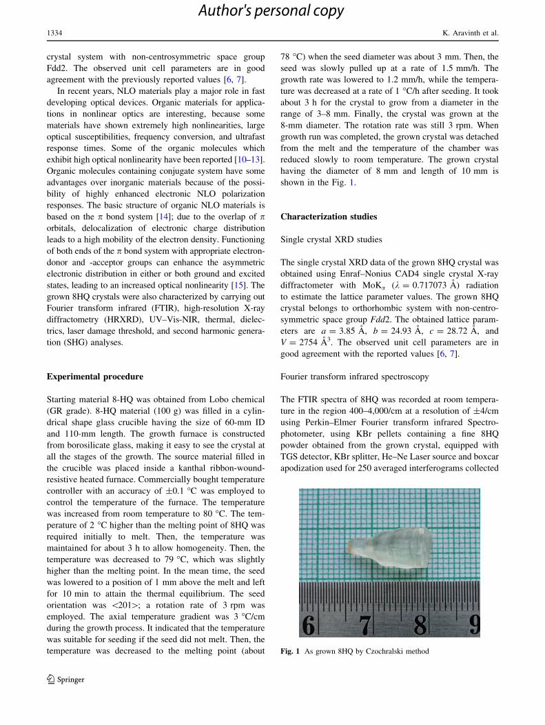
reduced slowly to room temperature. The grown crystal
having the diameter of 8 mm and length of 10 mm is
shown in the Fig. 1.
Characterization studies
Single crystal XRD studies
The single crystal XRD data of the grown 8HQ crystal was
obtained using Enraf–Nonius CAD4 single crystal X-ray
diffractometer with MoKa (k = 0.717073 A) radiation
to estimate the lattice parameter values. The grown 8HQ
crystal belongs to orthorhombic system with non-centro-
symmetric space group Fdd2. The obtained lattice param-
eters are a = 3.85 A, b = 24.93 A, c = 28.72 A, and
V = 2754 A3. The observed unit cell parameters are in
good agreement with the reported values [6, 7].
Fourier transform infrared spectroscopy
The FTIR spectra of 8HQ was recorded at room tempera-
ture in the region 400–4,000/cm at a resolution of ±4/cm
using Perkin–Elmer Fourier transform infrared Spectro-
photometer, using KBr pellets containing a fine 8HQ
powder obtained from the grown crystal, equipped with
TGS detector, KBr splitter, He–Ne Laser source and boxcar
apodization used for 250 averaged interferograms collected
Fig. 1 As grown 8HQ by Czochralski method
1334 K. Aravinth et al.
123
Author's personal copy
for both the sample and the background. In this technique,
almost all functional groups in a molecule absorb charac-
teristically within a definite range of frequency [16].
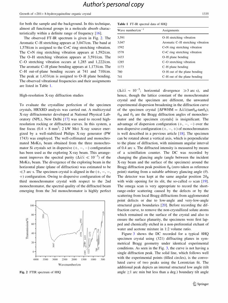
The observed FT-IR spectrum is given in Fig. 2. The
Aromatic C–H stretching appears at 3,047/cm. The band at
1,578/cm is assigned to the C=C ring stretching vibration.
The C=N ring stretching vibration appears at 1,592/cm.
The O–H stretching vibration appears at 3,591/cm. The
C–O stretching vibration occurs at 1,285 and 1,222/cm.
The aromatic C–H plane bending appears at 1,173/cm. The
C–H out-of-plane bending occurs at 741 and 710/cm.
The peak at 1,433/cm is assigned to O–H plane bending.
The observed vibrational frequencies and their assignments
are listed in Table 1.
High-resolution X-ray diffraction studies
To evaluate the crystalline perfection of the specimen
crystals, HRXRD analysis was carried out. A multicrystal
X-ray diffractometer developed at National Physical Lab-
oratory (NPL), New Delhi [17] was used to record high-
resolution rocking or diffraction curves. In this system, a
fine focus (0.4 9 8 mm2; 2 kW Mo) X-ray source ener-
gized by a well-stabilized Philips X-ray generator (PW
1743) was employed. The well-collimated and monochro-
mated MoKa1 beam obtained from the three monochro-
mator Si crystals set in dispersive (?, -, -) configuration
has been used as the exploring X-ray beam. This arrange-
ment improves the spectral purity (Dk/k � 10-5) of the
MoKa1 beam. The divergence of the exploring beam in the
horizontal plane (plane of diffraction) was estimated to be
�3 arc s. The specimen crystal is aligned in the (?, -, -,
?) configuration. Owing to dispersive configuration of the
third monochromator crystal with respect to the 2nd
monochromator, the spectral quality of the diffracted beam
emerging from the 3rd monochromator is highly perfect
(Dk/k * 10-5; horizontal divergence �3 arc s), and
hence, though the lattice constant of the monochromator
crystal and the specimen are different, the unwanted
experimental dispersion broadening in the diffraction curve
of the specimen crystal [DFWHM = Dk/k(tanhM-tanhS);
hM and hS are the Bragg diffraction angles of monochro-
mator and the specimen crystals] is insignificant. The
advantage of dispersion configuration (?, -, -) over the
non-dispersive configuration (?, -, ?) of monochromators
is well described in a previous article [18]. The specimen
can be rotated about a vertical axis, which is perpendicular
to the plane of diffraction, with minimum angular interval
of 0.4 arc s. The diffracted intensity is measured by means
of a scintillation counter. The DC was recorded by
changing the glancing angle (angle between the incident
X-ray beam and the surface of the specimen) around the
Bragg diffraction peak position hB (zero taken as reference
point) starting from a suitable arbitrary glancing angle (h).
The detector was kept at the same angular position 2hB
with wide opening for its slit, the so-called x scan [19].
The omega scan is very appropriate to record the short-
range-order scattering caused by the defects or by the
scattering from local Bragg diffractions from agglomerated
point defects or due to low-angle and very-low-angle
structural grain boundaries [20]. Before recording the dif-
fraction curve, to remove the non-crystallized solute atoms
which remained on the surface of the crystal and also to
ensure the surface planarity, the specimens were first lap-
ped and chemically etched in a non-preferential etchant of
water and acetone mixture in 1:2 volume ratio.
Figure 3 shows the DC recorded for a typical 8HQ
specimen crystal using (321) diffracting planes in sym-
metrical Bragg geometry under identical experimental
conditions. As seen in the Fig. 3, the curve is not having a
single diffraction peak. The solid line, which follows well
with the experimental points (filled circles), is the convo-
luted curve of two peaks using the Lorentzian fit. The
additional peak depicts an internal structural low angle (tilt
angle C1 arc min but less than a deg.) boundary tilt angle
4000 3500 3000 2500 2000 1500 1000 500
0
20
40
60
80
100
Wavenumber/cm
Tra
nsm
issi
on/%
3591
3047
1578
1592
1222
1285
1433 11
73
780 74
171
0
Fig. 2 FTIR spectrum of 8HQ
Table 1 FT-IR spectral data of 8HQ
Wave number/cm-1 Assignments
3,591 O–H stretching vibration
3,047 Aromatic C–H stretching vibration
1592 C=N ring stretching vibration
1578 C=C ring stretching vibration
1433 O–H plane bending
1285 C–O stretching vibration
1173 C–H plane bending
780 O–H out of the plane bending
741 C–H out of the plane bending
Growth of \201[ 8-hydroxyquinoline organic crystal 1335
123
Author's personal copy
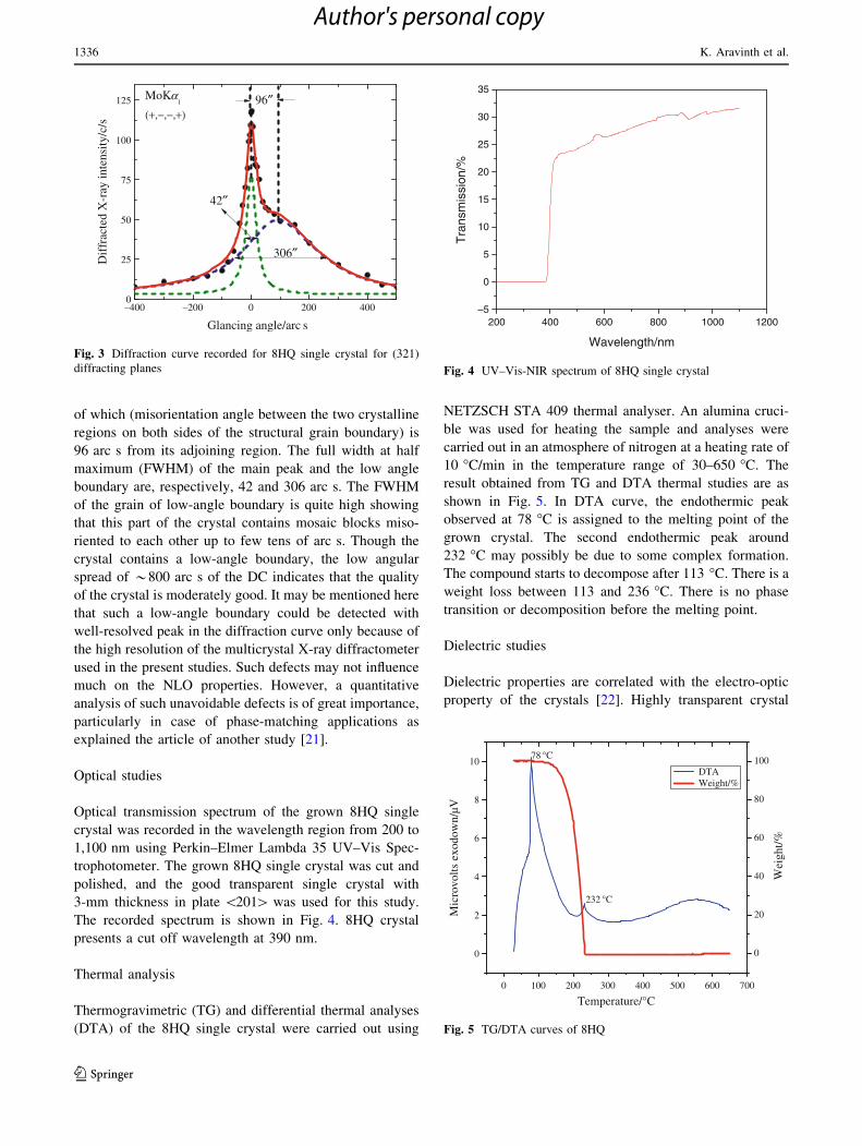
of which (misorientation angle between the two crystalline
regions on both sides of the structural grain boundary) is
96 arc s from its adjoining region. The full width at half
maximum (FWHM) of the main peak and the low angle
boundary are, respectively, 42 and 306 arc s. The FWHM
of the grain of low-angle boundary is quite high showing
that this part of the crystal contains mosaic blocks miso-
riented to each other up to few tens of arc s. Though the
crystal contains a low-angle boundary, the low angular
spread of *800 arc s of the DC indicates that the quality
of the crystal is moderately good. It may be mentioned here
that such a low-angle boundary could be detected with
well-resolved peak in the diffraction curve only because of
the high resolution of the multicrystal X-ray diffractometer
used in the present studies. Such defects may not influence
much on the NLO properties. However, a quantitative
analysis of such unavoidable defects is of great importance,
particularly in case of phase-matching applications as
explained the article of another study [21].
Optical studies
Optical transmission spectrum of the grown 8HQ single
crystal was recorded in the wavelength region from 200 to
1,100 nm using Perkin–Elmer Lambda 35 UV–Vis Spec-
trophotometer. The grown 8HQ single crystal was cut and
polished, and the good transparent single crystal with
3-mm thickness in plate \201[ was used for this study.
The recorded spectrum is shown in Fig. 4. 8HQ crystal
presents a cut off wavelength at 390 nm.
Thermal analysis
Thermogravimetric (TG) and differential thermal analyses
(DTA) of the 8HQ single crystal were carried out using
NETZSCH STA 409 thermal analyser. An alumina cruci-
ble was used for heating the sample and analyses were
carried out in an atmosphere of nitrogen at a heating rate of
10 �C/min in the temperature range of 30–650 �C. The
result obtained from TG and DTA thermal studies are as
shown in Fig. 5. In DTA curve, the endothermic peak
observed at 78 �C is assigned to the melting point of the
grown crystal. The second endothermic peak around
232 �C may possibly be due to some complex formation.
The compound starts to decompose after 113 �C. There is a
weight loss between 113 and 236 �C. There is no phase
transition or decomposition before the melting point.
Dielectric studies
Dielectric properties are correlated with the electro-optic
property of the crystals [22]. Highly transparent crystal
–400 –200 0 200 4000
25
50
75
100
125D
iffr
acte
d X
-ray
inte
nsity
/c/s
Glancing angle/arc s
96″MoKα1
(+,−,−,+)
306″
42″
Fig. 3 Diffraction curve recorded for 8HQ single crystal for (321)
diffracting planes
200 400 600 800 1000 1200–5
0
5
10
15
20
25
30
35
Tra
nsm
issi
on/%
Wavelength/nm
Fig. 4 UV–Vis-NIR spectrum of 8HQ single crystal
0 100 200 300 400 500 600 700
0
2
4
6
8
10 DTA Weight/%
Temperature/°C
Mic
rovo
lts e
xodo
wn/
µV
78 °C
232 °C
0
20
40
60
80
100
Wei
ght/%
Fig. 5 TG/DTA curves of 8HQ
1336 K. Aravinth et al.
123
Author's personal copy
sample of size 7 9 7 9 2 mm3 without any visible defects
was used for the dielectric measurements. The opposite
faces were polished and coated with graphite to obtain a
good conductivity surface layer. The capacitance and
dielectric loss factor were measured by the conventional
plate capacitor method with frequency range (100 Hz–
1 MHz) using Aglient 4284-A LCR meter at the range of
40–60 �C. The observations were made while cooling the
sample. Air capacitance (Cair) was also measured.
The dielectric constant of the 8HQ crystal was measured
as a function of frequency at different temperatures (40, 45,
50, 55, and 60 �C). The variations of dielectric constant
with the frequency at different temperatures is shown in the
Fig. 6. It is deduced that the dielectric constant is relatively
high at lower frequencies and decreases as the frequency
increases. The dielectric constant of materials is due to the
contribution of electronic, ionic, orientational, and space
charge polarizations which depend on the frequencies. At
low frequencies, all these polarizations are active [23]. The
dielectric constant of 8HQ crystal at 60 �C is 6.61, and this
value decreases to 3.61 at 40 �C for 100 Hz. The dielectric
constant is relatively high in the lower frequency and
almost saturated at 1,000 kHz.
Figure 7 shows the plot between the frequency and
dielectric loss at various temperatures. It is observed from
the plot that the dielectric loss decreases as the frequency
increases. The higher values of the dielectric loss at low
frequencies originate from space charge polarization. The
low dielectric loss value at higher frequencies indicates that
the grown 8HQ crystal contains minimum defects.
Laser damage threshold measurements
Optical damage in NLO material may severely affect the
performance of high-power laser systems as well as the
efficiency of the optical devices based on nonlinear pro-
cesses. Hence, high laser damage threshold is a significant
parameter for NLO crystal. The laser-induced damage
studies have been carried out for the Czochralski method-
grown 8HQ single crystal. Then, the samples were polished
using Al2O3 just before the laser damage threshold
measurements. A Q-switched Nd: YAG laser operating at
1,064-nm radiation was used. The laser was operated at the
repetition rate of 5 Hz with the pulse duration of 30 ns. For
the laser damage threshold measurement, 1-mm-diameter
beam was focused on the samples with a 30-cm-focal length
lens. The pulse energy of each shot was measured by using
combination of phototube and oscilloscope. The samples are
irradiated at different spots on the same plane at a similar
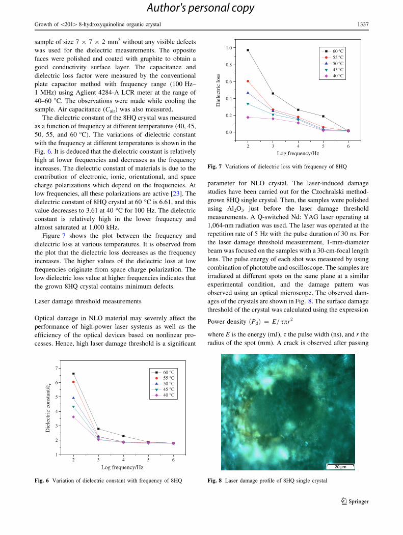
experimental condition, and the damage pattern was
observed using an optical microscope. The observed dam-
ages of the crystals are shown in Fig. 8. The surface damage
threshold of the crystal was calculated using the expression
Power density Pdð Þ ¼ E= spr2
where E is the energy (mJ), s the pulse width (ns), and r the
radius of the spot (mm). A crack is observed after passing
2 3 4 5 61
2
3
4
5
6
7
Die
lect
ric
cons
tant
/εr
Log frequency/Hz
60 °C 55 °C 50 °C 45 °C 40 °C
Fig. 6 Variation of dielectric constant with frequency of 8HQ
2 3 4 5 6
0.0
0.2
0.4
0.6
0.8
1.0
Die
lect
ric
loss
Log frequency/Hz
60 °C 55 °C 50 °C 45 °C 40 °C
Fig. 7 Variations of dielectric loss with frequency of 8HQ
Fig. 8 Laser damage profile of 8HQ single crystal
Growth of \201[ 8-hydroxyquinoline organic crystal 1337
123
Author's personal copy
75 mJ energy in 30 s. Multiple-shot surface laser damage
threshold of 8HQ was determined to be 0.88 GW/cm2 for
1,064-nm wavelength of Nd: YAG laser radiation.
SHG study
The SHG conversion efficiency of 8HQ was measured by
Kurtz powder techniques [24]. A Q-switched Nd: YAG
laser was used as light source. A laser beam of fundamental
wavelength 1,064 nm, 8 ns pulse width, with 10 Hz pulse
rate was made to fall normally on the sample cell. Second
harmonic radiation generated by the randomly oriented
microcrystals were focused by a lens and detected by the
photomultiplier tube. The energy per pulse of 1,064-nm
laser radiation attenuated using high-energy variable
attenuator is measured using an energy power meter which
is externally triggered by the Nd: YAG laser. The
conversion efficiency for SHG depends on power of the
fundamental beam, field-gain coefficient, size of the crys-
tal, and minimum beam waist. In present study, KDP
sample was used as the reference material, and output
power intensity of 8HQ has been found to be 0.8 times that
of KDP.
Conclusions
Single crystals of 8HQ were grown using the Czochralski
growth technique. The unit cell parameters of 8HQ were
confirmed by single crystal X-ray diffraction analysis.
The grown 8HQ crystal belongs to orthorhombic system
with non-centrosymmetric space group Fdd2. The crys-
talline perfection of the grown crystal is demonstrated as
observed by high-resolution X-ray diffraction studies.
FWHM value of the Czochralski method-grown 8HQ
crystal, which is obtained as 96 arc s, shows that the
crystalline perfection is moderately good. The functional
groups were confirmed by FTIR measurements. The UV–
Vis-NIR studies show that the grown 8HQ crystal’s cutoff
wavelength is around 390 nm. In the transmittance spec-
tra, it is evident that the 8HQ crystal has wide transpar-
ency range in the entire visible and NIR range. The
thermal behavior of the grown crystal was studied using
TG/DTA. 8HQ crystal decomposed at 113 �C. The
dielectric constant and dielectric loss studies of 8HQ
establish the normal behavior. Multiple-shot surface laser
damage threshold is determined to be 0.88 GW/cm2 at
1,064-nm laser radiation. The SHG efficiency of 8HQ is
0.8 times that of KDP.
Acknowledgements The authors are thankful to Dr. G. Bhagav-
annarayana, the National Physical Laboratory, New Delhi for
HRXRD studies.
References
1. Schavlev AE, Pankratov AN, Shalabay AV. DFT computational
studies on rotation barriers, tautomerism, intramolecular hydro-
gen bond, and solvent effects in 8-hydroxyquinoline. Int J
Quantum Chem. 2006;106:876.
2. Amati M, Belviso S, Cristinzianio PL, Minichino C, Lelj F,
Aiello I, La Deda M, Ghedini M. 8-Hydroxyquinoline monomer,
water adducts, and dimer. Environment influences on structure,
spectroscopic properties, and relative stability of cis and transconformers. J Phys Chem A. 2007;111:13403–14.
3. Bardez E, Devol I, Valeur B, Larrey B. Molecular fluorescence:
principles and applications. J Phys Chem. 1997;B101:7786.
4. Quan-song Li, Fang Wei-Hai. Theoretical studies on structures
and reactivity of 8-hydroxyquinoline and its one-water complex
in the ground and excited states. Chem Phys Lett. 2003;367:
637–44.
5. Camargo AJ, Napolitano HB, Zukerman-Schpector J. Theoretical
investigation of the intramolecular hydrogen bond formation,
non-linear optic properties, and electronic absorption spectra of
the 8-hydroxyquinoline. J Mol Struct. 2007;816:145–51.
6. Roychowdhury P. Crystal and molecular structure of 8-hydroxy-
quinoline. Acta Cryst. 1978;B34:1047.
7. Vijayan N, Bhagavannarayana G, Maurya KK, Pal S, Datta SN,
Gopalakrishnan Ramasamy P. Studies on the structural, thermal and
optical behavior of solution grown organic nonlinear optical mate-
rial: 8-hydroxyquinoline. Cryst Res Technol. 2007;42:195–200.
8. Krishnakumar V, Nagalakshmi R, Janaki P. Growth and spec-
troscopic characterization of a new organic nonlinear optical
crystal- 8-hydroxyquinoline. Spectrochim Acta Part A. 2005;61:
1097–103.
9. Rajasekaran M, Anbusrinivasan P, Mojumdar SC. Growth,
spectral and thermal characterization of 8-hydroxyquinoline.
J Therm Anal Calorim. 2010;100:827–30.
10. Hermann JP, Ducuing J. Third-order polarizabilities of long-
chain molecules. J Appl Phys. 1974;45:5100.
11. Oudar JL, Hierle R. An efficient organic crystal for nonlinear
optics: methyl-(2,4-dinitrophenyl)-aminopropanoate. J Appl Phys.
1977;48:2699.
12. Levine BF. Donor-acceptor charge transfer contributions to the
second order hyperpolarizability. Chem Phys Lett. 1976;37:516.
13. Levine BF, Bethea CG, Thurmond CD, Lynch RT, Bernstein JL. An
organic crystal with an exceptionally large optical second-harmonic
coefficient: 2-methyl-4-nitroaniline. J Appl Phys. 1979;50:2523.
14. Rai RN, Varma KBR. Growth and characterization of single
crystal of pentachloropyridine. J Cryst Growth. 2005;285:111–6.
15. Crasta V, Ravindrachary V, Lakshmi S, Pramod SN, Shridar MA,
Shashidhara Prasad J. Growth, characterization and crystal
structure analysis of 1-(4-chlorophenyl)-3-(4-chlorophenyl)-2-
propene-1-one. J Cryst Growth. 2005;275:e329–35.
16. Kalsi P. Spectroscopy of organic compounds. New Delhi: Wiley;
1985.
17. Lal K, Bhagavannarayana G. A high-resolution diffuse X-ray
scattering study of defects in dislocation free-silicon single
crystals grown by the float-Zone method and comparison with
Czochralski-grown crystals. J Appl Cryst. 1989;22:209.
18. Bhagavannarayana G, Kushwaha SK. Enhancement of SHG
efficiency by urea doping in ZTS single crystals and its correla-
tion with crystalline perfection as revealed by Kurtz powder and
high-resolution X-ray diffraction methods. J Appl Cryst. 2010;
43:154–62.
19. Senthilkumar K, MoorthyBabu S, Bhagavannarayana G. Study
of the influence of dopants on the crystalline perfection of fer-
roelectric glycine phosphate single crystals using high-resolution
X-ray diffraction analysis. J Appl Cryst. 2011;44:313–8.
1338 K. Aravinth et al.
123
Author's personal copy
20. Bhagavannarayana G, Ananthamurthy RV, Budakoti GC, Kumar
B, Bartwal KS. A study of the effect of annealing on Fe-doped
LiNbO3 by HRXRD, XRT, FT-IR. J Appl Cryst. 2005;38:768–71.
21. Bhagavannarayana G, Riscob B, Mohd Shakir. Growth and char-
acterization of L-leucine L-leucinium picrate single crystal: a new
nonlinear optical material. Mater Chem Phys. 2011;126:20–3.
22. Boomadevi S, Dhanasekaran R. Synthesis, crystal growth and char-
acterization of L-pyrrolidone-2-carboxylic acid (L-PCA) crystals.
J Cryst Growth. 2004;261:70–6.
23. Dharmaprakash SM, Mohan Rao P. Dielectric properties of
hydrated barium oxalate and barium cadmium oxalate crystals.
J Mater Sci Lett. 1989;8:1167–8.
24. Kurtz SK, Perry TT. A powder technique for the evaluation of
nonlinear optical materials. J Appl Phys. 1968;39:3798–813.
Growth of \201[ 8-hydroxyquinoline organic crystal 1339
123
Author's personal copy