Growth Inhibition and Apoptosis Induction by (+)- Cyanidan-3-ol in Hepatocellular Carcinoma
19
Growth Inhibition and Apoptosis Induction by (+)- Cyanidan-3-ol in Hepatocellular Carcinoma Jitender Monga 1 , Saurabh Pandit 2 , Rajinder Singh Chauhan 2 , Chetan Singh Chauhan 3 , Shailender Singh Chauhan 4 , Manu Sharma 1* 1 Department of Pharmacy, Jaypee University of Information Technology, Waknaghat, Himachal Pradesh, India, 2 Department of Biotechnology & Bioinformatics, Jaypee University of Information Technology, Waknaghat, Himachal Pradesh, India, 3 Department of Pharmacy, Bhupal Noble College of Pharmacy, Udaipur, Rajasthan, India, 4 Department of Gastroenterology, Post Graduate Institute of Medical Research, Chandigarh, India Abstract The objective of this study was to evaluate the cytotoxicity of (+)-cyanidan-3-ol (CD-3) in human hepatocellular carcinoma cell line (HepG2) and chemopreventive potential against hepatocellular carcinoma (HCC) in Balb/c mice. The HepG2 cell line was treated with CD-3 at various concentrations and the proliferation of the HepG2 cells was measure by 3-[4,5-dimethylthiazol-2-yl]-2,5-diphenyl-tetrazolium bromide (MTT), sulforhodamine B (SRB) and lactate dehydrogenase (LDH) assays. Cell apoptosis was detected by Hoechst 33258 (HO), Acridine orange/ethylene dibromide (AO/EB) staining, DNA fragmentation analysis and the apoptosis rate was detected by flow cytometry. The HCC tumor model was established in mice by injecting N-nitrosodiethylamine/carbon tetrachloride (NDEA/CCl 4 ) and the effect of CD-3 on tumor growth in-vivo was studied. The levels of liver injury markers, tumor markers, and oxidative stress were measured. The expression levels of apoptosis-related genes in in-vitro and in vivo models were determined by RT-PCR and ELISA. The CD-3 induced cell death was considered to be apoptotic by observing the typical apoptotic morphological changes under fluorescent microscopy and DNA fragmentation analysis. Annexin V/PI assay demonstrated that apoptosis increased with increase in the concentration of CD-3. The expression levels of apoptosis-related genes that belong to bcl-2 and caspase family were increased and AP-1 and NF-κB activities were significantly suppressed by CD-3. Immunohistochemistry data revealed less localization of p53, p65 and c-jun in CD-3 treated tumors as compared to localization in NDEA/CCl 4 treated tumors. Taken together, our data demonstrated that CD-3 could significantly inhibit the proliferation of HepG2 cells in-vitro and suppress HCC tumor growth in-vivo by apoptosis induction. Citation: Monga J, Pandit S, Chauhan RS, Chauhan CS, Chauhan SS, et al. (2013) Growth Inhibition and Apoptosis Induction by (+)-Cyanidan-3-ol in Hepatocellular Carcinoma. PLoS ONE 8(7): e68710. doi:10.1371/journal.pone.0068710 Editor: Arun Rishi, Wayne State University, United States of America Received February 18, 2013; Accepted June 3, 2013; Published July 24, 2013 Copyright: © 2013 Monga et al. This is an open-access article distributed under the terms of the Creative Commons Attribution License, which permits unrestricted use, distribution, and reproduction in any medium, provided the original author and source are credited. Funding: Funded by Jaypee University of Information Technology. The funders had no role in study design, data collection and analysis, decision to publish, or preparation of the manuscript. Competing interests: The authors have declared that no competing interests exist. * E-mail: [email protected]. Introduction Hepatocellular carcinoma (HCC) is one of the most frequent tumors representing the fifth commonest malignancy worldwide and the third cause of mortality from cancer. The regions of high incidence are Eastern and South-Eastern Asia, Middle and Western Africa, Southern Europe as well as South America and kill an astounding number of people every year [1]. Unfortunately, the overall response rate of liver cancer treatment is unsatisfactory mainly due to late diagnosis and poor treatment efficacy, especially resistance to chemotherapeutic drugs and metastasis to other organs [2]. Thus, the development of new and effective therapeutic strategies for liver cancer has a greater need and importance. In recent years, the number of natural products has acquired a lot of attention because of their ability to provide prevention and therapeutic efficacy against number of cancers [3]. Out of number of different classes of natural products, flavonoids represent a diverse group of low molecular weight polyphenolic compounds that are widely distributed in nature and renewed interest has been observed in recent years in the novel and multiple activities of flavonoids [4]. Acacia catechu Willd. (Fabaceae) also known as ‘Khadira’ has been extensively used as traditional medicine in India/Asia over a long period of time and a rich source of number of polyphenolic compounds. Previously, we have reported chemopreventive effect of aqueous extract of Acacia catechu in skin and mammary cancer rodent models [5,6]. (+)-Cyanidan-3-ol (CD-3) [3',4',5,7- tetrahydroxyflavan-3-ol] (Figure 1) is the most abundant PLOS ONE | www.plosone.org 1 July 2013 | Volume 8 | Issue 7 | e68710
Transcript of Growth Inhibition and Apoptosis Induction by (+)- Cyanidan-3-ol in Hepatocellular Carcinoma

Growth Inhibition and Apoptosis Induction by (+)-Cyanidan-3-ol in Hepatocellular CarcinomaJitender Monga1, Saurabh Pandit2, Rajinder Singh Chauhan2, Chetan Singh Chauhan3, Shailender SinghChauhan4, Manu Sharma1*
1 Department of Pharmacy, Jaypee University of Information Technology, Waknaghat, Himachal Pradesh, India, 2 Department of Biotechnology &Bioinformatics, Jaypee University of Information Technology, Waknaghat, Himachal Pradesh, India, 3 Department of Pharmacy, Bhupal Noble College ofPharmacy, Udaipur, Rajasthan, India, 4 Department of Gastroenterology, Post Graduate Institute of Medical Research, Chandigarh, India
Abstract
The objective of this study was to evaluate the cytotoxicity of (+)-cyanidan-3-ol (CD-3) in human hepatocellularcarcinoma cell line (HepG2) and chemopreventive potential against hepatocellular carcinoma (HCC) in Balb/c mice.The HepG2 cell line was treated with CD-3 at various concentrations and the proliferation of the HepG2 cells wasmeasure by 3-[4,5-dimethylthiazol-2-yl]-2,5-diphenyl-tetrazolium bromide (MTT), sulforhodamine B (SRB) and lactatedehydrogenase (LDH) assays. Cell apoptosis was detected by Hoechst 33258 (HO), Acridine orange/ethylenedibromide (AO/EB) staining, DNA fragmentation analysis and the apoptosis rate was detected by flow cytometry. TheHCC tumor model was established in mice by injecting N-nitrosodiethylamine/carbon tetrachloride (NDEA/CCl4) andthe effect of CD-3 on tumor growth in-vivo was studied. The levels of liver injury markers, tumor markers, andoxidative stress were measured. The expression levels of apoptosis-related genes in in-vitro and in vivo models weredetermined by RT-PCR and ELISA. The CD-3 induced cell death was considered to be apoptotic by observing thetypical apoptotic morphological changes under fluorescent microscopy and DNA fragmentation analysis. AnnexinV/PI assay demonstrated that apoptosis increased with increase in the concentration of CD-3. The expression levelsof apoptosis-related genes that belong to bcl-2 and caspase family were increased and AP-1 and NF-κB activitieswere significantly suppressed by CD-3. Immunohistochemistry data revealed less localization of p53, p65 and c-junin CD-3 treated tumors as compared to localization in NDEA/CCl4 treated tumors. Taken together, our datademonstrated that CD-3 could significantly inhibit the proliferation of HepG2 cells in-vitro and suppress HCC tumorgrowth in-vivo by apoptosis induction.
Citation: Monga J, Pandit S, Chauhan RS, Chauhan CS, Chauhan SS, et al. (2013) Growth Inhibition and Apoptosis Induction by (+)-Cyanidan-3-ol inHepatocellular Carcinoma. PLoS ONE 8(7): e68710. doi:10.1371/journal.pone.0068710
Editor: Arun Rishi, Wayne State University, United States of America
Received February 18, 2013; Accepted June 3, 2013; Published July 24, 2013
Copyright: © 2013 Monga et al. This is an open-access article distributed under the terms of the Creative Commons Attribution License, which permitsunrestricted use, distribution, and reproduction in any medium, provided the original author and source are credited.
Funding: Funded by Jaypee University of Information Technology. The funders had no role in study design, data collection and analysis, decision topublish, or preparation of the manuscript.
Competing interests: The authors have declared that no competing interests exist.
* E-mail: [email protected].
Introduction
Hepatocellular carcinoma (HCC) is one of the most frequenttumors representing the fifth commonest malignancy worldwideand the third cause of mortality from cancer. The regions ofhigh incidence are Eastern and South-Eastern Asia, Middleand Western Africa, Southern Europe as well as SouthAmerica and kill an astounding number of people every year[1]. Unfortunately, the overall response rate of liver cancertreatment is unsatisfactory mainly due to late diagnosis andpoor treatment efficacy, especially resistance tochemotherapeutic drugs and metastasis to other organs [2].Thus, the development of new and effective therapeuticstrategies for liver cancer has a greater need and importance.
In recent years, the number of natural products has acquireda lot of attention because of their ability to provide preventionand therapeutic efficacy against number of cancers [3]. Out ofnumber of different classes of natural products, flavonoidsrepresent a diverse group of low molecular weight polyphenoliccompounds that are widely distributed in nature and renewedinterest has been observed in recent years in the novel andmultiple activities of flavonoids [4]. Acacia catechu Willd.(Fabaceae) also known as ‘Khadira’ has been extensively usedas traditional medicine in India/Asia over a long period of timeand a rich source of number of polyphenolic compounds.Previously, we have reported chemopreventive effect ofaqueous extract of Acacia catechu in skin and mammarycancer rodent models [5,6]. (+)-Cyanidan-3-ol (CD-3) [3',4',5,7-tetrahydroxyflavan-3-ol] (Figure 1) is the most abundant
PLOS ONE | www.plosone.org 1 July 2013 | Volume 8 | Issue 7 | e68710

polyphenolic flavonoid in the Acacia catechu heartwood andstudies of the biological effects of CD-3 in cell culture and invivo models indicated that this compound can inhibit lipidperoxidation [7]. CD-3 is claimed to be effective in treatingcarbon tetrachloride induced liver damage [8] and also reportedto inhibit angiogenesis in-vivo [9]. However, there is no reporton the effect of CD-3 on hepatocellular carcinoma (HCC). Inthis paper, we report the chemopreventive and therapeuticefficacy of CD-3 against hepatocellular carcinoma by usingboth in-vitro and in vivo systems. Further, the underlyingcellular andmolecular mechanisms of CD-3 actions were alsoevaluated. Our data provided investigational evidence to carrythe potential development of CD-3 as an efficient and safecandidate for the prevention and/or therapy of liver cancer.
Materials and Methods
Antibodies and ReagentsAll the chemicals used in the study (analytical grade) were
obtained from Sigma Chemical Co. (St. Louis, MO, USA),Merck (Mumbai, India), and Sigma Himedia Laboratories(Mumbai, India). Antibodies against p53, p65, c-jun, bcl-2, baxand caspase-3 were obtained from Santa Cruz Biotechnology,Santa Cruz, CA (USA). Annexin V-FITC apoptosis detection kitwas obtained from EMD biosciences (Calbiochem, Inc, USA).
Extraction of Acacia catechu heartwood and isolationof (+)-cyanidan-3-ol
Acacia catechu heartwood was collected from Hamirpur,Himachal Pradesh, India during the month of September, 2011.The plant material was taxonomically identified andauthenticated by Dr. Sunil Dutta, Scientist, National MedicinalPlant Board, Ayush, New Delhi, India. A voucher specimen(AC-2011) was deposited in the herbarium at PharmacyDepartment, Jaypee University of Information Technology,Waknaghat, Himachal Pradesh.
A total of one kg of the dried powder of A. catechuheartwood was put in an aluminium pot with ten litre of waterand boiled for 5 h and was then allowed to stand for 24 h. Theextract was decanted and filtered through a fine muslin cloth toremove suspended materials. The filtrate was evaporated andthe residue obtained was air dried to obtain a solid mass (212
Figure 1. Chemical structure of (+)-cyanidan-3-ol. doi: 10.1371/journal.pone.0068710.g001
g), with 21.2%, yield. Solid mass (150 g) was added to five litrestainless steel beaker containing one litre distilled water. It wasboiled with constant stirring for complete dissolution and thenfiltered. It was then evaporated to 500 ml and allowed to standfor 24 h. The aqueous filtrate was rejected, and the residuewas dissolve in ethanol and filtered. The ethanolic solution wasevaporated to dryness and the residue was dissolved in 500 mlhot water and was allowed to stand for 24 h. The precipitatewas filtered and dried in air and the process of re-crystallizationfrom water was repeated three times (m.p. 95–6 °C, yield 37.5g, 25%). The white crystals of (+)-cyanidan-3-ol was isolatedfrom Acacia catechu heartwood and characterized by spectralanalysis. The purity of (+)-cyanidan-3-ol was confirmed byHPLC (Figure S1).
IR (νmax, KBr, cm-1): 3501.59, 3456.74, 3412.9, 3176.44 (br,O–H stretch), 2931.38 (C–H stretch), 1624.88, 1521.69 (C=Cstretch), 1183.59, 1143.77(C–O stretch). 1H NMR (CDCl3, δppm): 9.0370 (5H, br, 5 OH groups), 6.7355-6.7403 (1H, d, J=1.92, C-2′-H), 6.6757-6.6958 (1H, d, J= 8.04, C-5′-H),6.5774-6.6027 (1H, dd, J= 8.16, 1.96, C-6′-H), 5.8837-5.8894(1H, d, J= 2.28, C-8-H), 5.6976-5.7032 (1H, d, J= 2.24, C-6-H),4.4670-4.4856 (1H, d, J= 7.44, C-2-H), 3.7993-3.8514 (1H, m,C-3-H), 2.6536-2.7070 (2H, dd, J= 16.04, 5.36, C-4-H)13. CNMR (CDCl3, δ ppm): 156.33 (C-7), 156.05 (C-5), 155.23 (C-9),144.72 (C-3′), 144.60 (C-4′), 130.50 (C-1′), 118.27 (C-2′),114.92 (C-5′), 114.31 (C-6′), 98.96 (C-10), 95.04 (C-8), 93.80(C-6), 80.98 (C-2), 66.39 (C-3), 27.67 (C-4). ESI-MS (m/z):289(M+-1).
Cell culture and treatmentHuman hepatocarcinoma cell line (HepG2) was obtained
from National Centre for Cancer Science (NCCS), Pune andgrown as a monolayer in DMEM supplemented with 10% FBS(Fetal Bovine Serum), 100 µg/ml streptomycin and 100 units/mlpenicillin. Cells were incubated at 37 °C in an atmosphere of5% CO2. Cells were grown to 85% confluence and treated withCD-3 for 48 h. Human lymphocytes were isolated fromperipheral blood [10]. Lymphocytes were suspended incomplete RPMI 1640 medium supplemented with 10% FBS, 5g/ml phytohemagglutinin (PHA), and maintained at 37 °C in a5% CO2 humidified incubator. Stock solutions and dilutions ofCD-3 were prepared in culture medium. For 96 well plates, cellwere seeded at approximately 1.5 × 10⁴ cells per well. For 24well plates, cells were seeded at approximately 4 × 10⁴ cellsper well.
MTT assayThe cell viability of HepG2 cells was assessed by the MTT
colorimetric assay [11]. Briefly, the adherent HepG2 cells wereincubated in 96-well microtiter plates for 48 h at 37 °C in a 5%CO2 incubator. Following the addition of test compound, theplates were incubated for an additional 48 h. Control wellscontained medium alone and three replicate wells were used ateach point in the experiments. After 48 h incubation, MTTsolution (5 mg/ml in phosphate-buffered saline) was added andincubated for another 4 h. The resulting MTT/formazan productwas dissolved by 100 µl of isopropanol and the plates weregently shaken to solubilise the formed formazan. The amount
Chemopreventive Effect of (+)-Cyanidan-3-ol
PLOS ONE | www.plosone.org 2 July 2013 | Volume 8 | Issue 7 | e68710

of formazan was determined by measuring the absorbance(OD) at 570 nm using a Bio-Rad 550 enzyme-linkedimmunosorbent assay (ELISA) microplate reader.
Sulforhodamine B (SRB) assayGrowth inhibition was determined using the SRB assay
which estimates cell number indirectly by measuring total basicamino acids [12]. Briefly, the cells were incubated in 96-wellmicrotiter plates for 48 h. Following the addition of testcompound, the plates were incubated at 37 °C for an additional48 h in a 5% CO2 incubator. The culture medium was thendiscarded and the cells were fixed in situ by the gentle additionof 100 µl of cold 10% (w/v) trichloroacetic acid and incubatedfor 60 min at 4 °C. The supernatant was discarded and theplates were washed five times with tap water and air dried.SRB solution (100 µl) at 0.4% (w/v) in 1% acetic acid wasadded and plates were incubated for 20 min at roomtemperature. After staining, unbound dye was removed bywashing five times with 1% acetic acid and the plates were airdried. Bound stain was subsequently solubilised with 10 mMTris (pH 10.5) and the absorbance was read at 515 nm on aBio-Rad 550 ELISA microplate reader.
Lactate dehydrogenase (LDH) leakage assayIn LDH assay, leakage of the cytoplasmic located enzyme
LDH into the extracellular medium is measured [13]. LDHactivity was measured in both the supernatants and the celllysate fractions using Cyto-Tox 96, a non-radioactivecytotoxicity assay kit (Promega, WI, USA), in accordance withthe manufacturer’s instruction and the intensity of color isproportional to LDH activity. LDH release was calculated bymeasuring the absorbance at 490 nm using ELISA microplatereader.
Morphological changes of cellsAnalysis of cell morphological changes was evaluated for 48
h in the absence or presence of CD-3 at concentrations of 10,20, 30, 40 and 50 µg/ml in a 24-well plate. At the end of theincubation period, cells were observed under phase contrastinverted microscope at 200× magnification.
Hoechst assayDifferential staining with specific fluorochromes can be used
to distinguish cells undergoing apoptosis from viable andnecrotic cells. Analysis of changes in cell morphology wasevaluated by Hoechst fluorescence staining [14]. The cellswere cultured in 24-well plates to 85% confluence and thentreated with the compound at concentrations of 25 and 35µg/ml for 48 h at 37 °C. The cells were washed in PBS andfixed with 4% formaldehyde in PBS for 15 min and stained with10 µg/ml Hoechst 33258 (HO; Sigma, St. Louis, MO, USA) atroom temperature for 10 min in PBS. The apoptosis wasclassified by morphology and color of the cells, and quantified.Finally, after the cells were washed with PBS, morphologicalchanges were observed under a fluorescence microscope(Nikon Eclipse Ti, Japan).
Acridine orange/ ethidium bromide (AO/EB)fluorescence staining
Analysis of changes in cell morphology was evaluated usingAO/EB fluorescence staining [15]. Briefly, the suspension of(+)-cyanidan-3-ol-treated cells, 4 µl/ml of dye mixture,containing 100 µg/ml acridine orange (Sigma, USA) and 100µg/ml ethidium bromide (Sigma, USA) in PBS were added, andwashed once with PBS. After staining, cells were visualizedimmediately under a fluorescence microscope (Nikon EclipseTi, Japan).
Estimation of percentage (%) of DNA fragmentationThe cells were incubated with 25 and 35 µg/ml of CD-3 for
48 h at 37 °C. Percent DNA fragmentation in HepG2 cells wasassayed by the method of Kurita-Ochiai [16]. Briefly, DNA fromthe cells was released into the lysis buffer [0.2% Triton X-100,10 mM Tris, and 1 mM EDTA (pH 8.0)] by rupturing thenucleus. The supernatant received after centrifugation at14,000 g for 15 min, contained fragmented DNA while theintact DNA was in the pellet. The amount of DNA in both thesupernatant and pellet was estimated by diphenylamine (DPA)assay [17]. The percentage of DNA fragmentation wascalculated as the ratio of DNA in supernatant and DNA inpellet.
Detection of DNA fragmentation by agarose gelelectrophoresis
The cells were incubated with 25 and 35 µg/ml of CD-3 for48 h at 37 °C. Briefly, DNA from the cells was released into thelysis buffer [0.2% Triton X-100, 10 mM Tris, and 1 mM EDTA(pH 8.0)] by rupturing the nucleus. The DNA in the supernatantwas extracted using a 25:24:1 (v/v/v) equal volume of phenol:chloroform: isoamyl alcohol. The DNA was precipitated withethanol, air-dried and dissolved in TE buffer [5 mM Tris-HCl(pH 8.0) and 20 mM edetic acid containing RNase A (0.1mg/ml; Sigma)]. The samples were analyzed electrophoreticallyon 1% agarose gel containing 0.1 µg/ml ethidium bromide(EtBr, Sigma) [18].
Apoptosis Assay by Annexin V-FITC and PropidiumIodide (PI) Staining
Annexin V-propidium iodide double staining assay was doneto quantify apoptosis in HepG2 cells using FACscan flowcytometer (Becton-Dickinson, USA). After 48 h of CD-3treatment, cells were harvested, washed with ice-cold PBS,and resuspended in 200 µl of binding buffer (10 mM HEPES/NaOH pH 7.4, 140 mM NaCl, 2.5 mM CaCl2) and incubatedwith 5 µl of Annexin V-FITC for 10 min at room temperature inthe dark. Samples were washed with binding buffer,resuspended in PBS, counterstained with 5 µg/ml PI, and wereanalyzed by flow cytometry to identify apoptotic cells. Cellswithout treatment were used as a negative control. Cellsshowing up as Annexin V-/PI+ were recognized as necrotic, thatshowing up as Annexin V+/PI+ were taken as late apoptotic orsecondary apoptotic, whilst Annexin V+/PI- cells wererecognized as early or primary apoptotic cells [19].
Chemopreventive Effect of (+)-Cyanidan-3-ol
PLOS ONE | www.plosone.org 3 July 2013 | Volume 8 | Issue 7 | e68710

RNA purification and RT-PCR analysisThe expression of apoptosis-related genes, such as p53,
mdm2, p65, c-jun, c-fos, bcl-2, bax, cytochrome-c, caspase-3,-7, -8 and -9 were determined by RT-PCR. Total RNA isolationwas done using TRI-REAGENT (Molecular Research CentreInc. Ohio, USA). Single-strand cDNA was synthesized from 2μg total RNA using moloney murine leukemia virus reversetranscriptase (M-MLV RT). RT-PCR was done using one stepmethod of RT–PCR kit (QIAGEN, Germany). Briefly, 3 µg ofRNA template from different groups was used in RT-PCRreaction. The 2.5 µl of 5X QIAGEN one step RT-PCR bufferwas added followed by addition of 0.5 µl of dNTP mix(containing 10 mM of each dNTP). The 1 µl of each sense andantisense gene specific primers (from 10 µM stock) was added.Then 2µl QIAGEN one step RT-PCR enzyme mix and 1µlRNAase inhibitor (1U/µl) was added. Finally, PCR gradeRNAase free water was added to make total volume 25 µl.Mixed it gently by vortex and centrifuged to collect all thecomponents at the bottom of the PCR tubes. After an initialdenaturation step of 1 min at 94 °C, 39 amplification cycleswere performed. Each cycle included an initial denaturationstep at 94 °C for 1 min, annealing for 45s and extension at 72°C for 2 min. A final extension step of 5 min at 72 °C wasperformed in order to complete the PCR reaction. Theamplified products were analyzed on 1.5% agarose gelelectrophoresis and densitometric analysis of bands was doneby ImageJ Software (NIH, USA). The oligonucleotide primerpairs for RT-PCR used in the present study are presented intable S1.
Protein quantification by ELISAELISA was carried out in nuclear extract to quantify the level
of p53, p65, c-jun, bax, bcl-2 and caspase-3. The assay wasstandardized by revising the different concentration of antigensand antibodies. Wells were coated with antigens diluted in 100µl of 0.05 M carbonate buffer (pH 9.6) and kept overnight at 4°C in a humid chamber. Flicked the plate to remove theunbound antigen solution and wells were blocked with 1%bovine serum albumin (BSA) in 0.1 M phosphate bufferedsaline (pH 7.2) for 1 h at 37 °C. Flicked and wells were washedthrice with 200 µl of PBS containing 0.05% (v/v) Tween-20.Wells were then incubated with anti-p53, anti-p65 and anti-caspase-3 (1: 250), anti-c-jun and anti-bax (1: 500) and anti-bcl-2 (1: 600) primary antibodies, respectively, diluted in PBS(containing 0.05% Tween-20 and 1% BSA) and incubated for 2h at 37 °C. Plate was again washed and incubated with anti-rabbit secondary antibody (1: 1000) for 2 h at 37 °C. Wellswere washed further three times as described above and colorwas developed by addition of 2,2´-azino-di-(3-ethyl)-benothiozolinsulphonic acid (ABTS) reagent and absorbancewas measured at 405 nm by ELISA reader.
AnimalsMale Balb/C mice, 6-7 weeks old, 26-27 gm, were purchased
from Sanjay Biological Museum, Amritsar, Punjab and kept inthe Departmental animal house with controlled conditions oftemperature (23 ± 5°C), humidity (60 ± 5%) and light (12 hlight-dark cycles). They were fed a standard diet and water. All
animals were acclimatized for 1 week before experimentation.Five animals were housed in each polypropylene cage withsterile paddy husk (procured locally) as bedding. The animalcare and handling was done according to the guidelines set bythe World Health Organization (WHO), Geneva, Switzerland,and the Indian National Science Academy (INSA), New Delhi,India. The protocol was approved by Institutional Animal EthicalCommittee for animal handling (CPCSEA No. -13/RKN/BNCP-06).
Experimental design and tumor inductionAfter one week of acclimatization, the mice were randomly
divided into three groups (n = 10). Mice of group 1 served ascontrol and these animals were orally administered with oliveoil (100 µl/ mouse). Liver tumor was induced in group 2 and 3with N-nitrosodiethylamine (NDEA) (200 mg/kg, body weight)dissolved in olive oil and given once by intraperitonial route.After two weeks of NDEA administration, it was promotedthrough subcutaneous injection of carbon tetrachloride (CCl4; 3ml/kg) thrice a week for six weeks [20]. Group 3 animals weretreated as group 2 mice along with oral administration of 200 µlof (+)-cyanidan-3-ol at the dose level 200 mg/kg body weightusing a blunt tipped cannula, thrice a week throughout theexperiment. (+)-Cyanidan-3-ol was administered orally 1 weekbefore the initiation of NDEA treatment in group 3. At the end of20 weeks, the body weight of each animal was taken beforesacrifice. The mice were fasted overnight and killed by cervicaldecapitation. The greyish-white hyperplastic nodules wereidentified from the surrounding reddish brown liver tissue.Blood samples were collected from a common carotid andallowed to clot before centrifugation at 1000 g for 10 min at 4°C to separate serum. The liver tissue was isolated andwashed twice with ice-cold 0.1M phosphate buffer saline, pH7.4, blotted, dried and weighed. The relative liver weight wascalculated as the percentage ratio of liver weight to the bodyweight. A small portion of the tissue was fixed in 10% formalinfor histological examination. The remaining tissue was storedat −20 °C for biochemical estimations, mRNA expressionstudies and protein level quantification.
Biochemical estimationsThe activities of serum aspartate (AST) and alanine
transaminases (ALT) were analyzed by the method of Reitmanand Frankel [21] while alkaline phosphatase (ALP) and γ-glutamyltransferase (γ-GT) were estimated by methods ofBergmeyer [22] and Szasz [23] respectively. The total sialicacid (TSA) [24] and lipid associated sialic acid (LASA) [25]levels in the serum and liver tissue were also measured. In theliver tissue, malondialdehyde (MDA) [26], nitrite [27] andantioxidants such as superoxide dismutase (SOD) [28],catalase (CAT) [29], glutathione peroxidase (GPx) [30],glutathione reductase (GR) [31], glutathione-S-transferase(GST) [32], total thiol (T-SH) [33], reduced glutathione (GSH)[34], levels were determined. Protein thiols (PrPr-SHs) contentwas calculated from the difference between the values of T-SHand GSH and expressed as µmoles of PrPr-SH/mg protein.
Chemopreventive Effect of (+)-Cyanidan-3-ol
PLOS ONE | www.plosone.org 4 July 2013 | Volume 8 | Issue 7 | e68710

Histopathological assessmentLiver sections were made immediately from the liver of
different groups of animals, fixed in 10% formalin, dehydratedin gradual ethanol (50–100%), cleared in xylene, andembedded in paraffin. Sections (4–5 µm thick) were preparedand the pathological changes were observed microscopicallyafter staining with hematoxylin and eosin (H & E). These slideswere examined microscopically.
ImmunohistochemistryFormalin-fixed and paraffin-embedded liver tissue sections
with a thickness of 4-5 µm were de-paraffinized using xylene,rehydrated and treated with 3% H2O2 in PBS. Antigen retrievalwas done using 0.01% sodium-citrate buffer (pH 6.0) followedby blocking in PBST containing 0.1% BSA and 10% FBS.Primary antibody incubation (p53, p65 and c-jun; 1:100) wascarried out overnight at 4 °C. Slides were washed andincubated with HRP-labeled secondary antibody (1 h, RT,1:200). Slides were washed (PBS containing 0.1% Tween 20),color was developed using DAB+H2O2, counterstained withhaematoxylin and mounted in DPX (Sigma-Aldrich, USA).Images were captured using microscope (Nikon Eclipse Ti,Japan).
Statistical analysisData are presented as mean ± standard error of mean
(SEM). Data is subjected to one-way analysis of variance(ANOVA; 95% confidence interval), followed by Tukey’s posthoc test for the determination of level of significance (Softwareprism 6.0) and results were considered significant, if P < 0.05.
Results
Effect of CD-3 on human peripheral lymphocytesThe cytotoxic effect of CD-3 on human peripheral
lymphocytes was characterized by MTT assay. When differentconcentrations of CD-3 (10-100 μg/ ml) were added to normallymphocytes, the CD-3 did not show any toxic effect tolymphocytes after 48 h (Figure S2).
In vitro proliferation studies on humanhepatocarcinoma (HepG2) cells
HepG2 cells were exposed to CD-3 for about 48 h andcytotoxicity was determined with different cytotoxic assays. Theeffect of CD-3 against HepG2 cells (concentration range 10–50µg/ml) showed a decrease in percent cell viability in a dose-dependent manner, as compared with that of the control whenexamined by different cytotoxic assays. IC50 values (µg/ml) ofCD-3 on HepG2 cells obtained by MTT, SRB and LDH assaysare presented in Figure 2.
Effect of (+)-cyanidan-3-ol on the morphology of HepG2cells
Direct observation of the morphological changes of HepG2cells revealed growth inhibition and induction of cell death aftertreatment with CD-3. The morphological changes of HepG2cells treated with 10, 20, 30, 40 and 50 µg/ml CD-3 for 48 hcompared with the untreated cells. Formation of blebs on thecell membrane surface and protrusion of microspikes from thecells confirmed the occurrence of apoptosis. Cells undergoingapoptosis also displayed other types of morphological changessuch as rounded up cells that shrink and lose contact with
Figure 2. In vitro proliferation studies on human hepatocarcinoma (HepG2) cells. IC50value (μg/ml) of CD-3 was determinedusing MTT, SRB and LDH) leakage assays. Each value represents mean ± SEM of six replicates.doi: 10.1371/journal.pone.0068710.g002
Chemopreventive Effect of (+)-Cyanidan-3-ol
PLOS ONE | www.plosone.org 5 July 2013 | Volume 8 | Issue 7 | e68710

neighboring cells. The untreated cells showed a highconfluency of monolayer cells (Figure 3) compared to CD-3-treated cells, which showed a reduction in cell volume and cellshrinkage in a dose dependent manner (Figure 3a–e).
To test whether the decrease in cell viability observed aftertreatment with CD-3 is due to apoptosis, HepG2 cells werestained with Hoechst 33258 (HO) and acridine orange/ethidiumbromide (AO/EB) dye after exposure to 25 and 35 µg/ml CD-3.As shown in Figure 4, Hoechst staining, which correlated withthe presence of cells with typical apoptotic nuclear morphology(nuclear shrinkage, DNA condensation and fragmentation),was present in the cells treated with different concentrations of
Figure 3. Morphological evaluation of HepG2 cells byphase-contrast inverted microscope. Cell line (HepG2)treated with CD-3 at a) 10 µg/ml, b) 20 µg/ml, c) 30 µg/ml, d)40 µg/ml, and e) 50 µg/ml. NC negative control, SC solventcontrol, PC positive control-cisplatin 10 µg/ml.Scale bars 100µm.doi: 10.1371/journal.pone.0068710.g003
CD-3 for 48 h (Figure 4c and e), but not in the non-treatedcontrols (Figure 4a).
The morphological changes of the HepG2 cells-treated withCD-3 for 48 h at 25 and 35 µg/ml were also analyzed byacridine orange/ethidium bromide (AO/EB) fluorescencestaining. AO/EB staining showed a concentration-dependentincrease of apoptosis on treatment with CD-3, relative to thenegative control. Cells stained green are viable cells, whereas,with fragmented green nucleus represented early apoptoticcells, and yellow/green dots of condensed nuclei were of lateapoptosis. Cells treated with CD-3 exhibited characteristicchanges of apoptosis e.g. cell shrinkage, nuclearcondensation, fragmentation and formation of apoptotic bodies.Cells treated with 25 µg/ml of CD-3 showed signs of earlyapoptosis (Figure 4d). On the other hand, cells treated with 35μg/ml of CD-3 showed similar features but with extra featuresof late apoptosis with formation of apoptotic bodies (Figure 4f).
Effect of (+)-cyanidan-3-ol on DNA fragmentation inHepG2 cells
An important feature of cell apoptosis is the fragmentation ofgenomic DNA. To elucidate whether CD-3 decreased cellsurvival by induction of DNA fragmentation, genomic DNA wasisolated from HepG2 cells following exposure to 25 and 35
Figure 4. Apoptosis assay by Hoechst and acridineorange/ ethidium bromide. a and b) HepG2 control cells. cand d) HepG2 cells treated with 25 µg/ml of CD-3. e and f)HepG2 cells treated with 35 µg/ml of CD-3. Early apoptosis(EA) cells with apoptotic nuclei stained by Hoechst withfluorescence in blue spectrum (arrow); Chromatincondensation (CC); Late apoptosis (LA) stained acridineorange and ethidium bromide (AO/ EB) shown fluorescence ingreen and yellow/ orange spectrum (arrow).doi: 10.1371/journal.pone.0068710.g004
Chemopreventive Effect of (+)-Cyanidan-3-ol
PLOS ONE | www.plosone.org 6 July 2013 | Volume 8 | Issue 7 | e68710

µg/ml of CD-3 and the percent DNA fragmentation wasestimated. CD-3 treatment led to DNA fragmentation in HepG2cells in a dose dependant manner in comparison, the DNAfrom untreated cells (Figure 5). In addition, these results werealso confirmed by agarose gel electrophoresis of DNA. Asshown by the characteristic DNA laddering in agarose gels,apoptosis was induced at CD-3 concentrations of 25 and 35µg/ml (Figure 6).
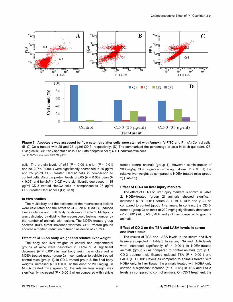
Flow cytometry analysisTo further confirm that CD-3 induces cell apoptosis, we also
used flow cytometry analysis with annexin V-FITC and PIdouble staining. HepG2 cells underwent apoptosis afterexposure to CD-3 at 25 and 35 μg/ml for 48 h. The percentageof apoptotic cells stained by Annexin V-FITC is shown in Figure7.
mRNA expression studies in HepG2 cells by RT-PCRTo elucidate cell signaling pathway activated by CD-3 in
HepG2 cells, RT-PCR analyses was used to measure themRNA expression of p53, mdm2, p65, c-jun, c-fos, bcl-2, bax,caspase-3, -7, -8 and -9 and cytochrome-c as well as theexpression of the β-actin (an internal standard). Thedensitometric analysis of mRNA expression in each group wasalso carried and statistically compared (Figure 8).
A significant increase (P < 0.0001) in mRNA expressionlevels of p53, bax, caspase-7, -8 and -9 was observed in 25and 35 µg/ml CD-3 treated HepG2 cells when compared tocontrol HepG2 cells. Also the expression of p53 (P < 0.001),bax (P < 0.01), caspase-7(P < 0.01), -8 (P < 0.05) and -9 (P <0.01) was significantly increased in 35 µg/ml CD-3 treatedHepG2 cells in comparison to 25 µg/ml CD-3 treated HepG2cells (Figure 8). A significant increase in the mRNA expressionlevel caspase-3 was observed in 25 µg/ml CD-3 treated HepG2
cells in comparison to control HepG2 cells. Whereas, mRNAexpression levels of caspase-3, was significantly increased (P< 0.0001) in 35 µg/ml CD-3 treated HepG2 cells in comparisonto control HepG2 cells. However, no significant difference wasobserved in the expression of caspase-3 in 25 and 35 µg/mlCD-3 treated HepG2 cells (Figure 8). The expression ofcytochrome-c was significantly increased (P < 0.001) in 25 and35 µg/ml CD-3 treated HepG2 cells when compared to controlHepG2 cells. No significant difference was observed in theexpression of cytochrome-c in 25 and 35 µg/ml CD-3 treatedHepG2 cells (Figure 8). A significant decrease (P < 0.0001) inmRNA expression levels of mdm2, p65, c-jun, c-fos and bcl-2was observed in 25 and 35 µg/ml CD-3 treated HepG2 cellswhen compared to control HepG2 cells. Also the expression ofmdm2 (P < 0.001), c-jun (P < 0.05), c-fos (P < 0.001) andbcl-2(P < 0.0001) was significantly decreased in 35 µg/ml CD-3treated HepG2 cells in comparison to 25 µg/ml CD-3 treatedHepG2 cells (Figure 8). No significant difference was observedin the expression of p65 in 25 and 35 µg/ml CD-3 treatedHepG2 cells (Figure 8).
Protein expression analysis of p53, p65, c-jun, bcl-2,bax and cleaved caspase-3 in HepG2 cells by ELISA
A significant increase in the protein levels of p53 (P <0.0001), bax (P < 0.001) and caspase-3(P < 0.01) wasobserved in cells treated with 25 µg/ml CD-3 in comparison tocontrol cells. However, cells treated with 35 µg/ml CD-3showed a significant increase (P < 0.0001) in protein levels ofp53, bax and cleaved caspase-3 in comparison to control cells.Also the protein levels of p53 (P < 0.001), bax (P < 0.01) weresignificantly increased in 35 µg/ml CD-3 treated HepG2 cells incomparison to 25 µg/ml CD-3 treated HepG2 cells. Nosignificant difference was observed in the protein level ofcleaved caspase-3 in 25 and 35 µg/ml CD-3 treated HepG2
Figure 5. Effect of CD-3 on DNA fragmentation in HepG2 cells. Values are mean ± SEM of three observations. aP < 0.0001w.r.t. control, bP < 0.0001 w.r.t. 25 µg/ml CD-3.doi: 10.1371/journal.pone.0068710.g005
Chemopreventive Effect of (+)-Cyanidan-3-ol
PLOS ONE | www.plosone.org 7 July 2013 | Volume 8 | Issue 7 | e68710

Figure 6. DNA fragmentation analysis by agarose gel electrophoresis. Lane M: DNA ladder (100 bp); Lane C: Control; Lanes1, and 2: cells treated with 25 and 35 μg/ml CD-3, respectively.doi: 10.1371/journal.pone.0068710.g006
Chemopreventive Effect of (+)-Cyanidan-3-ol
PLOS ONE | www.plosone.org 8 July 2013 | Volume 8 | Issue 7 | e68710
cells. The protein levels of p65 (P < 0.001), c-jun (P < 0.01)and bcl-2(P < 0.0001) were significantly decreased in 25 µg/mland 35 µg/ml CD-3 treated HepG2 cells in comparison tocontrol cells. Also the protein levels of p65 (P < 0.05), c-jun (P< 0.05) and bcl-2(P < 0.02) were significantly decreased in 35µg/ml CD-3 treated HepG2 cells in comparison to 25 µg/mlCD-3 treated HepG2 cells (Figure 9).
In vivo studiesThe multiplicity and the incidence of the macroscopic lesions
were calculated and the effect of CD-3 on NDEA/CCl4 inducedliver incidence and multiplicity is shown in Table 1. Multiplicitywas calculated by dividing the macroscopic lesions number bythe number of animals with lesions. The NDEA treated groupshowed 100% tumor incidence whereas, CD-3 treated groupsshowed a marked reduction of tumor incidence of 77.78%.
Effect of CD-3 on body weight and relative liver weightThe body and liver weights of control and experimental
groups of mice were described in Table 1. A significantdecrease (P < 0.001) in final body weight was observed inNDEA treated group (group 2) in comparison to vehicle treatedcontrol mice (group 1). In CD-3-treated group 3, the final bodyweights increased (P < 0.001) at the dose of 200 mg/kg. InNDEA treated mice (group 2), the relative liver weight wassignificantly increased (P < 0.001) when compared with vehicle
treated control animals (group 1). However, administration of200 mg/kg CD-3 significantly brought down (P < 0.001) therelative liver weight; as compared to NDEA treated mice (group2) (Table 1).
Effect of CD-3 on liver injury markersThe effect of CD-3 on liver injury markers is shown in Table
2. NDEA-treated (group 2) animals showed significantincreased (P < 0.001) serum ALT, AST, ALP and γ-GT ascompared to control (group 1) animals. In contrast, the CD-3-treated (group 3) animals at 200 mg/kg significantly decreased(P < 0.001) ALT, AST, ALP and γ-GT as compared to group 2animals.
Effect of CD-3 on the TSA and LASA levels in serumand liver tissue
The results of TSA and LASA levels in the serum and livertissue are depicted in Table 3. In serum, TSA and LASA levelswere increased significantly (P < 0.001) in NDEA-treatedanimals (group 2) as compared to control animals (group 1).CD-3 treatment significantly reduced TSA (P < 0.001) andLASA (P < 0.001) levels as compared to animals treated withNDEA only. In liver tissue, the animals treated with NDEA onlyshowed a significant increase (P < 0.001) in TSA and LASAlevels as compared to control animals. On CD-3 treatment, the
Figure 7. Apoptosis was assessed by flow cytometry after cells were stained with Annexin V-FITC and PI. (A) Control cells;(B–C) Cells treated with 25 and 35 µg/ml CD-3, respectively. (D) The summarized the percentage of cells in each quadrant. Q3:Living cells; Q4: Early apoptotic cells; Q2: Late apoptotic cells; Q1: Dead/Necrotic cells.doi: 10.1371/journal.pone.0068710.g007
Chemopreventive Effect of (+)-Cyanidan-3-ol
PLOS ONE | www.plosone.org 9 July 2013 | Volume 8 | Issue 7 | e68710

Figure 8. Expression of apoptosis-related genes in HepG2 cells after CD-3 treatment. mRNA expressions of β-actin, p53(wild) (A), mdm2 (B), p65 (C), c-jun (D), c-fos (E), bcl-2 (F), bax (G), caspase-3 (H), -7 (I), -8 (J), and -9 (K), and cytochrome-c (L)and their densitometric analysis in HepG2 cells. Values are mean ± SEM of four independent observations. aP < 0.0001 w.r.t.control, bP < 0.001 w.r.t. control, cP < 0.0001 w.r.t. control, dP < 0.001 w.r.t. control, eP < 0.0001 w.r.t. 25 µg/ml, fP < 0.001 w.r.t. 25µg/ml, gP < 0.01 w.r.t. 25 µg/ml, hP < 0.05 w.r.t. 25 µg/ml.doi: 10.1371/journal.pone.0068710.g008
Chemopreventive Effect of (+)-Cyanidan-3-ol
PLOS ONE | www.plosone.org 10 July 2013 | Volume 8 | Issue 7 | e68710

TSA and LASA levels were found to be decreased significantly(P < 0.001) as compared to NDEA-treated animals.
Effect of CD-3 on the lipid peroxidation and nitritelevels in liver tissue
The results showed a significant increase in MDA levels inNDEA-treated animals (group 2) as compared to controlanimals (group 1) (P < 0.001; Table 4). On CD-3 treatment(group 3), a significantly decrease (P < 0.001) in MDA levelswith respect to NDEA treated animals was observed. The nitritelevels were increased significantly in NDEA-treated animals (P< 0.001; Table 4). However, the animals treated with CD-3showed a significant decrease (P < 0.001) in nitrite levels withrespect to the animals treated with NDEA only.
Effect of CD-3 on the antioxidants status in liver tissueThe results of the antioxidants levels are depicted in Table 4.
A significant decrease in SOD and CAT activity were observedin NDEA-treated animals as compared to control animals (P <0.001) in liver. On CD-3 treatment, a significant increase (P <0.001) in SOD and CAT activities with respect to NDEA treated(group 2) animals was observed. A significant decrease (P <0.001) in the levels of reduced GSH, T-SH, and PrPr-SHcontent in mice liver was observed in NDEA-treated (group 2)animals. Administration of CD-3 in group 3 animals caused asignificant increase (P < 0.001) in GSH, T-SH and PrPr-SHlevels (Table 4). On the other hand, levels of GST, GPx, andGR were significantly decreased in NDEA-treated group 2animals (P < 0.001) (Table 4), whereas, in CD-3 treated group,a significant increase (P < 0.001) in the level of GST, GPx andGR was observed in comparison to NDEA-treated group.
mRNA expression studies by RT-PCR in NDEA-inducedhepatocarcinoma in Balb/c mice
To elucidate cell signaling pathway activated by CD-3 inNDEA-induced hepatocarcinoma in Balb/c mice, RT-PCRanalyses was used to measure the mRNA expression of p53,mdm2, p65, c-jun, c-fos, bcl-2, bax, caspase-3, -7, -8 and -9and cytochrome-c as well as the expression of the β-actin (aninternal standard). In the present study, two different primersfrom different exonic regions of p53 gene were used for mRNAexpression analysis. One primer was designed from exon 4-7(Primer A) and the second primer was from exon 10-11 (Primer
Table 1. Effect of CD-3 on NDEA/CCl4-inducedhepatocarcinogenesis.
Group
Micewithlesions/total ofmice
Incidence(%)a Multiplicity
Initialbodyweight(gm)
Finalbodyweight(gm)
Liverweight(gm)
Relativeliverweight(gm)
Group1
0/10 0.0 0.0026.38 ±0.19
27.51 ±0.17
1.49 ±0.009
5.43 ±0.024
Group2
6/6 100 4.8326.19 ±0.15
25.26 ±0.14
1.90 ±0.013
7.54 ±0.078
Group3
2/9 22.22 1.5*26.17 ±0.12
26.48± 0.13*
1.59 ±0.049*
6.02 ±0.18*
Group 1: Control; Group 2: NDEA/CCl4; Group 3: NDEA/CCl4/CD-3. Values arepresented as mean ± SEM of all animals per group. a percent mice with tumor; *
Significantly different from group 2 (P < 0.001).
Figure 9. Quantitative protein expression in HepG2 cells by ELISA. Cells treated with 25 and 35 µg/ml of CD-3. Data representthe means ± SEM of six independent observations. aP < 0.05 w.r.t. control, bP < 0.01 w.r.t. control, cP < 0.001 w.r.t. control, dP <0.0001 w.r.t. control, eP < 0.05 w.r.t. 25 µg/ml CD-3, fP < 0.01 w.r.t. 25 µg/ml CD-3, gP < 0.02 w.r.t. 25 µg/ml CD-3, hP < 0.001 w.r.t.25 µg/ml CD-3.doi: 10.1371/journal.pone.0068710.g009
Chemopreventive Effect of (+)-Cyanidan-3-ol
PLOS ONE | www.plosone.org 11 July 2013 | Volume 8 | Issue 7 | e68710

B). The densitometric analysis of mRNA expression in eachgroup was also carried and statistically compared (Figure 10).
In case of primer A mRNA expression level the p53 was verylow in vehicle treated control mice of group 1 animals which
Table 2. Effect of CD-3 on the activities of marker enzymesin the serum.
Groups Parameters
ALT (U/L) AST (U/L) ALP (U/L) γ-GT (U/L)Group 1 61.32 ± 1.80 81.65 ± 3.92 210.63 ± 3.90 35.66 ± 1.41Group 2 213.86 ± 4.25* 204.97 ± 2.51* 493.50 ± 8.08* 195.12 ± 4.63*
Group 3 131.46 ± 7.93* 156.28 ± 2.51* 290.71 ± 6.08* 111.81 ± 3.81*
Group 1: Control; Group 2: NDEA/CCl4; Group 3: NDEA/CCl4/CD-3. Data representthe means ± SEM in each group. *P < 0.001 versus vehicle treated and NDEA/CCl4or NDEA/CCl4/CD-3. Comparisons were made between Group 1 and Group 2 miceand between Group 2 and Group 3 mice.
Table 3. Effect of CD-3 on TSA and LASA levels.
Group Parameters TSA levels LASA levels
Serum (µg/ml)Liver tissue(µg/ml)
Serum (µg/ml)Liver tissue(µg/ml)
Group 1 1.91 ± 0.075 0.563 ± 0.038 0.599 ± 0.017 0.175 ± 0.003Group 2 5.71 ± 0.19* 1.19 ± 0.042* 1.77 ± 0.065* 0.711 ± 0.018*
Group 3 2.67 ± 0.10* 0.793 ± 0.031* 0.966 ± 0.028* 0.303 ± 0.013*
Group 1: Control; Group 2: NDEA/CCl4; Group 3: NDEA/CCl4/CD-3. Data representthe means ± SEM in each group. *P < 0.001 versus vehicle treated and NDEA/CCl4or NDEA/CCl4/CD-3. Comparisons were made between Group 1 and Group 2 miceand between Group 2 and Group 3 mice.
Table 4. Effect of CD-3 on oxidative stress markers inmouse liver.
Parameters Group 1 Group 2 Group 3MDA (nmol/ mg protein) 3.38 ± 0.065 17.71 ± 0.32* 7.44 ± 0.31*Nitrite (µg/mg protein) 0.500 ± 0.017 1.80 ± 0.031* 0.86 ± 0.025*CAT (U/ mg protein) 5.26 ± 0.079 1.19 ± 0.038* 4.18 ± 0.053*SOD (U/ mg protein) 18.55 ± 0.50 7.42 ± 0.85* 13.91 ± 0.55*Total thiols 17.12 ± 0.20 6.39 ± 0.25* 13.59 ± 0.23*GSH (nmol/ mg protein) 5.31 ± 0.16 1.69 ± 0.028* 3.98 ± 0.13*PrPr-SH 11.81 ± 0.24 4.7 ± 0.23* 9.59 ± 0.23*GST (GSH-CDNBconjugate/min/mg protein)
31.85 ± 0.70 9.59 ± 0.28* 24.26 ± 0.47*
GPx ((nmol NADPHoxidized/min/mg protein)
14.14 ± 0.40 3.95 ± 0.13* 9.28 ± 0.24*
GR ((nmol NADPH oxidized/min/mgprotein)
15.15 ± 0.19 3.13 ± 0.13* 9.84 ± 0.26*
Group 1: Control; Group 2: NDEA/CCl4; Group 3: NDEA/CCl 4/CD-3. Datarepresent the means ± SEM in each group. *P < 0.001 versus vehicle treated andNDEA/CCl4 or NDEA/CCl4/CD-3. Comparisons were made between Group 1 andGroup 2 mice and between Group 2 and Group 3 mice.
significantly increased (P < 0.0001) in NDEA-treated mice(group 2). However, CD-3 treatment in group 3 animalsresulted in significant decreased (P < 0.0001) expression ofp53 in comparison to NDEA treated liver tumors of group 2. Onthe other hand, mRNA expression level of p53 with primer Bwas high in vehicle treated control mice of group 1. However inNDEA-treated animals (group 2) the mRNA expression level ofp53 was significantly decreased (P < 0.0001) in comparison tovehicle treated control mice of group 1. No significantdifference was observed in mRNA expression level of p53 inCD-3 treated animals in comparison to NDEA treated livertumors of group 2 (Figure 10).
A significant increase (P < 0.0001) in the expression ofmdm2, p65, c-jun, c-fos and bcl-2 were observed in NDEA-treated mice (group 2) in comparison to vehicle treated controlmice of group 1. CD-3 treatment in group 3 animals resulted insignificant decreased (P < 0.0001) expression of mdm2, p65, c-jun, c-fos and bcl-2 in comparison to NDEA treated liver tumorsof group 2. However, the expression of mdm2 (P < 0.001), p65(P < 0.01), c-jun (P < 0.05), c-fos (P < 0.01) and bcl-2(P <0.001) were significantly decreased in CD-3 treated group 3mice in comparison to vehicle treated control mice of group 1(Figure 10). A significant increase (P < 0.0001) in theexpression of bax (P < 0.0001), caspase-3(P < 0.0001), -7 (P <0.0001), -8 (P < 0.0001), -9 (P < 0.0001) and cytochrome-c (P< 0.001) were observed in CD-3 treated group 3 mice liver incomparison to NDEA-treated liver tumors (group 2). However,the expression of bax (P < 0.0001), caspase-3(P < 0.0001), -7(P < 0.0001), -8 (P < 0.0001), -9 (P < 0.0001) and cytochrome-c (P < 0.0001) were significantly increased in CD-3 treatedgroup 3 mice in comparison to vehicle treated control mice ofgroup 1 (Figure 10).
Protein expression analysis of p53, p65, c-jun, bcl-2,bax and cleaved caspase-3 in liver tissues by ELISA
There was a significant increase (P < 0.001) in the proteinlevels of p53, p65, c-jun and bcl-2were observed in NDEA-treated mice (group 2) as compared to vehicle treated controlanimals (group 1) whereas, a significant decrease (P < 0.001)was observed in the protein levels of p53, p65, c-jun and bcl-2in CD-3 treated animals (group 3). The bax and cleavedcaspase-3 protein levels were significantly increased (P <0.001) in CD-3 treated mice (group 3) as compared to NDEAtreated mice (group 2) (Figure 11).
Histopathological observationsThe normal liver architecture with small uniform nuclei
radically arranged around the central vein was observed ingroup 1 animal tissue (Figure 12A). Whereas, in group 2(NDEA-treated mice) showed loss of cellular architecture. Thetumor architecture demonstrated severe centrilobular necrosis,degeneration in hepatocytes, congestion in the central veinsinusoids, proliferation of Kupffer cells, and nodular hyperplasiaof hepatocytes. Neoplastic cells were smaller than normal cellswith granular cytoplasm and larger hyperchromatic nuclei(Figure 12B & C). Architecture of liver sections of CD-3 treatedgroup 3 mice showed the features of a normal liver tissue withminimal inflammatory cell infiltration (Figure 12D).
Chemopreventive Effect of (+)-Cyanidan-3-ol
PLOS ONE | www.plosone.org 12 July 2013 | Volume 8 | Issue 7 | e68710

ImmunohistochemistryIn order to study the alterations in the induction of various
proteins, immunohistochemical localization of these proteins
were carried out using immunohistochemistry (IHC). Negativecontrol (without using specific antibody) from all the treatmentgroups did not show any detectable staining.
Figure 10. Expression of apoptosis-related genes in liver after CD-3 treatment. mRNA expressions of β-actin, p53 (mut) (A),p53 (wild) (B), mdm2 (C), p65 (D), c-jun (E), c-fos (F), bcl-2 (G), bax (H), caspase-3 (I), -7 (J), -8 (K), and -9 (L), and cytochrome-c(M) and their densitometric analysis in hepatocarcinoma. Values are mean ± SEM of four independent observations. aP < 0.0001w.r.t. control, bP < 0.001 w.r.t. control, cP < 0.01 w.r.t. control, dP < 0.0001 w.r.t. control, eP < 0.001 w.r.t. control, fP < 0.01 w.r.t.control, gP < 0.05 w.r.t. control, hP < 0.0001 w.r.t. NDEA/CCl4 (group 2), iP < 0.001 w.r.t. NDEA/CCl4 (group 2).doi: 10.1371/journal.pone.0068710.g010
Chemopreventive Effect of (+)-Cyanidan-3-ol
PLOS ONE | www.plosone.org 13 July 2013 | Volume 8 | Issue 7 | e68710

The specific localization of p53, p65 and c-jun was studied inall the treatment groups. IHC investigation of control animals(group 1) revealed normal liver architecture with small uniformnuclei radically arranged around the central vein with noexpression of p53 (Figure 13, panel A), p65 (Figure 14, panelA) and c-jun (Figure 15, panel A). In NDEA treated animals(group 2), intense localization of p53 (Figure 13, panel B), p65(Figure 14, panel B) and c-jun (Figure 15, panel B) was seenthroughout the lobules in hepatocytes, or in proliferatingductular cells as compared to respective control (group 1)which confirmed the increased expression of p53, p65 and c-jun in hepatocytes. In group 3 (NDEA/CD-3), a significant lesslocalization of p53 (Figure 13, panel C), p65 (Figure 14, panelC) and c-jun (Figure 15, panel C) was seen in hepatocytes.The staining was although also visible in the hepatocytes butwas faint as compared to NDEA treated group (group 2).
Discussion
Apoptosis is a genetically programmed cell deathmechanism that can be activated by various stimuli, includingthe activation of specific pro-apoptotic receptors, and cellularstress or injury caused by loss of growth factor signals, heatshock, irradiation, cytotoxic drugs, bacteria, and viruses.Disturbances in mechanisms that direct abnormal cells toundergo apoptosis perilously contribute to tumorigenesis; yielda logical target for potential therapeutic intervention [35].
This is the first report on the apoptosis induction andchemopreventive effect of CD-3 in HCC. Our datademonstrated that CD-3 could inhibit the proliferation of HepG2cell line in-vitro via apoptosis induction in dose-dependentmanner. The CD-3 also showed chemopreventive effect
against NDEA/CCl4 induced HCC tumor growth in-vivo. Wedemonstrated that CD-3 dose-dependently inhibited theproliferation of HepG2 cells in various cytotoxicity assays. As atest to confirm the cytotoxicity of CD-3, the HepG2 cells wereincubated with different concentrations of CD-3 and theirmorphological alterations were verified via a phase-contrastmicroscope. Results from the present study demonstrated thatthere were marked morphological changes in HepG2 cellsfollowing treatment with CD-3 which were indicative of cellapoptosis. The morphological changes that occurred inapoptotic cells were also perceived through fluorescencemicroscopy. It was possible to observe difference betweenearly and late apoptosis in the HepG2 cells, showing that CD-3exhibited a larger concentration response effect for lateapoptosis and a smaller effect for early apoptosis, in both of thestudied concentrations. Formation of DNA fragmentation is oneof the characteristic features observed in apoptotic cells and itis generally considered as the biochemical hallmark ofapoptosis [36]. The formation of a DNA ladder correlates withthe early morphological signs of apoptosis and has been widelyused as a distinctive marker of the apoptosis process [37]. Theladder-like appearance of DNA observed in the HepG2 cellstreated with CD-3, was also another indicator of the apoptosisinducing capability of the CD-3. In addition, a noticeablephenomenon was that the percent DNA fragmentation wasincreased after treatment with CD-3 when analyzed bydiphenylamine (DPA) assay. Data from an apoptosis assayshowed that CD-3 induced obvious apoptosis in HepG2 cancercells, presenting a concentration-dependent manner ofapoptosis-specific fragmentation. This showed that CD-3inhibited the proliferation of HepG2 cells via cell apoptosis.Flow cytometry analysis revealed that CD-3 treatment
Figure 11. Quantitative protein expression in different groups by ELISA. Group 1: Control; Group 2: NDEA/CCl4; Group 3:NDEA/CCl4/CD-3. Data represent the means ± SEM of six independent observations. *P < 0.001 versus vehicle treated andNDEA/CCl4 or NDEA/CCl4/CD-3. Comparisons were made between Group 1 and Group 2 mice and between Group 2 and Group 3mice.doi: 10.1371/journal.pone.0068710.g011
Chemopreventive Effect of (+)-Cyanidan-3-ol
PLOS ONE | www.plosone.org 14 July 2013 | Volume 8 | Issue 7 | e68710

increased the apoptotic cells. These results suggested that thegrowth inhibition of HepG2 cells by CD-3 was due to its abilityto induce apoptosis.
It is well documented that oxidative stress contributes tomultiple physiological events including cell proliferation andinflammation, mediated by modifying redox sensitive AP-1 andNF-κB pathways. Oxidative stress occurs when the criticalbalance is disrupted due to excess production of ROS ordepletion of antioxidants or both. During oxidative stress,malonaldehyde and other aldehydes are formed in thebiological system as a result of lipid peroxidation. The productsof LPO are considered mutagenic and carcinogenic as theycause damage to cellular macromolecules by generating ROS[38,39].
The current study showed an elevated level of MDA in liverof the animals treated with NDEA/CCl4 as compared to controlanimals. The increased level of lipid peroxidation may be dueto the poor antioxidant defense or inactivation of antioxidant
Figure 12. H & E stained sections of liver; 100 x. (A)Control group 1 revealed normal parenchymal cells withgranulated cytoplasm and small uniform nuclei radicallyarranged around the central vein. (B & C) NDEA treated group2 mice showing histology of a liver tumor that consists ofhepatocytes only without portal tracts, congestion in the centralvein and sinusoids, proliferation of Kupffer cells, and nodularhyperplasia of hepatocytes. The nuclei were mostly found to bepleomorphic with fine granular chromatin and occasionally withsmall nucleoli (B). The tumor architecture demonstrates severecentrilobular necrosis, degeneration in hepatocytes; extensivevacuolation was noticed in the cytoplasm encircling thenucleus. The sinuses were greatly dilated with hyperplastickupffer cells (C). (D) NDEA/CD-3 treated group 3 mice showingnormal cellular architecture with some hepatocytes showingfew neoplastically transformed cells with minimal inflammatorycell infiltration around the portal triads.doi: 10.1371/journal.pone.0068710.g012
enzymes in cancerous tissues. A significant decrease in MDAlevels by CD-3 treatment indicated their role in reducingoxidative stress, thus indicating its protective potential againstHCC. Nitric oxide radical (NO·) plays an important role inphysiological and pathological processes. An increased nitritelevel is generally associated with the process of carcinogenesis[40]. The present results showed that nitrite levels, an indicatorof NO·, were significantly decreased on CD-3 treatment.
Body’s antioxidant defense systems operate for scavengingROS to prevent the oxidative stress and antioxidant enzymes,namely, SOD and CAT acts as the first line of cellular defenseagainst oxidative damage [41]. NDEA/CCl4 administrationdecreased the activities of these antioxidant enzymes in theliver tissue, which may be related to saturation of SOD withsuperoxide radicals in tumor cells or a decrease or loss ofexpression of SOD. A decrease in SOD activity will result indecreased production of H2O2 which in turn affects CATactivity. The second line of cellular defense includes
Figure 13. Immunohistochemical analysis of p53expressions in mice liver. Panel ‘A’ photomicrographs showsliver section of control mice (group 1), Immunostained for p53with specific anti-p53; Panel ‘B’ photomicrograph showing liversection of NDEA treated mice (group 2), Immunostained forp53 with specific anti-p53, showing intense staining throughoutthe lobules in hepatocytes, or in proliferating ductular cells;Panel ‘C’ photomicrograph showing liver section of NDEA/CCl4/CD-3-treated mice (group 3), Immunostained for p53 withspecific anti-p53, showing less localization of p53 inhepatocytes. Scale bar 100µm.doi: 10.1371/journal.pone.0068710.g013
Chemopreventive Effect of (+)-Cyanidan-3-ol
PLOS ONE | www.plosone.org 15 July 2013 | Volume 8 | Issue 7 | e68710

glutathione antioxidant system that plays an important roleagainst free radicals [42]. Reduced glutathione and itsdependent enzymes, GPx, GR and GST serve as reliablemarkers of chemoprevention [43]. The present data showedsignificant decrease in the activities of GPx, GR, and GST inmice liver after NDEA/CCl4 exposure, resulting in considerabledecline in the activities of these enzymes. In response tooxidative stress, GPx works in tandem with CAT to scavengeexcess of H2O2 and lipid peroxides [44,45]. However, unlikeCAT activity, GPx activity also depends on the balancebetween the levels of GSH and GSSG [46]. Thus, decrease inGPx activity may be implicated in both free radical dependantinactivation of enzyme [47] and depletion of its co-substrates,i.e., GSH and NADPH [48]. The observed decrease in GPxactivity may also be due to reduced availability of GSH. Asignificant decrease in GR activity may be attributed to theimpaired conversion of GSSG into GSH and thus balancingGSH/GSSG ratio [49]. Thiol groups have ability to act asreducing agents and thus play a central role in coordinating the
Figure 14. Immunohistochemical analysis of p65expressions in mice liver. Panel ‘A’ photomicrographs showsliver section of control mice (group 1), Immunostained for p65with specific anti-p65; Panel ‘B’ photomicrograph showing liversection of NDEA treated mice (group 2), Immunostained forp65 with specific anti-p65, showing intense staining of Kupffercells; Panel ‘C’ photomicrograph showing liver section ofNDEA/CCl4/CD-3-treated mice (group 3), Immunostained forp65 with specific anti-p65, showing less localization of p65 inhepatocytes. Scale bar 100µm.doi: 10.1371/journal.pone.0068710.g014
antioxidant defense system [50]. The present findings revealedalterations in the levels of T-SH, GSH, and PrPr-SH inNDEA/CCl4-treated animals. The decrease in T-SH contentmight be contributed to reduction in GSH levels and/or changein PrPr-SH groups. The observed decrease in GSH levels maybe due to diminished activity of GR, which is a crucial enzymefor maintaining GSH/GSSG ratio in the cell. A significantrestoration in the activities of these enzymes after CD-3administration indicated that CD-3 acts as an effectiveantioxidant.
Experimental evidence suggests that apoptosis can bemediated by number of different ways and that there arenumerous regulatory molecules associated with those paths.p53 plays a key role in the process of apoptosis, DNA repairand tumor suppression pathways. Both extrinsic and intrinsicapoptotic pathways are activated by p53 [51]. Manychemopreventive agents are known to exert their anticancereffects through the induction of apoptosis via p53 dependent
Figure 15. Immunohistochemical analysis of c-junexpressions in mice liver. Panel ‘A’ photomicrographs showsliver section of control mice (group 1), Immunostained for c-junwith specific anti-c-jun; Panel ‘B’ photomicrograph showingliver section of NDEA treated mice (group 2), Immunostainedfor c-jun with specific anti-c-jun, showing marked induction of c-jun throughout the lobules in hepatocytes, or in proliferatingductular cells; Panel ‘C’ photomicrograph showing liver sectionof NDEA/CCl4/CD-3-treated mice (group 3), Immunostained forc-jun with specific anti-c-jun, showing less localization of c-junin hepatocytes. Scale bar 100µm.doi: 10.1371/journal.pone.0068710.g015
Chemopreventive Effect of (+)-Cyanidan-3-ol
PLOS ONE | www.plosone.org 16 July 2013 | Volume 8 | Issue 7 | e68710

mechanisms. The inactivation of p53 causes genetic instabilityleading to the accumulation of genetic alterations, which inducemalignant transformation of cells. In addition, it was found thatmutant p53 protein that frequently accumulates in humantumors facilitates tumor initiation and progression by a “gain offunction” mechanism [52,53]. In the present study, weobserved the mRNA and protein expression levels of p53 inCD-3 treated HepG2 cells using RT-PCR and ELISA,respectively. We found that CD-3 markedly increased themRNA and protein expression levels of p53 in HepG2 cells in adose-dependent manner. However, in in-vivo study theanalysis of our RT-PCR bands by using both p53 primer pairs,it appears that in mouse different isoforms were produced bysplicing mechanism and their level of expression compare toeach other decide the fate of the cell. We also suspect that inour case these different spliced products in cancerous tissuecontain some alteration in 3ʹ-UTR region which somehow wasrelated to apoptosis or inhibition of mdm2-p53 interaction.Moreover, in cancerous tissue exon 10 and 11 is known tocontain two transcript variants so it is quite possible (but notcertain) that second primer was not able to amplify some of theunknown spliced variant that may be produced at that area.Since, primer B position is responsible for transcript variant oneand two and that might be the possible region behind lowexpression level of mRNA in cancerous tissue. Similarly,immunohistochemistry studies indicated intense localization ofp53 throughout the lobules in hepatocytes, or in proliferatingductular cells in NDEA/CCl4-treated animals. However, lessimmunohistochemical localization of p53 was observed in CD-3treated animals. Mutant p53 proteins accumulate to high levelsin many cancer cells and the p53 protein and the p53 responseto DNA damage represent key points for therapeuticintervention. The mdm2 gene encodes a negative regulator ofthe p53 tumor suppressor. Mdm2 may impart some of itstumorigenic properties by increasing the degradation ofmultiple cellular proteins [54]. In the present investigation, asignificant increase in the mdm2 mRNA expression levels wasseen in both HepG2 cells and NDEA/CCl4-treated animals.However, a significant decrease in mdm2 expression levelswas observed on CD-3 treatment in both in-vitro and in vivomodels. Mdm2 is the primary cellular inhibitor of p53 in cancersand targeting the Mdm2-p53 interaction is an attractive cancertherapeutic strategy [55].
One of the major gene groups that regulate apoptosis is thebcl-2 family such as bax and bcl-2 proteins which elicitopposing effects on mitochondria. Enhancement of pro-apoptotic Bax over Bcl-2 proteins can enhance the permeabilityof the mitochondrial membrane, which in turn results in therelease of apoptogenic factors. Bcl-2, on the other hand,prevents this process by preserving mitochondrial integrity andblocks the release of soluble inter-membrane factors such ascytochrome c that activate the effectors of apoptosis [56]. Thus,it has been suggested that the ratio between the levels of pro-apoptotic and the anti-apoptotic factor determines whether acell responds to an apoptotic signal. The current studyindicated that, CD-3 up-regulated the mRNA and proteinexpressions of bax and down-regulated the mRNA and proteinexpression levels of bcl-2 in both HepG2 cells and NDEA/CCl4-
treated animals. Also, mRNA expression level of cytochrome-cwas increased in CD-3 treated HepG2 cells and NDEA/CCl4-treated animals. Hence, the ratio of pro-apoptotic proteins tothe anti-apoptotic proteins was altered in favor of apoptosis.Thus, the results suggested that an up-regulation of bax andthe corresponding down-regulation of bcl-2 proteins observedin this study may be one of the critical mechanisms throughwhich CD-3 induced apoptosis.
The induction of apoptosis is almost always associated withthe activation of caspases; a conserved family of enzymes thatirreversibly commit a cell to die. The release of cytochrome cfrom mitochondria to cytosol after being induced by a variety ofapoptosis-inducing agents leads to the formation ofapoptosome which forms a platform for the efficient processingand activation of caspase-9. Activation of caspase-9, in turn,cleaves effectors caspases such as caspase-3 and 7 whicheventually lead to apoptosis [57]. In the next series ofexperiment, we assessed the effect of CD-3 on the cascade ofcaspases. To investigate the effect of CD-3 on the caspasecascade, mRNA expression levels of caspase-3, -7, -8, and -9and protein level of cleaved caspase-3 were determined in ourexperiment. Results from the present study demonstrated thatmRNA expression levels of caspase-3, -7, -8 and -9 andprotein level of cleaved caspase-3 were increased in a bothHepG2 cells and NDEA/CCl4-treated animals on CD-3treatment. The activation of caspases-3, -7 and -9 is a result ofthe induction of the intrinsic pathway, while activationcaspase-8 and then caspase-3 and -7 may be the result of theinduction of the extrinsic pathway. In both pathways, theinitiator caspase cleaves and activates downstream effectorcaspases, such as caspase-3 and -7.
Activated protein-1 (AP-1) and nuclear factor-κB (NF-κB),two of important transcription factors, play important roles insignal transduction pathways of cell differentiation, proliferationand apoptosis in response to a variety of physiological andpathological stimuli [58,59]. AP-1 activation requires Jun andFos through the formation of homo and hetero-dimers, andregulates transcription of a broad range of genes involved incell differentiation and proliferation [60]. In the present study,we observed the mRNA expression levels of c-jun and c-fosand protein expression level of c-jun were markedly decreasedon CD-3 treatment in both HepG2 cells and NDEA/CCl4-treatedanimals. These results were also supported byimmonohistochemistry studies which had shown the markedinduction of c-jun throughout the lobules in hepatocytes, or inproliferating ductular cells in NDEA/CCl4-treated animals.However, less immunohistochemical localization of c-jun wasobserved in animals treated with CD-3. NF-κB is thought to up-regulate expression of genes that cause suppression of theapoptotic response in cancer cells [59]. In the present study, asignificant increase in the mRNA and protein expression levelsof NF-κB (p65) was observed in both HepG2 cells andNDEA/CCl4-treated animals. However, a significant decrease inthe mRNA and protein expression levels of p65 was observedon CD-3 treatment in both HepG2 cells and NDEA/CCl4-treatedanimals. Similarly, immunohistochemistry studies indicatedintense localization of p65 in NDEA/CCl4-treated animals.However, weak immunohistochemical localization of p65 was
Chemopreventive Effect of (+)-Cyanidan-3-ol
PLOS ONE | www.plosone.org 17 July 2013 | Volume 8 | Issue 7 | e68710

observed in animals treated with CD-3. Severalchemopreventive agents are inhibitors of NF-ĸB activation.These inhibitors can block any one or more steps in the NF-ĸBsignaling cascade, the translocation of NF-ĸB into the nucleus,DNA binding of the dimers and interactions with the basaltranscription activation. Thus, blockers of NF-κB should bebeneficial not only in prevention but also in the treatment ofcancer.
In conclusion, the present study demonstrated that CD-3could inhibit the proliferation of HCC cell line in-vitro andsuppresses HCC tumor growth in-vivo and consequentlyinduced caspase-dependent apoptosis via up-regulation ofp53, bax and down-regulation of bcl-2, AP-1 and NF-κB.Therefore, the results from this study provided criticallyimportant experimental facts to suggest that CD-3 may be apotential therapeutic agent for treating HCC.
Supporting Information
Figure S1. The representative chromatogram showing thepurity of CD-3 by analytical reverse phase HPLC. HPLC
experimental conditions were: Instrument: Waters 515 withauto-sampler and the photo diode array (PDA) detector(Waters 2996); HPLC column: Waters spherisorb symmetry(C18 (5.0 μm) 250 mm × 4.6 mm); Mobile phases: 0.1%trifluoroacetic acid (TFA): acetonitrile (85: 15, v/v); Flow rate:1.0 ml/min; Cycle time of analysis was 25 min at 30°C.(TIF)
Figure S2. Effect of CD-3 on the percentage viability ofnormal lymphocytes. (TIF)
Table S1. The oligonucleotide primer pairs used in reversetranscription polymerase chain reaction. (DOCX)
Author Contributions
Conceived and designed the experiments: MS CSC RSC.Performed the experiments: JM SP SSC . Analyzed the data:MS CSC . Contributed reagents/materials/analysis tools: MS.Wrote the manuscript: JM SP SSC .
References
1. Ferlay J, Shin HR, Bray F, Forman D, Mathers C et al. (2010)Estimates of worldwide burden of cancer in 2008: GLOBOCAN 2008.Int J Cancer 127: 2893-2917. doi:10.1002/ijc.25516. PubMed:21351269.
2. Abou-Alfa GK, Schwartz L, Ricci S, Amadori D, Santoro A et al. (2006)Phase II study of sorafenib in patients with advanced hepatocellularcarcinoma. J Clin Oncol 24: 4293-4300. doi:10.1200/JCO.2005.01.3441. PubMed: 16908937.
3. Ruhul Amin ARM, Kucuk O, Khuri ER, Shin DM (2009) Perspectives forcancer prevention with natural compounds. J Clin Oncol 27:2712-2725. doi:10.1200/JCO.2008.20.6235. PubMed: 19414669.
4. Barnes S, Prasain J (2005) Current progress in the use of traditionalmedicine and nutraceuticals. Curr Opin Plant Biol 8: 324-328. doi:10.1016/j.pbi.2005.03.010. PubMed: 15860430.
5. Monga J, Chauhan CS, Sharma M (2011) Human epithelial carcinomacytotoxicity and inhibition of DMBA/TPA induced squamous cellcarcinoma in Balb/c mice by Acacia catechu heartwood. J PharmPharmacol 63: 1470-1482. doi:10.1111/j.2042-7158.2011.01354.x.PubMed: 21988428.
6. Monga J, Chauhan CS, Sharma M (2012) Human breastadenocarcinoma cytotoxicity and modulation of 7,12-dimethylbenz[a]anthracene-induced mammary carcinoma in Balb/cmice by Acacia catechu heartwood. Integr Cancer Ther, [E pub ahead]
7. Lotito BS, Fraga CG (1998) CD-3 prevents human plasma oxidation.Free Radic Biol Med 24: 435-441. doi:10.1016/S0891-5849(97)00276-1. PubMed: 9438556.
8. Rage N, Dahanukar S, Karandikar SM (1984) Hepatoprotective effectof cyanidanol against carbon tetrachloride induced liver damage. IndianDrugs 22: 556-560.
9. Guruvayoorappan C, Kuttan G (2008) CD-3 inhibits tumourangiogenesis and regulates the production of nitric oxide and TNF-α inLPS-stimulated macrophages. Innate Immun 14: 160-174. doi:10.1177/1753425908093295. PubMed: 18562575.
10. Anderson R, Smit MJ, Van Rensburg CE (1993) Lysophospholipid-mediated inhibition of Na+, K(+)-adenosine triphosphatase is a possiblemechanism of immunorepressive activity of cyclosporin A. MolPharmacol 44: 605-614. PubMed: 8396720.
11. Denizot F, Lang R (1986) Rapid colorimetric assay for cell growth andsurvival. Modifications to the tetrazolium dye procedure giving improvedsensitivity and reliability. J Immunol Methods 89: 271-277. doi:10.1016/0022-1759(86)90368-6. PubMed: 3486233.
12. Vanicha V, Kanyawim K (2006) Sulforhodamine B colorimetric assayfor cytotoxicity screening. Nature 1: 1112-1116. PubMed: 17406391.
13. Saad S, Dakwar O, Said G, Abu-Hijleh G, Battah FA et al. (2006)Evaluation of medicinal plant hepatotoxicity in co-cultures of
hepatocytes and monocytes. Evid Based Complement Alternat Med 3:93-98. doi:10.1093/ecam/nel002. PubMed: 16550229.
14. Duarte RA, Mello ER, Araki C, Bolzani VS, Siqueirs Silva DH et al.(2010) Alkaloids extracted from Pterogyne nitens induce apoptosis inmalignant breast cell line. Tumor Biol 31: 513-522. doi:10.1007/s13277-010-0064-2. PubMed: 20700682.
15. Jimenez PC, Wilke DV, Takeara R, Lotufo TM, Pessoa C et al. (2008)Cytotoxicity activity of a dichloromethane extract and fractions obtainedfrom Eudistoma vannamei (Tunicata: Ascidiacea). Comp BiochemPhysiol A Mol Integr Physiol 151: 391-398. doi:10.1016/j.cbpa.2007.02.018. PubMed: 17400012.
16. Kurita-Ochiai T, Fukushima K, Ochiai K (1999) Lipopolysaccharidestimulates butyric acid-induced apoptosis in human peripheral bloodmononuclear cells. Infect Immun 67: 22-29. PubMed: 9864191.
17. Perandones CE, Illera VA, Peckham D, Stunz LL, Ashman RF (1993)Regulation of apoptosis in vitro in mature spleen T cells. J Immunol151: 3521-3529. PubMed: 8376790.
18. Walker PR, Kokileva L, LeBlanc J, Sikorska M (1993) Detection of theinitial stages of DNA fragmentation in apoptosis. BioTechniques 15:1032-1040. PubMed: 8292335.
19. Liao PC, Lieu PC (2005) Cell cycle specific induction of apoptosis andnecrosis by paclitaxel in the leukemic U937 cells. Life Sci 76:1623-1639. doi:10.1016/j.lfs.2004.09.021. PubMed: 15680171.
20. Singh BN, Singh BR, Sarma BK, Singh HB (2009) Potentialchemoprevention of N-nitrosodiethylamine-inducedhepatocarcinogenesis by polyphenolics from Acacia nilotica bark.Chem Biol Interact 181: 20-28. doi:10.1016/j.cbi.2009.05.007. PubMed:19446540.
21. Reitman S, Frankel SA (1957) A colorimetric method for determinationof serum glutamic oxaloacetic and glutamic pyruvic transaminases. AmJ Clin Pathol 28: 3856-3863.
22. Bergmeyer HU, Bernt E (1974) Aminotransferases and relatedenzymes. In: HU Bergmeyer. Methods of Enzymatic Analysis. pp.735-763.
23. Szasz G (1976) Reaction rate method for gamma glutamyl transferaseactivity in serum. Clin Chem 22: 2031-2055. PubMed: 1000801.
24. Plucinsky MC, Riley WM, Prorok JJ, Alhadeff JA (1986) Total and lipid-associated serum sialic acid levels in cancer patients with differentprimary sites and differing degrees of metastatic involvement. Cancer58: 2680-2685. doi:10.1002/1097-0142(19861215)58:12. PubMed:3779617.
25. Katopodis N, Hirshaut Y, Geller NL, Stock CC (1982) Lipid-associatedsialic acid test for the detection of human cancer. Cancer Res 42:5270-5275. PubMed: 7139630.
26. Wills ED (1966) Mechanisms of lipid peroxide formation in animaltissues. Biochem J 99: 667-676. PubMed: 5964963.
Chemopreventive Effect of (+)-Cyanidan-3-ol
PLOS ONE | www.plosone.org 18 July 2013 | Volume 8 | Issue 7 | e68710
27. Green LC, Wagner DA, Glogowski J, Skipper PL, Wishnok JS et al.(1982) Analysis of nitrate, nitrite, and [15n] nitrate in biological fluids.Anal Biochem 126: 131-138. doi:10.1016/0003-2697(82)90118-X.PubMed: 7181105.
28. Kono Y (1978) Generation of superoxide radicals during auto-oxidationof hydroxyl amine and an assay of superoxide dismutase. ArchBiochem Biophys 186: 189-195. doi:10.1016/0003-9861(78)90479-4.PubMed: 24422.
29. Aebi H (1984) Catalase in vitro. Methods Enzymol 105: 121-126. doi:10.1016/S0076-6879(84)05016-3. PubMed: 6727660.
30. Flohé L, Günzler WA (1984) Assays of glutathione peroxidase.Methods Enzymol 105: 114-121. doi:10.1016/S0076-6879(84)05015-1.PubMed: 6727659.
31. Carlberg I, Mannervik B (1985) Glutathione reductase. MethodsEnzymol 113: 484-490. doi:10.1016/S0076-6879(85)13062-4. PubMed:3003504.
32. Warholm M, Guthenberg C, Von Bahr C, Mannervik B (1985)Gluthathione transferases from human liver. Methods Enzymol 113:499-504. doi:10.1016/S0076-6879(85)13065-X. PubMed: 3003505.
33. Sedlak J, Lindsay RH (1968) Estimation of total, protein-bound, andnonprotein sulfhydryl groups in tissue with Ellman’s reagent. AnalBiochem 25: 192-205. doi:10.1016/0003-2697(68)90092-4. PubMed:4973948.
34. Ellman GL (1959) Tissue sulfhydryl groups. Arch Biochem Bios Phys82: 70-77. doi:10.1016/0003-9861(59)90090-6. PubMed: 13650640.
35. Hickman JA (1992) Apoptosis induced by anticancer drugs. CancerMetastasis Rev 11: 121-129. doi:10.1007/BF00048059. PubMed:1327566.
36. Fink SL, Cookson BT (2005) Apoptosis, pyroptosis, & necrosis:mechanistic description of dead & dying eukaryotic cells. Infect Immun73: 1907-1916. doi:10.1128/IAI.73.4.1907-1916.2005. PubMed:15784530.
37. Ariffin SHZ, Wan Omar WH, Ariffin ZZ, Safian MF, Senafi S, AbdulWahab RM (2009) Intrinsic anticarcinogenic effects of Pipersarmentosum ethanolic extract on a human hepatoma cell line. CancerCell Int 9: 6. doi:10.1186/1475-2867-9-6. PubMed: 19257877.
38. Rao GM, Rao CV, Pushpangadan P, Annie S (2006) Hepatoprotectiveeffects of rubiadin, a major constituent of Rubiá cordifolia Linn. JEthnopharmacol 103: 484-490. doi:10.1016/j.jep.2005.08.073.PubMed: 16213120.
39. Vásquez-Garzón VR, Arellanes-Robledo J, García-Román R, Aparicio-Rautista DI, Villa-Treviño S (2009) Inhibition of reactive oxygen speciesand preneoplastic lesions by quercetin through an antioxidant defensemechanism. Free Radic Res 43: 128-137. doi:10.1080/10715760802626535. PubMed: 19115120.
40. Esme H, Cemek M, Sezer M, Saglam H, Demir A et al. (2008) Highlevels of oxidative stress in patients with advanced lung cancer.Respirology 13: 112-116. doi:10.1111/j.1440-1843.2007.01212.x.PubMed: 18197920.
41. Ferreccio C, Gonzalez PC, Milosavjlevic SV, Marshall GG, Sancha AM(1998) Lung cancer and arsenic exposure in drinking water: a casecontrol study in northern Chile. Cad Saude Publica 14: 193-198. doi:10.1590/S0102-311X1998000100028.
42. Bitensky L (1990) Glutathione: Chemical, Biochemical and MedicalAspects. Cell Biochem Funct 8: 139. doi:10.1002/cbf.290080216.
43. Dasgupta T, Banerjee S, Yadava PK, Rao AR (2004) Chemopreventivepotential of Azadirachta indica (Neem) leaf extract in murine
carcinogenesis model systems. J Ethnopharmacol 92: 23-36. doi:10.1016/j.jep.2003.12.004. PubMed: 15099843.
44. Chen LH, Xi S, Cohen DA (1995) Liver antioxidant defenses in mice fedethanol and the AIN-76A diet. Alcohol 12: 453-457. doi:10.1016/0741-8329(95)00030-U. PubMed: 8519441.
45. Jenkins RR, Goldfarb A (1993) Introduction: oxidant stress, aging andexercise. Med Sci Sports Exerc 25: 210-212. PubMed: 8450723.
46. Łuczaj W, Skrzydlewska E (2004) Antioxidant properties of black tea inalcohol intoxication. Food Chem Toxicol 42: 2045-2051. doi:10.1016/j.fct.2004.08.009. PubMed: 15500941.
47. Pigeolet E, Corbisier P, Houbion A, Lambert D, Michiels C et al. (1990)Glutathione peroxidases, superoxide dismutase and catalaseinactivation by peroxides and oxygen derived free radicals. Mech AgingDev 51: 283-297. doi:10.1016/0047-6374(90)90078-T. PubMed:2308398.
48. Chandra R, Aneja R, Rewal C, Konduri R, Dass SK et al. (2000) Anopium alkaloid-papaverine ameliorates ethanol-induced hepatotoxicity:diminution of oxidative stress. Indian J Clin Biochem 15: 155-160. doi:10.1007/BF02883745. PubMed: 23105258.
49. Pollak N, Dölle C, Ziegler M (2007) The power to reduce: pyridinenucleotides-small molecules with a multitude of functions. Biochem J402: 205-218. doi:10.1042/BJ20061638. PubMed: 17295611.
50. Sen CK (1998) Redox signaling and the emerging therapeutic potentialof thiol antioxidants. Biochem Pharmacol 55: 1747-1758. doi:10.1016/S0006-2952(97)00672-2. PubMed: 9714292.
51. Maximov GK, Maximov KG (2008) The role of p53 tumor-suppressorprotein in apoptosis and carcinogenesis. Biotechnol Biotechnol Equip22: 664-668.
52. Agarwal ML, Taylor WR, Chernov MV, Chernova OB, Stark GR (1998)The p53 network. J Biol Chem 273: 1-4. doi:10.1074/jbc.273.1.1.PubMed: 9417035.
53. Mowat MR (1998) p53 in tumor progression: life, death and everything.Adv Cancer Res 74: 25-48. doi:10.1016/S0065-230X(08)60764-2.PubMed: 9561266.
54. Brekman A, Singh KE, Polotskaia A, Kundu N, Bargonetti J (2011) Ap53-independent role of Mdm2 in estrogen mediated activation ofbreast cancer cell proliferation. Breast Cancer Res; 13: R3. doi:10.1186/bcr2955. PubMed: 21223569.
55. Shangary S, Wang S (2008) Targeting the MDM2-p53 Interaction forCancer Therapy. Clin Cancer Res; 14: 5318-5324. doi:10.1158/1078-0432.CCR-07-5136. PubMed: 18765522.
56. Ashkenazi A, Herbst RS (2008) To kill a tumor cell: the potential ofproapoptotic receptor agonists. J Clin Invest 118: 1979-1990. doi:10.1172/JCI34359. PubMed: 18523647.
57. Ghavami S, Hashemi M, Ande SR, Yeganeh B, Xiao W et al. (2009)Apoptosis and cancer: mutations within caspase genes. J Med Genet46: 497-510. doi:10.1136/jmg.2009.066944. PubMed: 19505876.
58. Shaulian E, Karin M (2001) AP-1 in cell proliferation and survival.Oncogene 20: 2390-2400. doi:10.1038/sj.onc.1204383. PubMed:11402335.
59. Bellas RE, FitzGerald MJ, Fausto N, Sonenshein GE (1997) Inhibitionof NF-κB activity induces apoptosis in murine hepatocytes. Am J Pathol151: 891-896. PubMed: 9327720.
60. Angel P, Karin M (1991) The role of Jun, Fos, and the AP-1 complex incell-proliferation and transformation. Biochim Biophys Acta 1072:129-157. PubMed: 1751545.
Chemopreventive Effect of (+)-Cyanidan-3-ol
PLOS ONE | www.plosone.org 19 July 2013 | Volume 8 | Issue 7 | e68710