TÜRKİYE’DE EN ESKİ TÜRKÇE KİTABE / The Oldest Turkish Inscription In Turkey
Upload
independentCategory
view
0download
0
Grating inscription in picosecond regime in thin films of functionalized DNA
R. Czaplicki1*, O. Krupka1, Z. Essaïdi1, A. El-Ghayoury2, F. Kajzar1, J.G. Grote3, and B. Sahraoui1
1Laboratory POMA, UMR CNRS 6136, University of Angers, 2, Bd. Lavoisier, 49045 Angers Cedex, France 2Laboratory CIMMA, UMR CNRS 6200, University of Angers, 2, Bd. Lavoisier, 49045 Angers Cedex, France
3Air Force Research Laboratory Materials & Manufacturing Directorate, Wright-Patterson Air Force Base, 3005 Hobson Way, Dayton, OH 45433-7707, USA
*Corresponding author: [email protected]
Abstract: Polymers containing azo-benzene groups are useful holographic recording materials. In these materials the efficient mixed amplitude and phase gratings, frequently accompanied with photoinduced - surface relief gratings, can be inscribed with polarized laser light. The light-induced trans-cis-trans photoisomerization of azo-benzene groups is responsible for optical anisotropy in such systems. The aim of the present work is to study the dynamics of grating inscription in Disperse Red 1 doped deoxyrbonucleic acid- hexadecyltrimethylammonium material (DR1-DNA-CTMA) using 16 ps laser pulses (532 nm, 1.3 mJ). Results are compared with that obtained for other polymeric matrices loaded with DR1. The dynamics of the grating growth, due to repeated pulses from picosecond laser with 10 Hz repetition rate, was probed by measuring the intensity of the first order of diffraction of a cw He-Ne. We report results in function of the light polarization of writing beams. In this paper we present the first results of the grating inscription in functionalized DNA (in the picosecond pulse regime).
©2007 Optical Society of America
OCIS codes: (050.1950) Diffraction gratings; (240.4350) Nonlinear optics at surfaces; (160.4670) Optical materials; (090.1970) Diffractive optics
References and Links
1. K. G. Yager and C. J. Barrett, “All-optical patterning of azo polymer films,” Curr. Opin. Solid State Mater. Sci. 5, 487-494 (2001).
2. C. Cojocariu and P. Rochon, “Light-induced motions in azobenzene containing polymers,” Pure Appl. Chem. 76, 1479-1497 (2004).
3. R. Birabassov, N. Landraud, T. V. Galstyan, A. Ritcey, C. G. Bazuin, and T. Rahem, “Thick dye-doped poly(methyl methacrylate) films for real-time holography,” Appl. Opt. 37, 8264-8269 (1998).
4. C. J. Barrett, A. L. Natansohn, and P. L. Rochon, “Mechanism of optically inscribed high-efficiency diffraction gratings in Azo Polymer Films,” J. Phys. Chem. 100, 8836-8842 (1996).
5. D. Sek, E. Schab-Balcerzak, M. Solyga, and A. Miniewicz, “Polarisation-sensitive holographic recording in polyimide-containing azo-dye,” Synth. Met. 127, 89-93 (2002).
6. R. Loucif-Saibi, K. Nakatani, J. A. Delaire, M. Dumont, and Z. Sekkat, “Photoisomerization and Second Harmonic Generation in Disperse Red One-Doped and -Functionalized Poly(methylmethacrylate) Films,” Chem. Mater. 5, 229-236 (1993).
7. J. Kumar, L. Li, X. L. Jiang, D. Kim, T. S. Lee, and S. Tripathy, “Unusual polarization dependent optical erasure of surface relief gratings on azobenzene polymer films,” Appl. Phys. Lett. 72, 2502-2504 (1998).
8. I. Naydenova, T. Petrova, N. Tomova, V. Dragostinova, L. Nikolova, and T. Todorov, “Polarization holographic gratings with surface relief in amorphous azobenzene containing methacrylic copolymers,” Pure Appl. Opt. 7, 723-731 (1998).
9. G. S. He, Q. Zheng, P. N. Prasad, J. G. Grote, and F. K. Hopkins, “Infrared two-photon-excited visible lasing from a DNA-surfactant-chromophore complex,” Opt. Express 31, 359-361 (2006).
10. A. Samoc, A. Miniewicz, M. Samoc, and J. G. Grote, “Refractive-index anisotropy and optical dispersionin films of deoxyribonucleic acid,” J. Appl. Polym. Sci. 105, 236-245 (2007).
11. J. Grote, J. Hagen, J. Zetts, R. Nelson, D. Diggs, M. Stone, P. Yaney, E. Heckman, C. Zhang, W. Steier, A. Jen, L. Dalton, N. Ogata, M. Curley, S. Clarson, and F. Hopkins, “Investigation of polymers and marine-derived DNA in optoelectronics,” J. Phys. Chem. B 108, 8584-8591 (2004).
#84791 - $15.00 USD Received 3 Jul 2007; revised 8 Aug 2007; accepted 8 Aug 2007; published 2 Nov 2007
(C) 2007 OSA 12 November 2007 / Vol. 15, No. 23 / OPTICS EXPRESS 15268
12. P. Gupta, P. P. Markowicz, K. Baba, J. O’Reilly, M. Samoc, P. N. Prasad, and J. G. Grote, “DNA-Ormocer based biocomposite for fabrication of photonic structures,” J. Appl. Phys. Lett. 88, 213109-3 (2006).
13. B. Singh, N. Sariciftci, J. Grote, and F. Hopkins, “Bio-organic-semiconductor-field-effect-transistor based on deoxyribonucleic acid gate dielectric,” J. Appl. Phys. 100, 024514-4 (2006).
14. B. Derkowska, M. Wojdyla, W. Bala, K. Jaworowicz, M. Karpierz, J. G. Grote, O. Krupka, F. Kajzar, and B. Sahraoui, “Influence of different peripheral substituents on the nonlinear optical properties of cobalt phthalocyanine core,” J. Appl. Phys. 101, 0831121-8 (2007).
15. A. J. Steckl, “DNA – a new material for photonics?,” Nat. Photonics 1, 3-5 (2006). 16. V. Gabelica and E. D. Pauw, “Comparison between solution – phase stability and gas – phase kinetic
stability of oligodeoxynucleotide duplexes,” J. Mass Spectrom. 36, 397-402 (2001). 17. H. Wagenknecht, “Ladungstransfer durch die DNA (A),” Chemie in unserer Zeit 36, 318-330 (2002). 18. L. Tang, Z. Sun, J. Guo, Z. Wang, “Study on the interaction of anticancer drug mitoxantrone with DNA by
fluorescence and Raman spectroscopies,” Chin. Opt. Lett. 4, 101-104 (2006). 19. L. Wang, J. Yoshida, N. Ogata, S. Sasaki, and T. Kajiyama, “Selfassembled supramolecular films derived
from marine deoxyribonucleic acid (DNA)-cationic surfactant complexes: large-scale preparation and optical and thermal properties,” Chem. Mater. 13, 1273-1281 (2001).
20. E. Heckman, “The Development of an All-DNA-Based Electro-Optic Waveguide Modulator,” PhD Dissertation, University of Dayton (2006).
21. A. Ziegler, J. Stumpe, A. Toutianoush, and B. Tieke, “Photoorientation of azobenzene moieties in self-assembled polyelectrolyte multilayers,” Colloids Surf. A, 198, 777–784 (2002).
22. A. Bobrovsky, S. Ponomarenko, N. Boiko, V. Shibaev, E. Rebrov, A. Muzafarov, and J Stumpe, “Photochemistry and photoorientational phenomena in carbosilane dendrimers with terminal azobenzene groups,” Macromol. Chem. Phys. 203, 1539–1546 (2002).
23. L. Pan, Q. Yang, M. Jin, G. Sun, and Z. Jiang, “Temperature dependence of photo-induced birefringence in azo-doped polymers containing different substitutions,” J. Phys. D: Appl. Phys. 37, 1002–1006 (2004).
24. Z. Sekkat, D. Morichere, M. Dumont, R. Louicif-Saibi, and J. Delaire, “Photoisomerization of azobenzene derivatives in polymeric thin films,” J. Appl. Phys. 71, 1543–5 (1992).
25. E. Heckman, J. Hagen, P. Yaney, J. Grote, and F. Hopkins, “Processing techniques for deoxyribonucleic acid: biopolymer for photonics applications,” Appl. Phys. Lett. 87, 211115 (2005).
26. M. Dumont and A. El Osman, “On spontaneous and photoinduced orientational mobility of dye molecules in polymers,” Chem. Phys. 245, 437-462 (1999).
27. R. Czaplicki, S. Dabos-Seignon, F. Kajzar, M. Bakasse, J. Nizioł, M. Bednarz, and B. Sahraoui, “Functionalized Azo-Carbazole Compounds for Nonlinear Optical Application,” Proceedings of IEEE Conference on 8-th International Conference on Transparent Optical Networks ICTON 2006 (Nottingham, United Kingdom, 2006) 283-286.
28. M. G. Moharam, T. K. Gaylord, and R. Magnusson, “Criteria for Raman-Nath regime diffraction by phase gratings,” Opt. Commun. 32, 19-23 (1980).
29. A. Migalska-Zalas, Z. Sofiani, B. Sahraoui, I. V. Kityk, V. Yuvshenko, J.-L. Fillaut, J. Perruchon, T. J. J. Muller., “χ(2) Grating in Ru Derivative Chromophores Incorporated within the PMMA Polymer Matrices,” J. Phys. Chem. B, 108, 14942-14947 (2004).
30. I. V. Kityk, M. Makowska-Janusik, E. Gondek, L. Krzeminska, A. Danel, K. J. Plucinski, S. Benet, B. Sahraoui, “Optical poling of oligoether acrylate photopolymers doped by stilbene-benzoate derivative chromophores,” J. Phys.: Condens. Matter 16 231-239 (2004).
31. H. Taunaumang, M. Solyga, M. O. Tija, andA. Miniewicz, “On the efficient mixed amplitude and phase grating recording in vacuum deposited Disperse Red 1,” Thin Solid Films, 461, 316-324 (2004).
1. Introduction
Investigation of new materials for applications in optics and optoelectronics, particularly in optical data storage which are based on holographic recording, are the subjects of interest of many research groups [1-8]. Materials with nonlinear-optical properties are required for the creation of such systems. Recently deoxyribonucleic acid (DNA) - a biopolymer has been the subject of increasing interest due to its potential applications ranging from photonics to molecular electronics [9-15]. However, DNA itself is an optically passive material. In order to render it active one has to functionalize it with photoresponsive molecules. These molecules can be blended with the DNA matrix by intercalation in stacked layers of nucleic acid bases of the helix, by inserting into the major and minor grooves of DNA helix or by electrostatically stacking on the surface of DNA helix [16-18].
DNA-based materials, or inorganic salts of DNA, are water-soluble and have insufficient mechanical strength for the fabrication of optical and/or optoelectronic devices. Also the double helix is suppressed at temperatures above 90°C, making direct application to photonic and electronic devices improbable. Significant progress in the temperature stability and processability was made by the Chitose Institute of Science and Technology (CIST) group
#84791 - $15.00 USD Received 3 Jul 2007; revised 8 Aug 2007; accepted 8 Aug 2007; published 2 Nov 2007
(C) 2007 OSA 12 November 2007 / Vol. 15, No. 23 / OPTICS EXPRESS 15269
[19], by blending DNA with a hexadecyltrimethylammonium-chloride (CTMA) surfactant. Characterization suggests that the integrity of the double helix of the DNA-CTMA complex is maintained even at temperatures greater than 100°C [20] and the overall temperature stability is greater than 200°C [11]. In order to render this complex responsive to external stimuli, an appropriate functionalization with an active nonlinear optical (NLO) chromophore is required.
Azo-dye-containing media have been extensively studied with applications including reversible optical data storage, diffractive elements and optical switching [2-8]. Photochemical and photophysical properties of azo-functionalized polymers have been published elsewhere [21, 22]. The mechanism of photo isomerization in azobenzene compounds has also been studied in detail [1, 23, 24].
In this paper we report preliminary results of holographic grating inscription in polymeric thin films of DNA-CTMA complex containing azobenzene molecules. Due to the multiple trans-cis photo isomerization, a motion of the molecule in the matrix of DNA-CTMA, PMMA and PVK can be induced. These processes are investigated and compared in this work.
2. Experimental part
2.1 Processing of the materials investigated
The chromophore, 4-(4-Nitrophenylazo)diphenylamine (DR1), purchased from Aldrich, was purified by a double recrystallization from an absolute methanol solution. The hexadecyltrimethylammonium chloride (CTMA), poly(9-vinylcarbazole) (PVK), poly(methylmethacrylate) (PMMA) and organic solvents used in this study were also purchased from Aldrich and used as supplied, without any further purification. DNA sodium salts, extracted from salmon milt and roe, were provided by CIST. The molecular weight was measured resulting in Mw > 106 Daltons (Da) i.e. more than 2000 base pairs.
The high molecular weight DNA rendered inhomogeneous film thickness due to high solution viscosity. In order to reduce the viscosity of the DNA-based solutions an ultrasonic procedure was used in 18 MΩ cm deionized water which reduced the molecular weight of the DNA [24].
After sonification, aqueous solution DNA- Na+ was added drop-wise to one liter of aqueous CTMA solution and stirred at room temperature for 6 hours. The DNA-CTMA complex precipitate was collected by vacuum filtration through a 0.4 µm nylon filter, washed with deionized water (18 MΩ cm) and then dried in a vacuum oven at 35°C.
The DNA–CTMA complex was then dissolved in butanol and was functionalized with DR1 (30% in weight). For control, DR1 was also added to polymethylmethacrylate (PMMA) and polyvinylcarbazole (PVK) in trichloroethane, with the same concentration of DR1 chromophore. The resulting solutions of DR1-DNA-CTMA, DR1-PMMA and DR1-PVK were then filtered through a 0.4 µm pore size nylon syringe filter.
Spin deposition (angular speed: 1000 rpm) was used to produce thin films of DR1-DNA-CTMA, DR1-PMMA and DR1-PVK with controlled thickness (DR1-DNA-CTMA: 1.37 µm, DR1-PMMA: 1.4 µm, DR1-PVK: 1.2 µm) on glass slides. After deposition, the thin films were baked at 70°C for 60 min to remove any remaining solvent. In this process the good homogeneity was obtained (for all cases).
2.2 Study of photoinduced surface relief gratings
Materials which exhibit birefringence and dichroism can be used in applications including optical data storage and optical information processing. However, the process of photoinduced modification/organization of the material’s surface can not easily be explained. There are only a few models which try to describe this behavior [1], however, in the case of DNA, these models are not sufficient because of DNA’s complex structure. In addition, molecules based on azobenzenes, exhibit reversible trans-cis photoisomerization and thermal relaxation. The best known representative of an azobenzene derivative, with reversible photoisomerization, is DR1. These molecules can be oriented during polarized irradiation with absorbed light on the basis of angular hole burning model [26]. We used DR1 as the guest in a guest-host system,
#84791 - $15.00 USD Received 3 Jul 2007; revised 8 Aug 2007; accepted 8 Aug 2007; published 2 Nov 2007
(C) 2007 OSA 12 November 2007 / Vol. 15, No. 23 / OPTICS EXPRESS 15270

where as hosts different polymeric matrices were employed, namely: DNA-CTMA, PMMA and PVK.
The schematic of the experimental setup for the creation and study of the diffraction gratings is shown in Fig. 1 and its previous configuration was described by Czaplicki et al. [27]. Diffraction gratings were recorded by interfering two 16 ps laser beam pulses of 532 nm wavelength from a pulsed Nd:YAG laser (Continuum Leopard D-10) working at 10 Hz repetition rate and having a pulse energy of 1.3 mJ. Grating formation was monitored by a cw He-Ne (30 mW) laser operating at 632.8 nm by measuring the intensity of the first-order diffracted beam in the transmission mode. The polarization of writing beams was controlled by using a polarizer (P) and a half wave plate (λ/2), placed on the pathway of beams before the sample. The signal intensity of the first order of diffraction was measured with a photodiode (Centronic Series OSI 5) and this signal was monitored with the digital oscilloscope (Tektronix TDS 3054). The sample (S) was mounted on a stage perpendicular to the writing beams bisectrise.
Fig. 1. Schematic setup of the grating experiment, M – mirror, BS – beam splitter, P – polarizer, λ/2 – half wave plate, S - sample.
The surface morphology after grating inscription was investigated using Atomic Force
Microscopy (Pico SPM microscope of Molecular Imaging).
3. Results and discussion
With the s-s polarization configuration set-up, we compared the dynamics of the photoinduced inscription of the diffraction gratings for the three guest-host systems: DR1-DNA-CTMA, DR1-PMMA and DR1-PVK (see Fig. 2). We worked within the Bragg light scattering regime and observed only one order of diffraction [28]. The 532 nm light absorption in the sample caused a trans-cis isomerization process of DR1 [2]. We observed the dynamics of the formation of the diffraction grating in the sample as a function of time by measuring the intensity of the first order of diffraction (referred to as “the signal” from here on).
The dynamics of the diffraction signal, which is directly connected with grating formation exhibits very fast growth rate, after exposition to the laser light, for the case of both DR1-PMMA and DR1-DNA-CTMA. We observed that inscription of grating for DR1-DNA-CTMA was the fastest process when compared to the other matrices studied.
We also observed different behavior after switching off the inscription laser light (523 nm) for the studied systems. For DR1-PMMA and DR1-PVK the gratings appeared to be fairly stable. For DR1-DNA-CTMA we observed not negligible relaxation of the grating. After switching on the 532 nm inscription beams, we noticed continuation of the grating growth (not shown).
We also noted that for DR1-DNA-CTMA each pulse of the laser was able to create a readable grating (see inset to Fig. 3). In the case of DR1-PMMA and DR1-PVK we did not observe any relaxation after the inscribing pulses, whereas in the case of DR1-DNA-CTMA, visible relaxation had occurred, but nevertheless did not affect the fast growth rate of the
#84791 - $15.00 USD Received 3 Jul 2007; revised 8 Aug 2007; accepted 8 Aug 2007; published 2 Nov 2007
(C) 2007 OSA 12 November 2007 / Vol. 15, No. 23 / OPTICS EXPRESS 15271
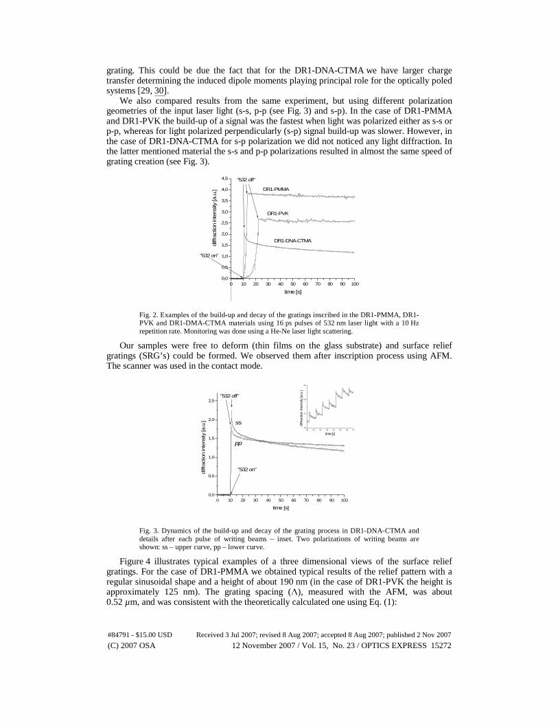
grating. This could be due the fact that for the DR1-DNA-CTMA we have larger charge transfer determining the induced dipole moments playing principal role for the optically poled systems [29, 30].
We also compared results from the same experiment, but using different polarization geometries of the input laser light (s-s, p-p (see Fig. 3) and s-p). In the case of DR1-PMMA and DR1-PVK the build-up of a signal was the fastest when light was polarized either as s-s or p-p, whereas for light polarized perpendicularly (s-p) signal build-up was slower. However, in the case of DR1-DNA-CTMA for s-p polarization we did not noticed any light diffraction. In the latter mentioned material the s-s and p-p polarizations resulted in almost the same speed of grating creation (see Fig. 3).
0 10 20 30 40 50 60 70 80 90 1000,0
0,5
1,0
1,5
2,0
2,5
3,0
3,5
4,0
4,5 "532 off"
"532 on"
DR1-PMMA
DR1-PVK
DR1-DNA-CTMA
diffr
actio
n in
tens
ity [a
.u.]
time [s]
Fig. 2. Examples of the build-up and decay of the gratings inscribed in the DR1-PMMA, DR1-PVK and DR1-DMA-CTMA materials using 16 ps pulses of 532 nm laser light with a 10 Hz repetition rate. Monitoring was done using a He-Ne laser light scattering.
Our samples were free to deform (thin films on the glass substrate) and surface relief gratings (SRG’s) could be formed. We observed them after inscription process using AFM. The scanner was used in the contact mode.
0 10 20 30 40 50 60 70 80 90 1000,0
0,5
1,0
1,5
2,0
2,5
0,0 0,1 0,2 0,3 0,4 0,5 0,6 0,70,0
0,2
0,4
0,6
diff
ract
ion
inte
nsity
[a.u
.]
time [s]
"532 on"
"532 off"
pp
ss
diffr
actio
n in
tens
ity [a
.u.]
time [s]
Fig. 3. Dynamics of the build-up and decay of the grating process in DR1-DNA-CTMA and details after each pulse of writing beams – inset. Two polarizations of writing beams are shown: ss – upper curve, pp – lower curve.
Figure 4 illustrates typical examples of a three dimensional views of the surface relief gratings. For the case of DR1-PMMA we obtained typical results of the relief pattern with a regular sinusoidal shape and a height of about 190 nm (in the case of DR1-PVK the height is approximately 125 nm). The grating spacing (Λ), measured with the AFM, was about 0.52 µm, and was consistent with the theoretically calculated one using Eq. (1):
#84791 - $15.00 USD Received 3 Jul 2007; revised 8 Aug 2007; accepted 8 Aug 2007; published 2 Nov 2007
(C) 2007 OSA 12 November 2007 / Vol. 15, No. 23 / OPTICS EXPRESS 15272

)sin2/( θλ=Λ (1) where λ is the wavelength of the writing beam and 2θ is the angle between the two writing beams [31].
From the Fig. 4 it can be seen that in the case of the PVK matrix the SRG does not have a regular shape, as it is for the case of the PMMA host, and there is no SRG in the case of DR1-DNA-CTMA. However, we did observe different topographies as compared to the surfaces before irradiation (height of irregular structure is in the range about 70 ÷ 160 nm). We propose two explanations for this behavior. Firstly, no SRG is created in the case of DNA-CTMA matrix; or secondly SRG relaxes immediately (see Fig. 2) and, therefore, we have a permanent refractive index and/or absorption coefficient change in the material bulk only (we observe a signal of the first order of diffraction even after few weeks).
Fig. 4. AFM three-dimensional views of surface topography for DR1-PMMA (a), DR1-PVK (b) and DR1-DNA-CTMA (c) after grating experiment.
3. Conclusions
In conclusion we have demonstrated the potential of using DNA-CTMA as a matrix material suitable for simple optical holography. We observed that guest-host system DR1-DNA-CTMA shows faster response to light irradiation than using PMMA or PVK as a host, however, there appears to be no visible formation of a surface relief grating as observed for DR1-PMMA and DR1-PVK. Finally, based on these preliminary results, we believe DR1-DNA-CTMA to be a very promising material in the field of dynamic holography, but further investigation is needed.
Acknowledgments
The authors would like to thank the financial support of European Office for Aerospace Research and Development in London (grant SPC # 064003).
Authors would like to thank Professor Andrzej Miniewicz (Wroclaw University of Technology, Poland) for helpful discussion.
Also, the authors acknowledge the Service Commun d’Imageries et Analyses Microscopiques of Université d’Angers for performing AFM measurements.
(a) (b)
(c)
#84791 - $15.00 USD Received 3 Jul 2007; revised 8 Aug 2007; accepted 8 Aug 2007; published 2 Nov 2007
(C) 2007 OSA 12 November 2007 / Vol. 15, No. 23 / OPTICS EXPRESS 15273
Copyright © 2022 FDOKUMEN