Graphene-Based Materials Prove to Be a Promising ... - MDPI
24
Biomedicines 2022, 10, 73. https://doi.org/10.3390/biomedicines10010073 www.mdpi.com/journal/biomedicines Review Graphene-Based Materials Prove to Be a Promising Candidate for Nerve Regeneration Following Peripheral Nerve Injury Mina Aleemardani 1,† , Pariya Zare 2,† , Amelia Seifalian 3 , Zohreh Bagher 4, * and Alexander M. Seifalian 5, * 1 Biomaterials and Tissue Engineering Group, Department of Materials Science and Engineering, Kroto Research Institute, The University of Sheffield, Sheffield S3 7HQ, UK; [email protected] 2 Department of Chemical Engineering, University of Tehran, Tehran 1417935840, Iran; [email protected] 3 Department of Surgery and Cancer, Imperial College London, London W12 0NN, UK; [email protected] 4 ENT and Head and Neck Research Centre, Hazrat Rasoul Akram Hospital, The Five Senses Health Institute, Iran University of Medical Sciences, Tehran 16844, Iran 5 Nanotechnology and Regenerative Medicine Commercialization Centre (NanoRegMed Ltd.), London BioScience Innovation Centre, London NW1 0NH, UK * Correspondence: [email protected] (Z.B.); [email protected] (A.M.S.); Tel.: +442076911122 (A.M.S.) † These authors contributed equally to this work. Abstract: Peripheral nerve injury is a common medical condition that has a great impact on patient quality of life. Currently, surgical management is considered to be a gold standard first-line treat- ment; however, is often not successful and requires further surgical procedures. Commercially available FDA- and CE- approved decellularized nerve conduits offer considerable benefits to pa- tients suffering from a completely transected nerve but they fail to support neural regeneration in gaps >30 mm. To address this unmet clinical need, current research is focused on biomaterial-based therapies to regenerate dysfunctional neural tissues, specifically damaged peripheral nerve, and spinal cord. Recently, attention has been paid to the capability of graphene-based materials (GBMs) to develop bifunctional scaffolds for promoting nerve regeneration, often via supporting enhanced neural differentiation. The unique features of GBMs have been applied to fabricate an electroactive conductive surface in order to direct stem cells and improve neural proliferation and differentiation. The use of GBMs for nerve tissue engineering (NTE) is considered an emerging technology bringing hope to peripheral nerve injury repair, with some products already in pre- clinical stages. This review assesses the last six years of research in the field of GBMs application in NTE, focusing on the fabrication and effects of GBMs for neurogenesis in various scaffold forms, including electrospun fibres, films, hydrogels, foams, 3D printing, and bioprinting. Keywords: graphene-based materials; nervous system; nerve tissue engineering; nerve proliferation; nerve differentiation; surgery; plastic surgery, regenerative medicine, biomedicine; functionalized graphene oxide; drug delivery; spinal cord injury 1. Introduction Millions of people are suffering from neurodegenerative disorders or acute injuries of the nervous system globally. The nervous system is divided into the central nervous system (CNS) and the peripheral nervous system (PNS). The CNS consists of the spinal cord and brain, and the PNS consists of all of the nerves and ganglia outside of the CNS (Figure 1) [1,2]. The PNS is statistically more susceptible to injury, compared to the CNS, secondary to disease and trauma, leading to neurite damage and neuron loss [3]. Each year, it is estimated that 18 per 100,000 people suffer from peripheral nerve injury (PNI) in developed countries, with the rate thought to be greater in developing countries [4,5]. PNI can permanently impact nervous functions, such as mobility and sensation, as mature neurons are terminally differentiated with no further cell division [6]. Spontaneous axonal regeneration in PNI has been seen in small gaps; however, regenerated nerve function is Citation: Aleemardani, M.; Zare, P.; Seifalian, A.; Bagher, Z.; Seifalian, A.M. Graphene-Based Materials Prove to Be a Promising Candidate for Nerve Regeneration Following Peripheral Nerve Injury. Biomedicines 2022, 10, 73. https://doi.org/10.3390/ biomedicines10010073 Academic Editor: Pietro Gentile Received: 30 November 2021 Accepted: 28 December 2021 Published: 30 December 2021 Publisher’s Note: MDPI stays neutral with regard to jurisdictional claims in published maps and institutional affil- iations. Copyright: © 2021 by the authors. Licensee MDPI, Basel, Switzerland. This article is an open access article distributed under the terms and conditions of the Creative Commons Attribution (CC BY) license (https://creativecommons.org/licenses/ by/4.0/).
-
Upload
khangminh22 -
Category
Documents
-
view
0 -
download
0
Transcript of Graphene-Based Materials Prove to Be a Promising ... - MDPI

Biomedicines 2022, 10, 73. https://doi.org/10.3390/biomedicines10010073 www.mdpi.com/journal/biomedicines
Review
Graphene-Based Materials Prove to Be a Promising Candidate
for Nerve Regeneration Following Peripheral Nerve Injury
Mina Aleemardani 1,†, Pariya Zare 2,†, Amelia Seifalian 3, Zohreh Bagher 4,* and Alexander M. Seifalian 5,*
1 Biomaterials and Tissue Engineering Group, Department of Materials Science and Engineering,
Kroto Research Institute, The University of Sheffield, Sheffield S3 7HQ, UK; [email protected] 2 Department of Chemical Engineering, University of Tehran, Tehran 1417935840, Iran; [email protected] 3 Department of Surgery and Cancer, Imperial College London, London W12 0NN, UK; [email protected] 4 ENT and Head and Neck Research Centre, Hazrat Rasoul Akram Hospital, The Five Senses Health Institute,
Iran University of Medical Sciences, Tehran 16844, Iran 5 Nanotechnology and Regenerative Medicine Commercialization Centre (NanoRegMed Ltd.), London
BioScience Innovation Centre, London NW1 0NH, UK
* Correspondence: [email protected] (Z.B.); [email protected] (A.M.S.); Tel.: +442076911122 (A.M.S.)
† These authors contributed equally to this work.
Abstract: Peripheral nerve injury is a common medical condition that has a great impact on patient
quality of life. Currently, surgical management is considered to be a gold standard first-line treat-
ment; however, is often not successful and requires further surgical procedures. Commercially
available FDA- and CE- approved decellularized nerve conduits offer considerable benefits to pa-
tients suffering from a completely transected nerve but they fail to support neural regeneration in
gaps >30 mm. To address this unmet clinical need, current research is focused on biomaterial-based
therapies to regenerate dysfunctional neural tissues, specifically damaged peripheral nerve, and
spinal cord. Recently, attention has been paid to the capability of graphene-based materials
(GBMs) to develop bifunctional scaffolds for promoting nerve regeneration, often via supporting
enhanced neural differentiation. The unique features of GBMs have been applied to fabricate an
electroactive conductive surface in order to direct stem cells and improve neural proliferation and
differentiation. The use of GBMs for nerve tissue engineering (NTE) is considered an emerging
technology bringing hope to peripheral nerve injury repair, with some products already in pre-
clinical stages. This review assesses the last six years of research in the field of GBMs application
in NTE, focusing on the fabrication and effects of GBMs for neurogenesis in various scaffold forms,
including electrospun fibres, films, hydrogels, foams, 3D printing, and bioprinting.
Keywords: graphene-based materials; nervous system; nerve tissue engineering; nerve
proliferation; nerve differentiation; surgery; plastic surgery, regenerative medicine, biomedicine;
functionalized graphene oxide; drug delivery; spinal cord injury
1. Introduction
Millions of people are suffering from neurodegenerative disorders or acute injuries
of the nervous system globally. The nervous system is divided into the central nervous
system (CNS) and the peripheral nervous system (PNS). The CNS consists of the spinal
cord and brain, and the PNS consists of all of the nerves and ganglia outside of the CNS
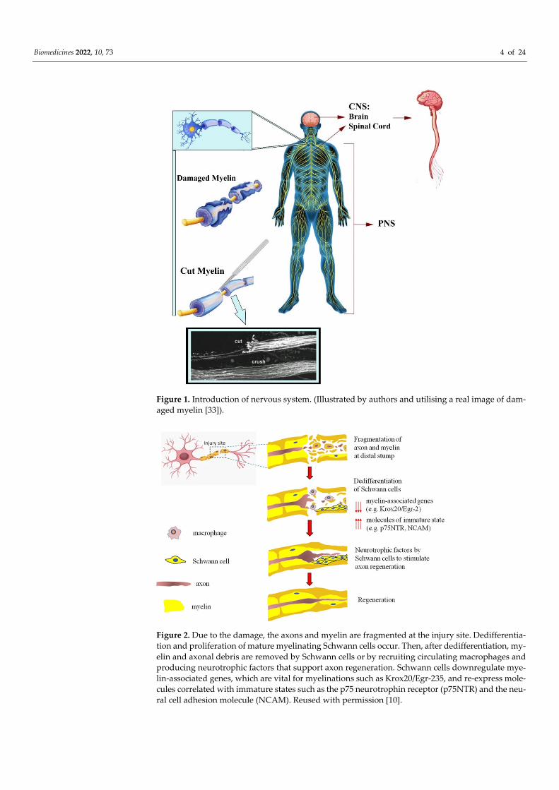
(Figure 1) [1,2]. The PNS is statistically more susceptible to injury, compared to the CNS,
secondary to disease and trauma, leading to neurite damage and neuron loss [3]. Each
year, it is estimated that 18 per 100,000 people suffer from peripheral nerve injury (PNI)
in developed countries, with the rate thought to be greater in developing countries [4,5].
PNI can permanently impact nervous functions, such as mobility and sensation, as mature
neurons are terminally differentiated with no further cell division [6]. Spontaneous axonal
regeneration in PNI has been seen in small gaps; however, regenerated nerve function is
Citation: Aleemardani, M.; Zare, P.;
Seifalian, A.; Bagher, Z.; Seifalian,
A.M. Graphene-Based Materials Prove
to Be a Promising Candidate for
Nerve Regeneration Following
Peripheral Nerve Injury.
Biomedicines 2022, 10, 73.
https://doi.org/10.3390/
biomedicines10010073
Academic Editor: Pietro Gentile
Received: 30 November 2021
Accepted: 28 December 2021
Published: 30 December 2021
Publisher’s Note: MDPI stays neutral
with regard to jurisdictional claims in
published maps and institutional affil-
iations.
Copyright: © 2021 by the authors.
Licensee MDPI, Basel, Switzerland.
This article is an open access article
distributed under the terms and
conditions of the Creative Commons
Attribution (CC BY) license
(https://creativecommons.org/licenses/
by/4.0/).
Biomedicines 2022, 10, 73 2 of 24
restricted, particularly in long-gap injuries. Following injury, the axonal membrane breaks
apart, causing degradation of the myelin sheath and infiltration of macrophages; within
the site of degeneration, the macrophages clear the debris where axonal regeneration
should start. Schwann cells are the main glial cells in the nervous system and the PNS,
providing a pathway for axonal regeneration and remyelination [7]. Schwann cells are
available in PNS and play a critical role when an injury occurs, responding to trauma by
detaching the myelin sheath and phagocytosing debris, encouraging cellular proliferation,
and releasing further cytokines to continue to recruit inflammatory cells to the damaged
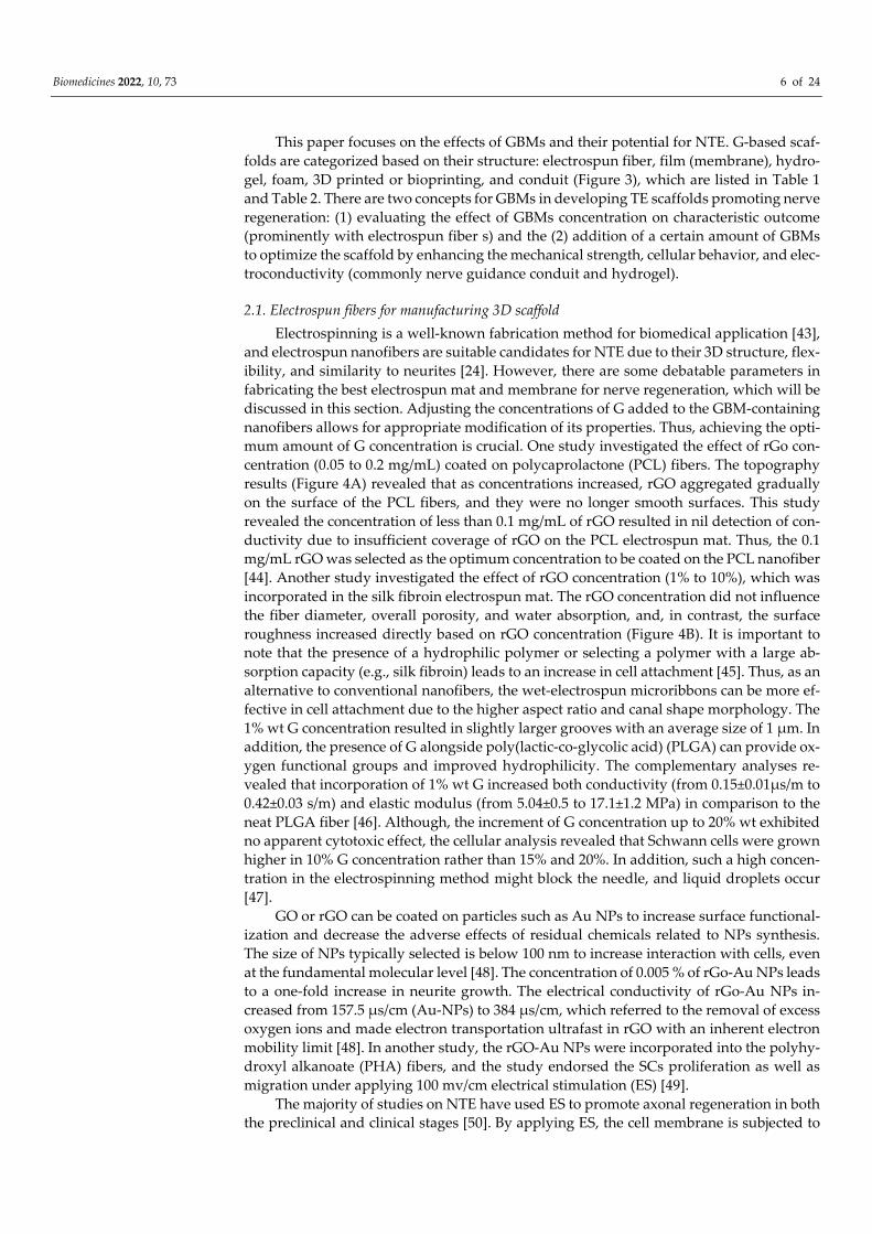
area [8,9]. The mature myelinating Schwann cells dedifferentiate by regaining the expres-
sion pattern related to immature Schwann cells, proliferate, and then redifferentiate,
which induces nerve repair (Figure 2) [10]. On the contrary, due to the lack of Schwann
cells in the CNS, the clinical treatments for CNS injuries are ineffective [1].
There are different types of nerve injury with various factors involved, creating a
challenge against ‘one size fits all’ conventional treatment and promoting precision treat-
ment tailored for the injury suffered by each patient. Several strategies offer potential
treatment for neural disorders, including anti-inflammatory (M2) medications, physio-
therapy, nerve grafting, and rehabilitation. Among these methods, autologous superficial
cutaneous nerves are known as the gold standard for bridging the nerve gap (> 30 mm);
however, there are several drawbacks, including major surgical risks, autologous graft
rejection, infection, donor shortage, and the likelihood of further surgeries [11]. Tissue
engineering (TE) proposes a promising alternative approach to overcome the existing
challenges in tissue regeneration [12–14]. To successfully achieve neural regeneration, var-
ious factors, including topographical, chemical, mechanical, and electrical cues, should be
considered. There have been ongoing attempts to develop biomaterial-based therapies to
regenerate dysfunctional neural tissues, primarily for a damaged PNS and spinal cord
[15,16].
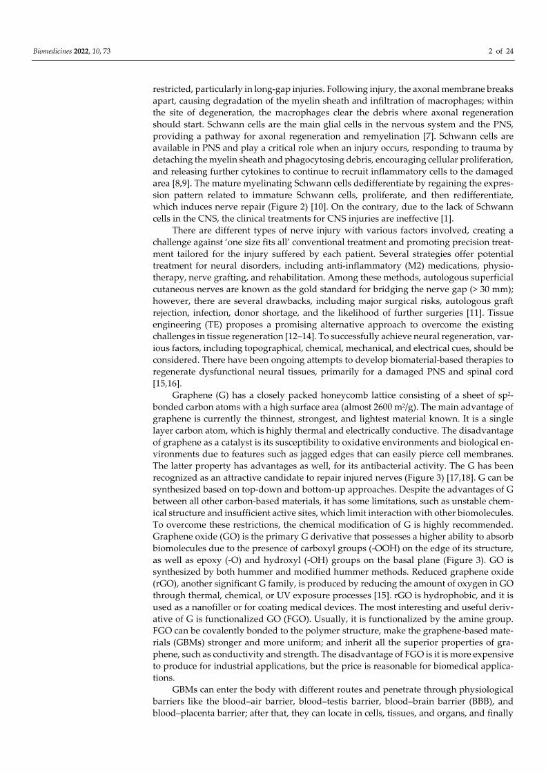
Graphene (G) has a closely packed honeycomb lattice consisting of a sheet of sp2-
bonded carbon atoms with a high surface area (almost 2600 m2/g). The main advantage of
graphene is currently the thinnest, strongest, and lightest material known. It is a single
layer carbon atom, which is highly thermal and electrically conductive. The disadvantage
of graphene as a catalyst is its susceptibility to oxidative environments and biological en-
vironments due to features such as jagged edges that can easily pierce cell membranes.
The latter property has advantages as well, for its antibacterial activity. The G has been
recognized as an attractive candidate to repair injured nerves (Figure 3) [17,18]. G can be
synthesized based on top-down and bottom-up approaches. Despite the advantages of G
between all other carbon-based materials, it has some limitations, such as unstable chem-
ical structure and insufficient active sites, which limit interaction with other biomolecules.
To overcome these restrictions, the chemical modification of G is highly recommended.
Graphene oxide (GO) is the primary G derivative that possesses a higher ability to absorb
biomolecules due to the presence of carboxyl groups (-OOH) on the edge of its structure,
as well as epoxy (-O) and hydroxyl (-OH) groups on the basal plane (Figure 3). GO is
synthesized by both hummer and modified hummer methods. Reduced graphene oxide
(rGO), another significant G family, is produced by reducing the amount of oxygen in GO
through thermal, chemical, or UV exposure processes [15]. rGO is hydrophobic, and it is
used as a nanofiller or for coating medical devices. The most interesting and useful deriv-
ative of G is functionalized GO (FGO). Usually, it is functionalized by the amine group.
FGO can be covalently bonded to the polymer structure, make the graphene-based mate-
rials (GBMs) stronger and more uniform; and inherit all the superior properties of gra-
phene, such as conductivity and strength. The disadvantage of FGO is it is more expensive
to produce for industrial applications, but the price is reasonable for biomedical applica-
tions.
GBMs can enter the body with different routes and penetrate through physiological
barriers like the blood–air barrier, blood–testis barrier, blood–brain barrier (BBB), and
blood–placenta barrier; after that, they can locate in cells, tissues, and organs, and finally

Biomedicines 2022, 10, 73 3 of 24
are excreted. This entrance can result in toxicity and genotoxicity, and various factors af-
fect it, including the lateral size, surface structure, functionalization, charge, impurities,
and aggregations. Additionally, among mentioned different parameters, the interaction
between nano-size GBMs, such as nanoparticles (NPs), nanoflakes and nanosheets, and
biological samples (in vitro, in vivo, and clinical trials) is the most crucial parameter for
toxicity. Generally, it has been represented that smaller nano-size GBMs induced greater
toxicity levels [19,20]. Among several mechanisms underlying GBMs toxicity, the produc-
tion of reactive oxygen species (ROS) within cells can lead to interactions with biomole-
cules (e.g., DNA and RNA) [15,21]. However, GBMs have flexible structures that can be
modified with other substances, including polymers and biomolecules, to enhance their
biocompatibility. GBMs composites have represented an appropriate interaction with
DNA and RNA for sensing and drug delivery approaches [2,22]. The oxygen-based func-
tional groups, GO and rGO, have a lower tendency to form aggregates, even after func-
tionalization, which allows them to cross BBB and improves their stability in blood circu-
lation. Because of this, GBMs can be promising in the development of drug carriers [23,24].
The unique properties of GBMs, such as high biocompatibility, electrical conductivity,
mechanical properties, elasticity, antibacterial properties, and potential surface modifica-
tion, can be applied for nerve tissue engineering (NTE) and regeneration [25–28]. GBMs
enhance interactions between neurons and support neural tissue regeneration by acting
as a bridge between regenerating neurones and retaining electrical conduction between
distal and proximal neurones [29,30].
The application of stem cells is a critical parameter to be considered in NTE [2]. Pre-
vious studies have demonstrated that induced pluripotent stem cells (iPSCs), neural crest
stem cells (NCSCs), and mesenchymal stem cells (MSCs) promote nerve regeneration by
differentiating into Schwann-like cells and secreting neurotrophic factors [1,31]. For ex-
ample, despite low differentiation of adipose-derived stem cells (ADSCs) in clinical stud-
ies, the conversion of neuron-like cells reached about 90% in the GO mat (glass coated
with GO) [32]. GBMs are still new in NTE, and there is a need to explore their full potency
in this field. This paper discusses the different aspects and characteristics of GBMs, re-
viewing the last six years of literature in the application of GBMs in NTE in its various
forms and structures.
Biomedicines 2022, 10, 73 4 of 24
Figure 1. Introduction of nervous system. (Illustrated by authors and utilising a real image of dam-
aged myelin [33]).
Figure 2. Due to the damage, the axons and myelin are fragmented at the injury site. Dedifferentia-
tion and proliferation of mature myelinating Schwann cells occur. Then, after dedifferentiation, my-
elin and axonal debris are removed by Schwann cells or by recruiting circulating macrophages and
producing neurotrophic factors that support axon regeneration. Schwann cells downregulate mye-
lin-associated genes, which are vital for myelinations such as Krox20/Egr-235, and re-express mole-
cules correlated with immature states such as the p75 neurotrophin receptor (p75NTR) and the neu-
ral cell adhesion molecule (NCAM). Reused with permission [10].

Biomedicines 2022, 10, 73 5 of 24
Figure 3. Schematic of graphene (G), graphene oxide (GO), reduced graphene oxide (rGO) and func-
tionalized graphene (FGO) materials in nerve tissue engineering, both in vitro and in vivo, in vari-
ous forms of scaffolds, such as film, electrospun mat, foam or sponge, hydrogel, 3D print, and con-
duit.
2. Tissue-engineered scaffolds with GBM and neural tissues
There are several scaffolds that can be applied to nerve repair and regeneration of the
CNS and PNS. At present, there is more evidence to support the treatment of PNI second-
ary to the PNS high regenerative capacity after damage, in contrast to the CNS [34]. For
NTE to progress, further research is required investigating the optimum parameters to
develop tissue-engineered scaffolds that substitute the grafting procedure. The main chal-
lenges to improve neural regeneration involve bypassing the harsh microenvironment
(e.g., inflammatory, and inhibitory) created following nerve injury. Following trauma or
neurological disease, the myelinated fiber tracts are damaged gradually and the blood
cells cross the broken BBB, invading the medullar tissues that lead to inflammatory re-
sponses. The excitatory neurotransmitters accumulation and inflammatory cytokines
cause formation of an inhibitory extracellular matrix (ECM) [35,36]. Chemical signals,
such as bacterial endotoxins and cytokines, direct macrophage inflammatory (M1) and
M2 phenotypes [37]. M1 promotes cell recruitment and proliferation, while M2 encour-
ages differentiation. The polarization phenotype, switching from M1 to M2, is influential
in promoting functional tissue regeneration [38]. Therefore, TE scaffolds can be applied to
stimulate endogenous nerve migration and control inflammatory cell infiltration.
GBMs have recently gained a lot of attention as promising candidates for the treat-
ment of nerve injury and nervous tissue regeneration. Beneficial properties of GBMs in-
clude supplying electrical conductivity, which enhances mechanical strength and cellular
behavior. The GBMs incorporation has been shown to improve the mechanical properties
of bio-composites; higher concentrations of GBMs result in more suitable mechanical
properties, which are adjustable by concentration for the target tissue [39–41]. In addition,
the hydrophobicity of NTE structures enhances with increasing G amounts, which affects
nerve cell attachment, proliferation, and differentiation. Further research should focus on
ideal concentrations of G to achieve optimal results [15,31,42].
Biomedicines 2022, 10, 73 6 of 24
This paper focuses on the effects of GBMs and their potential for NTE. G-based scaf-
folds are categorized based on their structure: electrospun fiber, film (membrane), hydro-
gel, foam, 3D printed or bioprinting, and conduit (Figure 3), which are listed in Table 1
and Table 2. There are two concepts for GBMs in developing TE scaffolds promoting nerve
regeneration: (1) evaluating the effect of GBMs concentration on characteristic outcome
(prominently with electrospun fiber s) and the (2) addition of a certain amount of GBMs
to optimize the scaffold by enhancing the mechanical strength, cellular behavior, and elec-
troconductivity (commonly nerve guidance conduit and hydrogel).
2.1. Electrospun fibers for manufacturing 3D scaffold
Electrospinning is a well-known fabrication method for biomedical application [43],
and electrospun nanofibers are suitable candidates for NTE due to their 3D structure, flex-
ibility, and similarity to neurites [24]. However, there are some debatable parameters in
fabricating the best electrospun mat and membrane for nerve regeneration, which will be
discussed in this section. Adjusting the concentrations of G added to the GBM-containing
nanofibers allows for appropriate modification of its properties. Thus, achieving the opti-
mum amount of G concentration is crucial. One study investigated the effect of rGo con-
centration (0.05 to 0.2 mg/mL) coated on polycaprolactone (PCL) fibers. The topography
results (Figure 4A) revealed that as concentrations increased, rGO aggregated gradually
on the surface of the PCL fibers, and they were no longer smooth surfaces. This study
revealed the concentration of less than 0.1 mg/mL of rGO resulted in nil detection of con-
ductivity due to insufficient coverage of rGO on the PCL electrospun mat. Thus, the 0.1
mg/mL rGO was selected as the optimum concentration to be coated on the PCL nanofiber
[44]. Another study investigated the effect of rGO concentration (1% to 10%), which was
incorporated in the silk fibroin electrospun mat. The rGO concentration did not influence
the fiber diameter, overall porosity, and water absorption, and, in contrast, the surface
roughness increased directly based on rGO concentration (Figure 4B). It is important to
note that the presence of a hydrophilic polymer or selecting a polymer with a large ab-
sorption capacity (e.g., silk fibroin) leads to an increase in cell attachment [45]. Thus, as an
alternative to conventional nanofibers, the wet-electrospun microribbons can be more ef-
fective in cell attachment due to the higher aspect ratio and canal shape morphology. The
1% wt G concentration resulted in slightly larger grooves with an average size of 1 μm. In
addition, the presence of G alongside poly(lactic-co-glycolic acid) (PLGA) can provide ox-
ygen functional groups and improved hydrophilicity. The complementary analyses re-
vealed that incorporation of 1% wt G increased both conductivity (from 0.15±0.01μs/m to
0.42±0.03 s/m) and elastic modulus (from 5.04±0.5 to 17.1±1.2 MPa) in comparison to the
neat PLGA fiber [46]. Although, the increment of G concentration up to 20% wt exhibited
no apparent cytotoxic effect, the cellular analysis revealed that Schwann cells were grown
higher in 10% G concentration rather than 15% and 20%. In addition, such a high concen-
tration in the electrospinning method might block the needle, and liquid droplets occur
[47].
GO or rGO can be coated on particles such as Au NPs to increase surface functional-
ization and decrease the adverse effects of residual chemicals related to NPs synthesis.
The size of NPs typically selected is below 100 nm to increase interaction with cells, even
at the fundamental molecular level [48]. The concentration of 0.005 % of rGo-Au NPs leads
to a one-fold increase in neurite growth. The electrical conductivity of rGo-Au NPs in-
creased from 157.5 μs/cm (Au-NPs) to 384 μs/cm, which referred to the removal of excess
oxygen ions and made electron transportation ultrafast in rGO with an inherent electron
mobility limit [48]. In another study, the rGO-Au NPs were incorporated into the polyhy-
droxyl alkanoate (PHA) fibers, and the study endorsed the SCs proliferation as well as
migration under applying 100 mv/cm electrical stimulation (ES) [49].
The majority of studies on NTE have used ES to promote axonal regeneration in both
the preclinical and clinical stages [50]. By applying ES, the cell membrane is subjected to
Biomedicines 2022, 10, 73 7 of 24
a degree of depolarization, and significant depolarization can induce cell migration, pro-
liferation, and axonal growth by modulating the distributions of ion channels (Figure 5)
[6,45,51]. It is evident that increasing G concentration increases the scaffold’s conductivity.
Additionally, increasing the voltage up to 100 mv/cm results in more axons of PC12 cells
sprout [7]. Wang et al. demonstrated that the presence of 100 mv/cm causes not only per-
fect in vitro analyzes (Schwann cell migration, proliferation, myelin gene expression, neu-
rotrophin secretion, and induced PC12 cell differentiation) but also promotes peripheral
nerve repair in vivo at a similar level to the gold standard autograft [49]. Although most
studies applied an intensity of 100 mv/cm [7,49,52], less intensity also leads to acceptable
results. For instance, applying ES with an intensity of 50 mv/cm for one h/day through
sodium dodecyl benzenesulfonate (DBS)/GO/polypyrrole-poly-l-lactic acid (PLLA) nano-
fiber can significantly promote neurite elongation and alignment [53]. Another study rep-
resents that significant intensity of ES may damage the neural cells and tissue, thus fabri-
cating electrical responsive scaffolds that can deliver electrical signals to neuron cells and
modulate cellular behavior for developing functional connection is highly recommended
[32,52,54]. Applying a nominal ES intensity of 10 mv (1h/day) alongside a superior con-
ductive scaffold (4% G/thermoplastic polyurethane) with conductivity of 33.45±0.78 S/m
would be an ideal candidate for guiding neural cell growth [32]. However, applying neg-
ligible intensity of ES demonstrated a positive effect on cell proliferation and differentia-
tion; some researchers believe in fabricating self-electroactive nanofibrous scaffolds with-
out requiring ES [45,55].
A number of studies have demonstrated that aligned nano and micro-electrospun
fibers can facilitate cell spreading, avoiding aggregation, and lead to improved cell migra-
tion and proliferation [44,56,57]. Furthermore, the anisotropic characteristic of the aligned
fibrous scaffold can significantly increase the mechanical properties along with cell-scaf-
fold integration, compared with random fibrous orientation [58]. Studies revealed that
aligned electrospun fibers can conduct neonatal nerve capillary growth as well as fiber
orientation to support functional nerve regeneration [46]. This environment is an ideal
candidate for peripheral nerve regeneration due to its morphological resemblance to ax-
ons [44]. Various studies have demonstrated the importance of the similarity of nanofibers
orientation to the ECM [7,27,45,47,48,59–63]. A minority of researchers neglect this param-
eter referring to orientation, which is possibly secondary to a lack of equipment for the
electrospinning of aligned nanofibers. Cell type is a significant parameter for electrospun
mats and is often controversial with selection. In addition to the mentioned stem cells in
the introduction section, in some nanofibrous mats research, cellular analyzes were re-
peated with different cell lines to validate their results. The most frequently used cells are
PC12 and Schwann cells [7,49,52,61]. Subsequently, their results can prove more reliable
for future or even clinical studies.
Like the electrospun fibers or mats, membranes (films) can be helpful in particular
NTE applications. The membranes are usually produced by the solvent casting method,
and the G, GO, or rGO concentration selected is typically between 0.1% and 1.5% wt to
achieve the best results in electrical conductivity, membrane flux, mechanical characteris-
tics, and cellular behavior [64–66]. Another study investigated the long-term biological
characteristics of graphene-based nanoscaffolds (GBN) in the PNS. To this aim, the GBN
was manufactured using a layer-by-layer casting technique. The results indicated that a
low concentration of GBN (4% G in the PCL scaffold) might be biocompatible since it ex-
erts no appreciable toxicity in sensitive tissues such as liver, kidney, lung, heart, and
spleen in the long-term repair (18 months) of peripheral nerves in vivo. The manufactured
scaffold had biologically regenerative effects on myelination, axonal outgrowth, and lo-
comotor function recovery [67].

Biomedicines 2022, 10, 73 8 of 24
Figure 4. (A) and (B) exhibiting the morphology of electrospun mats by increasing the GBMs con-
centration. Reused with permission from [44,45].
Figure 5. The schematic illustration of ES effect on neural injury regeneration (A) Injured neuron
without the conductive platform and electrical stimulation, (B) Injured neuron exposed conductive
platform and electrical stimulation. Reused with permission from [6].
2.2. Hydrogels
Hydrogels are 3D networks containing crosslinked hydrophilic polymer chains that
can absorb a large amount of water; it is this characteristic that allows them to mimic the
natural ECM [68]. Hydrogels are remarkably promising for application of the sustained
delivery of biomolecules and can be directly implanted defect site, as well as for TE appli-
cations [12]. Hydrogels are the most promising choice for the delivery of genes or cyto-
kines for nerve proliferation and differentiation. The addition of GBMs to hydrogels has

Biomedicines 2022, 10, 73 9 of 24
gained recent attention due to its desired resultant properties: (1) higher stability and con-
trolled delivery of molecules, GO in particular; (2) overcoming the notable weak point of
hydrogels, and low mechanical properties, by acting as a capable reinforcement; and (3)
creating conductive hydrogels that are highly useful for mimicking the natural environ-
ment in the nervous tissues or organs [69–73].
There are different aspects to investigate the effects of GBMs on hydrogels for NTE,
such as the effect of concentration, pore size, morphology, chemistry (related to the func-
tional groups), mechanical strength (elastic modulus in particular), and electrical conduc-
tivity [69–73]. GBMs are a candidate as a carrier for gene delivery and cell transfection,
which can be better controlled and sustained within hydrogels [58,63]. In a study focusing
on promoting BMSCs’ recruitment and stimulating sensory nerve regeneration, the stro-
mal cell-derived factor-1α (SDF-1α), a member of the chemokine family of pro-inflamma-
tory mediators, and pDNAs were used. To this aim, 25 kDa polyethylenimine (PEI) was
conjugated to nanoscale GO sheets to deliver pDNAs encoding bFGF (GO-PEI-bFGF) and
crosslinked by matrix metalloproteinase (MMP)-2. The crosslinking of GO-PEI sheets
keeps the DNA inside to prevent degradation and induce the responsive BMSCs transfec-
tion. This gene delivery system was encapsulated by a GO-based hydrogel that contains
SDF-1α. SDF-1α can recruit distant endogenous BMSCs, and MMP-2 activated the hydrol-
ysis of crosslinked GO-PEI and then started the BMSCs transfection towards the neural-
like cells (Figure 6A). Using GO resulted in several improvements, such as stable biomol-
ecule delivery and release in a controlled and sustained way. Although there are still some
concerns regarding GO biocompatibility, this study confirmed the dose-dependent cyto-
toxicity of nano (significantly >10 μg/mL) and microscale (even noticeable at 1 μg/mL)
GO. Conjugating the cationic PEI to the GO surface improved GO biocompatibility re-
markably (GO-PEI 1: 10 was the optimum) [69]. Gene delivery with the help of GBMs
promises hope by maximizing therapeutic efficacy, the substantial factor to consider for
delivery efficiency with gene transfection, and according to the reports; this type of engi-
neered delivery system can introduce treatments for CNS diseases [69,74].
To evaluate the impact of GBMs on the electrical conductivity of hydrogels, GBMs
can be applied solely, with a combination of other conductive biomaterials or even with
the addition of electric charges, to result in an appropriate range of conductivity. Intro-
ducing electric charges is a strategic method to stimulate the proliferation and differenti-
ation of nerve cells [72,75,76]. This is because excitable cells rely significantly on electrical
conductivity, and ion accumulation and flow, to coordinate cellular functions and signal
transduction [75,77]. In order to form a positively charged hydrogel, oligo(poly(ethylene
glycol) fumarate) (OPF) was crosslinked with [2-(methacryloyloxy)ethyl]trimethylammo-
nium chloride (MTAC). The results have shown enhanced neural cell adhesion, prolifer-
ation, and differentiation. Furthermore, from the dissociated embryonic chick dorsal root
ganglion (DRG) explant, it can be said that OPF hydrogels resulted in a combination of
neurons, neuronal support cells, and Schwann cells [75]. A study depicted that the intro-
duction of positive surface charges (OPF and OPF-MTAC hydrogels) to conductive car-
bon components (GO and carbon nanotube, CNT) can remarkably stimulate nerve cell
responses [72].
Using anti-inflammatory drug is an alternative approach to enable nerve regenera-
tion that can be combined with loading GBMs. A hydrogel was engineered using GO
sheets with four-armed polyethylene glycol and functionalized with diacerein as an M2
drug (4arm-PEG-diacerein or PD). The designed hydrogel was injectable and self-recov-
ery due to the strong physical interactions between GO and diacerein. The range of con-
ductivity of the PD/GO hydrogel was consistent with suitable conductivity for NTE ma-
terials [78,79]. It has been reported that a suitable range of electric conductivities is 1–10 S
m−1, which results in neuron growth, longer neurite length, faster neurite growth rate, and
better axon remyelination [78,79]. Further, PD/GO hydrogel provided an anti-inflamma-
tory microenvironment; synapses appeared around the cells. Therefore, this hydrogel can
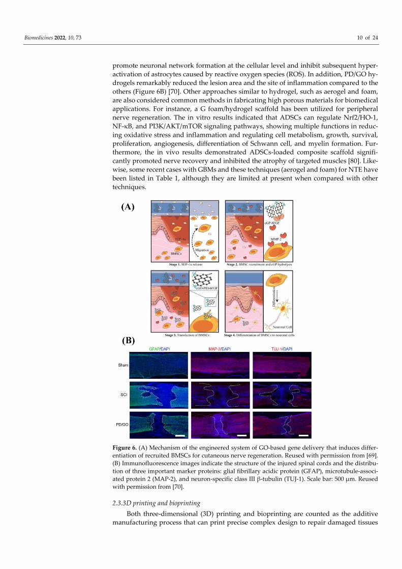
Biomedicines 2022, 10, 73 10 of 24
promote neuronal network formation at the cellular level and inhibit subsequent hyper-
activation of astrocytes caused by reactive oxygen species (ROS). In addition, PD/GO hy-
drogels remarkably reduced the lesion area and the site of inflammation compared to the
others (Figure 6B) [70]. Other approaches similar to hydrogel, such as aerogel and foam,
are also considered common methods in fabricating high porous materials for biomedical
applications. For instance, a G foam/hydrogel scaffold has been utilized for peripheral
nerve regeneration. The in vitro results indicated that ADSCs can regulate Nrf2/HO-1,
NF-κB, and PI3K/AKT/mTOR signaling pathways, showing multiple functions in reduc-
ing oxidative stress and inflammation and regulating cell metabolism, growth, survival,
proliferation, angiogenesis, differentiation of Schwann cell, and myelin formation. Fur-
thermore, the in vivo results demonstrated ADSCs-loaded composite scaffold signifi-
cantly promoted nerve recovery and inhibited the atrophy of targeted muscles [80]. Like-
wise, some recent cases with GBMs and these techniques (aerogel and foam) for NTE have
been listed in Table 1, although they are limited at present when compared with other
techniques.
Figure 6. (A) Mechanism of the engineered system of GO-based gene delivery that induces differ-
entiation of recruited BMSCs for cutaneous nerve regeneration. Reused with permission from [69].
(B) Immunofluorescence images indicate the structure of the injured spinal cords and the distribu-
tion of three important marker proteins: glial fibrillary acidic protein (GFAP), microtubule-associ-
ated protein 2 (MAP-2), and neuron-specific class III β-tubulin (TUJ-1). Scale bar: 500 μm. Reused
with permission from [70].
2.3.3D printing and bioprinting
Both three-dimensional (3D) printing and bioprinting are counted as the additive
manufacturing process that can print precise complex design to repair damaged tissues
Biomedicines 2022, 10, 73 11 of 24
or organs; however, the main difference between these two methods is that 3D bioprinting
builds customized structures from cells and supporting biomaterials (bioink), while 3D
printing solely uses biomaterials for printing [12]. Although there is a high volume of re-
search on 3D printing and bioprinting, the literature on their applications in NTE is still
relatively limited. The addition of GBMs can lead to a higher viscosity (due to the shear
thinning properties), hence improving printability. In addition, water-dispersible GBMs
can also be used as a component of bioink; however, the concentration plays a significant
role and therefore needs careful evaluation [15,81–83]. A study was performed using wa-
terborne biodegradable polyurethane with soft segments that mainly included poly(ε-ca-
prolactone) and 20 mol% of shorter poly(D,L-lactide) chains. The polyurethane dispersed
(25 ppm) in a cell culture medium then underwent a sol-gel transition near 37 °C with a
proper gel modulus. Afterwards, G or GO was mixed with polyurethane to prepare a G-
based bioink for neural stem cell printing, which resulted in a suitable bioink for printing
and cell survival (Figure 7). Interestingly, the addition of G or GO at a very low content
(25 ppm) not only resulted in a promising bioink but also significantly improved oxygen
metabolism (2- to 4-fold increment) and neural differentiation. On the basis of these re-
sults, it can be concluded that the optimum sample was PU/G since it presented better
efficacy, especially for cell proliferation and differentiation and oxygen metabolism [69].
It is worth mentioning that in order to achieve these precise conduits, 3D printing can be
applied alongside the other techniques, and the related studies are given in the conduit
section [25,28,83]. Table 1, highlight the recent studies in application of graphene based
materials for nerve regeneration.
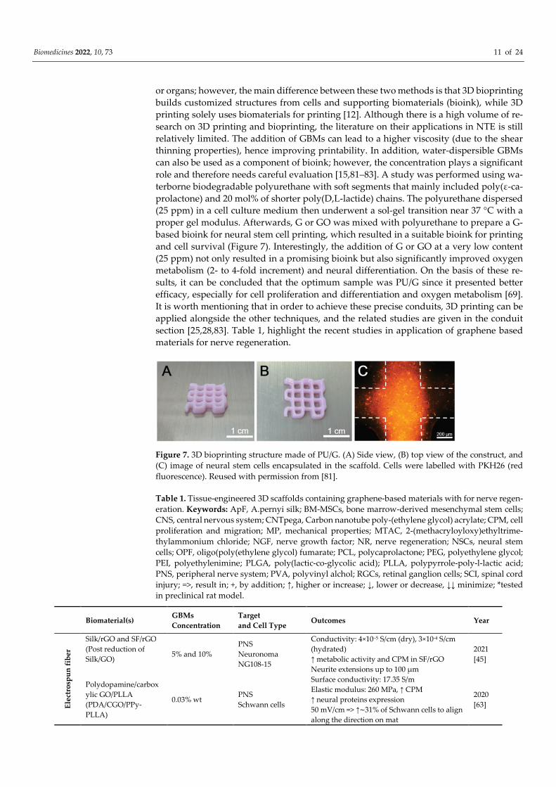
Figure 7. 3D bioprinting structure made of PU/G. (A) Side view, (B) top view of the construct, and
(C) image of neural stem cells encapsulated in the scaffold. Cells were labelled with PKH26 (red
fluorescence). Reused with permission from [81].
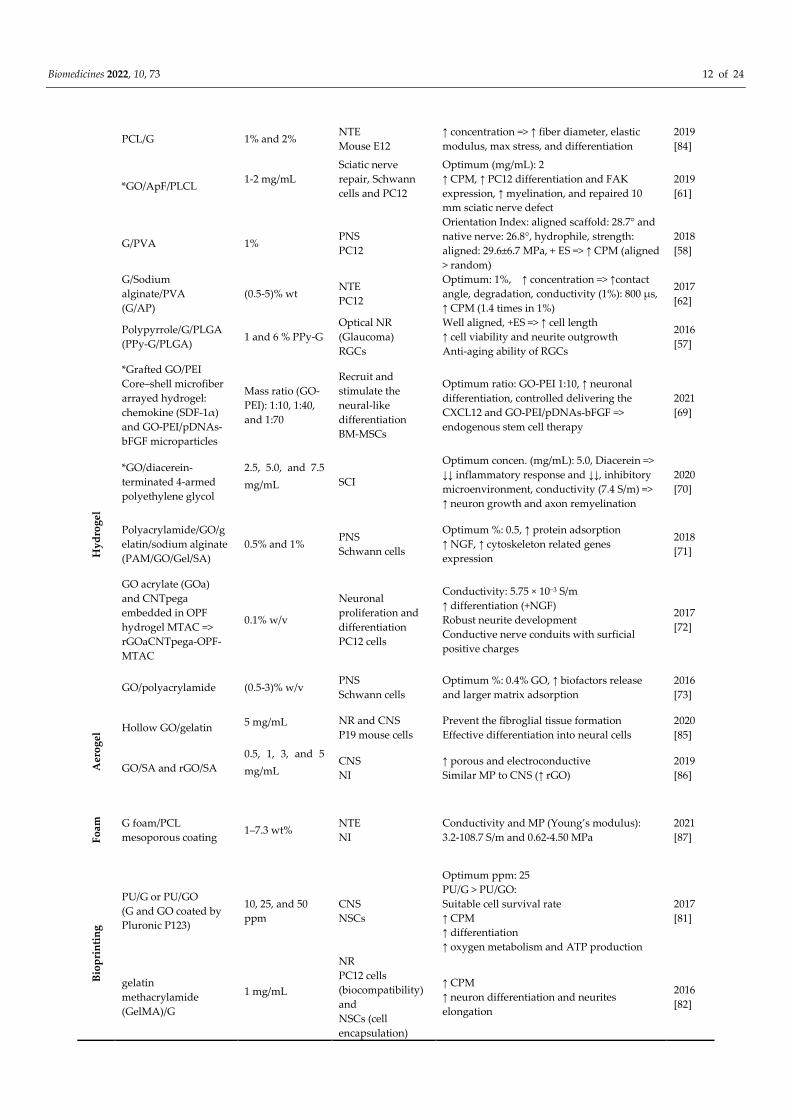
Table 1. Tissue-engineered 3D scaffolds containing graphene-based materials with for nerve regen-
eration. Keywords: ApF, A.pernyi silk; BM-MSCs, bone marrow-derived mesenchymal stem cells;
CNS, central nervous system; CNTpega, Carbon nanotube poly-(ethylene glycol) acrylate; CPM, cell
proliferation and migration; MP, mechanical properties; MTAC, 2-(methacryloyloxy)ethyltrime-
thylammonium chloride; NGF, nerve growth factor; NR, nerve regeneration; NSCs, neural stem
cells; OPF, oligo(poly(ethylene glycol) fumarate; PCL, polycaprolactone; PEG, polyethylene glycol;
PEI, polyethylenimine; PLGA, poly(lactic-co-glycolic acid); PLLA, polypyrrole-poly-l-lactic acid;
PNS, peripheral nerve system; PVA, polyvinyl alchol; RGCs, retinal ganglion cells; SCI, spinal cord
injury; =>, result in; +, by addition; ↑, higher or increase; ↓, lower or decrease, ↓↓ minimize; *tested
in preclinical rat model.
Biomaterial(s) GBMs
Concentration
Target
and Cell Type Outcomes Year
Ele
ctro
spu
n f
ibe
r
Silk/rGO and SF/rGO
(Post reduction of
Silk/GO)
5% and 10%
PNS
Neuronoma
NG108-15
Conductivity: 4×10⁻⁵ S/cm (dry), 3×10⁻⁴ S/cm
(hydrated)
↑ metabolic activity and CPM in SF/rGO
Neurite extensions up to 100 μm
2021
[45]
Polydopamine/carbox
ylic GO/PLLA
(PDA/CGO/PPy-
PLLA)
0.03% wt PNS
Schwann cells
Surface conductivity: 17.35 S/m
Elastic modulus: 260 MPa, ↑ CPM
↑ neural proteins expression
50 mV/cm => ↑∼31% of Schwann cells to align
along the direction on mat
2020
[63]
Biomedicines 2022, 10, 73 12 of 24
PCL/G 1% and 2% NTE
Mouse E12
↑ concentration => ↑ fiber diameter, elastic
modulus, max stress, and differentiation
2019
[84]
*GO/ApF/PLCL 1-2 mg/mL
Sciatic nerve
repair, Schwann
cells and PC12
Optimum (mg/mL): 2
↑ CPM, ↑ PC12 differentiation and FAK
expression, ↑ myelination, and repaired 10
mm sciatic nerve defect
2019
[61]
G/PVA 1% PNS
PC12
Orientation Index: aligned scaffold: 28.7° and
native nerve: 26.8°, hydrophile, strength:
aligned: 29.6±6.7 MPa, + ES => ↑ CPM (aligned
> random)
2018
[58]
G/Sodium
alginate/PVA
(G/AP)
(0.5-5)% wt NTE
PC12
Optimum: 1%, ↑ concentration => ↑contact
angle, degradation, conductivity (1%): 800 μs,
↑ CPM (1.4 times in 1%)
2017
[62]
Polypyrrole/G/PLGA
(PPy-G/PLGA) 1 and 6 % PPy-G
Optical NR
(Glaucoma)
RGCs
Well aligned, +ES => ↑ cell length
↑ cell viability and neurite outgrowth
Anti-aging ability of RGCs
2016
[57]
Hy
dro
ge
l
*Grafted GO/PEI
Core–shell microfiber
arrayed hydrogel:
chemokine (SDF-1α)
and GO-PEI/pDNAs-
bFGF microparticles
Mass ratio (GO-
PEI): 1:10, 1:40,
and 1:70
Recruit and
stimulate the
neural-like
differentiation
BM-MSCs
Optimum ratio: GO-PEI 1:10, ↑ neuronal
differentiation, controlled delivering the
CXCL12 and GO-PEI/pDNAs-bFGF =>
endogenous stem cell therapy
2021
[69]
*GO/diacerein-
terminated 4-armed
polyethylene glycol
2.5, 5.0, and 7.5
mg/mL
SCI
Optimum concen. (mg/mL): 5.0, Diacerein =>
↓↓ inflammatory response and ↓↓, inhibitory
microenvironment, conductivity (7.4 S/m) =>
↑ neuron growth and axon remyelination
2020
[70]
Polyacrylamide/GO/g
elatin/sodium alginate
(PAM/GO/Gel/SA)
0.5% and 1% PNS
Schwann cells
Optimum %: 0.5, ↑ protein adsorption
↑ NGF, ↑ cytoskeleton related genes
expression
2018
[71]
GO acrylate (GOa)
and CNTpega
embedded in OPF
hydrogel MTAC =>
rGOaCNTpega-OPF-
MTAC
0.1% w/v
Neuronal
proliferation and
differentiation
PC12 cells
Conductivity: 5.75 × 10⁻³ S/m
↑ differentiation (+NGF)
Robust neurite development
Conductive nerve conduits with surficial
positive charges
2017
[72]
GO/polyacrylamide (0.5-3)% w/v PNS
Schwann cells
Optimum %: 0.4% GO, ↑ biofactors release
and larger matrix adsorption
2016
[73]
Aer
og
el Hollow GO/gelatin
5 mg/mL
NR and CNS
P19 mouse cells
Prevent the fibroglial tissue formation
Effective differentiation into neural cells
2020
[85]
GO/SA and rGO/SA
0.5, 1, 3, and 5
mg/mL
CNS
NI
↑ porous and electroconductive
Similar MP to CNS (↑ rGO)
2019
[86]
Fo
am
G foam/PCL
mesoporous coating 1–7.3 wt%
NTE
NI
Conductivity and MP (Young’s modulus):
3.2-108.7 S/m and 0.62-4.50 MPa
2021
[87]
Bio
pri
nti
ng
PU/G or PU/GO
(G and GO coated by
Pluronic P123)
10, 25, and 50
ppm
CNS
NSCs
Optimum ppm: 25
PU/G > PU/GO:
Suitable cell survival rate
↑ CPM
↑ differentiation
↑ oxygen metabolism and ATP production
2017
[81]
gelatin
methacrylamide
(GelMA)/G
1 mg/mL
NR
PC12 cells
(biocompatibility)
and
NSCs (cell
encapsulation)
↑ CPM
↑ neuron differentiation and neurites
elongation
2016
[82]
Biomedicines 2022, 10, 73 13 of 24
2.4. Conduits for nerve guide
When the direct end-to-end tensionless repair for the injured nerve is not possible
(>30 mm gap), the first choice for management would be to perform autologous nerve
grafting [88]. Autologous nerve grafting is considered to be the gold standard treatment
for PNI; however, the procedure has multiple complications such as a shortage of donors,
donor-site morbidity (e.g., sensory loss and neuroma formation), and disease transmis-
sion [89]. The development of a polymer-based nerve guidance conduit (NGC) has made
considerable progress to encourage nerve repair and regeneration in a targeted manner
with their physical and chemical features; however, there is still a gap to achieve structural
and functional restoration that mimics the natural properties. Current research supports
the use of NGCs to repair large nerve defects, particularly in the PNS [90].
NGCs are used to bridge the nerve stumps and eliminate scar formation, help the
axon stretch, and form a suitable microenvironment to regenerate the injured nerve [91].
NGCs require great mechanical strength, as well as adequate elasticity to allow regular
muscle motions around the conduit without tube collapsing [72]. The recovery is ex-
tremely effective as nerve extension is hampered through the generation of scar tissue
during the repair process [92,93]. Injuries related to the PNS are common (13−23 out of
100,000 individuals are effect annually in developed countries [94]); so, research has been
focused on developing appropriate NGCs through several techniques to improve treat-
ment. To meet the various needs of a suitable NGC for nerve regeneration, “multifunc-
tional NGCs” are needed; hence, multiple biomaterials, biomolecules, and fabrication
techniques have been used in most case studies [25,59,93,95,96].
The properties of GBMs mean that they are promising biomaterials for the successful
fabrication of NGC. One successful example includes an NGC synthesized by a 3D gra-
phene mesh tube (GMT) and subsequently filled with alginate-gelatinmethacryloyl
(GelMA) hydrogel, which was also evaluated by animal studies (Sprague–Dawley rats).
The addition of alginate enhances the mechanical properties through a double network
structure and supports tube formation. To enable axonal guidance and neurons migration,
Netrin-1 (100 ng/mL) has been loaded (Figure 8A) [95]. The presence of G not only resulted
in the enhanced proliferation of Schwann cells and guided their alignments but also led
to a satisfactory Young’s modulus (725.8 ± 46.52 kPa) and electrical conductivity (6.8 ± 0.85
S/m). The release of netrin-1 was significant in directing axon pathfinding and neuronal
migration, which optimized tube formation ability at 100 ng/mL. In vivo studies showed
that the NGC successfully supported peripheral nerve regeneration, restored denervated
muscle, and was superior to the positive control (autologous graft). Additionally, the re-
vascularization of denervated muscle was achieved, which is a crucial factor for regener-
ation and recovery after PNI. Another achievement of this study was enhancing the sur-
vival and function of Schwann cells both in vitro and in vivo. The extraction and cultiva-
tion of Schwann cells is complex and inconvenient, so many studies concentrate on stem
cells such as adipose-derived stem cells (ADSCs) that can differentiate into Schwann-like,
neuron-like, and endothelial cells [95].
A further study utilized conductive hydrogel-based NGCs, made of GelMA/GO and
followed by chemical reduction, to improve the electrical properties of the hydrogel,
r(GO/GelMA) [97]. This is because restoring sp2-carbon bonds while minimizing rGO ag-
gregation can enhance the electrical characteristics of GO-loaded hydrogels [15]. The fab-
ricated NGCs had adequate electrical conductivity (r(GO/GelMA)>GO/GelMA), flexibil-
ity, mechanical strength, and permeability, and r(GO/GelMA) showed higher neuritogen-
esis enhancement of PC12 neuronal cells compared to previous studies. Muscle regenera-
tion after NGC implantation was investigated by Sprague–Dawley rats by calculating the
weight of the gastrocnemius muscles (both left and right). The left gastrocnemius muscle
showed serious atrophy at 4 weeks, and the GelMA group underwent considerable
shrinkage by 8 weeks. However, the other groups did not display atrophy and shrinkage;
instead, they exhibited muscle recovery, particularly the r(GO/GelMA) and autograft (Fig-
ure 8B) [97].

Biomedicines 2022, 10, 73 14 of 24
In order to optimize and evaluate the G concentration, a collagen-based NGC based
was prepared using the cryogel technique to regenerate the spinal cord. The cryogels were
stabilized by using amine-functionalized graphene as a nano-crosslinker and resulted in
super-macroporous cryogel (Figure 9A). With the addition of 0.1%, 0.5%, and 1% w/v of
G, an upward trend in electrical conductivity was recorded; however, there was no further
increment despite enhancing the concentration above 1% w/v, so this was found to be the
plateau for the greatest amount of conductivity. The differentiation of rat bone marrow-
derived mesenchymal stem cells (BM-MSCs) into neuronal-like cells has been demon-
strated in the presence of graphene and ES. Based on the cell studies and organotypic
spinal explants on samples, optimum neuronal differentiation was seen with 0.5% w/v
G/collagen cryogels. Research has shown that BM-MSCs-seeded cryogels were able to se-
crete ATP energy upon ES. 0.5% w/v crosslinked collagen cryogels supplied adequate me-
chanical and electrical cues that encouraged the significant extracellular secretion of ATP.
The cryogels have also shown that having a mixed population of M1 and M2 macrophages
is necessary for nerve tissue repair [98].
The introduction of micropatterns and bioactive substances into the inner wall of
NGCs play a significant role in regulating Schwann cells behavior, axons elongation, and
macrophages phenotype. Linear micropatterns with various ridges and grooves (3/3, 5/5,
10/10 and 30/30 μm) were made on poly(D,L-lactide-co-caprolactone) (PLCL) films; fol-
lowing this, surface aminolysis and GO electrostatic adsorption were conducted (Figure
9B). GO has been used to provide enhanced cell attachment and proliferation, as well as
electrical conductivity, in turn improving guiding. The GO-modified micropatterns accel-
erated the collective migration of Schwann cells and directed cells along with stripes by
the fastest rate on the 3/3-GO film that contains the largest force of cell adhesion. It also
resulted in tending macrophages to differentiate into the M2 type on the 3/3-GO film (op-
timum NGC). The NGCs were implanted to bridge the 10 mm rat sciatic nerve defects for
4 and 8 weeks. To investigate function recovery of regenerated sciatic nerves, at 4- and 8-
weeks post-operation, nerve conduction velocity and compound motor action potential
were measured. The 3/3-GO group represented higher values than other groups. Addi-
tionally, in the 3/3-GO group, myelinated nerve fibers and blood vessels were generated
more significantly than others after 8 weeks [99].

Biomedicines 2022, 10, 73 15 of 24
Figure 8. (A) Schematic of netrin-1-loaded GMT/hydrogel conduit preparation. (a, b) Growing G
onto a nickel mesh (CVD method), (c) covering G/nickel mesh with a precursor solution, (d) for-
mation of strong ionic bonds between alginate and Ca2+ ions due to immersing GMT into CaCl2
solution, (e, f) polymerization of GelMA under UV light, (g) etching nickel template, and (h) Im-
mersing the conduit in high concentrated netrin-1 solution. Reused with permission from [95]. (B)
Images of regeneration of muscle after NGC implantation in different groups. Reused with permis-
sion from [97].
Biomedicines 2022, 10, 73 16 of 24
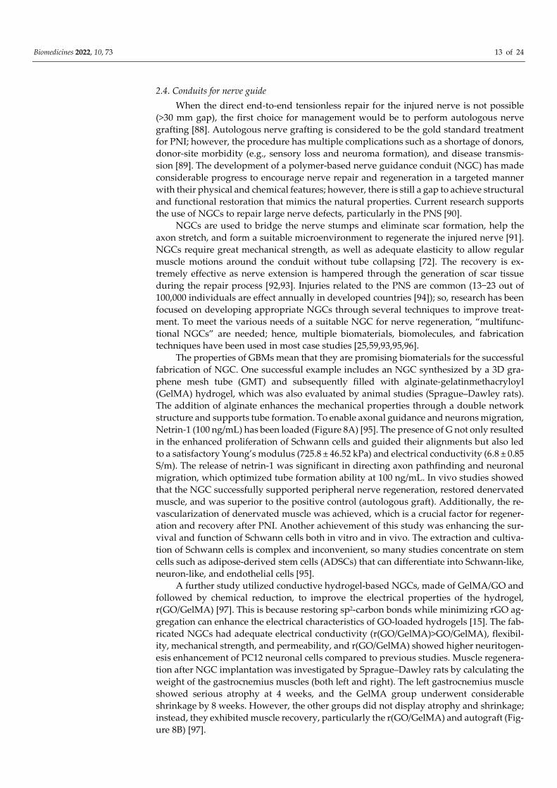
Figure 9. (A) Schematic of projected collagen graphene cryogel mechanism: due to spinal cord in-
jury, inflammatory cytokines and infiltration of inflammatory cells have been produced. By implant-
ing the cryogels, it will promote proliferation and stemness maintenance of BM-MSCs and secrete
anti-inflammatory biomolecules. Further, the presence of cryogels and macrophage infiltration will
stimulate high polarization of the M2/M1 phenotype. Reused with permission from [98]. (B) The
illustration depicts the PLCL film fabrication with stripe micropatterns and GO nanosheets and its
use in four steps: (1) creating micropatterns by thermal pressing of a polydimethylsiloxane template
onto a PLCL film, (2) aminolyzing by 1,6-hexanediamine then GO adsorption electrostatically, (3)
manufacturing micropatterned PLCL/GO conduit, and (4) implanting into a rat with sciatic nerve
defects. The middle schematic shows that the micropatterned PLCL/GO film can improve the direc-
tional migration of Schwann cells from their cell spheroids, induce the macrophages differentiation
into M2 type, and guide the neurites of N2a cells along with the patterns. Reused with permission
from [99].
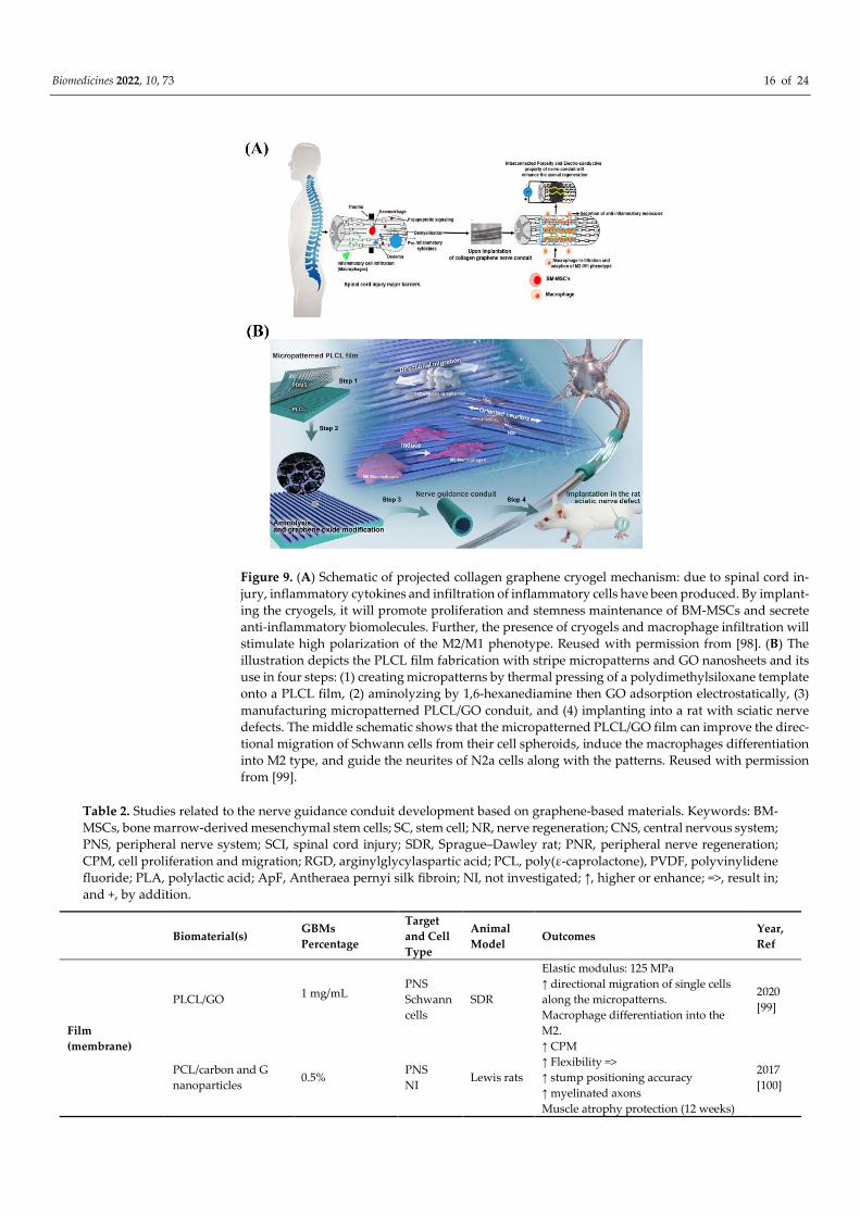
Table 2. Studies related to the nerve guidance conduit development based on graphene-based materials. Keywords: BM-
MSCs, bone marrow-derived mesenchymal stem cells; SC, stem cell; NR, nerve regeneration; CNS, central nervous system;
PNS, peripheral nerve system; SCI, spinal cord injury; SDR, Sprague–Dawley rat; PNR, peripheral nerve regeneration;
CPM, cell proliferation and migration; RGD, arginylglycylaspartic acid; PCL, poly(ε-caprolactone), PVDF, polyvinylidene
fluoride; PLA, polylactic acid; ApF, Antheraea pernyi silk fibroin; NI, not investigated; ↑, higher or enhance; =>, result in;
and +, by addition.
Biomaterial(s) GBMs
Percentage
Target
and Cell
Type
Animal
Model Outcomes
Year,
Ref
Film
(membrane)
PLCL/GO 1 mg/mL
PNS
Schwann
cells
SDR
Elastic modulus: 125 MPa
↑ directional migration of single cells
along the micropatterns.
Macrophage differentiation into the
M2.
2020
[99]
PCL/carbon and G
nanoparticles 0.5%
PNS
NI Lewis rats
↑ CPM
↑ Flexibility =>
↑ stump positioning accuracy
↑ myelinated axons
Muscle atrophy protection (12 weeks)
2017
[100]

Biomedicines 2022, 10, 73 17 of 24
Electrospun
fiber
PCL/collagen/G 0.5%, 1%, 1.5%,
and 2%
Sciatic
nerve
repair
MSCs
SDR
Well aligned
Optimum %: 1
↑ concentration => ↑ conductivity;
Conductivity (1%): 5.27×10⁻⁶ S/m
↑ concentration => ↓ tensile strength
and elastic modulus
2020
[54]
Dual-electrospun:
PCL, gelatin, and
polyaniline/G (PAG)
0-3% wt
PNS
BM-
MSCs
NI
Optimum: 2% PAG =>
↑ CPM
Conductivity:
10.8 × 10⁻⁵ S/cm
2020
[101]
Foam
G/PCL 2% wt
PNS
PC12
cells
NI
Elastic modulus: 2.67 MPa
Conductivity: 25 S/m
↑ porosity
↑ cell proliferation and extension
No cytotoxicity
2021
[102]
PVDF/GO 0.5%, 1%, 3%,
and 5% wt
PNS
PC12
cells
NI
↑ piezoelectricity and electrical
conductivity
High flexibility => easy and
appropriate NGC formation
↑ CPM (particularly 0.5% and 1%)
2019
[103]
Hydrogel
GMT/ hydrogel with
netrin-1 0.05%
PNS
Schwann
cells
SDR
Elastic modulus: 720 kPa
Conductivity: 6.8 S/m
GMT => orientation of PNR, O2 and
nutrition transport.
↑ levels of S100 and Sox10 (↑PNR).
2021
[95]
GO/GelMA then
chemically reduced
=>
r(GO/GelMA)
0.1% PC12
cells SDR
Conductivity: 4.4 × 10⁻³ S/m
(GO/GelMA) and 8.7 × 10⁻³ S/m
(rGO/GelMA)
↑ neuritogenesis: r(GO/GelMA) >
GO/GelMA
↑ PNR
↑ regrowth with myelination
2020
[97]
Chitosan/oxidized
hydroxyethyl
cellulose (CS/OHEC)
hydrogel loaded with
asiaticoside liposome
and rGO
0%, 1%, 2%, 4%,
6%, 8%, and 10%
PNS
PC12
cells
NI
Optimum: 8%
Conductivity (5.27 × 10⁻⁴ S/cm) + ES =>
↑ differentiation and ↑ CPM.
Asiaticoside released => no growth
and collagen secretion of fibroblasts =>
No scars for NR.
2020
[93]
Cryogel
Amino-
functionalized
G/collagen
0.1%, 0.5%, and
1% w/v
SCI
BM-
MSCs
Organoty
pic spinal
explant
culture
(spinal
cord from
SDR)
Optimum %: 0.5%
Conductivity: 3.8 × 10⁻³ S/cm and
mechanical cues: 100–347 kPa Young
Modulus => SC and NR
↑ ATP secretion
↑ MAP-2 kinase and β-tubulin III
expression
↑ CD90 and CD73 gene expression
2021
[98]
Multiple
techniques
Electrospinning,
molding, and freeze
drying:
ApF/PLCL/GO
2%
PNS
Schwann
cells
and
PC12
cells
(different
iation)
SDR
Effective guiding interface => ↑ CPM
and ↑ myelination
Tailored degradation and complete
degradation at 12 weeks
↑ axonal regrowth and remyelination
2020
[96]
Aligned electrospun
and film:
carboxylic GO-
polypyrrole/poly-L-
0.05% w/v
PNS
PC12 and
L929
SDR
↑ CPM
Conductivity: ~4.6 S/cm (after 4 weeks
of immersion)
2019
[104]
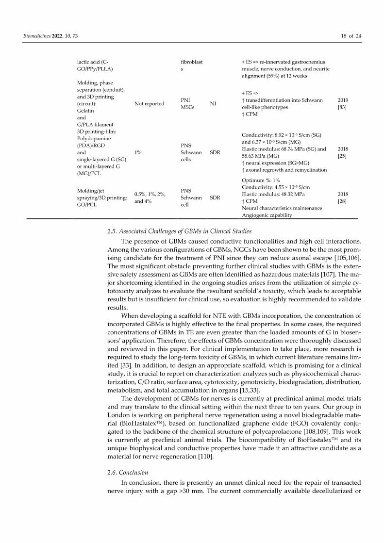
Biomedicines 2022, 10, 73 18 of 24
lactic acid (C-
GO/PPy/PLLA)
fibroblast
s
+ ES => re-innervated gastrocnemius
muscle, nerve conduction, and neurite
alignment (59%) at 12 weeks
Molding, phase
separation (conduit),
and 3D printing
(circuit):
Gelatin
and
G/PLA filament
Not reported PNI
MSCs NI
+ ES =>
↑ transdifferentiation into Schwann
cell-like phenotypes
↑ CPM
2019
[83]
3D printing-film:
Polydopamine
(PDA)/RGD
and
single-layered G (SG)
or multi-layered G
(MG)/PCL
1%
PNS
Schwann
cells
SDR
Conductivity: 8.92 × 10⁻³ S/cm (SG)
and 6.37 × 10⁻³ S/cm (MG)
Elastic modulus: 68.74 MPa (SG) and
58.63 MPa (MG)
↑ neural expression (SG>MG)
↑ axonal regrowth and remyelination
2018
[25]
Molding/jet
spraying/3D printing:
GO/PCL
0.5%, 1%, 2%,
and 4%
PNS
Schwann
cell
SDR
Optimum %: 1%
Conductivity: 4.55 × 10⁻⁵ S/cm
Elastic modulus: 48.32 MPa
↑ CPM
Neural characteristics maintenance
Angiogenic capability
2018
[28]
2.5. Associated Challenges of GBMs in Clinical Studies
The presence of GBMs caused conductive functionalities and high cell interactions.
Among the various configurations of GBMs, NGCs have been shown to be the most prom-
ising candidate for the treatment of PNI since they can reduce axonal escape [105,106].
The most significant obstacle preventing further clinical studies with GBMs is the exten-
sive safety assessment as GBMs are often identified as hazardous materials [107]. The ma-
jor shortcoming identified in the ongoing studies arises from the utilization of simple cy-
totoxicity analyzes to evaluate the resultant scaffold’s toxicity, which leads to acceptable
results but is insufficient for clinical use, so evaluation is highly recommended to validate
results.
When developing a scaffold for NTE with GBMs incorporation, the concentration of
incorporated GBMs is highly effective to the final properties. In some cases, the required
concentrations of GBMs in TE are even greater than the loaded amounts of G in biosen-
sors’ application. Therefore, the effects of GBMs concentration were thoroughly discussed
and reviewed in this paper. For clinical implementation to take place, more research is
required to study the long-term toxicity of GBMs, in which current literature remains lim-
ited [33]. In addition, to design an appropriate scaffold, which is promising for a clinical
study, it is crucial to report on characterization analyzes such as physicochemical charac-
terization, C/O ratio, surface area, cytotoxicity, genotoxicity, biodegradation, distribution,
metabolism, and total accumulation in organs [15,33].
The development of GBMs for nerves is currently at preclinical animal model trials
and may translate to the clinical setting within the next three to ten years. Our group in
London is working on peripheral nerve regeneration using a novel biodegradable mate-
rial (BioHastalex™), based on functionalized graphene oxide (FGO) covalently conju-
gated to the backbone of the chemical structure of polycaprolactone [108,109]. This work
is currently at preclinical animal trials. The biocompatibility of BioHastalex™ and its
unique biophysical and conductive properties have made it an attractive candidate as a
material for nerve regeneration [110].
2.6. Conclusion
In conclusion, there is presently an unmet clinical need for the repair of transacted
nerve injury with a gap >30 mm. The current commercially available decellularized or

Biomedicines 2022, 10, 73 19 of 24
biomaterial-type nerve conduits fail to support neural regeneration in gaps >30 mm. Gra-
phene and its derivatives are promising candidates in the treatment of nerve injury. Gra-
phene is a single-layer atom, with superior mechanical and chemical properties, which
include electrical and thermal conductivity, and strength (graphene is more than 100 times
stronger than steel). There are several products currently under development at research
centers as well as in industry for the development of nerve conduit from GBMs with bio-
functionalization using stem cells and growth factors.
Author Contributions: The research topic was initiated by A.M.S. and highlighted the subtitle. M.A.
and P.Z. equally worked on it and drafted the review. A.S. and Z.B. analyzed the data, tabulated
the tables with M.A. and P.Z., and helped with the discussion. A.M.S. critically reviewed the data
and expanded the review with his own experience and work on graphene oxide. Overall supervi-
sion of research carried out by Z.B. and A.M.S. All authors have read and agreed to the published
version of the manuscript.
Funding: This research received no external funding.
Institutional Review Board Statement: Not applicable, review article.
Informed Consent Statement: Not applicable, review article.
Data Availability Statement: Some data are available at www.NanoRegMed.com (accessed on 1
January 2022)
Conflicts of Interest: The authors declare no conflict of interest.
Abbreviations
3D Three-dimensional
ADSCs Adipose-derived stem cells
BBB Blood–brain barrier
CNS Central nervous system
DBS Dodecyl benzenesulfonate
DRG Dorsal root ganglion
ECM Extracellular matrix
ES Electrical stimulation
FGO Functionalized graphene oxide
G Graphene
GBMs Graphene-based materials
GBN Graphene-based nano scaffolds
GelMA Gelatinmethacryloyl
GFAP Glial fibrillary acidic protein
GMT Graphene mesh tube
GO Graphene oxide
iPSCs Induced pluripotent stem cells
M1 Direct macrophage inflammatory
M2 Anti-inflammatory
MAP-2 Microtubule-associated protein 2
MMP Matrix metalloproteinase
MSCs Mesenchymal stem cells
MTAC [2-(methacryloyloxy)ethyl]trimethylammonium chloride
NCSCs Neural crest stem cells
NGC Nerve guidance conduit
NPs Nanoparticles
NTE Nerve tissue engineering
OPF Oligo(poly(ethylene glycol) fumarate
PCL Polycaprolactone
PEI Polyethylenimine
PHA Polyhydroxyl alkanoate
PLCL Poly(D,L-lactide-co-caprolactone)
PLGA Poly(lactic-co-glycolic acid)
PLLA Poly-l-lactic acid

Biomedicines 2022, 10, 73 20 of 24
PNI Peripheral nerve injury
PNS Peripheral nervous system
rGO Reduced graphene oxide
ROS Reactive oxygen species
SDF-1α Stromal cell-derived factor-1α
TE Tissue engineering
References
1. Ahadian, S.; Obregón, R.; Ramón-Azcón, J.; Salazar, G.; Shiku, H.; Ramalingam, M.; Matsue, T. Carbon nanotubes and graphene-
based nanomaterials for stem cell differentiation and tissue regeneration. J. Nanosci. Nanotechnol. 2016, 16, 8862–8880.
2. Aydin, T.; Gurcan, C.; Taheri, H.; Yilmazer, A. Graphene based materials in neural tissue regeneration. Adv. Exp. Med. Biol. 2018,
1107, 129–142.
3. Reddy, S.; Xu, X.; Guo, T.; Zhu, R.; He, L.; Ramakrishana, S. Allotropic carbon (graphene oxide and reduced graphene oxide)
based biomaterials for neural regeneration. Curr. Opin. Biomed. Eng. 2018, 6, 120–129.
4. Soman, S.S.; Vijayavenkataraman, S. Perspectives on 3d bioprinting of peripheral nerve conduits. Int. J. Mol. Sci. 2020, 21, 5792.
5. Ko, C.-H.; Shie, M.-Y.; Lin, J.-H.; Chen, Y.-W.; Yao, C.-H.; Chen, Y.-S. Biodegradable bisvinyl sulfonemethyl-crosslinked gelatin
conduit promotes regeneration after peripheral nerve injury in adult rats. Sci. Rep. 2017, 7, 17489.
6. Zarrintaj, P.; Zangene, E.; Manouchehri, S.; Amirabad, L.M.; Baheiraei, N.; Hadjighasem, M.R.; Farokhi, M.; Ganjali, M.R.;
Walker, B.W.; Saeb, M.R. Conductive biomaterials as nerve conduits: Recent advances and future challenges. Appl. Mater. Today
2020, 20, 100784.
7. Zhang, C.; Fan, S.; Shao, H.; Hu, X.; Zhu, B.; Zhang, Y. Graphene trapped silk scaffolds integrate high conductivity and stability.
Carbon N. Y. 2019, 148, 16–27.
8. Adiguzel, E.; Yaşar, E.; Tecer, D.; Güzelküçük, Ü.; Taşkaynatan, M.A.; Kesikburun, S.; Özgül, A. Peripheral nerve injuries: Long
term follow-up results of rehabilitation. J. Back Musculoskelet. Rehabil. 2016, 29, 367–371.
9. Hassanzadeh, S.; Jalessi, M.; Jameie, S.B.; Khanmohammadi, M.; Bagher, Z.; Namjoo, Z.; Davachi, S.M. More attention on glial
cells to have better recovery after spinal cord injury. Biochem. Biophys. Reports 2021, 25, 100905.
10. Yao, Y.; Wang, C. Dedifferentiation: Inspiration for devising engineering strategies for regenerative medicine. NPJ Regen. Med.
2020, 5, 14.
11. Philips, C.; Cornelissen, M.; Carriel, V. Evaluation methods as quality control in the generation of decellularized peripheral
nerve allografts. J. Neural Eng. 2018, 15, 21003.
12. Aleemardani, M.; Bagher, Z.; Farhadi, M.; Chahsetareh, H.; Najafi, R.; Eftekhari, B.; Seifalian, A. Can Tissue Engineering Bring
Hope to the Development of Human Tympanic Membrane? Tissue Eng. Part B Rev. 2021, 27, 572_589.
13. Najafloo, R.; Majidi, J.; Asghari, A.; Aleemardani, M.; Kamrava, S.K.; Simorgh, S.; Seifalian, A.; Bagher, Z.; Seifalian, A.M. Mech-
anism of Anosmia Caused by Symptoms of COVID-19 and Emerging Treatments. ACS Chem. Neurosci. 2021, 12, 3795–3805.
14. Bagher, Z.; Asgari, N.; Bozorgmehr, P.; Kamrava, S.K.; Alizadeh, R.; Seifalian, A. Will Tissue-Engineering Strategies Bring New
Hope for the Reconstruction of Nasal Septal Cartilage? Curr. Stem Cell Res. Ther. 2020, 15, 144–154.
15. Zare, P.; Aleemardani, M.; Seifalian, A.; Bagher, Z.; Seifalian, A.M. Graphene Oxide: Opportunities and Challenges in Biomed-
icine. Nanomaterials 2021, 11, 1083.
16. Zarrintaj, P.; Saeb, M.R.; Ramakrishna, S.; Mozafari, M. Biomaterials selection for neuroprosthetics. Curr. Opin. Biomed. Eng.
2018, 6, 99–109.
17. Amani, H.; Mostafavi, E.; Arzaghi, H.; Davaran, S.; Akbarzadeh, A.; Akhavan, O.; Pazoki-Toroudi, H.; Webster, T.J. Three-
dimensional graphene foams: Synthesis, properties, biocompatibility, biodegradability, and applications in tissue engineering.
ACS Biomater. Sci. Eng. 2018, 5, 193–214.
18. Steel, E.M.; Sundararaghavan, H.G. Electrically conductive materials for nerve regeneration. In Neural Engineering; Springer:
Berlin/Heidelberg, Germany, 2016; pp. 145–179.
19. Xiaoli, F.; Qiyue, C.; Weihong, G.; Yaqing, Z.; Chen, H.; Junrong, W.; Longquan, S. Toxicology data of graphene-family nano-
materials: An update. Arch. Toxicol. 2020, 94, 1915–1939.
20. Devasena, T.; Francis, A.P.; Ramaprabhu, S. Toxicity of Graphene: An Update. Rev. Environ. Contam. Toxicol. Vol. 259 2021, 51–
76.
21. Ou, L.; Song, B.; Liang, H.; Liu, J.; Feng, X.; Deng, B.; Sun, T.; Shao, L. Toxicity of graphene-family nanoparticles: A general
review of the origins and mechanisms. Part. Fibre Toxicol. 2016, 13, 57.
22. Verre, A.F.; Faroni, A.; Iliut, M.; Silva, C.; Muryn, C.; Reid, A.J.; Vijayaraghavan, A. Improving the glial differentiation of human
Schwann-like adipose-derived stem cells with graphene oxide substrates. Interface Focus 2018, 8, 20180002.
23. Henna, T.K.; Nivitha, K.P.; Raphey, V.R.; Sabu, C.; Pramod, K. Functionalized Graphene for Drug Delivery Applications. In
Graphene Functionalization Strategies; Springer: Berlin/Heidelberg, Germany, 2019; pp. 247–278.
24. Portolés, M.T.; Serrano, M.C. Potentiality of Graphene-Based Materials for Neural Repair. In Graphene-Based Materials in Health
and Environment; Springer: Berlin/Heidelberg, Germany, 2016; pp. 159–190.

Biomedicines 2022, 10, 73 21 of 24
25. Qian, Y.; Zhao, X.; Han, Q.; Chen, W.; Li, H.; Yuan, W. An integrated multi-layer 3D-fabrication of PDA/RGD coated graphene
loaded PCL nanoscaffold for peripheral nerve restoration. Nat. Commun. 2018, 9, 323.
26. Zhang, Z.; Klausen, L.H.; Chen, M.; Dong, M. Electroactive Scaffolds for Neurogenesis and Myogenesis: Graphene-Based Na-
nomaterials. Small 2018, 14, 801983.
27. Heidari, M.; Bahrami, S.H.; Ranjbar-Mohammadi, M.; Milan, P.B. Smart electrospun nanofibers containing PCL/gelatin/gra-
phene oxide for application in nerve tissue engineering. Mater. Sci. Eng. C 2019, 103, 109768.
28. Qian, Y.; Song, J.; Zhao, X.; Chen, W.; Ouyang, Y.; Yuan, W.; Fan, C. 3D fabrication with integration molding of a graphene
oxide/polycaprolactone nanoscaffold for neurite regeneration and angiogenesis. Adv. Sci. 2018, 5, 1700499.
29. Pampaloni, N.P.; Lottner, M.; Giugliano, M.; Matruglio, A.; D’Amico, F.; Prato, M.; Garrido, J.A.; Ballerini, L.; Scaini, D. Single-
layer graphene modulates neuronal communication and augments membrane ion currents. Nat. Nanotechnol. 2018, 13, 755–764.
30. Dixon, A.R.; Jariwala, S.H.; Bilis, Z.; Loverde, J.R.; Pasquina, P.F.; Alvarez, L.M. Bridging the gap in peripheral nerve repair
with 3D printed and bioprinted conduits. Biomaterials 2018, 186, 44–63.
31. Convertino, D.; Fabbri, F.; Mishra, N.; Mainardi, M.; Cappello, V.; Testa, G.; Capsoni, S.; Albertazzi, L.; Luin, S.; Marchetti, L.
Graphene promotes axon elongation through local stall of Nerve Growth Factor signaling endosomes. Nano Lett. 2020, 20,
3633–3641.
32. Huang, Z.; Guo, Z.; Sun, M.; Fang, S.; Li, H. A study on graphene composites for peripheral nerve injury repair under electrical
stimulation. RSC Adv. 2019, 9, 28627–28635.
33. Grinsell, D.; Keating, C.P. Peripheral nerve reconstruction after injury: A review of clinical and experimental therapies. Biomed.
Res. Int. 2014, 2014, 698256.
34. Mahar, M.; Cavalli, V. Intrinsic mechanisms of neuronal axon regeneration. Nat. Rev. Neurosci. 2018, 19, 323–337.
35. Yang, Q.; Zhou, J. Neuroinflammation in the central nervous system: Symphony of glial cells. Glia 2019, 67, 1017–1035.
36. He, Z.; Zang, H.; Zhu, L.; Huang, K.; Yi, T.; Zhang, S.; Cheng, S. An anti-inflammatory peptide and brain-derived neurotrophic
factor-modified hyaluronan-methylcellulose hydrogel promotes nerve regeneration in rats with spinal cord injury. Int. J.
Nanomed. 2019, 14, 721.
37. Anderson, J.M.; Rodriguez, A.; Chang, D.T. Foreign body reaction to biomaterials. Semin. Immunol. 2008, 20, 86–100.
38. Holloway, J.L. Harnessing the immune response to improve functional healing. Sci. Transl. Med. 2018, 10, eaav3889.
39. Shende, P.; Pathan, N. Potential of carbohydrate-conjugated graphene assemblies in biomedical applications. Carbohydr. Polym.
2021, 255, 117385.
40. Papi, M. Graphene-Based Materials: Biological and Biomedical Applications. Int. J. Mol. Sci. 2021, 22, 672.
41. Daneshmandi, L.; Barajaa, M.; Tahmasbi Rad, A.; Sydlik, S.A.; Laurencin, C.T. Graphene-Based Biomaterials for Bone Regener-
ative Engineering: A Comprehensive Review of the Field and Considerations Regarding Biocompatibility and Biodegradation.
Adv. Healthc. Mater. 2021, 10, 2001414.
42. Mohan, V.B.; Lau, K.; Hui, D.; Bhattacharyya, D. Graphene-based materials and their composites: A review on production,
applications and product limitations. Compos. Part B Eng. 2018, 142, 200–220.
43. Aleemardani, M.; Solouk, A.; Akbari, S.; Dehghan, M.M.; Moeini, M. Silk-derived oxygen-generating electrospun patches for
enhancing tissue regeneration: Investigation of calcium peroxide role and its effects in controlled oxygen delivery. Materialia
2020, 14, 100877.
44. Huang, Z.; Sun, M.; Li, Y.; Guo, Z.; Li, H. Reduced graphene oxide-coated electrospun fibre: Effect of orientation, coverage and
electrical stimulation on Schwann cells behavior. J. Mater. Chem. B 2021, 9, 2656–2665.
45. Magaz, A.; Li, X.; Gough, J.E.; Blaker, J.J. Graphene oxide and electroactive reduced graphene oxide-based composite fibrous
scaffolds for engineering excitable nerve tissue. Mater. Sci. Eng. C 2021, 119, 111632.
46. Aval, N.A.; Emadi, R.; Valiani, A.; Kharaziha, M.; Karimipour, M.; Rahbarghazi, R. Nano-featured poly (lactide-co-glycolide)-
graphene microribbons as a promising substrate for nerve tissue engineering. Compos. Part B Eng. 2019, 173, 106863.
47. Zhao, Y.; Gong, J.; Niu, C.; Wei, Z.; Shi, J.; Li, G.; Yang, Y.; Wang, H. A new electrospun graphene-silk fibroin composite scaffolds
for guiding Schwann cells. J. Biomater. Sci. Polym. Ed. 2017, 28, 2171–2185.
48. Jaswal, R.; Shrestha, S.; Shrestha, B.K.; Kumar, D.; Park, C.H.; Kim, C.S. Nanographene enfolded AuNPs sophisticatedly syn-
chronized polycaprolactone based electrospun nanofibre scaffold for peripheral nerve regeneration. Mater. Sci. Eng. C 2020, 116,
111213.
49. Liu, Q.; Liu, G.; Liu, X.; Yang, M.; Xing, S.; Du, Y.; Xiong, X. Synthesis of an electrospun PHA/RGO/Au scaffold for peripheral
nerve regeneration: An in vitro study. Appl. Nanosci. 2020, 10, 687–694.
50. Du, J.; Zhen, G.; Chen, H.; Zhang, S.; Qing, L.; Yang, X.; Lee, G.; Mao, H.-Q.; Jia, X. Optimal electrical stimulation boosts stem
cell therapy in nerve regeneration. Biomaterials 2018, 181, 347–359.
51. Zhao, S.; Mehta, A.S.; Zhao, M. Biomedical applications of electrical stimulation. Cell. Mol. Life Sci. 2020, 77, 2681–2699.
52. Wang, J.; Cheng, Y.; Chen, L.; Zhu, T.; Ye, K.; Jia, C.; Wang, H.; Zhu, M.; Fan, C.; Mo, X. In vitro and in vivo studies of electro-
active reduced graphene oxide-modified nanofiber scaffolds for peripheral nerve regeneration. Acta Biomater. 2019, 84, 98–113.
53. Shang, L.; Huang, Z.; Pu, X.; Yin, G.; Chen, X. Preparation of graphene oxide-doped polypyrrole composite films with stable
conductivity and their effect on the elongation and alignment of neurite. ACS Biomater. Sci. Eng. 2019, 5, 1268–1278.
54. Dong, C.; Qiao, F.; Hou, W.; Yang, L.; Lv, Y. Graphene-based conductive fibrous scaffold boosts sciatic nerve regeneration and
functional recovery upon electrical stimulation. Appl. Mater. Today 2020, 21, 100870.

Biomedicines 2022, 10, 73 22 of 24
55. Mohseni, M.; SA, A.R.; Shirazi, F.H.; Nemati, N.H. Preparation and characterization of self-electrical stimuli conductive gellan
based nano scaffold for nerve regeneration containing chopped short spun nanofibers of PVDF/MCM41 and polyaniline/gra-
phene nanoparticles: Physical, mechanical and morphological. Int. J. Biol. Macromol. 2021, 167, 881–893.
56. Liu, C.; Huang, Z.; Pu, X.; Shang, L.; Yin, G.; Chen, X.; Cheng, S. Fabrication of carboxylic graphene oxide-composited polypyr-
role film for neurite growth under electrical stimulation. Front. Mater. Sci. 2019, 13, 258–267.
57. Yan, L.; Zhao, B.; Liu, X.; Li, X.; Zeng, C.; Shi, H.; Xu, X.; Lin, T.; Dai, L.; Liu, Y. Aligned nanofibers from polypyrrole/graphene
as electrodes for regeneration of optic nerve via electrical stimulation. ACS Appl. Mater. Interfaces 2016, 8, 6834–6840.
58. Golafshan, N.; Kharaziha, M.; Fathi, M.; Larson, B.L.; Giatsidis, G.; Masoumi, N. Anisotropic architecture and electrical stimu-
lation enhance neuron cell behaviour on a tough graphene embedded PVA: Alginate fibrous scaffold. RSC Adv. 2018, 8,
6381–6389.
59. Girao, A.F.; Sousa, J.; Dominguez-Bajo, A.; Gonzalez-Mayorga, A.; Bdikin, I.; Pujades-Otero, E.; Casan-Pastor, N.; Hortiguela,
M.J.; Otero-Irurueta, G.; Completo, A. 3D Reduced Graphene Oxide Scaffolds with a Combinatorial Fibrous-Porous Architec-
ture for Neural Tissue Engineering. ACS Appl. Mater. Interfaces 2020, 12, 38962–38975.
60. Guo, Z.; Liang, J.; Ankone, M.J.K.; Poot, A.A.; Grijpma, D.W.; Chen, H. Fabrication of poly (trimethylene carbonate)/reduced
graphene oxide-graft-poly (trimethylene carbonate) composite scaffolds for nerve regeneration. Biomed. Mater. 2019, 14, 24104.
61. Wang, J.; Zheng, W.; Chen, L.; Zhu, T.; Shen, W.; Fan, C.; Wang, H.; Mo, X. Enhancement of Schwann cells function using
graphene-oxide-modified nanofiber scaffolds for peripheral nerve regeneration. ACS Biomater. Sci. Eng. 2019, 5, 2444–2456.
62. Golafshan, N.; Kharaziha, M.; Fathi, M. Tough and conductive hybrid graphene-PVA: Alginate fibrous scaffolds for engineering
neural construct. Carbon N. Y. 2017, 111, 752–763.
63. Li, Y.; Huang, Z.; Pu, X.; Chen, X.; Yin, G.; Wang, Y.; Miao, D.; Fan, J.; Mu, J. Polydopamine/carboxylic graphene oxide-compo-
sited polypyrrole films for promoting adhesion and alignment of Schwann cells. Colloids Surf. B Biointerfaces 2020, 191, 110972.
64. Diban, N.; Sánchez-González, S.; Lázaro-Díez, M.; Ramos-Vivas, J.; Urtiaga, A. Facile fabrication of poly (ε-caprolactone)/gra-
phene oxide membranes for bioreactors in tissue engineering. J. Memb. Sci. 2017, 540, 219–228.
65. Soltani, S.; Ebrahimian-Hosseinabadi, M.; Kharazi, A.Z. Chitosan/graphene and poly (D, L-lactic-co-glycolic acid)/graphene
nano-composites for nerve tissue engineering. Tissue Eng. Regen. Med. 2016, 13, 684–690.
66. Ghafaralahi, S.; Ebrahimian-Hosseinabadi, M.; Zargar Kharazi, A. Poly (glycerol-sebacate)/poly (caprolactone)/graphene nano-
composites for nerve tissue engineering. J. Bioact. Compat. Polym. 2018, 33, 529–542.
67. Qian, Y.; Wang, X.; Song, J.; Chen, W.; Chen, S.; Jin, Y.; Ouyang, Y.; Yuan, W.-E.; Fan, C. Preclinical assessment on neuronal
regeneration in the injury-related microenvironment of graphene-based scaffolds. NPJ Regen. Med. 2021, 6, 31.
68. Farokhi, M.; Aleemardani, M.; Solouk, A.; Mirzadeh, H.; Teuschl, A.H.; Redl, H. Crosslinking strategies for silk fibroin hydro-
gels: Promising biomedical materials. Biomed. Mater. 2021, 16, 22004.
69. Zhang, C.; Yuan, T.-J.; Tan, M.-H.; Xu, X.-H.; Huang, Y.-F.; Peng, L.-H. Smart graphene-based hydrogel promotes recruitment
and neural-like differentiation of bone marrow derived mesenchymal stem cells in rat skin. Biomater. Sci. 2021, 9, 2146–2161.
70. Zhang, K.; Li, J.; Jin, J.; Dong, J.; Li, L.; Xue, B.; Wang, W.; Jiang, Q.; Cao, Y. Injectable, anti-inflammatory and conductive hy-
drogels based on graphene oxide and diacerein-terminated four-armed polyethylene glycol for spinal cord injury repair. Mater.
Des. 2020, 196, 109092.
71. Zhao, Y.; Wang, Y.; Niu, C.; Zhang, L.; Li, G.; Yang, Y. Construction of polyacrylamide/graphene oxide/gelatin/sodium alginate
composite hydrogel with bioactivity for promoting Schwann cells growth. J. Biomed. Mater. Res. Part A 2018, 106, 1951–1964.
72. Liu, X.; Miller, A.L.; Park, S.; Waletzki, B.E.; Zhou, Z.; Terzic, A.; Lu, L. Functionalized carbon nanotube and graphene oxide
embedded electrically conductive hydrogel synergistically stimulates nerve cell differentiation. ACS Appl. Mater. Interfaces 2017,
9, 14677–14690.
73. Li, G.; Zhao, Y.; Zhang, L.; Gao, M.; Kong, Y.; Yang, Y. Preparation of graphene oxide/polyacrylamide composite hydrogel and
its effect on Schwann cells attachment and proliferation. Colloids Surf. B Biointerfaces 2016, 143, 547–556.
74. Hsieh, T.; Huang, W.; Kang, Y.; Chu, C.; Liao, W.; Chen, Y.; Chen, S. Neurotensin-conjugated reduced graphene oxide with
multi-stage near-infrared-triggered synergic targeted neuron gene transfection in vitro and in vivo for neurodegenerative dis-
ease therapy. Adv. Healthc. Mater. 2016, 5, 3016–3026.
75. Dadsetan, M.; Knight, A.M.; Lu, L.; Windebank, A.J.; Yaszemski, M.J. Stimulation of neurite outgrowth using positively charged
hydrogels. Biomaterials 2009, 30, 3874–3881.
76. Madigan, N.N.; Chen, B.K.; Knight, A.M.; Rooney, G.E.; Sweeney, E.; Kinnavane, L.; Yaszemski, M.J.; Dockery, P.; O’Brien, T.;
McMahon, S.S. Comparison of cellular architecture, axonal growth, and blood vessel formation through cell-loaded polymer
scaffolds in the transected rat spinal cord. Tissue Eng. Part A 2014, 20, 2985–2997.
77. Chen, B.K.; Knight, A.M.; Madigan, N.N.; Gross, L.; Dadsetan, M.; Nesbitt, J.J.; Rooney, G.E.; Currier, B.L.; Yaszemski, M.J.;
Spinner, R.J. Comparison of polymer scaffolds in rat spinal cord: A step toward quantitative assessment of combinatorial ap-
proaches to spinal cord repair. Biomaterials 2011, 32, 8077–8086.
78. Binan, L.; Ajji, A.; De Crescenzo, G.; Jolicoeur, M. Approaches for neural tissue regeneration. Stem Cell Rev. Rep. 2014, 10, 44–59.
79. Zhou, L.; Fan, L.; Yi, X.; Zhou, Z.; Liu, C.; Fu, R.; Dai, C.; Wang, Z.; Chen, X.; Yu, P. Soft conducting polymer hydrogels cross-
linked and doped by tannic acid for spinal cord injury repair. ACS Nano 2018, 12, 10957–10967.
80. Huang, Q.; Cai, Y.; Yang, X.; Li, W.; Pu, H.; Liu, Z.; Liu, H.; Tamtaji, M.; Xu, F.; Sheng, L. Graphene foam/hydrogel scaffolds for
regeneration of peripheral nerve using ADSCs in a diabetic mouse model. Nano Res. 2021, 1–12. https://doi.org/10.1007/s12274-
021-3961-3.

Biomedicines 2022, 10, 73 23 of 24
81. Huang, C.T.; Kumar Shrestha, L.; Ariga, K.; Hsu, S.H. A graphene-polyurethane composite hydrogel as a potential bioink for
3D bioprinting and differentiation of neural stem cells. J. Mater. Chem. B 2017, 5, 8854–8864.
82. Zhu, W.; Harris, B.T.; Zhang, L.G. Gelatin methacrylamide hydrogel with graphene nanoplatelets for neural cell-laden 3D bi-
oprinting. In Proceedings of the 38th Annual International Conference of the IEEE Engineering in Medicine and Biology Society
(EMBC), Orlando, FL, USA, 16–20 August 2016; pp. 4185–4188.
83. Uz, M.; Donta, M.; Mededovic, M.; Sakaguchi, D.S.; Mallapragada, S.K. Development of gelatin and graphene-based nerve
regeneration conduits using three-dimensional (3D) printing strategies for electrical transdifferentiation of mesenchymal stem
cells. Ind. Eng. Chem. Res. 2019, 58, 7421–7427.
84. Ginestra, P. Manufacturing of polycaprolactone-Graphene fibers for nerve tissue engineering. J. Mech. Behav. Biomed. Mater.
2019, 100, 103387.
85. Zeinali, K.; Khorasani, M.T.; Rashidi, A.; Daliri Joupari, M. Preparation and characterization of graphene oxide aerogel/gelatin
as a hybrid scaffold for application in nerve tissue engineering. Int. J. Polym. Mater. Polym. Biomater. 2020, 70, 674–683.
86. Mansouri, N.; Al-Sarawi, S.F.; Mazumdar, J.; Losic, D. Advancing fabrication and properties of three-dimensional graphene–
alginate scaffolds for application in neural tissue engineering. RSC Adv. 2019, 9, 36838–36848.
87. Tolou, N.B.; Salimijazi, H.; Dikonimos, T.; Faggio, G.; Messina, G.; Tamburrano, A.; Aurora, A.; Lisi, N. Fabrication of 3D mon-
olithic graphene foam/polycaprolactone porous nanocomposites for bioapplications. J. Mater. Sci. 2021, 56, 5581–5594.
88. Allbright, K.O.; Bliley, J.M.; Havis, E.; Kim, D.; Dibernardo, G.A.; Grybowski, D.; Waldner, M.; James, I.B.; Sivak, W.N.; Rubin,
J.P. Delivery of adipose-derived stem cells in poloxamer hydrogel improves peripheral nerve regeneration. Muscle Nerve 2018,
58, 251–260.
89. Carriel, V.; Garrido-Gómez, J.; Hernández-Cortés, P.; Garzón, I.; García-García, S.; Sáez-Moreno, J.A.; del Carmen Sanchez-
Quevedo, M.; Campos, A.; Alaminos, M. Combination of fibrin-agarose hydrogels and adipose-derived mesenchymal stem
cells for peripheral nerve regeneration. J. Neural Eng. 2013, 10, 26022.
90. Santos, D.; Wieringa, P.; Moroni, L.; Navarro, X.; Valle, J. Del PEOT/PBT guides enhance nerve regeneration in long gap defects.
Adv. Healthc. Mater. 2017, 6, 1600298.
91. Kehoe, S.; Zhang, X.F.; Boyd, D. FDA approved guidance conduits and wraps for peripheral nerve injury: A review of materials
and efficacy. Injury 2012, 43, 553–572.
92. Guo, Q.; Liu, C.; Hai, B.; Ma, T.; Zhang, W.; Tan, J.; Fu, X.; Wang, H.; Xu, Y.; Song, C. Chitosan conduits filled with simvas-
tatin/Pluronic F-127 hydrogel promote peripheral nerve regeneration in rats. J. Biomed. Mater. Res. Part B Appl. Biomater. 2018,
106, 787–799.
93. Zheng, F.; Li, R.; He, Q.; Koral, K.; Tao, J.; Fan, L.; Xiang, R.; Ma, J.; Wang, N.; Yin, Y. The electrostimulation and scar inhibition
effect of chitosan/oxidized hydroxyethyl cellulose/reduced graphene oxide/asiaticoside liposome based hydrogel on peripheral
nerve regeneration in vitro. Mater. Sci. Eng. C 2020, 109, 110560.
94. Qing, L.; Chen, H.; Tang, J.; Jia, X. Exosomes and their microRNA cargo: New players in peripheral nerve regeneration.
Neurorehabil. Neural Repair 2018, 32, 765–776.
95. Huang, Q.; Cai, Y.; Zhang, X.; Liu, J.; Liu, Z.; Li, B.; Wong, H.; Xu, F.; Sheng, L.; Sun, D. Aligned graphene mesh-supported
double network natural hydrogel conduit loaded with netrin-1 for peripheral nerve regeneration. ACS Appl. Mater. Interfaces
2021, 13, 112–122.
96. Wang, J.; Cheng, Y.; Wang, H.; Wang, Y.; Zhang, K.; Fan, C.; Wang, H.; Mo, X. Biomimetic and hierarchical nerve conduits from
multifunctional nanofibers for guided peripheral nerve regeneration. Acta Biomater. 2020, 117, 180–191.
97. Park, J.; Jeon, J.; Kim, B.; Lee, M.S.; Park, S.; Lim, J.; Yi, J.; Lee, H.; Yang, H.S.; Lee, J.Y. Electrically Conductive Hydrogel Nerve
Guidance Conduits for Peripheral Nerve Regeneration. Adv. Funct. Mater. 2020, 30, 2003759.
98. Agarwal, G.; Kumar, N.; Srivastava, A. Highly elastic, electroconductive, immunomodulatory graphene crosslinked collagen
cryogel for spinal cord regeneration. Mater. Sci. Eng. C 2021, 118, 111518.
99. Zhang, D.; Yao, Y.; Duan, Y.; Yu, X.; Shi, H.; Nakkala, J.R.; Zuo, X.; Hong, L.; Mao, Z.; Gao, C. Surface-Anchored Graphene
Oxide Nanosheets on Cell-Scale Micropatterned Poly (D, L-lactide-co-caprolactone) Conduits Promote Peripheral Nerve Re-
generation. ACS Appl. Mater. Interfaces 2020, 12, 7915–7930.
100. Assaf, K.; Leal, C.V.; Derami, M.S.; de Rezende Duek, E.A.; Ceragioli, H.J.; de Oliveira, A.L.R. Sciatic nerve repair using poly
(ε-caprolactone) tubular prosthesis associated with nanoparticles of carbon and graphene. Brain Behav. 2017, 7, e00755.
101. Mohammadi, M.; Ramazani SaadatAbadi, A.; Mashayekhan, S.; Sanaei, R. Conductive multichannel PCL/gelatin conduit with
tunable mechanical and structural properties for peripheral nerve regeneration. J. Appl. Polym. Sci. 2020, 137, 49219.
102. Tolou, N.B.; Salimijazi, H.; Kharaziha, M.; Faggio, G.; Chierchia, R.; Lisi, N. A three-dimensional nerve guide conduit based on
graphene foam/polycaprolactone. Mater. Sci. Eng. C 2021, 126, 112110.
103. Abzan, N.; Kharaziha, M.; Labbaf, S. Development of three-dimensional piezoelectric polyvinylidene fluoride-graphene oxide
scaffold by non-solvent induced phase separation method for nerve tissue engineering. Mater. Des. 2019, 167, 107636.
104. Chen, X.; Liu, C.; Huang, Z.; Pu, X.; Shang, L.; Yin, G.; Xue, C. Preparation of carboxylic graphene oxide-composited polypyrrole
conduits and their effect on sciatic nerve repair under electrical stimulation. J. Biomed. Mater. Res. Part A 2019, 107, 2784–2795.
105. Bei, H.P.; Yang, Y.; Zhang, Q.; Tian, Y.; Luo, X.; Yang, M.; Zhao, X. Graphene-based nanocomposites for neural tissue engineer-
ing. Molecules 2019, 24, 658.
106. Magaz, A.; Faroni, A.; Gough, J.E.; Reid, A.J.; Li, X.; Blaker, J.J. Bioactive Silk-Based Nerve Guidance Conduits for Augmenting
Peripheral Nerve Repair. Adv. Healthc. Mater. 2018, 7, 1800308.

Biomedicines 2022, 10, 73 24 of 24
107. Bianco, A. Graphene: Safe or toxic? The two faces of the medal. Angew. Chemie Int. Ed. 2013, 52, 4986–4997.
108. Seifalian, A.M.; Hancock, S. Composite Material and Its Method of Production. Patent GB (GB1811090, 2017), WO, EP and US
(2020). (accessed on 12 April 2021).
109. Nezakati, T. Development of a Graphene-POSS-Nanocomposite Conductive Material for Biomedical Application. Ph.D. Thesis,
London University College, London, UK, 2016; Supervisor Professor Alexander Seifalian.
110. Nezakati, T.; Seifalian, A.; Tan, A.; Seifalian, A.M. Conductive polymers: Opportunities and challenges in biomedical applica-
tions. Chem. Rev. 2018, 118, 6766–6843.