GPAT Online Class for B Pharm students Human Anatomy ...
83
RIPER AUTONOMOUS NAAC & NBA (UG) SIRO- DSIR Raghavendra Institute of Pharmaceutical Education and Research - Autonomous K.R.Palli Cross, Chiyyedu, Anantapuramu, A. P- 515721 GPAT Online Class for B Pharm students Human Anatomy and Physiology part -1 (Cell, Cell junctions and transport mechanisms) By Dr. K. SOMASEKHAR REDDY, M. Pharm., Ph. D Associate Professor and Head Department of Pharmacology Raghavendra Institute of Pharmaceutical Education and Research (RIPER) – Autonomous, Ananthapuramu
-
Upload
khangminh22 -
Category
Documents
-
view
3 -
download
0
Transcript of GPAT Online Class for B Pharm students Human Anatomy ...

RIPERAUTONOMOUS
NAAC &
NBA (UG)
SIRO- DSIR
Raghavendra Institute of Pharmaceutical Education and Research - AutonomousK.R.Palli Cross, Chiyyedu, Anantapuramu, A. P- 515721
GPAT Online Class for B Pharm students
Human Anatomy and Physiology part -1(Cell, Cell junctions and transport mechanisms)
By
Dr. K. SOMASEKHAR REDDY, M. Pharm., Ph. D
Associate Professor and Head
Department of Pharmacology
Raghavendra Institute of Pharmaceutical Education and
Research (RIPER) – Autonomous, Ananthapuramu

RIPERAUTONOMOUS
NAAC &
NBA (UG)
SIRO- DSIR
Raghavendra Institute of Pharmaceutical Education and Research - AutonomousK.R.Palli Cross, Chiyyedu, Anantapuramu, A. P- 515721
Disclaimer
“The contents in this presentation are compiled fromdifferent sources, this presentation is delivered at theinterest of students and not involved any commercialbenefits. Thus the presenter shall not claim anycopyright for this presentation, and not responsible forany copyright issues if arise”
RIPERAUTONOMOUS
NAAC &
NBA (UG)
SIRO- DSIR
Raghavendra Institute of Pharmaceutical Education and Research - AutonomousK.R.Palli Cross, Chiyyedu, Anantapuramu, A. P- 515721
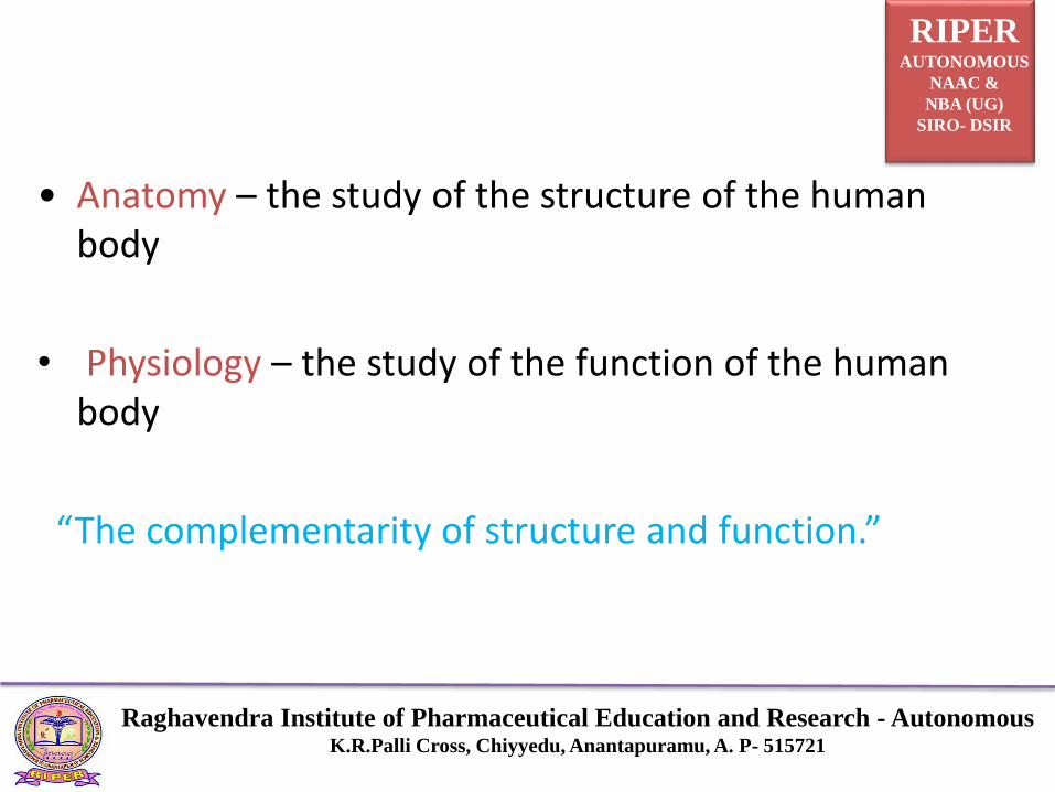
• Anatomy – the study of the structure of the human body
• Physiology – the study of the function of the human body
“The complementarity of structure and function.”

RIPERAUTONOMOUS
NAAC &
NBA (UG)
SIRO- DSIR
Raghavendra Institute of Pharmaceutical Education and Research - AutonomousK.R.Palli Cross, Chiyyedu, Anantapuramu, A. P- 515721
Levels of OrganizationSubatomic Particles – electrons, protons, and neutrons
Atom – hydrogen atom, lithium atom, etc.
Molecule – water molecule, glucose molecule, etc.
Macromolecule – protein molecule, DNA molecule, etc.
Organelle – mitochondrion, Golgi apparatus, nucleus, etc.
Cell – muscle cell, nerve cell, etc.
Tissue – epithelia, connective, muscle and nerve etc.
Organ – skin, femur, heart, kidney, etc.
Organ System – skeletal system, digestive system, etc.
Organism – the human

RIPERAUTONOMOUS
NAAC &
NBA (UG)
SIRO- DSIR
Raghavendra Institute of Pharmaceutical Education and Research - AutonomousK.R.Palli Cross, Chiyyedu, Anantapuramu, A. P- 515721
Characteristics of LifeMovement – change in position; motion
Responsiveness – reaction to a change
Growth – increase in body size; no change in shape
Reproduction – production of new organisms and newcells
Respiration – obtaining oxygen; removing carbon dioxide;releasing energy from foods
Digestion – breakdown of food substances into simplerforms

RIPERAUTONOMOUS
NAAC &
NBA (UG)
SIRO- DSIR
Raghavendra Institute of Pharmaceutical Education and Research - AutonomousK.R.Palli Cross, Chiyyedu, Anantapuramu, A. P- 515721
Absorption – passage of substances through membranesand into body fluids
Circulation – movement of substances in body fluids
Assimilation – changing of absorbed substances intochemically different forms
Excretion – removal of wastes produced by metabolicreactions

RIPERAUTONOMOUS
NAAC &
NBA (UG)
SIRO- DSIR
Raghavendra Institute of Pharmaceutical Education and Research - AutonomousK.R.Palli Cross, Chiyyedu, Anantapuramu, A. P- 515721
Maintenance of LifeLife depends on five (5) environmental factors
Water, Food, Oxygen, Heat, Pressure
• Water
- most abundant substance in body
- required for metabolic processes
- required for transport of substances
- regulates body temperature

RIPERAUTONOMOUS
NAAC &
NBA (UG)
SIRO- DSIR
Raghavendra Institute of Pharmaceutical Education and Research - AutonomousK.R.Palli Cross, Chiyyedu, Anantapuramu, A. P- 515721
• Food
- provides necessary nutrients
- supplies energy
- supplies raw materials
• Oxygen (gas)
- one-fifth of air
- used to release energy from nutrients
• Heat
- form of energy
- partly controls rate of metabolic reactions

RIPERAUTONOMOUS
NAAC &
NBA (UG)
SIRO- DSIR
Raghavendra Institute of Pharmaceutical Education and Research - AutonomousK.R.Palli Cross, Chiyyedu, Anantapuramu, A. P- 515721
• Pressure
- application of force on an object
- atmospheric pressure – important for breathing
- hydrostatic pressure – keeps blood flowing

RIPERAUTONOMOUS
NAAC &
NBA (UG)
SIRO- DSIR
Raghavendra Institute of Pharmaceutical Education and Research - AutonomousK.R.Palli Cross, Chiyyedu, Anantapuramu, A. P- 515721
Cell
Cell is the fundamental, structural and functional unit of all living organisms.
All cells arise from pre existing cells through theprocess of cell division.
Unicellular organisms – Organisms with single cell,capable of independent existence and carries allfunctions like digestion, excretion, respiration, growth& reproduction (Acellular). Examples, Amoeba,Euglena.

RIPERAUTONOMOUS
NAAC &
NBA (UG)
SIRO- DSIR
Raghavendra Institute of Pharmaceutical Education and Research - AutonomousK.R.Palli Cross, Chiyyedu, Anantapuramu, A. P- 515721
Multicellular organisms – Organisms with more than onecell
Cells in multicellular organisms vary in size & shapedepending on function
SHAPE: Parenchyma - Polyhedral cells performs storage.
Sclerenchyma - spindle shaped cells & providesmechanical support
Nerve cells- long and branched cells conducting nerveimpulses RBC -Biconcave & helps in carrying oxygen

RIPERAUTONOMOUS
NAAC &
NBA (UG)
SIRO- DSIR
Raghavendra Institute of Pharmaceutical Education and Research - AutonomousK.R.Palli Cross, Chiyyedu, Anantapuramu, A. P- 515721
Muscle cells- cylindrical or spindle shaped concerned with the movement of body parts.

RIPERAUTONOMOUS
NAAC &
NBA (UG)
SIRO- DSIR
Raghavendra Institute of Pharmaceutical Education and Research - AutonomousK.R.Palli Cross, Chiyyedu, Anantapuramu, A. P- 515721
➢ Different substances that make a cell are collectivelycalled
Protoplasm.
➢ Protoplasm is composed of
➢ Water - 70-80% Water is present in cell.
➢ Carbohydrates
➢ Lipids
➢ Proteins
➢ Electrolyte - Sodium (Na+), Potassium (K+), Magnesium (Mg2+), Calcium (Ca2+), Phosphate , Chloride (Cl-), and Bicarbonate (HC03 - ).

RIPERAUTONOMOUS
NAAC &
NBA (UG)
SIRO- DSIR
Raghavendra Institute of Pharmaceutical Education and Research - AutonomousK.R.Palli Cross, Chiyyedu, Anantapuramu, A. P- 515721
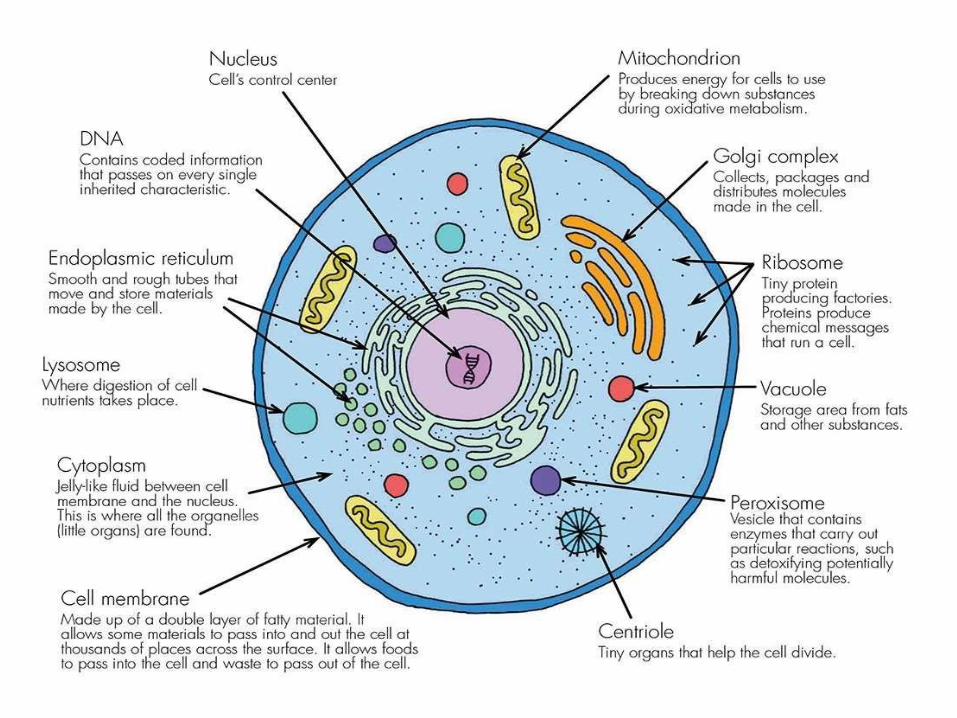
Components of Cell
Cell membrane Cytoplasm Nucleus

RIPERAUTONOMOUS
NAAC &
NBA (UG)
SIRO- DSIR
Raghavendra Institute of Pharmaceutical Education and Research - AutonomousK.R.Palli Cross, Chiyyedu, Anantapuramu, A. P- 515721
Cytoplasm
Cell Organelles Cellular inclusions
Membrane Non membraneous
bound

RIPERAUTONOMOUS
NAAC &
NBA (UG)
SIRO- DSIR
Raghavendra Institute of Pharmaceutical Education and Research - AutonomousK.R.Palli Cross, Chiyyedu, Anantapuramu, A. P- 515721
Cellular organelles
Membrane bound Non membraneous
• Mitochondria Ribosomes
• Endoplasmic reticulum Cytoskeleton
• Golgi apparatus Centrioles
• Lysosomes

RIPERAUTONOMOUS
NAAC &
NBA (UG)
SIRO- DSIR
Raghavendra Institute of Pharmaceutical Education and Research - AutonomousK.R.Palli Cross, Chiyyedu, Anantapuramu, A. P- 515721
Cell InclusionsStorage Products
Starch, Fats and Oils, Proteins
Secretory Products
Enzymes, hormones
Excretory products
Carbon dioxide, water


RIPERAUTONOMOUS
NAAC &
NBA (UG)
SIRO- DSIR
Raghavendra Institute of Pharmaceutical Education and Research - AutonomousK.R.Palli Cross, Chiyyedu, Anantapuramu, A. P- 515721
Cell wall• Outermost layer, non living ,rigid
• Found in bacterial cells, fungal cells and plant cells.
• Permeable
• Made up of cellulose (in bacteria- peptidoglycans,
• in fungus- Chitin)
• FUNCTION :
• Rigidity, mechanical support and protection

RIPERAUTONOMOUS
NAAC &
NBA (UG)
SIRO- DSIR
Raghavendra Institute of Pharmaceutical Education and Research - AutonomousK.R.Palli Cross, Chiyyedu, Anantapuramu, A. P- 515721
Cell membrane• Present in all cells, just below the cell wall in plant cells,
• outermost membrane in animal cells
• Semi-permeable
• Made up of phospholipids, proteins, carbohydrates and
• Cholesterol
• FUNCTION : It allows outward and inward movement
• of molecules across it like diffusion, osmosis,
• active transport, phagocytosis and pinocytosis

RIPERAUTONOMOUS
NAAC &
NBA (UG)
SIRO- DSIR
Raghavendra Institute of Pharmaceutical Education and Research - AutonomousK.R.Palli Cross, Chiyyedu, Anantapuramu, A. P- 515721
Links adjacent cells together by junctional complexes to form tissues.
Insulating Properties:- It acts as dielectric material of a charged condenser, thus cell membrane have very high insulating value

RIPERAUTONOMOUS
NAAC &
NBA (UG)
SIRO- DSIR
Raghavendra Institute of Pharmaceutical Education and Research - AutonomousK.R.Palli Cross, Chiyyedu, Anantapuramu, A. P- 515721
CYTOPLASM
• Semi fluid matrix present between cell membrane and
nuclear membrane
• It has various living cell inclusions called cell organelles
and non living substances called Ergastic substances
Cytosol
• The cytosol, the aqueous part of the cytoplasm outside all of the organelles, also contains its own distinctive proteins.
• It accounts for almost 70% of the total cell volume.
• Gelatinous substance consisting mainly of cytoskeleton filaments, organic molecules, salt and water.

CYTOSKELETON
and
System of fibers that not only maintains the structure of the cell but
also permit it to change shape and move.
The cytoskeleton is made up primarily of:- i)Microtubules
ii)Intermediate Filaments
iii)Microfilaments
along with protein that anchor tie them together.

Microtubules- These are long hollow structures approx. 25nm in diameter.
Determine shape of the cell, role in the contraction of the spindle and
movement of chromosomes and centrioles as well as in ciliary and flagellar
motion.
Intermediate Filaments- They are 8-14nm in diameter and are made up of
various subunits. They form a flexible scaffolding or cell and help it resist
external pressure.
In their absence cell ruptures more easily and when they are abnormal in
human, blistering in common.
The proteins that makeup intermediate filament are cell types specific and
are thus frequently used as cellular markers.
Microfilaments- They are long solid fibers 4-6 nm in diameter. They
comprise the contractile protein actin and are responsible for the cell
motion.

RIPERAUTONOMOUS
NAAC &
NBA (UG)
SIRO- DSIR
Raghavendra Institute of Pharmaceutical Education and Research - AutonomousK.R.Palli Cross, Chiyyedu, Anantapuramu, A. P- 515721
Function• They are involved in the
Movement of the chromosomes
Cell movement
Processes that move secretion granules in the cell
Movement of proteins within the cell membrane

RIPERAUTONOMOUS
NAAC &
NBA (UG)
SIRO- DSIR
Raghavendra Institute of Pharmaceutical Education and Research - AutonomousK.R.Palli Cross, Chiyyedu, Anantapuramu, A. P- 515721
NucleusSTRUCTURE
• Largest cell organelle present in eukaryotic cells
• It is usually spherical
• It has double layer nuclear membrane with nuclear pores
• It has transparent granular matrix called nucleoplasm,
• chromatin network composed of DNA and histone proteins
• It also has a spherical body called Nucleolus

RIPERAUTONOMOUS
NAAC &
NBA (UG)
SIRO- DSIR
Raghavendra Institute of Pharmaceutical Education and Research - AutonomousK.R.Palli Cross, Chiyyedu, Anantapuramu, A. P- 515721
FUNCTION: It is the control centre of the cell
It contains genetic material DNA which regulates all metabolic
activities of the body

MITOCHONDRIA
The mitochondria were first observed by Kolliker in 1850 asgranular
structures in the striated muscles.
Mitochondria are called the 'powerhouse of thecell'.
STRUCTURE-
Length- 5-12µm
Diameter- 0.5-1µm
Filamentous or globular inshape.

RIPERAUTONOMOUS
NAAC &
NBA (UG)
SIRO- DSIR
Raghavendra Institute of Pharmaceutical Education and Research - AutonomousK.R.Palli Cross, Chiyyedu, Anantapuramu, A. P- 515721
Components of Mitochondria are
Outer Membrane, Inner Membrane
Intermediate Space- space between outer and inner
membranes
Cristae - In foldings of inner membrane
Matrix- The space enclosed by inner membrane
The membranes are made up of phospholipids and proteins

RIPERAUTONOMOUS
NAAC &
NBA (UG)
SIRO- DSIR
Raghavendra Institute of Pharmaceutical Education and Research - AutonomousK.R.Palli Cross, Chiyyedu, Anantapuramu, A. P- 515721
Outermost Membrane-
a)It contains large numbers of integral membrane proteinscalled Porins.
These porins form channels that allow molecules of 5000daltons or less to pass.
b) Studded with enzymes concerned with biologicaloxidation .

RIPERAUTONOMOUS
NAAC &
NBA (UG)
SIRO- DSIR
Raghavendra Institute of Pharmaceutical Education and Research - AutonomousK.R.Palli Cross, Chiyyedu, Anantapuramu, A. P- 515721
Interior (Matrix) of the Mitochondria contains enzymes
concerned with ‘citric acid cycle’ and ‘respiratory chain
Major metabolic pathways involved in oxidation of
carbohydrates, lipids and amino acids and part of special
biosynthetic pathways involving urea and heme synthesis
are located in inner matrix.

RIPERAUTONOMOUS
NAAC &
NBA (UG)
SIRO- DSIR
Raghavendra Institute of Pharmaceutical Education and Research - AutonomousK.R.Palli Cross, Chiyyedu, Anantapuramu, A. P- 515721
Inner Membrane
• It contains ATPase and other enzymes concerned with synthesis
and metabolism of ATP.
• Contains enzymes of Electron Transport Chain.
The ultimate purpose of these mechanisms is oxidative
phosphorylation and synthesis of ATP.
Mitochondria has some protein synthesised by Mitochondrial
DNA.

RIPERAUTONOMOUS
NAAC &
NBA (UG)
SIRO- DSIR
Raghavendra Institute of Pharmaceutical Education and Research - AutonomousK.R.Palli Cross, Chiyyedu, Anantapuramu, A. P- 515721
Functions
Power generating units of the cells.
Important to maintain proper concentration of calcium ions within
the various compartments of the cell.
Energy transduction through respiration.
Responsible for thermogenesis.

RIPERAUTONOMOUS
NAAC &
NBA (UG)
SIRO- DSIR
Raghavendra Institute of Pharmaceutical Education and Research - AutonomousK.R.Palli Cross, Chiyyedu, Anantapuramu, A. P- 515721
Endoplasmic Reticulum
Network of tubular and flat vesicular structures in the
cytoplasm. An extensive network of closed, flattened
membrane-bounded sacs called cisternae .
Space inside the tubules is filled with Endoplasmic
Matrix.

TWO TYPES-
Smooth Endoplasmic Reticulum Rough Endoplasmic Reticulum
▪ Ribosomes absent▪ Site of synthesis of lipid and
steroid hormones.▪ Mainly present in lipid forming
cells such as adipocytes, interestitial cells of testis, glycogen storing cells of liver, adrenal cortex cells, muscle cells, leucocytes etc.
▪ Contains ribosomes▪ Site of protein
synthesis,processing and packaging.▪Mainly present in protein
forming cells such as pancreatic acinar cells ,Goblet cells
,antibody producing plasma cells, Nissl’s granules of nerve cells etc.

RIPERAUTONOMOUS
NAAC &
NBA (UG)
SIRO- DSIR
Raghavendra Institute of Pharmaceutical Education and Research - AutonomousK.R.Palli Cross, Chiyyedu, Anantapuramu, A. P- 515721
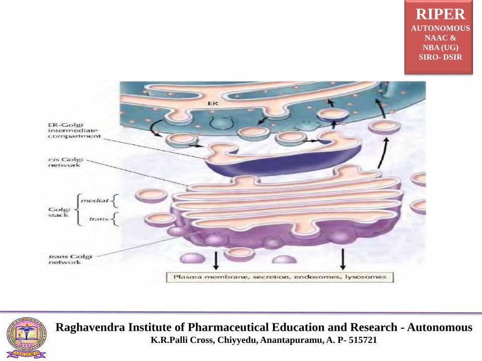
Golgi Bodies
Golgi Bodies is a collection of membrane enclosed sacs
composed of four or more stacked layers of thin, flat
enclosed vessels lying near the side of the nucleus.
Consist of multiple discrete compartments.
Consist of four functionally distinct regions:
RIPERAUTONOMOUS
NAAC &
NBA (UG)
SIRO- DSIR
Raghavendra Institute of Pharmaceutical Education and Research - AutonomousK.R.Palli Cross, Chiyyedu, Anantapuramu, A. P- 515721

RIPERAUTONOMOUS
NAAC &
NBA (UG)
SIRO- DSIR
Raghavendra Institute of Pharmaceutical Education and Research - AutonomousK.R.Palli Cross, Chiyyedu, Anantapuramu, A. P- 515721
The cis Golgi network
i) Golgi stack –which is divided into
a) The medial and
b) Trans sub compartments iii) The trans Golgi network.
Function
Wrapping and Packaging department of the cell.
Produces secretion granules i.e. membrane enclosed
complexes, which store hormones and enzymes in the
protein secreting cells, it packages proteins.

RIPERAUTONOMOUS
NAAC &
NBA (UG)
SIRO- DSIR
Raghavendra Institute of Pharmaceutical Education and Research - AutonomousK.R.Palli Cross, Chiyyedu, Anantapuramu, A. P- 515721
Site of formation of lysosomes i.e. large irregular
structures surrounded by membrane which are present
in the cytoplasm.
It adds certain carbohydrates to form glycoproteins,
which play an important role in the association of the
cells to form tissues

RIPERAUTONOMOUS
NAAC &
NBA (UG)
SIRO- DSIR
Raghavendra Institute of Pharmaceutical Education and Research - AutonomousK.R.Palli Cross, Chiyyedu, Anantapuramu, A. P- 515721
LysosomesThese are the irregular Structures surrounded by theUnit membraneMore acidic than rest of theCytoplasm and external bacteria as well as worn outCell components are digestedin them

RIPERAUTONOMOUS
NAAC &
NBA (UG)
SIRO- DSIR
Raghavendra Institute of Pharmaceutical Education and Research - AutonomousK.R.Palli Cross, Chiyyedu, Anantapuramu, A. P- 515721
• The interior is kept acidic(near pH 5.0) by the action of protonpump
• Lysosomes are cell hydrolases and they function best at the acidic pH.
Functions
Acts as a form of digestive (lytic ) system or the cell, becauseenzymes present in it can digest essentially all macromolecules.
Engulf worn out components of the cells in which they are located.
When a cell dies, lysosomal enzymes causes autolysis of theremanant . Thats why lysosomes are called as Suicidal Bags.
RIPERAUTONOMOUS
NAAC &
NBA (UG)
SIRO- DSIR
Raghavendra Institute of Pharmaceutical Education and Research - AutonomousK.R.Palli Cross, Chiyyedu, Anantapuramu, A. P- 515721
PeroxisomesA lipid bilayer membrane surrounds which regulateswhat enters or exits the peroxisomes.Urate oxidase crystalline core.Structure is similar to that of the lysosomes but with adifferent composition.Peroxisomes can be formed by the budding of ER, or bydivision.Contains oxidases that produces H2O2.

RIPERAUTONOMOUS
NAAC &
NBA (UG)
SIRO- DSIR
Raghavendra Institute of Pharmaceutical Education and Research - AutonomousK.R.Palli Cross, Chiyyedu, Anantapuramu, A. P- 515721
Catalases degrades hydrogen peroxide to yield water and oxygen
Proteins are directed to the Peroxisomes by a unique signalsequence with the help of protein chaperones, Peroxins.
Function
H2O2 metabolism and detoxification
Helps in Photorespiration in plants
Biosynthesis of lipids
Cholesterol and dolichol are synthesized in animals
Synthesis of bile acids in liver
Synthesis of plasmalogens (myelin sheath)

SUMMARY
COMPARTMENTS
Plasma Membrane
Cytosol
Mitochondria
Endoplasmic
Reticulum
Golgi apparutus
Lysosomes
Peroxisomes
Cyotoskeleton
Nucleus
MAJOR FUNCTIONS
Transport of ions and molecules
Metab. of carbohydrate, lipids
and amino acids
Energy production
Synthesis of proteins and lipids
Modification and sorting of
proteins
Cellular digestion
Utilization of H2O2
Cell Morphology and cell motility
DNA synthesis and Repair

RIPERAUTONOMOUS
NAAC &
NBA (UG)
SIRO- DSIR
Raghavendra Institute of Pharmaceutical Education and Research - AutonomousK.R.Palli Cross, Chiyyedu, Anantapuramu, A. P- 515721
Cell Junctions• Intercellular space in closely packed tissue is about 20nm.
The cells are bound together by the specific adhesiveglycoprotein.
• Modified cell membranes contributing in cohesion andcommunication are called Cell junctions.

RIPERAUTONOMOUS
NAAC &
NBA (UG)
SIRO- DSIR
Raghavendra Institute of Pharmaceutical Education and Research - AutonomousK.R.Palli Cross, Chiyyedu, Anantapuramu, A. P- 515721
Types of cell junctionsThere are three types of Cell Junctions
1. Occluding Junctions
2. Adhering Junctions
3. Communicating Junctions
Occluding Junctions
Found in epithelial tissues
Also known as “Tight Junctions”

RIPERAUTONOMOUS
NAAC &
NBA (UG)
SIRO- DSIR
Raghavendra Institute of Pharmaceutical Education and Research - AutonomousK.R.Palli Cross, Chiyyedu, Anantapuramu, A. P- 515721
• Do not allow passage of small molecules formimpermiable membrane.
Types
Zonula Occludens
Fascia Occludens
Zonula occludens
• Encircles the entire cell perimeter
• Occludes the intercellular space
RIPERAUTONOMOUS
NAAC &
NBA (UG)
SIRO- DSIR
Raghavendra Institute of Pharmaceutical Education and Research - AutonomousK.R.Palli Cross, Chiyyedu, Anantapuramu, A. P- 515721
Series of focal fusions
The adjacent cell membranes approach each other,outer leaflets fuse, diverge again then fuse again.
At fusions sites specific trans membranous proteinsnamed Occludins and Claudins perform the bindingfunction.
Occludins and claudins come out of cell membrane and
interact with each other and seal the surface of twoadjacent cells.

RIPERAUTONOMOUS
NAAC &
NBA (UG)
SIRO- DSIR
Raghavendra Institute of Pharmaceutical Education and Research - AutonomousK.R.Palli Cross, Chiyyedu, Anantapuramu, A. P- 515721
• These two proteins are attatched to three proteins called
ZO1, ZO2, ZO3.
• These three proteins helps in holding occludens and
claudins properly in their positions.
• This type of junction is present at apical region.
Location
Blood–Brain Barrier, Intestines, Nephrons
Skin.

RIPERAUTONOMOUS
NAAC &
NBA (UG)
SIRO- DSIR
Raghavendra Institute of Pharmaceutical Education and Research - AutonomousK.R.Palli Cross, Chiyyedu, Anantapuramu, A. P- 515721

RIPERAUTONOMOUS
NAAC &
NBA (UG)
SIRO- DSIR
Raghavendra Institute of Pharmaceutical Education and Research - AutonomousK.R.Palli Cross, Chiyyedu, Anantapuramu, A. P- 515721
Fascia occludens• A strip like tight junction of limited extent.
• Found between the endothelial cells of the blood vessels.
Adhering Junctions• Anchoring junctions
• Provide cell-cell or cell to basal lamina adherence

RIPERAUTONOMOUS
NAAC &
NBA (UG)
SIRO- DSIR
Raghavendra Institute of Pharmaceutical Education and Research - AutonomousK.R.Palli Cross, Chiyyedu, Anantapuramu, A. P- 515721
Types
Zonula adherence
Fascia adherence
Macula adherence (Desmosomes)
Hemidesmosomes
Zonula adherence
This is like a sticky belt present around the cell.
E. cadherins are the proteins helping in sticking the cellstogether in the presence of calcium.
E-cadherin links to adherent proteins in cytoplasm which are
Catenin, Vinculin.

RIPERAUTONOMOUS
NAAC &
NBA (UG)
SIRO- DSIR
Raghavendra Institute of Pharmaceutical Education and Research - AutonomousK.R.Palli Cross, Chiyyedu, Anantapuramu, A. P- 515721
Fascia adherenceStructurally it is similar to Zonula adherence
But its cell junction is strip-like and (not ring-like or
belt-like) i.e. Cardiac muscle cells.
Macula adherence (Desmosomes)Macula means a spot like.
This is also helpful for sticking the cells together but in
only some places.
These are not as strong as zona adherens.

RIPERAUTONOMOUS
NAAC &
NBA (UG)
SIRO- DSIR
Raghavendra Institute of Pharmaceutical Education and Research - AutonomousK.R.Palli Cross, Chiyyedu, Anantapuramu, A. P- 515721

RIPERAUTONOMOUS
NAAC &
NBA (UG)
SIRO- DSIR
Raghavendra Institute of Pharmaceutical Education and Research - AutonomousK.R.Palli Cross, Chiyyedu, Anantapuramu, A. P- 515721
The proteins which are helpful for adhering here areDesmocollins and Desmogleins.
Inside the cell, there is a special disc present which is
made up of desmoplachins and plakoglobins.
The cytoskeletal filaments attatched to this disc areintermediate fillaments and tonofillaments( keratin).
Present in simple epithelium, Stratified epithelium andalso in cardiac muscle cells.

RIPERAUTONOMOUS
NAAC &
NBA (UG)
SIRO- DSIR
Raghavendra Institute of Pharmaceutical Education and Research - AutonomousK.R.Palli Cross, Chiyyedu, Anantapuramu, A. P- 515721
HemidesmosomesThese junctions serve to anchor the epithelial cells to thebasal lamina.
A hemidesmosome is a spot like adhering junction whichgives appearance of a half desmosome.
In hemidesmosome transmembrane linker proteins areintegrins.
The cytoplasmic intermediate filaments of keratin are
inserted in to the attachment plaque.

RIPERAUTONOMOUS
NAAC &
NBA (UG)
SIRO- DSIR
Raghavendra Institute of Pharmaceutical Education and Research - AutonomousK.R.Palli Cross, Chiyyedu, Anantapuramu, A. P- 515721

RIPERAUTONOMOUS
NAAC &
NBA (UG)
SIRO- DSIR
Raghavendra Institute of Pharmaceutical Education and Research - AutonomousK.R.Palli Cross, Chiyyedu, Anantapuramu, A. P- 515721
Communicating Junctions
( Gap Junctions)• These are the window like structures present on the
lateral surface of each epithelial cells for the sake of
communication between the cells.
• It is made up of 6 types of proteins made up ofconnexins, so these 6 connexins combine to form aprotein channel called connexon.

RIPERAUTONOMOUS
NAAC &
NBA (UG)
SIRO- DSIR
Raghavendra Institute of Pharmaceutical Education and Research - AutonomousK.R.Palli Cross, Chiyyedu, Anantapuramu, A. P- 515721
• Characterized by presence of minute tubular passageways
• Provide direct cell to cell communication
• Tubular passages allow movement of ions and other
small molecules between adjacent cells
• These can be opened or closed when necessary
• In cardiac and smooth muscles the gap junction provideselectrical coupling of the adjacent cells
• Gap junctions are frequently found in embryonic
cells

RIPERAUTONOMOUS
NAAC &
NBA (UG)
SIRO- DSIR
Raghavendra Institute of Pharmaceutical Education and Research - AutonomousK.R.Palli Cross, Chiyyedu, Anantapuramu, A. P- 515721

RIPERAUTONOMOUS
NAAC &
NBA (UG)
SIRO- DSIR
Raghavendra Institute of Pharmaceutical Education and Research - AutonomousK.R.Palli Cross, Chiyyedu, Anantapuramu, A. P- 515721
RIPERAUTONOMOUS
NAAC &
NBA (UG)
SIRO- DSIR
Raghavendra Institute of Pharmaceutical Education and Research - AutonomousK.R.Palli Cross, Chiyyedu, Anantapuramu, A. P- 515721
Transport mechanisms• Transport of substances across the cell membrane is
necessary to maintain the normal functioning of the cellsin our body.
• Lipid soluble substances, water & urea can easily passthrough the lipid bilayer of the cell membrane.
• The lipid bilayer of the cell membrane is impermeable tolipid insoluble substances such as ions & charged orpolar molecules like glucose

RIPERAUTONOMOUS
NAAC &
NBA (UG)
SIRO- DSIR
Raghavendra Institute of Pharmaceutical Education and Research - AutonomousK.R.Palli Cross, Chiyyedu, Anantapuramu, A. P- 515721
• These substances pass through specialized proteinchannels, carrier proteins & active pump mechanisms.
• Large macromolecules are transported through vesicles.

RIPERAUTONOMOUS
NAAC &
NBA (UG)
SIRO- DSIR
Raghavendra Institute of Pharmaceutical Education and Research - AutonomousK.R.Palli Cross, Chiyyedu, Anantapuramu, A. P- 515721
Types• Passive transport
– Diffusion – simple, facilitated
–Osmosis
• Active transport
– Primary
– Secondary
• Vesicular transport
– Endocytosis
– Exocytosis
– Transcytosis

RIPERAUTONOMOUS
NAAC &
NBA (UG)
SIRO- DSIR
Raghavendra Institute of Pharmaceutical Education and Research - AutonomousK.R.Palli Cross, Chiyyedu, Anantapuramu, A. P- 515721
Diffusion-simple
• It is the movement of ions or molecules from a region oftheir high concentration to a region of their lowconcentration, without the expenditure of energy
• Movement is towards the concentration gradient untilan equilibrium is achieved.
Diffusion through lipid matrix
• Lipophilic/hydrophobic/nonpolar/uncharged substancessuch as O2, CO2, N2, fatty acids, alcohol, steroidhormones, etc.

RIPERAUTONOMOUS
NAAC &
NBA (UG)
SIRO- DSIR
Raghavendra Institute of Pharmaceutical Education and Research - AutonomousK.R.Palli Cross, Chiyyedu, Anantapuramu, A. P- 515721
• Water as it is a small molecule with high kinetic energy
• Urea
Ionic diffusion – through channel proteins
• Ions diffuse through the ion channels gated channels open when stimulus is given
– Voltage gated
– Ligand gated
– Mechano sensitive gated
• They are either open or gated
• Open/leak channels – ex: K+ channels

RIPERAUTONOMOUS
NAAC &
NBA (UG)
SIRO- DSIR
Raghavendra Institute of Pharmaceutical Education and Research - AutonomousK.R.Palli Cross, Chiyyedu, Anantapuramu, A. P- 515721
Facilitated diffusion
• Facilitated diffusion is the movement of specific
• molecules (or ions) across the plasma membrane
• assisted by a carrier protein
• The direction of movement is down the concentration gradient of the molecules concerned
• No energy required

RIPERAUTONOMOUS
NAAC &
NBA (UG)
SIRO- DSIR
Raghavendra Institute of Pharmaceutical Education and Research - AutonomousK.R.Palli Cross, Chiyyedu, Anantapuramu, A. P- 515721
Difference between carrier proteins & channel proteins
• Carrier proteins bind to larger molecules, and change theirshape so molecules can diffuse through.
• Channel proteins provide water filled pores for charged ions to pass through

RIPERAUTONOMOUS
NAAC &
NBA (UG)
SIRO- DSIR
Raghavendra Institute of Pharmaceutical Education and Research - AutonomousK.R.Palli Cross, Chiyyedu, Anantapuramu, A. P- 515721
Osmosis
Osmosis is the movement of water molecules (solvent)through a selectively permeable membrane/semipermeable membrane.
Water diffuses across a membrane from an area of highconcentration to an area of low concentration, Semi-permeable membrane is permeable to water, but not tothe solute i.e. sugar.

RIPERAUTONOMOUS
NAAC &
NBA (UG)
SIRO- DSIR
Raghavendra Institute of Pharmaceutical Education and Research - AutonomousK.R.Palli Cross, Chiyyedu, Anantapuramu, A. P- 515721
RIPERAUTONOMOUS
NAAC &
NBA (UG)
SIRO- DSIR
Raghavendra Institute of Pharmaceutical Education and Research - AutonomousK.R.Palli Cross, Chiyyedu, Anantapuramu, A. P- 515721
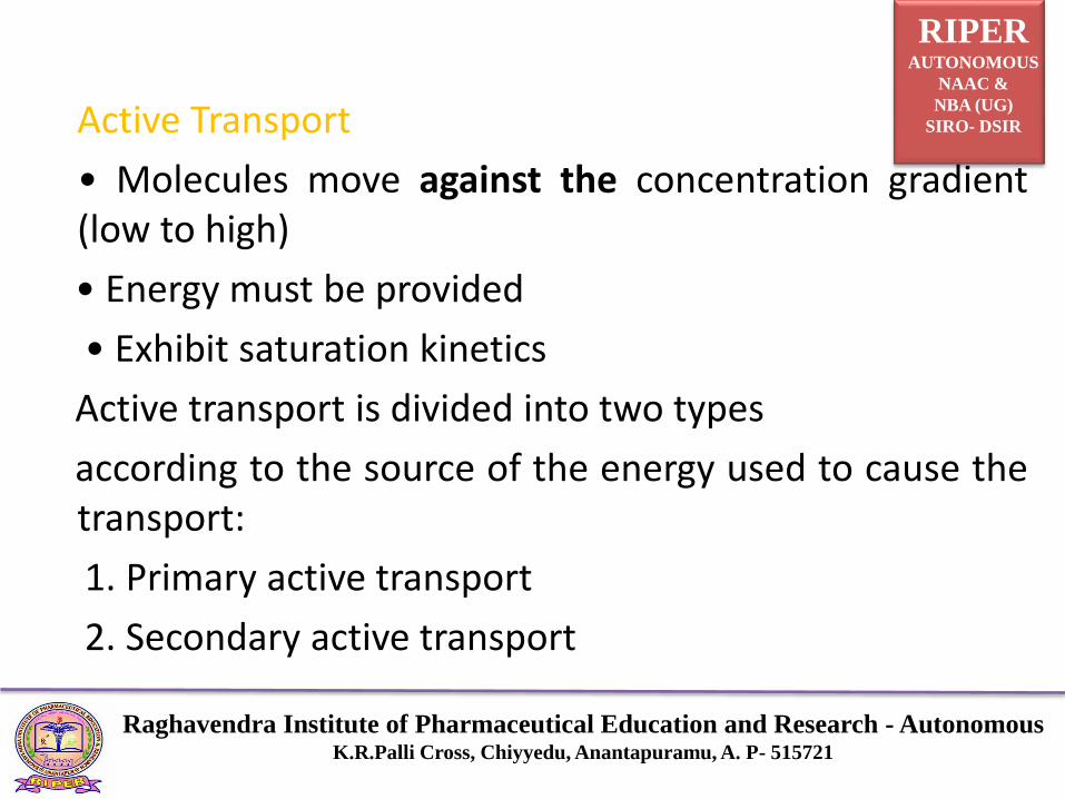
Active Transport
• Molecules move against the concentration gradient(low to high)
• Energy must be provided
• Exhibit saturation kinetics
Active transport is divided into two types
according to the source of the energy used to cause thetransport:
1. Primary active transport
2. Secondary active transport

RIPERAUTONOMOUS
NAAC &
NBA (UG)
SIRO- DSIR
Raghavendra Institute of Pharmaceutical Education and Research - AutonomousK.R.Palli Cross, Chiyyedu, Anantapuramu, A. P- 515721
Primary active transport
• They use the energy directly from the hydrolysis of ATP.
• Sodium potassium Pump
• Calcium pump
• Hydrogen Potassium pump
Secondary active transport
• Energy utilised in the transport of one substance helps in the movement of the other substance.
RIPERAUTONOMOUS
NAAC &
NBA (UG)
SIRO- DSIR
Raghavendra Institute of Pharmaceutical Education and Research - AutonomousK.R.Palli Cross, Chiyyedu, Anantapuramu, A. P- 515721
• Energy is derived secondarily, from energy that has beenstored in the form of ionic concentration differences ofsecondary molecular or ionic substances between thetwo sides of a cell membrane, created originally byprimary active transport.

RIPERAUTONOMOUS
NAAC &
NBA (UG)
SIRO- DSIR
Raghavendra Institute of Pharmaceutical Education and Research - AutonomousK.R.Palli Cross, Chiyyedu, Anantapuramu, A. P- 515721
Vesicular transport
Endocytosis, Exocytosis, Transcytosis
Endocytosis/Exocytosis
• For substances the cell needs to take in (endo = in) orexpel (exo = out), that are too large for passive or activetransport.
Endocytosis
• The material makes contact with the cell membranethen invaginates,Invagination is then pinched off leavingthe cell membrane intact.

RIPERAUTONOMOUS
NAAC &
NBA (UG)
SIRO- DSIR
Raghavendra Institute of Pharmaceutical Education and Research - AutonomousK.R.Palli Cross, Chiyyedu, Anantapuramu, A. P- 515721
Types of Endocytosis
1. Receptor mediated endocytosis
2. Phagocytosis (solids)
3. Pinocytosis (liquids)
Ex: White Blood Cells surround and engulf bacteria by
Phagocytosis.
• Pinocytosis – amino acids, fatty acids
• Receptor mediated endocytosis – LDL, Nerve growth
factor, vitamins, hormones, HIV virus entering the T
cell etc.,

RIPERAUTONOMOUS
NAAC &
NBA (UG)
SIRO- DSIR
Raghavendra Institute of Pharmaceutical Education and Research - AutonomousK.R.Palli Cross, Chiyyedu, Anantapuramu, A. P- 515721

RIPERAUTONOMOUS
NAAC &
NBA (UG)
SIRO- DSIR
Raghavendra Institute of Pharmaceutical Education and Research - AutonomousK.R.Palli Cross, Chiyyedu, Anantapuramu, A. P- 515721
Exocytosis
• The process of release of macromolecules from the cells to theexterior.
• Vesicles containing material to be exposed, bind to the cellmembrane
• Area of fusion breaks down leaving the contents of the vesicleoutside & the cell membrane intact
• Reverse pinocytosis or emeiocytosis
• Requires calcium & energy
• Secretory granules, hormones

RIPERAUTONOMOUS
NAAC &
NBA (UG)
SIRO- DSIR
Raghavendra Institute of Pharmaceutical Education and Research - AutonomousK.R.Palli Cross, Chiyyedu, Anantapuramu, A. P- 515721

RIPERAUTONOMOUS
NAAC &
NBA (UG)
SIRO- DSIR
Raghavendra Institute of Pharmaceutical Education and Research - AutonomousK.R.Palli Cross, Chiyyedu, Anantapuramu, A. P- 515721

RIPERAUTONOMOUS
NAAC &
NBA (UG)
SIRO- DSIR
Raghavendra Institute of Pharmaceutical Education and Research - AutonomousK.R.Palli Cross, Chiyyedu, Anantapuramu, A. P- 515721
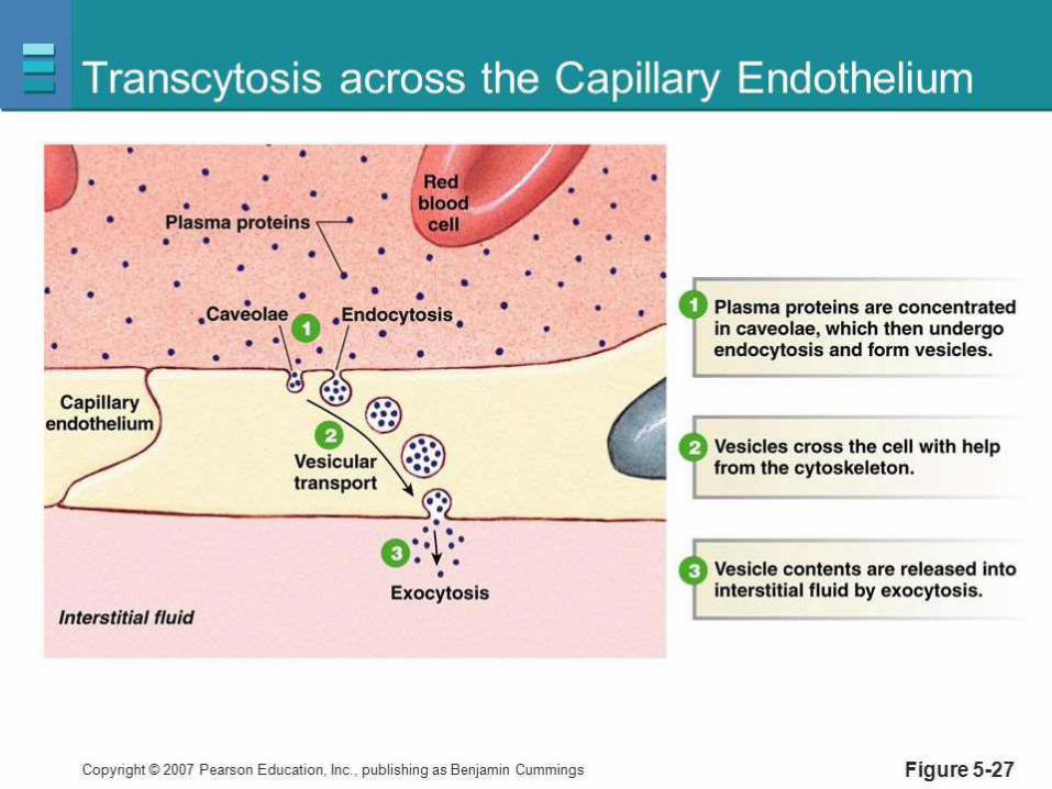
Transcytosis
• Vesicles endocytosed on one side of the cell; exocytosed on the opposite side.
• Caveolin mediated (Rafts & Caveolae)
• Also known as cytopempsis
• Transport of substances across the endothelial cells of blood vessels to interstitial fluid
RIPERAUTONOMOUS
NAAC &
NBA (UG)
SIRO- DSIR
Raghavendra Institute of Pharmaceutical Education and Research - AutonomousK.R.Palli Cross, Chiyyedu, Anantapuramu, A. P- 515721

RIPERAUTONOMOUS
NAAC &
NBA (UG)
SIRO- DSIR
Raghavendra Institute of Pharmaceutical Education and Research - AutonomousK.R.Palli Cross, Chiyyedu, Anantapuramu, A. P- 515721
THANK YOU EVERY ONE