Glycophenotypic Alterations Induced by Pteridium aquilinum in Mice Gastric Mucosa: Synergistic...
11
Glycophenotypic Alterations Induced by Pteridium aquilinum in Mice Gastric Mucosa: Synergistic Effect with Helicobacter pylori Infection Joana Gomes 1,2 , Ana Magalha ˜es 1 , Ana S. Carvalho 1 , Gilberto E. Hernandez 3 , Suzanne L. Papp 3 , Steven R. Head 3 , Vale ´ rie Michel 2 , Leonor David 1,4 , Fa ´ tima Ga ¨ rtner 1,5 , Eliette Touati 2 , Celso A. Reis 1,4,5 * 1 Institute of Molecular Pathology and Immunology of the University of Porto (IPATIMUP), Universidade do Porto, Porto, Portugal, 2 Institut Pasteur, Unite ´ de Pathogene `se de Helicobacter, Paris, France, 3 The Scripps Research Institute, La Jolla, California, United States of America, 4 Faculdade de Medicina, Universidade do Porto, Porto, Portugal, 5 Instituto de Cie ˆncias Biome ´dicas Abel Salazar, Universidade do Porto, Porto, Portugal Abstract The bracken fern Pteridium aquilinum is a plant known to be carcinogenic to animals. Epidemiological studies have shown an association between bracken fern exposure and gastric cancer development in humans. The biological effects of exposure to this plant within the gastric carcinogenesis process are not fully understood. In the present work, effects in the gastric mucosa of mice treated with Pteridium aquilinum were evaluated, as well as molecular mechanisms underlying the synergistic role with Helicobacter pylori infection. Our results showed that exposure to Pteridium aquilinum induces histomorphological modifications including increased expression of acidic glycoconjugates in the gastric mucosa. The transcriptome analysis of gastric mucosa showed that upon exposure to Pteridium aquilinum several glycosyltransferase genes were differently expressed, including Galntl4, C1galt1 and St3gal2, that are mainly involved in the biosynthesis of simple mucin-type carbohydrate antigens. Concomitant treatment with Pteridium aquilinum and infection with Helicobacter pylori also resulted in differently expressed glycosyltransferase genes underlying the biosynthesis of terminal sialylated Lewis antigens, including Sialyl-Lewis x . These results disclose the molecular basis for the altered pattern of glycan structures observed in the mice gastric mucosa. The gene transcription alterations and the induced glycophenotypic changes observed in the gastric mucosa contribute for the understanding of the molecular mechanisms underlying the role of Pteridium aquilinum in the gastric carcinogenesis process. Citation: Gomes J, Magalha ˜es A, Carvalho AS, Hernandez GE, Papp SL, et al. (2012) Glycophenotypic Alterations Induced by Pteridium aquilinum in Mice Gastric Mucosa: Synergistic Effect with Helicobacter pylori Infection. PLoS ONE 7(6): e38353. doi:10.1371/journal.pone.0038353 Editor: Yoshio Yamaoka, Veterans Affairs Medical Center (111D), United States of America Received January 12, 2012; Accepted May 8, 2012; Published June 13, 2012 Copyright: ß 2012 Gomes et al. This is an open-access article distributed under the terms of the Creative Commons Attribution License, which permits unrestricted use, distribution, and reproduction in any medium, provided the original author and source are credited. Funding: This work was supported by Fundac ¸a ˜o para a Cie ˆncia e a Tecnologia – [FCT (PIC/IC/82716/2007), grant number SFRH/BD/40563/2007 to J.G and SFRH/ BPD/75871/2011 to A.M.]; and the Programa de Acc ¸o ˜ es Universita ´rias Integradas Luso-Francesas - PAULIF [grant number LC/HS/ED/2010-308; F-TC04/11]. The resources and collaborative efforts provided by the Consortium for Functional Glycomics were funded by grant NIGMS-GM62116. The funders had no role in study design, data collection and analysis, decision to publish, or preparation of the manuscript. Competing Interests: The authors have declared that no competing interests exist. * E-mail: [email protected] Introduction The bracken fern Pteridium aquilinum (BF) is a common toxic plant that has high potential carcinogenic effects in animals and humans that consume BF or live in bracken-infested areas [1]. The BF, known to contain a toxin named ptaquiloside, is classified by the International Agency for Research on Cancer (IARC) as possibly carcinogenic to humans - group 2B [2], due to its carcinogenic and mutagenic effects [3]. Although clear carcino- genic effects have been shown in animals that consume BF [1,3], the relationship between BF exposure and human health remains to be clarified. BF toxicity in humans has been associated with the consumption of the plant in some oriental cultures, and also with the indirect exposure to contaminated groundwater, drinking milk from cows fed with BF, and inhalation of bracken spores [4–6]. Epidemiologic evidences support association between BF exposure and gastric cancer development [7–9]. In agreement, we recently described the direct DNA damaging and mutagenic effects of BF and its toxin ptaquiloside in gastric epithelial cells and gastric mucosa of exposed mice [10]. Gastric cancer is the second leading cause of cancer-related death in the world [11]. The most common form of gastric cancer is the ultimate stage of a carcinogenic pathway that is initiated by the infection with Helicobacter pylori (H. pylori) [12], a type I human carcinogen according to IARC [13]. The gastric lesions associated with H. pylori infection can evolve over decades from chronic gastritis, gastric atrophy, intestinal metaplasia, and dysplasia to gastric carcinoma [14]. The development of this pathway leading to gastric carcinoma is a multifactorial process which depends on the interplay between several factors such as H. pylori virulence factors, host genetic polymorphisms and other environmental factors that dictate the clinical outcome of the disease. Alterations in gastric glycophenotype are commonly observed during the gastric carcinogenic pathway and include increased expression of sialylated terminal structures, such as Sialyl-Lewis x (SLe x ) and Sialyl-Lewis a (SLe a ) [15–17], as well as aberrant expression of simple mucin-type carbohydrate antigens, as it is the case of Tn, Sialyl Tn (STn) and T antigens [18–20]. Some of these glycophenotypic alterations have been reported early in the process of H. pylori infection both in human gastric mucosa [15,16] PLoS ONE | www.plosone.org 1 June 2012 | Volume 7 | Issue 6 | e38353
-
Upload
independent -
Category
Documents
-
view
2 -
download
0
Transcript of Glycophenotypic Alterations Induced by Pteridium aquilinum in Mice Gastric Mucosa: Synergistic...
Glycophenotypic Alterations Induced by Pteridiumaquilinum in Mice Gastric Mucosa: Synergistic Effect withHelicobacter pylori InfectionJoana Gomes1,2, Ana Magalhaes1, Ana S. Carvalho1, Gilberto E. Hernandez3, Suzanne L. Papp3,
Steven R. Head3, Valerie Michel2, Leonor David1,4, Fatima Gartner1,5, Eliette Touati2, Celso A. Reis1,4,5*
1 Institute of Molecular Pathology and Immunology of the University of Porto (IPATIMUP), Universidade do Porto, Porto, Portugal, 2 Institut Pasteur, Unite de Pathogenese
de Helicobacter, Paris, France, 3 The Scripps Research Institute, La Jolla, California, United States of America, 4 Faculdade de Medicina, Universidade do Porto, Porto,
Portugal, 5 Instituto de Ciencias Biomedicas Abel Salazar, Universidade do Porto, Porto, Portugal
Abstract
The bracken fern Pteridium aquilinum is a plant known to be carcinogenic to animals. Epidemiological studies have shownan association between bracken fern exposure and gastric cancer development in humans. The biological effects ofexposure to this plant within the gastric carcinogenesis process are not fully understood. In the present work, effects in thegastric mucosa of mice treated with Pteridium aquilinum were evaluated, as well as molecular mechanisms underlying thesynergistic role with Helicobacter pylori infection. Our results showed that exposure to Pteridium aquilinum induceshistomorphological modifications including increased expression of acidic glycoconjugates in the gastric mucosa. Thetranscriptome analysis of gastric mucosa showed that upon exposure to Pteridium aquilinum several glycosyltransferasegenes were differently expressed, including Galntl4, C1galt1 and St3gal2, that are mainly involved in the biosynthesis ofsimple mucin-type carbohydrate antigens. Concomitant treatment with Pteridium aquilinum and infection with Helicobacterpylori also resulted in differently expressed glycosyltransferase genes underlying the biosynthesis of terminal sialylatedLewis antigens, including Sialyl-Lewisx. These results disclose the molecular basis for the altered pattern of glycan structuresobserved in the mice gastric mucosa. The gene transcription alterations and the induced glycophenotypic changesobserved in the gastric mucosa contribute for the understanding of the molecular mechanisms underlying the role ofPteridium aquilinum in the gastric carcinogenesis process.
Citation: Gomes J, Magalhaes A, Carvalho AS, Hernandez GE, Papp SL, et al. (2012) Glycophenotypic Alterations Induced by Pteridium aquilinum in Mice GastricMucosa: Synergistic Effect with Helicobacter pylori Infection. PLoS ONE 7(6): e38353. doi:10.1371/journal.pone.0038353
Editor: Yoshio Yamaoka, Veterans Affairs Medical Center (111D), United States of America
Received January 12, 2012; Accepted May 8, 2012; Published June 13, 2012
Copyright: � 2012 Gomes et al. This is an open-access article distributed under the terms of the Creative Commons Attribution License, which permitsunrestricted use, distribution, and reproduction in any medium, provided the original author and source are credited.
Funding: This work was supported by Fundacao para a Ciencia e a Tecnologia – [FCT (PIC/IC/82716/2007), grant number SFRH/BD/40563/2007 to J.G and SFRH/BPD/75871/2011 to A.M.]; and the Programa de Accoes Universitarias Integradas Luso-Francesas - PAULIF [grant number LC/HS/ED/2010-308; F-TC04/11]. Theresources and collaborative efforts provided by the Consortium for Functional Glycomics were funded by grant NIGMS-GM62116. The funders had no role in studydesign, data collection and analysis, decision to publish, or preparation of the manuscript.
Competing Interests: The authors have declared that no competing interests exist.
* E-mail: [email protected]
Introduction
The bracken fern Pteridium aquilinum (BF) is a common toxic
plant that has high potential carcinogenic effects in animals and
humans that consume BF or live in bracken-infested areas [1]. The
BF, known to contain a toxin named ptaquiloside, is classified by
the International Agency for Research on Cancer (IARC) as
possibly carcinogenic to humans - group 2B [2], due to its
carcinogenic and mutagenic effects [3]. Although clear carcino-
genic effects have been shown in animals that consume BF [1,3],
the relationship between BF exposure and human health remains
to be clarified. BF toxicity in humans has been associated with the
consumption of the plant in some oriental cultures, and also with
the indirect exposure to contaminated groundwater, drinking milk
from cows fed with BF, and inhalation of bracken spores [4–6].
Epidemiologic evidences support association between BF exposure
and gastric cancer development [7–9]. In agreement, we recently
described the direct DNA damaging and mutagenic effects of BF
and its toxin ptaquiloside in gastric epithelial cells and gastric
mucosa of exposed mice [10].
Gastric cancer is the second leading cause of cancer-related
death in the world [11]. The most common form of gastric cancer
is the ultimate stage of a carcinogenic pathway that is initiated by
the infection with Helicobacter pylori (H. pylori) [12], a type I human
carcinogen according to IARC [13]. The gastric lesions associated
with H. pylori infection can evolve over decades from chronic
gastritis, gastric atrophy, intestinal metaplasia, and dysplasia to
gastric carcinoma [14]. The development of this pathway leading
to gastric carcinoma is a multifactorial process which depends on
the interplay between several factors such as H. pylori virulence
factors, host genetic polymorphisms and other environmental
factors that dictate the clinical outcome of the disease.
Alterations in gastric glycophenotype are commonly observed
during the gastric carcinogenic pathway and include increased
expression of sialylated terminal structures, such as Sialyl-Lewisx
(SLex) and Sialyl-Lewisa (SLea) [15–17], as well as aberrant
expression of simple mucin-type carbohydrate antigens, as it is the
case of Tn, Sialyl Tn (STn) and T antigens [18–20]. Some of these
glycophenotypic alterations have been reported early in the
process of H. pylori infection both in human gastric mucosa [15,16]
PLoS ONE | www.plosone.org 1 June 2012 | Volume 7 | Issue 6 | e38353
and experimentally infected animal models [21,22]. Upon
infection, H. pylori is able to modulate the expression of several
host glycosylation-related genes, including the induction of a
glycosyltransferase that results in increased SLex expression [23].
Modified glycosylation has been proposed to play a role in the
development and progression of the disease [19,24]. Particularly in
gastric cancer, increased expression of sialylated antigens has been
associated with a worst prognosis [25].
The goal of this study is to characterize the alterations induced
by Pteridium aquilinum in the gastric mucosa and its possible
synergistic effects with H. pylori infection. Histomorphological
alterations and an altered glycophenotype were observed in the
gastric mucosa of mice exposed to BF in the presence or absence of
H. pylori infection. Furthermore, mice that were BF treated or
concomitantly infected with H. pylori displayed different levels of
glycosyltransferase genes expression as demonstrated using Glyco-
gene Chip arrays. The identification of genes involved in the
biosynthesis of carbohydrate antigens with a modified expression
in gastric mucosa may contribute to a better understanding of the
mechanisms involved in the development of gastric lesions
associated to BF exposure and the contribution of H. pylori
infection in this process.
Results
Phenotypic Alterations in Mice Gastric Mucosa inResponse to Pteridium aquilinum Exposure and/orH. pylori Infection
Histomorphological analysis of mice gastric mucosa stained with
hematoxylin and eosin (H&E) (Figure 1, Panel A, a-h) showed
various alterations in the BF treated (Group 2), H. pylori infected
(Group 3) and BF treated and H. pylori infected (Group 4) mice
groups when compared to the control (Group 1). The results
observed at both time points (4 or 7 weeks) for each experimental
condition were identical. Histological evaluation of inflammatory
cells (Table S1) showed that gastric mucosa of BF treated (Group
2) mice displayed a mild inflammation when compared to control
mice (Group1). Gastric mucosa of H. pylori infected (Group 3) mice
displayed moderate inflammation in 5 of 8 evaluated animals
while the others 3 animals showed mild inflammation. The
concomitantly BF treated and H. pylori infected mice (Group 4)
displayed severe inflammation with significant infiltrates of
inflammatory cells in the gastric mucosa of all animals compared
to control mice (Group1) in which the level of inflammatory
components was classified as absent even though very few
inflammatory cells could be observed (Figure 1, Panel A, a-h)
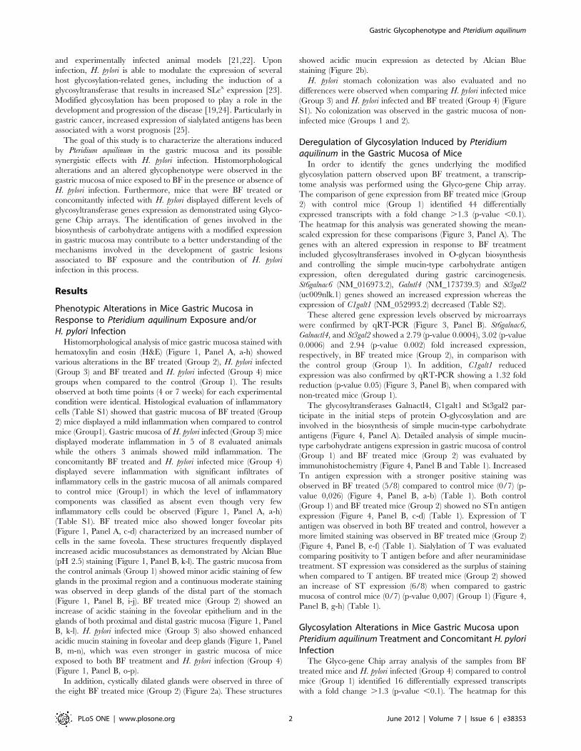
(Table S1). BF treated mice also showed longer foveolar pits
(Figure 1, Panel A, c-d) characterized by an increased number of
cells in the same foveola. These structures frequently displayed
increased acidic mucosubstances as demonstrated by Alcian Blue
(pH 2.5) staining (Figure 1, Panel B, k-l). The gastric mucosa from
the control animals (Group 1) showed minor acidic staining of few
glands in the proximal region and a continuous moderate staining
was observed in deep glands of the distal part of the stomach
(Figure 1, Panel B, i-j). BF treated mice (Group 2) showed an
increase of acidic staining in the foveolar epithelium and in the
glands of both proximal and distal gastric mucosa (Figure 1, Panel
B, k-l). H. pylori infected mice (Group 3) also showed enhanced
acidic mucin staining in foveolar and deep glands (Figure 1, Panel
B, m-n), which was even stronger in gastric mucosa of mice
exposed to both BF treatment and H. pylori infection (Group 4)
(Figure 1, Panel B, o-p).
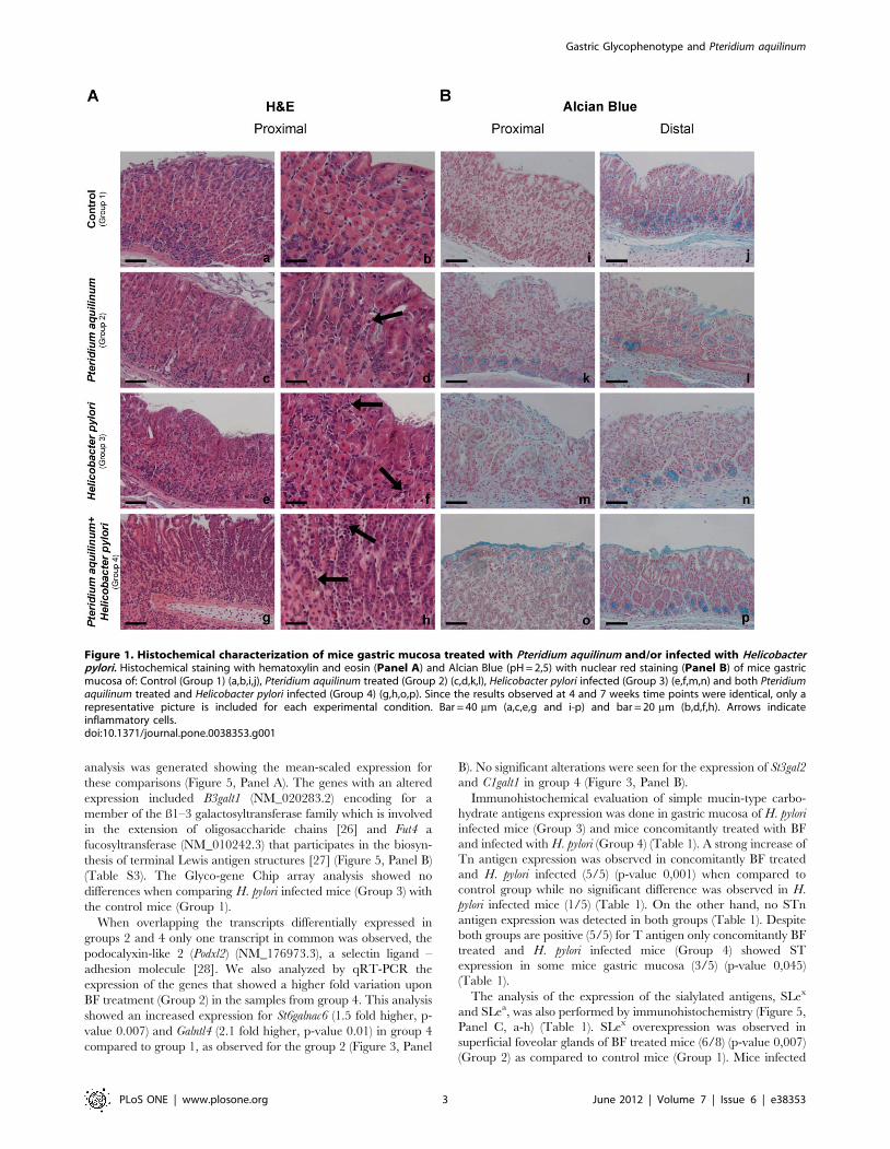
In addition, cystically dilated glands were observed in three of
the eight BF treated mice (Group 2) (Figure 2a). These structures
showed acidic mucin expression as detected by Alcian Blue
staining (Figure 2b).
H. pylori stomach colonization was also evaluated and no
differences were observed when comparing H. pylori infected mice
(Group 3) and H. pylori infected and BF treated (Group 4) (Figure
S1). No colonization was observed in the gastric mucosa of non-
infected mice (Groups 1 and 2).
Deregulation of Glycosylation Induced by Pteridiumaquilinum in the Gastric Mucosa of Mice
In order to identify the genes underlying the modified
glycosylation pattern observed upon BF treatment, a transcrip-
tome analysis was performed using the Glyco-gene Chip array.
The comparison of gene expression from BF treated mice (Group
2) with control mice (Group 1) identified 44 differentially
expressed transcripts with a fold change .1.3 (p-value ,0.1).
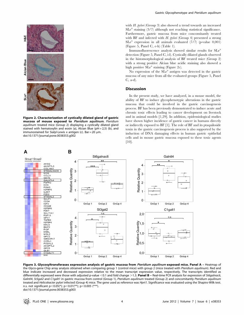
The heatmap for this analysis was generated showing the mean-
scaled expression for these comparisons (Figure 3, Panel A). The
genes with an altered expression in response to BF treatment
included glycosyltransferases involved in O-glycan biosynthesis
and controlling the simple mucin-type carbohydrate antigen
expression, often deregulated during gastric carcinogenesis.
St6galnac6 (NM_016973.2), Galntl4 (NM_173739.3) and St3gal2
(uc009nlk.1) genes showed an increased expression whereas the
expression of C1galt1 (NM_052993.2) decreased (Table S2).
These altered gene expression levels observed by microarrays
were confirmed by qRT-PCR (Figure 3, Panel B). St6galnac6,
Galnactl4, and St3gal2 showed a 2.79 (p-value 0.0004), 3.02 (p-value
0.0006) and 2.94 (p-value 0.002) fold increased expression,
respectively, in BF treated mice (Group 2), in comparison with
the control group (Group 1). In addition, C1galt1 reduced
expression was also confirmed by qRT-PCR showing a 1.32 fold
reduction (p-value 0.05) (Figure 3, Panel B), when compared with
non-treated mice (Group 1).
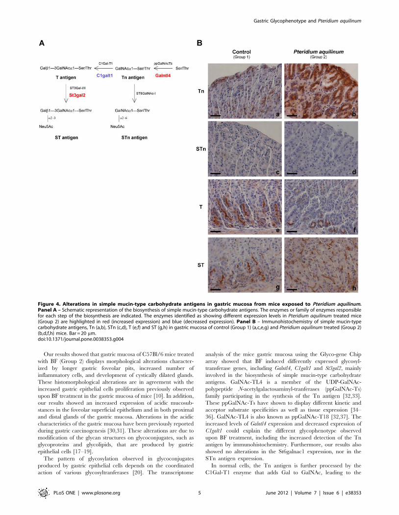
The glycosyltransferases Galnactl4, C1galt1 and St3gal2 par-
ticipate in the initial steps of protein O-glycosylation and are
involved in the biosynthesis of simple mucin-type carbohydrate
antigens (Figure 4, Panel A). Detailed analysis of simple mucin-
type carbohydrate antigens expression in gastric mucosa of control
(Group 1) and BF treated mice (Group 2) was evaluated by
immunohistochemistry (Figure 4, Panel B and Table 1). Increased
Tn antigen expression with a stronger positive staining was
observed in BF treated (5/8) compared to control mice (0/7) (p-
value 0,026) (Figure 4, Panel B, a-b) (Table 1). Both control
(Group 1) and BF treated mice (Group 2) showed no STn antigen
expression (Figure 4, Panel B, c-d) (Table 1). Expression of T
antigen was observed in both BF treated and control, however a
more limited staining was observed in BF treated mice (Group 2)
(Figure 4, Panel B, e-f) (Table 1). Sialylation of T was evaluated
comparing positivity to T antigen before and after neuraminidase
treatment. ST expression was considered as the surplus of staining
when compared to T antigen. BF treated mice (Group 2) showed
an increase of ST expression (6/8) when compared to gastric
mucosa of control mice (0/7) (p-value 0,007) (Group 1) (Figure 4,
Panel B, g-h) (Table 1).
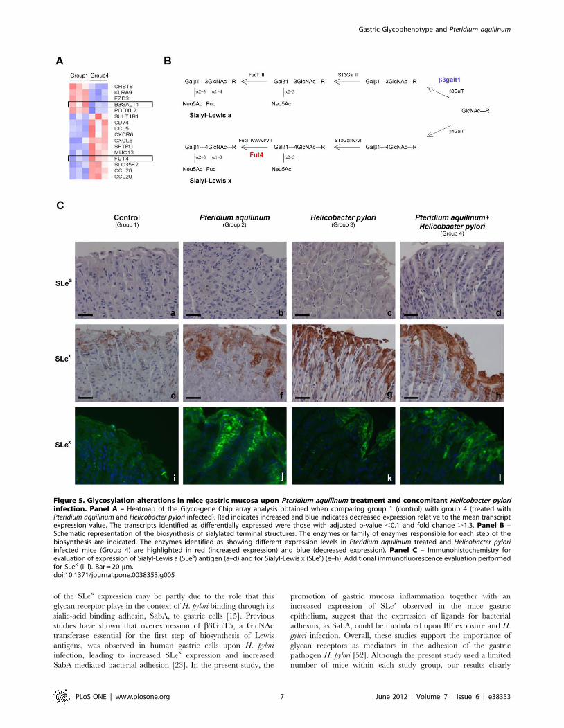
Glycosylation Alterations in Mice Gastric Mucosa uponPteridium aquilinum Treatment and Concomitant H. pyloriInfection
The Glyco-gene Chip array analysis of the samples from BF
treated mice and H. pylori infected (Group 4) compared to control
mice (Group 1) identified 16 differentially expressed transcripts
with a fold change .1.3 (p-value ,0.1). The heatmap for this
Gastric Glycophenotype and Pteridium aquilinum
PLoS ONE | www.plosone.org 2 June 2012 | Volume 7 | Issue 6 | e38353
analysis was generated showing the mean-scaled expression for
these comparisons (Figure 5, Panel A). The genes with an altered
expression included B3galt1 (NM_020283.2) encoding for a
member of the ß1–3 galactosyltransferase family which is involved
in the extension of oligosaccharide chains [26] and Fut4 a
fucosyltransferase (NM_010242.3) that participates in the biosyn-
thesis of terminal Lewis antigen structures [27] (Figure 5, Panel B)
(Table S3). The Glyco-gene Chip array analysis showed no
differences when comparing H. pylori infected mice (Group 3) with
the control mice (Group 1).
When overlapping the transcripts differentially expressed in
groups 2 and 4 only one transcript in common was observed, the
podocalyxin-like 2 (Podxl2) (NM_176973.3), a selectin ligand –
adhesion molecule [28]. We also analyzed by qRT-PCR the
expression of the genes that showed a higher fold variation upon
BF treatment (Group 2) in the samples from group 4. This analysis
showed an increased expression for St6galnac6 (1.5 fold higher, p-
value 0.007) and Galntl4 (2.1 fold higher, p-value 0.01) in group 4
compared to group 1, as observed for the group 2 (Figure 3, Panel
B). No significant alterations were seen for the expression of St3gal2
and C1galt1 in group 4 (Figure 3, Panel B).
Immunohistochemical evaluation of simple mucin-type carbo-
hydrate antigens expression was done in gastric mucosa of H. pylori
infected mice (Group 3) and mice concomitantly treated with BF
and infected with H. pylori (Group 4) (Table 1). A strong increase of
Tn antigen expression was observed in concomitantly BF treated
and H. pylori infected (5/5) (p-value 0,001) when compared to
control group while no significant difference was observed in H.
pylori infected mice (1/5) (Table 1). On the other hand, no STn
antigen expression was detected in both groups (Table 1). Despite
both groups are positive (5/5) for T antigen only concomitantly BF
treated and H. pylori infected mice (Group 4) showed ST
expression in some mice gastric mucosa (3/5) (p-value 0,045)
(Table 1).
The analysis of the expression of the sialylated antigens, SLex
and SLea, was also performed by immunohistochemistry (Figure 5,
Panel C, a-h) (Table 1). SLex overexpression was observed in
superficial foveolar glands of BF treated mice (6/8) (p-value 0,007)
(Group 2) as compared to control mice (Group 1). Mice infected
Figure 1. Histochemical characterization of mice gastric mucosa treated with Pteridium aquilinum and/or infected with Helicobacterpylori. Histochemical staining with hematoxylin and eosin (Panel A) and Alcian Blue (pH = 2,5) with nuclear red staining (Panel B) of mice gastricmucosa of: Control (Group 1) (a,b,i,j), Pteridium aquilinum treated (Group 2) (c,d,k,l), Helicobacter pylori infected (Group 3) (e,f,m,n) and both Pteridiumaquilinum treated and Helicobacter pylori infected (Group 4) (g,h,o,p). Since the results observed at 4 and 7 weeks time points were identical, only arepresentative picture is included for each experimental condition. Bar = 40 mm (a,c,e,g and i-p) and bar = 20 mm (b,d,f,h). Arrows indicateinflammatory cells.doi:10.1371/journal.pone.0038353.g001
Gastric Glycophenotype and Pteridium aquilinum
PLoS ONE | www.plosone.org 3 June 2012 | Volume 7 | Issue 6 | e38353
with H. pylori (Group 3) also showed a trend towards an increased
SLex staining (3/7) although not reaching statistical significance.
Furthermore, gastric mucosa from mice concomitantly treated
with BF and infected with H. pylori (Group 4) presented a strong
SLex expression in all animals evaluated (7/7) (p-value 0,001)
(Figure 5, Panel C, e-h) (Table 1).
Immunofluorescence analysis showed similar results for SLex
detection (Figure 5, Panel C, i-l). Cystically dilated glands observed
in the histomorphological analysis of BF treated mice (Group 2)
with a strong positive Alcian blue acidic staining also showed a
high positive SLex staining (Figure 2c).
No expression of the SLea antigen was detected in the gastric
mucosa of any mice from all the evaluated groups (Figure 5, Panel
C, a-d).
Discussion
In the present study, we have analyzed, in a mouse model, the
ability of BF to induce glycophenotypic alterations in the gastric
mucosa that could be involved in the gastric carcinogenesis
process. BF has been previously demonstrated to induce acute and
chronic toxic effects leading to cancer development on livestock
and in animal models [1,29]. In addition, epidemiological studies
have shown higher incidence of gastric cancer in humans directly
or indirectly exposed to BF [1]. The role of BF and its ptaquiloside
toxin in the gastric carcinogenesis process is also supported by the
induction of DNA damaging effects in human gastric epithelial
cells and in mouse gastric mucosa exposed to these toxic agents
[10].
Figure 2. Characterization of cystically dilated gland of gastricmucosa of mouse exposed to Pteridium aquilinum. Pteridiumaquilinum treated mice (Group 2) displaying a cystically dilated glandstained with hematoxylin and eosin (a), Alcian Blue (pH = 2,5) (b), andimmunostained for Sialyl-Lewis x antigen (c). Bar = 20 mm.doi:10.1371/journal.pone.0038353.g002
Figure 3. Glycosyltransferases expression analysis of gastric mucosa from Pteridium aquilinum exposed mice. Panel A – Heatmap ofthe Glyco-gene Chip array analysis obtained when comparing group 1 (control mice) with group 2 (mice treated with Pteridium aquilinum). Red andblue indicate increased and decreased expression relative to the mean transcript expression value, respectively. The transcripts identified asdifferentially expressed were those with adjusted p-value ,0.1 and fold change .1.3. Panel B – Real-time PCR analysis for expression of St6galnac6,Galntl4, St3gal2 and C1galt1 in gastric mucosa from control (Group 1), Pteridium aquilinum treated (Group 2) and concomitantly Pteridium aquilinumtreated and Helicobacter pylori infected (Group 4) mice. The gene used as reference was Hprt1. Significance was evaluated using the Shapiro-Wilk test.n.s. not significant p,0.05(*); p,0.01(**); p,0.005 (***).doi:10.1371/journal.pone.0038353.g003
Gastric Glycophenotype and Pteridium aquilinum
PLoS ONE | www.plosone.org 4 June 2012 | Volume 7 | Issue 6 | e38353
Our results showed that gastric mucosa of C57Bl/6 mice treated
with BF (Group 2) displays morphological alterations character-
ized by longer gastric foveolar pits, increased number of
inflammatory cells, and development of cystically dilated glands.
These histomorphological alterations are in agreement with the
increased gastric epithelial cells proliferation previously observed
upon BF treatment in the gastric mucosa of mice [10]. In addition,
our results showed an increased expression of acidic mucosub-
stances in the foveolar superficial epithelium and in both proximal
and distal glands of the gastric mucosa. Alterations in the acidic
characteristics of the gastric mucosa have been previously reported
during gastric carcinogenesis [30,31]. These alterations are due to
modification of the glycan structures on glycoconjugates, such as
glycoproteins and glycolipids, that are produced by gastric
epithelial cells [17–19].
The pattern of glycosylation observed in glycoconjugates
produced by gastric epithelial cells depends on the coordinated
action of various glycosyltranferases [20]. The transcriptome
analysis of the mice gastric mucosa using the Glyco-gene Chip
array showed that BF induced differently expressed glycosyl-
transferase genes, including Galntl4, C1galt1 and St3gal2, mainly
involved in the biosynthesis of simple mucin-type carbohydrate
antigens. GalNAc-TL4 is a member of the UDP-GalNAc-
polypeptide N-acetylgalactosaminyl-tranferases (ppGalNAc-Ts)
family participating in the synthesis of the Tn antigen [32,33].
These ppGalNAc-Ts have shown to display different kinetic and
acceptor substrate specificities as well as tissue expression [34–
36]. GalNAc-TL4 is also known as ppGalNAc-T18 [32,37]. The
increased levels of Galntl4 expression and decreased expression of
C1galt1 could explain the different glycophenotype observed
upon BF treatment, including the increased detection of the Tn
antigen by immunohistochemistry. Furthermore, our results also
showed no alterations in the St6galnac1 expression, nor in the
STn antigen expression.
In normal cells, the Tn antigen is further processed by the
C1Gal-T1 enzyme that adds Gal to GalNAc, leading to the
Figure 4. Alterations in simple mucin-type carbohydrate antigens in gastric mucosa from mice exposed to Pteridium aquilinum.Panel A – Schematic representation of the biosynthesis of simple mucin-type carbohydrate antigens. The enzymes or family of enzymes responsiblefor each step of the biosynthesis are indicated. The enzymes identified as showing different expression levels in Pteridium aquilinum treated mice(Group 2) are highlighted in red (increased expression) and blue (decreased expression). Panel B – Immunohistochemistry of simple mucin-typecarbohydrate antigens, Tn (a,b), STn (c,d), T (e,f) and ST (g,h) in gastric mucosa of control (Group 1) (a,c,e,g) and Pteridium aquilinum treated (Group 2)(b,d,f,h) mice. Bar = 20 mm.doi:10.1371/journal.pone.0038353.g004
Gastric Glycophenotype and Pteridium aquilinum
PLoS ONE | www.plosone.org 5 June 2012 | Volume 7 | Issue 6 | e38353
synthesis of core 1 structure (T antigen) [38]. The downregulation
of C1galt1 observed in BF treated mice (Group 2) explains the
weaker immunodetection of the T antigen in the gastric mucosa of
these mice. T antigen can be sialylated by sialyltransferases,
ST3Gal-I [39] or ST3Gal-II [40], leading to the biosynthesis of
ST antigen [41]. Furthermore, the increased expression of St3gal2
provides the molecular evidence supporting that most core 1
structures are a2,3 sialylated leading to the ST antigen biosyn-
thesis, as observed by the T antigen detection before and after
neuraminidase treatment. This increased detection of sialylated
glycoepitopes is in agreement with the increased histochemical
detection of acidic glycoconjugates observed. Alteration in
expression of sialyltransferases, enzymes that catalyze the transfer
of sialic acid to the terminal position of the carbohydrate group of
glycoproteins and glycolipids [42,43] has been described in various
diseases models, including cancer in which it can correlate with
cells invasion capacity [41]. St3gal2 has also been shown to
participate in the biosynthesis of gangliosides [40].
In addition, another sialyltransferase, St6galnac6 which is
specific for glycolipid acceptors, catalyzing the transfer of sialic
acid in an a2,6 linkage onto GalNAc in GT1b and GD1a, leads to
the synthesis of gangliosides GQ1ba and GT1aa [44,45]. These
glycolipids have been shown to play an important role in different
biological events such as cell-cell interaction, cell migration,
adhesion, and metastization [43]. The St6galnac6 enzyme also
showed to be upregulated in gastric mucosa of BF treated mice
(Group 2). This data is in agreement with the increased
inflammation observed in the gastric mucosa of these mice. These
features corroborate previous studies showing that changes in
a2,3-, a2,6-, and a2,8-sialic acid glycotopes can be induced during
inflammatory processes [23,46].
Alterations in host gastric mucosa glycosylation phenotype have
been reported early during H. pylori infection [15,16]. H. pylori is a
bacterium that infects the gastric mucosa being responsible for
most gastric diseases including the development of gastric cancer
for which it constitutes a major risk factor [14]. The interplay
between host genetic susceptibility factors, environmental factors,
and H. pylori infection underlies the progress in the carcinogenesis
process [47]. In the present study, we have further evaluated a
possible synergistic effect of H. pylori infection and BF exposure in
the gastric carcinogenesis process.
Our results showed that gastric mucosa of C57Bl/6 mice
infected with H. pylori displayed an increased number of
inflammatory cells in the gastric mucosa independently of BF
treatment. In addition, concomitant BF treatment and H.pylori
infection (Group 4) enhanced acidic mucin staining in foveolar
and deep glands of mice gastric mucosa when compared with the
control ones (Group 1). No altered levels in the expression of the
enzymes involved in the biosynthesis of simple-mucin type
antigens were observed in the microarray for this group. However,
real-time PCR analysis showed moderately increased levels of
expression of St6galnac6 and Galntl4 genes in the gastric mucosa of
these mice (Group 4). These differences may be due to the
different sensitivity of the two methodological approaches.
Alterations in glycosylation observed in the human gastric
mucosa of infected individuals included the expression of sialylated
terminal structures, such as SLex and SLea [15,16] due to modified
expression of glycosyltransferases [23], generating the biosynthesis
of additional glycan receptors for H. pylori adhesins [15].
Glycosyltransferases, like galactosyltransferases, sialyltransferases
and fucosyltransferases, are involved in such biosynthesis of
terminal Lewis antigens [20,48].
In our Glyco-gene Chip array analysis we did not observed
significant alterations in H. pylori infected group (Group 3).
However, the concomitant BF treatment and H. pylori infection
(Group 4) induced significant changes including down-regulation
in B3galt1 and an up-regulation in Fut4. The enzyme b3galt1
belongs to the b3-galactosyltransferases family that catalyses the
formation of type 1 structure Galb1-3GlcNac [26,49] that serves
as acceptor for the biosynthesis of type 1 Lewis antigens, including
SLea. On the other hand, another family of enzymes, the b4-
galactosyltransferases synthesize type 2 carbohydrate chains [50],
which are the acceptors for sialyltransferases and fucosyltrans-
ferases, like Fut4 [27] that participates in the biosynthesis of SLex.
No SLea immunostaining was observed in the gastric mucosa of all
different groups of mice, as previously described [51].
Our results showed an increased SLex expression when
comparing the control mice (Group 1) with BF treated mice with
(Group 4) or without (Group 2) H. pylori infection. The stronger
expression of SLex observed in gastric mucosa of mice concom-
itantly treated with BF and infected with H. pylori (Group 4)
corroborates the importance previously attributed to SLex during
chronic inflammation of gastric mucosa. The biological relevance
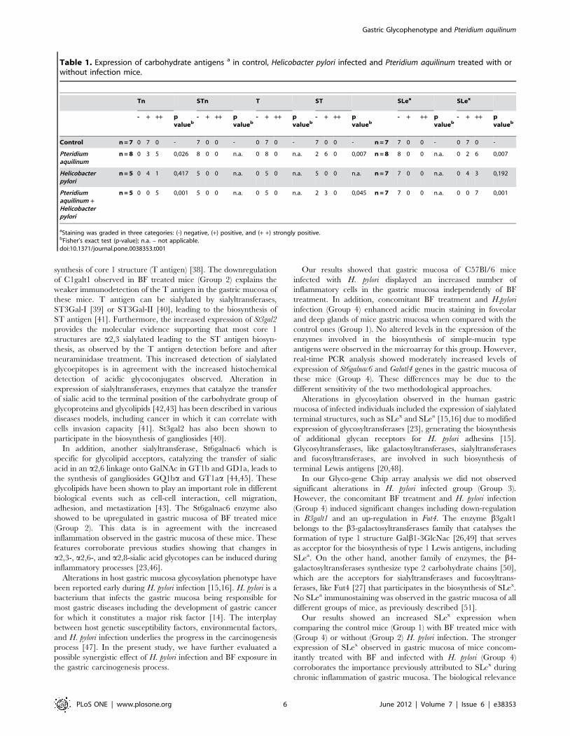
Table 1. Expression of carbohydrate antigens a in control, Helicobacter pylori infected and Pteridium aquilinum treated with orwithout infection mice.
Tn STn T ST SLea SLex
- + ++ pvalueb
- + ++ pvalueb
- + ++ pvalueb
- + ++ pvalueb
- + ++ pvalueb
- + ++ pvalueb
Control n = 7 0 7 0 - 7 0 0 - 0 7 0 - 7 0 0 - n = 7 7 0 0 - 0 7 0 -
Pteridiumaquilinum
n = 8 0 3 5 0,026 8 0 0 n.a. 0 8 0 n.a. 2 6 0 0,007 n = 8 8 0 0 n.a. 0 2 6 0,007
Helicobacterpylori
n = 5 0 4 1 0,417 5 0 0 n.a. 0 5 0 n.a. 5 0 0 n.a. n = 7 7 0 0 n.a. 0 4 3 0,192
Pteridiumaquilinum +Helicobacterpylori
n = 5 0 0 5 0,001 5 0 0 n.a. 0 5 0 n.a. 2 3 0 0,045 n = 7 7 0 0 n.a. 0 0 7 0,001
aStaining was graded in three categories: (-) negative, (+) positive, and (+ +) strongly positive.bFisher’s exact test (p-value); n.a. – not applicable.doi:10.1371/journal.pone.0038353.t001
Gastric Glycophenotype and Pteridium aquilinum
PLoS ONE | www.plosone.org 6 June 2012 | Volume 7 | Issue 6 | e38353
of the SLex expression may be partly due to the role that this
glycan receptor plays in the context of H. pylori binding through its
sialic-acid binding adhesin, SabA, to gastric cells [15]. Previous
studies have shown that overexpression of b3GnT5, a GlcNAc
transferase essential for the first step of biosynthesis of Lewis
antigens, was observed in human gastric cells upon H. pylori
infection, leading to increased SLex expression and increased
SabA mediated bacterial adhesion [23]. In the present study, the
promotion of gastric mucosa inflammation together with an
increased expression of SLex observed in the mice gastric
epithelium, suggest that the expression of ligands for bacterial
adhesins, as SabA, could be modulated upon BF exposure and H.
pylori infection. Overall, these studies support the importance of
glycan receptors as mediators in the adhesion of the gastric
pathogen H. pylori [52]. Although the present study used a limited
number of mice within each study group, our results clearly
Figure 5. Glycosylation alterations in mice gastric mucosa upon Pteridium aquilinum treatment and concomitant Helicobacter pyloriinfection. Panel A – Heatmap of the Glyco-gene Chip array analysis obtained when comparing group 1 (control) with group 4 (treated withPteridium aquilinum and Helicobacter pylori infected). Red indicates increased and blue indicates decreased expression relative to the mean transcriptexpression value. The transcripts identified as differentially expressed were those with adjusted p-value ,0.1 and fold change .1.3. Panel B –Schematic representation of the biosynthesis of sialylated terminal structures. The enzymes or family of enzymes responsible for each step of thebiosynthesis are indicated. The enzymes identified as showing different expression levels in Pteridium aquilinum treated and Helicobacter pyloriinfected mice (Group 4) are highlighted in red (increased expression) and blue (decreased expression). Panel C – Immunohistochemistry forevaluation of expression of Sialyl-Lewis a (SLea) antigen (a–d) and for Sialyl-Lewis x (SLex) (e–h). Additional immunofluorescence evaluation performedfor SLex (i–l). Bar = 20 mm.doi:10.1371/journal.pone.0038353.g005
Gastric Glycophenotype and Pteridium aquilinum
PLoS ONE | www.plosone.org 7 June 2012 | Volume 7 | Issue 6 | e38353
demonstrate significant glycophenotypic alterations induced by H.
pylori and BF.
In agreement with our data, it has been shown that H. pylori
infection induces changes in glycosylation in the gastric mucosa
of animal models such as Rhesus monkeys and Mongolian gerbils
[22,53]. Furthermore, synergistic effects of H. pylori infection and
the ingestion of a carcinogen present in the diet have been
observed in the Rhesus monkey model, including histomorpho-
logic alterations and modification of cancer-associated gene
expression [54].
While our observations demonstrate the glycophenotypic
alterations of host cells in response to BF exposure and H. pylori
infection, other studies have focused on H. pylori glycan-gene
expression. A recent study pointed out the importance of two H.
pylori genes, jhp0562, which encodes a glycosyltransferases involved
in the synthesis of the bacterial lipopolysacharide and the
immediately upstream gene, jhp0563 that encodes a b1,3
galactosyltransferase involved in bacterial Lewis antigen synthesis.
These genes are associated with different gastric pathologies
allowing the discrimination between peptic ulcers and gastritis in
some populations [55]. These studies further demonstrate the
important role that glycosylation plays in the modulation of host-
pathogen interactions.
In conclusion, our results show that the bracken fern Pteridium
aquilinum is able to induce various histomorphologic and molecular
changes in the gastric mucosa. These modifications include major
alterations at the transcriptional level of several genes, such as
glycosyltransferases involved in the biosynthesis of simple mucin
type carbohydrate antigens. Furthermore, the concomitant expo-
sure to BF and H. pylori infection also induced alterations at the
transcriptional level of glycosyltransferases participating in the
biosynthesis of terminal glycan Lewis antigens. These gene
transcription modifications underly the increased acidic glycocon-
jugate phenotype observed in BF exposed in gastric mucosa in the
context of H. pylori infection. Our results contribute for under-
standing the molecular mechanisms underlying the role of BF
pathogenic consequences in gastric mucosa and its synergistic
effects with H. pylori infection.
Materials and Methods
Animal Experiments and Tissue SamplesFour groups of six-week-old specific pathogen-free C57Bl/6
male mice (Charles River Laboratories) were used in this study
according with the Specific Guide for the Care and the Use of
Laboratory Animals of the Institut Pasteur, the European
Directive (2010/63/UE) and the corresponding French law on
animal experimentation. The protocol was also approved by the
Committee of Central Animal Facility Board of the Institut
Pasteur.
Each group consisted in 8 animals. Mice from the group 1
(control) received drinking water during all the experiment. The
group 2 was exposed to BF aqueous extracts, obtained as
previously described [56]. In this group mice received, during
the third and fourth weeks of the experiment, a drinking water
supplemented with BF extracts (250 mg BF/ml) as already
reported [10]. In the group 3, mice were orogastrically infected
with H. pylori strain SS1 (100 ml of 107 bacteria), twice during the
first week. In the group 4, mice were orogastrically infected with
H. pylori strain SS1 (100 ml of 107 bacteria) and treated with BF
extracts in the same conditions described for group 2. From each
experimental group, 4 mice were sacrificed after 4 and 7 weeks
from the beginning of the experiment and their stomach collected.
H. pylori gastric colonization was quantified in the gastric mucosa
as previously described [57].
Histochemistry and ImmunohistochemistryMice gastric mucosa fragments including antrum and corpus
were formalin-fixed and embedded in paraffin wax before serial
sections were cut.
H&E staining was done to observe morphological alterations in
gastric mucosa. Alcian blue (pH 2.5) (Merck) was used to detect
the presence of acidic mucins and nuclear red 1% used to stain the
nuclei.
A semi-quantitative evaluation of the inflammatory cells was
done in all mice gastric mucosa according to the Sidney
Classification System [58] and graded on a scale of absent, mild,
moderate and severe infiltrate (Table S1). A representative figure
of each group illustrates the results observed (Figure 1).
Immunohistochemical analysis of carbohydrate antigens was
performed according to the standard avidin-biotin-complex
staining method using the monoclonal antibodies described in
table 2 [59,60]. Analysis was performed in samples from both time
points. We used 7 gastric mucosa samples from control mice
(Group 1) and 8 samples from BF treated mice (Group 2) for all
the antigens evaluated. We used 5 gastric mucosa from H. pylori
infected (Group 3) and concomitantly BF treated and H. pylori
infected mice (Group 4) for Tn, STn, T and ST antigen evaluation
while 7 gastric mucosa were used to SLea and SLex antigens
detection (Table S1).
Briefly, tissue samples were deparaffinated, rehydrated and
incubated at 37uC with neuraminidase from Clostridium perfringes
type VI (Sigma) diluted in 0.1 M sodium acetate buffer (pH 5.5)
followed by washes in ice-cold when needed (recognition of ST
using 3C9 MAb). Sections were treated with 0.3% hydrogen
peroxide in methanol to block endogenous peroxidase, incubated
with normal rabbit serum diluted 1:5 in PBS with 10% of BSA and
then incubated overnight at 4uC with the respective monoclonal
primary antibody (Table 2) [59,60]. The slides were then washed
in PBS and incubated with biotinylated rabbit anti-mouse
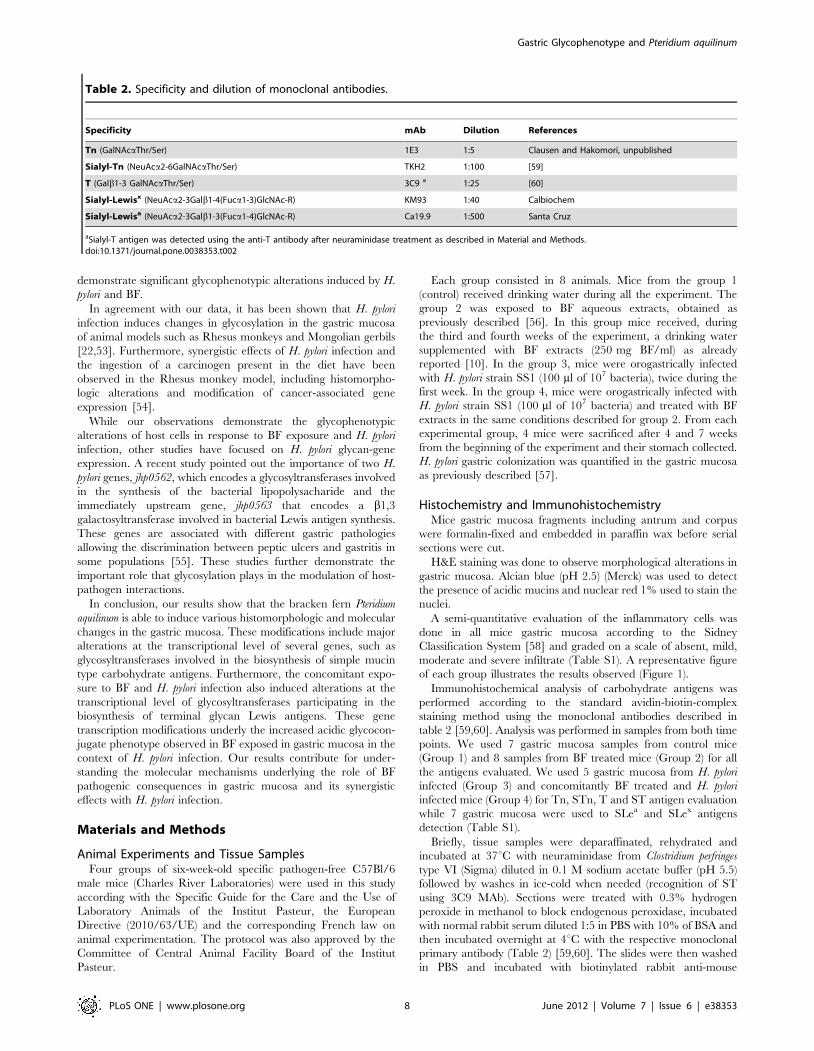
Table 2. Specificity and dilution of monoclonal antibodies.
Specificity mAb Dilution References
Tn (GalNAcaThr/Ser) 1E3 1:5 Clausen and Hakomori, unpublished
Sialyl-Tn (NeuAca2-6GalNAcaThr/Ser) TKH2 1:100 [59]
T (Galb1-3 GalNAcaThr/Ser) 3C9 a 1:25 [60]
Sialyl-Lewisx (NeuAca2-3Galb1-4(Fuca1-3)GlcNAc-R) KM93 1:40 Calbiochem
Sialyl-Lewisa (NeuAca2-3Galb1-3(Fuca1-4)GlcNAc-R) Ca19.9 1:500 Santa Cruz
aSialyl-T antigen was detected using the anti-T antibody after neuraminidase treatment as described in Material and Methods.doi:10.1371/journal.pone.0038353.t002
Gastric Glycophenotype and Pteridium aquilinum
PLoS ONE | www.plosone.org 8 June 2012 | Volume 7 | Issue 6 | e38353
secondary antibody (DakoCytomation) diluted 1:200 in PBS with
5% of BSA prior to the incubation with avidin-biotin peroxidase
complex (Vectastain Elite ABS kit). Sections were stained with
3,39-Diaminobenzidine tetrahydrochloride (Sigma) in a buffer
containing 0.1% hydrogen peroxide, counter-stained with Mayer’s
hematoxylin, dehydrated and mounted. Negative controls were
performed by replacing primary antibody with PBS. Staining was
graded as -, negative; +, positive; + +, strongly positive.
Statistical analysis was performed using the Chi Square and
Fisher’s exact tests with Statview 5.0 software. Differences were
considered statistically significant at p,0.05.
Immunofluorescence AssayMice gastric tissues were deparaffinated, rehydrated and
incubated with rabbit non-immune serum (DakoCytomation)
diluted 1:5 in PBS with 10% of BSA. Sections were incubated
overnight at 4uC with SLex antibody (KM93, Calbiochem) diluted
in PBS containing 5% of BSA. Sections were then washed in PBS
and incubated with FITC-conjugated rabbit anti-mouse immuno-
globulin (DakoCytomation) diluted 1:70 in PBS with 5% of BSA.
Sections were washed in PBS and incubated with DAPI 100 mg/
mL (Sigma). Samples were washed in PBS and mounted in
VectaShield (Vector Laboratories).
Glyco-gene Chip Arrays and Analysis of DataTotal RNA from gastric mucosa of 3 mice from each
experimental condition (biological replicates) sacrificed after 7
weeks from the beginning of the experiment, was extracted using
the RNeasy Plus Mini kit (QIAGEN) according to the manufac-
turer’s protocol. RNA yield and quality were determined using
NanoDrop ND-1000 spectrophotometer (THERMO Scientific).
A total of 12 samples were hybridized on the GlycoV4
oligonucleotide arrays specially developed for the Consortium
for Functional Glycomics - CFG, using the Affymetrix technology
(Affymetrix, USA) and containing probes that allow to detect the
expression of 1200 mouse glyco-related transcripts [61]. The
following classes of genes are represented on the GLYCOv4 Gene
Chip: Glycosyltransferases, Glycan-binding proteins (GBPs),
including C-type lectins, galectins and siglecs, Glycan degradation
proteins, Intercellular protein transport proteins, Notch pathway
proteins, Nucleotide sugar synthesis and transporter proteins, N-
glycan biosynthesis-related proteins, Adhesion molecules, Inter-
leukins and receptors, Mucins, Growth factors and receptors,
Cytokines, Chemokines, Conserved oligomeric Golgi (COG)
complex proteins and other miscellaneous proteins of interest.
The complete list of genes present on the GLYCOv4 Gene Chip
array is available at http://www.functionalglycomics.org/static/
consortium/resources/resourcecoree.shtml.
Data normalization was performed using RMA Express 1.0
with quantile normalization, median polish and background
adjustment [62,63].
The Limma package in the R software [64] was used to find
transcripts with differential expression. The fold changes and
standard errors were estimated by fitting a linear model for each
gene and empirical Bayes smoothing was applied to the standard
errors. Results are presented between two or more experimental
conditions as a fold change in expression level, the moderated t-
statistic, the p-value, and the adjusted p-value. The adjusted p-
value is the p-value adjusted for multiple testing using the
Benjamini and Hochberg’s method [65], to control the false
discovery rate of 0.1 or less. The transcripts identified as
differentially expressed were those with adjusted p-value ,0.1
and fold change .1.3.
Heatmaps were generated with dChip program comparing
group 1 (control) with group 2 (treated with BF) and comparing
group 1 (control) with group 4 (infected with H. pylori and treated
with BF). No heatmap was generated for group 3 (infected with H.
pylori) since no difference was observed when comparing to control
group 1. Red and blue indicates increased and decreased
expression relative to the mean transcript expression value,
respectively.
cDNA SynthesisFirst strand cDNA was produced from 2.5 mg of total RNA
from the same samples evaluated in the Glyco-gene Chip array,
using Superscript II Reverse Transcriptase kit (Invitrogen) in
accordance with the manufacturer’s instructions. The reaction
mixture was incubated at 25uC for 10 min, 42uC for 50 min and
15 min at 72uC.
Relative Quantitative Real-time PCR and StatisticalAnalysis
Relative Quantitative real-time PCR was performed in the
groups showing gene expression alterations detected in the
glycogene chip array.
qRT-PCR was done using the SYBRHGreen chemistry in a
7500 Fast Real Time PCR System (Applied Biosytems, Foster
City, CA, USA). The reaction mixture contained 2 ml cDNA
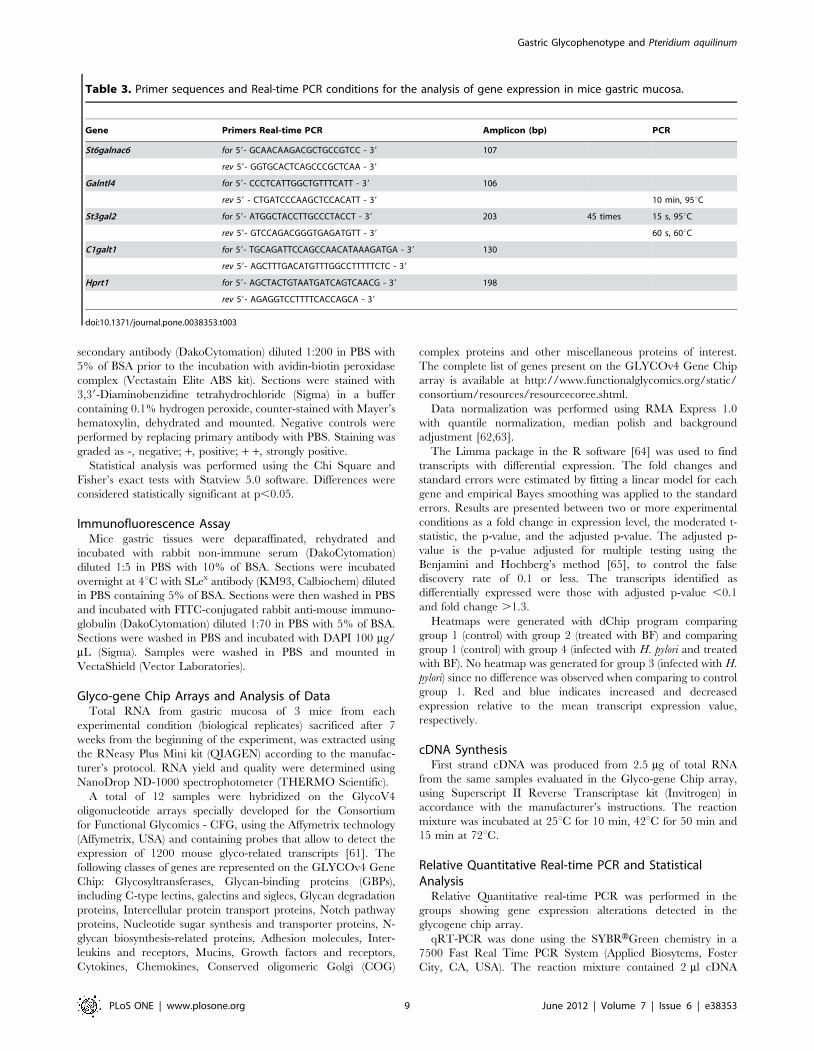
Table 3. Primer sequences and Real-time PCR conditions for the analysis of gene expression in mice gastric mucosa.
Gene Primers Real-time PCR Amplicon (bp) PCR
St6galnac6 for 59- GCAACAAGACGCTGCCGTCC - 39 107
rev 59- GGTGCACTCAGCCCGCTCAA - 39
Galntl4 for 59- CCCTCATTGGCTGTTTCATT - 39 106
rev 59 - CTGATCCCAAGCTCCACATT - 39 10 min, 95uC
St3gal2 for 59- ATGGCTACCTTGCCCTACCT - 39 203 45 times 15 s, 95uC
rev 59- GTCCAGACGGGTGAGATGTT - 39 60 s, 60uC
C1galt1 for 59- TGCAGATTCCAGCCAACATAAAGATGA - 39 130
rev 59- AGCTTTGACATGTTTGGCCTTTTTCTC - 39
Hprt1 for 59- AGCTACTGTAATGATCAGTCAACG - 39 198
rev 59- AGAGGTCCTTTTCACCAGCA - 39
doi:10.1371/journal.pone.0038353.t003
Gastric Glycophenotype and Pteridium aquilinum
PLoS ONE | www.plosone.org 9 June 2012 | Volume 7 | Issue 6 | e38353
(diluted 1:2 in water), 10.0 ml Power SYBRHGreen PCR Master
Mix (2x) (Applied Biosystems), and 0.48 ml of each 10 mM primer
(Sigma Genosys) in a final reaction volume of 20 ml. Primers
melting temperature, amplicon length and sequence are resumed
in Table 3. The cycling conditions were as follows: denaturation at
95uC for 10 min, followed by 45 cycles of 95uC for 15 s and 60uCfor 60 s, and a final step (95uC for 15 s, 60uC for 60 s, 95uC for
30 s, 60uC for 15 s to create a dissociation curve in order to assure
assay specificity. Calculations were made using the comparative
CT method (2-DDCT). Hprt1 was used as an endogenous control
gene for PCR normalization to the amount of RNA added to the
reverse transcription reactions, and the biological group 1 as the
calibrator. Triplicates of each biological group were analyzed
twice independently.
Normality of the data was in all cases confirmed by Shapiro-
Wilk test [66]. Welch none paired two sample t-test was used to
calculate the p-values for each gene, between biological groups
with the null hypothesis of equal means.
Supporting Information
Figure S1 Evaluation of Helicobacter pylori colonizationin gastric mucosa of infected mice (Group 3) andinfected and Pteridium aquilinum treated mice (Group4) at 4 and 7 weeks. Colonization represented by the number of
CFU/g of mice stomach tissue. Each point represents one mouse
and the mean value is also shown.
(TIF)
Table S1 Characterization of inflammation and immu-nohistochemistry analysis of carbohydrate antigens
expression in the gastric mucosa of all mice evaluatedin each experimental group.
(PDF)
Table S2 Significantly altered genes in Pteridiumaquilinum treated gastric mucosa (Group 2) in compar-ison with control (Group 1) from the Glyco-gene Chiparray analysis.
(PDF)
Table S3 Significantly altered genes in Pteridiumaquilinum treated and Helicobacter pylori infectedgastric mucosa (Group 4) in comparison with control(Group 1) from the Glyco-gene Chip array analysis.
(PDF)
Acknowledgments
We thank Prof. Hans Hansen from Faculty of Life Sciences, University of
Copenhagen, Denmark, for bracken fern extraction protocols and
materials. We thank Rune Matthiesen from IPATIMUP, University of
Porto, Portugal for statistical analysis; and Patrick Ave from Unite
d’Histopathologie Humaine et Modeles Animaux, Institut Pasteur for his
help and advices for the preparation of gastric samples for histology.
Author Contributions
Conceived and designed the experiments: JG ET CAR. Performed the
experiments: JG AM ASC GEH SLP VM. Analyzed the data: JG AM
ASC GEH SLP SRH LD FG ET CAR. Contributed reagents/materials/
analysis tools: JG SRH ET CAR. Wrote the paper: JG CAR.
References
1. Vetter J (2009) A biological hazard of our age: bracken fern [Pteridiumaquilinum (L.) Kuhn] - a review. Acta Vet Hung 57: 183–196.
2. IARC - International Agency for Research on Cancer (1986) Bracken fern
(Pteridium aquilinum) and some of its constituents. In: IARC Monographs on theEvaluation of Carcinogenic Risks to Humans, IARC Press, Lyon, France,
Vol.40, p.47.
3. Yamada K, Ojika M, Kigoshi H (2007) Ptaquiloside, the major toxin of bracken,
and related terpene glycosides: chemistry, biology and ecology. Nat Prod Rep24: 798–813.
4. Alonso-Amelot ME (1997) The link between bracken fern and stomach cancer:
Milk. Nutrition 13: 694–696.
5. Jensen PH, Jacobsen OS, Hansen HCB, Juhler RK (2008) Quantification ofPtaquiloside and Pterosin B in Soil and Groundwater Using Liquid
Chromatography-Tandem Mass Spectrometry (LC-MS/MS). J Agric FoodChem 56: 9848–9854.
6. Shahin M, Smith BL, Prakash AS (1999) Bracken carcinogens in the human
diet. Mutat Res 443: 69–79.
7. Alonso-Amelot ME, Avendano M (2001) Possible association between gastriccancer and bracken fern in Venezuela: an epidemiologic study. Int J Cancer 91:
252–259.
8. Galpin OP, Whitaker CJ, Whitaker R, Kassab JY (1990) Gastric cancer inGwynedd. Possible links with bracken. Br J Cancer 61: 737–740.
9. Marliere C, Wathern P, Freitas S, Castro M, Galvao M (1999) Bracken fern
(Pteridium aquilinum) consumption and oesophageal and stomach cancer in theOuro Preto region, Minas Gerais, Brazil. In: Taylor JAS, RT (ed) Bracken
Conference, Manchester, Bracken Fern: Toxicity, Biology and Control, 144–
149.
10. Gomes J, Magalhaes A, Michel V, Amado I, Aranha P, et al. (2012) Pteridiumaquilinum and its ptaquiloside toxin induce DNA damage response in gastric
epithelial cells, a link with gastric carcinogenesis. Toxicol Sci 126: 60–71.
11. Ferlay J, Shin H, Bray F, Forman D, Mathers C, et al. (2010) GLOBOCAN2008 Cancer Incidence and Mortality Worldwide: IARC CancerBase No 10.
12. Peek RM, Blaser MJ (2002) Helicobacter pylori and gastrointestinal tract
adenocarcinomas. Nat Rev Cancer 2: 28–37.
13. IARC - International Agency for Research on Cancer (1994) Infection withHelicobacter pylori. In: IARC Monographs on the Evaluation of Carcinogenic
Risks to Humans - Schistosomes, Liver Flukes and Helicobacter pylori -, IARCPress, Lyon, France, Vol.61, 177–240.
14. Correa P (1992) Human Gastric Carcinogenesis: A Multistep and Multifactorial
Process First American Cancer Society Award Lecture on Cancer Epidemiologyand Prevention. Cancer Res 52: 6735–6740.
15. Mahdavi J, Sonden B, Hurtig M, Olfat FO, Forsberg L, et al. (2002)
Helicobacter pylori SabA Adhesin in Persistent Infection and Chronic
Inflammation. Science 297: 573–578.
16. Ota H, Nakayama J, Momose M, Hayama M, Akamatsu T, et al. (1998)
Helicobacter pylori infection produces reversible glycosylation changes to gastric
mucins. Virchows Arch 433: 419–426.
17. Silva E, Teixeira A, David L, Carneiro F, Reis C, et al. (2002) Mucins as key
molecules for the classification of intestinal metaplasia of the stomach. Virchows
Arch 440: 311–317.
18. David L, Nesland J, Clausen H, Carneiro F, Sobrinho-Simoes M (1992) Simple
mucin-type carbohydrate antigens (Tn, sialosyl-Tn and T) in gastric mucosa,
carcinomas and metastases. APMIS Suppl 27: 162–172.
19. Pinho S, Marcos NT, Ferreira B, Carvalho AS, Oliveira MJ, et al. (2007)
Biological significance of cancer-associated sialyl-Tn antigen: Modulation of
malignant phenotype in gastric carcinoma cells. Cancer Lett 249: 157–170.
20. Reis CA, Osorio H, Silva L, Gomes C, David L (2010) Alterations in
glycosylation as biomarkers for cancer detection. J Clin Pathol 63: 322–329.
21. Linden S, Mahdavi J, Semino-Mora C, Olsen C, Carlstedt I, et al. (2008) Role of
ABO secretor status in mucosal innate immunity and H. pylori infection. PLoS
Pathog 4: e2.
22. Ohno T, Vallstrom A, Rugge M, Ota H, Graham DY, et al. (2011) Effects of
blood group antigen-binding adhesion expression during Helicobacter pylori
infection of Mongolian gerbils. J Infect Dis 203: 726–735.
23. Marcos NT, Magalhaes A, Ferreira B, Oliveira MJ, Carvalho AS, et al. (2008)
Helicobacter pylori induces beta3GnT5 in human gastric cell lines, modulating
expression of the SabA ligand sialyl-Lewis x. J Clin Invest 118: 2325–2336.
24. Fuster MM, Esko JD (2005) The sweet and sour of cancer: glycans as novel
therapeutic targets. Nat Rev Cancer 5: 526–542.
25. Amado M, Carneiro F, Seixas M, Clausen H, Sobrinho-Simoes M (1998)
Dimeric sialyl-Le(x) expression in gastric carcinoma correlates with venous
invasion and poor outcome. Gastroenterology 114: 462–470.
26. Hennet T, Dinter A, Kuhnert P, Mattu TS, Rudd PM, et al. (1998) Genomic
Cloning and Expression of Three Murine UDP-galactose: b-N-Acetylglucosa-
mine b1,3-Galactosyltransferase Genes. J Biol Chem 273: 58–65.
27. Ozawa M, Muramatsu T (1996) Molecular Cloning and Expression of a Mouse
a-1,3 Fucosyltransferase Gene That Shows Homology with the Humana-1,3
Fucosyltransferase IV Gene. J Biochem (Tokyo) 119: 302–308.
28. Fieger CB, Sassetti CM, Rosen SD (2003) Endoglycan, a member of the CD34
family, functions as an L-selectin ligand through modification with tyrosine
sulfation and sialyl Lewis x. J Biol Chem 278: 27390–27398.
Gastric Glycophenotype and Pteridium aquilinum
PLoS ONE | www.plosone.org 10 June 2012 | Volume 7 | Issue 6 | e38353
29. Smith BL (2004) Bracken Fern (genus Pteridium) Toxicity - a Global Problem.
In: Acamovic TS, CS; Pennycott, TW, Eds. (ed) Poisonous Plants and RelatedToxins. Cabi Publishing, 227–240.
30. Filipe MI, Lake BD (1990) Histochemistry in Pathology. Appendix 3, 2 edn.
Edinburgh: Churchill Livingstone, p 451.31. Reis CA, David L, Correa P, Carneiro F, de Bolos C, et al. (1999) Intestinal
metaplasia of human stomach displays distinct patterns of mucin (MUC1,MUC2, MUC5AC, and MUC6) expression. Cancer Res 59: 1003–1007.
32. Bennett EP, Mandel U, Clausen H, Gerken TA, Fritz TA, et al. (2011) Control
of Mucin-Type O-Glycosylation - A Classification of the Polypeptide GalNAc-transferase Gene Family. Glycobiology 22: 736–756.
33. Ten Hagen KG, Fritz TA, Tabak LA (2003) All in the family: the UDP-GalNAc:polypeptide N-acetylgalactosaminyltransferases. Glycobiology 13: 1R-
16R.34. Gomes J, Marcos NT, Berois N, Osinaga E, Magalhaes A, et al. (2009)
Expression of UDP-N-acetyl-D-galactosamine: Polypeptide N-acetylgalactosa-
minyltransferase-6 in Gastric Mucosa, Intestinal Metaplasia, and GastricCarcinoma. J Histochem Cytochem 57: 79–86.
35. Mandel U, Hassan H, Therkildsen MH, Rygaard J, Jakobsen MH, et al. (1999)Expression of polypeptide GalNAc-transferases in stratified epithelia and
squamous cell carcinomas: immunohistological evaluation using monoclonal
antibodies to three members of the GalNAc-transferase family. Glycobiology 9:43–52.
36. Tian E, Ten Hagen K (2009) Recent insights into the biological roles of mucin-type O-glycosylation. Glycoconj J 26: 325–334.
37. Raman J, Guan Y, Perrine CL, Gerken TA, Tabak LA (2011) UDP-N-Acetyl-a-D-galactosamine:polypeptide N-acetylgalactosaminyltransferases: Completion of
the Family Tree. Glycobiology 22: 768–777.
38. Ju T, Brewer K, D’Souza A, Cummings RD, Canfield WM (2002) Cloning andExpression of Human Core 1 b1,3-Galactosyltransferase. J Biol Chem 277: 178–
186.39. Lee Y-C, Kurosawa N, Hamamoto T, Nakaoka T, Tsuji S (1993) Molecular
cloning and expression of Galb1,3GalNAca2,3-sialyltransferase from mouse
brain. Eur J Biochem 216: 377–385.40. Lee YC, Kojima N, Wada E, Kurosawa N, Nakaoka T, et al. (1994) Cloning
and expression of cDNA for a new type of Gal beta 1,3GalNAc alpha 2,3-sialyltransferase. J Biol Chem 269: 10028–10033.
41. Dall’Olio F, Chiricolo M (2001) Sialyltransferases in cancer. Glycoconj J 18:841–850.
42. Harduin-Lepers A, Vallejo-Ruiz V, Krzewinski-Recchi M-A, Samyn-Petit B,
Julien S, et al. (2001) The human sialyltransferase family. Biochimie 83: 727–737.
43. Takashima S (2008) Characterization of mouse sialyltransferase genes: theirevolution and diversity. Biosci Biotechnol Biochem 72: 1155–1167.
44. Chandrasekaran EV, Xue J, Xia J, Locke RD, Patil SA, et al. (2011)
Mammalian Sialyltransferase ST3Gal-II: Its Exchange Sialylation CatalyticProperties Allow Labeling of Sialyl Residues in Mucin-Type Sialylated
Glycoproteins and Specific Gangliosides. Biochemistry 50: 9475–9487.45. Okajima T, Chen H-H, Ito H, Kiso M, Tai T, et al. (2000) Molecular Cloning
and Expression of Mouse GD1a/GT1aa/GQ1ba Synthase (ST6GalNAc VI)Gene. J Biol Chem 275: 6717–6723.
46. Yasukawa Z, Sato C, Kitajima K (2005) Inflammation-dependent changes in
a2,3-, a2,6-, and a2,8-sialic acid glycotopes on serum glycoproteins in mice.Glycobiology 15: 827–837.
47. Atherton JC (2006) The pathogenesis of Helicobacter pylori-induced gastro-duodenal diseases. Annu Rev Pathol 1: 63–96.
48. Carvalho AS, Harduin-Lepers A, Magalhaes A, Machado E, Mendes N, et al.
(2010) Differential expression of alpha-2,3-sialyltransferases and alpha-1,3/4-fucosyltransferases regulates the levels of sialyl Lewis a and sialyl Lewis x in
gastrointestinal carcinoma cells. Int J Biochem Cell Biol 42: 80–89.
49. Amado M, Almeida R, Carneiro F, Levery SB, Holmes EH, et al. (1998) AFamily of Human b3-Galactosyltransferases. J Biol Chem 273: 12770–12778.
50. Almeida R, Amado M, David L, Levery SB, Holmes EH, et al. (1997) A Familyof Human b4-Galactosyltransferases. J Biol Chem 272: 31979–31991.
51. Magalhaes A, Gomes J, Ismail MN, Haslam SM, Mendes N, et al. (2009) Fut2-
null mice display an altered glycosylation profile and impaired BabA-mediatedHelicobacter pylori adhesion to gastric mucosa. Glycobiology 19: 1525–1536.
52. Magalhaes A, Reis CA (2010) Helicobacter pylori adhesion to gastric epithelialcells is mediated by glycan receptors. Braz J Med Biol Res 43: 611–618.
53. Solnick JV, Hansen LM, Salama NR, Boonjakuakul JK, Syvanen M (2004)Modification of Helicobacter pylori outer membrane protein expression during
experimental infection of rhesus macaques. Proc Natl Acad Sci U S A 101:
2106–2111.54. Liu H, Merrell DS, Semino-Mora C, Goldman M, Rahman A, et al (2009) Diet
synergistically affects Helicobacter pylori-induced gastric carcinogenesis in nonhu-man primates. Gastroenterology 137: 1367–1379.
55. Matsuda M, Shiota S, Matsunari O, Watada M, Murakami K, et al. (2011)
Prevalence of two homologous genes encoding glycosyltransferases of Helicobacter
pylori in the United States and Japan. Gastroenterology 26: 1451–1456.
56. Rasmussen LH, Bruun Hansen HC, Lauren D (2005) Sorption, degradation andmobility of ptaquiloside, a carcinogenic Bracken (Pteridium sp.) constituent, in
the soil environment. Chemosphere 58: 823–835.57. Ferrero R, Thiberge J, Huerre M, Labigne A (1998) Immune responses of
specific-pathogen-free mice to chronic Helicobacter pylori (strain SS1) infection.
Infect Immun 66: 1349–1355.58. Dixon MF, Genta RM, Yardley JH, Correa P (1996) Classification and grading
of gastritis. The updated Sydney System. International Workshop on theHistopathology of Gastritis, Houston 1994. Am J Surg Pathol 20: 1161–1181.
59. Kjeldsen T, Clausen H, Hirohashi S, Ogawa T, Iijima H, et al. (1988)
Preparation and Characterization of Monoclonal Antibodies Directed to theTumor-associated O-linked Sialosyl-2R6 a-N-Acetylgalactosaminyl (Sialosyl-
Tn) Epitope. Cancer Res 48: 2214–2220.60. Zen K, Notarfrancesco K, Oorschot V, Slot JW, Fisher AB, et al. (1998)
Generation and characterization of monoclonal antibodies to alveolar type IIcell lamellar body membrane. Am J Physiol 275: L172-L183.
61. Lockhart DJ, Dong H, Byrne MC, Follettie MT, Gallo MV, et al. (1996)
Expression monitoring by hybridization to high-density oligonucleotide arrays.Nat Biotech 14: 1675–1680.
62. Bolstad BM, Irizarry RA, Astrand M, Speed TP (2003) A comparison ofnormalization methods for high density oligonucleotide array data based on
variance and bias. Bioinformatics 19: 185–193.
63. Irizarry RA, Bolstad BM, Collin F, Cope LM, Hobbs B, et al. (2003) Summariesof Affymetrix GeneChip probe level data. Nucleic Acids Res 31: e15.
64. Smyth GK (2004) Linear Models and Empirical Bayes Methods for AssessingDifferential Expression in Microarray Experiments. Stat Appl Genet Mol Biol 3:
Article3.65. Benjamini Y, Hochberg Y (1995) Controlling the False Discovery Rate: A
Practical and Powerful Approach to Multiple Testing. In: Journal of the Royal
Statistical Society. Series B (Methodological), Blackwell Publishing for the RoyalStatistical Society, Vol. 57, No. 1, 289–300.
66. Royston P (1995) Remark AS R94: A remark on Algorithm AS 181: The W testfor normality. Applied Statistics 44: 547–551.
Gastric Glycophenotype and Pteridium aquilinum
PLoS ONE | www.plosone.org 11 June 2012 | Volume 7 | Issue 6 | e38353