Genotypic characterization of symptomatic hepatitis E virus (HEV) infections in Egypt
11
Genotypic characterization of symptomatic hepatitis E virus (HEV) infections in Egypt Jason T. Blackard 1 , Susan D. Rouster 1 , Soad Nady 1 , Gehan Galal 1 , Naglaa Marzuuk 2 , Marwaa M. Rafaat 2 , Enas Daef 2 , Salwa Seif El Din 2 , Robert H. Purcell 3 , Suzanne U. Emerson 3 , Kenneth E. Sherman 1 , and M. Tarek Shata 1 1 Division of Digestive Diseases, University of Cincinnati College of Medicine 2 Microbiology Department, Faculty of Medicine, Assuit University, Egypt 3 Laboratory of Infectious Diseases, National Institute of Allergy and Infectious Diseases Abstract Background—Hepatitis E virus (HEV) is a common cause of acute viral hepatitis (AVH) in many developing countries. In Egypt, HEV seroprevalence is among the highest in the world; however, only a very limited number of Egyptian HEV sequences are currently available. Objectives—The objectives were to determine the HEV genotype(s) currently circulating in Egypt. Study Design—AVH patients without serologic evidence of hepatitis A, B, and C viruses were evaluated for possible HEV infection using serologic assays for anti-HEV IgM and anti-HEV IgG and real-time PCR for HEV RNA. Stool suspensions from suspected cases were inoculated into rhesus macaques to confirm the presence of HEV. Sequence analysis was utilized to determine HEV genotype. Results—Of 287 subjects with AVH enrolled, 58 had serologic evidence of acute HEV infection. Stool samples for two of these patients were repeatedly positive for HEV RNA by real-time PCR. Macaques experimentally inoculated with these human stools also developed viremia. Sequence analysis of open reading frame (ORF) 1 demonstrated that these isolates belonged to HEV genotype 1 and were 3.9% – 9.5% divergent from other genotype 1 isolates. ORF2 was 5.3% – 8.7% divergent from previously reported Egyptian isolates. Conclusions—This study strongly suggests that genotype 1 HEV related to other North African isolates is circulating in acute symptomatic patients in Egypt. Further evaluation of genotypic variability is underway in this highly endemic cohort and is considered an important component of our increased understanding of HEV pathogenesis. Keywords Hepatitis E virus (HEV); Egypt; symptomatic; genotype; diversity Address requests for reprints to: Jason Blackard, PhD, Division of Digestive Diseases, University of Cincinnati College of Medicine, ML 0595, 231 Albert Sabin Way, Cincinnati, OH 45267, Phone:(513) 558-4389, Fax: (513) 558-1744, Email: [email protected]. Competing interests: None declared Approval for human studies was provided by the University of Cincinnati IRB. Animal studies were approved by the Animal Care and Use Committees of Bioqual Inc. and the National Institute of Allergy and Infectious Diseases (Bethesda, Maryland, USA). Publisher's Disclaimer: This is a PDF file of an unedited manuscript that has been accepted for publication. As a service to our customers we are providing this early version of the manuscript. The manuscript will undergo copyediting, typesetting, and review of the resulting proof before it is published in its final citable form. Please note that during the production process errors may be discovered which could affect the content, and all legal disclaimers that apply to the journal pertain. NIH Public Access Author Manuscript J Clin Virol. Author manuscript; available in PMC 2010 October 1. Published in final edited form as: J Clin Virol. 2009 October ; 46(2): 140–144. doi:10.1016/j.jcv.2009.07.007. NIH-PA Author Manuscript NIH-PA Author Manuscript NIH-PA Author Manuscript
-
Upload
independent -
Category
Documents
-
view
6 -
download
0
Transcript of Genotypic characterization of symptomatic hepatitis E virus (HEV) infections in Egypt

Genotypic characterization of symptomatic hepatitis E virus (HEV)infections in Egypt
Jason T. Blackard1, Susan D. Rouster1, Soad Nady1, Gehan Galal1, Naglaa Marzuuk2,Marwaa M. Rafaat2, Enas Daef2, Salwa Seif El Din2, Robert H. Purcell3, Suzanne U.Emerson3, Kenneth E. Sherman1, and M. Tarek Shata11 Division of Digestive Diseases, University of Cincinnati College of Medicine2 Microbiology Department, Faculty of Medicine, Assuit University, Egypt3 Laboratory of Infectious Diseases, National Institute of Allergy and Infectious Diseases
AbstractBackground—Hepatitis E virus (HEV) is a common cause of acute viral hepatitis (AVH) in manydeveloping countries. In Egypt, HEV seroprevalence is among the highest in the world; however,only a very limited number of Egyptian HEV sequences are currently available.
Objectives—The objectives were to determine the HEV genotype(s) currently circulating in Egypt.
Study Design—AVH patients without serologic evidence of hepatitis A, B, and C viruses wereevaluated for possible HEV infection using serologic assays for anti-HEV IgM and anti-HEV IgGand real-time PCR for HEV RNA. Stool suspensions from suspected cases were inoculated intorhesus macaques to confirm the presence of HEV. Sequence analysis was utilized to determine HEVgenotype.
Results—Of 287 subjects with AVH enrolled, 58 had serologic evidence of acute HEV infection.Stool samples for two of these patients were repeatedly positive for HEV RNA by real-time PCR.Macaques experimentally inoculated with these human stools also developed viremia. Sequenceanalysis of open reading frame (ORF) 1 demonstrated that these isolates belonged to HEV genotype1 and were 3.9% – 9.5% divergent from other genotype 1 isolates. ORF2 was 5.3% – 8.7% divergentfrom previously reported Egyptian isolates.
Conclusions—This study strongly suggests that genotype 1 HEV related to other North Africanisolates is circulating in acute symptomatic patients in Egypt. Further evaluation of genotypicvariability is underway in this highly endemic cohort and is considered an important component ofour increased understanding of HEV pathogenesis.
KeywordsHepatitis E virus (HEV); Egypt; symptomatic; genotype; diversity
Address requests for reprints to: Jason Blackard, PhD, Division of Digestive Diseases, University of Cincinnati College of Medicine,ML 0595, 231 Albert Sabin Way, Cincinnati, OH 45267, Phone:(513) 558-4389, Fax: (513) 558-1744, Email: [email protected] interests: None declaredApproval for human studies was provided by the University of Cincinnati IRB. Animal studies were approved by the Animal Care andUse Committees of Bioqual Inc. and the National Institute of Allergy and Infectious Diseases (Bethesda, Maryland, USA).Publisher's Disclaimer: This is a PDF file of an unedited manuscript that has been accepted for publication. As a service to our customerswe are providing this early version of the manuscript. The manuscript will undergo copyediting, typesetting, and review of the resultingproof before it is published in its final citable form. Please note that during the production process errors may be discovered which couldaffect the content, and all legal disclaimers that apply to the journal pertain.
NIH Public AccessAuthor ManuscriptJ Clin Virol. Author manuscript; available in PMC 2010 October 1.
Published in final edited form as:J Clin Virol. 2009 October ; 46(2): 140–144. doi:10.1016/j.jcv.2009.07.007.
NIH
-PA Author Manuscript
NIH
-PA Author Manuscript
NIH
-PA Author Manuscript

BackgroundHepatitis E virus (HEV) is an enterically transmitted virus that represents a common cause ofacute viral hepatitis (AVH) in many developing countries 1. Although most patients with HEVrecover fully, mortality rates of ~1% in the general population and up to 20% in pregnantwomen have been reported 2. Based on genomic variability observed among HEV isolates,four genotypes have been identified 3. As with other hepatitis viruses, genotype may play arole in disease severity 1, 4.
In Egypt, the prevalence of anti-HEV is among the highest of any country in the world,approaching 80% 5–10; yet, outbreaks have not been reported. A prospective study alsoestimated the HEV incidence at 41.6/1000 person-years 11; however, none of theseroconverting individuals gave a history compatible with AVH during the observation period.Similarly, HEV infection among pregnant women was not associated with history of jaundiceor liver disease as reported elsewhere 11. Reasons for the infrequent clinical hepatitis remainunclear but could be the result of early childhood exposure leading to inapparent infection.Alternatively, the predominant HEV genotype(s) in Egypt could be less virulent than thosecirculating in Asia.
ObjectivesOnly a very limited number of HEV sequences are currently available from North Africa;therefore, we investigated HEV infection and genotypes in two Egyptian communities.
MethodsStudy participants and sample collection
In Egypt, patients with acute viral hepatitis (AVH) are routinely referred to specialty feverhospitals. Suspected acute hepatitis patients were enrolled after providing informed consent.Clinical data were obtained by the treating physician and recorded in a pre-designed clinicalresearch datasheet. From 2006–08, we enrolled 287 acute hepatitis subjects. Patients wereserologically screened for hepatitis A virus (HAV), hepatitis B virus (HBV), and hepatitis Cvirus (HCV) using rapid tests for anti-HAV IgM (CTK Bioteck Inc, San Diego, CA), anti-HBVcore IgM (IND Diagnostic, Canada), and anti-HCV IgG (Clinpro International, Union City,CA).
Serologic testing for HEV infectionSera from patients with no evidence of HAV, HBV, or HCV infection were tested for possibleHEV infection using the commercial anti-HEV IgM kit (HEV-IgM ELISA 3.0, MPDiagnostics, formerly Genelabs Diagnostic, Singapore). Anti-HEV IgG was measured asdescribed previously using an in-house ELISA 12. Using the manufacturer’s criteria for positiveanti-HEV IgM, 58 acute HEV infections were identified; 44 were subjects with acutesymptomatic hepatitis, while 14 were contacts (mostly asymptomatic) of symptomatic cases.
Experimental HEV infectionRhesus macaques derived from a hepatitis virus-free colony were housed at Bioqual Inc.(Rockville, Maryland, USA) and maintained according to the American Association ofAccreditation of Laboratory Animal Care guidelines. For the current study, one animal eachwas inoculated with serum or a 20% suspension of stool derived from HEV-infected Egyptianpatient TS00223 or TS00226 to confirm the presence of HEV. This study was approved by theAnimal Care and Use Committees of Bioqual Inc. and the National Institute of Allergy andInfectious Diseases (Bethesda, Maryland, USA).
Blackard et al. Page 2
J Clin Virol. Author manuscript; available in PMC 2010 October 1.
NIH
-PA Author Manuscript
NIH
-PA Author Manuscript
NIH
-PA Author Manuscript

Real-time RT-PCR for HEV infectionViral RNA was extracted from 20% stool suspensions from humans or macaques using TriZoland resuspended in DEPC-treated water. Viral RNA was extracted from 140 uL of patientserum using the QIAamp Viral RNA kit (QIAGEN, Valencia, CA). To determine the presenceof HEV RNA in patient stool samples, two real-time RT-PCR methods were employed. Themethod of Jothikumar et al. amplifies ORF 3 and detects as few as 4 genome equivalent copiesof HEV 13, while the method of Gyarmati et al. amplifies ORF 2 and detects 1–20 genomeequivalents of HEV 14. Positive controls included HEV-infected rhesus stool samplescorresponding to genotypes 1–4. A negative control with no input RNA was included in eachrun. 1:100 and 1:1000 dilutions of stool-derived RNA were also tested to reduce the effects ofPCR inhibitory substances.
Reverse transcriptase (RT)-PCR amplification of HEVTo analyze genotype diversity, qualitative RT-PCR assays for ORF 1, ORF 2, and the ORF2/3overlap region were employed. Primer sequences for ORF1 were CTG GCA TYA CTA CTGCYA TTG AGC (outer sense; nts 64 – 87), CCA TCR ARR CAG TAA GTG CGG TC (outerantisense; nucleotides [nts] 459 – 581), CTG CCY TKG CGA ATG CTG TGG (inner sense;nts 112 – 132), and GGC AGW RTA CCA RCG CTG AAC ATC (inner antisense; nts 375 –398) 15, 16. Primer sequences for ORF2 were CTG TTT AAY CTT GCT GAC AC (outer sense;nts 6342 – 6361), TGA AAG CCA WAG CAC ATC (6633 – 6650), GAC AGA ATT RATTTC GTC GGC TGG (inner sense; nts 6380 – 6403), and CTT GTT CRT GYT GGT TRTCAT AAT C (inner antisense; nts 6552 – 6576) 15. Primer sequences for ORF2/3 were GCRGTG GTT TCT GGG GTG AC (outer sense; nts 5279 – 5298), CTG GGM TYG GTC DCGCCA AG (outer antisense; nts 5485 – 5321), GYT GAT TCT CAG CCC TTC GC (inner sense;nts 5302 – 5321), and GMY TGG TCD CGC CAA GHG GA (inner antisense; nts 5419 – 5428)17. One-step RT-PCR conditions were 50°C for 60 minutes, 94°C for 10 minutes, and 40 cyclesof 94°C for 1 minute, 53°C for 45 seconds, and 72°C for 1 minute, followed by an additional10 minutes at 72°C. Nested PCR conditions were 94°C for 2 minutes, 40 cycles of 94°C for 1minute, 53°C for 45 seconds, and 72°C for 1 minute, followed by an additional 10 minutes at72°C. Negative controls included one reaction with no reverse transcriptase and a separatereaction with no RNA. PCR products were gel purified and directly sequenced in bothdirections.
Phylogenetic analysesThe HEV genotype was determined by direct sequencing of the PCR fragment. All alignmentswere performed using Clustal X version 1.64b 18. GenBank references used to identify theHEV genotype included genotypes 1 (AF076239, M80581, AF051830, D11093, AY230202,AY204877, and X98292), 2 (M74506), 3 (AB246676, AF082843, AF060668, and AB189075)and 4 (AJ272108, AB220975, AB220978, and AB220979). Sequences from Egypt and Algeria- AF051350, AF051351, and AF051352 19 -were also included.
ResultsDemographic and clinical characterization
58 HEV-infected subjects were identified. 52% of the population was female. The average agewas 8 years. Median ALT and AST values were 94 (interquartile range [IR]: 60, 112) and 89(IR: 51, 920), respectively. These levels were 5.4 (0.3, 52.4) and 3.6 (0.3, 89.7) fold above theupper limit of normal (ULN), respectively. 35 (60%) individuals reported jaundice, while 36(62%) reported fever. 18 (30%) individuals reported contact with animals. The source of waterwas public/outside the residence for 24 (41%), residential tap water for 18 (31%), or wells for16 (28%).
Blackard et al. Page 3
J Clin Virol. Author manuscript; available in PMC 2010 October 1.
NIH
-PA Author Manuscript
NIH
-PA Author Manuscript
NIH
-PA Author Manuscript

Virologic detection of HEV infection by real-time RT-PCRStool suspensions from 56 individuals were negative for HEV RNA by real-time PCR but wererepeatedly positive for two patients – TS00223 and TS00226. TS00223 was a 24 year-oldfemale from the Assuit area who was 4 months pregnant at the time of diagnosis. She presentedwith acute onset of fatigue, dark urine, yellow sclera, and abdominal pain for 3 days. Serumand stool was collected three days after disease onset. She was negative for HAV, HBV, andHCV. ALT and AST values were 94 and 96, respectively; these values are 7.8 and 8.0 foldabove the ULN. She recovered completely within 2 weeks and was followed for 9 months withno morbidity. She had a normal delivery of a healthy baby. TS00226 was a 10 year-old femalefrom a distinct village also in the Assuit area. TS00226 was not related to TS00223, althoughthey did share the same well water source. She presented with acute onset of fatigue, loss ofappetite, diarrhea, dark urine, and yellow sclera for 4 days. Serum and stool were collectedfour days after disease onset. She was negative for HAV, HBV, and HCV. ALT and AST valueswere 89 and 85, respectively; these values are 7.4 and 7.1 fold above the ULN. She recoveredcompletely within 2 weeks and was followed for 9 months with no morbidity.
While HEV RNA was detectable in stool samples for TS00223 and TS00226, thecorresponding serum samples did not have detectable HEV RNA. Similarly, a subset ofindividuals with no detectable stool HEV RNA was also negative for HEV RNA in theircorresponding sera. For TS00223 and TS00226, there was insufficient RNA to determine theHEV genotype by qualitative RT-PCR; therefore, experimental inoculation of macaques withhuman serum and/or stool samples was attempted. Both animals inoculated with stoolsuspensions from these patients became positive for HEV RNA by real-time PCR, althoughneither corresponding serum sample transmitted HEV to the macaques.
HEV genotype variabilityStool samples from animals that were inoculated with stool suspensions from TS00223 andTS00226 were utilized in qualitative RT-PCR assays to determine the HEV genotype. Analysisof ORF1 demonstrated that TS00223 and TS00226 grouped with other African isolatesrepresenting HEV genotype 1 (Figure 1A). While they were only 0.4% divergent from oneanother at the nucleotide level, they were 3.9%–9.5% divergent from other genotype 1 isolates.No data from other Egyptian isolates were available for this ORF; however, TS00223 andTS00226 were divergent from isolates from Chad and Morocco.
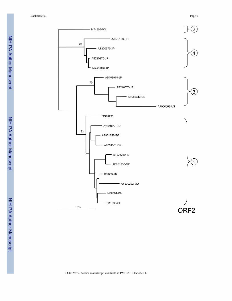
For ORF2, only TS00223 could be amplified. This sample belonged to HEV genotype 1 andwas 7.6%–11.2% divergent from other previously reported isolates. TS00223 was also 8.7%and 5.3% divergent from isolates AF051351-EG and AF051352-EG, respectively, previouslycollected from Egypt in 1993–94 (Figure 1B).
Analysis of the highly conserved ORF2/3 overlap region also confirmed that TS00223 andTS00226 belonged to HEV genotype 1 (Figure 1C). Both TS00223 and TS00226 were identicalto one another and were 0.0%–3.2% divergent from other genotype 1 isolates. TS00223 andTS00226 were identical to Egyptian isolate AF051351-EG but 2.1% divergent from isolateAF051352-EG.
DiscussionUniversal community exposure is likely responsible for the high seroprevalence of HEV inEgypt 5, 8, 11. However, HEV-associated AVH is relatively uncommon, leading someinvestigators to suggest that HEV strains in Egypt may be less virulent than in other geographiclocations 6, 11. Tsarev et al. previously reported on two sporadic cases of HEV infectionrecovered from male army recruits living in the Cairo area in 1993–94 19. These isolates were
Blackard et al. Page 4
J Clin Virol. Author manuscript; available in PMC 2010 October 1.
NIH
-PA Author Manuscript
NIH
-PA Author Manuscript
NIH
-PA Author Manuscript

closely related to other African isolates. Our finding of genotype 1 further supports thecirculation of HEV genotype 1 throughout Egypt. Importantly, because our samples werecollected in 2006–08 as part of a large prospective study of HEV infection in the cities of Assuit(Upper Egypt) and Mansoura (in the Delta), they represent the HEV isolates currentlycirculating in the central region of Egypt. While the intragenotypic variability observedsuggests geographic diversity and/or viral evolution with time in this population, continuedevaluation of HEV variation in this and other endemic settings is clearly warranted.
The detection of HEV RNA in only two stool samples in the current study is somewhatsurprising given that HEV RNA has been detected in the stool in epidemic settings 20–22.However, the presence of viremia in the serum may be less frequent 23, 24. Furthermore,variable detection rates could reflect differences in the viral extraction methodology, assaysensitivity, and/or a highly divergent HEV strain that is not readily detected by current PCR-based assays. However, in the current analysis, we utilized two highly sensitive, real-time PCRassays that readily detected HEV RNA in human serum and stool in previous studies 13, 14.Additionally, HEV genotype 1 strains related to other North African isolates were detected,suggesting that assay sensitivity and viral divergence were unlikely to explain our findings.Importantly, the low frequency of HEV RNA detection in Egypt could also reflect true low-level viremia in HEV-infected individuals that merits additional investigation. This is furthersupported by the fact that HEV viremia was detected but only after experimental inoculationof human stool suspensions into macaques. Collectively, these data suggest that an attenuatedgenotype 1 may be circulating in Egypt and could be responsible for the low rate of HEVmortality observed.
AcknowledgmentsThis work was presented at the 13th International Symposium on Viral Hepatitis and Liver Disease held in Washington,DC in March 2009. This work was supported by an NIAID R21 (AI 067868) award to MTS, NIDDK K24 (DK 070528)to KES, and the Division of Intramural Research at NIAID.
AbbreviationsHEV
hepatitis E virus
HAV hepatitis A virus
HBV hepatitis B virus
HCV hepatitis C virus
AVH acute viral hepatitis
ORF open reading frame
RT reverse transcriptase
ULN upper limit of normal
Blackard et al. Page 5
J Clin Virol. Author manuscript; available in PMC 2010 October 1.
NIH
-PA Author Manuscript
NIH
-PA Author Manuscript
NIH
-PA Author Manuscript

SD standard deviation
ALT alanine transaminase
AST aspartate transaminase
S/C sample/cut off ratio
IU international units
nts nucleotides
bp base pair
References1. Emerson SU, Purcell RH. Hepatitis E virus. Reviews in Medical Virology May–Jun;2003 13(3):145–
154. [PubMed: 12740830]2. Mushahwar I. Hepatitis E virus: molecular virology, clinical features, diagnosis, transmission,
epidemiology, and prevention. Journal of Medical Virology 2008;80(4):646–658. [PubMed:18297720]
3. Okamoto H. Genetic variability and evolution of hepatitis E virus. Virus Research 2007;127(2):216–228. [PubMed: 17363102]
4. Mizuo H, Yazaki Y, Sugawara K, et al. Possible risk factors for the transmission of hepatitis E virusand for the severe form of hepatitis E acquired locally in Hokkaido, Japan. J Med Virol Jul;2005 76(3):341–349. [PubMed: 15902701]
5. Fix AD, Abdel-Hamid M, Purcell RH, et al. Prevalence of antibodies to hepatitis E in two rural Egyptiancommunities. American Journal of Tropical Medicine and Hygiene 2000;62(4):519–523. [PubMed:11220771]
6. Stoszek SK, Abdel-Hamid M, Saleh DA, et al. High prevalence of hepatitis E antibodies in pregnantEgyptian women. Transactions of the Royal Society of Tropical Medicine and Hygiene 2006;100(2):95–101. [PubMed: 16257426]
7. Divizia M, Gabrieli R, Stefanoni ML, et al. HAV and HEV infection in hospitalised hepatitis patientsin Alexandria, Egypt. European Journal of Epidemiology 1999;15(7):603–609. [PubMed: 10543349]
8. Abe K, Li TC, Ding X, et al. International collaborative survey on epidemiology of hepatitis E virusin 11 countries. Southeast Asian Journal of Tropical Medicine and Public Health 2006;37(1):90–95.[PubMed: 16771218]
9. Abdel Hady SI, El-Din MS, El-Din M. A high hepatitis E virus (HEV) seroprevalence among unpaidblood donors and haemodialysis patients in Egypt. Journal of the Egyptian Public Health Association1998;73(3–4):165–179. [PubMed: 17219919]
10. El-Esnawy N. Examination for hepatitis E virus in wastewater treatment plants and workers by nestedRT-PCR and ELISA. Journal of the Egyptian Public Health Association 2000;75(1–2):219–231.[PubMed: 17219857]
11. Stoszek SK, Engle RE, Abdel-Hamid M, et al. Hepatitis E antibody seroconversion without diseasein highly endemic rural Egyptian communities. Transactions of the Royal Society of TropicalMedicine and Hygiene 2006;100 (2):89–94. [PubMed: 16257427]
12. Shata MT, Barrett A, Shire NJ, et al. Characterization of hepatitis E-specific cell-mediated immuneresponse using IFN-gamma ELISPOT assay. Journal of Immunological Methods 2007;328(1–2):152–161. [PubMed: 17905301]
Blackard et al. Page 6
J Clin Virol. Author manuscript; available in PMC 2010 October 1.
NIH
-PA Author Manuscript
NIH
-PA Author Manuscript
NIH
-PA Author Manuscript

13. Jothikumar N, Cromeans TL, Robertson BH, Meng XJ, Hill V. A broadly reactive one-step real-timeRT-PCR assay for rapid and sensitive detection of hepatitis E virus. Journal of Virological Methods2006;131(1):65–71. [PubMed: 16125257]
14. Gyarmati P, Mohammed N, Norder H, Blomberg J, Belák S, Widén F. Universal detection of hepatitisE virus by two real-time PCR assays: TaqMan and Primer-Probe Energy Transfer. Journal ofVirological Methods 2007;146(1–2):226–235. [PubMed: 17825434]
15. Schlauder GG, Desai SM, Zanetti AR, Tassopoulos NC, Mushahwar IK. Novel hepatitis E virus(HEV) isolates from Europe: evidence for additional genotypes of HEV. Journal of Medical VirologyMar;1999 57(3):243–251. [PubMed: 10022795]
16. Wang Y, Zhang H, Ling R, Li H, Harrison TJ. The complete sequence of hepatitis E virus genotype4 reveals an alternative strategy for translation of open reading frames 2 and 3. Journal of GeneralVirology Jul;2000 81(Pt 7):1675–1686. [PubMed: 10859372]
17. Inoue J, Takahashi M, Yazaki Y, Tsuda F, Okamoto H. Development and validation of an improvedRT-PCR assay with nested universal primers for detection of hepatitis E virus strains with significantsequence divergence. Journal of Virological Methods Nov;2006 137(2):325–333. [PubMed:16901555]
18. Thompson J, Gibson T, Plewniak F, Jeanmougin F, Higgins D. The CLUSTAL_X windows interface:flexible strategies for multiple sequence alignment aided by quality analysis tools. Nucleic AcidsResearch 1997;25(24):4876–4882. [PubMed: 9396791]
19. Tsarev SA, Binn LN, Gomatos PJ, et al. Phylogenetic analysis of hepatitis E virus isolates from Egypt.Journal of Medical Virology Jan;1999 57(1):68–74. [PubMed: 9890424]
20. Boccia D, Guthmann JP, Klovstad H, et al. High mortality associated with an outbreak of hepatitisE among displaced persons in Darfur, Sudan. Clinical Infectious Diseases 2006;42(12):1679–1684.[PubMed: 16705571]
21. Isaäcson M, Frean J, He J, Seriwatana J, Innis B. An outbreak of hepatitis E in Northern Namibia,1983. American Journal of Tropical Medicine and Hygiene 2000;62(5):619–625. [PubMed:11289674]
22. Chobe LP, Chadha MS, Banerjee K, Arankalle V. Detection of HEV RNA in faeces, by RT-PCRduring the epidemics of hepatitis E in India (1976–1995). Journal of Viral Hepatitis 1997;4(2):129–133. [PubMed: 9097269]
23. Kantala T, Maunula L, von Bonsdorff CH, Peltomaa J, Lappalainen M. Hepatitis E virus in patientswith unexplained hepatitis in Finland. Journal of Clinical Virology. 2009Epub ahead of print
24. Zhu G, Qu Y, Jin N, et al. Seroepidemiology and molecular characterization of hepatitis E virus inJilin, China. Infection 2008;36(2):140–146. [PubMed: 18330507]
25. Felsenstein J. Confidence limits on phylogenies: an approach using the bootstrap. Evolution1985;39:783–791.
Blackard et al. Page 7
J Clin Virol. Author manuscript; available in PMC 2010 October 1.
NIH
-PA Author Manuscript
NIH
-PA Author Manuscript
NIH
-PA Author Manuscript

Blackard et al. Page 8
J Clin Virol. Author manuscript; available in PMC 2010 October 1.
NIH
-PA Author Manuscript
NIH
-PA Author Manuscript
NIH
-PA Author Manuscript
Blackard et al. Page 9
J Clin Virol. Author manuscript; available in PMC 2010 October 1.
NIH
-PA Author Manuscript
NIH
-PA Author Manuscript
NIH
-PA Author Manuscript

Figure 1.Phylogenetic analysis of HEV sequences from two symptomatic Egyptian patients. A) ORF 1(242 base pairs [bp]; B) ORF2/3 overlap region (97 bp); C) ORF 2 (98 bp). Reference sequencesare denoted by their GenBank accession numbers and the following country-specificabbreviations: Mexico (MX), United States (US), Japan (JP), China (CH), Chad (CD),Morocco (MO), India (IN), Pakistan (PA), Nepal (NP), Egypt (EG), and Algeria (AL). Thetwo Egyptian samples characterized in the current study are highlighted in bold. The statisticalrobustness and reliability of the branching order within each phylogenetic tree were confirmedby bootstrap analysis using 100 replicates 25. Sequences from the current study – with primer
Blackard et al. Page 10
J Clin Virol. Author manuscript; available in PMC 2010 October 1.
NIH
-PA Author Manuscript
NIH
-PA Author Manuscript
NIH
-PA Author Manuscript

sequences removed – have been submitted to GenBank under accession numbers FJ423076 –FJ423080.
Blackard et al. Page 11
J Clin Virol. Author manuscript; available in PMC 2010 October 1.
NIH
-PA Author Manuscript
NIH
-PA Author Manuscript
NIH
-PA Author Manuscript