Apoptosis during ZIKA Virus infection: too soon or too late?
Upload
khangminh22Category
view
1download
0
Genome-wide Integrative Analysis of Zika-Virus-Infected Neuronal Stem Cells Reveals Roles for MicroRNAs in Cell Cycle and Stemness
Jason W. Dang1,2,3, Shashi Kant Tiwari1,2, Yue Qin1,2,4, Tariq M. Rana1,2,5,6,7,8,*
1Department of Pediatrics, University of California, San Diego, 9500 Gilman Drive, MC 0762, La Jolla, CA 92093, USA
2Division of Genetics, University of California, San Diego, 9500 Gilman Drive, MC 0762, La Jolla, CA 92093, USA
3Department of Bioengineering, University of California, San Diego, 9500 Gilman Drive, MC 0412, La Jolla, CA 92093, USA
4Bioinformatics and Systems Biology, University of California, San Diego, 9500 Gilman Drive, MC 0419, La Jolla, CA 92093, USA
5Program in Immunology, University of California, San Diego, 9500 Gilman Drive, MC 0762, La Jolla, CA 92093, USA
6Institute for Genomic Medicine, University of California, San Diego, 9500 Gilman Drive, MC 0762, La Jolla, CA 92093, USA
7Moores Cancer Center, University of California, San Diego, 9500 Gilman Drive, MC 0762, La Jolla, CA 92093, USA
8Lead Contact
SUMMARY
Zika virus (ZIKV) infection is implicated in severe fetal developmental disorders, including
microcephaly. MicroRNAs (miRNAs) post-transcriptionally regulate numerous processes
associated with viral infection and neurodegeneration, but their contribution to ZIKV pathogenesis
This is an open access article under the CC BY-NC-ND license (http://creativecommons.org/licenses/by-nc-nd/4.0/).*Correspondence: [email protected] CONTRIBUTIONSJ.W.D. designed and performed experiments, analyzed the data, and wrote the manuscript; S.K.T. designed and performed experiments, analyzed the data, and participated in figure preparation; Y.Q. analyzed the data and participated in figure preparation; and T.M.R. contributed to the concept and design, data analysis and interpretation, and manuscript writing. All authors approved the final version of this manuscript.
SUPPLEMENTAL INFORMATIONSupplemental Information can be found online at https://doi.org/10.1016/j.celrep.2019.05.059.
DECLARATION OF INTERESTST.M.R. is a founder of ViRx Pharmaceuticals and a member of its scientific advisory board. The terms of this arrangement have been reviewed and approved by the University of California, San Diego, in accordance with its conflict of interest policies.
DATA AND SOFTWARE AVAILABILITYRaw and processed data are provided in the Gene Expression Omnibus (accession number GSE113640; subseries GSE113636 for RNA-seq, GSE113638 for iCLIP-seq, and GSE113639 for miRNA-seq).
HHS Public AccessAuthor manuscriptCell Rep. Author manuscript; available in PMC 2019 August 09.
Published in final edited form as:Cell Rep. 2019 June 18; 27(12): 3618–3628.e5. doi:10.1016/j.celrep.2019.05.059.
Author M
anuscriptA
uthor Manuscript
Author M
anuscriptA
uthor Manuscript

is unclear. We analyzed the mRNA and miRNA transcriptomes of human neuronal stem cells
(hNSCs) during infection with ZIKV MR766 and Paraiba strains. Integration of the miRNA and
mRNA expression data into regulatory interaction networks showed that ZIKV infection resulted
in miRNA-mediated repression of genes regulating the cell cycle, stem cell maintenance, and
neurogenesis. Bioinformatics analysis of Argonaute-bound RNAs in ZIKV-infected hNSCs
identified a number of miRNAs with predicted involvement in microcephaly, including miR-124–3p, which dysregulates NSC maintenance through repression of the transferrin receptor (TFRC).
Consistent with this, ZIKV infection upregulated miR-124–3p and downregulated TFRC mRNA
in ZIKV-infected hNSCs and mouse brain tissue. These data provide insights into the roles of
miRNAs in ZIKV pathogenesis, particularly the microcephaly phenotype.
Graphical Abstract
In Brief
Dang et al. investigate the dysregulation of miRNAs in Zika-virus-infected neural stem cells and
identify miR-124–3p as a potential regulator of Zika-virus-mediated microcephaly.
INTRODUCTION
Zika virus (ZIKV) is a re-emerging arbovirus belonging to the Flaviviridae family and has
recently been linked to severe fetal abnormalities, including microcephaly and fetal growth
restriction (Brasil et al., 2016; Lazear and Diamond, 2016; Sarno et al., 2016). In vitro and
in vivo studies have shown that ZIKV preferentially infects neuronal stem and/or progenitor
Dang et al. Page 2
Cell Rep. Author manuscript; available in PMC 2019 August 09.
Author M
anuscriptA
uthor Manuscript
Author M
anuscriptA
uthor Manuscript

cells and immature neurons in the developing brain and dysregulates processes involved in
cell-cycle progression, differentiation, apoptosis, autophagy, and immune activation (Cugola
et al., 2016; Dang et al., 2016; Li et al., 2016a, 2016b; Liang et al., 2016; Tang et al., 2016).
However, the molecular mechanisms by which ZIKV perturbs the transcriptomic landscape
or leads to microcephaly are not well understood.
MicroRNAs (miRNAs) are a class of small non-coding RNAs (~22 nt in length) that play
critical roles in regulating protein expression. miRNAs act post-transcriptionally by binding
to partially complementary sites in the 3′ UTR of target mRNAs. This sequence-specific
interaction leads to translational repression or mRNA degradation through Argonaute
proteins within the RNA-induced silencing complex (RISC), which cleave the mRNA and
recruit other proteins that repress translation or promote degradation. The mRNA targeting
specificity of miRNAs is controlled by many factors, including base pairing between the
miRNA 5′ seed sequence and mRNA 3′-UTR sequence, cooperativity between multiple
miRNA-binding sites, and the position of miRNA-binding sites in the targeted mRNA
(Agarwal et al., 2015; Ambros, 2004; Bartel, 2009; Cloney, 2016; Grimson et al., 2007;
Lewis et al., 2005; Pasquinelli, 2012). This flexibility means that individual miRNAs are
capable of repressing the translation of hundreds of target mRNAs (Baek et al., 2008;
Selbach et al., 2008). As a result, miRNAs are known to play pivotal roles in the post-
transcriptional regulation of numerous biological processes.
Little is currently known about the role of miRNAs in ZIKV pathogenesis and microcephaly.
Given their documented roles in regulating neurodegeneration, viral infection, and innate
immunity (Eacker et al., 2009; Lanford et al., 2010; Liu et al., 2012; O’Connell et al., 2010;
Sullivan and Ganem, 2005; Taganov et al., 2006; Wang et al., 2006), we hypothesized that
miRNAs may play a significant role in ZIKV pathogenesis, particularly the effects on the
developing brain. Here, we report that ZIKV infection dysregulates both coding gene and
miRNA transcriptomes of human neuronal stem cells (hNSCs). We performed meta-analyses
and constructed regulatory interaction networks to integrate the miRNA and mRNA
expression data, with the goal of shedding light on the potential role of miRNA-mediated
target gene repression during ZIKV infection. We identified a number of miRNAs, including
let-7c and miR-124–3p, that mediate the suppression of gene networks involved in cell-cycle
progression and stem cell maintenance. Collectively, our data provide insight into the
function of miRNA-regulated networks in ZIKV-induced pathogenesis, particularly as it
pertains to microcephaly.
RESULTS
ZIKV MR766 and Paraiba Modulate the mRNA Transcriptome of hNSCs
To investigate the role of miRNAs during ZIKV pathogenesis, we performed next-generation
sequencing of RNA isolated from hNSCs infected with ZIKV strains MR766 (African
origin) and Paraiba (Brazilian origin) for 3 days (Figure 1A). Consistent with previous
studies showing that ZIKV strains show differences in rates of viral replication (Cugola et
al., 2016; Simonin et al., 2016), we found ~10-fold higher levels of infection with MR766
than with Paraiba on the first 3 days after inoculation of hNSCs at the same MOI of 1, as
demonstrated by qRT-PCR analysis of ZIKV RNA (Figure 1B). Immunostaining of ZIKV
Dang et al. Page 3
Cell Rep. Author manuscript; available in PMC 2019 August 09.
Author M
anuscriptA
uthor Manuscript
Author M
anuscriptA
uthor Manuscript

envelope protein (ZIKVE) (Figure 1C) further confirmed higher rates of infection, with
MR766 infecting approximately 70% of cells and Paraiba infecting ~30% after 3 days post-
inoculation. In addition, we confirmed that ZIKV MR766 produces ~6-fold more infectious
viral particles than does ZIKV Paraiba in plaque-forming assays (Figure S1A).
The Paraiba strain had a less significant impact on gene expression in hNSCs, suggesting an
overall lower rate of infection as previously indicated (Figures 1B and 1D; Tables S1 and
S2). ZIKV MR766 significantly upregulated 1,159 genes and downregulated 1,120 genes
(Figures 1D and 1E), compared with only 112 and 178 genes that were significantly
upregulated and downregulated, respectively, in ZIKV Paraiba-infected hNSCs (Figures 1D
and 1F). In addition, we identified a total of 52 and 52 genes that were commonly
upregulated and downregulated, respectively, in both MR766- and Paraiba-infected cells
(Figure 1D).
We next performed gene set enrichment analysis (GSEA) of the differentially expressed
genes. In MR766-infected hNSCs, the upregulated genes were enriched in functions related
to chromosome organization and cell-cycle processes (Figure S1B), whereas the
downregulated genes were involved in gene expression, biosynthetic processes, and cell
death (Figure S1C). The processes most affected by MR766 infection were those governing
chromosome organization, metabolism, cell cycle, and cell stress (Figure S1D), which is
consistent with previous reports (Tang et al., 2016). In contrast, the upregulated and
downregulated genes in Paraiba-infected hNSCs were all largely related to metabolism and
biosynthetic processes, with additional enrichment of genes involved in tissue development
and neurogenesis (Figures S1E–S1G).
We considered that the transcriptomic differences induced by ZIKV MR766 and Paraiba
might be a consequence of their differing infection rates. Therefore, we infected hNSCs with
MR766 at an MOI of 1 for 2 days or with Paraiba at MOIs of 1 or 3 for 2 or 4 days. We then
analyzed the top 20 most differentially expressed genes between both strains to assess their
similarity as a function of MOI and time point (Figure 1G). Indeed, we found that the
Paraiba- and MR766-induced differential gene expression profiles became increasingly
similar as the Paraiba MOI and time post-infection increased, indicating that the differential
pattern of gene expression was a reflection of the infection level rather than the strain per se.
Collectively, these findings indicate that ZIKV MR766 infection and Paraiba infection of
hNSCs cause dysregulation of a number of pathways involved in neurogenesis, suggesting
that they may contribute to the microcephaly phenotype.
miRNAs Regulate Processes Implicated in ZIKV-Induced Microcephaly
Because miRNAs are potent post-transcriptional regulators, we examined their contribution
to the changes in the transcriptome of ZIKV-infected hNSCs. We profiled differential
miRNA expression at 3 days post-infection by microRNA sequencing (miRNA-seq)
(Figures 2A–2C; Table S3). Although ZIKV MR766 induced a more robust change in the
mRNA transcriptome than did Paraiba infection (as described earlier), ZIKV Paraiba
induced a significantly greater change in miRNAs, in terms of both quantity and magnitude,
likely due to the differing rates of viral replication within neural stem cells. Interestingly, we
Dang et al. Page 4
Cell Rep. Author manuscript; available in PMC 2019 August 09.
Author M
anuscriptA
uthor Manuscript
Author M
anuscriptA
uthor Manuscript

observed more differentially expressed mRNAs during MR766 viral infection but fewer
differentially expressed miRNAs.
To understand the potential mechanistic roles of miRNAs in ZIKV infection and the
associated neurodegenerative pathology, we utilized the predictive algorithms TargetScan
(Agarwal et al., 2015), miRANDA (Betel et al., 2008), and miRTarBase (Chou et al., 2016)
to identify putative mRNA targets of the differentially expressed miRNAs. These algorithms
evaluate target seed sequence pairing, site numbers, conservation, and site context scores to
predict targets with high confidence. To identify miRNA-mRNA interactions that may be
regulated by ZIKV infection, we then cross-checked the list of putative mRNA targets with
the mRNAs shown to be most significantly altered by ZIKV infection of hNSCs. GSEA of
the dataset indicated that mRNAs satisfying both criteria (i.e., directly modulated by ZIKV
infection and putative targets of differentially expressed miRNAs) were enriched in
functions related to transcriptional regulation, metabolism, cellular stress response, cell
cycle, tissue development, neurogenesis and nervous system development, cell death, and
neuron differentiation (Figure 2D). Using qRT-PCR, we validated these data by confirming
that ZIKV infection down-regulates NESTIN and PAX6 expression, both of which are
involved in NSC maintenance (Figure S2A). Similarly, analysis of the datasets from ZIKV
Paraiba-infected cells also identified mRNAs likely to be involved in processes related to
metabolism, tissue development, neurogenesis, and neuron differentiation (Figure 2E).
These data indicate that pathways potentially involved in neurodegeneration feature
prominently among the host miRNA-mRNA networks dysregulated by infection of hNSCs
with both ZIKV MR766 and Paraiba.
To more precisely map the miRNA-regulated pathways that may contribute to ZIKV
pathogenesis, we constructed integrative networks of the ZIKV-modulated miRNAs and
miRNA-regulated mRNAs. Genes that were downregulated by ZIKV infection and enriched
in gene ontology (GO) functions related to ‘‘cell cycle’’ and ‘‘G1/S transition,’’ ‘‘defense
response to virus,’’ and ‘‘brain development’’ (Figure 2F, blue hexagons) were cross-
referenced with potential miRNA regulators concomitantly upregulated upon ZIKV
infection (Figure 2F, red circles). Likewise, genes that were upregulated by ZIKV infection
and enriched in ‘‘viral process,’’ ‘‘apoptosis,’’ ‘‘NF-κB (nuclear factor κB) signaling,’’ and
‘‘cell cycle arrest’’ were cross-referenced with potential miRNA regulators concomitantly
downregulated by ZIKV infection (Figure S2B). These networks indicate that some
differentially expressed mRNAs–such as TP53 or CDK6–may be derepressed, and thereby
upregulated following infection, due to the downregulation of multiple putative miRNAs.
The miRNAs identified from these analyses included many with functions relevant to the
pathogenic ZIKV phenotype, including G1/S transition, defense response to virus, brain
development (Figure 2F), viral process, apoptosis, NF-κB signaling, and cell-cycle arrest
(Figure S2B). We also generated networks of miRNA targets downregulated by ZIKV
(Figure S2C) and grouped them by their GO function (Figure S2D). With these analyses, we
observed downregulated genes involved in biological processes, including nervous system
development, cell-cycle transition, and DNA damage repair. Collectively, these interaction
networks identify a number of miRNAs and mRNAs perturbed during ZIKV infection.
Further studies including gain and loss of function should be performed to assess the ability
Dang et al. Page 5
Cell Rep. Author manuscript; available in PMC 2019 August 09.
Author M
anuscriptA
uthor Manuscript
Author M
anuscriptA
uthor Manuscript

of ZIKV to dysregulate multiple miRNAs with effects on the same target mRNA (Figure
2F).
AGO-iCLIP-Seq Identifies Dysregulated miRNA-mRNA Interactions in ZIKV-Infected hNSCs
miRNAs contribute to post-transcriptional regulation of gene expression by associating with
Argonaute proteins (AGOs) to repress target gene expression through either mRNA
degradation or translational repression within the RISC. Differential expression of miRNAs
during viral infection may hint at an important role in viral pathogenesis; however, it does
not necessarily indicate a biological function. Thus, to further elucidate the role of miRNA-
mRNA networks in ZIKV infection of hNSCs, we performed Argonaute crosslinking and
immunoprecipitation followed by sequencing (AGO-iCLIP-seq), which identifies miRNAs
and mRNAs bound to AGOs within the RISC (Chi et al., 2009; Haecker et al., 2012; Hafner
et al., 2010; König et al., 2010, 2011). To accomplish this, neural stem cells were infected
with Paraiba isolates at MOI 1 for 4 days to achieve higher viral titers without significant
cell loss and UV crosslinked to covalently bind RNA-protein complexes. Lysates were
treated with RNase T1 and immunoprecipitated with a pan-AGO-specific monoclonal
antibody to identify all miRNAs and target mRNAs within the RISC following stringent
high salt washes. Libraries were generated and sequenced to identify miRNAs and target
mRNAs with single-nucleotide resolution. Moreover, AGO-iCLIP-seq may identify
biologically significant miRNAs whose expression levels are not changed during viral
infection but show greater loading in the RISC or have non-canonical binding sites outside
of the 3′ UTR. Furthermore, miRNAs may post-transcriptionally regulate gene expression
through various processes, including mRNA degradation and translational repression, both
pathways of which are mediated though the association with RISC and AGO. Thus, through
analyzing RNAs bound by AGO using AGO-iCLIP-seq, we can address both mRNA
degradation and translational repression, which may not necessarily be reflected in RNA-
sequencing (RNA-seq) data.
As expected, AGO-associated material from cells was enriched in putative miRNA-binding
sites within mRNA 3′ UTRs rather than 5′ UTRs or the coding region of a gene (CDS) of
cellular mRNAs (Figure S3A). Interestingly, in ZIKV-infected neural stem cells, we
identified many sequences bound to AGO throughout the ZIKV genome (Figure 3A),
suggesting that ZIKV gene expression is regulated by host miRNAs. Further experiments
should be performed to determine the possible role of miRNAs in targeting viral UTRs as an
antiviral host response during ZIKV infection. ZIKV is known to dysregulate many host
pathways at the RNA and protein levels, including autophagy via AKT/mTOR signaling
(Liang et al., 2016), mitosis (Onorati et al., 2016), and splicing (Hu et al., 2017); thus, we
did not observe a clear correlation between ZIKV-induced miRNA expression, mRNA target
expression, and AGO binding at a transcriptome level. However, there are cases in which
miRNA-seq and AGO-iCLIP-seq data are correlated. For example, the miRNA-seq results
indicate that miR-1246 and miR-335 are both upregulated during ZIKV infection and show
enhanced binding to AGO (Figures S3B and S3C). Conversely, expression of miR-129–2
and miR-139 was decreased following ZIKV Paraiba infection, which is consistent with
their attenuated binding to AGO (Figures S3D and S3E).
Dang et al. Page 6
Cell Rep. Author manuscript; available in PMC 2019 August 09.
Author M
anuscriptA
uthor Manuscript
Author M
anuscriptA
uthor Manuscript

Because ZIKV is able to dysregulate many different pathways, we wanted to identify
miRNAs specifically targeting genes relevant to microcephaly by repressing host gene
expression. For this, the miRNA-seq and AGO datasets were screened against the
Harmonizome database (Rouillard et al., 2016), which ranks miRNAs using an aggregate
score based on putative target genes associated with microcephaly, curated from the
Comparative Toxicogenomics Database (Davis et al., 2017). This analysis identified a
number of miRNAs potentially regulating genes significant to microcephaly, from which we
selected two, hsa-let-7c and miR-124–3p, for more detailed investigation (Figure 3B).
We first analyzed the potential role of let-7c in ZIKV pathogenesis because of its well-
known role in regulating stem cell self-renewal (Büssing et al., 2008; Melton et al., 2010).
We confirmed that ZIKV infection did, indeed, upregulate let-7c expression (Figure 3C) and
AGO binding (Figure 3D), although this change was only significant for ZIKV Paraiba-
infected cells under the conditions used here. Since the let-7c target gene high-mobility
group AT-hook 2 (HMGA2) has previously been shown to govern self-renewal of NSCs
(Nishino et al., 2008), we analyzed HMGA2 expression and binding of AGO to the HMGA2 3′ UTR following ZIKV infection. Consistent with previous data (Yu et al., 2015) and
TargetScan predictions, we observed increased AGO binding to sequences in the HMGA2 3′ UTR (Figure 3E) and downregulation of HMGA2 mRNA levels (Figure 3F) upon ZIKV
infection. These data, therefore, suggest that ZIKV induces let-7c transcription and
downregulation of its target genes, including the established regulator of NSC renewal,
HMGA2.
The Harmonizome database analysis also predicted miR-124–3p to be involved in the ZIKV-
induced microcephaly phenotype (Figure S4A). Similar to let-7c, this mRNA also shows a
slight, although not statistically significant, upregulated expression (Figure S4B) but an
increased association with AGO (Figure S4C) after ZIKV infection of hNSCs. Since no
miR-124–3p targets with potential functions in neural stem cell biology or ZIKV
pathogenesis have yet been identified, we looked for genes that were significantly
downregulated by ZIKV Paraiba and MR766 infection of hNSCs and are also predicted
target genes of miR-124–3p (Figure S4D). We selected the transferrin receptor (TFRC) from
the 8 potential target genes identified–FLRT3, LAMC1, NRCAM, TFRC, C3orf58, NRP1,
TXNRD1, and RCAN1–for further analysis, since it has known roles in stem cell self-
renewal (Schonberg et al., 2015). Moreover, TFRC shows high species conservation of the
putative miR-124-3p-binding site, suggesting that its regulation by miR-124–3p has an
important function (Figures S4E and S4F). Notably, ZIKV infection of hNSCs resulted in
increased AGO binding at the TFRC 3′ UTR and in a concomitant decrease in TFRC mRNA levels (Figures 4A and 4B).
We next sought to confirm the relevance of our findings in vivo using Ifnar1−/− mice. Six
days after ZIKV infection, the mice were sacrificed, and the brains were removed, sectioned,
and stained for ZIKVE and neuronal cell markers to identify infected cells. We found that
the ZIKVE colocalized with the NSC marker SOX2 in the hippocampus and subventricular
zone regions (Figures S4G and S4H), consistent with previous reports (Li et al., 2016a,
2016b) that ZIKV preferentially infects these cells. Moreover, qRT-PCR analysis of brain
tissue confirmed that TFRC mRNA levels were downregulated in the infected, compared
Dang et al. Page 7
Cell Rep. Author manuscript; available in PMC 2019 August 09.
Author M
anuscriptA
uthor Manuscript
Author M
anuscriptA
uthor Manuscript

with uninfected, mice (Figure 4C). Thus, we have identified two miRNA-mRNA interactions
that could contribute to the neurodegenerative phenotype induced by ZIKV infection.
Potential Role for miR-124–3p–TFRC Interactions in the Maintenance of NSCs In Vivo
To investigate the role of miR-124–3p and its putative target TFRC in ZIKV-mediated
microcephaly, neurosphere growth kinetics were evaluated (Dang et al., 2016; Tiwari et al.,
2014b) following miR-124–3p overexpression and TFRC knockdown. miR-124 mimic
overexpression downregulated TFRC mRNA level (Figure 4D) and reduced the size of
neurospheres, as compared to scrambled miRNA transfected samples (Figures 4E and 4F).
Similarly, knockdown of TFRC by small interfering RNA (siRNA) in hNSC-derived
neurospheres resulted in attenuated neurosphere size, as compared to a scrambled control
group, consistent with miR-124–3p overexpression (Figures 4G–4I).
Next, the direct interaction between miR-124–3p and TFRC was investigated, using a dual
luciferase assay containing the TFRC 3′ UTR and miRNA seed sequence target site. To
validate whether miR-124–3p targets TFRC through this putative miR-124–3p binding site
in the 3′ UTR, we utilized a luciferase reporter construct (pGL3) in which the human TFRC
3′ UTR, containing either a wild-type (WT) or mutant miR-124–3p binding sequence, was
placed immediately downstream of the luciferase gene. 293FT cells were co-transfected with
WT or mutant type (Mut) reporter constructs with miR-124–3p mimics and pRLuc null
plasmid expressing Renilla luciferase. Luciferase reporter activity of WT constructs was
significantly reduced in cells co-transfected with miR-124–3p (Figure 4J). However,
luciferase activity was not abolished in reporter constructs containing the mutant target
sequence, indicating that miR-124–3p represses TFRC expression by specifically binding to
the predicted target sites in the 3′ UTR of TFRC (Figure 4J).
To gain insight into the mechanism by which TFRC downregulation might affect NSCs, we
examined the effects of siRNA-mediated TFRC knockdown in hNSCs on TFRC target genes
involved in cell-cycle regulation. Several studies have shown that TFR plays a role in
glioblastoma stem cell proliferation and self-renewal through the FOXM1 transcriptional
regulatory signaling loop, which decreases the expression of cell-cycle genes (Schonberg et
al., 2015; Silvestroff et al., 2013). Notably, we found that siRNA-mediated knockdown of
TFRC or ZIKV infection decreased the expression of FOXM1 as well as down-stream
targets of the FOXM1 regulatory axis–AURKA, CCNB2, CDC25A, CDK1, CENPF,
MELK, and PLK1–which play important roles in cell-cycle regulation (Figure 4K).
Collectively, the results presented here shed light on the functional role of miRNAs,
particularly miR-124–3p, in post-transcriptional regulation of ZIKV-infected hNSCs and the
associated microcephaly phenotype.
DISCUSSION
In this study, we performed integrative analyses of coding and non-coding transcriptomes in
hNSCs, which revealed miRNA-mRNA networks that may be dysregulated during ZIKV
infection and may contribute to the microcephaly phenotype. Analysis of the dynamic
transcriptomic landscape and RNAs bound to AGO following ZIKV infection revealed the
Dang et al. Page 8
Cell Rep. Author manuscript; available in PMC 2019 August 09.
Author M
anuscriptA
uthor Manuscript
Author M
anuscriptA
uthor Manuscript
dysregulation of genes associated with cell cycle, neurogenesis, stem cell maintenance, and
metabolism. While previous studies have shown that ZIKV-induced perturbation of cell
cycle, neurogenesis, and stem-cell-related processes contributes to the microcephaly
phenotype, only a few mechanisms have been proposed to explain how ZIKV modulates
these pathways (Dang et al., 2016; Gabriel et al., 2017; Hamel et al., 2015; Li et al., 2016a;
Liang et al., 2016; Onorati et al., 2016; Tang et al., 2016). Our findings point to miRNA-
mediated dysregulated gene expression as a potential contributing factor to the microcephaly
phenotype.
One key finding from analysis of miRNA-mRNA networks in ZIKV-infected cells is that
multiple differentially expressed miRNAs may potentially regulate the same mRNA targets
implicated in microcephaly. For instance, miR-125a-3p and miR-125a-5p, which were
upregulated by ZIKV infection, are both negative regulators of MAVS, an essential signaling
protein in the RIG-I and type I interferon (IFN) response pathways of the innate immune
system (Baril et al., 2009). miR-320c and miR-7–5p, also upregulated by ZIKV, target
SIN3A mRNA, which encodes a STAT3-interacting repressor with an essential role in the
IFN-mediated antiviral response (Icardi et al., 2012). Previous work showed that ZIKV
inhibits type I IFN production through a mechanism involving ZIKV NS5 binding to STAT2
to promote its proteasomal degradation (Grant et al., 2016; Kumar et al., 2016). Thus, our
findings reveal a potential mechanism by which miRNAs mediate suppression of IFN
signaling in ZIKV-infected hNSCs and suggest that multiple miRNAs work in concert to
suppress immunity- and neurodegeneration-related gene networks. Future work involving
gain- and loss-of-function studies of specific miRNAs would further reveal the effects of
ZIKV on proposed miRNA-mRNA networks.
Many of the miRNAs differentially expressed upon ZIKV infection showed enhanced
binding to AGO. Using the Harmonizome database, we screened these for miRNAs and
target genes with the potential ability to dysregulate neurogenesis and induce microcephaly.
We confirmed an association between let-7c and HMGA2 in hNSCs, as previously described
(Nishino et al., 2008). Interestingly, HMGA2 expression is also repressed in cells infected
by human cytomegalovirus, which can also cause birth defects such as microcephaly
(Shlapobersky et al., 2006).
In addition to let-7c, hsa-miR-124–3p was identified as a ZIKV-modulated miRNA with a
potential role in microcephaly. miR-124–3p has also been shown to be upregulated in THP-1
cells latently infected with human cytomegalovirus (Fu et al., 2014; Gérardin et al., 2018).
The putative miR-124–3p target gene TFRC encodes the transferrin receptor TFR, which
regulates cellular iron uptake and metabolism and plays a role in stem cell renewal and cell-
cycle regulation (Sanchez et al., 2006; Schonberg et al., 2015; Silvestroff et al., 2013). In
addition, TFRC1 has been shown to promote the proliferation of rat NSCs (Silvestroff et al.,
2013). Interestingly, TFRC is highly upregulated in cancer stem cells and plays a role in
glioblastoma stem cell renewal through an iron-dependent pathway involving the
transcriptional regulators STAT3 and FOXM1 (Schonberg et al., 2015). Targeting of iron
metabolic pathways can decrease cancer stem cell growth in vitro and in vivo (Schonberg et
al., 2015). Moreover, iron negatively regulates replication of the flavivirus hepatitis C virus
by binding to the Mg2+ binding pocket of the viral polymerase NS5B (Fillebeen and
Dang et al. Page 9
Cell Rep. Author manuscript; available in PMC 2019 August 09.
Author M
anuscriptA
uthor Manuscript
Author M
anuscriptA
uthor Manuscript

Pantopoulos, 2010; Fillebeen et al., 2005). Thus, there are multiple potential mechanisms by
which miRNA-mediated downregulation of TFRC might dysregulate host meta-bolic
processes and perturb the cell cycle during ZIKV infection.
Collectively, the data presented here identify miRNA-regulated transcriptional networks
involved in self-renewal, cell-cycle progression, and neurogenesis in ZIKV-infected hNSCs,
providing a possible mechanism by which the virus inflicts neuronal damage during brain
development.
STAR★METHODS
CONTACT FOR REAGENT AND RESOURCE SHARING
Further information and requests for resources and reagents should be directed to the Lead
Contact, Tariq Rana ([email protected]).
EXPERIMENTAL MODEL AND SUBJECT DETAILS
Cell Lines and Culture Conditions—All cells were maintained at 37°C in a humidified
5% CO2 atmosphere. Vero cells were maintained in Eagle’s Minimum Essential Medium
(EMEM; ATCC, 30–2003) supplemented with 10% fetal bovine serum (FBS; GIBCO) and
antibiotics. Human NSCs (ThermoFisher, A15654) were cultured in StemPro NSC SFM
medium consisting of Knockout DMEM/F-12 media supplemented with 2 mM GlutaMax,
20 ng/ml basic fibroblast growth factor, 20 ng/ml epidermal growth factor, and 2% StemPro
Neural Supplement (ThermoFisher, A1050901) on Matrigel- or CELLStart-coated plates
following the manufacturer’s instructions.
ZIKV Propagation—ZIKV prototype MR766 (National Institutes of Health, LC002520.1)
and Brazilian strain Paraiba (Stevenson Laboratory, University of Miami Life Science and
Technology Park, KX280026.1) were propagated in the low passage Vero cell line. Vero
cells were infected with virus at an MOI of 1 in EMEM medium supplemented with 10%
FBS. The medium was refreshed 4 h after infection and the viral supernatant was collected
at 5 days post-infection. Viral titers were assessed using iScript One-Step RT-PCR kit (Bio-
Rad). Viral copy numbers were calculated from a standard curve of in vitro-transcribed viral
RNA transcripts.
Ifnar −/− Mouse Handling—All studies were conducted in accordance with protocols
approved by the Institutional Review Board of the University of California, San Diego. All
animal work was performed in accordance with the guidelines of the Institutional Animal
Care and Use Committee of the University of California, San Diego.
Ifnar−/− mice 4–5 weeks old were purchased from MMRRC Jackson Laboratories and
housed according to regulatory standards approved by the Institutional Review Board of the
University of California, San Diego.
ZIKV Infection of Mice—All studies were conducted in accordance with protocols
approved by the Institutional Review Board of the University of California, San Diego. All
Dang et al. Page 10
Cell Rep. Author manuscript; available in PMC 2019 August 09.
Author M
anuscriptA
uthor Manuscript
Author M
anuscriptA
uthor Manuscript

animal work was performed in accordance with the guidelines of the Institutional Animal
Care and Use Committee of the University of California, San Diego.
Ifnar−/− mice (4–5 weeks old; MMRRC Jackson Laboratories) were infected by
intraperitoneal injection of 2.5 × 10⁷ genome equivalents ZIKV Paraiba (500 μL of viral
stock with 5 × 10⁴ genome equivalents/μl) or 1.6 × 10⁸ MR766 (500 μL of viral stock with
3.2 × 10⁵ genome equivalents/μl). Mice were sacrificed at 6 days post-infection and brains
were collected for RNA extraction and/or immunostaining (described above).
METHOD DETAILS
Plaque-Forming Assay—Vero cells were seeded in 12-well plates and incubated at 37°C
in a 5% CO2 incubator until they reached ~90%–100% confluency (~3 days). Cells were
infected with serial 10-fold dilutions of ZIKV for 4–6 h and then overlaid with 4% agarose
and incubated for 4 days. Cells were then fixed with 4% formaldehyde and stained with
0.1% crystal violet solution in 20% ethanol. Plaques were visualized under a microscope and
counted. Plaque-forming units (PFU) were calculated as ([number of plaques 3 ZIKV
dilution]) / infection volume) and are expressed as PFU/ml.
Immunofluorescence Microscopy—To assess ZIKV infection, hNSCs were fixed at
24, 48, and 72 h post-infection and immunostained as described previously (Dang et al.,
2016). In brief, ZIKV- and mock-infected hNSCs were fixed with 4% paraformaldehyde
(PFA) in phosphate-buffered saline (PBS) for 20 min at room temperature. Cells were
permeabilized by incubation in 0.1% Triton X-100 for 5 min at room temperature and then
blocked in 5% bovine serum albumin for 30 min. Cells were then incubated overnight at 4°C
with a mouse anti-ZIKVE/anti-flavivirus group antigen (1:500, Millipore MAB10216),
which is directed against the flavivirus envelope protein. Cells were washed with PBS and
incubated for 1 h at room temperature with fluorescein isothiocyanate (FITC)-conjugated
anti-mouse IgG. The nuclei were stained with Hoechst 33258 before analysis.
Immunostained cells were imaged using a Leica fluorescence microscope (DMI 3000B).
For staining of brain sections, at the end of the experiment, mice were transcardially
perfused with normal saline (0.9% NaCl) followed by ice-cold 4% PFA (pH 7.2) under deep
anesthesia, as described previously (Tiwari et al., 2014a). Brains were removed and post-
fixed in 10% PFA overnight at 4°C followed by cryopreservation in 10%, 20%, and 30%
(w/v) sucrose in PBS. Serial coronal sections of 30 μm thickness were cut using a freezing
cryostat (Leica Biosystems, CM3050s) beginning at the bregma −1.50 to −3.50 mm through
the dorsal hippocampus encompassing the dentate gyrus region and +0.26 to −2.5 mm
through the SVZ. Free-floating sections were washed, antigen retrieval was performed with
citrate buffer (pH 6.2), and the sections were blocked with 3% normal goat serum, 0.1%
Triton X-100, and 0.5% bovine serum albumin for 2 h. Sections were then incubated with
mouse anti-ZIKVE antibody (1:500), rabbit anti-SOX2, or rabbit anti-NeuN (1:100) for 24 h
at 4°C. Sections were then stained with secondary antibodies (anti-mouse and anti-rabbit
Alexa Fluor 488 at 1:200; anti-rabbit, anti-mouse, and anti-goat Alexa Fluor 594 at 1:200),
washed, mounted with DAPI-containing Hard Set Anti-Fade mounting medium
(Vectashield, Vector Laboratories, CA, USA), and stored in the dark at 4°C. Slides were
Dang et al. Page 11
Cell Rep. Author manuscript; available in PMC 2019 August 09.
Author M
anuscriptA
uthor Manuscript
Author M
anuscriptA
uthor Manuscript

analyzed using an inverted Leica fluorescence microscope (DMI 3000B) or a Leica SP5
confocal with Resonant Scanner microscope with Leica LAS Lite Software.
RNA-seq and miRNA-seq Library Preparation—RNA-seq libraries were generated
using the NEBNext Ultra II Directional RNA Library Kit for Illumina (NEB, E7760L)
according to the manufacturer’s instructions. miRNA-seq libraries were generated by
ligating Truseq 3′ and 5′ adapters using T4 RNA Ligase2 and T4 RNA Ligase,
respectively. RNAs were reverse transcribed using Superscript II Reverse Transcriptase and
PCR amplified using Q5 master mix (NEB M0494S). Sample quality was assessed using a
high-sensitivity bioanalyzer.
RNA Extraction, cDNA Synthesis, and qRT-PCR—Total RNA was extracted from
hNSCs using a miRNeasy Mini Kit (QIAGEN, 217004) according to the manufacturer’s
instructions. RNA samples were treated with RNase-free DNase (QIAGEN), and cDNA was
generated from 500 ng RNA/sample using iScript Mastermix (Bio-Rad) according to the
manufacturer’s instructions. qPCR was performed with SYBR Green PCR Master Mix (Bio-
Rad) using a Roche LightCycler 480 using ZIKV-specific forward
(TTGGTCATGATACTGCTGATTGC) and reverse (CCCTCCACGAA GTCTCTATTGC)
primers.
AGO-iCLIP-seq Library Preparation—AGO-iCLIP-seq libraries were prepared
according to previously published methods (Huppertz et al., 2014; Konig et al., 2011).
Briefly, hNSCs were infected with ZIKV Paraiba at an MOI of 1 and UV crosslinked with
150 mJ/cm2 at 254 nm on ice. Cell pellets were harvested and lysed (50 mM Tris-HCl, pH
7.4, 100 mM NaCl, 1% NP-40, 0.1% SDS, 0.5% sodium deoxycholate, 1/100 protease
inhibitor cocktail III, Calbiochem). RNAs were partially digested using RNase I (Ambion,
AM2295) and Turbo DNase. Digested RNAs were incubated with washed protein A/G
Dynabeads (Thermo Scientific, 88802) and 10 μg AGO 2A8 antibody (Millipore, Mill-
MABE56) on a rotator overnight at 4°C. RNAs were immunoprecipitated and washed twice
with a high-salt buffer (50 mM Tris-HCl, pH 7.4, 1 M NaCl. 1 mM EDTA, 1% NP-40, 0.1%
SDS, 0.5% sodium deoxycholate) and twice with a wash buffer (20 mM Tris-HCl, pH 7.4,
10 mM MgCl2, 0.2% Tween-20).
To prepare CLIP-seq libraries, 3′ ends of immunoprecipitated RNAs were dephosphorylated
using PNK (Promega) for 20 min and then washed with high-salt buffer and twice with wash
buffer (composition as above). Pre-adenylated L3 linkers were ligated to RNAs on
resuspended beads using RNA ligase (NEB) at 16°C overnight in a thermocycler. RNAs
were washed sequentially with wash buffer, high-salt buffer twice, and wash buffer to
remove excess linker and enzyme. The 5′ ends were radiolabeled using 32P-γ-ATP (Perkin
Elmer, blu002250uc) and PNK.
Samples resolved by SDS-PAGE using 4%–12% NuPAGE Bis-Tris gels (Invitrogen) using 1
s MOPS running buffer following the manufacturer’s instructions. RNA-protein complexes
were transferred to nitrocellulose membranes and RNAs were released by proteinase K
treatment and urea elution of the membranes. RNAs were recovered with RNA phenol/
Dang et al. Page 12
Cell Rep. Author manuscript; available in PMC 2019 August 09.
Author M
anuscriptA
uthor Manuscript
Author M
anuscriptA
uthor Manuscript

chloroform (Ambion, 9722) using Phase Lock Gel Heavy tubes and precipitated overnight
with sodium acetate and ethanol.
Precipitated RNAs were reverse transcribed using Superscript III reverse transcriptase
(Invitrogen) and indexing primers. cDNAs were gel purified using precast 6% Tris/Borate/
EDTA-urea gels (Invitrogen) according to the manufacturer’s instructions. Following gel
purification, cDNAs were circularized using Circligase II (Epicenter) and linearized with
BamHI (NEB). Linearized cDNAs were precipitated, PCR amplified using P5/P3 Solexa
primers, and sequenced.
RNA-Seq and miRNA-Seq Data Analysis—RNA was extracted from hNSCs as
described above and then ribo-depleted. RNA and miRNA sequencing were performed using
an Illumina NextSeq 500 with an average of 20 million and 5 million reads per sample,
respectively.
For RNA-seq analyses, the single-end reads that passed Illumina filters were filtered for
reads aligning to tRNA, rRNA, adaptor sequences, and spike-in controls. The reads were
then aligned to UCSC hg19 reference genome using TopHat (v 1.4.1). DUST scores were
calculated with PRINSEQ Lite (v 0.20.3), and low-complexity reads (DUST > 4) were
removed from the BAM files. The alignment results were parsed using SAMtools to
generate SAM files. Read counts to each genomic feature were obtained with the htseq-
count program (v 0.6.0) using the ‘‘union’’ option. After removing absent features (zero
counts in all samples), the raw counts were imported into R/Bioconductor package DESeq2
to identify genes differentially expressed between samples. DESeq2 normalizes counts by
dividing each column of the count table (samples) by the size factor of the column. The size
factor was calculated by dividing the samples by the geometric means of the genes. This
brought the count values to a common scale suitable for comparison. P values for differential
expression were calculated using a binomial test for differences between the base means of
two conditions. The p values were adjusted for multiple test correction using the Benjamini–
Hochberg algorithm to control the false discovery rate. Cluster analyses, including principal
component analysis and hierarchical clustering, were performed using standard algorithms
and metrics.
For miRNA-seq analyses, quality control was assessed using FastQC. Reads were aligned to
the genome with bowtie2 using the following reference and annotations:
Homo_sapiens.GRCh38.dna.primary_assembly.fa (NCBI) and
Homo_sapiens.GRCh38.86.gtf (NCBI). Random 100 unmapped reads were generated and
compared using BLAST (NCBI). Partek was used to generate read counts, RPKM, and the
mapping summary. Genes with read count values < 1 across all samples were filtered out.
DESeq2 was used to calculate the fold change, p value, and adjusted p value for
differentially expressed miRNAs.
Gene ontology analyses of biological processes were performed using The Database for
Annotation, Visualization and Integrated Discovery (DAVID) (Huang et al., 2009). Grouped
functional pathway/gene ontology network and miR–mRNA target analyses were performed
using Cytoscape with the ClueGo and CyTargetLinker add-ons (Bindea et al., 2009; Kutmon
Dang et al. Page 13
Cell Rep. Author manuscript; available in PMC 2019 August 09.
Author M
anuscriptA
uthor Manuscript
Author M
anuscriptA
uthor Manuscript

et al., 2013; Shannon et al., 2003). miRNA target predictions were performed using
TargetScan, miRTarBase, and miRANDA (Agarwal et al., 2015; Betel et al., 2008; Chou et
al., 2016; Kutmon et al., 2013). Density and cumulative density plots were generated in R
after calculating the cumulative context scores of a given mRNA based on miRNA target
sites within the 3′-UTR (Wu et al., 2016).
AGO-iCLIP-seq Bioinformatic Analysis—Peak calling was performed following
previously established protocols (Grozhik et al., 2017) with some modifications. First, 3′ adapters were trimmed using flexbar (Dodt et al., 2012) and demultiplexed using pyCRAC
(Webb et al., 2014). Control and infected samples were aggregated into separate files and
subsampled for equal number of reads before proceeding. PCR duplicates were collapsed
with pyCRAC and concatenated reads were aligned to hg38 or ZIKV kx280026.1 genome
using Novocraft. To identify CIMS (crosslink-induced mutation sites), separate bed files
were generated containing mutation coordinates and read coordinates by CTK-1.0.3. The
CIMS algorithm was used to call mutation sites. CITS (crosslink-induced truncation sites)
were identified using only forward reads.
To identify differential iCLIP-seq peaks, CITS and CIMS sites were combined for the two
infected and control samples and DESeq2 was performed to determine statistically
significant CITS/CIMS sites. Gene symbols were added using ChIPpeakAnno (Zhu et al.,
2010), TxDb.Hsapiens.UCSC.hg38.knownGene, and rtracklayer (Lawrence et al., 2009).
From the differential sites, FASTA sequences were generated using bedtools2–2.26.0 and
analyzed for miRNA seed sequences using Supermatcher. The accession number for the
AGO-iCLIP-seq data reported in this paper is GEO: GSE113640.
Transfection of miR-124–3p mimics and siTFRC gene in human NSC—hsa-
miR-124–3p oligonucleotide mimics (sequence: UAAGGCACGCGGUGAAUGCC),
scrambled control (sequence: UCACAACC UCCUAGAAAGAGUAGA), and siRNA for
TFRC were obtained from Dharmacon (USA). hNSC were seeded in 12-well Matrigel
coated plates at the density of 1 × 10⁶ cells/well approximately 24h before transfection.
hNSC were transfected with miRNA mimic, siTFRC or scrambled controls in antibiotic free
Opti-MEM medium with final concentration of 50nM of miRNA mimic and 25nM of
siTFRC for 48h. RNA was extracted to assess transfection efficiency and knockdown. The
size of neurospheres was measured by ImageJ in control, miRNA mimic and siTFRC
transfected groups.
miR-124–3p 3′UTR target Dual Glo Luciferase Assay—To verify the direct
regulation of TFRC expression by miR-124–3p, dual glo luciferase assay was performed as
described earlier (Hu et al., 2012; Shin et al., 2014). Briefly, the 3′UTR of human TFRC
gene (ENST00000540528.1) was cloned into a pGL3 vector back-bone (miTarget miRNA
3′-UTR target clones) obtained from GeneWiz (USA). The mutant construct was created by
substituting TGTATCG for the WT sequence GTGCCTT within the miR-124–3p binding
site in the 3′-UTR. For the reporter assay, 293FT cells were cultured for 24h in 24-well
culture plate, followed by co-transfection of wild-type (WT) or mutant (MT) reporter
constructs (50ng/well), pGL3-Promoter vector, miR-124–3p oligonucleotide (25nM/well)
and pRLuc null plasmid expressing Renilla Luciferase (200ng/well) with
Dang et al. Page 14
Cell Rep. Author manuscript; available in PMC 2019 August 09.
Author M
anuscriptA
uthor Manuscript
Author M
anuscriptA
uthor Manuscript

lipofectamine-2000. Renilla luciferase activity was used as transfection normalization
control for the miR-124–3p 3′UTR TFRC luciferase assay. After 48hr of transfection, cells
were collected, and luciferase activity was measured by dual-luciferase reporter assay
(Promega, Madison, WI, USA) as per manufacturer’s instructions. The firefly luciferase
activity was normalized by Renilla luciferase activity for each transfected well.
QUANTIFICATION AND STATISTICAL ANALYSIS
Statistical analysis was carried out using GraphPad Prism software. Differences between
group means were analyzed by Student’s t test. Statistical details regarding individual
experiments can be found in the figure legends section. Differentially expressed genes in the
RNA-seq data were analyzed using DESeq2. A adjusted p value ≤ 0.05 was considered
statistically significant.
Supplementary Material
Refer to Web version on PubMed Central for supplementary material.
ACKNOWLEDGMENTS
We thank Gene Yeo and Amy Pasquinelli for their advice on AGO-iCLIP experiments. We also thank Steve Head and the staff of the Next Generation Sequencing core facility at The Scripps Research Institute for help with the HT-seq and data analysis and members of the Rana lab for helpful discussions and advice. This work was supported in part by NIH grants to T.M.R. (CA177322, DP1DA039562, AI125103, and DA046171).
REFERENCES
Agarwal V, Bell GW, Nam JW, and Bartel DP (2015). Predicting effective microRNA target sites in mammalian mRNAs. eLife 4, e05005.
Ambros V (2004). The functions of animal microRNAs. Nature 431, 350–355. [PubMed: 15372042]
Baek D, Villén J, Shin C, Camargo FD, Gygi SP, and Bartel DP (2008). The impact of microRNAs on protein output. Nature 455, 64–71. [PubMed: 18668037]
Baril M, Racine ME, Penin F, and Lamarre D (2009). MAVS dimer is a crucial signaling component of innate immunity and the target of hepatitis C virus NS3/4A protease. J. Virol 83, 1299–1311. [PubMed: 19036819]
Bartel DP (2009). MicroRNAs: target recognition and regulatory functions. Cell 136, 215–233. [PubMed: 19167326]
Betel D, Wilson M, Gabow A, Marks DS, and Sander C (2008). The microRNA.org resource: targets and expression. Nucleic Acids Res 36, D149–D153. [PubMed: 18158296]
Bindea G, Mlecnik B, Hackl H, Charoentong P, Tosolini M, Kirilovsky A, Fridman WH, Pagè s F, Trajanoski Z, and Galon J (2009). ClueGO: a Cytoscape plug-in to decipher functionally grouped gene ontology and pathway annotation networks. Bioinformatics 25, 1091–1093. [PubMed: 19237447]
Brasil P, Sequeira PC, Freitas AD, Zogbi HE, Calvet GA, de Souza RV, Siqueira AM, de Mendonca MC, Nogueira RM, de Filippis AM, and Solomon T (2016). Guillain-Barré syndrome associated with Zika virus infection. Lancet 387, 1482. [PubMed: 27115821]
Büssing I, Slack FJ, and Grosshans H (2008). let-7 microRNAs in development, stem cells and cancer. Trends Mol. Med 14, 400–409. [PubMed: 18674967]
Chi SW, Zang JB, Mele A, and Darnell RB (2009). Argonaute HITS-CLIP decodes microRNA-mRNA interaction maps. Nature 460, 479–486. [PubMed: 19536157]
Dang et al. Page 15
Cell Rep. Author manuscript; available in PMC 2019 August 09.
Author M
anuscriptA
uthor Manuscript
Author M
anuscriptA
uthor Manuscript
Chou CH, Chang NW, Shrestha S, Hsu SD, Lin YL, Lee WH, Yang CD, Hong HC, Wei TY, Tu SJ, et al. (2016). miRTarBase 2016: updates to the experimentally validated miRNA-target interactions database. Nucleic Acids Res 44 (D1), D239–D247. [PubMed: 26590260]
Cloney R (2016). Non-coding RNA: deciphering the rules of microRNA targeting. Nat. Rev. Genet 17, 718.
Cugola FR, Fernandes IR, Russo FB, Freitas BC, Dias JL, Guimarães KP, Benazzato C, Almeida N, Pignatari GC, Romero S, et al. (2016). The Brazilian Zika virus strain causes birth defects in experimental models. Nature 534, 267–271. [PubMed: 27279226]
Dang J, Tiwari SK, Lichinchi G, Qin Y, Patil VS, Eroshkin AM, and Rana TM (2016). Zika virus depletes neural progenitors in human cerebral organoids through activation of the innate immune receptor TLR3. Cell Stem Cell 19, 258–265. [PubMed: 27162029]
Davis AP, Grondin CJ, Johnson RJ, Sciaky D, King BL, McMorran R, Wiegers J, Wiegers TC, and Mattingly CJ (2017). The Comparative Toxicogenomics Database: update 2017. Nucleic Acids Res 45 (D1), D972–D978. [PubMed: 27651457]
Dodt M, Roehr JT, Ahmed R, and Dieterich C (2012). FLEXBAR–flexible barcode and adapter processing for next-generation sequencing platforms. Biology (Basel) 1, 895–905. [PubMed: 24832523]
Eacker SM, Dawson TM, and Dawson VL (2009). Understanding micro-RNAs in neurodegeneration. Nat. Rev. Neurosci 10, 837–841. [PubMed: 19904280]
Fillebeen C, and Pantopoulos K (2010). Iron inhibits replication of infectious hepatitis C virus in permissive Huh7.5.1 cells. J. Hepatol 53, 995–999. [PubMed: 20813419]
Fillebeen C, Rivas-Estilla AM, Bisaillon M, Ponka P, Muckenthaler M, Hentze MW, Koromilas AE, and Pantopoulos K (2005). Iron inactivates the RNA polymerase NS5B and suppresses subgenomic replication of hepatitis C Virus. J. Biol. Chem 280, 9049–9057. [PubMed: 15637067]
Fu M, Gao Y, Zhou Q, Zhang Q, Peng Y, Tian K, Wang J, and Zheng X (2014). Human cytomegalovirus latent infection alters the expression of cellular and viral microRNA. Gene 536, 272–278. [PubMed: 24361963]
Gabriel E, Ramani A, Karow U, Gottardo M, Natarajan K, Gooi LM, Goranci-Buzhala G, Krut O, Peters F, Nikolic M, et al. (2017). Recent Zika virus isolates induce premature differentiation of neural progenitors in human brain organoids. Cell Stem Cell 20, 397–406.e5. [PubMed: 28132835]
Gérardin P, Ramos RC, Jungmann P, de Oliveira JRM, Amara A, and Gressens P (2018). Zika epidemic: a step towards understanding the infectious causes of microcephaly? Lancet Infect. Dis 18, 15–16. [PubMed: 29303729]
Grant A, Ponia SS, Tripathi S, Balasubramaniam V, Miorin L, Sourisseau M, Schwarz MC, Sánchez-Seco MP, Evans MJ, Best SM, and García-Sastre A (2016). Zika virus targets human STAT2 to inhibit type I interferon signaling. Cell Host Microbe 19, 882–890. [PubMed: 27212660]
Grimson A, Farh KK, Johnston WK, Garrett-Engele P, Lim LP, and Bartel DP (2007). MicroRNA targeting specificity in mammals: determinants beyond seed pairing. Mol. Cell 27, 91–105. [PubMed: 17612493]
Grozhik AV, Linder B, Olarerin-George AO, and Jaffrey SR (2017). Mapping m6A at individual-nucleotide resolution using crosslinking and immunoprecipitation (miCLIP). Methods Mol. Biol 1562, 55–78. [PubMed: 28349454]
Haecker I, Gay LA, Yang Y, Hu J, Morse AM, McIntyre LM, and Renne R (2012). Ago HITS-CLIP expands understanding of Kaposi’s sarcoma-associated herpesvirus miRNA function in primary effusion lymphomas. PLoS Pathog 8, e1002884. [PubMed: 22927820]
Hafner M, Landthaler M, Burger L, Khorshid M, Hausser J, Berninger P, Rothballer A, Ascano M Jr., Jungkamp AC, Munschauer M, et al. (2010). Transcriptome-wide identification of RNA-binding protein and microRNA target sites by PAR-CLIP. Cell 141, 129–141. [PubMed: 20371350]
Hamel R, Dejarnac O, Wichit S, Ekchariyawat P, Neyret A, Luplertlop N, Perera-Lecoin M, Surasombatpattana P, Talignani L, Thomas F, et al. (2015). Biology of Zika virus infection in human skin cells. J. Virol 89, 8880–8896. [PubMed: 26085147]
Dang et al. Page 16
Cell Rep. Author manuscript; available in PMC 2019 August 09.
Author M
anuscriptA
uthor Manuscript
Author M
anuscriptA
uthor Manuscript

Hu G, Yao H, Chaudhuri AD, Duan M, Yelamanchili SV, Wen H, Cheney PD, Fox HS, and Buch S (2012). Exosome-mediated shuttling of microRNA-29 regulates HIV Tat and morphine-mediated neuronal dysfunction. Cell Death Dis 3, e381. [PubMed: 22932723]
Hu B, Huo Y, Yang L, Chen G, Luo M, Yang J, and Zhou J (2017). ZIKV infection effects changes in gene splicing, isoform composition and lncRNA expression in human neural progenitor cells. Virol. J 14, 217. [PubMed: 29116029]
Huang da W., Sherman BT, and Lempicki RA (2009). Systematic and integrative analysis of large gene lists using DAVID bioinformatics resources. Nat. Protoc 4, 44–57. [PubMed: 19131956]
Huppertz I, Attig J, D’Ambrogio A, Easton LE, Sibley CR, Sugimoto Y, Tajnik M, König J, and Ule J (2014). iCLIP: protein-RNA interactions at nucleotide resolution. Methods 65, 274–287. [PubMed: 24184352]
Icardi L, Mori R, Gesellchen V, Eyckerman S, De Cauwer L, Verhelst J, Vercauteren K, Saelens X, Meuleman P, Leroux-Roels G, et al. (2012). The Sin3a repressor complex is a master regulator of STAT transcriptional activity. Proc. Natl. Acad. Sci. USA 109, 12058–12063. [PubMed: 22783022]
König J, Zarnack K, Rot G, Curk T, Kayikci M, Zupan B, Turner DJ, Luscombe NM, and Ule J (2010). iCLIP reveals the function of hnRNP particles in splicing at individual nucleotide resolution. Nat. Struct. Mol. Biol 17, 909–915. [PubMed: 20601959]
Konig J, Zarnack K, Rot G, Curk T, Kayikci M, Zupan B, Turner DJ, Luscombe NM, and Ule J (2011). iCLIP–transcriptome-wide mapping of protein-RNA interactions with individual nucleotide resolution. J. Vis. Exp (50), 2638. [PubMed: 21559008]
Kumar A, Hou S, Airo AM, Limonta D, Mancinelli V, Branton W, Power C, and Hobman TC (2016). Zika virus inhibits type-I interferon production and downstream signaling. EMBO Rep 17, 1766–1775. [PubMed: 27797853]
Kutmon M, Kelder T, Mandaviya P, Evelo CT, and Coort SL (2013). CyTargetLinker: a cytoscape app to integrate regulatory interactions in network analysis. PLoS ONE 8, e82160. [PubMed: 24340000]
Lanford RE, Hildebrandt-Eriksen ES, Petri A, Persson R, Lindow M, Munk ME, Kauppinen S, and Ørum H (2010). Therapeutic silencing of microRNA-122 in primates with chronic hepatitis C virus infection. Science 327, 198–201. [PubMed: 19965718]
Lawrence M, Gentleman R, and Carey V (2009). rtracklayer: an R package for interfacing with genome browsers. Bioinformatics 25, 1841–1842. [PubMed: 19468054]
Lazear HM, and Diamond MS (2016). Zika virus: new clinical syndromes and its emergence in the Western Hemisphere. J. Virol 90, 4864–4875. [PubMed: 26962217]
Lewis BP, Burge CB, and Bartel DP (2005). Conserved seed pairing, often flanked by adenosines, indicates that thousands of human genes are microRNA targets. Cell 120, 15–20. [PubMed: 15652477]
Li H, Handsaker B, Wysoker A, Fennell T, Ruan J, Homer N, Marth G, Abecasis G, and Durbin R; 1000 Genome Project Data Processing Subgroup (2009). The Sequence Alignment/Map format and SAMtools. Bioinformatics 25, 2078–2079. [PubMed: 19505943]
Li C, Xu D, Ye Q, Hong S, Jiang Y, Liu X, Zhang N, Shi L, Qin CF, and Xu Z (2016a). Zika virus disrupts neural progenitor development and leads to microcephaly in mice. Cell Stem Cell 19, 120–126. [PubMed: 27179424]
Li H, Saucedo-Cuevas L, Regla-Nava JA, Chai G, Sheets N, Tang W, Terskikh AV, Shresta S, and Gleeson JG (2016b). Zika virus infects neural progenitors in the adult mouse brain and alters proliferation. Cell Stem Cell 19, 593–598. [PubMed: 27545505]
Liang Q, Luo Z, Zeng J, Chen W, Foo SS, Lee SA, Ge J, Wang S, Goldman SA, Zlokovic BV, et al. (2016). Zika virus NS4A and NS4B proteins deregulate Akt-mTOR signaling in human fetal neural stem cells to inhibit neurogenesis and induce autophagy. Cell Stem Cell 19, 663–671. [PubMed: 27524440]
Liu N, Landreh M, Cao K, Abe M, Hendriks GJ, Kennerdell JR, Zhu Y, Wang LS, and Bonini NM (2012). The microRNA miR-34 modulates ageing and neurodegeneration in Drosophila. Nature 482, 519–523. [PubMed: 22343898]
Dang et al. Page 17
Cell Rep. Author manuscript; available in PMC 2019 August 09.
Author M
anuscriptA
uthor Manuscript
Author M
anuscriptA
uthor Manuscript
Love MI, Huber W, and Anders S (2014). Moderated estimation of fold change and dispersion for RNA-seq data with DESeq2. Genome Biol 15, 550. [PubMed: 25516281]
Melton C, Judson RL, and Blelloch R (2010). Opposing microRNA families regulate self-renewal in mouse embryonic stem cells. Nature 463, 621–626. [PubMed: 20054295]
Nishino J, Kim I, Chada K, and Morrison SJ (2008). Hmga2 promotes neural stem cell self-renewal in young but not old mice by reducing p16Ink4a and p19Arf Expression. Cell 135, 227–239. [PubMed: 18957199]
O’Connell RM, Rao DS, Chaudhuri AA, and Baltimore D (2010). Physiological and pathological roles for microRNAs in the immune system. Nat. Rev. Immunol 10, 111–122. [PubMed: 20098459]
Onorati M, Li Z, Liu F, Sousa AMM, Nakagawa N, Li M, Dell’Anno MT, Gulden FO, Pochareddy S, Tebbenkamp ATN, et al. (2016). Zika virus disrupts phospho-TBK1 localization and mitosis in human neuroepithelial stem cells and radial glia. Cell Rep 16, 2576–2592. [PubMed: 27568284]
Pasquinelli AE (2012). MicroRNAs and their targets: recognition, regulation and an emerging reciprocal relationship. Nat. Rev. Genet 13, 271–282. [PubMed: 22411466]
Rouillard AD, Gundersen GW, Fernandez NF, Wang Z, Monteiro CD, McDermott MG, and Ma’ayan A (2016). The harmonizome: a collection of processed datasets gathered to serve and mine knowledge about genes and proteins. Database (Oxford) 2016, baw100.
Sanchez M, Galy B, Dandekar T, Bengert P, Vainshtein Y, Stolte J, Muckenthaler MU, and Hentze MW (2006). Iron regulation and the cell cycle: identification of an iron-responsive element in the 3′-untranslated region of human cell division cycle 14A mRNA by a refined microarray-based screening strategy. J. Biol. Chem 281, 22865–22874. [PubMed: 16760464]
Sarno M, Sacramento GA, Khouri R, do Rosário MS, Costa F, Archanjo G, Santos LA, Nery N Jr., Vasilakis N, Ko AI, and de Almeida AR (2016). Zika virus infection and stillbirths: a case of hydrops fetalis, hydranencephaly and fetal demise. PLoS Negl. Trop. Dis 10, e0004517. [PubMed: 26914330]
Schonberg DL, Miller TE, Wu Q, Flavahan WA, Das NK, Hale JS, Hubert CG, Mack SC, Jarrar AM, Karl RT, et al. (2015). Preferential iron trafficking characterizes glioblastoma stem-like cells. Cancer Cell 28, 441–455. [PubMed: 26461092]
Selbach M, Schwanhäusser B, Thierfelder N, Fang Z, Khanin R, and Rajewsky N (2008). Widespread changes in protein synthesis induced by microRNAs. Nature 455, 58–63. [PubMed: 18668040]
Shannon P, Markiel A, Ozier O, Baliga NS, Wang JT, Ramage D, Amin N, Schwikowski B, and Ideker T (2003). Cytoscape: a software environment for integrated models of biomolecular interaction networks. Genome Res 13, 2498–2504. [PubMed: 14597658]
Shin J, Shin Y, Oh SM, Yang H, Yu WJ, Lee JP, Huh SO, Lee SH, Suh YH, Chung S, and Kim HS (2014). MiR-29b controls fetal mouse neurogenesis by regulating ICAT-mediated Wnt/β-catenin signaling. Cell Death Dis 5, e1473. [PubMed: 25321480]
Shlapobersky M, Sanders R, Clark C, and Spector DH (2006). Repression of HMGA2 gene expression by human cytomegalovirus involves the IE2 86-kilodalton protein and is necessary for efficient viral replication and inhibition of cyclin A transcription. J. Virol 80, 9951–9961. [PubMed: 17005673]
Silvestroff L, Franco PG, and Pasquini JM (2013). Neural and oligodendrocyte progenitor cells: transferrin effects on cell proliferation. ASN Neuro 5, e00107. [PubMed: 23368675]
Simonin Y, Loustalot F, Desmetz C, Foulongne V, Constant O, Fournier-Wirth C, Leon F, Molè s JP, Goubaud A, Lemaitre JM, et al. (2016). Zika virus strains potentially display different infectious profiles in human neural cells. EBioMedicine 12, 161–169. [PubMed: 27688094]
Sullivan CS, and Ganem D (2005). MicroRNAs and viral infection. Mol. Cell 20, 3–7. [PubMed: 16209940]
Taganov KD, Boldin MP, Chang KJ, and Baltimore D (2006). NF-kappaB-dependent induction of microRNA miR-146, an inhibitor targeted to signaling proteins of innate immune responses. Proc. Natl. Acad. Sci. USA 103, 12481–12486. [PubMed: 16885212]
Tang H, Hammack C, Ogden SC, Wen Z, Qian X, Li Y, Yao B, Shin J, Zhang F, Lee EM, et al. (2016). Zika virus infects human cortical neural progenitors and attenuates their growth. Cell Stem Cell 18, 587–590. [PubMed: 26952870]
Dang et al. Page 18
Cell Rep. Author manuscript; available in PMC 2019 August 09.
Author M
anuscriptA
uthor Manuscript
Author M
anuscriptA
uthor Manuscript
Tiwari M, Sharma LK, Vanegas D, Callaway DA, Bai Y, Lechleiter JD, and Herman B (2014a). A nonapoptotic role for CASP2/caspase 2: modulation of autophagy. Autophagy 10, 1054–1070. [PubMed: 24879153]
Tiwari SK, Agarwal S, Seth B, Yadav A, Nair S, Bhatnagar P, Karmakar M, Kumari M, Chauhan LK, Patel DK, et al. (2014b). Curcumin-loaded nanoparticles potently induce adult neurogenesis and reverse cognitive deficits in Alzheimer’s disease model via canonical Wnt/b-catenin pathway. ACS Nano 8, 76–103. [PubMed: 24467380]
Trapnell C, Pachter L, and Salzberg SL (2009). TopHat: discovering splice junctions with RNA-Seq. Bioinformatics 25, 1105–1111. [PubMed: 19289445]
Wang XH, Aliyari R, Li WX, Li HW, Kim K, Carthew R, Atkinson P, and Ding SW (2006). RNA interference directs innate immunity against viruses in adult Drosophila. Science 312, 452–454. [PubMed: 16556799]
Webb S, Hector RD, Kudla G, and Granneman S (2014). PAR-CLIP data indicate that Nrd1-Nab3-dependent transcription termination regulates expression of hundreds of protein coding genes in yeast. Genome Biol 15, R8. [PubMed: 24393166]
Wu YE, Parikshak NN, Belgard TG, and Geschwind DH (2016). Genome-wide, integrative analysis implicates microRNA dysregulation in autism spectrum disorder. Nat. Neurosci 19, 1463–1476. [PubMed: 27571009]
Yu KR, Shin JH, Kim JJ, Koog MG, Lee JY, Choi SW, Kim HS, Seo Y, Lee S, Shin TH, et al. (2015). Rapid and efficient direct conversion of human adult somatic cells into neural stem cells by HMGA2/let-7b. Cell Rep 10, 441–452. [PubMed: 25600877]
Zhu LJ, Gazin C, Lawson ND, Pagè s H, Lin SM, Lapointe DS, and Green MR (2010). ChIPpeakAnno: a Bioconductor package to annotate ChIP-seq and ChIP-chip data. BMC Bioinformatics 11, 237. [PubMed: 20459804]
Dang et al. Page 19
Cell Rep. Author manuscript; available in PMC 2019 August 09.
Author M
anuscriptA
uthor Manuscript
Author M
anuscriptA
uthor Manuscript

Highlights
• Transcriptomic profiling identified dysregulated miRNAs in ZIKV-infected
stem cells
• miRNAs regulate the cell cycle, stem cell maintenance, and neurogenesis
pathways
• miR-124–3p regulates expression of transferrin receptor (TFRC) mRNA
Dang et al. Page 20
Cell Rep. Author manuscript; available in PMC 2019 August 09.
Author M
anuscriptA
uthor Manuscript
Author M
anuscriptA
uthor Manuscript

Figure 1. Genome-wide Integrative Analysis of miRNAs in ZIKV-Infected hNSCs(A) Experimental design. hNSCs were infected with ZIKV MR766 or Paraiba for 3 days at
an MOI of 1. Total RNA was analyzed by RNA-seq or miRNA-seq to identify miRNA-
regulated networks of genes implicated in ZIKV pathogenesis. DE, differentially expressed.
(B) qRT-PCR analysis of ZIKV MR766 and ZIKV Paraiba copy number on days 1, 2, and 3
post-inoculation of hNSCs at an MOI of 1. Data are means ± SEM of biological triplicates.
(C) Fluorescence immunostaining of ZIKV envelope protein (ZIKVE) in hNSCs on days 1,
2, and 3 post-infection with ZIKV MR766 or Paraiba at an MOI of 1. Nuclei were stained
with DAPI. Scale bar, 100 μm.
(D) Venn diagram of differentially expressed genes in ZIKV MR766- and Paraiba-infected
hNSCs at 3 days post-infection at an MOI of 1. Up, upregulated; down, downregulated.
(E and F) Volcano plots of the differentially expressed coding genes in (E) MR766-infected
and (F) Paraiba-infected hNSCs at 3 days post-infection at an MOI of 1. Blue circles
represent significantly (adjusted p < 0.05) differentially expressed genes. The size of each
circle is proportional to the square root of the base mean expression of the gene.
(G) Scatterplot matrix comparing the relative gene expression of 20 top differentially
expressed genes in ZIKV MR766- and Paraiba-infected hNSCs under varying MOIs and
time points. Plots show the correlation between differentially expressed genes in MR766-
versus Paraiba-infected neural stem cells infected under 4 conditions: with MR766 at an
MOI of 1 and analyzed 2 days post-infection; with Paraiba at an MOI of 1 and analyzed 2 or
4 days post-infection; or with Paraiba at an MOI of 3 and analyzed 2 days post-infection.
Dang et al. Page 21
Cell Rep. Author manuscript; available in PMC 2019 August 09.
Author M
anuscriptA
uthor Manuscript
Author M
anuscriptA
uthor Manuscript

Points represent the mean relative expression level of 20 top differentially expressed genes
relative to mock-infected cells in each condition. n = 6 biological replicates.
See also Figure S1.
Dang et al. Page 22
Cell Rep. Author manuscript; available in PMC 2019 August 09.
Author M
anuscriptA
uthor Manuscript
Author M
anuscriptA
uthor Manuscript

Figure 2. Relationship between Differentially Expressed miRNAs and Putative mRNA Targets in ZIKV-Infected hNSCs(A and B) Volcano plots of differentially expressed miRNAs in (A) MR766- and (B)
Paraiba-infected hNSCs at 3 days post-infection. Blue circles represent significantly
(adjusted p < 0.05) differentially expressed miRNAs. The size of each circle is proportional
to the square root of the base mean expression of the gene.
(C) Comparative dot plot of differentially expressed miRNAs in MR766 (MR)- and Paraiba
(PA)-infected hNSCs. The size of each circle is proportional to the square root of the base
mean expression of the gene.
Dang et al. Page 23
Cell Rep. Author manuscript; available in PMC 2019 August 09.
Author M
anuscriptA
uthor Manuscript
Author M
anuscriptA
uthor Manuscript

(D and E) Gene set enrichment analyses (GSEAs) of putative miRNA targets differentially
expressed in hNSCs during MR766 (D) and Paraiba (E) infection. Blue represents
downregulated mRNAs targeted by upregulated miRNAs; red represents upregulated
mRNAs targeted by downregulated miRNAs. The size of the dot is proportional to the
number of genes in that enriched GSEA biological category.
(F) Integrative regulatory network analyses showing upregulated miRNAs (red circles)
targeting downregulated putative mRNA targets (blue hexagons) based on TargetScan,
miRANDA, and miRTarBase. The number of edges between miRNAs and mRNAs is equal
to the number of algorithms predicting the miR-mRNA interaction. The blue or red color
intensity is proportional to the fold change in expression during ZIKV infection (darker
represents larger change).
See also Figure S2.
Dang et al. Page 24
Cell Rep. Author manuscript; available in PMC 2019 August 09.
Author M
anuscriptA
uthor Manuscript
Author M
anuscriptA
uthor Manuscript
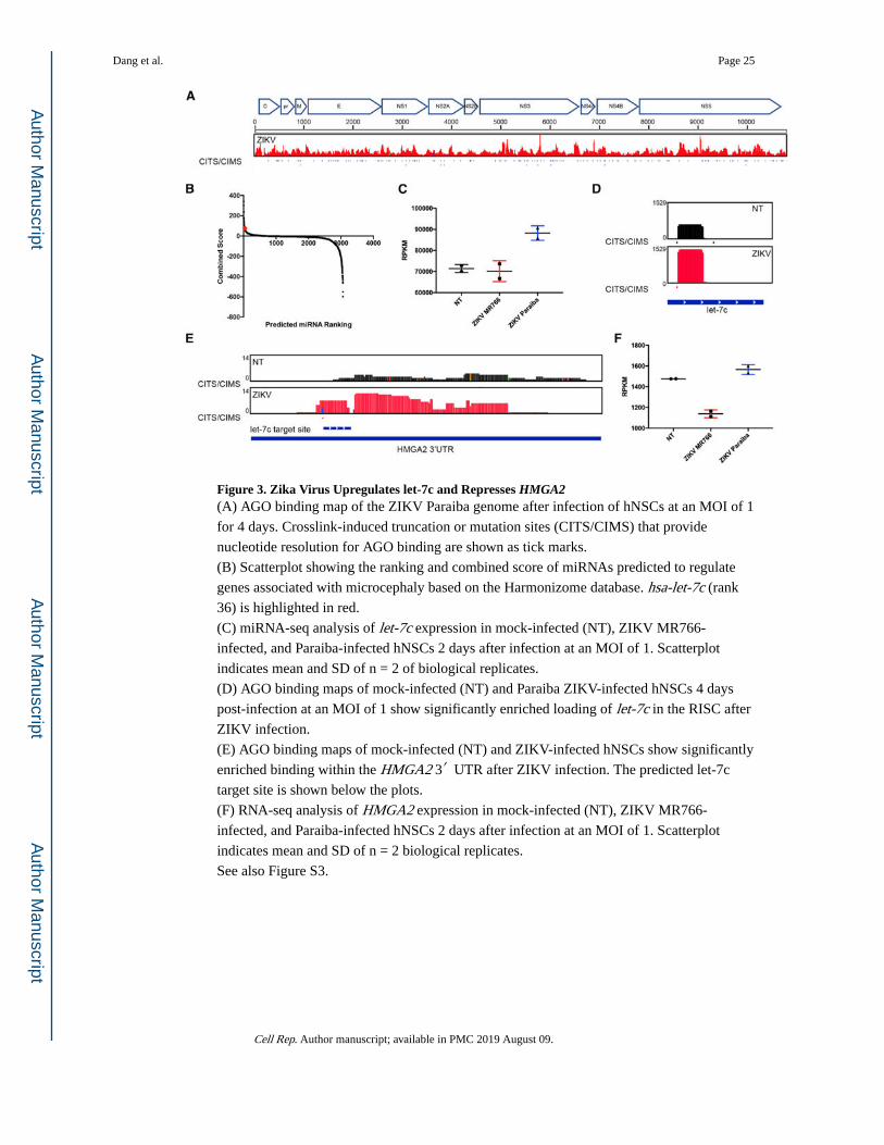
Figure 3. Zika Virus Upregulates let-7c and Represses HMGA2(A) AGO binding map of the ZIKV Paraiba genome after infection of hNSCs at an MOI of 1
for 4 days. Crosslink-induced truncation or mutation sites (CITS/CIMS) that provide
nucleotide resolution for AGO binding are shown as tick marks.
(B) Scatterplot showing the ranking and combined score of miRNAs predicted to regulate
genes associated with microcephaly based on the Harmonizome database. hsa-let-7c (rank
36) is highlighted in red.
(C) miRNA-seq analysis of let-7c expression in mock-infected (NT), ZIKV MR766-
infected, and Paraiba-infected hNSCs 2 days after infection at an MOI of 1. Scatterplot
indicates mean and SD of n = 2 of biological replicates.
(D) AGO binding maps of mock-infected (NT) and Paraiba ZIKV-infected hNSCs 4 days
post-infection at an MOI of 1 show significantly enriched loading of let-7c in the RISC after
ZIKV infection.
(E) AGO binding maps of mock-infected (NT) and ZIKV-infected hNSCs show significantly
enriched binding within the HMGA2 3′ UTR after ZIKV infection. The predicted let-7c
target site is shown below the plots.
(F) RNA-seq analysis of HMGA2 expression in mock-infected (NT), ZIKV MR766-
infected, and Paraiba-infected hNSCs 2 days after infection at an MOI of 1. Scatterplot
indicates mean and SD of n = 2 biological replicates.
See also Figure S3.
Dang et al. Page 25
Cell Rep. Author manuscript; available in PMC 2019 August 09.
Author M
anuscriptA
uthor Manuscript
Author M
anuscriptA
uthor Manuscript
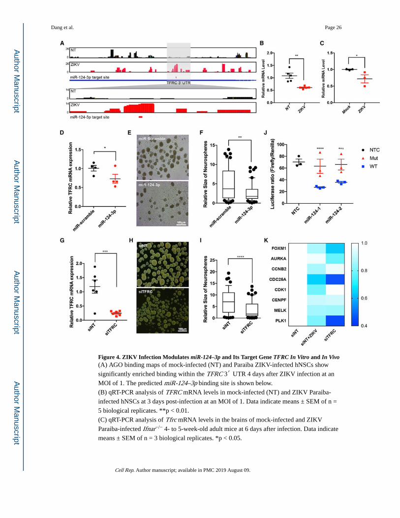
Figure 4. ZIKV Infection Modulates miR-124–3p and Its Target Gene TFRC In Vitro and In Vivo(A) AGO binding maps of mock-infected (NT) and Paraiba ZIKV-infected hNSCs show
significantly enriched binding within the TFRC 3′ UTR 4 days after ZIKV infection at an
MOI of 1. The predicted miR-124–3p binding site is shown below.
(B) qRT-PCR analysis of TFRC mRNA levels in mock-infected (NT) and ZIKV Paraiba-
infected hNSCs at 3 days post-infection at an MOI of 1. Data indicate means ± SEM of n =
5 biological replicates. **p < 0.01.
(C) qRT-PCR analysis of Tfrc mRNA levels in the brains of mock-infected and ZIKV
Paraiba-infected Ifnar−/− 4- to 5-week-old adult mice at 6 days after infection. Data indicate
means ± SEM of n = 3 biological replicates. *p < 0.05.
Dang et al. Page 26
Cell Rep. Author manuscript; available in PMC 2019 August 09.
Author M
anuscriptA
uthor Manuscript
Author M
anuscriptA
uthor Manuscript
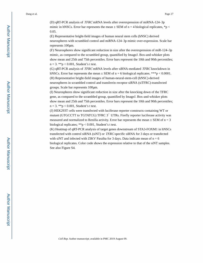
(D) qRT-PCR analysis of TFRC mRNA levels after overexpression of miRNA-124–3p
mimic in hNSCs. Error bar represents the mean ± SEM of n = 4 biological replicates, *p <
0.05.
(E) Representative bright-field images of human neural stem cells (hNSC) derived
neurospheres with scrambled control and miRNA-124–3p mimic over-expression. Scale bar
represents 100μm.
(F) Neurospheres show significant reduction in size after the overexpression of miR-124–3p
mimic, as compared to the scrambled group, quantified by ImageJ. Box-and-whisker plots
show mean and 25th and 75th percentiles. Error bars represent the 10th and 90th percentiles;
n = 3. **p < 0.001, Student’s t test.
(G) qRT-PCR analysis of TFRC mRNA levels after siRNA-mediated TFRC knockdown in
hNSCs. Error bar represents the mean ± SEM of n = 6 biological replicates. ***p < 0.0001.
(H) Representative bright-field images of human-neural-stem-cell (hNSC)-derived
neurospheres in scrambled control and transferrin receptor siRNA (siTFRC)-transfected
groups. Scale bar represents 100μm.
(I) Neurospheres show significant reduction in size after the knocking down of the TFRC
gene, as compared to the scrambled group, quantified by ImageJ. Box-and-whisker plots
show mean and 25th and 75th percentiles. Error bars represent the 10th and 90th percentiles;
n = 3. **p < 0.001, Student’s t test.
(J) HEK293T cells were transfected with luciferase reporter constructs containing WT or
mutant (GTGCCTT to TGTATCG) TFRC 3′ UTRs. Firefly reporter luciferase activity was
measured and normalized to Renilla activity. Error bar represents the mean ± SEM of n = 3
biological replicates; **p < 0.001, Student’s t test.
(K) Heatmap of qRT-PCR analysis of target genes downstream of STA3-FOXM1 in hNSCs
transfected with control siRNA (siNT) or TFRC-specific siRNA for 3 days or transfected
with siNT and infected with ZIKV Paraiba for 3 days. Data indicate mean of n = 6
biological replicates. Color code shows the expression relative to that of the siNT samples.
See also Figure S4.
Dang et al. Page 27
Cell Rep. Author manuscript; available in PMC 2019 August 09.
Author M
anuscriptA
uthor Manuscript
Author M
anuscriptA
uthor Manuscript

Author M
anuscriptA
uthor Manuscript
Author M
anuscriptA
uthor Manuscript
Dang et al. Page 28
KE
Y R
ESO
UR
CE
S TA
BL
E
RE
AG
EN
T o
r R
ESO
UR
CE
SOU
RC
EID
EN
TIF
IER
Ant
ibod
ies
mou
se a
nti-
ZIK
VE
/ant
i-fl
aviv
irus
gro
up a
ntig
enM
illip
ore
Cat
# M
AB
1021
6, R
RID
:AB
_827
205
rabb
it an
ti-SO
X2
Abc
amC
at#
AB
9795
9, R
RID
:AB
_234
1193
rabb
it an
ti-N
euN
Mill
ipor
eC
at#
AB
N78
, RR
ID:A
B_1
0807
945
AG
O 2
A8
antib
ody
Mill
ipor
eC
at#
MA
BE
56, R
RID
:AB
_112
1438
8
Bac
teri
al a
nd V
irus
Str
ains
ZIK
V p
roto
type
MR
766
Nat
iona
l Ins
titut
es o
f H
ealth
LC
0025
20.1
ZIK
V B
razi
lian
stra
in P
arai
baSt
even
son
Lab
orat
ory,
U
nive
rsity
of
Mia
mi L
ife
Scie
nce
and
Tech
nolo
gy P
ark
KX
2800
26.1
Che
mic
als,
Pep
tides
, and
Rec
ombi
nant
Pro
tein
s
Stem
Pro
Neu
ral S
uppl
emen
tT
herm
oFis
her
A10
5090
1
Eag
le’s
Min
imum
Ess
entia
l Med
ium
AT
CC
30–2
003
RN
ase
IA
mbi
onA
M22
95
RN
A p
heno
l/chl
orof
orm
Am
bion
9722
32P-
γ-A
TP
Perk
in E
lmer
blu0
0225
0uc
Cri
tical
Com
mer
cial
Ass
ays
iScr
ipt O
ne-S
tep
RT-
PCR
kit
Bio
-Rad
1708
892
NE
BN
ext U
ltra
II D
irec
tiona
l RN
A L
ibra
ry K
it fo
r Il
lum
ina
NE
BE
7760
L
Q5
mas
ter
mix
NE
BM
0494
S
miR
Nea
sy M
ini K
itQ
IAG
EN
2170
04
iScr
ipt M
aste
rmix
Bio
-Rad
1708
891
SYB
R G
reen
PC
R M
aste
r M
ixB
io-R
ad17
2527
0
prot
ein
A/G
Dyn
abea
dsT
herm
oFis
her
8880
2
4–12
% N
uPA
GE
Bis
-Tri
s ge
lsIn
vitr
ogen
NP0
321B
OX
Supe
rscr
ipt I
II r
ever
se tr
ansc
ript
ase
Invi
trog
en18
0800
93
Cell Rep. Author manuscript; available in PMC 2019 August 09.
Author M
anuscriptA
uthor Manuscript
Author M
anuscriptA
uthor Manuscript
Dang et al. Page 29
RE
AG
EN
T o
r R
ESO
UR
CE
SOU
RC
EID
EN
TIF
IER
prec
ast 6
% T
ris/
Bor
ate/
ED
TA-u
rea
gels
Invi
trog
enE
C62
65B
OX
Cir
clig
ase
IIE
pice
nter
CL
9021
K
Dep
osite
d D
ata
Raw
and
pro
cess
ed d
ata
GE
OG
SE11
3640
Raw
and
pro
cess
ed R
NA
-seq
dat
aG
EO
GSE
1136
36
Raw
and
pro
cess
ed A
GO
-iC
LIP
-seq
dat
aG
EO
GSE
1136
38
Raw
and
pro
cess
ed m
iRN
A-s
eq d
ata
GE
OG
SE11
3639
Exp
erim
enta
l Mod
els:
Cel
l Lin
es
Hum
an n
eura
l ste
m c
ells
The
rmoF
ishe
rA
1565
4
Ver
oA
TC
CC
CL
-81
Exp
erim
enta
l Mod
els:
Org
anis
ms/
Stra
ins
B6.
129S
2-If
nar1
tm1A
gt/M
mja
xM
MR
C J
acks
on L
abor
ator
ies
3204
5-JA
X
Olig
onuc
leot
ides
RT-
qPC
R Z
IKV
-F: T
TG
GT
CA
TG
ATA
CT
GC
TG
AT
TG
CID
TN
/A
RT-
qPC
R Z
IKV
-R: C
CC
TC
CA
CG
AA
GT
CT
CTA
TT
GC
IDT
N/A
L3
linke
r (H
PLC
-pur
ifie
d): /
5rA
pp/A
GA
TC
GG
AA
GA
GC
G G
TT
CA
G/3
ddC
/ID
TN
/A
Cut
olig
o (P
AG
E-p
urif
ied)
: GT
TC
AG
GA
TC
CA
CG
AC
GC
T C
TT
C/3
ddC
/ID
TN
/A
P5 S
olex
a PC
R p
rim
er: A
AT
GA
TAC
GG
CG
AC
CA
CC
GA
GA
TC
TAC
AC
TC
TT
TC
CC
TAC
AC
GA
CG
CT
C
TT
CC
GA
TC
TID
TN
/A
P3 S
olex
a PC
R p
rim
er: C
AA
GC
AG
AA
GA
CG
GC
ATA
CG
AG
AT
CG
GT
CT
CG
GC
AT
TC
CT
GC
TG
AA
C
CG
CT
CT
TC
CG
AT
CT
IDT
N/A
RT
1: /5
phos
/Nna
accN
NN
AG
AT
CG
GA
AG
AG
CG
TC
GT
Gga
tcC
TG
AA
CC
GC
IDT
N/A
RT
2: /5
phos
/NN
acaa
NN
NA
GA
TC
GG
AA
GA
GC
GT
CG
TG
gatc
CT
GA
AC
CG
CID
TN
/A
RT
3: /5
phos
/NN
attg
NN
NA
GA
TC
GG
AA
GA
GC
GT
CG
TG
gatc
CT
GA
AC
CG
CID
TN
/A
RT
4: /5
phos
/NN
aggt
NN
NA
GA
TC
GG
AA
GA
GC
GT
CG
TG
gatc
CT
GA
AC
CG
CID
TN
/A
RT
5: /5
phos
/NN
cgcc
NN
NA
GA
TC
GG
AA
GA
GC
GT
CG
T G
gatc
CT
GA
AC
CG
CID
TN
/A
RT
6: /5
phos
/NN
ccgg
NN
NA
GA
TC
GG
AA
GA
GC
GT
CG
TG
gatc
CT
GA
AC
CG
CID
TN
/A
Soft
war
e an
d A
lgor
ithm
s
Cell Rep. Author manuscript; available in PMC 2019 August 09.

Author M
anuscriptA
uthor Manuscript
Author M
anuscriptA
uthor Manuscript
Dang et al. Page 30
RE
AG
EN
T o
r R
ESO
UR
CE
SOU
RC
EID
EN
TIF
IER
TopH
at v
1.4
.1(T
rapn
ell e
t al.,
200
9)ht
tps:
//ccb
.jhu.
edu/
soft
war
e/to
phat
/inde
x.sh
tml
PRIN
SEQ
v 0
.20.
3ht
tp://
prin
seq.
sour
cefo
rge.
net
SAM
tool
s(L
i et a
l., 2
009)
http
://sa
mto
ols.
sour
cefo
rge.
net
DE
Seq2
(Lov
e et
al.,
201
4)ht
tps:
//bio
cond
ucto
r.org
/pac
kage
s/re
leas
e/bi
oc/
htm
l/DE
Seq2
.htm
l
Part
ekht
tp://
ww
w.p
arte
k.co
m
The
Dat
abas
e fo
r A
nnot
atio
n, V
isua
lizat
ion
and
Inte
grat
ed D
isco
very
(D
AV
ID)
(Hua
ng e
t al.,
200
9)ht
tps:
//dav
id.n
cifc
rf.g
ov
Cyt
osca
pe(S
hann
on e
t al.,
200
3)ht
tps:
//cyt
osca
pe.o
rg
Targ
etSc
an(A
garw
al e
t al.,
201
5)ht
tp://
ww
w.ta
rget
scan
.org
/ver
t_72
/
miR
TarB
ase
(Cho
u et
al.,
201
6)ht
tp://
mir
tarb
ase.
mbc
.nct
u.ed
u.tw
/php
/inde
x.ph
p
miR
AN
DA
(Bet
el e
t al.,
200
8)ht
tp://
cbio
.msk
cc.o
rg/m
icro
rna_
data
/man
ual.h
tml
pyC
RA
C(W
ebb
et a
l., 2
014)
http
://sa
nder
gran
nem
an.b
io.e
d.ac
.uk/
pyC
RA
C_s
oftw
are
flex
bar
(Dod
t et a
l., 2
012)
http
s://g
ithub
.com
/seq
an/f
lexb
ar/w
iki/M
anua
l
Nov
ocra
ftht
tp://
ww
w.n
ovoc
raft
.com
ChI
Ppea
kAnn
o(Z
hu e
t al.,
201
0)ht
tps:
//bio
cond
ucto
r.org
/pac
kage
s/re
leas
e/bi
oc/
htm
l/ChI
Ppea
kAnn
o.ht
ml
rtra
ckla
yer
(Law
renc
e et
al.,
200
9)ht
tps:
//ww
w.b
ioco
nduc
tor.o
rg/p
acka
ges/
rele
ase/
bioc
/htm
l/rtr
ackl
ayer
.htm
l
Supe
rmat
cher
http
://em
boss
.sou
rcef
orge
.net
/app
s/re
leas
e/6.
6/em
boss
/app
s/su
perm
atch
er.h
tml
Gra
phPa
d Pr
ism
http
s://w
ww
.gra
phpa
d.co
m
Cell Rep. Author manuscript; available in PMC 2019 August 09.
Copyright © 2022 FDOKUMEN