Genetic and biophysical basis for bupivacaine-induced ST segment elevation and VT/VF. Anesthesia...
10
Genetic and biophysical basis for bupivacaine-induced ST segment elevation and VT/VF. Anesthesia unmasked Brugada syndrome Kevin Vernooy * , Serge Sicouri † , Robert Dumaine † , Kui Hong † , Antonio Oliva † , Elena Burashnikov † , Carl Timmermans * , Tammo Delhaas * , Harry J.G.M Crijns * , Charles Antzelevitch † , Luz-Maria Rodriguez * , and Ramon Brugada ‡ * Department of Cardiology, Academic Hospital Maastricht, Maastricht, the Netherlands, ² Masonic Medical Research Laboratory, Utica, New York, USA, ‡ Montreal Heart Institute, Montreal, QC, Canada. Abstract BACKGROUND—Brugada syndrome is an inherited disease associated with sudden cardiac death. The electrocardiographic pattern associated with Brugada syndrome has been linked to the use of sodium channel blockers, including antiarrhythmics, trycyclics and anesthetics. OBJECTIVE—We report a case of bupivacaine-induced Brugada syndrome, in which we investigated the genetic, biophysical and path physiological mechanism involved. METHODS AND RESULTS—The patient developed a Brugada-like electrocardiographic pattern twice under the influence of bupivacaine. The first occurrence was accompanied by ventricular tachycardia (VT) which subsided after withdrawal of the anesthetic. The VT was also observed during co-administration of diltiazem and isosorbide-5-mononitrate, agents thought to facilitate ST segment elevation in the Brugada syndrome. Genetic analysis revealed a missense mutation in the α subunit of the cardiac sodium channel, SCN5A. Biophysical analysis by whole-cell patch-clamping revealed a reduction in sodium current as a result of the mutation. The study of bupivacaine in the wedge model revealed use-dependent changes in conduction, heterogeneous loss of the action potential dome in RV epicardium and phase 2 re-entry when the preparations were pretreated with low concentrations of the calcium channel blocker verapamil. CONCLUSION—Our findings indicate that bupivacaine may induce the electrocardiographic and arrhythmic manifestations of the Brugada syndrome in silent carriers of SCN5A mutations. The data have important implications in the management of patients who develop ST segment elevation when under the influence of anesthetics such as bupivacaine. Keywords Bupivacaine; Ventricular arrhythmias; Brugada Syndrome Introduction Bupivacaine is a potent long-acting anesthetic recommended for infiltration, peripheral nerve block, epidural and spinal anesthesia. It blocks the generation and the conduction of nerve Addresses for correspondence: Ramon Brugada, MD, Associate Professor of Medicine, University of Montreal, Director Clinical Cardiovascular Genetics Center, Montreal Heart Institute, 5000 Rue Belanger, Montreal H1T-2L2. Phone: +1 514 376 3330, Fax: +1 514 376 1355, E-mail: [email protected] Rodriguez, MD, PhD, Director Clinical Electrophysiology, Department of Cardiology, Academic Hospital Maastricht, PO BOX 5800, 6202 AZ, Maastricht, The Netherlands. Phone: +31 43 3875095, Fax: +31 43 3875104, E-mail: [email protected] NIH Public Access Author Manuscript Heart Rhythm. Author manuscript; available in PMC 2007 September 24. Published in final edited form as: Heart Rhythm. 2006 September ; 3(9): 1074–1078. NIH-PA Author Manuscript NIH-PA Author Manuscript NIH-PA Author Manuscript
-
Upload
independent -
Category
Documents
-
view
0 -
download
0
Transcript of Genetic and biophysical basis for bupivacaine-induced ST segment elevation and VT/VF. Anesthesia...
Genetic and biophysical basis for bupivacaine-induced STsegment elevation and VT/VF. Anesthesia unmasked Brugadasyndrome
Kevin Vernooy*, Serge Sicouri†, Robert Dumaine†, Kui Hong†, Antonio Oliva†, ElenaBurashnikov†, Carl Timmermans*, Tammo Delhaas*, Harry J.G.M Crijns*, CharlesAntzelevitch†, Luz-Maria Rodriguez*, and Ramon Brugada‡
*Department of Cardiology, Academic Hospital Maastricht, Maastricht, the Netherlands, †Masonic MedicalResearch Laboratory, Utica, New York, USA, ‡Montreal Heart Institute, Montreal, QC, Canada.
AbstractBACKGROUND—Brugada syndrome is an inherited disease associated with sudden cardiac death.The electrocardiographic pattern associated with Brugada syndrome has been linked to the use ofsodium channel blockers, including antiarrhythmics, trycyclics and anesthetics.
OBJECTIVE—We report a case of bupivacaine-induced Brugada syndrome, in which weinvestigated the genetic, biophysical and path physiological mechanism involved.
METHODS AND RESULTS—The patient developed a Brugada-like electrocardiographic patterntwice under the influence of bupivacaine. The first occurrence was accompanied by ventriculartachycardia (VT) which subsided after withdrawal of the anesthetic. The VT was also observed duringco-administration of diltiazem and isosorbide-5-mononitrate, agents thought to facilitate ST segmentelevation in the Brugada syndrome. Genetic analysis revealed a missense mutation in the α subunitof the cardiac sodium channel, SCN5A. Biophysical analysis by whole-cell patch-clamping revealeda reduction in sodium current as a result of the mutation. The study of bupivacaine in the wedgemodel revealed use-dependent changes in conduction, heterogeneous loss of the action potentialdome in RV epicardium and phase 2 re-entry when the preparations were pretreated with lowconcentrations of the calcium channel blocker verapamil.
CONCLUSION—Our findings indicate that bupivacaine may induce the electrocardiographic andarrhythmic manifestations of the Brugada syndrome in silent carriers of SCN5A mutations. The datahave important implications in the management of patients who develop ST segment elevation whenunder the influence of anesthetics such as bupivacaine.
KeywordsBupivacaine; Ventricular arrhythmias; Brugada Syndrome
IntroductionBupivacaine is a potent long-acting anesthetic recommended for infiltration, peripheral nerveblock, epidural and spinal anesthesia. It blocks the generation and the conduction of nerve
Addresses for correspondence: Ramon Brugada, MD, Associate Professor of Medicine, University of Montreal, Director ClinicalCardiovascular Genetics Center, Montreal Heart Institute, 5000 Rue Belanger, Montreal H1T-2L2. Phone: +1 514 376 3330, Fax: +1514 376 1355, E-mail: [email protected] Rodriguez, MD, PhD, Director Clinical Electrophysiology, Department ofCardiology, Academic Hospital Maastricht, PO BOX 5800, 6202 AZ, Maastricht, The Netherlands. Phone: +31 43 3875095, Fax: +3143 3875104, E-mail: [email protected]
NIH Public AccessAuthor ManuscriptHeart Rhythm. Author manuscript; available in PMC 2007 September 24.
Published in final edited form as:Heart Rhythm. 2006 September ; 3(9): 1074–1078.
NIH
-PA Author Manuscript
NIH
-PA Author Manuscript
NIH
-PA Author Manuscript

impulses by inhibiting sodium channel current in nerve membranes. This results in an increasedthreshold for electrical excitation, a slower impulse propagation, and reduced rate of rise ofthe action potential of the nerve membrane.1 Although bupivacaine has few side effects whenproperly administered, it can produce ventricular arrhythmias and cardiac depression at toxiclevels.1
Brugada syndrome is an inherited sudden death syndrome whose electrocardiographic andarrhythmic manifestations can be unmasked or exacerbated in the presence of agents that blockthe cardiac sodium channel.2 It is characterized by an ST segment elevation on theelectrocardiogram (ECG) in the precordial leads V1 to V3, and a right bundle branch blockpattern in the absence of structural heart disease.3 The syndrome has in 19984 been linked tomutations in SCN5A, the gene that encodes for the α subunit for the sodium channel and sincethen over 80 mutations in SCN5A have been identified.
We report a case of Brugada-like ECG associated with unexplained ventriculartachyarrhythmias developing during epidural infusion of bupivacaine. We identified a mutationin SCN5A and delineated the biophysical mechanisms involved in the bupivacaine-inducedphenotype by whole-cell patch-clamp technique and in the arterially-perfused wedgepreparation.
MethodsCase
A 56-year-old man underwent lung volume reduction surgery for chronic obstructivepulmonary disease. His medical history revealed a transient ischemic attack and lungemphysema for 13 years. The preoperative ECG showed a sinus rhythm with a heart rate of90 bpm, low voltages, non-specific T-wave changes and atrial dilatation. The echocardiogramshowed left ventricular concentric hypertrophy, left atrial and right ventricular (RV) dilatationwith normal left and right ventricular function. Routine preoperative blood tests, includingkidney function tests and serum electrolytes, were normal.
Before induction of general anesthesia with propofol (2.5 mg/kg/hr), an epidural catheter wasinserted and a combination of 0.25% bupivacaine and 1 mcg/ml sufentanil was started at 7 ml/h. The first day postoperatively the ECG showed a remarkable pattern: sinus rhythm of 90bpm, right bundle branch block-like pattern, down-sloping ST segment elevation in leads V1to V3, known as Brugada-like ECG pattern as well as frequent ventricular extrasystoles witha left bundle branch morphology and horizontal axis (Figure 1). The incidence of theseextrasystoles increased dramatically resulting in ventricular tachycardia (VT) and electricalstorm. Lidocaine and amiodarone were administered in an attempt to convert the arrhythmia.Isosorbide-5-mononitrate and diltiazem were started because of suspected RV overload. Fivedays after surgery, bupivacaine was withdrawn due to malfunction of the epidural catheter andpiritramide perfusion (2 mg/hr IV) was started. Surprisingly, the next day the ventriculararrhythmias were no longer present and the typical Brugada-like ECG disappeared.
Four days later, a new surgical intervention, performed to correct persistent air leakage,necessitated the insertion of an epidural catheter and administration of 0.125% bupivacaineand 1 mcg/ml sufentanil at 8 ml/hr. Surgery was successfully carried out under the sameanesthetic conditions as before. On the first day after surgery the ECG once again displayed aBrugada-like pattern. The epidural catheter was withdrawn the following day because ofmalfunction and the Brugada-like ECG disappeared. The patient did not experience feverduring the admission. The patient was discharged after two weeks.
Vernooy et al. Page 2
Heart Rhythm. Author manuscript; available in PMC 2007 September 24.
NIH
-PA Author Manuscript
NIH
-PA Author Manuscript
NIH
-PA Author Manuscript

Genetic analysisAfter informed consent was obtained, genomic DNA was isolated from peripheral bloodleukocytes using a commercial kit (Gentra System, Puregene). The exons of SCN5A wereamplified and analyzed by direct sequencing. Polymerase chain reaction products were purifiedwith a commercial reagent (ExoSAP-IT, USB) and were directly sequenced from bothdirections with the use of ABI PRISM 3100 Automatic DNA Sequencer.
Site-directed mutagenesisMutant SCN5A channel cDNAs were prepared by site-directed mutagenesis of the plasmidpcDNA-SCN5A in vector pcDNA3.1 + (Invitrogen, Carlsbad, CA, USA). The SCN5Atranscript used contains Q1077. To create the mutant, the “Megaprimer” method ofmutagenesis was used, as previously described.5
ElectrophysiologyTSA201 cells were co-transfected with pcDNA-SCN5A (wild-type or mutant) and the humansodium channel subunit-β1 (SCN1B) in pc DNA3.1 (pcDNA-Hβ1) construct using the calciumphosphate precipitation method, as previously described.5 The cells were grown on polylysine-coated 35-mm culture dishes and placed in a temperature-controlled chamber forelectrophysiologic study (Medical Systems, Greenvale, NY, USA). Standard whole-cell patch-clamp technique was used to measure currents in transfected TSA201 cells as previouslydescribed.6
Wedge modelTo assess whether the electrocardiographic and arrhythmic manifestations of bupivacaine aredue to its direct actions on the ventricular myocardium, we evaluated the effects of theanesthetic in 5 arterially-perfused RV wedge preparations. The detailed methods employed forisolation, perfusion and recording of transmembrane activity have been previously reported.7 Briefly, transmural wedge preparations with dimensions of approximately 20–25 mm by 10–12 mm by 8–10 mm (transmural width) were dissected from the right ventricle of male doghearts. The preparations were then placed in a small tissue bath and arterially-perfused withTyrode’s solution at 35–36°C. The preparations were continuously stimulated endocardiallyat a basic cycle length of 2000 msec. After a stabilization period (~1–2 hours), transmural ECGand transmembrane action potentials were simultaneously recorded from epicardial (Epi) andmid-myocardial (M) regions. The effect of bupivacaine (Sigma, Foster City, CA) was assessed45 to 60 min after the addition of the drug to the perfusate at concentrations of 10 and 20 ug/ml. In 3 experiments, the calcium channel blocker verapamil was introduced at relatively lowconcentration (0.1 uM) to sensitize the preparation.
ResultsGenetic analysis
Genetic analysis revealed a novel missense mutation (G1743E, SCN5A Genbank accessionnumber AC137587) in the α-sub-unit of the sodium channel, SCN5A (Figure 2). This mutationis located between segments 5 and 6 of domain 4, in a highly conserved amino-acid acrossspecies. This mutation was not present in 200 control individuals of the same ethnicbackground. We did not identify any other variation in the codifying segments of SCN5A.
As routine clinical follow-up for a patient with Brugada syndrome, the patient’s family hasbeen approached by the proband regarding participation in clinical and genetic screening atthe Maastricht University Hospital for mutations. Only one of the brothers has agreed toparticipate. He had an ECG positive at baseline and was found to have the same genetic
Vernooy et al. Page 3
Heart Rhythm. Author manuscript; available in PMC 2007 September 24.
NIH
-PA Author Manuscript
NIH
-PA Author Manuscript
NIH
-PA Author Manuscript

mutation as the proband, therefore proving this to be a familial disease discovered by the useof bupivacaine in one of the genetic carriers with a normal ECG.
Biophysical analysisIn TSA201 cells, expression of the mutant channel G1743E produced a markedly reducedsodium channel current (INa) when compared to wild type (WT), a mechanism known to leadto ST segment elevation and the Brugada syndrome phenotype (Figure 2).5
Wedge modelIn all 5 experiments, the sodium channel blocker bupivacaine (10–20 uM) produced use-dependent slowing of conduction, evidenced by a prolongation of the QRS complex of thetransmural ECG. Reduced excitability necessitated an increase in stimulus intensity in orderto maintain 1:1 activation, particularly at faster rates (basic cycle length ≤ 400 msec). Loss ofthe action potential dome in the RV epicardium was observed in 3 out of 3 preparationspretreated with verapamil (0.1uM) (Figure 3), but not in the two wedge preparations that werenot pretreated with verapamil. Verapamil (0.1uM) alone accentuated the action potential notchbut did not lead to loss of the action potential dome in any of the preparations tested (3/3).Heterogeneous loss of the dome permitted the development of phase 2 re-entry due topropagation of the action potential dome from the epicardial sites where it was maintained tosites where it was lost, thus generating a closely coupled extrasystole. Prominent J waves,negative T waves, and T wave alternans, were apparent in the ECG (Figures 3 and 4).
DiscussionAn ECG pattern characterized by coved-type ST segment elevation in the right precordial leadsis observed in the congenital form of the Brugada syndrome, but may also be induced bymedications and external factors.8,9 Brugada syndrome affects young individuals with noprevious history of cardiac disease. It is characterized by the presence of ST segment elevationin leads V1 to V3, and the occurrence of malignant arrhythmias in structurally normal hearts.3 ST segment elevation in the Brugada syndrome is thought to be due to an accentuation ofthe RV epicardial action potential notch secondary to rebalancing of the ion channel currentsactive at the end of phase 1 of the epicardial ventricular action potential.10 An arrhythmogenicsubstrate can be created under these conditions due to loss of the action potential dome inepicardium but not endocardium, resulting in the development of a marked transmuraldispersion of repolarization and refractoriness. The heterogeneous loss of the action potentialdome leads to the creation of a vulnerable window across the RV wall during which anextrasystole generated by the same substrate can induce VT and ventricular fibrillation (VF).
Thus, ST segment elevation and electrical instability of the myocardium can result from anoutward shift in the balance of currents active at the end of phase 1 of the action potential.Vagotonic agents, IK-ATP activators, and high potassium levels can facilitate this byaugmenting outward currents, whereas sodium channel blockers, cocaine, antidepressants,ischemia, acidosis and antihistaminics like terfenadine can accomplish this by reducing inwardcurrents.11 Loss of function mutations in SCN5A, responsible for the Brugada syndrome, shiftsthe balance of current by reducing sodium channel current. A febrile state has also been shownto facilitate ST segment elevation in the Brugada syndrome owing to its ability to reduce sodiumchannel current.12 A reduction in inward currents causes ST elevation more readily in the rightventricle because of the prominence of the transient outward current (Ito) in this part of theheart, accounting for its manifestation in the right precordial leads in Brugada syndrome.7
With the use of genetic analysis, we provide the first evidence that bupivacaine can unmask aBrugada syndrome, leading to the development of the substrate for VT/VF. Phillips et al13
Vernooy et al. Page 4
Heart Rhythm. Author manuscript; available in PMC 2007 September 24.
NIH
-PA Author Manuscript
NIH
-PA Author Manuscript
NIH
-PA Author Manuscript
previously published a case-report of a 77-year-old man with a characteristic Brugada-likeECG during epidural infusion of bupivacaine. Although no arrhythmias were observed theysuggested that the development of Brugada-like ECG changes during bupivacaine warrantsimmediate discontinuation to prevent potentially fatal arrhythmias.
Like flecainide and ajmaline, sodium channel blockers used to unmask the Brugada syndrome,bupivacaine inhibits the sodium channel current.14 Experimental data from isolated rabbitheart indicate that bupivacaine can facilitate reentrant ventricular arrhythmias because it slowsof conduction and prolongs the ventricular effective refractory period.1 The present studydemonstrates the action of bupivacaine to create both a transmural and epicardial dispersionof repolarization secondary to its inhibition of INa. This effect of the drug is facilitated bypretreatment with low concentrations of the calcium channel blocker, verapamil. This isconsistent with the observation that VT in our patient was observed during co-administrationof a calcium channel blocker. The effect of calcium channel block to facilitate sodium channelinhibition-induced Brugada phenotype was recently reported by us in experimental models ofthe Brugada syndrome.15 It is noteworthy that VT occurrence was also associated with co-administration of isosorbide-5-mononitrate, another agent thought to facilitate ST segmentelevation and arrhythmogenicity in the Brugada syndrome.
ConclusionThe data provide the first genetic and biophysical evidence for an effect of epidural anesthesiawith bupivacaine to unmask a subclinical form of the Brugada syndrome leading to thedevelopment of life-threatening arrhythmias. Thus, bupivacaine may be added to the list ofdrugs including flecainide, ajmaline and procainamide capable of unmasking the Brugadasyndrome. Physicians should be aware that when using anesthetics, the development of STsegment elevation may be a warning sign for electrical instability and that immediate correctivemeasures are needed.
Acknowledgements
The present study was supported by The Netherlands Organization for Health Research and Development (KV), grant2000T036 from the Netherlands Heart Foundation (TD) and grant HL47678 and HL 66169 from NHLBI (CA andRB), grants from the American Heart Association (CA and RB) and NYS and Florida Grand Lodges F.& A.M and bythe Ramon Brugada Sr. Foundation.
References1. de La Coussaye JE, Brugada J, Allessie MA. Electrophysiologic and arrhythmogenic effects of
bupivacaine. A study with high-resolution ventricular epicardial mapping in rabbit hearts.Anesthesiology 1992 July;77(1):132–41. [PubMed: 1609986]
2. Brugada R, Brugada J, Antzelevitch C, Kirsch GE, Potenza D, Towbin JA, Brugada P. Sodium channelblockers identify risk for sudden death in patients with ST-segment elevation and right bundle branchblock but structurally normal hearts. Circulation 2000 February 8;101(5):510–5. [PubMed: 10662748]
3. Brugada P, Brugada J. Right bundle branch block, persistent ST segment elevation and sudden cardiacdeath: a distinct clinical and electrocardiographic syndrome. A multi-center report. J Am Coll Cardiol1992 November 15;20(6):1391–6. [PubMed: 1309182]
4. Chen Q, Kirsch GE, Zhang D, Brugada R, Brugada J, Brugada P, Potenza D, Moya A, Borggrefe M,Breithardt G, Ortiz-Lopez R, Wang Z, Antzelevitch C, O’Brien RE, Schulze-Bahr E, Keating MT,Towbin JA, Wang Q. Genetic basis and molecular mechanism for idiopathic ventricular fibrillation.Nature 1998 March 19;392(6673):293–6. [PubMed: 9521325]
5. Dumaine R, Towbin JA, Brugada P, Vatta M, Nesterenko DV, Nesterenko VV, Brugada J, BrugadaR, Antzelevitch C. Ionic mechanisms responsible for the electrocardiographic phenotype of theBrugada syndrome are temperature dependent. Circ Res 1999 October 29;85(9):803–9. [PubMed:10532948]
Vernooy et al. Page 5
Heart Rhythm. Author manuscript; available in PMC 2007 September 24.
NIH
-PA Author Manuscript
NIH
-PA Author Manuscript
NIH
-PA Author Manuscript
6. Hong K, Guerchicoff A, Pollevick GD, Oliva A, Dumaine R, de ZM, Burashnikov E, Wu YS, BrugadaJ, Brugada P, Brugada R. Cryptic 5′ splice site activation in SCN5A associated with Brugada syndrome.J Mol Cell Cardiol 2005 April;38(4):555–60. [PubMed: 15808832]
7. Di Diego JM, Cordeiro JM, Goodrow RJ, Fish JM, Zygmunt AC, Perez GJ, Scornik FS, AntzelevitchC. Ionic and cellular basis for the predominance of the Brugada syndrome phenotype in males.Circulation 2002 October 8;106(15):2004–11. [PubMed: 12370227]
8. Wilde AA, Antzelevitch C, Borggrefe M, Brugada J, Brugada R, Brugada P, Corrado D, Hauer RN,Kass RS, Nademanee K, Priori SG, Towbin JA. Proposed diagnostic criteria for the Brugada syndrome.Eur Heart J 2002 November;23(21):1648–54. [PubMed: 12448445]
9. Darbar D, Yang T, Churchwell K, Wilde AA, Roden DM. Unmasking of brugada syndrome by lithium.Circulation 2005 September 13;112(11):1527–31. [PubMed: 16144991]
10. Antzelevitch C, Brugada P, Brugada J, Brugada R, Shimizu W, Gussak I, Perez Riera AR. Brugadasyndrome: a decade of progress. Circ Res 2002 December 13;91(12):1114–8. [PubMed: 12480811]
11. Antzelevitch C. Brugada syndrome: clinical, genetic, molecular, cellular and ionic aspects. ExpertRev Cardiovasc Ther 2003 July;1(2):177–85. [PubMed: 15030278]
12. Antzelevitch C, Brugada R. Fever and Brugada syndrome. Pacing Clin Electrophysiol 2002November;25(11):1537–9. [PubMed: 12494608]
13. Phillips N, Priestley M, Denniss AR, Uther JB. Brugada-type electrocardiographic pattern inducedby epidural bupivacaine. Anesth Analg 2003 July;97(1):264–7. [PubMed: 12818979]table
14. Valenzuela C, Snyders DJ, Bennett PB, Tamargo J, Hondeghem LM. Stereoselective block of cardiacsodium channels by bupivacaine in guinea pig ventricular myocytes. Circulation 1995 November15;92(10):3014–24. [PubMed: 7586272]
15. Fish JM, Antzelevitch C. Role of Sodium and Calcium Channel Block in Unmasking the BrugadaSyndrome. Heart Rhythm 2004;1(2):210–217. [PubMed: 15851155]
Vernooy et al. Page 6
Heart Rhythm. Author manuscript; available in PMC 2007 September 24.
NIH
-PA Author Manuscript
NIH
-PA Author Manuscript
NIH
-PA Author Manuscript
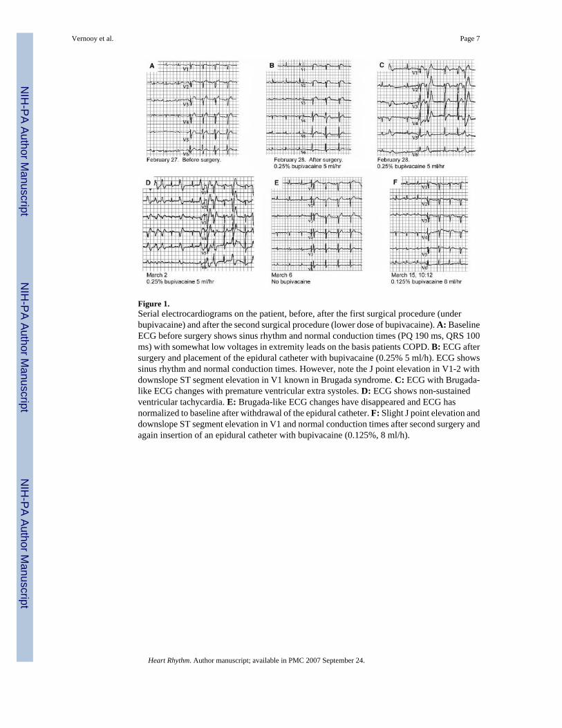
Figure 1.Serial electrocardiograms on the patient, before, after the first surgical procedure (underbupivacaine) and after the second surgical procedure (lower dose of bupivacaine). A: BaselineECG before surgery shows sinus rhythm and normal conduction times (PQ 190 ms, QRS 100ms) with somewhat low voltages in extremity leads on the basis patients COPD. B: ECG aftersurgery and placement of the epidural catheter with bupivacaine (0.25% 5 ml/h). ECG showssinus rhythm and normal conduction times. However, note the J point elevation in V1-2 withdownslope ST segment elevation in V1 known in Brugada syndrome. C: ECG with Brugada-like ECG changes with premature ventricular extra systoles. D: ECG shows non-sustainedventricular tachycardia. E: Brugada-like ECG changes have disappeared and ECG hasnormalized to baseline after withdrawal of the epidural catheter. F: Slight J point elevation anddownslope ST segment elevation in V1 and normal conduction times after second surgery andagain insertion of an epidural catheter with bupivacaine (0.125%, 8 ml/h).
Vernooy et al. Page 7
Heart Rhythm. Author manuscript; available in PMC 2007 September 24.
NIH
-PA Author Manuscript
NIH
-PA Author Manuscript
NIH
-PA Author Manuscript

Figure 2.A: Sequencing analysis results of the patient, showing a missense mutation (G1743E) in theα subunit of the cardiac sodium channel. Mutation G1743E in SCN5A reduce expression ofNaV1.5. B: Representative current recordings of the unmutated (WT) channel and mutantG1743E elicited by a series of step potentials from − 60 mV to +20 mV in 5 mV incrementsfrom a holding membrane potential of − 100 mV. Note the different voltage calibration forWT vs. mutant indicating an important decrease in sodium current in the mutant channel. C:Average peak current-voltage relationship from 7 WT and G1743E cells. (Average ± SEM).
Vernooy et al. Page 8
Heart Rhythm. Author manuscript; available in PMC 2007 September 24.
NIH
-PA Author Manuscript
NIH
-PA Author Manuscript
NIH
-PA Author Manuscript
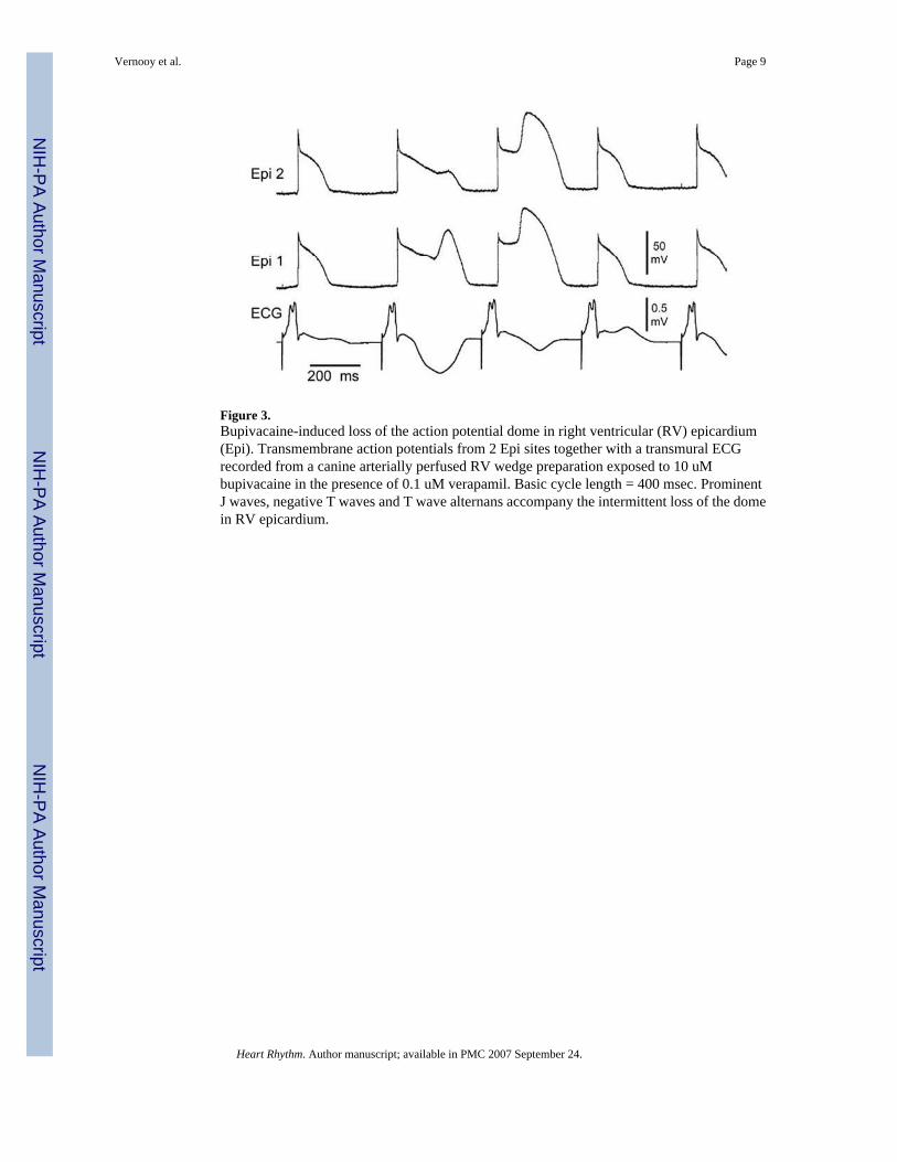
Figure 3.Bupivacaine-induced loss of the action potential dome in right ventricular (RV) epicardium(Epi). Transmembrane action potentials from 2 Epi sites together with a transmural ECGrecorded from a canine arterially perfused RV wedge preparation exposed to 10 uMbupivacaine in the presence of 0.1 uM verapamil. Basic cycle length = 400 msec. ProminentJ waves, negative T waves and T wave alternans accompany the intermittent loss of the domein RV epicardium.
Vernooy et al. Page 9
Heart Rhythm. Author manuscript; available in PMC 2007 September 24.
NIH
-PA Author Manuscript
NIH
-PA Author Manuscript
NIH
-PA Author Manuscript
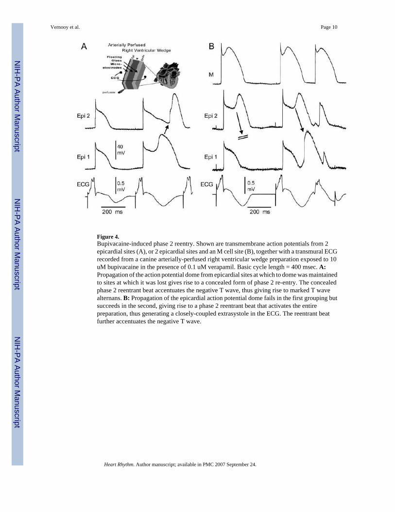
Figure 4.Bupivacaine-induced phase 2 reentry. Shown are transmembrane action potentials from 2epicardial sites (A), or 2 epicardial sites and an M cell site (B), together with a transmural ECGrecorded from a canine arterially-perfused right ventricular wedge preparation exposed to 10uM bupivacaine in the presence of 0.1 uM verapamil. Basic cycle length = 400 msec. A:Propagation of the action potential dome from epicardial sites at which to dome was maintainedto sites at which it was lost gives rise to a concealed form of phase 2 re-entry. The concealedphase 2 reentrant beat accentuates the negative T wave, thus giving rise to marked T wavealternans. B: Propagation of the epicardial action potential dome fails in the first grouping butsucceeds in the second, giving rise to a phase 2 reentrant beat that activates the entirepreparation, thus generating a closely-coupled extrasystole in the ECG. The reentrant beatfurther accentuates the negative T wave.
Vernooy et al. Page 10
Heart Rhythm. Author manuscript; available in PMC 2007 September 24.
NIH
-PA Author Manuscript
NIH
-PA Author Manuscript
NIH
-PA Author Manuscript