BRUGADA PHENOCOPY: CASE STUDY Zulyadaini, E. 1 Fajri ...
66
BRUGADA PHENOCOPY: CASE STUDY Zulyadaini, E. 1 Fajri, A., A. 2 1 Departement of Cardiology, Mitra Siaga Hospital, Tegal, Indonesia 2 General Practitioner, Mitra Siaga Hospital, Tegal, Indonesia ABSTRACT Overview: The Brugada Phenocopy is an entity where etiologically can be distinguished from congenital Brugada Syndrome. The Brugada Phenocopy is characterized by precordial ECG abnormalities in V1-3 in the form of an electrocardiogram Type 1 or 2, but with several accompanying clinical conditions, such as myocardial ischemia, acute pulmonary embolism, metabolic disorders, electrolyte disturbances (especially hyper-hypokalaemia), or fever. At The Brugada Phenocopy, clinical improvement in the underlying disease makes ECG morphology turns normal. The key to the pathophysiology of The Brugada Syndrome can distinguish true Brugada Syndrome from Brugada Phenocopy. Method: This study using analytical description methods, using literature review in various worldwide journal to determine Brugada Phenocopy or Brugada Syndrome. Result: A male, 49 years old, with complaints of atypical chest pain with a 2-day fever. Routine blood tests obtained normal leukocytes with granulocytosis, widal immunoserological titers Salmonella thypii O-H 1/160 and Salmonella parathypii O-H 1/160. At the initial ECG examination, ST segment elevation was obtained at V1-V3 with Brugada morphology type 1 without reciprocal change in other leads which was meaningful with incomplete RBBB morphology (rSR ') without a presentation of Slurred S wave at lead V5-6. The patient was given a loading dose of Aspirin and clopidogrel with infusion of antipyretic, repeated ECG examination, a significant decrease in ST segment elevation, especially at V2-V3. On the second day the patient became afebrile, obtained a segment normalization of ST at V2-V3, with morphology V1 incomplete RBBB obtained. Conclusion: This case of ECG resolution in afebrile conditions, consistent with the morphology of the Brugada Phenocopy. In cases like this, follow-up needs to be done especially for the true exclusion of Brugada Syndromes with electrophysiological studies, given the risk of Brugada Syndome’s of Sudden Cardiac Death Keywords: Brugada Phenocopy, Brugada Syndrome, ECG, Sudden Death, ST Segment Elevation
-
Upload
khangminh22 -
Category
Documents
-
view
3 -
download
0
Transcript of BRUGADA PHENOCOPY: CASE STUDY Zulyadaini, E. 1 Fajri ...
BRUGADA PHENOCOPY: CASE STUDY
Zulyadaini, E. 1 Fajri, A., A.2
1 Departement of Cardiology, Mitra Siaga Hospital, Tegal, Indonesia
2 General Practitioner, Mitra Siaga Hospital, Tegal, Indonesia
ABSTRACT
Overview: The Brugada Phenocopy is an entity where etiologically can be distinguished from
congenital Brugada Syndrome. The Brugada Phenocopy is characterized by precordial ECG
abnormalities in V1-3 in the form of an electrocardiogram Type 1 or 2, but with several
accompanying clinical conditions, such as myocardial ischemia, acute pulmonary embolism,
metabolic disorders, electrolyte disturbances (especially hyper-hypokalaemia), or fever. At The
Brugada Phenocopy, clinical improvement in the underlying disease makes ECG morphology
turns normal. The key to the pathophysiology of The Brugada Syndrome can distinguish true
Brugada Syndrome from Brugada Phenocopy.
Method: This study using analytical description methods, using literature review in various
worldwide journal to determine Brugada Phenocopy or Brugada Syndrome.
Result: A male, 49 years old, with complaints of atypical chest pain with a 2-day fever. Routine
blood tests obtained normal leukocytes with granulocytosis, widal immunoserological titers
Salmonella thypii O-H 1/160 and Salmonella parathypii O-H 1/160. At the initial ECG
examination, ST segment elevation was obtained at V1-V3 with Brugada morphology type 1
without reciprocal change in other leads which was meaningful with incomplete RBBB
morphology (rSR ') without a presentation of Slurred S wave at lead V5-6. The patient was given
a loading dose of Aspirin and clopidogrel with infusion of antipyretic, repeated ECG examination,
a significant decrease in ST segment elevation, especially at V2-V3. On the second day the
patient became afebrile, obtained a segment normalization of ST at V2-V3, with morphology V1
incomplete RBBB obtained.
Conclusion: This case of ECG resolution in afebrile conditions, consistent with the
morphology of the Brugada Phenocopy. In cases like this, follow-up needs to be done especially
for the true exclusion of Brugada Syndromes with electrophysiological studies, given the risk of
Brugada Syndome’s of Sudden Cardiac Death
Keywords: Brugada Phenocopy, Brugada Syndrome, ECG, Sudden Death, ST Segment
Elevation

Acute Ischemic Stroke Following Thrombolytic Therapy With
Streptokinase For ST Elevation Myocardial Infarction: A Case Report
Prabowo, B.K.1, Sulistyo, N.A.2, Sudiyoko2, Sukmadja, D.1
1General Practitioner, Tidar General Hospital, Magelang
2Cardiologist, Cardiology and Vascular Department, Tidar General Hospital, Magelang
Introduction ST elevation myocardial infarction (STEMI) generally reflects an acute total coronary artery
occlusion and should be treated with revascularization therapy as soon as possible. Primary
percutaneous coronary intervention (PCI) is superior to thrombolytic therapy. However, there
are many limitations to PCI such as the absence of a nearby PCI center. Thrombolytic
therapy itself has many major complications such as intracranial hemorrhage which is well
documented. Here, we present a case of ischemic stroke in STEMI patient treated with
thrombolytic therapy.
Case Illustration A 55 years old female presented with typical chest pain with a pain onset of 2 hours prior to
admission. Patient’s vital sign as follows, blood pressure 130/90mmHg, heart rate 86 bpm,
and oxygen saturation 97% on room air. Physical examination revealed no murmurs or rales.
12-lead ECG showed ST elevation in II, III, and aVF leads that indicates an acute inferior
STEMI. She underwent thrombolytic therapy using Streptokinase 1.5 million IU over an hour.
A few minutes later, she developed motor aphasia and later on showed a decrease of
consciousness with GCS 5/15. The computed tomography scan showed a right occipital lobe
infarct.
Discussion All thrombolytic agents shared a common mechanism of activating plasminogen into plasmin
which in turn activates the fibrin degradation pathway. The most dangerous side effect of
thrombolytic therapy is bleeding, such as intracranial hemorrhage, which is due to depletion
of clotting factors and lysis of recently formed hemostatic plugs. However, an ischemic stroke
incidence after thrombolytic therapy is not well documented and the pathophysiology is
remain unknown.
Conclusion Hemorrhagic stroke is not the only complication of thrombolysis, ischemic stroke can occur
even if it is an extremely rare complication. However, the underlying pathophysiology is
remain unknown.
Keywords: STEMI, Ischemic Stroke, Thrombolytic Therapy

Atrial Fibrillation with Abberant Conduction in The Wolf-Parkinson-White
Syndrome: A Case Report
Sutikno, Mugi Tri
Emergency Departement Harapan Ibu Hospital, Purbalingga, Indonesia
ABSTRACT
Atrial fibrillation is the most dangerous arrhythmia associated with WPW syndrome.
Approximately 30% to 40% of patients with WPW syndrome will develop AF. It can cause
hypotension, decreased coronary perfusion, ventricular fibrillation (VF), and sudden death. A 21
years old woman came to emergency room (ER) with a chief complain palpitation. Palpitation
accompanied with dizziness and dyspnea, no history of syncope or chest pain. The ECG
showed irregular wide complex tachyarrhythmias with rate 187 bpm and unstable hemodynamic.
Thorax X-Ray showed cardiomegaly with mild pulmonary edema. Cardioversion successfully
terminate the tachyarrhythmias. ECG in sinus rhythm revealed WPW pattern ECG. From the
ECG was also shown that the location of accessory pathway (AP) is posteroseptal wall of the
right ventricle. In WPW syndrome, the atrial impulse can reach the ventricles not only through
the atrioventricular (AV) node but also through the bypass tract. It can cause rapid ventricular
rate, shortening diastolic filling time, and decreased cardiac output. In this condition make
unstable hemodynamic. In recent study, patients suffered from AF RVR with unstable
hemodynamic in WPW syndrome, cardioversion receives a Class 1 Recommendation. In this
case, cardioversion 200 Joule biphasic was given and successfully terminate the
tachyarrhythmias. Hemodynamic was stable after cardioversion. For maintenance dose,
amiodarone 3x200 mg was given to control rate and rhytm. Treatment of AF with unstable
hemodynamic in the WPW syndrome includes cardioversion and antiarrhythmic agents.
Keywords: Atrial Fibrillation with Abberant Conduction, WPW Syndrome, Cardioversion,
Antiarrhythmic Agents

A 20 Year Old Woman with Primigravida and Neglected Tetralogy of
Fallot, an Ignorance that Leads to Mortality: A Case Report
Catelya LG1, Rahma AA2, Setiabudi PA3.
1Ahmad Dahlan Hospital, Kediri, Indonesia
2Gambiran General Hospital, Kediri, Indonesia
3Ngudi Waluyo General Hospital, Blitar, Indonesia
Background
Tetralogy of Fallot (ToF) is the most common cyanotic congenital heart disease, with an
estimated overall prevalence of 3000 per one million births. Pregnancy in uncorrected ToF
carries serious risk including increased maternal morbidity (62.5%), mortality up to 15%, and
poor perinatal outcome.
Case description
A 20-year-old woman, G1P0A0, with 25th weeks of pregnancy was admitted to Emergency
Room due to haemoptoe and dyspnoea. Past medical history was recurrent lower respiratory
tract infection (LRTI) when she was a child, without dyspnoea on effort, cyanosis, nor
clubbing fingers. The patient was diagnosed with LRTI and receive ceftriaxone, tranexamic
acid, and salbutamol inhalation for 2 days. Dyspnoea worsens, followed by oedema in four
extremities. An echocardiography examination revealed moderate Tricuspid Regurgitation,
pulmonary stenosis, moderate ventricular septal defect, right ventricular hypertrophy, and an
overriding aorta. Based on this, the diagnosis of Tetralogy of Fallot was made. The patient
then transferred to the Intensive Care Unit. Unfortunately after 6 days, the patient finally
passed away due to acute lung oedema.
Conclusion
This case demonstrates that ToF in primigravida is very dangerous and even can lead to
mortality, especially when ToF was not recognized early and left untreated. Raising
awareness about the early recognition and risk of ToF, especially in pregnancy condition
among society is important for better prognosis of this disease.
Keywords: Tetralogy of Fallot, Primigravida, Early Recognition.

COMPLETE ATRIOVENTRICULAR BLOCK AND CARDIOGENIC SHOCK
FOLLOWING MYOCARDIAL INFARCTION: DOUBLE TROUBLE IN
DAMAGED HEART
Putra, HB1; Rosyadi, RN2
1Emergency Department Dr. Ramelan Naval Hospital, Surabaya, Indonesia,
2Cardiology Department Dr. Ramelan Naval Hospital,
Surabaya, Indonesia.
BACKGROUND Heart block is complication that may accompany Acute Myocardial Infarction (AMI), the
incidence rate reaches up to 20% in patients who suffer from inferior AMI3. Cardiogenic shock
can also complicate AMI occurs in the range from 5 to15%4. Both of them are associated with a
greater mortality rate.
CASE DESCRIPTION A 64 years old male brought to our emergency department with shortness of breath and
syncope after playing saxophone. He always smokes a pack of cigarette per day. Previous
history of hypertension and diabetes was denied.
He looks delirium with systolic blood pressure 57 mmHg, his pulse 36 beat per minute. His
ECG showed ST elevation in II, III, aVF and complete AV block. Random blood glucose was
161mg/dl and HbA1C 6.6%.
Sulfas Atropine was immediately given following aspirin and clopidogrel. Fibrinolytic therapy
was performed due to primary PCI was unavailable in that night. Thirty minutes after fibrinolytic
therapy completely done, his pulse and blood pressure rose slowly. Unfortunately, in one hour
later, he got dramatic cardiac arrest. Cardiac resuscitation was done immediately, adrenalin was
given, and his pulse successfully back after fifteen minutes of resuscitation. Dopamine and
dobutamine was given continuously to prevent recurrent cardiogenic shock.
The day after, his blood pressure 110/60mmHg and pulse 61 bpm. He discharged after nine
days of treatment.
DISCUSSION In this case, inferior IMA has the responsibility for developing a heart block. Decrease blood
flow in the right coronary artery can disturb the function of the AV node. Hypokinesia of the heart wall and lower ejection fraction causes cardiogenic shock. Strategic reperfusion is needed immediately.
CONCLUSION
Proper identification and reperfusion in AMI are needed to restore blood flow in infarct related area and prevent permanent damage of the myocardial cell.
Keywords: Acute Coronary Syndrome, Complete Atrioventricular Block, Cardiogenic Shock,
Streptokinase, Fibrinolytic Therapy.

REFERENCES
1. Ahmadali S, Mitra M, Ali G, Ali S. Conduction Disturbances in Acute Myocardial
Infarction: A Clinical Study and Brief Review of the Literature. Hellenic J Cardiol 2009;
50: 179-184
2. Armstrong PW, Gershlick AH, Goldstein P, et al. Fibrinolysis or primary PCI in ST-
segment elevation myocardial infarction. N Engl J Med 2013;368:1379-87. DOI:
10.1056/NEJMoa1301092
3. Bacci MR, Santos JAB, Nogueira LFF, et al.Acute myocardial infarction and heart block:
a challenge to emergency physicians. BMJ Case Reports 2013. DOI:10.1136/bcr-2012-
008168 1
4. Haitham H, Samir S. Location of Acute Myocardial Infarction and Associated Arrhythmias
and Outcome. Clin. Cardiol. 32, 5, 274–277 (2009). DOI:10.1002/clc.
Periodicals, Inc.
5. Ibanez B, James S, Agewall S, et all. 2017 ESC Guidelines for the management of acute
myocardial infarction in patients presenting with ST-segment elevation. European Heart
Journal (2018)39, 119–177. DOI:10.1093/eurheartj/ehx393
6. Prakash H, Tanush G, Chandrasekar P, et all. Complete Heart Block Complicating ST-
Segment Elevation Myocardial Infarction. JACC: Clinical Electrophysiology Vol.1,
No.6,2015. DOI: 10.1016/j.jacep.2015.08.007
7. Thiele H, Ohman EM, Desch S, et all. Management of cardiogenic shock. European
Heart Journal (2015) 36, 1223–1230. doi:10.1093/eurheartj/ehv051

CASE REPORT: PREGNANCY IN A WOMAN WITH PULMONARY
ARTERIAL HYPERTENSION AND PATENT DUCTUS ARTERIOSUS AT TYPE
C HOSPITAL IN SOUTH BORNEO WITH MINIMAL FACILITIES.
Muliawan Rahmat1, Wijaya N. Suyasa2
1Balangan Hospital, Balangan, South Borneo, Indonesia
2Pertamina Tanjung Hospital, Tabalong, South Borneo, Indonesia
ABSTRACT
The mortality rate of pregnancy with pulmonary arterial hypertension is high. Current
recommendations suggest that patients with pulmonary hypertension should be strongly advised
to avoid pregnancy with the provision of clear contraceptive advice and termination of pregnancy
should be considered in its eventuality. Some women do not regard termination as an
acceptable option and carry on with their pregnancy. We describe a 29-year-old para 233 weeks
pregnant woman presented with dyspnea existing for 4 weeks without other significant
symptoms. The patient was diagnosed with patent ductus arteriosus and presented symptoms
since 7-year-old. Her previous echocardiogram showed left ventricular hypertrophy, pulmonary
regurgitation, severe tricuspid regurgitation, severe pulmonary hypertension and patent ductus
arteriosus with a left to the right shunt. The right ventricle systolic pressure was estimated to be
126 mmHg. Left and right ventricular systolic function still on a normal range. On first trimester
antenatal care we suggested the termination option because of the high mortality risk but the
patient refused the option. During pregnancy, she did not receive medication like sildenafil other
than folic acid and calcium supplement. We planned to refer the patient to type-A Hospital for
further management and an elective caesarean section but the patient also refused it. She
preferred to have an elective caesarean section in July 2019 at type C Pertamina Hospital with
cardiologist support. This case illustrates a rare pregnancy in a woman with pulmonary arterial
hypertension and patent ductus arteriosus case at type C hospital that needs a multidisciplinary
approach in its management.
Keywords: Pulmonary Arterial Hypertension, Pregnancy, Type C Hospital, Patent Ductus
Arteriosus.

PROGRESSIVE HAEMODYNAMICS CHANGES IN AORTIC STENOSIS
PATIENT, A CASE REPORT
SUDIARINI NI MADE
Badan Rumah Sakit Umum Daerah Tabanan, Tabanan, Bali, Indonesia
ABSTRACT
Aortic stenosis (AS) is obstruction to outflow of blood flow from the left ventricle to the aorta. AS
is a progressive disease and the possibility of rapid hemodynamic progression need to be
considered. Echocardiography is the key diagnostic tool for diagnosis, quantification of stenosis
severity. Aortic valve replacement is recommended for most symptomatic patient with evidence
of significant aortic stenosis. Asymptomatic older patients require careful follow-up for the
development of symptoms, including angina, syncope, and heart failure. Aortic valve
replacement often results in marked improvement in symptoms and survival. Case report,
female 50 years old came to ER with chest pain and moderate dyspnea, there were late systolic
murmur in right upper sternal border, blood pressure 90/60 mmhg, ECG revealed sinus
tachycardia 110x/minute with left axis deviation, left ventricular hyperthrophy with strain and
PVC occasional. Chest xray found cardiomegaly 66%, dilatation of aortic arch.
Echocardiography revealed thickening of left ventricle dimention with EF 36%, mild MR, and
thickening of aortic wall. From the above finding presumptive diagnose of aortic stenosis was
made. At ICCU haemodynamic were observed unstable, there were decline of BP becoming
63/42 mmhg, palpitation and dyspnea. The ECG becoming PVC bigeminy. Because of unstable
haemodynamic, the patient was treated with dobutamin drip start from 10mcg/kgbb/minute, there
is no changes in BP, so that dobutamin dose was increased becoming 20mcg/kgbb/minute and
vascon also given start 0,05 mcg/kgbb/minute. BP is still 65/44 mmhg, vascon dose becoming
0,5mcg/kgbb/minute, 15 minute later the BP becoming 97/63 mmhg. She also treated with
furosemid 5mg/hour since she also experience dyspnea with monitoring urine production,
symptoms and vital sign. On day 3 at ICCU the BP was stable, moderate dyspnea. She was
given oral furosemid, spironolacton, vascon 0,5 mcg/kgbb/’ and dobutamin 5 mcg/kgbb/’. This
patient need further evaluation and consider for AVR.
Keyword : Aortic Stenosis, Haemodynamics Progression, AVR, Echocardiography

Rupture Sinus of Valsava Aneurysms in Pregnant Patient : A Case Report
Rahmah D1, Afiati1, Maulana R1, Hidayat S2, Yosephine C2
1General Practitioner, RSUD dr. Drajat Prawiranegara, Serang, Indonesia
2Cardiologist, RSUD dr. Drajat Prawiranegara, Serang, Indonesia
Background: Sinus of Valsava (SV) aneurysm occurs in less than 1% of all congenital
cardiac anomalies according to some reports, with the most complication is being rupture.
Case: A 22-year-old aterm pregnant woman on the active phase of the first stage of labor
presented to emergency department. The patient’s medical history was HFpEF with valve
disease. Transthoracic echocardiography (TEE) examination 7 months earlier showed left
ventricular hypertrophy with EF 61%, diastolic dysfunction grade II, severe aortic regurgitation
with prolapse aortic LOC, mild mitral regurgitation, mild tricuspid regurgitation, normal RV
contractility and TAPSE 2,6 cm. Physical examination at ER revealed a well-appearing woman
in no respiratory distress, heart rate of 98 beats/min and a blood pressure of 160/70 mmHg.
Auscultation of the heart was notable for the presence of systolic murmur grade 3/6. 12-lead
electrocardiogram showed normal sinus rhythm with evidence of left ventricular hypertrophy and
strain. Prenatal evaluation by obstetrician and cardiologist decided to deliver by elective
caesarean. Nonetheless shortly after come to ER, the patient gives birth 2000 g baby
spontaneously. TTE was performed two days after delivery. It showed sinus valsava rupture 0,4-
0,5 cm, severe aortic regurgitation, mild trivial mitral regurgitation, and dilated LV with LVEF
58%. The patient was monitored closely by cardiologist and obstetrician.
Discussion: Hemodynamic changes during pregnancy can be dangerous in women with
cardiac disease. Moreover, inability to adapt the hemodynamic changes may compromise the
uteroplacental circulation that has been associated with inadequate fetal growth and
development. In this case, the rupture of aneurysm of sinus of Valsalva might have been
triggered by the hyperdynamic state during labor coupled with underlying valve disease. It is not
clear when surgery should be performed in an asymptomatic patient with a ruptured sinus of
Valsalva aneurysm.
Conclusion: This case raises several issue including the importance of pregnancy risk
assessment in all women with cardiac diseases of childbearing age before and after conception
and management of cardiovascular diseases during pregnancy. Prenatal counseling and
management are fundamental components of the care of these patients. A delivery plan should
be made with details of timing, mode of delivery and also post-partum surveillance.

Figure 1. Transthoracic echocardiogram in apical 3-chamber view shows the ruptured sinus of valsava (arrow) and TTE with color-flow doppler (right)
Keyword: Sinus Valsava, Pregnancy
Challenging Early Management of Total AV-Block associated ST-Segment
Elevation Myocardial Infarction Infero-Postero-Lateral in Hospitals without
Percutaneous Coronary Intervention (PCI) and Pacemaker Implantation
Facilities: A Case Report
Wiwid Santiko1,3, Aldi Setyo Avianto2,3, Eddy Susatyo1,4
1Medical Doctor, Faculty of Medicine, Public Health and Nursing, Universitas Gadjah Mada, Yogyakarta, Indonesia
2Medical Doctor, Faculty of Medicine, Universitas Islam Sultan Agung, Semarang, Indonesia
3Emergency Departement of Soetrasno Hospital, Rembang, Indonesia
4 Internist,
Departement of Internal Medicine, Soetrasno Hospital, Rembang, Indonesia
ABSTRACT
Total AV-Block (TAVB) is a heart conduction system disorder associated with idiopathic
fibrosis without significant heart disease like ST-Segment Elevation Myocardial Infarction
(STEMI). The incidence and prevalence of TAVB associated STEMI are still rare and most
hospitals do not have Percutaneous Coronary Intervention (PCI) and Pacemaker Implantation
facilities. Early management of TAVB associated STEMI is needed to reduce mortality.
This study reports the case of a 58-year-old woman with TAVB associated STEMI infero-
postero-lateral in Soetrasno Rembang Hospital, initial treatment until the patient was referred.
A 58-year-old woman with sudden weakness from one hour before entering the hospital
come to the Emergency Room, Soetrasno Hospital without a history of Myocardial Infarction.
She also felt discomfort, restless and diaphoresis but did not feel typical and atypical chest pain.
The blood pressure measurement, pulse, and oxygen saturation were 60 mmHg per palpation, a
weak pulse of 24 per minute, and 91%, respectively. The Electrocardiography examination
obtained total AV-block wave with ST-Segment Elevation Infero-postero-lateral. Then, the
patient was given oxygen 3 liters per minute and Atropine Sulfate (SA) starting 0,5 mg
intravenously, up to 3 mg. In the initial evaluation, the heart rate reaches 58 per minute, blood
pressure measurement is 66/47 mmHg and oxygen saturation is 99%. Then, the patient was
given a syringe pump of dopamine starting 5 μg/kg per minute, aspilets 320 mg, and clopidogrel
300 mg, loading dose. The improvement of circulation obtained on second evaluation with blood
pressure, pulse, and oxygen saturation were 94/62 mmHg, 79 per minute, and 99%,
respectively. After the improvement of circulation, the patient referred.
The Conclusion is early management of Total AV-Block associated STEMI Infero-
postero-lateral requires rapid response, and can be given atropine sulfate and dopamine in
hospitals without PCI and Pacemaker Implantation facilities to improve circulation and reduce
mortality.
Keywords : Total AV-Block, STEMI, Pacemaker, Atropine Sulfate, Dopamine
Deep Vein Thrombosis at the 37th week of Pregnancy: A Case Report
Suyani, N.A.1, Astiawati, T.2
1.General practitioner; Dr. Iskak General hospital, Tulungagung, East Java, Indonesia
2.Cardiologist and Intensivist; Dr. Iskak General hospital, Tulungagung, East Java, Indonesia
Introduction: Since, the diagnosis and management DVT in pregnancy is challenging,
awareness among physician which early diagnosis and prompt treatment help in reducing
maternal morbidity and mortality.
Case Illustration: In this case, a 30-year-old woman G1P0A0 at the 37th week of
pregnancy came to the emergency department with complaints of swelling and pain on her left
leg for 10 days before admission. On physical examination, her vital sign, cardiologic, respiratory
and abdominal examination were normal. However, her left leg was edematous. Laboratory tests
following admission revealed: HGB 10.0 g/dL, platelets 331x103/µl, D-dimer 2000 ng/dL, PT 9s,
INR 0.84, aPTT 28s. Venous Doppler ultrasound of the left leg showed thrombus in the left
femoral vein as well as in popliteal vein. Immediately, treatment was started with UFH 4000 IU
loading dose followed by 900 units per hour as continuous IV infusion for 3 days and
compression stocking. Throughout the therapy, her aPTT was maintained within 60-80. The
patient was planned to have a cesarean delivery, therefore, the heparin was stopped 12 h
before the delivery. Further, she was maintained on LMWH (Enoxaparin) twice a day after 12 h
of delivery for 3 days and put on warfarin on the third day after delivery until INR value reached
between 2 to 3. Before discharge, a repeat Doppler study showed a decreasing size of the
thrombus. The patient was discharged with warfarin.
Discussion: Diagnosis of DVT during pregnancy is confirmed by compression
ultrasonography, Doppler USG, and MRI. D-dimer has lost its importance during pregnancy
when diagnosing thrombosis of a pregnant woman. LMWHs are replacing UFH as the first-
choice medications for VTE treatment and prophylaxis in pregnancy. In high-risk women, it is
recommended to convert LMWH to UFH at least 36 h prior to delivery and stop the UFH infusion
4–6 h prior to anticipated delivery. Therapeutic anticoagulant therapy should be continued for
the duration of the pregnancy and for at least 6 weeks postnatally and until at least 3 months of
treatment has been given in total. Women who require more than 6 weeks of postpartum
anticoagulation therapy may be bridged to warfarin or a direct oral anticoagulant if not
breastfeeding.
Conclusion: The diagnosis and management of DVT during pregnancy poses a considerable
challenge. Without jeopardizing the mother and the baby, planning an appropriate treatment is
the best way to eliminate the risks of serious complication like PE and mortality.
Keywords: Deep Vein Thrombosis; Pregnancy

ECG CHANGES IN LEPTOSPIRA MYOCARDITIS: CASE STUDY
Zulyadaini, E.1 Fajri, A.,A.2
1Departement of Cardiology, Mitra Siaga Hospital, Tegal, Indonesia
2General Practitioner, Mitra Siaga Hospital, Tegal, Indonesia
ABSTRACT
Overview: Leptospirosis is a zoonotic infectious disease transmitted by animals contaminated
by the spirocheta bacteria of the genus Spira. Leptospirosis often occurs mainly in tropical
countries like Indonesia. Cardiac complications are often found in people with leptopirosis,
especially with severe presentation (fulminant).
Method: This study using analytical description methods, using literature review in various
worldwide journal to determine ECG change in Leptospira Myocarditis.
Result: A Men, 47 years old, present with complaints of pounding and 2-day onset chest pain
with atralgia and myalgia. Renal function in these patients decreases accompanied by
hematological disorders of mild anemia with thrombocytopenia and leukocytosis with dominant
granulocytes. Confirm the diagnosis of leptospirosis with a positive IgM Leptospira qualitative
examination. The patient becomes hypotensive 2 hours after being treated in the ward. On ECG,
PR prolongation, Nonspecific ST-T abnormality (Incomplete RBBB-Brugada Phenocopy, T Flat
V3-V4) which evolved into Slight ST Depression at V3, ST Depression at lead V4-V6, and sinus
tachycardia on day 3, later evolved further into Lead III, V1 and V2 T-Wave Inversion, T Biphasic
V3 on day 5 after treatment. The patient experienced improvement after being treated for 7 days
in the ICU and returned home after 9 days of treatment with ceftriaxone therapy,
methylprednisolone, inotropic and fluid supportive therapy.
Conclusion: Complicated cases of leptospirosis can involve the cardiac, where the diagnosis
of leptospirosis can be found with the initial presentation of cardiogenic shock with a non-specific
ST-T abnormality ECG, conduction disorders in the form of branch block and AV Block. The
ECG picture of myocarditis can take many forms, where the picture of an increase in the ST
segment, Incomplete RBBB with various forms of AV Block in patients with fever, can increase
the suspicion of a diagnosis of myocarditis.
Keywords: ECG, Myocarditis, Nonspecific ST-T Abnormality, Leptospirosis

A New Onset Giant T-Wave Inversion with Prolonged QT interval in
an Elderly Woman: ACS or Not ACS ?
Ilmasari D1, Mappiare M2, Suwandi MG2
1 General Practitioner, Hardjolukito Air Force Hospital, Yogyakarta, Indonesia;
2 Cardiologist, Hardjolukito Air Force Hospital, Yogyakarta, Indonesia
Introduction: Giant negative T waves (GNTs) is defined negative T waves with greater
than 10 mm amplitude have been associated with a variety of clinical conditions, cardiac or
not cardiac pathologies. Acute myocardial infarction (AMI) is the most common condition
associated with GNT in the ECG. Risk factors for QTc prolongation can be divided into two
main categories, i.e. congenital and acquired abnormalities. We report a new onset giant T-
wave inversion, whether it is ACS or not ACS and challenging diagnosis & therapy in elderly
patient.
Case Report: A 86-year-old woman came to ED with chief complaint of vomiting.
Hemodynamic was stable and no abnormal findings in physical examination. ECG in ED
revealed sinus rhythm, HR 100bpm, no ST-T wave changes (Figure 1A). During hospitality,
she was still vomiting and had epigastric pain. ECG showed normal sinus rhythm with giant
T-wave inversion throughout the precordial leads with prolonged QT interval (corrected QT
was 626 ms) (Figure 1B). Diagnosis was NSTE-ACS. Sign of raised ICP wasn’t found. She
received DAPT, anticoagulant, ARB, and atorvastatin. In 36 hours after admission, she had
non-sustained ventricular tachycardia. Coronary angiography was then performed, which not
significant CAD revealed. Transthoracal echocardiography (TTE) showed preserved
ventricular systolic function with no regional wall abnormality.
Discussion : The giant T-wave inversion appears as a manifestation of ventricular
repolarization abnormalities, and are associated with various clinical conditions could be
cardiac or not cardiac problems, such as myocardial infarction, hypertrophic
cardiomyopathy, central nervous system diseases, electrolyte imbalance, LQTS, or drug
effects. In this case, patient is elderly with atypical chest pain accompanied with ECG
alteration, we treated her as ACS. Antithrombotic therapy is the mainstay of ACS
management. The use of antiplatelet and anticoagulant agents in elderly patients requires
careful dose adjustment and prudent evaluation of bleeding risk. From admission she
received metocloperamide, where metoclopramide has pharmacologic characteristics that
can contribute to the prolongation of the QT interval and risk for life threatening polymorphic
ventricular tachycardia (i.e., Torsades de pointes). One of the mechanism is affecting the
sympatho-vagal balance within the heart through its D2 receptor antagonism.
Conclusion : New-onset giant T-wave inversion with prolonged QT interval is an
uncommon, but unique ECG manifestation. T-wave inversion associated with or without QTc
prolongation requires detail history taking, physical examination and additional diagnostic
modalities to reach correct diagnosis. Drug induced, metocloperamide is suspected as a
caused of ecg alteration in this patient. As physician, a prompt diagnosis and adjustment
therapy in elderly is needed to be concerned more.

Figure 1
Keywords : New onset giant T-wave inversion, prolonged QT interval, acute coronary
syndrome, elderly
Figure 1A
Figure 1B

Evaluation Programme of Therapeutic on Goodpasture’s Syndrome
Ratna Indrawati, Rizqy Aulia, A. Harsoyo
Esa Unggul University, Jakarta, Indonesia
Mayapada Hospital South Jakarta, Indonesia
Gatot Soebroto Army Center Hospital, Jakarta, Indonesia
ABSTRACT
Introduction: Goodpasture’s syndrome is a rare clinical entity with prevalence of less
than 1 case per million population. The prognosis of the disease is not good because there is
no exact research could assessment for the correct therapeutic and management. Case
report: A 26-year-old male with increasing shortless of breath of 3 days. He had cough with
mucoid expectoration for 6 days with streaky hemoptysis. When he was admissed at J
Hospital about 3 days, the physical examination showed patient presented pale, weakness,
blood pressure 140/95 mmHg. Laboratory examination showed Hb 9.2g/dL so he got
transfusion 250 cc. He was diagnosed acute on chronic kidney disease, anemia,
hypertension grade I, cardiac heart failure et cause hypertension heart disease, electrolyte
imbalance, pneumonia, and hemoptoe. Because of there was no improvement, family
suggested to move another hospital. At R Hospital, laboratory examination showed
decreased kidney function and haemoglobin progressively. Because of that haemodialysis
was performed. A few days later, additional laboratory examination was performed, the
results were ANA test positive and ANCA negative.
First reaction from family to the disease is the most important things. The reaction can
predict what the family can do the patient. Because of the disease is a rare condition, the
physician should assest carefully with every changing clinical marker. The complication of
disease is pulmonary renal syndrome, that’s means among physician, family, and patient
should aware with changes that can occur suddenly at certain times. The result is the
important role of the medical team, family, and patients to remain consistent in complying
with medical management and strict supervision of the general condition of patients and
medical patients.
Keyword : Goodpasture Syndrome, Evaluation, Programme

Hyperviscosity Syndrome in Adult with Double Outlet Right Ventricle
Prasetyo RB*, Paranita I*
General Practioner of Damanhuri Hospital Barabai, Kalimantan Selatan
Background: Secondary polycythaemia vera (PV) is physiological response to tissue
hypoxia due to congenital heart disease, lung disease,and etc. Double outlet right ventricular
(DORV) is one of the congenital heart disease that can make secondary PV, with resultant
increase in serum erytropoietin level. PV cyanotic patients experience symptoms caused by
the detrimental efects of hyperviscosity on tissue oxygen delivery rather than by a high
haematocrit itself.
Case Report: A 32 years old male came to Damanhuri Hospital with headache, blurred
vision, and difficult to communicate. There is no sign of weakness of limbs, but he have
history of DORV. Physical examination revealed holosistolic murmur, clubbing finger and
swollen foot. His saturation was 75-80%. Chest X ray shown cardiomegaly, ECG revealed
RAD, RBBB and RVH. Transthoracal echocardiography shown Aorta and pulmonary artery
was out from right ventricle and there is great subpulmonic VSD. He got routine therapy
furosemide, spironolactone and digoxin. His hemoglobin level 21.1 g/dl and hematocrit was
71.9%, ureum 32 mg/dL and creatinin serum 1.6 mg/dL. He was done 3 times phlebotomy
with the final hematocrite was 65 %. After that all the hyperviscosity manifestation was better.
Discussion: Hypoxia increases erythropoetin, which in turn stimulates the bone marrow to
produce increased numbers of circulating red cells, enhancing oxygen carrying capacity as
well as producing an increase in the erythrocyte mass, haematocrit, and whole blood
viscosity. Secondary PV characterized by hyperviscosity simptom such as headache, visual
disturbance, cerebrovascular accident, myocard infark or other over trombothic event. Main
goal of secondary PV is to maintain hematocrit level <65% to reduce the risk of thrombosis,
on the contrary, patients who undergo frequent venesection have a higher incidence of
vascular occlusion. In DORV patient, there are no studies defining optimal hematocrit level.
The current clinical practise is to phlebotomize patients with DORV and secondary PV when
they present with symptomatic hyperviscosity.
Conclussion: Secondary PV can increased incidence of cerebrovascular disease.
Diagnosis is based on clinical history, exercise testing and laboratory finding. We perform
phlebotomy for patient to reduce hematocrit level to decrease cerebrovascular disease or
other hiperviscosity manifestation.
Keywords: Polycythaemia vera, Hyperviscosity, Double Outlet Right Ventricular,
Phlebotomy

Heart Failure as A Complication of STEMI in hyperglicemic state: A
Challenge for Rural Hospital
Trisnasari PA1, Ovinita S2, Putra W2, Karim A3
1General Practitioner, Siak General Hospital, Siak Sri Indrapura,Indonesia 2Internship Doctor, Siak General Hospital, Siak Sri Indrapura,Indonesia
3Department of Internal Medicine, Siak General Hospital, Siak Sri Indrapura,Indonesia
Background: Heart failure is a frequent complication of myocardial infarct. Hyperglicemia is
common during acute myocardial infarct. it is thought induced many factors which develop the
atherosclerosis and myocardial damage. Based on the latest ESC Guideline, coronary
revascularization should be performed when significant CAD is still present with onset > 12 h,
moreover with signs of heart failure or shock. Since there are limited facilities in rural hospital, it
became a challenge in determining appropriate management.
Case Illustration: A 54 year-old man was referred to ER with symptoms of acute pulmonary
edema and burning chest pain. The patient was smoker with overweight BMI, no chronic
diseases history but there was family history of stroke. A day before, patient came to the Primary
Health Care with epigastric pain and treated as dyspepsia (ECG can not be performed). Patient
was fully conscious with tachycardia and hypotension. ECG showed on-going anteroseptal
infarction. Chest x-ray disclosed heart enlargement and acute pulmonary edema. Laboratory
findings showed hyperglycemia, leukocytosis, and elevated troponin I. Patient had been planned
to be referred to advance hospital, but the family refused. Patient was treated with oxygen and
received acetylsalicylic acid 320 mg, clopidogrel 300 mg, simvastatin 20 mg, bisoprolol 5 mg,
and rapid-acting insulin. Patient was admitted to ICU and passed away 8 hours later.
Conclusion: Treatment of heart failure as a complication of STEMI with hyperglicemia
involved multidisciplinary approach and integrated management. Implementing national
recommendation of STEMI on community level is a challenge for health providers in rural area.
Different approach, especially early diagnose and referral management should be well
performed in rural area to improve life expectancy. Ultimately, revascularization through PCI or
thrombolitic agents hold an important role as a life-saving treatment.
Keyword: STEMI, Hyperglicemia, Heart Failure, Rural.
A New Equivocal ST-Segment Elevation came with Abrupt Deterioration
in Hemodynamics Coincidence with Pleural Effusion in 93-Year Old
Woman during Hospitalization (In-Hospital STEMI) A Case Report
Nugrahanti, S.S, Wibowo, W.A
PKU Muhammadiyah Solo Hospital
Central Java ,Indonesia
Background : In-hospital ST-segment elevation myocardial infarction (STEMI) is a unique
clinical entity distinct from that of out-of-hospital STEMI. Patients developing in-hospital
STEMI has prolonged lengths of hospital stay and in-hospital mortality ten fold higher than
out-patient STEMI.
Case : A 93 year old woman came to the emergency room complained of having 3 days
fever accompanied with general weakness, loss of appetite and vomitus. There was pleural
effusion with oedema pulmonum on chest X Ray. On the sixth days of care, the patient
complained of dyspnea and got unsconsciousness abruptly. Blood pressure was 35/15,
Respiratory was 22, Pulse was 132. The ECG showed ST segment elevation on V1-V6 with
a new incomplete LBBB. The cardiac marker was markedly increased (HsTropI : 13264).
The patient was treated with norepinefrin , dobutamin, dual anti-platelet and got
heparinization with UFH.
Discussion : In-hospital STEMI is an in hospital development of ST-segment elevation
which can be equivalents in conjunction with elevated cardiac biomarker and abrupt
deterioration in hemodynamics. Patients with in-hospital STEMI less frequently present with
typical angina symptoms and an electrocardiogram is often obtained owing to changes in
clinical status. The in-hospital STEMI patients were older and had more comorbid conditions.
There is an increased risk of cardiogenic shock with acute STEMI in elderly.
Conclusions : Patients with in-hospital STEMI less frequently present with typical angina
symptoms. A cardiogenic shock is common with acute STEMI in elderly.
Keyword : In-hospital STEMI, ST-segment Elevation, Elderly
References
1. Levine GN, et al. 2018. In- Hospital ST-Segment Elevation Myocardial Infarction :
Improving Diagnosis, Triage and Treatment
2. Wenger, N.K. 2016. STEMI at elderly age

APPROACH OF RARE CONGENITAL AND ACQUIRED TOTAL
ATRIOVENTRICULAR BLOCK MANIFESTED DURING PREGNANCY IN
DISTRICT HOSPITAL: CASE SERIES
Alexsandro R1, Inggriani MP2, Betsy R3, Mahbubi M4
Affiliation/Institution:
1Internship General Practitioner at Soewondo District Hospital, Pati, Indonesia
2Faculty of Medicine, Public Health and Nursing,
Universitas Gadjah Mada, Yogyakarta, Indonesia; 3Faculty of Medicine, University of Tarumanagara, Jakarta, Indonesia
4bCardiologist at Soewondo District Hospital, Pati, Indonesia
Correspondence: [email protected]
ABSTRACT
Total atrioventricular block (TAVB) is rarely found in young pregnant women. Adult TAVB may
present as either congenital or acquired. Management of TAVB during pregnancy requires
deliberate consideration as feto-maternal outcome is concerned.
CASE 1: We describe a case of congenital TAVB with narrow QRS complex in a 28-year-old
woman at 40th week of pregnancy with a rate of 50 bpm, without structural heart disease. She
was stable without pacing throughout the process of labor induction and vaginal delivery.
CASE 2: A 39-year-old pregnant woman at 32nd week of gestation presented with new onset
acquired TAVB with narrow QRS complex and a rate of 42 bpm, without structural heart disease.
The presence of autoimmune antibodies is the potential cause of her TAVB. Her bradycardia
was symptomatic with dyspnea. Temporary pacemaker (TPM) was implanted during pregnancy.
DISCUSSION: Congenital and acquired TAVB poses different management strategy.
Congenital TAVB is irreversible due to autoimmune or genetic. Whereas, acquired TAVB in this
case is potentially reversible after immunosuppressive therapy restores interference of
autoimmune antibodies to atrioventricular nodal calcium channels. Asymptomatic TAVB woman
with stable hemodynamics is considered low-risk and can be managed during labor without
TPM. ACC/AHA/HRS guideline concluded that evidence is insufficient for comparing treatment
strategy for asymptomatic individuals. However, clinicians should be concern with high incidence
of late sudden death with congenital CAVB, so that permanent pacing is reasonable in
asymptomatic adults. Meanwhile, our acquired TAVB patient was symptomatic due to
bradycardia, thus received TPM during pregnancy. ESC guideline suggested pacemaker
implantation at any stage of pregnancy for alleviation of symptomatic bradycardia.
CONCLUSION: PPM implantation can be deferred until after delivery in stable, asymptomatic
TAVB, temporary pacing during delivery is unnecessary. Recovery potential of TAVB is
expected in the acquired form only. Vaginal delivery is safe for pregnant women with
asymptomatic TAVB.
Keywords: Total Atrioventricular Block, Pregnancy, Bradycardia, Management, Pacemaker

Ischemic Stroke in Young Woman with Marfan Syndrome: A Case Report
Ardelia YP1, Mappiare M2, Suwandi MG2
1General Practicioner in Air Force Centre Hospital dr.S.Hardjolukito Yogyakarta
2Cardiologist in Air Force Centre Hospital dr.S.Hardjolukito Yogyakarta
Background : Strokes in young adults comprising 10%–15% of all stroke patients.1
Cardioembolic stroke were up to one third of ischemic strokes in young adults which mostly
have no risk factors for atherosclerosis.2,3 Neurovascular disorders in patients with Marfan
syndrome were likely related to cardiac source of embolism.4
Case Presentation : A 31-year-old female patient presented with shortness of breath
when lying down. She had right hemiparesis started 20 days earlier and was diagnosed with
ischemic stroke. From examination, the patient had tall stature,175 cm height, ectomorphic
with long extremities, pectus excavatum, upper extremities showed arachnodactyly, and
positive thumb sign. Her father and daughter showed similar posture. A III/IV diastolic murmur
was heard at right upper sternal border. There were ascites and oedema in lower extremities.
ECG showed sinus rhythm, LAD, LVH with strain. Long-term ECG did not find out atrial
fibrillation. Chest X-ray revealed cardiomegaly and dilatation of aorta. Transthoracic
echocardiography showed LV systolic dysfunction with EF 40%, severe aortic regurgitation
(AR) from a dilated aorta, dilated LA and LV with spontaneous echo contrast (LV-SEC).
Cardiac MSCT revealed aneurysm of proximal ascending aorta (diameter 7.93 cm) without
dissection, no identifiable atherosclerotic plaques, and normal coronary arteries. We
diagnosed her as congestive heart failure due to severe AR in Marfan syndrome, complicated
by an ischemic stroke. Patient was treated with diuretics, ACE-I, beta blockers and vitamin K
antagonist.
Discussion : Involvement of the cardiovascular system in Marfan syndrome particularly
aortic dilatation and dissection.5 In this case, the physical features, family history and aortic
aneurysm met the criteria of the revised Ghent criteria for Marfan syndrome.6 There were
dilated left heart chambers with LV-SEC and severe AR due to aortic dilatation that has been
considered as a predisposition to thromboembolism and ischemic stroke. This supported by
normal coronary arteries and no risk factors for atherosclerosis. Oral anticoagulation was
given for this consideration.
Conclusion : Neurovascular complications of Marfan syndrome are rare and generally are
ischemic in nature. Mostly the source was identified from cardiac source or aortic dissection.4
Keywords : Ischemic Stroke, Young Woman, Aortic Dilatation, Marfan Syndrome

References :
1. Smajlović D. Stroke In young adults : epidemiology and prevention. Vasc Health Risk
Manag. 2015 Feb 24;11:157-64.
2. Grau AJ, Weimar C, Buggle F, et al. Risk Factors, outcome, and treatment in subtypes of
ischemic stroke: The German Stroke Data Bank. Stroke 2001; 32:2559-66
3. Hart RG. Cardiogenic embolism to the brain. Lancet. 1992;339: 589–594.
4. Wityk RJ, Zanferrari C, Oppenheimer S. Neurovascular Complications of Marfan
Syndrome A Retrospective, Hospital-Based Study. Stroke. 2002;33: 680-684
5. Dean JC. Marfan syndrome : Clinical diagnosis and mamagement. Eur J Hum Genet
2007;15: 724-33
6. Loeys BL, Dietz HC, Braverman AC, et al. The revised Ghent nosology for theMarfan
syndrome. J Med. Genet. 2010;47: 476-85
Hemoptysis in Adults; It’s Not Always Tuberculosis!
A Rare Case of Eisenmenger Syndrome in Uncorrected Ventricular Septal
Defect
Setiaji D1, Hartopo AB1,2, Arso IA1,2
1PDHI Islamic General Hospital, Yogyakarta, Indonesia
2 Department of Cardiology and Vascular Medicine, Faculty of Medicine, Public Health and Nursing Universitas Gadjah Mada,
Sardjito General Hospital, Yogyakarta, Indonesia
ABSTRACT
Background: Hemoptysis is a complaint that is quite often encountered in the emergency
room (ER) with tuberculosis (TB) is one of the differential diagnoses, especially in an
endemic country such as Indonesia. Pulmonary hypertension with/ or without Eisenmenger
syndrome (ES) should be considered as a diagnosis in congenital heart disease (CHD)
patient who hasn’t been corrected that came to the ER with hemoptysis.
Case illustration: A 21-year-old male came to the ER with a chief complaint bloody
cough for 3 days before admission. The patient had a history with the same complaint 4
months before and was treated in isolation room with a diagnosis post-tuberculosis
obstructive syndrome because the patient had a history of being diagnosed with TB and
given anti-tuberculosis medication 3 years ago. On assessment, he was found to have mild
dyspnea in functional class II/III, oxygen saturation of 85% on room air, III/VI pan-systolic
murmur at the left sternal border, normal jugular venous pressure and no evidence of
pretibial edema. The patient was then referred to referral hospital with diagnoses suspected
Eisenmenger syndrome in the uncorrected ventricular septal defect (VSD)
Discussion: Eisenmenger syndrome represents the severe end-stage illness of CHD with
pulmonary hypertension cause reverse right-to-left shunting across the defect. VSDs in adult
patients reported up to 50% of patients with a large (>1.5 cm in diameter) defect.
Hemoptysis is a common complication of Eisenmenger syndrome and has been reported as
the cause of death in 11-29% of patients. It can be caused by many etiologies with
pulmonary artery thrombosis found in 21% to 29% of patients. Treatment of hemoptysis in
these patients is challenging but often self-limited; however, it can be severe and life-
threatening.
Conclusion: Hemoptysis is a common complication of Eisenmenger syndrome and
reported as the cause of death in 11-29% of patients with ES.
Keywords: Hemoptysis, Eisenmenger Syndrome, Congenital Heart Disease

A 34-year-old Women with Supraventrivular Tachycardia treated by
Intravenous Diltiazem: A Case Report
Aldi Setyo Avianto 1,3, Wiwid Santiko 2,3, Eddy Susatyo2,4
1Medical Doctor, Faculty of Medicine, Universitas Islam Sultan Agung, Semarang, Indonesia
2Medical Doctor, Faculty of Medicine, Public Health and Nursing, Universitas Gadjah Mada, Yogyakarta, Indonesia
3Emergency Departement of Soetrasno Hospital, Rembang, Indonesia
4 Internist,
Departement of Internal Medicine, Soetrasno Hospital, Rembang, Indonesia
ABSTRACT
Supraventricular Tachycardia (SVT) is a dysrhythmia characterized by a rate > 100 beats per
minute (bpm) and a narrow complex QRS < 120 milliseconds. The most types of SVT are
Atrioventricular Nodal Reentrant Tachycardia (AVNRT), Atrioventricular Reciprocating
Tachycardia (AVRT), Sinus Nodal Reentrant Tachycardia (SNRT), and Intraatrial Reentrant
Tachycardia (IART) with prevalence 60%, 30%, 5 % and less than 5%, respectively.
Diltiazem is a nondihydropyridine Calcium Channel Blocker (CCB), known as possible
treatment of stable SVT.
The purpose of this research reported A 34-year-old Women with Supraventricular
Tachycardia treated by Intravenous diltazem.
A 34-year-old of women come to emergency room with palpitation. She also felt discomfort,
but there are no other symptoms like chest pain, diaphoresis, dyspnoea, and altered mental
status. The blood pressure measurement, heart rate, and oxygen saturation are 132/76
mmHg, 194 bpm, and 99%, respectively. The electrocardiography (ECG) examination
showed SVT wave with a rate 196 bpm. The patient do not respond with vagal maneuver,
and given by Intravenous Diltiazem 0.25 mg/kgbb with slightly delcined of 178 bpm. The
second dose of 0.35 mg/kgbb intravenous Diltiazem was given. The evaluation of ECG
examination showed Normo-sinus Rhythm with a rate of 74 bpm and the symptoms
releaved.
The conclusion is intravenous diltiazem can be used as first line treatment of stable SVT that
not respond of Vagal Maneuvers in Emergency room. Further research needed to compare
effectivity and efficacy of intravenous Diltiazem with other possible drug.
Keywords: Supraventricular Tachycardia, Diltiazem, Palpitation, ECG

A Case Report: Supraventricular Tachycardia as An Initial
Manifestation of Dengue Haemorrhagic Fever
Prasasti, N 1), Nurkusumasari, N 2)
1) General Practitioner in PKU Muhammadiyah Surakarta Hospital, Central Java.
2) Cardiologist in PKU Muhammadiyah Surakarta Hospital, Central Java
ABSTRACT
Dengue fever is an acute viral illness that become major international public health
concerns over the past three decades. Dengue fever can present with various organs
involvement, including cardiac complications. Supraventricular tachycardia was one of them,
which rarely reported in dengue patients. An 18 years old boy came to emergency room with
sudden onset of palpitation. The palpitation associated with dizziness, weakness dan fever
since one day before. There was no serious medical illness on his past medical history. On
general examination, the blood pressure was 100/80, RR 26 x/minutes, HR 180 bpm and
had a temperature of 38,9oC. The cardiovascular examination was unremarkable. Early ECG
showed supraventricular tachycardia. Since that, vagal manuver was attempted but failed.
Subsequently, patient was given with amiodaron 150 mg and her cardiac rhythm reverted to
sinus rhythm. Fever was found until third day of admission. There was also petechie on
lower extremity. The blood parameters showed leukosit count of 2,93 x 10³/ ul, trombosit
count of 127 x 103/ul, haemoglobin 14,7 g/dL, and haematocrit concentrate of 41,4%. Patient
was confirming a diagnosis of dengue haemorrhagic fever due to clinically findings and daily
blood count that showed rising haematocrit, besides of decreasing of trombosit until sixth
day of admission. Patient was treated with adequat fluid and antipyretics. Patient was
recovery and discharged after eighth day of admission. Electrophysiology study and catheter
ablation was planned at one week follow up after patient has been discharged.
Keywords: Supraventricular Tachycardia, Dengue Haemorrhagic Fever, Arrythmia,
Catheter Ablation

Management of Peripartum Cardiomyopathy: a Case Report
Sukmadja, D., Sulistyo, N. A., Sudiyoko, Prabowo, B. K.
Department of Cardiology and Vascular Medicine
Tidar District General Hospital Magelang, Central Java, Indonesia
Introduction: Peripartum cardiomyopathy (PPCM) is a rare, idiopathic, and life-threatening
condition which presents in the last month of pregnancy or in the first 5 months after delivery.
Risk factors include advanced maternal age, pre-eclampsia, multiple gestation, and black race.
Effective treatment and attention in minimizing potential adverse effects are important to reduce
mortality rate.
Case Illustration: A 40-year-old, P3A0, postpartum woman was referred to our hospital,
presenting shortness of breath worsened from 10 days before admission. Her vital signs were
within normal limit. We found cardiomegaly from heart border percussion and crackles sound
from both lung basal, regular S1 and S2 sounds with no murmur from auscultation. We also
found pitting edema from both calves. Chest x-ray revealed cardiomegaly with pulmonary edema
and bilateral pleural effusion. Electrocardiogram (ECG) demonstrated sinus rhythm with Left
Ventricular Hypertrophy. Some treatments given during hospitalization were Furosemide,
Isosorbide Dinitrate, Candesartan, Spironolactone. Echocardiography was performed seven
days after discharge from hospital, revealed normal heart chamber dimension with reduced
global LV function with ejection fraction 48%.
.
Discussion: We could diagnose PPCM from findings of heart failure signs and symptoms,
supported with X-Ray and ECG results that detected cardiomegaly, and echocardiography
which found reduced ejection fraction. Some drug classes that can improve the myocardial
function are diuretics, ACE inhibitors, angiotensin-receptor blockers, beta blockers, and nitrates.
The drug choices should consider several determinants, including half-life, bioavailability,
adverse effects, protein binding, molecular weight, lipid solubility and breastfeeding status.
Conclusion: An effective PPCM management needs awareness of many factors, such as
drug classes, pharmacodynamic and pharmacokinetic profile, adverse effects, and
breastfeeding status.
Keyword: Peripartum Cardiomyopathy, Heart Failure, Drug Classes, Cardiomegaly

Multiple Brain Abscesses in A Patient with Double Inlet Left
Ventricle
AL - Ma’ Arij A L1, M. Ali 2
1Medical Faculty of University of North Sumatra, Medan, Indonesia
2Department of Paediatric, Medical Faculty of University of North Sumatra, Medan, Indonesia
Background: Double inlet left ventricle (DILV) is a common form of univentricular
atrioventricular connection. In most forms of DILV, The right ventricle is often small and both
mitral and tricuspid valves open into enlarged left ventricle.The positions of the great arteries
are reversed. In addition, there are defects in atrial and ventricular septa ( ASD and VSD )
following the DILV. Neglected cases will lead to some complications. Brain abscess is one of
the frequent complications.
Case: A five – year – old patient was consulted to the neurosurgery department with the
main complaint of headache and fever for three weeks with no histories of seizure nor
vomitting. The head CT scan result was cerebral abscess in right and left fronto – temporo –
parietal lobes. He was given ampicillin, ceftriaxone, and metronidazole by the pediatrician.
The GCS score was 15. The body temperature was 38,10C , The respiratory rate was 22
times in a minute. There was no motor deficit found. The lips, tongue, and fingertips were
cyanotic. The clubbing fingers were found. The cardiac auscultation showed a 3/6 systolic
murmur in pulmonic region, a 3/6 systolic murmur in left sternal border, a loud P2 sound. The
chest X-ray showed cardiomegaly.The echocardiography showed double inlet ventricle,
TGA, severe pulmonic stenosis, and large VSD. The haemoglobin count was 18,4 g/dL,
leukocytosis was found ( 18610/uL), and the hematocryte count was 60%.
Conclusion: The pathophysiology of this case is based on the right - to - left shunt
present in CHD that allows bacteria colonizing the airway to pass through the cerebral
circulation. In addition, the polycythemia that the patient developed leads to tissue hypoxia
and ischemia that together with the viscosity of the blood, creates a niche for bacteria
growth.
Keywords: Cerebral Abscesses, Cyanotic Congenital Heart Diseasse, Double Inlet Left
Ventricle

Management of Atrial Fibrillation with Right Ventricle Origin Premature
Ventricular Contraction: A Case Study
Corresponding Authors:
Ningrum IUM, Medical Doctor, Panti Waluyo Hospital, Surakarta, Indonesia
Savitri MOD, Medical Doctor, Panti Waluyo Hospital, Surakarta, Indonesia
Nugraha MT, Cardiologist, RS Panti Waluyo Surakarta, Indonesia
ABSTRACT
Atrial fibrillation (AF) is the most common cardiac arrhythmia managed by emergency and
acute general physicians. Even though digoxin has been used to treat AF for over two
centuries, more recent studies have shown that it may occasionally be detrimental. The
objective of this clinical case report is to highlight important areas of controversy regarding
the safety and efficacy of antiarrhythmic agents. A 64-year-old male presented to the ED with
palpitation and chest discomfort. The patient reported a long history of hypertension and
recent ischemic stroke one month prior to admission. A clinical diagnosis of AF with rapid
ventricular response (CHA2DS2VASc score: 5) was made. Cardiomegaly was seen in chest
x-ray. Surface ECG also showed multiple PVC of RVOT origin and left ventricular
hypertrophy. Anti-arrhythmic treatment was initiated using intravenous digoxin 500 mcg. The
patient was transferred into high care unit and received KCl IV 25meq q.d, irbesartan 300 mg
PO q.d, bisoprolol 5mg PO q.d., warfarin 2mg IV q.d, and ISDN 5mg PO t.i.d; resulting in
conversion to sinus rhythm in 24 hours and resolution of symptoms within 3 days. Identifying
specific cardiac disease (particularly ischemia or heart failure) associated with AF in this
patient is crucial to further determine appropriate antiarrhythmic agent. In a recent meta
analysis of 28 trials, digoxin seemed to be inferior to beta-blockers (BB) or calcium channel
blockers (CCB) but superior to placebo for rate control in AF. Special attention should be
given to this patient considering the presence of RVOT PVC with high suspicion of structural
heart disease. According to the current guidelines for the management of such patients, BB
are the drug of choice. Based on recent evidence, digoxin is more likely to be prescribed in
specific clinical situations namely patients with advanced heart failure, difficult to control
ventricular rate in AF, or persistent rather than paroxysmal AF.

New Onset Atrial Fibrillation After Percutaneous Coronary Intervention : A
Case Report
Febryanto1, Komang Ayu Sutarga1, Hardjo Prawira2, Onny Witjaksono2
1General Practitioner in St. Carolus Hospital, Jakarta, Indonesia
2Cardiologist in Department of Cardiology, St. Carolus Hospital, Jakarta, Indonesia
INTRODUCTION
Atrial Fibrillation (AF) is a major arrhythmia with a high prevalence among the population.
Percutaneous Coronary Intervention (PCI) have been the fastest growing major invasive
procedures in the past decade for improved outcome of patients with myocardial infarction. The
incidence of new onset atrial fibrillation (NOAF) after PCI though being infrequent, varied
between 6,54 and 7,9%. There is limited information on the incidence and prognostic impact of
NOAF following PCI.
CASE PRESENTATION
A 52 years old male was referred to ER department with NSTEMI, and PCI was scheduled to be
done. The main complaint was chest pain spread to shoulder region and left arm since 8 hours
before hospital admission. The vital signs were within normal limits with regular heart rate. The
ECG showed ST depression in inferior and anterolateral, with increased troponin T level. Initial
treatment was given to the patient, echocardiography was performed with results of EF of 51%
and mild diastolic dysfunction. Coronary angiogram showed stenosis at the distal LAD and RCA.
PCI was proceeded, stent was inserted. During monitoring after insertion, ECG showed sudden
changes to AF with rate of 150 bpm. Intravenous amiodarone and oral beta blocker were given
to the patient, and heart rhythm was restored to normal 6 hours after therapy was administered.
Triple therapy of anticoagulant consisted of aspirin, ticagrelor, enoxaparin were given as
maintenance therapy.
DISCUSSION
The patient had regular rhythm prior to the procedure, and became AF during stent insertion.
Heart rate changes during PCI, can be used as a prognostic factor in patients with myocardial
infarction. The occurrence of AF in this patient may seen as a complication of the reperfusion
procedure. However, AF can also as a result of catheter placement at the right atrium.
CONCLUSION
NOAF after PCI in this patient was caused by reperfusion in the affected arteries. Triple
antithrombotic therapy was given to the patient to prevent cerebro cardiovascular disease.
Keywords : NOAF, PCI, Reperfusion

Pulmonary Embolism Enigma in Deep Vein Thrombosis Patient
Setjoadi, D1; Rosyadi, RN.2
1 Emergency Departement Dr. Ramelan Naval Hospital, Surabaya, Indonesia. 2 Cardiology Departement Dr. Ramelan Naval Hospital, Surabaya, Indonesia.
ABSTRACT
Venous thromboembolism (VTE) encompasses two interrelated conditions, deep vein
thrombosis (DVT) and pulmonary embolism (PE), which has significant morbidity and mortality.
DVT is the presence of coagulated blood, a thrombus, inside deep venous. Thrombus may
become fragmented or dislodged and migrates to obstruct pulmonary arteries, causing life-
threatening PE. Case overview of adult patient with symptomatic lower limb DVT with PE who
passed away. 42-years-old man was referred to Ramelan Navy Hospital with left lower extremity
edema and two weeks’ immobilization. At emergency department his vital signs were BP 139/96
mmHg, HR 97 bpm and oxygen saturation 98% on room air. His body mass index was obese.
Examination findings included normal heart and lung sounds, with tender, swollen, and
erythematous left lower extremity. ECG, chest x-ray and left leg Doppler USG were performed.
He was diagnosed with iliofemoral DVT.
His medications included low molecular weight heparin (lovenox 0,6ml b.i.d) and oral
anticoagulant (xarelto 15mg b.i.d), analgesic, and leg compression. On second day, suddenly he
complained chest discomfort and breathing difficulty. His hemodynamic collapsed in an hour,
GCS 221, BP 80/60 mmHg, HR 180 bpm, RR 28 bpm, 94% oxygen saturation with 15 lpm mask,
and poor perfusion in extremities. His clinical findings, BGA, and ECG suggest acute PE with
shock. Unfortunately, the patient passed away despite all promptly active management. Lower-
extremity DVT, the most common venous thrombosis, is the underlying source of 90% acute PE.
As a cause of sudden death, massive PE is second only to sudden cardiac death. The clinical
conundrum is variability of DVT and PE presentation. Pre-test assessment using prediction rule
based on clinical parameters has been helpful in determine diagnostic work-up and patient
prognosis. The mortality incidence of PE and DVT can be significantly reduced by embracing a
prophylactic strategy in high risk patients.
Keywords: venous thromboembolism, pulmonary embolism, deep venous thrombosis,
anticoagulants

Recurrent Syncope in End Stage Renal Disease Patient
Kurniawan, C.(1) Arjono, R.M.(2) Priatmo. S(3)
(1)
General Physician at Bethesda Hospital Yogyakarta, Indonesia (2)
Cardiologist at Bethesda Hospital Yogyakarta, Indonesia (3)
Internist at Bethesda Hospital Yogyakarta, Indonesia
Case Illustration : 33 years old male brought to ED complaining syncope that occurs 2
times in last 24 hours, with preceding symptoms of palpitation and cold sweating. He denies
experiencing any convulsion, shortness of breath, nor chest pain. His family report that the
syncope occurs at least 2-3 times a week since the last 4 weeks. Patient was a ESRD patient,
and doing 3-4 times a day peritoneal dialysis.
On physical exam, BP 90/60, pulse ±150 irregular, RR 24. Chest examination shows
cardiomegaly and clear lungs. ECG shows atrial fibrillation rhythm with rapid ventricular
response, HR ±160bpm, ventricular ectopic beats; and there was period of non-sustained VT.
Patient was admitted to ICCU, and when syncope occurs, episode of VT was recorded on the
monitor. Echocardiography shows dilated all heart chambers; with systolic dysfunction, and
reduced EF. A diagnosis of uremic cardiomyopathy was proposed.
Discussion : Terms uremic cardiomyopathy describes cardiac remodeling condition as
consequences of underlying CKD. The most common cardiac findings are cardiomegaly, LVH,
and systolic dysfunction. Patient with CKD may present a wide spectrum of arrhythmia.
Supraventricular tachyarrhythmia such as AF, will increase the risk of stroke, while ventricular
tachyarrhythmia may lead to syncope even SCD. Arrhytmogenesis in this patient was related to
many factors such as: LV hypertrophy, sympathetic hyperactivity, increased amount of uraemic
toxin; and electrolyte disturbances. Management of this patient requires multidisciplinary team,
and patient preferences also should be taken into account. Physician must consider whether
choosing rate or rhythm control, initiating stroke prevention, anticoagulation with dose
adjustment that has to be matched with patient bleeding risk. ESC Current Opinion on CKD and
Arrhythmia: Conclusion from KDIGO Controversies Conference (2018) provides an decision
making algorithm that could be used as a guide to decision making.
Conclusion : We present a case of fatal cardiac arrhythmia that manifested as recurrent
syncope in ESRD patient.
Keywords : Syncope, Uremic, Cardiomyopathy, Arrhythmia

Pulseless Ventricular Tachycardia : Lethal Adverse Effect of
Streptokinase in ST Elevation Myocardial Infarction Management, Case
Report
Faisal Hafidh
Overview : Fibrinolytic become one of the management of ST elevation myocardial
infarction (STEMI), beside mechanical reperfusion. For this case, we used streptokinase for
fibrinolytic. Because it is non-selective fibrinolytic, it has many side effects. One of them is
pulseless ventricular tachycardia. If it doesn’t manage carefully, it raise mortality and
morbidity.
Method : Streptokinase was given within 30 minutes in 100 cc normal saline. After 10
minutes after fibrinolytic completed, patient had seizure and unconscious. ECG monitor
shown that there were ventricular tachycardia and no pulse palpable. Patient was given
defibrillation 200 joule and then patient resusitated. In 2 cycle of CPR, patient was conscious
again, spontaneus breathing and ECG monitor shown that there was sinus rhythm wis 72
bpm. After that, patient given amiodaron then transferred to ICCU. Then, patient was
discharged after 4 days in hospital.
Result : There is electrical heterogenecity between epicardium and endocardium during
ventricular repolarization or depolarization It will stimulate early repolarization because of
sodium/ potassium/ calcium changes. This heterogenecity will increases repolarization
dispersion and causes phase 2 re-entry-related ventricular arrhythmias.
Conclusion : Acute myocardial infarction is one of most important factor for initiation of
ventricular arrhythmias.
Keywords : Pulseless Venctricular Tachcardia, ST Elevation Myocardial Infarction,
Streptokinase, Adverse Effect

ST Segment Elevation on ECG in Acute Coronary Syndrome Patient: The
Unusual Change from Anterior-septal to Inferior
Akbar A.1, Loebis I.M.2, Hadiyat G.I.2
1 General practitioner, Hasna Medika Cardiovascular Hospital, Cirebon, Indonesia
2 Cardiologist, Hasna Medika Cardiovascular Hospital, Cirebon, Indonesia
Background : Electrocardiogram (ECG) is an important tool in diagnosing acute coronary
syndrome (ACS). ST-segment elevation of at least two contiguous leads combined with acute
onset of chest pain are considered suggestive of ongoing total coronary artery occlusion. It is
possible to predict the affected coronary artery through localizing ST segment elevation found
on the ECG. We aim to discuss the unusual change of affected localization found on the ECG.
Case Illustration and discussion : A 57 year old male was referred with anterior-septal ST
elevation myocardial infarction (STEMI). On hospital arrival, the chief complaint was substernal
chest pain with onset of four hours which radiated to the left jaw and vomited twice during
hospital transfer. He had a history of tobacco abuse (3 packs/day for 37 years) and elevated
body mass index. His vital signs and physical examination were unremarkable. ECG record in
the previous hospital showed sinus bradycardia rhythm (58 beats/minutes) with ST elevation in
V1-V3 leads and depression in I also V5-6 leads. A new ECG was recorded and showed a
normal sinus rhythm (63 beats/minutes) with ST elevation in II, III, aVF (inferior) leads, while V1-
6 (precordial) leads were within normal limits. Cardiac catheterization was done immediately.
Angiography result revealed a 90% stenosis in the ostial part of left anterior descending (LAD)
artery with a left coronary artery dominance. Percutaneous transluminal coronary angioplasty
(PTCA) was done using a drug-eluting stent (DES) and no residual stenosis was found.
Conclusion :The invasive coronary angiography which allows visualization of coronary vessels
is still the gold standard to localize the coronary artery disease. Localization of ECG change
might be caused by the less usual anatomical distribution of coronary vessel in this person.
Keywords : ST Elevation Myocardial Infarction, Electrocardiogram, Coronary Angiography,
Localization Change
ST-Segment Elevation in Lead aVR is A Deadly Finding in Acute Coronary Syndrome :
A Case Report
F. A. G. Munte1, S. I. Sitompul2
1General Practitioner, Puskesmas Pendang, Barito Selatan, Kalimantan Tengah, Indonesia
2Cardiologist, RSUD Doris Sylvanus, Palangka Raya, Kalimantan Tengah, Indonesia
Background
Lead aVR is often ignored and used only to ensure the correct placement of other 11
leads, as it is oriented to the upper-right side of the heart and not adjacent to other leads.
However, ST-segment elevation (STE) in aVR is associated with high mortality and severe
coronary artery disease (CAD) in patients with acute coronary syndrome (ACS).
Case Illustration
A 54-year-old man came to our primary care with chest pain since one day before,
accompanied with sweating, nausea and mild shortness of breath during activity. He was a
heavy smoker and had history of uncontrolled diabetes and hypertension. Physical
examination showed BP 130/90 mmHg, HR 110 beats/min, RR 30 breaths/min. His ECG
showed sinus tachycardia, 2 mm STE in aVR and V1 with ST depressions in I-III, aVF and
V4–V6. He was given oxygen, sublingual ISDN 5mg, acetylsalicylic acid 160mg, clopidogrel
300mg, bisoprolol 2.5mg, captopril 12.5mg and simvastatin 20mg. His family refused referral
to advanced hospital. Ten hours later, he had a cardiac arrest after having a bowel
movement. We performed CPR but the patient couldn’t be saved.
Discussion
STE in aVR ≥ 0.5mm is associated with a 4-fold increase in mortality and highly
associated with the left main (LMCA), left anterior descending (LAD), and 3-vessel CAD. In
this patient, STE in aVR and V1 reflect transmural infarction of basal septum that implies
involvement of the proximal LAD or LMCA because the basal septum is supplied by the first
septal perforator artery (a very proximal branch of the LAD). Concomitantly, ST depression
in anterolateral-inferior leads reflects diffuse subendocardial ischemia producing reciprocal
STE in aVR.
Conclusion
STE in aVR in the setting of ACS is an indicator of poor outcomes. It is important that
this ECG finding be recognized by general physicians.
Keywords: ACS; aVR; ST elevation; fatwiadi

Image:
Figure: ECG on Admission.

Supraventricular Tachycardia in a Type 2 Diabetic Patient with Diabetic
Ketoacidosis: a Rare Complication or Just a Coincidence.
Reza Yunita Sari1, Martha Oktavia Dewi Savitri2
1General Practitioner in Panti Rahayu Hospital Grobogan,
2General Practitioner in Panti Waluyo Hospital Surakarta
email: [email protected]@gmail.com
Background : There has been increasing reports of type 2 diabetics presenting with
diabetic ketoacidosis (DKA) and arrhythmias are a rare complication of DKA.
Case Presentation : A 40-year-old woman presented with dypsnea, nausea, vomiting,
and .pain on her left arm after being bitten by a centipede. She has a 2-year history of
uncontrolled type 2 DM. She was tachycardic, dehydrated with Kussmaul respirations. Her
vital signs showed a RR of 44 breaths/min, HR of 167 bpm, BP of 107/71 mmHg,
temperature of 37,1o C, and SaO2 of 88%. Laboratory tests revealed a glucose concentration
of >600 mg/dL and a leukocytosis (29000/µL). BGA showed metabolic acidosis. The ECG
detected SVT. Chest x-ray showed pulmonary edema. Diagnoses of DKA, SVT, suspected
sepsis, and abscess on the right antebrachium.
Figure 1. Initial ECG
The patient soon received 2,000 mL of normal saline and 10 units of regular insulin. We
decided to administer amiodarone 150 mg over 10 minute, followed by 360 mg over next 6
hour, then 540 mg over remaining 18 hour. She was then treated with ceftriaxon 2x1 g,
novorapid 4 IU/hour, lovenox 2x0.4 IU subcutaneously, aspilet 1x80 mg, and 50 ml of 8.4%
sodium bicarbonate in normal saline 30 drops per minute. SVT was converted to sinus
rhythm after 16 hours. The patient was discharged with a good outcome after 5 days.
Discussion and Conclusion : Cardiac arrhythmias indeed have not been recognized
as a common complication of DKA. However the acidosis is the key clinical feature found in
DKA that can precipitate arrhythmias as consequences of re-entry, pulsus alternans, and
early/delayed after-depolarizations that alter the action potential conductions. Acidosis
condition triggers ischemic myocard causing contractility failure, which may contribute to

hemodynamic instability. Moreover, autonomic dysfunction resulting from the high levels of
glucose was believed to be the root cause of the arrhythmias. Although DKA can cause life-
threatening arrhythmias, it is usually reversible with appropriate treatment consisting of
intravenous fluids, insulin, and correction of acidosis itself with sodium bicarbonate.
Amiodarone is known to be efficacious and relatively safe for treating SVT. However in this
case, the patient was thought to be hemodynamically unstable, and synchronized
cardioversion should be employed so the sinus rhythm could be revealed earlier.
Keyword : Supraventricular Tachycardia, Diabetic Ketoacidosis

Successful Electrical Cardioversion in Late Pregnancy Woman With
Supraventricular Tachycardia: A Case Report
Setiaji D1, Hartopo AB1,2, Arso IA1,2
1PDHI Islamic General Hospital, Yogyakarta, Indonesia
2 Department of Cardiology and Vascular Medicine, Faculty of Medicine, Public Health and Nursing Universitas Gadjah Mada,
Sardjito General Hospital, Yogyakarta, Indonesia
ABSTRACT
Introduction: Pregnancy can precipitate cardiac arrhythmia and supraventricular
tachycardia (SVT) is considered the most frequent sustained arrhythmia in pregnancy with
prevalence numbers around 24 of 100.000 pregnancies. In general, the pharmacological
approach in this population is similar to that in the non-pregnant patient, with fetal safety
being a special consideration before administered therapy
Case illustration: A 34-year-old female G3P2A0 with 35 weeks of gestation came to the
emergency department with sudden onset palpitations 2 hours before admission. She had no
previous history of any major medical illness. The clinical examination revealed the patient
had a regular pulse rate of 198/minute and blood pressure was 80/50 mmHg. The
electrocardiogram showed the presence of SVT. Synchronized cardioversion with 50 joules
was performed. Patient’s rhythm was converted to sinus tachycardia with pulse rate
120/minute and blood pressure was 90/60 mmHg. The patient was admitted to ICCU
immediately after cardioversion and discharged from the hospital without any adverse
effects.
Discussion: Incidence of SVT increase mainly in 3rd - trimester pregnancies. The
increasing frequency of arrhythmias and symptoms in this period may be a result of
hemodynamic, hormonal, autonomic, and emotional changes that increase myocardial
irritability and faster heart rate that alter tissue excitability, initiating a re-entry circuit as a
main mechanism of SVT. The risk of low birth weight, preterm labor, and fetal distress
increase in the pregnant woman with SVT. The first choice treatment in a stable SVT is a
carotid massage with intravenous adenosine as the next choice if unsuccessful. While in the
case of unstable SVT, electrical cardioversion is safe for all trimesters, with fetal monitoring
required before and after cardioversion.
Conclusion: SVT is the most common arrhythmia case in pregnant women and electrical
cardioversion as the first treatment in a hemodynamically unstable patient for all trimesters
pregnancy.
Keywords: Arrhythmia, SVT, Pregnancy, Cardioversion

Supraventricular Tachycardia in Pregnancy: Management and Life Saving
in Primary Hospital
Sutikno, Mugi Tri
Emergency Department Harapan Ibu Hospital, Purbalingga, Indonesia
ABSTRACT
Supraventricular Tachycardia (SVT) is the most common type of arrhytmia in pregnancy.
The increasing of plasma volume, hyperdinamic circulation, catecholamines, and adrenergic
receptor sensitivity in pregnancy can predispose SVT through atrial stretch. A 24 years-old
pregnant woman, multigravida at 19 week pregnant came to the Emergency Room with a chief
complain palpitation. Palpitation was accompanied with dyspnea, no history of chest pain or
stroke. She had palpitation previously and relieved by resting. The ECG showed regular narrow
complex tachyarrhythmias with rate 180 bpm and stable hemodinamic. In this case, vagal
maneuvers and intravenous digoxin were given and the ECG converts to normal sinus rythm,
rate 85 bpm, without preexcitation. For acute management of SVT with stable hemodynamic
includes vagal maneuvers, adenosine, and intravenous selective-B blocker are first
recomended, but not all hospitals have. Intravenous verapamil and procainamide may be
reasonable for acute treatment of SVT in pregnancy when adenosine and beta blockers are
inefective or contraindicated. Intravenous amiodarone may be considered for acute treatment in
pregnant patients with potentially life-threatening SVT when other therapies are ineffective or
contraindicated. Digoxin can be effective for ongoing management in pregnant patients with
highly symptomatic SVT. Digoxin is safe for pregnancy. The maintenance dose, verapamil 2x40
mg per oral is given to control rate and rhytm. However, catheter ablation should be considered
for highly symptomatic, recurrent, and drug-refractory SVT. Acute Management of SVT in
pregnancy with stable hemodynamic includes vagal maneuvers and antiarrhythmic drugs.
Keywords: Supraventricular Tachycardia; Supraventricular Tachycardia In Pregnancy; Vagal
Maneuvers; Antiarrhythmic Drugs.

TEMPORARY COMPLETE ATRIOVENTRICULAR BLOCK ON BEZOLD-
JARISCH REFLEX IN INFERIOR ST-ELEVATION MYOCARDIAL
INFARCTION AFTER DUAL ANTI PLATELET THERAPY AND
MORPHINE WITHOUT EARLY REVASCULARIZATION: A CASE REPORT
Farissa I P, Trisnawan M H,
Budhi Asih Regional Public Hospital, Jakarta, Indonesia
INTRODUCTION
Atrioventricular (AV) block incidents on artery coronary syndrome is about 3-14%. Complete
atrioventricular block becomes a severe manifestation of atrioventricular conduction
disorders. Complete atrioventricular block often occurs in inferior infarction and usually
occurs within the first 5 days.
CASE ILLUSTRATION
A 33 years old man complained for chest pain as squeezing and heaviness since 30
minutes. Vital signs were as follows : blood pressure 75/41 mmHg, respiratory rate of 23/min,
and regular pulse of 55/min, oxygen saturation 98%. Chest examination were clear, no
murmur or gallop on heart examination. The extremities were cold and clammy. The ECG
showed ST elevation on lead II, III, and AVF with reciprocal in V3-V6 and complete AV Block.
After giving normal saline 300 cc, aspilet 320 mg and clopidogrel 300 mg and morphin 2 mg,
Later chest pain lessen and ECG showed sinus rhythm. The laboratory test on admission
showed troponin I 0.376 ng/mL. The patient was stabilized before reffered for primary PCI.
DISCUSSION
There are 2 mechanisms bradyarrythmia in inferior myocardial infarction. The first is
cardioinhibitory reflex (Bezold-Jarisch) appearing from vagal efferent in the ischemic
inferoposterior ventricular wall. This reflex increase parasympathetic activity and inhibit
sympathetic activity, producing bradycardia, vasodilation, and hypotension. Transient stimuli
to the vagal afferen caused temporary effect. The second is effect of AV nodal ischemia
associated with hypoprfusion of the AV nodal artery.
CONCLUSION
Bradyarrhythmia of Bezold-jarisch reflex in this case is present as temporary complete AV
block and also might suggest a transient ischemia due to av nodal artery occlusion
.
Keyword: Complete AV Block, Bezold-Jarisch Reflex, Inferior STEMI

Unsuccessful Synchronized Cardioversion in Young Woman with
Supraventricular Tachycardia; a case perspicacity.
Baskara, A.A.N.N1, Hutomo, P.2
1General Practitioner in Sleman District Hospital, Yogyakarta, Indonesia
2Cardiologist in Sleman District Hospital, Yogyakarta, Indonesia
ABSTRACT
SVT is a common arrhythmia found all over the world. The management for this
arrhythmia includes pharmacological cardioversion as well as electrical cardioversion –
depending on the patient’s hemodynamic stability. Synchronized cardioversion should
immediately be done in an unstable SVT, or could also be an alternative in hemodynamically
stable arrhythmia. Although proven to have high favorable outcome, various factors might
influence its success.
A 19-year-old woman presented to the ER with chief complaint of pounding heart
alongside light-headedness and weak-feeling since two hours before admission. Anamnesis,
vital sign, physical examination and ECG confirmed stable SVT (HR 210bpm).
She was also admitted a month ago due to similar complaint – intravenous
antiarrhythmic was administered, and was dismissed with oral verapamil. BMI and electrolyte
concentration were normal. Carotid sinus massage and 0,5mg intravenous digoxin
administration failed to convert patient’s arrhythmia. Cardiologist advised to perform sedated
synchronized cardioversion starting from 50 Joule due to complaint of feeling weak. Energy
augmentation up to 200 Joule was performed, yet still failed to convert the SVT. Administration
of Diltiazem infusion 0,25mg/kgBW was then advised – conversion into sinus rhythm was finally
achieved (HR 84bpm). EP study showed typical AVNRT.
Electrical cardioversion device used has been maintained well and routinely checked for
its function. Numerous factors contribute to the outcome of cardioversion; selection of energy
and phase, local calcium concentration, and transthoracic impedance-influencing factors.
From this patient, keen attention was put into several matters; pressure applied to the
pad (5.1 kgf), time-spared between each shock, and pad placement – these all could be done in
a virtue. Thus, transthoracic impedance is expected to be reduced, resulting in higher total
energy reaching the heart and cardioversion is accomplished.
Thorough understanding and implementation in factors attributing to transthoracic
impedance in synchronized cardioversion is crucial in increasing the success rate of arrhythmia
including SVT.
Keywords: Synchronized cardioversion, supraventricular tachycardia, transthoracic
impedance.
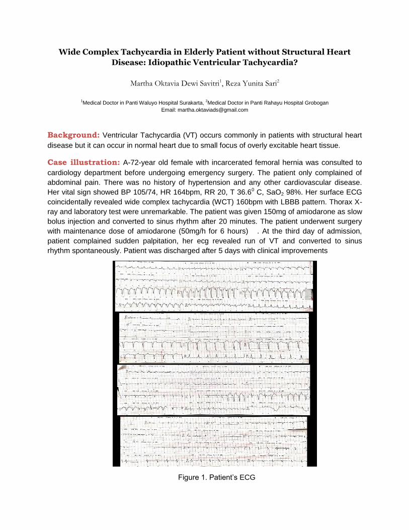
Wide Complex Tachycardia in Elderly Patient without Structural Heart
Disease: Idiopathic Ventricular Tachycardia?
Martha Oktavia Dewi Savitri1, Reza Yunita Sari2
1Medical Doctor in Panti Waluyo Hospital Surakarta,
2Medical Doctor in Panti Rahayu Hospital Grobogan
Email: [email protected]
Background: Ventricular Tachycardia (VT) occurs commonly in patients with structural heart
disease but it can occur in normal heart due to small focus of overly excitable heart tissue.
Case illustration: A-72-year old female with incarcerated femoral hernia was consulted to
cardiology department before undergoing emergency surgery. The patient only complained of
abdominal pain. There was no history of hypertension and any other cardiovascular disease.
Her vital sign showed BP 105/74, HR 164bpm, RR 20, T 36.60 C, SaO2 98%. Her surface ECG
coincidentally revealed wide complex tachycardia (WCT) 160bpm with LBBB pattern. Thorax X-
ray and laboratory test were unremarkable. The patient was given 150mg of amiodarone as slow
bolus injection and converted to sinus rhythm after 20 minutes. The patient underwent surgery
with maintenance dose of amiodarone (50mg/h for 6 hours) . At the third day of admission,
patient complained sudden palpitation, her ecg revealed run of VT and converted to sinus
rhythm spontaneously. Patient was discharged after 5 days with clinical improvements
Figure 1. Patient’s ECG
Discussion: Patient’s ECG meets the criteria for VT.using Brugada criteria. VT in this patient
might be considered as idiopathic VT with RVOT origin due to absence of clinically apparent
SHD. Idiopathic VT might be found in 10% of patients with VT. Repetitive non sustained-VT
occurs in 60-92% of cases while incessant VT occurs only occasionally. Intravenous amiodarone
used in this case is more effective than lidocaine to terminate stable VT with improved survival at
24 hours. This arrhythmia needs periodic follow up with reassessment of ventricular function
Conclusion: Amiodarone may be prescribed as treatment option for idiopathic VT especially
in elderly
Keyword: WCT, Idiopathic VT, Amiodarone

Trombolitic After Anticoagulan Theraphy on Acute Miocard Infark;
A Case Report
Mega RA1 ; Mahbubi M2
1General Practitioner at RSU Fastabiq Sehat PKU Muhammadiyah, Pati, Indonesia
2Cardiologist at RSU Fastabiq Sehat PKU Muhammadiyah, Pati, Indonesia
ABSTRACT
Cardiogenic shock continues to be the most common cause of death in patients hospitalized
with acute myocardial infarction. Cardiogenic shock is an extreme presentation of Heart
Failure after miocardial infarction. Whereas cardiogenic shock is widely appreciated as the
major complication of hemodynamic compromise, less severe HF states on the spectrum are
more common and also have major adverse consequences. At this case, there was a 47
years old man, with a chest pain since 3 days before going on hospital, and increased 4 hour
before going on ER. The blood presure was 100/60 without a sign of syok, and ECG was
present a STEMI inferior witch ST depresi in V1-V3. Before refers to our hospital,
anticoagulan (foundapharinux) was givem in hospital before. In our hospital not capable PCI
so, will doing trombolitik for next. A sreptokinase just given half unit, and still chest pain, and
increased ¾ dose until all dose. And suddenly during trombolitic the vital sign was decrease,
BP was 70/50, HR 52x/sec, with dypsnea, rhonkhi, sign of syok, and followed by
cardiopulmonary arrest in pulseless electrical activity. CPR and intubation was given almost
60 minutes but eventually died in asystole.
Keyword : Trombolitic, Anticoagulan, Acute Miocard Infarct

Improving Pulmonary Hypertension in Acute Condition
Paramarisa, Aliffadinya1 ; Gunadi, Abraham S. 1 ; Rosyadi, Ragil N. 2
1 Emergency Department Dr. Ramelan Naval Hospital, Surabaya, Indonesia,
2 Cardiology Department Dr. Ramelan Naval Hospital, Surabaya, Indonesia.
ABSTRACT
Recognition of pulmonary hypertension (PH) in clinical practice over the past 30 years has been
increase. It is likely that this rise in PH diagnoses is attributable to multiple factors, including
increase awareness by clinician, routine use of diagnostic tools such a Doppler
echocardiography, and availability of the many PH – specific drug2. PH is often characterized by
a progessive and sustained increase in pulmonary vascular resistance that eventually may lead
to right ventricular failure. It can be a life-threatening condition if untreated3. 53 years old male
came with shortness of breath which he has felt since 3 days ago and worsening few hours
before came to our ED. From history taking, we acknowledged that he had ASD since 20 years
ago, and was treated with spironolactone and beraprost. Unfortunately, he didn’t take beraprost
anymore for the past month. He appeared somnolent and labored breathing. His blood pressure
was 80/60 mmHg, with 112 bpm heart rate, and SpO2 of 78-80%. On physical examination,
auscultation sounds a second heart (s2) hardens with splitting. His ECG showed 120 bpm sinus
rhytm and RBBB. CBC, SE, blood chemistry finding were unremarkable. Arterial blood gas
showed respiratory acidosis with pH 6.80 pCO2 66,3 pO2 48.5 HCO3 10.3 BE -24.2. Chest x-
ray showed cardiomegaly. Endotracheal intubation was performed and 100% FiO2 was given.
Despite the intervention, after 30 minutes of mechanical ventilation, his SpO2 didn’t increase.
We decided to give him beraprost. Two tablets of beraprost were grinded and administrated via
nasogastric tube. Approximately 20-30 minutes after the administration of beraprost, his SpO2
increased until 98%. We assumed his SpO2 increased due to the administration of Beraprost as
it is proven to improve high pulmonary artery pressure even in acute condition.
Keywords : Pulmonary Hypertension, ASD, Beraprost

Association of Mean Platelet Volume with Failure of Percutaneous
Intraarterial Thrombolysis in Acute Limb Ischemia
Priambodo, R.R.1, Ismail, M.T.1, Hariawan, H1
Department of Cardiology and Vascular Medicine, Faculty of Medicine, Universitas Gadjah Mada, Sardjito General Hospital,
Yogyakarta, Indonesia.
Background: Platelet activation plays a major role in pathogenesis of arterial thrombosis.
The degree of platelet activation in the thrombus is thought to affect the vulnerability to
thrombolytic therapy. One of indicator of platelet activation is mean platelet volume. The aim
of this study is to describe the association of mean platelet volume with failure of
percutaneous intraarterial thrombolysis in acute limb ischemia.
Method: A cross sectional study was conducted in Sardjito General Hospital. Data were
collected from January 2016 to May 2019. Patients with acute limb ishemia underwent
percutaneous intraarterial thrombolysis were included. Measurement of mean platelet
volume was performed on admission. Success or failure of percutaneous intraarterial
thrombolysis was assessed with arteriography. Independent T-Test was used to estimate the
difference of mean platelet volume between two groups.
Result: Fifty six patients were included in this study. There were 11 patients (17,86%) with
failed percutaneous intrarterial thrombolysis. Mean platelet volume was significantly higher in
patients with failed percutaneous intraarterial thrombolysis compared to patients with
successful percutaneous intraarterial thrombolysis (10.16 ± 1.09 fl vs 9.29 ± 1.27 fl, 95 CI=
0.002-1.746, p=0.049).
Conclusion: There is an association of mean platelet volume with failure of percutaneus
intraarterial thrombolysis in acute limb ischemia.
Keywords: Acute Limb Ischemia, Mean Platelet Volume, Percutaneous Intraarterial
Thrombolysis, Platelet Activation

Baseline characteristics, management, and in-hospital outcomes of patients
with non-ST-elevation acute coronary syndrome with high and very high
risk stratification without invasive strategies in Tarakan, North Kalimantan
: A Study from Tarakan Registry of Acute Coronary Syndrome
Jauhari, H 1, Ahmad, F 2, Syamsul, D.S 2, Sentono, W.D 3
1 General Practitioner, Tarakan Regional Public Hospital, North Kalimantan, Indonesia
2 Cardiologist, Tarakan Regional Public Hospital, North Kalimantan, Indonesia
3 Cardiologist, Soemarno Sosroatmodjo Regional Public Hospital, North Kalimantan, Indonesia
Background: Invasive strategies had an important role in the management of patients with
non-ST-elevation acute coronary syndrome (NSTE-ACS) especially in patients with high and
very high-risk stratification. North Kalimantan is a province in Indonesia with the highest
prevalence of heart disease based on physician diagnosis, adopted from RISKESDAS 2018.
Tarakan General Hospital is the main referral hospital in North Kalimantan which does not have
facilities to implement invasive strategies in the management of NSTE-ACS patients and
geographically is not possible to refer patients to other hospitals in a short time. Objective: To
identify the baseline characteristics, management and in-hospital outcomes of patients with high
risk and very high-risk NSTE-ACS without invasive strategies in Tarakan Regional Public
Hospital. Method: We collected retrospective registry in Tarakan General Public Hospital
between January 2017 and December 2018. Web-based data entry was used and the data
were centrally managed. Result: The mean age was 59.12 ± 11.43 years and 76.4% were
male. The mean length of stay was 6.23 ± 2.38 days. History of diabetes mellitus and smoker
were present in 21.17% and hypertension in 55.29%. Normal ECG found in 7.05% patients.
Echocardiography was performed in 36.47% of patients and the result were 38.7% of patients
with low EF. Most of the patients with mild TIMI Risk (49.41 %) and 16.47 % patients with
GRACE score more than 140. High Killip classes were defined in 36,48 % of patients. 28.23 %
of patients had very high-risk CRUSADE Score. During hospitalization, only a minority of the
patients did not receive beta-blockers and ACE inhibitors or ARBs therapy. In-hospital
mortality was 9.42 %. Conclusion: NSTE-ACS is common in male and older age patients.
Comprehensive risk stratification is needed to determine the suitable management. Invasive
strategy is needed to reduce in-hospital mortality.
Keywords: NSTE-ACS, risk stratification, invasive strategy.

Cardiovascular Risk Profile of Indonesian Olympic Athletes
Maharjito AB*, Handayani I*
General Practitioner, Rumah Sakit Olahraga Nasional, Jakarta, Indonesia
Background: Athletes are assumed to have low cardiovascular risk due to young age and
regular physical exercise programs, and are believed to have a healthy lifestyle. However, there
is little information about cardiovascular risk profiles in athletes. The aim of this sudy was to
obtain an overview of cardiovascular risk profiles in Indonesian Olympic ahthletes.
Methods: Study of 62 Indonesian Olympic athletes (39 males, 23 females) who underwent
medical examination at Rumah Sakit Olahraga Nasional. Prevalence and distribution of
cardiovascular risk factors (obesity, hypertension, diabetes, hyperlipidemia, metabolic syndrome
and chronic kidney disease) were assessed in relation to age, gender and sports.
Results: The female group had a higher body mass index, diastolic blood pressure and lower
HDL compared to the male group (p<0.05). Significant differences (p<0.05) was also found on
body mass index, systolic blood pressure, diastolic blood pressure and triglycerides among
sports. HDL decreases and LDL increases according to the increase in age groups.
Hyperlipidemia was the most common risk factor (46.7%) followed by obesity (20.9%). The
highest percentage of hyperlipidemia was found in swimming (60%) and the lowest was in
archery (14.3%). Percentage of athletes with no risk factor was 41.9%, while 45.2 % had 1 risk
factor and 12.9% had 2 risk factors. Taekwondo had the highest number of athletes with no risk
factors while weightlifting had the highest number of athletes with 2 risk factors.
Conclusion: Indonesian Olympic athletes was not free from cardiovascular risk factors,
especially hyperlipidemia (46.7%). The proportion of Indonesian Olympic athletes without
cardiovascular risk factors was 41.9% and 12.9% had 2 risk factors.
Keyword : Cardiovascular Risk Factor, Olympic Athletes, Hyperlipidemia

CORRELATION BETWEEN SKELETAL MUSCLE-TO-VISCERAL FAT
RATIO AND VISCERAL-TO-SUBCUTANEOUS FAT RATIO WITH BLOOD
PRESSURE AMONG ADULT POPULATION IN SUTER-KINTAMANI, BALI
PROVINCE, INDONESIA
Utomo. M. F. P1, Yuliantini. N. N1, Teguh. N. A1, Erawan. I. G. N. A. T2
Medical Student, Faculty of Medicine, Udayana University, Bali, Indonesia
Department of Internal Medicine, Faculty of Medicine, Udayana University/Sanglah General Hospital, Bali, Indonesia
ABSTRACT
Obesity became a main risk factor that caused metabolic syndrome and cardiovascular
disease. BMI not only indicator for claimed the obesity. Nowdays, effect of an increase
visceral fat and a decrease in muscle mass on hypertension hasn’t been investigated.
Bioelectrical Impedance Analytic (BIA) is a simple tool for specific value of body composition.
This study was aimed to examine the association between skeletal muscle-to-visceral fat
ratio and visceral-to-subcutaneous fat ratio toward blood pressure in adult population. This is
an analytic observational study who involved 160 adults in Suter Village, Kintamani, Bali
Province. Primary data were taken from “Puskesmas Keliling” program during August 2018.
Demographic data, anthropometric measurement using BIA were recorded. We also
measured the blood pressure, collect the personal history with interview, and general status
examination. Data were analyzed using univariate and bivariate analysis. A total of 160
cases were conducted for this research. Most of respondent in the study were females
(63.8%) with mean age was 56.79±11.56. From a total respondent, 62.5% were diagnosed
with hypertension (hypertension grade I 37.5%; grade II 25%). The median of visceral-to-
subcutaneous fat ratio and skeletal muscle-to-visceral fat ratio were 2.653 and 0.311. We
found correlation between skeletal muscle-to-visceral fat ratio and blood pressure (systolic,
p<0.001; r=-0.400, diastolic, p<0.001; r=-0.458). We also found correlation between visceral-
to-subcutaneous fat ratio with blood pressure (systolic, p=0.009; r=0.205, diastolic, p<0.001;
r=0.273). Skeletal muscle-to-visceral fat ratio is significantly increased to subject with systolic
and diastolic blood pressure with moderate correlation. Visceral-to-subcutaneous fat ratio
also significance, but we found weak correlation toward blood pressure. Future study needed
to give specific factor may associated with hypertension.
Keywords: Blood Pressure, Body Composition, Hypertension, Obesity, BIA

Electrocardiogram Interpretation Skills Among General Practitioners
in Islands and Remote Areas: Case of Maluku Utara Province
Budi DS1, Gharini PPR2
1 Tolofuo Primary Health Care, Halmahera Barat, Indonesia
2 Department of Cardiology and Vascular Medicine, Faculty of Medicine, Public Health and Nursing, Universitas
Gadjah Mada - Dr.Sardjito General Hospital, Yogyakarta, Indonesia
Background: ECG interpretation by general practitioner as primary health care and
emergency service has important role to manage cardiovascular disease including life
threatening disorder, especially in remote area. Meanwhile, Maluku Utara has many islands
and remote areas.
Aim: This research aims to evaluate the ECG interpretation skill among general
practitioners in Maluku Utara Province.
Method: General practitioners in Maluku Utara province were requested to complete a
web-based ECG questionnaire containing 16 mini cases with ECG strip on June 2019. The
Questions were designed to assess ECG interpretation accuracy. Each true answer of mini
case was given 1 score. Information about employment status, age, sex, work institutions,
ACLS training, and ECG training were also collected.
Result: We obtained 91 questionnaires filled by general practitioners of estimated 250s in
Maluku Utara, consist of PTT (Pegawai Tidak Tetap) or contract doctors (n=38, 41.76%),
followed by internship doctors (n=33, 36.26%), and PNS (Pegawai Negeri Sipil) / CPNS
(Calon Pegawai Negeri Sipil) (n=20, 21.98%). Most are women (n=47, 51.64%) with
average age of 27.47 years old. Average score is 9.3 of 16, with median of 10 of 16. The
multivariate analysis shows that age has significant association in declining the skill (adj.
OR= -0.37, p<0.001). A recent experience in ACLS training (less than 3 years) significantly
leads to a higher score (adj. OR =1.71, p=0.039), along with ECG training (adj. OR=1.63,
p=0.042). Sex, work institutions, and employment status have no significant association with
ECG interpretation skill.
Conclusion: Increasing age is a factor in decreasing electrocardiogram interpretation
skill, while ACLS and ECG training are considered important to improve electrocardiogram
interpretation skills among general practitioners in Maluku Utara.
Keyword: electrocardiogram interpretation, general practitioner, Maluku Utara

Factors Predicting Left Ventricular Dysfunction Following Anterior ST
Elevation Myocardial Infarction
Prabowo, B.K.1, Sulistyo, N.A.2, Sudiyoko2, Sukmadja, D.1
1General Practitioner, Tidar General Hospital, Magelang
2Cardiologist, Cardiology and Vascular Department, Tidar General Hospital, Magelang
Background: Left ventricular (LV) systolic function is an important prognostic factor in
coronary heart disease. Although reperfusion therapy has been found effective in lowering
acute myocardial infarction (AMI) complications, left ventricular ejection fraction (LVEF)
impairment is a common consequence of AMI especially anterior ST Elevation Myocardial
Infarction (STEMI)
Objectives: The aim of this study was to estimate the incidence of LVEF depression and
to determine factors of LV dysfunction after anterior STEMI.
Methods: In this retrospective study, we obtained the data from anterior STEMI patients’
medical records from January 2017 till December 2018 and undergone Trans Thoracic
Echocardiography (TTE) to evaluate the LV function. A total of 45 anterior STEMI patients
were divided into two groups according to the incidence of LV dysfunction. LVEF was
considered depressed when ejection fraction was less than 40%. The univariate relationship
analysis between baseline characteristic and LV dysfunction incidence were assessed with
Chi-Square. Statistical significance defined by p <0.05.
Results: A total of 45 patients (mean age 55.02±8.64 years, male gender 91.1%) with
anterior STEMI following echocardiography were included. The mean level of left ventricular
ejection fraction is 55.53±12.34%. The LV dysfunction rate was found to be 20% in this study
population. In the univariate analysis we found that non-thrombolytic patients (OR: 2.24;
95%CI: 0.48-10.35; p=0.252) and history of dyslipidemia (OR: 4.86; 95%CI: 0.58-40.55;
p=0.173) shows a positive association but had no statistically significance correlation with LV
dysfunction incidence. However, history of smoking (OR: 0.5; 95%CI: 0.11-2.25; p=0.29),
hypertension (OR: 0.894; 95%CI: 0.21-3.88; p=0.59), and diabetes (OR: 0.625; 95%CI: 0.07-
5.97; p=0.57) shows a protective risk but had no statistically significance correlation with LV
dysfunction.
Conclusion: There was no significant association between non-thrombolytic patient,
history of dyslipidemia, diabetes, smoking, and hypertension with LV dysfunction incidence
after anterior STEMI.
Keywords: Left Ventricular Dysfunction, Myocardial Infarction, LVEF

Hospitalization of Chronic Heart Failure Patients in Rural Area: The
Case of Rumah Sakit Chasan Boesoirie Ternate, Maluku Utara
Budi DS1, Rizal MF2, Pratiwi D3
1 Puskesmas Tolofuo, Halmahera Barat, Indonesia
2 Department of Health Policy and Management, FK-KMK Universitas Gadjah Mada, Indonesia
3 Department of Cardiovascular, RSUD Chasan Boesoirie, Ternate, Indonesia
Background: Prolong hospital stay for CHF patients is known to be correlated with the
increasing rate of all types of readmission and mortality.
Aim: This study aims to examine factors associated with prolonged hospitalization (>7
days) of CHF patients in RSUD Chasan Boesoerie.
Method: Data was taken from medical records from January 2016 to August 2018. We
collected information about age, sex, insurance type, length of stay, also type and number of
comorbid. To analyze factors associated with prolonged hospitalization, we use multiple
logistic regression methods.
Result: We obtained 351 hospitalizations with complete information on those variables.
There are 57.26% male patients (n=201) with a mean average age of 57.73 years (median=
59 years). The insurance type is mostly BPJS PBI (n=203; 57.83%), followed by BPJS Non-
PBI (n=106; 30.2%), and no health insurance (n=42; 11.97%). The average hospitalization is
6.24 days with a median of 5 days. The number of patients with prolonged hospitalization is
123 (35.04%); The average number of comorbid is 2.1. The proportion of patients with
Pneumonia, DM, COPD, and Kidney Disease, are 8.83%, 9.97%, 11.97%, and 23.36%
consecutively. We found that there is no significant association of age and type of comorbid
with prolonged hospitalization, while the number of comorbid has a positive association (adj.
OR=1.54, p<0.001). Women are more likely to have prolonged hospitalization (adj. OR=1.79,
p=0.019). A positive correlation was found also for health insurance type, that BPJS PBI
patients and no insurance patient, compared to BPJS Non-PBI patients, are less likely to
have prolonged hospitalization (adj. OR=0.52, p=0.013 and adj. OR=0.24, p=0.002
consecutively).
Conclusion: Factors such as women, BPJS PBI, and no health insurance, and the
number of comorbid have an association with prolonged hospitalization of CHF patient in
RSUD Chasan Boesoirie, that should be taken into account when managing CHF patient.
Keywords: chronic heart failure, hospitalization, rural area

IMPACT OF TIME TO REPERFUSION ON LEFT VENTRICULAR
FUNCTION IN PATIENTS WITH ST SEGMENT ELEVATION
MYOCARDIAL INFARCTION UNDERGOING PRIMARY PERCUTANEOUS
CORONARY INTERVENTION
Nugraha, I.W.1, Taufiq, N.2, Hartopo, A.B.2
1Resident Department of Cardiology and Vascular Medicine Faculty of Medicine Universitas Gadjah Mada – Dr. Sardjito
Hospital Yogyakarta, Indonesia
2Staff Department of Cardiology and Vascular Medicine Faculty of Medicine Universitas Gadjah Mada Dr. Sardjito Hospital
Yogyakarta, Indonesia
Background: Duration of ischemia determines infarct size and survival after myocardial
infarction. Rapid recovery of coronary artery blood flow with primary percutaneous coronary
intervention (primary PCI) can limit the extent of infarction and improve left ventricular
function. We hypothesized that shorter time to reperfusion with early restoration of coronary
perfusion may preserve left ventricular function in patients with ST Segment Elevation
Myocardial Infarction (STEMI) who underwent primary PCI.
Methods: This was a cross-sectional study enrolling 40 consecutive patients with STEMI
who underwent primary PCI at Sardjito Hospital from December 1st, 2018 – January 15th,
2019. The wire crossing time is calculated from the onset of typical anginal chest pain until
the guide wire crossing the infarct-related artery during the primary PCI procedure.
Echocardiography examination is performed within 48 hours after the primary PCI. Patients
were divided into 2 groups based on mean time to reperfusion, i.e early reperfusion and
delayed reperfusion group. Association between time to reperfusion and the
echocardiography parameter was assessed by Independent Sample T-Test. The value of p
<0.05 was considered statistically significant.
Results: A total of 40 patients were included in this study. Mean time to reperfusion was
12.73 ± 5.22 hours. Patients with reperfusion time >12.73 hours (n = 18) had a significantly
lower LVEF (44.33 ± 11.03% vs. 56.32 ± 12.49%, p = 0.003), lower e’ velocities (6.47 ± 2.32
cm/s vs. 7.98 ± 1.59 cm/s, p = 0.02), and significantly higher E/e’ ratio (11.86 ± 4.86 vs. 7.76
± 2.23, p = 0.001) compared to those having early reperfusion (n = 22). Time to reperfusion
was a variable that independently influence the E/e’ ratio value (r = 0.463; p = 0.003).
Conclusion: Longer time to reperfusion is associated with left ventricular dysfunction in
STEMI patients who underwent primary PCI.
Keywords: STEMI, Primary PCI, time to reperfusion, left ventricular function

Predictors Length of Stay of Patients with Acute Coronary Syndrome in
Cardiovascular Intensive Care Unit of Sardjito Hospital
Inayati, F. N., Bagaswoto, H. P., Setianto, B.Y
Department of Cardiology and Vascular Medicine Faculty of Medicine, Public Health, and Nursing Universitas Gadjah Mada-Dr.
Sardjito Hospital, Yogyakarta, Indonesia
Background: Acute coronary syndrome (ACS) is a leading cause of hospitalization in the
cardiovascular intensive care unit (CICU). The recommendation of CICU length of stay for ACS
patients is less than 5 days. Identifying factors that influence the CICU length of stay (LOS) of
ACS patients is necessary to improve health cost-effectiveness, especially in the universal
coverage era. Hence, this study aimed to analyse the predictors of LOS in CICU for ACS
patients.
Method: A total of 216 consecutive ACS patients from Sardjito Cardiovascular Intensive Care
registry (SCIENCE) who admitted to CICU from March to June 2019 were enrolled in this study.
According to LOS in CICU, the patients were divided into LOS <5 days and LOS ≥5 days group.
Bivariate and multivariate analysis was performed to determine predictors of LOS in CICU.
Result: There were 37 patients (17.1%) who had LOS ≥5 days. They were more likely men
(67.6% vs 32.4%) and older than 60 years old (81.1% vs 18.9%). Geriatric age, ventilator
utilization, shock, and GRACE score ≥140 were independent predictors of LOS ≥5 days with HR
2.88 (CI: 1.02-8.11), 11.99 (CI: 2.89-48.10), 3.56 (CI: 1.27-9.95), and 3.70 (CI: 1.43-9.54)
respectively.
Conclusion: Factors affecting LOS in CICU of ACS patients include geriatric age, ventilator
utilization, shock, and GRACE score ≥140. Ventilator utilization is the strongest predictor of LOS
in CCU.
Keywords: Acute Coronary Syndrome, Cardiovascular Intensive Care Unit, Length of Stay

Relationship between Platelet-to-Lymphocyte Ratio, Neutrophil-to-
Lymphocyte Ratio, De Ritis Ratio and In-hospital Length of Stay in
ST-Segment-Elevation Myocardial Infarction Cases
Sukmadja, D., Sulistyo, N. A., Sudiyoko, Prabowo, B. K.
Cardiology and Vascular Department, Tidar District General Hospital Magelang, Central Java, Indonesia
ABSTRACT
Introduction: In-hospital length of stay has become one of important outcomes that should
be considered due to health insurance policy. Platelet-to-Lymphocyte Ratio (PLR) has been
introduced as a pro-thrombotic marker. Neutrophil to Lymphocyte Ratio (NLR) has been found
as a pro-inflammatory marker. Several studies discovered that De Ritis (AST/ALT) Ratio can
become a potential marker for end organ damage in acute myocardial infarction.
Objective: To find relationship between PLR, NLR, De Ritis Ratio and In-hospital Length of
Stay in ST-segment-elevation myocardial infarction (STEMI) patients in Tidar District General
Hospital.
Methods: This study included 186 patients with STEMI, during the period of January 2017 to
December 2018 in Tidar District General Hospital. Patients who were referred and not survived
were excluded. Platelet, Neutrophil, Lymphocyte, AST, and ALT serum level was obtained on
admission data and analyzed with Spearman correlation towards the duration of in-hospital
length of stay.
Results: The median age of all patients was 57.0 ± 11.65 years, with 83.9% male patients.
The mean level of PLR, NLR and De Ritis Ratio were 17.78±12.05, 4.71±3.93 and 1.89±1.28.
The mean level of duration of hospitalization was 6.04±2.29 days. Spearman test showed non-
significant correlation from PLR and De Ritis Ratio (p=0.185, r=0.098; p=0718, r=-0.030) and
significant positive correlation from NLR (p= 0,019, r= 0,172) towards in-hospital length of stay.
Conclusion: NLR had positive correlation towards in-hospital length of stay in STEMI cases.
There was no significant correlation between PLR, De Ritis ratio and In-hospital length of stay.
Keyword: Platelet-to-Lymphocyte Ratio, Neutrophil-to-Lymphocyte Ratio, De Ritis Ratio, In-
Hospital Length of Stay, STEMI

ST elevation in lead augmented Vector Right, do emergency room doctors notice?
a forgotten lead.
Simanjuntak, Vincent. L. H, MD.
General Practitioner, Hanau Region Public Hospital, Central Kalimantan, Indonesia.
ABSTRACT
Background: Acute Myocardial Infarction is a condition that needs rapid and proper
intervention managements, a good knowledge to diagnose and to select appropriate treatment options rests solely on the correct identification of ST Elevation based on ECG reading skill by the emergency physician, such as detecting ST elevation in lead aVR, including lead V1 or aVL, and/or diffuse ST depresion on precordial leads, are tell tale sign of on going process of LMCA or proximal LAD occlusion, or triple vessels disease. the confusion to identify the ECG findings, diagnose and treat the patient as either STEMI or NSTEMI may lead to further delay in the proper treatment when the complete resources of treatment are available.
Objective: The objectives of this study were to find out; 1) the knowdge of ER doctor about
aVR ST elevation in ECG finding; 2) whether ER doctors are paying enough attention to the lead itself; 3) to identify whether it had been taught during ACLS course as recalled by ER doctors.
Methods: This was a descriptive research using survey questionnaire approach, the samples
were obtained using simple random sampling methods, emergency room GP doctors, who have received valid ACLS training in the last 3 years, which are serving around Indonesia, there are 60 doctors chosen as samples, based on inclusion and exclusion criteria.
Results: The result of this research are; 1) 46.67% (28) of respondents have decend
knowledge, while 43,34% (26) respondents have poor knowledge, and only 10% (6) of the respondents have good level of knowledge; 2) only 16.7% (10) who actively paying attention to lead aVR; 3) only 6.7% (4) of ER doctors who remembered having been taught of aVR STEMI.
Conclusion: The conclusion of this study are, ER doctors mostly have decend knowledge
about aVR ST elevation, however almost half of the samples have poor knowledge, only few ER doctors are actually paying attention to lead aVR for ischemic finding, and most ER doctor do not receive training of aVR STEMI during ACLS.
Keywords: level of knowledge, aVR, general physician, emergency room, ACLS.

The Relationship between Mean Platelet Volume (MPV) and Severity of
Artheroclerosis in Patients with Stable Coronary Artery Disease (SCAD)
Husna, A.A., Hartopo, A.B., Puspitawati, I., Arso, I.A.
Department of Cardiology and Vascular Medicine Faculty of Medicine, Public Health, and Nursing Universitas Gadjah Mada -Dr.
Sardjito Hospital, Yogyakarta, Indonesia
ABSTRACT
Platelets play an important role in the pathogenesis of coronary artery disease (CAD). Mean
Platelet Volume (MPV) is an index of platelet size that can be correlated with the functional
status of platelets. This study aimed to assess the relationship between mean platelet volume
(MPV) and severity of atherosceloris in patients with stable coronary artery disease (SCAD) in
RSUP dr. Sardjito Yogyakarta.
Method : A total of 172 patients with SCAD who underwent coronary angiography were
included in this study. The severity of CAD was determined by calculating Gensini Score (GS)
based on patient’s angiographic data. All demographic and clinical features were collected
retrospectively. MPV were obtained on admission.
Result : Mean age of the study population was 58.05 + 8.48 years, of whom 134 male (77.9%)
and 38 were female (22.1%). Of the patients, 97.1% had CAD, 29.7% had diabetes mellitus,
64.5% had hypertension, 51.7% had dyslipidemia, and 60.5% were smokers. Among the
patients, 60,5% using Dual Antiplatelet Theraphy (DAPT). Mean of Gensini Score was 64.6%.
According to Gensini Score, 5 of the patients (2.9%) had normal coronary arteries (GS: 0), 29 of
the patients (16.9%) had minimal CAD (GS: 1-19), and 138 of them (80.2%) had severe CAD
(GS ≥ 20). Mean MPV values in the groups were 9.12 + 1.61 fL, 9.97 + 1.39 fL, and 9.93 + 1.32.
According to spearman correlation analysis, there was no significant correlation between
Gensini Score and MPV values in these patients (p=0.792). Although, there was significant
differences in the groups between smokers and non-smoker (p=0,036), and patients using
DAPT and Single Antiplatelet Theraphy (SAPT) (p=0,036).
Conclusion : There was no significant association between MPV levels and the severity of
artherosclerosis described by Gensini Score in this study.
Keywords : Gensini Score (GS), Mean Platelet Volume (MPV), Stable Coronary Artery
Disease (SCAD)

Validation Study of A Simplified Risk Scoring System to Predict Mortality in
Cardiovascular Intensive Care Unit of Sardjito General Hospital
Wijaya. S. N., Bagaswoto H. P., Setianto B. Y.
Department of Cardiology and Vascular Medicine Faculty of Medicine, Public Health, and Nursing Universitas Gadjah Mada Dr.
Sardjito Hospital, Yogyakarta, Indonesia
Background: The number of visits and mortality rates of cardiovascular intensive care unit
(CICU) patients have been increasing from time to time. Appropriate identification of patients
with high mortality risk is crucial. A Simplified Risk Scoring System to predict mortality in
Cardiovascular Intensive Care Unit was developed with some parameters that include age ≥ 60
years old, presence of pneumonia, mechanical ventilation utilization, creatinine level > 1.5 mg/dl,
serum glutamate-pyruvate transaminase > 50 U/L, and ejection fraction <40%. This study aimed
to validate the performance of Simplified Risk Scoring System in predicting in-hospital mortality
of CICU patients in Sardjito General Hospital.
Methods: The design of this study is cross-sectional and data were obtained from SCIENCE
(Sardjito Cardiovascular Intensive Care) registry. The research subjects were the 306 CICU
patients of Sardjito hospital (mean age 60.19 +13.74) between March-June 2019. Statistical
analysis using a 2x2 table was performed to calculate the sensitivity and specificity. The
discrimination performance was measured with an area under the curve (AUC).
Results: The mortality rate of 306 subjects in this research was 17.32% (53 subjects).
Simplified Risk Scoring System in this study presented a good ability in predicting mortality with
an Area under ROC (AuROC) of 87.2% with CI 0.817-0.928. The sensitivity and specificity were
84% and 67% respectively.
Conclusion: This scoring system can provide excellent discriminant value in predicting in-
hospital mortality with satisfying sensitivity and specificity results.
Keywords: Scoring System; Mortality; Cardiovascular Intensive Care Unit;

Portopulmonary Hypertension: A Case Report
Siddiq,F.1 Dinarti, LK.2
1 Resident of Cardiology and Vascular Medicine Gadjah Mada University/ Sardjito General Hospital
2 Staff of Cardiology and Vascular Medicine Gadjah Mada University/ Sardjito General Hospital
Background : Patients with liver disease are at risk for pulmonary hypertension (PH) due to
high flow, increased pulmonary venous volume, this condition we called as portopulmonary
hypertension (POPH). POPH is an obstructive, constrictive arteriopathy that has pathologic
findings similar with idiopathic pulmonary artery hypertension.
Case Illustration : A 62-year-old man was admitted to Emergency Room due to
hematemesis dan melena. Past medical history was hepatic cirrhosis and has performed
ligation of esophageal varices two years ago. The clinical presentation was weakness followed
by ascites. We found cardiomegaly and ascites in physical examination with the ECG shows
right axis deviation and right bundle branch block. Chest X-Ray result was cardiomegaly with
normal appearance of lung. An echocardiography examination revealed right atrial and
ventricular dilatation, LV “D” shaped, tricuspid regurgitation moderate, and high probability of
PH. Based on this, the diagnosis of pulmonary arterial hypertension associated with portal
hypertension was made.
Conclusion : Liver disease can cause portal hypertension. This increased pressure causes
blood to bypass the liver and resulted the blood vessels of the lungs are then exposed to
possible toxic substances, and this can damage the small arteries of the lungs, causing
pulmonary arterial hypertension (PAH). Physical examination, ECG, Chest X-Ray and
Transthoracic doppler echocardiography plays an important role in the evaluation of
symptomatic cirrhotic patients with suspected POPH. A clinical diagnosis of POPH can be made
with the documentation of compatible hemodynamics in a patient with portal hypertension in the
absence of coexisting conditions associated with pulmonary hypertension. Histopathologic and
clinical similarities between POPH and other forms of PAH have provided a rationale for the use
of PA-targeted therapies in POPH.
Keywords: Pulmonary hypertension, Portopulmonary hypertension, Hepatic cirrhosis

Coronary Slow Flow Phenomenon
Baiq G. R. Faharani1, Nahar Taufiq2, Anggoro B.Hartopo2
1 Resident of Department of Cardiology and Vascular Medicine Universitas Gadjah Mada / RSUP dr Sardjito
2 Staff of Department of Cardiology and Vascular Medicine Universitas Gadjah Mada / RSUP dr Sardjito
Introduction
The coronary slow flow phenomenon (CSFP) is an angiographic finding characterised by delayed opacification of the distal vasculature in the absence of obstructive epicardial coronary artery disease (CAD). Recently CSFP has been found to be associated with increased QTc dispersions. The corrected TIMI frame count (CTFC) introduced by Gibson, is a quantitative and reproducible index of coronary artery flow. It represents the number of cine frames required for contrast to reach a prespecified distal coronary artery landmark. Coronary slow flow phenomenon is defined as CTFC greater than 2 standard deviations (SD) from the normal published range, which is 21±3.
Case Report A 66-year-old man came to the emergency room of Dr. Sardjito Hospital with chief complaint chest pain. It radiated to the right and left arms, accompanied with diaphoeresis and abdominal pain. Patients have a history of asthma and risk factors for hypertension. On ECG examination from previous hospital was obtained with sinus rhythm, frequency 43 times / minute, Right Axis Deviation, bigemini ventricle extrasystol, and the QTc dispersion are 120 msec. When on the emergency room at Dr. Sardjito hospital, the ECG showed a sinus rhythm, Right Axis Deviation, frequency 75 times / minute, with periodic slow ventricular tachycardia. On the laboratory examination, the results of cardiac enzym obtained Hs troponin I 38,455 ng / L. The patient was diagnosed with NSTEMI killip I TR 3/7 GS 99 with sustained slow VT / AIVR (very high risk). From immediate invasive strategy, results : Left Main (LM) are normal; Proximal ectatic Left Anterior Descenden (LAD), 30% mid stenosis, slow flow; Left Circumflex artery (LCx) proximal to 50% stenosis, thrombus appears distal, slow flow; Proximal Right Coronary Artery (RCA), mid 30% stenosis, slow flow. Because of high burden thrombus, optimal medical therapy with antithrombotic is chosen (no coronary intervention), and concluded with Coronary Artery Disease (CAD) non-significant with slow flow.
Discussion Based on coroangiography, there is a slowdown in contrast flow in all coronary
arteries. The number of frames from osteal to distal LAD was obtained 65 frames with CTFC 38. This indicated that there was a slowdown in contrast flow in LAD. The exact pathogenesis of CSFP is still not clear and is probably multi factorial. Different theories have been postulated about the cause of small vessel dysfunction based on observations including microvascular tone dysfunction, endothelial thickening in small vessels, patchy fibrosis in the biopsy specimen taken from the right ventricle, and impaired endothelial release of nitric oxide (NO). Coronary slow flow has been described to be associated with life-threatening arrhythmias and sudden cardiac death, probably due to increased QTc dispersion.
Conclusion The exact pathogenesis of CSFP is still not clear and is probably multi factorial, and
it can be associated with life-threatening arrhythmias.
Keywords : coronary slow flow, QTc dispersion, life-threatening arrhythmia.
Acute Superior Mesenteric Artery Ischemia and Intracerebral Haemorrhage, Balancing Ischemic and Bleeding Risks Management : a case
report
I.H. Sulistya1, M.T. Ismail2, L.K. Dinarti2, H. Hariawan2
1 Resident of Department of Cardiology and Vascular Medicine, Faculty of Medicine, Public Health and Nursing,
Universitas Gadjah Mada 2 Staff of Department of Cardiology and Vascular Medicine, Faculty of Medicine, Public Health and Nursing,
Universitas Gadjah Mada
ABSTRACT
A 59 years old man presented to the emergency department with acute severe
abdominal pain with normal abdominal examination. He had history of diabetes mellitus,
hypertension, and atrial fibrillation. Abdominal MSCT revealed superior mesenteric artery
occlusion. At emergency room the patient developed a sudden decreased of consciousness.
Head CT scan was performed and showed intracerebral haemorrhage at dextra basal ganglia,
intraventricular hematome of bilateral lateralis ventricle, tertius and quartus ventricle, and
subarrachnoid hematome of dextra parietal lobe. Neurosurgeon team performed decompression
craniectomy, hematoma evacuation, and external ventricular drainage. One day after,
endovascular approach was choosen for the mesenteric ischemia with arteriography, thrombus
aspiration, and local intraartery heparin administration. Laparoscopy evaluation showed no
intestinal infarction (succesfull thromboaspiration). But unfortunately, during intensive care unit
management, his condition worsened caused by severe sepsis and acute renal failure.

Monophasic Synovial Sarcoma in Left Ventricle: An Extremely Rare Case
Muthmaina A1, Dwianingsih EK2, Bagaswoto HP3, Mumpuni H3
1Resident of Cardiology and Vascular Medicine Gadjah Mada University/ Sardjito General Hospital
2Staff of Pathology Anatomy Gadjah Mada University/ Sardjito General Hospital
3Staff of Cardiology and Vascular Medicine Gadjah Mada University/ Sardjito General Hospital
Case Illustration
Male 19 yo, suddenly unconscious while working, he went to Banyumas General Hospital and suspected to have congenital heart disease. He then referred to Klaten Islamic Hospital, and underwent echocardiographic examination. They found a mass in left ventricle (LV), then he referred to Sardjito General Hospital, with diagnosis suspected LV Myxoma. From the echocardiography in Sardjito General Hospital, we found two LV masses, size 2.6 x 1.5 cm that attached to PML, and 1.2 x 2.1 with peduncle attached to AML and obstructed LV outflow tract (LVOT), the masses was confirmed by Cardiac Multi Slice Computed Tomography (MSCT). He underwent open heart surgery for mitral valve replacement and LV mass evacuation, the mass was sent to pathology anatomy (PA) department, with the biopsy and immunohistochemistry (IHC) with BCI2, desmin, and Ki67 result was monophasic synovial sarcoma with proliferation index 25%. The patient was discharged, and plan to get chemotherapy and radiotherapy.
Discussion
Synovial Sarcoma (SS) incidence around 5-10% of sarcoma, with 17.6% case occur in children and young adult, and 68.7% case appear in extremities, whereas only 5.3% and 2.2% appear in intra-thoracic and other place respectively. Synovial Sarcoma of the heart is extremely rare, and two times more common in right side than left side heart. The monophasic SS consisted solely of sarcomatous components and often diagnostically challenging.
Conclusion
Monophasic SS of the left ventricle is an extremely rare case, and often diagnostically challenging.
Keywords: Monophasic Synovial Sarcoma, Left Ventricle, Young Adult

‘WINKING SIGN’ A NEW COROANGIOGRAPHY MARKER IN PATIENTS
ACUTE MYOCARDIAL INFARCTION COMPLICATED BY VENTRICULAR
SEPTAL RUPTURE
Herman H.1 , Taufiq N.2, Setianto B.Y.2
1Resident of Department of Cardiology and Vascular Medicine, Faculty of Medicine, Gadjah Mada University, Yogyakarta
2Staff of Department of Cardiology and Vascular Medicine, Faculty of Medicine, Gadjah Mada University, Yogyakarta
ABSTRACT
Introduction: Ventricular Septal Rupture (VSR) is one of the most lethal complications of
Acute Myocardial Infarction (AMI). Commonly VSR happened in 3-5 days after AMI with a wide
range of clinical presentations. When the clinical presentation doesn’t make significant changes
to hemodynamic parameters, diagnosis of VSR may be delayed or misunderstood as the
progression of myocardial infarction. One study from Kamal Sharma found that there’s a specific
‘winking sign’ of Infarct Related Artery (IRA) that lies over the ruptured septum of the ventricle.
Case: A male 81 years old came to district hospital with typical chest pain and diagnosed and
treated as Non ST-Elevation Acute Coronary Syndrome (NSTEACS), after 5 days of admission
the chest pain didn’t relief, recurrent ST-elevation documented on ECG. Patient referred to
Sardjito Hospital, at the ER patients still had chest pain, we found new harsh pansystolic
murmur at the apex of the heart. We found total occlusion at proximal of Left Anterior
Descending (LAD) artery from coroangiography, then we performed Plain Old Balloon
Angioplasty (POBA) at the LAD. After the flow of contrast reaches the distal part of LAD, there’s
a picture similar like myocardial bridging that was winking every phase of systole. We confirmed
the diagnosis of VSR with echocardiography then we found the gap at the apex part of
ventricular septal.
Discussion: Winking sign first described in 2017 from the study in India that define there’s a
similarity among coroangiography findings of patients that had VSR. It defined as transient
occlusion of IRA from inside to outside manner that lies over the VSR at systolic phase and
back to normal during diastolic. Mechanism of winking sign hasn’t been established but there
were three mechanisms that have been proposed as hypothesis such as venturi effects,
mechanical and hemodynamic theory. Winking sign could be the additional diagnostic sign that
could raise awareness of cardiologist, especially at the catheterization lab, when the time range
of VSR happened before echocardiography performed or within catheterization.
Keywords: Winking Sign, acute myocardial infarction, ventricular septal rupture, diagnostic,
coroangiography

Cardiac Computed Tomography as Pre-Procedural Planning for
Atrial Septal Defect Closure: A Case Report
Pramadya V. Mustafiza1, Putrika P.R. Gharini2, Hasanah Mumpuni2
1Resident of Cardiology and Vascular Medicine Gadjah Mada University/ Sardjito General Hospital
2Staff of Cardiology and Vascular Medicine Gadjah Mada University/ Sardjito General Hospital
Case Illustration
A 29-year-old woman presented to Cardiology Outpatient Clinic with dyspnea on effort
and easily fatigue when doing daily activities since last 5 years. Exam was significant for right
ventricular heaving, fixed-split second heart sound, and systolic murmur heard best at the left
sternal border in the third to fourth intercostal space with positive Carvallo’s sign. ECG showed
sinus rhythm with right axis deviation, complete right bundle branch block, and right ventricular
hypertrophy. Transthoracic echocardiography revealed a secundum ASD. Subsequent
transesophageal echocardiography revealed echo-dropout in the interatrial septum diameter
19–25 mm and all rims were sufficient except aortic rim (only 2mm). Color Doppler interrogation
of the interatrial septum revealed significant left-to-right shunting. Right heart catheterization
revealed a high flow low resistance of secundum ASD with mild pulmonary hypertension (mPAP
30 mmHg). Then patient underwent percutaneous ASD closure. Transesophageal
echocardiography during the procedure revealed a larger defect with suspicion of mitral rim
anomaly. Procedure was canceled and patient had a cardiac CT. Cardiac CT revealed a
secundum ASD with largest diameter 36 mm, rim aorta and posterior were floppy, and anomaly
of mitral rim. Patient then was sent to undergo surgery ASD.
Figure 1. Cardiac CT revealed, a) largest diameter of secundum ASD, b) size of superior and mitral rim with anomaly of mitral rim
Discussion This patient fulfilled criteria for ASD closure because she was symptomatic and had
significant sign of RV overload. Result of transesophageal echocardiography permitted patient
to get percutaneous ASD closure because of defect diameter was less than 35 mm and only

one rim which was insufficient. Cardiac CT finding revealed diameter defect more than 35 mm,
superior and aortic rims were floppy, and mitral rim was abnormal. Those findings were
suspected causing percutaneous device could not be well deployed. Actually the safety and
efficacy of percutaneous closure of atrial septal defects (ASDs) was determined by several
variables, including defect size, presence of adequate rim tissue, relationship to other cardiac
structures, and associated congenital anomalies. Cardiac CTA was a high resolution and wide-
field view techniques that allow accurately evaluation of the size, location, and specific structural
and spatial characteristics of ASDs which is critical for successful percutaneous closure.
Conclusion
Cardiac CTA provides an alternative to currently available imaging methods for the evaluation and assessment of large ASDs before percutaneous defect closure.
Keywords: atrial septal defect, cardiac CT, pre-procedural planning

Loss of Fam13a exacerbates pulmonary arterial hypertension by promoting endothelial-to mesenchymal transition
Rinastiti P1, Ikeda K2, Kuribayashi Y2, Miyagawa K2, Yagi K2, Suzuki Y1,2,
Hiirata K1 and Emoto N1,2
1 Cardiovascular Medicine, Internal Medicine, Kobe University Graduate School of Medicine, Kobe, Japan
2 Clinical Pharmacy, Kobe Pharmaceutical University, Kobe, Japan
ABSTRACT
Pulmonary arterial hypertension (PAH) is a rare and progressive disease with low
survival rate despite current available treatment. PAH is mainly characterized by extensive
vascular remodeling, and together with vascular vasoconstriction, lead to increase in pulmonary
arterial pressure and right heart ventricular failure. Pulmonary vascular remodeling itself is
known to be multifactorial, in which the underlying molecular mechanisms remain to be
elucidated. In our study we analyzed a potential role of family with sequence similarity 13
member A (Fam13a), a gene that is genetically associated with lung diseases such as chronic
obstructive pulmonary disease and pulmonary fibrosis, in the pathogenesis of PAH. We first
identified that Fam13a was expressed in various types of cells in the lung including pulmonary
vasculatures. Moreover, Fam13a expression was downregulated in mouse lung after chronic
exposure to hypoxia. We have generated Fam13a-deficient mice, and induced PAH by
exposing mice to chronic hypoxia. Loss of Fam13a caused higher RVSP and increased right
ventricular mass compared to those in wild-type mice. Fam13a-deficient mice also demonstrate
the reduction in small pulmonary arteries, and increase in pulmonary artery muscularization. We
found that several endothelial cell markers were downregulated and some of the mesenchymal
markers and transcriptional factors such as Snail1 and Twist were increased in the lung of
Fam13a-deficient mice. These data suggest that loss of Fam13a enhanced the endothelial-to-
mesenchymal transition in the lung. Immunohistochemistry for endothelial and mesenchymal
markes demonstrated that significantly more endothelial cells became positive for mesenchymal
markers in small pulmonary artery of Fam13a-deficient mice. Mechanistically, we found that
overexpression of Fam13a prevented the endothelial-to-mesenchymal transition induced by
TGF-β and IL-1β. Taken together, our data revealed that Fam13a plays an important role in the
development of PAH potentially by promoting the endothelial-to mesenchymal transition.
Keywords: PAH, endothelial-to-mesenchymal transition, pulmonary circulation