Case Study 1 - MyCred
26
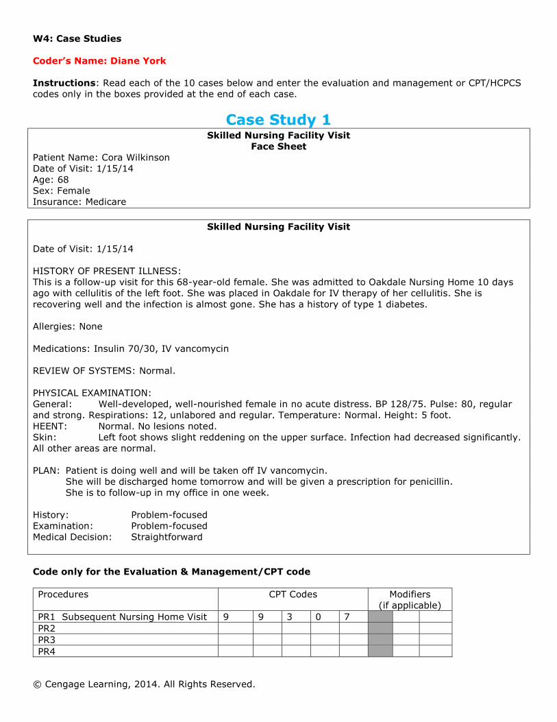
W4: Case Studies © Cengage Learning, 2014. All Rights Reserved. Coder’s Name: Diane York Instructions: Read each of the 10 cases below and enter the evaluation and management or CPT/HCPCS codes only in the boxes provided at the end of each case. Case Study 1 Skilled Nursing Facility Visit Face Sheet Patient Name: Cora Wilkinson Date of Visit: 1/15/14 Age: 68 Sex: Female Insurance: Medicare Skilled Nursing Facility Visit Date of Visit: 1/15/14 HISTORY OF PRESENT ILLNESS: This is a follow-up visit for this 68-year-old female. She was admitted to Oakdale Nursing Home 10 days ago with cellulitis of the left foot. She was placed in Oakdale for IV therapy of her cellulitis. She is recovering well and the infection is almost gone. She has a history of type 1 diabetes. Allergies: None Medications: Insulin 70/30, IV vancomycin REVIEW OF SYSTEMS: Normal. PHYSICAL EXAMINATION: General: Well-developed, well-nourished female in no acute distress. BP 128/75. Pulse: 80, regular and strong. Respirations: 12, unlabored and regular. Temperature: Normal. Height: 5 foot. HEENT: Normal. No lesions noted. Skin: Left foot shows slight reddening on the upper surface. Infection had decreased significantly. All other areas are normal. PLAN: Patient is doing well and will be taken off IV vancomycin. She will be discharged home tomorrow and will be given a prescription for penicillin. She is to follow-up in my office in one week. History: Problem-focused Examination: Problem-focused Medical Decision: Straightforward Code only for the Evaluation & Management/CPT code Procedures CPT Codes Modifiers (if applicable) PR1 Subsequent Nursing Home Visit 9 9 3 0 7 PR2 PR3 PR4
-
Upload
khangminh22 -
Category
Documents
-
view
4 -
download
0
Transcript of Case Study 1 - MyCred
W4: Case Studies
© Cengage Learning, 2014. All Rights Reserved.
Coder’s Name: Diane York Instructions: Read each of the 10 cases below and enter the evaluation and management or CPT/HCPCS codes only in the boxes provided at the end of each case.
Case Study 1 Skilled Nursing Facility Visit
Face Sheet Patient Name: Cora Wilkinson Date of Visit: 1/15/14 Age: 68 Sex: Female Insurance: Medicare
Skilled Nursing Facility Visit Date of Visit: 1/15/14 HISTORY OF PRESENT ILLNESS: This is a follow-up visit for this 68-year-old female. She was admitted to Oakdale Nursing Home 10 days ago with cellulitis of the left foot. She was placed in Oakdale for IV therapy of her cellulitis. She is recovering well and the infection is almost gone. She has a history of type 1 diabetes. Allergies: None Medications: Insulin 70/30, IV vancomycin REVIEW OF SYSTEMS: Normal. PHYSICAL EXAMINATION: General: Well-developed, well-nourished female in no acute distress. BP 128/75. Pulse: 80, regular and strong. Respirations: 12, unlabored and regular. Temperature: Normal. Height: 5 foot. HEENT: Normal. No lesions noted. Skin: Left foot shows slight reddening on the upper surface. Infection had decreased significantly. All other areas are normal. PLAN: Patient is doing well and will be taken off IV vancomycin.
She will be discharged home tomorrow and will be given a prescription for penicillin. She is to follow-up in my office in one week.
History: Problem-focused Examination: Problem-focused Medical Decision: Straightforward Code only for the Evaluation & Management/CPT code Procedures CPT Codes Modifiers
(if applicable) PR1 Subsequent Nursing Home Visit 9 9 3 0 7 PR2 PR3 PR4

W4: Case Studies
© Cengage Learning, 2014. All Rights Reserved.
Case Study 2 Inpatient Face Sheet
Patient Name: Sarah White Admit Date: 1/06/2014 Discharge Date: 1/10/2014 Sex: Female Age: 64 Disposition: Home Admitting Diagnoses: 1. Neutropenic sepsis 2. Status post-chemotherapy for non-Hodgkin’s lymphoma 3. Hypertension Discharge Diagnoses: 1. Pancytopenia with neutropenic sepsis secondary to chemotherapy 2. Non-Hodgkin’s lymphoma 3. Hypoalbuminemia 4. Hypertension
Discharge Summary
Admitted: 1/06/2014 Discharge: 1/10/2014 ADMISSION DIAGNOSES: 1. Neutropenic sepsis 2. Status post-chemotherapy for non-Hodgkin’s lymphoma 3. Hypertension DISCHARGE DIAGNOSES: 1. Pancytopenia with neutropenic sepsis secondary to chemotherapy 2. Non-Hodgkin’s lymphoma 3. Hypoalbuminemia 4. Hypertension HISTORY: This is a 64-year-old female with non-Hodgkin’s lymphoma, currently undergoing chemotherapy. The patient was evaluated by oncology in follow-up and found to be neutropenic as well as febrile. PHYSICAL EXAMINATION: Vital Signs: Blood pressure 132/90. Temperature was 102. HEENT: Dry oral mucous membranes. No thrush or herpetic lesions. Neck: Supple, no adenopathy. Lungs: Clear. Heart: Slightly tachycardiac, no murmur. Abdomen: Soft, nontender. Extremities: Pulses bilateral. Decreased muscle tone. LABORATORY DATA: Chemistries revealed total protein 5.8, albumin 2.4. Calcium 7.3, 7.6. The follow-up chem-7 revealed CO2 of 25, chloride 111.
W4: Case Studies
© Cengage Learning, 2014. All Rights Reserved.
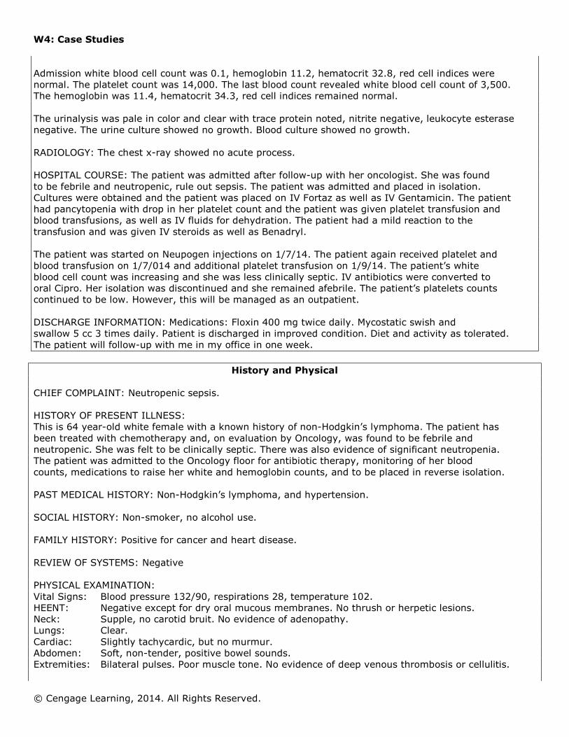
Admission white blood cell count was 0.1, hemoglobin 11.2, hematocrit 32.8, red cell indices were normal. The platelet count was 14,000. The last blood count revealed white blood cell count of 3,500. The hemoglobin was 11.4, hematocrit 34.3, red cell indices remained normal. The urinalysis was pale in color and clear with trace protein noted, nitrite negative, leukocyte esterase negative. The urine culture showed no growth. Blood culture showed no growth. RADIOLOGY: The chest x-ray showed no acute process. HOSPITAL COURSE: The patient was admitted after follow-up with her oncologist. She was found to be febrile and neutropenic, rule out sepsis. The patient was admitted and placed in isolation. Cultures were obtained and the patient was placed on IV Fortaz as well as IV Gentamicin. The patient had pancytopenia with drop in her platelet count and the patient was given platelet transfusion and blood transfusions, as well as IV fluids for dehydration. The patient had a mild reaction to the transfusion and was given IV steroids as well as Benadryl. The patient was started on Neupogen injections on 1/7/14. The patient again received platelet and blood transfusion on 1/7/014 and additional platelet transfusion on 1/9/14. The patient’s white blood cell count was increasing and she was less clinically septic. IV antibiotics were converted to oral Cipro. Her isolation was discontinued and she remained afebrile. The patient’s platelets counts continued to be low. However, this will be managed as an outpatient. DISCHARGE INFORMATION: Medications: Floxin 400 mg twice daily. Mycostatic swish and swallow 5 cc 3 times daily. Patient is discharged in improved condition. Diet and activity as tolerated. The patient will follow-up with me in my office in one week.
History and Physical CHIEF COMPLAINT: Neutropenic sepsis. HISTORY OF PRESENT ILLNESS: This is 64 year-old white female with a known history of non-Hodgkin’s lymphoma. The patient has been treated with chemotherapy and, on evaluation by Oncology, was found to be febrile and neutropenic. She was felt to be clinically septic. There was also evidence of significant neutropenia. The patient was admitted to the Oncology floor for antibiotic therapy, monitoring of her blood counts, medications to raise her white and hemoglobin counts, and to be placed in reverse isolation. PAST MEDICAL HISTORY: Non-Hodgkin’s lymphoma, and hypertension. SOCIAL HISTORY: Non-smoker, no alcohol use. FAMILY HISTORY: Positive for cancer and heart disease. REVIEW OF SYSTEMS: Negative PHYSICAL EXAMINATION: Vital Signs: Blood pressure 132/90, respirations 28, temperature 102. HEENT: Negative except for dry oral mucous membranes. No thrush or herpetic lesions. Neck: Supple, no carotid bruit. No evidence of adenopathy. Lungs: Clear. Cardiac: Slightly tachycardic, but no murmur. Abdomen: Soft, non-tender, positive bowel sounds. Extremities: Bilateral pulses. Poor muscle tone. No evidence of deep venous thrombosis or cellulitis.
W4: Case Studies
© Cengage Learning, 2014. All Rights Reserved.
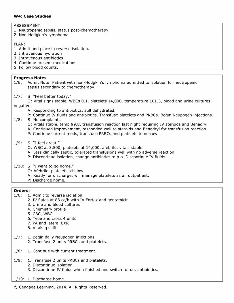
ASSESSMENT: 1. Neutropenic sepsis, status post-chemotherapy 2. Non-Hodgkin’s lymphoma PLAN: 1. Admit and place in reverse isolation. 2. Intravenous hydration 3. Intravenous antibiotics 4. Continue present medications. 5. Follow blood counts. Progress Notes 1/6: Admit Note: Patient with non-Hodgkin’s lymphoma admitted to isolation for neutropenic
sepsis secondary to chemotherapy.
1/7: S: “Feel better today.” O: Vital signs stable, WBCs 0.1, platelets 14,000, temperature 101.3, blood and urine cultures
negative A: Responding to antibiotics, still dehydrated. P: Continue IV fluids and antibiotics. Transfuse platelets and PRBCs. Begin Neupogen injections.
1/8: S: No complaints O: Vitals stable, temp 99.8, transfusion reaction last night requiring IV steroids and Benadryl A: Continued improvement, responded well to steroids and Benadryl for transfusion reaction. P: Continue current meds, transfuse PRBCs and platelets tomorrow.
1/9: S: “I feel great.” O: WBC at 3,500, platelets at 14,000, afebrile, vitals stable A: Less clinically septic, tolerated transfusions well with no adverse reaction. P: Discontinue isolation, change antibiotics to p.o. Discontinue IV fluids.
1/10: S: “I want to go home.” O: Afebrile, platelets still low A: Ready for discharge, will manage platelets as an outpatient. P: Discharge home.
Orders: 1/6: 1. Admit to reverse isolation.
2. IV fluids at 83 cc/h with IV Fortaz and gentamicin 3. Urine and blood cultures 4. Chemistry profile 5. CBC, WBC 6. Type and cross 4 units 7. PA and lateral CXR 8. Vitals q shift
1/7: 1. Begin daily Neupogen injections. 2. Transfuse 2 units PRBCs and platelets.
1/8: 1. Continue with current treatment. 1/9: 1. Transfuse 2 units PRBCs and platelets.
2. Discontinue isolation. 3. Discontinue IV fluids when finished and switch to p.o. antibiotics.
1/10: 1. Discharge home.

W4: Case Studies
© Cengage Learning, 2014. All Rights Reserved.
Code only for the Evaluation & Management/CPT code. Procedures CPT Codes Modifiers
(if applicable) PR1 Inpatient Consultation 9 9 2 2 1 PR2 Subsequent Hospital Care 9 9 2 3 2 PR3 Subsequent Hospital Care 9 9 2 3 2 PR4 Subsequent Hospital Care 9 9 2 3 1 PR5 Discharge Services
9 9 2 3 8
Case Study 3 Inpatient Face Sheet
Patient Name: Jasmine Delaware Admit Date: 1/11/14 Discharge Date: 1/15/14 Sex: Female Age: 50 Disposition: Home Admit Diagnoses: 1. Right upper lobe lesion 2. Asthmatic bronchitis 3. Depression Discharge Diagnoses: 1. Non-small cell carcinoma right upper lobe 2. Metastasized to hilar and thoracic lymph nodes 3. Chronic obstructive pulmonary disease 4. Depression Procedures: 1. Flexible bronchoscopy 2. Right upper lobe lobectomy
Discharge Summary
Admitted: 1/11/14 Discharged: 1/15/14 DISCHARGE DIAGNOSES: 1. Non-small-cell carcinoma right upper lobe of lung with metastasis to hilar and thoracic lymph nodes 2. Chronic obstructive pulmonary disease 3. Depression PROCEDURES PERFORMED: 1. Flexible bronchoscopy 2. Right upper lobe lobectomy HISTORY OF PRESENT ILLNESS:

W4: Case Studies
© Cengage Learning, 2014. All Rights Reserved.
This is a 50-year-old female with a 3 cm lesion in the right upper lobe. She had an episode of bronchitis in January. Subsequent chest x-ray revealed a lesion in the right upper lobe. A CAT scan of the chest was performed and the presence of the lesion in the right upper lobe was confirmed. HOSPITAL COURSE: The patient underwent flexible bronchoscopy with right upper lobectomy on January 11, 2014. The findings were a 3-cm lesion in the right upper lobe with metastasis to the lymph nodes. The patient tolerated the procedure well. Vital signs remained stable. There was minimal chest tube drainage. She was advanced to a regular diet the second post-operative day. LABORATORY DATA: Routine laboratory work on admission showed a potassium of 4.0, BUN 10, creatinine 0.6. WBCs 8.8, hemoglobin 13.7 and hematocrit 38.2. Platelet count 288,000. The urinalysis was negative. PT was 10.1. Discharge laboratory was unchanged with the exception of BUN 12, creatinine 0.9. Hemoglobin 11.3 and hematocrit 34.2 EKG: Sinus rhythm IMAGING: The pre-operative chest x-ray showed a 3.0 cm suspicious nodule in the right upper lobe with chronic obstructive pulmonary disease. Post-operative chest x-ray showed good expansion of the right middle and lower lobes. The patient was discharged on the fourth post-operative day in satisfactory condition. Regular diet as tolerated. She is to limit activity for the next 3 weeks. She will follow-up in my office in 1 week. Discharge medications include: Vicodin 1 tablet P.O. q4h prn for pain. Elavil 150 mg h.s., Ventolin 2 puffs q.i.d.
History and Physical
CHIEF COMPLAINT: Right upper lobe lesion. HISTORY OF PRESENT ILLNESS: This is a 50-year-old female with a 3 cm lesion in the right upper lobe. She had an episode of bronchitis in January. Subsequent chest x-ray revealed a lesion in the right upper lobe. A CAT scan of the chest was performed and the presence of the lesion in the right upper lobe was confirmed. The patient is admitted at this time for a bronchoscopy and right thoracotomy. REVIEW OF SYSTEMS: Patient denies hematemesis, melena, and angina pectoris. There are no complaints of syncope, claudication, or edema. HEENT: No masses, pupils equal, round, reactive to light. No oral cavity lesions. No evidence of JVD; thyroid is not enlarged. No carotid bruits. Chest: Symmetrical. Lungs: Clear to auscultation and percussion. No wheezing. Heart: No murmurs, no gallops, regular rhythm. Abdomen: No masses, no organomegaly. Extremities: No cyanosis, clubbing, or edema. Good peripheral pulses. PAST MEDICAL HISTORY: She has asthmatic bronchitis and has been hospitalized twice in the past for bronchitis. Patient is currently treated for depression. She has no history of diabetes mellitus, hypertension, myocardial infarction, or neurological deficits. She has had no surgeries. MEDICATIONS: Elavil 150 mg h.s., Ventolin 2 puffs q.i.d.

W4: Case Studies
© Cengage Learning, 2014. All Rights Reserved.
ALLERGIES: None known SOCIAL HISTORY: She smokes two packs of cigarettes per day and has smoked for 30 years. FAMILY HISTORY: Non-contributory IMPRESSION: 1. Right upper lobe lesion; 2. Rule out bronchogenic carcinoma; 3. Rule out benign lesion; 4. Asthmatic bronchitis; 5. Depression PLAN: Patient is admitted for bronchoscopy and right thoracotomy with right upper lobectomy. The procedures and the risks involved were fully explained to the patient and all questions answered, and an informed consent was signed by the patient.
Operative Report
DATE OF OPERATION: 1/11/14 PREOPERATIVE DIAGNOSIS: Right Upper Lobe Lesion POSTOPERATIVE DIAGNOSIS: Carcinoma of the right upper lobe with metastasis to hilar and thoracic lymph nodes SURGEON: Heather Schoonover, M.D. OPERATIVE PROCEDURE: 1. Flexible bronchoscopy; 2. Right upper lobe lobectomy INDICATIONS: Female patient with a 3-cm lesion centrally located in the right upper lobe. FINDINGS: The bronchoscopy was negative. On thoracotomy there was a 3-cm lesion centrally located in the right upper lobe. There were positive nodes in the hilar and thoracic lymph nodes. DESCRIPTION OF PROCEDURE: Under general anesthesia the flexible bronchoscope was introduced through both lumen of the endotracheal tube. The carina was normal. Both the right and left bronchial trees were visualized down to the subsegmental level. There was no evidence of endobronchial lesions. The bronchoscopy was negative. After prepping and draping the operative area a right posterolateral thoracotomy was made. The incision was deepened through the skin, subcutaneous tissue, and latissimus dorsi muscle. The serratus anterior muscles were retracted anteriorly and the chest was entered through the fifth intercostal space. On exploration of the right lung there was a 3-cm lesion centrally located in the right upper lobe. A right total lobectomy was performed based upon the above findings. Surrounding lymph nodes were inspected and biopsies were obtained from both the hilar and surrounding thoracic nodes. Frozen section was positive for non-small cell carcinoma. The bronchial resection margin was negative for tumor. The inferior pulmonary ligament was taken all of the way up to the inferior pulmonary vein. The bronchial stump was checked up to a pressure of 35 mmHg and there was no air leak. Hemostasis was again secured. A chest tube was placed through a separate stab wound and secured to the skin with 0 silk. The incision was closed using #2 Vicryl pericostal sutures, #1 Vicryl for the latissimus dorsi muscle, 2-0 Vicryl for the subcutaneous tissue, and staples for the skin. The estimated blood loss was less than 200 cc. The patient tolerated the procedure very well and was taken to the recovery room in good condition with stable vital signs. PROGRESS NOTES:

W4: Case Studies
© Cengage Learning, 2014. All Rights Reserved.
1/11: Admit Note: A 50-year-old female found to have a 3 cm lesion in the right upper lobe. She is admitted at this time for flexible bronchoscopy and right thoracotomy.
1/12: S: Complains of incisional pain
O: Vital signs stable, labs within normal limits, minimal chest tube drainage A: Post-op CXR shows good expansion of right middle and lower lobes. P: Patient doing well from surgical standpoint, will remove chest tube in a.m.
1/13: S: Less pain, depressed with diagnosis
O: Vital remain stable, afebrile, good lung sounds A: Progressing nicely. P: Advance to full diet, increase ambulation.
1/14: S: Feels better today.
O: Afebrile, vital signs stable, labs look good A: Incisions clean and dry with no redness P: Possible discharge tomorrow
1/15: S: Ready to go home.
O: Discharge labs and CXR within normal limits A: Incisions healing well. P: Discharge patient.
ORDERS: 1/11: 1. Admit patient.
2. Have consents signed for flexible bronchoscopy and right thoracotomy. 3. Place pre-op diagnostics on chart.
1/12: 1. Ambulate patient.
2. Repeat CXR. 3. Repeat labs.
1/13: 1. Advance to full diet.
2. Increase ambulation. 1/14: 1. No new orders 1/15: 1. Discharge patient. Code only for CPT and modifiers. Procedures CPT Codes Modifiers
(if applicable) PR1 Initial Hospital Care 9 9 2 2 1 PR2 Subsequent Hosp Care 9 9 2 3 2 PR3 Subsequent Hosp Care 9 9 2 3 2 PR4 Subsequent Hosp Care 9 9 2 3 1 PR5 Discharge Services 9 9 2 3 8
Case Study 4 Inpatient Face Sheet
Patient Name: Ronda Parker

W4: Case Studies
© Cengage Learning, 2014. All Rights Reserved.
Admit Date: 1/14/14 Discharge Date: 1/15/14 Sex: Female Age: 24 Disposition: Home Admitting Diagnosis: Pre-term Labor Discharge Diagnosis: Pre-term Labor
Labor and Delivery History and Physical
History: Patient is a 24-year-old female, gravida 2, para 0, Abortus 0, who had her prenatal care at the Women’s Clinic. She presented to labor and delivery with the complaint of abdominal pain and cramps. Her membrane is intact. LMP: 05/20/. EGA of 31.4 weeks. Ultrasound at 6 weeks. No complications during this pregnancy. PAST MEDICAL HISTORY: Non-contributory PAST SURGICAL HISTORY: None MEDINCE: None during pregnancy ALLERGIES: No known allergies PRE-NATAL LABS: Rh +, Rubella BL, VDRL NR, GC -, Chlamydia -, Pap -, AB Screen 0, Hepatitis Screen -, Diabetic Screen 153 PHYSICAL EXAM: Vital signs: BP 135/82, Temp. 98.4, Pulse 102, Resp. 21, FHTs 145 General: No acute diseases HEENT: No asymmetry Neck: No asymmetry Heart: Regular rate and rhythm Lungs: Clear to auscultation bilaterally Abdomen: Soft, non-tender, non-distended, + bowel sounds Extremities: No edema Neuro: No deficits Cervix 1/th Presenting Part VTX/FFN + Impression: 24-year-old G2P000 at 31.4 weeks with PTL. Plan: 1. Admit
2. U/S History: Detailed Examination: Detailed Medical Decision: Low complexity Progress Notes: 1/14: Admit note: Patient admitted to labor and delivery with pre-term labor at 31.4 weeks’
gestation. Patient immediately started on MgSo4. S: Patient denies HA/CP/SOB/CTX/RUQ pain.
W4: Case Studies
© Cengage Learning, 2014. All Rights Reserved.
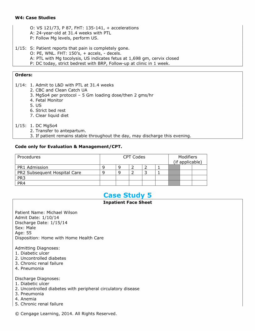
O: VS 121/73, P 87, FHT: 135-141, + accelerations A: 24-year-old at 31.4 weeks with PTL P: Follow Mg levels, perform US.
1/15: S: Patient reports that pain is completely gone.
O: PE, WNL. FHT: 150’s, + accels, - decels. A: PTL with Mg tocolysis, US indicates fetus at 1,698 gm, cervix closed P: DC today, strict bedrest with BRP, Follow-up at clinic in 1 week.
Orders: 1/14: 1. Admit to L&D with PTL at 31.4 weeks
2. CBC and Clean Catch UA 3. MgSo4 per protocol – 5 Gm loading dose/then 2 gms/hr 4. Fetal Monitor 5. US 6. Strict bed rest 7. Clear liquid diet
1/15: 1. DC MgSo4
2. Transfer to antepartum. 3. If patient remains stable throughout the day, may discharge this evening.
Code only for Evaluation & Management/CPT. Procedures CPT Codes Modifiers
(if applicable) PR1 Admission 9 9 2 2 1 PR2 Subsequent Hospital Care 9 9 2 3 1 PR3 PR4
Case Study 5 Inpatient Face Sheet
Patient Name: Michael Wilson Admit Date: 1/10/14 Discharge Date: 1/15/14 Sex: Male Age: 55 Disposition: Home with Home Health Care Admitting Diagnoses: 1. Diabetic ulcer 2. Uncontrolled diabetes 3. Chronic renal failure 4. Pneumonia Discharge Diagnoses: 1. Diabetic ulcer 2. Uncontrolled diabetes with peripheral circulatory disease 3. Pneumonia 4. Anemia 5. Chronic renal failure

W4: Case Studies
© Cengage Learning, 2014. All Rights Reserved.
Procedure: 1. Excisional debridement of decubitus ulcer
Discharge summary
Admitted: 1/10/14 Discharged: 1/15/14 ADMITTING DIAGNOSIS: 1. Diabetic ulcer 2. Uncontrolled diabetes 3. Chronic renal failure 4. Pneumonia DISCHARGE DIAGNOSIS: 1. Diabetic ulcer 2. Uncontrolled diabetes with peripheral circulatory disease 3. Pneumonia 4. Anemia 5. Chronic renal failure PROCEDURE: 1. Excisional debridement of decubitus ulcer HISTORY: This is a 55-year-old male who was admitted through the emergency room for elevated blood sugars, a necrotic heel ulcer of the left foot. The patient was admitted for control of his blood sugars and treatment of the heel ulcer. PAST MEDICAL HISTORY: This patient has a long history of type 1 diabetes, chronic renal failure, coronary artery disease with history of CABG, peripheral vascular disease with subsequent below knee amputation of the right leg. HOSPITAL COURSE: The patient was admitted to the hospital and started on intravenous antibiotic therapy. The patient was placed on sliding scale insulin therapy as well as wound care for the heel necrosis. The patient’s left heel ulcer was debrided of all necrotic tissue on 1/11/14. There was no cellulitis of the foot; however, there were multiple areas of skin breakdown on the foot. The patient had no feeling in his left foot, secondary to severe diabetic neuropathy. The patient was continued on local wound care and antibiotic therapy. The patient’s renal failure was monitored and fluids restricted. The patient’s chest x-ray was positive for left lower lobe pneumonia. Sputum culture was not ordered as the patient was already on intravenous antibiotic therapy for the skin ulcer. LABORATORY DATA: Hemoglobin 9.4, hematocrit 29.6, WBC 8,600, platelet count 336,000. Urinalysis was normal except for a small amount of bacteria, and proteinuria. Sodium was 130, potassium 4.1, chloride 92, CO2 32, glucose 270, BUN 53, and creatinine 3.2. DISCHARGE MEDICATIONS: 70/30 insulin in the morning and 20 units in the evening Cardizem CD 180 mg once daily Imdur 60 mg once a day Lasix 80 mg once a day
W4: Case Studies
© Cengage Learning, 2014. All Rights Reserved.
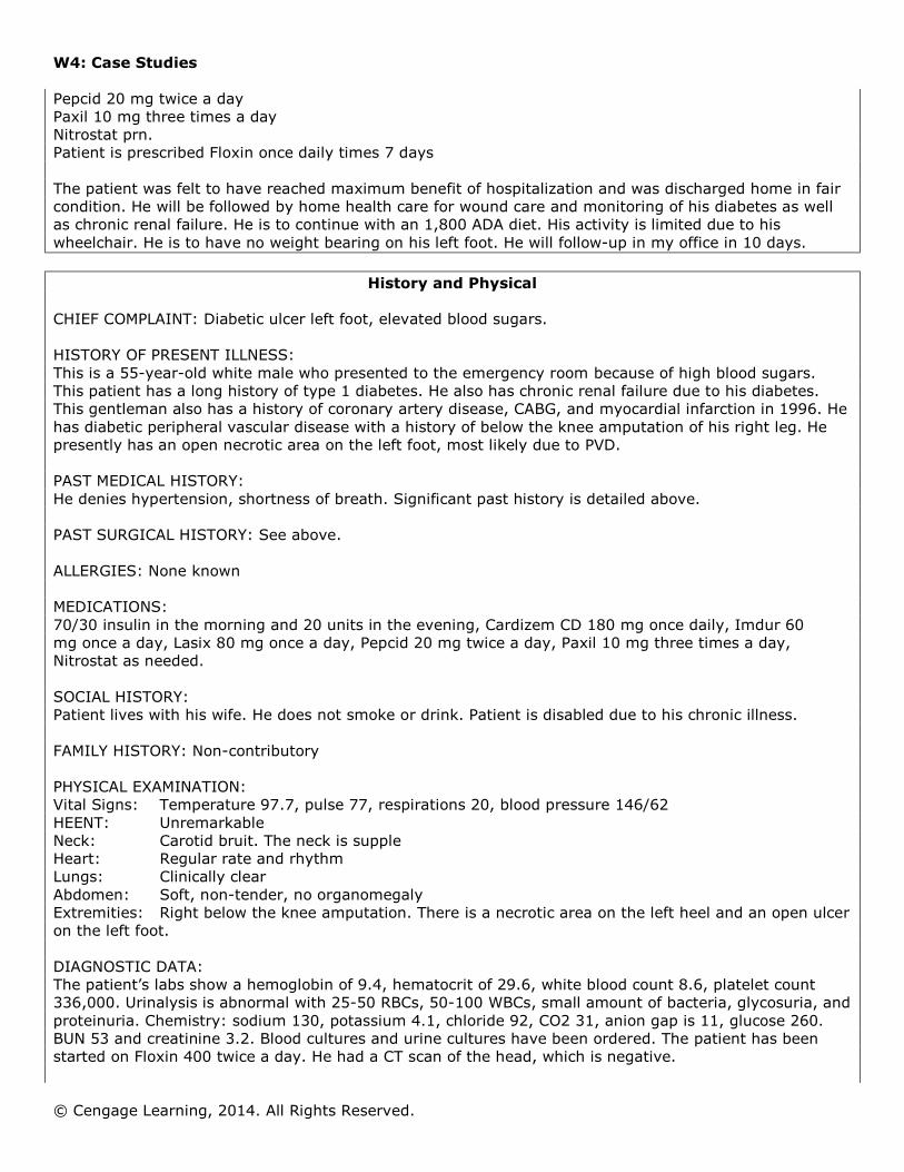
Pepcid 20 mg twice a day Paxil 10 mg three times a day Nitrostat prn. Patient is prescribed Floxin once daily times 7 days The patient was felt to have reached maximum benefit of hospitalization and was discharged home in fair condition. He will be followed by home health care for wound care and monitoring of his diabetes as well as chronic renal failure. He is to continue with an 1,800 ADA diet. His activity is limited due to his wheelchair. He is to have no weight bearing on his left foot. He will follow-up in my office in 10 days.
History and Physical
CHIEF COMPLAINT: Diabetic ulcer left foot, elevated blood sugars. HISTORY OF PRESENT ILLNESS: This is a 55-year-old white male who presented to the emergency room because of high blood sugars. This patient has a long history of type 1 diabetes. He also has chronic renal failure due to his diabetes. This gentleman also has a history of coronary artery disease, CABG, and myocardial infarction in 1996. He has diabetic peripheral vascular disease with a history of below the knee amputation of his right leg. He presently has an open necrotic area on the left foot, most likely due to PVD. PAST MEDICAL HISTORY: He denies hypertension, shortness of breath. Significant past history is detailed above. PAST SURGICAL HISTORY: See above. ALLERGIES: None known MEDICATIONS: 70/30 insulin in the morning and 20 units in the evening, Cardizem CD 180 mg once daily, Imdur 60 mg once a day, Lasix 80 mg once a day, Pepcid 20 mg twice a day, Paxil 10 mg three times a day, Nitrostat as needed. SOCIAL HISTORY: Patient lives with his wife. He does not smoke or drink. Patient is disabled due to his chronic illness. FAMILY HISTORY: Non-contributory PHYSICAL EXAMINATION: Vital Signs: Temperature 97.7, pulse 77, respirations 20, blood pressure 146/62 HEENT: Unremarkable Neck: Carotid bruit. The neck is supple Heart: Regular rate and rhythm Lungs: Clinically clear Abdomen: Soft, non-tender, no organomegaly Extremities: Right below the knee amputation. There is a necrotic area on the left heel and an open ulcer on the left foot. DIAGNOSTIC DATA: The patient’s labs show a hemoglobin of 9.4, hematocrit of 29.6, white blood count 8.6, platelet count 336,000. Urinalysis is abnormal with 25-50 RBCs, 50-100 WBCs, small amount of bacteria, glycosuria, and proteinuria. Chemistry: sodium 130, potassium 4.1, chloride 92, CO2 31, anion gap is 11, glucose 260. BUN 53 and creatinine 3.2. Blood cultures and urine cultures have been ordered. The patient has been started on Floxin 400 twice a day. He had a CT scan of the head, which is negative.
W4: Case Studies
© Cengage Learning, 2014. All Rights Reserved.
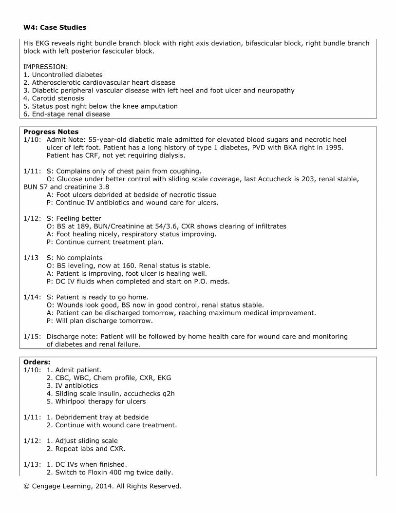
His EKG reveals right bundle branch block with right axis deviation, bifascicular block, right bundle branch block with left posterior fascicular block. IMPRESSION: 1. Uncontrolled diabetes 2. Atherosclerotic cardiovascular heart disease 3. Diabetic peripheral vascular disease with left heel and foot ulcer and neuropathy 4. Carotid stenosis 5. Status post right below the knee amputation 6. End-stage renal disease Progress Notes 1/10: Admit Note: 55-year-old diabetic male admitted for elevated blood sugars and necrotic heel
ulcer of left foot. Patient has a long history of type 1 diabetes, PVD with BKA right in 1995. Patient has CRF, not yet requiring dialysis.
1/11: S: Complains only of chest pain from coughing.
O: Glucose under better control with sliding scale coverage, last Accucheck is 203, renal stable, BUN 57 and creatinine 3.8
A: Foot ulcers debrided at bedside of necrotic tissue P: Continue IV antibiotics and wound care for ulcers.
1/12: S: Feeling better
O: BS at 189, BUN/Creatinine at 54/3.6, CXR shows clearing of infiltrates A: Foot healing nicely, respiratory status improving. P: Continue current treatment plan.
1/13 S: No complaints
O: BS leveling, now at 160. Renal status is stable. A: Patient is improving, foot ulcer is healing well. P: DC IV fluids when completed and start on P.O. meds.
1/14: S: Patient is ready to go home.
O: Wounds look good, BS now in good control, renal status stable. A: Patient can be discharged tomorrow, reaching maximum medical improvement. P: Will plan discharge tomorrow.
1/15: Discharge note: Patient will be followed by home health care for wound care and monitoring
of diabetes and renal failure.
Orders: 1/10: 1. Admit patient.
2. CBC, WBC, Chem profile, CXR, EKG 3. IV antibiotics 4. Sliding scale insulin, accuchecks q2h 5. Whirlpool therapy for ulcers
1/11: 1. Debridement tray at bedside
2. Continue with wound care treatment. 1/12: 1. Adjust sliding scale
2. Repeat labs and CXR. 1/13: 1. DC IVs when finished.
2. Switch to Floxin 400 mg twice daily.

W4: Case Studies
© Cengage Learning, 2014. All Rights Reserved.
1/14: 1. Repeat labs. 1/15: 1. Discharge home with home health care Code only for CPT. Procedures CPT Codes Modifiers
(if applicable) PR1 Hospital Admission 9 9 2 2 1 PR2 Subsequent Hospital Care 9 9 2 3 2 2 5 PR3 Debridement of Decubitis Ulcer 1 1 0 4 2 PR4 Subsequent Hospital Care 9 9 2 3 1 PR5 Subsequent Hospital Care 9 9 2 3 1 PR6 Subsequent Hospital Care 9 9 2 3 1 PR7 Discharge Services 9 9 2 3 8
Case Study 6 Inpatient Face Sheet
Patient Name: Maureen Pranger Admit Date: 1/07/14 Discharge Date: 1/10/14 Sex: Female Age: 72 Disposition: Home Admitting Diagnosis: 1. Chest pain, rule out acute coronary artery disease Discharge Diagnoses: 1. Coronary Artery Disease 2. Unstable Angina 3. Atrial Fibrillation 4. Secondary Degree AV Block 5. Status Post Percutaneous Transluminal Coronary Angioplasty Procedures: 1. Diagnostic left heart catheterization, percutaneous transluminal coronary angioplasty, coronary Angiograms
Discharge Summary
Admitted: 1/07/14 Discharged: 1/10/14 ADMITTING DIAGNOSES 1. Chest pain, rule out acute coronary artery disease DISCHARGE DIAGNOSES: 1. Chest pain, acute intermediate coronary syndrome 2. History of arteriosclerosis of the native coronary vessels 3. Atrial fibrillation

W4: Case Studies
© Cengage Learning, 2014. All Rights Reserved.
4. Second degree atrioventricular block PROCEDURE PERFORMED: 1. Left heart catheterization, selective coronary angiography, percutaneous transluminal coronary angioplasty. HOSPITAL COURSE: This is a 72-year-old female with a history of coronary artery disease, status post percutaneous transluminal coronary angioplasty several months ago. She came in with acute intermediate coronary syndrome. A myocardial infarction was ruled out. In view of these events, it was decided to perform a diagnostic heart catheterization and possible percutaneous transluminal coronary angioplasty versus bypass surgery. A diagnostic heart catheterization was performed and showed the following: the left main coronary artery was open; the left anterior descending artery was open. There was a previous stent in the circumflex system, which had no obstruction. There was a totally occluded distal right coronary artery. There was some collateral circulation filling the right coronary artery. In view of this, it was felt that the patient would benefit from percutaneous transluminal coronary angioplasty, so the patient received IV Heparin, ReoPro and intracoronary nitroglycerin and we were able to open the distal right coronary artery with balloon angioplasty. The patient began ambulation the day after the above procedure. The patient is stable at discharge. Discharge to home on Atenolol 25 mg once a day, Monopril 10 mg once a day, and aspirin. She will follow a low-cholesterol, low-fat diet. She is to follow-up in my office in 1 week.
History and Physical
CHIEF COMPLAINT: Chest pain. HISTORY OF PRESENT ILLNESS: This is a 72-year-old white female with a history of coronary artery disease, status post percutaneous transluminal coronary angioplasty and stent implantation last summer, who has been taking Tenormin 50 mg once a day, Monopril 10 mg once a day, one aspirin a day, and nitroglycerin prn. She experienced an episode of palpitation and lightheadedness last night, and this morning she started having chest pain. The patient called 911 and the EMS staff found her with a very fast rhythm, heart rate of 160 per minute accompanied with atrial fibrillation. She denied any prior history of palpitations and denies chest pains prior to this episode. The patient denies diabetes mellitus or high blood pressure. She denies history of myocardial infarction in the past. She has a strong family history of coronary artery disease. She denies alcohol or smoking. PAST MEDICAL HISTORY: The past history is only pertinent for coronary artery disease and low HDL, post-menopausal. PAST SURGICAL HISTORY: History of back surgery ALLERGIES: Sulfa REVIEW OF SYSTEMS: See history of present illness. Patient denies paroxysmal nocturnal dyspnea, orthopnea, leg swelling, fatigue, or loss of consciousness. PHYSICAL EXAMINATION: General Appearance: Patient is alert, cooperative, and in no acute distress. Vital Signs: Her heart rhythm is in regular. Telemetry shows a second-degree atrioventricular block, Mobitz type I, with beats 2 and 4, 2:3 conduction. No electrocardiogram evidence of ischemia. Head and Neck: Neck is supple. No jugular venous distention, no carotid bruits.
W4: Case Studies
© Cengage Learning, 2014. All Rights Reserved.
Lungs: Clear Heart: Irregular rate and rhythm. No murmur, gallop or rub. Abdomen: Soft, non-tender. Bowel sounds present. No tenderness on rebound. Extremities: No cyanosis or edema. Bilateral peripheral pulses. ASSESSMENT: 1.Prolonged chest pain, rule out acute coronary artery disease 2.Mobitz type I second degree atrioventricular block 3.Coronary artery disease, status post stent implantation eight months ago PLAN: 1. Admit to telemetry. Obtain cardiac enzymes and serial electrocardiograms. 2. Hold Tenormin and place on Norvasc and nitrates. 3. Cardiac catheterization and electrophysiology study
Procedure Note
Date: 1/07/14 Procedure: Left Heart Catheterization; Selective Coronary Angiography; PTCA of Distal Right Coronary Artery Pre-Operative Diagnosis: Unstable angina r/o CAD Post-Operative Diagnosis: CAD Surgeon: Anthony C. Stamper, MD Procedure Note: A diagnostic left heart catheterization was performed and showed the following: the left main coronary artery was open. The left anterior descending artery was open. There was a previous stent in the circumflex system, which had no obstruction. There was a totally occluded distal right coronary artery. There was some collateral circulation filling the right coronary artery. In view of this, it was felt that the patient would benefit from percutaneous transluminal coronary angioplasty, so the patient received IV Heparin, ReoPro, and intracoronary nitroglycerin and we were able to open the distal right coronary artery with balloon angioplasty. There was no clot formation or dissection. The patient returned to the floor in stable condition. Progress Notes 1/07: S: Chest pain
O: EKG and enzymes negative for AMI A: Unstable angina, probable coronary occlusion P: Heart catheterization and possible PTCA
1/09 S: Patient feels great, no chest pain.
O: Labs within normal limits, EKG stable; patient ambulating. A: CAD P: Observe a few more days.
1/09 S: No chest pain, wants to go home.
O: Vitals stable, heart sounds good, regular rhythm A: Patient is stable, continues to do well. P: Plan discharge for a.m.
1/10: S: “Never felt better.”
O: CXR clear, labs and vitals within normal limits
W4: Case Studies
© Cengage Learning, 2014. All Rights Reserved.
A: Patient stable for discharge. P: Discharge now.
Orders 1/07: 1. Admit patient to telemetry.
2. Vitals q4h. 3. Prepare for Left Heart Catheterization and possible PTCA 4. Continue home medications. 5. Serial EKGs 6. Cardiac enzymes 7. Cardiac diet 8. Hold Tenormin; place on Norvasc and IV nitrates.
1/09: 1. Ambulate. 1/09 1. Discontinue all IVs. 1/10: 1: Discharge patient. Code only for CPT. Procedures CPT Codes Modifiers
(if applicable) PR1 Initial Hospital Care 9 9 2 2 1 PR2 Subsequent Hospital Care 9 9 2 3 1 PR3 Subsequent Hospital Care 9 9 2 3 1 PR4 Discharge Services 9 9 2 3 8
Case Study 7 Inpatient Face Sheet
Patient Name: Joleen Barker Admitted: 1/14/14 Discharged: 1/17/14 Sex: Female Age: 35 Disposition: Home Admitting Diagnosis: 1. Ovarian cyst Discharge Diagnoses: 1. Ovarian cyst 2. Post-operative blood loss 3. Urinary tract infection Procedures: 1. Total abdominal hysterectomy 2. Bilateral salpingo-oophorectomy 3. Exploratory laparotomy
Discharge Summary
Admitted: 1/14/14
W4: Case Studies
© Cengage Learning, 2014. All Rights Reserved.
Discharged: 1/17/14 DISCHARGE DIAGNOSES: 1. Ovarian cyst 2. Uterine adhesions 3. Urinary tract infection 4. Post-operative anemia PROCEDURES PERFORMED: 1. Exploratory laparotomy 2. Hysterectomy 3. Bilateral salpingo-oophorectomy HISTORY: This 35-year-old female has experienced pelvic pain for 3 months duration. Pelvic ultrasound revealed a right ovarian cystic mass. She was admitted for elective surgery. PAST MEDICAL HISTORY: Refer to History and Physical for complete history. HOSPITAL COURSE: Patient underwent exploratory laparotomy that revealed a benign cyst of the right ovary and adhesions of the uterus requiring hysterectomy and bilateral salpingo-oophorectomy. She tolerated the surgery without complication. Post-operative course was significant for post-operative anemia, requiring transfusion of PRBCs. She also was noted to have elevated temperature, and cultures indicated a urinary tract infection. She was already on IV antibiotics post-surgery that would also cover the UTI. She progressed well, in spite of the anemia and the UTI, and was discharged in good condition on 1/17/14. She was given a prescription for antibiotics and iron pills. Activity as tolerated; however, no driving for 2 weeks. Diet as tolerated. She is to see me in 3 days for post-operative follow-up and staple removal.
History and Physical
CHIEF COMPLAINT: Pelvic pain HISTORY OF PRESENT ILLNESS: This 35-year-old gravida 3, para 3, has complained of pelvic pain for 3 months duration. Pelvic ultrasound revealed her right ovary to be enlarged with a large cystic mass. She is admitted today for elective exploratory laparotomy. The patient understands that she may require a hysterectomy and possible salpingo-oophorectomy. PAST MEDICAL HISTORY: Negative, except for childbirth. PAST SURGICAL HISTORY: No surgeries. SOCIAL HISTORY: Non-smoker, non-drinker FAMILY HISTORY: Non-contributory. ALLERGIES: Penicillin MEDICATIONS: None REVIEW OF SYSTEMS: HEENT: Within normal limits

W4: Case Studies
© Cengage Learning, 2014. All Rights Reserved.
Neck: Supple. No lymphadenopathy. Heart: Regular rhythm, no murmurs Lungs: Clear to auscultation Abdomen: Soft. Good bowel sounds. Pelvic pain on palpation. Rectal: Deferred Extremities: Within normal limits. IMPRESSION: 1. Ovarian cyst PLAN: 1. Exploratory laparotomy Procedure Note Date of Procedure: 1/14/14 Preoperative Diagnosis: Ovarian cyst, right Postoperative Diagnosis: Right ovarian cyst Surgeon: Dr. Hollie Schoonover Anesthesiologist: Dr. Layne Allen Procedure: Exploratory laparotomy Hysterectomy, Bilateral salpingo-oophorectomy DESCRIPTION OF PROCEDURE: Following administration of general anesthesia, the patient’s abdomen was prepped and draped in sterile manner. A midline incision was done below the umbilicus to the pubis symphysis and then taken down to the fascia. The muscles were separated and the peritoneum was cut, taking care to avoid the bladder and bowel. There was a large cystic mass on the right ovary measuring 6 x 8 centimeters. The right ovary and tube with the cyst intact were removed. The left tube and ovary were removed in similar fashion. The decision was made to perform a hysterectomy and this was carried out without complications. All bleeders were ligated using 0-Vicryl. The pelvic cavity was irrigated with saline. The abdomen was then closed using 2-0 Dexon, and the skin was closed using staples. The patient tolerated the procedure and was discharged to the recovery room in good condition. Estimated blood loss: 400 cc Pathology Report Specimens:
1. Uterus 2. Left ovary and fallopian tube 3. Right ovary and fallopian tube with cystic mass
Microscopic Diagnosis: 1. Uterus: Mild chronic cervicitis 2. Left fallopian tube and ovary: Atrophic ovary with normal fallopian tube 3. Right fallopian tube and ovary: Normal right tube and benign cyst of the ovary

W4: Case Studies
© Cengage Learning, 2014. All Rights Reserved.
Progress Notes: 1/14: Patient admitted to surgical floor following exploratory laparotomy, hysterectomy, and bilateral
salpingo-oophorectomy. Patient tolerated the procedure well. Is fully awake and complains of moderate surgical pain. Patient will remain NPO until the evening meal and then will have a soft diet. Ambulate upon full recovery from anesthesia.
1/14: S: Moderate pain
O: Incision clean and dry, vital signs good. H/H 9.8/35.4. Was 12.9/42.3 on admission. Temp elevated to 101.2.
A: Probable post-op anemia P: Monitor H/H, continue with present pain medication.
1/15: S: Feels okay.
O: Incision clean and dry, vitals good. H/H dropped to 8.10/32.2. Temp still elevated, 100.1. A: Possible occult infection P: Will culture to r/o infection. Continue to monitor H/H.
1/16: S: Complains of minor pain at operative site.
O: Vital signs good. H/H at 10.9/39.8 after transfusion. Temperature 99.9 A: Urine culture revealed UTI. Treated with antibiotics. Ambulating freely, progressing well. P: Change to p.o. antibiotics, monitor H/H, probable discharge tomorrow.
1/17: S: Feels well, ready to go home.
O: Incision healing well, vital signs good. H/H at 11.2/42.1. Temperature normal. A: Ready for discharge. P: Discharge home; follow-up in office in 1 week
Orders: 1/14: 1. Admit patient to surgical floor from recovery
2. Standard post-operative orders 3. Vital signs q 6 hours
1/15: 1. Blood and urine cultures to rule out infection
2. Transfuse two units PRBCs. 3. Continue with IV antibiotics.
1/16: 1. Change IV antibiotics to p.o.
2. Continue to monitor H/H. 1/17: 1. Discharge home. Code only for CPT. Procedures CPT Codes Modifiers
(if applicable) PR1 Salpingo-oophorectomy, bilateral 5 8 7 2 0 PR2 Subsequent Hospital Care 9 9 2 3 2 PR3 Subsequent Hospital Care 9 9 2 3 1 PR4 Discharge Service 9 9 2 3 8
Case Study 8 Emergency Room Visit

W4: Case Studies
© Cengage Learning, 2014. All Rights Reserved.
Face Sheet
Patient Name: Elizabeth Colter Date of Service: 01/01/14 Age: 25 Sex: Female Admitting Diagnosis: Injury to left wrist Discharge Diagnosis: Sprain left wrist Disposition: Home
Emergency Room Visit
DATE OF SERVICE: 1/01/14 HISTORY OF PRESENT ILLNESS: A 25-year-old female was at work today when a bread tray fell on her left wrist. She has persistent pain in the area, which is exacerbated with moving the wrist and hand. She describes the pain as very severe. PAST MEDICAL HISTORY: Non-contributory. ALLERGIES: None. PHYSICAL EXAMINATION: General: Well-developed, well-nourished female in moderate distress. Vitals are stable. Skin: Warm and dry. HEENT: Unremarkable. Chest: Symmetrical. Extremities: The left wrist is tender and mildly swollen especially over the distal ulna with no gross
deformity. Normal range of motion against resistance with moderate pain. No abrasions or lacerations. Normal distal neurosensory examination. The remainder of the extremity examination is within normal limits.
Neurological: She is awake, alert, and oriented times three with no focal neurologic deficits. RADIOLOGY EXAMINATION: The patient was taken to the x-ray room where the wrist was x-rayed. No acute fractures appreciated in the AP and lateral views. IMPRESSION: Left wrist sprain. The patient was placed in a padded splint and was given a prescription for Darvocet. She will followup with her physician next week. History: Expanded problem-focused Examination: Detailed Medical Decision Making: Low decision Code only for CPT. Procedures CPT Codes Modifiers
(if applicable) PR1 Emergency Dept. Services 9 9 2 8 2 PR2 PR3 PR4

W4: Case Studies
© Cengage Learning, 2014. All Rights Reserved.
Case Study 9 Inpatient Face Sheet
Patient Name: Sally Kramer Admit Date: 1/05/14 Discharge Date: 1/14/14 Sex: Female Age: 35 Disposition: Home Admitting Diagnoses: 1. Induction of labor 2. Severe pre-eclampsia 3. Intrauterine growth retardation Discharge Diagnoses: 1. Severe pre-eclampsia 2. Accelerated hypertension 3. Intrauterine growth retardation 4. Pre-term at 36 weeks Procedure: 1. Primary low transverse C-section
Discharge Summary
Admitted: 1/05/14 Discharged: 1/14/14 ADMITTING DIAGNOSES: 1. Induction of labor 2. Severe pre-eclampsia 3. IUGR DISCHARGE DIAGNOSES: 1. Severe pre-eclampsia 2. Accelerated hypertension 3. IUGR Patient is a 35-year-old, gravida 3, para 2, admitted for induction due to severe pre-eclampsia and late decelerations. She was admitted for induction and began on Pitocin. However, due to persistent late decelerations, patient underwent a primary low flap transverse cesarean section with delivery of a 4 pound 12 ounce liveborn male with 6, 9 Apgars. There were no post-operative problems other than accelerated hypertension, for which the patient was started on Apresoline and Aldomet. Last blood pressure reading was 180/90. Discharge hemoglobin and hematocrit were 11.3 and 32.4 and platelet count was 122. The platelet count had been as low as 94,000. The patient’s magnesium levels when she was on magnesium ranged between 4.5 and 4.0, her electrolytes were normal, alkaline phosphatase 262 and 304. The hemoglobin and hematocrit on admission were 13 and 37, platelet count was 158,000. The PT and PTT were normal. The urinalysis was negative. The patient was discharged in satisfactory condition with diet and activity as tolerated. Patient will continue to take Aldomet and Apresoline. I will see the patient in my office in 1 week for staple removal.

W4: Case Studies
© Cengage Learning, 2014. All Rights Reserved.
History and Physical
HISTORY OF PRESENT ILLNESS: The patient is a 35-year-old gravida 3, para 2, due date 1/05/14, whom I saw in my office with blood pressure of 154/104, 150/98, 2+ protein. She was immediately sent to the hospital for admission for induction of labor. ANTEPARTUM HISTORY: She has had two vaginal deliveries in the past. Her ultrasound was consistent with dates. Her group B Strep was negative. She has a history of hypertension. PAST MEDICAL HISTORY: Usual childhood diseases. FAMILY HISTORY: Non-contributory ALLERGIES: None known PHYSICAL EXAMINATION: Vital Signs: Blood pressure ranged from 154/90 to 168/104 HEENT: Within normal limits Chest: Clear to percussion and auscultation Heart: Normal sinus rhythm Breasts: Without masses or discharge Abdomen: Gravid Extremities: Without clubbing or cyanosis. There was +1 edema. Reflexes were +2. Cervical: Cervical examination revealed she was 1 cm. She was admitted and Pitocin was started. Decreased variability and occasional late decelerations were noted. The Pitocin was once again stopped. She began having contractions on her own with recurrent late decelerations. Oxygen was started. She was turned on her side and the late decels continued. Magnesium was started at 6 g and then 1 g an hour for the pre-eclampsia. There was no response to the medication; consequently, the patient will undergo a primary low transverse C-section.
Procedure Note
Date: 1/05/14 PREOPERATIVE DIAGNOSES: Intrauterine pregnancy 36 weeks, pre-eclampsia, fetal Distress POSTOPERATIVE DIAGNOSES: Intrauterine pregnancy 36 weeks, severe pre-eclampsia, persistent
late decelerations with intrauterine growth retardation PROCEDURE: Primary low transverse C-section SURGEON: Nicolas Todd, M.D. ANESTHESIA: Spinal The patient was taken to the operating room. After adequate level of spinal anesthesia, Foley catheter was inserted. She was prepped and draped in the usual sterile fashion. A Pfannenstiel incision was made taken down through the subcutaneous tissue to the fascia. The fascia was scored and taken transversely. Rectus muscle was split. The peritoneum was opened.

W4: Case Studies
© Cengage Learning, 2014. All Rights Reserved.
The uterus was incised in a low transverse manner with delivery of 6-9 Apgar liveborn male. The cord was noted to be thin. The placenta was delivered. It was noted to be small. Uterus, tubes, and ovaries were noted to be normal. The uterus was closed in two separate layers with running locked 0 chromic. Posterior peritoneum was closed with 0 chromic. The uterus was placed back into the abdomen; anterior peritoneum was closed with 0 chromic. The fascia was closed with running 0 Vicryl and the skin was closed with staples. ESTIMATED BLOOD LOSS: 600 cc FLUIDS RECEIVED: Ringer’s lactate The patient tolerated the procedure well and left the recovery room in satisfactory condition. PROGRESS NOTES: 1/05: Admit note: Gravida 3, para 2, 35-year-old white female admitted for induction of labor due to
severe pre-eclampsia. Patient was admitted directly from my office. She is at 36 weeks’ gestation by dates. There is evidence of IUGR also. Patient has had a benign prenatal course with the exception of hypertension. Patient was started on Pitocin without success. There were persistent late decelerations noted and patient was transferred to surgical suite for immediate C-section. Patient delivered a 4 pound, 12 ounce liveborn male infant.
1/06: S: Feels well, other than surgical pain.
O: BP 160/95 A: Incision clean and dry, no redness or tenderness. P: Continue BP meds, advance diet and activity.
1/07: S: Feeling well
O: Good post-op course, BP 168/104 A: Accelerated hypertension P: Continue meds.
1/08 S: No pain, ambulating
O: BP better, but still high at 150/98 A: Continues with elevated BP. P: Doing well, will discharge.
1/09: S: Ready for home.
O: Vitals stable, except BP, afebrile, incisions healing A: Hypertension P: Discharge, follow hypertension as outpatient.
ORDERS: 1/05: 1. Admit patient to obstetrics.
2. IV Pitocin for induction 3. Monitor BP q 15 min 4. CBC, WBC, Chem profile 5. Magnesium IV
1/05: 1. Prep for C-section. 1/06: 1. Continue IV fluids.
2. Start patient on Apresoline and Aldomet for BP control. 3. Advance diet.
1/07: 1. Continue BP meds.

W4: Case Studies
© Cengage Learning, 2014. All Rights Reserved.
2. Discontinue IVs. 1/08: 1. Continue meds. 1/09 1. Discharge.
Code only for CPT. Procedures CPT Codes Modifiers
(if applicable) PR1 Admission 9 9 2 2 1 5 7 PR2 Subsequent Hospital Care 9 9 0 2 4 PR3 Subsequent Hospital Care 9 9 0 2 4 PR4 Subsequent Hospital Care 9 9 0 2 4 PR5 Discharge Services 9 9 2 3 8
Case Study 10 Office Visit
Patient’s Name: Eunice Freeman Date of Visit: 1/31/14 Age: 17 Sex: Female Insurance: Commercial HISTORY OF PRESENT ILLNESS: This is a first-time visit for this 17-year-old female who was referred to me from the Rosedale Hospital emergency room. The patient was seen in the emergency room on Sunday with neck and back discomfort due to an automobile accident. The patient was a restrained driver. The car received front-end damage that occurred at a moderate rate of speed. According to the emergency room report, there was no loss of consciousness. There is no evidence of hearing disorder, headaches, or blurry vision. No initial neurologic complaints. She is wearing a neck brace. PAST MEDICAL HISTORY: Negative. PHYSICAL EXAMINATION: Vital Signs: Blood pressure: 120/70, Temperature normal. Pulse 68. Respiration 20. General: Well-developed, well-nourished female in no acute distress, but has mild discomfort in the
neck and lower back. HEENT: Pupils are equal and reactive. Fundoscopic is normal. Negative Battle sign. Neck: Mildly tender in the paracervical muscles, but no deformity is noted. No hematoma noted. Back: Lower back is minimally tender in the paralumbar muscles. Chest: The chest wall is non-tender. Lungs: Clear bilaterally with no rales, rhonchi, or wheezes. Abdomen: Soft and non-tender. Extremities: No clubbing, cyanosis, or edema. Range of motion is intact. Neurological: Cranial nerves II-XII are intact. Plantar reflexes are downward going bilaterally. MEDICAL DECISION: Patient brought the x-rays taken in the emergency room and they were reviewed. C-spine is unremarkable. Lumbosacral spine shows evidence of transitional vertebral body with partial lumbarization of S1, with no evidence of acute fracture or subluxation. IMPRESSION: Neck and lumbar back strain secondary to automobile accident.

W4: Case Studies
© Cengage Learning, 2014. All Rights Reserved.
She was given instructions for rest for the next 24 hours. Prescriptions for Darvocet N100 q.i.d., p.r.n. for pain and Robaxin 750 q.i.d. for spasms p.r.n. She is to continue wearing the neck brace to help keep the neck area still. She is to follow-up with me in 1 week. History: Detailed Examination: Detailed Medical Decision: Low complexity Code only for HCPCS. Procedures CPT Codes Modifiers
(if applicable) PR1 Office Visit New Patient 9 9 2 0 3 PR2 PR3 PR4


![REFERENCES [1]. Dhammika K. Wijayasinghe,” a case study ...](https://static.fdokumen.com/doc/165x107/6321a293887d24588e03e15b/references-1-dhammika-k-wijayasinghe-a-case-study-.jpg)