Genetic analysis of Dobrava-Belgrade virus from western Serbia - a newly detected focus in the...
10
ORIGINAL ARTICLE Genetic Analysis of Dobrava–Belgrade Virus from Western Serbia – A Newly Detected Focus in the Balkan Peninsula G. Stamenkovi c 1 , V. Nikoli c 2 , J. Blagojevi c 1 , V. Bugarski-Stanojevi c 1 , T. Adna devi c 1 , M. Stanojevi c 2 and M. Vujo sevi c 1 1 Department of Genetic Research, Institute for biological research “Sini sa Stankovi c”, University of Belgrade, Belgrade, Serbia 2 Institute for Microbiology and Immunology, Faculty of Medicine, University of Belgrade, Belgrade, Serbia Impacts • Among hantavirus infection in Europe, species Dobrava-Belgrade virus causes the most severe hemorrhagic fever with renal syndrome with high mortality rate. First prerequisite for organized prevention are epidemiologi- cal studies of hantavirus infection in animal reservoirs with genetic analyses as a precise tool for definition of hantavirus species. • Four of six examined focuses in Serbia have hantavirus-positive Apodemus flavilollis mice. This finding points widespread presence of the virus and the necessity of continuous control of rodent infection, especially in places with frequent human visits. • Phylogenetic confirmation of the presence of Dobrava–Belgrade virus in A. flavilollis in new detected focus (Western Serbia) is the first detection of this hantavirus species in animals in Serbia. Keywords: Hantavirus; Dobrava–Belgrade virus (DOBV); Apodemus flavicollis; Balkan Peninsula Correspondence: G. Stamenkovi c. PhD, Research associate, Department of Genetic Research, Institute for biological research “Sini sa Stankovi c”, University of Belgrade, Despot Stephan Blvd 142, Belgrade, 11000, Serbia. Tel.: +381 11 207 83 30; Fax: +381 11 276 14 33; E-mail: [email protected], [email protected] Mladen Vujo sevi c and Maja Stanojevi c have contributed equally. Received for publication January 8, 2014 doi: 10.1111/zph.12136 Summary Dobrava–Belgrade virus (DOBV) is a hantavirus species that causes the most severe form of haemorrhagic fever with renal syndrome (HFRS) in Europe. DOBV has been detected in three Apodemus rodents: A. flavicollis, A. agrarius and A. ponticus. These emerging viruses appear throughout the Balkan Peninsula including Serbia as its central part. In this study, we examined the seroprevalence, molecular epidemiology and phylogenetics of DOBV from A. flavicollis captured at six Serbian localities. Furthermore, we applied microsatellite typing of host ani- mal genome to analyse the role of host kinship in DOBV animal transmission. The overall IgG seropositivity rate over 3 years (2008–2010) was 11.9% (22/185). All seropositive samples were subjected to RT-PCR and DNA sequencing for S and L genome segments (pos. 291–1079 nt and 2999–3316 nt, respectively). DOBV was genetically detected in three samples from mountain Tara in western Serbia, a newly detected DOBV focus in the Balkans. No sequence data from human cases from Serbia are available for the studied period. However, collected DOBV isolates in this work phylogenetically clustered together with isolates from Serbian human cases dating from 2002, with 1.9% nucleotide divergence. We determined the level of kinship between seropositive and seronegative animal groups and found no significant difference, suggesting that horizontal virus trans- mission in the studied population was the same within and among the hatches. Our findings are the first genetic detection of DOBV in rodents in Serbia. We confirm wide and continuous hantavirus presence in the examined parts of the Balkans, underlying the necessity of continual monitoring of hantavirus circula- tion in A. flavicollis. © 2014 Blackwell Verlag GmbH 1 Zoonoses and Public Health
Transcript of Genetic analysis of Dobrava-Belgrade virus from western Serbia - a newly detected focus in the...
ORIGINAL ARTICLE
Genetic Analysis of Dobrava–Belgrade Virus from WesternSerbia – A Newly Detected Focus in the Balkan PeninsulaG. Stamenkovi�c1, V. Nikoli�c2, J. Blagojevi�c1, V. Bugarski-Stanojevi�c1, T. Adna�devi�c1, M. Stanojevi�c2 andM. Vujo�sevi�c1
1 Department of Genetic Research, Institute for biological research “Sini�sa Stankovi�c”, University of Belgrade, Belgrade, Serbia2 Institute for Microbiology and Immunology, Faculty of Medicine, University of Belgrade, Belgrade, Serbia
Impacts
• Among hantavirus infection in Europe, species Dobrava-Belgrade virus
causes the most severe hemorrhagic fever with renal syndrome with high
mortality rate. First prerequisite for organized prevention are epidemiologi-
cal studies of hantavirus infection in animal reservoirs with genetic analyses
as a precise tool for definition of hantavirus species.
• Four of six examined focuses in Serbia have hantavirus-positive Apodemus
flavilollis mice. This finding points widespread presence of the virus and the
necessity of continuous control of rodent infection, especially in places with
frequent human visits.
• Phylogenetic confirmation of the presence of Dobrava–Belgrade virus inA. flavilollis in new detected focus (Western Serbia) is the first detection of
this hantavirus species in animals in Serbia.
Keywords:
Hantavirus; Dobrava–Belgrade virus (DOBV);
Apodemus flavicollis; Balkan Peninsula
Correspondence:
G. Stamenkovi�c. PhD, Research associate,
Department of Genetic Research, Institute for
biological research “Sini�sa Stankovi�c”,
University of Belgrade, Despot Stephan Blvd
142, Belgrade, 11000, Serbia. Tel.: +381 11
207 83 30; Fax: +381 11 276 14 33; E-mail:
Mladen Vujo�sevi�c and Maja Stanojevi�c have
contributed equally.
Received for publication January 8, 2014
doi: 10.1111/zph.12136
Summary
Dobrava–Belgrade virus (DOBV) is a hantavirus species that causes the most
severe form of haemorrhagic fever with renal syndrome (HFRS) in Europe.
DOBV has been detected in three Apodemus rodents: A. flavicollis, A. agrarius
and A. ponticus. These emerging viruses appear throughout the Balkan Peninsula
including Serbia as its central part. In this study, we examined the seroprevalence,
molecular epidemiology and phylogenetics of DOBV from A. flavicollis captured
at six Serbian localities. Furthermore, we applied microsatellite typing of host ani-
mal genome to analyse the role of host kinship in DOBV animal transmission.
The overall IgG seropositivity rate over 3 years (2008–2010) was 11.9% (22/185).
All seropositive samples were subjected to RT-PCR and DNA sequencing for S
and L genome segments (pos. 291–1079 nt and 2999–3316 nt, respectively).
DOBV was genetically detected in three samples from mountain Tara in western
Serbia, a newly detected DOBV focus in the Balkans. No sequence data from
human cases from Serbia are available for the studied period. However, collected
DOBV isolates in this work phylogenetically clustered together with isolates from
Serbian human cases dating from 2002, with 1.9% nucleotide divergence. We
determined the level of kinship between seropositive and seronegative animal
groups and found no significant difference, suggesting that horizontal virus trans-
mission in the studied population was the same within and among the hatches.
Our findings are the first genetic detection of DOBV in rodents in Serbia. We
confirm wide and continuous hantavirus presence in the examined parts of the
Balkans, underlying the necessity of continual monitoring of hantavirus circula-
tion in A. flavicollis.
© 2014 Blackwell Verlag GmbH 1
Zoonoses and Public Health
Introduction
Hantaviruses (family Bunyaviridae, genus Hantavirus)
cause at least two types of human zoonoses: haemorrhag-
ic fever with renal syndrome (HFRS) and hantavirus car-
diopulmonary syndrome (HCPS). Hantaviruses are
enveloped, with a negative sense three-segmented RNA
genome, consisting of large – L (6.5–6.6 kb), medium –M (3.6–3.7 kb) and small – S (1.7–2.4 kb) segments,
coding for viral polymerase, viral glycoprotein precursor
further processed into two separate envelope glycopro-
teins (G1 and G2) and viral nucleocapsid protein, respec-
tively (Jonsson et al., 2010). On the basis of complete
protein sequences coded by the S and M segments, Maes
et al. (2009) proposed classification of hantaviruses into
four main groups, with about 20 different species that
subdivide into lineages and members.
Hantaviruses are rodent borne (families Muridae and
Cricetidae), each species being mainly associated with a
particular rodent host as the source of human infection
and/or disease (Maes et al., 2009; Jonsson et al., 2010).
New published data have extended hantavirus hosts to
insectivores (families Soricidae and Talpidae) and bats
(order Chiroptera) (Guo et al., 2013). The first isolation of
Dobrava–Belgrade virus was made from a patient suffering
from HFRS in Belgrade, Serbia in 1992 (Gligi�c et al., 1992).
Soon thereafter, the existence of a new hantavirus strain
was confirmed by isolation and analysis of a virus genome
segment obtained from Apodemus flavicollis captured in
Dobrava, Slovenia (Av�si�c-Zupanc et al., 1992). In 1993,
based on a 420 bp sequence of the S fragment, the Belgrade
virus and Dobrava virus were found to be genetically highly
similar (Taller et al., 1993). Therefore, both viruses are
now considered to be the same and named Dobrava–Bel-grade virus (DOBV). In the following years, DOBV was
detected throughout the Balkan Peninsula (Papa, 2012).
Furthermore, it is associated with different rodent host spe-
cies that are members of the subfamily Murinae. Earlier
phylogenetic analysis revealed separate genetic lineages or
genotypes of DOBV hosted by different Apodemus species
(Papa, 2012). However, new proposed classification subdi-
vided DOBV species into four genotypes (Dobrava, Kurki-
no, Saaremaa and Sochi) in relation to their phylogeny,
host reservoir, geographical distribution and pathogenicity
for humans (Klempa et al., 2013; Vaheri et al., 2013). The
Kurkino and Saaremaa genotypes are associated with the
striped field mouse, Apodemus agrarius, Dobrava genotype
with the yellow-necked field mouse, A. flavicollis, while
Sochi genotype is found in the Caucasian wood mouse,
Apodemus ponticus. DOBV hantaviruses induce HFRS with
significant differences in case-fatality rate, from >10% in
DOBV hosted in A. flavicollis to ≤1% in DOBV hosted in
A. agrarius (Gligi�c, 2008; Papa, 2012).
Epidemiological studies from Serbia described sporadic
individual human cases or episodic outbreaks of HFRS
induced by hantavirus infection. The first serologically con-
firmed hantavirus infection in HFRS case in Serbia was
reported in 1979 (Digli�si�c et al., 1994). Up to now, a num-
ber of serologically confirmed hantavirus infections in
HFRS cases were observed yearly, with outbreaks occurring
in 1986, 1989 and 1995–96 (Gligi�c et al., 1988, 1989, 1992;
Digli�si�c et al., 1994; Av�si�c-Zupanc et al., 2000). The most
recent available data about HFRS patients in Serbia refer to
2002, during a large epidemic in Serbia and Montenegro
with 128 confirmed cases, including genetic detection of
DOBV (Papa et al., 2006). However, hantaviral genetic
analyses obtained from rodent populations in Serbia are
very scarce. Up to now, limited genetic data are available:
Puumala-like virus genome isolated from the house mouse,
Mus musculus (Digli�si�c et al., 1994) and Tula virus genome
(TULV) isolated from Microtus subterraneus and M. arvalis
(Song et al., 2002; Nikoli�c et al., 2014).
The yellow-necked field mouse, A. flavicollis, is the most
common woodland rodent through much of Central and
Eastern Europe (Macdonald and Barrett, 1993). According
to current knowledge, this species is also the most frequent
natural reservoir of DOBV in the Balkan Peninsula (Av�si�c-
Zupanc et al., 2000; Papa et al., 2001). The purpose of our
study was to explore the prevalence of hantavirus infection
in A. flavicollis in several localities with high frequencies of
human visits, especially the National Park Tara, touristic
destination in western Serbia with over 100 000 visitors
annually from all over the region and Europe.
Furthermore, the aim was to study the influence of ani-
mal kinship in hantavirus transmission, in the context of
molecular genome analyses. Detection and nucleotide
sequencing of hantavirus genome was set up to determine
hantavirus species and genome characteristics.
Materials and Methods
Rodent trapping and species identification
Rodents were trapped on several occasions during the 3-
year study period (2008–2010), at six different trapping
sites in central, western and southern Serbia (Fig. 1). Traps
were set in mesophilic oak and hornbeam forests that are
habitat for A. flavicollis, subject species of our investigation.
We used long worth traps provided with hay, bait and food.
There were 100 traps arranged in four rows with 10 m dis-
tances between traps and between rows. The animals were
treated according to Directive 2010/63/EU of the European
Parliament and the Council of 22 September 2010 on the
protection of animals used for scientific purposes. Blood
and tissue (liver, kidney or lung) samples were taken from
each captured animal and stored at �20°C until use. Con-
cerning lower stability of RNA molecule, we avoided
© 2014 Blackwell Verlag GmbH2
Genetic Analyses of DOBV from Newly Detected Focus G. Stamenkovi�c et al.
repeated freeze-thawing cycles and RNA isolation was per-
formed immediately after tissue collection. Nuclear DNA
was extracted from animal livers using a DNeasy Blood and
Tissue Kit (QIAgen, Hilden, Germany). A. flavicollis species
status was confirmed by recently developed ISSR-PCR
analyses (for details see Bugarski-Stanojevi�c et al., 2011).
Kinship testing by microsatellite typing
Microsatellites were typed by allele detection of six microsat-
ellite loci developed specifically for A. flavicollis (Gockel
et al., 1997; Harr et al., 2000; Gryczy�nska-Siemiaztkowskaet al., 2008). Amplifications were performed in two separate
multiplex reactions using a QIAgen multiplex PCR kit (QIA-
gen, Hilden, Germany) with fluorescence labelled forward
primers. One reaction included the primer set: MSAf-16,
MSAA-5 and MSAA-6, while that for the other reaction was
as follows: As-12, As-20 and MsAf-22. Six loci for each indi-
vidual were genotyped by capillary electrophoresis on an
ABI PRISM 3130 Genetic Analyzer (Applied Biosystem, Fos-
ter City, CA, USA). Fragment length was determined by
internal size standard (GeneScen LIZ500, Applied Biosys-
tem), and genotypes of loci were scored by GENEMAPER
version 3.0 and GENOTYPER version 2.5 (Applied Biosys-
tems, Foster City, CA, USA). Using ML-relate software, we
calculated the maximum likelihood relatedness coefficient
(ML relatedness) based on detected microsatellite alleles in
animal’s genome. ML relatedness value determines belong-
ing to the same hatch, that is, relationship at the kinship
level of parents, full siblings and half siblings (Kalinowski
et al., 2006). The obtained results were analysed statistically
using one-way ANOVA and Fisher’s exact test, respectively.
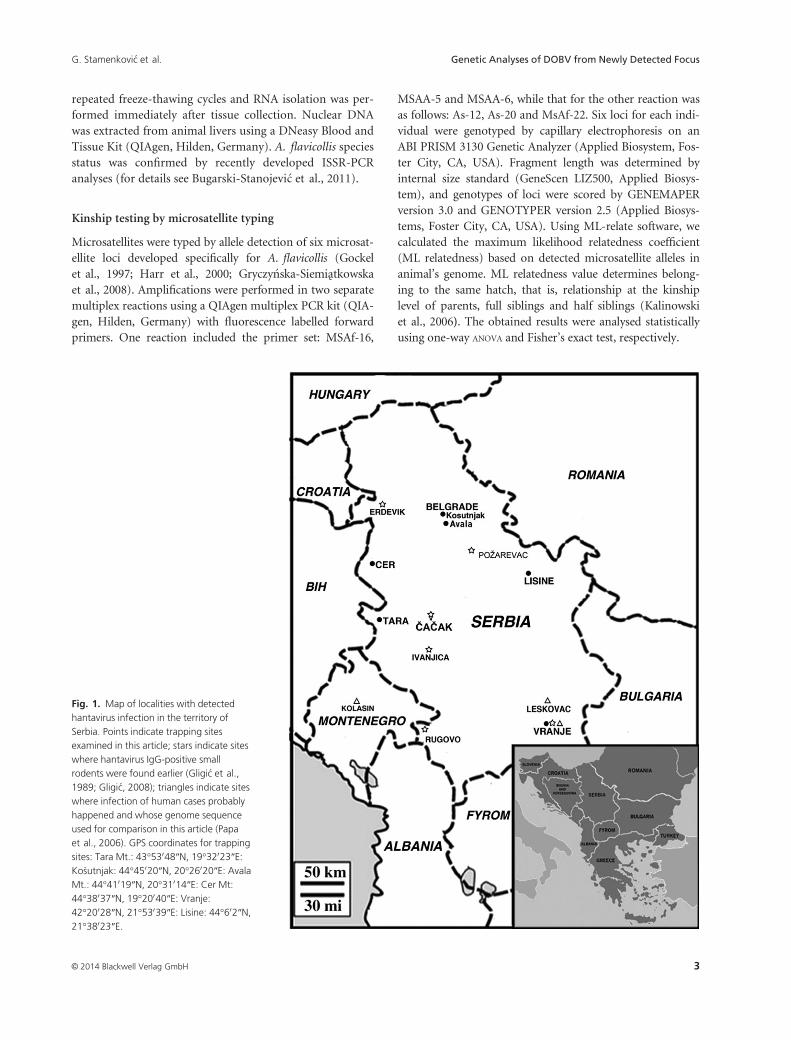
Fig. 1. Map of localities with detected
hantavirus infection in the territory of
Serbia. Points indicate trapping sites
examined in this article; stars indicate sites
where hantavirus IgG-positive small
rodents were found earlier (Gligi�c et al.,
1989; Gligi�c, 2008); triangles indicate sites
where infection of human cases probably
happened and whose genome sequence
used for comparison in this article (Papa
et al., 2006). GPS coordinates for trapping
sites: Tara Mt.: 43°53048″N, 19°32023″E:Ko�sutnjak: 44°45020″N, 20°26020″E: AvalaMt.: 44°41019″N, 20°31014″E: Cer Mt:
44°38037″N, 19°20040″E: Vranje:42°20028″N, 21°53039″E: Lisine: 44°602″N,21°38023″E.
© 2014 Blackwell Verlag GmbH 3
G. Stamenkovi�c et al. Genetic Analyses of DOBV from Newly Detected Focus
Serology
Hantavirus IgG antibodies in rodent sera were detected by
indirect immunofluorescent antibody assay (IFA) by fluo-
rescein-isothiocyanate-conjugated sheep anti-mouse IgG
(H) (Institute for Application of Nuclear Energy, Belgrade,
Serbia). Briefly, cultures of Vero E6 cells were separately
infected with the HTNV strain 76–118. Rodent sera were
serially diluted (1: 16 to 1: 32) and applied on previously
prepared antigen spot slides. After incubation, slides were
fixed and examined by fluorescence microscopy (Av�si�c-
Zupanc et al., 2000). Control samples, hantavirus IgG-
positive and IgG-negative rodent serums, were provided by
National Reference Laboratory for ARBO viruses and HF
viruses, Institute of Virology, Vaccines and Sera - Torlak,
Belgrade, Serbia. Sera that gave a characteristic cytoplasmic
fluorescence in hantavirus-infected cells, at any or both
mentioned dilutions, were considered positive. All readings
were confirmed by double check.
RT-PCR and sequencing
All rodent samples found to be seropositive were examined
by reverse transcription-polymerase chain reaction (RT-
PCR) for the presence of hantavirus genomic RNA. Total
RNA was extracted from lung, liver or kidney tissues using
the TRIZOL Reagent (GibcoBRL, Invitrogen, Karlsruhe,
Germany). Cross-contamination was avoided in accor-
dance with the principles of good laboratory practice for
detection of pathogens by PCR (separation of pre-PCR and
post-PCR procedures, strict unidirectional flow of speci-
mens and use of decontamination procedure using bleach
between handling different samples etc.). Small pieces of
tissues, approximately 10 mg, were homogenized in liquid
nitrogen using a mortar and pestle. The mixture was then
transferred to a 1.5 ml tube containing 1 ml of TRIZOL.
RNA was isolated following the manufacturer’s protocol.
The RNA pellet was air-dried and dissolved in 25 lL of
RNase-free water with 5 U of RNase inhibitor.
Extracted RNA was reverse-transcribed and amplified by
a nested RT-PCR protocol, using the one-step RNA PCR
Kit (Qiagen, Hilden, Germany) for the outer reaction and
the Taq PCR Core Kit (Qiagen, Hilden, Germany) for the
inner PCR reaction. For L segment detection, a set of
degenerate primers for all known hantaviruses, amplifying
the 390 bp part of the highly conserved region, was used
(Klempa et al., 2006). We designed primer pairs for nested
RT-PCR, which amplified 1322 bp and 1283 bp parts of
the S segment (1stPCR: DOB-S-F1 50-GTAGTAGGCTCCCTAAAAAGC-30, DOB-S-R1 50-GGGATTACATAAAGCATGGGA-30: 2ndPCR: DOB-S-F2 50-CACTACACTAAAGATGGCAA-30, DOB-S-R2 50-GGATAATGCAACAAATACA-ATTA-30). Negative control (RNA/DNA free sterile water)
has been included in all rounds of RNA isolation and RT-
PCR. For every sample, universal primers for mouse beta-
actin mRNA: 50-GTGGGCCGCTCTAGGCACCAA-30 and
50-TCTTTGATGTCACGCACGATTTC-3’ were used as
external positive control for RT-PCR (Ponte et al., 1984).
RT-PCR products were visualized by electrophoresis using
1% agarose gel. Positive samples were sequenced in both
directions by dye-terminator DNA sequencing on an ABI
310 Genetic Analyzer (Applied Biosystem, Foster City, CA,
USA).
Sequence comparison and phylogenetic analysis
All sequence data in the NCBI database by 2013 accepted as
DOBV and Saaremaa hantavirus were used for comparison
with Serbian (RS) sequences. Analysed part of L segment
sequences was 318 bp in length (nt 2999–3316, Fig. 2). Ssegment sequences corresponding to the 789 bp part (nt
291–1079) were compared with 36 animal isolates (Fig. 3a).
Further, phylogenetic analysis of the 528 bp part of the S
segment (nt 387–914), which includes human and animal
isolates, was performed with 32 additional sequences
(Fig. 3b). Each phylogenetic tree was outgroup rooted by
reference sequences cited in figure legend.
Multiple nucleotide sequence alignments were processed
in a CLUSTAL W algorithm (Larkin et al., 2007). The best-
fit nucleotide substitution model for aligned sequences was
determined by jModeltest 0.1.1 software (Posada, 2008)
using all 88 proposed models. Further, phylogenetic analy-
sis was performed with PHYML v.3.0 (Guindon et al.,
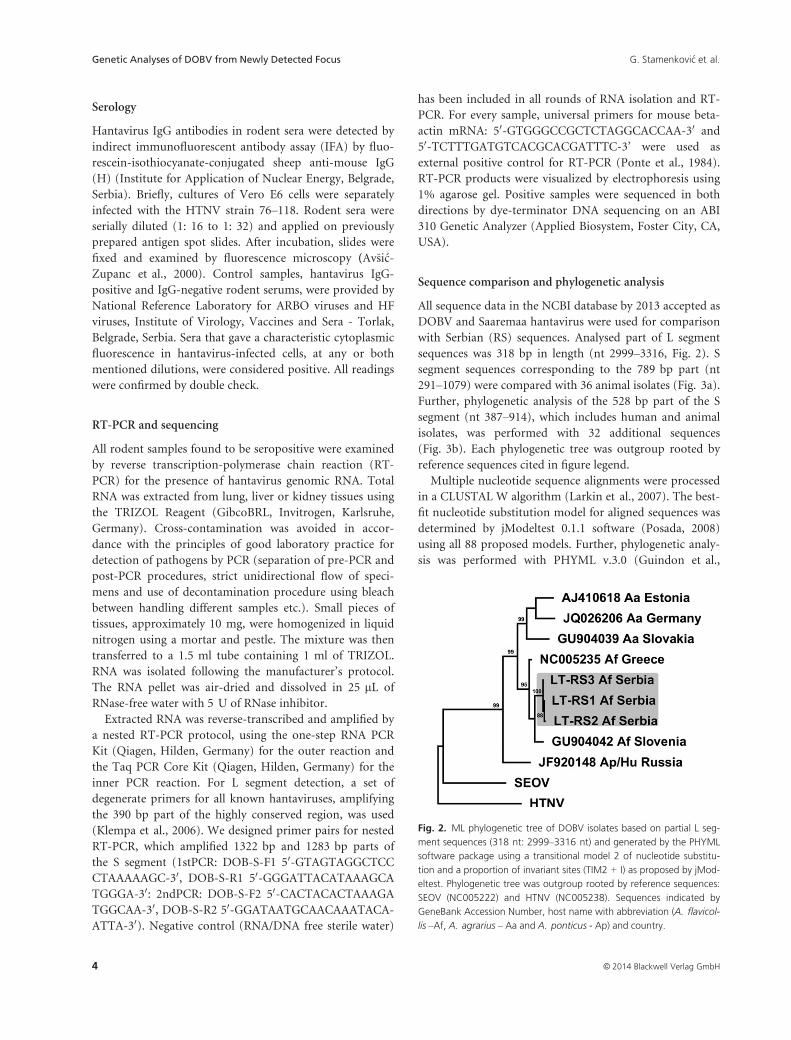
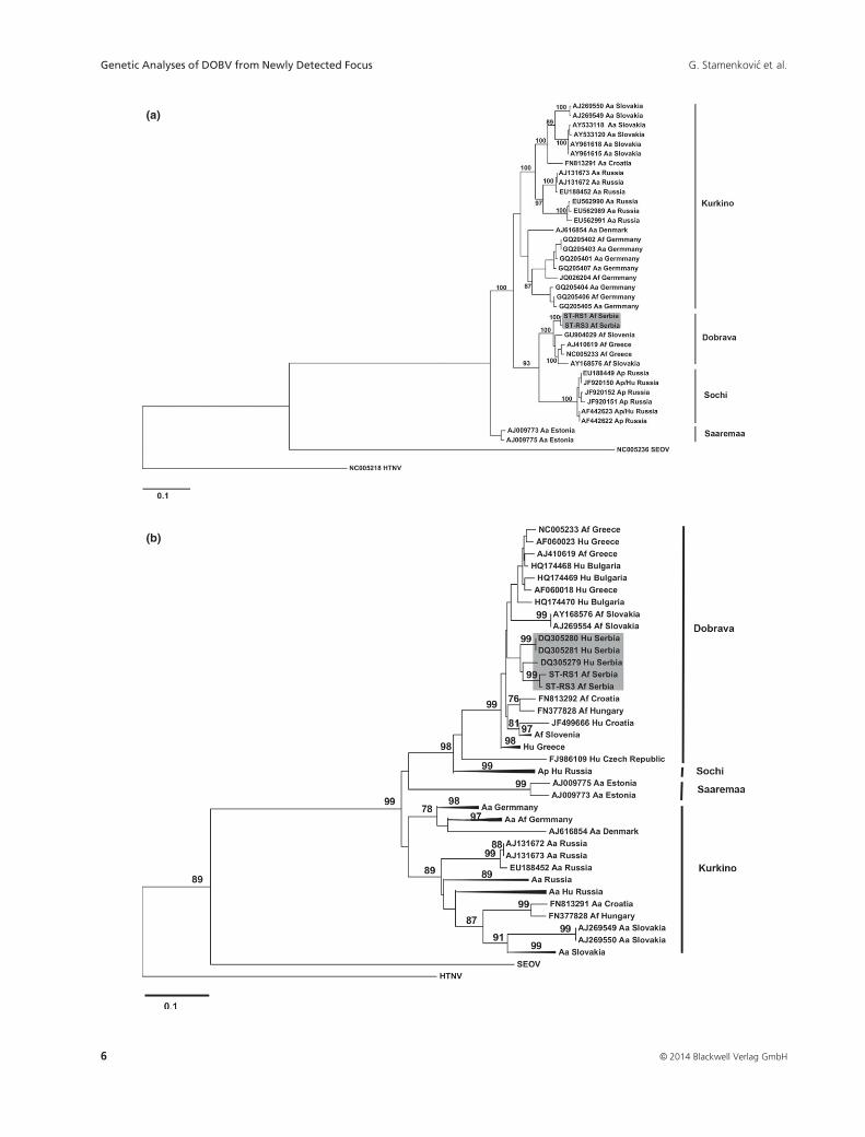
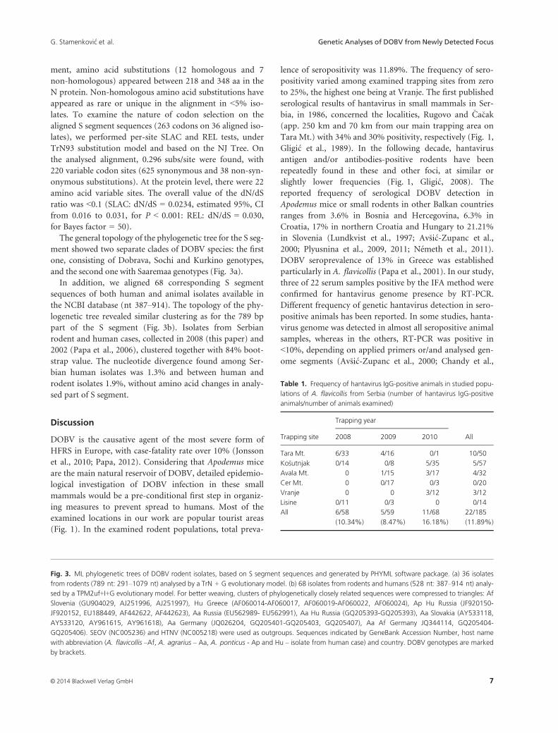
Fig. 2. ML phylogenetic tree of DOBV isolates based on partial L seg-
ment sequences (318 nt: 2999–3316 nt) and generated by the PHYML
software package using a transitional model 2 of nucleotide substitu-
tion and a proportion of invariant sites (TIM2 + I) as proposed by jMod-
eltest. Phylogenetic tree was outgroup rooted by reference sequences:
SEOV (NC005222) and HTNV (NC005238). Sequences indicated by
GeneBank Accession Number, host name with abbreviation (A. flavicol-
lis –Af, A. agrarius – Aa and A. ponticus - Ap) and country.
© 2014 Blackwell Verlag GmbH4
Genetic Analyses of DOBV from Newly Detected Focus G. Stamenkovi�c et al.
2010) and PAUP v. 4.0 software packages (Swoffor, 2000).
The evolutionary history was inferred by neighbour-joining
(NJ), maximum likelihood (ML) and minimum evolution
statistical methods, showing a high level of congruence
between these methods. Bootstrap support for the tree
nodes of the reconstructed phylogenetic trees was calcu-
lated with 1000 replicates. Bootstrap values that exceeded
70% were considered significant.
Alignments were screened for recombination using a
Bootscan recombination detection approach as imple-
mented in the RDP3 program (Martin et al., 2010). Overall
selection pressure, measured as the mean ratio of non-syn-
onymous (dN) to synonymous substitutions (dS) per site
(dN/dS), was estimated using the single likelihood ancestor
counting (SLAC) and random effects likelihood (REL)
methods from the HyPhy package (Pond and Frost, 2005),
available at http://www.datamonkey.org.
Results
During the three-year study period (2008–2010), a total of
185 A. flavicollis were characterized by intersimple
sequence repeat (ISSR) markers. The number of trapped
animals at different sites varied yearly during the study per-
iod (Table 1). Positive finding of specific hantavirus IgG
was detected in the total of 22/185 (11.89%) blood samples,
of which only three reacted positive on 1 : 32 dilution.
Seropositive animals were detected in all but two trapping
sites: Cer Mt. and Lisine. The highest prevalence of IgG
antibody was observed in Vranje and on Tara Mt., 25% (3/
12) and 20% (10/50), respectively.
All seropositive samples were further analysed by genetic
detection of hantavirus sequences. Amplification from
beta-actin mRNA, as external control, was present in all
examined samples by RT-PCR. Three of 22 seropositive
samples were positive on RT-PCR for hantavirus L seg-
ment, and the isolates were marked: RS1 to RS3. Two the
three animals were also positive in the nested PCR for S
segment (RS1 and RS3). All RT-PCR-positive animals were
trapped at the same site on mountain Tara, western Serbia
(Fig. 1). An initial BLAST search demonstrated the highest
similarity of RS sequences with isolates submitted in NCBI
database as DOBV and Saaremaa virus. Further phyloge-
netic analyses of the obtained RS sequences, in both L and
S segments, showed that all three genomes belong to the
DOBV-Dobrava genotype (Fig. 2 and 3a).
Relatedness between hantavirus-positive rodents
Levels of kinship were calculated for the Tara Mt. popula-
tion, based on six microsatellite loci by ML-relate software.
Comparison of mean ML relatedness of seropositive
(0.048 � 0.080, min = 0.000, max = 0.290) vs. seronega-
tive animals (0.037 � 0.075, min = 0.000, max = 0.520)
did not reveal statistical significance (F1,405 = 0.55,
P = 0.460). The frequency of relatives was higher in the
seronegative group (96.6% in the seronegative group vs.
85.7% in the seropositive group), but this difference was
also found not to be statistically significant (P = 0.356).
Phylogenetic analysis of the partial L segment sequences
We successfully recovered a partial L segment from three
isolates (sequences LT-RS1 to LT-RS3). The L segment
sequences recovered (318 bp in length, pos. 2999–3316)encoded 106 amino acids (pos. 988–1087) of the RNA-
dependent RNA polymerase. The mean nucleotide distance
among nine aligned L segment sequences was 0.137 (0.006–0.195, SD = 0.056). Amino acid divergence for the exam-
ined region was 3.4%. Nucleotide and amino acid diver-
gence among the newly detected sequences from Serbia was
1.1% and 1.3%, respectively. Comparison of the corre-
sponding region of DOBV-Dobrava and DOBV-Kurkino
genotypes revealed a nucleotide divergence of 5.4% and
14.8%, respectively.
In the L segment phylogenetic tree, our sequences from
A. flavicollis formed a distinct clade with Slovenian and
Greek DOBV isolates from the same species. Moreover,
DOBV-Kurkino and DOBV-Saaremaa have a monophyletic
origin, while DOBV-Sochi clustered into a separate branch
(Fig. 2).
Phylogenetic analysis of the partial S segment sequences
The obtained 789-bp (nt 291–1079) DOBV S segment
sequences (ST-RS1 and ST-RS3) comprise 263 amino acids
of the N protein (aa 86–348). Prior to phylogenetic analy-
ses, screening for recombination between aligned sequences
was performed and no putative recombinant regions could
be detected. Mean nucleotide distance found for DOBV
alignment of the examined S segment part was 0.113 (0.0–0.155, SD = 0.035), with amino acid divergence 1.9%.
Comparative analysis of the DOBV S segment revealed the
nucleotide divergence within DOBV-Dobrava, DOBV-So-
chi and DOBV-Kurkino clusters: 3.6%, 1.6% and 9.0%,
respectively. Nucleotide divergence between RS samples
was 0.8%, with amino acid divergence 0.4%.
Consensus sequence analysis from the obtained align-
ment (BioEdit v.7.0.0., Hall, 1999) with 95% threshold fre-
quencies for inclusion in consensus revealed that RS
isolates have three unique synonymous substitutions
(T584C, T733G, C771T). Amino acid sequences for RS1
and RS3 were identical to the DOBV reference sequence
(NC_005233), with the exception of two non-synonymous
changes (A328G and Y98C) in RS3, which are unique in
the alignment. Within the overall analysed S segment align-
© 2014 Blackwell Verlag GmbH 5
G. Stamenkovi�c et al. Genetic Analyses of DOBV from Newly Detected Focus
(a)
(b)
© 2014 Blackwell Verlag GmbH6
Genetic Analyses of DOBV from Newly Detected Focus G. Stamenkovi�c et al.
ment, amino acid substitutions (12 homologous and 7
non-homologous) appeared between 218 and 348 aa in the
N protein. Non-homologous amino acid substitutions have
appeared as rare or unique in the alignment in <5% iso-
lates. To examine the nature of codon selection on the
aligned S segment sequences (263 codons on 36 aligned iso-
lates), we performed per-site SLAC and REL tests, under
TrN93 substitution model and based on the NJ Tree. On
the analysed alignment, 0.296 subs/site were found, with
220 variable codon sites (625 synonymous and 38 non-syn-
onymous substitutions). At the protein level, there were 22
amino acid variable sites. The overall value of the dN/dS
ratio was <0.1 (SLAC: dN/dS = 0.0234, estimated 95%, CI
from 0.016 to 0.031, for P < 0.001: REL: dN/dS = 0.030,
for Bayes factor = 50).
The general topology of the phylogenetic tree for the S seg-
ment showed two separate clades of DOBV species: the first
one, consisting of Dobrava, Sochi and Kurkino genotypes,
and the second one with Saaremaa genotypes (Fig. 3a).
In addition, we aligned 68 corresponding S segment
sequences of both human and animal isolates available in
the NCBI database (nt 387–914). The topology of the phy-logenetic tree revealed similar clustering as for the 789 bp
part of the S segment (Fig. 3b). Isolates from Serbian
rodent and human cases, collected in 2008 (this paper) and
2002 (Papa et al., 2006), clustered together with 84% boot-
strap value. The nucleotide divergence found among Ser-
bian human isolates was 1.3% and between human and
rodent isolates 1.9%, without amino acid changes in analy-
sed part of S segment.
Discussion
DOBV is the causative agent of the most severe form of
HFRS in Europe, with case-fatality rate over 10% (Jonsson
et al., 2010; Papa, 2012). Considering that Apodemus mice
are the main natural reservoir of DOBV, detailed epidemio-
logical investigation of DOBV infection in these small
mammals would be a pre-conditional first step in organiz-
ing measures to prevent spread to humans. Most of the
examined locations in our work are popular tourist areas
(Fig. 1). In the examined rodent populations, total preva-
lence of seropositivity was 11.89%. The frequency of sero-
positivity varied among examined trapping sites from zero
to 25%, the highest one being at Vranje. The first published
serological results of hantavirus in small mammals in Ser-
bia, in 1986, concerned the localities, Rugovo and �Ca�cak
(app. 250 km and 70 km from our main trapping area on
Tara Mt.) with 34% and 30% positivity, respectively (Fig. 1,
Gligi�c et al., 1989). In the following decade, hantavirus
antigen and/or antibodies-positive rodents have been
repeatedly found in these and other foci, at similar or
slightly lower frequencies (Fig. 1, Gligi�c, 2008). The
reported frequency of serological DOBV detection in
Apodemus mice or small rodents in other Balkan countries
ranges from 3.6% in Bosnia and Hercegovina, 6.3% in
Croatia, 17% in northern Croatia and Hungary to 21.21%
in Slovenia (Lundkvist et al., 1997; Av�si�c-Zupanc et al.,
2000; Plyusnina et al., 2009, 2011; N�emeth et al., 2011).
DOBV seroprevalence of 13% in Greece was established
particularly in A. flavicollis (Papa et al., 2001). In our study,
three of 22 serum samples positive by the IFA method were
confirmed for hantavirus genome presence by RT-PCR.
Different frequency of genetic hantavirus detection in sero-
positive animals has been reported. In some studies, hanta-
virus genome was detected in almost all seropositive animal
samples, whereas in the others, RT-PCR was positive in
<10%, depending on applied primers or/and analysed gen-
ome segments (Av�si�c-Zupanc et al., 2000; Chandy et al.,
Fig. 3. ML phylogenetic trees of DOBV rodent isolates, based on S segment sequences and generated by PHYML software package. (a) 36 isolates
from rodents (789 nt: 291–1079 nt) analysed by a TrN + G evolutionary model. (b) 68 isolates from rodents and humans (528 nt: 387–914 nt) analy-
sed by a TPM2uf+I+G evolutionary model. For better weaving, clusters of phylogenetically closely related sequences were compressed to triangles: Af
Slovenia (GU904029, AJ251996, AJ251997), Hu Greece (AF060014-AF060017, AF060019-AF060022, AF060024), Ap Hu Russia (JF920150-
JF920152, EU188449, AF442622, AF442623), Aa Russia (EU562989- EU562991), Aa Hu Russia (GQ205393-GQ205393), Aa Slovakia (AY533118,
AY533120, AY961615, AY961618), Aa Germany (JQ026204, GQ205401-GQ205403, GQ205407), Aa Af Germany JQ344114, GQ205404-
GQ205406). SEOV (NC005236) and HTNV (NC005218) were used as outgroups. Sequences indicated by GeneBank Accession Number, host name
with abbreviation (A. flavicollis –Af, A. agrarius – Aa, A. ponticus - Ap and Hu – isolate from human case) and country. DOBV genotypes are marked
by brackets.
Table 1. Frequency of hantavirus IgG-positive animals in studied popu-
lations of A. flavicollis from Serbia (number of hantavirus IgG-positive
animals/number of animals examined)
Trapping site
Trapping year
All2008 2009 2010
Tara Mt. 6/33 4/16 0/1 10/50
Ko�sutnjak 0/14 0/8 5/35 5/57
Avala Mt. 0 1/15 3/17 4/32
Cer Mt. 0 0/17 0/3 0/20
Vranje 0 0 3/12 3/12
Lisine 0/11 0/3 0 0/14
All 6/58
(10.34%)
5/59
(8.47%)
11/68
16.18%)
22/185
(11.89%)
© 2014 Blackwell Verlag GmbH 7
G. Stamenkovi�c et al. Genetic Analyses of DOBV from Newly Detected Focus
2013). In our work, PCR-positive animals were among
those positive for hantavirus IgG on dilution 1:32. Other
samples, IgG-positive on dilution 1:16, were RT-PCR nega-
tive, which indicate the shorter period of detectable viremia
in relation to long-lasting low titre of antibody response
(Jonsson et al., 2010; N�emeth et al., 2011).
All RT-PCR-positive animals were caught on Tara Mt.
tourist complex where seropositivity was among the highest
(20%). Up to now, western Serbia, where mountain Tara is
located, has not been detected as a hantavirus focus in the
animal reservoir. Regarding human infection, the majority
of reported serologically documented DOBV human infec-
tions so far in Serbia occurred in the south of the country,
represented by the Vranje trapping site in this study (Papa
et al., 2006). Two of the locations in our work were Ko�sut-
njak and Avala, in the municipality of Belgrade, where the
first human DOBV case was detected (Gligi�c et al., 1992).
During the period of animal collection, 40 cases of human
hantavirus infection were immunologically detected in Ser-
bia (unpublished data from National Reference Laboratory
for ARBO viruses and HF viruses). Unfortunately, epidemi-
ological data regarding exact or most probable place of
infection are very scarce or completely missing. Detection
of seropositive animals throughout our 3-year study period
and identification of a new infection focus in west Serbia
indicates the need for permanent monitoring. Preventive
tasting should include not only previously confirmed
hantavirus localities, but also other areas, especially forests
with frequent human visits.
We investigated the influence of kinship on DOBV trans-
mission in the examined A. flavicollis population from
Tara. While the frequency of relatives was higher in sero-
negative in relation to seropositive group, the difference did
not reach statistical significance. We have to underline that
we did not detect any immigrants, that is, animals without
relatives in the examined population. This result could be a
consequence of the small size of the trapping area (app.
5000 m2) and behavioural characteristics of Apodemus
rodents, such as a small home range. Investigation of TULV
and PUUV spread in populations of Cricetidae species
revealed that infected animals were more closely related to
each other than non-infected ones, emphasizing the impor-
tance of virus transmission in hatches (Deter et al., 2008).
Our preliminary results did not support this trend of trans-
mission for A. flavicollis as a host, probably due to differ-
ences in social organization of different host species.
Molecular analyses
Conversely, to previous rather extensive epizootical studies
of hantavirus distribution in Serbia, based on serological
detection, up to now, molecular studies are very scarce (Di-
gli�si�c et al., 1994; Song et al., 2002; Nikoli�c et al., 2014). We
found three A. flavicollis captured on mountain Tara to be
positive for hantavirus genome by nested RT-PCR for a par-
tial L segment (RS1 to RS3), whereas two isolates (RS1 and
RS3) were also positive for a partial S segment. Absence of
amplification for the S segment in the RS2 isolate confirms
better detection sensitivity by degenerate primers for the L
segment even in cases of strict hantavirus genotypes (Klem-
pa et al., 2006). Comparison of Serbian L and S segment
isolates with animal isolates clustered them together with
DOBV hosted by A. flavicollis from Greece, Slovakia and
Slovenia, revealing host-related clustering (Fig. 2 and 3a).
The same phylogenetic relation was shown by Papa (2012)
in analyses of complete sequence of DOBV L segments.
With the aim to elucidate nucleotide sequence relations
between Serbian human and animal isolates, we analysed
all NCBI database accepted as DOBV and Saaremaa
sequences for the corresponding S segment (68 isolates with
the 528 bp part). The phylogenetic tree again showed strict
geographical clustering (Fig. 3b). Consequently, Serbian
human and rodent isolates clustered together with 0.019
nucleotide distance. It is important to emphasize that
human isolates were collected in 2002 at sites about 200–400 km away from mountain Tara, where we found
infected A. flavicollis 6 years later (Fig. 1). A similar trend,
concerning high phylogenetic similarity regardless of tem-
poral and spatial distance, has also been shown in Greek
and Slovenian isolates from humans and animals (Fig. 3b).
This phenomenon points to unidirectional transmission of
infection, from mice to man. In accordance with that, the
short residence time of the virus in human tissue could be
insufficient to induce fixation of virus genome changes in
different hosts (Schilling et al., 2007).
According to the classification by Klempa et al. (2013),
analysed isolates from Serbia (RS-1 to RS-3 from A. flavi-
collis and DQ305279 to DQ305281 from human serum)
belong to DOBV species and Dobrava genotypes (Fig. 3A
and B).
Although the examined fragment of the L segment is
rather conservative, the obtained LT-RS sequences were
found to be slightly polymorphic (genetic divergence
1.1%). The analysed DOBV L segment protein coding
sequence includes the complete motif B and almost com-
plete inter-motif (between A and B motif) of the L protein
(pos. 988–1087 aa). Motifs B, E and pre-motif A found in
all RNA-dependent RNA polymerases are responsible for
positioning of the template and primer relative to the active
site (Nemirov et al., 2003). In all aligned DOBV L protein
sequences in our study, motif B (pos. 1044–1065 aa) was
fully conserved and the A-to-B inter-motif was highly con-
served with rare amino acid changes, suggesting their
importance in enzyme function (Kukkonen et al., 2005).
The examined part of the S segment partly codes for the
conserved amino-terminal N protein that forms the main
© 2014 Blackwell Verlag GmbH8
Genetic Analyses of DOBV from Newly Detected Focus G. Stamenkovi�c et al.
human IgG epitope (pos. 1–118 aa) and genome RNA-
binding domain (pos. 175–217 aa) important for RNP
assembly (Kaukinen et al., 2005). These parts of the N pro-
tein are very conservative, characterized by a strict cluster
of 15 lysines/arginines (pos. 136–213 aa), present in all iso-
lates included in the S segment alignment. The function of
the portion of the N protein analysed in our study with
detected amino acid changes (218–348 aa) is currently
unknown (Kaukinen et al., 2005). However, the type of
detected amino acid changes and evidence of purifying
selection (x < 0.1) in action on this region imply a high
tendency for protein structure conservation. Consequently,
examined part of S segment has lower nucleotide diver-
gence in relation to examined part of L segment (11.3%
versus 13.7%, respectively), conversely to results given in
comparison with their whole-nucleotide sequences (Nemi-
rov et al., 2003; Papa, 2012).
Our findings are the first genetic confirmation of DOBV
presence in wild rodents in Serbia, initially detected sero-
logically three decades ago. We confirmed the persistence
of hantavirus seroprevalence in A. flavicollis in Serbia. The
finding of a new hantavirus focus in western Serbia points
to widespread presence of the virus and the necessity of
continuous control of rodent infection, especially in places
with frequent human visits.
Acknowledgements
We are very grateful to Dr Ana Gligi�c and Dr Bojana
Bo�zovi�c for useful advice during realization of this study
and Bo�zica Jankovi�c for excellent technical assistance (Insti-
tute of Virology, Vaccines and Sera - Torlak, National Ref-
erence Laboratory for ARBO viruses and HF viruses,
Belgrade, R Serbia). This work was financed by a grant
from the Ministry of Education, Science and Technological
Development of the Republic of Serbia, Contract No.
173003 and Contract No. 175024.
Disclosure Statement
No competing financial interests exist.
References
Av�si�c-Zupanc, T., S. Y. Xiao, R. Stojanovi�c, A. Gligi�c, G. van der
Groen, and J. W. LeDuc, 1992: Characterization of dobrava
virus: a hantavirus from slovenia. Yugoslavia. J. Med. Virol.
38, 132–137.
Av�si�c-Zupanc, T., K. Nemirov, M. Petrovec, T. Trilar, M. Poljak,
A. Vaheri, and A. Plyusnin, 2000: Genetic analysis of wild-type
Dobrava hantavirus in Slovenia: co-existence of two distinct
genetic lineages within the same natural focus. J. Gen. Virol.
81, 1747–1755.
Bugarski-Stanojevi�c, V., J. Blagojevi�c, G. Stamenkovi�c, T.
Adna�devi�c, E. Giagia-Athanasopoulou, and M. Vujo�sevi�c,
2011: Comparative study of the phylogenetic structure in six
Apodemus species (Mammalia, Rodentia) inferred from ISSR-
PCR data. Syst. Biodivers. 9, 95–106.
Chandy, S., R. G. Ulrich, M. Schlegel, R. Petraityte, K. Sasnaus-
kas, D. J. Prakash, V. Balraj, P. Abraham, and G. Sridharan,
2013: Hantavirus infection among wild small mammals in
Vellore, South India. Zoonoses Public Hlth. 60, 336–340.
Deter, J., Y. Chaval, M. Galan, B. Gauffre, S. Morand, H. Hen-
ttonen, J. Laakkonen, L. Voutilainen, N. Charbonnel, and J. F.
Cosson, 2008: Kinship, dispersal and hantavirus transmission
in bank and common voles. Arch. Virol. 153, 435–444.
Digli�si�c, G., S. Y. Xiao, A. Gligi�c, M. Obradovi�c, R. Stojanovi�c,
D. Velimirovi�c, V. Luka�c, C. A. Rossi, and J. W. LeDuc, 1994:
Isolation of a Puumala-like virus from Mus musculus captured
in Yugoslavia and its association with severe hemorrhagic
fever with renal syndrome. J. Infect. Dis. 169, 204–207.
Gligi�c, A. 2008: Etiology of hemorrhagic fever with renal syn-
drome, viruses and their reservoirs. In: Kova�cevi�c, Z., D. Jova-
novi�c, A. Gligi�c, and V. Skatari�c, (eds), Hemorrhagic Fever with
Renal Syndrome. pp. 17–34. Medicinski faklultet, Kragujevac.
Gligi�c, A., M. Obradovi�c, R. Stojanovi�c, D. Hlaca, B. Antonijev-
i�c, A. Arnautovi�c, J. M. Gaon Fru�si�c, P. Lee, D. Goldgaber,
et al., 1988: Hemorrhagic fever with renal syndrome in Yugo-
slavia: detection of hantaviral antigen and antibody in wild
rodents and serological diagnosis of human disease. Scand. J.
Infect. Dis. 20, 261–266.
Gligi�c, A., M. Fru�si�c, M. Obradovi�c, R. Stojanovi�c, D. Hlaca, C.
J. Jr Gibbs, R. Yanagihara, C. H. Calisher, and D. C. Gajdu�sek,
1989: Hemorrhagic fever with renal syndrome in Yugoslavia:
antigenic characterization of hantaviruses isolated from
Apodemus flavicollis and Clethrionomys glareolus. Am. J. Trop.
Med. Hyg. 41, 109–115.
Gligi�c, A., N. Dimkovi�c, S. Y. Xiao, G. J. Buckle, D. Jovanovi�c,
D. Velimirovi�c, R. Stojanovi�c, M. Obradovic, G. Digli�si�c, and
J. Mici�c, 1992: Belgrade virus: a new hantavirus causing severe
hemorrhagic fever with renal syndrome in Yugoslavia.
J. Infect. Dis. 166, 113–120.
Gockel, J., B. Harr, C. Schl€otterer, W. Arnold, G. Gerlach, and
D. Tautz, 1997: Isolation and characterization of microsatel-
lite loci from Apodemus flavicollis (rodentia, muridae) and
Clethrionomys glareolus (rodentia, cricetidae). Mol. Ecol. 6,
597–599.
Gryczy�nska-Siemiaztkowska, A., T. Gortat, A. Kozakiewicz, R.Rutkowski, J. Pomorski, and M. Kozakiewicz, 2008: Multiple
paternity in a wild population of the yellow-necked mouse
Apodemus flavicollis. Acta Theriol. 53, 251–258.
Guindon, S., J. F. Dufayard, V. Lefort, M. Anisimova, W. Hord-
ijk, and O. Gascuel, 2010: New algorithms and methods to
estimate maximum-likelihood phylogenies: assessing the per-
formance of PhyML 3.0. Syst. Biol. 59, 307–321.Guo, W. P., X. D. Lin, W. Wang, J. H. Tian, M. L. Cong, H. L.
Zhang, M. R. Wang, R. H. Zhou, J. B. Wang, M. H. Li, J. Xu,
E. C. Holmes, and Y. Z. Zhang, 2013: Phylogeny and origins
© 2014 Blackwell Verlag GmbH 9
G. Stamenkovi�c et al. Genetic Analyses of DOBV from Newly Detected Focus
of hantaviruses harbored by bats, insectivores, and rodents.
PLoS Pathog. 9, e1003159.
Hall, T. A., 1999: BioEdit: a user-friendly biological sequence
alignment editor and analysis program for Windows 95/98/
NT. Nucl. Acids Symp. Ser. 41, 95–98.
Harr, B., K. Musolf, and G. Gerlach, 2000: Characterization and
isolation of DNA microsatellite primers in wood mice (Apode-
mus sylvaticus, Rodentia). Mol. Ecol. 9, 1664–1665.
Jonsson, C. B., L. T. Figueiredo, and O. Vapalahti, 2010: A global
perspective on hantavirus ecology, epidemiology, and disease.
Clin. Microbiol. Rev. 23, 412–441.
Kalinowski, S. T., A. P. Wagner, and M. L. Taper, 2006:
ML-Relate: a computer program for maximum likelihood
estimation of relatedness and relationship. Mol. Ecol. Notes 6,
576–579.
Kaukinen, P., A. Vaheri, and A. Plyusnin, 2005: Hantavirus
nucleocapsid protein: a multifunctional molecule with both
housekeeping and ambassadorial duties. Arch. Virol. 150,
1693–1713.
Klempa, B., E. Fichet-Calvet, E. Lecompte, B. Auste, V. Aniskin,
H. Meisel, C. Denys, L. Koivogui, J. terMeulen, and D. H.
Kr€uger, 2006: Hantavirus in African wood mouse. Guinea.
Emerg. Infect. Dis. 12, 838–840.
Klempa, B., T. Avsic-Zupanc, J. Clement, T. K. Dzagurova, H.
Henttonen, P. Heyman, F. Jakab, D. H. Kruger, P. Maes, A.
Papa, E. A. Tkachenko, R. G. Ulrich, O. Vapalahti, and A.
Vaheri, 2013: Complex evolution and epidemiology of Dobra-
va-Belgrade hantavirus: definition of genotypes and their
characteristics. Arch. Virol. 158, 521–529.
Kukkonen, S. K., A. Vaheri, and A. Plyusnin, 2005: L protein,
the RNA-dependent RNA polymerase of hantaviruses. Arch.
Virol. 150, 533–556.
Larkin, M. A., G. Blackshields, N. P. Brown, R. Chenna, P. A.
McGettigan, H. McWilliam, F. Valentin, I. M. Wallace, A.
Wilm, R. Lopez, J. D. Thompson, T. J. Gibson, and D. G. Hig-
gins, 2007: Clustal W and Clustal X version 2.0. Bioinformatics
23, 2947–2948.
Lundkvist, A., M. Huki�c, J. H€orling, M. Gilljam, S. Nichol, and
B. Niklasson, 1997: Puumala and Dobrava viruses cause hem-
orrhagic fever with renal syndrome in Bosnia-Herzegovina:
evidence of highly cross-neutralizing antibody responses in
early patient sera. J. Med. Virol. 53, 51–59.
Macdonald, D. W., and P. Barrett, 1993: Mammals of Britain
and Europe. Harper Collins, London, UK.
Maes, P., B. Klempa, J. Clement, J. Matthijnssens, D. C. Gajdusek,
D. H. Kr€uger, andM. Van Ranst, 2009: A proposal for new
criteria for the classification of hantaviruses, based on S and M
segment protein sequences. Infect. Genet. Evol. 9, 813–820.
Martin, D. P., P. Lemey, M. Lott, V. Moulton, D. Posada, and P.
Lefeuvre, 2010: RDP3: a flexible and fast computer program for
analyzing recombination. Bioinformatics 26, 2462–2463.
N�emeth, V., M. Madai, A. Mar�aczi, B. B�erczi, G. Horv�ath, M.
Oldal, P. Kisfali, K. B�anyai, and F. Jakab, 2011: Detection of
Dobrava-Belgrade hantavirus using recombinant-nucleocap-
sid-based enzyme-linked immunosorbent assay and SYBR
Green-based real-time reverse transcriptase-polymerase chain
reaction. Arch. Virol. 156, 1655–1660.
Nemirov, K., O. Vapalahti, A. Papa, A. Plyusnina, A. Lundkvist,
A. Antoniadis, A. Vaheri, and A. Plyusnin, 2003: Genetic char-
acterization of new Dobrava hantavirus isolate from Greece.
J. Med. Virol. 69, 408–416.
Nikoli�c, V., N. Stajkovi�c, G. Stamenkovi�c, R. �Cekanac, P.
Maru�si�c, M. �Silji�c, A. Gligi�c, and M. Stanojevi�c, 2014: Evi-
dence of recombination in Tula virus strains from Serbia.
Infect. Genet. Evol. 21, 472–478.
Papa, A., 2012: Dobrava-Belgrade virus: Phylogeny, epidemiol-
ogy, disease. Antiviral Res. 95, 104–117.
Papa, A., K. Nemirov, H. Henttonen, J. Niemimaa, A. Antonia-
dis, A. Vaheri, A. Plyusnin, and O. Vapalahti, 2001: Isolation
of Dobrava virus from Apodemus flavicollis in Greece. J. Clin.
Microbiol. 39, 2291–2293.
Papa, A., B. Bojovi�c, and A. Antoniadis, 2006: Hantaviruses in
Serbia and Montenegro. Emerg. Infect. Dis. 12, 1015–1018.
Plyusnina, A., E. Ferenczi, G. R. R�acz, K. Nemirov, A. Lundkvist,
A. Vaheri, O. Vapalahti, and A. Plyusnin, 2009: Co-circulation
of three pathogenic hantaviruses: Puumala, Dobrava, and
Saaremaa in Hungary. J. Med. Virol. 81, 2045–2052.
Plyusnina, A., L. C. Krajinovi�c, J. Margaleti�c, J. Niemimaa, K.
Nemirov,�A. Lundkvist, A. Markoti�c, M. Mileti�c-Medved, T.
Av�si�c-�Zupanc, H. Henttonen, and A. Plyusnin, 2011: Genetic
evidence for the presence of two distinct hantaviruses associ-
ated with Apodemus mice in Croatia and analysis of local
strains. J. Med. Virol. 83, 108–114.
Pond, S. L., and S. D. Frost, 2005: Datamonkey: rapid detection
of selective pressure on individual sites of codon alignments.
Bioinformatics 21, 2531–2533.
Ponte, P., S. Y. Ng, J. Engel, P. Gunning, and L. Kedes, 1984:
Evolutionary conservation in the untranslated regions of actin
mRNAs: DNA sequence of a human b-actin cDNA. Nucleic
Acids Res. 12, 1687–1696.
Posada, D., 2008: jModelTest: phylogenetic model averaging.
Mol. Biol. Evol. 25, 1253–1256.
Schilling, S., P. Emmerich, B. Klempa, B. Auste, E. Schnaith, H.
Schmitz, D. H. Kr€uger, S. G€unther, and H. Meisel, 2007:
Hantavirus disease outbreak in Germany: limitations of rou-
tine serological diagnostics and clustering of virus sequences
of human and rodent origin. J. Clin. Microbiol. 45, 3008–3014.
Song, J., A. Gligi�c, and R. Yanagihara, 2002: Identification of
Tula hantavirus in Pitymys subterraneus captured in the �Ca�cak
region of Serbia - Yugoslavia. Int. J. Infect. Dis. 6, 31–36.
Swoffor, D. L., 2000: PAUP*: Phylogenetic Analysis Using Parsi-
mony and Other Methods. Sinauer, Sunderland, MA.
Taller, A., S. Xiao, M. Godec, A. Gligi�c, T. Av�si�c-Zupanc, L. G.
Goldfarb, R. Yanagihara, and D. M. Asher, 1993: Belgrade
virus, a cause of hemorrhagic fever with renal syndrome in
the Balkans, is closely related to Dobrava virus of field mice.
J. Infect. Dis. 168, 750–753.
Vaheri, A., T. Strandin, J. Hepojoki, T. Sironen, H. Henttonen,
S. M€akel€a, and J. Mustonen, 2013: Uncovering the mysteries
of hantavirus infections. Nat. Rev. Microbiol. 11, 539–550.
© 2014 Blackwell Verlag GmbH10
Genetic Analyses of DOBV from Newly Detected Focus G. Stamenkovi�c et al.