Generation of subject-specific, dynamic, multisegment ankle and foot models to improve orthotic...
10
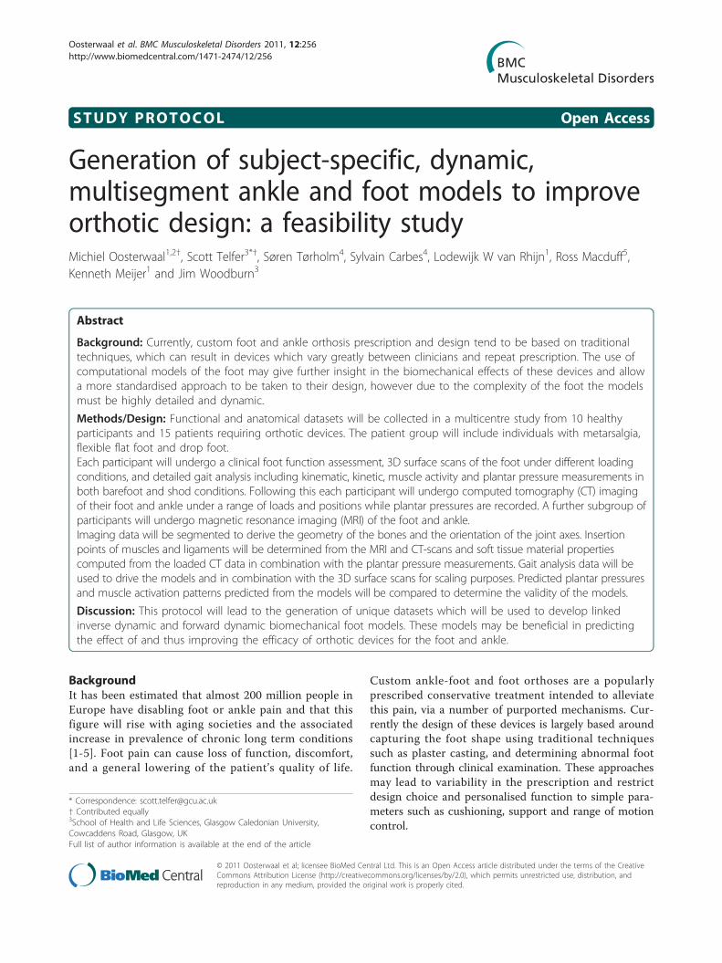
STUDY PROTOCOL Open Access Generation of subject-specific, dynamic, multisegment ankle and foot models to improve orthotic design: a feasibility study Michiel Oosterwaal 1,2† , Scott Telfer 3*† , Søren Tørholm 4 , Sylvain Carbes 4 , Lodewijk W van Rhijn 1 , Ross Macduff 5 , Kenneth Meijer 1 and Jim Woodburn 3 Abstract Background: Currently, custom foot and ankle orthosis prescription and design tend to be based on traditional techniques, which can result in devices which vary greatly between clinicians and repeat prescription. The use of computational models of the foot may give further insight in the biomechanical effects of these devices and allow a more standardised approach to be taken to their design, however due to the complexity of the foot the models must be highly detailed and dynamic. Methods/Design: Functional and anatomical datasets will be collected in a multicentre study from 10 healthy participants and 15 patients requiring orthotic devices. The patient group will include individuals with metarsalgia, flexible flat foot and drop foot. Each participant will undergo a clinical foot function assessment, 3D surface scans of the foot under different loading conditions, and detailed gait analysis including kinematic, kinetic, muscle activity and plantar pressure measurements in both barefoot and shod conditions. Following this each participant will undergo computed tomography (CT) imaging of their foot and ankle under a range of loads and positions while plantar pressures are recorded. A further subgroup of participants will undergo magnetic resonance imaging (MRI) of the foot and ankle. Imaging data will be segmented to derive the geometry of the bones and the orientation of the joint axes. Insertion points of muscles and ligaments will be determined from the MRI and CT-scans and soft tissue material properties computed from the loaded CT data in combination with the plantar pressure measurements. Gait analysis data will be used to drive the models and in combination with the 3D surface scans for scaling purposes. Predicted plantar pressures and muscle activation patterns predicted from the models will be compared to determine the validity of the models. Discussion: This protocol will lead to the generation of unique datasets which will be used to develop linked inverse dynamic and forward dynamic biomechanical foot models. These models may be beneficial in predicting the effect of and thus improving the efficacy of orthotic devices for the foot and ankle. Background It has been estimated that almost 200 million people in Europe have disabling foot or ankle pain and that this figure will rise with aging societies and the associated increase in prevalence of chronic long term conditions [1-5]. Foot pain can cause loss of function, discomfort, and a general lowering of the patient’ s quality of life. Custom ankle-foot and foot orthoses are a popularly prescribed conservative treatment intended to alleviate this pain, via a number of purported mechanisms. Cur- rently the design of these devices is largely based around capturing the foot shape using traditional techniques such as plaster casting, and determining abnormal foot function through clinical examination. These approaches may lead to variability in the prescription and restrict design choice and personalised function to simple para- meters such as cushioning, support and range of motion control. * Correspondence: [email protected] † Contributed equally 3 School of Health and Life Sciences, Glasgow Caledonian University, Cowcaddens Road, Glasgow, UK Full list of author information is available at the end of the article Oosterwaal et al. BMC Musculoskeletal Disorders 2011, 12:256 http://www.biomedcentral.com/1471-2474/12/256 © 2011 Oosterwaal et al; licensee BioMed Central Ltd. This is an Open Access article distributed under the terms of the Creative Commons Attribution License (http://creativecommons.org/licenses/by/2.0), which permits unrestricted use, distribution, and reproduction in any medium, provided the original work is properly cited.
-
Upload
healthfoodinnovationmanagement -
Category
Documents
-
view
1 -
download
0
Transcript of Generation of subject-specific, dynamic, multisegment ankle and foot models to improve orthotic...
STUDY PROTOCOL Open Access
Generation of subject-specific, dynamic,multisegment ankle and foot models to improveorthotic design: a feasibility studyMichiel Oosterwaal1,2†, Scott Telfer3*†, Søren Tørholm4, Sylvain Carbes4, Lodewijk W van Rhijn1, Ross Macduff5,Kenneth Meijer1 and Jim Woodburn3
Abstract
Background: Currently, custom foot and ankle orthosis prescription and design tend to be based on traditionaltechniques, which can result in devices which vary greatly between clinicians and repeat prescription. The use ofcomputational models of the foot may give further insight in the biomechanical effects of these devices and allowa more standardised approach to be taken to their design, however due to the complexity of the foot the modelsmust be highly detailed and dynamic.
Methods/Design: Functional and anatomical datasets will be collected in a multicentre study from 10 healthyparticipants and 15 patients requiring orthotic devices. The patient group will include individuals with metarsalgia,flexible flat foot and drop foot.Each participant will undergo a clinical foot function assessment, 3D surface scans of the foot under different loadingconditions, and detailed gait analysis including kinematic, kinetic, muscle activity and plantar pressure measurements inboth barefoot and shod conditions. Following this each participant will undergo computed tomography (CT) imagingof their foot and ankle under a range of loads and positions while plantar pressures are recorded. A further subgroup ofparticipants will undergo magnetic resonance imaging (MRI) of the foot and ankle.Imaging data will be segmented to derive the geometry of the bones and the orientation of the joint axes. Insertionpoints of muscles and ligaments will be determined from the MRI and CT-scans and soft tissue material propertiescomputed from the loaded CT data in combination with the plantar pressure measurements. Gait analysis data will beused to drive the models and in combination with the 3D surface scans for scaling purposes. Predicted plantar pressuresand muscle activation patterns predicted from the models will be compared to determine the validity of the models.
Discussion: This protocol will lead to the generation of unique datasets which will be used to develop linkedinverse dynamic and forward dynamic biomechanical foot models. These models may be beneficial in predictingthe effect of and thus improving the efficacy of orthotic devices for the foot and ankle.
BackgroundIt has been estimated that almost 200 million people inEurope have disabling foot or ankle pain and that thisfigure will rise with aging societies and the associatedincrease in prevalence of chronic long term conditions[1-5]. Foot pain can cause loss of function, discomfort,and a general lowering of the patient’s quality of life.
Custom ankle-foot and foot orthoses are a popularlyprescribed conservative treatment intended to alleviatethis pain, via a number of purported mechanisms. Cur-rently the design of these devices is largely based aroundcapturing the foot shape using traditional techniquessuch as plaster casting, and determining abnormal footfunction through clinical examination. These approachesmay lead to variability in the prescription and restrictdesign choice and personalised function to simple para-meters such as cushioning, support and range of motioncontrol.
* Correspondence: [email protected]† Contributed equally3School of Health and Life Sciences, Glasgow Caledonian University,Cowcaddens Road, Glasgow, UKFull list of author information is available at the end of the article
Oosterwaal et al. BMC Musculoskeletal Disorders 2011, 12:256http://www.biomedcentral.com/1471-2474/12/256
© 2011 Oosterwaal et al; licensee BioMed Central Ltd. This is an Open Access article distributed under the terms of the CreativeCommons Attribution License (http://creativecommons.org/licenses/by/2.0), which permits unrestricted use, distribution, andreproduction in any medium, provided the original work is properly cited.
Computational modelling of the human body - rangingfrom the force interactions of joints to the way cells com-municate with each other - has advanced significantly inthe past few decades and it is now an important and usefultool for researchers and clinicians. These models provide amethod of simulating and assessing interventions that arebeing developed, reducing the time and risk involved withtrialling in humans. This approach is particularly appealingfor studying foot biomechanics due to the challenging nat-ure of directly investigating the internal loading and move-ments of the complex structure of bones and soft tissuesof the foot that occur during gait.A small but growing body of research, primarily based
around finite element (FE) analysis, has studied the footusing models based on different combinations of gait ana-lysis, pressure distribution measurements, computedtomography (CT) and magnetic resonance (MR) imagingof the foot. This has provided insights into inflammationof the plantar fascia [6,7], pressure assessment of the dia-betic foot [8] and therapeutic footwear [9].Lower limb musculoskeletal biomechanics is an area
where extensive modelling work has been successfullycarried out, supporting the development and assessmentof a range of treatments and interventions [10-13]. Interms of the foot however, these models have tended torepresent it as a single rigid segment, and it is onlyrecently that progress has been made in incorporatingsome of the intrinsic joints of the foot [14].With these factors in mind, it is suggested that there is
potential for highly detailed biomechanical foot models tobe used in the process of designing orthotic interventionsfor the foot and ankle. These could lead to the develop-ment of devices and prescription paradigms which couldimprove the efficacy of these devices and benefit thepatient, as well as reducing long term treatment costs forthe healthcare provider.
ModelsThis article describes a protocol that has been developedto generate the biomechanical and anatomical datarequired to produce two linked foot models: a forwarddynamic model that combines a multibody approachwith FE analysis; and an inverse dynamic model whichdescribes the musculoskeletal interactions of the system.The forward dynamic model will be developed using theMadymo software platform (TASS, Rijswijk), and theinverse dynamic model in the AnyBody modelling system(AnyBody Technology, Aalborg). Generally, forwarddynamic models need joint torques and/or muscle forcesas an input to compute kinematics, and for a complexstructure like the foot this information can be generatedfrom inverse dynamic models driven by motion capturedata. Using the same dataset to construct both models
leads to the possibility of a combination of both modelsin a final application.Inverse dynamic modelStandard gait analysis and inverse dynamic models of thelower extremity consider the foot as a single rigid segment.Some models have been developed that describe the footin more detail, however to the authors’ knowledge nokinematic model has attempted to describe all 26 bones.The proposed kinematic model is scalable and parametricand will integrate into the existing AnyBody whole bodymusculoskeletal model (Figure 1). The model will containall of the ligaments and muscles of the foot and ankle. Bycombing the inverse dynamic modelling with an optimisa-tion algorithm the model will provide insight in functionof the foot and leg muscles during gait.Forward dynamic modelThe forward dynamic model is a combined multibodyand FE approach. Bones, joints, ligaments and musclesare defined as multibody elements. This multibodymodel is surrounded by a freeform FE sheet, representingthe skin. The soft tissue characteristics are implementedby loading functions that connect the sheet to the multi-body elements. This combination leads to a computation-ally less complex model compared to a full FE model ofthe foot and ankle. This reduction could be improved bya full multibody representation of the ankle-foot com-plex, however in this model computation of the plantarpressure would be impossible. In the proposed modelthis is solved by a FE mesh representing the plantar sur-face, leading to the possibility of performing a dynamicsimulation to compute the deforming plantar surface.An existing model (Figure 1) has previously been devel-
oped from a combination of data from a post-mortemhuman subject and information derived from existing lit-erature [15]. The generation of one complete dataset leadsto a consistent model. Beyond this, the generation of data-sets from several subjects creates the possibility of scalingthe model. This scaling will be extended for patientsrequiring foot and ankle orthoses.The overall aim of this work is to develop detailed and
accurate biomechanical models of the foot and anklewhich can be used to inform the design of foot andankle orthoses by predicting the biomechanical effectsof the device.
Methods/DesignStudy DesignThis study is a feasibility/pilot study to generate data thatwill be used to develop the biomechanical foot modelsdescribed in the previous section. It is a multicentrestudy with data being collected at the motion capture labof Glasgow Caledonian University (GCU) and RadiologyDepartment of Glasgow Royal Infirmary, and at the
Oosterwaal et al. BMC Musculoskeletal Disorders 2011, 12:256http://www.biomedcentral.com/1471-2474/12/256
Page 2 of 10

motion capture lab of Maastricht University MedicalCentre (MUMC+) and Department of Radiology atMUMC+. The data collection will take a period of sixmonths for each centre. Data collection will last approxi-mately four hours for each subject. The data acquisitionwill be independent, and after acquisition the data will bepooled and used to develop both models.
Ethical ConsiderationThis study will be conducted in accordance to theDeclaration of Helsinki. Ethical approval for this studyhas been granted by the West of Scotland ResearchEthics Committee (application reference 10/S1001/24)and National Health Service Greater Glasgow and ClydeResearch and Development Committee (referenceGN10RH187) for the UK site. At the Dutch centre, thestudy was granted approval by the Medical Ethical Com-mittee azM/UM (reference number NL31656.068.10/MEC 08-2-028).
ParticipantsTwo groups of participants will be investigated, healthyindividuals and patients with foot and/or ankle problems.Healthy volunteers will be recruited via convenience sam-pling from staff bodies of the test centres. Participantswith pathological foot problems will be informed aboutthe study by their orthopaedic surgeon (MUMC+) orpodiatrist (GCU) during the course of attending a routineappointment at a local foot and ankle clinic. Table 1 givesan overview of the study population.Inclusion criteriaIn both groups, healthy feet and group pathological feet,participants will be included if they are physically able towalk at least 20 meters barefoot and unaided. In addition,
participants must be in age group 18-50 years and havefeet of size 38-44 (EUR). All participants will be fullycompetent and be able to give informed consent.In addition to this general inclusion criteria, participants
in the patient group must fall into one of the followingcategories: 1) Patients with metatarsalgia on their rightfoot, who would be prescribed pressure relieving footorthoses. Metatarsalgia will be clinically diagnosed as thepresence of one or more features of spontaneous pain ortenderness at one or more metatarsophalangeal joints eli-cited by firm pressure and/or movement of the joint. 2)Patients with flexible flat foot deformities on their rightfoot, who would be prescribed foot orthoses to improvealignment. For the purposes of this study, flexible flat footis diagnosed as a correctable relaxed calcaneal stance posi-tion greater than six degrees everted with a naviculartuberosity height lower than 35 mm. 3) Hemiplegic strokepatients who would be prescribed an ankle foot orthosison their right foot to control the motion of their ankleduring gait.Exclusion criteriaParticipants in both groups will be excluded if they havea diagnosable disease with known involvement of thelower limb and foot including, for example, diabetesmellitus, peripheral vascular disease and rheumatoid
Figure 1 Foot models. Graphical representation of a) Current inverse dynamic model, b) Current forward dynamic model
Table 1 Study population
Total Maastricht Glasgow
Healthy subjects 10 5 5
Patients requiring: 15 7 8
Pressure releasing orthotics 7 3 4
Alignment improving orthotics 7 4 3
Ankle foot orthosis for motion control 1 0 1
Oosterwaal et al. BMC Musculoskeletal Disorders 2011, 12:256http://www.biomedcentral.com/1471-2474/12/256
Page 3 of 10

arthritis. Pregnant and lactating women will not be elig-able for participation due to the radiation dose asso-ciated with the CT scans that are part of this protocol.To be eligible for inclusion in the healthy group, partici-
pants must not currently be receiving treatment for anyfoot or ankle conditions or have any significant history offoot or ankle trauma, injury, fracture or dislocation.
Clarification of sample sizeThe exploratory nature of this study makes it difficult tocalculate power requirements for statistical purposes.Therefore, a pragmatic approach has been taken with thenumber of subjects chosen by the opinion of experts tocover a broad scope of variation of foot problems. Abroad range of subjects in terms of foot size, age, BMI,will be included for development of scaling methods andto test the validity of the models.
Study procedureParticipants will initially be asked to attend to themotion capture laboratory at GCU or MUMC+. Eachparticipant will undergo a clinical foot assessment, 3Dsurface scanning of the foot and gait analysis. Each par-ticipant will then undergo a set of CT scans and a sub-set of participants, the five healthy subjects tested atGCU, will also undergo MRI scans of the foot at anearby medical imaging facility.The full protocol will last approximately four hours.
Research teams at both institutions have extensiveexperience of collecting and processing the functionaland imaging measurements required for this study. AtGCU, the clinical assessment will be carried out by aclinician with over 20 years’ experience. At MUMC+,the clinical assessment will be carried out by a clinicianwith over 5 years’ experience.
Clinical foot assessmentEach participant will first have an extended clinical footfunction assessment during which range of joint motion,muscle strength, posture and impairments such as pain,stiffness and deformity will be recorded. The clinicianwill also assess the participant’s ability to carry out thetasks required during the remainder of the protocol,particularly in the case of those with neuromuscularconditions. Each participant will complete the Manche-ster Foot Pain and Disability Questionnaire [16] and thefoot-function index [17,18].
3-D surface geometryEach participant will be asked to stand with their rightfoot in a 3D foot surface scanner (Easy Foot Scan;OrthoBaltic, Kaunas, Lithuania) and scans will be takenwith: minimal weight on the foot (< 5% body weight);50% body weight on the foot; and > 95% body weight
on the foot. The participant will be asked before eachscan if they are comfortable maintaining the relatedlevel of weight bearing on the foot and the scan willonly be carried out if they are able to maintain thisload. These scans will take approximately one minuteeach including positioning. Supports will be providedand a researcher will be nearby to reduce the risk offalls.
Gait analysisEach participant will undergo a comprehensive assess-ment of their gait, in both barefoot and shod conditions.During gait analysis, kinematic, kinetic, electromyo-graphic (EMG) and plantar pressure measurementsfrom the participant’s right foot and leg will be collectedsimultaneously during the stance phase of gait.Kinematic measurementsKinematic data will be collected using a 12 camera Qua-lisys system (Glasgow) and an eight camera Vicon sys-tem (Maastricht). Residual errors of < 1 mm are deemedacceptable for both systems.Bony and tracking landmarks (see Table 2) will be
identified through physical palpation of the relevantareas on the foot and leg by trained researchers. Onceidentified, these points are indicated on the skin withnon-permanent marker. Passive, reflective markers(Qualisys AB, Gothenburg, Sweden) will be attached atthese points using double sided tape. The marker modelused is an adapted version of that used in the multi-seg-ment foot model described in Hyslop et al (2010) [19].The model has been extended with additional markerson the thigh, hips, lesser toes and the lateral cuneiform.For the shod trials, the participants are provided with
standardised footwear (Flextop Diabetic Black shoes,Reed Medical, Blackburn, UK). A number of the footmounted markers used in the barefoot trials will beremoved for the shod trials (see Table 2 for details), andholes are cut into the shoes to allow the remaining mar-kers to be visualised by the motion capture system andto move during walking without interference.Kinetic measurementsKinetic measurements will be taken at both centresusing Kistler force plates (Kistler Instrument Corp.,Amherst, NY) synchronised with and recorded throughthe QTM software (GCU) or Nexus software (MUMC+)at a frequency of 2400 Hz.Electromyographic measurementsAll parts of the protocol relating to surface EMG mea-surement will be carried out in accordance with theguidelines produced by the Surface Electromyographyfor the Non-Invasive Assessment of Muscles (SENIAM)project [20]. These guidelines cover the location andorientation of electrode placement, skin preparation andsignal tests for each muscle.
Oosterwaal et al. BMC Musculoskeletal Disorders 2011, 12:256http://www.biomedcentral.com/1471-2474/12/256
Page 4 of 10

Trigno wireless EMG systems (Delsys Inc, Boston,MA) will be used to collect the EMG measurements atboth centres. The electrode units will be attached to thefollowing muscles: tibialis anterior, gastrocnemius med-ialis, gastrocnemius lateralis, soleus, peroneus longus,vastus lateralis, rectus femoris, and biceps femoris.
Signals from each muscle will be checked in real timeusing EMGworks software (Delsys Inc, Boston, MA)while performing the exercises described in theSENIAM guidelines.Reference measurements will be taken for each muscle
in the form of maximal voluntary isometric contractions
Table 2 Kinematics
Number Landmark/Location Label Name Marker Size (mm) Barefoot trials only (B) or shod and barefoot (S)
1 Right iliac crest RAIC 19 S
2 Left iliac crest LAIC 19 S
3 Right posterior superior iliac spine RPSI 19 S
4 Left posterior superior iliac spine LPSI 19 S
5 Right greater trochanter RGT 12 S
6 Left greater trochanter LGT 12 S
7 Thigh 1st THI1 19 S
8 Thigh 2nd THI2 19 S
9 Lateral knee LKNE 12 S
10 Tibial tuberosity TTUB 7 S
11 Head of fibula HFIB 7 S
12 Shin 1st SHN1 19 S
13 Shin 2nd SHN2 19 S
14 Superior calcaneum SCAL 7 S
15 Inferior calcaneum ICAL 7 S
16 Medial malleolus MMAL 7 S
17 Medial calcaneum MCAL 7 S
18 Tuberosity navicular NAV 7 S
19 Proximal 1st met head P1MT 7 B
20 Central 1st met C1MT 7 (on 20 mm wand) B
21 Medial 1st met head M1MT 7 S
22 Lateral 1st met head L1MH 7 B
23 Hallux 1st HLX1 7 B
24 Hallux 2nd HLX2 7 B
25 Hallux 3rd HLX3 7 B
26 Lateral malleolus LMAL 7 S
27 Lateral calcaneum LCAL 7 S
28 Cuboid CUB 7 B
29 Proximal 5th met P5MT 7 S
30 Distal 5th met D5MT 4 S
31 Intermediate cuneiform ICUN 7 B
32 Lateral cuneiform LCUN 7 B
33 2nd met head D2MT 4 B
34 3rd met head D3MT 4 B
35 4th met head D4MT 4 B
36 2nd proximal phalanx D2PP 4 B
37 3rd proximal phalanx D3PP 4 B
38 4th proximal phalanx D4PP 4 B
39 5th proximal phalanx D5PP 4 B
40 2nd distal phalanx (on nail) D2DP 4 B
41 3rd distal phalanx (on nail) D3DP 4 B
42 4th distal phalanx (on nail) D4DP 4 B
43 5th distal phalanx (on nail) D5DP 4 B
Overview of the kinematic marker set
Oosterwaal et al. BMC Musculoskeletal Disorders 2011, 12:256http://www.biomedcentral.com/1471-2474/12/256
Page 5 of 10

(MVICs). Measurements will be recorded for five sec-onds in total with the participant being asked to gradu-ally build up the force they apply over the first twoseconds, and maintain their maximum effort for theremainder of the contraction. Each contraction isrepeated three times in a non-consecutive randomisedorder with at least one minute recovery time betweenexercises.For the MVIC and gait components of the testing,
EMG signals from the Trigno sensors will be recordedthrough the analogue channels of QTM or VICON soft-ware at a frequency of 2400 Hz.Plantar pressure measurementsFor barefoot trials, plantar pressure measurements willbe collected using a 0.5 m Footscan® plate (RSscanInternational, Lammerdries, Belgium) recording at 500Hz. The plate is mounted directly on top of the forceplate and secured in place using double sided tape. Theeffect of this setup on the accuracy of the force platewas assessed using the CalTester® quality assurance tool(C-Motion Inc, Germantown, MA) and errors werefound to be within acceptable limits. To ensure highlevels of accuracy in the pressure measurements, thepressure plate is dynamically calibrated with the verticalforce signals from the force plate.In-shoe plantar pressure measurements will be made
using the Pedar® system (Novel, GmbH, Munich, Ger-many) at 50 Hz. In addition, pressure under the sole ofthe shoe will be recorded during these trials using theFootscan® plate.TestingA static trial will be recorded with the participant in arelaxed standing pose in the motion capture area. Theparticipant will then be asked to walk barefoot at a selfselected speed along the motion capture area such thattheir right foot strikes the centre of the pressure plate.Five successful walking trials will be recorded. This willthen be repeated for the shod trials.
ImagingComputed tomography (CT)CT scans of the leg and foot will be undertaken in hos-pital radiology centres. Each participant will undergofour scans under a variety of different conditions. Firstthe knee, shank and foot will be scanned with the parti-cipant asked to apply only minimal loading to the platewith the foot in a neutral position (90 degrees flexion).Then three scans imaging the foot and ankle only willbe taken in a randomised order: foot loaded to 50%body weight with the foot in the neutral position; footloaded to 50% body weight with the foot 25° of plantarflexion; 50% body weight with 10° of dorsiflexion.To allow the participant to apply force on the foot
while being scanned, a novel loading rig was developed
and fabricated (Figure 2). Previously, several investiga-tors have described similar systems (14) but to theauthors’ knowledge this is the first device that allowsthe position of the foot in relation to the lower leg to beeasily and quickly manipulated.The loading rig takes the form of a chair fixed on to a
plywood base plate and a plate for the participant topush against with their foot at the other end. The natureof CT means that metallic objects can cause interferencein the image, an effect known as scattering. To avoidthis, the steel components which allow the loading plateto be repositioned are kept behind the scan plane. Theloading plate can be easily moved closer to or furtherfrom the chair at fixed 20 mm intervals to suit the parti-cipant’s leg length. A standard bathroom scale with alarge LED display is mounted on the loading plate andis used to provide feedback to the participant on thelevel of loading being applied. The relatively short timeframe of the CT scan (5-10 seconds) allows mediumlevel loads to be applied and maintained over its dura-tion. During the scans, a Pedar pressure insole (Novel,GmbH, Munich, Germany) is placed between the footand the loading plate. In addition, using the pen marksmade on the foot to guide placement, radiopaque mar-kers (4 mm diameter Beekley Spots®, Oncology ImagingSystems, East Hoathly, UK) are attached at the samepoints as the motion capture markers during gaitanalysis.The following parameters were used to acquire the
four scans on an Aquilion 64 slice scanner (Toshiba,Tokyo, Japan) at the Glasgow centre or a Brilliance 64slices (Phillips, Amsterdam, Netherlands) at Maastricht:120 kVp, 100 mAs, 1.0 mm collimination, 1.0 mm effec-tive slice thickness, pitch factor 41, rotation time 0.5seconds, B30S medium smooth reconstruction kernel,512 × 512 matrix.MRDue to the limitations of soft tissue information that canbe inferred from CT imaging, a subset of five partici-pants will also have MR scans taken of their right foot.This will take place on a different day to the rest of thetesting, but within six weeks of the initial assessment.For the MR scans the foot is placed in a suitable ima-
ging coil and foam padding is placed around the foot toprevent movement during the scan. Images will beacquired using a 3T system (Siemens Verio; Erlangen,Germany). Three scans will be taken in total, a T2-weighted scan covering the full foot and ankle, and twoT1-weighted scans, one of the rearfoot/midfoot complexand one of the forefoot/midfoot complex.Scanning parameters for the T2 scan (trueFISP 3D
volume) are: repetition time, 9.8 ms; echo time, 4.92 ms;flip angle, 35°; field of view, 290 mm; slice thickness, 0.6mm (no slice gap); slices per slab, 144; matrix, 256 ×
Oosterwaal et al. BMC Musculoskeletal Disorders 2011, 12:256http://www.biomedcentral.com/1471-2474/12/256
Page 6 of 10

216 (interpolated); phase encoding, anterior to posterior;number of averages, 2.Scanning parameters for the T1 scan (Space 3D
volume) are: repetition time 700 ms; echo time 22 ms;flip angle 105°; field of view, 150 mm; slice thickness 0.8mm (no slice gap); slices per slab, 94; matrix 320 × 290(interpolated); phase encoding anterior to posterior;averages, 2.4.
Data ProcessingGait analysis dataThe kinematic data, the kinetic data and the plantarpressure will be processed by using Nexus (Vicon,Oxford, UK) software (Maastricht) or Qualisys TrackManager (Qualisys, Gothenburg, Sweden) software(Glasgow) and saved in the C3D format.Kinematic and kinetic measurements will be used as
an input for the inverse dynamic model and to validatethe forward dynamic model. The dataset of one healthysubject will be used to develop the first model. After-wards the datasets of the other healthy subjects will beused to refine the initial model, develop kinematic andkinetic rules and a morphing-based scaling facility sothe models can be personalised. After the developmentof the morphing algorithm the combination of this algo-rithm and the two models will be used to predict theeffect of insoles.
Data from the foot assessments in combination withthe 3D surface scans will be processed to develop analgorithm for personalisation of the models with a noninvasive, low-end method. It is intended to investigate ifan algorithm can be developed to drive the musculoske-letal model directly from plantar pressure measure-ments, allowing the kinematic parameters of the lowerextremities to be determined without the need for a fullmotion capture system.Imaging dataCT data will be segmented into the individual bones ofthe foot using Mimics (Materialise, Leuven, Belgium)image processing software. The CT-data will be used tocompute the joint axes of the foot, by correlating thepositions of the bones in the various positions. Theloaded CT-data in combination with the pressure mea-surement will be used to gain insight in the soft tissuecharacteristics of the foot. Insertion and via points will beidentified from partial segmentation of the MR data anddescribed as co-ordinates on the bones segmented fromthe CT data. Insertion points will be defined as the centreof the area of insertion.
Data AnalysisInitial validation of the inverse dynamic model will becarried out by visual inspection of predicted and mea-sured (via EMG) muscle timings. Formal validation of
Figure 2 Loading rig. A: loading plate can be moved linearly to accommodate different leg lengths; B: plate can be tilted to give plantar/dorsiflexion of the foot; C: plate can be tilted to invert/evert the foot.
Oosterwaal et al. BMC Musculoskeletal Disorders 2011, 12:256http://www.biomedcentral.com/1471-2474/12/256
Page 7 of 10

both models will be carried out by comparing plantarpressure measurements taken during gait analysis tothose predicted by the forward dynamic model. Intra-class correlation coefficients (ICC2, k) will be used tocompare peak and average pressures at the hallux, thelesser toes, each of the metatarsal heads, the midfootregion, and the lateral and medial heel. Future studieswill investigate the validity of the models for predictingbiomechanical changes induced by orthoses.
DiscussionThe primary aim of this study is to collect data for thedevelopment of two biomechanical foot models. A singledataset that includes a clinical foot assessment, gait analy-sis and imaging data has, to the authors’ knowledge, notbeen combined previously for the generation of a biome-chanical foot model [21]. The geometry of existing FEmodels tend to be based on an MRI [22] or CT dataset[23] for one subject, while material properties are obtainedfrom the literature and describe averages of larger groupsof subjects. Cheung et al [24] have used an FE model tosimulate several phases of the gait cycle, deriving boundaryconditions for these static simulations from EMG andground reaction force measurements.To the authors’ knowledge, multibody models of the
foot describing the level of detail proposed here have notpreviously been attempted. An inverse dynamic modelwith three segments has been developed by Saraswat et al.[14], however the anatomical information was derivedfrom literature, making it impossible to produce a perso-nalised model.Various FE models have been used in combination with
insole models [25-32]. These models have been developedin varying complexity, from 2D FE simulations of the sec-ond ray [26] to the inclusion of nonlinear material proper-ties [27]. These studies are mainly parametric studies inwhich several properties of the insole are simulated in astatic simulation of the mid-stance phase. Variations canbe made in geometry [27] or material properties [25], or acombination [28]. The computational complexity of highlydetailed FE models however, means that only a staticsimulation can be run.A driving input of the models that will be developed in
this study is the kinematic data. This data is acquired by aset of skin mounted markers. Markers are positioned onbony-landmarks, taking into account the influence of mus-cles, tendons and ligaments on skin motion. Previouslyreported bone pin studies show the distinction betweenthe movements of the skin markers and the bones [33].This distinction has two causes: soft tissue movement [34]and the rigid body violation [35]. The latter point is solvedby introducing a 26 segment model. The problem of softtissue movement is partially addressed by performingloaded CT with radio-opaque markers and having the
option to include direction dependent weighting factors inthe AnyBody model. A bone pin study involving all boneswould be difficult due to the small size of the bones. Inaddition, the confounding effect of this invasive methodon the motion pattern of gait is not known.Validation of the models is partly performed by compar-
ison of the muscle activity patterns predicted by theinverse dynamic model to those recorded during gait.EMG can be acquired by surface electrodes or by intra-muscular measurements. Both methods have advantagesand disadvantages. In this study surface EMG is used tominimise the influence of the measurements on normalgait as EMG is not used as an input for the model, only asa validation tool.During the motion analysis force measurements are
performed in 3D by a force plate and in one dimension bythe plantar pressure plate. The force plate measures a glo-bal force vector of the ground reaction force, averaged inspace. A pressure plate measures the individual verticalcomponent of the ground reaction force over the full plan-tar surface. Ideally these measurements should be com-bined, yielding a high resolution measurement of threedimensional force vectors. Attempts have been made todevelop this type of device, however no suitable and vali-dated system is currently commercially available.Weight bearing CT scans are performed with approxi-
mately 50% body weight. This is lower than would beencountered during normal gait, however the restrictionsof the scanner mean that the participant needs to be in asitting position so that the full foot and ankle can beimaged [36]. Commean et al [8] have demonstrated thereliability of weight-bearing CT in a sitting position.Despite these limitations, the protocol will produce
unique datasets consisting of detailed anatomic anddynamic measurements. These will be used to developscalable biomechanical foot models to improve under-standing of foot function and to attempt to predict theeffect of foot and ankle orthotic design so that the designof these devices can be optimised prior to manufacture,potentially removing some of the variability in form andfunction that is seen with currently prescribed devices.Driving the models using dynamic plantar pressure ratherthan kinematic measurements could make this approachparticularly useful and accessible for clinicians, avoidingthe cost and time associated with full motion capture ana-lysis. If validation of the models is successful, the next stepwill be to run a clinical trial to test if the use of the modelfor the development of foot and ankle orthoses leads toimproved efficacy.
Acknowledgements and FundingThis work is being funded through the European Commission FrameworkSeven Program (grant number NMP2-SE-2009-228893) as part of the A-FOOTPRINT project (http://www.afootprint.eu). The Easy Foot Scan has been
Oosterwaal et al. BMC Musculoskeletal Disorders 2011, 12:256http://www.biomedcentral.com/1471-2474/12/256
Page 8 of 10
supplied by OrthoBaltic, the Footscan® plate by RSscan International, andthe Mimics software by Materialise. These companies are partners in theproject but did not have any input into the design of this protocol.
Author details1NUTRIM, Department of Human Movement Sciences, Maastricht UniversityMedical Centre +, PO 5800, 6202 AZ Maastricht, The Netherlands. 2CAPHRI,Dep. of Orthopaedic Surgery, Maastricht University Medical Centre +, PO5800, 6202 AZ Maastricht, The Netherlands. 3School of Health and LifeSciences, Glasgow Caledonian University, Cowcaddens Road, Glasgow, UK.4AnyBody Technology A/S, Niels Jernes Vej 10, DK-9220 Aalborg East,Denmark. 5Department of Radiology, Glasgow Royal Infirmary, Glasgow, UK.
Authors’ contributionsMO and Telfer will be responsible for data collection and prepared the initialdraft of this manuscript. The original concept for this study was conceivedby JW, Tørholm and LWvR as part of the A-FOOTPRINT project. SC has beenresponsible for the coupling of the modelling with the inverse dynamicmodel. RM has contributed to the imaging protocol. KM has contributed tothe gait analysis protocol. All authors contributed to the development of theprotocol and approved the final draft of this manuscript.
Competing interestsTørholm is joint founder of AnyBody Technology, a commercial developer ofbiomechanical modelling software. It is planned that the foot model will bemade available through the company’s repository of biomechanical models.SC is employed by AnyBody Technology.
Received: 8 July 2011 Accepted: 10 November 2011Published: 10 November 2011
References1. Garrow AP, Silman AJ, Macfarlane GJ: The Cheshire Foot Pain and
Disability Survey: a population survey assessing prevalence andassociations. Pain 2004, 110:378-384.
2. Barr EL, Browning C, Lord SR, Menz HB, Kendig H: Foot and leg problemsare important determinants of functional status in community dwellingolder people. Disabil Rehabil 2005, 27:917-923.
3. Spahn G, Schiele R, Hell AK, Klinger HM, Jung R, Langlotz A: Theprevalence of pain and deformities in the feet of adolescents. Results ofa cross-sectional study. Z Orthop Ihre Grenzgeb 2004, 142:389-396.
4. Brem H, Sheehan P, Rosenberg HJ, Schneider JS, Boulton AJ: Evidence-based protocol for diabetic foot ulcers. Plast Reconstr Surg 2006,117:193S-209S, discussion 210S-211S.
5. Michelson J, Easley M, Wigley FM, Hellmann D: Foot and ankle problemsin rheumatoid arthritis. Foot Ankle Int 1994, 15:608-613.
6. Cheng HY, Lin CL, Wang HW, Chou SW: Finite element analysis of plantarfascia under stretch-the relative contribution of windlass mechanismand Achilles tendon force. J Biomech 2008, 41:1937-1944.
7. Cheng HY, Lin CL, Chou SW, Wang HW: Nonlinear finite element analysisof the plantar fascia due to the windlass mechanism. Foot Ankle Int 2008,29:845-851.
8. Commean PK, Mueller MJ, Smith KE, Hastings M, Klaesner J, Pilgram T,Robertson DD: Reliability and validity of combined imaging andpressures assessment methods for diabetic feet. Arch Phys Med Rehabil2002, 83:497-505.
9. Erdemir A, Saucerman JJ, Lemmon D, Loppnow B, Turso B, Ulbrecht JS,Cavanagh PR: Local plantar pressure relief in therapeutic footwear:design guidelines from finite element models. J Biomech 2005,38:1798-1806.
10. Chao EY, Armiger RS, Yoshida H, Lim J, Haraguchi N: Virtual InteractiveMusculoskeletal System (VIMS) in orthopaedic research, education andclinical patient care. J Orthop Surg Res 2007, 2:2.
11. Fang L, Jia X, Wang R: Modeling and simulation of muscle forces oftrans-tibial amputee to study effect of prosthetic alignment. Clin Biomech(Bristol, Avon) 2007, 22:1125-1131.
12. Blana D, Hincapie JG, Chadwick EK, Kirsch RF: A musculoskeletal model ofthe upper extremity for use in the development of neuroprostheticsystems. J Biomech 2008, 41:1714-1721.
13. Be’ery-Lipperman M, Gefen A: A method of quantification of stressshielding in the proximal femur using hierarchical computationalmodeling. Comput Methods Biomech Biomed Engin 2006, 9:35-44.
14. Saraswat P, Andersen MS, Macwilliams BA: A musculoskeletal foot modelfor clinical gait analysis. J Biomech 2010, 43:1645-1652.
15. Ruimerman R, Oosterwaal M, Guldemond NA: Optimalisatie vaninlegzoolontwerp door gebruik van computer simulaties. NederlandsTijdschrift voor Orthopaedie 2009, 16:47.
16. Garrow AP, Papageorgiou AC, Silman AJ, Thomas E, Jayson MI,Macfarlane GJ: Development and validation of a questionnaire to assessdisabling foot pain. Pain 2000, 85:107-113.
17. Budiman-Mak E, Conrad KJ, Roach KE: The Foot Function Index: a measureof foot pain and disability. J Clin Epidemiol 1991, 44:561-570.
18. Kuyvenhoven MM, Gorter KJ, Zuithoff P, Budiman-Mak E, Conrad KJ,Post MW: The foot function index with verbal rating scales (FFI-5pt): Aclinimetric evaluation and comparison with the original FFI. J Rheumatol2002, 29:1023-1028.
19. Hyslop E, Woodburn J, McInnes IB, Semple R, Newcombe L, Hendry G,Rafferty D, De Mits S, Turner DE: A reliability study of biomechanical footfunction in psoriatic arthritis based on a novel multi-segmented footmodel. Gait Posture 2010, 32:619-626.
20. Hermens HJ: European Recommendations for Surface ElectroMyoGraphy,Results of the SENIAM project Enschede: Roessingh Research andDevelopment; 1999.
21. Klein Horsman MD, Koopman HFJM, van der Helm FCT, Prosé LP,Veeger HEJ: Morphological muscle and joint parameters formusculoskeletal modelling of the lower extremity. Clinical Biomechanics2007, 22:239-247.
22. Cheung JT, Zhang M, An KN: Effects of plantar fascia stiffness on thebiomechanical responses of the ankle-foot complex. Clinical Biomechanics2004, 19:839-846.
23. Chen WP, Tang FT, Ju CW: Stress distribution of the foot during mid-stance to push-off in barefoot gait: a 3-D finite element analysis. ClinicalBiomechanics 2001, 16:614-620.
24. Cheung JTM, Vries G, de Nigg BM: Biomechanical effects of midfootfusion - a finite element study. J Biomech 2007, 40:S326.
25. Cheung JT, Zhang M: A 3-dimensional finite element model of thehuman foot and ankle for insole design. Arch Phys Med Rehabil 2005,86:353-358.
26. Lemmon D, Shiang TY, Hashmi A, Ulbrecht JS, Cavanagh PR: The effect ofinsoles in therapeutic footwear-a finite element approach. J Biomech1997, 30:615-620.
27. Chen WP, Ju CW, Tang FT: Effects of total contact insoles on the plantarstress redistribution: a finite element analysis. Clin Biomech (Bristol, Avon)2003, 18:S17-24.
28. Actis RL, Ventura LB, Lott DJ, Smith KE, Commean PK, Hastings MK,Mueller MJ: Multi-plug insole design to reduce peak plantar pressureon the diabetic foot during walking. Med Biol Eng Comput 2008,46:363-371.
29. Cheung JTM, Zhang M: Parametric design of pressure-relieving footorthosis using statistics-based finite element method. Med Eng Phys 2008,30:269-277.
30. Goske S, Erdemir A, Petre M, Budhabhatti S, Cavanagh PR: Reduction ofplantar heel pressures: Insole design using finite element analysis. JBiomech 2006, 39:2363-2370.
31. Barani Z, Haghpanahi M, Katoozian H: Three dimensional stress analysis ofdiabetic insole: a finite element approach. Technol Health Care 2005,13:185-192.
32. Hsu YC, Gung YW, Shih SL, Feng CK, Wei SH, Yu CH, Chen CS: Using anoptimization approach to design an insole for lowering plantar fasciastress–a finite element study. Ann Biomed Eng 2008, 36:1345-1352.
33. Nester C, Jones RK, Liu A, Howard D, Lundberg A, Arndt A, Lundgren P,Stacoff A, Wolf P: Foot kinematics during walking measured using boneand surface mounted markers. J Biomech 2007, 40:3412-3423.
34. Chen S-J, Mukul M, Chou L-S: Soft-Tissue Movement at the Foot Duringthe Stance Phase of Walking. J Am Podiatr Med Assoc 2011, 101:25-34.
35. Nester CJ, Liu AM, Ward E, Howard D, Cocheba J, Derrick T: Error in thedescription of foot kinematics due to violation of rigid bodyassumptions. J Biomech 2010, 43:666-672.
Oosterwaal et al. BMC Musculoskeletal Disorders 2011, 12:256http://www.biomedcentral.com/1471-2474/12/256
Page 9 of 10

36. Weijers R, Walenkamp G, van Mameren H, van den Hout JAAM: Changes ofthe soft tissue of the forefoot during loading: a volumetric study. TheFoot 2003, 13:70-75.
Pre-publication historyThe pre-publication history for this paper can be accessed here:http://www.biomedcentral.com/1471-2474/12/256/prepub
doi:10.1186/1471-2474-12-256Cite this article as: Oosterwaal et al.: Generation of subject-specific,dynamic, multisegment ankle and foot models to improve orthoticdesign: a feasibility study. BMC Musculoskeletal Disorders 2011 12:256.
Submit your next manuscript to BioMed Centraland take full advantage of:
• Convenient online submission
• Thorough peer review
• No space constraints or color figure charges
• Immediate publication on acceptance
• Inclusion in PubMed, CAS, Scopus and Google Scholar
• Research which is freely available for redistribution
Submit your manuscript at www.biomedcentral.com/submit
Oosterwaal et al. BMC Musculoskeletal Disorders 2011, 12:256http://www.biomedcentral.com/1471-2474/12/256
Page 10 of 10