General, Organic, and Biological Chemistry - OpenStax CNX
419
General, Organic, and Biological Chemistry: A Cellular Perspective Collection edited by: Mike Springer Content authors: OpenStax and Cnx Biology Based on: Biology <http://legacy.cnx.org/content/col11448/1.10>. Online: <https://legacy.cnx.org/content/col12104/1.1> This selection and arrangement of content as a collection is copyrighted by Mike Springer. Creative Commons Attribution License 4.0 http://creativecommons.org/licenses/by/4.0/ Collection structure revised: 2016/12/15 PDF Generated: 2018/11/07 16:48:39 For copyright and attribution information for the modules contained in this collection, see the "Attributions" section at the end of the collection. 1
-
Upload
khangminh22 -
Category
Documents
-
view
1 -
download
0
Transcript of General, Organic, and Biological Chemistry - OpenStax CNX
General, Organic, and BiologicalChemistry: A Cellular PerspectiveCollection edited by: Mike SpringerContent authors: OpenStax and Cnx BiologyBased on: Biology <http://legacy.cnx.org/content/col11448/1.10>.Online: <https://legacy.cnx.org/content/col12104/1.1>This selection and arrangement of content as a collection is copyrighted by Mike Springer.Creative Commons Attribution License 4.0 http://creativecommons.org/licenses/by/4.0/Collection structure revised: 2016/12/15PDF Generated: 2018/11/07 16:48:39For copyright and attribution information for the modules contained in this collection, see the "Attributions"section at the end of the collection.
1

2
This OpenStax book is available for free at https://legacy.cnx.org/content/col12104/1.1
Table of ContentsUnit 1. Atomic and Molecular Structure
Chapter 1: The Structure of Atoms . . . . . . . . . . . . . . . . . . . . . . . . . . . . . . . . . 51.1 Phases and Classification of Matter . . . . . . . . . . . . . . . . . . . . . . . . . . . . . 61.2 Atoms, Isotopes, Ions, and Molecules: The Building Blocks . . . . . . . . . . . . . . . 181.3 The Periodic Table . . . . . . . . . . . . . . . . . . . . . . . . . . . . . . . . . . . . 31
Chapter 2: Measurement . . . . . . . . . . . . . . . . . . . . . . . . . . . . . . . . . . . . . 432.1 Measurements . . . . . . . . . . . . . . . . . . . . . . . . . . . . . . . . . . . . . . 432.2 Mathematical Treatment of Measurement Results . . . . . . . . . . . . . . . . . . . . 51
Chapter 3: The Structure of Molecules . . . . . . . . . . . . . . . . . . . . . . . . . . . . . . 653.1 Chemical Formulas . . . . . . . . . . . . . . . . . . . . . . . . . . . . . . . . . . . . 653.2 Molecular and Ionic Compounds . . . . . . . . . . . . . . . . . . . . . . . . . . . . . 743.3 Covalent Bonding . . . . . . . . . . . . . . . . . . . . . . . . . . . . . . . . . . . . . 833.4 Lewis Symbols and Structures . . . . . . . . . . . . . . . . . . . . . . . . . . . . . . 923.5 Water . . . . . . . . . . . . . . . . . . . . . . . . . . . . . . . . . . . . . . . . . . . 1143.6 Brønsted-Lowry Acids and Bases . . . . . . . . . . . . . . . . . . . . . . . . . . . . . 1233.7 pH and pOH . . . . . . . . . . . . . . . . . . . . . . . . . . . . . . . . . . . . . . . . 1313.8 Carbon . . . . . . . . . . . . . . . . . . . . . . . . . . . . . . . . . . . . . . . . . . 1383.9 Hydrocarbons . . . . . . . . . . . . . . . . . . . . . . . . . . . . . . . . . . . . . . . 1453.10 Alcohols and Ethers . . . . . . . . . . . . . . . . . . . . . . . . . . . . . . . . . . . 1763.11 Aldehydes, Ketones, Carboxylic Acids, and Esters . . . . . . . . . . . . . . . . . . . 1853.12 Amines and Amides . . . . . . . . . . . . . . . . . . . . . . . . . . . . . . . . . . . 1963.13 Intermolecular Forces . . . . . . . . . . . . . . . . . . . . . . . . . . . . . . . . . . 209
Chapter 4: Biological Macromolecules: Large Molecules of Life . . . . . . . . . . . . . . . . 2314.1 Synthesis of Biological Macromolecules . . . . . . . . . . . . . . . . . . . . . . . . . 2324.2 Carbohydrates . . . . . . . . . . . . . . . . . . . . . . . . . . . . . . . . . . . . . . . 2334.3 Lipids . . . . . . . . . . . . . . . . . . . . . . . . . . . . . . . . . . . . . . . . . . . 2424.4 Proteins . . . . . . . . . . . . . . . . . . . . . . . . . . . . . . . . . . . . . . . . . . 2494.5 Nucleic Acids . . . . . . . . . . . . . . . . . . . . . . . . . . . . . . . . . . . . . . . 258
Unit 2. The CellChapter 5: Cell Structure . . . . . . . . . . . . . . . . . . . . . . . . . . . . . . . . . . . . . 269
5.1 Studying Cells . . . . . . . . . . . . . . . . . . . . . . . . . . . . . . . . . . . . . . . 2695.2 Eukaryotic Cells . . . . . . . . . . . . . . . . . . . . . . . . . . . . . . . . . . . . . . 272
Chapter 6: Structure and Function of Plasma Membranes . . . . . . . . . . . . . . . . . . . 2856.1 Components and Structure . . . . . . . . . . . . . . . . . . . . . . . . . . . . . . . . 2866.2 Passive Transport . . . . . . . . . . . . . . . . . . . . . . . . . . . . . . . . . . . . . 2936.3 Active Transport . . . . . . . . . . . . . . . . . . . . . . . . . . . . . . . . . . . . . . 301
Chapter 7: Metabolism . . . . . . . . . . . . . . . . . . . . . . . . . . . . . . . . . . . . . . . 3117.1 Nutrition and Energy Production . . . . . . . . . . . . . . . . . . . . . . . . . . . . . 3137.2 Digestive System Processes . . . . . . . . . . . . . . . . . . . . . . . . . . . . . . . 3197.3 Energy and Metabolism . . . . . . . . . . . . . . . . . . . . . . . . . . . . . . . . . . 3257.4 Potential, Kinetic, Free, and Activation Energy . . . . . . . . . . . . . . . . . . . . . . 3287.5 ATP: Adenosine Triphosphate . . . . . . . . . . . . . . . . . . . . . . . . . . . . . . . 3347.6 Overview of Photosynthesis . . . . . . . . . . . . . . . . . . . . . . . . . . . . . . . . 3377.7 Collision Theory . . . . . . . . . . . . . . . . . . . . . . . . . . . . . . . . . . . . . . 3417.8 Factors Affecting Reaction Rates . . . . . . . . . . . . . . . . . . . . . . . . . . . . . 3517.9 Enzymes . . . . . . . . . . . . . . . . . . . . . . . . . . . . . . . . . . . . . . . . . . 355
Unit 3. GeneticsChapter 8: DNA, Genes, and Proteins . . . . . . . . . . . . . . . . . . . . . . . . . . . . . . 373
8.1 DNA Structure and Sequencing . . . . . . . . . . . . . . . . . . . . . . . . . . . . . . 3748.2 The Genetic Code . . . . . . . . . . . . . . . . . . . . . . . . . . . . . . . . . . . . . 3818.3 Eukaryotic Transcription . . . . . . . . . . . . . . . . . . . . . . . . . . . . . . . . . . 3868.4 Ribosomes and Protein Synthesis . . . . . . . . . . . . . . . . . . . . . . . . . . . . 390
Appendix A: The Periodic Table of Elements . . . . . . . . . . . . . . . . . . . . . . . . . . . . . 401Appendix B: Measurements and the Metric System . . . . . . . . . . . . . . . . . . . . . . . . . 403Index . . . . . . . . . . . . . . . . . . . . . . . . . . . . . . . . . . . . . . . . . . . . . . . . . . . 409

This OpenStax book is available for free at https://legacy.cnx.org/content/col12104/1.1
1 | THE STRUCTURE OFATOMS
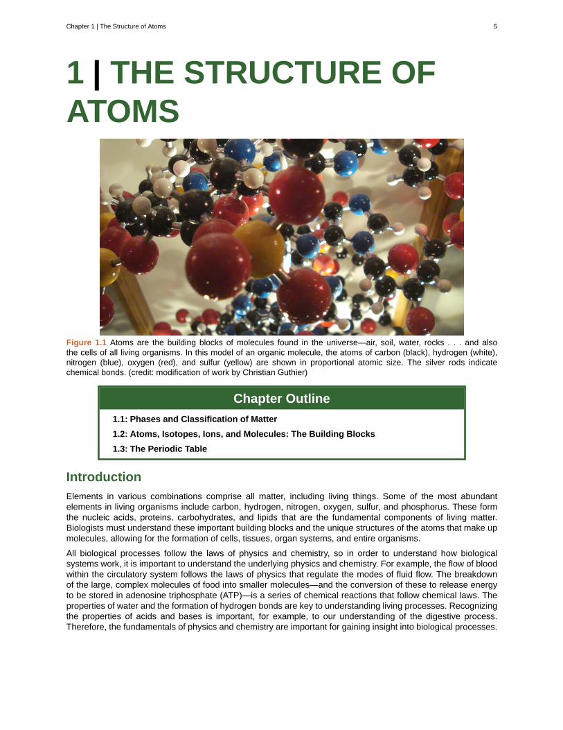
Figure 1.1 Atoms are the building blocks of molecules found in the universe—air, soil, water, rocks . . . and alsothe cells of all living organisms. In this model of an organic molecule, the atoms of carbon (black), hydrogen (white),nitrogen (blue), oxygen (red), and sulfur (yellow) are shown in proportional atomic size. The silver rods indicatechemical bonds. (credit: modification of work by Christian Guthier)
Chapter Outline
1.1: Phases and Classification of Matter
1.2: Atoms, Isotopes, Ions, and Molecules: The Building Blocks
1.3: The Periodic Table
Introduction
Elements in various combinations comprise all matter, including living things. Some of the most abundantelements in living organisms include carbon, hydrogen, nitrogen, oxygen, sulfur, and phosphorus. These formthe nucleic acids, proteins, carbohydrates, and lipids that are the fundamental components of living matter.Biologists must understand these important building blocks and the unique structures of the atoms that make upmolecules, allowing for the formation of cells, tissues, organ systems, and entire organisms.
All biological processes follow the laws of physics and chemistry, so in order to understand how biologicalsystems work, it is important to understand the underlying physics and chemistry. For example, the flow of bloodwithin the circulatory system follows the laws of physics that regulate the modes of fluid flow. The breakdownof the large, complex molecules of food into smaller molecules—and the conversion of these to release energyto be stored in adenosine triphosphate (ATP)—is a series of chemical reactions that follow chemical laws. Theproperties of water and the formation of hydrogen bonds are key to understanding living processes. Recognizingthe properties of acids and bases is important, for example, to our understanding of the digestive process.Therefore, the fundamentals of physics and chemistry are important for gaining insight into biological processes.
Chapter 1 | The Structure of Atoms 5
1.1 | Phases and Classification of Matter
By the end of this section, you will be able to:
• Describe the basic properties of each physical state of matter: solid, liquid, and gas
• Define and give examples of atoms and molecules
• Classify matter as an element, compound, homogeneous mixture, or heterogeneous mixture with regardto its physical state and composition
• Distinguish between mass and weight
• Apply the law of conservation of matter
Matter is defined as anything that occupies space and has mass, and it is all around us. Solids and liquids aremore obviously matter: We can see that they take up space, and their weight tells us that they have mass. Gasesare also matter; if gases did not take up space, a balloon would stay collapsed rather than inflate when filled withgas.
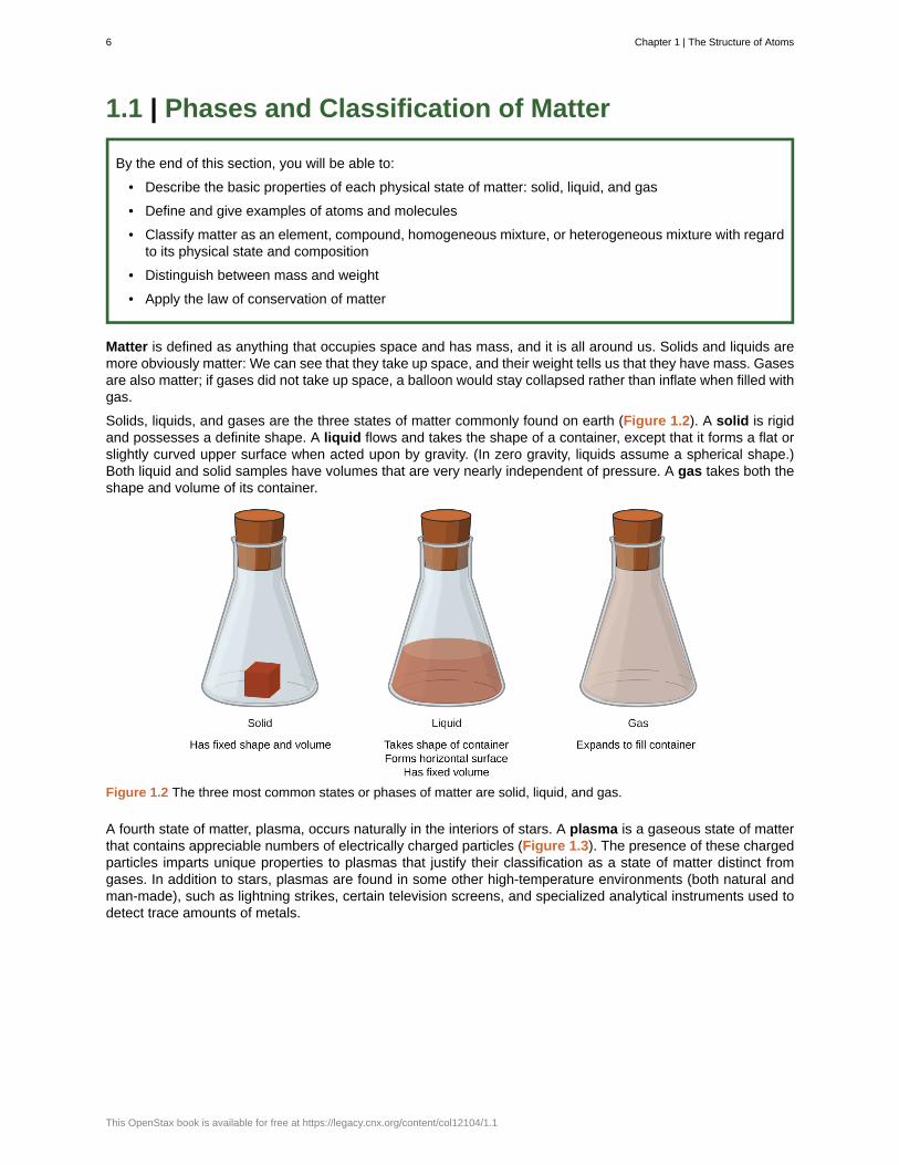
Solids, liquids, and gases are the three states of matter commonly found on earth (Figure 1.2). A solid is rigidand possesses a definite shape. A liquid flows and takes the shape of a container, except that it forms a flat orslightly curved upper surface when acted upon by gravity. (In zero gravity, liquids assume a spherical shape.)Both liquid and solid samples have volumes that are very nearly independent of pressure. A gas takes both theshape and volume of its container.
Figure 1.2 The three most common states or phases of matter are solid, liquid, and gas.
A fourth state of matter, plasma, occurs naturally in the interiors of stars. A plasma is a gaseous state of matterthat contains appreciable numbers of electrically charged particles (Figure 1.3). The presence of these chargedparticles imparts unique properties to plasmas that justify their classification as a state of matter distinct fromgases. In addition to stars, plasmas are found in some other high-temperature environments (both natural andman-made), such as lightning strikes, certain television screens, and specialized analytical instruments used todetect trace amounts of metals.
6 Chapter 1 | The Structure of Atoms
This OpenStax book is available for free at https://legacy.cnx.org/content/col12104/1.1

Figure 1.3 A plasma torch can be used to cut metal. (credit: “Hypertherm”/Wikimedia Commons)
In a tiny cell in a plasma television, the plasma emits ultraviolet light, which in turn causes the display at thatlocation to appear a specific color. The composite of these tiny dots of color makes up the image that yousee. Watch this video (http://openstax.org/l/16plasma) to learn more about plasma and the places youencounter it.
Some samples of matter appear to have properties of solids, liquids, and/or gases at the same time. This canoccur when the sample is composed of many small pieces. For example, we can pour sand as if it were a liquidbecause it is composed of many small grains of solid sand. Matter can also have properties of more than onestate when it is a mixture, such as with clouds. Clouds appear to behave somewhat like gases, but they areactually mixtures of air (gas) and tiny particles of water (liquid or solid).
The mass of an object is a measure of the amount of matter in it. One way to measure an object’s mass is tomeasure the force it takes to accelerate the object. It takes much more force to accelerate a car than a bicyclebecause the car has much more mass. A more common way to determine the mass of an object is to use abalance to compare its mass with a standard mass.
Although weight is related to mass, it is not the same thing. Weight refers to the force that gravity exerts on anobject. This force is directly proportional to the mass of the object. The weight of an object changes as the forceof gravity changes, but its mass does not. An astronaut’s mass does not change just because she goes to themoon. But her weight on the moon is only one-sixth her earth-bound weight because the moon’s gravity is only
Chapter 1 | The Structure of Atoms 7
one-sixth that of the earth’s. She may feel “weightless” during her trip when she experiences negligible externalforces (gravitational or any other), although she is, of course, never “massless.”
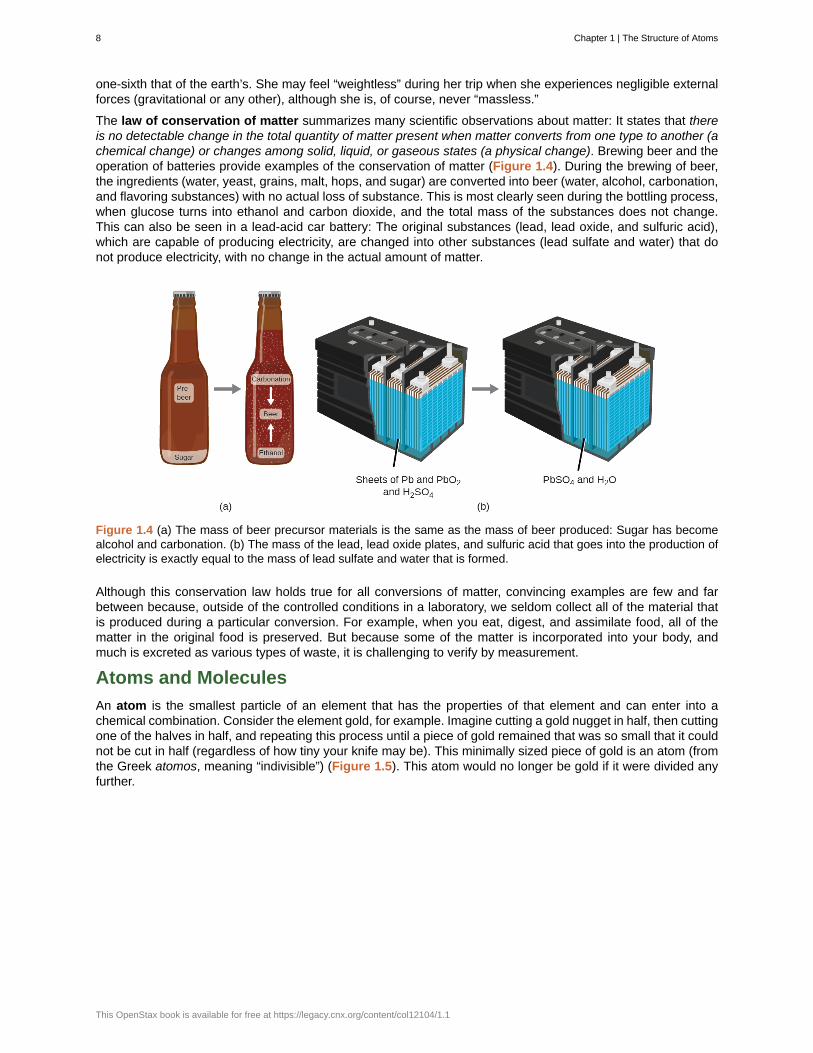
The law of conservation of matter summarizes many scientific observations about matter: It states that thereis no detectable change in the total quantity of matter present when matter converts from one type to another (achemical change) or changes among solid, liquid, or gaseous states (a physical change). Brewing beer and theoperation of batteries provide examples of the conservation of matter (Figure 1.4). During the brewing of beer,the ingredients (water, yeast, grains, malt, hops, and sugar) are converted into beer (water, alcohol, carbonation,and flavoring substances) with no actual loss of substance. This is most clearly seen during the bottling process,when glucose turns into ethanol and carbon dioxide, and the total mass of the substances does not change.This can also be seen in a lead-acid car battery: The original substances (lead, lead oxide, and sulfuric acid),which are capable of producing electricity, are changed into other substances (lead sulfate and water) that donot produce electricity, with no change in the actual amount of matter.
Figure 1.4 (a) The mass of beer precursor materials is the same as the mass of beer produced: Sugar has becomealcohol and carbonation. (b) The mass of the lead, lead oxide plates, and sulfuric acid that goes into the production ofelectricity is exactly equal to the mass of lead sulfate and water that is formed.
Although this conservation law holds true for all conversions of matter, convincing examples are few and farbetween because, outside of the controlled conditions in a laboratory, we seldom collect all of the material thatis produced during a particular conversion. For example, when you eat, digest, and assimilate food, all of thematter in the original food is preserved. But because some of the matter is incorporated into your body, andmuch is excreted as various types of waste, it is challenging to verify by measurement.
Atoms and Molecules
An atom is the smallest particle of an element that has the properties of that element and can enter into achemical combination. Consider the element gold, for example. Imagine cutting a gold nugget in half, then cuttingone of the halves in half, and repeating this process until a piece of gold remained that was so small that it couldnot be cut in half (regardless of how tiny your knife may be). This minimally sized piece of gold is an atom (fromthe Greek atomos, meaning “indivisible”) (Figure 1.5). This atom would no longer be gold if it were divided anyfurther.
8 Chapter 1 | The Structure of Atoms
This OpenStax book is available for free at https://legacy.cnx.org/content/col12104/1.1
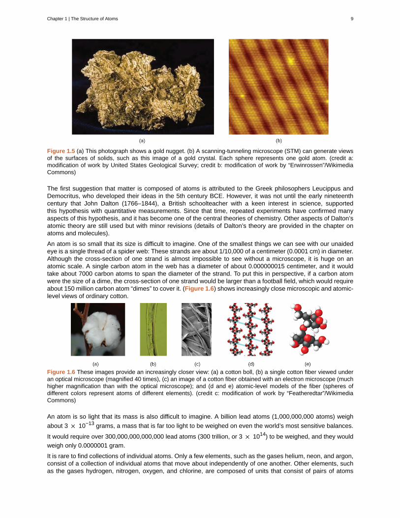
Figure 1.5 (a) This photograph shows a gold nugget. (b) A scanning-tunneling microscope (STM) can generate viewsof the surfaces of solids, such as this image of a gold crystal. Each sphere represents one gold atom. (credit a:modification of work by United States Geological Survey; credit b: modification of work by “Erwinrossen”/WikimediaCommons)
The first suggestion that matter is composed of atoms is attributed to the Greek philosophers Leucippus andDemocritus, who developed their ideas in the 5th century BCE. However, it was not until the early nineteenthcentury that John Dalton (1766–1844), a British schoolteacher with a keen interest in science, supportedthis hypothesis with quantitative measurements. Since that time, repeated experiments have confirmed manyaspects of this hypothesis, and it has become one of the central theories of chemistry. Other aspects of Dalton’satomic theory are still used but with minor revisions (details of Dalton’s theory are provided in the chapter onatoms and molecules).
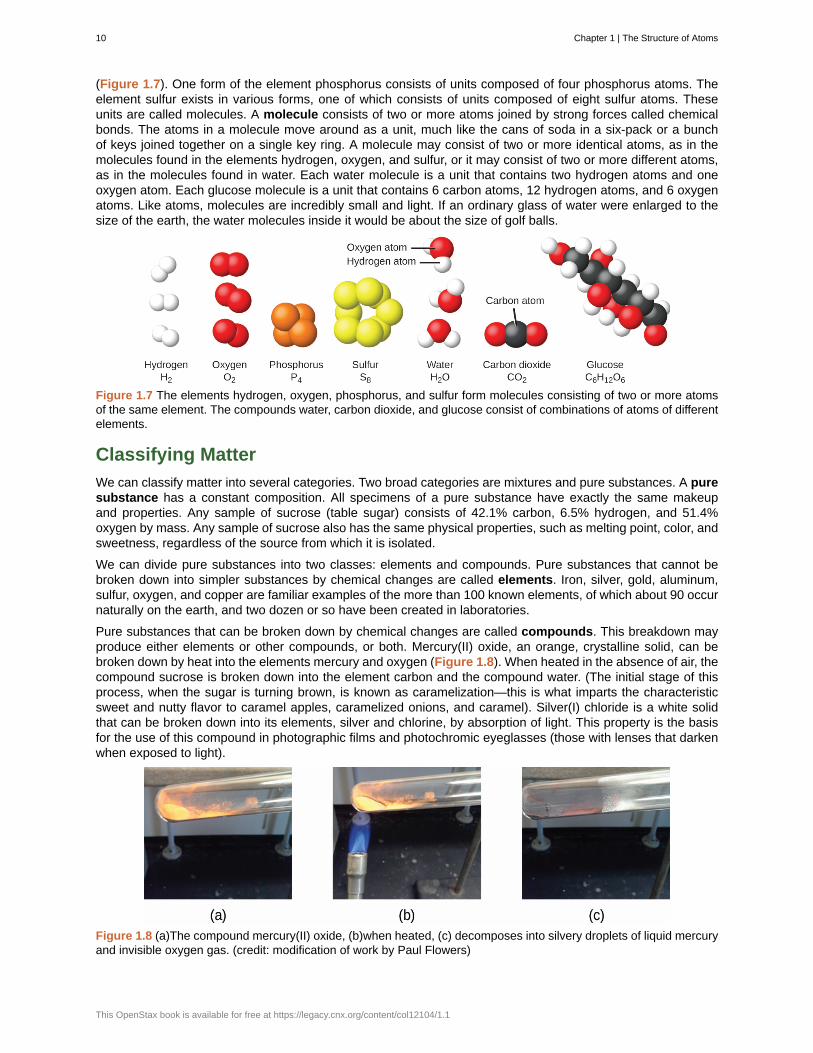
An atom is so small that its size is difficult to imagine. One of the smallest things we can see with our unaidedeye is a single thread of a spider web: These strands are about 1/10,000 of a centimeter (0.0001 cm) in diameter.Although the cross-section of one strand is almost impossible to see without a microscope, it is huge on anatomic scale. A single carbon atom in the web has a diameter of about 0.000000015 centimeter, and it wouldtake about 7000 carbon atoms to span the diameter of the strand. To put this in perspective, if a carbon atomwere the size of a dime, the cross-section of one strand would be larger than a football field, which would requireabout 150 million carbon atom “dimes” to cover it. (Figure 1.6) shows increasingly close microscopic and atomic-level views of ordinary cotton.
Figure 1.6 These images provide an increasingly closer view: (a) a cotton boll, (b) a single cotton fiber viewed underan optical microscope (magnified 40 times), (c) an image of a cotton fiber obtained with an electron microscope (muchhigher magnification than with the optical microscope); and (d and e) atomic-level models of the fiber (spheres ofdifferent colors represent atoms of different elements). (credit c: modification of work by “Featheredtar”/WikimediaCommons)
An atom is so light that its mass is also difficult to imagine. A billion lead atoms (1,000,000,000 atoms) weigh
about 3 × 10⁻¹³ grams, a mass that is far too light to be weighed on even the world’s most sensitive balances.
It would require over 300,000,000,000,000 lead atoms (300 trillion, or 3 × 10¹⁴) to be weighed, and they would
weigh only 0.0000001 gram.
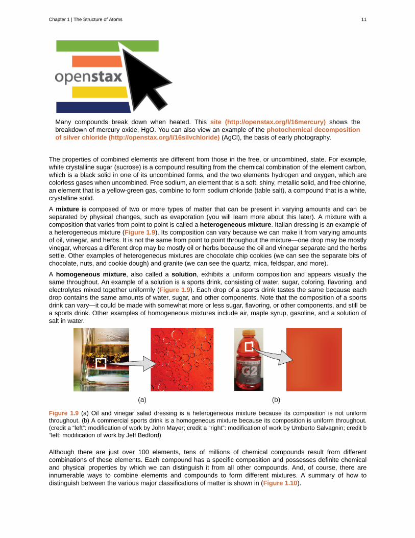
It is rare to find collections of individual atoms. Only a few elements, such as the gases helium, neon, and argon,consist of a collection of individual atoms that move about independently of one another. Other elements, suchas the gases hydrogen, nitrogen, oxygen, and chlorine, are composed of units that consist of pairs of atoms
Chapter 1 | The Structure of Atoms 9
(Figure 1.7). One form of the element phosphorus consists of units composed of four phosphorus atoms. Theelement sulfur exists in various forms, one of which consists of units composed of eight sulfur atoms. Theseunits are called molecules. A molecule consists of two or more atoms joined by strong forces called chemicalbonds. The atoms in a molecule move around as a unit, much like the cans of soda in a six-pack or a bunchof keys joined together on a single key ring. A molecule may consist of two or more identical atoms, as in themolecules found in the elements hydrogen, oxygen, and sulfur, or it may consist of two or more different atoms,as in the molecules found in water. Each water molecule is a unit that contains two hydrogen atoms and oneoxygen atom. Each glucose molecule is a unit that contains 6 carbon atoms, 12 hydrogen atoms, and 6 oxygenatoms. Like atoms, molecules are incredibly small and light. If an ordinary glass of water were enlarged to thesize of the earth, the water molecules inside it would be about the size of golf balls.
Figure 1.7 The elements hydrogen, oxygen, phosphorus, and sulfur form molecules consisting of two or more atomsof the same element. The compounds water, carbon dioxide, and glucose consist of combinations of atoms of differentelements.
Classifying Matter
We can classify matter into several categories. Two broad categories are mixtures and pure substances. A puresubstance has a constant composition. All specimens of a pure substance have exactly the same makeupand properties. Any sample of sucrose (table sugar) consists of 42.1% carbon, 6.5% hydrogen, and 51.4%oxygen by mass. Any sample of sucrose also has the same physical properties, such as melting point, color, andsweetness, regardless of the source from which it is isolated.
We can divide pure substances into two classes: elements and compounds. Pure substances that cannot bebroken down into simpler substances by chemical changes are called elements. Iron, silver, gold, aluminum,sulfur, oxygen, and copper are familiar examples of the more than 100 known elements, of which about 90 occurnaturally on the earth, and two dozen or so have been created in laboratories.
Pure substances that can be broken down by chemical changes are called compounds. This breakdown mayproduce either elements or other compounds, or both. Mercury(II) oxide, an orange, crystalline solid, can bebroken down by heat into the elements mercury and oxygen (Figure 1.8). When heated in the absence of air, thecompound sucrose is broken down into the element carbon and the compound water. (The initial stage of thisprocess, when the sugar is turning brown, is known as caramelization—this is what imparts the characteristicsweet and nutty flavor to caramel apples, caramelized onions, and caramel). Silver(I) chloride is a white solidthat can be broken down into its elements, silver and chlorine, by absorption of light. This property is the basisfor the use of this compound in photographic films and photochromic eyeglasses (those with lenses that darkenwhen exposed to light).
Figure 1.8 (a)The compound mercury(II) oxide, (b)when heated, (c) decomposes into silvery droplets of liquid mercuryand invisible oxygen gas. (credit: modification of work by Paul Flowers)
10 Chapter 1 | The Structure of Atoms
This OpenStax book is available for free at https://legacy.cnx.org/content/col12104/1.1
Many compounds break down when heated. This site (http://openstax.org/l/16mercury) shows thebreakdown of mercury oxide, HgO. You can also view an example of the photochemical decompositionof silver chloride (http://openstax.org/l/16silvchloride) (AgCl), the basis of early photography.
The properties of combined elements are different from those in the free, or uncombined, state. For example,white crystalline sugar (sucrose) is a compound resulting from the chemical combination of the element carbon,which is a black solid in one of its uncombined forms, and the two elements hydrogen and oxygen, which arecolorless gases when uncombined. Free sodium, an element that is a soft, shiny, metallic solid, and free chlorine,an element that is a yellow-green gas, combine to form sodium chloride (table salt), a compound that is a white,crystalline solid.
A mixture is composed of two or more types of matter that can be present in varying amounts and can beseparated by physical changes, such as evaporation (you will learn more about this later). A mixture with acomposition that varies from point to point is called a heterogeneous mixture. Italian dressing is an example ofa heterogeneous mixture (Figure 1.9). Its composition can vary because we can make it from varying amountsof oil, vinegar, and herbs. It is not the same from point to point throughout the mixture—one drop may be mostlyvinegar, whereas a different drop may be mostly oil or herbs because the oil and vinegar separate and the herbssettle. Other examples of heterogeneous mixtures are chocolate chip cookies (we can see the separate bits ofchocolate, nuts, and cookie dough) and granite (we can see the quartz, mica, feldspar, and more).
A homogeneous mixture, also called a solution, exhibits a uniform composition and appears visually thesame throughout. An example of a solution is a sports drink, consisting of water, sugar, coloring, flavoring, andelectrolytes mixed together uniformly (Figure 1.9). Each drop of a sports drink tastes the same because eachdrop contains the same amounts of water, sugar, and other components. Note that the composition of a sportsdrink can vary—it could be made with somewhat more or less sugar, flavoring, or other components, and still bea sports drink. Other examples of homogeneous mixtures include air, maple syrup, gasoline, and a solution ofsalt in water.
Figure 1.9 (a) Oil and vinegar salad dressing is a heterogeneous mixture because its composition is not uniformthroughout. (b) A commercial sports drink is a homogeneous mixture because its composition is uniform throughout.(credit a “left”: modification of work by John Mayer; credit a “right”: modification of work by Umberto Salvagnin; credit b“left: modification of work by Jeff Bedford)
Although there are just over 100 elements, tens of millions of chemical compounds result from differentcombinations of these elements. Each compound has a specific composition and possesses definite chemicaland physical properties by which we can distinguish it from all other compounds. And, of course, there areinnumerable ways to combine elements and compounds to form different mixtures. A summary of how todistinguish between the various major classifications of matter is shown in (Figure 1.10).
Chapter 1 | The Structure of Atoms 11
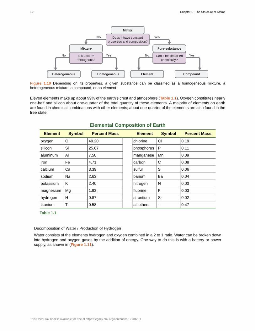
Figure 1.10 Depending on its properties, a given substance can be classified as a homogeneous mixture, aheterogeneous mixture, a compound, or an element.
Eleven elements make up about 99% of the earth’s crust and atmosphere (Table 1.1). Oxygen constitutes nearlyone-half and silicon about one-quarter of the total quantity of these elements. A majority of elements on earthare found in chemical combinations with other elements; about one-quarter of the elements are also found in thefree state.
Elemental Composition of Earth
Element Symbol Percent Mass Element Symbol Percent Mass
oxygen O 49.20 chlorine Cl 0.19
silicon Si 25.67 phosphorus P 0.11
aluminum Al 7.50 manganese Mn 0.09
iron Fe 4.71 carbon C 0.08
calcium Ca 3.39 sulfur S 0.06
sodium Na 2.63 barium Ba 0.04
potassium K 2.40 nitrogen N 0.03
magnesium Mg 1.93 fluorine F 0.03
hydrogen H 0.87 strontium Sr 0.02
titanium Ti 0.58 all others - 0.47
Table 1.1
Decomposition of Water / Production of Hydrogen
Water consists of the elements hydrogen and oxygen combined in a 2 to 1 ratio. Water can be broken downinto hydrogen and oxygen gases by the addition of energy. One way to do this is with a battery or powersupply, as shown in (Figure 1.11).
12 Chapter 1 | The Structure of Atoms
This OpenStax book is available for free at https://legacy.cnx.org/content/col12104/1.1
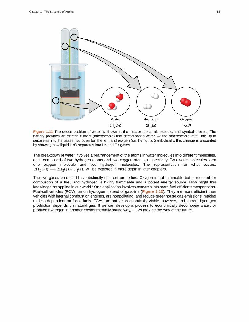
Figure 1.11 The decomposition of water is shown at the macroscopic, microscopic, and symbolic levels. Thebattery provides an electric current (microscopic) that decomposes water. At the macroscopic level, the liquidseparates into the gases hydrogen (on the left) and oxygen (on the right). Symbolically, this change is presentedby showing how liquid H2O separates into H2 and O2 gases.
The breakdown of water involves a rearrangement of the atoms in water molecules into different molecules,each composed of two hydrogen atoms and two oxygen atoms, respectively. Two water molecules formone oxygen molecule and two hydrogen molecules. The representation for what occurs,2H2 O(l) ⟶ 2H2(g) + O2(g), will be explored in more depth in later chapters.
The two gases produced have distinctly different properties. Oxygen is not flammable but is required forcombustion of a fuel, and hydrogen is highly flammable and a potent energy source. How might thisknowledge be applied in our world? One application involves research into more fuel-efficient transportation.Fuel-cell vehicles (FCV) run on hydrogen instead of gasoline (Figure 1.12). They are more efficient thanvehicles with internal combustion engines, are nonpolluting, and reduce greenhouse gas emissions, makingus less dependent on fossil fuels. FCVs are not yet economically viable, however, and current hydrogenproduction depends on natural gas. If we can develop a process to economically decompose water, orproduce hydrogen in another environmentally sound way, FCVs may be the way of the future.
Chapter 1 | The Structure of Atoms 13
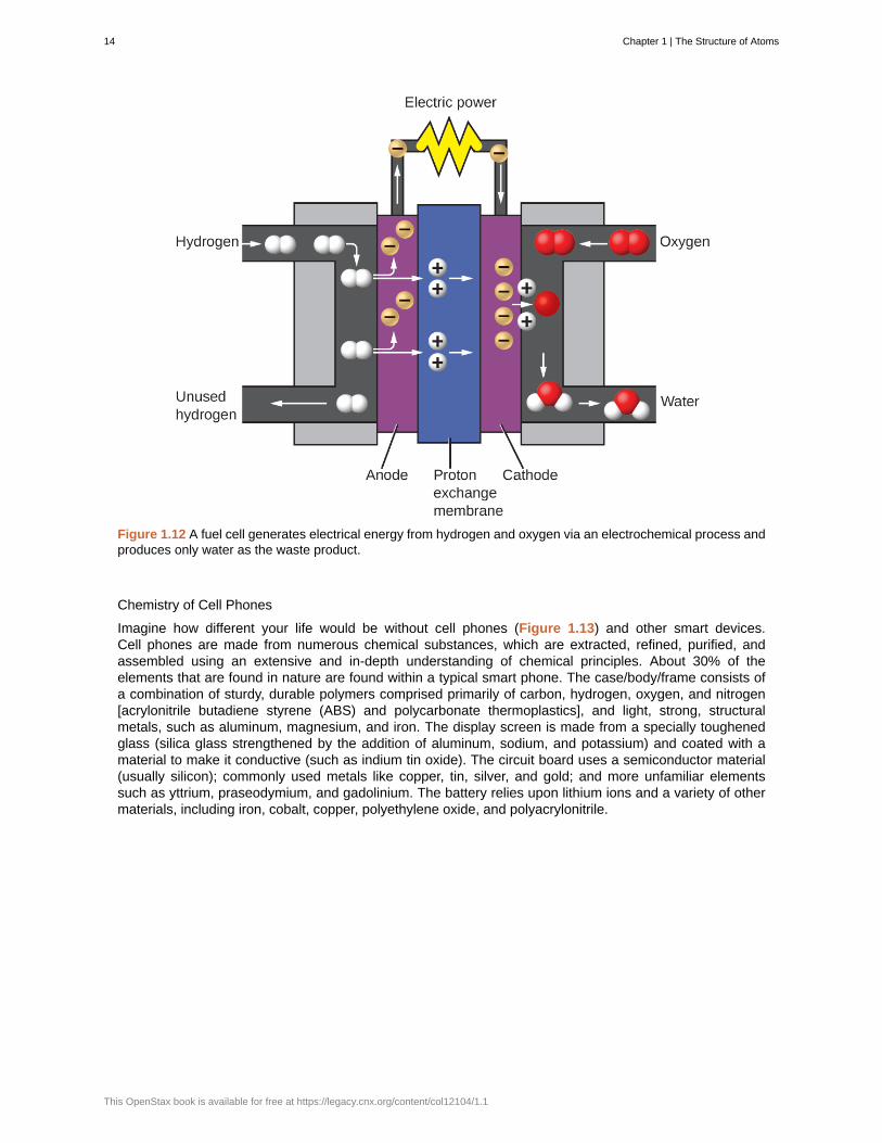
Figure 1.12 A fuel cell generates electrical energy from hydrogen and oxygen via an electrochemical process andproduces only water as the waste product.
Chemistry of Cell Phones
Imagine how different your life would be without cell phones (Figure 1.13) and other smart devices.Cell phones are made from numerous chemical substances, which are extracted, refined, purified, andassembled using an extensive and in-depth understanding of chemical principles. About 30% of theelements that are found in nature are found within a typical smart phone. The case/body/frame consists ofa combination of sturdy, durable polymers comprised primarily of carbon, hydrogen, oxygen, and nitrogen[acrylonitrile butadiene styrene (ABS) and polycarbonate thermoplastics], and light, strong, structuralmetals, such as aluminum, magnesium, and iron. The display screen is made from a specially toughenedglass (silica glass strengthened by the addition of aluminum, sodium, and potassium) and coated with amaterial to make it conductive (such as indium tin oxide). The circuit board uses a semiconductor material(usually silicon); commonly used metals like copper, tin, silver, and gold; and more unfamiliar elementssuch as yttrium, praseodymium, and gadolinium. The battery relies upon lithium ions and a variety of othermaterials, including iron, cobalt, copper, polyethylene oxide, and polyacrylonitrile.
14 Chapter 1 | The Structure of Atoms
This OpenStax book is available for free at https://legacy.cnx.org/content/col12104/1.1
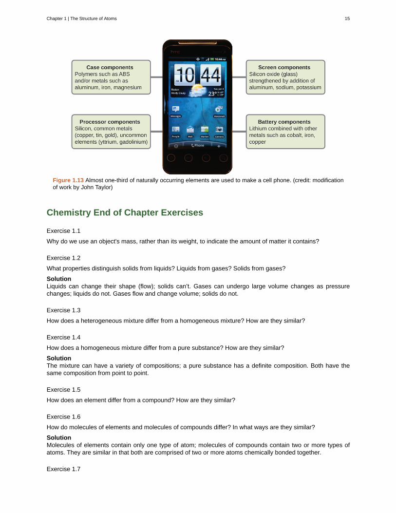
Figure 1.13 Almost one-third of naturally occurring elements are used to make a cell phone. (credit: modificationof work by John Taylor)
Chemistry End of Chapter Exercises
Exercise 1.1
Why do we use an object's mass, rather than its weight, to indicate the amount of matter it contains?
Exercise 1.2
What properties distinguish solids from liquids? Liquids from gases? Solids from gases?
SolutionLiquids can change their shape (flow); solids can’t. Gases can undergo large volume changes as pressurechanges; liquids do not. Gases flow and change volume; solids do not.
Exercise 1.3
How does a heterogeneous mixture differ from a homogeneous mixture? How are they similar?
Exercise 1.4
How does a homogeneous mixture differ from a pure substance? How are they similar?
SolutionThe mixture can have a variety of compositions; a pure substance has a definite composition. Both have thesame composition from point to point.
Exercise 1.5
How does an element differ from a compound? How are they similar?
Exercise 1.6
How do molecules of elements and molecules of compounds differ? In what ways are they similar?
SolutionMolecules of elements contain only one type of atom; molecules of compounds contain two or more types ofatoms. They are similar in that both are comprised of two or more atoms chemically bonded together.
Exercise 1.7
Chapter 1 | The Structure of Atoms 15

How does an atom differ from a molecule? In what ways are they similar?
Exercise 1.8
Many of the items you purchase are mixtures of pure compounds. Select three of these commercial productsand prepare a list of the ingredients that are pure compounds.
SolutionAnswers will vary. Sample answer: Gatorade contains water, sugar, dextrose, citric acid, salt, sodium chloride,monopotassium phosphate, and sucrose acetate isobutyrate.
Exercise 1.9
Classify each of the following as an element, a compound, or a mixture:
(a) copper
(b) water
(c) nitrogen
(d) sulfur
(e) air
(f) sucrose
(g) a substance composed of molecules each of which contains two iodine atoms
(h) gasoline
Exercise 1.10
Classify each of the following as an element, a compound, or a mixture:
(a) iron
(b) oxygen
(c) mercury oxide
(d) pancake syrup
(e) carbon dioxide
(f) a substance composed of molecules each of which contains one hydrogen atom and one chlorine atom
(g) baking soda
(h) baking powder
Solution(a) element; (b) element; (c) compound; (d) mixture, (e) compound; (f) compound; (g) compound; (h) mixture
Exercise 1.11
A sulfur atom and a sulfur molecule are not identical. What is the difference?
Exercise 1.12
How are the molecules in oxygen gas, the molecules in hydrogen gas, and water molecules similar? How dothey differ?
SolutionIn each case, a molecule consists of two or more combined atoms. They differ in that the types of atoms changefrom one substance to the next.
Exercise 1.13
We refer to astronauts in space as weightless, but not without mass. Why?
Exercise 1.14
16 Chapter 1 | The Structure of Atoms
This OpenStax book is available for free at https://legacy.cnx.org/content/col12104/1.1
As we drive an automobile, we don't think about the chemicals consumed and produced. Prepare a list of theprincipal chemicals consumed and produced during the operation of an automobile.
SolutionGasoline (a mixture of compounds), oxygen, and to a lesser extent, nitrogen are consumed. Carbon dioxide andwater are the principal products. Carbon monoxide and nitrogen oxides are produced in lesser amounts.
Exercise 1.15
Matter is everywhere around us. Make a list by name of fifteen different kinds of matter that you encounter everyday. Your list should include (and label at least one example of each) the following: a solid, a liquid, a gas, anelement, a compound, a homogenous mixture, a heterogeneous mixture, and a pure substance.
Exercise 1.16
When elemental iron corrodes it combines with oxygen in the air to ultimately form red brown iron(III) oxide whichwe call rust. (a) If a shiny iron nail with an initial mass of 23.2 g is weighed after being coated in a layer of rust,would you expect the mass to have increased, decreased, or remained the same? Explain. (b) If the mass of theiron nail increases to 24.1 g, what mass of oxygen combined with the iron?
Solution(a) Increased as it would have combined with oxygen in the air thus increasing the amount of matter andtherefore the mass. (b) 0.9 g
Exercise 1.17
As stated in the text, convincing examples that demonstrate the law of conservation of matter outside of thelaboratory are few and far between. Indicate whether the mass would increase, decrease, or stay the same forthe following scenarios where chemical reactions take place:
(a) Exactly one pound of bread dough is placed in a baking tin. The dough is cooked in an oven at 350 °Freleasing a wonderful aroma of freshly baked bread during the cooking process. Is the mass of the baked loafless than, greater than, or the same as the one pound of original dough? Explain.
(b) When magnesium burns in air a white flaky ash of magnesium oxide is produced. Is the mass of magnesiumoxide less than, greater than, or the same as the original piece of magnesium? Explain.
(c) Antoine Lavoisier, the French scientist credited with first stating the law of conservation of matter, heateda mixture of tin and air in a sealed flask to produce tin oxide. Did the mass of the sealed flask and contentsdecrease, increase, or remain the same after the heating?
Exercise 1.18
Yeast converts glucose to ethanol and carbon dioxide during anaerobic fermentation as depicted in the simplechemical equation here:
glucose ⟶ ethanol + carbon dioxide
(a) If 200.0 g of glucose is fully converted, what will be the total mass of ethanol and carbon dioxide produced?
(b) If the fermentation is carried out in an open container, would you expect the mass of the container andcontents after fermentation to be less than, greater than, or the same as the mass of the container and contentsbefore fermentation? Explain.
(c) If 97.7 g of carbon dioxide is produced, what mass of ethanol is produced?
Solution(a) 200.0 g; (b) The mass of the container and contents would decrease as carbon dioxide is a gaseous productand would leave the container. (c) 102.3 g
Chapter 1 | The Structure of Atoms 17
1.2 | Atoms, Isotopes, Ions, and Molecules: The
Building Blocks
By the end of this section, you will be able to:
• Define matter and elements
• Describe the interrelationship between protons, neutrons, and electrons
• Compare the ways in which electrons can be donated or shared between atoms
• Explain the ways in which naturally occurring elements combine to create molecules, cells, tissues,organ systems, and organisms
At its most fundamental level, life is made up of matter. Matter is any substance that occupies space andhas mass. Elements are unique forms of matter with specific chemical and physical properties that cannot bebroken down into smaller substances by ordinary chemical reactions. There are 118 elements, but only 92 occurnaturally. The remaining elements are synthesized in laboratories and are unstable.
Each element is designated by its chemical symbol, which is a single capital letter or, when the first letter isalready “taken” by another element, a combination of two letters. Some elements follow the English term for theelement, such as C for carbon and Ca for calcium. Other elements’ chemical symbols derive from their Latinnames; for example, the symbol for sodium is Na, referring to natrium, the Latin word for sodium.
The four elements common to all living organisms are oxygen (O), carbon (C), hydrogen (H), and nitrogen (N). Inthe non-living world, elements are found in different proportions, and some elements common to living organismsare relatively rare on the earth as a whole, as shown in Table 1.2. For example, the atmosphere is rich in nitrogenand oxygen but contains little carbon and hydrogen, while the earth’s crust, although it contains oxygen and asmall amount of hydrogen, has little nitrogen and carbon. In spite of their differences in abundance, all elementsand the chemical reactions between them obey the same chemical and physical laws regardless of whether theyare a part of the living or non-living world.
Approximate Percentage of Elements in Living Organisms (Humans) Comparedto the Non-living World
Element Life (Humans) Atmosphere Earth’s Crust
Oxygen (O) 65% 21% 46%
Carbon (C) 18% trace trace
Hydrogen (H) 10% trace 0.1%
Nitrogen (N) 3% 78% trace
Table 1.2
The Structure of the Atom
To understand how elements come together, we must first discuss the smallest component or building block of anelement, the atom. An atom is the smallest unit of matter that retains all of the chemical properties of an element.For example, one gold atom has all of the properties of gold in that it is a solid metal at room temperature. Agold coin is simply a very large number of gold atoms molded into the shape of a coin and containing smallamounts of other elements known as impurities. Gold atoms cannot be broken down into anything smaller whilestill retaining the properties of gold.
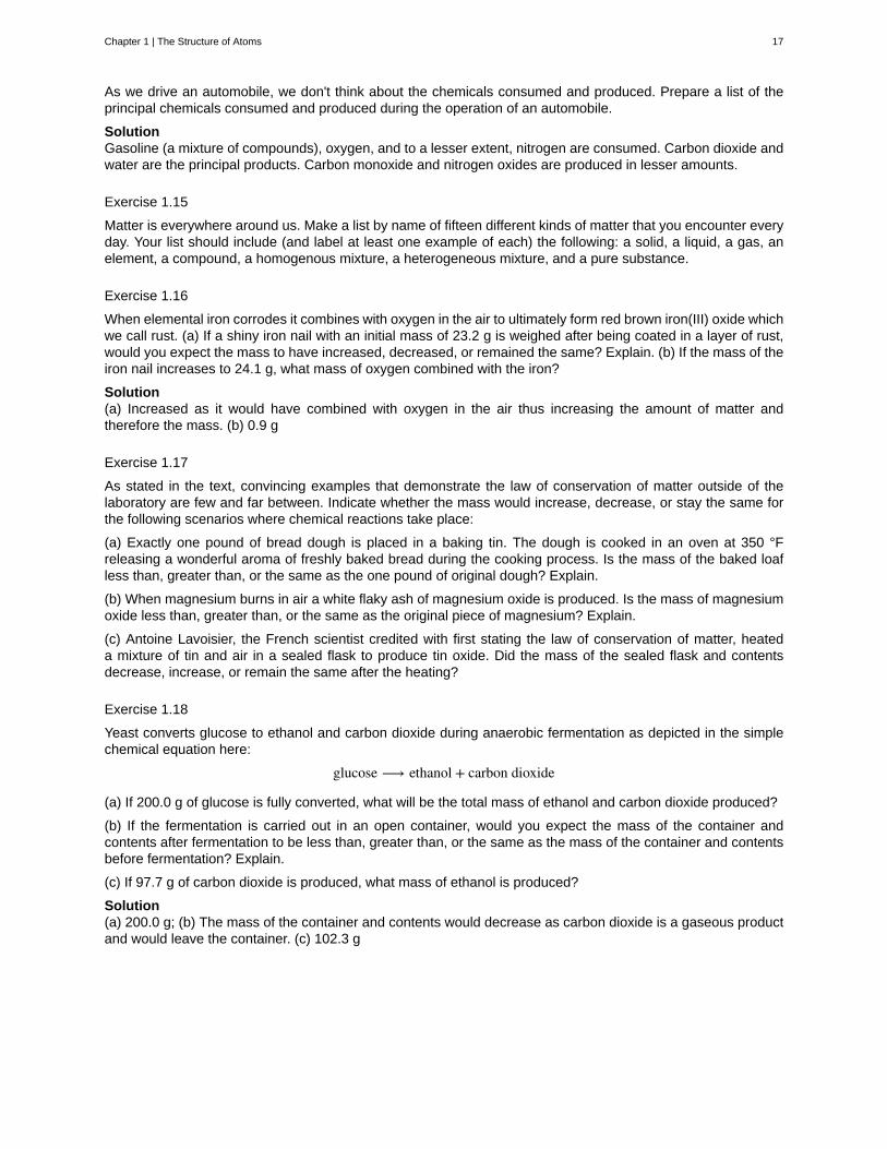
An atom is composed of two regions: the nucleus, which is in the center of the atom and contains protonsand neutrons, and the outermost region of the atom which holds its electrons in orbit around the nucleus, asillustrated in Figure 1.14. Atoms contain protons, electrons, and neutrons, among other subatomic particles. The
18 Chapter 1 | The Structure of Atoms
This OpenStax book is available for free at https://legacy.cnx.org/content/col12104/1.1
only exception is hydrogen (H), which is made of one proton and one electron with no neutrons.
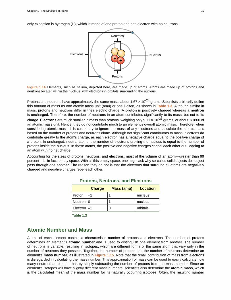
Figure 1.14 Elements, such as helium, depicted here, are made up of atoms. Atoms are made up of protons andneutrons located within the nucleus, with electrons in orbitals surrounding the nucleus.
Protons and neutrons have approximately the same mass, about 1.67 × 10⁻²⁴ grams. Scientists arbitrarily definethis amount of mass as one atomic mass unit (amu) or one Dalton, as shown in Table 1.3. Although similar inmass, protons and neutrons differ in their electric charge. A proton is positively charged whereas a neutronis uncharged. Therefore, the number of neutrons in an atom contributes significantly to its mass, but not to its
charge. Electrons are much smaller in mass than protons, weighing only 9.11 × 10⁻²⁸ grams, or about 1/1800 ofan atomic mass unit. Hence, they do not contribute much to an element’s overall atomic mass. Therefore, whenconsidering atomic mass, it is customary to ignore the mass of any electrons and calculate the atom’s massbased on the number of protons and neutrons alone. Although not significant contributors to mass, electrons docontribute greatly to the atom’s charge, as each electron has a negative charge equal to the positive charge ofa proton. In uncharged, neutral atoms, the number of electrons orbiting the nucleus is equal to the number ofprotons inside the nucleus. In these atoms, the positive and negative charges cancel each other out, leading toan atom with no net charge.
Accounting for the sizes of protons, neutrons, and electrons, most of the volume of an atom—greater than 99percent—is, in fact, empty space. With all this empty space, one might ask why so-called solid objects do not justpass through one another. The reason they do not is that the electrons that surround all atoms are negativelycharged and negative charges repel each other.
Protons, Neutrons, and Electrons
Charge Mass (amu) Location
Proton +1 1 nucleus
Neutron 0 1 nucleus
Electron –1 0 orbitals
Table 1.3
Atomic Number and Mass
Atoms of each element contain a characteristic number of protons and electrons. The number of protonsdetermines an element’s atomic number and is used to distinguish one element from another. The numberof neutrons is variable, resulting in isotopes, which are different forms of the same atom that vary only in thenumber of neutrons they possess. Together, the number of protons and the number of neutrons determine anelement’s mass number, as illustrated in Figure 1.15. Note that the small contribution of mass from electronsis disregarded in calculating the mass number. This approximation of mass can be used to easily calculate howmany neutrons an element has by simply subtracting the number of protons from the mass number. Since anelement’s isotopes will have slightly different mass numbers, scientists also determine the atomic mass, whichis the calculated mean of the mass number for its naturally occurring isotopes. Often, the resulting number
Chapter 1 | The Structure of Atoms 19
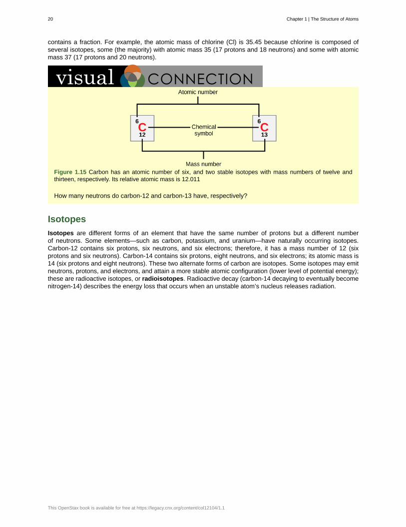
contains a fraction. For example, the atomic mass of chlorine (Cl) is 35.45 because chlorine is composed ofseveral isotopes, some (the majority) with atomic mass 35 (17 protons and 18 neutrons) and some with atomicmass 37 (17 protons and 20 neutrons).
Figure 1.15 Carbon has an atomic number of six, and two stable isotopes with mass numbers of twelve andthirteen, respectively. Its relative atomic mass is 12.011
How many neutrons do carbon-12 and carbon-13 have, respectively?
Isotopes
Isotopes are different forms of an element that have the same number of protons but a different numberof neutrons. Some elements—such as carbon, potassium, and uranium—have naturally occurring isotopes.Carbon-12 contains six protons, six neutrons, and six electrons; therefore, it has a mass number of 12 (sixprotons and six neutrons). Carbon-14 contains six protons, eight neutrons, and six electrons; its atomic mass is14 (six protons and eight neutrons). These two alternate forms of carbon are isotopes. Some isotopes may emitneutrons, protons, and electrons, and attain a more stable atomic configuration (lower level of potential energy);these are radioactive isotopes, or radioisotopes. Radioactive decay (carbon-14 decaying to eventually becomenitrogen-14) describes the energy loss that occurs when an unstable atom’s nucleus releases radiation.
20 Chapter 1 | The Structure of Atoms
This OpenStax book is available for free at https://legacy.cnx.org/content/col12104/1.1

Carbon DatingCarbon is normally present in the atmosphere in the form of gaseous compounds like carbon dioxide and
methane. Carbon-14 (14C) is a naturally occurring radioisotope that is created in the atmosphere from
atmospheric 14N (nitrogen) by the addition of a neutron and the loss of a proton because of cosmic rays.
This is a continuous process, so more 14C is always being created. As a living organism incorporates 14C
initially as carbon dioxide fixed in the process of photosynthesis, the relative amount of 14C in its body is
equal to the concentration of 14C in the atmosphere. When an organism dies, it is no longer ingesting 14C,
so the ratio between 14C and 12C will decline as 14C decays gradually to 14N by a process called betadecay—the emission of electrons or positrons. This decay gives off energy in a slow process.
After approximately 5,730 years, half of the starting concentration of 14C will have been converted back to14N. The time it takes for half of the original concentration of an isotope to decay back to its more stable
form is called its half-life. Because the half-life of 14C is long, it is used to date formerly living objects such
as old bones or wood. Comparing the ratio of the 14C concentration found in an object to the amount of 14Cdetected in the atmosphere, the amount of the isotope that has not yet decayed can be determined. On thebasis of this amount, the age of the material, such as the pygmy mammoth shown in Figure 1.16, can becalculated with accuracy if it is not much older than about 50,000 years. Other elements have isotopes with
different half lives. For example, 40K (potassium-40) has a half-life of 1.25 billion years, and 235U (Uranium235) has a half-life of about 700 million years. Through the use of radiometric dating, scientists can studythe age of fossils or other remains of extinct organisms to understand how organisms have evolved fromearlier species.
Figure 1.16 The age of carbon-containing remains less than about 50,000 years old, such as this pygmymammoth, can be determined using carbon dating. (credit: Bill Faulkner, NPS)
Link to Learning
Chapter 1 | The Structure of Atoms 21
To learn more about atoms, isotopes, and how to tell one isotope from another, run the simulation.(This media type is not supported in this reader. Click to open media in browser.)(http://legacy.cnx.org/content/m44390/1.21/#eip-id1165071748010)
The Periodic Table
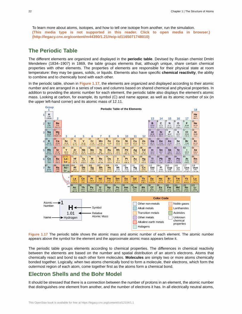
The different elements are organized and displayed in the periodic table. Devised by Russian chemist DmitriMendeleev (1834–1907) in 1869, the table groups elements that, although unique, share certain chemicalproperties with other elements. The properties of elements are responsible for their physical state at roomtemperature: they may be gases, solids, or liquids. Elements also have specific chemical reactivity, the abilityto combine and to chemically bond with each other.
In the periodic table, shown in Figure 1.17, the elements are organized and displayed according to their atomicnumber and are arranged in a series of rows and columns based on shared chemical and physical properties. Inaddition to providing the atomic number for each element, the periodic table also displays the element’s atomicmass. Looking at carbon, for example, its symbol (C) and name appear, as well as its atomic number of six (inthe upper left-hand corner) and its atomic mass of 12.11.
Figure 1.17 The periodic table shows the atomic mass and atomic number of each element. The atomic numberappears above the symbol for the element and the approximate atomic mass appears below it.
The periodic table groups elements according to chemical properties. The differences in chemical reactivitybetween the elements are based on the number and spatial distribution of an atom’s electrons. Atoms thatchemically react and bond to each other form molecules. Molecules are simply two or more atoms chemicallybonded together. Logically, when two atoms chemically bond to form a molecule, their electrons, which form theoutermost region of each atom, come together first as the atoms form a chemical bond.
Electron Shells and the Bohr Model
It should be stressed that there is a connection between the number of protons in an element, the atomic numberthat distinguishes one element from another, and the number of electrons it has. In all electrically neutral atoms,
22 Chapter 1 | The Structure of Atoms
This OpenStax book is available for free at https://legacy.cnx.org/content/col12104/1.1
the number of electrons is the same as the number of protons. Thus, each element, at least when electricallyneutral, has a characteristic number of electrons equal to its atomic number.
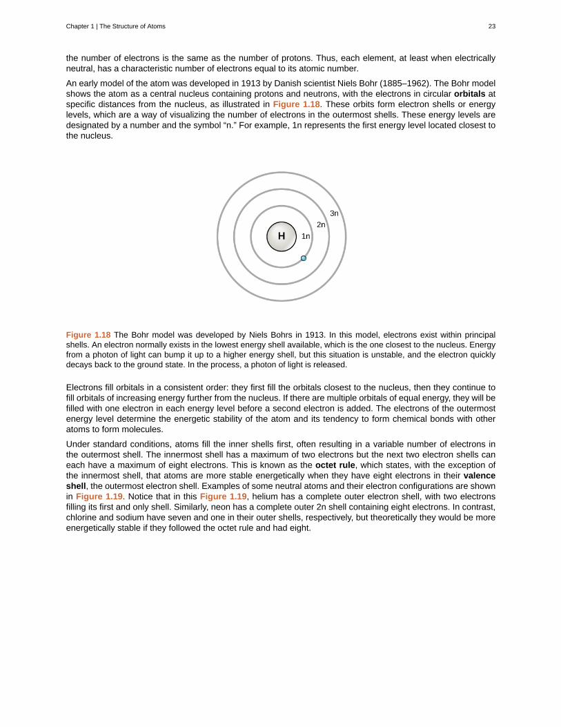
An early model of the atom was developed in 1913 by Danish scientist Niels Bohr (1885–1962). The Bohr modelshows the atom as a central nucleus containing protons and neutrons, with the electrons in circular orbitals atspecific distances from the nucleus, as illustrated in Figure 1.18. These orbits form electron shells or energylevels, which are a way of visualizing the number of electrons in the outermost shells. These energy levels aredesignated by a number and the symbol “n.” For example, 1n represents the first energy level located closest tothe nucleus.
Figure 1.18 The Bohr model was developed by Niels Bohrs in 1913. In this model, electrons exist within principalshells. An electron normally exists in the lowest energy shell available, which is the one closest to the nucleus. Energyfrom a photon of light can bump it up to a higher energy shell, but this situation is unstable, and the electron quicklydecays back to the ground state. In the process, a photon of light is released.
Electrons fill orbitals in a consistent order: they first fill the orbitals closest to the nucleus, then they continue tofill orbitals of increasing energy further from the nucleus. If there are multiple orbitals of equal energy, they will befilled with one electron in each energy level before a second electron is added. The electrons of the outermostenergy level determine the energetic stability of the atom and its tendency to form chemical bonds with otheratoms to form molecules.
Under standard conditions, atoms fill the inner shells first, often resulting in a variable number of electrons inthe outermost shell. The innermost shell has a maximum of two electrons but the next two electron shells caneach have a maximum of eight electrons. This is known as the octet rule, which states, with the exception ofthe innermost shell, that atoms are more stable energetically when they have eight electrons in their valenceshell, the outermost electron shell. Examples of some neutral atoms and their electron configurations are shownin Figure 1.19. Notice that in this Figure 1.19, helium has a complete outer electron shell, with two electronsfilling its first and only shell. Similarly, neon has a complete outer 2n shell containing eight electrons. In contrast,chlorine and sodium have seven and one in their outer shells, respectively, but theoretically they would be moreenergetically stable if they followed the octet rule and had eight.
Chapter 1 | The Structure of Atoms 23
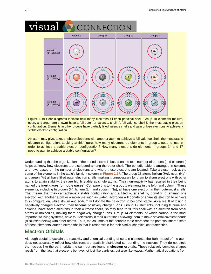
Figure 1.19 Bohr diagrams indicate how many electrons fill each principal shell. Group 18 elements (helium,neon, and argon are shown) have a full outer, or valence, shell. A full valence shell is the most stable electronconfiguration. Elements in other groups have partially filled valence shells and gain or lose electrons to achieve astable electron configuration.
An atom may give, take, or share electrons with another atom to achieve a full valence shell, the most stableelectron configuration. Looking at this figure, how many electrons do elements in group 1 need to lose inorder to achieve a stable electron configuration? How many electrons do elements in groups 14 and 17need to gain to achieve a stable configuration?
Understanding that the organization of the periodic table is based on the total number of protons (and electrons)helps us know how electrons are distributed among the outer shell. The periodic table is arranged in columnsand rows based on the number of electrons and where these electrons are located. Take a closer look at thesome of the elements in the table’s far right column in Figure 1.17. The group 18 atoms helium (He), neon (Ne),and argon (Ar) all have filled outer electron shells, making it unnecessary for them to share electrons with otheratoms to attain stability; they are highly stable as single atoms. Their non-reactivity has resulted in their beingnamed the inert gases (or noble gases). Compare this to the group 1 elements in the left-hand column. Theseelements, including hydrogen (H), lithium (Li), and sodium (Na), all have one electron in their outermost shells.That means that they can achieve a stable configuration and a filled outer shell by donating or sharing oneelectron with another atom or a molecule such as water. Hydrogen will donate or share its electron to achievethis configuration, while lithium and sodium will donate their electron to become stable. As a result of losing anegatively charged electron, they become positively charged ions. Group 17 elements, including fluorine andchlorine, have seven electrons in their outmost shells, so they tend to fill this shell with an electron from otheratoms or molecules, making them negatively charged ions. Group 14 elements, of which carbon is the mostimportant to living systems, have four electrons in their outer shell allowing them to make several covalent bonds(discussed below) with other atoms. Thus, the columns of the periodic table represent the potential shared stateof these elements’ outer electron shells that is responsible for their similar chemical characteristics.
Electron Orbitals
Although useful to explain the reactivity and chemical bonding of certain elements, the Bohr model of the atomdoes not accurately reflect how electrons are spatially distributed surrounding the nucleus. They do not circlethe nucleus like the earth orbits the sun, but are found in electron orbitals. These relatively complex shapesresult from the fact that electrons behave not just like particles, but also like waves. Mathematical equations from
24 Chapter 1 | The Structure of Atoms
This OpenStax book is available for free at https://legacy.cnx.org/content/col12104/1.1
quantum mechanics known as wave functions can predict within a certain level of probability where an electronmight be at any given time. The area where an electron is most likely to be found is called its orbital.
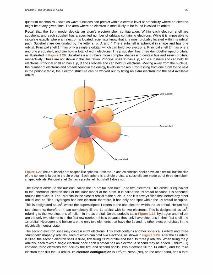
Recall that the Bohr model depicts an atom’s electron shell configuration. Within each electron shell aresubshells, and each subshell has a specified number of orbitals containing electrons. While it is impossible tocalculate exactly where an electron is located, scientists know that it is most probably located within its orbitalpath. Subshells are designated by the letter s, p, d, and f. The s subshell is spherical in shape and has oneorbital. Principal shell 1n has only a single s orbital, which can hold two electrons. Principal shell 2n has one sand one p subshell, and can hold a total of eight electrons. The p subshell has three dumbbell-shaped orbitals,as illustrated in Figure 1.20. Subshells d and f have more complex shapes and contain five and seven orbitals,respectively. These are not shown in the illustration. Principal shell 3n has s, p, and d subshells and can hold 18electrons. Principal shell 4n has s, p, d and f orbitals and can hold 32 electrons. Moving away from the nucleus,the number of electrons and orbitals found in the energy levels increases. Progressing from one atom to the nextin the periodic table, the electron structure can be worked out by fitting an extra electron into the next availableorbital.
Figure 1.20 The s subshells are shaped like spheres. Both the 1n and 2n principal shells have an s orbital, but the sizeof the sphere is larger in the 2n orbital. Each sphere is a single orbital. p subshells are made up of three dumbbell-shaped orbitals. Principal shell 2n has a p subshell, but shell 1 does not.
The closest orbital to the nucleus, called the 1s orbital, can hold up to two electrons. This orbital is equivalentto the innermost electron shell of the Bohr model of the atom. It is called the 1s orbital because it is sphericalaround the nucleus. The 1s orbital is the closest orbital to the nucleus, and it is always filled first, before any otherorbital can be filled. Hydrogen has one electron; therefore, it has only one spot within the 1s orbital occupied.
This is designated as 1s1, where the superscripted 1 refers to the one electron within the 1s orbital. Helium has
two electrons; therefore, it can completely fill the 1s orbital with its two electrons. This is designated as 1s2,referring to the two electrons of helium in the 1s orbital. On the periodic table Figure 1.17, hydrogen and heliumare the only two elements in the first row (period); this is because they only have electrons in their first shell, the1s orbital. Hydrogen and helium are the only two elements that have the 1s and no other electron orbitals in theelectrically neutral state.
The second electron shell may contain eight electrons. This shell contains another spherical s orbital and three“dumbbell” shaped p orbitals, each of which can hold two electrons, as shown in Figure 1.20. After the 1s orbitalis filled, the second electron shell is filled, first filling its 2s orbital and then its three p orbitals. When filling the porbitals, each takes a single electron; once each p orbital has an electron, a second may be added. Lithium (Li)contains three electrons that occupy the first and second shells. Two electrons fill the 1s orbital, and the third
electron then fills the 2s orbital. Its electron configuration is 1s22s1. Neon (Ne), on the other hand, has a total
Chapter 1 | The Structure of Atoms 25
of ten electrons: two are in its innermost 1s orbital and eight fill its second shell (two each in the 2s and three porbitals); thus, it is an inert gas and energetically stable as a single atom that will rarely form a chemical bondwith other atoms. Larger elements have additional orbitals, making up the third electron shell. While the conceptsof electron shells and orbitals are closely related, orbitals provide a more accurate depiction of the electronconfiguration of an atom because the orbital model specifies the different shapes and special orientations of allthe places that electrons may occupy.
Link to Learning
Watch this visual animation to see the spatial arrangement of the p and s orbitals. (This media type isnot supported in this reader. Click to open media in browser.) (http://legacy.cnx.org/content/m44390/1.21/#eip-id5846277)
Chemical Reactions and Molecules
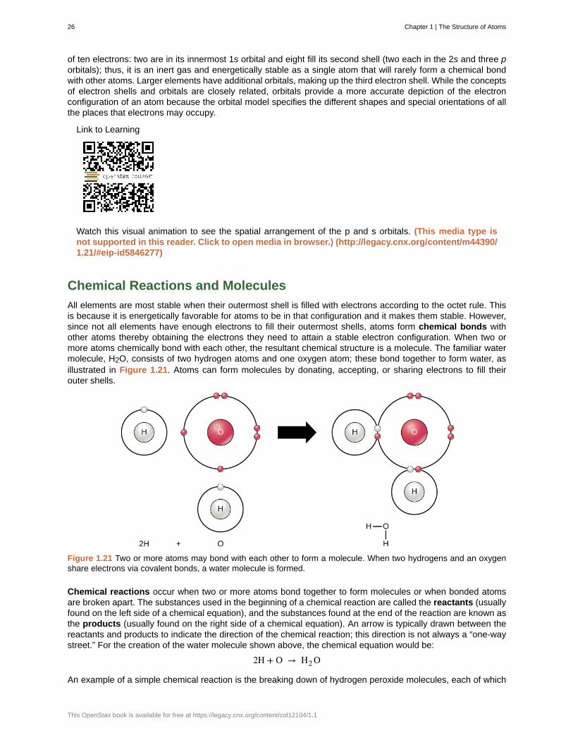
All elements are most stable when their outermost shell is filled with electrons according to the octet rule. Thisis because it is energetically favorable for atoms to be in that configuration and it makes them stable. However,since not all elements have enough electrons to fill their outermost shells, atoms form chemical bonds withother atoms thereby obtaining the electrons they need to attain a stable electron configuration. When two ormore atoms chemically bond with each other, the resultant chemical structure is a molecule. The familiar watermolecule, H2O, consists of two hydrogen atoms and one oxygen atom; these bond together to form water, asillustrated in Figure 1.21. Atoms can form molecules by donating, accepting, or sharing electrons to fill theirouter shells.
Figure 1.21 Two or more atoms may bond with each other to form a molecule. When two hydrogens and an oxygenshare electrons via covalent bonds, a water molecule is formed.
Chemical reactions occur when two or more atoms bond together to form molecules or when bonded atomsare broken apart. The substances used in the beginning of a chemical reaction are called the reactants (usuallyfound on the left side of a chemical equation), and the substances found at the end of the reaction are known asthe products (usually found on the right side of a chemical equation). An arrow is typically drawn between thereactants and products to indicate the direction of the chemical reaction; this direction is not always a “one-waystreet.” For the creation of the water molecule shown above, the chemical equation would be:
2H + O → H2 O
An example of a simple chemical reaction is the breaking down of hydrogen peroxide molecules, each of which
26 Chapter 1 | The Structure of Atoms
This OpenStax book is available for free at https://legacy.cnx.org/content/col12104/1.1
consists of two hydrogen atoms bonded to two oxygen atoms (H2O2). The reactant hydrogen peroxide is brokendown into water, containing one oxygen atom bound to two hydrogen atoms (H2O), and oxygen, which consistsof two bonded oxygen atoms (O2). In the equation below, the reaction includes two hydrogen peroxide moleculesand two water molecules. This is an example of a balanced chemical equation, wherein the number of atomsof each element is the same on each side of the equation. According to the law of conservation of matter, thenumber of atoms before and after a chemical reaction should be equal, such that no atoms are, under normalcircumstances, created or destroyed.
2H2 O2 (hydrogen peroxide) → 2H2 O (water) + O2 (oxygen)
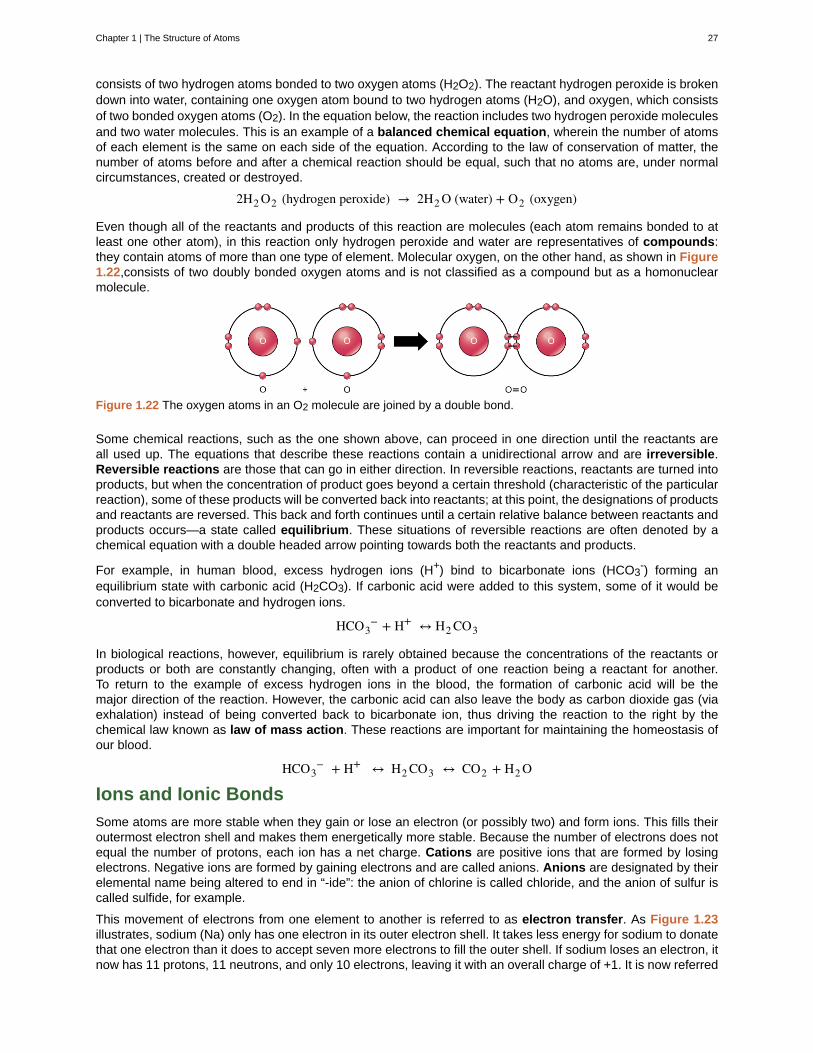
Even though all of the reactants and products of this reaction are molecules (each atom remains bonded to atleast one other atom), in this reaction only hydrogen peroxide and water are representatives of compounds:they contain atoms of more than one type of element. Molecular oxygen, on the other hand, as shown in Figure1.22,consists of two doubly bonded oxygen atoms and is not classified as a compound but as a homonuclearmolecule.
Figure 1.22 The oxygen atoms in an O2 molecule are joined by a double bond.
Some chemical reactions, such as the one shown above, can proceed in one direction until the reactants areall used up. The equations that describe these reactions contain a unidirectional arrow and are irreversible.Reversible reactions are those that can go in either direction. In reversible reactions, reactants are turned intoproducts, but when the concentration of product goes beyond a certain threshold (characteristic of the particularreaction), some of these products will be converted back into reactants; at this point, the designations of productsand reactants are reversed. This back and forth continues until a certain relative balance between reactants andproducts occurs—a state called equilibrium. These situations of reversible reactions are often denoted by achemical equation with a double headed arrow pointing towards both the reactants and products.
For example, in human blood, excess hydrogen ions (H+) bind to bicarbonate ions (HCO3-) forming an
equilibrium state with carbonic acid (H2CO3). If carbonic acid were added to this system, some of it would beconverted to bicarbonate and hydrogen ions.
HCO3− + H+ ↔ H2 CO3
In biological reactions, however, equilibrium is rarely obtained because the concentrations of the reactants orproducts or both are constantly changing, often with a product of one reaction being a reactant for another.To return to the example of excess hydrogen ions in the blood, the formation of carbonic acid will be themajor direction of the reaction. However, the carbonic acid can also leave the body as carbon dioxide gas (viaexhalation) instead of being converted back to bicarbonate ion, thus driving the reaction to the right by thechemical law known as law of mass action. These reactions are important for maintaining the homeostasis ofour blood.
HCO3− + H+ ↔ H2 CO3 ↔ CO2 + H2 O
Ions and Ionic Bonds
Some atoms are more stable when they gain or lose an electron (or possibly two) and form ions. This fills theiroutermost electron shell and makes them energetically more stable. Because the number of electrons does notequal the number of protons, each ion has a net charge. Cations are positive ions that are formed by losingelectrons. Negative ions are formed by gaining electrons and are called anions. Anions are designated by theirelemental name being altered to end in “-ide”: the anion of chlorine is called chloride, and the anion of sulfur iscalled sulfide, for example.
This movement of electrons from one element to another is referred to as electron transfer. As Figure 1.23illustrates, sodium (Na) only has one electron in its outer electron shell. It takes less energy for sodium to donatethat one electron than it does to accept seven more electrons to fill the outer shell. If sodium loses an electron, itnow has 11 protons, 11 neutrons, and only 10 electrons, leaving it with an overall charge of +1. It is now referred
Chapter 1 | The Structure of Atoms 27
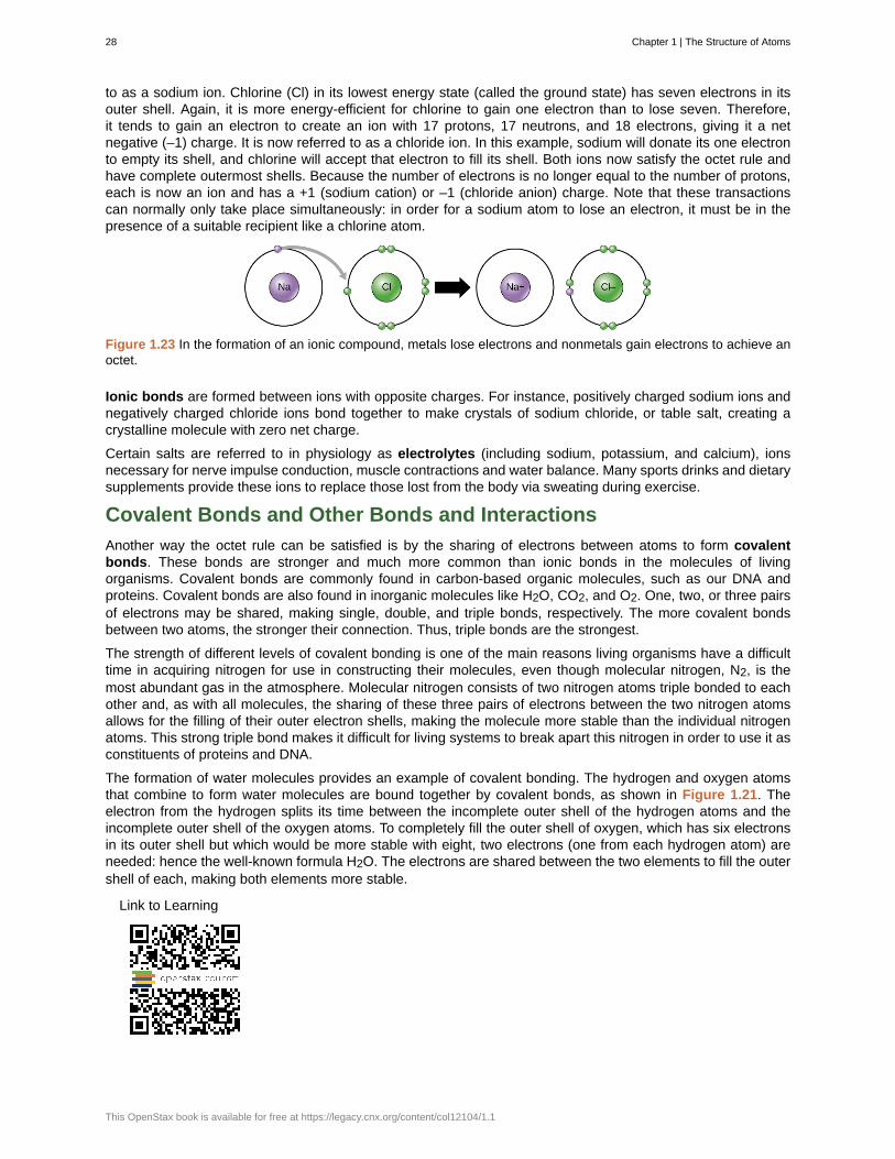
to as a sodium ion. Chlorine (Cl) in its lowest energy state (called the ground state) has seven electrons in itsouter shell. Again, it is more energy-efficient for chlorine to gain one electron than to lose seven. Therefore,it tends to gain an electron to create an ion with 17 protons, 17 neutrons, and 18 electrons, giving it a netnegative (–1) charge. It is now referred to as a chloride ion. In this example, sodium will donate its one electronto empty its shell, and chlorine will accept that electron to fill its shell. Both ions now satisfy the octet rule andhave complete outermost shells. Because the number of electrons is no longer equal to the number of protons,each is now an ion and has a +1 (sodium cation) or –1 (chloride anion) charge. Note that these transactionscan normally only take place simultaneously: in order for a sodium atom to lose an electron, it must be in thepresence of a suitable recipient like a chlorine atom.
Figure 1.23 In the formation of an ionic compound, metals lose electrons and nonmetals gain electrons to achieve anoctet.
Ionic bonds are formed between ions with opposite charges. For instance, positively charged sodium ions andnegatively charged chloride ions bond together to make crystals of sodium chloride, or table salt, creating acrystalline molecule with zero net charge.
Certain salts are referred to in physiology as electrolytes (including sodium, potassium, and calcium), ionsnecessary for nerve impulse conduction, muscle contractions and water balance. Many sports drinks and dietarysupplements provide these ions to replace those lost from the body via sweating during exercise.
Covalent Bonds and Other Bonds and Interactions
Another way the octet rule can be satisfied is by the sharing of electrons between atoms to form covalentbonds. These bonds are stronger and much more common than ionic bonds in the molecules of livingorganisms. Covalent bonds are commonly found in carbon-based organic molecules, such as our DNA andproteins. Covalent bonds are also found in inorganic molecules like H2O, CO2, and O2. One, two, or three pairsof electrons may be shared, making single, double, and triple bonds, respectively. The more covalent bondsbetween two atoms, the stronger their connection. Thus, triple bonds are the strongest.
The strength of different levels of covalent bonding is one of the main reasons living organisms have a difficulttime in acquiring nitrogen for use in constructing their molecules, even though molecular nitrogen, N2, is themost abundant gas in the atmosphere. Molecular nitrogen consists of two nitrogen atoms triple bonded to eachother and, as with all molecules, the sharing of these three pairs of electrons between the two nitrogen atomsallows for the filling of their outer electron shells, making the molecule more stable than the individual nitrogenatoms. This strong triple bond makes it difficult for living systems to break apart this nitrogen in order to use it asconstituents of proteins and DNA.
The formation of water molecules provides an example of covalent bonding. The hydrogen and oxygen atomsthat combine to form water molecules are bound together by covalent bonds, as shown in Figure 1.21. Theelectron from the hydrogen splits its time between the incomplete outer shell of the hydrogen atoms and theincomplete outer shell of the oxygen atoms. To completely fill the outer shell of oxygen, which has six electronsin its outer shell but which would be more stable with eight, two electrons (one from each hydrogen atom) areneeded: hence the well-known formula H2O. The electrons are shared between the two elements to fill the outershell of each, making both elements more stable.
Link to Learning
28 Chapter 1 | The Structure of Atoms
This OpenStax book is available for free at https://legacy.cnx.org/content/col12104/1.1
View this short video to see an animation of ionic and covalent bonding. (This media type is not supportedin this reader. Click to open media in browser.) (http://legacy.cnx.org/content/m44390/1.21/#eip-id1166283807593)
Polar Covalent Bonds
There are two types of covalent bonds: polar and nonpolar. In a polar covalent bond, shown in Figure 1.24,the electrons are unequally shared by the atoms and are attracted more to one nucleus than the other. Becauseof the unequal distribution of electrons between the atoms of different elements, a slightly positive (δ+) or slightlynegative (δ–) charge develops. This partial charge is an important property of water and accounts for many ofits characteristics.
Water is a polar molecule, with the hydrogen atoms acquiring a partial positive charge and the oxygen a partialnegative charge. This occurs because the nucleus of the oxygen atom is more attractive to the electronsof the hydrogen atoms than the hydrogen nucleus is to the oxygen’s electrons. Thus oxygen has a higherelectronegativity than hydrogen and the shared electrons spend more time in the vicinity of the oxygen nucleusthan they do near the nucleus of the hydrogen atoms, giving the atoms of oxygen and hydrogen slightly negativeand positive charges, respectively. Another way of stating this is that the probability of finding a shared electronnear an oxygen nucleus is more likely than finding it near a hydrogen nucleus. Either way, the atom’s relativeelectronegativity contributes to the development of partial charges whenever one element is significantly moreelectronegative than the other, and the charges generated by these polar bonds may then be used for theformation of hydrogen bonds based on the attraction of opposite partial charges. (Hydrogen bonds, whichare discussed in detail below, are weak bonds between slightly positively charged hydrogen atoms to slightlynegatively charged atoms in other molecules.) Since macromolecules often have atoms within them that differ inelectronegativity, polar bonds are often present in organic molecules.
Nonpolar Covalent Bonds
Nonpolar covalent bonds form between two atoms of the same element or between different elements thatshare electrons equally. For example, molecular oxygen (O2) is nonpolar because the electrons will be equallydistributed between the two oxygen atoms.
Another example of a nonpolar covalent bond is methane (CH4), also shown in Figure 1.24. Carbon has fourelectrons in its outermost shell and needs four more to fill it. It gets these four from four hydrogen atoms, eachatom providing one, making a stable outer shell of eight electrons. Carbon and hydrogen do not have the sameelectronegativity but are similar; thus, nonpolar bonds form. The hydrogen atoms each need one electron fortheir outermost shell, which is filled when it contains two electrons. These elements share the electrons equallyamong the carbons and the hydrogen atoms, creating a nonpolar covalent molecule.
Chapter 1 | The Structure of Atoms 29
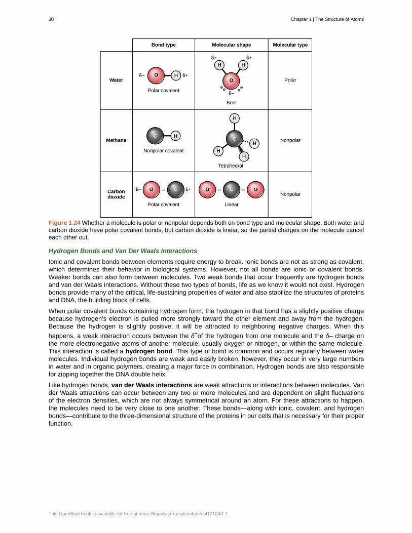
Figure 1.24 Whether a molecule is polar or nonpolar depends both on bond type and molecular shape. Both water andcarbon dioxide have polar covalent bonds, but carbon dioxide is linear, so the partial charges on the molecule canceleach other out.
Hydrogen Bonds and Van Der Waals Interactions
Ionic and covalent bonds between elements require energy to break. Ionic bonds are not as strong as covalent,which determines their behavior in biological systems. However, not all bonds are ionic or covalent bonds.Weaker bonds can also form between molecules. Two weak bonds that occur frequently are hydrogen bondsand van der Waals interactions. Without these two types of bonds, life as we know it would not exist. Hydrogenbonds provide many of the critical, life-sustaining properties of water and also stabilize the structures of proteinsand DNA, the building block of cells.
When polar covalent bonds containing hydrogen form, the hydrogen in that bond has a slightly positive chargebecause hydrogen’s electron is pulled more strongly toward the other element and away from the hydrogen.Because the hydrogen is slightly positive, it will be attracted to neighboring negative charges. When this
happens, a weak interaction occurs between the δ+of the hydrogen from one molecule and the δ– charge onthe more electronegative atoms of another molecule, usually oxygen or nitrogen, or within the same molecule.This interaction is called a hydrogen bond. This type of bond is common and occurs regularly between watermolecules. Individual hydrogen bonds are weak and easily broken; however, they occur in very large numbersin water and in organic polymers, creating a major force in combination. Hydrogen bonds are also responsiblefor zipping together the DNA double helix.
Like hydrogen bonds, van der Waals interactions are weak attractions or interactions between molecules. Vander Waals attractions can occur between any two or more molecules and are dependent on slight fluctuationsof the electron densities, which are not always symmetrical around an atom. For these attractions to happen,the molecules need to be very close to one another. These bonds—along with ionic, covalent, and hydrogenbonds—contribute to the three-dimensional structure of the proteins in our cells that is necessary for their properfunction.
30 Chapter 1 | The Structure of Atoms
This OpenStax book is available for free at https://legacy.cnx.org/content/col12104/1.1
Pharmaceutical ChemistPharmaceutical chemists are responsible for the development of new drugs and trying to determine themode of action of both old and new drugs. They are involved in every step of the drug development process.Drugs can be found in the natural environment or can be synthesized in the laboratory. In many cases,potential drugs found in nature are changed chemically in the laboratory to make them safer and moreeffective, and sometimes synthetic versions of drugs substitute for the version found in nature.
After the initial discovery or synthesis of a drug, the chemist then develops the drug, perhaps chemicallyaltering it, testing it to see if the drug is toxic, and then designing methods for efficient large-scale production.Then, the process of getting the drug approved for human use begins. In the United States, drug approval ishandled by the Food and Drug Administration (FDA) and involves a series of large-scale experiments usinghuman subjects to make sure the drug is not harmful and effectively treats the condition it aims to treat. Thisprocess often takes several years and requires the participation of physicians and scientists, in addition tochemists, to complete testing and gain approval.
An example of a drug that was originally discovered in a living organism is Paclitaxel (Taxol), an anti-cancerdrug used to treat breast cancer. This drug was discovered in the bark of the pacific yew tree. Anotherexample is aspirin, originally isolated from willow tree bark. Finding drugs often means testing hundreds ofsamples of plants, fungi, and other forms of life to see if any biologically active compounds are found withinthem. Sometimes, traditional medicine can give modern medicine clues to where an active compound canbe found. For example, the use of willow bark to make medicine has been known for thousands of years,dating back to ancient Egypt. It was not until the late 1800s, however, that the aspirin molecule, known asacetylsalicylic acid, was purified and marketed for human use.
Occasionally, drugs developed for one use are found to have unforeseen effects that allow these drugs tobe used in other, unrelated ways. For example, the drug minoxidil (Rogaine) was originally developed totreat high blood pressure. When tested on humans, it was noticed that individuals taking the drug wouldgrow new hair. Eventually the drug was marketed to men and women with baldness to restore lost hair.
The career of the pharmaceutical chemist may involve detective work, experimentation, and drugdevelopment, all with the goal of making human beings healthier.
1.3 | The Periodic Table
By the end of this section, you will be able to:
• State the periodic law and explain the organization of elements in the periodic table
• Predict the general properties of elements based on their location within the periodic table
• Identify metals, nonmetals, and metalloids by their properties and/or location on the periodic table
As early chemists worked to purify ores and discovered more elements, they realized that various elementscould be grouped together by their similar chemical behaviors. One such grouping includes lithium (Li), sodium(Na), and potassium (K): These elements all are shiny, conduct heat and electricity well, and have similarchemical properties. A second grouping includes calcium (Ca), strontium (Sr), and barium (Ba), which also areshiny, good conductors of heat and electricity, and have chemical properties in common. However, the specificproperties of these two groupings are notably different from each other. For example: Li, Na, and K are muchmore reactive than are Ca, Sr, and Ba; Li, Na, and K form compounds with oxygen in a ratio of two of their atomsto one oxygen atom, whereas Ca, Sr, and Ba form compounds with one of their atoms to one oxygen atom.Fluorine (F), chlorine (Cl), bromine (Br), and iodine (I) also exhibit similar properties to each other, but theseproperties are drastically different from those of any of the elements above.
Dimitri Mendeleev in Russia (1869) and Lothar Meyer in Germany (1870) independently recognized that therewas a periodic relationship among the properties of the elements known at that time. Both published tables
Chapter 1 | The Structure of Atoms 31
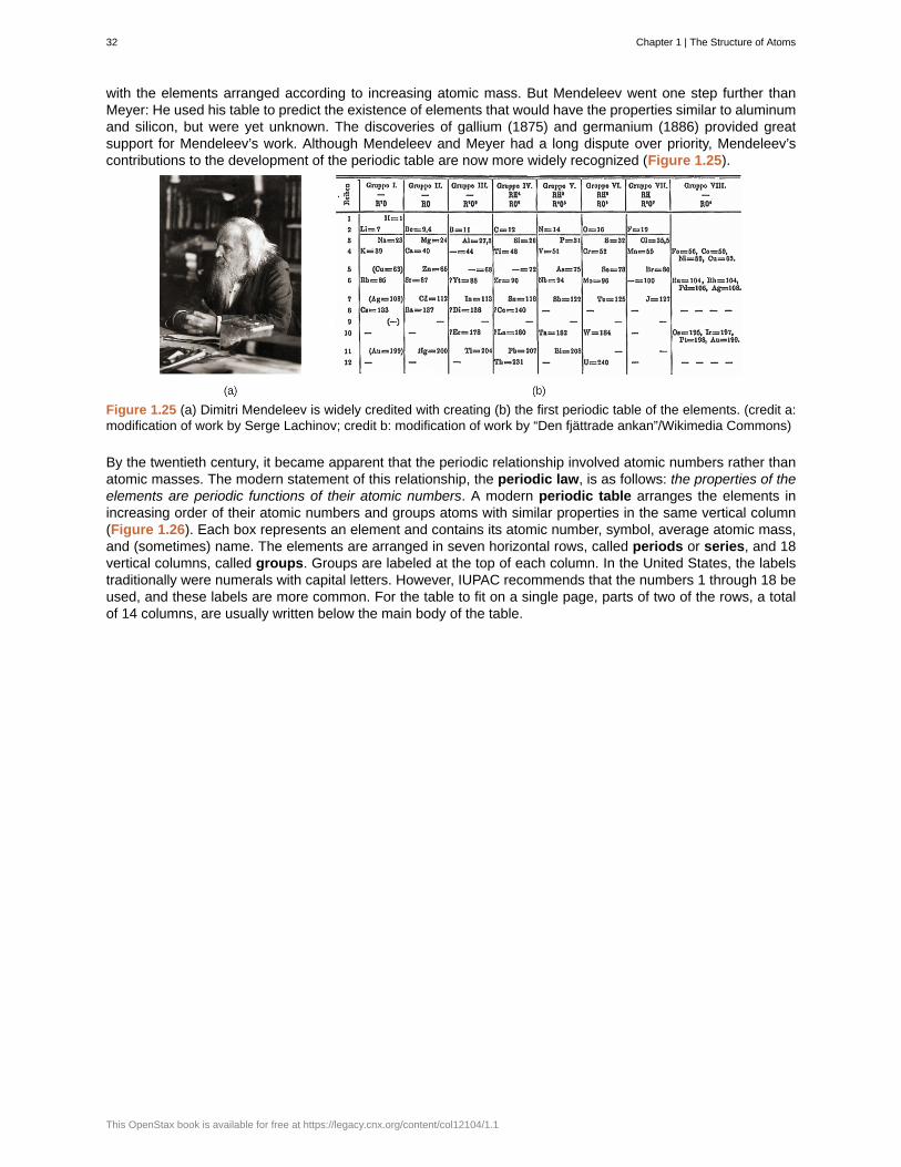
with the elements arranged according to increasing atomic mass. But Mendeleev went one step further thanMeyer: He used his table to predict the existence of elements that would have the properties similar to aluminumand silicon, but were yet unknown. The discoveries of gallium (1875) and germanium (1886) provided greatsupport for Mendeleev’s work. Although Mendeleev and Meyer had a long dispute over priority, Mendeleev’scontributions to the development of the periodic table are now more widely recognized (Figure 1.25).
Figure 1.25 (a) Dimitri Mendeleev is widely credited with creating (b) the first periodic table of the elements. (credit a:modification of work by Serge Lachinov; credit b: modification of work by “Den fjättrade ankan”/Wikimedia Commons)
By the twentieth century, it became apparent that the periodic relationship involved atomic numbers rather thanatomic masses. The modern statement of this relationship, the periodic law, is as follows: the properties of theelements are periodic functions of their atomic numbers. A modern periodic table arranges the elements inincreasing order of their atomic numbers and groups atoms with similar properties in the same vertical column(Figure 1.26). Each box represents an element and contains its atomic number, symbol, average atomic mass,and (sometimes) name. The elements are arranged in seven horizontal rows, called periods or series, and 18vertical columns, called groups. Groups are labeled at the top of each column. In the United States, the labelstraditionally were numerals with capital letters. However, IUPAC recommends that the numbers 1 through 18 beused, and these labels are more common. For the table to fit on a single page, parts of two of the rows, a totalof 14 columns, are usually written below the main body of the table.
32 Chapter 1 | The Structure of Atoms
This OpenStax book is available for free at https://legacy.cnx.org/content/col12104/1.1
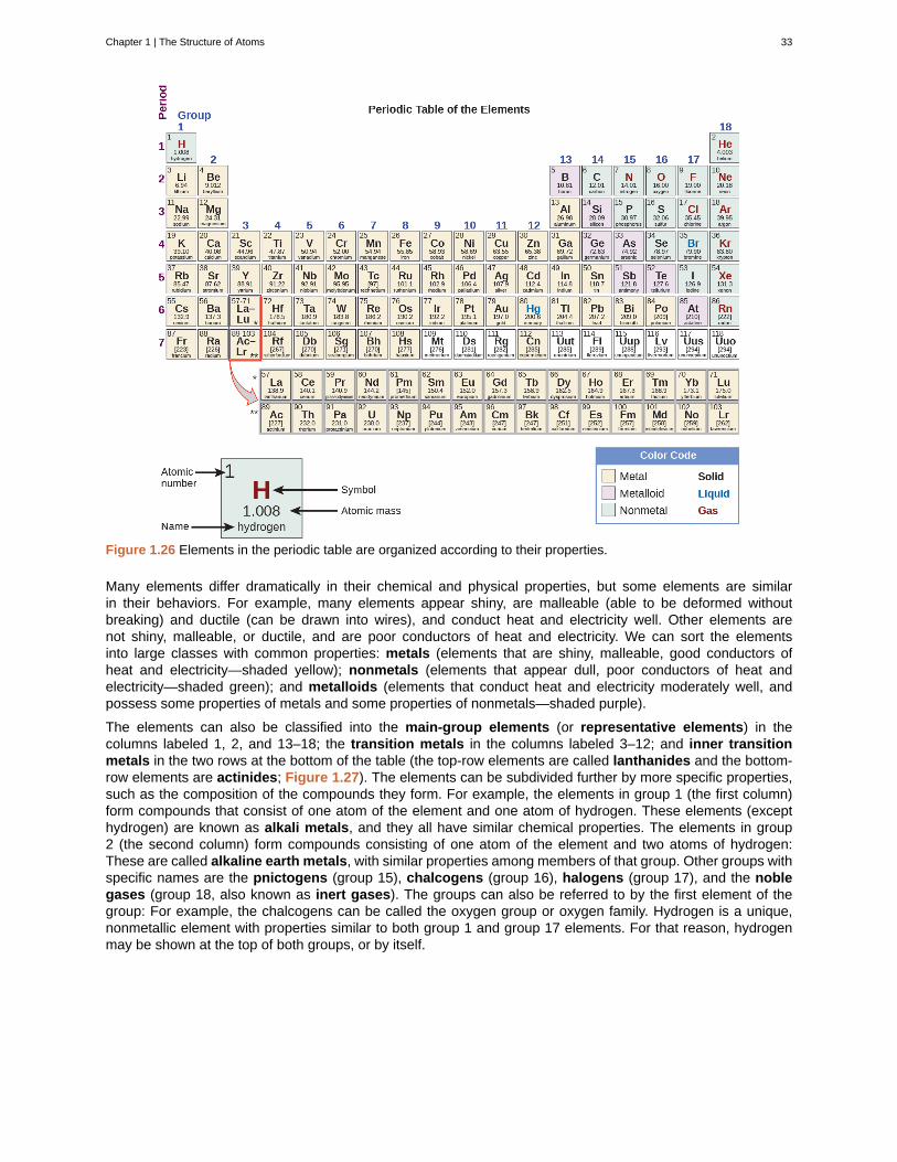
Figure 1.26 Elements in the periodic table are organized according to their properties.
Many elements differ dramatically in their chemical and physical properties, but some elements are similarin their behaviors. For example, many elements appear shiny, are malleable (able to be deformed withoutbreaking) and ductile (can be drawn into wires), and conduct heat and electricity well. Other elements arenot shiny, malleable, or ductile, and are poor conductors of heat and electricity. We can sort the elementsinto large classes with common properties: metals (elements that are shiny, malleable, good conductors ofheat and electricity—shaded yellow); nonmetals (elements that appear dull, poor conductors of heat andelectricity—shaded green); and metalloids (elements that conduct heat and electricity moderately well, andpossess some properties of metals and some properties of nonmetals—shaded purple).
The elements can also be classified into the main-group elements (or representative elements) in thecolumns labeled 1, 2, and 13–18; the transition metals in the columns labeled 3–12; and inner transitionmetals in the two rows at the bottom of the table (the top-row elements are called lanthanides and the bottom-row elements are actinides; Figure 1.27). The elements can be subdivided further by more specific properties,such as the composition of the compounds they form. For example, the elements in group 1 (the first column)form compounds that consist of one atom of the element and one atom of hydrogen. These elements (excepthydrogen) are known as alkali metals, and they all have similar chemical properties. The elements in group2 (the second column) form compounds consisting of one atom of the element and two atoms of hydrogen:These are called alkaline earth metals, with similar properties among members of that group. Other groups withspecific names are the pnictogens (group 15), chalcogens (group 16), halogens (group 17), and the noblegases (group 18, also known as inert gases). The groups can also be referred to by the first element of thegroup: For example, the chalcogens can be called the oxygen group or oxygen family. Hydrogen is a unique,nonmetallic element with properties similar to both group 1 and group 17 elements. For that reason, hydrogenmay be shown at the top of both groups, or by itself.
Chapter 1 | The Structure of Atoms 33

Figure 1.27 The periodic table organizes elements with similar properties into groups.
Click on this link (http://openstax.org/l/16Periodic) for an interactive periodic table, which you can use toexplore the properties of the elements (includes podcasts and videos of each element). You may also wantto try this one that shows photos of all the elements.
Example 1.1
Naming Groups of Elements
Atoms of each of the following elements are essential for life. Give the group name for the following elements:
(a) chlorine
(b) calcium
(c) sodium
(d) sulfur
Solution
The family names are as follows:
(a) halogen
(b) alkaline earth metal
(c) alkali metal
(d) chalcogen
Check Your Learning
Give the group name for each of the following elements:
34 Chapter 1 | The Structure of Atoms
This OpenStax book is available for free at https://legacy.cnx.org/content/col12104/1.1
(a) krypton
(b) selenium
(c) barium
(d) lithium
Answer:
(a) noble gas; (b) chalcogen; (c) alkaline earth metal; (d) alkali metal
In studying the periodic table, you might have noticed something about the atomic masses of some of theelements. Element 43 (technetium), element 61 (promethium), and most of the elements with atomic number84 (polonium) and higher have their atomic mass given in square brackets. This is done for elements thatconsist entirely of unstable, radioactive isotopes (you will learn more about radioactivity in the nuclear chemistrychapter). An average atomic weight cannot be determined for these elements because their radioisotopes mayvary significantly in relative abundance, depending on the source, or may not even exist in nature. The numberin square brackets is the atomic mass number (an approximate atomic mass) of the most stable isotope of thatelement.
Chemistry End of Chapter Exercises
Exercise 1.19
Using the periodic table, classify each of the following elements as a metal or a nonmetal, and then furtherclassify each as a main-group (representative) element, transition metal, or inner transition metal:
(a) uranium
(b) bromine
(c) strontium
(d) neon
(e) gold
(f) americium
(g) rhodium
(h) sulfur
(i) carbon
(j) potassium
Solution(a) metal, inner transition metal; (b) nonmetal, representative element; (c) metal, representative element; (d)nonmetal, representative element; (e) metal, transition metal; (f) metal, inner transition metal; (g) metal, transitionmetal; (h) nonmetal, representative element; (i) nonmetal, representative element; (j) metal, representativeelement
Exercise 1.20
Using the periodic table, classify each of the following elements as a metal or a nonmetal, and then furtherclassify each as a main-group (representative) element, transition metal, or inner transition metal:
(a) cobalt
(b) europium
(c) iodine
(d) indium
(e) lithium
(f) oxygen
(g) cadmium
Chapter 1 | The Structure of Atoms 35
(h) terbium
(i) rhenium
Exercise 1.21
Using the periodic table, identify the lightest member of each of the following groups:
(a) noble gases
(b) alkaline earth metals
(c) alkali metals
(d) chalcogens
Solution(a) He; (b) Be; (c) Li; (d) O
Exercise 1.22
Using the periodic table, identify the heaviest member of each of the following groups:
(a) alkali metals
(b) chalcogens
(c) noble gases
(d) alkaline earth metals
Exercise 1.23
Use the periodic table to give the name and symbol for each of the following elements:
(a) the noble gas in the same period as germanium
(b) the alkaline earth metal in the same period as selenium
(c) the halogen in the same period as lithium
(d) the chalcogen in the same period as cadmium
Solution(a) krypton, Kr; (b) calcium, Ca; (c) fluorine, F; (d) tellurium, Te
Exercise 1.24
Use the periodic table to give the name and symbol for each of the following elements:
(a) the halogen in the same period as the alkali metal with 11 protons
(b) the alkaline earth metal in the same period with the neutral noble gas with 18 electrons
(c) the noble gas in the same row as an isotope with 30 neutrons and 25 protons
(d) the noble gas in the same period as gold
Exercise 1.25
Write a symbol for each of the following neutral isotopes. Include the atomic number and mass number for each.
(a) the alkali metal with 11 protons and a mass number of 23
(b) the noble gas element with 75 neutrons in its nucleus and 54 electrons in the neutral atom
(c) the isotope with 33 protons and 40 neutrons in its nucleus
(d) the alkaline earth metal with 88 electrons and 138 neutrons
Solution
(a) 1123 Na ; (b) 54
129 Xe ; (c) 3373 As ; (d) 88
226 Ra
Exercise 1.26
36 Chapter 1 | The Structure of Atoms
This OpenStax book is available for free at https://legacy.cnx.org/content/col12104/1.1

Write a symbol for each of the following neutral isotopes. Include the atomic number and mass number for each.
(a) the chalcogen with a mass number of 125
(b) the halogen whose longest-lived isotope is radioactive
(c) the noble gas, used in lighting, with 10 electrons and 10 neutrons
(d) the lightest alkali metal with three neutrons
Chapter 1 | The Structure of Atoms 37
actinide
alkali metal
alkaline earth metal
anion
atom
atom
atomic mass
atomic number
balanced chemical equation
cation
chalcogen
chemical bond
chemical reaction
chemical reactivity
compound
compound
covalent bond
electrolyte
electron
electron configuration
electron orbital
electron transfer
electronegativity
element
element
equilibrium
KEY TERMS
inner transition metal in the bottom of the bottom two rows of the periodic table
element in group 1
element in group 2
negative ion that is formed by an atom gaining one or more electrons
smallest particle of an element that can enter into a chemical combination
the smallest unit of matter that retains all of the chemical properties of an element
calculated mean of the mass number for an element’s isotopes
total number of protons in an atom
statement of a chemical reaction with the number of each type of atom equalizedfor both the products and reactants
positive ion that is formed by an atom losing one or more electrons
element in group 16
interaction between two or more of the same or different atoms that results in the formation ofmolecules
process leading to the rearrangement of atoms in molecules
the ability to combine and to chemically bond with each other
pure substance that can be decomposed into two or more elements
substance composed of molecules consisting of atoms of at least two different elements
type of strong bond formed between two of the same or different elements; forms whenelectrons are shared between atoms
ion necessary for nerve impulse conduction, muscle contractions and water balance
negatively charged subatomic particle that resides outside of the nucleus in the electron orbital; lacksfunctional mass and has a negative charge of –1 unit
arrangement of electrons in an atom’s electron shell (for example, 1s22s22p6)
how electrons are spatially distributed surrounding the nucleus; the area where an electron ismost likely to be found
movement of electrons from one element to another; important in creation of ionic bonds
ability of some elements to attract electrons (often of hydrogen atoms), acquiring partialnegative charges in molecules and creating partial positive charges on the hydrogen atoms
substance that is composed of a single type of atom; a substance that cannot be decomposed by achemical change
one of 118 unique substances that cannot be broken down into smaller substances; each element hasunique properties and a specified number of protons
steady state of relative reactant and product concentration in reversible chemical reactions in aclosed system
38 Chapter 1 | The Structure of Atoms
This OpenStax book is available for free at https://legacy.cnx.org/content/col12104/1.1
gas
group
halogen
heterogeneous mixture
homogeneous mixture
hydrogen bond
inert gas
inert gas
inner transition metal
ion
ionic bond
irreversible chemical reaction
isotope
lanthanide
law of conservation of matter
law of mass action
liquid
main-group element
mass
mass number
matter
matter
metal
metalloid
mixture
molecule
molecule
neutron
noble gas
state in which matter has neither definite volume nor shape
vertical column of the periodic table
element in group 17
combination of substances with a composition that varies from point to point
(also, solution) combination of substances with a composition that is uniformthroughout
weak bond between slightly positively charged hydrogen atoms to slightly negatively chargedatoms in other molecules
(also, noble gas) element with filled outer electron shell that is unreactive with other atoms
(also, noble gas) element in group 18
(also, lanthanide or actinide) element in the bottom two rows; if in the first row, alsocalled lanthanide, or if in the second row, also called actinide
atom or chemical group that does not contain equal numbers of protons and electrons
chemical bond that forms between ions with opposite charges (cations and anions)
chemical reaction where reactants proceed uni-directionally to form products
one or more forms of an element that have different numbers of neutrons
inner transition metal in the top of the bottom two rows of the periodic table
when matter converts from one type to another or changes form, there is nodetectable change in the total amount of matter present
chemical law stating that the rate of a reaction is proportional to the concentration of thereacting substances
state of matter that has a definite volume but indefinite shape
(also, representative element) element in columns 1, 2, and 12–18
fundamental property indicating amount of matter
total number of protons and neutrons in an atom
anything that occupies space and has mass
anything that has mass and occupies space
element that is shiny, malleable, good conductor of heat and electricity
element that conducts heat and electricity moderately well, and possesses some properties of metalsand some properties of nonmetals
matter that can be separated into its components by physical means
bonded collection of two or more atoms of the same or different elements
two or more atoms chemically bonded together
uncharged particle that resides in the nucleus of an atom; has a mass of one amu
see inert gas
Chapter 1 | The Structure of Atoms 39
noble gas
nonmetal
nonpolar covalent bond
nucleus
octet rule
orbital
period
periodic law
periodic table
periodic table
plasma
pnictogen
polar covalent bond
product
proton
pure substance
radioisotope
reactant
representative element
reversible chemical reaction
series
solid
transition metal
valence shell
van der Waals interaction
weight
(also, inert gas) element in group 18
element that appears dull, poor conductor of heat and electricity
type of covalent bond that forms between atoms when electrons are shared equallybetween them
core of an atom; contains protons and neutrons
rule that atoms are most stable when they hold eight electrons in their outermost shells
region surrounding the nucleus; contains electrons
(also, series) horizontal row of the periodic table
properties of the elements are periodic function of their atomic numbers.
organizational chart of elements indicating the atomic number and atomic mass of each element;provides key information about the properties of the elements
table of the elements that places elements with similar chemical properties close together
gaseous state of matter containing a large number of electrically charged atoms and/or molecules
element in group 15
type of covalent bond that forms as a result of unequal sharing of electrons, resulting inthe creation of slightly positive and slightly negative charged regions of the molecule
molecule found on the right side of a chemical equation
positively charged particle that resides in the nucleus of an atom; has a mass of one amu and a chargeof +1
homogeneous substance that has a constant composition
isotope that emits radiation composed of subatomic particles to form more stable elements
molecule found on the left side of a chemical equation
(also, main-group element) element in columns 1, 2, and 12–18
chemical reaction that functions bi-directionally, where products may turn intoreactants if their concentration is great enough
(also, period) horizontal row of the period table
state of matter that is rigid, has a definite shape, and has a fairly constant volume
element in columns 3–11
outermost shell of an atom
very weak interaction between molecules due to temporary charges attractingatoms that are very close together
force that gravity exerts on an object
CHAPTER SUMMARY
1.1 Phases and Classification of Matter
Matter is anything that occupies space and has mass. The basic building block of matter is the atom, the
40 Chapter 1 | The Structure of Atoms
This OpenStax book is available for free at https://legacy.cnx.org/content/col12104/1.1
smallest unit of an element that can enter into combinations with atoms of the same or other elements. In manysubstances, atoms are combined into molecules. On earth, matter commonly exists in three states: solids, offixed shape and volume; liquids, of variable shape but fixed volume; and gases, of variable shape and volume.Under high-temperature conditions, matter also can exist as a plasma. Most matter is a mixture: It is composedof two or more types of matter that can be present in varying amounts and can be separated by physicalmeans. Heterogeneous mixtures vary in composition from point to point; homogeneous mixtures have thesame composition from point to point. Pure substances consist of only one type of matter. A pure substancecan be an element, which consists of only one type of atom and cannot be broken down by a chemical change,or a compound, which consists of two or more types of atoms.
1.2 Atoms, Isotopes, Ions, and Molecules: The Building Blocks
Matter is anything that occupies space and has mass. It is made up of elements. All of the 92 elements thatoccur naturally have unique qualities that allow them to combine in various ways to create molecules, which inturn combine to form cells, tissues, organ systems, and organisms. Atoms, which consist of protons, neutrons,and electrons, are the smallest units of an element that retain all of the properties of that element. Electronscan be transferred, shared, or cause charge disparities between atoms to create bonds, including ionic,covalent, and hydrogen bonds, as well as van der Waals interactions.
1.3 The Periodic Table
The discovery of the periodic recurrence of similar properties among the elements led to the formulation of theperiodic table, in which the elements are arranged in order of increasing atomic number in rows known asperiods and columns known as groups. Elements in the same group of the periodic table have similar chemicalproperties. Elements can be classified as metals, metalloids, and nonmetals, or as a main-group elements,transition metals, and inner transition metals. Groups are numbered 1–18 from left to right. The elements ingroup 1 are known as the alkali metals; those in group 2 are the alkaline earth metals; those in 15 are thepnictogens; those in 16 are the chalcogens; those in 17 are the halogens; and those in 18 are the noble gases.
ART CONNECTION QUESTIONS
27. Figure 1.15 How many neutrons do carbon-12and carbon-13 have, respectively?
28. Figure 1.19 An atom may give, take, or shareelectrons with another atom to achieve a full valenceshell, the most stable electron configuration. Looking
at this figure, how many electrons do elements ingroup 1 need to lose in order to achieve a stableelectron configuration? How many electrons doelements in groups 14 and 17 need to gain toachieve a stable configuration?
REVIEW QUESTIONS
29. If xenon has an atomic number of 54 and a massnumber of 108, how many neutrons does it have?
a. 54b. 27c. 100d. 108
30. Atoms that vary in the number of neutrons foundin their nuclei are called ________.
a. ionsb. neutronsc. neutral atomsd. isotopes
31. Potassium has an atomic number of 19. What isits electron configuration?
a. shells 1 and 2 are full, and shell 3 has nineelectrons
b. shells 1, 2 and 3 are full and shell 4 hasthree electrons
c. shells 1, 2 and 3 are full and shell 4 has oneelectron
d. shells 1, 2 and 3 are full and no otherelectrons are present
32. Which type of bond represents a weak chemicalbond?
a. hydrogen bondb. atomic bondc. covalent bondd. nonpolar covalent bond
Chapter 1 | The Structure of Atoms 41

CRITICAL THINKING QUESTIONS
33. What makes ionic bonds different from covalentbonds?
34. Why are hydrogen bonds and van der Waalsinteractions necessary for cells?
42 Chapter 1 | The Structure of Atoms
This OpenStax book is available for free at https://legacy.cnx.org/content/col12104/1.1
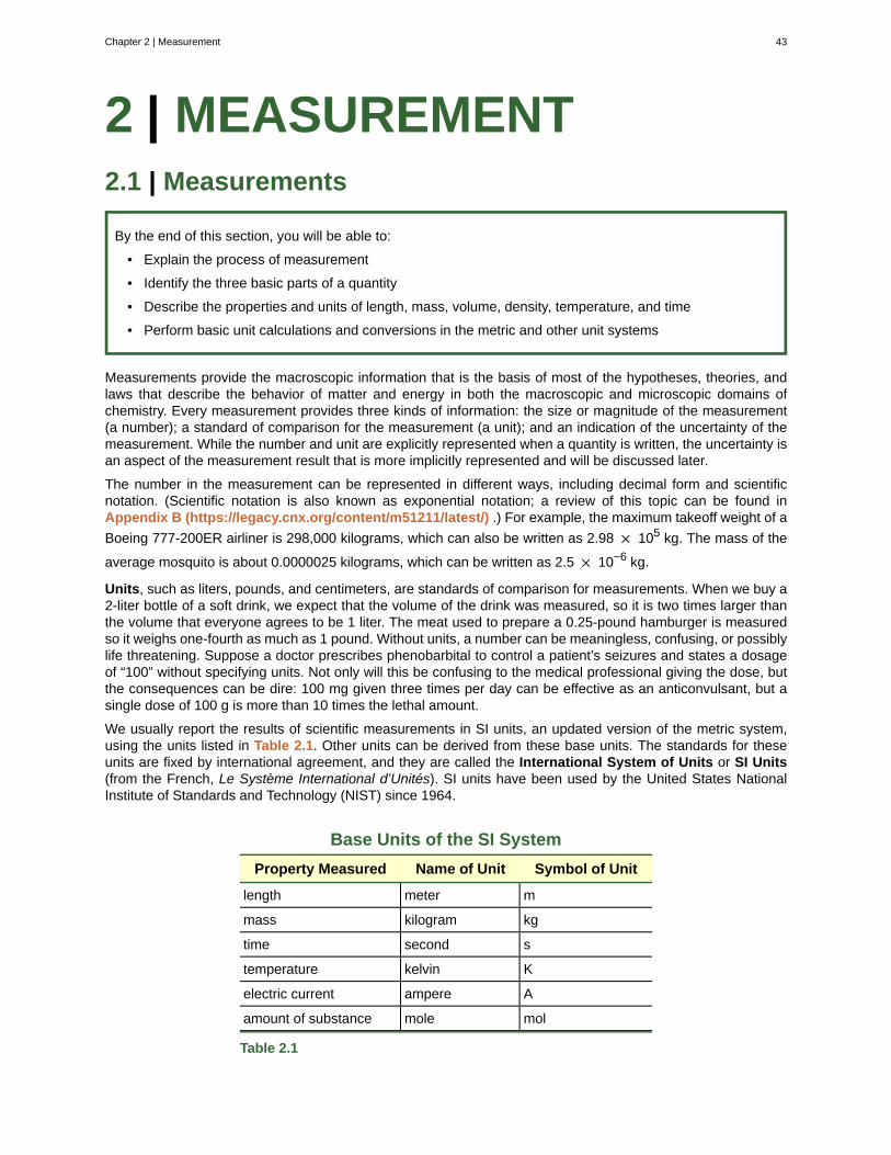
2 | MEASUREMENT2.1 | Measurements
By the end of this section, you will be able to:
• Explain the process of measurement
• Identify the three basic parts of a quantity
• Describe the properties and units of length, mass, volume, density, temperature, and time
• Perform basic unit calculations and conversions in the metric and other unit systems
Measurements provide the macroscopic information that is the basis of most of the hypotheses, theories, andlaws that describe the behavior of matter and energy in both the macroscopic and microscopic domains ofchemistry. Every measurement provides three kinds of information: the size or magnitude of the measurement(a number); a standard of comparison for the measurement (a unit); and an indication of the uncertainty of themeasurement. While the number and unit are explicitly represented when a quantity is written, the uncertainty isan aspect of the measurement result that is more implicitly represented and will be discussed later.
The number in the measurement can be represented in different ways, including decimal form and scientificnotation. (Scientific notation is also known as exponential notation; a review of this topic can be found inAppendix B (https://legacy.cnx.org/content/m51211/latest/) .) For example, the maximum takeoff weight of a
Boeing 777-200ER airliner is 298,000 kilograms, which can also be written as 2.98 × 10⁵ kg. The mass of the
average mosquito is about 0.0000025 kilograms, which can be written as 2.5 × 10⁻⁶ kg.
Units, such as liters, pounds, and centimeters, are standards of comparison for measurements. When we buy a2-liter bottle of a soft drink, we expect that the volume of the drink was measured, so it is two times larger thanthe volume that everyone agrees to be 1 liter. The meat used to prepare a 0.25-pound hamburger is measuredso it weighs one-fourth as much as 1 pound. Without units, a number can be meaningless, confusing, or possiblylife threatening. Suppose a doctor prescribes phenobarbital to control a patient’s seizures and states a dosageof “100” without specifying units. Not only will this be confusing to the medical professional giving the dose, butthe consequences can be dire: 100 mg given three times per day can be effective as an anticonvulsant, but asingle dose of 100 g is more than 10 times the lethal amount.
We usually report the results of scientific measurements in SI units, an updated version of the metric system,using the units listed in Table 2.1. Other units can be derived from these base units. The standards for theseunits are fixed by international agreement, and they are called the International System of Units or SI Units(from the French, Le Système International d’Unités). SI units have been used by the United States NationalInstitute of Standards and Technology (NIST) since 1964.
Base Units of the SI System
Property Measured Name of Unit Symbol of Unit
length meter m
mass kilogram kg
time second s
temperature kelvin K
electric current ampere A
amount of substance mole mol
Table 2.1
Chapter 2 | Measurement 43
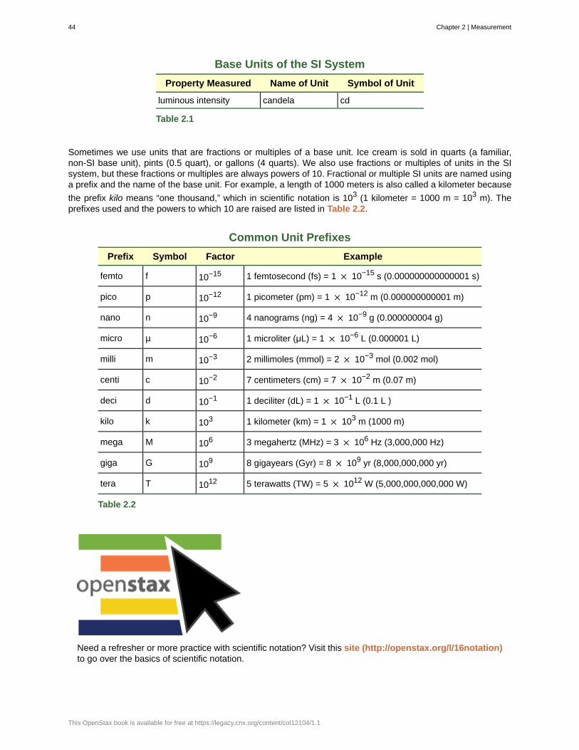
Base Units of the SI System
Property Measured Name of Unit Symbol of Unit
luminous intensity candela cd
Table 2.1
Sometimes we use units that are fractions or multiples of a base unit. Ice cream is sold in quarts (a familiar,non-SI base unit), pints (0.5 quart), or gallons (4 quarts). We also use fractions or multiples of units in the SIsystem, but these fractions or multiples are always powers of 10. Fractional or multiple SI units are named usinga prefix and the name of the base unit. For example, a length of 1000 meters is also called a kilometer because
the prefix kilo means “one thousand,” which in scientific notation is 103 (1 kilometer = 1000 m = 103 m). Theprefixes used and the powers to which 10 are raised are listed in Table 2.2.
Common Unit Prefixes
Prefix Symbol Factor Example
femto f 10−15 1 femtosecond (fs) = 1 × 10⁻¹⁵ s (0.000000000000001 s)
pico p 10−12 1 picometer (pm) = 1 × 10⁻¹² m (0.000000000001 m)
nano n 10−9 4 nanograms (ng) = 4 × 10⁻⁹ g (0.000000004 g)
micro µ 10−6 1 microliter (μL) = 1 × 10⁻⁶ L (0.000001 L)
milli m 10−3 2 millimoles (mmol) = 2 × 10⁻³ mol (0.002 mol)
centi c 10−2 7 centimeters (cm) = 7 × 10⁻² m (0.07 m)
deci d 10−1 1 deciliter (dL) = 1 × 10⁻¹ L (0.1 L )
kilo k 103 1 kilometer (km) = 1 × 10³ m (1000 m)
mega M 106 3 megahertz (MHz) = 3 × 10⁶ Hz (3,000,000 Hz)
giga G 109 8 gigayears (Gyr) = 8 × 10⁹ yr (8,000,000,000 yr)
tera T 1012 5 terawatts (TW) = 5 × 10¹² W (5,000,000,000,000 W)
Table 2.2
Need a refresher or more practice with scientific notation? Visit this site (http://openstax.org/l/16notation)to go over the basics of scientific notation.
44 Chapter 2 | Measurement
This OpenStax book is available for free at https://legacy.cnx.org/content/col12104/1.1
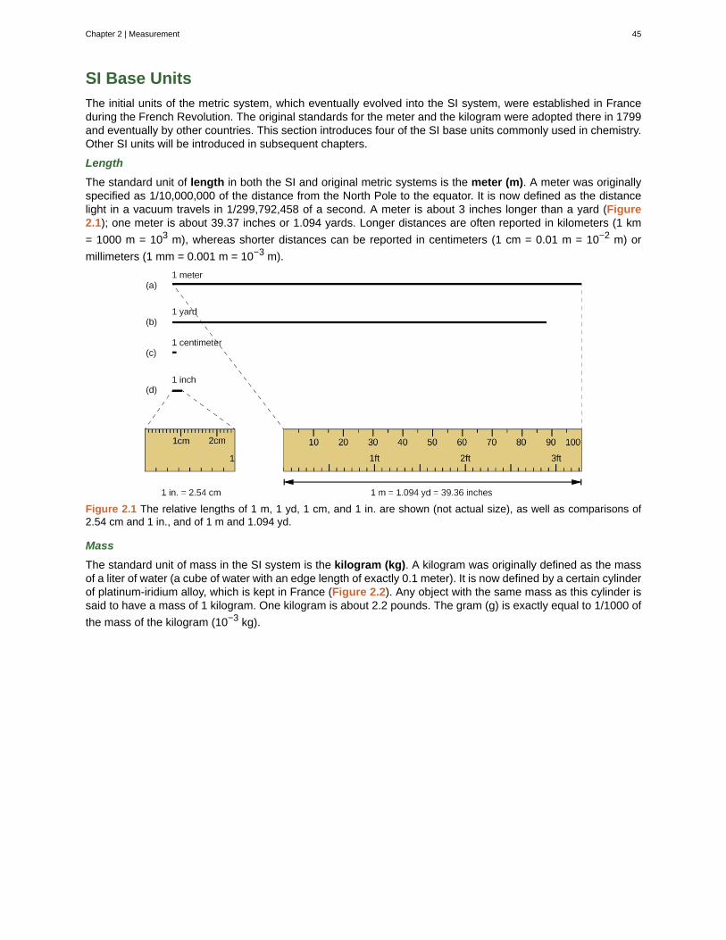
SI Base Units
The initial units of the metric system, which eventually evolved into the SI system, were established in Franceduring the French Revolution. The original standards for the meter and the kilogram were adopted there in 1799and eventually by other countries. This section introduces four of the SI base units commonly used in chemistry.Other SI units will be introduced in subsequent chapters.
Length
The standard unit of length in both the SI and original metric systems is the meter (m). A meter was originallyspecified as 1/10,000,000 of the distance from the North Pole to the equator. It is now defined as the distancelight in a vacuum travels in 1/299,792,458 of a second. A meter is about 3 inches longer than a yard (Figure2.1); one meter is about 39.37 inches or 1.094 yards. Longer distances are often reported in kilometers (1 km
= 1000 m = 103 m), whereas shorter distances can be reported in centimeters (1 cm = 0.01 m = 10−2 m) or
millimeters (1 mm = 0.001 m = 10−3 m).
Figure 2.1 The relative lengths of 1 m, 1 yd, 1 cm, and 1 in. are shown (not actual size), as well as comparisons of2.54 cm and 1 in., and of 1 m and 1.094 yd.
Mass
The standard unit of mass in the SI system is the kilogram (kg). A kilogram was originally defined as the massof a liter of water (a cube of water with an edge length of exactly 0.1 meter). It is now defined by a certain cylinderof platinum-iridium alloy, which is kept in France (Figure 2.2). Any object with the same mass as this cylinder issaid to have a mass of 1 kilogram. One kilogram is about 2.2 pounds. The gram (g) is exactly equal to 1/1000 of
the mass of the kilogram (10−3 kg).
Chapter 2 | Measurement 45
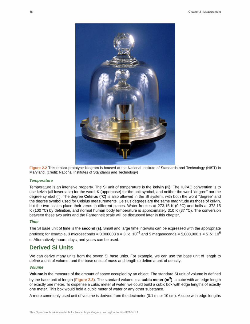
Figure 2.2 This replica prototype kilogram is housed at the National Institute of Standards and Technology (NIST) inMaryland. (credit: National Institutes of Standards and Technology)
Temperature
Temperature is an intensive property. The SI unit of temperature is the kelvin (K). The IUPAC convention is touse kelvin (all lowercase) for the word, K (uppercase) for the unit symbol, and neither the word “degree” nor thedegree symbol (°). The degree Celsius (°C) is also allowed in the SI system, with both the word “degree” andthe degree symbol used for Celsius measurements. Celsius degrees are the same magnitude as those of kelvin,but the two scales place their zeros in different places. Water freezes at 273.15 K (0 °C) and boils at 373.15K (100 °C) by definition, and normal human body temperature is approximately 310 K (37 °C). The conversionbetween these two units and the Fahrenheit scale will be discussed later in this chapter.
Time
The SI base unit of time is the second (s). Small and large time intervals can be expressed with the appropriate
prefixes; for example, 3 microseconds = 0.000003 s = 3 × 10⁻⁶ and 5 megaseconds = 5,000,000 s = 5 × 10⁶
s. Alternatively, hours, days, and years can be used.
Derived SI Units
We can derive many units from the seven SI base units. For example, we can use the base unit of length todefine a unit of volume, and the base units of mass and length to define a unit of density.
Volume
Volume is the measure of the amount of space occupied by an object. The standard SI unit of volume is defined
by the base unit of length (Figure 2.3). The standard volume is a cubic meter (m3), a cube with an edge lengthof exactly one meter. To dispense a cubic meter of water, we could build a cubic box with edge lengths of exactlyone meter. This box would hold a cubic meter of water or any other substance.
A more commonly used unit of volume is derived from the decimeter (0.1 m, or 10 cm). A cube with edge lengths
46 Chapter 2 | Measurement
This OpenStax book is available for free at https://legacy.cnx.org/content/col12104/1.1
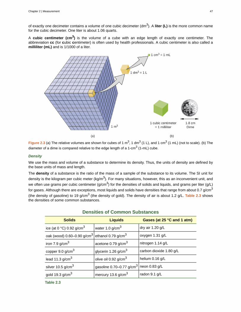
of exactly one decimeter contains a volume of one cubic decimeter (dm3). A liter (L) is the more common namefor the cubic decimeter. One liter is about 1.06 quarts.
A cubic centimeter (cm3) is the volume of a cube with an edge length of exactly one centimeter. Theabbreviation cc (for cubic centimeter) is often used by health professionals. A cubic centimeter is also called amilliliter (mL) and is 1/1000 of a liter.
Figure 2.3 (a) The relative volumes are shown for cubes of 1 m3, 1 dm3 (1 L), and 1 cm3 (1 mL) (not to scale). (b) The
diameter of a dime is compared relative to the edge length of a 1-cm3 (1-mL) cube.
Density
We use the mass and volume of a substance to determine its density. Thus, the units of density are defined bythe base units of mass and length.
The density of a substance is the ratio of the mass of a sample of the substance to its volume. The SI unit for
density is the kilogram per cubic meter (kg/m3). For many situations, however, this as an inconvenient unit, and
we often use grams per cubic centimeter (g/cm3) for the densities of solids and liquids, and grams per liter (g/L)
for gases. Although there are exceptions, most liquids and solids have densities that range from about 0.7 g/cm3
(the density of gasoline) to 19 g/cm3 (the density of gold). The density of air is about 1.2 g/L. Table 2.3 showsthe densities of some common substances.
Densities of Common Substances
Solids Liquids Gases (at 25 °C and 1 atm)
ice (at 0 °C) 0.92 g/cm3 water 1.0 g/cm3 dry air 1.20 g/L
oak (wood) 0.60–0.90 g/cm3 ethanol 0.79 g/cm3 oxygen 1.31 g/L
iron 7.9 g/cm3 acetone 0.79 g/cm3 nitrogen 1.14 g/L
copper 9.0 g/cm3 glycerin 1.26 g/cm3 carbon dioxide 1.80 g/L
lead 11.3 g/cm3 olive oil 0.92 g/cm3 helium 0.16 g/L
silver 10.5 g/cm3 gasoline 0.70–0.77 g/cm3 neon 0.83 g/L
gold 19.3 g/cm3 mercury 13.6 g/cm3 radon 9.1 g/L
Table 2.3
Chapter 2 | Measurement 47
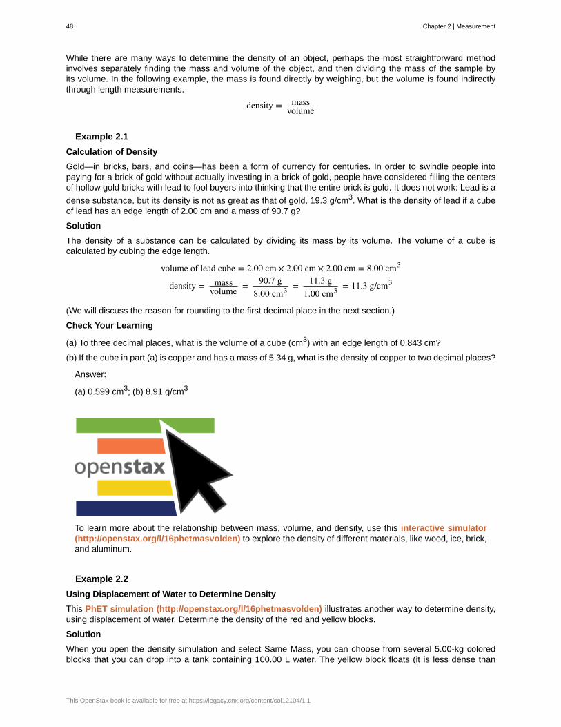
While there are many ways to determine the density of an object, perhaps the most straightforward methodinvolves separately finding the mass and volume of the object, and then dividing the mass of the sample byits volume. In the following example, the mass is found directly by weighing, but the volume is found indirectlythrough length measurements.
density = massvolume
Example 2.1
Calculation of Density
Gold—in bricks, bars, and coins—has been a form of currency for centuries. In order to swindle people intopaying for a brick of gold without actually investing in a brick of gold, people have considered filling the centersof hollow gold bricks with lead to fool buyers into thinking that the entire brick is gold. It does not work: Lead is a
dense substance, but its density is not as great as that of gold, 19.3 g/cm3. What is the density of lead if a cubeof lead has an edge length of 2.00 cm and a mass of 90.7 g?
Solution
The density of a substance can be calculated by dividing its mass by its volume. The volume of a cube iscalculated by cubing the edge length.
volume of lead cube = 2.00 cm × 2.00 cm × 2.00 cm = 8.00 cm3
density = massvolume = 90.7 g
8.00 cm3 = 11.3 g1.00 cm3 = 11.3 g/cm3
(We will discuss the reason for rounding to the first decimal place in the next section.)
Check Your Learning
(a) To three decimal places, what is the volume of a cube (cm3) with an edge length of 0.843 cm?
(b) If the cube in part (a) is copper and has a mass of 5.34 g, what is the density of copper to two decimal places?
Answer:
(a) 0.599 cm3; (b) 8.91 g/cm3
To learn more about the relationship between mass, volume, and density, use this interactive simulator(http://openstax.org/l/16phetmasvolden) to explore the density of different materials, like wood, ice, brick,and aluminum.
Example 2.2
Using Displacement of Water to Determine Density
This PhET simulation (http://openstax.org/l/16phetmasvolden) illustrates another way to determine density,using displacement of water. Determine the density of the red and yellow blocks.
Solution
When you open the density simulation and select Same Mass, you can choose from several 5.00-kg coloredblocks that you can drop into a tank containing 100.00 L water. The yellow block floats (it is less dense than
48 Chapter 2 | Measurement
This OpenStax book is available for free at https://legacy.cnx.org/content/col12104/1.1
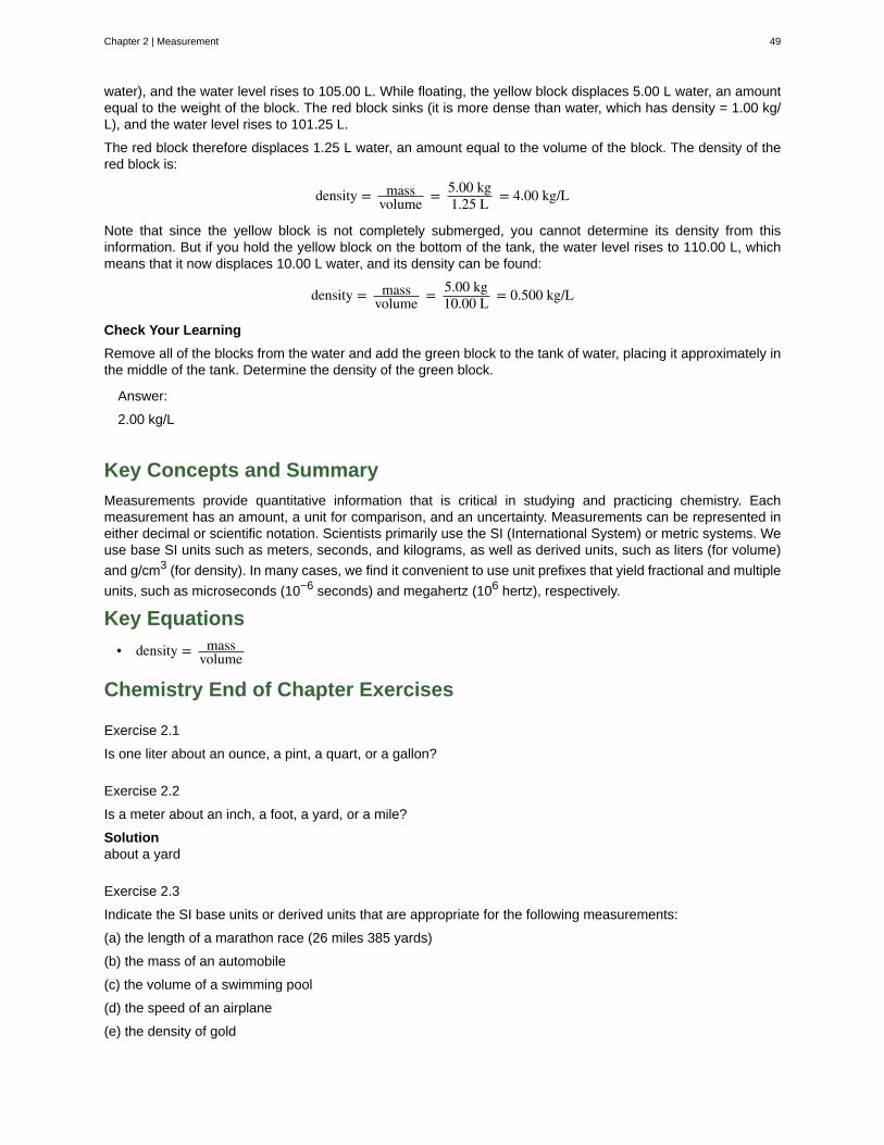
water), and the water level rises to 105.00 L. While floating, the yellow block displaces 5.00 L water, an amountequal to the weight of the block. The red block sinks (it is more dense than water, which has density = 1.00 kg/L), and the water level rises to 101.25 L.
The red block therefore displaces 1.25 L water, an amount equal to the volume of the block. The density of thered block is:
density = massvolume = 5.00 kg
1.25 L = 4.00 kg/L
Note that since the yellow block is not completely submerged, you cannot determine its density from thisinformation. But if you hold the yellow block on the bottom of the tank, the water level rises to 110.00 L, whichmeans that it now displaces 10.00 L water, and its density can be found:
density = massvolume = 5.00 kg
10.00 L = 0.500 kg/L
Check Your Learning
Remove all of the blocks from the water and add the green block to the tank of water, placing it approximately inthe middle of the tank. Determine the density of the green block.
Answer:
2.00 kg/L
Key Concepts and Summary
Measurements provide quantitative information that is critical in studying and practicing chemistry. Eachmeasurement has an amount, a unit for comparison, and an uncertainty. Measurements can be represented ineither decimal or scientific notation. Scientists primarily use the SI (International System) or metric systems. Weuse base SI units such as meters, seconds, and kilograms, as well as derived units, such as liters (for volume)
and g/cm3 (for density). In many cases, we find it convenient to use unit prefixes that yield fractional and multiple
units, such as microseconds (10−6 seconds) and megahertz (106 hertz), respectively.
Key Equations
• density = massvolume
Chemistry End of Chapter Exercises
Exercise 2.1
Is one liter about an ounce, a pint, a quart, or a gallon?
Exercise 2.2
Is a meter about an inch, a foot, a yard, or a mile?
Solutionabout a yard
Exercise 2.3
Indicate the SI base units or derived units that are appropriate for the following measurements:
(a) the length of a marathon race (26 miles 385 yards)
(b) the mass of an automobile
(c) the volume of a swimming pool
(d) the speed of an airplane
(e) the density of gold
Chapter 2 | Measurement 49
(f) the area of a football field
(g) the maximum temperature at the South Pole on April 1, 1913
Exercise 2.4
Indicate the SI base units or derived units that are appropriate for the following measurements:
(a) the mass of the moon
(b) the distance from Dallas to Oklahoma City
(c) the speed of sound
(d) the density of air
(e) the temperature at which alcohol boils
(f) the area of the state of Delaware
(g) the volume of a flu shot or a measles vaccination
Solution(a) kilograms; (b) meters; (c) kilometers/second; (d) kilograms/cubic meter; (e) kelvin; (f) square meters; (g) cubicmeters
Exercise 2.5
Give the name and symbol of the prefixes used with SI units to indicate multiplication by the following exactquantities.
(a) 103
(b) 10−2
(c) 0.1
(d) 10−3
(e) 1,000,000
(f) 0.000001
Exercise 2.6
Give the name of the prefix and the quantity indicated by the following symbols that are used with SI base units.
(a) c
(b) d
(c) G
(d) k
(e) m
(f) n
(g) p
(h) T
Solution
(a) centi-, × 10−2; (b) deci-, × 10−1; (c) Giga-, × 109; (d) kilo-, × 103; (e) milli-, × 10−3; (f) nano-, × 10−9;
(g) pico-, × 10−12; (h) tera-, × 1012
Exercise 2.7
A large piece of jewelry has a mass of 132.6 g. A graduated cylinder initially contains 48.6 mL water. When thejewelry is submerged in the graduated cylinder, the total volume increases to 61.2 mL.
(a) Determine the density of this piece of jewelry.
50 Chapter 2 | Measurement
This OpenStax book is available for free at https://legacy.cnx.org/content/col12104/1.1
(b) Assuming that the jewelry is made from only one substance, what substance is it likely to be? Explain.
Exercise 2.8
Visit this PhET density simulation (http://openstax.org/l/16phetmasvolden) and select the Same VolumeBlocks.
(a) What are the mass, volume, and density of the yellow block?
(b) What are the mass, volume and density of the red block?
(c) List the block colors in order from smallest to largest mass.
(d) List the block colors in order from lowest to highest density.
(e) How are mass and density related for blocks of the same volume?
Solution(a) 8.00 kg, 5.00 L, 1.60 kg/L; (b) 2.00 kg, 5.00 L, 0.400 kg/L; (c) red < green < blue < yellow; (d) If the volumesare the same, then the density is directly proportional to the mass.
Exercise 2.9
Visit this PhET density simulation (http://openstax.org/l/16phetmasvolden) and select Custom Blocks andthen My Block.
(a) Enter mass and volume values for the block such that the mass in kg is less than the volume in L. What doesthe block do? Why? Is this always the case when mass < volume?
(b) Enter mass and volume values for the block such that the mass in kg is more than the volume in L. Whatdoes the block do? Why? Is this always the case when mass > volume?
(c) How would (a) and (b) be different if the liquid in the tank were ethanol instead of water?
(d) How would (a) and (b) be different if the liquid in the tank were mercury instead of water?
Exercise 2.10
Visit this PhET density simulation (http://openstax.org/l/16phetmasvolden) and select Mystery Blocks.
(a) Pick one of the Mystery Blocks and determine its mass, volume, density, and its likely identity.
(b) Pick a different Mystery Block and determine its mass, volume, density, and its likely identity.
(c) Order the Mystery Blocks from least dense to most dense. Explain.
Solution(a) (b) Answer is one of the following. A/yellow: mass = 65.14 kg, volume = 3.38 L, density = 19.3 kg/L, likelyidentity = gold. B/blue: mass = 0.64 kg, volume = 1.00 L, density = 0.64 kg/L, likely identity = apple. C/green:mass = 4.08 kg, volume = 5.83 L, density = 0.700 kg/L, likely identity = gasoline. D/red: mass = 3.10 kg, volume= 3.38 L, density = 0.920 kg/L, likely identity = ice; and E/purple: mass = 3.53 kg, volume = 1.00 L, density = 3.53kg/L, likely identity = diamond. (c) B/blue/apple (0.64 kg/L) < C/green/gasoline (0.700 kg/L) < D/red/ice (0.920kg/L) < E/purple/diamond (3.53 kg/L) < A/yellow/gold (19.3 kg/L)
2.2 | Mathematical Treatment of Measurement Results
By the end of this section, you will be able to:
• Explain the dimensional analysis (factor label) approach to mathematical calculations involvingquantities
• Use dimensional analysis to carry out unit conversions for a given property and computations involvingtwo or more properties
It is often the case that a quantity of interest may not be easy (or even possible) to measure directly but insteadmust be calculated from other directly measured properties and appropriate mathematical relationships. For
Chapter 2 | Measurement 51
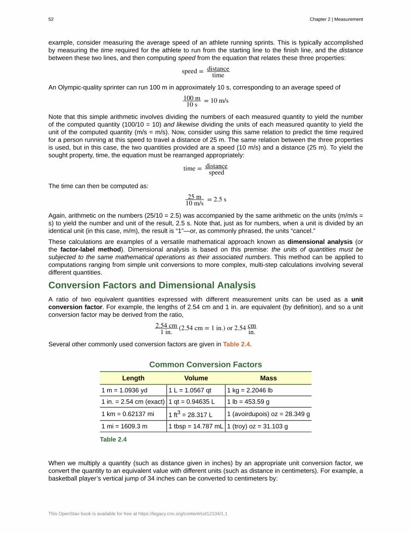
example, consider measuring the average speed of an athlete running sprints. This is typically accomplishedby measuring the time required for the athlete to run from the starting line to the finish line, and the distancebetween these two lines, and then computing speed from the equation that relates these three properties:
speed = distancetime
An Olympic-quality sprinter can run 100 m in approximately 10 s, corresponding to an average speed of
100 m10 s = 10 m/s
Note that this simple arithmetic involves dividing the numbers of each measured quantity to yield the numberof the computed quantity (100/10 = 10) and likewise dividing the units of each measured quantity to yield theunit of the computed quantity (m/s = m/s). Now, consider using this same relation to predict the time requiredfor a person running at this speed to travel a distance of 25 m. The same relation between the three propertiesis used, but in this case, the two quantities provided are a speed (10 m/s) and a distance (25 m). To yield thesought property, time, the equation must be rearranged appropriately:
time = distancespeed
The time can then be computed as:
25 m10 m/s = 2.5 s
Again, arithmetic on the numbers (25/10 = 2.5) was accompanied by the same arithmetic on the units (m/m/s =s) to yield the number and unit of the result, 2.5 s. Note that, just as for numbers, when a unit is divided by anidentical unit (in this case, m/m), the result is “1”—or, as commonly phrased, the units “cancel.”
These calculations are examples of a versatile mathematical approach known as dimensional analysis (orthe factor-label method). Dimensional analysis is based on this premise: the units of quantities must besubjected to the same mathematical operations as their associated numbers. This method can be applied tocomputations ranging from simple unit conversions to more complex, multi-step calculations involving severaldifferent quantities.
Conversion Factors and Dimensional Analysis
A ratio of two equivalent quantities expressed with different measurement units can be used as a unitconversion factor. For example, the lengths of 2.54 cm and 1 in. are equivalent (by definition), and so a unitconversion factor may be derived from the ratio,
2.54 cm1 in. (2.54 cm = 1 in.) or 2.54 cm
in.
Several other commonly used conversion factors are given in Table 2.4.
Common Conversion Factors
Length Volume Mass
1 m = 1.0936 yd 1 L = 1.0567 qt 1 kg = 2.2046 lb
1 in. = 2.54 cm (exact) 1 qt = 0.94635 L 1 lb = 453.59 g
1 km = 0.62137 mi 1 ft3 = 28.317 L 1 (avoirdupois) oz = 28.349 g
1 mi = 1609.3 m 1 tbsp = 14.787 mL 1 (troy) oz = 31.103 g
Table 2.4
When we multiply a quantity (such as distance given in inches) by an appropriate unit conversion factor, weconvert the quantity to an equivalent value with different units (such as distance in centimeters). For example, abasketball player’s vertical jump of 34 inches can be converted to centimeters by:
52 Chapter 2 | Measurement
This OpenStax book is available for free at https://legacy.cnx.org/content/col12104/1.1
34 in. × 2.54 cm1 in.
= 86 cm
Since this simple arithmetic involves quantities, the premise of dimensional analysis requires that we multiplyboth numbers and units. The numbers of these two quantities are multiplied to yield the number of the product
quantity, 86, whereas the units are multiplied to yield in. × cmin. . Just as for numbers, a ratio of identical units is
also numerically equal to one, in.in. = 1, and the unit product thus simplifies to cm. (When identical units divide to
yield a factor of 1, they are said to “cancel.”) Using dimensional analysis, we can determine that a unit conversionfactor has been set up correctly by checking to confirm that the original unit will cancel, and the result will containthe sought (converted) unit.
Example 2.3
Using a Unit Conversion Factor
The mass of a competition frisbee is 125 g. Convert its mass to ounces using the unit conversion factor derivedfrom the relationship 1 oz = 28.349 g (Table 2.4).
Solution
If we have the conversion factor, we can determine the mass in kilograms using an equation similar the one usedfor converting length from inches to centimeters.
x oz = 125 g × unit conversion factor
We write the unit conversion factor in its two forms:
1 oz28.349 g and 28.349 g
1 oz
The correct unit conversion factor is the ratio that cancels the units of grams and leaves ounces.
x oz = 125 g × 1 oz28.349 g
= ⎛⎝ 12528.349
⎞⎠oz
= 4.41 oz (three significant figures)
Check Your Learning
Convert a volume of 9.345 qt to liters.
Answer:
8.844 L
Beyond simple unit conversions, the factor-label method can be used to solve more complex problems involvingcomputations. Regardless of the details, the basic approach is the same—all the factors involved in thecalculation must be appropriately oriented to insure that their labels (units) will appropriately cancel and/orcombine to yield the desired unit in the result. This is why it is referred to as the factor-label method. As yourstudy of chemistry continues, you will encounter many opportunities to apply this approach.
Example 2.4
Computing Quantities from Measurement Results and Known Mathematical Relations
What is the density of common antifreeze in units of g/mL? A 4.00-qt sample of the antifreeze weighs 9.26 lb.
Solution
Since density = massvolume , we need to divide the mass in grams by the volume in milliliters. In general: the
number of units of B = the number of units of A × unit conversion factor. The necessary conversion factors are
given in Table 2.4: 1 lb = 453.59 g; 1 L = 1.0567 qt; 1 L = 1,000 mL. We can convert mass from pounds to grams
Chapter 2 | Measurement 53
in one step:
9.26 lb × 453.59 g1 lb
= 4.20 × 10³ g
We need to use two steps to convert volume from quarts to milliliters.
1. Convert quarts to liters.
4.00 qt × 1 L1.0567 qt = 3.78 L
2. Convert liters to milliliters.
3.78 L × 1000 mL1 L
= 3.78 × 10³ mL
Then,
density = 4.20 × 10³ g3.78 × 103 mL
= 1.11 g/mL
Alternatively, the calculation could be set up in a way that uses three unit conversion factors sequentially asfollows:
9.26 lb4.00 qt × 453.59 g
1 lb×
1.0567 qt1 L
× 1 L1000 mL = 1.11 g/mL
Check Your Learning
What is the volume in liters of 1.000 oz, given that 1 L = 1.0567 qt and 1 qt = 32 oz (exactly)?
Answer:
2.956 × 10⁻² L
Example 2.5
Computing Quantities from Measurement Results and Known Mathematical Relations
While being driven from Philadelphia to Atlanta, a distance of about 1250 km, a 2014 Lamborghini AventadorRoadster uses 213 L gasoline.
(a) What (average) fuel economy, in miles per gallon, did the Roadster get during this trip?
(b) If gasoline costs $3.80 per gallon, what was the fuel cost for this trip?
Solution
(a) We first convert distance from kilometers to miles:
1250 km × 0.62137 mi1 km = 777 mi
and then convert volume from liters to gallons:
213 L ×1.0567 qt
1 L× 1 gal
4 qt = 56.3 gal
Then,
(average) mileage = 777 mi56.3 gal = 13.8 miles/gallon = 13.8 mpg
Alternatively, the calculation could be set up in a way that uses all the conversion factors sequentially, as follows:
1250 km213 L
× 0.62137 mi1 km
× 1 L1.0567 qt ×
4 qt1 gal = 13.8 mpg
(b) Using the previously calculated volume in gallons, we find:
54 Chapter 2 | Measurement
This OpenStax book is available for free at https://legacy.cnx.org/content/col12104/1.1
56.3 gal × $3.801 gal = $214
Check Your Learning
A Toyota Prius Hybrid uses 59.7 L gasoline to drive from San Francisco to Seattle, a distance of 1300 km (twosignificant digits).
(a) What (average) fuel economy, in miles per gallon, did the Prius get during this trip?
(b) If gasoline costs $3.90 per gallon, what was the fuel cost for this trip?
Answer:
(a) 51 mpg; (b) $62
Conversion of Temperature Units
We use the word temperature to refer to the hotness or coldness of a substance. One way we measure a changein temperature is to use the fact that most substances expand when their temperature increases and contractwhen their temperature decreases. The mercury or alcohol in a common glass thermometer changes its volumeas the temperature changes. Because the volume of the liquid changes more than the volume of the glass, wecan see the liquid expand when it gets warmer and contract when it gets cooler.
To mark a scale on a thermometer, we need a set of reference values: Two of the most commonly used are thefreezing and boiling temperatures of water at a specified atmospheric pressure. On the Celsius scale, 0 °C isdefined as the freezing temperature of water and 100 °C as the boiling temperature of water. The space betweenthe two temperatures is divided into 100 equal intervals, which we call degrees. On the Fahrenheit scale, thefreezing point of water is defined as 32 °F and the boiling temperature as 212 °F. The space between these twopoints on a Fahrenheit thermometer is divided into 180 equal parts (degrees).
Defining the Celsius and Fahrenheit temperature scales as described in the previous paragraph results in aslightly more complex relationship between temperature values on these two scales than for different units ofmeasure for other properties. Most measurement units for a given property are directly proportional to oneanother (y = mx). Using familiar length units as one example:
length in feet = ⎛⎝ 1 ft12 in.
⎞⎠× length in inches
where y = length in feet, x = length in inches, and the proportionality constant, m, is the conversion factor. TheCelsius and Fahrenheit temperature scales, however, do not share a common zero point, and so the relationshipbetween these two scales is a linear one rather than a proportional one (y = mx + b). Consequently, convertinga temperature from one of these scales into the other requires more than simple multiplication by a conversionfactor, m, it also must take into account differences in the scales’ zero points (b).
The linear equation relating Celsius and Fahrenheit temperatures is easily derived from the two temperaturesused to define each scale. Representing the Celsius temperature as x and the Fahrenheit temperature as y, theslope, m, is computed to be:
m = ΔyΔx = 212 °F − 32 °F
100 °C − 0 °C = 180 °F100 °C = 9 °F
5 °C
The y-intercept of the equation, b, is then calculated using either of the equivalent temperature pairs, (100 °C,212 °F) or (0 °C, 32 °F), as:
b = y − mx = 32 °F − 9 °F5 °C × 0 °C = 32 °F
The equation relating the temperature scales is then:
T°F = ⎛⎝9 °F5 °C × T°C
⎞⎠+ 32 °C
An abbreviated form of this equation that omits the measurement units is:
T°F = ⎛⎝95 × T°C⎞⎠+ 32
Chapter 2 | Measurement 55
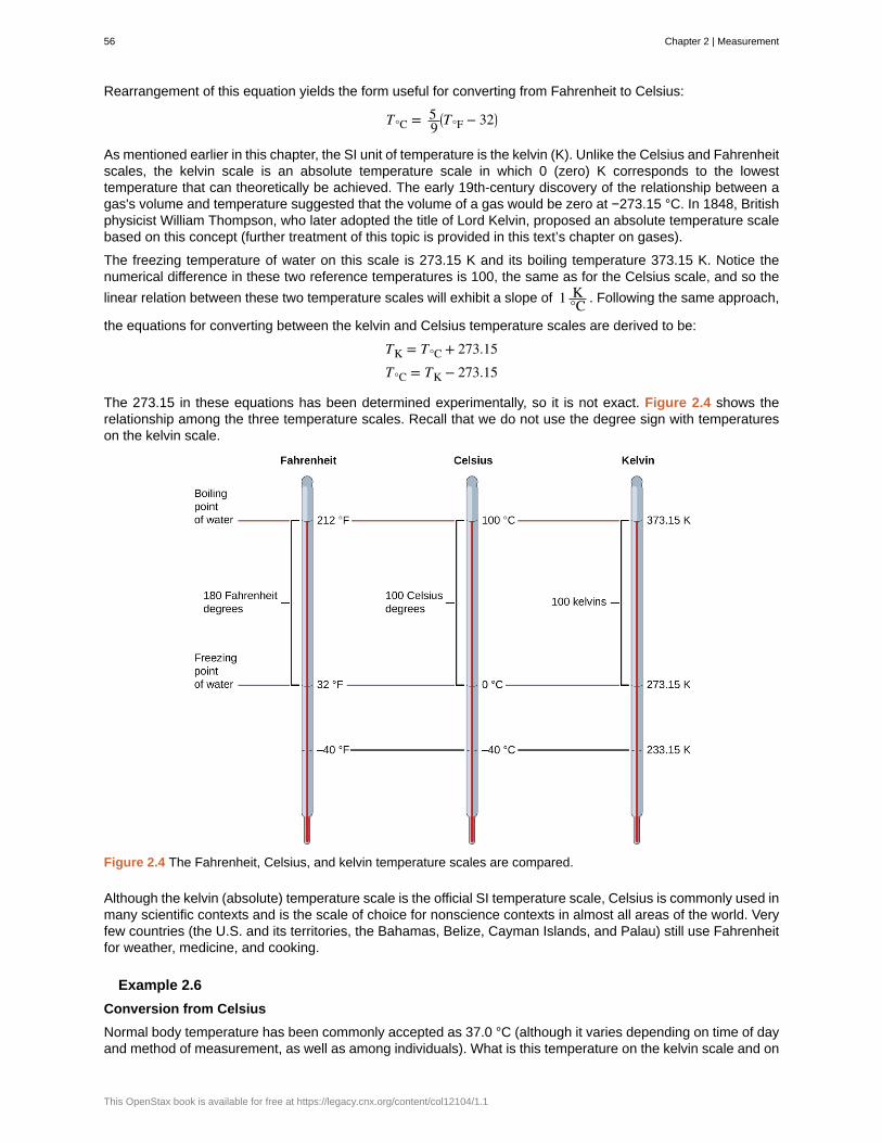
Rearrangement of this equation yields the form useful for converting from Fahrenheit to Celsius:
T°C = 59⎛⎝T°F − 32⎞⎠
As mentioned earlier in this chapter, the SI unit of temperature is the kelvin (K). Unlike the Celsius and Fahrenheitscales, the kelvin scale is an absolute temperature scale in which 0 (zero) K corresponds to the lowesttemperature that can theoretically be achieved. The early 19th-century discovery of the relationship between agas's volume and temperature suggested that the volume of a gas would be zero at −273.15 °C. In 1848, Britishphysicist William Thompson, who later adopted the title of Lord Kelvin, proposed an absolute temperature scalebased on this concept (further treatment of this topic is provided in this text’s chapter on gases).
The freezing temperature of water on this scale is 273.15 K and its boiling temperature 373.15 K. Notice thenumerical difference in these two reference temperatures is 100, the same as for the Celsius scale, and so the
linear relation between these two temperature scales will exhibit a slope of 1 K°C . Following the same approach,
the equations for converting between the kelvin and Celsius temperature scales are derived to be:
TK = T°C + 273.15T°C = TK − 273.15
The 273.15 in these equations has been determined experimentally, so it is not exact. Figure 2.4 shows therelationship among the three temperature scales. Recall that we do not use the degree sign with temperatureson the kelvin scale.
Figure 2.4 The Fahrenheit, Celsius, and kelvin temperature scales are compared.
Although the kelvin (absolute) temperature scale is the official SI temperature scale, Celsius is commonly used inmany scientific contexts and is the scale of choice for nonscience contexts in almost all areas of the world. Veryfew countries (the U.S. and its territories, the Bahamas, Belize, Cayman Islands, and Palau) still use Fahrenheitfor weather, medicine, and cooking.
Example 2.6
Conversion from Celsius
Normal body temperature has been commonly accepted as 37.0 °C (although it varies depending on time of dayand method of measurement, as well as among individuals). What is this temperature on the kelvin scale and on
56 Chapter 2 | Measurement
This OpenStax book is available for free at https://legacy.cnx.org/content/col12104/1.1
the Fahrenheit scale?
Solution
K = °C + 273.15 = 37.0 + 273.2 = 310.2 K
°F = 95°C + 32.0 = ⎛⎝95 × 37.0⎞⎠+ 32.0 = 66.6 + 32.0 = 98.6 °F
Check Your Learning
Convert 80.92 °C to K and °F.
Answer:
354.07 K, 177.7 °F
Example 2.7
Conversion from Fahrenheit
Baking a ready-made pizza calls for an oven temperature of 450 °F. If you are in Europe, and your oventhermometer uses the Celsius scale, what is the setting? What is the kelvin temperature?
Solution
°C = 59(°F − 32) = 5
9(450 − 32) = 59 × 418 = 232 °C ⟶ set oven to 230 °C ⎛
⎝two significant figures ⎞⎠
K = °C + 273.15 = 230 + 273 = 503 K ⟶ 5.0 × 10² K ⎛⎝two significant figures ⎞⎠
Check Your Learning
Convert 50 °F to °C and K.
Answer:
10 °C, 280 K
Key Concepts and Summary
Measurements are made using a variety of units. It is often useful or necessary to convert a measured quantityfrom one unit into another. These conversions are accomplished using unit conversion factors, which are derivedby simple applications of a mathematical approach called the factor-label method or dimensional analysis.This strategy is also employed to calculate sought quantities using measured quantities and appropriatemathematical relations.
Key Equations
• T°C = ⎛⎝59 × T°F⎞⎠− 32
• T°F = ⎛⎝95 × T°C⎞⎠+ 32
• TK = °C + 273.15
• T°C = K − 273.15
Chemistry End of Chapter Exercises
Exercise 2.11
Write conversion factors (as ratios) for the number of:
(a) yards in 1 meter
(b) liters in 1 liquid quart
Chapter 2 | Measurement 57
(c) pounds in 1 kilogram
Solution
(a)1.0936 yd
1 m ; (b) 0.94635 L1 qt ; (c) 2.2046 lb
1 kg
Exercise 2.12
Write conversion factors (as ratios) for the number of:
(a) kilometers in 1 mile
(b) liters in 1 cubic foot
(c) grams in 1 ounce
Exercise 2.13
The label on a soft drink bottle gives the volume in two units: 2.0 L and 67.6 fl oz. Use this information to derivea conversion factor between the English and metric units. How many significant figures can you justify in yourconversion factor?
Solution2.0 L
67.6 fl oz = 0.030 L1 fl oz
Only two significant figures are justified.
Exercise 2.14
The label on a box of cereal gives the mass of cereal in two units: 978 grams and 34.5 oz. Use this informationto find a conversion factor between the English and metric units. How many significant figures can you justify inyour conversion factor?
Exercise 2.15
Soccer is played with a round ball having a circumference between 27 and 28 in. and a weight between 14 and16 oz. What are these specifications in units of centimeters and grams?
Solution68–71 cm; 400–450 g
Exercise 2.16
A woman's basketball has a circumference between 28.5 and 29.0 inches and a maximum weight of 20 ounces(two significant figures). What are these specifications in units of centimeters and grams?
Exercise 2.17
How many milliliters of a soft drink are contained in a 12.0-oz can?
Solution355 mL
Exercise 2.18
A barrel of oil is exactly 42 gal. How many liters of oil are in a barrel?
Exercise 2.19
The diameter of a red blood cell is about 3 × 10⁻⁴ in. What is its diameter in centimeters?
Solution
8 × 10⁻⁴ cm
Exercise 2.20
58 Chapter 2 | Measurement
This OpenStax book is available for free at https://legacy.cnx.org/content/col12104/1.1

The distance between the centers of the two oxygen atoms in an oxygen molecule is 1.21 × 10⁻⁸ cm. What is
this distance in inches?
Exercise 2.21
Is a 197-lb weight lifter light enough to compete in a class limited to those weighing 90 kg or less?
Solutionyes; weight = 89.4 kg
Exercise 2.22
A very good 197-lb weight lifter lifted 192 kg in a move called the clean and jerk. What was the mass of theweight lifted in pounds?
Exercise 2.23
Many medical laboratory tests are run using 5.0 μL blood serum. What is this volume in milliliters?
Solution
5.0 × 10⁻³ mL
Exercise 2.24
If an aspirin tablet contains 325 mg aspirin, how many grams of aspirin does it contain?
Exercise 2.25
Use scientific (exponential) notation to express the following quantities in terms of the SI base units in Table 2.2:
(a) 0.13 g
(b) 232 Gg
(c) 5.23 pm
(d) 86.3 mg
(e) 37.6 cm
(f) 54 μm
(g) 1 Ts
(h) 27 ps
(i) 0.15 mK
Solution
(a) 1.3 × 10⁻⁴ kg; (b) 2.32 × 10⁸ kg; (c) 5.23 × 10⁻¹² m; (d) 8.63 × 10⁻⁵ kg; (e) 3.76 × 10⁻¹ m; (f) 5.4 ×10⁻⁵ m; (g) 1 × 10¹² s; (h) 2.7 × 10⁻¹¹ s; (i) 1.5 × 10⁻⁴ K
Exercise 2.26
Complete the following conversions between SI units.
(a) 612 g = ________ mg
(b) 8.160 m = ________ cm
(c) 3779 μg = ________ g
(d) 781 mL = ________ L
(e) 4.18 kg = ________ g
(f) 27.8 m = ________ km
(g) 0.13 mL = ________ L
(h) 1738 km = ________ m
Chapter 2 | Measurement 59
(i) 1.9 Gg = ________ g
Exercise 2.27
Gasoline is sold by the liter in many countries. How many liters are required to fill a 12.0-gal gas tank?
Solution45.4 L
Exercise 2.28
Milk is sold by the liter in many countries. What is the volume of exactly 1/2 gal of milk in liters?
Exercise 2.29
A long ton is defined as exactly 2240 lb. What is this mass in kilograms?
Solution
1.0160 × 10³ kg
Exercise 2.30
Make the conversion indicated in each of the following:
(a) the men’s world record long jump, 29 ft 4¼ in., to meters
(b) the greatest depth of the ocean, about 6.5 mi, to kilometers
(c) the area of the state of Oregon, 96,981 mi2, to square kilometers
(d) the volume of 1 gill (exactly 4 oz) to milliliters
(e) the estimated volume of the oceans, 330,000,000 mi3, to cubic kilometers.
(f) the mass of a 3525-lb car to kilograms
(g) the mass of a 2.3-oz egg to grams
Exercise 2.31
Make the conversion indicated in each of the following:
(a) the length of a soccer field, 120 m (three significant figures), to feet
(b) the height of Mt. Kilimanjaro, at 19,565 ft the highest mountain in Africa, to kilometers
(c) the area of an 8.5 × 11-inch sheet of paper in cm2
(d) the displacement volume of an automobile engine, 161 in.3, to liters
(e) the estimated mass of the atmosphere, 5.6 × 10¹⁵ tons, to kilograms
(f) the mass of a bushel of rye, 32.0 lb, to kilograms
(g) the mass of a 5.00-grain aspirin tablet to milligrams (1 grain = 0.00229 oz)
Solution(a) 394 ft(b) 5.9634 km
(c) 6.0 × 10²
(d) 2.64 L
(e) 5.1 × 10¹⁸ kg
(f) 14.5 kg(g) 324 mg
Exercise 2.32
Many chemistry conferences have held a 50-Trillion Angstrom Run (two significant figures). How long is this run
60 Chapter 2 | Measurement
This OpenStax book is available for free at https://legacy.cnx.org/content/col12104/1.1

in kilometers and in miles? (1 Å = 1 × 10⁻¹⁰ m)
Exercise 2.33
A chemist’s 50-Trillion Angstrom Run (see Exercise 2.32) would be an archeologist’s 10,900 cubit run. How long
is one cubit in meters and in feet? (1 Å = 1 × 10⁻⁸ cm)
Solution0.46 m; 1.5 ft/cubit
Exercise 2.34
The gas tank of a certain luxury automobile holds 22.3 gallons according to the owner’s manual. If the density ofgasoline is 0.8206 g/mL, determine the mass in kilograms and pounds of the fuel in a full tank.
Exercise 2.35
As an instructor is preparing for an experiment, he requires 225 g phosphoric acid. The only container readilyavailable is a 150-mL Erlenmeyer flask. Is it large enough to contain the acid, whose density is 1.83 g/mL?
SolutionYes, the acid's volume is 123 mL.
Exercise 2.36
To prepare for a laboratory period, a student lab assistant needs 125 g of a compound. A bottle containing 1/4 lbis available. Did the student have enough of the compound?
Exercise 2.37
A chemistry student is 159 cm tall and weighs 45.8 kg. What is her height in inches and weight in pounds?
Solution62.6 in (about 5 ft 3 in.) and 101 lb
Exercise 2.38
In a recent Grand Prix, the winner completed the race with an average speed of 229.8 km/h. What was his speedin miles per hour, meters per second, and feet per second?
Exercise 2.39
Solve these problems about lumber dimensions.
(a) To describe to a European how houses are constructed in the US, the dimensions of “two-by-four” lumbermust be converted into metric units. The thickness × width × length dimensions are 1.50 in. × 3.50 in. ×8.00 ft in the US. What are the dimensions in cm × cm × m?
(b) This lumber can be used as vertical studs, which are typically placed 16.0 in. apart. What is that distance incentimeters?
Solution(a) 3.81 cm × 8.89 cm × 2.44 m; (b) 40.6 cm
Exercise 2.40
The mercury content of a stream was believed to be above the minimum considered safe—1 part per billion(ppb) by weight. An analysis indicated that the concentration was 0.68 parts per billion. What quantity of mercury
in grams was present in 15.0 L of the water, the density of which is 0.998 g/ml? (1 ppb Hg = 1 ng Hg1 g water)
Exercise 2.41
Chapter 2 | Measurement 61
Calculate the density of aluminum if 27.6 cm3 has a mass of 74.6 g.
Solution
2.70 g/cm3
Exercise 2.42
Osmium is one of the densest elements known. What is its density if 2.72 g has a volume of 0.121 cm3?
Exercise 2.43
Calculate these masses.
(a) What is the mass of 6.00 cm3 of mercury, density = 13.5939 g/cm3?
(b) What is the mass of 25.0 mL octane, density = 0.702 g/cm3?
Solution(a) 81.6 g; (b) 17.6 g
Exercise 2.44
Calculate these masses.
(a) What is the mass of 4.00 cm3 of sodium, density = 0.97 g/cm3 ?
(b) What is the mass of 125 mL gaseous chlorine, density = 3.16 g/L?
Exercise 2.45
Calculate these volumes.
(a) What is the volume of 25 g iodine, density = 4.93 g/cm3?
(b) What is the volume of 3.28 g gaseous hydrogen, density = 0.089 g/L?
Solution(a) 5.1 mL; (b) 37 L
Exercise 2.46
Calculate these volumes.
(a) What is the volume of 11.3 g graphite, density = 2.25 g/cm3?
(b) What is the volume of 39.657 g bromine, density = 2.928 g/cm3?
Exercise 2.47
Convert the boiling temperature of gold, 2966 °C, into degrees Fahrenheit and kelvin.
Solution5371 °F, 3239 K
Exercise 2.48
Convert the temperature of scalding water, 54 °C, into degrees Fahrenheit and kelvin.
Exercise 2.49
Convert the temperature of the coldest area in a freezer, −10 °F, to degrees Celsius and kelvin.
Solution−23 °C, 250 K
Exercise 2.50
62 Chapter 2 | Measurement
This OpenStax book is available for free at https://legacy.cnx.org/content/col12104/1.1
Convert the temperature of dry ice, −77 °C, into degrees Fahrenheit and kelvin.
Exercise 2.51
Convert the boiling temperature of liquid ammonia, −28.1 °F, into degrees Celsius and kelvin.
Solution−33.4 °C, 239.8 K
Exercise 2.52
The label on a pressurized can of spray disinfectant warns against heating the can above 130 °F. What are thecorresponding temperatures on the Celsius and kelvin temperature scales?
Exercise 2.53
The weather in Europe was unusually warm during the summer of 1995. The TV news reported temperatures ashigh as 45 °C. What was the temperature on the Fahrenheit scale?
Solution113 °F
Chapter 2 | Measurement 63
Celsius (°C)
cubic centimeter (cm3 or cc)
cubic meter (m3)
density
dimensional analysis
Fahrenheit
kelvin (K)
kilogram (kg)
length
liter (L)
meter (m)
milliliter (mL)
second (s)
SI units (International System of Units)
unit
unit conversion factor
volume
KEY TERMS
unit of temperature; water freezes at 0 °C and boils at 100 °C on this scale
volume of a cube with an edge length of exactly 1 cm
SI unit of volume
ratio of mass to volume for a substance or object
(also, factor-label method) versatile mathematical approach that can be applied tocomputations ranging from simple unit conversions to more complex, multi-step calculations involvingseveral different quantities
unit of temperature; water freezes at 32 °F and boils at 212 °F on this scale
SI unit of temperature; 273.15 K = 0 ºC
standard SI unit of mass; 1 kg = approximately 2.2 pounds
measure of one dimension of an object
(also, cubic decimeter) unit of volume; 1 L = 1,000 cm3
standard metric and SI unit of length; 1 m = approximately 1.094 yards
1/1,000 of a liter; equal to 1 cm3
SI unit of time
standards fixed by international agreement in the InternationalSystem of Units (Le Système International d’Unités)
standard of comparison for measurements
ratio of equivalent quantities expressed with different units; used to convert from oneunit to a different unit
amount of space occupied by an object
64 Chapter 2 | Measurement
This OpenStax book is available for free at https://legacy.cnx.org/content/col12104/1.1
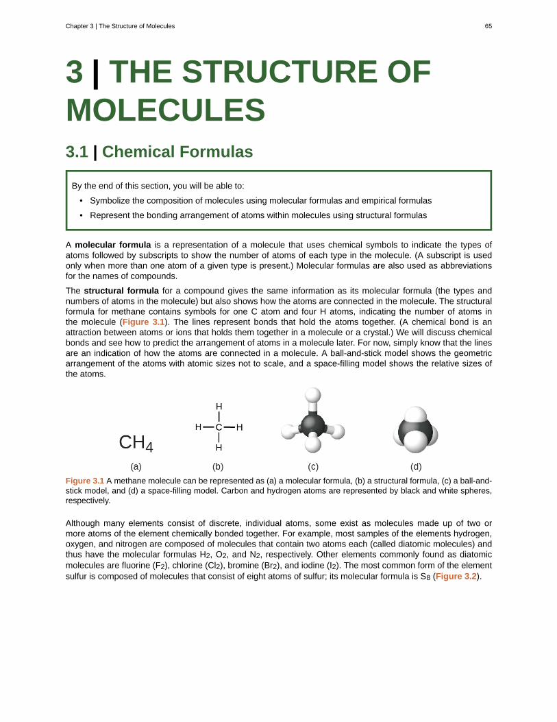
3 | THE STRUCTURE OFMOLECULES3.1 | Chemical Formulas
By the end of this section, you will be able to:
• Symbolize the composition of molecules using molecular formulas and empirical formulas
• Represent the bonding arrangement of atoms within molecules using structural formulas
A molecular formula is a representation of a molecule that uses chemical symbols to indicate the types ofatoms followed by subscripts to show the number of atoms of each type in the molecule. (A subscript is usedonly when more than one atom of a given type is present.) Molecular formulas are also used as abbreviationsfor the names of compounds.
The structural formula for a compound gives the same information as its molecular formula (the types andnumbers of atoms in the molecule) but also shows how the atoms are connected in the molecule. The structuralformula for methane contains symbols for one C atom and four H atoms, indicating the number of atoms inthe molecule (Figure 3.1). The lines represent bonds that hold the atoms together. (A chemical bond is anattraction between atoms or ions that holds them together in a molecule or a crystal.) We will discuss chemicalbonds and see how to predict the arrangement of atoms in a molecule later. For now, simply know that the linesare an indication of how the atoms are connected in a molecule. A ball-and-stick model shows the geometricarrangement of the atoms with atomic sizes not to scale, and a space-filling model shows the relative sizes ofthe atoms.
Figure 3.1 A methane molecule can be represented as (a) a molecular formula, (b) a structural formula, (c) a ball-and-stick model, and (d) a space-filling model. Carbon and hydrogen atoms are represented by black and white spheres,respectively.
Although many elements consist of discrete, individual atoms, some exist as molecules made up of two ormore atoms of the element chemically bonded together. For example, most samples of the elements hydrogen,oxygen, and nitrogen are composed of molecules that contain two atoms each (called diatomic molecules) andthus have the molecular formulas H2, O2, and N2, respectively. Other elements commonly found as diatomicmolecules are fluorine (F2), chlorine (Cl2), bromine (Br2), and iodine (I2). The most common form of the elementsulfur is composed of molecules that consist of eight atoms of sulfur; its molecular formula is S8 (Figure 3.2).
Chapter 3 | The Structure of Molecules 65
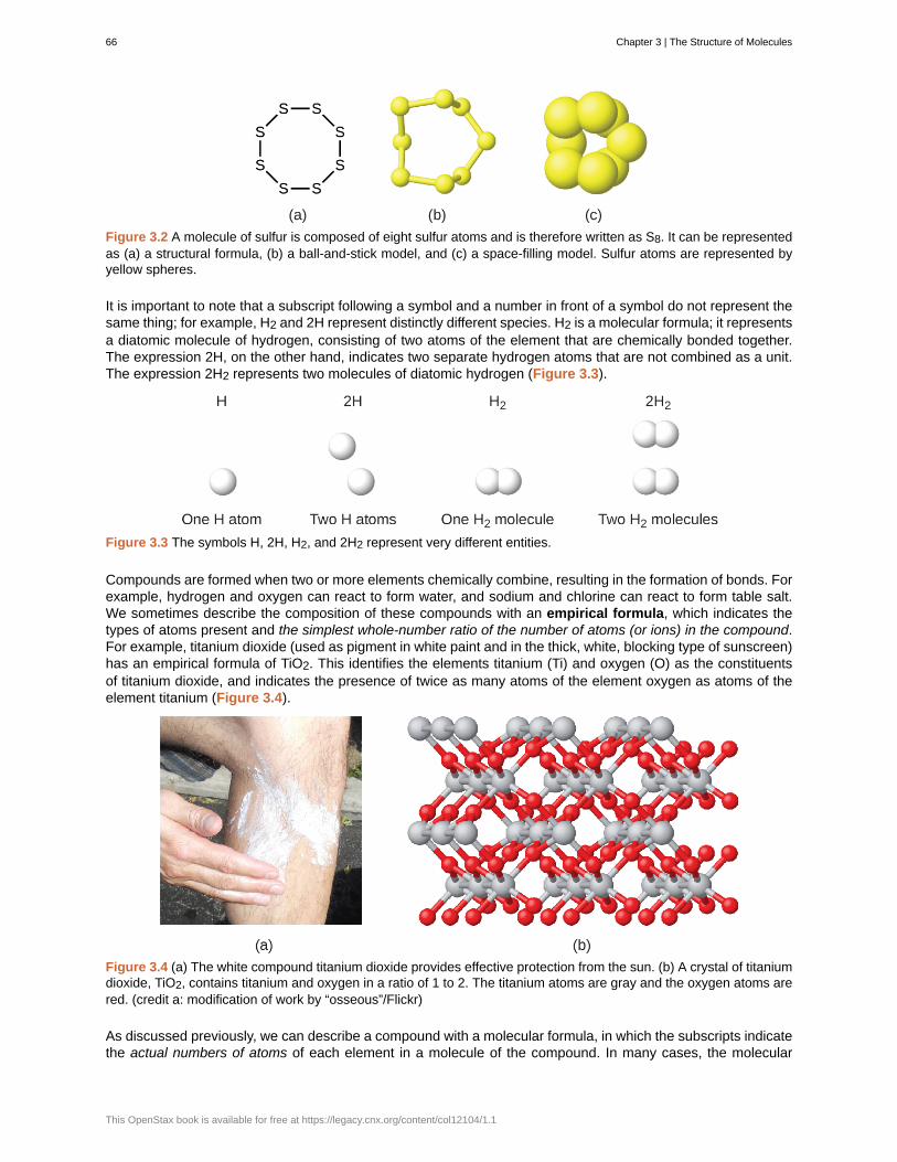
Figure 3.2 A molecule of sulfur is composed of eight sulfur atoms and is therefore written as S8. It can be representedas (a) a structural formula, (b) a ball-and-stick model, and (c) a space-filling model. Sulfur atoms are represented byyellow spheres.
It is important to note that a subscript following a symbol and a number in front of a symbol do not represent thesame thing; for example, H2 and 2H represent distinctly different species. H2 is a molecular formula; it representsa diatomic molecule of hydrogen, consisting of two atoms of the element that are chemically bonded together.The expression 2H, on the other hand, indicates two separate hydrogen atoms that are not combined as a unit.The expression 2H2 represents two molecules of diatomic hydrogen (Figure 3.3).
Figure 3.3 The symbols H, 2H, H2, and 2H2 represent very different entities.
Compounds are formed when two or more elements chemically combine, resulting in the formation of bonds. Forexample, hydrogen and oxygen can react to form water, and sodium and chlorine can react to form table salt.We sometimes describe the composition of these compounds with an empirical formula, which indicates thetypes of atoms present and the simplest whole-number ratio of the number of atoms (or ions) in the compound.For example, titanium dioxide (used as pigment in white paint and in the thick, white, blocking type of sunscreen)has an empirical formula of TiO2. This identifies the elements titanium (Ti) and oxygen (O) as the constituentsof titanium dioxide, and indicates the presence of twice as many atoms of the element oxygen as atoms of theelement titanium (Figure 3.4).
Figure 3.4 (a) The white compound titanium dioxide provides effective protection from the sun. (b) A crystal of titaniumdioxide, TiO2, contains titanium and oxygen in a ratio of 1 to 2. The titanium atoms are gray and the oxygen atoms arered. (credit a: modification of work by “osseous”/Flickr)
As discussed previously, we can describe a compound with a molecular formula, in which the subscripts indicatethe actual numbers of atoms of each element in a molecule of the compound. In many cases, the molecular
66 Chapter 3 | The Structure of Molecules
This OpenStax book is available for free at https://legacy.cnx.org/content/col12104/1.1
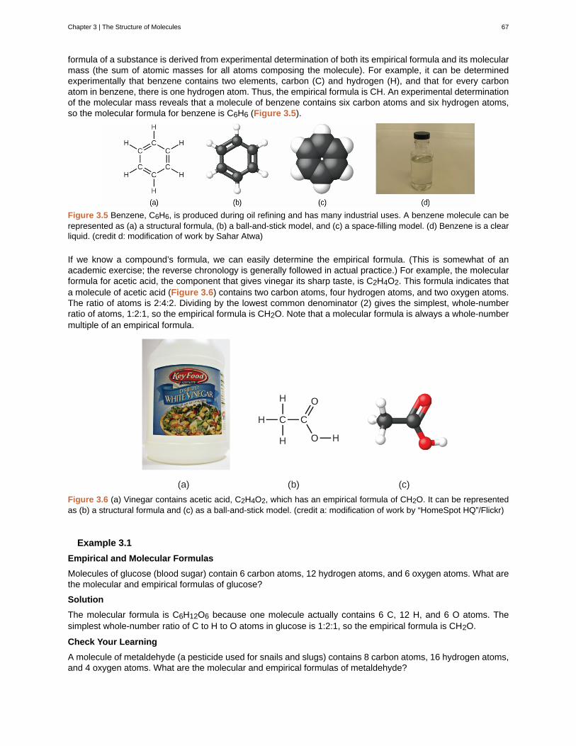
formula of a substance is derived from experimental determination of both its empirical formula and its molecularmass (the sum of atomic masses for all atoms composing the molecule). For example, it can be determinedexperimentally that benzene contains two elements, carbon (C) and hydrogen (H), and that for every carbonatom in benzene, there is one hydrogen atom. Thus, the empirical formula is CH. An experimental determinationof the molecular mass reveals that a molecule of benzene contains six carbon atoms and six hydrogen atoms,so the molecular formula for benzene is C6H6 (Figure 3.5).
Figure 3.5 Benzene, C6H6, is produced during oil refining and has many industrial uses. A benzene molecule can berepresented as (a) a structural formula, (b) a ball-and-stick model, and (c) a space-filling model. (d) Benzene is a clearliquid. (credit d: modification of work by Sahar Atwa)
If we know a compound’s formula, we can easily determine the empirical formula. (This is somewhat of anacademic exercise; the reverse chronology is generally followed in actual practice.) For example, the molecularformula for acetic acid, the component that gives vinegar its sharp taste, is C2H4O2. This formula indicates thata molecule of acetic acid (Figure 3.6) contains two carbon atoms, four hydrogen atoms, and two oxygen atoms.The ratio of atoms is 2:4:2. Dividing by the lowest common denominator (2) gives the simplest, whole-numberratio of atoms, 1:2:1, so the empirical formula is CH2O. Note that a molecular formula is always a whole-numbermultiple of an empirical formula.
Figure 3.6 (a) Vinegar contains acetic acid, C2H4O2, which has an empirical formula of CH2O. It can be representedas (b) a structural formula and (c) as a ball-and-stick model. (credit a: modification of work by “HomeSpot HQ”/Flickr)
Example 3.1
Empirical and Molecular Formulas
Molecules of glucose (blood sugar) contain 6 carbon atoms, 12 hydrogen atoms, and 6 oxygen atoms. What arethe molecular and empirical formulas of glucose?
Solution
The molecular formula is C6H12O6 because one molecule actually contains 6 C, 12 H, and 6 O atoms. Thesimplest whole-number ratio of C to H to O atoms in glucose is 1:2:1, so the empirical formula is CH2O.
Check Your Learning
A molecule of metaldehyde (a pesticide used for snails and slugs) contains 8 carbon atoms, 16 hydrogen atoms,and 4 oxygen atoms. What are the molecular and empirical formulas of metaldehyde?
Chapter 3 | The Structure of Molecules 67

Answer:
Molecular formula, C8H16O4; empirical formula, C2H4O
You can explore molecule building (http://openstax.org/l/16molbuilding) using an online simulation.
Lee Cronin
What is it that chemists do? According to Lee Cronin (Figure 3.7), chemists make very complicatedmolecules by “chopping up” small molecules and “reverse engineering” them. He wonders if we could “makea really cool universal chemistry set” by what he calls “app-ing” chemistry. Could we “app” chemistry?
In a 2012 TED talk, Lee describes one fascinating possibility: combining a collection of chemical “inks”with a 3D printer capable of fabricating a reaction apparatus (tiny test tubes, beakers, and the like) tofashion a “universal toolkit of chemistry.” This toolkit could be used to create custom-tailored drugs to fighta new superbug or to “print” medicine personally configured to your genetic makeup, environment, andhealth situation. Says Cronin, “What Apple did for music, I’d like to do for the discovery and distribution ofprescription drugs.”[1] View his full talk (http://openstax.org/l/16LeeCronin) at the TED website.
1. Lee Cronin, “Print Your Own Medicine,” Talk presented at TED Global 2012, Edinburgh, Scotland, June 2012.
68 Chapter 3 | The Structure of Molecules
This OpenStax book is available for free at https://legacy.cnx.org/content/col12104/1.1

Figure 3.7 Chemist Lee Cronin has been named one of the UK’s 10 most inspirational scientists. The youngestchair at the University of Glasgow, Lee runs a large research group, collaborates with many scientists worldwide,has published over 250 papers in top scientific journals, and has given more than 150 invited talks. His researchfocuses on complex chemical systems and their potential to transform technology, but also branches intonanoscience, solar fuels, synthetic biology, and even artificial life and evolution. (credit: image courtesy of LeeCronin)
It is important to be aware that it may be possible for the same atoms to be arranged in different ways:Compounds with the same molecular formula may have different atom-to-atom bonding and therefore differentstructures. For example, could there be another compound with the same formula as acetic acid, C2H4O2? Andif so, what would be the structure of its molecules?
If you predict that another compound with the formula C2H4O2 could exist, then you demonstrated goodchemical insight and are correct. Two C atoms, four H atoms, and two O atoms can also be arranged to form amethyl formate, which is used in manufacturing, as an insecticide, and for quick-drying finishes. Methyl formatemolecules have one of the oxygen atoms between the two carbon atoms, differing from the arrangement in aceticacid molecules. Acetic acid and methyl formate are examples of isomers—compounds with the same chemicalformula but different molecular structures (Figure 3.8). Note that this small difference in the arrangement of theatoms has a major effect on their respective chemical properties. You would certainly not want to use a solutionof methyl formate as a substitute for a solution of acetic acid (vinegar) when you make salad dressing.
Chapter 3 | The Structure of Molecules 69
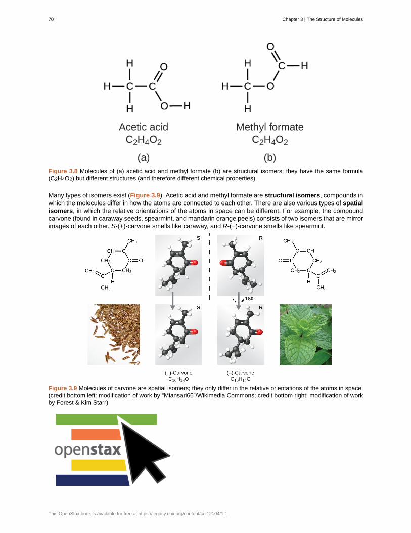
Figure 3.8 Molecules of (a) acetic acid and methyl formate (b) are structural isomers; they have the same formula(C2H4O2) but different structures (and therefore different chemical properties).
Many types of isomers exist (Figure 3.9). Acetic acid and methyl formate are structural isomers, compounds inwhich the molecules differ in how the atoms are connected to each other. There are also various types of spatialisomers, in which the relative orientations of the atoms in space can be different. For example, the compoundcarvone (found in caraway seeds, spearmint, and mandarin orange peels) consists of two isomers that are mirrorimages of each other. S-(+)-carvone smells like caraway, and R-(−)-carvone smells like spearmint.
Figure 3.9 Molecules of carvone are spatial isomers; they only differ in the relative orientations of the atoms in space.(credit bottom left: modification of work by “Miansari66”/Wikimedia Commons; credit bottom right: modification of workby Forest & Kim Starr)
70 Chapter 3 | The Structure of Molecules
This OpenStax book is available for free at https://legacy.cnx.org/content/col12104/1.1

Select this link (http://openstax.org/l/16isomers) to view an explanation of isomers, spatial isomers, andwhy they have different smells (select the video titled “Mirror Molecule: Carvone”).
Key Concepts and Summary
A molecular formula uses chemical symbols and subscripts to indicate the exact numbers of different atoms ina molecule or compound. An empirical formula gives the simplest, whole-number ratio of atoms in a compound.A structural formula indicates the bonding arrangement of the atoms in the molecule. Ball-and-stick and space-filling models show the geometric arrangement of atoms in a molecule. Isomers are compounds with the samemolecular formula but different arrangements of atoms.
Chemistry End of Chapter Exercises
Exercise 3.1
Explain why the symbol for an atom of the element oxygen and the formula for a molecule of oxygen differ.
SolutionThe symbol for the element oxygen, O, represents both the element and one atom of oxygen. A molecule ofoxygen, O2, contains two oxygen atoms; the subscript 2 in the formula must be used to distinguish the diatomicmolecule from two single oxygen atoms.
Exercise 3.2
Explain why the symbol for the element sulfur and the formula for a molecule of sulfur differ.
Exercise 3.3
Write the molecular and empirical formulas of the following compounds:
(a)
(b)
(c)
(d)
Solution(a) molecular CO2, empirical CO2; (b) molecular C2H2, empirical CH; (c) molecular C2H4, empirical CH2; (d)molecular H2SO4, empirical H2SO4
Chapter 3 | The Structure of Molecules 71
Exercise 3.4
Write the molecular and empirical formulas of the following compounds:
(a)
(b)
(c)
(d)
Exercise 3.5
Determine the empirical formulas for the following compounds:
(a) caffeine, C8H10N4O2
(b) fructose, C12H22O11
(c) hydrogen peroxide, H2O2
(d) glucose, C6H12O6
(e) ascorbic acid (vitamin C), C6H8O6
Solution(a) C4H5N2O; (b) C12H22O11; (c) HO; (d) CH2O; (e) C3H4O3
72 Chapter 3 | The Structure of Molecules
This OpenStax book is available for free at https://legacy.cnx.org/content/col12104/1.1
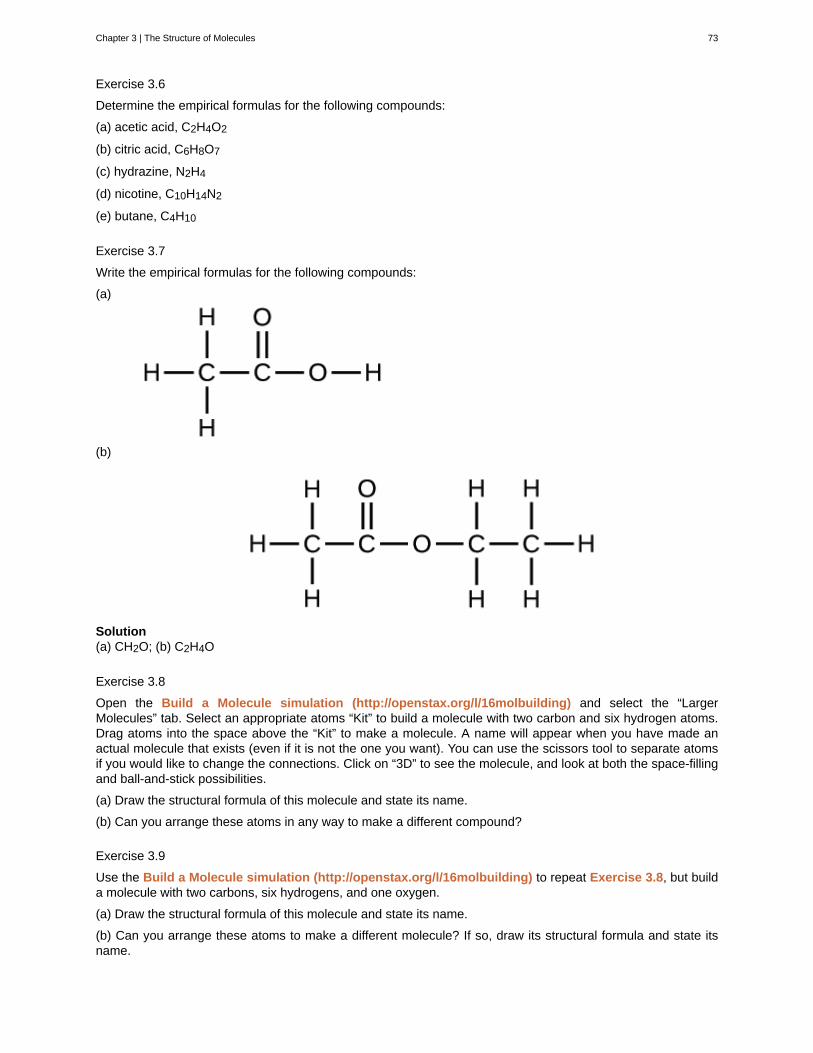
Exercise 3.6
Determine the empirical formulas for the following compounds:
(a) acetic acid, C2H4O2
(b) citric acid, C6H8O7
(c) hydrazine, N2H4
(d) nicotine, C10H14N2
(e) butane, C4H10
Exercise 3.7
Write the empirical formulas for the following compounds:
(a)
(b)
Solution(a) CH2O; (b) C2H4O
Exercise 3.8
Open the Build a Molecule simulation (http://openstax.org/l/16molbuilding) and select the “LargerMolecules” tab. Select an appropriate atoms “Kit” to build a molecule with two carbon and six hydrogen atoms.Drag atoms into the space above the “Kit” to make a molecule. A name will appear when you have made anactual molecule that exists (even if it is not the one you want). You can use the scissors tool to separate atomsif you would like to change the connections. Click on “3D” to see the molecule, and look at both the space-fillingand ball-and-stick possibilities.
(a) Draw the structural formula of this molecule and state its name.
(b) Can you arrange these atoms in any way to make a different compound?
Exercise 3.9
Use the Build a Molecule simulation (http://openstax.org/l/16molbuilding) to repeat Exercise 3.8, but builda molecule with two carbons, six hydrogens, and one oxygen.
(a) Draw the structural formula of this molecule and state its name.
(b) Can you arrange these atoms to make a different molecule? If so, draw its structural formula and state itsname.
Chapter 3 | The Structure of Molecules 73

(c) How are the molecules drawn in (a) and (b) the same? How do they differ? What are they called (the type ofrelationship between these molecules, not their names).
Solution
(a) ethanol
(b) methoxymethane, more commonly known as dimethyl ether
(c) These molecules have the same chemical composition (types and number of atoms) but different chemicalstructures. They are structural isomers.
Exercise 3.10
Use the Build a Molecule simulation (http://openstax.org/l/16molbuilding) to repeat Exercise 3.8, but builda molecule with three carbons, seven hydrogens, and one chlorine.
(a) Draw the structural formula of this molecule and state its name.
(b) Can you arrange these atoms to make a different molecule? If so, draw its structural formula and state itsname.
(c) How are the molecules drawn in (a) and (b) the same? How do they differ? What are they called (the type ofrelationship between these molecules, not their names)?
3.2 | Molecular and Ionic Compounds
By the end of this section, you will be able to:
• Define ionic and molecular (covalent) compounds
• Predict the type of compound formed from elements based on their location within the periodic table
• Determine formulas for simple ionic compounds
In ordinary chemical reactions, the nucleus of each atom (and thus the identity of the element) remainsunchanged. Electrons, however, can be added to atoms by transfer from other atoms, lost by transfer to otheratoms, or shared with other atoms. The transfer and sharing of electrons among atoms govern the chemistryof the elements. During the formation of some compounds, atoms gain or lose electrons, and form electricallycharged particles called ions (Figure 3.10).
74 Chapter 3 | The Structure of Molecules
This OpenStax book is available for free at https://legacy.cnx.org/content/col12104/1.1
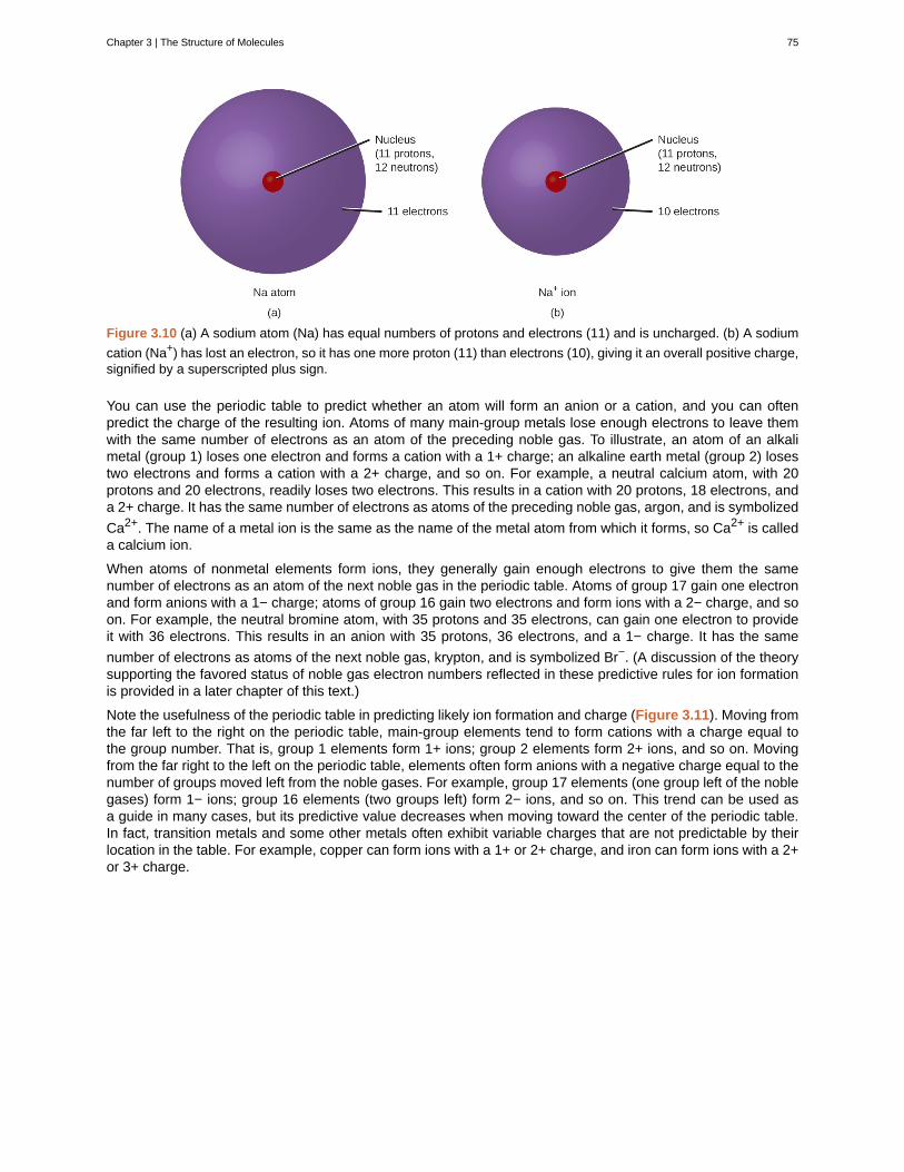
Figure 3.10 (a) A sodium atom (Na) has equal numbers of protons and electrons (11) and is uncharged. (b) A sodium
cation (Na+) has lost an electron, so it has one more proton (11) than electrons (10), giving it an overall positive charge,signified by a superscripted plus sign.
You can use the periodic table to predict whether an atom will form an anion or a cation, and you can oftenpredict the charge of the resulting ion. Atoms of many main-group metals lose enough electrons to leave themwith the same number of electrons as an atom of the preceding noble gas. To illustrate, an atom of an alkalimetal (group 1) loses one electron and forms a cation with a 1+ charge; an alkaline earth metal (group 2) losestwo electrons and forms a cation with a 2+ charge, and so on. For example, a neutral calcium atom, with 20protons and 20 electrons, readily loses two electrons. This results in a cation with 20 protons, 18 electrons, anda 2+ charge. It has the same number of electrons as atoms of the preceding noble gas, argon, and is symbolized
Ca2+. The name of a metal ion is the same as the name of the metal atom from which it forms, so Ca2+ is calleda calcium ion.
When atoms of nonmetal elements form ions, they generally gain enough electrons to give them the samenumber of electrons as an atom of the next noble gas in the periodic table. Atoms of group 17 gain one electronand form anions with a 1− charge; atoms of group 16 gain two electrons and form ions with a 2− charge, and soon. For example, the neutral bromine atom, with 35 protons and 35 electrons, can gain one electron to provideit with 36 electrons. This results in an anion with 35 protons, 36 electrons, and a 1− charge. It has the same
number of electrons as atoms of the next noble gas, krypton, and is symbolized Br−. (A discussion of the theorysupporting the favored status of noble gas electron numbers reflected in these predictive rules for ion formationis provided in a later chapter of this text.)
Note the usefulness of the periodic table in predicting likely ion formation and charge (Figure 3.11). Moving fromthe far left to the right on the periodic table, main-group elements tend to form cations with a charge equal tothe group number. That is, group 1 elements form 1+ ions; group 2 elements form 2+ ions, and so on. Movingfrom the far right to the left on the periodic table, elements often form anions with a negative charge equal to thenumber of groups moved left from the noble gases. For example, group 17 elements (one group left of the noblegases) form 1− ions; group 16 elements (two groups left) form 2− ions, and so on. This trend can be used asa guide in many cases, but its predictive value decreases when moving toward the center of the periodic table.In fact, transition metals and some other metals often exhibit variable charges that are not predictable by theirlocation in the table. For example, copper can form ions with a 1+ or 2+ charge, and iron can form ions with a 2+or 3+ charge.
Chapter 3 | The Structure of Molecules 75
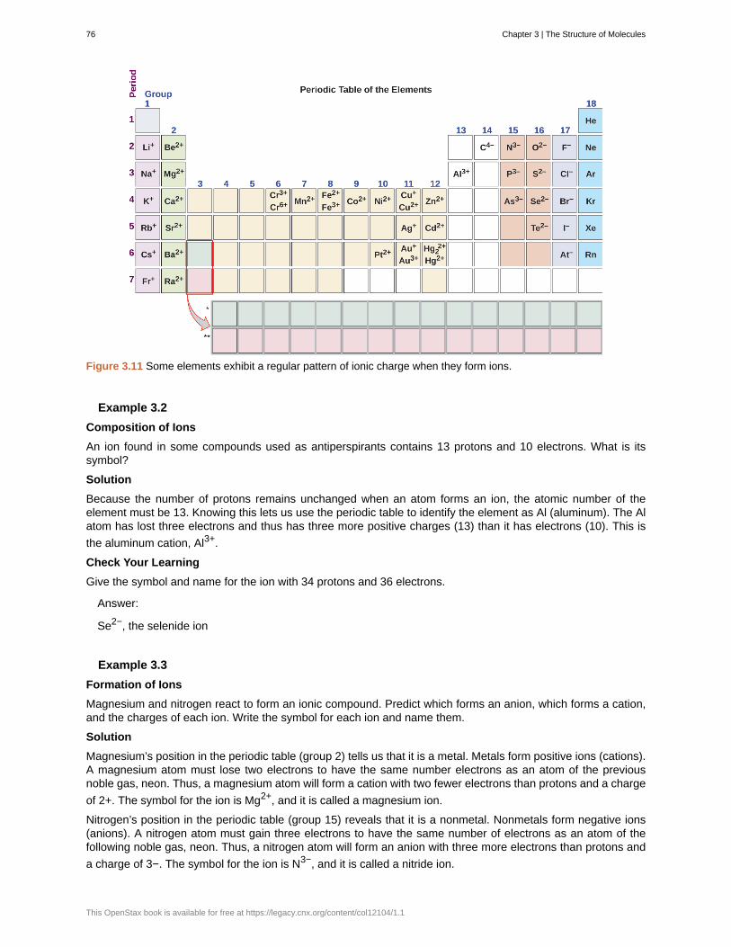
Figure 3.11 Some elements exhibit a regular pattern of ionic charge when they form ions.
Example 3.2
Composition of Ions
An ion found in some compounds used as antiperspirants contains 13 protons and 10 electrons. What is itssymbol?
Solution
Because the number of protons remains unchanged when an atom forms an ion, the atomic number of theelement must be 13. Knowing this lets us use the periodic table to identify the element as Al (aluminum). The Alatom has lost three electrons and thus has three more positive charges (13) than it has electrons (10). This is
the aluminum cation, Al3+.
Check Your Learning
Give the symbol and name for the ion with 34 protons and 36 electrons.
Answer:
Se2−, the selenide ion
Example 3.3
Formation of Ions
Magnesium and nitrogen react to form an ionic compound. Predict which forms an anion, which forms a cation,and the charges of each ion. Write the symbol for each ion and name them.
Solution
Magnesium’s position in the periodic table (group 2) tells us that it is a metal. Metals form positive ions (cations).A magnesium atom must lose two electrons to have the same number electrons as an atom of the previousnoble gas, neon. Thus, a magnesium atom will form a cation with two fewer electrons than protons and a charge
of 2+. The symbol for the ion is Mg2+, and it is called a magnesium ion.
Nitrogen’s position in the periodic table (group 15) reveals that it is a nonmetal. Nonmetals form negative ions(anions). A nitrogen atom must gain three electrons to have the same number of electrons as an atom of thefollowing noble gas, neon. Thus, a nitrogen atom will form an anion with three more electrons than protons and
a charge of 3−. The symbol for the ion is N3−, and it is called a nitride ion.
76 Chapter 3 | The Structure of Molecules
This OpenStax book is available for free at https://legacy.cnx.org/content/col12104/1.1
Check Your Learning
Aluminum and carbon react to form an ionic compound. Predict which forms an anion, which forms a cation, andthe charges of each ion. Write the symbol for each ion and name them.
Answer:
Al will form a cation with a charge of 3+: Al3+, an aluminum ion. Carbon will form an anion with a charge of
4−: C4−, a carbide ion.
The ions that we have discussed so far are called monatomic ions, that is, they are ions formed from onlyone atom. We also find many polyatomic ions. These ions, which act as discrete units, are electrically chargedmolecules (a group of bonded atoms with an overall charge). Some of the more important polyatomic ions arelisted in Table 3.1. Oxyanions are polyatomic ions that contain one or more oxygen atoms. At this point in yourstudy of chemistry, you should memorize the names, formulas, and charges of the most common polyatomicions. Because you will use them repeatedly, they will soon become familiar.
Common Polyatomic Ions
Name Formula Related Acid Formula
ammonium NH4+
hydronium H3 O+
peroxide O22-
hydroxide OH-
acetate CH3 COO-acetic acid CH3COOH
cyanide CN− hydrocyanic acid HCN
azide N3-
hydrazoic acid HN3
carbonate CO32- carbonic acid H2CO3
bicarbonate HCO3-
nitrate NO3−
nitric acid HNO3
nitrite NO2−
nitrous acid HNO2
sulfate SO42− sulfiric acid H2SO4
hydrogen sulfate HSO4−
sulfite SO32− sulfurous acid H2SO3
hydrogen sulfite HSO3−
phosphate PO43− phosphoric acid H3PO4
hydrogen phosphate HPO42−
dihydrogen phosphate H2 PO4−
Table 3.1
Chapter 3 | The Structure of Molecules 77
Common Polyatomic Ions
Name Formula Related Acid Formula
perchlorate ClO4−
perchloric acid HClO4
chlorate ClO3−
chloric acid HClO3
chlorite ClO2−
chlorous acid HClO2
hypochlorite ClO− hypochlorous acid HClO
chromate CrO42− chromic acid H2Cr2O4
dichromate Cr2 O72− dichromic acid H2Cr2O7
permanganate MnO4−
permanganic acid HMnO4
Table 3.1
Note that there is a system for naming some polyatomic ions; -ate and -ite are suffixes designating polyatomicions containing more or fewer oxygen atoms. Per- (short for “hyper”) and hypo- (meaning “under”) are prefixesmeaning more oxygen atoms than -ate and fewer oxygen atoms than -ite, respectively. For example, perchlorate
is ClO4− , chlorate is ClO3
− , chlorite is ClO2− and hypochlorite is ClO−. Unfortunately, the number of
oxygen atoms corresponding to a given suffix or prefix is not consistent; for example, nitrate is NO3− while
sulfate is SO42−. This will be covered in more detail in the next module on nomenclature.
The nature of the attractive forces that hold atoms or ions together within a compound is the basis forclassifying chemical bonding. When electrons are transferred and ions form, ionic bonds result. Ionic bonds areelectrostatic forces of attraction, that is, the attractive forces experienced between objects of opposite electricalcharge (in this case, cations and anions). When electrons are “shared” and molecules form, covalent bondsresult. Covalent bonds are the attractive forces between the positively charged nuclei of the bonded atomsand one or more pairs of electrons that are located between the atoms. Compounds are classified as ionic ormolecular (covalent) on the basis of the bonds present in them.
Ionic Compounds
When an element composed of atoms that readily lose electrons (a metal) reacts with an element composedof atoms that readily gain electrons (a nonmetal), a transfer of electrons usually occurs, producing ions. Thecompound formed by this transfer is stabilized by the electrostatic attractions (ionic bonds) between the ions ofopposite charge present in the compound. For example, when each sodium atom in a sample of sodium metal
(group 1) gives up one electron to form a sodium cation, Na+, and each chlorine atom in a sample of chlorine
gas (group 17) accepts one electron to form a chloride anion, Cl−, the resulting compound, NaCl, is composed
of sodium ions and chloride ions in the ratio of one Na+ ion for each Cl− ion. Similarly, each calcium atom (group2) can give up two electrons and transfer one to each of two chlorine atoms to form CaCl2, which is composed
of Ca2+ and Cl− ions in the ratio of one Ca2+ ion to two Cl− ions.
A compound that contains ions and is held together by ionic bonds is called an ionic compound. The periodictable can help us recognize many of the compounds that are ionic: When a metal is combined with one or morenonmetals, the compound is usually ionic. This guideline works well for predicting ionic compound formation formost of the compounds typically encountered in an introductory chemistry course. However, it is not always true(for example, aluminum chloride, AlCl3, is not ionic).
You can often recognize ionic compounds because of their properties. Ionic compounds are solids that typicallymelt at high temperatures and boil at even higher temperatures. For example, sodium chloride melts at 801 °Cand boils at 1413 °C. (As a comparison, the molecular compound water melts at 0 °C and boils at 100 °C.) Insolid form, an ionic compound is not electrically conductive because its ions are unable to flow (“electricity” is theflow of charged particles). When molten, however, it can conduct electricity because its ions are able to move
78 Chapter 3 | The Structure of Molecules
This OpenStax book is available for free at https://legacy.cnx.org/content/col12104/1.1
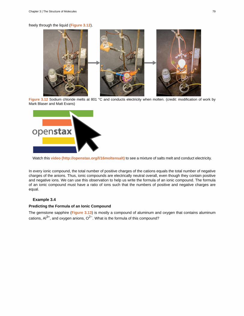
freely through the liquid (Figure 3.12).
Figure 3.12 Sodium chloride melts at 801 °C and conducts electricity when molten. (credit: modification of work byMark Blaser and Matt Evans)
Watch this video (http://openstax.org/l/16moltensalt) to see a mixture of salts melt and conduct electricity.
In every ionic compound, the total number of positive charges of the cations equals the total number of negativecharges of the anions. Thus, ionic compounds are electrically neutral overall, even though they contain positiveand negative ions. We can use this observation to help us write the formula of an ionic compound. The formulaof an ionic compound must have a ratio of ions such that the numbers of positive and negative charges areequal.
Example 3.4
Predicting the Formula of an Ionic Compound
The gemstone sapphire (Figure 3.13) is mostly a compound of aluminum and oxygen that contains aluminum
cations, Al3+, and oxygen anions, O2−. What is the formula of this compound?
Chapter 3 | The Structure of Molecules 79
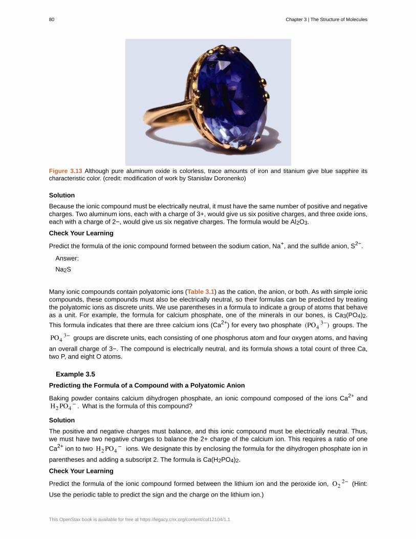
Figure 3.13 Although pure aluminum oxide is colorless, trace amounts of iron and titanium give blue sapphire itscharacteristic color. (credit: modification of work by Stanislav Doronenko)
Solution
Because the ionic compound must be electrically neutral, it must have the same number of positive and negativecharges. Two aluminum ions, each with a charge of 3+, would give us six positive charges, and three oxide ions,each with a charge of 2−, would give us six negative charges. The formula would be Al2O3.
Check Your Learning
Predict the formula of the ionic compound formed between the sodium cation, Na+, and the sulfide anion, S2−.
Answer:
Na2S
Many ionic compounds contain polyatomic ions (Table 3.1) as the cation, the anion, or both. As with simple ioniccompounds, these compounds must also be electrically neutral, so their formulas can be predicted by treatingthe polyatomic ions as discrete units. We use parentheses in a formula to indicate a group of atoms that behaveas a unit. For example, the formula for calcium phosphate, one of the minerals in our bones, is Ca3(PO4)2.
This formula indicates that there are three calcium ions (Ca2+) for every two phosphate (PO43−) groups. The
PO43− groups are discrete units, each consisting of one phosphorus atom and four oxygen atoms, and having
an overall charge of 3−. The compound is electrically neutral, and its formula shows a total count of three Ca,two P, and eight O atoms.
Example 3.5
Predicting the Formula of a Compound with a Polyatomic Anion
Baking powder contains calcium dihydrogen phosphate, an ionic compound composed of the ions Ca2+ andH2 PO4
− . What is the formula of this compound?
Solution
The positive and negative charges must balance, and this ionic compound must be electrically neutral. Thus,we must have two negative charges to balance the 2+ charge of the calcium ion. This requires a ratio of one
Ca2+ ion to two H2 PO4− ions. We designate this by enclosing the formula for the dihydrogen phosphate ion in
parentheses and adding a subscript 2. The formula is Ca(H2PO4)2.
Check Your Learning
Predict the formula of the ionic compound formed between the lithium ion and the peroxide ion, O22− (Hint:
Use the periodic table to predict the sign and the charge on the lithium ion.)
80 Chapter 3 | The Structure of Molecules
This OpenStax book is available for free at https://legacy.cnx.org/content/col12104/1.1
Answer:
Li2O2
Because an ionic compound is not made up of single, discrete molecules, it may not be properly symbolizedusing a molecular formula. Instead, ionic compounds must be symbolized by a formula indicating the relativenumbers of its constituent ions. For compounds containing only monatomic ions (such as NaCl) and formany compounds containing polyatomic ions (such as CaSO4), these formulas are just the empirical formulasintroduced earlier in this chapter. However, the formulas for some ionic compounds containing polyatomic ions
are not empirical formulas. For example, the ionic compound sodium oxalate is comprised of Na+ and C2 O42−
ions combined in a 2:1 ratio, and its formula is written as Na2C2O4. The subscripts in this formula are not thesmallest-possible whole numbers, as each can be divided by 2 to yield the empirical formula, NaCO2. Thisis not the accepted formula for sodium oxalate, however, as it does not accurately represent the compound’s
polyatomic anion, C2 O42−.
Molecular Compounds
Many compounds do not contain ions but instead consist solely of discrete, neutral molecules. These molecularcompounds (covalent compounds) result when atoms share, rather than transfer (gain or lose), electrons.Covalent bonding is an important and extensive concept in chemistry, and it will be treated in considerable detailin a later chapter of this text. We can often identify molecular compounds on the basis of their physical properties.Under normal conditions, molecular compounds often exist as gases, low-boiling liquids, and low-melting solids,although many important exceptions exist.
Whereas ionic compounds are usually formed when a metal and a nonmetal combine, covalent compoundsare usually formed by a combination of nonmetals. Thus, the periodic table can help us recognize many of thecompounds that are covalent. While we can use the positions of a compound’s elements in the periodic tableto predict whether it is ionic or covalent at this point in our study of chemistry, you should be aware that this isa very simplistic approach that does not account for a number of interesting exceptions. Shades of gray existbetween ionic and molecular compounds, and you’ll learn more about those later.
Example 3.6
Predicting the Type of Bonding in Compounds
Predict whether the following compounds are ionic or molecular:
(a) KI, the compound used as a source of iodine in table salt
(b) H2O2, the bleach and disinfectant hydrogen peroxide
(c) CHCl3, the anesthetic chloroform
(d) Li2CO3, a source of lithium in antidepressants
Solution
(a) Potassium (group 1) is a metal, and iodine (group 17) is a nonmetal; KI is predicted to be ionic.
(b) Hydrogen (group 1) is a nonmetal, and oxygen (group 16) is a nonmetal; H2O2 is predicted to be molecular.
(c) Carbon (group 14) is a nonmetal, hydrogen (group 1) is a nonmetal, and chlorine (group 17) is a nonmetal;CHCl3 is predicted to be molecular.
(d) Lithium (group 1) is a metal, and carbonate is a polyatomic ion; Li2CO3 is predicted to be ionic.
Check Your Learning
Using the periodic table, predict whether the following compounds are ionic or covalent:
(a) SO2
(b) CaF2
(c) N2H4
(d) Al2(SO4)3
Chapter 3 | The Structure of Molecules 81
Answer:
(a) molecular; (b) ionic; (c) molecular; (d) ionic
Key Concepts and Summary
Metals (particularly those in groups 1 and 2) tend to lose the number of electrons that would leave them withthe same number of electrons as in the preceding noble gas in the periodic table. By this means, a positivelycharged ion is formed. Similarly, nonmetals (especially those in groups 16 and 17, and, to a lesser extent, thosein Group 15) can gain the number of electrons needed to provide atoms with the same number of electrons asin the next noble gas in the periodic table. Thus, nonmetals tend to form negative ions. Positively charged ionsare called cations, and negatively charged ions are called anions. Ions can be either monatomic (containing onlyone atom) or polyatomic (containing more than one atom).
Compounds that contain ions are called ionic compounds. Ionic compounds generally form from metals andnonmetals. Compounds that do not contain ions, but instead consist of atoms bonded tightly together inmolecules (uncharged groups of atoms that behave as a single unit), are called covalent compounds. Covalentcompounds usually form from two nonmetals.
Chemistry End of Chapter Exercises
Exercise 3.11
Using the periodic table, predict whether the following chlorides are ionic or covalent: KCl, NCl3, ICl, MgCl2,PCl5, and CCl4.
SolutionIonic: KCl, MgCl2; Covalent: NCl3, ICl, PCl5, CCl4
Exercise 3.12
Using the periodic table, predict whether the following chlorides are ionic or covalent: SiCl4, PCl3, CaCl2, CsCl,CuCl2, and CrCl3.
Exercise 3.13
For each of the following compounds, state whether it is ionic or covalent. If it is ionic, write the symbols for theions involved:
(a) NF3
(b) BaO
(c) (NH4)2CO3
(d) Sr(H2PO4)2
(e) IBr
(f) Na2O
Solution
(a) covalent; (b) ionic, Ba2+, O2−; (c) ionic, NH4+ , CO3
2−; (d) ionic, Sr2+, H2 PO4− ; (e) covalent; (f) ionic,
Na+, O2−
Exercise 3.14
For each of the following compounds, state whether it is ionic or covalent, and if it is ionic, write the symbols forthe ions involved:
(a) KClO4
(b) Mg(C2H3O2)2
(c) H2S
82 Chapter 3 | The Structure of Molecules
This OpenStax book is available for free at https://legacy.cnx.org/content/col12104/1.1
(d) Ag2S
(e) N2Cl4
(f) Co(NO3)2
Exercise 3.15
For each of the following pairs of ions, write the formula of the compound they will form:
(a) Ca2+, S2−
(b) NH4+ , SO4
2−
(c) Al3+, Br−
(d) Na+, HPO42−
(e) Mg2+, PO43−
Solution(a) CaS; (b) (NH4)2SO4; (c) AlBr3; (d) Na2HPO4; (e) Mg3 (PO4)2
Exercise 3.16
For each of the following pairs of ions, write the formula of the compound they will form:
(a) K+, O2−
(b) NH4+ , PO4
3−
(c) Al3+, O2−
(d) Na+, CO32−
(e) Ba2+, PO43−
3.3 | Covalent Bonding
By the end of this section, you will be able to:
• Describe the formation of covalent bonds
• Define electronegativity and assess the polarity of covalent bonds
Ionic bonding results from the electrostatic attraction of oppositely charged ions that are typically produced bythe transfer of electrons between metallic and nonmetallic atoms. A different type of bonding results from themutual attraction of atoms for a “shared” pair of electrons. Such bonds are called covalent bonds. Covalentbonds are formed between two atoms when both have similar tendencies to attract electrons to themselves(i.e., when both atoms have identical or fairly similar ionization energies and electron affinities). For example,two hydrogen atoms bond covalently to form an H2 molecule; each hydrogen atom in the H2 molecule has twoelectrons stabilizing it, giving each atom the same number of valence electrons as the noble gas He.
Compounds that contain covalent bonds exhibit different physical properties than ionic compounds. Because theattraction between molecules, which are electrically neutral, is weaker than that between electrically chargedions, covalent compounds generally have much lower melting and boiling points than ionic compounds. In fact,many covalent compounds are liquids or gases at room temperature, and, in their solid states, they are typicallymuch softer than ionic solids. Furthermore, whereas ionic compounds are good conductors of electricity whendissolved in water, most covalent compounds are insoluble in water; since they are electrically neutral, they arepoor conductors of electricity in any state.
Chapter 3 | The Structure of Molecules 83
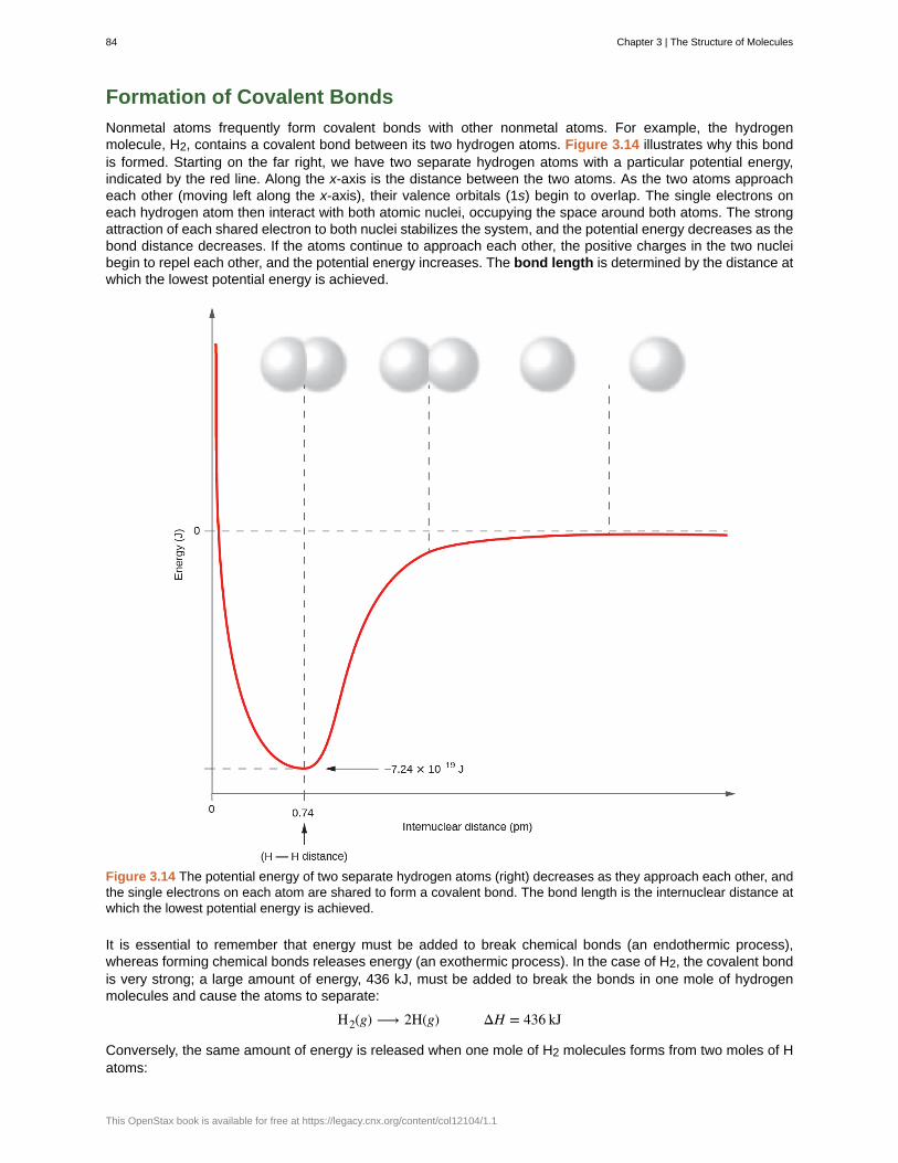
Formation of Covalent Bonds
Nonmetal atoms frequently form covalent bonds with other nonmetal atoms. For example, the hydrogenmolecule, H2, contains a covalent bond between its two hydrogen atoms. Figure 3.14 illustrates why this bondis formed. Starting on the far right, we have two separate hydrogen atoms with a particular potential energy,indicated by the red line. Along the x-axis is the distance between the two atoms. As the two atoms approacheach other (moving left along the x-axis), their valence orbitals (1s) begin to overlap. The single electrons oneach hydrogen atom then interact with both atomic nuclei, occupying the space around both atoms. The strongattraction of each shared electron to both nuclei stabilizes the system, and the potential energy decreases as thebond distance decreases. If the atoms continue to approach each other, the positive charges in the two nucleibegin to repel each other, and the potential energy increases. The bond length is determined by the distance atwhich the lowest potential energy is achieved.
Figure 3.14 The potential energy of two separate hydrogen atoms (right) decreases as they approach each other, andthe single electrons on each atom are shared to form a covalent bond. The bond length is the internuclear distance atwhich the lowest potential energy is achieved.
It is essential to remember that energy must be added to break chemical bonds (an endothermic process),whereas forming chemical bonds releases energy (an exothermic process). In the case of H2, the covalent bondis very strong; a large amount of energy, 436 kJ, must be added to break the bonds in one mole of hydrogenmolecules and cause the atoms to separate:
H2(g) ⟶ 2H(g) ΔH = 436 kJ
Conversely, the same amount of energy is released when one mole of H2 molecules forms from two moles of Hatoms:
84 Chapter 3 | The Structure of Molecules
This OpenStax book is available for free at https://legacy.cnx.org/content/col12104/1.1
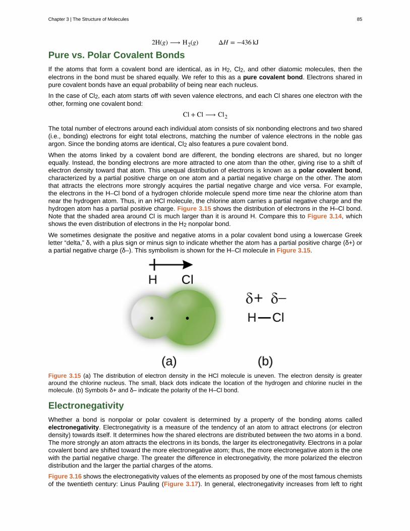
2H(g) ⟶ H2(g) ΔH = −436 kJ
Pure vs. Polar Covalent Bonds
If the atoms that form a covalent bond are identical, as in H2, Cl2, and other diatomic molecules, then theelectrons in the bond must be shared equally. We refer to this as a pure covalent bond. Electrons shared inpure covalent bonds have an equal probability of being near each nucleus.
In the case of Cl2, each atom starts off with seven valence electrons, and each Cl shares one electron with theother, forming one covalent bond:
Cl + Cl ⟶ Cl2
The total number of electrons around each individual atom consists of six nonbonding electrons and two shared(i.e., bonding) electrons for eight total electrons, matching the number of valence electrons in the noble gasargon. Since the bonding atoms are identical, Cl2 also features a pure covalent bond.
When the atoms linked by a covalent bond are different, the bonding electrons are shared, but no longerequally. Instead, the bonding electrons are more attracted to one atom than the other, giving rise to a shift ofelectron density toward that atom. This unequal distribution of electrons is known as a polar covalent bond,characterized by a partial positive charge on one atom and a partial negative charge on the other. The atomthat attracts the electrons more strongly acquires the partial negative charge and vice versa. For example,the electrons in the H–Cl bond of a hydrogen chloride molecule spend more time near the chlorine atom thannear the hydrogen atom. Thus, in an HCl molecule, the chlorine atom carries a partial negative charge and thehydrogen atom has a partial positive charge. Figure 3.15 shows the distribution of electrons in the H–Cl bond.Note that the shaded area around Cl is much larger than it is around H. Compare this to Figure 3.14, whichshows the even distribution of electrons in the H2 nonpolar bond.
We sometimes designate the positive and negative atoms in a polar covalent bond using a lowercase Greekletter “delta,” δ, with a plus sign or minus sign to indicate whether the atom has a partial positive charge (δ+) ora partial negative charge (δ–). This symbolism is shown for the H–Cl molecule in Figure 3.15.
Figure 3.15 (a) The distribution of electron density in the HCl molecule is uneven. The electron density is greateraround the chlorine nucleus. The small, black dots indicate the location of the hydrogen and chlorine nuclei in themolecule. (b) Symbols δ+ and δ– indicate the polarity of the H–Cl bond.
Electronegativity
Whether a bond is nonpolar or polar covalent is determined by a property of the bonding atoms calledelectronegativity. Electronegativity is a measure of the tendency of an atom to attract electrons (or electrondensity) towards itself. It determines how the shared electrons are distributed between the two atoms in a bond.The more strongly an atom attracts the electrons in its bonds, the larger its electronegativity. Electrons in a polarcovalent bond are shifted toward the more electronegative atom; thus, the more electronegative atom is the onewith the partial negative charge. The greater the difference in electronegativity, the more polarized the electrondistribution and the larger the partial charges of the atoms.
Figure 3.16 shows the electronegativity values of the elements as proposed by one of the most famous chemistsof the twentieth century: Linus Pauling (Figure 3.17). In general, electronegativity increases from left to right
Chapter 3 | The Structure of Molecules 85
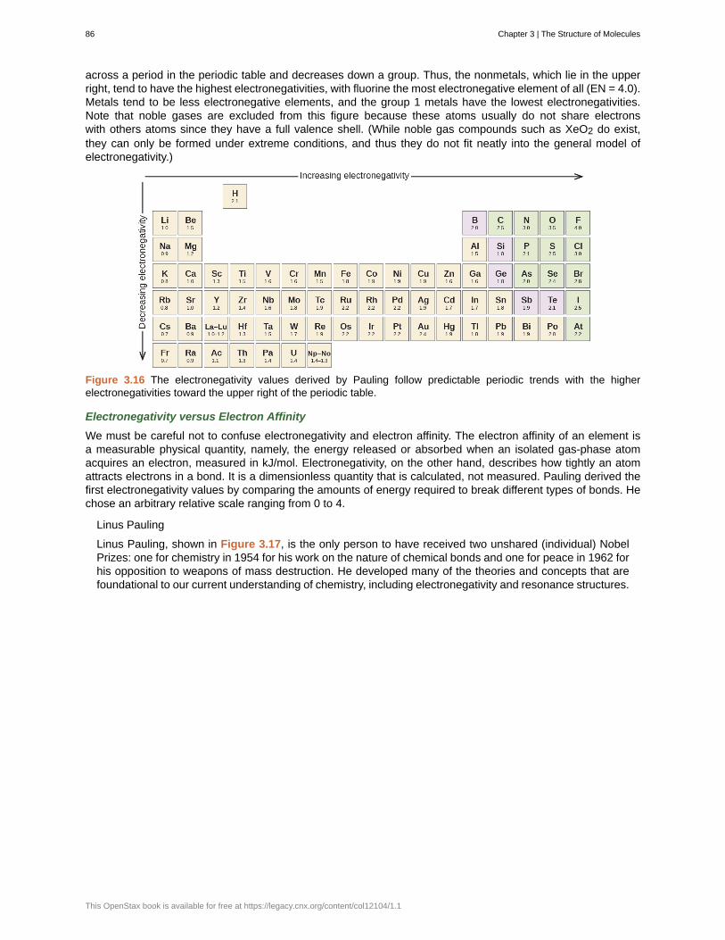
across a period in the periodic table and decreases down a group. Thus, the nonmetals, which lie in the upperright, tend to have the highest electronegativities, with fluorine the most electronegative element of all (EN = 4.0).Metals tend to be less electronegative elements, and the group 1 metals have the lowest electronegativities.Note that noble gases are excluded from this figure because these atoms usually do not share electronswith others atoms since they have a full valence shell. (While noble gas compounds such as XeO2 do exist,they can only be formed under extreme conditions, and thus they do not fit neatly into the general model ofelectronegativity.)
Figure 3.16 The electronegativity values derived by Pauling follow predictable periodic trends with the higherelectronegativities toward the upper right of the periodic table.
Electronegativity versus Electron Affinity
We must be careful not to confuse electronegativity and electron affinity. The electron affinity of an element isa measurable physical quantity, namely, the energy released or absorbed when an isolated gas-phase atomacquires an electron, measured in kJ/mol. Electronegativity, on the other hand, describes how tightly an atomattracts electrons in a bond. It is a dimensionless quantity that is calculated, not measured. Pauling derived thefirst electronegativity values by comparing the amounts of energy required to break different types of bonds. Hechose an arbitrary relative scale ranging from 0 to 4.
Linus Pauling
Linus Pauling, shown in Figure 3.17, is the only person to have received two unshared (individual) NobelPrizes: one for chemistry in 1954 for his work on the nature of chemical bonds and one for peace in 1962 forhis opposition to weapons of mass destruction. He developed many of the theories and concepts that arefoundational to our current understanding of chemistry, including electronegativity and resonance structures.
86 Chapter 3 | The Structure of Molecules
This OpenStax book is available for free at https://legacy.cnx.org/content/col12104/1.1
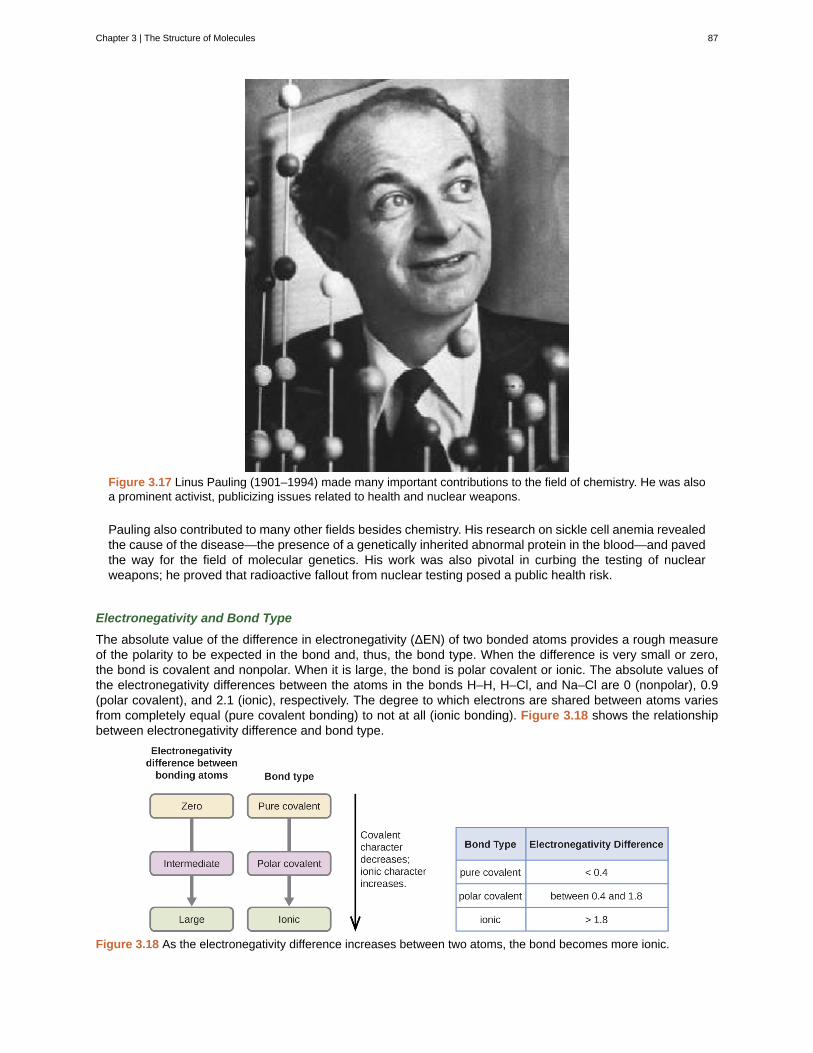
Figure 3.17 Linus Pauling (1901–1994) made many important contributions to the field of chemistry. He was alsoa prominent activist, publicizing issues related to health and nuclear weapons.
Pauling also contributed to many other fields besides chemistry. His research on sickle cell anemia revealedthe cause of the disease—the presence of a genetically inherited abnormal protein in the blood—and pavedthe way for the field of molecular genetics. His work was also pivotal in curbing the testing of nuclearweapons; he proved that radioactive fallout from nuclear testing posed a public health risk.
Electronegativity and Bond Type
The absolute value of the difference in electronegativity (ΔEN) of two bonded atoms provides a rough measureof the polarity to be expected in the bond and, thus, the bond type. When the difference is very small or zero,the bond is covalent and nonpolar. When it is large, the bond is polar covalent or ionic. The absolute values ofthe electronegativity differences between the atoms in the bonds H–H, H–Cl, and Na–Cl are 0 (nonpolar), 0.9(polar covalent), and 2.1 (ionic), respectively. The degree to which electrons are shared between atoms variesfrom completely equal (pure covalent bonding) to not at all (ionic bonding). Figure 3.18 shows the relationshipbetween electronegativity difference and bond type.
Figure 3.18 As the electronegativity difference increases between two atoms, the bond becomes more ionic.
Chapter 3 | The Structure of Molecules 87
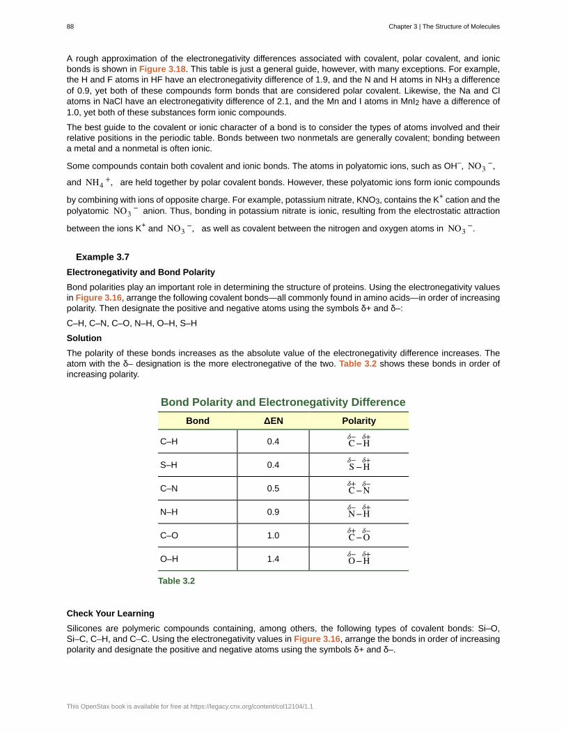
A rough approximation of the electronegativity differences associated with covalent, polar covalent, and ionicbonds is shown in Figure 3.18. This table is just a general guide, however, with many exceptions. For example,the H and F atoms in HF have an electronegativity difference of 1.9, and the N and H atoms in NH3 a differenceof 0.9, yet both of these compounds form bonds that are considered polar covalent. Likewise, the Na and Clatoms in NaCl have an electronegativity difference of 2.1, and the Mn and I atoms in MnI2 have a difference of1.0, yet both of these substances form ionic compounds.
The best guide to the covalent or ionic character of a bond is to consider the types of atoms involved and theirrelative positions in the periodic table. Bonds between two nonmetals are generally covalent; bonding betweena metal and a nonmetal is often ionic.
Some compounds contain both covalent and ionic bonds. The atoms in polyatomic ions, such as OH–, NO3−,
and NH4+, are held together by polar covalent bonds. However, these polyatomic ions form ionic compounds
by combining with ions of opposite charge. For example, potassium nitrate, KNO3, contains the K+ cation and thepolyatomic NO3
− anion. Thus, bonding in potassium nitrate is ionic, resulting from the electrostatic attraction
between the ions K+ and NO3−, as well as covalent between the nitrogen and oxygen atoms in NO3
−.
Example 3.7
Electronegativity and Bond Polarity
Bond polarities play an important role in determining the structure of proteins. Using the electronegativity valuesin Figure 3.16, arrange the following covalent bonds—all commonly found in amino acids—in order of increasingpolarity. Then designate the positive and negative atoms using the symbols δ+ and δ–:
C–H, C–N, C–O, N–H, O–H, S–H
Solution
The polarity of these bonds increases as the absolute value of the electronegativity difference increases. Theatom with the δ– designation is the more electronegative of the two. Table 3.2 shows these bonds in order ofincreasing polarity.
Bond Polarity and Electronegativity Difference
Bond ΔEN Polarity
C–H 0.4 Cδ−
−Hδ+
S–H 0.4 Sδ−
−Hδ+
C–N 0.5 Cδ+
−Nδ−
N–H 0.9 Nδ−
−Hδ+
C–O 1.0 Cδ+
−Oδ−
O–H 1.4 Oδ−
−Hδ+
Table 3.2
Check Your Learning
Silicones are polymeric compounds containing, among others, the following types of covalent bonds: Si–O,Si–C, C–H, and C–C. Using the electronegativity values in Figure 3.16, arrange the bonds in order of increasingpolarity and designate the positive and negative atoms using the symbols δ+ and δ–.
88 Chapter 3 | The Structure of Molecules
This OpenStax book is available for free at https://legacy.cnx.org/content/col12104/1.1
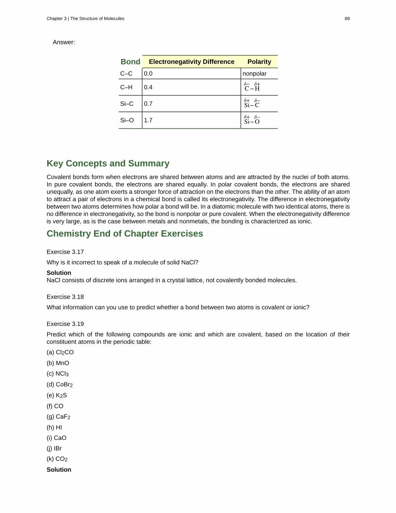
Answer:
Bond Electronegativity Difference Polarity
C–C 0.0 nonpolar
C–H 0.4 Cδ−
−Hδ+
Si–C 0.7 Siδ+
−Cδ−
Si–O 1.7 Siδ+
−Oδ−
Key Concepts and Summary
Covalent bonds form when electrons are shared between atoms and are attracted by the nuclei of both atoms.In pure covalent bonds, the electrons are shared equally. In polar covalent bonds, the electrons are sharedunequally, as one atom exerts a stronger force of attraction on the electrons than the other. The ability of an atomto attract a pair of electrons in a chemical bond is called its electronegativity. The difference in electronegativitybetween two atoms determines how polar a bond will be. In a diatomic molecule with two identical atoms, there isno difference in electronegativity, so the bond is nonpolar or pure covalent. When the electronegativity differenceis very large, as is the case between metals and nonmetals, the bonding is characterized as ionic.
Chemistry End of Chapter Exercises
Exercise 3.17
Why is it incorrect to speak of a molecule of solid NaCl?
SolutionNaCl consists of discrete ions arranged in a crystal lattice, not covalently bonded molecules.
Exercise 3.18
What information can you use to predict whether a bond between two atoms is covalent or ionic?
Exercise 3.19
Predict which of the following compounds are ionic and which are covalent, based on the location of theirconstituent atoms in the periodic table:
(a) Cl2CO
(b) MnO
(c) NCl3
(d) CoBr2
(e) K2S
(f) CO
(g) CaF2
(h) HI
(i) CaO
(j) IBr
(k) CO2
Solution
Chapter 3 | The Structure of Molecules 89
ionic: (b), (d), (e), (g), and (i); covalent: (a), (c), (f), (h), (j), and (k)
Exercise 3.20
Explain the difference between a nonpolar covalent bond, a polar covalent bond, and an ionic bond.
Exercise 3.21
From its position in the periodic table, determine which atom in each pair is more electronegative:
(a) Br or Cl
(b) N or O
(c) S or O
(d) P or S
(e) Si or N
(f) Ba or P
(g) N or K
Solution(a) Cl; (b) O; (c) O; (d) S; (e) N; (f) P; (g) N
Exercise 3.22
From its position in the periodic table, determine which atom in each pair is more electronegative:
(a) N or P
(b) N or Ge
(c) S or F
(d) Cl or S
(e) H or C
(f) Se or P
(g) C or Si
Exercise 3.23
From their positions in the periodic table, arrange the atoms in each of the following series in order of increasingelectronegativity:
(a) C, F, H, N, O
(b) Br, Cl, F, H, I
(c) F, H, O, P, S
(d) Al, H, Na, O, P
(e) Ba, H, N, O, As
Solution(a) H, C, N, O, F; (b) H, I, Br, Cl, F; (c) H, P, S, O, F; (d) Na, Al, H, P, O; (e) Ba, H, As, N, O
Exercise 3.24
From their positions in the periodic table, arrange the atoms in each of the following series in order of increasingelectronegativity:
(a) As, H, N, P, Sb
(b) Cl, H, P, S, Si
(c) Br, Cl, Ge, H, Sr
(d) Ca, H, K, N, Si
90 Chapter 3 | The Structure of Molecules
This OpenStax book is available for free at https://legacy.cnx.org/content/col12104/1.1
(e) Cl, Cs, Ge, H, Sr
Exercise 3.25
Which atoms can bond to sulfur so as to produce a positive partial charge on the sulfur atom?
SolutionN, O, F, and Cl
Exercise 3.26
Which is the most polar bond?
(a) C–C
(b) C–H
(c) N–H
(d) O–H
(e) Se–H
Exercise 3.27
Identify the more polar bond in each of the following pairs of bonds:
(a) HF or HCl
(b) NO or CO
(c) SH or OH
(d) PCl or SCl
(e) CH or NH
(f) SO or PO
(g) CN or NN
Solution(a) HF; (b) CO; (c) OH; (d) PCl; (e) NH; (f) PO; (g) CN
Exercise 3.28
Which of the following molecules or ions contain polar bonds?
(a) O3
(b) S8
(c) O22−
(d) NO3−
(e) CO2
(f) H2S
(g) BH4−
Chapter 3 | The Structure of Molecules 91
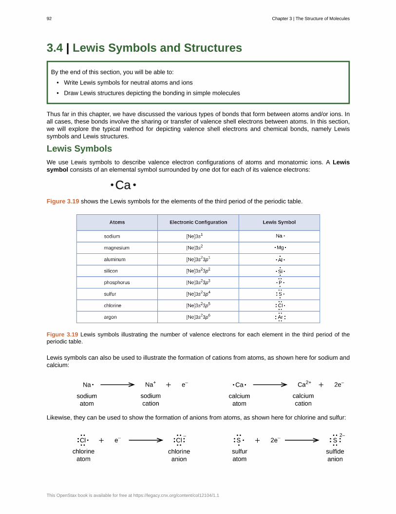
3.4 | Lewis Symbols and Structures
By the end of this section, you will be able to:
• Write Lewis symbols for neutral atoms and ions
• Draw Lewis structures depicting the bonding in simple molecules
Thus far in this chapter, we have discussed the various types of bonds that form between atoms and/or ions. Inall cases, these bonds involve the sharing or transfer of valence shell electrons between atoms. In this section,we will explore the typical method for depicting valence shell electrons and chemical bonds, namely Lewissymbols and Lewis structures.
Lewis Symbols
We use Lewis symbols to describe valence electron configurations of atoms and monatomic ions. A Lewissymbol consists of an elemental symbol surrounded by one dot for each of its valence electrons:
Figure 3.19 shows the Lewis symbols for the elements of the third period of the periodic table.
Figure 3.19 Lewis symbols illustrating the number of valence electrons for each element in the third period of theperiodic table.
Lewis symbols can also be used to illustrate the formation of cations from atoms, as shown here for sodium andcalcium:
Likewise, they can be used to show the formation of anions from atoms, as shown here for chlorine and sulfur:
92 Chapter 3 | The Structure of Molecules
This OpenStax book is available for free at https://legacy.cnx.org/content/col12104/1.1
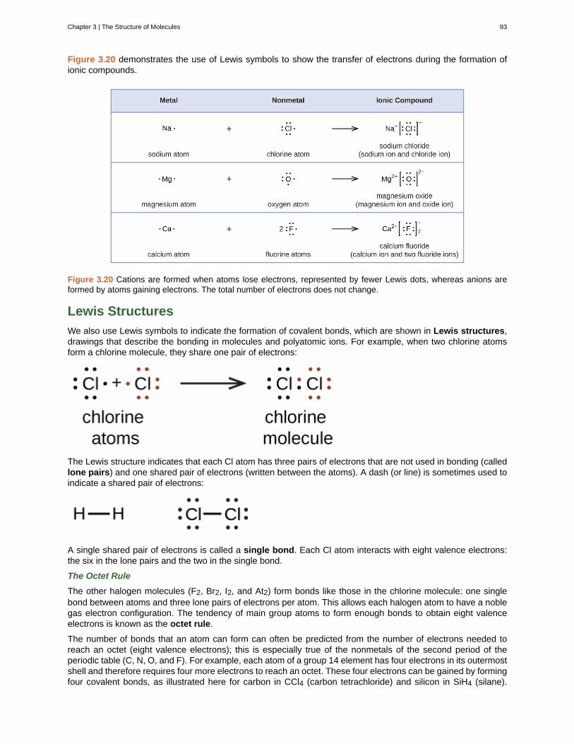
Figure 3.20 demonstrates the use of Lewis symbols to show the transfer of electrons during the formation ofionic compounds.
Figure 3.20 Cations are formed when atoms lose electrons, represented by fewer Lewis dots, whereas anions areformed by atoms gaining electrons. The total number of electrons does not change.
Lewis Structures
We also use Lewis symbols to indicate the formation of covalent bonds, which are shown in Lewis structures,drawings that describe the bonding in molecules and polyatomic ions. For example, when two chlorine atomsform a chlorine molecule, they share one pair of electrons:
The Lewis structure indicates that each Cl atom has three pairs of electrons that are not used in bonding (calledlone pairs) and one shared pair of electrons (written between the atoms). A dash (or line) is sometimes used toindicate a shared pair of electrons:
A single shared pair of electrons is called a single bond. Each Cl atom interacts with eight valence electrons:the six in the lone pairs and the two in the single bond.
The Octet Rule
The other halogen molecules (F2, Br2, I2, and At2) form bonds like those in the chlorine molecule: one singlebond between atoms and three lone pairs of electrons per atom. This allows each halogen atom to have a noblegas electron configuration. The tendency of main group atoms to form enough bonds to obtain eight valenceelectrons is known as the octet rule.
The number of bonds that an atom can form can often be predicted from the number of electrons needed toreach an octet (eight valence electrons); this is especially true of the nonmetals of the second period of theperiodic table (C, N, O, and F). For example, each atom of a group 14 element has four electrons in its outermostshell and therefore requires four more electrons to reach an octet. These four electrons can be gained by formingfour covalent bonds, as illustrated here for carbon in CCl4 (carbon tetrachloride) and silicon in SiH4 (silane).
Chapter 3 | The Structure of Molecules 93
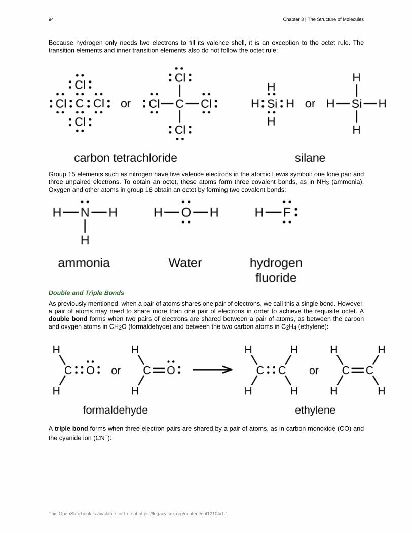
Because hydrogen only needs two electrons to fill its valence shell, it is an exception to the octet rule. Thetransition elements and inner transition elements also do not follow the octet rule:
Group 15 elements such as nitrogen have five valence electrons in the atomic Lewis symbol: one lone pair andthree unpaired electrons. To obtain an octet, these atoms form three covalent bonds, as in NH3 (ammonia).Oxygen and other atoms in group 16 obtain an octet by forming two covalent bonds:
Double and Triple Bonds
As previously mentioned, when a pair of atoms shares one pair of electrons, we call this a single bond. However,a pair of atoms may need to share more than one pair of electrons in order to achieve the requisite octet. Adouble bond forms when two pairs of electrons are shared between a pair of atoms, as between the carbonand oxygen atoms in CH2O (formaldehyde) and between the two carbon atoms in C2H4 (ethylene):
A triple bond forms when three electron pairs are shared by a pair of atoms, as in carbon monoxide (CO) and
the cyanide ion (CN–):
94 Chapter 3 | The Structure of Molecules
This OpenStax book is available for free at https://legacy.cnx.org/content/col12104/1.1
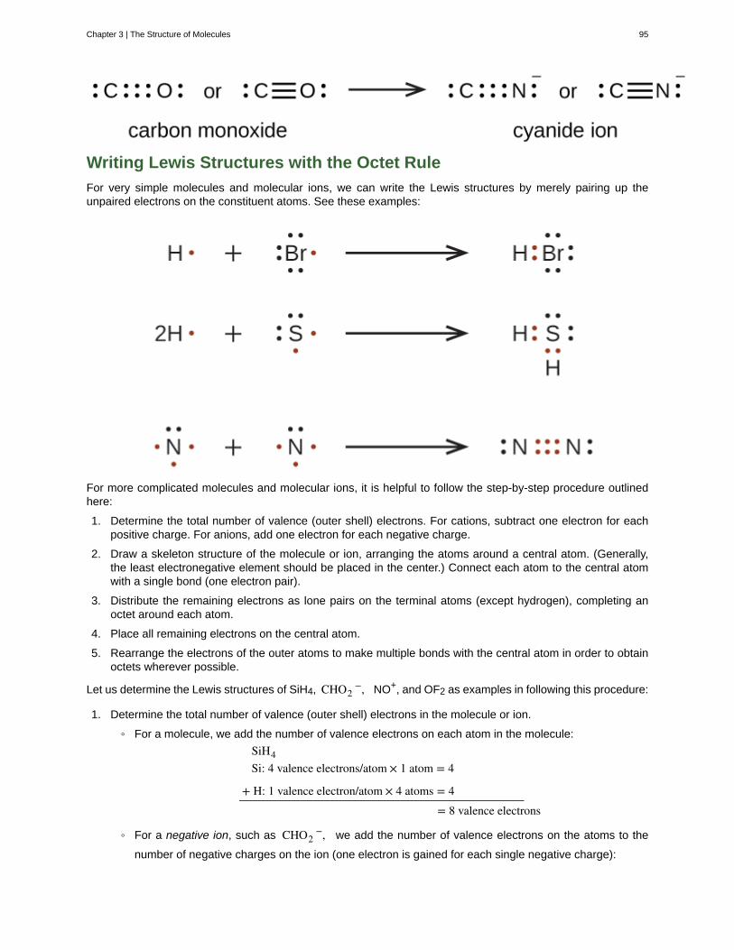
Writing Lewis Structures with the Octet Rule
For very simple molecules and molecular ions, we can write the Lewis structures by merely pairing up theunpaired electrons on the constituent atoms. See these examples:
For more complicated molecules and molecular ions, it is helpful to follow the step-by-step procedure outlinedhere:
1. Determine the total number of valence (outer shell) electrons. For cations, subtract one electron for eachpositive charge. For anions, add one electron for each negative charge.
2. Draw a skeleton structure of the molecule or ion, arranging the atoms around a central atom. (Generally,the least electronegative element should be placed in the center.) Connect each atom to the central atomwith a single bond (one electron pair).
3. Distribute the remaining electrons as lone pairs on the terminal atoms (except hydrogen), completing anoctet around each atom.
4. Place all remaining electrons on the central atom.
5. Rearrange the electrons of the outer atoms to make multiple bonds with the central atom in order to obtainoctets wherever possible.
Let us determine the Lewis structures of SiH4, CHO2−, NO+, and OF2 as examples in following this procedure:
1. Determine the total number of valence (outer shell) electrons in the molecule or ion.
◦ For a molecule, we add the number of valence electrons on each atom in the molecule:
SiH4Si: 4 valence electrons/atom × 1 atom = 4
+ H: 1 valence electron/atom × 4 atoms = 4= 8 valence electrons
◦ For a negative ion, such as CHO2−, we add the number of valence electrons on the atoms to the
number of negative charges on the ion (one electron is gained for each single negative charge):
Chapter 3 | The Structure of Molecules 95
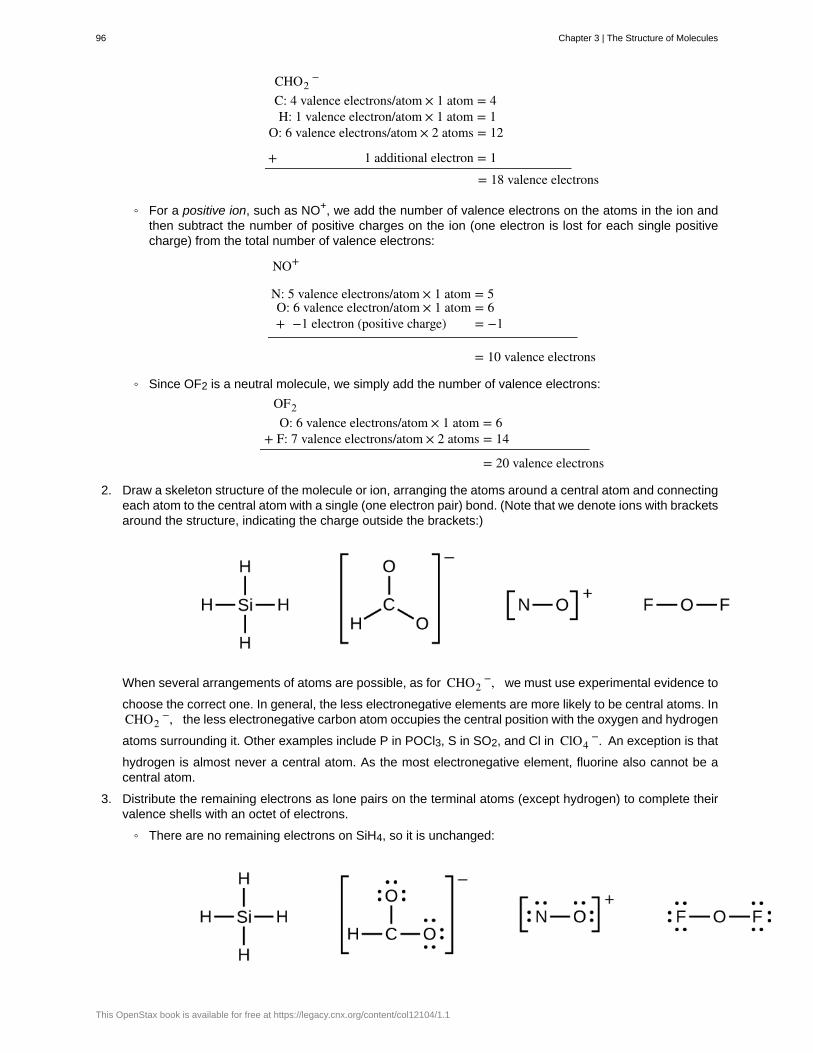
CHO2−
C: 4 valence electrons/atom × 1 atom = 4H: 1 valence electron/atom × 1 atom = 1
O: 6 valence electrons/atom × 2 atoms = 12
+ 1 additional electron = 1= 18 valence electrons
◦ For a positive ion, such as NO+, we add the number of valence electrons on the atoms in the ion andthen subtract the number of positive charges on the ion (one electron is lost for each single positivecharge) from the total number of valence electrons:
NO+
N: 5 valence electrons/atom × 1 atom = 5O: 6 valence electron/atom × 1 atom = 6+ −1 electron (positive charge) = −1
= 10 valence electrons
◦ Since OF2 is a neutral molecule, we simply add the number of valence electrons:
OF2O: 6 valence electrons/atom × 1 atom = 6
+ F: 7 valence electrons/atom × 2 atoms = 14
= 20 valence electrons
2. Draw a skeleton structure of the molecule or ion, arranging the atoms around a central atom and connectingeach atom to the central atom with a single (one electron pair) bond. (Note that we denote ions with bracketsaround the structure, indicating the charge outside the brackets:)
When several arrangements of atoms are possible, as for CHO2−, we must use experimental evidence to
choose the correct one. In general, the less electronegative elements are more likely to be central atoms. InCHO2
−, the less electronegative carbon atom occupies the central position with the oxygen and hydrogen
atoms surrounding it. Other examples include P in POCl3, S in SO2, and Cl in ClO4−. An exception is that
hydrogen is almost never a central atom. As the most electronegative element, fluorine also cannot be acentral atom.
3. Distribute the remaining electrons as lone pairs on the terminal atoms (except hydrogen) to complete theirvalence shells with an octet of electrons.
◦ There are no remaining electrons on SiH4, so it is unchanged:
96 Chapter 3 | The Structure of Molecules
This OpenStax book is available for free at https://legacy.cnx.org/content/col12104/1.1
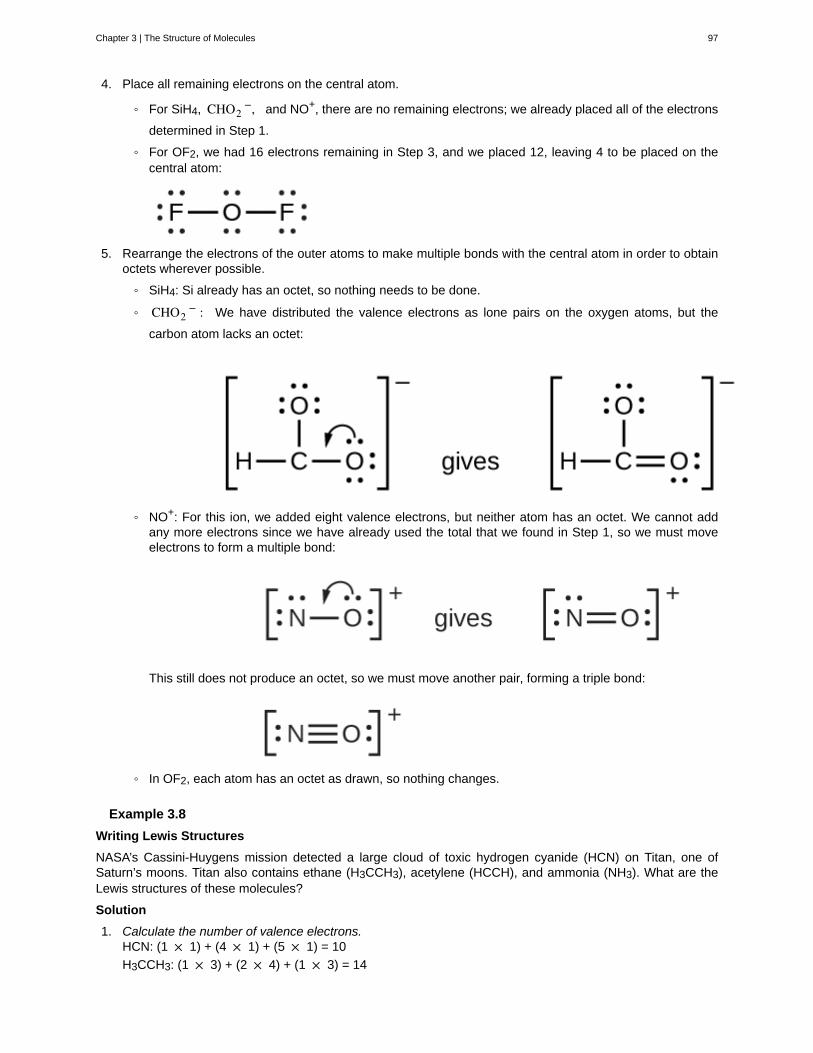
4. Place all remaining electrons on the central atom.
◦ For SiH4, CHO2−, and NO+, there are no remaining electrons; we already placed all of the electrons
determined in Step 1.
◦ For OF2, we had 16 electrons remaining in Step 3, and we placed 12, leaving 4 to be placed on thecentral atom:
5. Rearrange the electrons of the outer atoms to make multiple bonds with the central atom in order to obtainoctets wherever possible.
◦ SiH4: Si already has an octet, so nothing needs to be done.
◦ CHO2− : We have distributed the valence electrons as lone pairs on the oxygen atoms, but the
carbon atom lacks an octet:
◦ NO+: For this ion, we added eight valence electrons, but neither atom has an octet. We cannot addany more electrons since we have already used the total that we found in Step 1, so we must moveelectrons to form a multiple bond:
This still does not produce an octet, so we must move another pair, forming a triple bond:
◦ In OF2, each atom has an octet as drawn, so nothing changes.
Example 3.8
Writing Lewis Structures
NASA’s Cassini-Huygens mission detected a large cloud of toxic hydrogen cyanide (HCN) on Titan, one ofSaturn’s moons. Titan also contains ethane (H3CCH3), acetylene (HCCH), and ammonia (NH3). What are theLewis structures of these molecules?
Solution
1. Calculate the number of valence electrons.HCN: (1 × 1) + (4 × 1) + (5 × 1) = 10
H3CCH3: (1 × 3) + (2 × 4) + (1 × 3) = 14
Chapter 3 | The Structure of Molecules 97
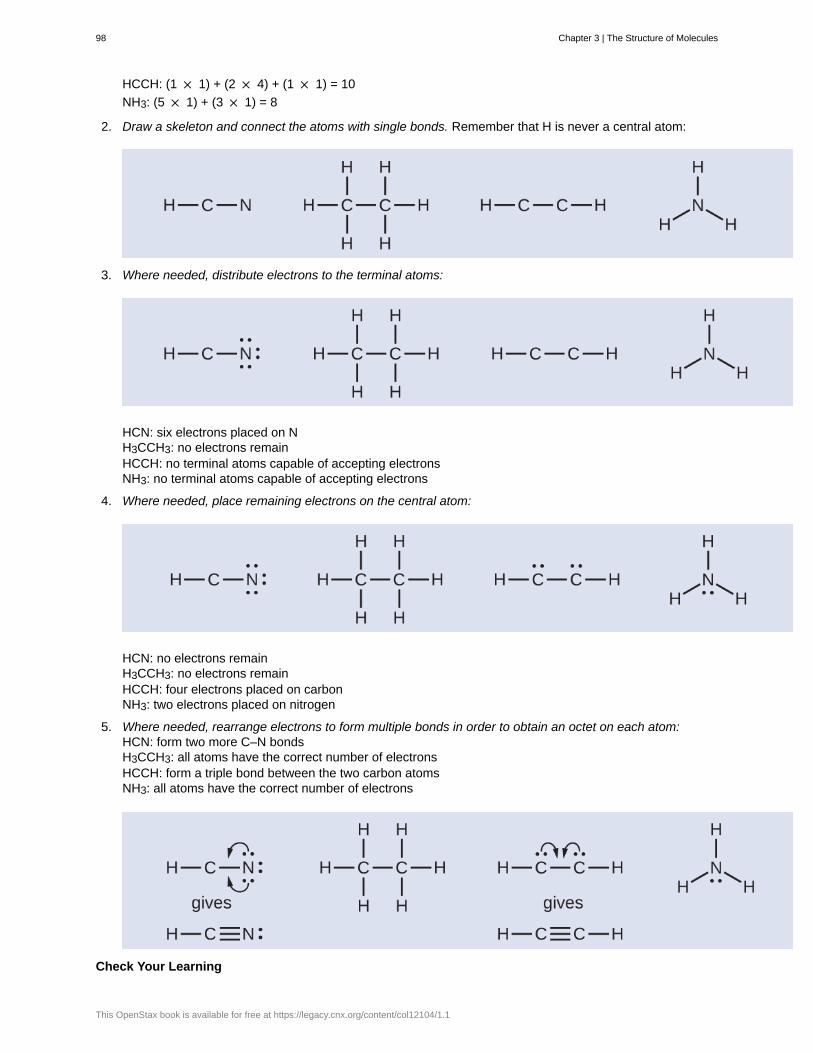
HCCH: (1 × 1) + (2 × 4) + (1 × 1) = 10
NH3: (5 × 1) + (3 × 1) = 8
2. Draw a skeleton and connect the atoms with single bonds. Remember that H is never a central atom:
3. Where needed, distribute electrons to the terminal atoms:
HCN: six electrons placed on NH3CCH3: no electrons remainHCCH: no terminal atoms capable of accepting electronsNH3: no terminal atoms capable of accepting electrons
4. Where needed, place remaining electrons on the central atom:
HCN: no electrons remainH3CCH3: no electrons remainHCCH: four electrons placed on carbonNH3: two electrons placed on nitrogen
5. Where needed, rearrange electrons to form multiple bonds in order to obtain an octet on each atom:HCN: form two more C–N bondsH3CCH3: all atoms have the correct number of electronsHCCH: form a triple bond between the two carbon atomsNH3: all atoms have the correct number of electrons
Check Your Learning
98 Chapter 3 | The Structure of Molecules
This OpenStax book is available for free at https://legacy.cnx.org/content/col12104/1.1
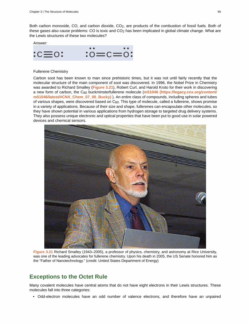
Both carbon monoxide, CO, and carbon dioxide, CO2, are products of the combustion of fossil fuels. Both ofthese gases also cause problems: CO is toxic and CO2 has been implicated in global climate change. What arethe Lewis structures of these two molecules?
Answer:
Fullerene Chemistry
Carbon soot has been known to man since prehistoric times, but it was not until fairly recently that themolecular structure of the main component of soot was discovered. In 1996, the Nobel Prize in Chemistrywas awarded to Richard Smalley (Figure 3.21), Robert Curl, and Harold Kroto for their work in discoveringa new form of carbon, the C60 buckminsterfullerene molecule (m51046 (https://legacy.cnx.org/content/m51046/latest/#CNX_Chem_07_00_Bucky) ). An entire class of compounds, including spheres and tubesof various shapes, were discovered based on C60. This type of molecule, called a fullerene, shows promisein a variety of applications. Because of their size and shape, fullerenes can encapsulate other molecules, sothey have shown potential in various applications from hydrogen storage to targeted drug delivery systems.They also possess unique electronic and optical properties that have been put to good use in solar powereddevices and chemical sensors.
Figure 3.21 Richard Smalley (1943–2005), a professor of physics, chemistry, and astronomy at Rice University,was one of the leading advocates for fullerene chemistry. Upon his death in 2005, the US Senate honored him asthe “Father of Nanotechnology.” (credit: United States Department of Energy)
Exceptions to the Octet Rule
Many covalent molecules have central atoms that do not have eight electrons in their Lewis structures. Thesemolecules fall into three categories:
• Odd-electron molecules have an odd number of valence electrons, and therefore have an unpaired
Chapter 3 | The Structure of Molecules 99
electron.
• Electron-deficient molecules have a central atom that has fewer electrons than needed for a noble gasconfiguration.
• Hypervalent molecules have a central atom that has more electrons than needed for a noble gasconfiguration.
Odd-electron Molecules
We call molecules that contain an odd number of electrons free radicals. Nitric oxide, NO, is an example of anodd-electron molecule; it is produced in internal combustion engines when oxygen and nitrogen react at hightemperatures.
To draw the Lewis structure for an odd-electron molecule like NO, we follow the same five steps we would forother molecules, but with a few minor changes:
1. Determine the total number of valence (outer shell) electrons. The sum of the valence electrons is 5 (fromN) + 6 (from O) = 11. The odd number immediately tells us that we have a free radical, so we know that notevery atom can have eight electrons in its valence shell.
2. Draw a skeleton structure of the molecule. We can easily draw a skeleton with an N–O single bond:N–O
3. Distribute the remaining electrons as lone pairs on the terminal atoms. In this case, there is no central atom,so we distribute the electrons around both atoms. We give eight electrons to the more electronegative atomin these situations; thus oxygen has the filled valence shell:
4. Place all remaining electrons on the central atom. Since there are no remaining electrons, this step doesnot apply.
5. Rearrange the electrons to make multiple bonds with the central atom in order to obtain octets whereverpossible. We know that an odd-electron molecule cannot have an octet for every atom, but we want to geteach atom as close to an octet as possible. In this case, nitrogen has only five electrons around it. To movecloser to an octet for nitrogen, we take one of the lone pairs from oxygen and use it to form a NO doublebond. (We cannot take another lone pair of electrons on oxygen and form a triple bond because nitrogenwould then have nine electrons:)
Electron-deficient Molecules
We will also encounter a few molecules that contain central atoms that do not have a filled valence shell.Generally, these are molecules with central atoms from groups 2 and 13, outer atoms that are hydrogen, or otheratoms that do not form multiple bonds. For example, in the Lewis structures of beryllium dihydride, BeH2, andboron trifluoride, BF3, the beryllium and boron atoms each have only four and six electrons, respectively. It ispossible to draw a structure with a double bond between a boron atom and a fluorine atom in BF3, satisfyingthe octet rule, but experimental evidence indicates the bond lengths are closer to that expected for B–F singlebonds. This suggests the best Lewis structure has three B–F single bonds and an electron deficient boron.The reactivity of the compound is also consistent with an electron deficient boron. However, the B–F bonds areslightly shorter than what is actually expected for B–F single bonds, indicating that some double bond characteris found in the actual molecule.
100 Chapter 3 | The Structure of Molecules
This OpenStax book is available for free at https://legacy.cnx.org/content/col12104/1.1
An atom like the boron atom in BF3, which does not have eight electrons, is very reactive. It readily combineswith a molecule containing an atom with a lone pair of electrons. For example, NH3 reacts with BF3 because thelone pair on nitrogen can be shared with the boron atom:
Hypervalent Molecules
Elements in the second period of the periodic table (n = 2) can accommodate only eight electrons in their valenceshell orbitals because they have only four valence orbitals (one 2s and three 2p orbitals). Elements in the thirdand higher periods (n ≥ 3) have more than four valence orbitals and can share more than four pairs of electronswith other atoms because they have empty d orbitals in the same shell. Molecules formed from these elementsare sometimes called hypervalent molecules. Figure 3.22 shows the Lewis structures for two hypervalentmolecules, PCl5 and SF6.
Figure 3.22 In PCl5, the central atom phosphorus shares five pairs of electrons. In SF6, sulfur shares six pairs ofelectrons.
In some hypervalent molecules, such as IF5 and XeF4, some of the electrons in the outer shell of the centralatom are lone pairs:
Chapter 3 | The Structure of Molecules 101
When we write the Lewis structures for these molecules, we find that we have electrons left over after filling thevalence shells of the outer atoms with eight electrons. These additional electrons must be assigned to the centralatom.
Example 3.9
Writing Lewis Structures: Octet Rule Violations
Xenon is a noble gas, but it forms a number of stable compounds. We examined XeF4 earlier. What are theLewis structures of XeF2 and XeF6?
Solution
We can draw the Lewis structure of any covalent molecule by following the six steps discussed earlier. In thiscase, we can condense the last few steps, since not all of them apply.
1. Calculate the number of valence electrons:XeF2: 8 + (2 × 7) = 22
XeF6: 8 + (6 × 7) = 50
2. Draw a skeleton joining the atoms by single bonds. Xenon will be the central atom because fluorine cannotbe a central atom:
3. Distribute the remaining electrons.XeF2: We place three lone pairs of electrons around each F atom, accounting for 12 electrons and givingeach F atom 8 electrons. Thus, six electrons (three lone pairs) remain. These lone pairs must be placedon the Xe atom. This is acceptable because Xe atoms have empty valence shell d orbitals and canaccommodate more than eight electrons. The Lewis structure of XeF2 shows two bonding pairs and threelone pairs of electrons around the Xe atom:
XeF6: We place three lone pairs of electrons around each F atom, accounting for 36 electrons. Twoelectrons remain, and this lone pair is placed on the Xe atom:
Check Your Learning
The halogens form a class of compounds called the interhalogens, in which halogen atoms covalently bond to
102 Chapter 3 | The Structure of Molecules
This OpenStax book is available for free at https://legacy.cnx.org/content/col12104/1.1
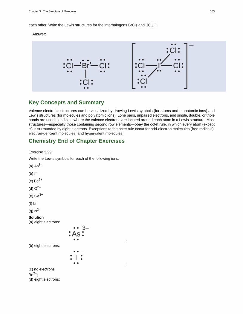
each other. Write the Lewis structures for the interhalogens BrCl3 and ICl4−.
Answer:
Key Concepts and Summary
Valence electronic structures can be visualized by drawing Lewis symbols (for atoms and monatomic ions) andLewis structures (for molecules and polyatomic ions). Lone pairs, unpaired electrons, and single, double, or triplebonds are used to indicate where the valence electrons are located around each atom in a Lewis structure. Moststructures—especially those containing second row elements—obey the octet rule, in which every atom (exceptH) is surrounded by eight electrons. Exceptions to the octet rule occur for odd-electron molecules (free radicals),electron-deficient molecules, and hypervalent molecules.
Chemistry End of Chapter Exercises
Exercise 3.29
Write the Lewis symbols for each of the following ions:
(a) As3–
(b) I–
(c) Be2+
(d) O2–
(e) Ga3+
(f) Li+
(g) N3–
Solution(a) eight electrons:
;(b) eight electrons:
;(c) no electrons
Be2+;(d) eight electrons:
Chapter 3 | The Structure of Molecules 103
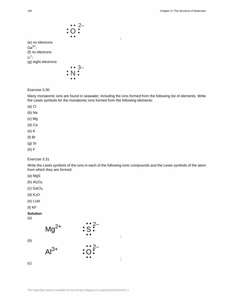
;(e) no electrons
Ga3+;(f) no electrons
Li+;(g) eight electrons:
Exercise 3.30
Many monatomic ions are found in seawater, including the ions formed from the following list of elements. Writethe Lewis symbols for the monatomic ions formed from the following elements:
(a) Cl
(b) Na
(c) Mg
(d) Ca
(e) K
(f) Br
(g) Sr
(h) F
Exercise 3.31
Write the Lewis symbols of the ions in each of the following ionic compounds and the Lewis symbols of the atomfrom which they are formed:
(a) MgS
(b) Al2O3
(c) GaCl3
(d) K2O
(e) Li3N
(f) KF
Solution(a)
;(b)
;(c)
104 Chapter 3 | The Structure of Molecules
This OpenStax book is available for free at https://legacy.cnx.org/content/col12104/1.1
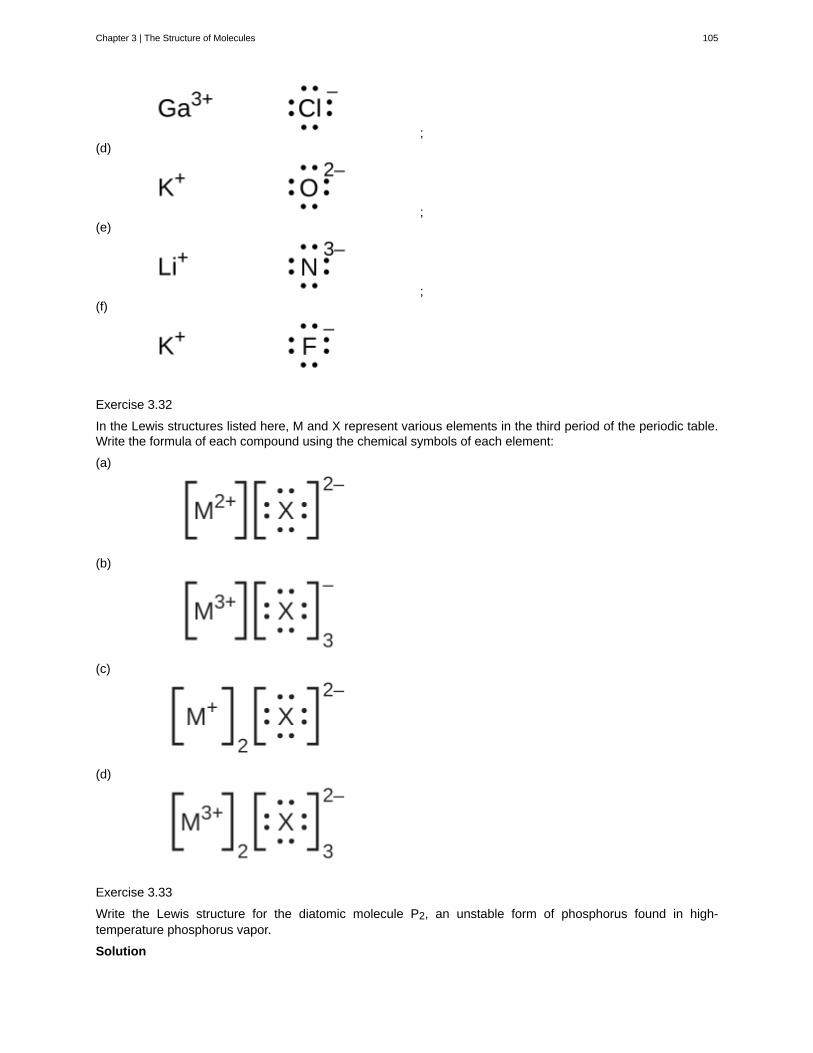
;(d)
;(e)
;(f)
Exercise 3.32
In the Lewis structures listed here, M and X represent various elements in the third period of the periodic table.Write the formula of each compound using the chemical symbols of each element:
(a)
(b)
(c)
(d)
Exercise 3.33
Write the Lewis structure for the diatomic molecule P2, an unstable form of phosphorus found in high-temperature phosphorus vapor.
Solution
Chapter 3 | The Structure of Molecules 105
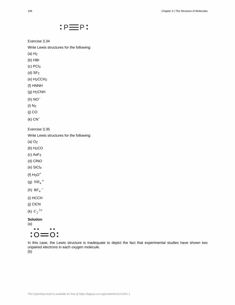
Exercise 3.34
Write Lewis structures for the following:
(a) H2
(b) HBr
(c) PCl3
(d) SF2
(e) H2CCH2
(f) HNNH
(g) H2CNH
(h) NO–
(i) N2
(j) CO
(k) CN–
Exercise 3.35
Write Lewis structures for the following:
(a) O2
(b) H2CO
(c) AsF3
(d) ClNO
(e) SiCl4
(f) H3O+
(g) NH4+
(h) BF4−
(i) HCCH
(j) ClCN
(k) C22+
Solution(a)
In this case, the Lewis structure is inadequate to depict the fact that experimental studies have shown twounpaired electrons in each oxygen molecule.(b)
106 Chapter 3 | The Structure of Molecules
This OpenStax book is available for free at https://legacy.cnx.org/content/col12104/1.1
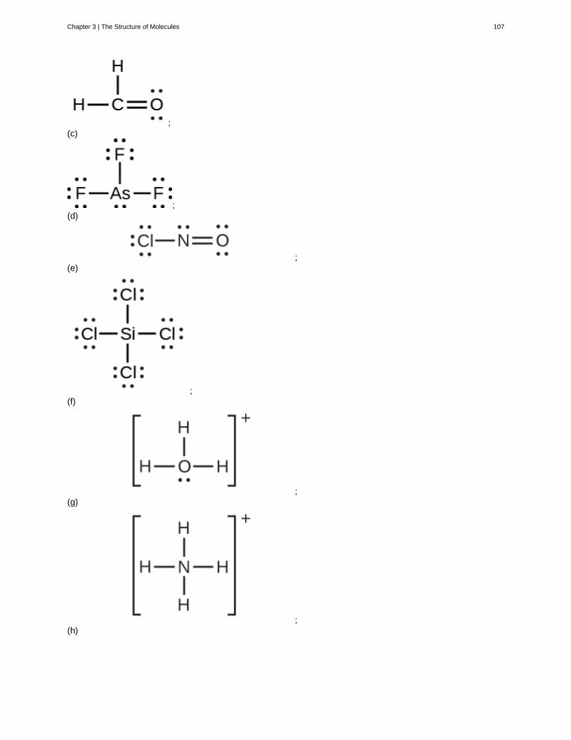
;(c)
;(d)
;(e)
;(f)
;(g)
;(h)
Chapter 3 | The Structure of Molecules 107
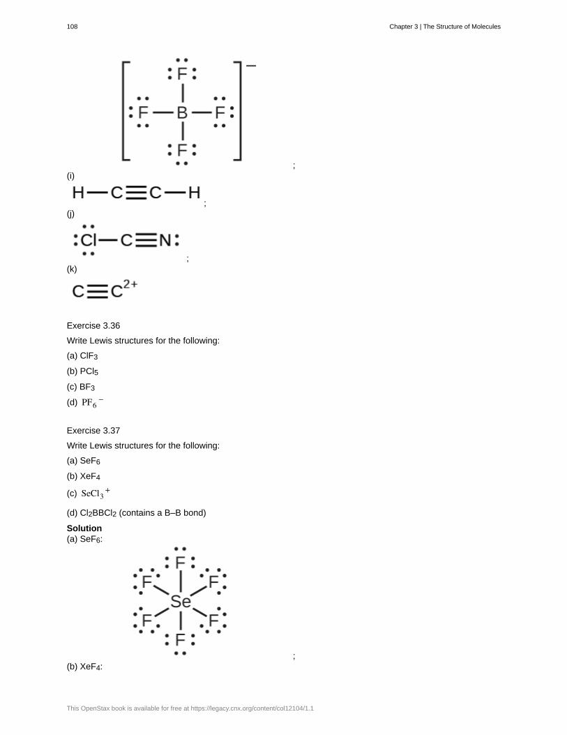
;(i)
;(j)
;(k)
Exercise 3.36
Write Lewis structures for the following:
(a) ClF3
(b) PCl5
(c) BF3
(d) PF6−
Exercise 3.37
Write Lewis structures for the following:
(a) SeF6
(b) XeF4
(c) SeCl3+
(d) Cl2BBCl2 (contains a B–B bond)
Solution(a) SeF6:
;(b) XeF4:
108 Chapter 3 | The Structure of Molecules
This OpenStax book is available for free at https://legacy.cnx.org/content/col12104/1.1
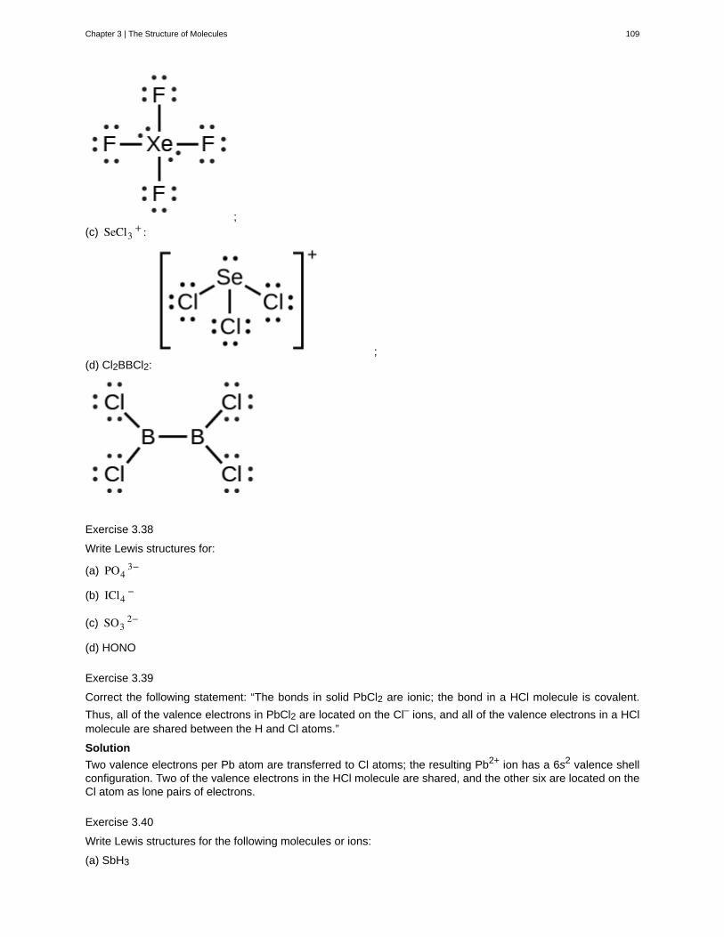
;(c) SeCl3
+ :
;(d) Cl2BBCl2:
Exercise 3.38
Write Lewis structures for:
(a) PO43−
(b) ICl4−
(c) SO32−
(d) HONO
Exercise 3.39
Correct the following statement: “The bonds in solid PbCl2 are ionic; the bond in a HCl molecule is covalent.
Thus, all of the valence electrons in PbCl2 are located on the Cl– ions, and all of the valence electrons in a HClmolecule are shared between the H and Cl atoms.”
Solution
Two valence electrons per Pb atom are transferred to Cl atoms; the resulting Pb2+ ion has a 6s2 valence shellconfiguration. Two of the valence electrons in the HCl molecule are shared, and the other six are located on theCl atom as lone pairs of electrons.
Exercise 3.40
Write Lewis structures for the following molecules or ions:
(a) SbH3
Chapter 3 | The Structure of Molecules 109
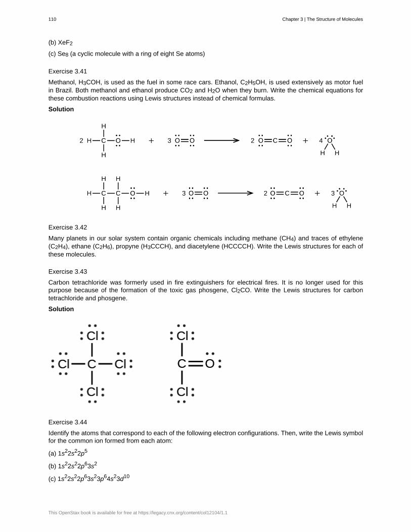
(b) XeF2
(c) Se8 (a cyclic molecule with a ring of eight Se atoms)
Exercise 3.41
Methanol, H3COH, is used as the fuel in some race cars. Ethanol, C2H5OH, is used extensively as motor fuelin Brazil. Both methanol and ethanol produce CO2 and H2O when they burn. Write the chemical equations forthese combustion reactions using Lewis structures instead of chemical formulas.
Solution
Exercise 3.42
Many planets in our solar system contain organic chemicals including methane (CH4) and traces of ethylene(C2H4), ethane (C2H6), propyne (H3CCCH), and diacetylene (HCCCCH). Write the Lewis structures for each ofthese molecules.
Exercise 3.43
Carbon tetrachloride was formerly used in fire extinguishers for electrical fires. It is no longer used for thispurpose because of the formation of the toxic gas phosgene, Cl2CO. Write the Lewis structures for carbontetrachloride and phosgene.
Solution
Exercise 3.44
Identify the atoms that correspond to each of the following electron configurations. Then, write the Lewis symbolfor the common ion formed from each atom:
(a) 1s22s22p5
(b) 1s22s22p63s2
(c) 1s22s22p63s23p64s23d10
110 Chapter 3 | The Structure of Molecules
This OpenStax book is available for free at https://legacy.cnx.org/content/col12104/1.1
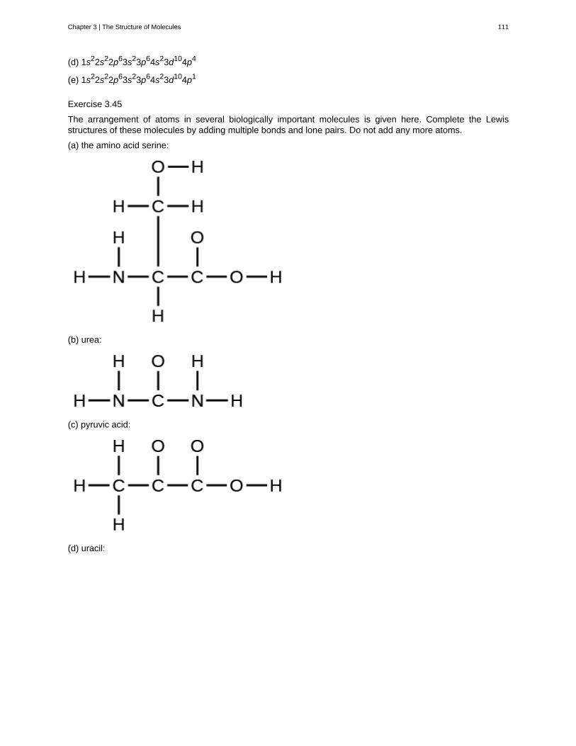
(d) 1s22s22p63s23p64s23d104p4
(e) 1s22s22p63s23p64s23d104p1
Exercise 3.45
The arrangement of atoms in several biologically important molecules is given here. Complete the Lewisstructures of these molecules by adding multiple bonds and lone pairs. Do not add any more atoms.
(a) the amino acid serine:
(b) urea:
(c) pyruvic acid:
(d) uracil:
Chapter 3 | The Structure of Molecules 111
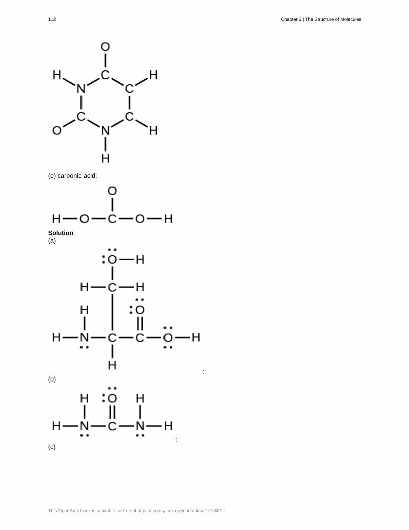
(e) carbonic acid:
Solution(a)
;(b)
;(c)
112 Chapter 3 | The Structure of Molecules
This OpenStax book is available for free at https://legacy.cnx.org/content/col12104/1.1
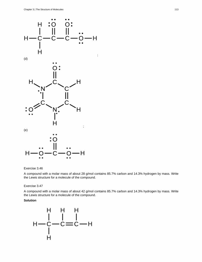
;(d)
;(e)
Exercise 3.46
A compound with a molar mass of about 28 g/mol contains 85.7% carbon and 14.3% hydrogen by mass. Writethe Lewis structure for a molecule of the compound.
Exercise 3.47
A compound with a molar mass of about 42 g/mol contains 85.7% carbon and 14.3% hydrogen by mass. Writethe Lewis structure for a molecule of the compound.
Solution
Chapter 3 | The Structure of Molecules 113
Exercise 3.48
Two arrangements of atoms are possible for a compound with a molar mass of about 45 g/mol that contains52.2% C, 13.1% H, and 34.7% O by mass. Write the Lewis structures for the two molecules.
Exercise 3.49
How are single, double, and triple bonds similar? How do they differ?
SolutionEach bond includes a sharing of electrons between atoms. Two electrons are shared in a single bond; fourelectrons are shared in a double bond; and six electrons are shared in a triple bond.
3.5 | Water
By the end of this section, you will be able to:
• Describe the properties of water that are critical to maintaining life
• Explain why water is an excellent solvent
• Provide examples of water’s cohesive and adhesive properties
• Discuss the role of acids, bases, and buffers in homeostasis
Why do scientists spend time looking for water on other planets? Why is water so important? It is because wateris essential to life as we know it. Water is one of the more abundant molecules and the one most critical to life onEarth. Approximately 60–70 percent of the human body is made up of water. Without it, life as we know it simplywould not exist.
The polarity of the water molecule and its resulting hydrogen bonding make water a unique substance withspecial properties that are intimately tied to the processes of life. Life originally evolved in a watery environment,and most of an organism’s cellular chemistry and metabolism occur inside the watery contents of the cell’scytoplasm. Special properties of water are its high heat capacity and heat of vaporization, its ability to dissolvepolar molecules, its cohesive and adhesive properties, and its dissociation into ions that leads to the generationof pH. Understanding these characteristics of water helps to elucidate its importance in maintaining life.
Water’s Polarity
One of water’s important properties is that it is composed of polar molecules: the hydrogen and oxygen withinwater molecules (H2O) form polar covalent bonds. While there is no net charge to a water molecule, the polarityof water creates a slightly positive charge on hydrogen and a slightly negative charge on oxygen, contributingto water’s properties of attraction. Water’s charges are generated because oxygen is more electronegativethan hydrogen, making it more likely that a shared electron would be found near the oxygen nucleus than thehydrogen nucleus, thus generating the partial negative charge near the oxygen.
As a result of water’s polarity, each water molecule attracts other water molecules because of the oppositecharges between water molecules, forming hydrogen bonds. Water also attracts or is attracted to other polarmolecules and ions. A polar substance that interacts readily with or dissolves in water is referred to ashydrophilic (hydro- = “water”; -philic = “loving”). In contrast, non-polar molecules such as oils and fats do notinteract well with water, as shown in Figure 3.23 and separate from it rather than dissolve in it, as we see insalad dressings containing oil and vinegar (an acidic water solution). These nonpolar compounds are calledhydrophobic (hydro- = “water”; -phobic = “fearing”).
114 Chapter 3 | The Structure of Molecules
This OpenStax book is available for free at https://legacy.cnx.org/content/col12104/1.1
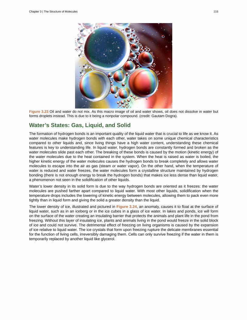
Figure 3.23 Oil and water do not mix. As this macro image of oil and water shows, oil does not dissolve in water butforms droplets instead. This is due to it being a nonpolar compound. (credit: Gautam Dogra).
Water’s States: Gas, Liquid, and Solid
The formation of hydrogen bonds is an important quality of the liquid water that is crucial to life as we know it. Aswater molecules make hydrogen bonds with each other, water takes on some unique chemical characteristicscompared to other liquids and, since living things have a high water content, understanding these chemicalfeatures is key to understanding life. In liquid water, hydrogen bonds are constantly formed and broken as thewater molecules slide past each other. The breaking of these bonds is caused by the motion (kinetic energy) ofthe water molecules due to the heat contained in the system. When the heat is raised as water is boiled, thehigher kinetic energy of the water molecules causes the hydrogen bonds to break completely and allows watermolecules to escape into the air as gas (steam or water vapor). On the other hand, when the temperature ofwater is reduced and water freezes, the water molecules form a crystalline structure maintained by hydrogenbonding (there is not enough energy to break the hydrogen bonds) that makes ice less dense than liquid water,a phenomenon not seen in the solidification of other liquids.
Water’s lower density in its solid form is due to the way hydrogen bonds are oriented as it freezes: the watermolecules are pushed farther apart compared to liquid water. With most other liquids, solidification when thetemperature drops includes the lowering of kinetic energy between molecules, allowing them to pack even moretightly than in liquid form and giving the solid a greater density than the liquid.
The lower density of ice, illustrated and pictured in Figure 3.24, an anomaly, causes it to float at the surface ofliquid water, such as in an iceberg or in the ice cubes in a glass of ice water. In lakes and ponds, ice will formon the surface of the water creating an insulating barrier that protects the animals and plant life in the pond fromfreezing. Without this layer of insulating ice, plants and animals living in the pond would freeze in the solid blockof ice and could not survive. The detrimental effect of freezing on living organisms is caused by the expansionof ice relative to liquid water. The ice crystals that form upon freezing rupture the delicate membranes essentialfor the function of living cells, irreversibly damaging them. Cells can only survive freezing if the water in them istemporarily replaced by another liquid like glycerol.
Chapter 3 | The Structure of Molecules 115

Figure 3.24 Hydrogen bonding makes ice less dense than liquid water. The (a) lattice structure of ice makes it lessdense than the freely flowing molecules of liquid water, enabling it to (b) float on water. (credit a: modification of work
by Jane Whitney, image created using Visual Molecular Dynamics (VMD) software[2]
; credit b: modification of work byCarlos Ponte)
Click here (http://openstaxcollege.org/l/ice_lattice2) to see a 3-D animation of the structure of an ice lattice.
(Image credit: Jane Whitney. Image created using Visual Molecular Dynamics VMD software.[3]
)
Water’s High Heat Capacity
Water’s high heat capacity is a property caused by hydrogen bonding among water molecules. Water has thehighest specific heat capacity of any liquids. Specific heat is defined as the amount of heat one gram of asubstance must absorb or lose to change its temperature by one degree Celsius. For water, this amount is onecalorie. It therefore takes water a long time to heat and long time to cool. In fact, the specific heat capacity ofwater is about five times more than that of sand. This explains why the land cools faster than the sea. Due to itshigh heat capacity, water is used by warm blooded animals to more evenly disperse heat in their bodies: it actsin a similar manner to a car’s cooling system, transporting heat from warm places to cool places, causing thebody to maintain a more even temperature.
Water’s Heat of Vaporization
Water also has a high heat of vaporization, the amount of energy required to change one gram of a liquidsubstance to a gas. A considerable amount of heat energy (586 cal) is required to accomplish this changein water. This process occurs on the surface of water. As liquid water heats up, hydrogen bonding makes itdifficult to separate the liquid water molecules from each other, which is required for it to enter its gaseousphase (steam). As a result, water acts as a heat sink or heat reservoir and requires much more heat to boil thandoes a liquid such as ethanol (grain alcohol), whose hydrogen bonding with other ethanol molecules is weakerthan water’s hydrogen bonding. Eventually, as water reaches its boiling point of 100° Celsius (212° Fahrenheit),the heat is able to break the hydrogen bonds between the water molecules, and the kinetic energy (motion)between the water molecules allows them to escape from the liquid as a gas. Even when below its boiling point,water’s individual molecules acquire enough energy from other water molecules such that some surface watermolecules can escape and vaporize: this process is known as evaporation.
The fact that hydrogen bonds need to be broken for water to evaporate means that a substantial amountof energy is used in the process. As the water evaporates, energy is taken up by the process, coolingthe environment where the evaporation is taking place. In many living organisms, including in humans, the
2. W. Humphrey W., A. Dalke, and K. Schulten, “VMD—Visual Molecular Dynamics,” Journal of Molecular Graphics 14 (1996): 33-38.3. W. Humphrey W., A. Dalke, and K. Schulten, “VMD—Visual Molecular Dynamics,” Journal of Molecular Graphics 14 (1996): 33-38.
116 Chapter 3 | The Structure of Molecules
This OpenStax book is available for free at https://legacy.cnx.org/content/col12104/1.1
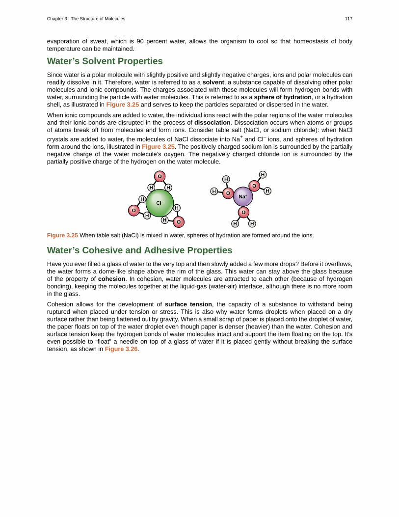
evaporation of sweat, which is 90 percent water, allows the organism to cool so that homeostasis of bodytemperature can be maintained.
Water’s Solvent Properties
Since water is a polar molecule with slightly positive and slightly negative charges, ions and polar molecules canreadily dissolve in it. Therefore, water is referred to as a solvent, a substance capable of dissolving other polarmolecules and ionic compounds. The charges associated with these molecules will form hydrogen bonds withwater, surrounding the particle with water molecules. This is referred to as a sphere of hydration, or a hydrationshell, as illustrated in Figure 3.25 and serves to keep the particles separated or dispersed in the water.
When ionic compounds are added to water, the individual ions react with the polar regions of the water moleculesand their ionic bonds are disrupted in the process of dissociation. Dissociation occurs when atoms or groupsof atoms break off from molecules and form ions. Consider table salt (NaCl, or sodium chloride): when NaCl
crystals are added to water, the molecules of NaCl dissociate into Na+ and Cl– ions, and spheres of hydrationform around the ions, illustrated in Figure 3.25. The positively charged sodium ion is surrounded by the partiallynegative charge of the water molecule’s oxygen. The negatively charged chloride ion is surrounded by thepartially positive charge of the hydrogen on the water molecule.
Figure 3.25 When table salt (NaCl) is mixed in water, spheres of hydration are formed around the ions.
Water’s Cohesive and Adhesive Properties
Have you ever filled a glass of water to the very top and then slowly added a few more drops? Before it overflows,the water forms a dome-like shape above the rim of the glass. This water can stay above the glass becauseof the property of cohesion. In cohesion, water molecules are attracted to each other (because of hydrogenbonding), keeping the molecules together at the liquid-gas (water-air) interface, although there is no more roomin the glass.
Cohesion allows for the development of surface tension, the capacity of a substance to withstand beingruptured when placed under tension or stress. This is also why water forms droplets when placed on a drysurface rather than being flattened out by gravity. When a small scrap of paper is placed onto the droplet of water,the paper floats on top of the water droplet even though paper is denser (heavier) than the water. Cohesion andsurface tension keep the hydrogen bonds of water molecules intact and support the item floating on the top. It’seven possible to “float” a needle on top of a glass of water if it is placed gently without breaking the surfacetension, as shown in Figure 3.26.
Chapter 3 | The Structure of Molecules 117
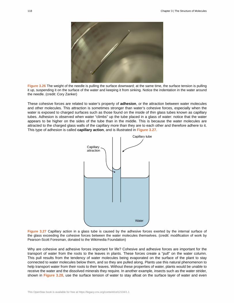
Figure 3.26 The weight of the needle is pulling the surface downward; at the same time, the surface tension is pullingit up, suspending it on the surface of the water and keeping it from sinking. Notice the indentation in the water aroundthe needle. (credit: Cory Zanker)
These cohesive forces are related to water’s property of adhesion, or the attraction between water moleculesand other molecules. This attraction is sometimes stronger than water’s cohesive forces, especially when thewater is exposed to charged surfaces such as those found on the inside of thin glass tubes known as capillarytubes. Adhesion is observed when water “climbs” up the tube placed in a glass of water: notice that the waterappears to be higher on the sides of the tube than in the middle. This is because the water molecules areattracted to the charged glass walls of the capillary more than they are to each other and therefore adhere to it.This type of adhesion is called capillary action, and is illustrated in Figure 3.27.
Figure 3.27 Capillary action in a glass tube is caused by the adhesive forces exerted by the internal surface ofthe glass exceeding the cohesive forces between the water molecules themselves. (credit: modification of work byPearson-Scott Foresman, donated to the Wikimedia Foundation)
Why are cohesive and adhesive forces important for life? Cohesive and adhesive forces are important for thetransport of water from the roots to the leaves in plants. These forces create a “pull” on the water column.This pull results from the tendency of water molecules being evaporated on the surface of the plant to stayconnected to water molecules below them, and so they are pulled along. Plants use this natural phenomenon tohelp transport water from their roots to their leaves. Without these properties of water, plants would be unable toreceive the water and the dissolved minerals they require. In another example, insects such as the water strider,shown in Figure 3.28, use the surface tension of water to stay afloat on the surface layer of water and even
118 Chapter 3 | The Structure of Molecules
This OpenStax book is available for free at https://legacy.cnx.org/content/col12104/1.1
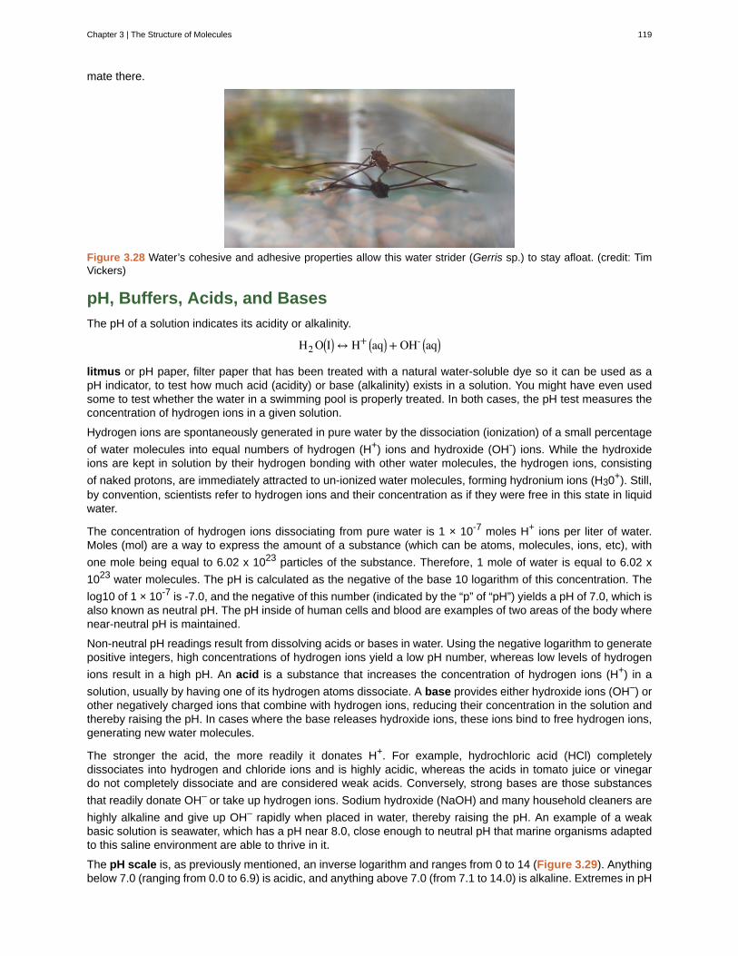
mate there.
Figure 3.28 Water’s cohesive and adhesive properties allow this water strider (Gerris sp.) to stay afloat. (credit: TimVickers)
pH, Buffers, Acids, and Bases
The pH of a solution indicates its acidity or alkalinity.
H2 O⎛⎝I⎞⎠↔ H+ ⎛⎝aq⎞⎠+ OH- ⎛⎝aq⎞⎠
litmus or pH paper, filter paper that has been treated with a natural water-soluble dye so it can be used as apH indicator, to test how much acid (acidity) or base (alkalinity) exists in a solution. You might have even usedsome to test whether the water in a swimming pool is properly treated. In both cases, the pH test measures theconcentration of hydrogen ions in a given solution.
Hydrogen ions are spontaneously generated in pure water by the dissociation (ionization) of a small percentage
of water molecules into equal numbers of hydrogen (H+) ions and hydroxide (OH-) ions. While the hydroxideions are kept in solution by their hydrogen bonding with other water molecules, the hydrogen ions, consisting
of naked protons, are immediately attracted to un-ionized water molecules, forming hydronium ions (H30+). Still,by convention, scientists refer to hydrogen ions and their concentration as if they were free in this state in liquidwater.
The concentration of hydrogen ions dissociating from pure water is 1 × 10⁻⁷ moles H+ ions per liter of water.Moles (mol) are a way to express the amount of a substance (which can be atoms, molecules, ions, etc), with
one mole being equal to 6.02 x 10²³ particles of the substance. Therefore, 1 mole of water is equal to 6.02 x
1023 water molecules. The pH is calculated as the negative of the base 10 logarithm of this concentration. The
log10 of 1 × 10⁻⁷ is -7.0, and the negative of this number (indicated by the “p” of “pH”) yields a pH of 7.0, which isalso known as neutral pH. The pH inside of human cells and blood are examples of two areas of the body wherenear-neutral pH is maintained.
Non-neutral pH readings result from dissolving acids or bases in water. Using the negative logarithm to generatepositive integers, high concentrations of hydrogen ions yield a low pH number, whereas low levels of hydrogen
ions result in a high pH. An acid is a substance that increases the concentration of hydrogen ions (H+) in a
solution, usually by having one of its hydrogen atoms dissociate. A base provides either hydroxide ions (OH–) orother negatively charged ions that combine with hydrogen ions, reducing their concentration in the solution andthereby raising the pH. In cases where the base releases hydroxide ions, these ions bind to free hydrogen ions,generating new water molecules.
The stronger the acid, the more readily it donates H+. For example, hydrochloric acid (HCl) completelydissociates into hydrogen and chloride ions and is highly acidic, whereas the acids in tomato juice or vinegardo not completely dissociate and are considered weak acids. Conversely, strong bases are those substances
that readily donate OH– or take up hydrogen ions. Sodium hydroxide (NaOH) and many household cleaners are
highly alkaline and give up OH– rapidly when placed in water, thereby raising the pH. An example of a weakbasic solution is seawater, which has a pH near 8.0, close enough to neutral pH that marine organisms adaptedto this saline environment are able to thrive in it.
The pH scale is, as previously mentioned, an inverse logarithm and ranges from 0 to 14 (Figure 3.29). Anythingbelow 7.0 (ranging from 0.0 to 6.9) is acidic, and anything above 7.0 (from 7.1 to 14.0) is alkaline. Extremes in pH
Chapter 3 | The Structure of Molecules 119
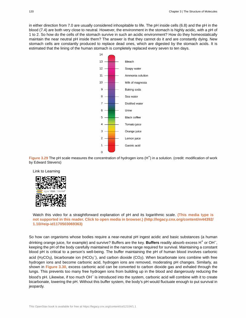
in either direction from 7.0 are usually considered inhospitable to life. The pH inside cells (6.8) and the pH in theblood (7.4) are both very close to neutral. However, the environment in the stomach is highly acidic, with a pH of1 to 2. So how do the cells of the stomach survive in such an acidic environment? How do they homeostaticallymaintain the near neutral pH inside them? The answer is that they cannot do it and are constantly dying. Newstomach cells are constantly produced to replace dead ones, which are digested by the stomach acids. It isestimated that the lining of the human stomach is completely replaced every seven to ten days.
Figure 3.29 The pH scale measures the concentration of hydrogen ions (H+) in a solution. (credit: modification of workby Edward Stevens)
Link to Learning
Watch this video for a straightforward explanation of pH and its logarithmic scale. (This media type isnot supported in this reader. Click to open media in browser.) (http://legacy.cnx.org/content/m44392/1.10/#eip-id1170503069363)
So how can organisms whose bodies require a near-neutral pH ingest acidic and basic substances (a human
drinking orange juice, for example) and survive? Buffers are the key. Buffers readily absorb excess H+ or OH–,keeping the pH of the body carefully maintained in the narrow range required for survival. Maintaining a constantblood pH is critical to a person’s well-being. The buffer maintaining the pH of human blood involves carbonic
acid (H2CO3), bicarbonate ion (HCO3–), and carbon dioxide (CO2). When bicarbonate ions combine with free
hydrogen ions and become carbonic acid, hydrogen ions are removed, moderating pH changes. Similarly, asshown in Figure 3.30, excess carbonic acid can be converted to carbon dioxide gas and exhaled through thelungs. This prevents too many free hydrogen ions from building up in the blood and dangerously reducing the
blood’s pH. Likewise, if too much OH– is introduced into the system, carbonic acid will combine with it to createbicarbonate, lowering the pH. Without this buffer system, the body’s pH would fluctuate enough to put survival injeopardy.
120 Chapter 3 | The Structure of Molecules
This OpenStax book is available for free at https://legacy.cnx.org/content/col12104/1.1
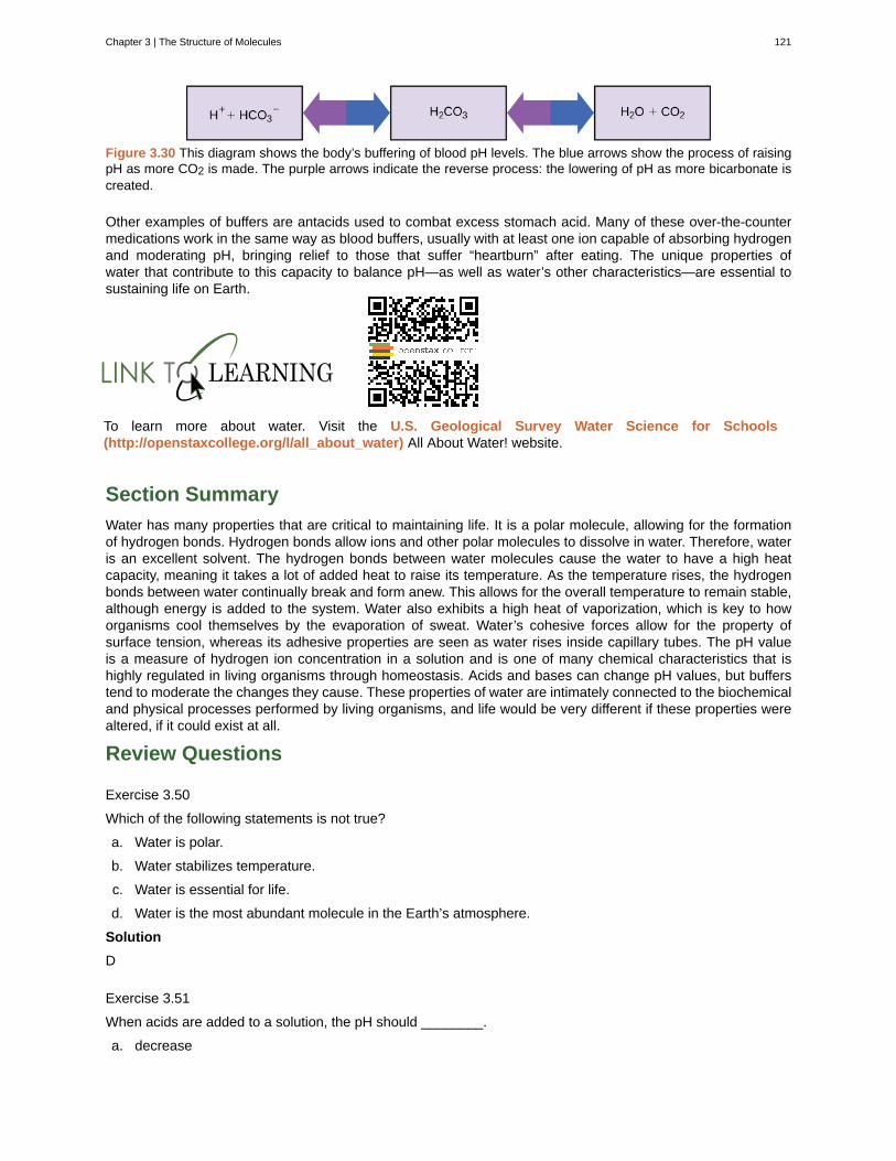
Figure 3.30 This diagram shows the body’s buffering of blood pH levels. The blue arrows show the process of raisingpH as more CO2 is made. The purple arrows indicate the reverse process: the lowering of pH as more bicarbonate iscreated.
Other examples of buffers are antacids used to combat excess stomach acid. Many of these over-the-countermedications work in the same way as blood buffers, usually with at least one ion capable of absorbing hydrogenand moderating pH, bringing relief to those that suffer “heartburn” after eating. The unique properties ofwater that contribute to this capacity to balance pH—as well as water’s other characteristics—are essential tosustaining life on Earth.
To learn more about water. Visit the U.S. Geological Survey Water Science for Schools(http://openstaxcollege.org/l/all_about_water) All About Water! website.
Section Summary
Water has many properties that are critical to maintaining life. It is a polar molecule, allowing for the formationof hydrogen bonds. Hydrogen bonds allow ions and other polar molecules to dissolve in water. Therefore, wateris an excellent solvent. The hydrogen bonds between water molecules cause the water to have a high heatcapacity, meaning it takes a lot of added heat to raise its temperature. As the temperature rises, the hydrogenbonds between water continually break and form anew. This allows for the overall temperature to remain stable,although energy is added to the system. Water also exhibits a high heat of vaporization, which is key to howorganisms cool themselves by the evaporation of sweat. Water’s cohesive forces allow for the property ofsurface tension, whereas its adhesive properties are seen as water rises inside capillary tubes. The pH valueis a measure of hydrogen ion concentration in a solution and is one of many chemical characteristics that ishighly regulated in living organisms through homeostasis. Acids and bases can change pH values, but bufferstend to moderate the changes they cause. These properties of water are intimately connected to the biochemicaland physical processes performed by living organisms, and life would be very different if these properties werealtered, if it could exist at all.
Review Questions
Exercise 3.50
Which of the following statements is not true?
a. Water is polar.
b. Water stabilizes temperature.
c. Water is essential for life.
d. Water is the most abundant molecule in the Earth’s atmosphere.
Solution
D
Exercise 3.51
When acids are added to a solution, the pH should ________.
a. decrease
Chapter 3 | The Structure of Molecules 121
b. increase
c. stay the same
d. cannot tell without testing
Solution
A
Exercise 3.52
A molecule that binds up excess hydrogen ions in a solution is called a(n) ________.
a. acid
b. isotope
c. base
d. donator
Solution
C
Exercise 3.53
Which of the following statements is true?
a. Acids and bases cannot mix together.
b. Acids and bases will neutralize each other.
c. Acids, but not bases, can change the pH of a solution.
d. Acids donate hydroxide ions (OH–); bases donate hydrogen ions (H+).
Solution
B
Free Response
Exercise 3.54
Discuss how buffers help prevent drastic swings in pH.
Solution
Buffers absorb the free hydrogen ions and hydroxide ions that result from chemical reactions. Because they canbond these ions, they prevent increases or decreases in pH. An example of a buffer system is the bicarbonatesystem in the human body. This system is able to absorb hydrogen and hydroxide ions to prevent changes in pHand keep cells functioning properly.
Exercise 3.55
Why can some insects walk on water?
Solution
Some insects can walk on water, although they are heavier (denser) than water, because of the surface tensionof water. Surface tension results from cohesion, or the attraction between water molecules at the surface of thebody of water (the liquid-air/gas interface).
122 Chapter 3 | The Structure of Molecules
This OpenStax book is available for free at https://legacy.cnx.org/content/col12104/1.1
3.6 | Brønsted-Lowry Acids and Bases
By the end of this section, you will be able to:
• Identify acids, bases, and conjugate acid-base pairs according to the Brønsted-Lowry definition
• Write equations for acid and base ionization reactions
• Use the ion-product constant for water to calculate hydronium and hydroxide ion concentrations
• Describe the acid-base behavior of amphiprotic substances
Acids and bases have been known for a long time. When Robert Boyle characterized them in 1680, he notedthat acids dissolve many substances, change the color of certain natural dyes (for example, they change litmusfrom blue to red), and lose these characteristic properties after coming into contact with alkalis (bases). In theeighteenth century, it was recognized that acids have a sour taste, react with limestone to liberate a gaseoussubstance (now known to be CO2), and interact with alkalis to form neutral substances. In 1815, Humphry Davycontributed greatly to the development of the modern acid-base concept by demonstrating that hydrogen isthe essential constituent of acids. Around that same time, Joseph Louis Gay-Lussac concluded that acids aresubstances that can neutralize bases and that these two classes of substances can be defined only in terms ofeach other. The significance of hydrogen was reemphasized in 1884 when Svante Arrhenius defined an acid asa compound that dissolves in water to yield hydrogen cations (now recognized to be hydronium ions) and a baseas a compound that dissolves in water to yield hydroxide anions.
In an earlier chapter on chemical reactions, we defined acids and bases as Arrhenius did: We identified an acid
as a compound that dissolves in water to yield hydronium ions (H3O+) and a base as a compound that dissolves
in water to yield hydroxide ions (OH−). This definition is not wrong; it is simply limited.
Later, we extended the definition of an acid or a base using the more general definition proposed in 1923 bythe Danish chemist Johannes Brønsted and the English chemist Thomas Lowry. Their definition centers on the
proton, H+. A proton is what remains when the most common isotope of hydrogen, 11 H, loses an electron. A
compound that donates a proton to another compound is called a Brønsted-Lowry acid, and a compound thataccepts a proton is called a Brønsted-Lowry base. An acid-base reaction is the transfer of a proton from aproton donor (acid) to a proton acceptor (base). In a subsequent chapter of this text we will introduce the mostgeneral model of acid-base behavior introduced by the American chemist G. N. Lewis.
Acids may be compounds such as HCl or H2SO4, organic acids like acetic acid (CH3COOH) or ascorbic acid
(vitamin C), or H2O. Anions (such as HSO4−, H2 PO4
−, HS−, and HCO3−) and cations (such as H3O+,
NH4+, and [Al(H2 O)6]3+) may also act as acids. Bases fall into the same three categories. Bases may be
neutral molecules (such as H2O, NH3, and CH3NH2), anions (such as OH−, HS−, HCO3−, CO3
2−, F−, and
PO43−), or cations (such as [Al(H2 O)5 OH]2+). The most familiar bases are ionic compounds such as NaOH
and Ca(OH)2, which contain the hydroxide ion, OH−. The hydroxide ion in these compounds accepts a protonfrom acids to form water:
H+ + OH− ⟶ H2 O
We call the product that remains after an acid donates a proton the conjugate base of the acid. This species isa base because it can accept a proton (to re-form the acid):
Chapter 3 | The Structure of Molecules 123
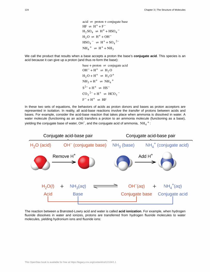
acid ⇌ proton + conjugate baseHF ⇌ H+ + F−
H2 SO4 ⇌ H+ + HSO4−
H2 O ⇌ H+ + OH−
HSO4− ⇌ H+ + SO4
2−
NH4+ ⇌ H+ + NH3
We call the product that results when a base accepts a proton the base’s conjugate acid. This species is anacid because it can give up a proton (and thus re-form the base):
base + proton ⇌ conjugate acidOH− + H+ ⇌ H2 OH2 O + H+ ⇌ H3 O+
NH3 + H+ ⇌ NH4+
S2− + H+ ⇌ HS−
CO32− + H+ ⇌ HCO3
−
F− + H+ ⇌ HF
In these two sets of equations, the behaviors of acids as proton donors and bases as proton acceptors arerepresented in isolation. In reality, all acid-base reactions involve the transfer of protons between acids andbases. For example, consider the acid-base reaction that takes place when ammonia is dissolved in water. Awater molecule (functioning as an acid) transfers a proton to an ammonia molecule (functioning as a base),
yielding the conjugate base of water, OH−, and the conjugate acid of ammonia, NH4+ :
The reaction between a Brønsted-Lowry acid and water is called acid ionization. For example, when hydrogenfluoride dissolves in water and ionizes, protons are transferred from hydrogen fluoride molecules to watermolecules, yielding hydronium ions and fluoride ions:
124 Chapter 3 | The Structure of Molecules
This OpenStax book is available for free at https://legacy.cnx.org/content/col12104/1.1
When we add a base to water, a base ionization reaction occurs in which protons are transferred from watermolecules to base molecules. For example, adding pyridine to water yields hydroxide ions and pyridinium ions:
Notice that both these ionization reactions are represented as equilibrium processes. The relative extent to whichthese acid and base ionization reactions proceed is an important topic treated in a later section of this chapter. Inthe preceding paragraphs we saw that water can function as either an acid or a base, depending on the natureof the solute dissolved in it. In fact, in pure water or in any aqueous solution, water acts both as an acid and abase. A very small fraction of water molecules donate protons to other water molecules to form hydronium ionsand hydroxide ions:
This type of reaction, in which a substance ionizes when one molecule of the substance reacts with anothermolecule of the same substance, is referred to as autoionization.
Pure water undergoes autoionization to a very slight extent. Only about two out of every 109 molecules in asample of pure water are ionized at 25 °C. The equilibrium constant for the ionization of water is called the ion-product constant for water (Kw):
H2 O(l) + H2 O(l) ⇌ H3 O+(aq) + OH−(aq) Kw = [H3 O+][OH−]
The slight ionization of pure water is reflected in the small value of the equilibrium constant; at 25 °C, Kw
has a value of 1.0 × 10⁻¹⁴. The process is endothermic, and so the extent of ionization and the resulting
concentrations of hydronium ion and hydroxide ion increase with temperature. For example, at 100 °C, the value
for Kw is about 5.6 × 10⁻¹³, roughly 50 times larger than the value at 25 °C.
Chapter 3 | The Structure of Molecules 125
Example 3.10
Ion Concentrations in Pure Water
What are the hydronium ion concentration and the hydroxide ion concentration in pure water at 25 °C?
Solution
The autoionization of water yields the same number of hydronium and hydroxide ions. Therefore, in pure water,
[H3O+] = [OH−]. At 25 °C:
Kw = [H3 O+][OH−] = [H3 O+]2 = [OH−]2 = 1.0 × 10⁻¹⁴
So:
[H3 O+] = [OH−] = 1.0 × 10−14 = 1.0 × 10⁻⁷ M
The hydronium ion concentration and the hydroxide ion concentration are the same, and we find that both equal
1.0 × 10⁻⁷ M.
Check Your Learning
The ion product of water at 80 °C is 2.4 × 10⁻¹³. What are the concentrations of hydronium and hydroxide ions
in pure water at 80 °C?
Answer:
[H3O+] = [OH−] = 4.9 × 10⁻⁷ M
It is important to realize that the autoionization equilibrium for water is established in all aqueous solutions.Adding an acid or base to water will not change the position of the equilibrium. Example 3.11 demonstrates thequantitative aspects of this relation between hydronium and hydroxide ion concentrations.
Example 3.11
The Inverse Proportionality of [H3O+] and [OH−]
A solution of carbon dioxide in water has a hydronium ion concentration of 2.0 × 10⁻⁶ M. What is the
concentration of hydroxide ion at 25 °C?
Solution
We know the value of the ion-product constant for water at 25 °C:
2H2 O(l) ⇌ H3 O+(aq) + OH−(aq) Kw = [H3 O+][OH−] = 1.0 × 10⁻¹⁴
Thus, we can calculate the missing equilibrium concentration.
Rearrangement of the Kw expression yields that [OH−] is directly proportional to the inverse of [H3O+]:
[OH−] = Kw[H3 O+]
= 1.0 × 10⁻¹⁴
2.0 × 10−6 = 5.0 × 10⁻⁹
The hydroxide ion concentration in water is reduced to 5.0 × 10⁻⁹ M as the hydronium ion concentration
increases to 2.0 × 10⁻⁶ M. This is expected from Le Châtelier’s principle; the autoionization reaction shifts to
the left to reduce the stress of the increased hydronium ion concentration and the [OH−] is reduced relative tothat in pure water.
A check of these concentrations confirms that our arithmetic is correct:
Kw = [H3 O+][OH−] = (2.0 × 10⁻⁶)(5.0 × 10⁻⁹) = 1.0 × 10⁻¹⁴
Check Your Learning
What is the hydronium ion concentration in an aqueous solution with a hydroxide ion concentration of 0.001 M
126 Chapter 3 | The Structure of Molecules
This OpenStax book is available for free at https://legacy.cnx.org/content/col12104/1.1
at 25 °C?
Answer:
[H3O+] = 1 × 10⁻¹¹ M
Amphiprotic Species
Like water, many molecules and ions may either gain or lose a proton under the appropriate conditions. Suchspecies are said to be amphiprotic. Another term used to describe such species is amphoteric, which is a moregeneral term for a species that may act either as an acid or a base by any definition (not just the Brønsted-Lowryone). Consider for example the bicarbonate ion, which may either donate or accept a proton as shown here:
HCO3−(aq) + H2 O(l) ⇌ CO3
2−(aq) + H3 O+(aq)
HCO3−(aq) + H2 O(l) ⇌ H2 CO3(aq) + OH−(aq)
Example 3.12
Representing the Acid-Base Behavior of an Amphoteric Substance
Write separate equations representing the reaction of HSO3−
(a) as an acid with OH−
(b) as a base with HI
Solution
(a) HSO3−(aq) + OH−(aq) ⇌ SO3
2−(aq) + H2 O(l)
(b) HSO3−(aq) + HI(aq) ⇌ H2 SO3(aq) + I−(aq)
Check Your Learning
Write separate equations representing the reaction of H2 PO4−
(a) as a base with HBr
(b) as an acid with OH−
Answer:
(a) H2 PO4−(aq) + HBr(aq) ⇌ H3 PO4(aq) + Br−(aq); (b)
H2 PO4−(aq) + OH−(aq) ⇌ HPO4
2−(aq) + H2 O(l)
Key Concepts and Summary
A compound that can donate a proton (a hydrogen ion) to another compound is called a Brønsted-Lowryacid. The compound that accepts the proton is called a Brønsted-Lowry base. The species remaining after aBrønsted-Lowry acid has lost a proton is the conjugate base of the acid. The species formed when a Brønsted-Lowry base gains a proton is the conjugate acid of the base. Thus, an acid-base reaction occurs when a proton istransferred from an acid to a base, with formation of the conjugate base of the reactant acid and formation of theconjugate acid of the reactant base. Amphiprotic species can act as both proton donors and proton acceptors.
Water is the most important amphiprotic species. It can form both the hydronium ion, H3O+, and the hydroxide
ion, OH− when it undergoes autoionization:
2H2 O(l) ⇌ H3 O+(aq) + OH−(aq)
The ion product of water, Kw is the equilibrium constant for the autoionization reaction:
Chapter 3 | The Structure of Molecules 127
Kw = [H3 O+][OH−] = 1.0 × 10⁻¹⁴ at 25 °C
Key Equations
• Kw = [H3O+][OH−] = 1.0 × 10⁻¹⁴ (at 25 °C)
Chemistry End of Chapter Exercises
Exercise 3.56
Write equations that show NH3 as both a conjugate acid and a conjugate base.
SolutionOne example for NH3 as a conjugate acid: NH2
− + H+ ⟶ NH3; as a conjugate base:
NH4+(aq) + OH−(aq) ⟶ NH3(aq) + H2 O(l)
Exercise 3.57
Write equations that show H2 PO4− acting both as an acid and as a base.
Exercise 3.58
Show by suitable net ionic equations that each of the following species can act as a Brønsted-Lowry acid:
(a) H3 O+
(b) HCl
(c) NH3
(d) CH3CO2H
(e) NH4+
(f) HSO4−
Solution(a) H3 O+(aq) ⟶ H+(aq) + H2 O(l); (b) HCl(aq) ⟶ H+(aq) + Cl−(aq); (c)
NH3(aq) ⟶ H+(aq) + NH2−(aq); (d) CH3 CO2 H(aq) ⟶ H+(aq) + CH3 CO2
−(aq); (e)
NH4+(aq) ⟶ H+(aq) + NH3(aq); (f) HSO4
−(aq) ⟶ H+(aq) + SO42−(aq)
Exercise 3.59
Show by suitable net ionic equations that each of the following species can act as a Brønsted-Lowry acid:
(a) HNO3
(b) PH4+
(c) H2S
(d) CH3CH2COOH
(e) H2 PO4−
(f) HS−
Exercise 3.60
Show by suitable net ionic equations that each of the following species can act as a Brønsted-Lowry base:
(a) H2O
128 Chapter 3 | The Structure of Molecules
This OpenStax book is available for free at https://legacy.cnx.org/content/col12104/1.1
(b) OH−
(c) NH3
(d) CN−
(e) S2−
(f) H2 PO4−
Solution(a) H2 O(l) + H+(aq) ⟶ H3 O+(aq); (b) OH−(aq) + H+(aq) ⟶ H2 O(l); (c)
NH3(aq) + H+(aq) ⟶ NH4+(aq); (d) CN−(aq) + H+(aq) ⟶ HCN(aq); (e) S2−(aq) + H+(aq) ⟶ HS−(aq);
(f) H2 PO4−(aq) + H+(aq) ⟶ H3 PO4(aq)
Exercise 3.61
Show by suitable net ionic equations that each of the following species can act as a Brønsted-Lowry base:
(a) HS−
(b) PO43−
(c) NH2−
(d) C2H5OH
(e) O2−
(f) H2 PO4−
Exercise 3.62
What is the conjugate acid of each of the following? What is the conjugate base of each?
(a) OH−
(b) H2O
(c) HCO3−
(d) NH3
(e) HSO4−
(f) H2O2
(g) HS−
(h) H5 N2+
Solution
(a) H2O, O2−; (b) H3O+, OH−; (c) H2CO3, CO32−; (d) NH4
+, NH2−; (e) H2SO4, SO4
2−; (f) H3 O2+,
HO2−; (g) H2S; S2−; (h) H6 N2
2+, H4N2
Exercise 3.63
What is the conjugate acid of each of the following? What is the conjugate base of each?
(a) H2S
(b) H2 PO4−
(c) PH3
Chapter 3 | The Structure of Molecules 129
(d) HS−
(e) HSO3−
(f) H3 O2+
(g) H4N2
(h) CH3OH
Exercise 3.64
Identify and label the Brønsted-Lowry acid, its conjugate base, the Brønsted-Lowry base, and its conjugate acidin each of the following equations:
(a) HNO3 + H2 O ⟶ H3 O+ + NO3−
(b) CN− + H2 O ⟶ HCN + OH−
(c) H2 SO4 + Cl− ⟶ HCl + HSO4−
(d) HSO4− + OH− ⟶ SO4
2− + H2 O
(e) O2− + H2 O ⟶ 2OH−
(f) [Cu(H2 O)3(OH)]+ + [Al(H2 O)6]3+ ⟶ [Cu(H2 O)4]2+ + [Al(H2 O)5(OH)]2+
(g) H2 S + NH2− ⟶ HS− + NH3
SolutionThe labels are Brønsted-Lowry acid = BA; its conjugate base = CB; Brønsted-Lowry base = BB; its conjugate
acid = CA. (a) HNO3(BA), H2O(BB), H3O+(CA), NO3−(CB); (b) CN−(BB), H2O(BA), HCN(CA), OH−(CB);
(c) H2SO4(BA), Cl−(BB), HCl(CA), HSO4−(CB); (d) HSO4
−(BA), OH−(BB), SO42− (CB), H2O(CA); (e)
O2−(BB), H2O(BA) OH−(CB and CA); (f) [Cu(H2O)3(OH)]+(BB), [Al(H2O)6]3+(BA), [Cu(H2O)4]2+(CA),
[Al(H2O)5(OH)]2+(CB); (g) H2S(BA), NH2−(BB), HS−(CB), NH3(CA)
Exercise 3.65
Identify and label the Brønsted-Lowry acid, its conjugate base, the Brønsted-Lowry base, and its conjugate acidin each of the following equations:
(a) NO2− + H2 O ⟶ HNO2 + OH−
(b) HBr + H2 O ⟶ H3 O+ + Br−
(c) HS− + H2 O ⟶ H2 S + OH−
(d) H2 PO4− + OH− ⟶ HPO4
2− + H2 O
(e) H2 PO4− + HCl ⟶ H3 PO4 + Cl−
(f) [Fe(H2 O)5(OH)]2+ + [Al(H2 O)6]3+ ⟶ [Fe(H2 O)6]3+ + [Al(H2 O)5(OH)]2+
(g) CH3 OH + H− ⟶ CH3 O− + H2
Exercise 3.66
What are amphiprotic species? Illustrate with suitable equations.
SolutionAmphiprotic species may either gain or lose a proton in a chemical reaction, thus acting as a base or an acid.
130 Chapter 3 | The Structure of Molecules
This OpenStax book is available for free at https://legacy.cnx.org/content/col12104/1.1
An example is H2O. As an acid:
H2 O(aq) + NH3(aq) ⇌ NH4+(aq) + OH−(aq). As a base: H2 O(aq) + HCl(aq) ⇌ H3 O+(aq) + Cl−(aq)
Exercise 3.67
State which of the following species are amphiprotic and write chemical equations illustrating the amphiproticcharacter of these species:
(a) H2O
(b) H2 PO4−
(c) S2−
(d) CO32−
(e) HSO4−
Exercise 3.68
State which of the following species are amphiprotic and write chemical equations illustrating the amphiproticcharacter of these species.
(a) NH3
(b) HPO4−
(c) Br−
(d) NH4+
(e) ASO43−
Solutionamphiprotic: (a) NH3 + H3 O+ ⟶ NH4 OH + H2 O, NH3 + OCH3
− ⟶ NH2− + CH3 OH; (b)
HPO42− + OH− ⟶ PO4
3− + H2 O, HPO42− + HClO4 ⟶ H2 PO4
− + ClO4−; not amphiprotic: (c) Br−; (d)
NH4+; (e) AsO4
3−
Exercise 3.69
Is the self ionization of water endothermic or exothermic? The ionization constant for water (Kw) is 2.9 × 10⁻¹⁴
at 40 °C and 9.3 × 10⁻¹⁴ at 60 °C.
3.7 | pH and pOH
By the end of this section, you will be able to:
• Explain the characterization of aqueous solutions as acidic, basic, or neutral
• Express hydronium and hydroxide ion concentrations on the pH and pOH scales
• Perform calculations relating pH and pOH
As discussed earlier, hydronium and hydroxide ions are present both in pure water and in all aqueous solutions,and their concentrations are inversely proportional as determined by the ion product of water (Kw). Theconcentrations of these ions in a solution are often critical determinants of the solution’s properties andthe chemical behaviors of its other solutes, and specific vocabulary has been developed to describe theseconcentrations in relative terms. A solution is neutral if it contains equal concentrations of hydronium and
Chapter 3 | The Structure of Molecules 131
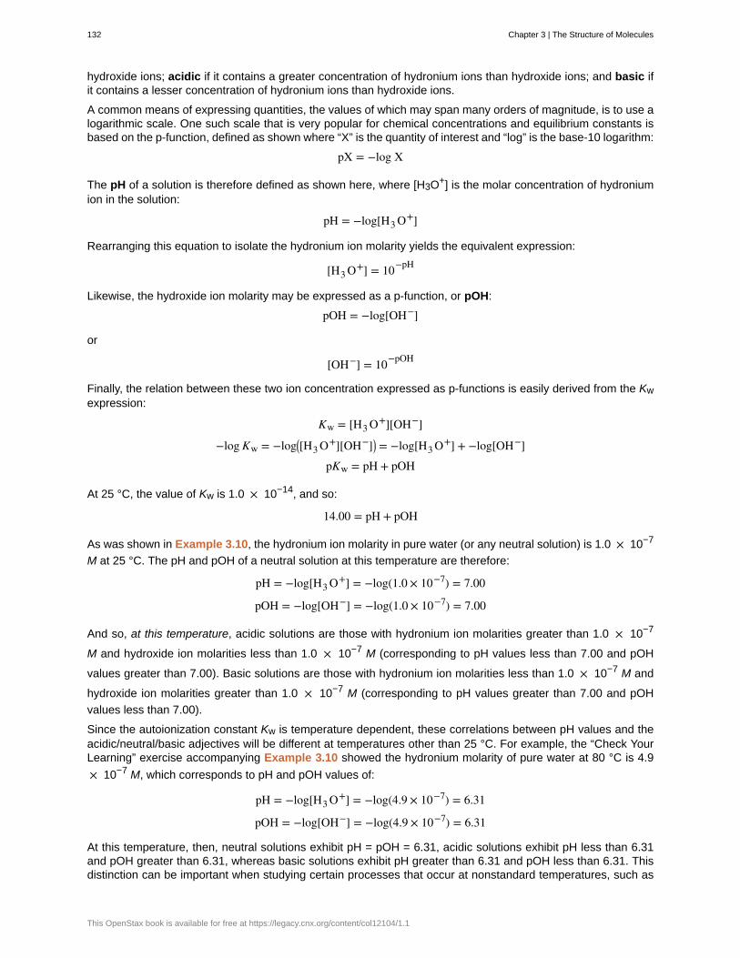
hydroxide ions; acidic if it contains a greater concentration of hydronium ions than hydroxide ions; and basic ifit contains a lesser concentration of hydronium ions than hydroxide ions.
A common means of expressing quantities, the values of which may span many orders of magnitude, is to use alogarithmic scale. One such scale that is very popular for chemical concentrations and equilibrium constants isbased on the p-function, defined as shown where “X” is the quantity of interest and “log” is the base-10 logarithm:
pX = −log X
The pH of a solution is therefore defined as shown here, where [H3O+] is the molar concentration of hydroniumion in the solution:
pH = −log[H3 O+]
Rearranging this equation to isolate the hydronium ion molarity yields the equivalent expression:
[H3 O+] = 10−pH
Likewise, the hydroxide ion molarity may be expressed as a p-function, or pOH:
pOH = −log[OH−]
or
[OH−] = 10−pOH
Finally, the relation between these two ion concentration expressed as p-functions is easily derived from the Kwexpression:
Kw = [H3 O+][OH−]
−log Kw = −log⎛⎝[H3 O+][OH−]⎞⎠ = −log[H3 O+] + −log[OH−]
pKw = pH + pOH
At 25 °C, the value of Kw is 1.0 × 10⁻¹⁴, and so:
14.00 = pH + pOH
As was shown in Example 3.10, the hydronium ion molarity in pure water (or any neutral solution) is 1.0 × 10⁻⁷
M at 25 °C. The pH and pOH of a neutral solution at this temperature are therefore:
pH = −log[H3 O+] = −log(1.0 × 10⁻⁷) = 7.00
pOH = −log[OH−] = −log(1.0 × 10⁻⁷) = 7.00
And so, at this temperature, acidic solutions are those with hydronium ion molarities greater than 1.0 × 10⁻⁷
M and hydroxide ion molarities less than 1.0 × 10⁻⁷ M (corresponding to pH values less than 7.00 and pOH
values greater than 7.00). Basic solutions are those with hydronium ion molarities less than 1.0 × 10⁻⁷ M and
hydroxide ion molarities greater than 1.0 × 10⁻⁷ M (corresponding to pH values greater than 7.00 and pOH
values less than 7.00).
Since the autoionization constant Kw is temperature dependent, these correlations between pH values and theacidic/neutral/basic adjectives will be different at temperatures other than 25 °C. For example, the “Check YourLearning” exercise accompanying Example 3.10 showed the hydronium molarity of pure water at 80 °C is 4.9
× 10−7 M, which corresponds to pH and pOH values of:
pH = −log[H3 O+] = −log(4.9 × 10⁻⁷) = 6.31
pOH = −log[OH−] = −log(4.9 × 10⁻⁷) = 6.31
At this temperature, then, neutral solutions exhibit pH = pOH = 6.31, acidic solutions exhibit pH less than 6.31and pOH greater than 6.31, whereas basic solutions exhibit pH greater than 6.31 and pOH less than 6.31. Thisdistinction can be important when studying certain processes that occur at nonstandard temperatures, such as
132 Chapter 3 | The Structure of Molecules
This OpenStax book is available for free at https://legacy.cnx.org/content/col12104/1.1
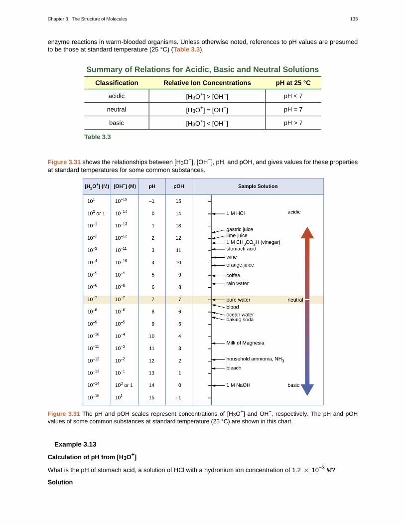
enzyme reactions in warm-blooded organisms. Unless otherwise noted, references to pH values are presumedto be those at standard temperature (25 °C) (Table 3.3).
Summary of Relations for Acidic, Basic and Neutral Solutions
Classification Relative Ion Concentrations pH at 25 °C
acidic [H3O+] > [OH−] pH < 7
neutral [H3O+] = [OH−] pH = 7
basic [H3O+] < [OH−] pH > 7
Table 3.3
Figure 3.31 shows the relationships between [H3O+], [OH−], pH, and pOH, and gives values for these propertiesat standard temperatures for some common substances.
Figure 3.31 The pH and pOH scales represent concentrations of [H3O+] and OH−, respectively. The pH and pOHvalues of some common substances at standard temperature (25 °C) are shown in this chart.
Example 3.13
Calculation of pH from [H3O+]
What is the pH of stomach acid, a solution of HCl with a hydronium ion concentration of 1.2 × 10⁻³ M?
Solution
Chapter 3 | The Structure of Molecules 133
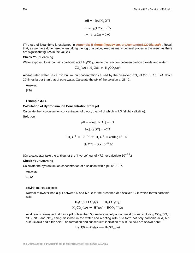
pH = −log[H3 O+]
= −log(1.2 × 10⁻³)
= −(−2.92) = 2.92
(The use of logarithms is explained in Appendix B (https://legacy.cnx.org/content/m51209/latest/) . Recallthat, as we have done here, when taking the log of a value, keep as many decimal places in the result as thereare significant figures in the value.)
Check Your Learning
Water exposed to air contains carbonic acid, H2CO3, due to the reaction between carbon dioxide and water:
CO2(aq) + H2 O(l) ⇌ H2 CO3(aq)
Air-saturated water has a hydronium ion concentration caused by the dissolved CO2 of 2.0 × 10⁻⁶ M, about
20-times larger than that of pure water. Calculate the pH of the solution at 25 °C.
Answer:
5.70
Example 3.14
Calculation of Hydronium Ion Concentration from pH
Calculate the hydronium ion concentration of blood, the pH of which is 7.3 (slightly alkaline).
Solution
pH = −log[H3 O+] = 7.3
log[H3 O+] = −7.3
[H3 O+] = 10−7.3 or [H3 O+] = antilog of −7.3
[H3 O+] = 5 × 10⁻⁸ M
(On a calculator take the antilog, or the “inverse” log, of −7.3, or calculate 10−7.3.)
Check Your Learning
Calculate the hydronium ion concentration of a solution with a pH of −1.07.
Answer:
12 M
Environmental Science
Normal rainwater has a pH between 5 and 6 due to the presence of dissolved CO2 which forms carbonicacid:
H2 O(l) + CO2(g) ⟶ H2 CO3(aq)
H2 CO3(aq) ⇌ H+(aq) + HCO3−(aq)
Acid rain is rainwater that has a pH of less than 5, due to a variety of nonmetal oxides, including CO2, SO2,SO3, NO, and NO2 being dissolved in the water and reacting with it to form not only carbonic acid, butsulfuric acid and nitric acid. The formation and subsequent ionization of sulfuric acid are shown here:
H2 O(l) + SO3(g) ⟶ H2 SO4(aq)
134 Chapter 3 | The Structure of Molecules
This OpenStax book is available for free at https://legacy.cnx.org/content/col12104/1.1
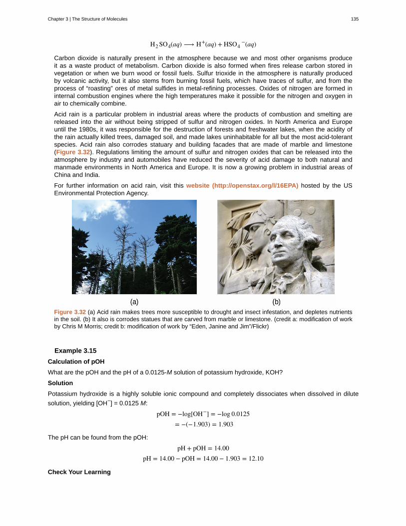
H2 SO4(aq) ⟶ H+(aq) + HSO4−(aq)
Carbon dioxide is naturally present in the atmosphere because we and most other organisms produceit as a waste product of metabolism. Carbon dioxide is also formed when fires release carbon stored invegetation or when we burn wood or fossil fuels. Sulfur trioxide in the atmosphere is naturally producedby volcanic activity, but it also stems from burning fossil fuels, which have traces of sulfur, and from theprocess of “roasting” ores of metal sulfides in metal-refining processes. Oxides of nitrogen are formed ininternal combustion engines where the high temperatures make it possible for the nitrogen and oxygen inair to chemically combine.
Acid rain is a particular problem in industrial areas where the products of combustion and smelting arereleased into the air without being stripped of sulfur and nitrogen oxides. In North America and Europeuntil the 1980s, it was responsible for the destruction of forests and freshwater lakes, when the acidity ofthe rain actually killed trees, damaged soil, and made lakes uninhabitable for all but the most acid-tolerantspecies. Acid rain also corrodes statuary and building facades that are made of marble and limestone(Figure 3.32). Regulations limiting the amount of sulfur and nitrogen oxides that can be released into theatmosphere by industry and automobiles have reduced the severity of acid damage to both natural andmanmade environments in North America and Europe. It is now a growing problem in industrial areas ofChina and India.
For further information on acid rain, visit this website (http://openstax.org/l/16EPA) hosted by the USEnvironmental Protection Agency.
Figure 3.32 (a) Acid rain makes trees more susceptible to drought and insect infestation, and depletes nutrientsin the soil. (b) It also is corrodes statues that are carved from marble or limestone. (credit a: modification of workby Chris M Morris; credit b: modification of work by “Eden, Janine and Jim”/Flickr)
Example 3.15
Calculation of pOH
What are the pOH and the pH of a 0.0125-M solution of potassium hydroxide, KOH?
Solution
Potassium hydroxide is a highly soluble ionic compound and completely dissociates when dissolved in dilute
solution, yielding [OH−] = 0.0125 M:
pOH = −log[OH−] = −log 0.0125= −(−1.903) = 1.903
The pH can be found from the pOH:
pH + pOH = 14.00pH = 14.00 − pOH = 14.00 − 1.903 = 12.10
Check Your Learning
Chapter 3 | The Structure of Molecules 135
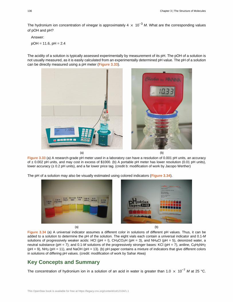
The hydronium ion concentration of vinegar is approximately 4 × 10⁻³ M. What are the corresponding values
of pOH and pH?
Answer:
pOH = 11.6, pH = 2.4
The acidity of a solution is typically assessed experimentally by measurement of its pH. The pOH of a solution isnot usually measured, as it is easily calculated from an experimentally determined pH value. The pH of a solutioncan be directly measured using a pH meter (Figure 3.33).
Figure 3.33 (a) A research-grade pH meter used in a laboratory can have a resolution of 0.001 pH units, an accuracyof ± 0.002 pH units, and may cost in excess of $1000. (b) A portable pH meter has lower resolution (0.01 pH units),lower accuracy (± 0.2 pH units), and a far lower price tag. (credit b: modification of work by Jacopo Werther)
The pH of a solution may also be visually estimated using colored indicators (Figure 3.34).
Figure 3.34 (a) A universal indicator assumes a different color in solutions of different pH values. Thus, it can beadded to a solution to determine the pH of the solution. The eight vials each contain a universal indicator and 0.1-Msolutions of progressively weaker acids: HCl (pH = l), CH3CO2H (pH = 3), and NH4Cl (pH = 5), deionized water, aneutral substance (pH = 7); and 0.1-M solutions of the progressively stronger bases: KCl (pH = 7), aniline, C6H5NH2(pH = 9), NH3 (pH = 11), and NaOH (pH = 13). (b) pH paper contains a mixture of indicators that give different colorsin solutions of differing pH values. (credit: modification of work by Sahar Atwa)
Key Concepts and Summary
The concentration of hydronium ion in a solution of an acid in water is greater than 1.0 × 10⁻⁷ M at 25 °C.
136 Chapter 3 | The Structure of Molecules
This OpenStax book is available for free at https://legacy.cnx.org/content/col12104/1.1
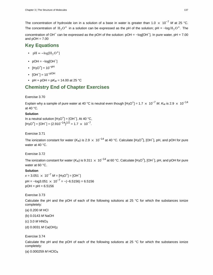
The concentration of hydroxide ion in a solution of a base in water is greater than 1.0 × 10⁻⁷ M at 25 °C.
The concentration of H3 O+ in a solution can be expressed as the pH of the solution; pH = −log H3 O+. The
concentration of OH− can be expressed as the pOH of the solution: pOH = −log[OH−]. In pure water, pH = 7.00and pOH = 7.00
Key Equations
• pH = −log[H3 O+]
• pOH = −log[OH−]
• [H3O+] = 10−pH
• [OH−] = 10−pOH
• pH + pOH = pKw = 14.00 at 25 °C
Chemistry End of Chapter Exercises
Exercise 3.70
Explain why a sample of pure water at 40 °C is neutral even though [H3O+] = 1.7 × 10⁻⁷ M. Kw is 2.9 × 10⁻¹⁴
at 40 °C.
Solution
In a neutral solution [H3O+] = [OH−]. At 40 °C,
[H3O+] = [OH−] = (2.910−14)1/2 = 1.7 × 10⁻⁷.
Exercise 3.71
The ionization constant for water (Kw) is 2.9 × 10⁻¹⁴ at 40 °C. Calculate [H3O+], [OH−], pH, and pOH for pure
water at 40 °C.
Exercise 3.72
The ionization constant for water (Kw) is 9.311 × 10⁻¹⁴ at 60 °C. Calculate [H3O+], [OH−], pH, and pOH for pure
water at 60 °C.
Solution
x = 3.051 × 10⁻⁷ M = [H3O+] = [OH−]
pH = −log3.051 × 10−7 = −(−6.5156) = 6.5156
pOH = pH = 6.5156
Exercise 3.73
Calculate the pH and the pOH of each of the following solutions at 25 °C for which the substances ionizecompletely:
(a) 0.200 M HCl
(b) 0.0143 M NaOH
(c) 3.0 M HNO3
(d) 0.0031 M Ca(OH)2
Exercise 3.74
Calculate the pH and the pOH of each of the following solutions at 25 °C for which the substances ionizecompletely:
(a) 0.000259 M HClO4
Chapter 3 | The Structure of Molecules 137
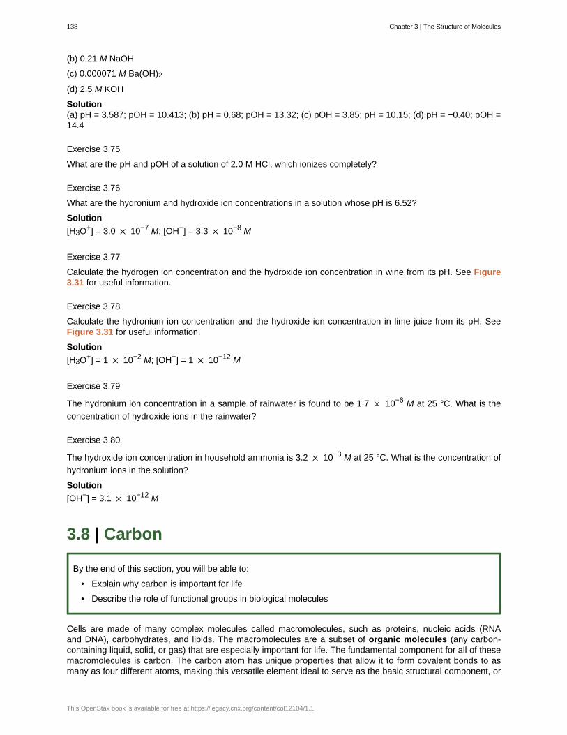
(b) 0.21 M NaOH
(c) 0.000071 M Ba(OH)2
(d) 2.5 M KOH
Solution(a) pH = 3.587; pOH = 10.413; (b) pH = 0.68; pOH = 13.32; (c) pOH = 3.85; pH = 10.15; (d) pH = −0.40; pOH =14.4
Exercise 3.75
What are the pH and pOH of a solution of 2.0 M HCl, which ionizes completely?
Exercise 3.76
What are the hydronium and hydroxide ion concentrations in a solution whose pH is 6.52?
Solution
[H3O+] = 3.0 × 10⁻⁷ M; [OH−] = 3.3 × 10⁻⁸ M
Exercise 3.77
Calculate the hydrogen ion concentration and the hydroxide ion concentration in wine from its pH. See Figure3.31 for useful information.
Exercise 3.78
Calculate the hydronium ion concentration and the hydroxide ion concentration in lime juice from its pH. SeeFigure 3.31 for useful information.
Solution
[H3O+] = 1 × 10⁻² M; [OH−] = 1 × 10⁻¹² M
Exercise 3.79
The hydronium ion concentration in a sample of rainwater is found to be 1.7 × 10⁻⁶ M at 25 °C. What is the
concentration of hydroxide ions in the rainwater?
Exercise 3.80
The hydroxide ion concentration in household ammonia is 3.2 × 10⁻³ M at 25 °C. What is the concentration of
hydronium ions in the solution?
Solution
[OH−] = 3.1 × 10⁻¹² M
3.8 | Carbon
By the end of this section, you will be able to:
• Explain why carbon is important for life
• Describe the role of functional groups in biological molecules
Cells are made of many complex molecules called macromolecules, such as proteins, nucleic acids (RNAand DNA), carbohydrates, and lipids. The macromolecules are a subset of organic molecules (any carbon-containing liquid, solid, or gas) that are especially important for life. The fundamental component for all of thesemacromolecules is carbon. The carbon atom has unique properties that allow it to form covalent bonds to asmany as four different atoms, making this versatile element ideal to serve as the basic structural component, or
138 Chapter 3 | The Structure of Molecules
This OpenStax book is available for free at https://legacy.cnx.org/content/col12104/1.1
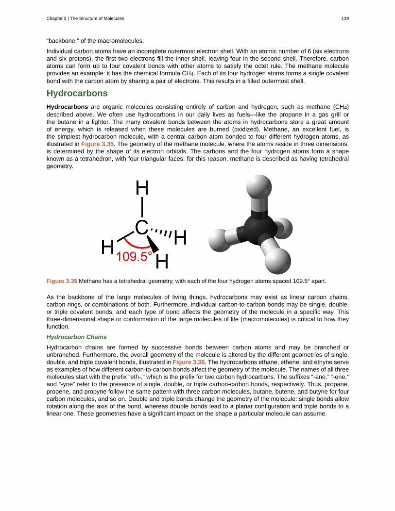
“backbone,” of the macromolecules.
Individual carbon atoms have an incomplete outermost electron shell. With an atomic number of 6 (six electronsand six protons), the first two electrons fill the inner shell, leaving four in the second shell. Therefore, carbonatoms can form up to four covalent bonds with other atoms to satisfy the octet rule. The methane moleculeprovides an example: it has the chemical formula CH4. Each of its four hydrogen atoms forms a single covalentbond with the carbon atom by sharing a pair of electrons. This results in a filled outermost shell.
Hydrocarbons
Hydrocarbons are organic molecules consisting entirely of carbon and hydrogen, such as methane (CH4)described above. We often use hydrocarbons in our daily lives as fuels—like the propane in a gas grill orthe butane in a lighter. The many covalent bonds between the atoms in hydrocarbons store a great amountof energy, which is released when these molecules are burned (oxidized). Methane, an excellent fuel, isthe simplest hydrocarbon molecule, with a central carbon atom bonded to four different hydrogen atoms, asillustrated in Figure 3.35. The geometry of the methane molecule, where the atoms reside in three dimensions,is determined by the shape of its electron orbitals. The carbons and the four hydrogen atoms form a shapeknown as a tetrahedron, with four triangular faces; for this reason, methane is described as having tetrahedralgeometry.
Figure 3.35 Methane has a tetrahedral geometry, with each of the four hydrogen atoms spaced 109.5° apart.
As the backbone of the large molecules of living things, hydrocarbons may exist as linear carbon chains,carbon rings, or combinations of both. Furthermore, individual carbon-to-carbon bonds may be single, double,or triple covalent bonds, and each type of bond affects the geometry of the molecule in a specific way. Thisthree-dimensional shape or conformation of the large molecules of life (macromolecules) is critical to how theyfunction.
Hydrocarbon Chains
Hydrocarbon chains are formed by successive bonds between carbon atoms and may be branched orunbranched. Furthermore, the overall geometry of the molecule is altered by the different geometries of single,double, and triple covalent bonds, illustrated in Figure 3.36. The hydrocarbons ethane, ethene, and ethyne serveas examples of how different carbon-to-carbon bonds affect the geometry of the molecule. The names of all threemolecules start with the prefix “eth-,” which is the prefix for two carbon hydrocarbons. The suffixes “-ane,” “-ene,”and “-yne” refer to the presence of single, double, or triple carbon-carbon bonds, respectively. Thus, propane,propene, and propyne follow the same pattern with three carbon molecules, butane, butene, and butyne for fourcarbon molecules, and so on. Double and triple bonds change the geometry of the molecule: single bonds allowrotation along the axis of the bond, whereas double bonds lead to a planar configuration and triple bonds to alinear one. These geometries have a significant impact on the shape a particular molecule can assume.
Chapter 3 | The Structure of Molecules 139
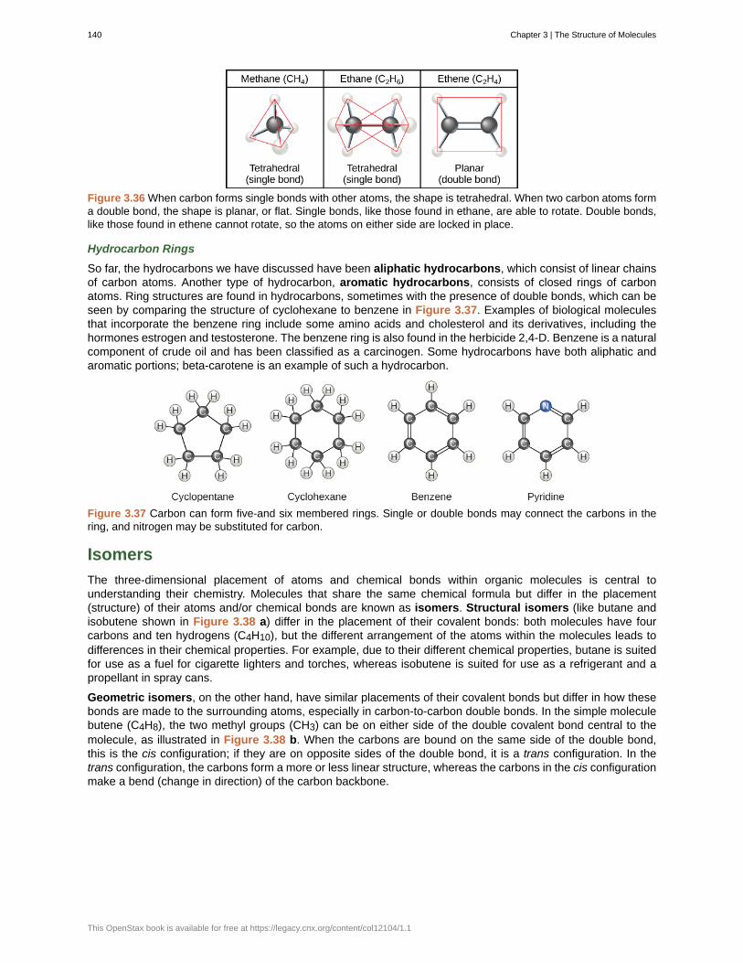
Figure 3.36 When carbon forms single bonds with other atoms, the shape is tetrahedral. When two carbon atoms forma double bond, the shape is planar, or flat. Single bonds, like those found in ethane, are able to rotate. Double bonds,like those found in ethene cannot rotate, so the atoms on either side are locked in place.
Hydrocarbon Rings
So far, the hydrocarbons we have discussed have been aliphatic hydrocarbons, which consist of linear chainsof carbon atoms. Another type of hydrocarbon, aromatic hydrocarbons, consists of closed rings of carbonatoms. Ring structures are found in hydrocarbons, sometimes with the presence of double bonds, which can beseen by comparing the structure of cyclohexane to benzene in Figure 3.37. Examples of biological moleculesthat incorporate the benzene ring include some amino acids and cholesterol and its derivatives, including thehormones estrogen and testosterone. The benzene ring is also found in the herbicide 2,4-D. Benzene is a naturalcomponent of crude oil and has been classified as a carcinogen. Some hydrocarbons have both aliphatic andaromatic portions; beta-carotene is an example of such a hydrocarbon.
Figure 3.37 Carbon can form five-and six membered rings. Single or double bonds may connect the carbons in thering, and nitrogen may be substituted for carbon.
Isomers
The three-dimensional placement of atoms and chemical bonds within organic molecules is central tounderstanding their chemistry. Molecules that share the same chemical formula but differ in the placement(structure) of their atoms and/or chemical bonds are known as isomers. Structural isomers (like butane andisobutene shown in Figure 3.38 a) differ in the placement of their covalent bonds: both molecules have fourcarbons and ten hydrogens (C4H10), but the different arrangement of the atoms within the molecules leads todifferences in their chemical properties. For example, due to their different chemical properties, butane is suitedfor use as a fuel for cigarette lighters and torches, whereas isobutene is suited for use as a refrigerant and apropellant in spray cans.
Geometric isomers, on the other hand, have similar placements of their covalent bonds but differ in how thesebonds are made to the surrounding atoms, especially in carbon-to-carbon double bonds. In the simple moleculebutene (C4H8), the two methyl groups (CH3) can be on either side of the double covalent bond central to themolecule, as illustrated in Figure 3.38 b. When the carbons are bound on the same side of the double bond,this is the cis configuration; if they are on opposite sides of the double bond, it is a trans configuration. In thetrans configuration, the carbons form a more or less linear structure, whereas the carbons in the cis configurationmake a bend (change in direction) of the carbon backbone.
140 Chapter 3 | The Structure of Molecules
This OpenStax book is available for free at https://legacy.cnx.org/content/col12104/1.1
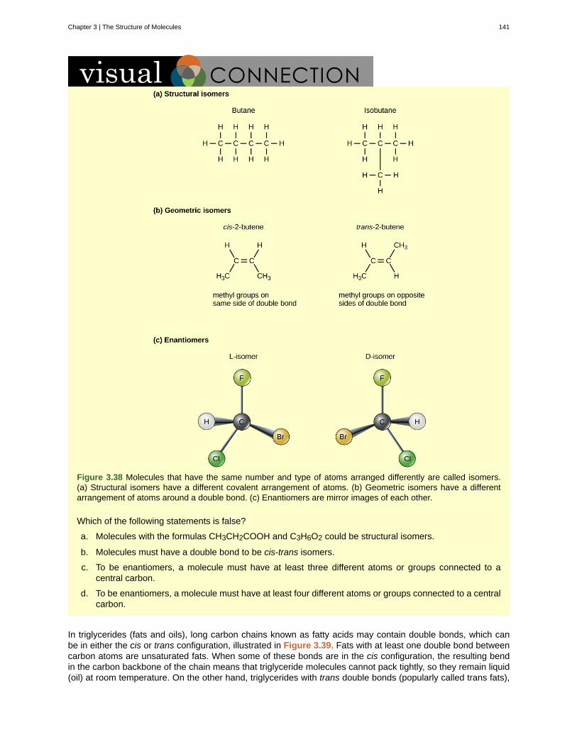
Figure 3.38 Molecules that have the same number and type of atoms arranged differently are called isomers.(a) Structural isomers have a different covalent arrangement of atoms. (b) Geometric isomers have a differentarrangement of atoms around a double bond. (c) Enantiomers are mirror images of each other.
Which of the following statements is false?
a. Molecules with the formulas CH3CH2COOH and C3H6O2 could be structural isomers.
b. Molecules must have a double bond to be cis-trans isomers.
c. To be enantiomers, a molecule must have at least three different atoms or groups connected to acentral carbon.
d. To be enantiomers, a molecule must have at least four different atoms or groups connected to a centralcarbon.
In triglycerides (fats and oils), long carbon chains known as fatty acids may contain double bonds, which canbe in either the cis or trans configuration, illustrated in Figure 3.39. Fats with at least one double bond betweencarbon atoms are unsaturated fats. When some of these bonds are in the cis configuration, the resulting bendin the carbon backbone of the chain means that triglyceride molecules cannot pack tightly, so they remain liquid(oil) at room temperature. On the other hand, triglycerides with trans double bonds (popularly called trans fats),
Chapter 3 | The Structure of Molecules 141
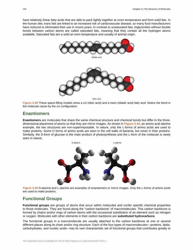
have relatively linear fatty acids that are able to pack tightly together at room temperature and form solid fats. Inthe human diet, trans fats are linked to an increased risk of cardiovascular disease, so many food manufacturershave reduced or eliminated their use in recent years. In contrast to unsaturated fats, triglycerides without doublebonds between carbon atoms are called saturated fats, meaning that they contain all the hydrogen atomsavailable. Saturated fats are a solid at room temperature and usually of animal origin.
Figure 3.39 These space-filling models show a cis (oleic acid) and a trans (eliadic acid) fatty acid. Notice the bend inthe molecule cause by the cis configuration.
Enantiomers
Enantiomers are molecules that share the same chemical structure and chemical bonds but differ in the three-dimensional placement of atoms so that they are mirror images. As shown in Figure 3.40, an amino acid alanineexample, the two structures are non-superimposable. In nature, only the L-forms of amino acids are used tomake proteins. Some D forms of amino acids are seen in the cell walls of bacteria, but never in their proteins.Similarly, the D-form of glucose is the main product of photosynthesis and the L-form of the molecule is rarelyseen in nature.
Figure 3.40 D-alanine and L-alanine are examples of enantiomers or mirror images. Only the L-forms of amino acidsare used to make proteins.
Functional Groups
Functional groups are groups of atoms that occur within molecules and confer specific chemical propertiesto those molecules. They are found along the “carbon backbone” of macromolecules. This carbon backbone isformed by chains and/or rings of carbon atoms with the occasional substitution of an element such as nitrogenor oxygen. Molecules with other elements in their carbon backbone are substituted hydrocarbons.
The functional groups in a macromolecule are usually attached to the carbon backbone at one or severaldifferent places along its chain and/or ring structure. Each of the four types of macromolecules—proteins, lipids,carbohydrates, and nucleic acids—has its own characteristic set of functional groups that contributes greatly to
142 Chapter 3 | The Structure of Molecules
This OpenStax book is available for free at https://legacy.cnx.org/content/col12104/1.1

its differing chemical properties and its function in living organisms.
A functional group can participate in specific chemical reactions. Some of the important functional groupsin biological molecules are shown in Figure 3.41; they include: hydroxyl, methyl, carbonyl, carboxyl, amino,phosphate, and sulfhydryl. These groups play an important role in the formation of molecules like DNA, proteins,carbohydrates, and lipids. Functional groups are usually classified as hydrophobic or hydrophilic depending ontheir charge or polarity characteristics. An example of a hydrophobic group is the non-polar methane molecule.Among the hydrophilic functional groups is the carboxyl group found in amino acids, some amino acid sidechains, and the fatty acids that form triglycerides and phospholipids. This carboxyl group ionizes to release
hydrogen ions (H+) from the COOH group resulting in the negatively charged COO- group; this contributes to thehydrophilic nature of whatever molecule it is found on. Other functional groups, such as the carbonyl group, havea partially negatively charged oxygen atom that may form hydrogen bonds with water molecules, again makingthe molecule more hydrophilic.
Figure 3.41 The functional groups shown here are found in many different biological molecules.
Hydrogen bonds between functional groups (within the same molecule or between different molecules) areimportant to the function of many macromolecules and help them to fold properly into and maintain theappropriate shape for functioning. Hydrogen bonds are also involved in various recognition processes, such asDNA complementary base pairing and the binding of an enzyme to its substrate, as illustrated in Figure 3.42.
Chapter 3 | The Structure of Molecules 143
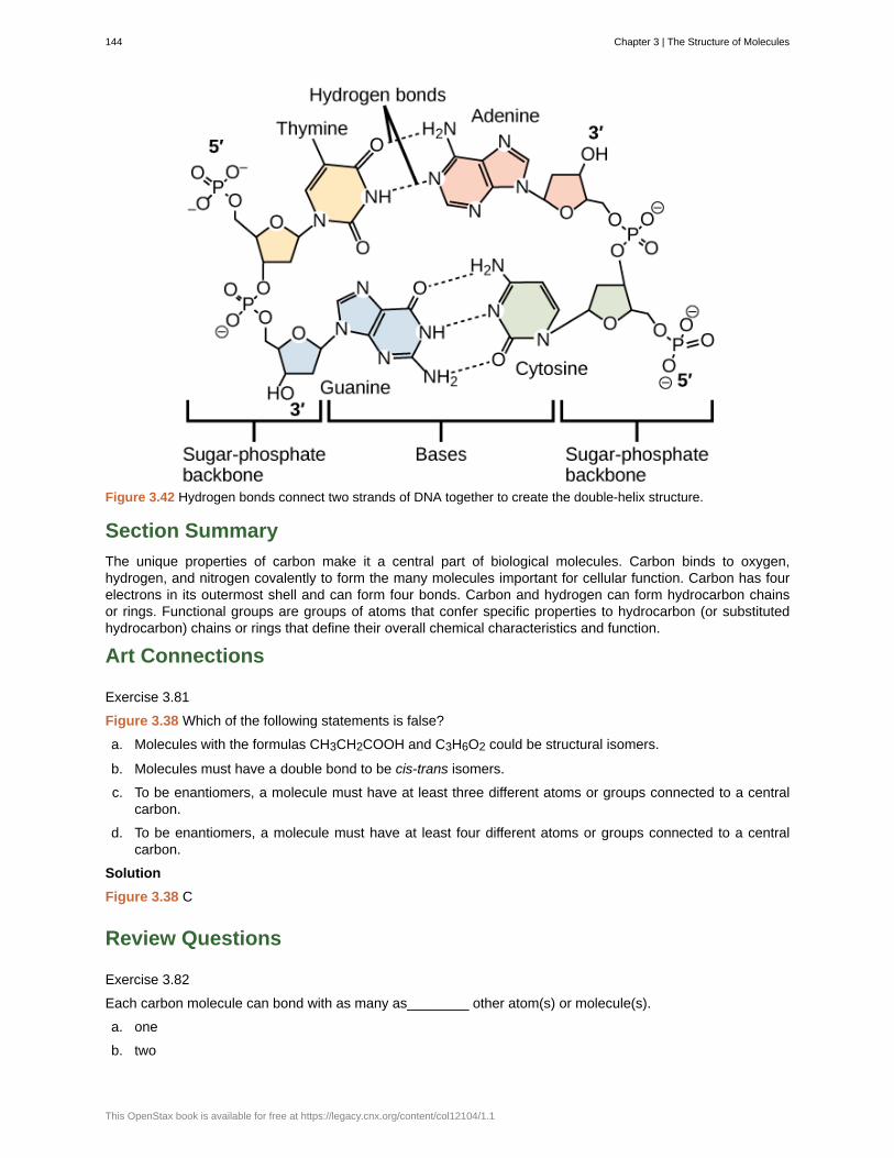
Figure 3.42 Hydrogen bonds connect two strands of DNA together to create the double-helix structure.
Section Summary
The unique properties of carbon make it a central part of biological molecules. Carbon binds to oxygen,hydrogen, and nitrogen covalently to form the many molecules important for cellular function. Carbon has fourelectrons in its outermost shell and can form four bonds. Carbon and hydrogen can form hydrocarbon chainsor rings. Functional groups are groups of atoms that confer specific properties to hydrocarbon (or substitutedhydrocarbon) chains or rings that define their overall chemical characteristics and function.
Art Connections
Exercise 3.81
Figure 3.38 Which of the following statements is false?
a. Molecules with the formulas CH3CH2COOH and C3H6O2 could be structural isomers.
b. Molecules must have a double bond to be cis-trans isomers.
c. To be enantiomers, a molecule must have at least three different atoms or groups connected to a centralcarbon.
d. To be enantiomers, a molecule must have at least four different atoms or groups connected to a centralcarbon.
Solution
Figure 3.38 C
Review Questions
Exercise 3.82
Each carbon molecule can bond with as many as________ other atom(s) or molecule(s).
a. one
b. two
144 Chapter 3 | The Structure of Molecules
This OpenStax book is available for free at https://legacy.cnx.org/content/col12104/1.1
c. six
d. four
Solution
D
Exercise 3.83
Which of the following is not a functional group that can bond with carbon?
a. sodium
b. hydroxyl
c. phosphate
d. carbonyl
Solution
A
Free Response
Exercise 3.84
What property of carbon makes it essential for organic life?
Solution
Carbon is unique and found in all living things because it can form up to four covalent bonds between atoms ormolecules. These can be nonpolar or polar covalent bonds, and they allow for the formation of long chains ofcarbon molecules that combine to form proteins and DNA.
Exercise 3.85
Compare and contrast saturated and unsaturated triglycerides.
Solution
Saturated triglycerides contain no double bonds between carbon atoms; they are usually solid at roomtemperature. Unsaturated triglycerides contain at least one double bond between carbon atoms and are usuallyliquid at room temperature.
3.9 | Hydrocarbons
By the end of this section, you will be able to:
• Explain the importance of hydrocarbons and the reason for their diversity
• Name saturated and unsaturated hydrocarbons, and molecules derived from them
• Describe the reactions characteristic of saturated and unsaturated hydrocarbons
• Identify structural and geometric isomers of hydrocarbons
The largest database[4] of organic compounds lists about 10 million substances, which include compoundsoriginating from living organisms and those synthesized by chemists. The number of potential organic
compounds has been estimated[5] at 1060—an astronomically high number. The existence of so many organicmolecules is a consequence of the ability of carbon atoms to form up to four strong bonds to other carbon atoms,resulting in chains and rings of many different sizes, shapes, and complexities.
4. This is the Beilstein database, now available through the Reaxys site (www.elsevier.com/online-tools/reaxys).5. Peplow, Mark. “Organic Synthesis: The Robo-Chemist,” Nature 512 (2014): 20–2.
Chapter 3 | The Structure of Molecules 145
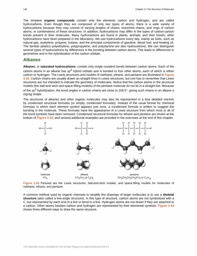
The simplest organic compounds contain only the elements carbon and hydrogen, and are calledhydrocarbons. Even though they are composed of only two types of atoms, there is a wide variety ofhydrocarbons because they may consist of varying lengths of chains, branched chains, and rings of carbonatoms, or combinations of these structures. In addition, hydrocarbons may differ in the types of carbon-carbonbonds present in their molecules. Many hydrocarbons are found in plants, animals, and their fossils; otherhydrocarbons have been prepared in the laboratory. We use hydrocarbons every day, mainly as fuels, such asnatural gas, acetylene, propane, butane, and the principal components of gasoline, diesel fuel, and heating oil.The familiar plastics polyethylene, polypropylene, and polystyrene are also hydrocarbons. We can distinguishseveral types of hydrocarbons by differences in the bonding between carbon atoms. This leads to differences ingeometries and in the hybridization of the carbon orbitals.
Alkanes
Alkanes, or saturated hydrocarbons, contain only single covalent bonds between carbon atoms. Each of the
carbon atoms in an alkane has sp3 hybrid orbitals and is bonded to four other atoms, each of which is eithercarbon or hydrogen. The Lewis structures and models of methane, ethane, and pentane are illustrated in Figure3.43. Carbon chains are usually drawn as straight lines in Lewis structures, but one has to remember that Lewisstructures are not intended to indicate the geometry of molecules. Notice that the carbon atoms in the structuralmodels (the ball-and-stick and space-filling models) of the pentane molecule do not lie in a straight line. Because
of the sp3 hybridization, the bond angles in carbon chains are close to 109.5°, giving such chains in an alkane azigzag shape.
The structures of alkanes and other organic molecules may also be represented in a less detailed mannerby condensed structural formulas (or simply, condensed formulas). Instead of the usual format for chemicalformulas in which each element symbol appears just once, a condensed formula is written to suggest thebonding in the molecule. These formulas have the appearance of a Lewis structure from which most or all ofthe bond symbols have been removed. Condensed structural formulas for ethane and pentane are shown at thebottom of Figure 3.43, and several additional examples are provided in the exercises at the end of this chapter.
Figure 3.43 Pictured are the Lewis structures, ball-and-stick models, and space-filling models for molecules ofmethane, ethane, and pentane.
A common method used by organic chemists to simplify the drawings of larger molecules is to use a skeletalstructure (also called a line-angle structure). In this type of structure, carbon atoms are not symbolized with aC, but represented by each end of a line or bend in a line. Hydrogen atoms are not drawn if they are attached toa carbon. Other atoms besides carbon and hydrogen are represented by their elemental symbols. Figure 3.44shows three different ways to draw the same structure.
146 Chapter 3 | The Structure of Molecules
This OpenStax book is available for free at https://legacy.cnx.org/content/col12104/1.1
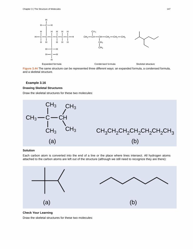
Figure 3.44 The same structure can be represented three different ways: an expanded formula, a condensed formula,and a skeletal structure.
Example 3.16
Drawing Skeletal Structures
Draw the skeletal structures for these two molecules:
Solution
Each carbon atom is converted into the end of a line or the place where lines intersect. All hydrogen atomsattached to the carbon atoms are left out of the structure (although we still need to recognize they are there):
Check Your Learning
Draw the skeletal structures for these two molecules:
Chapter 3 | The Structure of Molecules 147
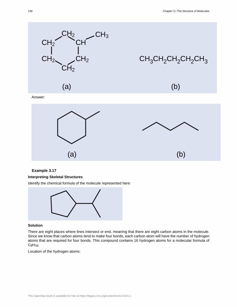
Answer:
Example 3.17
Interpreting Skeletal Structures
Identify the chemical formula of the molecule represented here:
Solution
There are eight places where lines intersect or end, meaning that there are eight carbon atoms in the molecule.Since we know that carbon atoms tend to make four bonds, each carbon atom will have the number of hydrogenatoms that are required for four bonds. This compound contains 16 hydrogen atoms for a molecular formula ofC8H16.
Location of the hydrogen atoms:
148 Chapter 3 | The Structure of Molecules
This OpenStax book is available for free at https://legacy.cnx.org/content/col12104/1.1
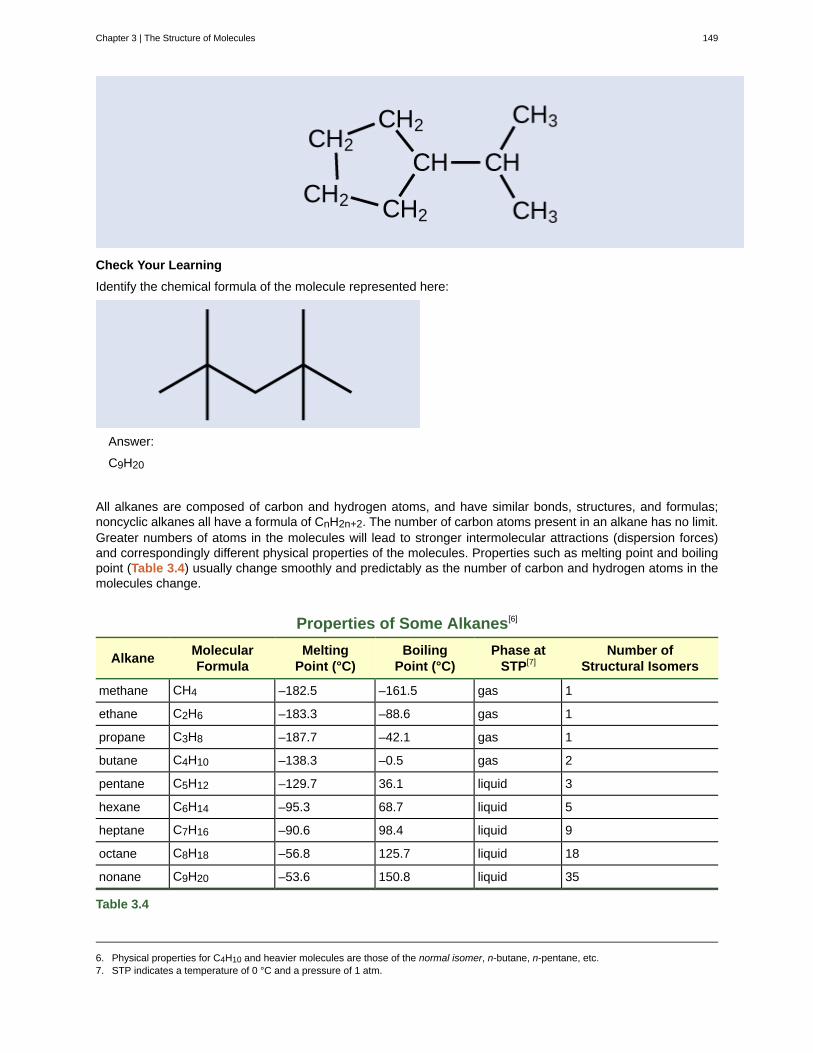
Check Your Learning
Identify the chemical formula of the molecule represented here:
Answer:
C9H20
All alkanes are composed of carbon and hydrogen atoms, and have similar bonds, structures, and formulas;noncyclic alkanes all have a formula of CnH2n+2. The number of carbon atoms present in an alkane has no limit.Greater numbers of atoms in the molecules will lead to stronger intermolecular attractions (dispersion forces)and correspondingly different physical properties of the molecules. Properties such as melting point and boilingpoint (Table 3.4) usually change smoothly and predictably as the number of carbon and hydrogen atoms in themolecules change.
Properties of Some Alkanes[6]
AlkaneMolecularFormula
MeltingPoint (°C)
BoilingPoint (°C)
Phase atSTP[7]
Number ofStructural Isomers
methane CH4 –182.5 –161.5 gas 1
ethane C2H6 –183.3 –88.6 gas 1
propane C3H8 –187.7 –42.1 gas 1
butane C4H10 –138.3 –0.5 gas 2
pentane C5H12 –129.7 36.1 liquid 3
hexane C6H14 –95.3 68.7 liquid 5
heptane C7H16 –90.6 98.4 liquid 9
octane C8H18 –56.8 125.7 liquid 18
nonane C9H20 –53.6 150.8 liquid 35
Table 3.4
6. Physical properties for C4H10 and heavier molecules are those of the normal isomer, n-butane, n-pentane, etc.7. STP indicates a temperature of 0 °C and a pressure of 1 atm.
Chapter 3 | The Structure of Molecules 149
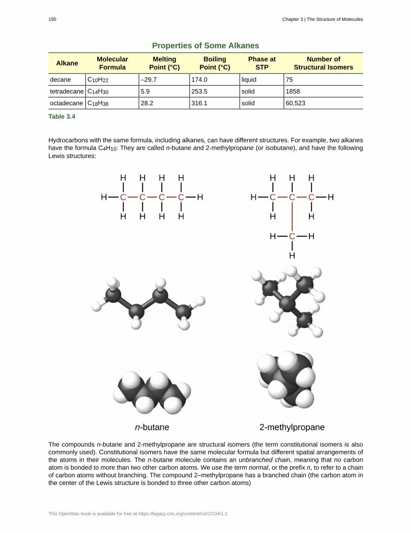
Properties of Some Alkanes
AlkaneMolecularFormula
MeltingPoint (°C)
BoilingPoint (°C)
Phase atSTP
Number ofStructural Isomers
decane C10H22 –29.7 174.0 liquid 75
tetradecane C14H30 5.9 253.5 solid 1858
octadecane C18H38 28.2 316.1 solid 60,523
Table 3.4
Hydrocarbons with the same formula, including alkanes, can have different structures. For example, two alkaneshave the formula C4H10: They are called n-butane and 2-methylpropane (or isobutane), and have the followingLewis structures:
The compounds n-butane and 2-methylpropane are structural isomers (the term constitutional isomers is alsocommonly used). Constitutional isomers have the same molecular formula but different spatial arrangements ofthe atoms in their molecules. The n-butane molecule contains an unbranched chain, meaning that no carbonatom is bonded to more than two other carbon atoms. We use the term normal, or the prefix n, to refer to a chainof carbon atoms without branching. The compound 2–methylpropane has a branched chain (the carbon atom inthe center of the Lewis structure is bonded to three other carbon atoms)
150 Chapter 3 | The Structure of Molecules
This OpenStax book is available for free at https://legacy.cnx.org/content/col12104/1.1
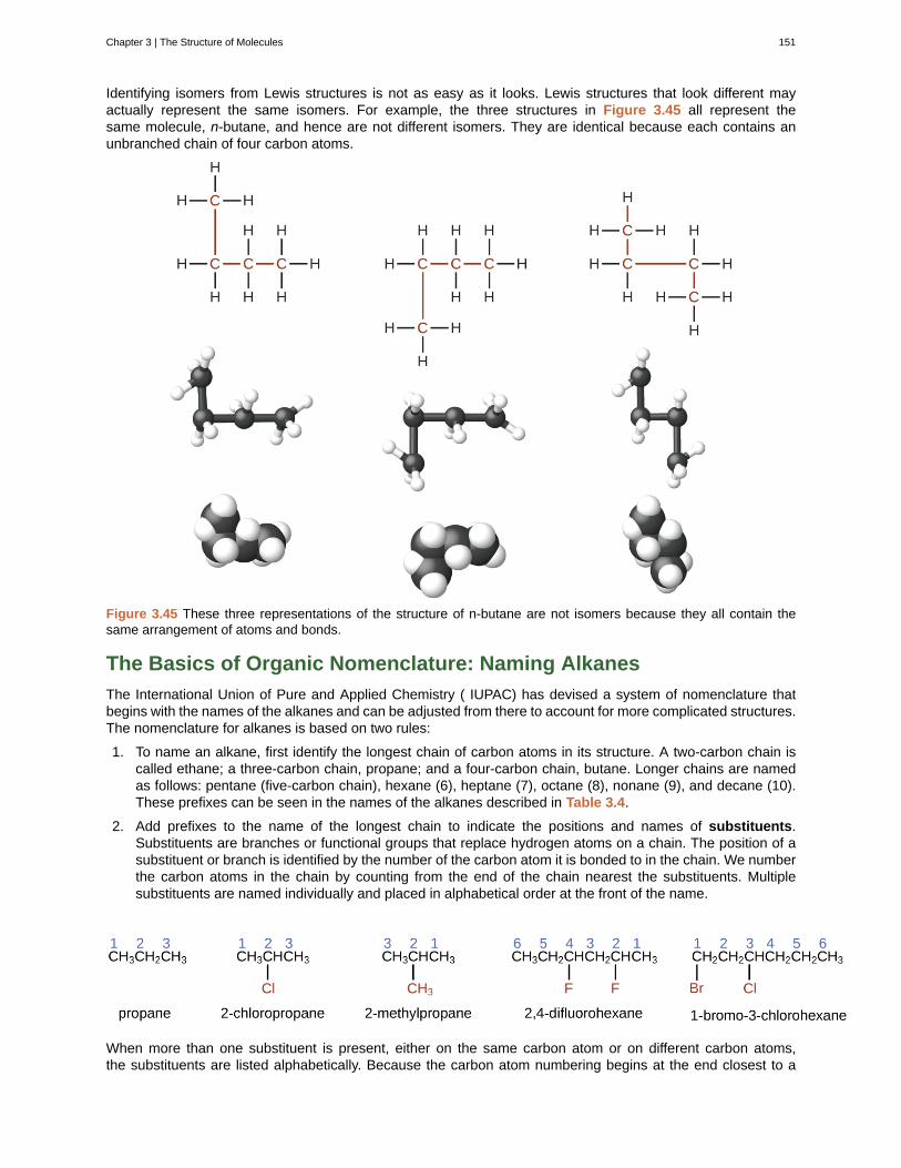
Identifying isomers from Lewis structures is not as easy as it looks. Lewis structures that look different mayactually represent the same isomers. For example, the three structures in Figure 3.45 all represent thesame molecule, n-butane, and hence are not different isomers. They are identical because each contains anunbranched chain of four carbon atoms.
Figure 3.45 These three representations of the structure of n-butane are not isomers because they all contain thesame arrangement of atoms and bonds.
The Basics of Organic Nomenclature: Naming Alkanes
The International Union of Pure and Applied Chemistry ( IUPAC) has devised a system of nomenclature thatbegins with the names of the alkanes and can be adjusted from there to account for more complicated structures.The nomenclature for alkanes is based on two rules:
1. To name an alkane, first identify the longest chain of carbon atoms in its structure. A two-carbon chain iscalled ethane; a three-carbon chain, propane; and a four-carbon chain, butane. Longer chains are namedas follows: pentane (five-carbon chain), hexane (6), heptane (7), octane (8), nonane (9), and decane (10).These prefixes can be seen in the names of the alkanes described in Table 3.4.
2. Add prefixes to the name of the longest chain to indicate the positions and names of substituents.Substituents are branches or functional groups that replace hydrogen atoms on a chain. The position of asubstituent or branch is identified by the number of the carbon atom it is bonded to in the chain. We numberthe carbon atoms in the chain by counting from the end of the chain nearest the substituents. Multiplesubstituents are named individually and placed in alphabetical order at the front of the name.
When more than one substituent is present, either on the same carbon atom or on different carbon atoms,the substituents are listed alphabetically. Because the carbon atom numbering begins at the end closest to a
Chapter 3 | The Structure of Molecules 151

substituent, the longest chain of carbon atoms is numbered in such a way as to produce the lowest numberfor the substituents. The ending -o replaces -ide at the end of the name of an electronegative substituent(in ionic compounds, the negatively charged ion ends with -ide like chloride; in organic compounds, suchatoms are treated as substituents and the -o ending is used). The number of substituents of the same type isindicated by the prefixes di- (two), tri- (three), tetra- (four), and so on (for example, difluoro- indicates two fluoridesubstituents).
Example 3.18
Naming Halogen-substituted Alkanes
Name the molecule whose structure is shown here:
Solution
The four-carbon chain is numbered from the end with the chlorine atom. This puts the substituents on positions1 and 2 (numbering from the other end would put the substituents on positions 3 and 4). Four carbon atomsmeans that the base name of this compound will be butane. The bromine at position 2 will be described byadding 2-bromo-; this will come at the beginning of the name, since bromo- comes before chloro- alphabetically.The chlorine at position 1 will be described by adding 1-chloro-, resulting in the name of the molecule being2-bromo-1-chlorobutane.
Check Your Learning
Name the following molecule:
Answer:
3,3-dibromo-2-iodopentane
We call a substituent that contains one less hydrogen than the corresponding alkane an alkyl group. The nameof an alkyl group is obtained by dropping the suffix -ane of the alkane name and adding -yl:
152 Chapter 3 | The Structure of Molecules
This OpenStax book is available for free at https://legacy.cnx.org/content/col12104/1.1
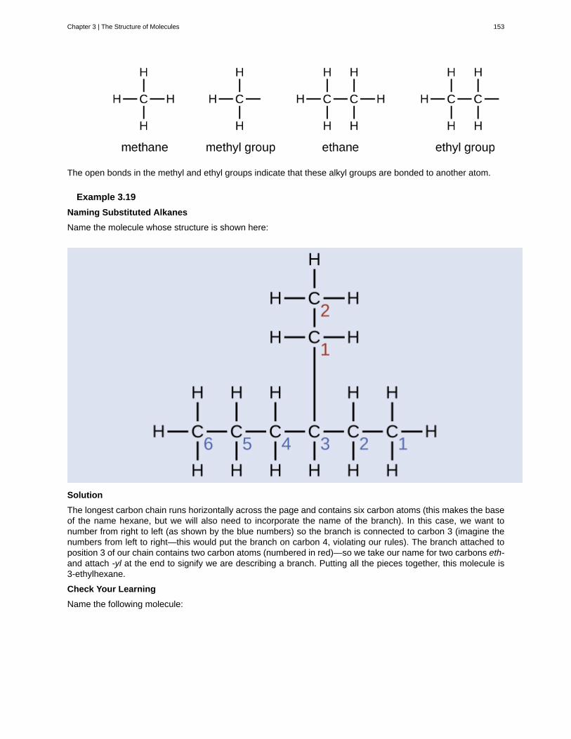
The open bonds in the methyl and ethyl groups indicate that these alkyl groups are bonded to another atom.
Example 3.19
Naming Substituted Alkanes
Name the molecule whose structure is shown here:
Solution
The longest carbon chain runs horizontally across the page and contains six carbon atoms (this makes the baseof the name hexane, but we will also need to incorporate the name of the branch). In this case, we want tonumber from right to left (as shown by the blue numbers) so the branch is connected to carbon 3 (imagine thenumbers from left to right—this would put the branch on carbon 4, violating our rules). The branch attached toposition 3 of our chain contains two carbon atoms (numbered in red)—so we take our name for two carbons eth-and attach -yl at the end to signify we are describing a branch. Putting all the pieces together, this molecule is3-ethylhexane.
Check Your Learning
Name the following molecule:
Chapter 3 | The Structure of Molecules 153
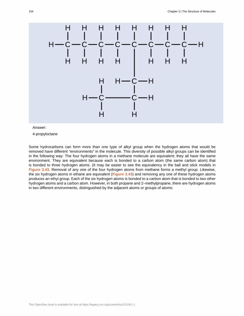
Answer:
4-propyloctane
Some hydrocarbons can form more than one type of alkyl group when the hydrogen atoms that would beremoved have different “environments” in the molecule. This diversity of possible alkyl groups can be identifiedin the following way: The four hydrogen atoms in a methane molecule are equivalent; they all have the sameenvironment. They are equivalent because each is bonded to a carbon atom (the same carbon atom) thatis bonded to three hydrogen atoms. (It may be easier to see the equivalency in the ball and stick models inFigure 3.43. Removal of any one of the four hydrogen atoms from methane forms a methyl group. Likewise,the six hydrogen atoms in ethane are equivalent (Figure 3.43) and removing any one of these hydrogen atomsproduces an ethyl group. Each of the six hydrogen atoms is bonded to a carbon atom that is bonded to two otherhydrogen atoms and a carbon atom. However, in both propane and 2–methylpropane, there are hydrogen atomsin two different environments, distinguished by the adjacent atoms or groups of atoms:
154 Chapter 3 | The Structure of Molecules
This OpenStax book is available for free at https://legacy.cnx.org/content/col12104/1.1
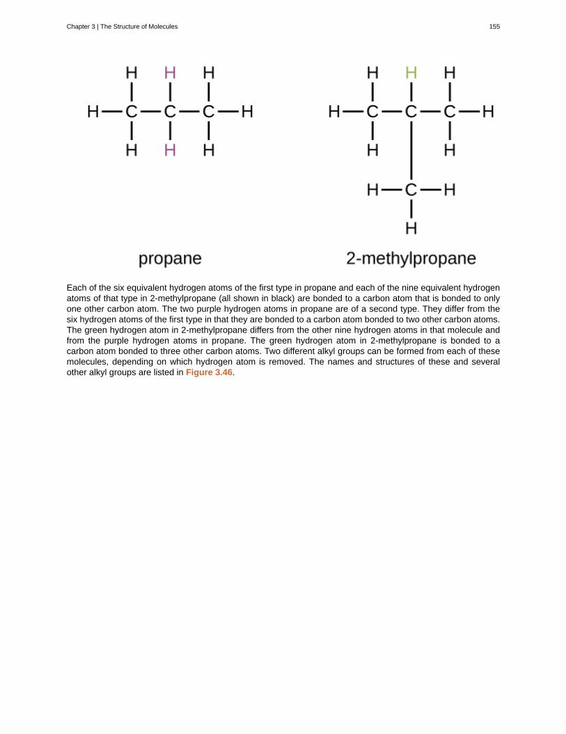
Each of the six equivalent hydrogen atoms of the first type in propane and each of the nine equivalent hydrogenatoms of that type in 2-methylpropane (all shown in black) are bonded to a carbon atom that is bonded to onlyone other carbon atom. The two purple hydrogen atoms in propane are of a second type. They differ from thesix hydrogen atoms of the first type in that they are bonded to a carbon atom bonded to two other carbon atoms.The green hydrogen atom in 2-methylpropane differs from the other nine hydrogen atoms in that molecule andfrom the purple hydrogen atoms in propane. The green hydrogen atom in 2-methylpropane is bonded to acarbon atom bonded to three other carbon atoms. Two different alkyl groups can be formed from each of thesemolecules, depending on which hydrogen atom is removed. The names and structures of these and severalother alkyl groups are listed in Figure 3.46.
Chapter 3 | The Structure of Molecules 155
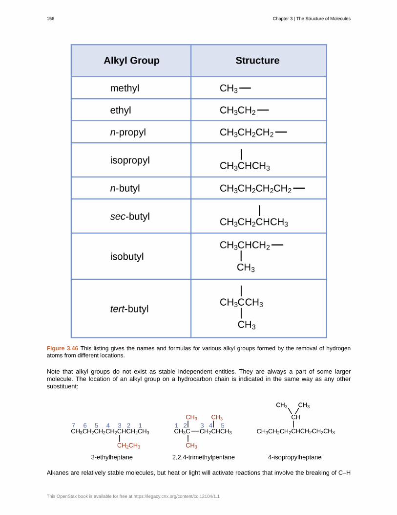
Figure 3.46 This listing gives the names and formulas for various alkyl groups formed by the removal of hydrogenatoms from different locations.
Note that alkyl groups do not exist as stable independent entities. They are always a part of some largermolecule. The location of an alkyl group on a hydrocarbon chain is indicated in the same way as any othersubstituent:
Alkanes are relatively stable molecules, but heat or light will activate reactions that involve the breaking of C–H
156 Chapter 3 | The Structure of Molecules
This OpenStax book is available for free at https://legacy.cnx.org/content/col12104/1.1
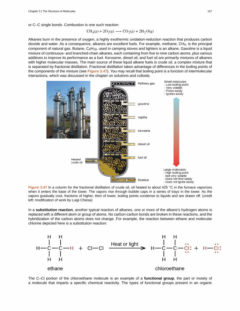
or C–C single bonds. Combustion is one such reaction:
CH4(g) + 2O2(g) ⟶ CO2(g) + 2H2 O(g)
Alkanes burn in the presence of oxygen, a highly exothermic oxidation-reduction reaction that produces carbondioxide and water. As a consequence, alkanes are excellent fuels. For example, methane, CH4, is the principalcomponent of natural gas. Butane, C4H10, used in camping stoves and lighters is an alkane. Gasoline is a liquidmixture of continuous- and branched-chain alkanes, each containing from five to nine carbon atoms, plus variousadditives to improve its performance as a fuel. Kerosene, diesel oil, and fuel oil are primarily mixtures of alkaneswith higher molecular masses. The main source of these liquid alkane fuels is crude oil, a complex mixture thatis separated by fractional distillation. Fractional distillation takes advantage of differences in the boiling points ofthe components of the mixture (see Figure 3.47). You may recall that boiling point is a function of intermolecularinteractions, which was discussed in the chapter on solutions and colloids.
Figure 3.47 In a column for the fractional distillation of crude oil, oil heated to about 425 °C in the furnace vaporizeswhen it enters the base of the tower. The vapors rise through bubble caps in a series of trays in the tower. As thevapors gradually cool, fractions of higher, then of lower, boiling points condense to liquids and are drawn off. (creditleft: modification of work by Luigi Chiesa)
In a substitution reaction, another typical reaction of alkanes, one or more of the alkane’s hydrogen atoms isreplaced with a different atom or group of atoms. No carbon-carbon bonds are broken in these reactions, and thehybridization of the carbon atoms does not change. For example, the reaction between ethane and molecularchlorine depicted here is a substitution reaction:
The C–Cl portion of the chloroethane molecule is an example of a functional group, the part or moiety ofa molecule that imparts a specific chemical reactivity. The types of functional groups present in an organic
Chapter 3 | The Structure of Molecules 157
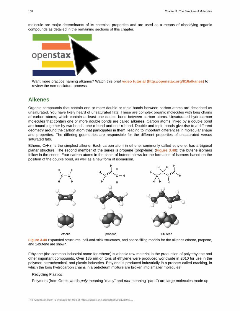
molecule are major determinants of its chemical properties and are used as a means of classifying organiccompounds as detailed in the remaining sections of this chapter.
Want more practice naming alkanes? Watch this brief video tutorial (http://openstax.org/l/16alkanes) toreview the nomenclature process.
Alkenes
Organic compounds that contain one or more double or triple bonds between carbon atoms are described asunsaturated. You have likely heard of unsaturated fats. These are complex organic molecules with long chainsof carbon atoms, which contain at least one double bond between carbon atoms. Unsaturated hydrocarbonmolecules that contain one or more double bonds are called alkenes. Carbon atoms linked by a double bondare bound together by two bonds, one σ bond and one π bond. Double and triple bonds give rise to a differentgeometry around the carbon atom that participates in them, leading to important differences in molecular shapeand properties. The differing geometries are responsible for the different properties of unsaturated versussaturated fats.
Ethene, C2H4, is the simplest alkene. Each carbon atom in ethene, commonly called ethylene, has a trigonalplanar structure. The second member of the series is propene (propylene) (Figure 3.48); the butene isomersfollow in the series. Four carbon atoms in the chain of butene allows for the formation of isomers based on theposition of the double bond, as well as a new form of isomerism.
Figure 3.48 Expanded structures, ball-and-stick structures, and space-filling models for the alkenes ethene, propene,and 1-butene are shown.
Ethylene (the common industrial name for ethene) is a basic raw material in the production of polyethylene andother important compounds. Over 135 million tons of ethylene were produced worldwide in 2010 for use in thepolymer, petrochemical, and plastic industries. Ethylene is produced industrially in a process called cracking, inwhich the long hydrocarbon chains in a petroleum mixture are broken into smaller molecules.
Recycling Plastics
Polymers (from Greek words poly meaning “many” and mer meaning “parts”) are large molecules made up
158 Chapter 3 | The Structure of Molecules
This OpenStax book is available for free at https://legacy.cnx.org/content/col12104/1.1
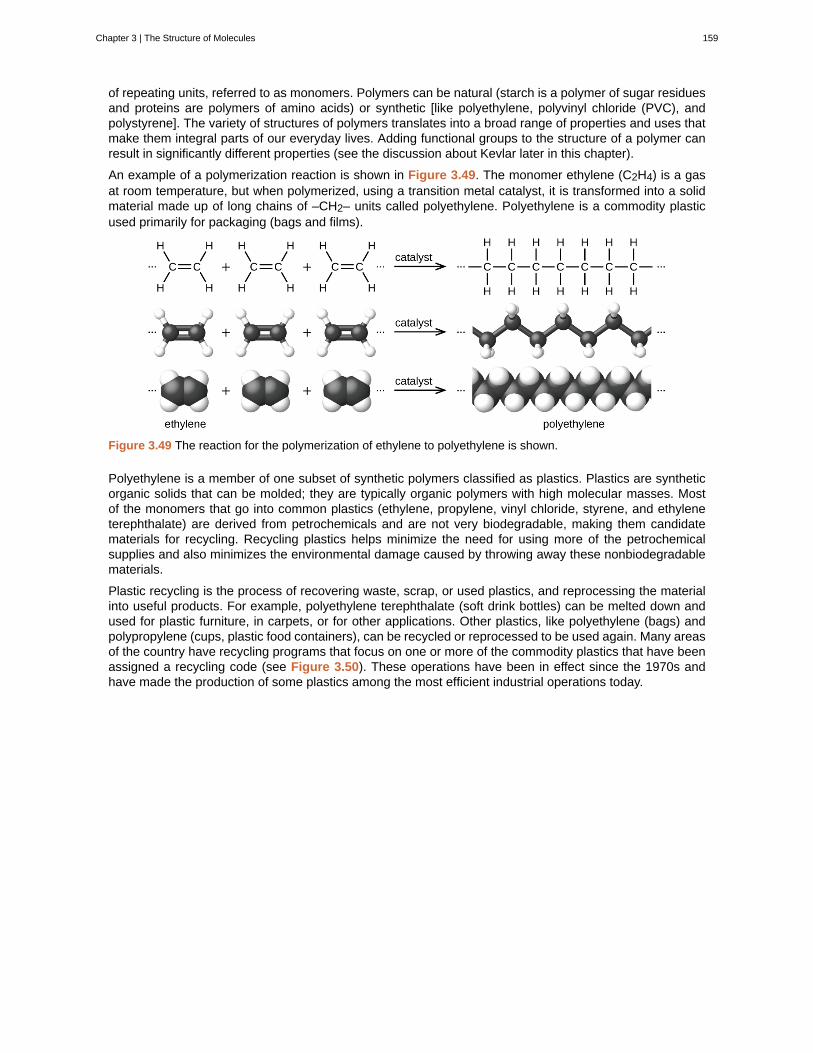
of repeating units, referred to as monomers. Polymers can be natural (starch is a polymer of sugar residuesand proteins are polymers of amino acids) or synthetic [like polyethylene, polyvinyl chloride (PVC), andpolystyrene]. The variety of structures of polymers translates into a broad range of properties and uses thatmake them integral parts of our everyday lives. Adding functional groups to the structure of a polymer canresult in significantly different properties (see the discussion about Kevlar later in this chapter).
An example of a polymerization reaction is shown in Figure 3.49. The monomer ethylene (C2H4) is a gasat room temperature, but when polymerized, using a transition metal catalyst, it is transformed into a solidmaterial made up of long chains of –CH2– units called polyethylene. Polyethylene is a commodity plasticused primarily for packaging (bags and films).
Figure 3.49 The reaction for the polymerization of ethylene to polyethylene is shown.
Polyethylene is a member of one subset of synthetic polymers classified as plastics. Plastics are syntheticorganic solids that can be molded; they are typically organic polymers with high molecular masses. Mostof the monomers that go into common plastics (ethylene, propylene, vinyl chloride, styrene, and ethyleneterephthalate) are derived from petrochemicals and are not very biodegradable, making them candidatematerials for recycling. Recycling plastics helps minimize the need for using more of the petrochemicalsupplies and also minimizes the environmental damage caused by throwing away these nonbiodegradablematerials.
Plastic recycling is the process of recovering waste, scrap, or used plastics, and reprocessing the materialinto useful products. For example, polyethylene terephthalate (soft drink bottles) can be melted down andused for plastic furniture, in carpets, or for other applications. Other plastics, like polyethylene (bags) andpolypropylene (cups, plastic food containers), can be recycled or reprocessed to be used again. Many areasof the country have recycling programs that focus on one or more of the commodity plastics that have beenassigned a recycling code (see Figure 3.50). These operations have been in effect since the 1970s andhave made the production of some plastics among the most efficient industrial operations today.
Chapter 3 | The Structure of Molecules 159
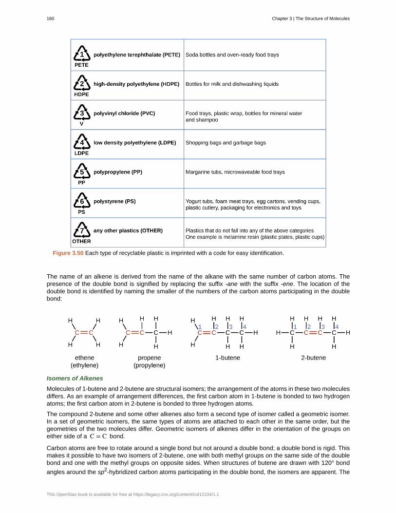
Figure 3.50 Each type of recyclable plastic is imprinted with a code for easy identification.
The name of an alkene is derived from the name of the alkane with the same number of carbon atoms. Thepresence of the double bond is signified by replacing the suffix -ane with the suffix -ene. The location of thedouble bond is identified by naming the smaller of the numbers of the carbon atoms participating in the doublebond:
Isomers of Alkenes
Molecules of 1-butene and 2-butene are structural isomers; the arrangement of the atoms in these two moleculesdiffers. As an example of arrangement differences, the first carbon atom in 1-butene is bonded to two hydrogenatoms; the first carbon atom in 2-butene is bonded to three hydrogen atoms.
The compound 2-butene and some other alkenes also form a second type of isomer called a geometric isomer.In a set of geometric isomers, the same types of atoms are attached to each other in the same order, but thegeometries of the two molecules differ. Geometric isomers of alkenes differ in the orientation of the groups oneither side of a C = C bond.
Carbon atoms are free to rotate around a single bond but not around a double bond; a double bond is rigid. Thismakes it possible to have two isomers of 2-butene, one with both methyl groups on the same side of the doublebond and one with the methyl groups on opposite sides. When structures of butene are drawn with 120° bond
angles around the sp2-hybridized carbon atoms participating in the double bond, the isomers are apparent. The
160 Chapter 3 | The Structure of Molecules
This OpenStax book is available for free at https://legacy.cnx.org/content/col12104/1.1
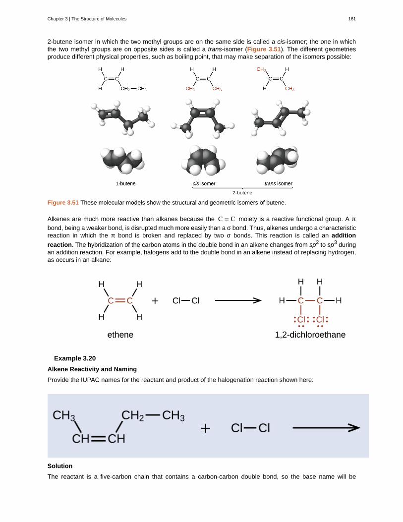
2-butene isomer in which the two methyl groups are on the same side is called a cis-isomer; the one in whichthe two methyl groups are on opposite sides is called a trans-isomer (Figure 3.51). The different geometriesproduce different physical properties, such as boiling point, that may make separation of the isomers possible:
Figure 3.51 These molecular models show the structural and geometric isomers of butene.
Alkenes are much more reactive than alkanes because the C = C moiety is a reactive functional group. A π
bond, being a weaker bond, is disrupted much more easily than a σ bond. Thus, alkenes undergo a characteristicreaction in which the π bond is broken and replaced by two σ bonds. This reaction is called an addition
reaction. The hybridization of the carbon atoms in the double bond in an alkene changes from sp2 to sp3 duringan addition reaction. For example, halogens add to the double bond in an alkene instead of replacing hydrogen,as occurs in an alkane:
Example 3.20
Alkene Reactivity and Naming
Provide the IUPAC names for the reactant and product of the halogenation reaction shown here:
Solution
The reactant is a five-carbon chain that contains a carbon-carbon double bond, so the base name will be
Chapter 3 | The Structure of Molecules 161
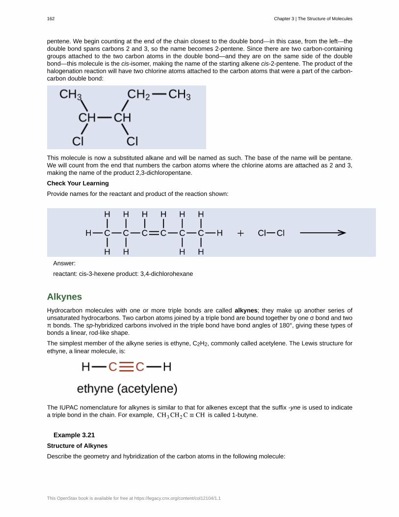
pentene. We begin counting at the end of the chain closest to the double bond—in this case, from the left—thedouble bond spans carbons 2 and 3, so the name becomes 2-pentene. Since there are two carbon-containinggroups attached to the two carbon atoms in the double bond—and they are on the same side of the doublebond—this molecule is the cis-isomer, making the name of the starting alkene cis-2-pentene. The product of thehalogenation reaction will have two chlorine atoms attached to the carbon atoms that were a part of the carbon-carbon double bond:
This molecule is now a substituted alkane and will be named as such. The base of the name will be pentane.We will count from the end that numbers the carbon atoms where the chlorine atoms are attached as 2 and 3,making the name of the product 2,3-dichloropentane.
Check Your Learning
Provide names for the reactant and product of the reaction shown:
Answer:
reactant: cis-3-hexene product: 3,4-dichlorohexane
Alkynes
Hydrocarbon molecules with one or more triple bonds are called alkynes; they make up another series ofunsaturated hydrocarbons. Two carbon atoms joined by a triple bond are bound together by one σ bond and twoπ bonds. The sp-hybridized carbons involved in the triple bond have bond angles of 180°, giving these types ofbonds a linear, rod-like shape.
The simplest member of the alkyne series is ethyne, C2H2, commonly called acetylene. The Lewis structure forethyne, a linear molecule, is:
The IUPAC nomenclature for alkynes is similar to that for alkenes except that the suffix -yne is used to indicatea triple bond in the chain. For example, CH3 CH2 C ≡ CH is called 1-butyne.
Example 3.21
Structure of Alkynes
Describe the geometry and hybridization of the carbon atoms in the following molecule:
162 Chapter 3 | The Structure of Molecules
This OpenStax book is available for free at https://legacy.cnx.org/content/col12104/1.1

Solution
Carbon atoms 1 and 4 have four single bonds and are thus tetrahedral with sp3 hybridization. Carbon atoms 2and 3 are involved in the triple bond, so they have linear geometries and would be classified as sp hybrids.
Check Your Learning
Identify the hybridization and bond angles at the carbon atoms in the molecule shown:
Answer:
carbon 1: sp, 180°; carbon 2: sp, 180°; carbon 3: sp2, 120°; carbon 4: sp2, 120°; carbon 5: sp3, 109.5°
Chemically, the alkynes are similar to the alkenes. Since the C ≡ C functional group has two π bonds, alkynes
typically react even more readily, and react with twice as much reagent in addition reactions. The reaction ofacetylene with bromine is a typical example:
Acetylene and the other alkynes also burn readily. An acetylene torch takes advantage of the high heat ofcombustion for acetylene.
Aromatic Hydrocarbons
Benzene, C6H6, is the simplest member of a large family of hydrocarbons, called aromatic hydrocarbons.These compounds contain ring structures and exhibit bonding that must be described using the resonancehybrid concept of valence bond theory or the delocalization concept of molecular orbital theory. (To review theseconcepts, refer to the earlier chapters on chemical bonding). The resonance structures for benzene, C6H6, are:
Chapter 3 | The Structure of Molecules 163
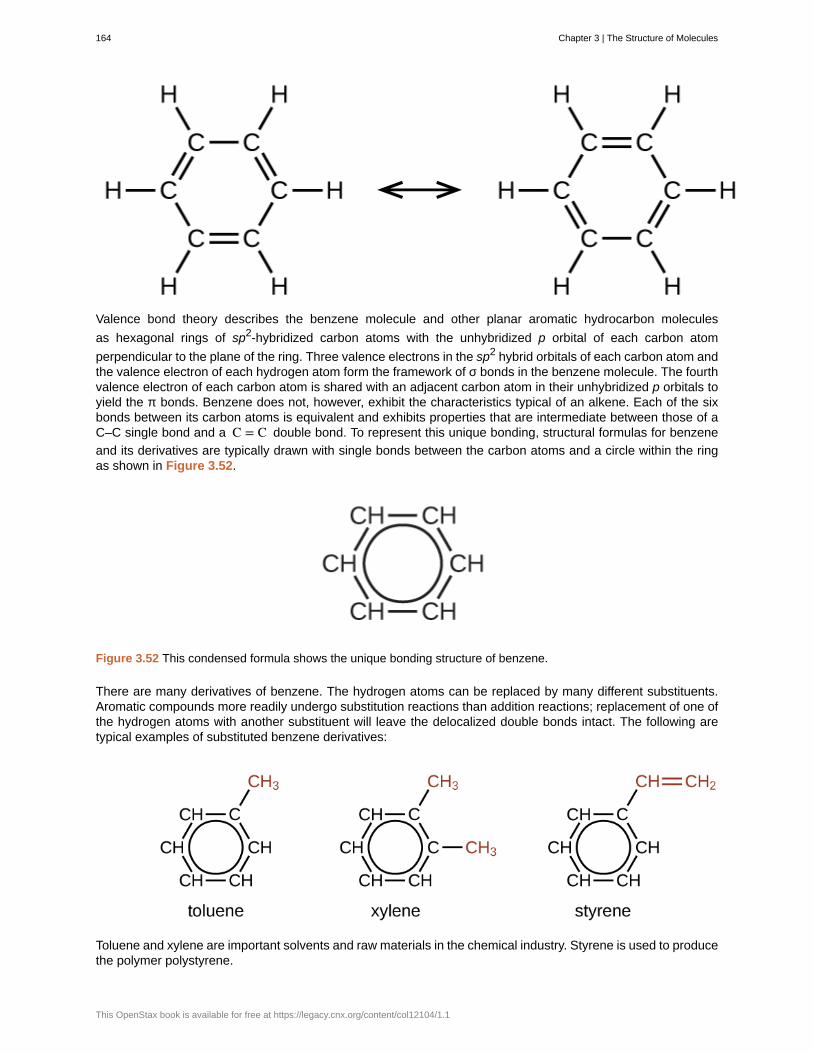
Valence bond theory describes the benzene molecule and other planar aromatic hydrocarbon molecules
as hexagonal rings of sp2-hybridized carbon atoms with the unhybridized p orbital of each carbon atom
perpendicular to the plane of the ring. Three valence electrons in the sp2 hybrid orbitals of each carbon atom andthe valence electron of each hydrogen atom form the framework of σ bonds in the benzene molecule. The fourthvalence electron of each carbon atom is shared with an adjacent carbon atom in their unhybridized p orbitals toyield the π bonds. Benzene does not, however, exhibit the characteristics typical of an alkene. Each of the sixbonds between its carbon atoms is equivalent and exhibits properties that are intermediate between those of aC–C single bond and a C = C double bond. To represent this unique bonding, structural formulas for benzene
and its derivatives are typically drawn with single bonds between the carbon atoms and a circle within the ringas shown in Figure 3.52.
Figure 3.52 This condensed formula shows the unique bonding structure of benzene.
There are many derivatives of benzene. The hydrogen atoms can be replaced by many different substituents.Aromatic compounds more readily undergo substitution reactions than addition reactions; replacement of one ofthe hydrogen atoms with another substituent will leave the delocalized double bonds intact. The following aretypical examples of substituted benzene derivatives:
Toluene and xylene are important solvents and raw materials in the chemical industry. Styrene is used to producethe polymer polystyrene.
164 Chapter 3 | The Structure of Molecules
This OpenStax book is available for free at https://legacy.cnx.org/content/col12104/1.1
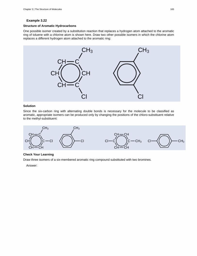
Example 3.22
Structure of Aromatic Hydrocarbons
One possible isomer created by a substitution reaction that replaces a hydrogen atom attached to the aromaticring of toluene with a chlorine atom is shown here. Draw two other possible isomers in which the chlorine atomreplaces a different hydrogen atom attached to the aromatic ring:
Solution
Since the six-carbon ring with alternating double bonds is necessary for the molecule to be classified asaromatic, appropriate isomers can be produced only by changing the positions of the chloro-substituent relativeto the methyl-substituent:
Check Your Learning
Draw three isomers of a six-membered aromatic ring compound substituted with two bromines.
Answer:
Chapter 3 | The Structure of Molecules 165
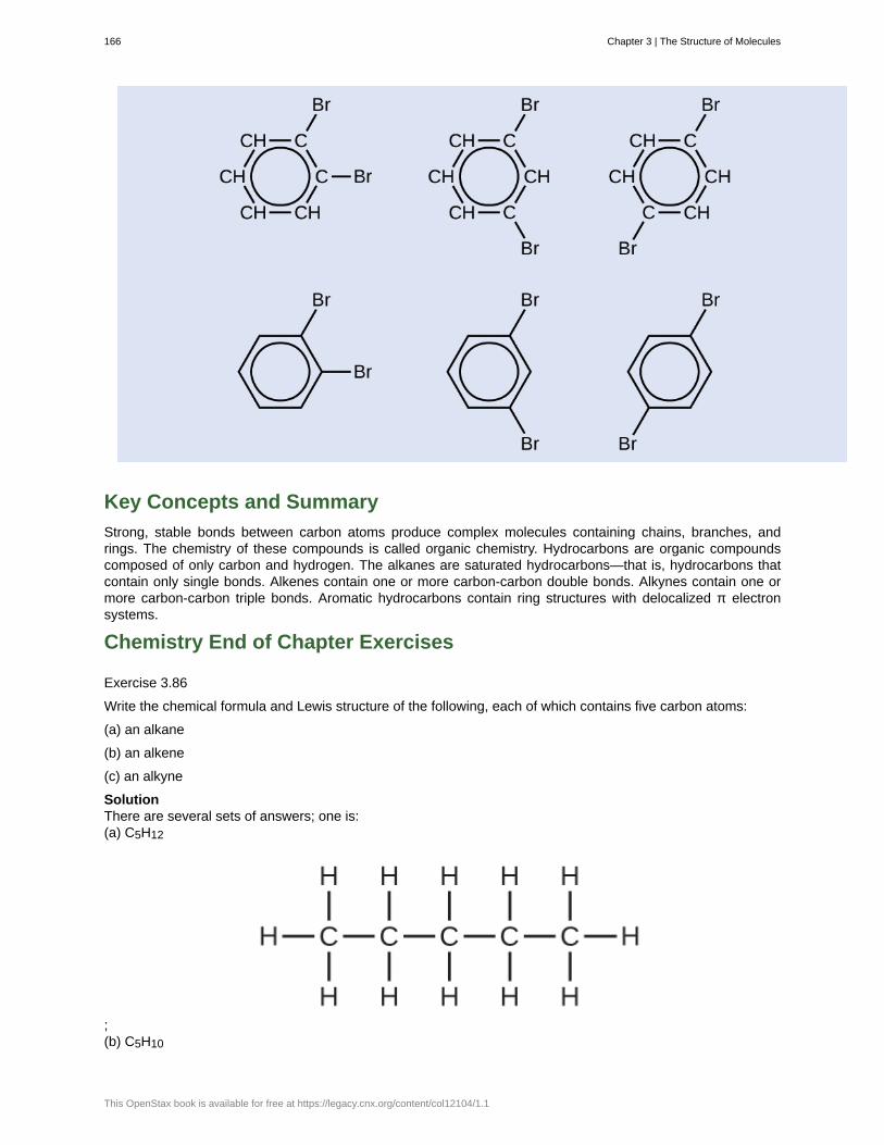
Key Concepts and Summary
Strong, stable bonds between carbon atoms produce complex molecules containing chains, branches, andrings. The chemistry of these compounds is called organic chemistry. Hydrocarbons are organic compoundscomposed of only carbon and hydrogen. The alkanes are saturated hydrocarbons—that is, hydrocarbons thatcontain only single bonds. Alkenes contain one or more carbon-carbon double bonds. Alkynes contain one ormore carbon-carbon triple bonds. Aromatic hydrocarbons contain ring structures with delocalized π electronsystems.
Chemistry End of Chapter Exercises
Exercise 3.86
Write the chemical formula and Lewis structure of the following, each of which contains five carbon atoms:
(a) an alkane
(b) an alkene
(c) an alkyne
SolutionThere are several sets of answers; one is:(a) C5H12
;(b) C5H10
166 Chapter 3 | The Structure of Molecules
This OpenStax book is available for free at https://legacy.cnx.org/content/col12104/1.1
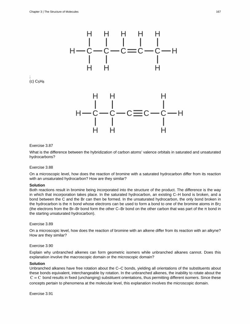
;(c) C5H8
Exercise 3.87
What is the difference between the hybridization of carbon atoms’ valence orbitals in saturated and unsaturatedhydrocarbons?
Exercise 3.88
On a microscopic level, how does the reaction of bromine with a saturated hydrocarbon differ from its reactionwith an unsaturated hydrocarbon? How are they similar?
SolutionBoth reactions result in bromine being incorporated into the structure of the product. The difference is the wayin which that incorporation takes place. In the saturated hydrocarbon, an existing C–H bond is broken, and abond between the C and the Br can then be formed. In the unsaturated hydrocarbon, the only bond broken inthe hydrocarbon is the π bond whose electrons can be used to form a bond to one of the bromine atoms in Br2(the electrons from the Br–Br bond form the other C–Br bond on the other carbon that was part of the π bond inthe starting unsaturated hydrocarbon).
Exercise 3.89
On a microscopic level, how does the reaction of bromine with an alkene differ from its reaction with an alkyne?How are they similar?
Exercise 3.90
Explain why unbranched alkenes can form geometric isomers while unbranched alkanes cannot. Does thisexplanation involve the macroscopic domain or the microscopic domain?
SolutionUnbranched alkanes have free rotation about the C–C bonds, yielding all orientations of the substituents aboutthese bonds equivalent, interchangeable by rotation. In the unbranched alkenes, the inability to rotate about theC = C bond results in fixed (unchanging) substituent orientations, thus permitting different isomers. Since these
concepts pertain to phenomena at the molecular level, this explanation involves the microscopic domain.
Exercise 3.91
Chapter 3 | The Structure of Molecules 167

Explain why these two molecules are not isomers:
Exercise 3.92
Explain why these two molecules are not isomers:
SolutionThey are the same compound because each is a saturated hydrocarbon containing an unbranched chain of sixcarbon atoms.
Exercise 3.93
How does the carbon-atom hybridization change when polyethylene is prepared from ethylene?
Exercise 3.94
Write the Lewis structure and molecular formula for each of the following hydrocarbons:
(a) hexane
(b) 3-methylpentane
(c) cis-3-hexene
(d) 4-methyl-1-pentene
(e) 3-hexyne
(f) 4-methyl-2-pentyne
Solution(a) C6H14
168 Chapter 3 | The Structure of Molecules
This OpenStax book is available for free at https://legacy.cnx.org/content/col12104/1.1
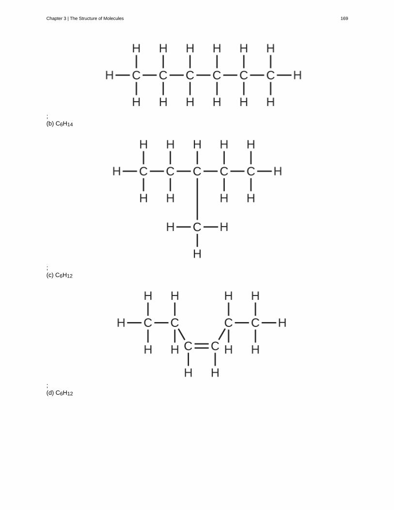
;(b) C6H14
;(c) C6H12
;(d) C6H12
Chapter 3 | The Structure of Molecules 169
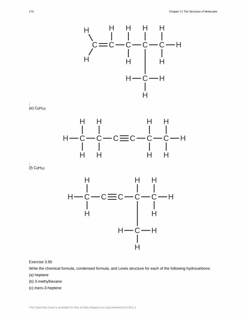
;(e) C6H10
;(f) C6H10
Exercise 3.95
Write the chemical formula, condensed formula, and Lewis structure for each of the following hydrocarbons:
(a) heptane
(b) 3-methylhexane
(c) trans-3-heptene
170 Chapter 3 | The Structure of Molecules
This OpenStax book is available for free at https://legacy.cnx.org/content/col12104/1.1
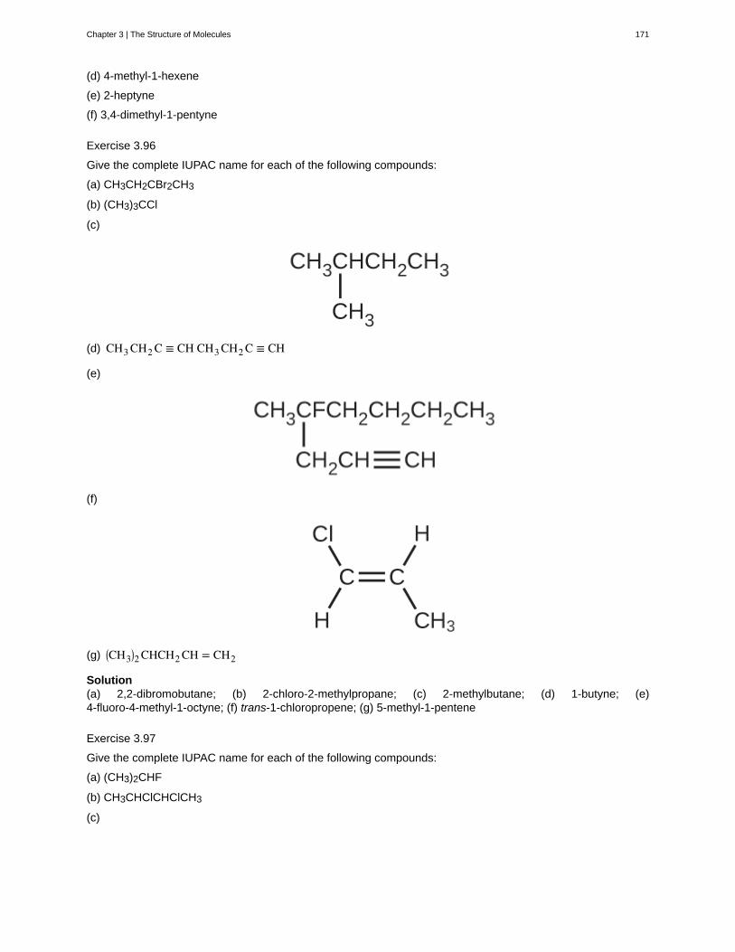
(d) 4-methyl-1-hexene
(e) 2-heptyne
(f) 3,4-dimethyl-1-pentyne
Exercise 3.96
Give the complete IUPAC name for each of the following compounds:
(a) CH3CH2CBr2CH3
(b) (CH3)3CCl
(c)
(d) CH3 CH2 C ≡ CH CH3 CH2 C ≡ CH
(e)
(f)
(g) ⎛⎝CH3⎞⎠2 CHCH2 CH = CH2
Solution(a) 2,2-dibromobutane; (b) 2-chloro-2-methylpropane; (c) 2-methylbutane; (d) 1-butyne; (e)4-fluoro-4-methyl-1-octyne; (f) trans-1-chloropropene; (g) 5-methyl-1-pentene
Exercise 3.97
Give the complete IUPAC name for each of the following compounds:
(a) (CH3)2CHF
(b) CH3CHClCHClCH3
(c)
Chapter 3 | The Structure of Molecules 171
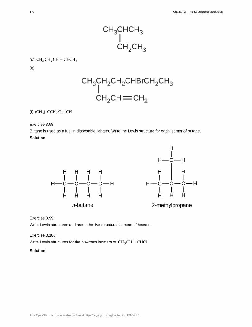
(d) CH3 CH2 CH = CHCH3
(e)
(f) ⎛⎝CH3⎞⎠3 CCH2 C ≡ CH
Exercise 3.98
Butane is used as a fuel in disposable lighters. Write the Lewis structure for each isomer of butane.
Solution
Exercise 3.99
Write Lewis structures and name the five structural isomers of hexane.
Exercise 3.100
Write Lewis structures for the cis–trans isomers of CH3 CH = CHCl.
Solution
172 Chapter 3 | The Structure of Molecules
This OpenStax book is available for free at https://legacy.cnx.org/content/col12104/1.1

Exercise 3.101
Write structures for the three isomers of the aromatic hydrocarbon xylene, C6H4(CH3)2.
Exercise 3.102
Isooctane is the common name of the isomer of C8H18 used as the standard of 100 for the gasoline octanerating:
(a) What is the IUPAC name for the compound?
(b) Name the other isomers that contain a five-carbon chain with three methyl substituents.
Solution(a) 2,2,4-trimethylpentane; (b) 2,2,3-trimethylpentane, 2,3,4-trimethylpentane, and 2,3,3-trimethylpentane:
Exercise 3.103
Write Lewis structures and IUPAC names for the alkyne isomers of C4H6.
Exercise 3.104
Write Lewis structures and IUPAC names for all isomers of C4H9Cl.
Solution
Chapter 3 | The Structure of Molecules 173
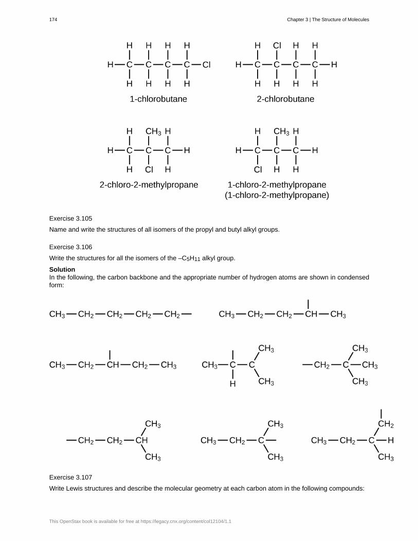
Exercise 3.105
Name and write the structures of all isomers of the propyl and butyl alkyl groups.
Exercise 3.106
Write the structures for all the isomers of the –C5H11 alkyl group.
SolutionIn the following, the carbon backbone and the appropriate number of hydrogen atoms are shown in condensedform:
Exercise 3.107
Write Lewis structures and describe the molecular geometry at each carbon atom in the following compounds:
174 Chapter 3 | The Structure of Molecules
This OpenStax book is available for free at https://legacy.cnx.org/content/col12104/1.1
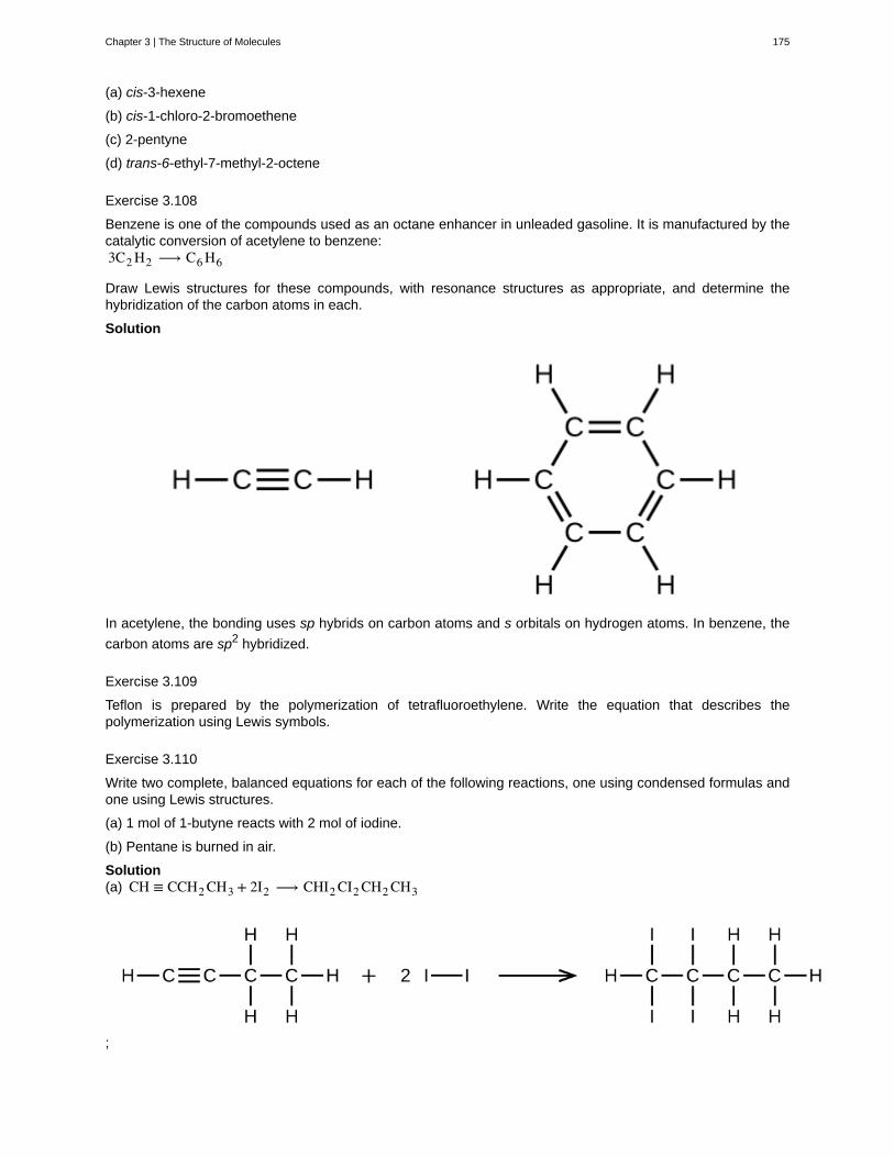
(a) cis-3-hexene
(b) cis-1-chloro-2-bromoethene
(c) 2-pentyne
(d) trans-6-ethyl-7-methyl-2-octene
Exercise 3.108
Benzene is one of the compounds used as an octane enhancer in unleaded gasoline. It is manufactured by thecatalytic conversion of acetylene to benzene:3C2 H2 ⟶ C6 H6
Draw Lewis structures for these compounds, with resonance structures as appropriate, and determine thehybridization of the carbon atoms in each.
Solution
In acetylene, the bonding uses sp hybrids on carbon atoms and s orbitals on hydrogen atoms. In benzene, the
carbon atoms are sp2 hybridized.
Exercise 3.109
Teflon is prepared by the polymerization of tetrafluoroethylene. Write the equation that describes thepolymerization using Lewis symbols.
Exercise 3.110
Write two complete, balanced equations for each of the following reactions, one using condensed formulas andone using Lewis structures.
(a) 1 mol of 1-butyne reacts with 2 mol of iodine.
(b) Pentane is burned in air.
Solution(a) CH ≡ CCH2 CH3 + 2I2 ⟶ CHI2 CI2 CH2 CH3
;
Chapter 3 | The Structure of Molecules 175
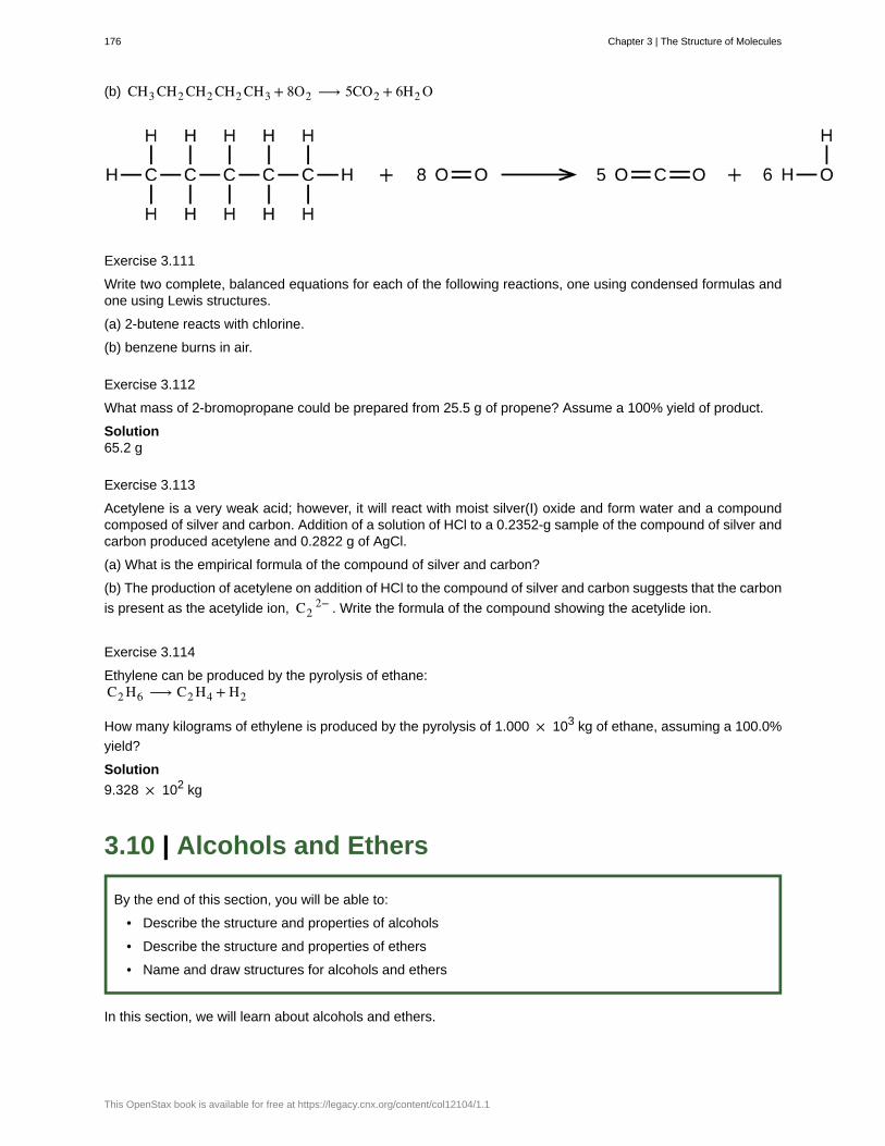
(b) CH3 CH2 CH2 CH2 CH3 + 8O2 ⟶ 5CO2 + 6H2 O
Exercise 3.111
Write two complete, balanced equations for each of the following reactions, one using condensed formulas andone using Lewis structures.
(a) 2-butene reacts with chlorine.
(b) benzene burns in air.
Exercise 3.112
What mass of 2-bromopropane could be prepared from 25.5 g of propene? Assume a 100% yield of product.
Solution65.2 g
Exercise 3.113
Acetylene is a very weak acid; however, it will react with moist silver(I) oxide and form water and a compoundcomposed of silver and carbon. Addition of a solution of HCl to a 0.2352-g sample of the compound of silver andcarbon produced acetylene and 0.2822 g of AgCl.
(a) What is the empirical formula of the compound of silver and carbon?
(b) The production of acetylene on addition of HCl to the compound of silver and carbon suggests that the carbon
is present as the acetylide ion, C22− . Write the formula of the compound showing the acetylide ion.
Exercise 3.114
Ethylene can be produced by the pyrolysis of ethane:C2 H6 ⟶ C2 H4 + H2
How many kilograms of ethylene is produced by the pyrolysis of 1.000 × 10³ kg of ethane, assuming a 100.0%
yield?
Solution
9.328 × 10² kg
3.10 | Alcohols and Ethers
By the end of this section, you will be able to:
• Describe the structure and properties of alcohols
• Describe the structure and properties of ethers
• Name and draw structures for alcohols and ethers
In this section, we will learn about alcohols and ethers.
176 Chapter 3 | The Structure of Molecules
This OpenStax book is available for free at https://legacy.cnx.org/content/col12104/1.1
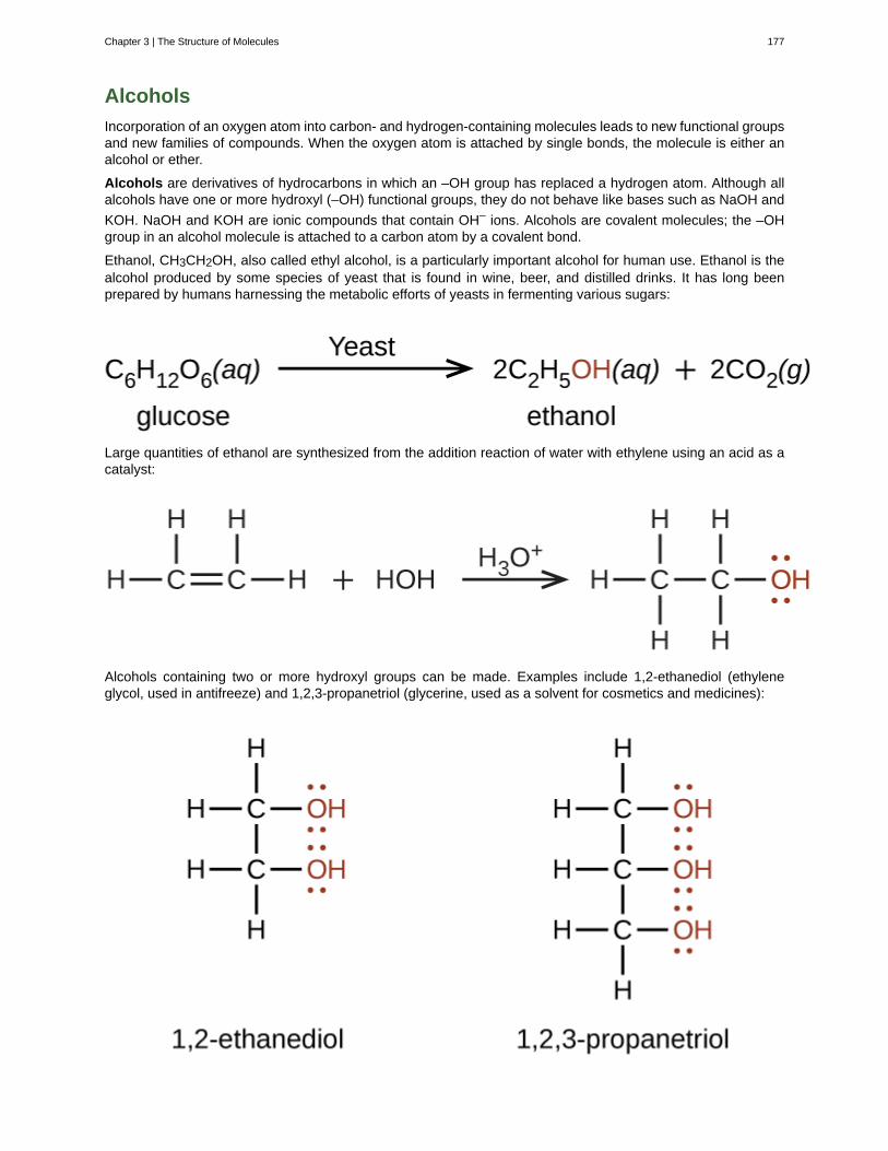
Alcohols
Incorporation of an oxygen atom into carbon- and hydrogen-containing molecules leads to new functional groupsand new families of compounds. When the oxygen atom is attached by single bonds, the molecule is either analcohol or ether.
Alcohols are derivatives of hydrocarbons in which an –OH group has replaced a hydrogen atom. Although allalcohols have one or more hydroxyl (–OH) functional groups, they do not behave like bases such as NaOH and
KOH. NaOH and KOH are ionic compounds that contain OH– ions. Alcohols are covalent molecules; the –OHgroup in an alcohol molecule is attached to a carbon atom by a covalent bond.
Ethanol, CH3CH2OH, also called ethyl alcohol, is a particularly important alcohol for human use. Ethanol is thealcohol produced by some species of yeast that is found in wine, beer, and distilled drinks. It has long beenprepared by humans harnessing the metabolic efforts of yeasts in fermenting various sugars:
Large quantities of ethanol are synthesized from the addition reaction of water with ethylene using an acid as acatalyst:
Alcohols containing two or more hydroxyl groups can be made. Examples include 1,2-ethanediol (ethyleneglycol, used in antifreeze) and 1,2,3-propanetriol (glycerine, used as a solvent for cosmetics and medicines):
Chapter 3 | The Structure of Molecules 177
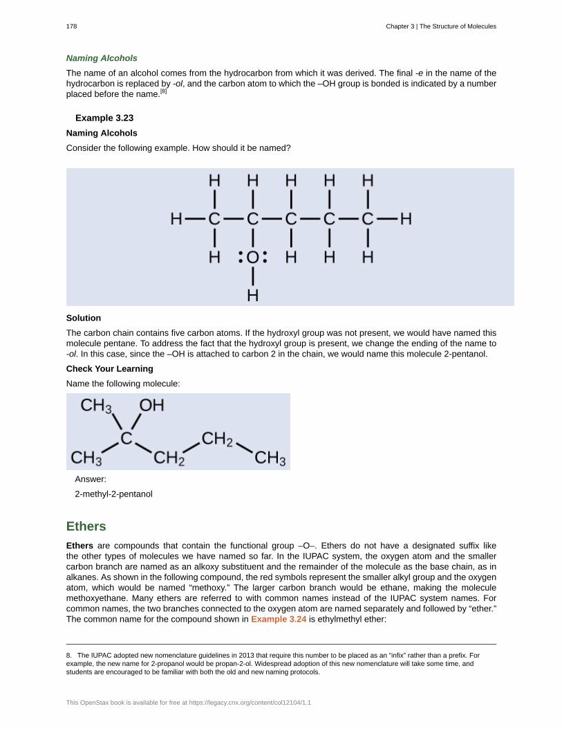
Naming Alcohols
The name of an alcohol comes from the hydrocarbon from which it was derived. The final -e in the name of thehydrocarbon is replaced by -ol, and the carbon atom to which the –OH group is bonded is indicated by a numberplaced before the name.[8]
Example 3.23
Naming Alcohols
Consider the following example. How should it be named?
Solution
The carbon chain contains five carbon atoms. If the hydroxyl group was not present, we would have named thismolecule pentane. To address the fact that the hydroxyl group is present, we change the ending of the name to-ol. In this case, since the –OH is attached to carbon 2 in the chain, we would name this molecule 2-pentanol.
Check Your Learning
Name the following molecule:
Answer:
2-methyl-2-pentanol
Ethers
Ethers are compounds that contain the functional group –O–. Ethers do not have a designated suffix likethe other types of molecules we have named so far. In the IUPAC system, the oxygen atom and the smallercarbon branch are named as an alkoxy substituent and the remainder of the molecule as the base chain, as inalkanes. As shown in the following compound, the red symbols represent the smaller alkyl group and the oxygenatom, which would be named “methoxy.” The larger carbon branch would be ethane, making the moleculemethoxyethane. Many ethers are referred to with common names instead of the IUPAC system names. Forcommon names, the two branches connected to the oxygen atom are named separately and followed by “ether.”The common name for the compound shown in Example 3.24 is ethylmethyl ether:
8. The IUPAC adopted new nomenclature guidelines in 2013 that require this number to be placed as an “infix” rather than a prefix. Forexample, the new name for 2-propanol would be propan-2-ol. Widespread adoption of this new nomenclature will take some time, andstudents are encouraged to be familiar with both the old and new naming protocols.
178 Chapter 3 | The Structure of Molecules
This OpenStax book is available for free at https://legacy.cnx.org/content/col12104/1.1
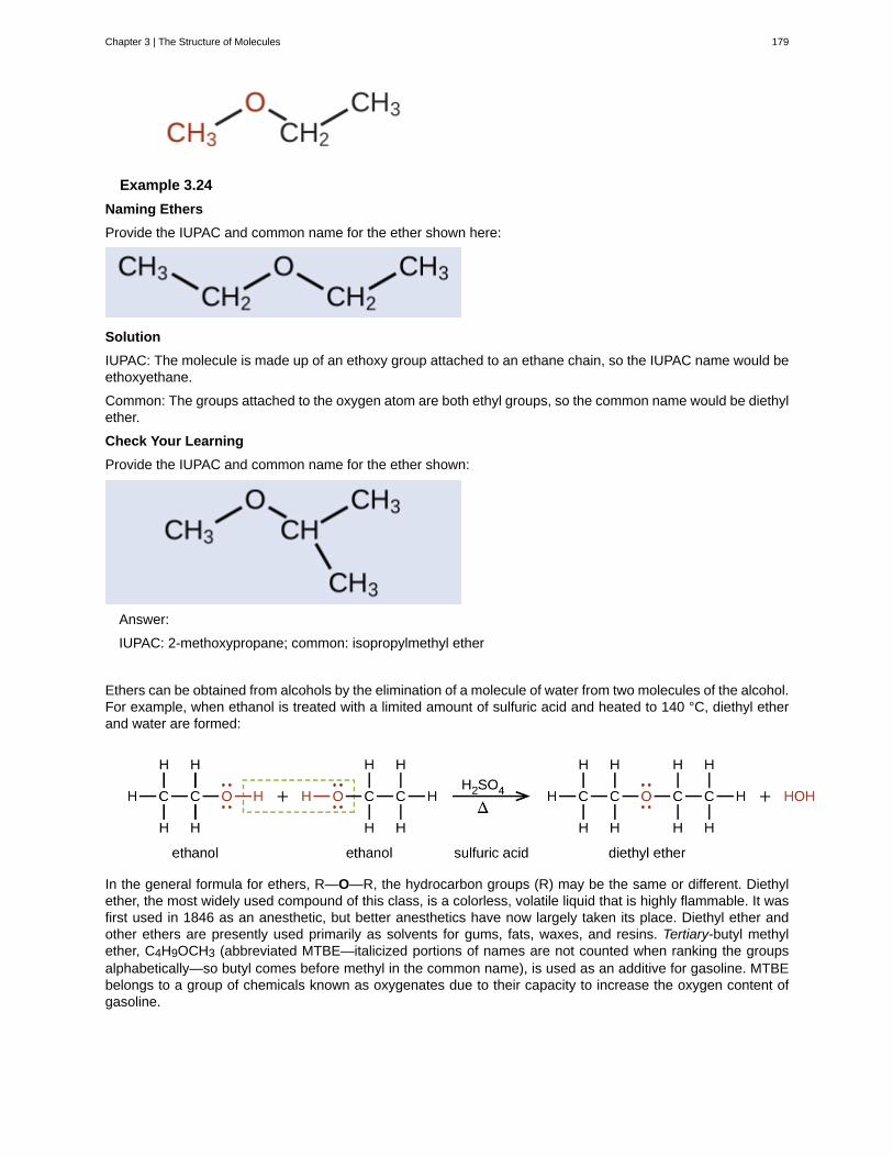
Example 3.24
Naming Ethers
Provide the IUPAC and common name for the ether shown here:
Solution
IUPAC: The molecule is made up of an ethoxy group attached to an ethane chain, so the IUPAC name would beethoxyethane.
Common: The groups attached to the oxygen atom are both ethyl groups, so the common name would be diethylether.
Check Your Learning
Provide the IUPAC and common name for the ether shown:
Answer:
IUPAC: 2-methoxypropane; common: isopropylmethyl ether
Ethers can be obtained from alcohols by the elimination of a molecule of water from two molecules of the alcohol.For example, when ethanol is treated with a limited amount of sulfuric acid and heated to 140 °C, diethyl etherand water are formed:
In the general formula for ethers, R—O—R, the hydrocarbon groups (R) may be the same or different. Diethylether, the most widely used compound of this class, is a colorless, volatile liquid that is highly flammable. It wasfirst used in 1846 as an anesthetic, but better anesthetics have now largely taken its place. Diethyl ether andother ethers are presently used primarily as solvents for gums, fats, waxes, and resins. Tertiary-butyl methylether, C4H9OCH3 (abbreviated MTBE—italicized portions of names are not counted when ranking the groupsalphabetically—so butyl comes before methyl in the common name), is used as an additive for gasoline. MTBEbelongs to a group of chemicals known as oxygenates due to their capacity to increase the oxygen content ofgasoline.
Chapter 3 | The Structure of Molecules 179
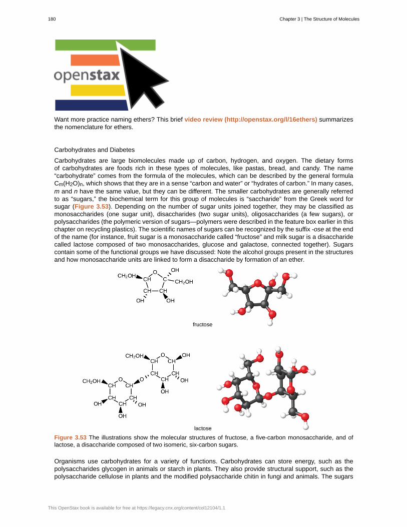
Want more practice naming ethers? This brief video review (http://openstax.org/l/16ethers) summarizesthe nomenclature for ethers.
Carbohydrates and Diabetes
Carbohydrates are large biomolecules made up of carbon, hydrogen, and oxygen. The dietary formsof carbohydrates are foods rich in these types of molecules, like pastas, bread, and candy. The name“carbohydrate” comes from the formula of the molecules, which can be described by the general formulaCm(H2O)n, which shows that they are in a sense “carbon and water” or “hydrates of carbon.” In many cases,m and n have the same value, but they can be different. The smaller carbohydrates are generally referredto as “sugars,” the biochemical term for this group of molecules is “saccharide” from the Greek word forsugar (Figure 3.53). Depending on the number of sugar units joined together, they may be classified asmonosaccharides (one sugar unit), disaccharides (two sugar units), oligosaccharides (a few sugars), orpolysaccharides (the polymeric version of sugars—polymers were described in the feature box earlier in thischapter on recycling plastics). The scientific names of sugars can be recognized by the suffix -ose at the endof the name (for instance, fruit sugar is a monosaccharide called “fructose” and milk sugar is a disaccharidecalled lactose composed of two monosaccharides, glucose and galactose, connected together). Sugarscontain some of the functional groups we have discussed: Note the alcohol groups present in the structuresand how monosaccharide units are linked to form a disaccharide by formation of an ether.
Figure 3.53 The illustrations show the molecular structures of fructose, a five-carbon monosaccharide, and oflactose, a disaccharide composed of two isomeric, six-carbon sugars.
Organisms use carbohydrates for a variety of functions. Carbohydrates can store energy, such as thepolysaccharides glycogen in animals or starch in plants. They also provide structural support, such as thepolysaccharide cellulose in plants and the modified polysaccharide chitin in fungi and animals. The sugars
180 Chapter 3 | The Structure of Molecules
This OpenStax book is available for free at https://legacy.cnx.org/content/col12104/1.1
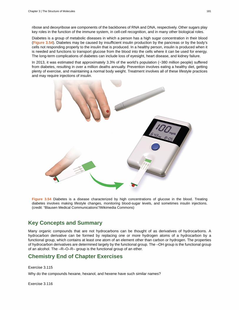
ribose and deoxyribose are components of the backbones of RNA and DNA, respectively. Other sugars playkey roles in the function of the immune system, in cell-cell recognition, and in many other biological roles.
Diabetes is a group of metabolic diseases in which a person has a high sugar concentration in their blood(Figure 3.54). Diabetes may be caused by insufficient insulin production by the pancreas or by the body’scells not responding properly to the insulin that is produced. In a healthy person, insulin is produced when itis needed and functions to transport glucose from the blood into the cells where it can be used for energy.The long-term complications of diabetes can include loss of eyesight, heart disease, and kidney failure.
In 2013, it was estimated that approximately 3.3% of the world’s population (~380 million people) sufferedfrom diabetes, resulting in over a million deaths annually. Prevention involves eating a healthy diet, gettingplenty of exercise, and maintaining a normal body weight. Treatment involves all of these lifestyle practicesand may require injections of insulin.
Figure 3.54 Diabetes is a disease characterized by high concentrations of glucose in the blood. Treatingdiabetes involves making lifestyle changes, monitoring blood-sugar levels, and sometimes insulin injections.(credit: “Blausen Medical Communications”/Wikimedia Commons)
Key Concepts and Summary
Many organic compounds that are not hydrocarbons can be thought of as derivatives of hydrocarbons. Ahydrocarbon derivative can be formed by replacing one or more hydrogen atoms of a hydrocarbon by afunctional group, which contains at least one atom of an element other than carbon or hydrogen. The propertiesof hydrocarbon derivatives are determined largely by the functional group. The –OH group is the functional groupof an alcohol. The –R–O–R– group is the functional group of an ether.
Chemistry End of Chapter Exercises
Exercise 3.115
Why do the compounds hexane, hexanol, and hexene have such similar names?
Exercise 3.116
Chapter 3 | The Structure of Molecules 181
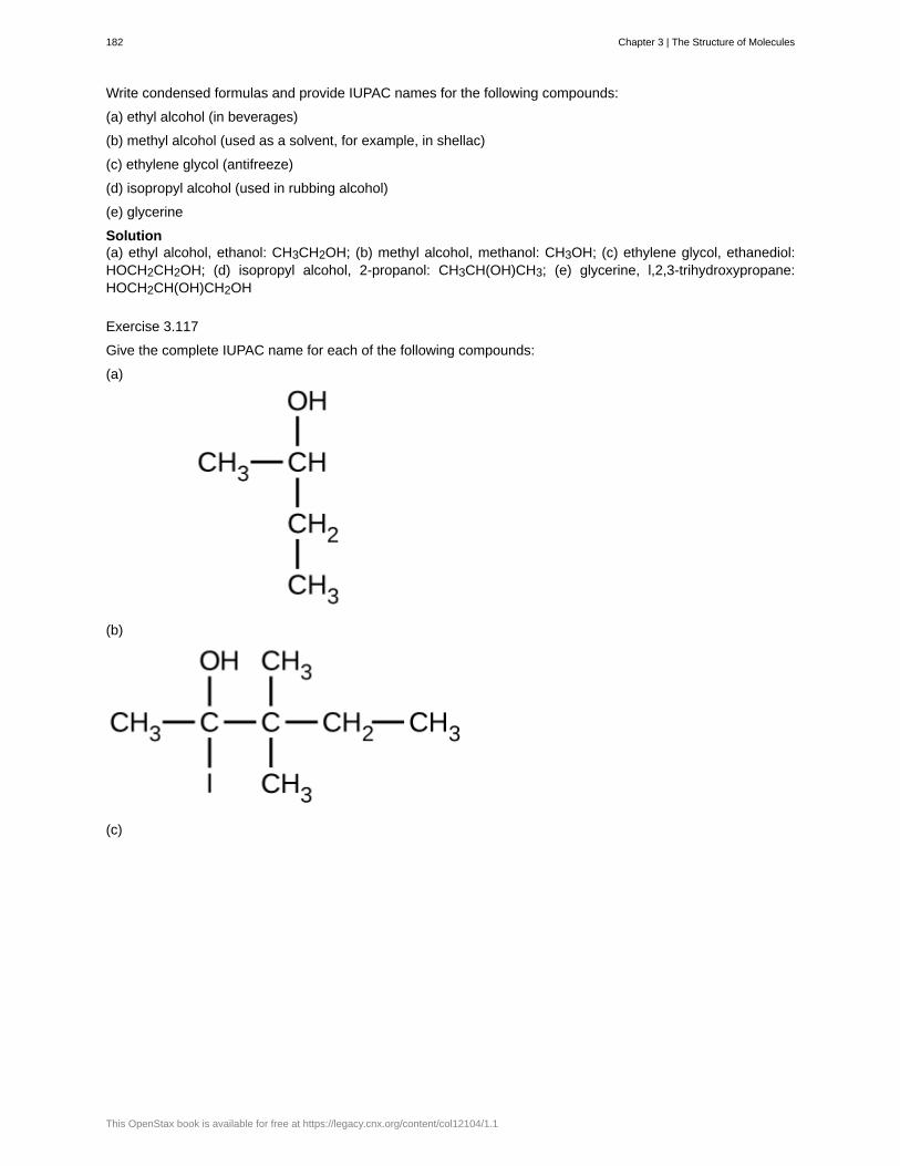
Write condensed formulas and provide IUPAC names for the following compounds:
(a) ethyl alcohol (in beverages)
(b) methyl alcohol (used as a solvent, for example, in shellac)
(c) ethylene glycol (antifreeze)
(d) isopropyl alcohol (used in rubbing alcohol)
(e) glycerine
Solution(a) ethyl alcohol, ethanol: CH3CH2OH; (b) methyl alcohol, methanol: CH3OH; (c) ethylene glycol, ethanediol:HOCH2CH2OH; (d) isopropyl alcohol, 2-propanol: CH3CH(OH)CH3; (e) glycerine, l,2,3-trihydroxypropane:HOCH2CH(OH)CH2OH
Exercise 3.117
Give the complete IUPAC name for each of the following compounds:
(a)
(b)
(c)
182 Chapter 3 | The Structure of Molecules
This OpenStax book is available for free at https://legacy.cnx.org/content/col12104/1.1

Exercise 3.118
Give the complete IUPAC name and the common name for each of the following compounds:
(a)
(b)
(c)
Solution(a) 1-ethoxybutane, butyl ethyl ether; (b) 1-ethoxypropane, ethyl propyl ether; (c) 1-methoxypropane, methylpropyl ether
Exercise 3.119
Write the condensed structures of both isomers with the formula C2H6O. Label the functional group of eachisomer.
Exercise 3.120
Write the condensed structures of all isomers with the formula C2H6O2. Label the functional group (or groups)of each isomer.
SolutionHOCH2CH2OH, two alcohol groups; CH3OCH2OH, ether and alcohol groups
Exercise 3.121
Draw the condensed formulas for each of the following compounds:
(a) dipropyl ether
(b) 2,2-dimethyl-3-hexanol
Chapter 3 | The Structure of Molecules 183
(c) 2-ethoxybutane
Exercise 3.122
MTBE, Methyl tert-butyl ether, CH3OC(CH3)3, is used as an oxygen source in oxygenated gasolines. MTBE ismanufactured by reacting 2-methylpropene with methanol.
(a) Using Lewis structures, write the chemical equation representing the reaction.
(b) What volume of methanol, density 0.7915 g/mL, is required to produce exactly 1000 kg of MTBE, assuminga 100% yield?
Solution(a)
;
(b) 4.593 × 10² L
Exercise 3.123
Write two complete balanced equations for each of the following reactions, one using condensed formulas andone using Lewis structures.
(a) propanol is converted to dipropyl ether
(b) propene is treated with water in dilute acid.
Exercise 3.124
Write two complete balanced equations for each of the following reactions, one using condensed formulas andone using Lewis structures.
(a) 2-butene is treated with water in dilute acid
(b) ethanol is dehydrated to yield ethene
Solution(a) CH3 CH = CHCH3 + H2 O ⟶ CH3 CH2 CH(OH)CH3
;(b) CH3 CH2 OH ⟶ CH2 = CH2 + H2 O
184 Chapter 3 | The Structure of Molecules
This OpenStax book is available for free at https://legacy.cnx.org/content/col12104/1.1
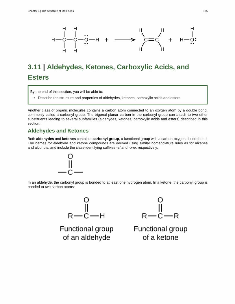
3.11 | Aldehydes, Ketones, Carboxylic Acids, and
Esters
By the end of this section, you will be able to:
• Describe the structure and properties of aldehydes, ketones, carboxylic acids and esters
Another class of organic molecules contains a carbon atom connected to an oxygen atom by a double bond,commonly called a carbonyl group. The trigonal planar carbon in the carbonyl group can attach to two othersubstituents leading to several subfamilies (aldehydes, ketones, carboxylic acids and esters) described in thissection.
Aldehydes and Ketones
Both aldehydes and ketones contain a carbonyl group, a functional group with a carbon-oxygen double bond.The names for aldehyde and ketone compounds are derived using similar nomenclature rules as for alkanesand alcohols, and include the class-identifying suffixes -al and -one, respectively:
In an aldehyde, the carbonyl group is bonded to at least one hydrogen atom. In a ketone, the carbonyl group isbonded to two carbon atoms:
Chapter 3 | The Structure of Molecules 185
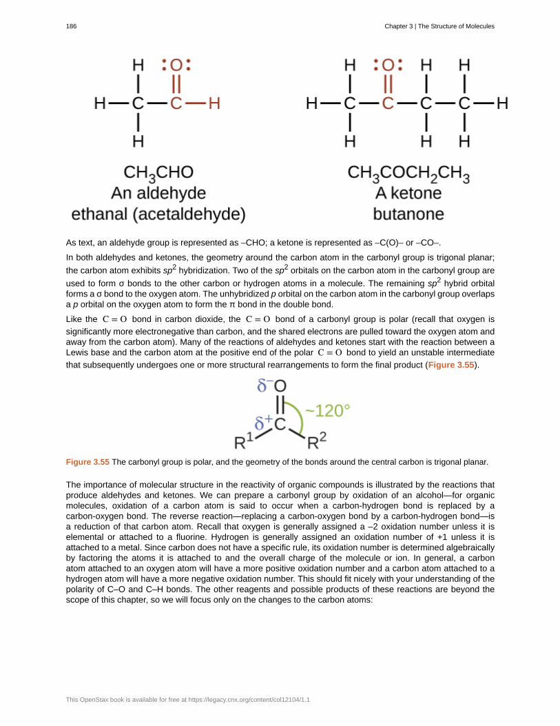
As text, an aldehyde group is represented as –CHO; a ketone is represented as –C(O)– or –CO–.
In both aldehydes and ketones, the geometry around the carbon atom in the carbonyl group is trigonal planar;
the carbon atom exhibits sp2 hybridization. Two of the sp2 orbitals on the carbon atom in the carbonyl group are
used to form σ bonds to the other carbon or hydrogen atoms in a molecule. The remaining sp2 hybrid orbitalforms a σ bond to the oxygen atom. The unhybridized p orbital on the carbon atom in the carbonyl group overlapsa p orbital on the oxygen atom to form the π bond in the double bond.
Like the C = O bond in carbon dioxide, the C = O bond of a carbonyl group is polar (recall that oxygen is
significantly more electronegative than carbon, and the shared electrons are pulled toward the oxygen atom andaway from the carbon atom). Many of the reactions of aldehydes and ketones start with the reaction between aLewis base and the carbon atom at the positive end of the polar C = O bond to yield an unstable intermediate
that subsequently undergoes one or more structural rearrangements to form the final product (Figure 3.55).
Figure 3.55 The carbonyl group is polar, and the geometry of the bonds around the central carbon is trigonal planar.
The importance of molecular structure in the reactivity of organic compounds is illustrated by the reactions thatproduce aldehydes and ketones. We can prepare a carbonyl group by oxidation of an alcohol—for organicmolecules, oxidation of a carbon atom is said to occur when a carbon-hydrogen bond is replaced by acarbon-oxygen bond. The reverse reaction—replacing a carbon-oxygen bond by a carbon-hydrogen bond—isa reduction of that carbon atom. Recall that oxygen is generally assigned a –2 oxidation number unless it iselemental or attached to a fluorine. Hydrogen is generally assigned an oxidation number of +1 unless it isattached to a metal. Since carbon does not have a specific rule, its oxidation number is determined algebraicallyby factoring the atoms it is attached to and the overall charge of the molecule or ion. In general, a carbonatom attached to an oxygen atom will have a more positive oxidation number and a carbon atom attached to ahydrogen atom will have a more negative oxidation number. This should fit nicely with your understanding of thepolarity of C–O and C–H bonds. The other reagents and possible products of these reactions are beyond thescope of this chapter, so we will focus only on the changes to the carbon atoms:
186 Chapter 3 | The Structure of Molecules
This OpenStax book is available for free at https://legacy.cnx.org/content/col12104/1.1
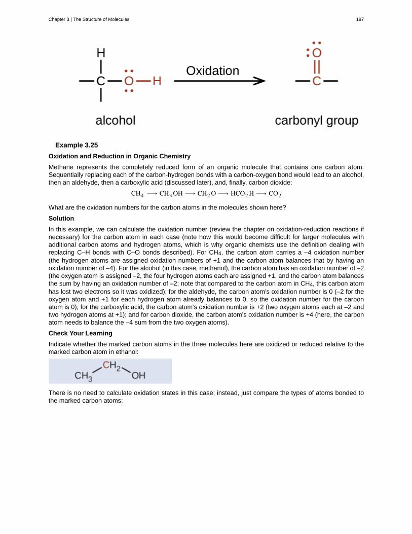
Example 3.25
Oxidation and Reduction in Organic Chemistry
Methane represents the completely reduced form of an organic molecule that contains one carbon atom.Sequentially replacing each of the carbon-hydrogen bonds with a carbon-oxygen bond would lead to an alcohol,then an aldehyde, then a carboxylic acid (discussed later), and, finally, carbon dioxide:
CH4 ⟶ CH3 OH ⟶ CH2 O ⟶ HCO2 H ⟶ CO2
What are the oxidation numbers for the carbon atoms in the molecules shown here?
Solution
In this example, we can calculate the oxidation number (review the chapter on oxidation-reduction reactions ifnecessary) for the carbon atom in each case (note how this would become difficult for larger molecules withadditional carbon atoms and hydrogen atoms, which is why organic chemists use the definition dealing withreplacing C–H bonds with C–O bonds described). For CH4, the carbon atom carries a –4 oxidation number(the hydrogen atoms are assigned oxidation numbers of +1 and the carbon atom balances that by having anoxidation number of –4). For the alcohol (in this case, methanol), the carbon atom has an oxidation number of –2(the oxygen atom is assigned –2, the four hydrogen atoms each are assigned +1, and the carbon atom balancesthe sum by having an oxidation number of –2; note that compared to the carbon atom in CH4, this carbon atomhas lost two electrons so it was oxidized); for the aldehyde, the carbon atom’s oxidation number is 0 (–2 for theoxygen atom and +1 for each hydrogen atom already balances to 0, so the oxidation number for the carbonatom is 0); for the carboxylic acid, the carbon atom’s oxidation number is +2 (two oxygen atoms each at –2 andtwo hydrogen atoms at +1); and for carbon dioxide, the carbon atom’s oxidation number is +4 (here, the carbonatom needs to balance the –4 sum from the two oxygen atoms).
Check Your Learning
Indicate whether the marked carbon atoms in the three molecules here are oxidized or reduced relative to themarked carbon atom in ethanol:
There is no need to calculate oxidation states in this case; instead, just compare the types of atoms bonded tothe marked carbon atoms:
Chapter 3 | The Structure of Molecules 187
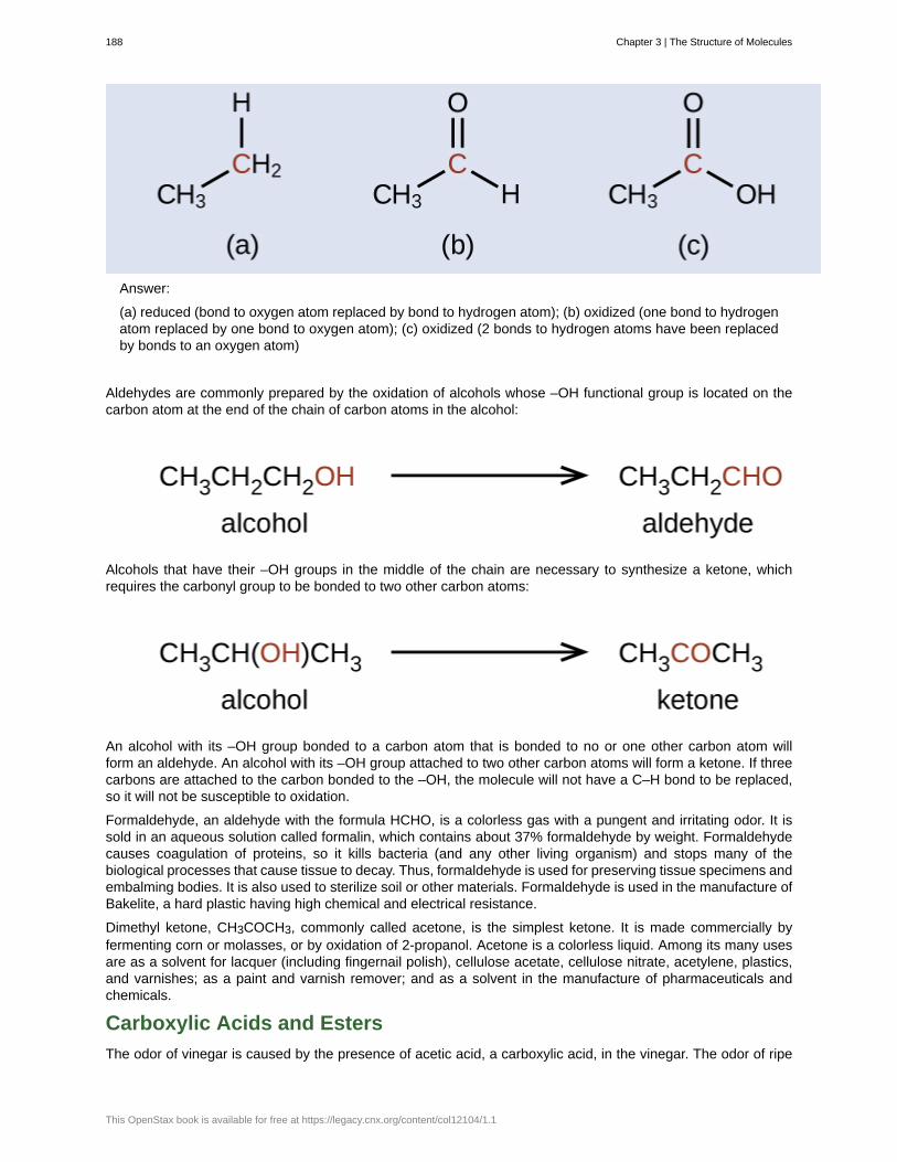
Answer:
(a) reduced (bond to oxygen atom replaced by bond to hydrogen atom); (b) oxidized (one bond to hydrogenatom replaced by one bond to oxygen atom); (c) oxidized (2 bonds to hydrogen atoms have been replacedby bonds to an oxygen atom)
Aldehydes are commonly prepared by the oxidation of alcohols whose –OH functional group is located on thecarbon atom at the end of the chain of carbon atoms in the alcohol:
Alcohols that have their –OH groups in the middle of the chain are necessary to synthesize a ketone, whichrequires the carbonyl group to be bonded to two other carbon atoms:
An alcohol with its –OH group bonded to a carbon atom that is bonded to no or one other carbon atom willform an aldehyde. An alcohol with its –OH group attached to two other carbon atoms will form a ketone. If threecarbons are attached to the carbon bonded to the –OH, the molecule will not have a C–H bond to be replaced,so it will not be susceptible to oxidation.
Formaldehyde, an aldehyde with the formula HCHO, is a colorless gas with a pungent and irritating odor. It issold in an aqueous solution called formalin, which contains about 37% formaldehyde by weight. Formaldehydecauses coagulation of proteins, so it kills bacteria (and any other living organism) and stops many of thebiological processes that cause tissue to decay. Thus, formaldehyde is used for preserving tissue specimens andembalming bodies. It is also used to sterilize soil or other materials. Formaldehyde is used in the manufacture ofBakelite, a hard plastic having high chemical and electrical resistance.
Dimethyl ketone, CH3COCH3, commonly called acetone, is the simplest ketone. It is made commercially byfermenting corn or molasses, or by oxidation of 2-propanol. Acetone is a colorless liquid. Among its many usesare as a solvent for lacquer (including fingernail polish), cellulose acetate, cellulose nitrate, acetylene, plastics,and varnishes; as a paint and varnish remover; and as a solvent in the manufacture of pharmaceuticals andchemicals.
Carboxylic Acids and Esters
The odor of vinegar is caused by the presence of acetic acid, a carboxylic acid, in the vinegar. The odor of ripe
188 Chapter 3 | The Structure of Molecules
This OpenStax book is available for free at https://legacy.cnx.org/content/col12104/1.1
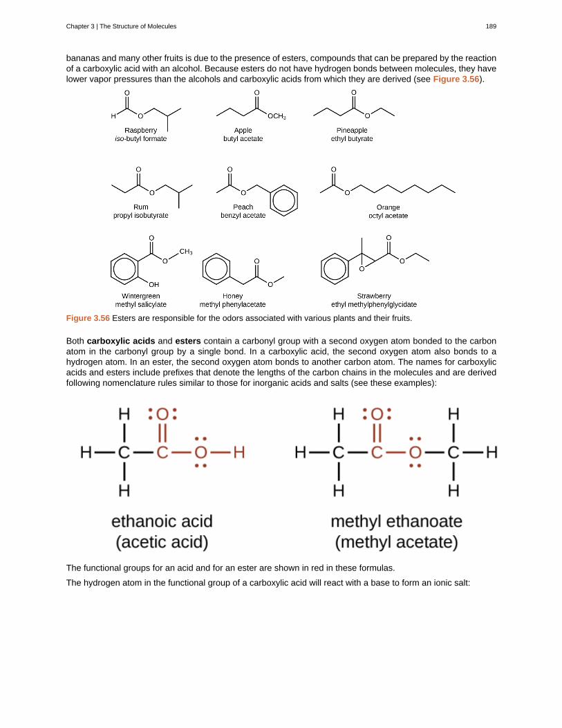
bananas and many other fruits is due to the presence of esters, compounds that can be prepared by the reactionof a carboxylic acid with an alcohol. Because esters do not have hydrogen bonds between molecules, they havelower vapor pressures than the alcohols and carboxylic acids from which they are derived (see Figure 3.56).
Figure 3.56 Esters are responsible for the odors associated with various plants and their fruits.
Both carboxylic acids and esters contain a carbonyl group with a second oxygen atom bonded to the carbonatom in the carbonyl group by a single bond. In a carboxylic acid, the second oxygen atom also bonds to ahydrogen atom. In an ester, the second oxygen atom bonds to another carbon atom. The names for carboxylicacids and esters include prefixes that denote the lengths of the carbon chains in the molecules and are derivedfollowing nomenclature rules similar to those for inorganic acids and salts (see these examples):
The functional groups for an acid and for an ester are shown in red in these formulas.
The hydrogen atom in the functional group of a carboxylic acid will react with a base to form an ionic salt:
Chapter 3 | The Structure of Molecules 189
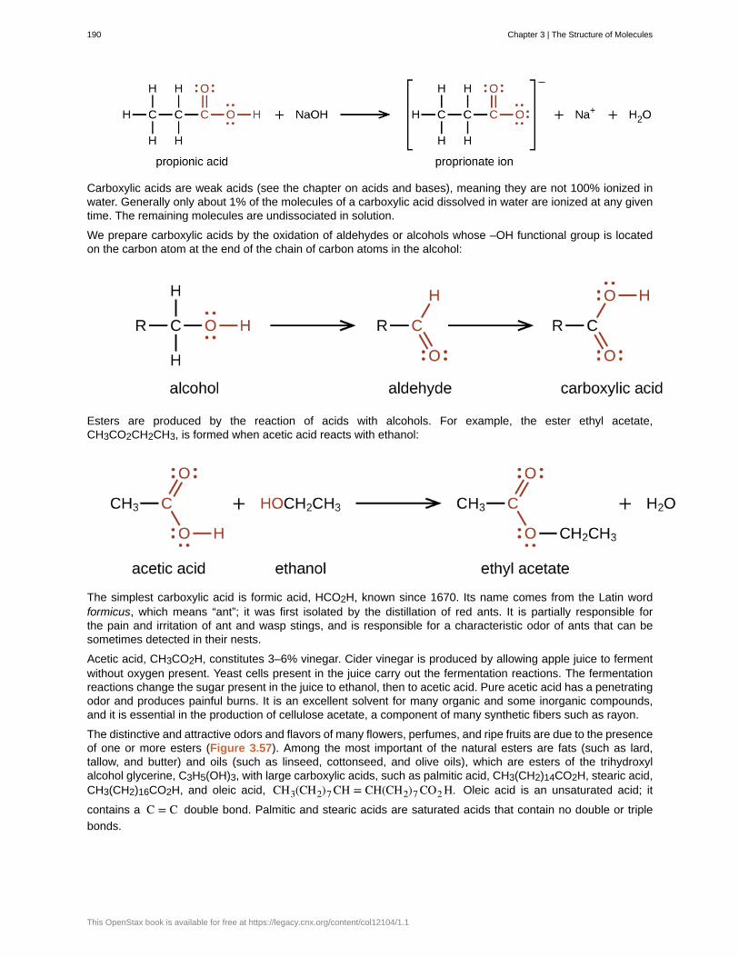
Carboxylic acids are weak acids (see the chapter on acids and bases), meaning they are not 100% ionized inwater. Generally only about 1% of the molecules of a carboxylic acid dissolved in water are ionized at any giventime. The remaining molecules are undissociated in solution.
We prepare carboxylic acids by the oxidation of aldehydes or alcohols whose –OH functional group is locatedon the carbon atom at the end of the chain of carbon atoms in the alcohol:
Esters are produced by the reaction of acids with alcohols. For example, the ester ethyl acetate,CH3CO2CH2CH3, is formed when acetic acid reacts with ethanol:
The simplest carboxylic acid is formic acid, HCO2H, known since 1670. Its name comes from the Latin wordformicus, which means “ant”; it was first isolated by the distillation of red ants. It is partially responsible forthe pain and irritation of ant and wasp stings, and is responsible for a characteristic odor of ants that can besometimes detected in their nests.
Acetic acid, CH3CO2H, constitutes 3–6% vinegar. Cider vinegar is produced by allowing apple juice to fermentwithout oxygen present. Yeast cells present in the juice carry out the fermentation reactions. The fermentationreactions change the sugar present in the juice to ethanol, then to acetic acid. Pure acetic acid has a penetratingodor and produces painful burns. It is an excellent solvent for many organic and some inorganic compounds,and it is essential in the production of cellulose acetate, a component of many synthetic fibers such as rayon.
The distinctive and attractive odors and flavors of many flowers, perfumes, and ripe fruits are due to the presenceof one or more esters (Figure 3.57). Among the most important of the natural esters are fats (such as lard,tallow, and butter) and oils (such as linseed, cottonseed, and olive oils), which are esters of the trihydroxylalcohol glycerine, C3H5(OH)3, with large carboxylic acids, such as palmitic acid, CH3(CH2)14CO2H, stearic acid,CH3(CH2)16CO2H, and oleic acid, CH3(CH2)7 CH = CH(CH2)7 CO2 H. Oleic acid is an unsaturated acid; it
contains a C = C double bond. Palmitic and stearic acids are saturated acids that contain no double or triple
bonds.
190 Chapter 3 | The Structure of Molecules
This OpenStax book is available for free at https://legacy.cnx.org/content/col12104/1.1
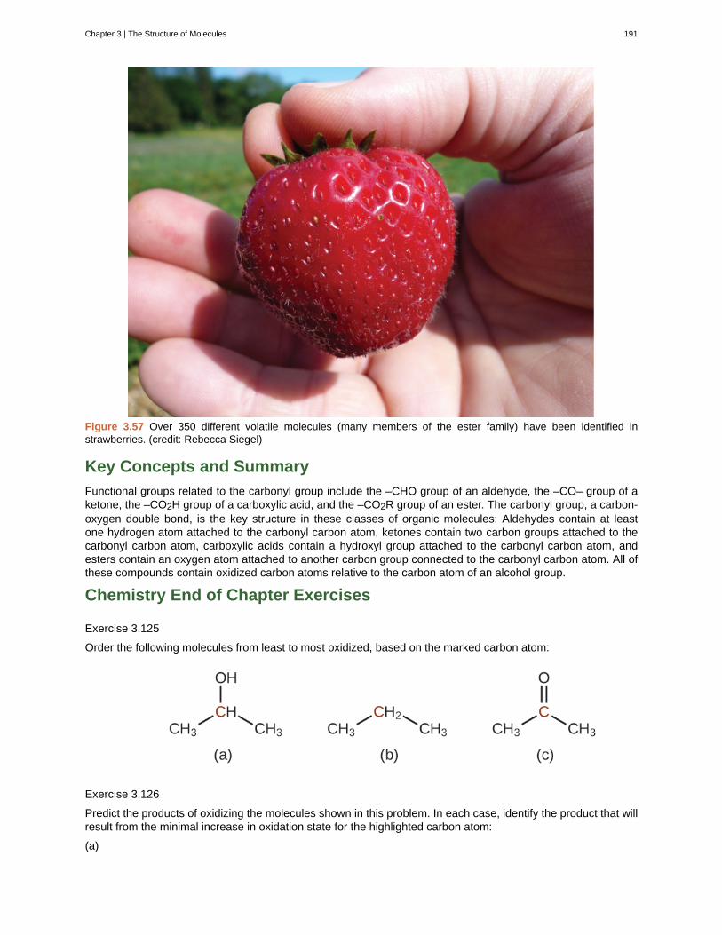
Figure 3.57 Over 350 different volatile molecules (many members of the ester family) have been identified instrawberries. (credit: Rebecca Siegel)
Key Concepts and Summary
Functional groups related to the carbonyl group include the –CHO group of an aldehyde, the –CO– group of aketone, the –CO2H group of a carboxylic acid, and the –CO2R group of an ester. The carbonyl group, a carbon-oxygen double bond, is the key structure in these classes of organic molecules: Aldehydes contain at leastone hydrogen atom attached to the carbonyl carbon atom, ketones contain two carbon groups attached to thecarbonyl carbon atom, carboxylic acids contain a hydroxyl group attached to the carbonyl carbon atom, andesters contain an oxygen atom attached to another carbon group connected to the carbonyl carbon atom. All ofthese compounds contain oxidized carbon atoms relative to the carbon atom of an alcohol group.
Chemistry End of Chapter Exercises
Exercise 3.125
Order the following molecules from least to most oxidized, based on the marked carbon atom:
Exercise 3.126
Predict the products of oxidizing the molecules shown in this problem. In each case, identify the product that willresult from the minimal increase in oxidation state for the highlighted carbon atom:
(a)
Chapter 3 | The Structure of Molecules 191
(b)
(c)
Solution(a)
;(b)
;(c)
192 Chapter 3 | The Structure of Molecules
This OpenStax book is available for free at https://legacy.cnx.org/content/col12104/1.1
Exercise 3.127
Predict the products of reducing the following molecules. In each case, identify the product that will result fromthe minimal decrease in oxidation state for the highlighted carbon atom:
(a)
(b)
(c)
Exercise 3.128
Explain why it is not possible to prepare a ketone that contains only two carbon atoms.
SolutionA ketone contains a group bonded to two additional carbon atoms; thus, a minimum of three carbon atoms areneeded.
Exercise 3.129
How does hybridization of the substituted carbon atom change when an alcohol is converted into an aldehyde?An aldehyde to a carboxylic acid?
Exercise 3.130
Chapter 3 | The Structure of Molecules 193
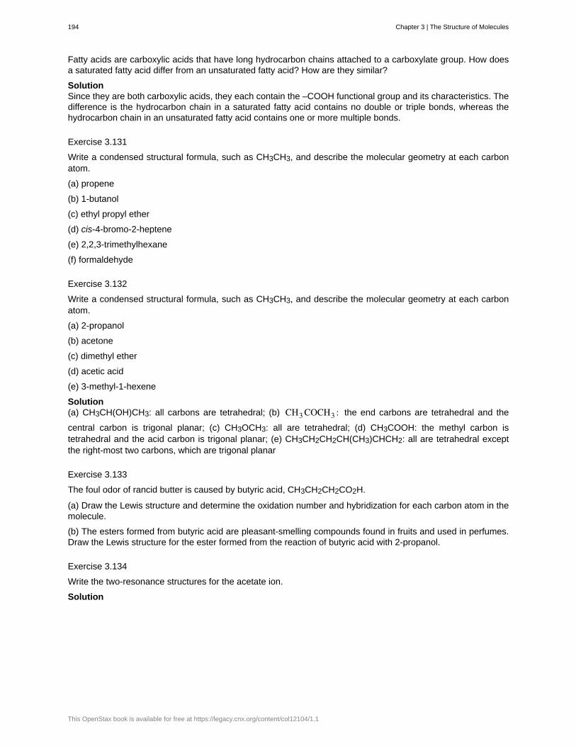
Fatty acids are carboxylic acids that have long hydrocarbon chains attached to a carboxylate group. How doesa saturated fatty acid differ from an unsaturated fatty acid? How are they similar?
SolutionSince they are both carboxylic acids, they each contain the –COOH functional group and its characteristics. Thedifference is the hydrocarbon chain in a saturated fatty acid contains no double or triple bonds, whereas thehydrocarbon chain in an unsaturated fatty acid contains one or more multiple bonds.
Exercise 3.131
Write a condensed structural formula, such as CH3CH3, and describe the molecular geometry at each carbonatom.
(a) propene
(b) 1-butanol
(c) ethyl propyl ether
(d) cis-4-bromo-2-heptene
(e) 2,2,3-trimethylhexane
(f) formaldehyde
Exercise 3.132
Write a condensed structural formula, such as CH3CH3, and describe the molecular geometry at each carbonatom.
(a) 2-propanol
(b) acetone
(c) dimethyl ether
(d) acetic acid
(e) 3-methyl-1-hexene
Solution(a) CH3CH(OH)CH3: all carbons are tetrahedral; (b) CH3 COCH3 : the end carbons are tetrahedral and the
central carbon is trigonal planar; (c) CH3OCH3: all are tetrahedral; (d) CH3COOH: the methyl carbon istetrahedral and the acid carbon is trigonal planar; (e) CH3CH2CH2CH(CH3)CHCH2: all are tetrahedral exceptthe right-most two carbons, which are trigonal planar
Exercise 3.133
The foul odor of rancid butter is caused by butyric acid, CH3CH2CH2CO2H.
(a) Draw the Lewis structure and determine the oxidation number and hybridization for each carbon atom in themolecule.
(b) The esters formed from butyric acid are pleasant-smelling compounds found in fruits and used in perfumes.Draw the Lewis structure for the ester formed from the reaction of butyric acid with 2-propanol.
Exercise 3.134
Write the two-resonance structures for the acetate ion.
Solution
194 Chapter 3 | The Structure of Molecules
This OpenStax book is available for free at https://legacy.cnx.org/content/col12104/1.1
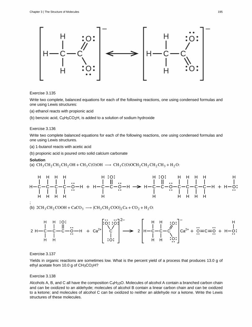
Exercise 3.135
Write two complete, balanced equations for each of the following reactions, one using condensed formulas andone using Lewis structures:
(a) ethanol reacts with propionic acid
(b) benzoic acid, C6H5CO2H, is added to a solution of sodium hydroxide
Exercise 3.136
Write two complete balanced equations for each of the following reactions, one using condensed formulas andone using Lewis structures.
(a) 1-butanol reacts with acetic acid
(b) propionic acid is poured onto solid calcium carbonate
Solution(a) CH3 CH2 CH2 CH2 OH + CH3 C(O)OH ⟶ CH3 C(O)OCH2 CH2 CH2 CH3 + H2 O:
;(b) 2CH3 CH2 COOH + CaCO3 ⟶ ⎛
⎝CH3 CH2 COO⎞⎠2 Ca + CO2 + H2 O:
Exercise 3.137
Yields in organic reactions are sometimes low. What is the percent yield of a process that produces 13.0 g ofethyl acetate from 10.0 g of CH3CO2H?
Exercise 3.138
Alcohols A, B, and C all have the composition C4H10O. Molecules of alcohol A contain a branched carbon chainand can be oxidized to an aldehyde; molecules of alcohol B contain a linear carbon chain and can be oxidizedto a ketone; and molecules of alcohol C can be oxidized to neither an aldehyde nor a ketone. Write the Lewisstructures of these molecules.
Chapter 3 | The Structure of Molecules 195
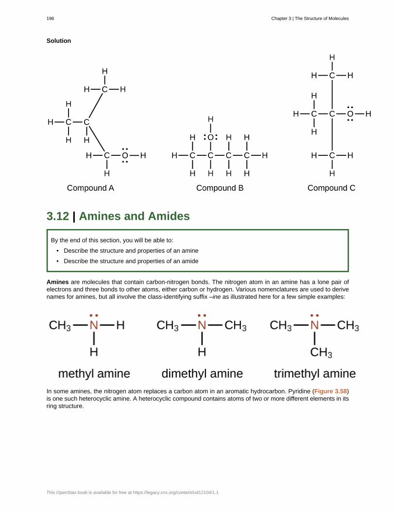
Solution
3.12 | Amines and Amides
By the end of this section, you will be able to:
• Describe the structure and properties of an amine
• Describe the structure and properties of an amide
Amines are molecules that contain carbon-nitrogen bonds. The nitrogen atom in an amine has a lone pair ofelectrons and three bonds to other atoms, either carbon or hydrogen. Various nomenclatures are used to derivenames for amines, but all involve the class-identifying suffix –ine as illustrated here for a few simple examples:
In some amines, the nitrogen atom replaces a carbon atom in an aromatic hydrocarbon. Pyridine (Figure 3.58)is one such heterocyclic amine. A heterocyclic compound contains atoms of two or more different elements in itsring structure.
196 Chapter 3 | The Structure of Molecules
This OpenStax book is available for free at https://legacy.cnx.org/content/col12104/1.1
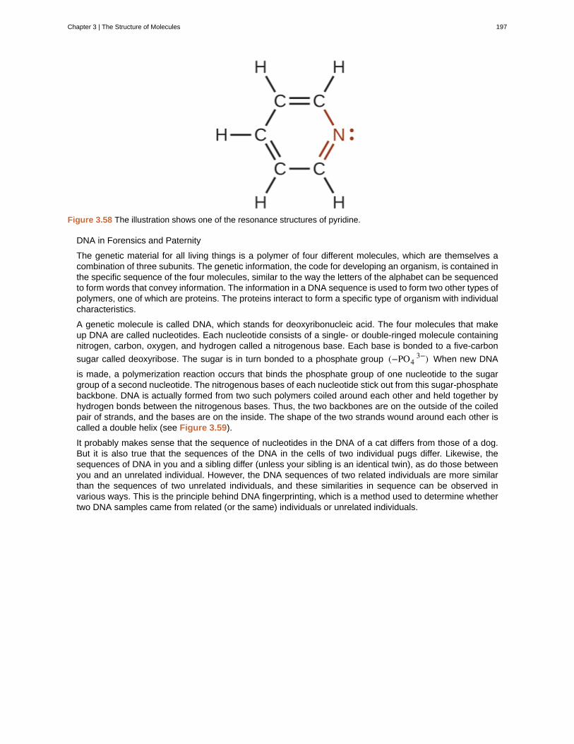
Figure 3.58 The illustration shows one of the resonance structures of pyridine.
DNA in Forensics and Paternity
The genetic material for all living things is a polymer of four different molecules, which are themselves acombination of three subunits. The genetic information, the code for developing an organism, is contained inthe specific sequence of the four molecules, similar to the way the letters of the alphabet can be sequencedto form words that convey information. The information in a DNA sequence is used to form two other types ofpolymers, one of which are proteins. The proteins interact to form a specific type of organism with individualcharacteristics.
A genetic molecule is called DNA, which stands for deoxyribonucleic acid. The four molecules that makeup DNA are called nucleotides. Each nucleotide consists of a single- or double-ringed molecule containingnitrogen, carbon, oxygen, and hydrogen called a nitrogenous base. Each base is bonded to a five-carbon
sugar called deoxyribose. The sugar is in turn bonded to a phosphate group (−PO43−) When new DNA
is made, a polymerization reaction occurs that binds the phosphate group of one nucleotide to the sugargroup of a second nucleotide. The nitrogenous bases of each nucleotide stick out from this sugar-phosphatebackbone. DNA is actually formed from two such polymers coiled around each other and held together byhydrogen bonds between the nitrogenous bases. Thus, the two backbones are on the outside of the coiledpair of strands, and the bases are on the inside. The shape of the two strands wound around each other iscalled a double helix (see Figure 3.59).
It probably makes sense that the sequence of nucleotides in the DNA of a cat differs from those of a dog.But it is also true that the sequences of the DNA in the cells of two individual pugs differ. Likewise, thesequences of DNA in you and a sibling differ (unless your sibling is an identical twin), as do those betweenyou and an unrelated individual. However, the DNA sequences of two related individuals are more similarthan the sequences of two unrelated individuals, and these similarities in sequence can be observed invarious ways. This is the principle behind DNA fingerprinting, which is a method used to determine whethertwo DNA samples came from related (or the same) individuals or unrelated individuals.
Chapter 3 | The Structure of Molecules 197
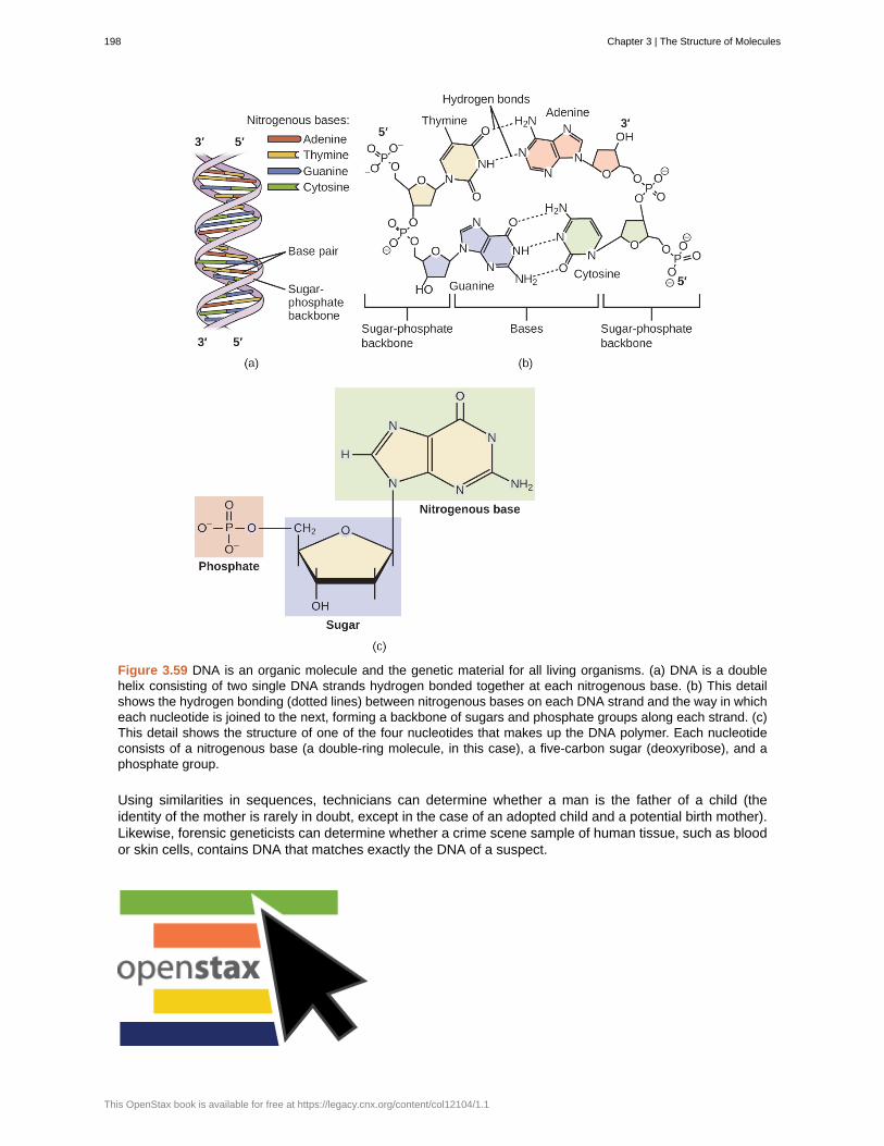
Figure 3.59 DNA is an organic molecule and the genetic material for all living organisms. (a) DNA is a doublehelix consisting of two single DNA strands hydrogen bonded together at each nitrogenous base. (b) This detailshows the hydrogen bonding (dotted lines) between nitrogenous bases on each DNA strand and the way in whicheach nucleotide is joined to the next, forming a backbone of sugars and phosphate groups along each strand. (c)This detail shows the structure of one of the four nucleotides that makes up the DNA polymer. Each nucleotideconsists of a nitrogenous base (a double-ring molecule, in this case), a five-carbon sugar (deoxyribose), and aphosphate group.
Using similarities in sequences, technicians can determine whether a man is the father of a child (theidentity of the mother is rarely in doubt, except in the case of an adopted child and a potential birth mother).Likewise, forensic geneticists can determine whether a crime scene sample of human tissue, such as bloodor skin cells, contains DNA that matches exactly the DNA of a suspect.
198 Chapter 3 | The Structure of Molecules
This OpenStax book is available for free at https://legacy.cnx.org/content/col12104/1.1
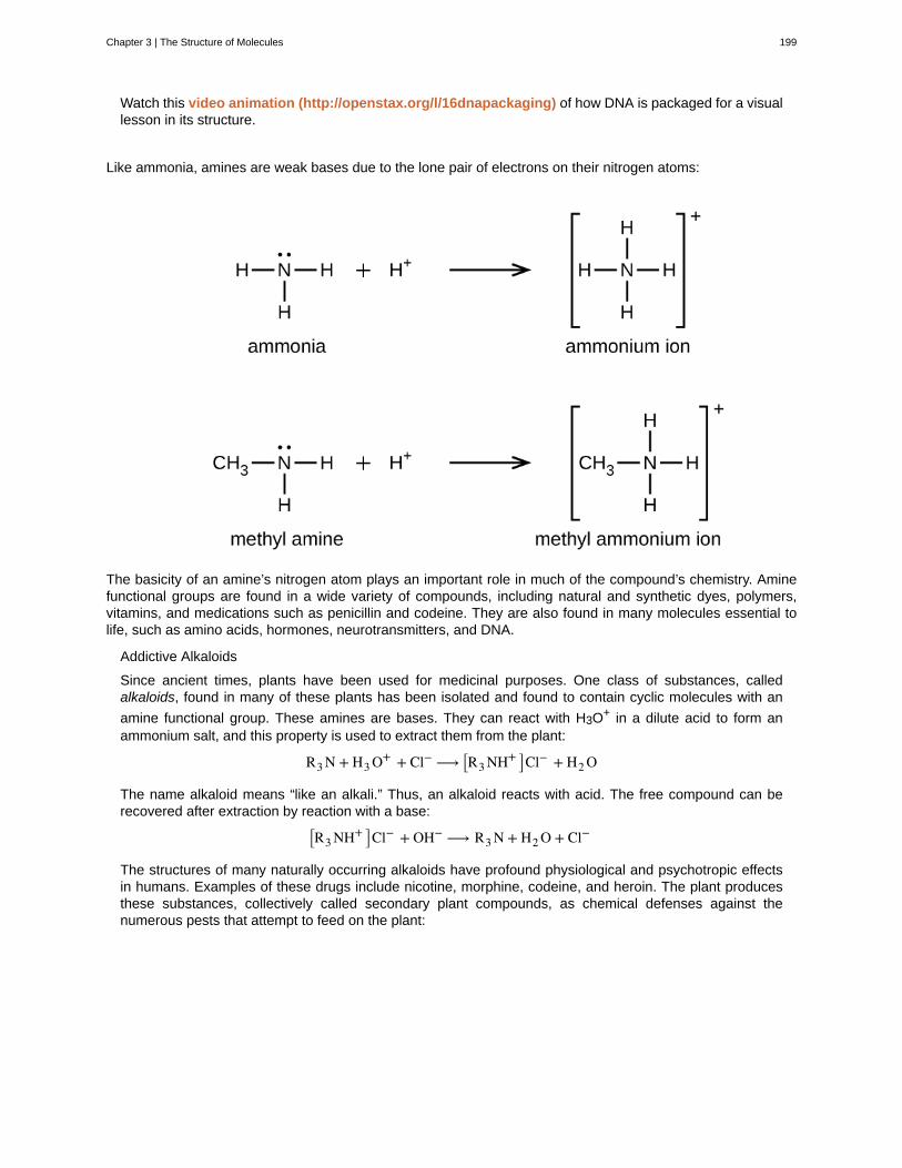
Watch this video animation (http://openstax.org/l/16dnapackaging) of how DNA is packaged for a visuallesson in its structure.
Like ammonia, amines are weak bases due to the lone pair of electrons on their nitrogen atoms:
The basicity of an amine’s nitrogen atom plays an important role in much of the compound’s chemistry. Aminefunctional groups are found in a wide variety of compounds, including natural and synthetic dyes, polymers,vitamins, and medications such as penicillin and codeine. They are also found in many molecules essential tolife, such as amino acids, hormones, neurotransmitters, and DNA.
Addictive Alkaloids
Since ancient times, plants have been used for medicinal purposes. One class of substances, calledalkaloids, found in many of these plants has been isolated and found to contain cyclic molecules with an
amine functional group. These amines are bases. They can react with H3O+ in a dilute acid to form anammonium salt, and this property is used to extract them from the plant:
R3 N + H3 O+ + Cl− ⟶ ⎡⎣R3 NH+ ⎤⎦Cl− + H2 O
The name alkaloid means “like an alkali.” Thus, an alkaloid reacts with acid. The free compound can berecovered after extraction by reaction with a base:
⎡⎣R3 NH+ ⎤⎦Cl− + OH− ⟶ R3 N + H2 O + Cl−
The structures of many naturally occurring alkaloids have profound physiological and psychotropic effectsin humans. Examples of these drugs include nicotine, morphine, codeine, and heroin. The plant producesthese substances, collectively called secondary plant compounds, as chemical defenses against thenumerous pests that attempt to feed on the plant:
Chapter 3 | The Structure of Molecules 199
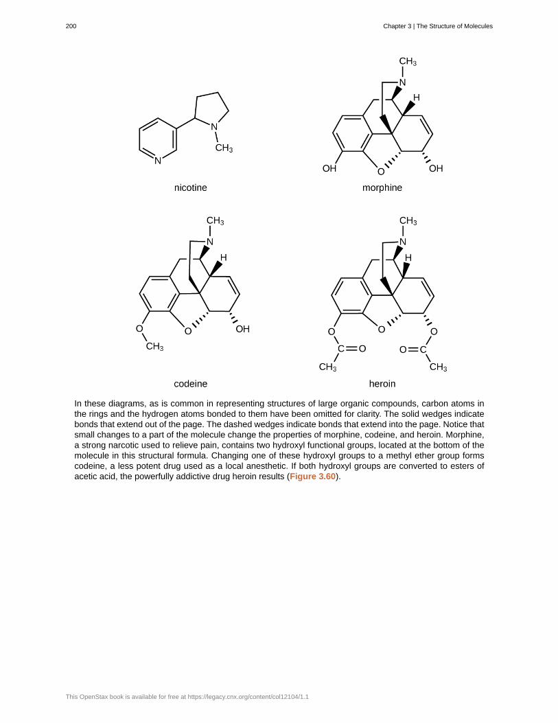
In these diagrams, as is common in representing structures of large organic compounds, carbon atoms inthe rings and the hydrogen atoms bonded to them have been omitted for clarity. The solid wedges indicatebonds that extend out of the page. The dashed wedges indicate bonds that extend into the page. Notice thatsmall changes to a part of the molecule change the properties of morphine, codeine, and heroin. Morphine,a strong narcotic used to relieve pain, contains two hydroxyl functional groups, located at the bottom of themolecule in this structural formula. Changing one of these hydroxyl groups to a methyl ether group formscodeine, a less potent drug used as a local anesthetic. If both hydroxyl groups are converted to esters ofacetic acid, the powerfully addictive drug heroin results (Figure 3.60).
200 Chapter 3 | The Structure of Molecules
This OpenStax book is available for free at https://legacy.cnx.org/content/col12104/1.1
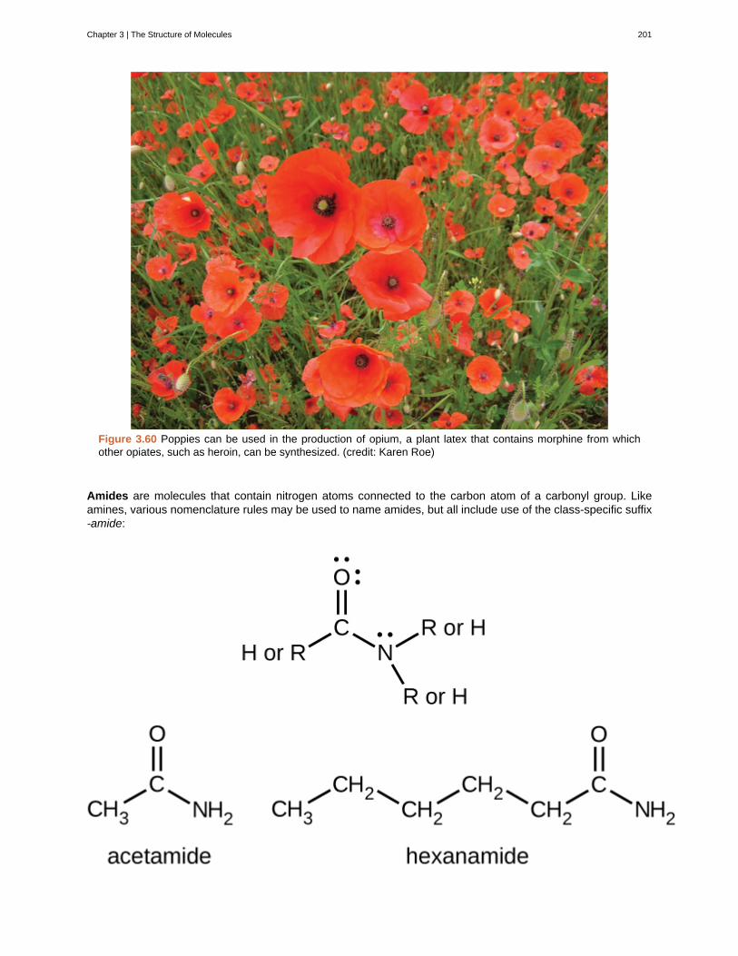
Figure 3.60 Poppies can be used in the production of opium, a plant latex that contains morphine from whichother opiates, such as heroin, can be synthesized. (credit: Karen Roe)
Amides are molecules that contain nitrogen atoms connected to the carbon atom of a carbonyl group. Likeamines, various nomenclature rules may be used to name amides, but all include use of the class-specific suffix-amide:
Chapter 3 | The Structure of Molecules 201
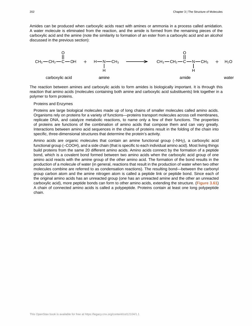
Amides can be produced when carboxylic acids react with amines or ammonia in a process called amidation.A water molecule is eliminated from the reaction, and the amide is formed from the remaining pieces of thecarboxylic acid and the amine (note the similarity to formation of an ester from a carboxylic acid and an alcoholdiscussed in the previous section):
The reaction between amines and carboxylic acids to form amides is biologically important. It is through thisreaction that amino acids (molecules containing both amine and carboxylic acid substituents) link together in apolymer to form proteins.
Proteins and Enzymes
Proteins are large biological molecules made up of long chains of smaller molecules called amino acids.Organisms rely on proteins for a variety of functions—proteins transport molecules across cell membranes,replicate DNA, and catalyze metabolic reactions, to name only a few of their functions. The propertiesof proteins are functions of the combination of amino acids that compose them and can vary greatly.Interactions between amino acid sequences in the chains of proteins result in the folding of the chain intospecific, three-dimensional structures that determine the protein’s activity.
Amino acids are organic molecules that contain an amine functional group (–NH2), a carboxylic acidfunctional group (–COOH), and a side chain (that is specific to each individual amino acid). Most living thingsbuild proteins from the same 20 different amino acids. Amino acids connect by the formation of a peptidebond, which is a covalent bond formed between two amino acids when the carboxylic acid group of oneamino acid reacts with the amine group of the other amino acid. The formation of the bond results in theproduction of a molecule of water (in general, reactions that result in the production of water when two othermolecules combine are referred to as condensation reactions). The resulting bond—between the carbonylgroup carbon atom and the amine nitrogen atom is called a peptide link or peptide bond. Since each ofthe original amino acids has an unreacted group (one has an unreacted amine and the other an unreactedcarboxylic acid), more peptide bonds can form to other amino acids, extending the structure. (Figure 3.61)A chain of connected amino acids is called a polypeptide. Proteins contain at least one long polypeptidechain.
202 Chapter 3 | The Structure of Molecules
This OpenStax book is available for free at https://legacy.cnx.org/content/col12104/1.1
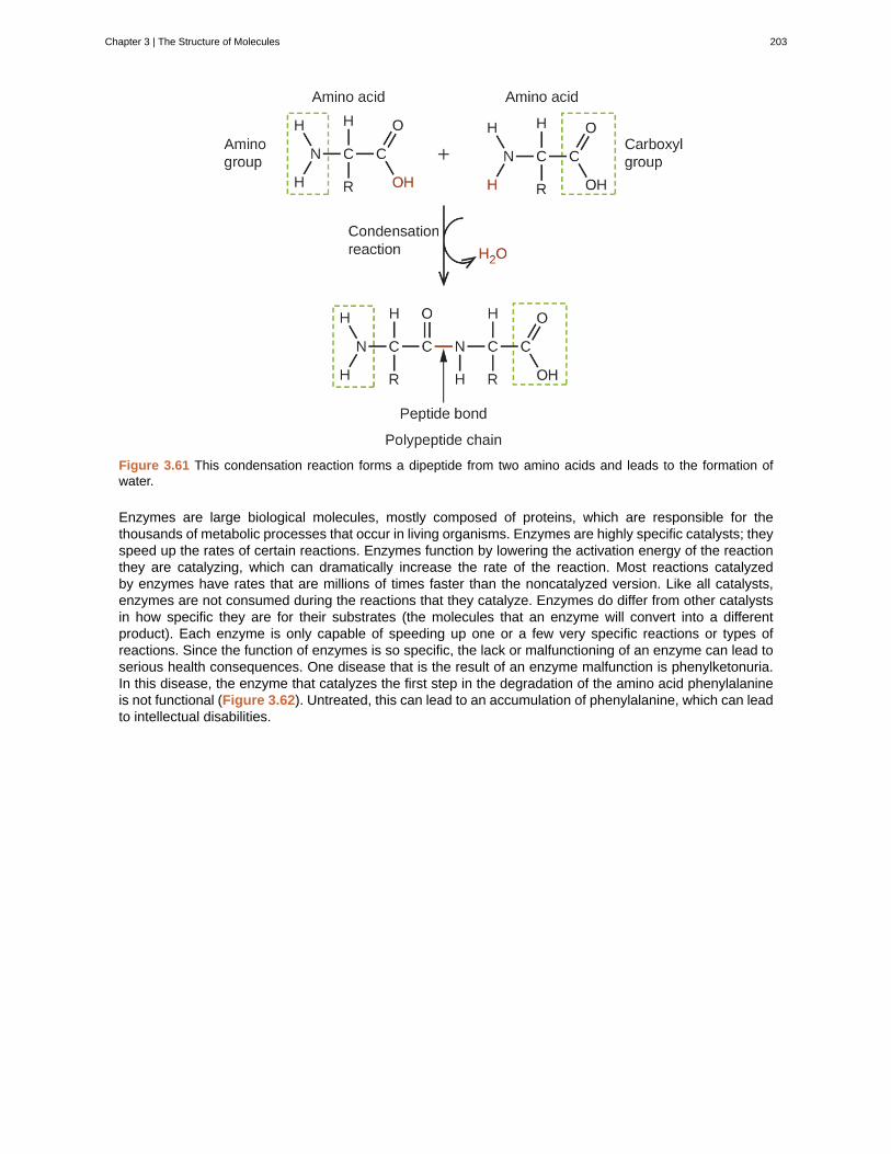
Figure 3.61 This condensation reaction forms a dipeptide from two amino acids and leads to the formation ofwater.
Enzymes are large biological molecules, mostly composed of proteins, which are responsible for thethousands of metabolic processes that occur in living organisms. Enzymes are highly specific catalysts; theyspeed up the rates of certain reactions. Enzymes function by lowering the activation energy of the reactionthey are catalyzing, which can dramatically increase the rate of the reaction. Most reactions catalyzedby enzymes have rates that are millions of times faster than the noncatalyzed version. Like all catalysts,enzymes are not consumed during the reactions that they catalyze. Enzymes do differ from other catalystsin how specific they are for their substrates (the molecules that an enzyme will convert into a differentproduct). Each enzyme is only capable of speeding up one or a few very specific reactions or types ofreactions. Since the function of enzymes is so specific, the lack or malfunctioning of an enzyme can lead toserious health consequences. One disease that is the result of an enzyme malfunction is phenylketonuria.In this disease, the enzyme that catalyzes the first step in the degradation of the amino acid phenylalanineis not functional (Figure 3.62). Untreated, this can lead to an accumulation of phenylalanine, which can leadto intellectual disabilities.
Chapter 3 | The Structure of Molecules 203
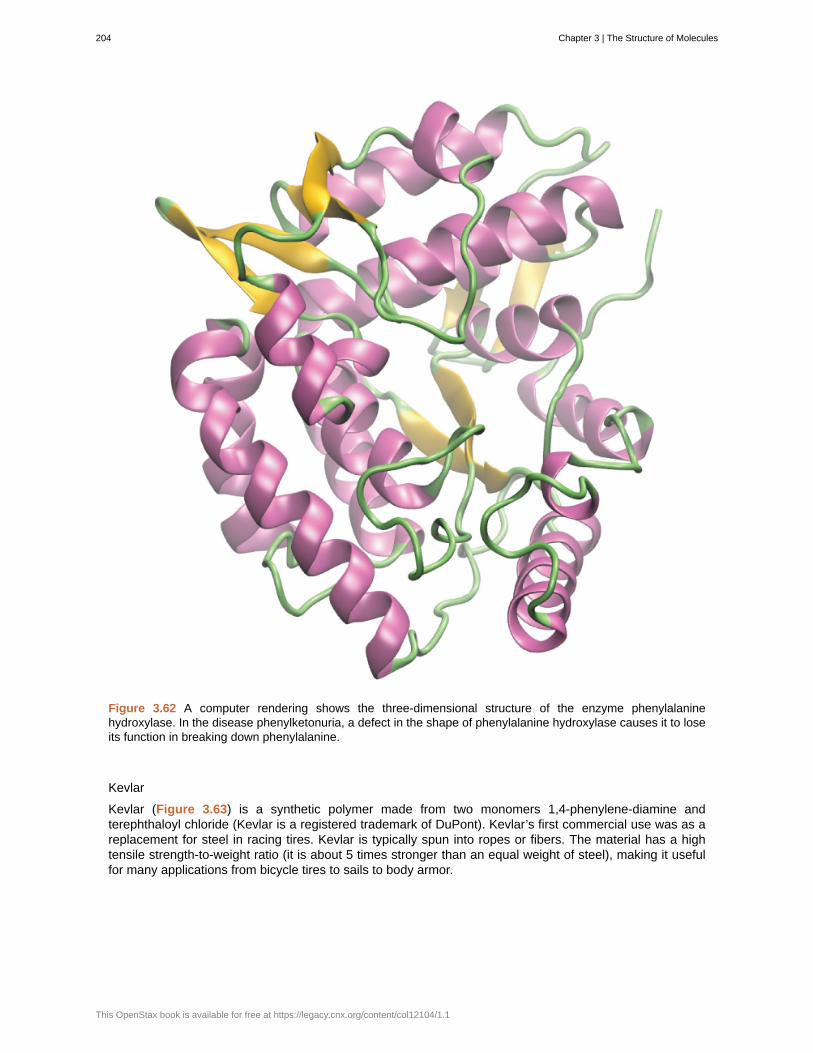
Figure 3.62 A computer rendering shows the three-dimensional structure of the enzyme phenylalaninehydroxylase. In the disease phenylketonuria, a defect in the shape of phenylalanine hydroxylase causes it to loseits function in breaking down phenylalanine.
Kevlar
Kevlar (Figure 3.63) is a synthetic polymer made from two monomers 1,4-phenylene-diamine andterephthaloyl chloride (Kevlar is a registered trademark of DuPont). Kevlar’s first commercial use was as areplacement for steel in racing tires. Kevlar is typically spun into ropes or fibers. The material has a hightensile strength-to-weight ratio (it is about 5 times stronger than an equal weight of steel), making it usefulfor many applications from bicycle tires to sails to body armor.
204 Chapter 3 | The Structure of Molecules
This OpenStax book is available for free at https://legacy.cnx.org/content/col12104/1.1

Figure 3.63 This illustration shows the formula for polymeric Kevlar.
The material owes much of its strength to hydrogen bonds between polymer chains (refer back to thechapter on intermolecular interactions). These bonds form between the carbonyl group oxygen atom (whichhas a partial negative charge due to oxygen’s electronegativity) on one monomer and the partially positivelycharged hydrogen atom in the N–H bond of an adjacent monomer in the polymer structure (see dashed linein Figure 3.64). There is additional strength derived from the interaction between the unhybridized p orbitalsin the six-membered rings, called aromatic stacking.
Figure 3.64 The diagram shows the polymer structure of Kevlar, with hydrogen bonds between polymer chainsrepresented by dotted lines.
Kevlar may be best known as a component of body armor, combat helmets, and face masks. Since the1980s, the US military has used Kevlar as a component of the PASGT (personal armor system for groundtroops) helmet and vest. Kevlar is also used to protect armored fighting vehicles and aircraft carriers. Civilianapplications include protective gear for emergency service personnel such as body armor for police officersand heat-resistant clothing for fire fighters. Kevlar based clothing is considerably lighter and thinner thanequivalent gear made from other materials (Figure 3.65).
Chapter 3 | The Structure of Molecules 205
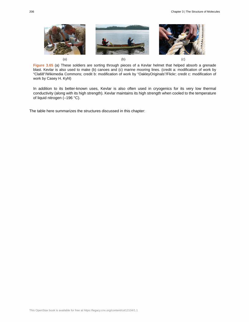
Figure 3.65 (a) These soldiers are sorting through pieces of a Kevlar helmet that helped absorb a grenadeblast. Kevlar is also used to make (b) canoes and (c) marine mooring lines. (credit a: modification of work by“Cla68”/Wikimedia Commons; credit b: modification of work by “OakleyOriginals”/Flickr; credit c: modification ofwork by Casey H. Kyhl)
In addition to its better-known uses, Kevlar is also often used in cryogenics for its very low thermalconductivity (along with its high strength). Kevlar maintains its high strength when cooled to the temperatureof liquid nitrogen (–196 °C).
The table here summarizes the structures discussed in this chapter:
206 Chapter 3 | The Structure of Molecules
This OpenStax book is available for free at https://legacy.cnx.org/content/col12104/1.1
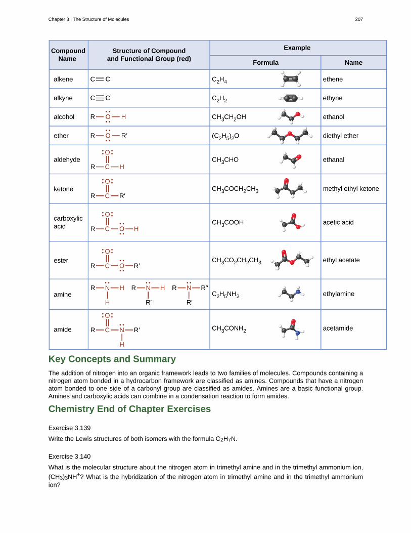
Key Concepts and Summary
The addition of nitrogen into an organic framework leads to two families of molecules. Compounds containing anitrogen atom bonded in a hydrocarbon framework are classified as amines. Compounds that have a nitrogenatom bonded to one side of a carbonyl group are classified as amides. Amines are a basic functional group.Amines and carboxylic acids can combine in a condensation reaction to form amides.
Chemistry End of Chapter Exercises
Exercise 3.139
Write the Lewis structures of both isomers with the formula C2H7N.
Exercise 3.140
What is the molecular structure about the nitrogen atom in trimethyl amine and in the trimethyl ammonium ion,
(CH3)3NH+? What is the hybridization of the nitrogen atom in trimethyl amine and in the trimethyl ammoniumion?
Chapter 3 | The Structure of Molecules 207
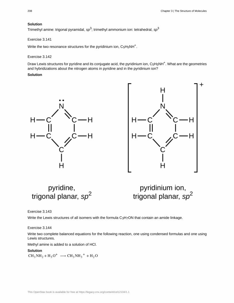
Solution
Trimethyl amine: trigonal pyramidal, sp3; trimethyl ammonium ion: tetrahedral, sp3
Exercise 3.141
Write the two resonance structures for the pyridinium ion, C5H5NH+.
Exercise 3.142
Draw Lewis structures for pyridine and its conjugate acid, the pyridinium ion, C5H5NH+. What are the geometriesand hybridizations about the nitrogen atoms in pyridine and in the pyridinium ion?
Solution
Exercise 3.143
Write the Lewis structures of all isomers with the formula C3H7ON that contain an amide linkage.
Exercise 3.144
Write two complete balanced equations for the following reaction, one using condensed formulas and one usingLewis structures.
Methyl amine is added to a solution of HCl.
SolutionCH3 NH2 + H3 O+ ⟶ CH3 NH3
+ + H2 O
208 Chapter 3 | The Structure of Molecules
This OpenStax book is available for free at https://legacy.cnx.org/content/col12104/1.1
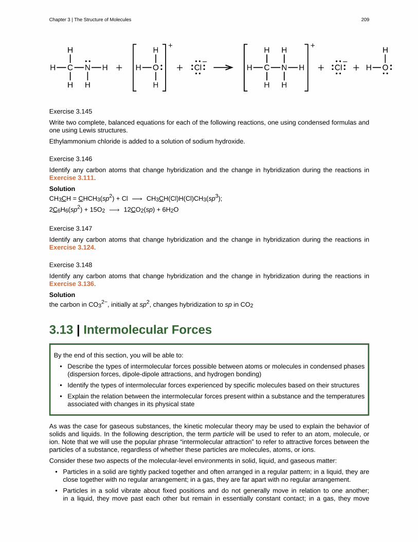
Exercise 3.145
Write two complete, balanced equations for each of the following reactions, one using condensed formulas andone using Lewis structures.
Ethylammonium chloride is added to a solution of sodium hydroxide.
Exercise 3.146
Identify any carbon atoms that change hybridization and the change in hybridization during the reactions inExercise 3.111.
Solution
CH3CH = CHCH3(sp2) + Cl ⟶ CH3CH(Cl)H(Cl)CH3(sp3);
2C6H6(sp2) + 15O2 ⟶ 12CO2(sp) + 6H2O
Exercise 3.147
Identify any carbon atoms that change hybridization and the change in hybridization during the reactions inExercise 3.124.
Exercise 3.148
Identify any carbon atoms that change hybridization and the change in hybridization during the reactions inExercise 3.136.
Solution
the carbon in CO32−, initially at sp2, changes hybridization to sp in CO2
3.13 | Intermolecular Forces
By the end of this section, you will be able to:
• Describe the types of intermolecular forces possible between atoms or molecules in condensed phases(dispersion forces, dipole-dipole attractions, and hydrogen bonding)
• Identify the types of intermolecular forces experienced by specific molecules based on their structures
• Explain the relation between the intermolecular forces present within a substance and the temperaturesassociated with changes in its physical state
As was the case for gaseous substances, the kinetic molecular theory may be used to explain the behavior ofsolids and liquids. In the following description, the term particle will be used to refer to an atom, molecule, orion. Note that we will use the popular phrase “intermolecular attraction” to refer to attractive forces between theparticles of a substance, regardless of whether these particles are molecules, atoms, or ions.
Consider these two aspects of the molecular-level environments in solid, liquid, and gaseous matter:
• Particles in a solid are tightly packed together and often arranged in a regular pattern; in a liquid, they areclose together with no regular arrangement; in a gas, they are far apart with no regular arrangement.
• Particles in a solid vibrate about fixed positions and do not generally move in relation to one another;in a liquid, they move past each other but remain in essentially constant contact; in a gas, they move
Chapter 3 | The Structure of Molecules 209
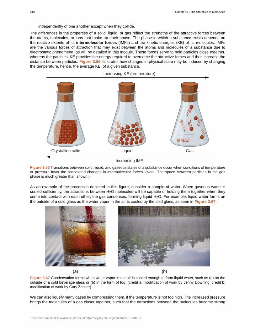
independently of one another except when they collide.
The differences in the properties of a solid, liquid, or gas reflect the strengths of the attractive forces betweenthe atoms, molecules, or ions that make up each phase. The phase in which a substance exists depends onthe relative extents of its intermolecular forces (IMFs) and the kinetic energies (KE) of its molecules. IMFsare the various forces of attraction that may exist between the atoms and molecules of a substance due toelectrostatic phenomena, as will be detailed in this module. These forces serve to hold particles close together,whereas the particles’ KE provides the energy required to overcome the attractive forces and thus increase thedistance between particles. Figure 3.66 illustrates how changes in physical state may be induced by changingthe temperature, hence, the average KE, of a given substance.
Figure 3.66 Transitions between solid, liquid, and gaseous states of a substance occur when conditions of temperatureor pressure favor the associated changes in intermolecular forces. (Note: The space between particles in the gasphase is much greater than shown.)
As an example of the processes depicted in this figure, consider a sample of water. When gaseous water iscooled sufficiently, the attractions between H2O molecules will be capable of holding them together when theycome into contact with each other; the gas condenses, forming liquid H2O. For example, liquid water forms onthe outside of a cold glass as the water vapor in the air is cooled by the cold glass, as seen in Figure 3.67.
Figure 3.67 Condensation forms when water vapor in the air is cooled enough to form liquid water, such as (a) on theoutside of a cold beverage glass or (b) in the form of fog. (credit a: modification of work by Jenny Downing; credit b:modification of work by Cory Zanker)
We can also liquefy many gases by compressing them, if the temperature is not too high. The increased pressurebrings the molecules of a gas closer together, such that the attractions between the molecules become strong
210 Chapter 3 | The Structure of Molecules
This OpenStax book is available for free at https://legacy.cnx.org/content/col12104/1.1
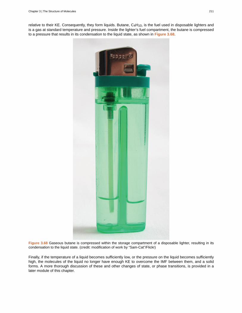
relative to their KE. Consequently, they form liquids. Butane, C4H10, is the fuel used in disposable lighters andis a gas at standard temperature and pressure. Inside the lighter’s fuel compartment, the butane is compressedto a pressure that results in its condensation to the liquid state, as shown in Figure 3.68.
Figure 3.68 Gaseous butane is compressed within the storage compartment of a disposable lighter, resulting in itscondensation to the liquid state. (credit: modification of work by “Sam-Cat”/Flickr)
Finally, if the temperature of a liquid becomes sufficiently low, or the pressure on the liquid becomes sufficientlyhigh, the molecules of the liquid no longer have enough KE to overcome the IMF between them, and a solidforms. A more thorough discussion of these and other changes of state, or phase transitions, is provided in alater module of this chapter.
Chapter 3 | The Structure of Molecules 211
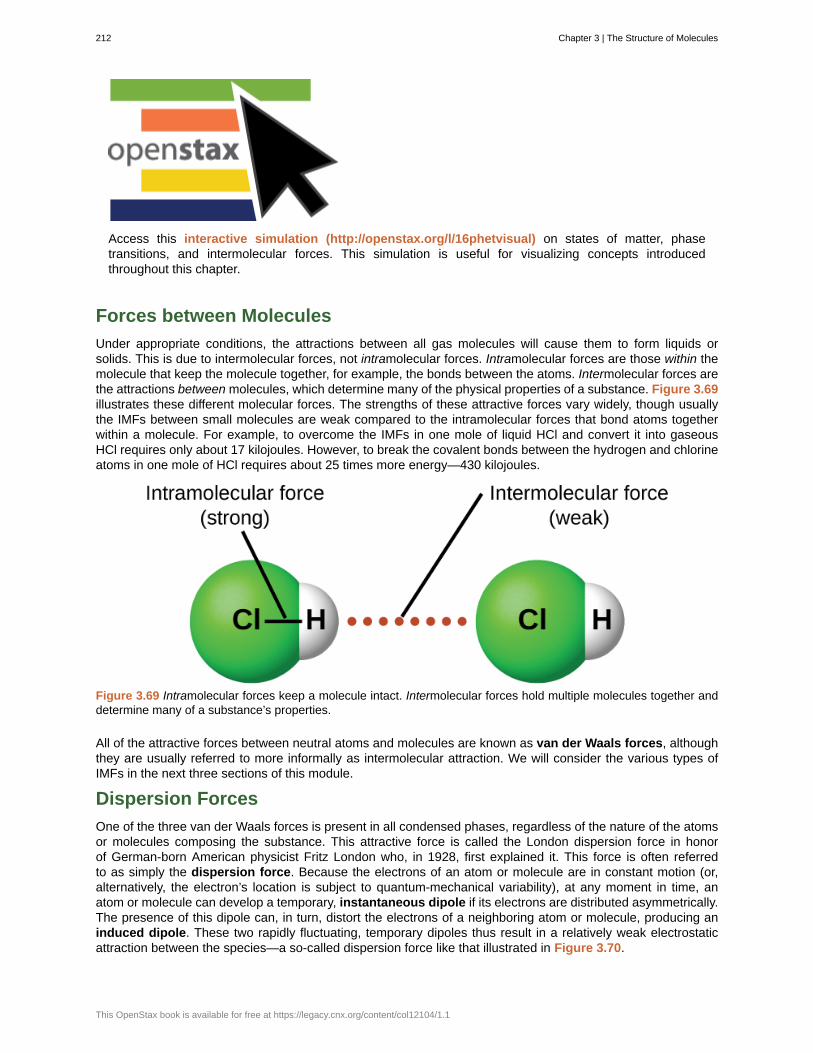
Access this interactive simulation (http://openstax.org/l/16phetvisual) on states of matter, phasetransitions, and intermolecular forces. This simulation is useful for visualizing concepts introducedthroughout this chapter.
Forces between Molecules
Under appropriate conditions, the attractions between all gas molecules will cause them to form liquids orsolids. This is due to intermolecular forces, not intramolecular forces. Intramolecular forces are those within themolecule that keep the molecule together, for example, the bonds between the atoms. Intermolecular forces arethe attractions between molecules, which determine many of the physical properties of a substance. Figure 3.69illustrates these different molecular forces. The strengths of these attractive forces vary widely, though usuallythe IMFs between small molecules are weak compared to the intramolecular forces that bond atoms togetherwithin a molecule. For example, to overcome the IMFs in one mole of liquid HCl and convert it into gaseousHCl requires only about 17 kilojoules. However, to break the covalent bonds between the hydrogen and chlorineatoms in one mole of HCl requires about 25 times more energy—430 kilojoules.
Figure 3.69 Intramolecular forces keep a molecule intact. Intermolecular forces hold multiple molecules together anddetermine many of a substance’s properties.
All of the attractive forces between neutral atoms and molecules are known as van der Waals forces, althoughthey are usually referred to more informally as intermolecular attraction. We will consider the various types ofIMFs in the next three sections of this module.
Dispersion Forces
One of the three van der Waals forces is present in all condensed phases, regardless of the nature of the atomsor molecules composing the substance. This attractive force is called the London dispersion force in honorof German-born American physicist Fritz London who, in 1928, first explained it. This force is often referredto as simply the dispersion force. Because the electrons of an atom or molecule are in constant motion (or,alternatively, the electron’s location is subject to quantum-mechanical variability), at any moment in time, anatom or molecule can develop a temporary, instantaneous dipole if its electrons are distributed asymmetrically.The presence of this dipole can, in turn, distort the electrons of a neighboring atom or molecule, producing aninduced dipole. These two rapidly fluctuating, temporary dipoles thus result in a relatively weak electrostaticattraction between the species—a so-called dispersion force like that illustrated in Figure 3.70.
212 Chapter 3 | The Structure of Molecules
This OpenStax book is available for free at https://legacy.cnx.org/content/col12104/1.1
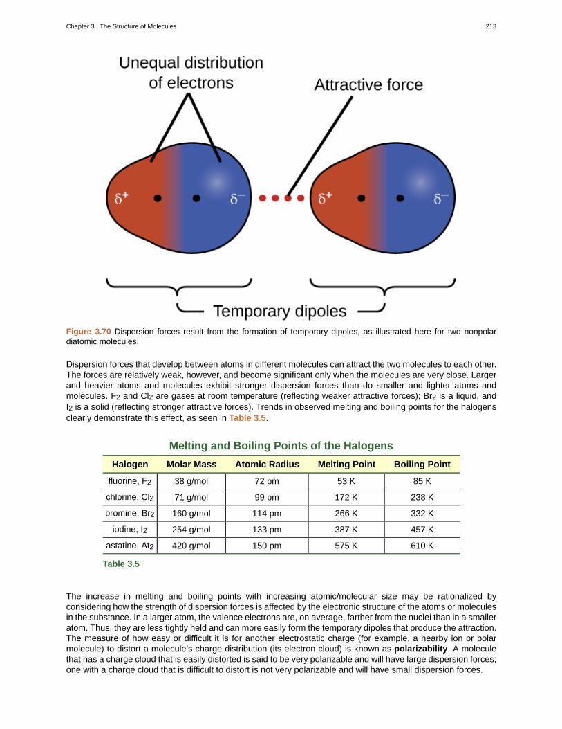
Figure 3.70 Dispersion forces result from the formation of temporary dipoles, as illustrated here for two nonpolardiatomic molecules.
Dispersion forces that develop between atoms in different molecules can attract the two molecules to each other.The forces are relatively weak, however, and become significant only when the molecules are very close. Largerand heavier atoms and molecules exhibit stronger dispersion forces than do smaller and lighter atoms andmolecules. F2 and Cl2 are gases at room temperature (reflecting weaker attractive forces); Br2 is a liquid, andI2 is a solid (reflecting stronger attractive forces). Trends in observed melting and boiling points for the halogensclearly demonstrate this effect, as seen in Table 3.5.
Melting and Boiling Points of the Halogens
Halogen Molar Mass Atomic Radius Melting Point Boiling Point
fluorine, F2 38 g/mol 72 pm 53 K 85 K
chlorine, Cl2 71 g/mol 99 pm 172 K 238 K
bromine, Br2 160 g/mol 114 pm 266 K 332 K
iodine, I2 254 g/mol 133 pm 387 K 457 K
astatine, At2 420 g/mol 150 pm 575 K 610 K
Table 3.5
The increase in melting and boiling points with increasing atomic/molecular size may be rationalized byconsidering how the strength of dispersion forces is affected by the electronic structure of the atoms or moleculesin the substance. In a larger atom, the valence electrons are, on average, farther from the nuclei than in a smalleratom. Thus, they are less tightly held and can more easily form the temporary dipoles that produce the attraction.The measure of how easy or difficult it is for another electrostatic charge (for example, a nearby ion or polarmolecule) to distort a molecule’s charge distribution (its electron cloud) is known as polarizability. A moleculethat has a charge cloud that is easily distorted is said to be very polarizable and will have large dispersion forces;one with a charge cloud that is difficult to distort is not very polarizable and will have small dispersion forces.
Chapter 3 | The Structure of Molecules 213
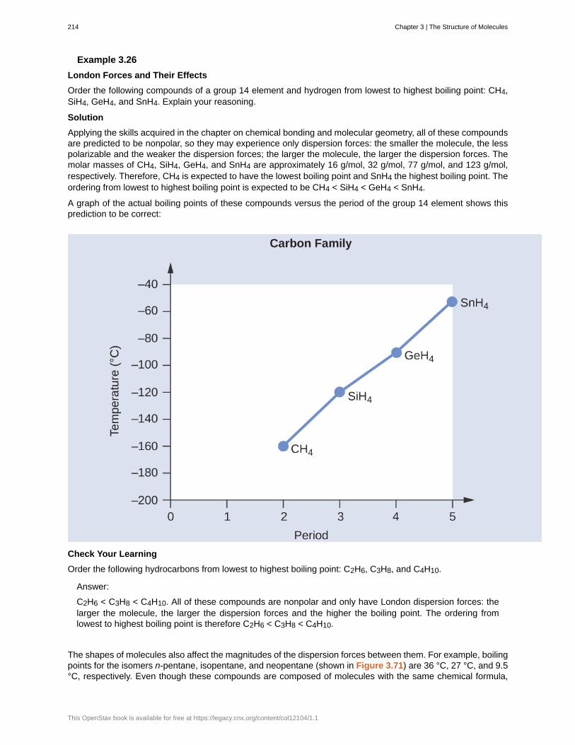
Example 3.26
London Forces and Their Effects
Order the following compounds of a group 14 element and hydrogen from lowest to highest boiling point: CH4,SiH4, GeH4, and SnH4. Explain your reasoning.
Solution
Applying the skills acquired in the chapter on chemical bonding and molecular geometry, all of these compoundsare predicted to be nonpolar, so they may experience only dispersion forces: the smaller the molecule, the lesspolarizable and the weaker the dispersion forces; the larger the molecule, the larger the dispersion forces. Themolar masses of CH4, SiH4, GeH4, and SnH4 are approximately 16 g/mol, 32 g/mol, 77 g/mol, and 123 g/mol,respectively. Therefore, CH4 is expected to have the lowest boiling point and SnH4 the highest boiling point. Theordering from lowest to highest boiling point is expected to be CH4 < SiH4 < GeH4 < SnH4.
A graph of the actual boiling points of these compounds versus the period of the group 14 element shows thisprediction to be correct:
Check Your Learning
Order the following hydrocarbons from lowest to highest boiling point: C2H6, C3H8, and C4H10.
Answer:
C2H6 < C3H8 < C4H10. All of these compounds are nonpolar and only have London dispersion forces: thelarger the molecule, the larger the dispersion forces and the higher the boiling point. The ordering fromlowest to highest boiling point is therefore C2H6 < C3H8 < C4H10.
The shapes of molecules also affect the magnitudes of the dispersion forces between them. For example, boilingpoints for the isomers n-pentane, isopentane, and neopentane (shown in Figure 3.71) are 36 °C, 27 °C, and 9.5°C, respectively. Even though these compounds are composed of molecules with the same chemical formula,
214 Chapter 3 | The Structure of Molecules
This OpenStax book is available for free at https://legacy.cnx.org/content/col12104/1.1

C5H12, the difference in boiling points suggests that dispersion forces in the liquid phase are different, beinggreatest for n-pentane and least for neopentane. The elongated shape of n-pentane provides a greater surfacearea available for contact between molecules, resulting in correspondingly stronger dispersion forces. The morecompact shape of isopentane offers a smaller surface area available for intermolecular contact and, therefore,weaker dispersion forces. Neopentane molecules are the most compact of the three, offering the least availablesurface area for intermolecular contact and, hence, the weakest dispersion forces. This behavior is analogousto the connections that may be formed between strips of VELCRO brand fasteners: the greater the area of thestrip’s contact, the stronger the connection.
Figure 3.71 The strength of the dispersion forces increases with the contact area between molecules, as demonstratedby the boiling points of these pentane isomers.
Geckos and Intermolecular Forces
Geckos have an amazing ability to adhere to most surfaces. They can quickly run up smooth walls andacross ceilings that have no toe-holds, and they do this without having suction cups or a sticky substanceon their toes. And while a gecko can lift its feet easily as it walks along a surface, if you attempt to pick itup, it sticks to the surface. How are geckos (as well as spiders and some other insects) able to do this?Although this phenomenon has been investigated for hundreds of years, scientists only recently uncoveredthe details of the process that allows geckos’ feet to behave this way.
Geckos’ toes are covered with hundreds of thousands of tiny hairs known as setae, with each seta, inturn, branching into hundreds of tiny, flat, triangular tips called spatulae. The huge numbers of spatulaeon its setae provide a gecko, shown in Figure 3.72, with a large total surface area for sticking to asurface. In 2000, Kellar Autumn, who leads a multi-institutional gecko research team, found that geckosadhered equally well to both polar silicon dioxide and nonpolar gallium arsenide. This proved that geckosstick to surfaces because of dispersion forces—weak intermolecular attractions arising from temporary,synchronized charge distributions between adjacent molecules. Although dispersion forces are very weak,the total attraction over millions of spatulae is large enough to support many times the gecko’s weight.
In 2014, two scientists developed a model to explain how geckos can rapidly transition from “sticky” to “non-sticky.” Alex Greaney and Congcong Hu at Oregon State University described how geckos can achieve thisby changing the angle between their spatulae and the surface. Geckos’ feet, which are normally nonsticky,become sticky when a small shear force is applied. By curling and uncurling their toes, geckos can alternatebetween sticking and unsticking from a surface, and thus easily move across it. Further investigations mayeventually lead to the development of better adhesives and other applications.
Chapter 3 | The Structure of Molecules 215
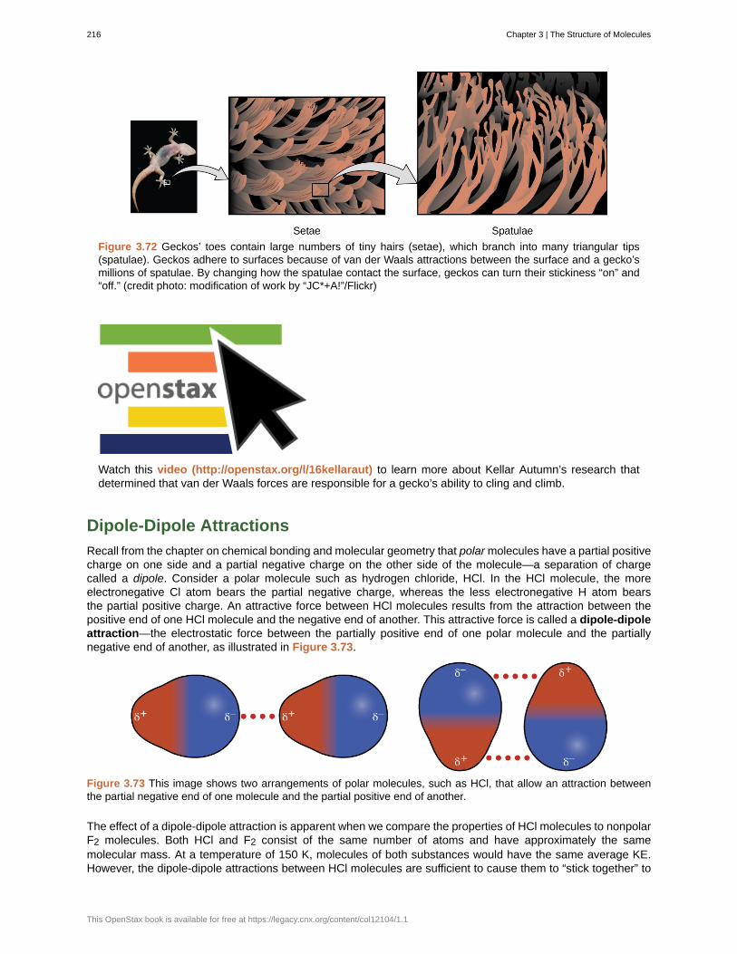
Figure 3.72 Geckos’ toes contain large numbers of tiny hairs (setae), which branch into many triangular tips(spatulae). Geckos adhere to surfaces because of van der Waals attractions between the surface and a gecko’smillions of spatulae. By changing how the spatulae contact the surface, geckos can turn their stickiness “on” and“off.” (credit photo: modification of work by “JC*+A!”/Flickr)
Watch this video (http://openstax.org/l/16kellaraut) to learn more about Kellar Autumn’s research thatdetermined that van der Waals forces are responsible for a gecko’s ability to cling and climb.
Dipole-Dipole Attractions
Recall from the chapter on chemical bonding and molecular geometry that polar molecules have a partial positivecharge on one side and a partial negative charge on the other side of the molecule—a separation of chargecalled a dipole. Consider a polar molecule such as hydrogen chloride, HCl. In the HCl molecule, the moreelectronegative Cl atom bears the partial negative charge, whereas the less electronegative H atom bearsthe partial positive charge. An attractive force between HCl molecules results from the attraction between thepositive end of one HCl molecule and the negative end of another. This attractive force is called a dipole-dipoleattraction—the electrostatic force between the partially positive end of one polar molecule and the partiallynegative end of another, as illustrated in Figure 3.73.
Figure 3.73 This image shows two arrangements of polar molecules, such as HCl, that allow an attraction betweenthe partial negative end of one molecule and the partial positive end of another.
The effect of a dipole-dipole attraction is apparent when we compare the properties of HCl molecules to nonpolarF2 molecules. Both HCl and F2 consist of the same number of atoms and have approximately the samemolecular mass. At a temperature of 150 K, molecules of both substances would have the same average KE.However, the dipole-dipole attractions between HCl molecules are sufficient to cause them to “stick together” to
216 Chapter 3 | The Structure of Molecules
This OpenStax book is available for free at https://legacy.cnx.org/content/col12104/1.1
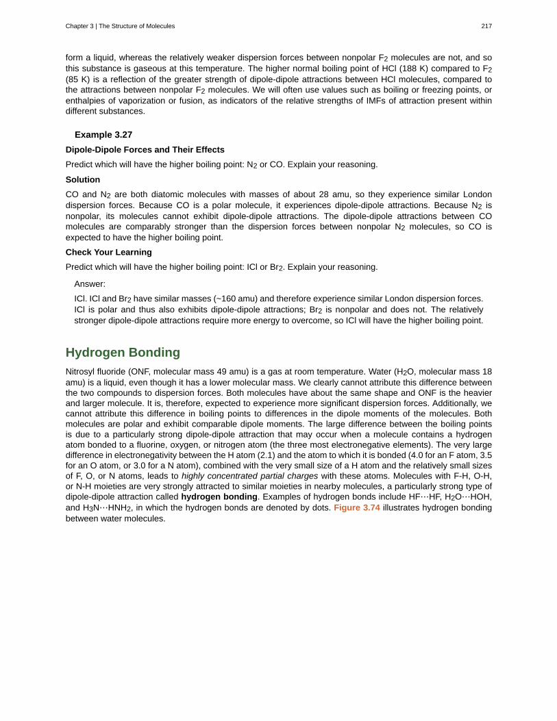
form a liquid, whereas the relatively weaker dispersion forces between nonpolar F2 molecules are not, and sothis substance is gaseous at this temperature. The higher normal boiling point of HCl (188 K) compared to F2(85 K) is a reflection of the greater strength of dipole-dipole attractions between HCl molecules, compared tothe attractions between nonpolar F2 molecules. We will often use values such as boiling or freezing points, orenthalpies of vaporization or fusion, as indicators of the relative strengths of IMFs of attraction present withindifferent substances.
Example 3.27
Dipole-Dipole Forces and Their Effects
Predict which will have the higher boiling point: N2 or CO. Explain your reasoning.
Solution
CO and N2 are both diatomic molecules with masses of about 28 amu, so they experience similar Londondispersion forces. Because CO is a polar molecule, it experiences dipole-dipole attractions. Because N2 isnonpolar, its molecules cannot exhibit dipole-dipole attractions. The dipole-dipole attractions between COmolecules are comparably stronger than the dispersion forces between nonpolar N2 molecules, so CO isexpected to have the higher boiling point.
Check Your Learning
Predict which will have the higher boiling point: ICl or Br2. Explain your reasoning.
Answer:
ICl. ICl and Br2 have similar masses (~160 amu) and therefore experience similar London dispersion forces.ICl is polar and thus also exhibits dipole-dipole attractions; Br2 is nonpolar and does not. The relativelystronger dipole-dipole attractions require more energy to overcome, so ICl will have the higher boiling point.
Hydrogen Bonding
Nitrosyl fluoride (ONF, molecular mass 49 amu) is a gas at room temperature. Water (H2O, molecular mass 18amu) is a liquid, even though it has a lower molecular mass. We clearly cannot attribute this difference betweenthe two compounds to dispersion forces. Both molecules have about the same shape and ONF is the heavierand larger molecule. It is, therefore, expected to experience more significant dispersion forces. Additionally, wecannot attribute this difference in boiling points to differences in the dipole moments of the molecules. Bothmolecules are polar and exhibit comparable dipole moments. The large difference between the boiling pointsis due to a particularly strong dipole-dipole attraction that may occur when a molecule contains a hydrogenatom bonded to a fluorine, oxygen, or nitrogen atom (the three most electronegative elements). The very largedifference in electronegativity between the H atom (2.1) and the atom to which it is bonded (4.0 for an F atom, 3.5for an O atom, or 3.0 for a N atom), combined with the very small size of a H atom and the relatively small sizesof F, O, or N atoms, leads to highly concentrated partial charges with these atoms. Molecules with F-H, O-H,or N-H moieties are very strongly attracted to similar moieties in nearby molecules, a particularly strong type ofdipole-dipole attraction called hydrogen bonding. Examples of hydrogen bonds include HF⋯HF, H2O⋯HOH,and H3N⋯HNH2, in which the hydrogen bonds are denoted by dots. Figure 3.74 illustrates hydrogen bondingbetween water molecules.
Chapter 3 | The Structure of Molecules 217
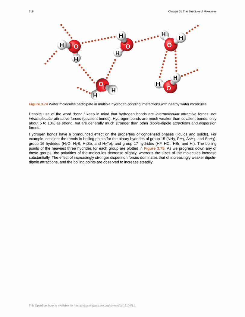
Figure 3.74 Water molecules participate in multiple hydrogen-bonding interactions with nearby water molecules.
Despite use of the word “bond,” keep in mind that hydrogen bonds are intermolecular attractive forces, notintramolecular attractive forces (covalent bonds). Hydrogen bonds are much weaker than covalent bonds, onlyabout 5 to 10% as strong, but are generally much stronger than other dipole-dipole attractions and dispersionforces.
Hydrogen bonds have a pronounced effect on the properties of condensed phases (liquids and solids). Forexample, consider the trends in boiling points for the binary hydrides of group 15 (NH3, PH3, AsH3, and SbH3),group 16 hydrides (H2O, H2S, H2Se, and H2Te), and group 17 hydrides (HF, HCl, HBr, and HI). The boilingpoints of the heaviest three hydrides for each group are plotted in Figure 3.75. As we progress down any ofthese groups, the polarities of the molecules decrease slightly, whereas the sizes of the molecules increasesubstantially. The effect of increasingly stronger dispersion forces dominates that of increasingly weaker dipole-dipole attractions, and the boiling points are observed to increase steadily.
218 Chapter 3 | The Structure of Molecules
This OpenStax book is available for free at https://legacy.cnx.org/content/col12104/1.1
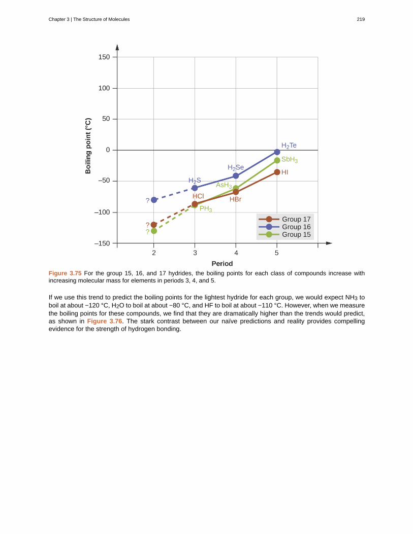
Figure 3.75 For the group 15, 16, and 17 hydrides, the boiling points for each class of compounds increase withincreasing molecular mass for elements in periods 3, 4, and 5.
If we use this trend to predict the boiling points for the lightest hydride for each group, we would expect NH3 toboil at about −120 °C, H2O to boil at about −80 °C, and HF to boil at about −110 °C. However, when we measurethe boiling points for these compounds, we find that they are dramatically higher than the trends would predict,as shown in Figure 3.76. The stark contrast between our naïve predictions and reality provides compellingevidence for the strength of hydrogen bonding.
Chapter 3 | The Structure of Molecules 219
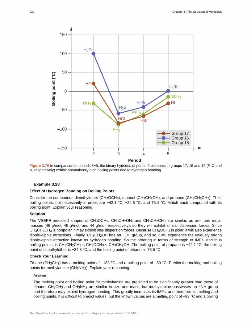
Figure 3.76 In comparison to periods 3−5, the binary hydrides of period 2 elements in groups 17, 16 and 15 (F, O andN, respectively) exhibit anomalously high boiling points due to hydrogen bonding.
Example 3.28
Effect of Hydrogen Bonding on Boiling Points
Consider the compounds dimethylether (CH3OCH3), ethanol (CH3CH2OH), and propane (CH3CH2CH3). Theirboiling points, not necessarily in order, are −42.1 °C, −24.8 °C, and 78.4 °C. Match each compound with itsboiling point. Explain your reasoning.
Solution
The VSEPR-predicted shapes of CH3OCH3, CH3CH2OH, and CH3CH2CH3 are similar, as are their molarmasses (46 g/mol, 46 g/mol, and 44 g/mol, respectively), so they will exhibit similar dispersion forces. SinceCH3CH2CH3 is nonpolar, it may exhibit only dispersion forces. Because CH3OCH3 is polar, it will also experiencedipole-dipole attractions. Finally, CH3CH2OH has an −OH group, and so it will experience the uniquely strongdipole-dipole attraction known as hydrogen bonding. So the ordering in terms of strength of IMFs, and thusboiling points, is CH3CH2CH3 < CH3OCH3 < CH3CH2OH. The boiling point of propane is −42.1 °C, the boilingpoint of dimethylether is −24.8 °C, and the boiling point of ethanol is 78.5 °C.
Check Your Learning
Ethane (CH3CH3) has a melting point of −183 °C and a boiling point of −89 °C. Predict the melting and boilingpoints for methylamine (CH3NH2). Explain your reasoning.
Answer:
The melting point and boiling point for methylamine are predicted to be significantly greater than those ofethane. CH3CH3 and CH3NH2 are similar in size and mass, but methylamine possesses an −NH groupand therefore may exhibit hydrogen bonding. This greatly increases its IMFs, and therefore its melting andboiling points. It is difficult to predict values, but the known values are a melting point of −93 °C and a boiling
220 Chapter 3 | The Structure of Molecules
This OpenStax book is available for free at https://legacy.cnx.org/content/col12104/1.1
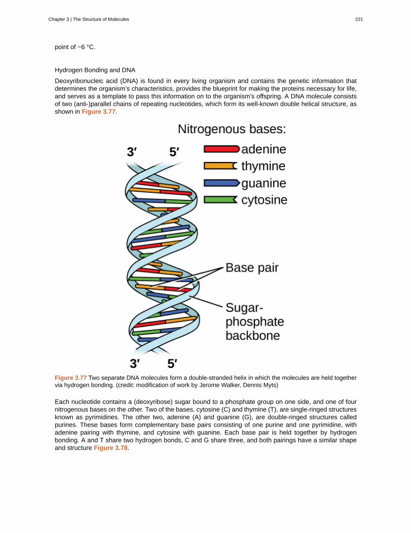
point of −6 °C.
Hydrogen Bonding and DNA
Deoxyribonucleic acid (DNA) is found in every living organism and contains the genetic information thatdetermines the organism’s characteristics, provides the blueprint for making the proteins necessary for life,and serves as a template to pass this information on to the organism’s offspring. A DNA molecule consistsof two (anti-)parallel chains of repeating nucleotides, which form its well-known double helical structure, asshown in Figure 3.77.
Figure 3.77 Two separate DNA molecules form a double-stranded helix in which the molecules are held togethervia hydrogen bonding. (credit: modification of work by Jerome Walker, Dennis Myts)
Each nucleotide contains a (deoxyribose) sugar bound to a phosphate group on one side, and one of fournitrogenous bases on the other. Two of the bases, cytosine (C) and thymine (T), are single-ringed structuresknown as pyrimidines. The other two, adenine (A) and guanine (G), are double-ringed structures calledpurines. These bases form complementary base pairs consisting of one purine and one pyrimidine, withadenine pairing with thymine, and cytosine with guanine. Each base pair is held together by hydrogenbonding. A and T share two hydrogen bonds, C and G share three, and both pairings have a similar shapeand structure Figure 3.78.
Chapter 3 | The Structure of Molecules 221
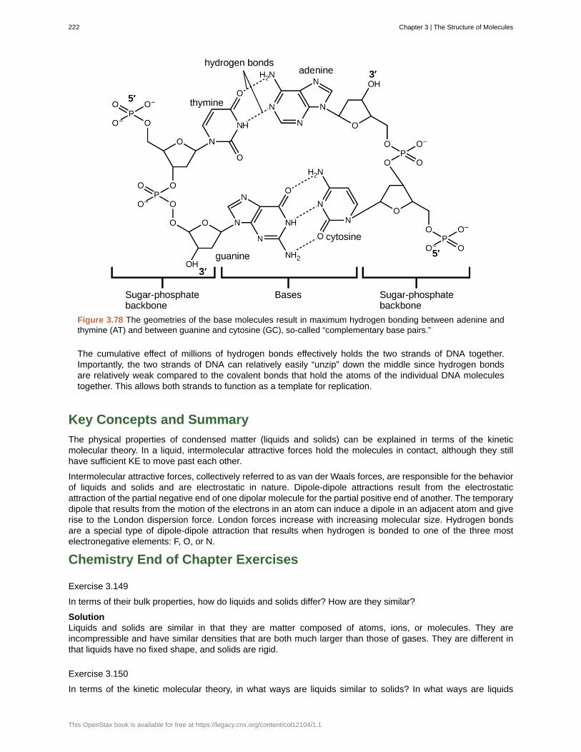
Figure 3.78 The geometries of the base molecules result in maximum hydrogen bonding between adenine andthymine (AT) and between guanine and cytosine (GC), so-called “complementary base pairs.”
The cumulative effect of millions of hydrogen bonds effectively holds the two strands of DNA together.Importantly, the two strands of DNA can relatively easily “unzip” down the middle since hydrogen bondsare relatively weak compared to the covalent bonds that hold the atoms of the individual DNA moleculestogether. This allows both strands to function as a template for replication.
Key Concepts and Summary
The physical properties of condensed matter (liquids and solids) can be explained in terms of the kineticmolecular theory. In a liquid, intermolecular attractive forces hold the molecules in contact, although they stillhave sufficient KE to move past each other.
Intermolecular attractive forces, collectively referred to as van der Waals forces, are responsible for the behaviorof liquids and solids and are electrostatic in nature. Dipole-dipole attractions result from the electrostaticattraction of the partial negative end of one dipolar molecule for the partial positive end of another. The temporarydipole that results from the motion of the electrons in an atom can induce a dipole in an adjacent atom and giverise to the London dispersion force. London forces increase with increasing molecular size. Hydrogen bondsare a special type of dipole-dipole attraction that results when hydrogen is bonded to one of the three mostelectronegative elements: F, O, or N.
Chemistry End of Chapter Exercises
Exercise 3.149
In terms of their bulk properties, how do liquids and solids differ? How are they similar?
SolutionLiquids and solids are similar in that they are matter composed of atoms, ions, or molecules. They areincompressible and have similar densities that are both much larger than those of gases. They are different inthat liquids have no fixed shape, and solids are rigid.
Exercise 3.150
In terms of the kinetic molecular theory, in what ways are liquids similar to solids? In what ways are liquids
222 Chapter 3 | The Structure of Molecules
This OpenStax book is available for free at https://legacy.cnx.org/content/col12104/1.1
different from solids?
Exercise 3.151
In terms of the kinetic molecular theory, in what ways are liquids similar to gases? In what ways are liquidsdifferent from gases?
SolutionThey are similar in that the atoms or molecules are free to move from one position to another. They differ in thatthe particles of a liquid are confined to the shape of the vessel in which they are placed. In contrast, a gas willexpand without limit to fill the space into which it is placed.
Exercise 3.152
Explain why liquids assume the shape of any container into which they are poured, whereas solids are rigid andretain their shape.
Exercise 3.153
What is the evidence that all neutral atoms and molecules exert attractive forces on each other?
SolutionAll atoms and molecules will condense into a liquid or solid in which the attractive forces exceed the kineticenergy of the molecules, at sufficiently low temperature.
Exercise 3.154
Open the PhET States of Matter Simulation (http://openstax.org/l/16phetvisual) to answer the followingquestions:
(a) Select the Solid, Liquid, Gas tab. Explore by selecting different substances, heating and cooling the systems,and changing the state. What similarities do you notice between the four substances for each phase (solid, liquid,gas)? What differences do you notice?
(b) For each substance, select each of the states and record the given temperatures. How do the giventemperatures for each state correlate with the strengths of their intermolecular attractions? Explain.
(c) Select the Interaction Potential tab, and use the default neon atoms. Move the Ne atom on the right andobserve how the potential energy changes. Select the Total Force button, and move the Ne atom as before.When is the total force on each atom attractive and large enough to matter? Then select the Component Forcesbutton, and move the Ne atom. When do the attractive (van der Waals) and repulsive (electron overlap) forcesbalance? How does this relate to the potential energy versus the distance between atoms graph? Explain.
Exercise 3.155
Define the following and give an example of each:
(a) dispersion force
(b) dipole-dipole attraction
(c) hydrogen bond
Solution(a) Dispersion forces occur as an atom develops a temporary dipole moment when its electrons are distributedasymmetrically about the nucleus. This structure is more prevalent in large atoms such as argon or radon. Asecond atom can then be distorted by the appearance of the dipole in the first atom. The electrons of the secondatom are attracted toward the positive end of the first atom, which sets up a dipole in the second atom. Thenet result is rapidly fluctuating, temporary dipoles that attract one another (example: Ar). (b) A dipole-dipoleattraction is a force that results from an electrostatic attraction of the positive end of one polar molecule for thenegative end of another polar molecule (example: ICI molecules attract one another by dipole-dipole interaction).(c) Hydrogen bonds form whenever a hydrogen atom is bonded to one of the more electronegative atoms,such as a fluorine, oxygen, or nitrogen atom. The electrostatic attraction between the partially positive hydrogenatom in one molecule and the partially negative atom in another molecule gives rise to a strong dipole-dipoleinteraction called a hydrogen bond (example: HF⋯HF).
Chapter 3 | The Structure of Molecules 223
Exercise 3.156
The types of intermolecular forces in a substance are identical whether it is a solid, a liquid, or a gas. Why thendoes a substance change phase from a gas to a liquid or to a solid?
Exercise 3.157
Why do the boiling points of the noble gases increase in the order He < Ne < Ar < Kr < Xe?
SolutionThe London forces typically increase as the number of electrons increase.
Exercise 3.158
Neon and HF have approximately the same molecular masses.
(a) Explain why the boiling points of Neon and HF differ.
(b) Compare the change in the boiling points of Ne, Ar, Kr, and Xe with the change of the boiling points of HF,HCl, HBr, and HI, and explain the difference between the changes with increasing atomic or molecular mass.
Exercise 3.159
Arrange each of the following sets of compounds in order of increasing boiling point temperature:
(a) HCl, H2O, SiH4
(b) F2, Cl2, Br2
(c) CH4, C2H6, C3H8
(d) O2, NO, N2
Solution(a) SiH4 < HCl < H2O; (b) F2 < Cl2 < Br2; (c) CH4 < C2H6 < C3H8; (d) N2 < O2 < NO
Exercise 3.160
The molecular mass of butanol, C4H9OH, is 74.14; that of ethylene glycol, CH2(OH)CH2OH, is 62.08, yet theirboiling points are 117.2 °C and 174 °C, respectively. Explain the reason for the difference.
Exercise 3.161
On the basis of intermolecular attractions, explain the differences in the boiling points of n–butane (−1 °C) andchloroethane (12 °C), which have similar molar masses.
SolutionOnly rather small dipole-dipole interactions from C-H bonds are available to hold n-butane in the liquid state.Chloroethane, however, has rather large dipole interactions because of the Cl-C bond; the interaction is thereforestronger, leading to a higher boiling point.
Exercise 3.162
On the basis of dipole moments and/or hydrogen bonding, explain in a qualitative way the differences in theboiling points of acetone (56.2 °C) and 1-propanol (97.4 °C), which have similar molar masses.
Exercise 3.163
The melting point of H2O(s) is 0 °C. Would you expect the melting point of H2S(s) to be −85 °C, 0 °C, or 185 °C?Explain your answer.
Solution−85 °C. Water has stronger hydrogen bonds so it melts at a higher temperature.
Exercise 3.164
Silane (SiH4), phosphine (PH3), and hydrogen sulfide (H2S) melt at −185 °C, −133 °C, and −85 °C, respectively.What does this suggest about the polar character and intermolecular attractions of the three compounds?
224 Chapter 3 | The Structure of Molecules
This OpenStax book is available for free at https://legacy.cnx.org/content/col12104/1.1
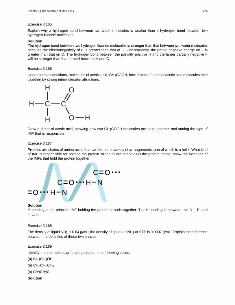
Exercise 3.165
Explain why a hydrogen bond between two water molecules is weaker than a hydrogen bond between twohydrogen fluoride molecules.
SolutionThe hydrogen bond between two hydrogen fluoride molecules is stronger than that between two water moleculesbecause the electronegativity of F is greater than that of O. Consequently, the partial negative charge on F isgreater than that on O. The hydrogen bond between the partially positive H and the larger partially negative Fwill be stronger than that formed between H and O.
Exercise 3.166
Under certain conditions, molecules of acetic acid, CH3COOH, form “dimers,” pairs of acetic acid molecules heldtogether by strong intermolecular attractions:
Draw a dimer of acetic acid, showing how two CH3COOH molecules are held together, and stating the type ofIMF that is responsible.
Exercise 3.167
Proteins are chains of amino acids that can form in a variety of arrangements, one of which is a helix. What kindof IMF is responsible for holding the protein strand in this shape? On the protein image, show the locations ofthe IMFs that hold the protein together:
SolutionH-bonding is the principle IMF holding the protein strands together. The H-bonding is between the N − H and
C = O.
Exercise 3.168
The density of liquid NH3 is 0.64 g/mL; the density of gaseous NH3 at STP is 0.0007 g/mL. Explain the differencebetween the densities of these two phases.
Exercise 3.169
Identify the intermolecular forces present in the following solids:
(a) CH3CH2OH
(b) CH3CH2CH3
(c) CH3CH2Cl
Solution
Chapter 3 | The Structure of Molecules 225

(a) hydrogen bonding, dipole-dipole attraction, and dispersion forces; (b) dispersion forces; (c) dipole-dipoleattraction and dispersion forces
226 Chapter 3 | The Structure of Molecules
This OpenStax book is available for free at https://legacy.cnx.org/content/col12104/1.1
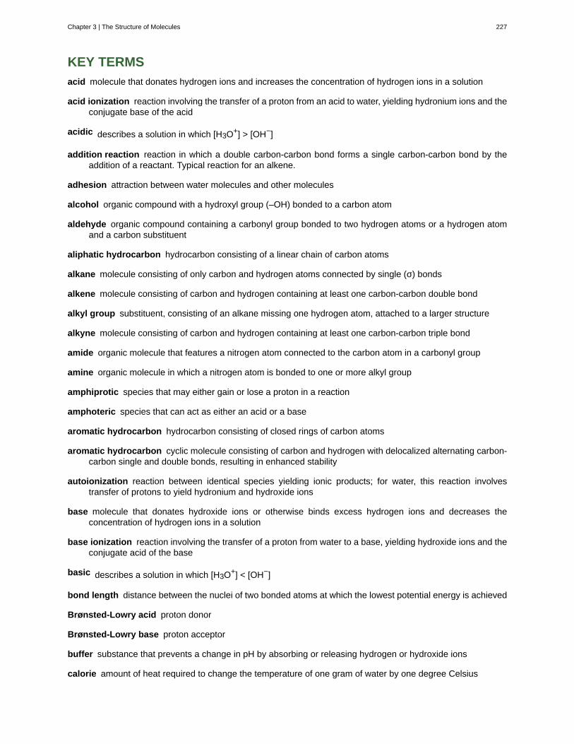
acid
acid ionization
acidic
addition reaction
adhesion
alcohol
aldehyde
aliphatic hydrocarbon
alkane
alkene
alkyl group
alkyne
amide
amine
amphiprotic
amphoteric
aromatic hydrocarbon
aromatic hydrocarbon
autoionization
base
base ionization
basic
bond length
Brønsted-Lowry acid
Brønsted-Lowry base
buffer
calorie
KEY TERMS
molecule that donates hydrogen ions and increases the concentration of hydrogen ions in a solution
reaction involving the transfer of a proton from an acid to water, yielding hydronium ions and theconjugate base of the acid
describes a solution in which [H3O+] > [OH−]
reaction in which a double carbon-carbon bond forms a single carbon-carbon bond by theaddition of a reactant. Typical reaction for an alkene.
attraction between water molecules and other molecules
organic compound with a hydroxyl group (–OH) bonded to a carbon atom
organic compound containing a carbonyl group bonded to two hydrogen atoms or a hydrogen atomand a carbon substituent
hydrocarbon consisting of a linear chain of carbon atoms
molecule consisting of only carbon and hydrogen atoms connected by single (σ) bonds
molecule consisting of carbon and hydrogen containing at least one carbon-carbon double bond
substituent, consisting of an alkane missing one hydrogen atom, attached to a larger structure
molecule consisting of carbon and hydrogen containing at least one carbon-carbon triple bond
organic molecule that features a nitrogen atom connected to the carbon atom in a carbonyl group
organic molecule in which a nitrogen atom is bonded to one or more alkyl group
species that may either gain or lose a proton in a reaction
species that can act as either an acid or a base
hydrocarbon consisting of closed rings of carbon atoms
cyclic molecule consisting of carbon and hydrogen with delocalized alternating carbon-carbon single and double bonds, resulting in enhanced stability
reaction between identical species yielding ionic products; for water, this reaction involvestransfer of protons to yield hydronium and hydroxide ions
molecule that donates hydroxide ions or otherwise binds excess hydrogen ions and decreases theconcentration of hydrogen ions in a solution
reaction involving the transfer of a proton from water to a base, yielding hydroxide ions and theconjugate acid of the base
describes a solution in which [H3O+] < [OH−]
distance between the nuclei of two bonded atoms at which the lowest potential energy is achieved
proton donor
proton acceptor
substance that prevents a change in pH by absorbing or releasing hydrogen or hydroxide ions
amount of heat required to change the temperature of one gram of water by one degree Celsius
Chapter 3 | The Structure of Molecules 227
capillary action
carbonyl group
carboxylic acid
cohesion
conjugate acid
conjugate base
covalent bond
covalent bond
covalent compound
dipole-dipole attraction
dispersion force
dissociation
double bond
electronegativity
empirical formula
enantiomers
ester
ether
evaporation
free radical
functional group
functional group
geometric isomer
heat of vaporization of water
hydrocarbon
hydrogen bonding
occurs because water molecules are attracted to charges on the inner surfaces of narrowtubular structures such as glass tubes, drawing the water molecules to the sides of the tubes
carbon atom double bonded to an oxygen atom
organic compound containing a carbonyl group with an attached hydroxyl group
intermolecular forces between water molecules caused by the polar nature of water; responsible forsurface tension
substance formed when a base gains a proton
substance formed when an acid loses a proton
attractive force between the nuclei of a molecule’s atoms and pairs of electrons between theatoms
bond formed when electrons are shared between atoms
(also, molecular compound) composed of molecules formed by atoms of two or moredifferent elements
intermolecular attraction between two permanent dipoles
(also, London dispersion force) attraction between two rapidly fluctuating, temporary dipoles;significant only when particles are very close together
release of an ion from a molecule such that the original molecule now consists of an ion and the
charged remains of the original, such as when water dissociates into H+ and OH-
covalent bond in which two pairs of electrons are shared between two atoms
tendency of an atom to attract electrons in a bond to itself
formula showing the composition of a compound given as the simplest whole-number ratio ofatoms
molecules that share overall structure and bonding patterns, but differ in how the atoms are threedimensionally placed such that they are mirror images of each other
organic compound containing a carbonyl group with an attached oxygen atom that is bonded to a carbonsubstituent
organic compound with an oxygen atom that is bonded to two carbon atoms
separation of individual molecules from the surface of a body of water, leaves of a plant, or the skinof an organism
molecule that contains an odd number of electrons
group of atoms that provides or imparts a specific function to a carbon skeleton
part of an organic molecule that imparts a specific chemical reactivity to the molecule
isomer with similar bonding patterns differing in the placement of atoms alongside a doublecovalent bond
high amount of energy required for liquid water to turn into water vapor
molecule that consists only of carbon and hydrogen
occurs when exceptionally strong dipoles attract; bonding that exists when hydrogen isbonded to one of the three most electronegative elements: F, O, or N
228 Chapter 3 | The Structure of Molecules
This OpenStax book is available for free at https://legacy.cnx.org/content/col12104/1.1

hydrophilic
hydrophobic
hypervalent molecule
induced dipole
instantaneous dipole
intermolecular force
ion-product constant for water (Kw)
ionic bond
ionic compound
isomers
isomers
ketone
Lewis structure
Lewis symbol
litmus paper
lone pair
molecular compound
molecular formula
monatomic ion
neutral
octet rule
organic compound
organic molecule
oxyanion
pH
pH paper
pH scale
describes ions or polar molecules that interact well with other polar molecules such as water
describes uncharged non-polar molecules that do not interact well with polar molecules such aswater
molecule containing at least one main group element that has more than eight electronsin its valence shell
temporary dipole formed when the electrons of an atom or molecule are distorted by theinstantaneous dipole of a neighboring atom or molecule
temporary dipole that occurs for a brief moment in time when the electrons of an atom ormolecule are distributed asymmetrically
noncovalent attractive force between atoms, molecules, and/or ions
equilibrium constant for the autoionization of water
electrostatic forces of attraction between the oppositely charged ions of an ionic compound
compound composed of cations and anions combined in ratios, yielding an electrically neutralsubstance
compounds with the same chemical formula but different structures
molecules that differ from one another even though they share the same chemical formula
organic compound containing a carbonyl group with two carbon substituents attached to it
diagram showing lone pairs and bonding pairs of electrons in a molecule or an ion
symbol for an element or monatomic ion that uses a dot to represent each valence electron in theelement or ion
(also, pH paper) filter paper that has been treated with a natural water-soluble dye that changes itscolor as the pH of the environment changes so it can be used as a pH indicator
two (a pair of) valence electrons that are not used to form a covalent bond
(also, covalent compound) composed of molecules formed by atoms of two or moredifferent elements
formula indicating the composition of a molecule of a compound and giving the actualnumber of atoms of each element in a molecule of the compound.
ion composed of a single atom
describes a solution in which [H3O+] = [OH−]
guideline that states main group atoms will form structures in which eight valence electrons interactwith each nucleus, counting bonding electrons as interacting with both atoms connected by the bond
natural or synthetic compound that contains carbon
any molecule containing carbon (except carbon dioxide)
polyatomic anion composed of a central atom bonded to oxygen atoms
logarithmic measure of the concentration of hydronium ions in a solution
see litmus paper
scale ranging from zero to 14 that is inversely proportional to the concentration of hydrogen ions in a
Chapter 3 | The Structure of Molecules 229
pOH
polar covalent bond
polarizability
polyatomic ion
pure covalent bond
saturated hydrocarbon
single bond
skeletal structure
solvent
spatial isomers
specific heat capacity
sphere of hydration
structural formula
structural isomer
structural isomers
substituent
substituted hydrocarbon
substitution reaction
surface tension
triple bond
van der Waals force
solution
logarithmic measure of the concentration of hydroxide ions in a solution
covalent bond between atoms of different electronegativities; a covalent bond with apositive end and a negative end
measure of the ability of a charge to distort a molecule’s charge distribution (electron cloud)
ion composed of more than one atom
(also, nonpolar covalent bond) covalent bond between atoms of identicalelectronegativities
molecule containing carbon and hydrogen that has only single bonds between carbonatoms
bond in which a single pair of electrons is shared between two atoms
shorthand method of drawing organic molecules in which carbon atoms are represented bythe ends of lines and bends in between lines, and hydrogen atoms attached to the carbon atoms are notshown (but are understood to be present by the context of the structure)
substance capable of dissolving another substance
compounds in which the relative orientations of the atoms in space differ
the amount of heat one gram of a substance must absorb or lose to change itstemperature by one degree Celsius
when a polar water molecule surrounds charged or polar molecules thus keeping themdissolved and in solution
shows the atoms in a molecule and how they are connected
one of two substances that have the same molecular formula but different physical andchemical properties because their atoms are bonded differently
molecules that share a chemical formula but differ in the placement of their chemical bonds
branch or functional group that replaces hydrogen atoms in a larger hydrocarbon chain
hydrocarbon chain or ring containing an atom of another element in place of one ofthe backbone carbons
reaction in which one atom replaces another in a molecule
tension at the surface of a body of liquid that prevents the molecules from separating; createdby the attractive cohesive forces between the molecules of the liquid
bond in which three pairs of electrons are shared between two atoms
attractive or repulsive force between molecules, including dipole-dipole, dipole-induceddipole, and London dispersion forces; does not include forces due to covalent or ionic bonding, or theattraction between ions and molecules
230 Chapter 3 | The Structure of Molecules
This OpenStax book is available for free at https://legacy.cnx.org/content/col12104/1.1

4 | BIOLOGICALMACROMOLECULES:LARGE MOLECULES OFLIFE
Figure 4.1 Foods such as bread, fruit, and cheese are rich sources of biological macromolecules. (credit: modificationof work by Bengt Nyman)
Chapter Outline
4.1: Synthesis of Biological Macromolecules
4.2: Carbohydrates
4.3: Lipids
4.4: Proteins
4.5: Nucleic Acids
Introduction
Food provides the body with the nutrients it needs to survive. Many of these critical nutrients are biologicalmacromolecules, or large molecules, necessary for life. These macromolecules (polymers) are built fromdifferent combinations of smaller organic molecules (monomers). What specific types of biologicalmacromolecules do living things require? How are these molecules formed? What functions do they serve? In
Chapter 4 | Biological Macromolecules: Large Molecules of Life 231
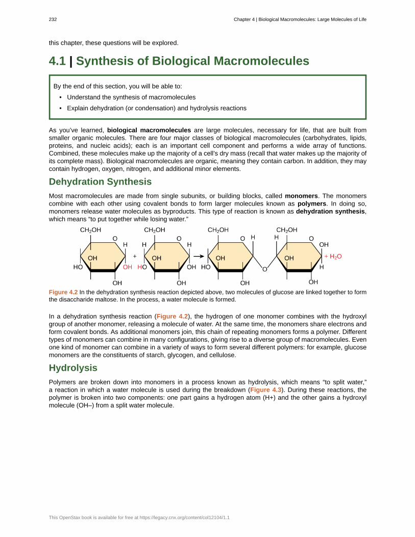
this chapter, these questions will be explored.
4.1 | Synthesis of Biological Macromolecules
By the end of this section, you will be able to:
• Understand the synthesis of macromolecules
• Explain dehydration (or condensation) and hydrolysis reactions
As you’ve learned, biological macromolecules are large molecules, necessary for life, that are built fromsmaller organic molecules. There are four major classes of biological macromolecules (carbohydrates, lipids,proteins, and nucleic acids); each is an important cell component and performs a wide array of functions.Combined, these molecules make up the majority of a cell’s dry mass (recall that water makes up the majority ofits complete mass). Biological macromolecules are organic, meaning they contain carbon. In addition, they maycontain hydrogen, oxygen, nitrogen, and additional minor elements.
Dehydration Synthesis
Most macromolecules are made from single subunits, or building blocks, called monomers. The monomerscombine with each other using covalent bonds to form larger molecules known as polymers. In doing so,monomers release water molecules as byproducts. This type of reaction is known as dehydration synthesis,which means “to put together while losing water.”
Figure 4.2 In the dehydration synthesis reaction depicted above, two molecules of glucose are linked together to formthe disaccharide maltose. In the process, a water molecule is formed.
In a dehydration synthesis reaction (Figure 4.2), the hydrogen of one monomer combines with the hydroxylgroup of another monomer, releasing a molecule of water. At the same time, the monomers share electrons andform covalent bonds. As additional monomers join, this chain of repeating monomers forms a polymer. Differenttypes of monomers can combine in many configurations, giving rise to a diverse group of macromolecules. Evenone kind of monomer can combine in a variety of ways to form several different polymers: for example, glucosemonomers are the constituents of starch, glycogen, and cellulose.
Hydrolysis
Polymers are broken down into monomers in a process known as hydrolysis, which means “to split water,”a reaction in which a water molecule is used during the breakdown (Figure 4.3). During these reactions, thepolymer is broken into two components: one part gains a hydrogen atom (H+) and the other gains a hydroxylmolecule (OH–) from a split water molecule.
232 Chapter 4 | Biological Macromolecules: Large Molecules of Life
This OpenStax book is available for free at https://legacy.cnx.org/content/col12104/1.1
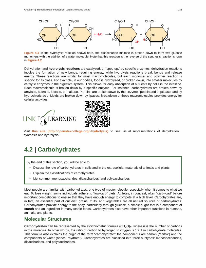
Figure 4.3 In the hydrolysis reaction shown here, the disaccharide maltose is broken down to form two glucosemonomers with the addition of a water molecule. Note that this reaction is the reverse of the synthesis reaction shownin Figure 4.2.
Dehydration and hydrolysis reactions are catalyzed, or “sped up,” by specific enzymes; dehydration reactionsinvolve the formation of new bonds, requiring energy, while hydrolysis reactions break bonds and releaseenergy. These reactions are similar for most macromolecules, but each monomer and polymer reaction isspecific for its class. For example, in our bodies, food is hydrolyzed, or broken down, into smaller molecules bycatalytic enzymes in the digestive system. This allows for easy absorption of nutrients by cells in the intestine.Each macromolecule is broken down by a specific enzyme. For instance, carbohydrates are broken down byamylase, sucrase, lactase, or maltase. Proteins are broken down by the enzymes pepsin and peptidase, and byhydrochloric acid. Lipids are broken down by lipases. Breakdown of these macromolecules provides energy forcellular activities.
Visit this site (http://openstaxcollege.org/l/hydrolysis) to see visual representations of dehydrationsynthesis and hydrolysis.
4.2 | Carbohydrates
By the end of this section, you will be able to:
• Discuss the role of carbohydrates in cells and in the extracellular materials of animals and plants
• Explain the classifications of carbohydrates
• List common monosaccharides, disaccharides, and polysaccharides
Most people are familiar with carbohydrates, one type of macromolecule, especially when it comes to what weeat. To lose weight, some individuals adhere to “low-carb” diets. Athletes, in contrast, often “carb-load” beforeimportant competitions to ensure that they have enough energy to compete at a high level. Carbohydrates are,in fact, an essential part of our diet; grains, fruits, and vegetables are all natural sources of carbohydrates.Carbohydrates provide energy to the body, particularly through glucose, a simple sugar that is a component ofstarch and an ingredient in many staple foods. Carbohydrates also have other important functions in humans,animals, and plants.
Molecular Structures
Carbohydrates can be represented by the stoichiometric formula (CH2O)n, where n is the number of carbonsin the molecule. In other words, the ratio of carbon to hydrogen to oxygen is 1:2:1 in carbohydrate molecules.This formula also explains the origin of the term “carbohydrate”: the components are carbon (“carbo”) and thecomponents of water (hence, “hydrate”). Carbohydrates are classified into three subtypes: monosaccharides,disaccharides, and polysaccharides.
Chapter 4 | Biological Macromolecules: Large Molecules of Life 233

Monosaccharides
Monosaccharides (mono- = “one”; sacchar- = “sweet”) are simple sugars, the most common of which isglucose. In monosaccharides, the number of carbons usually ranges from three to seven. Most monosaccharidenames end with the suffix -ose. If the sugar has an aldehyde group (the functional group with the structure R-CHO), it is known as an aldose, and if it has a ketone group (the functional group with the structure RC(=O)R'),it is known as a ketose. Depending on the number of carbons in the sugar, they also may be known as trioses(three carbons), pentoses (five carbons), and or hexoses (six carbons). See Figure 4.4 for an illustration of themonosaccharides.
Figure 4.4 Monosaccharides are classified based on the position of their carbonyl group and the number of carbonsin the backbone. Aldoses have a carbonyl group (indicated in green) at the end of the carbon chain, and ketoses havea carbonyl group in the middle of the carbon chain. Trioses, pentoses, and hexoses have three, five, and six carbonbackbones, respectively.
The chemical formula for glucose is C6H12O6. In humans, glucose is an important source of energy. Duringcellular respiration, energy is released from glucose, and that energy is used to help make adenosinetriphosphate (ATP). Plants synthesize glucose using carbon dioxide and water, and glucose in turn is used forenergy requirements for the plant. Excess glucose is often stored as starch that is catabolized (the breakdownof larger molecules by cells) by humans and other animals that feed on plants.
Galactose (part of lactose, or milk sugar) and fructose (found in sucrose, in fruit) are other commonmonosaccharides. Although glucose, galactose, and fructose all have the same chemical formula (C6H12O6),they differ structurally and chemically (and are known as isomers) because of the different arrangement offunctional groups around the asymmetric carbon; all of these monosaccharides have more than one asymmetric
234 Chapter 4 | Biological Macromolecules: Large Molecules of Life
This OpenStax book is available for free at https://legacy.cnx.org/content/col12104/1.1
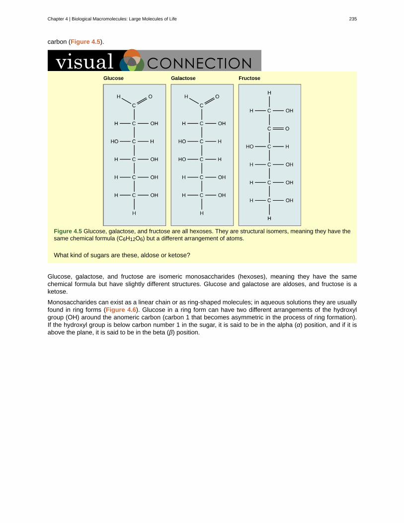
carbon (Figure 4.5).
Figure 4.5 Glucose, galactose, and fructose are all hexoses. They are structural isomers, meaning they have thesame chemical formula (C6H12O6) but a different arrangement of atoms.
What kind of sugars are these, aldose or ketose?
Glucose, galactose, and fructose are isomeric monosaccharides (hexoses), meaning they have the samechemical formula but have slightly different structures. Glucose and galactose are aldoses, and fructose is aketose.
Monosaccharides can exist as a linear chain or as ring-shaped molecules; in aqueous solutions they are usuallyfound in ring forms (Figure 4.6). Glucose in a ring form can have two different arrangements of the hydroxylgroup (OH) around the anomeric carbon (carbon 1 that becomes asymmetric in the process of ring formation).If the hydroxyl group is below carbon number 1 in the sugar, it is said to be in the alpha (α) position, and if it isabove the plane, it is said to be in the beta (β) position.
Chapter 4 | Biological Macromolecules: Large Molecules of Life 235
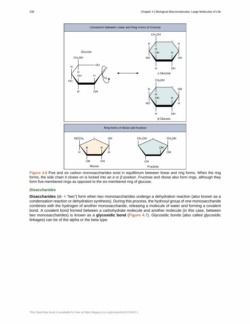
Figure 4.6 Five and six carbon monosaccharides exist in equilibrium between linear and ring forms. When the ringforms, the side chain it closes on is locked into an α or β position. Fructose and ribose also form rings, although theyform five-membered rings as opposed to the six-membered ring of glucose.
Disaccharides
Disaccharides (di- = “two”) form when two monosaccharides undergo a dehydration reaction (also known as acondensation reaction or dehydration synthesis). During this process, the hydroxyl group of one monosaccharidecombines with the hydrogen of another monosaccharide, releasing a molecule of water and forming a covalentbond. A covalent bond formed between a carbohydrate molecule and another molecule (in this case, betweentwo monosaccharides) is known as a glycosidic bond (Figure 4.7). Glycosidic bonds (also called glycosidiclinkages) can be of the alpha or the beta type.
236 Chapter 4 | Biological Macromolecules: Large Molecules of Life
This OpenStax book is available for free at https://legacy.cnx.org/content/col12104/1.1
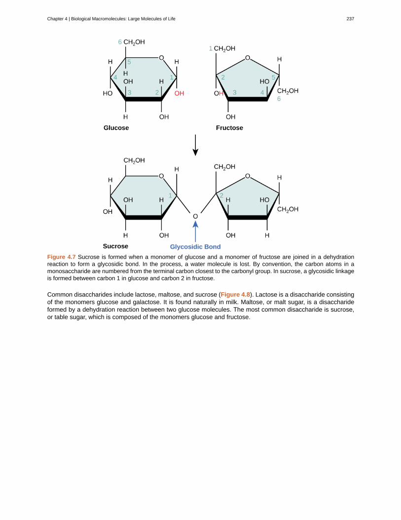
Figure 4.7 Sucrose is formed when a monomer of glucose and a monomer of fructose are joined in a dehydrationreaction to form a glycosidic bond. In the process, a water molecule is lost. By convention, the carbon atoms in amonosaccharide are numbered from the terminal carbon closest to the carbonyl group. In sucrose, a glycosidic linkageis formed between carbon 1 in glucose and carbon 2 in fructose.
Common disaccharides include lactose, maltose, and sucrose (Figure 4.8). Lactose is a disaccharide consistingof the monomers glucose and galactose. It is found naturally in milk. Maltose, or malt sugar, is a disaccharideformed by a dehydration reaction between two glucose molecules. The most common disaccharide is sucrose,or table sugar, which is composed of the monomers glucose and fructose.
Chapter 4 | Biological Macromolecules: Large Molecules of Life 237
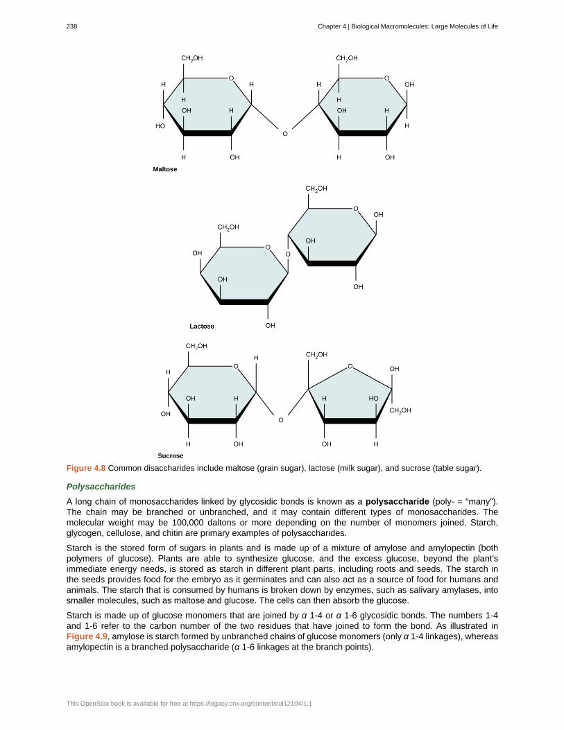
Figure 4.8 Common disaccharides include maltose (grain sugar), lactose (milk sugar), and sucrose (table sugar).
Polysaccharides
A long chain of monosaccharides linked by glycosidic bonds is known as a polysaccharide (poly- = “many”).The chain may be branched or unbranched, and it may contain different types of monosaccharides. Themolecular weight may be 100,000 daltons or more depending on the number of monomers joined. Starch,glycogen, cellulose, and chitin are primary examples of polysaccharides.
Starch is the stored form of sugars in plants and is made up of a mixture of amylose and amylopectin (bothpolymers of glucose). Plants are able to synthesize glucose, and the excess glucose, beyond the plant’simmediate energy needs, is stored as starch in different plant parts, including roots and seeds. The starch inthe seeds provides food for the embryo as it germinates and can also act as a source of food for humans andanimals. The starch that is consumed by humans is broken down by enzymes, such as salivary amylases, intosmaller molecules, such as maltose and glucose. The cells can then absorb the glucose.
Starch is made up of glucose monomers that are joined by α 1-4 or α 1-6 glycosidic bonds. The numbers 1-4and 1-6 refer to the carbon number of the two residues that have joined to form the bond. As illustrated inFigure 4.9, amylose is starch formed by unbranched chains of glucose monomers (only α 1-4 linkages), whereasamylopectin is a branched polysaccharide (α 1-6 linkages at the branch points).
238 Chapter 4 | Biological Macromolecules: Large Molecules of Life
This OpenStax book is available for free at https://legacy.cnx.org/content/col12104/1.1
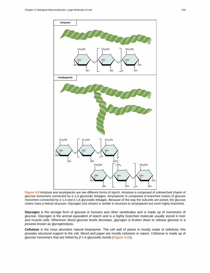
Figure 4.9 Amylose and amylopectin are two different forms of starch. Amylose is composed of unbranched chains ofglucose monomers connected by α 1,4 glycosidic linkages. Amylopectin is composed of branched chains of glucosemonomers connected by α 1,4 and α 1,6 glycosidic linkages. Because of the way the subunits are joined, the glucosechains have a helical structure. Glycogen (not shown) is similar in structure to amylopectin but more highly branched.
Glycogen is the storage form of glucose in humans and other vertebrates and is made up of monomers ofglucose. Glycogen is the animal equivalent of starch and is a highly branched molecule usually stored in liverand muscle cells. Whenever blood glucose levels decrease, glycogen is broken down to release glucose in aprocess known as glycogenolysis.
Cellulose is the most abundant natural biopolymer. The cell wall of plants is mostly made of cellulose; thisprovides structural support to the cell. Wood and paper are mostly cellulosic in nature. Cellulose is made up ofglucose monomers that are linked by β 1-4 glycosidic bonds (Figure 4.10).
Chapter 4 | Biological Macromolecules: Large Molecules of Life 239
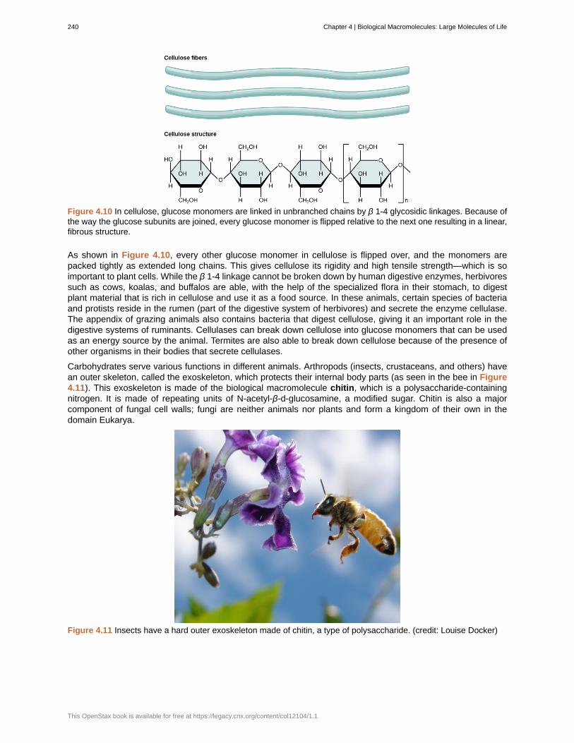
Figure 4.10 In cellulose, glucose monomers are linked in unbranched chains by β 1-4 glycosidic linkages. Because ofthe way the glucose subunits are joined, every glucose monomer is flipped relative to the next one resulting in a linear,fibrous structure.
As shown in Figure 4.10, every other glucose monomer in cellulose is flipped over, and the monomers arepacked tightly as extended long chains. This gives cellulose its rigidity and high tensile strength—which is soimportant to plant cells. While the β 1-4 linkage cannot be broken down by human digestive enzymes, herbivoressuch as cows, koalas, and buffalos are able, with the help of the specialized flora in their stomach, to digestplant material that is rich in cellulose and use it as a food source. In these animals, certain species of bacteriaand protists reside in the rumen (part of the digestive system of herbivores) and secrete the enzyme cellulase.The appendix of grazing animals also contains bacteria that digest cellulose, giving it an important role in thedigestive systems of ruminants. Cellulases can break down cellulose into glucose monomers that can be usedas an energy source by the animal. Termites are also able to break down cellulose because of the presence ofother organisms in their bodies that secrete cellulases.
Carbohydrates serve various functions in different animals. Arthropods (insects, crustaceans, and others) havean outer skeleton, called the exoskeleton, which protects their internal body parts (as seen in the bee in Figure4.11). This exoskeleton is made of the biological macromolecule chitin, which is a polysaccharide-containingnitrogen. It is made of repeating units of N-acetyl-β-d-glucosamine, a modified sugar. Chitin is also a majorcomponent of fungal cell walls; fungi are neither animals nor plants and form a kingdom of their own in thedomain Eukarya.
Figure 4.11 Insects have a hard outer exoskeleton made of chitin, a type of polysaccharide. (credit: Louise Docker)
240 Chapter 4 | Biological Macromolecules: Large Molecules of Life
This OpenStax book is available for free at https://legacy.cnx.org/content/col12104/1.1
Registered DietitianObesity is a worldwide health concern, and many diseases such as diabetes and heart disease arebecoming more prevalent because of obesity. This is one of the reasons why registered dietitians areincreasingly sought after for advice. Registered dietitians help plan nutrition programs for individuals invarious settings. They often work with patients in health care facilities, designing nutrition plans to treat andprevent diseases. For example, dietitians may teach a patient with diabetes how to manage blood sugarlevels by eating the correct types and amounts of carbohydrates. Dietitians may also work in nursing homes,schools, and private practices.
To become a registered dietitian, one needs to earn at least a bachelor’s degree in dietetics, nutrition,food technology, or a related field. In addition, registered dietitians must complete a supervised internshipprogram and pass a national exam. Those who pursue careers in dietetics take courses in nutrition,chemistry, biochemistry, biology, microbiology, and human physiology. Dietitians must become experts inthe chemistry and physiology (biological functions) of food (proteins, carbohydrates, and fats).
Benefits of Carbohydrates
Are carbohydrates good for you? People who wish to lose weight are often told that carbohydrates are badfor them and should be avoided. Some diets completely forbid carbohydrate consumption, claiming that a low-carbohydrate diet helps people to lose weight faster. However, carbohydrates have been an important part ofthe human diet for thousands of years; artifacts from ancient civilizations show the presence of wheat, rice, andcorn in our ancestors’ storage areas.
Carbohydrates should be supplemented with proteins, vitamins, and fats to be parts of a well-balanced diet.Calorie-wise, a gram of carbohydrate provides 4.3 Kcal. For comparison, fats provide 9 Kcal/g, a less desirableratio. Carbohydrates contain soluble and insoluble elements; the insoluble part is known as fiber, which is mostlycellulose. Fiber has many uses; it promotes regular bowel movement by adding bulk, and it regulates the rateof consumption of blood glucose. Fiber also helps to remove excess cholesterol from the body: fiber binds tothe cholesterol in the small intestine, then attaches to the cholesterol and prevents the cholesterol particlesfrom entering the bloodstream, and then cholesterol exits the body via the feces. Fiber-rich diets also havea protective role in reducing the occurrence of colon cancer. In addition, a meal containing whole grains andvegetables gives a feeling of fullness. As an immediate source of energy, glucose is broken down during theprocess of cellular respiration, which produces ATP, the energy currency of the cell. Without the consumption ofcarbohydrates, the availability of “instant energy” would be reduced. Eliminating carbohydrates from the diet isnot the best way to lose weight. A low-calorie diet that is rich in whole grains, fruits, vegetables, and lean meat,together with plenty of exercise and plenty of water, is the more sensible way to lose weight.
For an additional perspective on carbohydrates, explore “Biomolecules: the Carbohydrates” through thisinteractive animation (http://openstaxcollege.org/l/carbohydrates) .
Chapter 4 | Biological Macromolecules: Large Molecules of Life 241
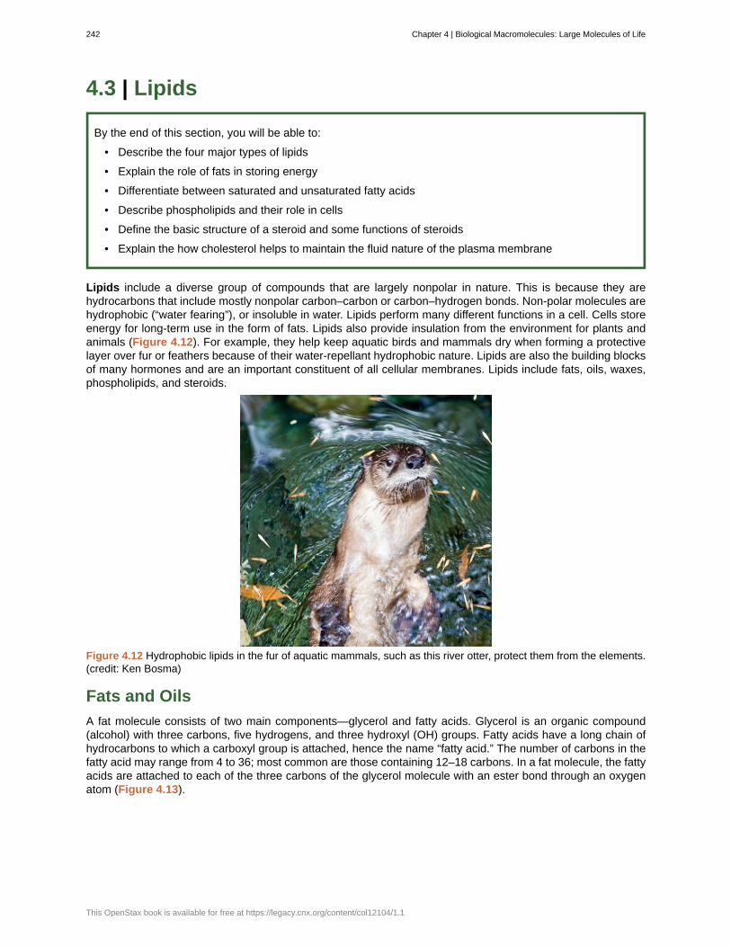
4.3 | Lipids
By the end of this section, you will be able to:
• Describe the four major types of lipids
• Explain the role of fats in storing energy
• Differentiate between saturated and unsaturated fatty acids
• Describe phospholipids and their role in cells
• Define the basic structure of a steroid and some functions of steroids
• Explain the how cholesterol helps to maintain the fluid nature of the plasma membrane
Lipids include a diverse group of compounds that are largely nonpolar in nature. This is because they arehydrocarbons that include mostly nonpolar carbon–carbon or carbon–hydrogen bonds. Non-polar molecules arehydrophobic (“water fearing”), or insoluble in water. Lipids perform many different functions in a cell. Cells storeenergy for long-term use in the form of fats. Lipids also provide insulation from the environment for plants andanimals (Figure 4.12). For example, they help keep aquatic birds and mammals dry when forming a protectivelayer over fur or feathers because of their water-repellant hydrophobic nature. Lipids are also the building blocksof many hormones and are an important constituent of all cellular membranes. Lipids include fats, oils, waxes,phospholipids, and steroids.
Figure 4.12 Hydrophobic lipids in the fur of aquatic mammals, such as this river otter, protect them from the elements.(credit: Ken Bosma)
Fats and Oils
A fat molecule consists of two main components—glycerol and fatty acids. Glycerol is an organic compound(alcohol) with three carbons, five hydrogens, and three hydroxyl (OH) groups. Fatty acids have a long chain ofhydrocarbons to which a carboxyl group is attached, hence the name “fatty acid.” The number of carbons in thefatty acid may range from 4 to 36; most common are those containing 12–18 carbons. In a fat molecule, the fattyacids are attached to each of the three carbons of the glycerol molecule with an ester bond through an oxygenatom (Figure 4.13).
242 Chapter 4 | Biological Macromolecules: Large Molecules of Life
This OpenStax book is available for free at https://legacy.cnx.org/content/col12104/1.1
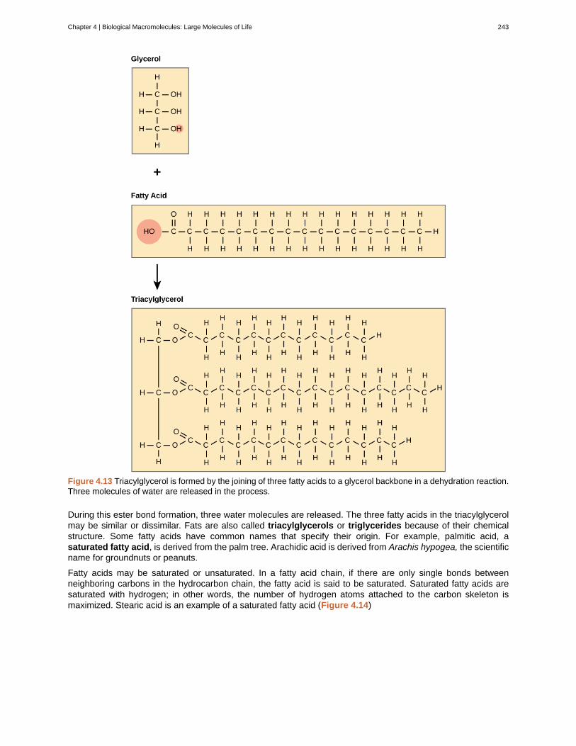
Figure 4.13 Triacylglycerol is formed by the joining of three fatty acids to a glycerol backbone in a dehydration reaction.Three molecules of water are released in the process.
During this ester bond formation, three water molecules are released. The three fatty acids in the triacylglycerolmay be similar or dissimilar. Fats are also called triacylglycerols or triglycerides because of their chemicalstructure. Some fatty acids have common names that specify their origin. For example, palmitic acid, asaturated fatty acid, is derived from the palm tree. Arachidic acid is derived from Arachis hypogea, the scientificname for groundnuts or peanuts.
Fatty acids may be saturated or unsaturated. In a fatty acid chain, if there are only single bonds betweenneighboring carbons in the hydrocarbon chain, the fatty acid is said to be saturated. Saturated fatty acids aresaturated with hydrogen; in other words, the number of hydrogen atoms attached to the carbon skeleton ismaximized. Stearic acid is an example of a saturated fatty acid (Figure 4.14)
Chapter 4 | Biological Macromolecules: Large Molecules of Life 243

Figure 4.14 Stearic acid is a common saturated fatty acid.
When the hydrocarbon chain contains a double bond, the fatty acid is said to be unsaturated. Oleic acid is anexample of an unsaturated fatty acid (Figure 4.15).
Figure 4.15 Oleic acid is a common unsaturated fatty acid.
Most unsaturated fats are liquid at room temperature and are called oils. If there is one double bond in themolecule, then it is known as a monounsaturated fat (e.g., olive oil), and if there is more than one double bond,then it is known as a polyunsaturated fat (e.g., canola oil).
When a fatty acid has no double bonds, it is known as a saturated fatty acid because no more hydrogen maybe added to the carbon atoms of the chain. A fat may contain similar or different fatty acids attached to glycerol.Long straight fatty acids with single bonds tend to get packed tightly and are solid at room temperature. Animalfats with stearic acid and palmitic acid (common in meat) and the fat with butyric acid (common in butter) areexamples of saturated fats. Mammals store fats in specialized cells called adipocytes, where globules of fatoccupy most of the cell’s volume. In plants, fat or oil is stored in many seeds and is used as a source of energyduring seedling development. Unsaturated fats or oils are usually of plant origin and contain cis unsaturated fattyacids. Cis and trans indicate the configuration of the molecule around the double bond. If hydrogens are presentin the same plane, it is referred to as a cis fat; if the hydrogen atoms are on two different planes, it is referredto as a trans fat. The cis double bond causes a bend or a “kink” that prevents the fatty acids from packingtightly, keeping them liquid at room temperature (Figure 4.16). Olive oil, corn oil, canola oil, and cod liver oilare examples of unsaturated fats. Unsaturated fats help to lower blood cholesterol levels whereas saturated fatscontribute to plaque formation in the arteries.
244 Chapter 4 | Biological Macromolecules: Large Molecules of Life
This OpenStax book is available for free at https://legacy.cnx.org/content/col12104/1.1
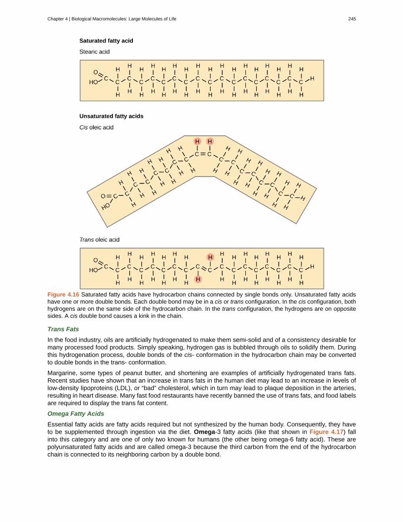
Figure 4.16 Saturated fatty acids have hydrocarbon chains connected by single bonds only. Unsaturated fatty acidshave one or more double bonds. Each double bond may be in a cis or trans configuration. In the cis configuration, bothhydrogens are on the same side of the hydrocarbon chain. In the trans configuration, the hydrogens are on oppositesides. A cis double bond causes a kink in the chain.
Trans Fats
In the food industry, oils are artificially hydrogenated to make them semi-solid and of a consistency desirable formany processed food products. Simply speaking, hydrogen gas is bubbled through oils to solidify them. Duringthis hydrogenation process, double bonds of the cis- conformation in the hydrocarbon chain may be convertedto double bonds in the trans- conformation.
Margarine, some types of peanut butter, and shortening are examples of artificially hydrogenated trans fats.Recent studies have shown that an increase in trans fats in the human diet may lead to an increase in levels oflow-density lipoproteins (LDL), or “bad” cholesterol, which in turn may lead to plaque deposition in the arteries,resulting in heart disease. Many fast food restaurants have recently banned the use of trans fats, and food labelsare required to display the trans fat content.
Omega Fatty Acids
Essential fatty acids are fatty acids required but not synthesized by the human body. Consequently, they haveto be supplemented through ingestion via the diet. Omega-3 fatty acids (like that shown in Figure 4.17) fallinto this category and are one of only two known for humans (the other being omega-6 fatty acid). These arepolyunsaturated fatty acids and are called omega-3 because the third carbon from the end of the hydrocarbonchain is connected to its neighboring carbon by a double bond.
Chapter 4 | Biological Macromolecules: Large Molecules of Life 245
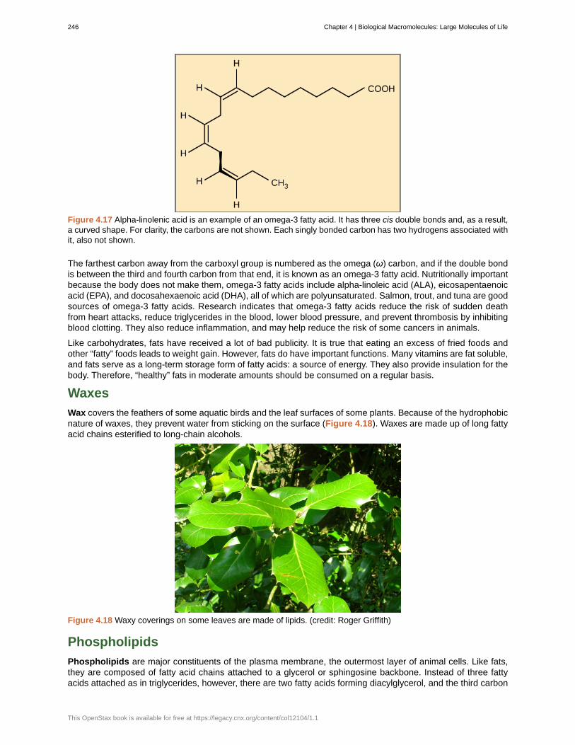
Figure 4.17 Alpha-linolenic acid is an example of an omega-3 fatty acid. It has three cis double bonds and, as a result,a curved shape. For clarity, the carbons are not shown. Each singly bonded carbon has two hydrogens associated withit, also not shown.
The farthest carbon away from the carboxyl group is numbered as the omega (ω) carbon, and if the double bondis between the third and fourth carbon from that end, it is known as an omega-3 fatty acid. Nutritionally importantbecause the body does not make them, omega-3 fatty acids include alpha-linoleic acid (ALA), eicosapentaenoicacid (EPA), and docosahexaenoic acid (DHA), all of which are polyunsaturated. Salmon, trout, and tuna are goodsources of omega-3 fatty acids. Research indicates that omega-3 fatty acids reduce the risk of sudden deathfrom heart attacks, reduce triglycerides in the blood, lower blood pressure, and prevent thrombosis by inhibitingblood clotting. They also reduce inflammation, and may help reduce the risk of some cancers in animals.
Like carbohydrates, fats have received a lot of bad publicity. It is true that eating an excess of fried foods andother “fatty” foods leads to weight gain. However, fats do have important functions. Many vitamins are fat soluble,and fats serve as a long-term storage form of fatty acids: a source of energy. They also provide insulation for thebody. Therefore, “healthy” fats in moderate amounts should be consumed on a regular basis.
Waxes
Wax covers the feathers of some aquatic birds and the leaf surfaces of some plants. Because of the hydrophobicnature of waxes, they prevent water from sticking on the surface (Figure 4.18). Waxes are made up of long fattyacid chains esterified to long-chain alcohols.
Figure 4.18 Waxy coverings on some leaves are made of lipids. (credit: Roger Griffith)
Phospholipids
Phospholipids are major constituents of the plasma membrane, the outermost layer of animal cells. Like fats,they are composed of fatty acid chains attached to a glycerol or sphingosine backbone. Instead of three fattyacids attached as in triglycerides, however, there are two fatty acids forming diacylglycerol, and the third carbon
246 Chapter 4 | Biological Macromolecules: Large Molecules of Life
This OpenStax book is available for free at https://legacy.cnx.org/content/col12104/1.1
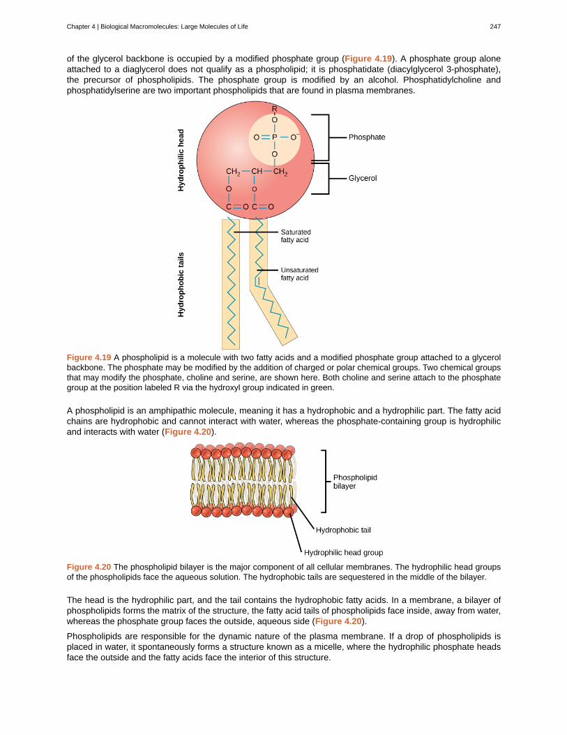
of the glycerol backbone is occupied by a modified phosphate group (Figure 4.19). A phosphate group aloneattached to a diaglycerol does not qualify as a phospholipid; it is phosphatidate (diacylglycerol 3-phosphate),the precursor of phospholipids. The phosphate group is modified by an alcohol. Phosphatidylcholine andphosphatidylserine are two important phospholipids that are found in plasma membranes.
Figure 4.19 A phospholipid is a molecule with two fatty acids and a modified phosphate group attached to a glycerolbackbone. The phosphate may be modified by the addition of charged or polar chemical groups. Two chemical groupsthat may modify the phosphate, choline and serine, are shown here. Both choline and serine attach to the phosphategroup at the position labeled R via the hydroxyl group indicated in green.
A phospholipid is an amphipathic molecule, meaning it has a hydrophobic and a hydrophilic part. The fatty acidchains are hydrophobic and cannot interact with water, whereas the phosphate-containing group is hydrophilicand interacts with water (Figure 4.20).
Figure 4.20 The phospholipid bilayer is the major component of all cellular membranes. The hydrophilic head groupsof the phospholipids face the aqueous solution. The hydrophobic tails are sequestered in the middle of the bilayer.
The head is the hydrophilic part, and the tail contains the hydrophobic fatty acids. In a membrane, a bilayer ofphospholipids forms the matrix of the structure, the fatty acid tails of phospholipids face inside, away from water,whereas the phosphate group faces the outside, aqueous side (Figure 4.20).
Phospholipids are responsible for the dynamic nature of the plasma membrane. If a drop of phospholipids isplaced in water, it spontaneously forms a structure known as a micelle, where the hydrophilic phosphate headsface the outside and the fatty acids face the interior of this structure.
Chapter 4 | Biological Macromolecules: Large Molecules of Life 247
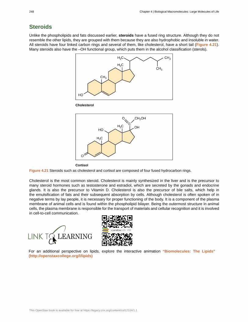
Steroids
Unlike the phospholipids and fats discussed earlier, steroids have a fused ring structure. Although they do notresemble the other lipids, they are grouped with them because they are also hydrophobic and insoluble in water.All steroids have four linked carbon rings and several of them, like cholesterol, have a short tail (Figure 4.21).Many steroids also have the –OH functional group, which puts them in the alcohol classification (sterols).
Figure 4.21 Steroids such as cholesterol and cortisol are composed of four fused hydrocarbon rings.
Cholesterol is the most common steroid. Cholesterol is mainly synthesized in the liver and is the precursor tomany steroid hormones such as testosterone and estradiol, which are secreted by the gonads and endocrineglands. It is also the precursor to Vitamin D. Cholesterol is also the precursor of bile salts, which help inthe emulsification of fats and their subsequent absorption by cells. Although cholesterol is often spoken of innegative terms by lay people, it is necessary for proper functioning of the body. It is a component of the plasmamembrane of animal cells and is found within the phospholipid bilayer. Being the outermost structure in animalcells, the plasma membrane is responsible for the transport of materials and cellular recognition and it is involvedin cell-to-cell communication.
For an additional perspective on lipids, explore the interactive animation “Biomolecules: The Lipids”(http://openstaxcollege.org/l/lipids)
248 Chapter 4 | Biological Macromolecules: Large Molecules of Life
This OpenStax book is available for free at https://legacy.cnx.org/content/col12104/1.1
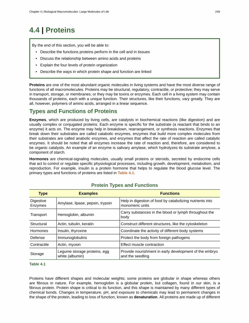
4.4 | Proteins
By the end of this section, you will be able to:
• Describe the functions proteins perform in the cell and in tissues
• Discuss the relationship between amino acids and proteins
• Explain the four levels of protein organization
• Describe the ways in which protein shape and function are linked
Proteins are one of the most abundant organic molecules in living systems and have the most diverse range offunctions of all macromolecules. Proteins may be structural, regulatory, contractile, or protective; they may servein transport, storage, or membranes; or they may be toxins or enzymes. Each cell in a living system may containthousands of proteins, each with a unique function. Their structures, like their functions, vary greatly. They areall, however, polymers of amino acids, arranged in a linear sequence.
Types and Functions of Proteins
Enzymes, which are produced by living cells, are catalysts in biochemical reactions (like digestion) and areusually complex or conjugated proteins. Each enzyme is specific for the substrate (a reactant that binds to anenzyme) it acts on. The enzyme may help in breakdown, rearrangement, or synthesis reactions. Enzymes thatbreak down their substrates are called catabolic enzymes, enzymes that build more complex molecules fromtheir substrates are called anabolic enzymes, and enzymes that affect the rate of reaction are called catalyticenzymes. It should be noted that all enzymes increase the rate of reaction and, therefore, are considered tobe organic catalysts. An example of an enzyme is salivary amylase, which hydrolyzes its substrate amylose, acomponent of starch.
Hormones are chemical-signaling molecules, usually small proteins or steroids, secreted by endocrine cellsthat act to control or regulate specific physiological processes, including growth, development, metabolism, andreproduction. For example, insulin is a protein hormone that helps to regulate the blood glucose level. Theprimary types and functions of proteins are listed in Table 4.1.
Protein Types and Functions
Type Examples Functions
DigestiveEnzymes
Amylase, lipase, pepsin, trypsinHelp in digestion of food by catabolizing nutrients intomonomeric units
Transport Hemoglobin, albuminCarry substances in the blood or lymph throughout thebody
Structural Actin, tubulin, keratin Construct different structures, like the cytoskeleton
Hormones Insulin, thyroxine Coordinate the activity of different body systems
Defense Immunoglobulins Protect the body from foreign pathogens
Contractile Actin, myosin Effect muscle contraction
StorageLegume storage proteins, eggwhite (albumin)
Provide nourishment in early development of the embryoand the seedling
Table 4.1
Proteins have different shapes and molecular weights; some proteins are globular in shape whereas othersare fibrous in nature. For example, hemoglobin is a globular protein, but collagen, found in our skin, is afibrous protein. Protein shape is critical to its function, and this shape is maintained by many different types ofchemical bonds. Changes in temperature, pH, and exposure to chemicals may lead to permanent changes inthe shape of the protein, leading to loss of function, known as denaturation. All proteins are made up of different
Chapter 4 | Biological Macromolecules: Large Molecules of Life 249
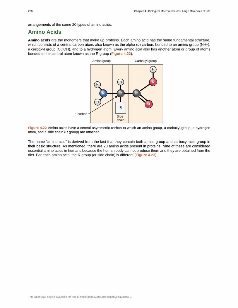
arrangements of the same 20 types of amino acids.
Amino Acids
Amino acids are the monomers that make up proteins. Each amino acid has the same fundamental structure,which consists of a central carbon atom, also known as the alpha (α) carbon, bonded to an amino group (NH2),a carboxyl group (COOH), and to a hydrogen atom. Every amino acid also has another atom or group of atomsbonded to the central atom known as the R group (Figure 4.22).
Figure 4.22 Amino acids have a central asymmetric carbon to which an amino group, a carboxyl group, a hydrogenatom, and a side chain (R group) are attached.
The name "amino acid" is derived from the fact that they contain both amino group and carboxyl-acid-group intheir basic structure. As mentioned, there are 20 amino acids present in proteins. Nine of these are consideredessential amino acids in humans because the human body cannot produce them and they are obtained from thediet. For each amino acid, the R group (or side chain) is different (Figure 4.23).
250 Chapter 4 | Biological Macromolecules: Large Molecules of Life
This OpenStax book is available for free at https://legacy.cnx.org/content/col12104/1.1
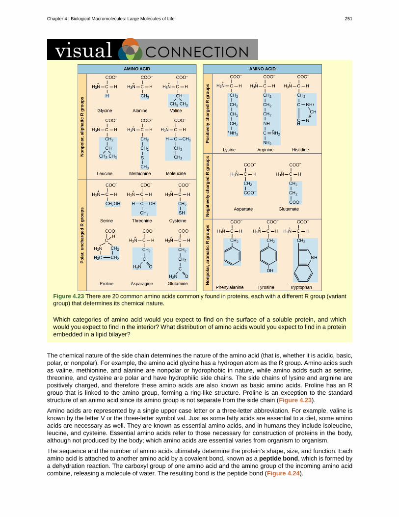
Figure 4.23 There are 20 common amino acids commonly found in proteins, each with a different R group (variantgroup) that determines its chemical nature.
Which categories of amino acid would you expect to find on the surface of a soluble protein, and whichwould you expect to find in the interior? What distribution of amino acids would you expect to find in a proteinembedded in a lipid bilayer?
The chemical nature of the side chain determines the nature of the amino acid (that is, whether it is acidic, basic,polar, or nonpolar). For example, the amino acid glycine has a hydrogen atom as the R group. Amino acids suchas valine, methionine, and alanine are nonpolar or hydrophobic in nature, while amino acids such as serine,threonine, and cysteine are polar and have hydrophilic side chains. The side chains of lysine and arginine arepositively charged, and therefore these amino acids are also known as basic amino acids. Proline has an Rgroup that is linked to the amino group, forming a ring-like structure. Proline is an exception to the standardstructure of an animo acid since its amino group is not separate from the side chain (Figure 4.23).
Amino acids are represented by a single upper case letter or a three-letter abbreviation. For example, valine isknown by the letter V or the three-letter symbol val. Just as some fatty acids are essential to a diet, some aminoacids are necessary as well. They are known as essential amino acids, and in humans they include isoleucine,leucine, and cysteine. Essential amino acids refer to those necessary for construction of proteins in the body,although not produced by the body; which amino acids are essential varies from organism to organism.
The sequence and the number of amino acids ultimately determine the protein's shape, size, and function. Eachamino acid is attached to another amino acid by a covalent bond, known as a peptide bond, which is formed bya dehydration reaction. The carboxyl group of one amino acid and the amino group of the incoming amino acidcombine, releasing a molecule of water. The resulting bond is the peptide bond (Figure 4.24).
Chapter 4 | Biological Macromolecules: Large Molecules of Life 251
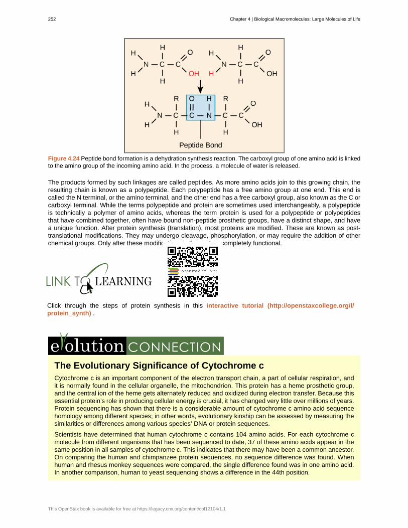
Figure 4.24 Peptide bond formation is a dehydration synthesis reaction. The carboxyl group of one amino acid is linkedto the amino group of the incoming amino acid. In the process, a molecule of water is released.
The products formed by such linkages are called peptides. As more amino acids join to this growing chain, theresulting chain is known as a polypeptide. Each polypeptide has a free amino group at one end. This end iscalled the N terminal, or the amino terminal, and the other end has a free carboxyl group, also known as the C orcarboxyl terminal. While the terms polypeptide and protein are sometimes used interchangeably, a polypeptideis technically a polymer of amino acids, whereas the term protein is used for a polypeptide or polypeptidesthat have combined together, often have bound non-peptide prosthetic groups, have a distinct shape, and havea unique function. After protein synthesis (translation), most proteins are modified. These are known as post-translational modifications. They may undergo cleavage, phosphorylation, or may require the addition of otherchemical groups. Only after these modifications is the protein completely functional.
Click through the steps of protein synthesis in this interactive tutorial (http://openstaxcollege.org/l/protein_synth) .
The Evolutionary Significance of Cytochrome cCytochrome c is an important component of the electron transport chain, a part of cellular respiration, andit is normally found in the cellular organelle, the mitochondrion. This protein has a heme prosthetic group,and the central ion of the heme gets alternately reduced and oxidized during electron transfer. Because thisessential protein’s role in producing cellular energy is crucial, it has changed very little over millions of years.Protein sequencing has shown that there is a considerable amount of cytochrome c amino acid sequencehomology among different species; in other words, evolutionary kinship can be assessed by measuring thesimilarities or differences among various species’ DNA or protein sequences.
Scientists have determined that human cytochrome c contains 104 amino acids. For each cytochrome cmolecule from different organisms that has been sequenced to date, 37 of these amino acids appear in thesame position in all samples of cytochrome c. This indicates that there may have been a common ancestor.On comparing the human and chimpanzee protein sequences, no sequence difference was found. Whenhuman and rhesus monkey sequences were compared, the single difference found was in one amino acid.In another comparison, human to yeast sequencing shows a difference in the 44th position.
252 Chapter 4 | Biological Macromolecules: Large Molecules of Life
This OpenStax book is available for free at https://legacy.cnx.org/content/col12104/1.1
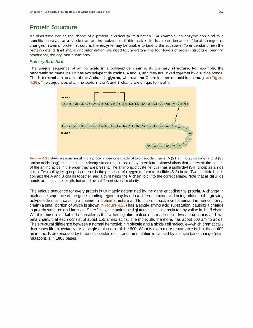
Protein Structure
As discussed earlier, the shape of a protein is critical to its function. For example, an enzyme can bind to aspecific substrate at a site known as the active site. If this active site is altered because of local changes orchanges in overall protein structure, the enzyme may be unable to bind to the substrate. To understand how theprotein gets its final shape or conformation, we need to understand the four levels of protein structure: primary,secondary, tertiary, and quaternary.
Primary Structure
The unique sequence of amino acids in a polypeptide chain is its primary structure. For example, thepancreatic hormone insulin has two polypeptide chains, A and B, and they are linked together by disulfide bonds.The N terminal amino acid of the A chain is glycine, whereas the C terminal amino acid is asparagine (Figure4.25). The sequences of amino acids in the A and B chains are unique to insulin.
Figure 4.25 Bovine serum insulin is a protein hormone made of two peptide chains, A (21 amino acids long) and B (30amino acids long). In each chain, primary structure is indicated by three-letter abbreviations that represent the namesof the amino acids in the order they are present. The amino acid cysteine (cys) has a sulfhydryl (SH) group as a sidechain. Two sulfhydryl groups can react in the presence of oxygen to form a disulfide (S-S) bond. Two disulfide bondsconnect the A and B chains together, and a third helps the A chain fold into the correct shape. Note that all disulfidebonds are the same length, but are drawn different sizes for clarity.
The unique sequence for every protein is ultimately determined by the gene encoding the protein. A change innucleotide sequence of the gene’s coding region may lead to a different amino acid being added to the growingpolypeptide chain, causing a change in protein structure and function. In sickle cell anemia, the hemoglobin βchain (a small portion of which is shown in Figure 4.26) has a single amino acid substitution, causing a changein protein structure and function. Specifically, the amino acid glutamic acid is substituted by valine in the β chain.What is most remarkable to consider is that a hemoglobin molecule is made up of two alpha chains and twobeta chains that each consist of about 150 amino acids. The molecule, therefore, has about 600 amino acids.The structural difference between a normal hemoglobin molecule and a sickle cell molecule—which dramaticallydecreases life expectancy—is a single amino acid of the 600. What is even more remarkable is that those 600amino acids are encoded by three nucleotides each, and the mutation is caused by a single base change (pointmutation), 1 in 1800 bases.
Chapter 4 | Biological Macromolecules: Large Molecules of Life 253
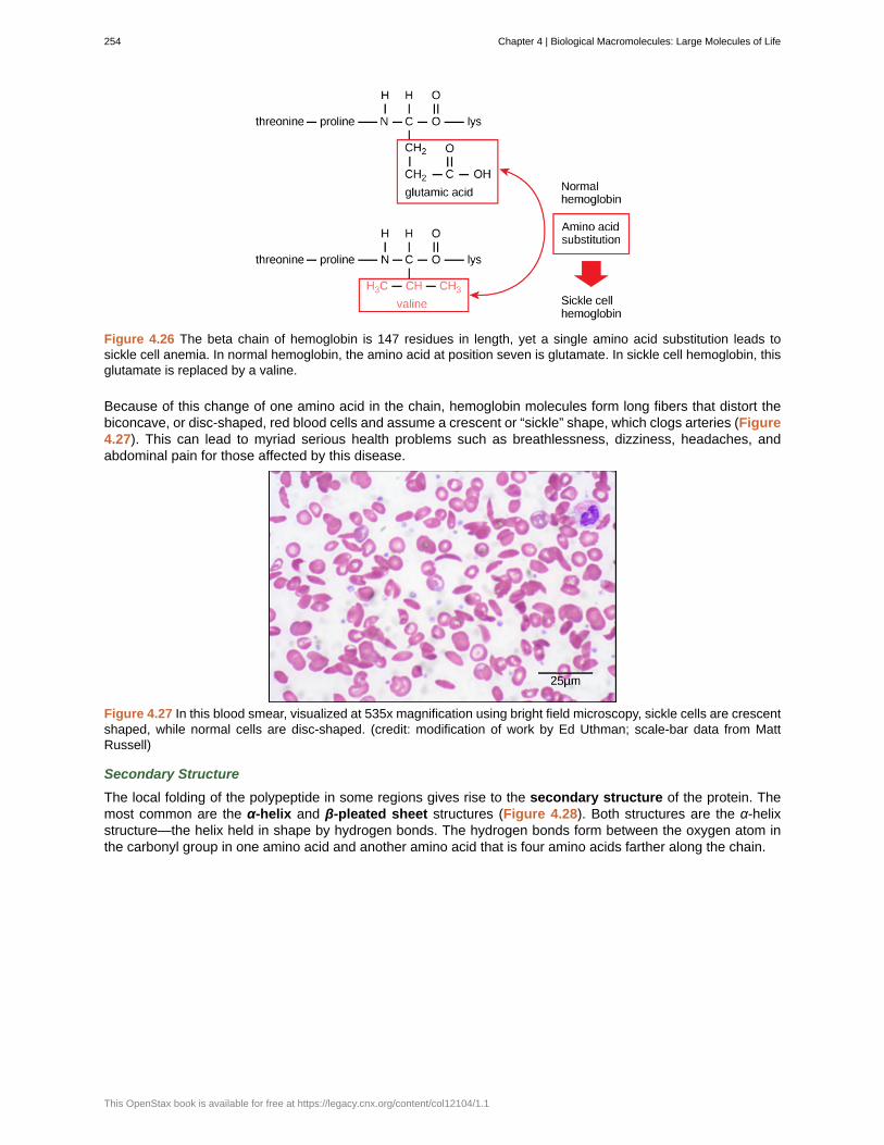
Figure 4.26 The beta chain of hemoglobin is 147 residues in length, yet a single amino acid substitution leads tosickle cell anemia. In normal hemoglobin, the amino acid at position seven is glutamate. In sickle cell hemoglobin, thisglutamate is replaced by a valine.
Because of this change of one amino acid in the chain, hemoglobin molecules form long fibers that distort thebiconcave, or disc-shaped, red blood cells and assume a crescent or “sickle” shape, which clogs arteries (Figure4.27). This can lead to myriad serious health problems such as breathlessness, dizziness, headaches, andabdominal pain for those affected by this disease.
Figure 4.27 In this blood smear, visualized at 535x magnification using bright field microscopy, sickle cells are crescentshaped, while normal cells are disc-shaped. (credit: modification of work by Ed Uthman; scale-bar data from MattRussell)
Secondary Structure
The local folding of the polypeptide in some regions gives rise to the secondary structure of the protein. Themost common are the α-helix and β-pleated sheet structures (Figure 4.28). Both structures are the α-helixstructure—the helix held in shape by hydrogen bonds. The hydrogen bonds form between the oxygen atom inthe carbonyl group in one amino acid and another amino acid that is four amino acids farther along the chain.
254 Chapter 4 | Biological Macromolecules: Large Molecules of Life
This OpenStax book is available for free at https://legacy.cnx.org/content/col12104/1.1
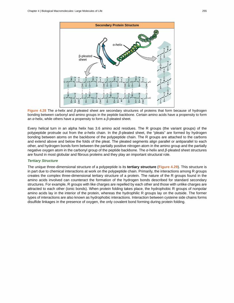
Figure 4.28 The α-helix and β-pleated sheet are secondary structures of proteins that form because of hydrogenbonding between carbonyl and amino groups in the peptide backbone. Certain amino acids have a propensity to forman α-helix, while others have a propensity to form a β-pleated sheet.
Every helical turn in an alpha helix has 3.6 amino acid residues. The R groups (the variant groups) of thepolypeptide protrude out from the α-helix chain. In the β-pleated sheet, the “pleats” are formed by hydrogenbonding between atoms on the backbone of the polypeptide chain. The R groups are attached to the carbonsand extend above and below the folds of the pleat. The pleated segments align parallel or antiparallel to eachother, and hydrogen bonds form between the partially positive nitrogen atom in the amino group and the partiallynegative oxygen atom in the carbonyl group of the peptide backbone. The α-helix and β-pleated sheet structuresare found in most globular and fibrous proteins and they play an important structural role.
Tertiary Structure
The unique three-dimensional structure of a polypeptide is its tertiary structure (Figure 4.29). This structure isin part due to chemical interactions at work on the polypeptide chain. Primarily, the interactions among R groupscreates the complex three-dimensional tertiary structure of a protein. The nature of the R groups found in theamino acids involved can counteract the formation of the hydrogen bonds described for standard secondarystructures. For example, R groups with like charges are repelled by each other and those with unlike charges areattracted to each other (ionic bonds). When protein folding takes place, the hydrophobic R groups of nonpolaramino acids lay in the interior of the protein, whereas the hydrophilic R groups lay on the outside. The formertypes of interactions are also known as hydrophobic interactions. Interaction between cysteine side chains formsdisulfide linkages in the presence of oxygen, the only covalent bond forming during protein folding.
Chapter 4 | Biological Macromolecules: Large Molecules of Life 255
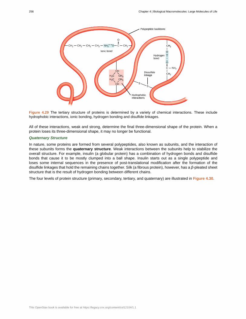
Figure 4.29 The tertiary structure of proteins is determined by a variety of chemical interactions. These includehydrophobic interactions, ionic bonding, hydrogen bonding and disulfide linkages.
All of these interactions, weak and strong, determine the final three-dimensional shape of the protein. When aprotein loses its three-dimensional shape, it may no longer be functional.
Quaternary Structure
In nature, some proteins are formed from several polypeptides, also known as subunits, and the interaction ofthese subunits forms the quaternary structure. Weak interactions between the subunits help to stabilize theoverall structure. For example, insulin (a globular protein) has a combination of hydrogen bonds and disulfidebonds that cause it to be mostly clumped into a ball shape. Insulin starts out as a single polypeptide andloses some internal sequences in the presence of post-translational modification after the formation of thedisulfide linkages that hold the remaining chains together. Silk (a fibrous protein), however, has a β-pleated sheetstructure that is the result of hydrogen bonding between different chains.
The four levels of protein structure (primary, secondary, tertiary, and quaternary) are illustrated in Figure 4.30.
256 Chapter 4 | Biological Macromolecules: Large Molecules of Life
This OpenStax book is available for free at https://legacy.cnx.org/content/col12104/1.1
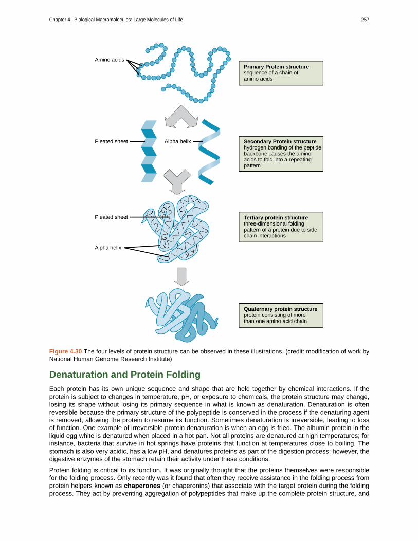
Figure 4.30 The four levels of protein structure can be observed in these illustrations. (credit: modification of work byNational Human Genome Research Institute)
Denaturation and Protein Folding
Each protein has its own unique sequence and shape that are held together by chemical interactions. If theprotein is subject to changes in temperature, pH, or exposure to chemicals, the protein structure may change,losing its shape without losing its primary sequence in what is known as denaturation. Denaturation is oftenreversible because the primary structure of the polypeptide is conserved in the process if the denaturing agentis removed, allowing the protein to resume its function. Sometimes denaturation is irreversible, leading to lossof function. One example of irreversible protein denaturation is when an egg is fried. The albumin protein in theliquid egg white is denatured when placed in a hot pan. Not all proteins are denatured at high temperatures; forinstance, bacteria that survive in hot springs have proteins that function at temperatures close to boiling. Thestomach is also very acidic, has a low pH, and denatures proteins as part of the digestion process; however, thedigestive enzymes of the stomach retain their activity under these conditions.
Protein folding is critical to its function. It was originally thought that the proteins themselves were responsiblefor the folding process. Only recently was it found that often they receive assistance in the folding process fromprotein helpers known as chaperones (or chaperonins) that associate with the target protein during the foldingprocess. They act by preventing aggregation of polypeptides that make up the complete protein structure, and
Chapter 4 | Biological Macromolecules: Large Molecules of Life 257
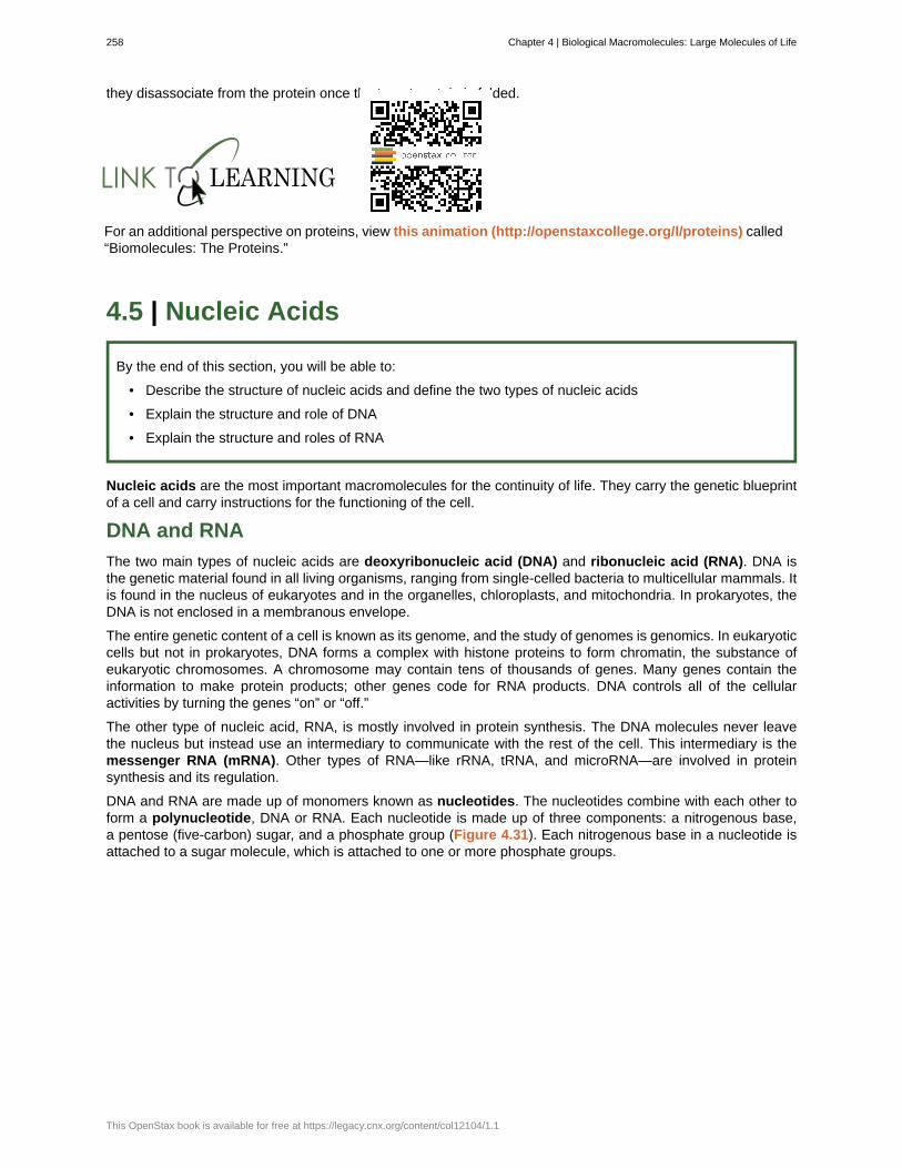
they disassociate from the protein once the target protein is folded.
For an additional perspective on proteins, view this animation (http://openstaxcollege.org/l/proteins) called“Biomolecules: The Proteins.”
4.5 | Nucleic Acids
By the end of this section, you will be able to:
• Describe the structure of nucleic acids and define the two types of nucleic acids
• Explain the structure and role of DNA
• Explain the structure and roles of RNA
Nucleic acids are the most important macromolecules for the continuity of life. They carry the genetic blueprintof a cell and carry instructions for the functioning of the cell.
DNA and RNA
The two main types of nucleic acids are deoxyribonucleic acid (DNA) and ribonucleic acid (RNA). DNA isthe genetic material found in all living organisms, ranging from single-celled bacteria to multicellular mammals. Itis found in the nucleus of eukaryotes and in the organelles, chloroplasts, and mitochondria. In prokaryotes, theDNA is not enclosed in a membranous envelope.
The entire genetic content of a cell is known as its genome, and the study of genomes is genomics. In eukaryoticcells but not in prokaryotes, DNA forms a complex with histone proteins to form chromatin, the substance ofeukaryotic chromosomes. A chromosome may contain tens of thousands of genes. Many genes contain theinformation to make protein products; other genes code for RNA products. DNA controls all of the cellularactivities by turning the genes “on” or “off.”
The other type of nucleic acid, RNA, is mostly involved in protein synthesis. The DNA molecules never leavethe nucleus but instead use an intermediary to communicate with the rest of the cell. This intermediary is themessenger RNA (mRNA). Other types of RNA—like rRNA, tRNA, and microRNA—are involved in proteinsynthesis and its regulation.
DNA and RNA are made up of monomers known as nucleotides. The nucleotides combine with each other toform a polynucleotide, DNA or RNA. Each nucleotide is made up of three components: a nitrogenous base,a pentose (five-carbon) sugar, and a phosphate group (Figure 4.31). Each nitrogenous base in a nucleotide isattached to a sugar molecule, which is attached to one or more phosphate groups.
258 Chapter 4 | Biological Macromolecules: Large Molecules of Life
This OpenStax book is available for free at https://legacy.cnx.org/content/col12104/1.1
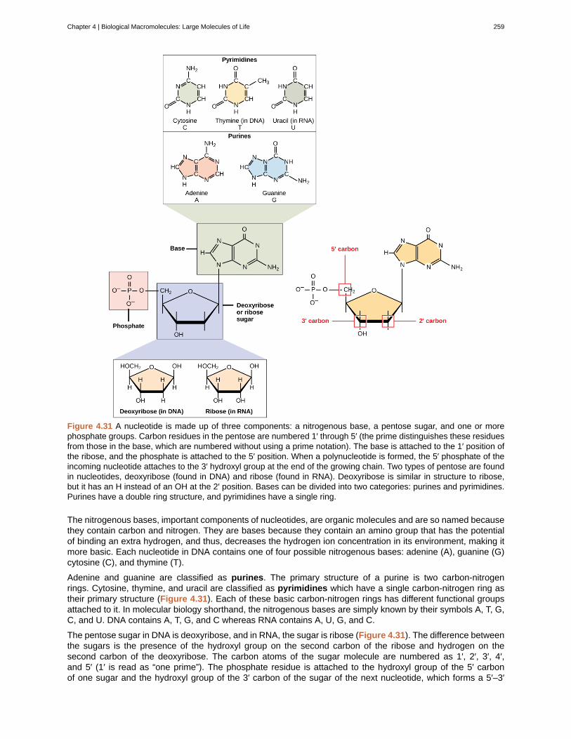
Figure 4.31 A nucleotide is made up of three components: a nitrogenous base, a pentose sugar, and one or morephosphate groups. Carbon residues in the pentose are numbered 1′ through 5′ (the prime distinguishes these residuesfrom those in the base, which are numbered without using a prime notation). The base is attached to the 1′ position ofthe ribose, and the phosphate is attached to the 5′ position. When a polynucleotide is formed, the 5′ phosphate of theincoming nucleotide attaches to the 3′ hydroxyl group at the end of the growing chain. Two types of pentose are foundin nucleotides, deoxyribose (found in DNA) and ribose (found in RNA). Deoxyribose is similar in structure to ribose,but it has an H instead of an OH at the 2′ position. Bases can be divided into two categories: purines and pyrimidines.Purines have a double ring structure, and pyrimidines have a single ring.
The nitrogenous bases, important components of nucleotides, are organic molecules and are so named becausethey contain carbon and nitrogen. They are bases because they contain an amino group that has the potentialof binding an extra hydrogen, and thus, decreases the hydrogen ion concentration in its environment, making itmore basic. Each nucleotide in DNA contains one of four possible nitrogenous bases: adenine (A), guanine (G)cytosine (C), and thymine (T).
Adenine and guanine are classified as purines. The primary structure of a purine is two carbon-nitrogenrings. Cytosine, thymine, and uracil are classified as pyrimidines which have a single carbon-nitrogen ring astheir primary structure (Figure 4.31). Each of these basic carbon-nitrogen rings has different functional groupsattached to it. In molecular biology shorthand, the nitrogenous bases are simply known by their symbols A, T, G,C, and U. DNA contains A, T, G, and C whereas RNA contains A, U, G, and C.
The pentose sugar in DNA is deoxyribose, and in RNA, the sugar is ribose (Figure 4.31). The difference betweenthe sugars is the presence of the hydroxyl group on the second carbon of the ribose and hydrogen on thesecond carbon of the deoxyribose. The carbon atoms of the sugar molecule are numbered as 1′, 2′, 3′, 4′,and 5′ (1′ is read as “one prime”). The phosphate residue is attached to the hydroxyl group of the 5′ carbonof one sugar and the hydroxyl group of the 3′ carbon of the sugar of the next nucleotide, which forms a 5′–3′
Chapter 4 | Biological Macromolecules: Large Molecules of Life 259
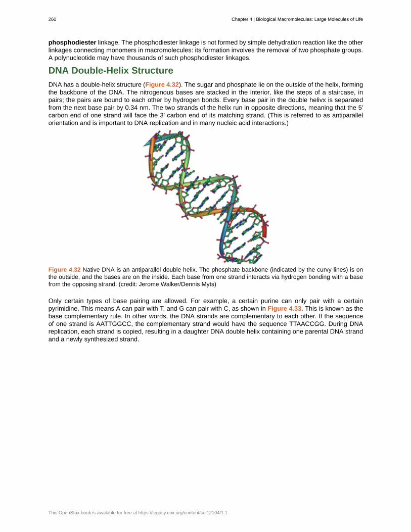
phosphodiester linkage. The phosphodiester linkage is not formed by simple dehydration reaction like the otherlinkages connecting monomers in macromolecules: its formation involves the removal of two phosphate groups.A polynucleotide may have thousands of such phosphodiester linkages.
DNA Double-Helix Structure
DNA has a double-helix structure (Figure 4.32). The sugar and phosphate lie on the outside of the helix, formingthe backbone of the DNA. The nitrogenous bases are stacked in the interior, like the steps of a staircase, inpairs; the pairs are bound to each other by hydrogen bonds. Every base pair in the double helivx is separatedfrom the next base pair by 0.34 nm. The two strands of the helix run in opposite directions, meaning that the 5′carbon end of one strand will face the 3′ carbon end of its matching strand. (This is referred to as antiparallelorientation and is important to DNA replication and in many nucleic acid interactions.)
Figure 4.32 Native DNA is an antiparallel double helix. The phosphate backbone (indicated by the curvy lines) is onthe outside, and the bases are on the inside. Each base from one strand interacts via hydrogen bonding with a basefrom the opposing strand. (credit: Jerome Walker/Dennis Myts)
Only certain types of base pairing are allowed. For example, a certain purine can only pair with a certainpyrimidine. This means A can pair with T, and G can pair with C, as shown in Figure 4.33. This is known as thebase complementary rule. In other words, the DNA strands are complementary to each other. If the sequenceof one strand is AATTGGCC, the complementary strand would have the sequence TTAACCGG. During DNAreplication, each strand is copied, resulting in a daughter DNA double helix containing one parental DNA strandand a newly synthesized strand.
260 Chapter 4 | Biological Macromolecules: Large Molecules of Life
This OpenStax book is available for free at https://legacy.cnx.org/content/col12104/1.1
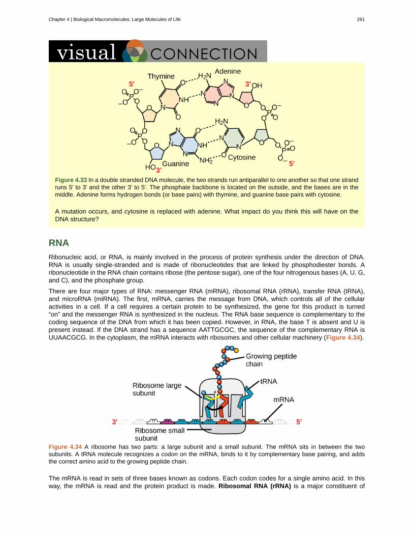
Figure 4.33 In a double stranded DNA molecule, the two strands run antiparallel to one another so that one strandruns 5′ to 3′ and the other 3′ to 5′. The phosphate backbone is located on the outside, and the bases are in themiddle. Adenine forms hydrogen bonds (or base pairs) with thymine, and guanine base pairs with cytosine.
A mutation occurs, and cytosine is replaced with adenine. What impact do you think this will have on theDNA structure?
RNA
Ribonucleic acid, or RNA, is mainly involved in the process of protein synthesis under the direction of DNA.RNA is usually single-stranded and is made of ribonucleotides that are linked by phosphodiester bonds. Aribonucleotide in the RNA chain contains ribose (the pentose sugar), one of the four nitrogenous bases (A, U, G,and C), and the phosphate group.
There are four major types of RNA: messenger RNA (mRNA), ribosomal RNA (rRNA), transfer RNA (tRNA),and microRNA (miRNA). The first, mRNA, carries the message from DNA, which controls all of the cellularactivities in a cell. If a cell requires a certain protein to be synthesized, the gene for this product is turned“on” and the messenger RNA is synthesized in the nucleus. The RNA base sequence is complementary to thecoding sequence of the DNA from which it has been copied. However, in RNA, the base T is absent and U ispresent instead. If the DNA strand has a sequence AATTGCGC, the sequence of the complementary RNA isUUAACGCG. In the cytoplasm, the mRNA interacts with ribosomes and other cellular machinery (Figure 4.34).
Figure 4.34 A ribosome has two parts: a large subunit and a small subunit. The mRNA sits in between the twosubunits. A tRNA molecule recognizes a codon on the mRNA, binds to it by complementary base pairing, and addsthe correct amino acid to the growing peptide chain.
The mRNA is read in sets of three bases known as codons. Each codon codes for a single amino acid. In thisway, the mRNA is read and the protein product is made. Ribosomal RNA (rRNA) is a major constituent of
Chapter 4 | Biological Macromolecules: Large Molecules of Life 261
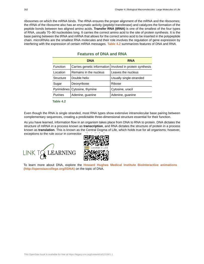
ribosomes on which the mRNA binds. The rRNA ensures the proper alignment of the mRNA and the ribosomes;the rRNA of the ribosome also has an enzymatic activity (peptidyl transferase) and catalyzes the formation of thepeptide bonds between two aligned amino acids. Transfer RNA (tRNA) is one of the smallest of the four typesof RNA, usually 70–90 nucleotides long. It carries the correct amino acid to the site of protein synthesis. It is thebase pairing between the tRNA and mRNA that allows for the correct amino acid to be inserted in the polypeptidechain. microRNAs are the smallest RNA molecules and their role involves the regulation of gene expression byinterfering with the expression of certain mRNA messages. Table 4.2 summarizes features of DNA and RNA.
Features of DNA and RNA
DNA RNA
Function Carries genetic information Involved in protein synthesis
Location Remains in the nucleus Leaves the nucleus
Structure Double helix Usually single-stranded
Sugar Deoxyribose Ribose
Pyrimidines Cytosine, thymine Cytosine, uracil
Purines Adenine, guanine Adenine, guanine
Table 4.2
Even though the RNA is single stranded, most RNA types show extensive intramolecular base pairing betweencomplementary sequences, creating a predictable three-dimensional structure essential for their function.
As you have learned, information flow in an organism takes place from DNA to RNA to protein. DNA dictates thestructure of mRNA in a process known as transcription, and RNA dictates the structure of protein in a processknown as translation. This is known as the Central Dogma of Life, which holds true for all organisms; however,exceptions to the rule occur in connection with viral infections.
To learn more about DNA, explore the Howard Hughes Medical Institute BioInteractive animations(http://openstaxcollege.org/l/DNA) on the topic of DNA.
262 Chapter 4 | Biological Macromolecules: Large Molecules of Life
This OpenStax book is available for free at https://legacy.cnx.org/content/col12104/1.1
alpha-helix structure (α-helix)
amino acid
beta-pleated sheet (β-pleated)
biological macromolecule
carbohydrate
cellulose
chaperone
chitin
dehydration synthesis
denaturation
deoxyribonucleic acid (DNA)
disaccharide
enzyme
glycogen
glycosidic bond
hormone
hydrolysis
lipid
messenger RNA (mRNA)
monomer
monosaccharide
nucleic acid
nucleotide
omega fat
peptide bond
KEY TERMS
type of secondary structure of proteins formed by folding of the polypeptide intoa helix shape with hydrogen bonds stabilizing the structure
monomer of a protein; has a central carbon or alpha carbon to which an amino group, a carboxylgroup, a hydrogen, and an R group or side chain is attached; the R group is different for all 20 amino acids
secondary structure found in proteins in which “pleats” are formed by hydrogenbonding between atoms on the backbone of the polypeptide chain
large molecule necessary for life that is built from smaller organic molecules
biological macromolecule in which the ratio of carbon to hydrogen and to oxygen is 1:2:1;carbohydrates serve as energy sources and structural support in cells and form the a cellular exoskeletonof arthropods
polysaccharide that makes up the cell wall of plants; provides structural support to the cell
(also, chaperonin) protein that helps nascent protein in the folding process
type of carbohydrate that forms the outer skeleton of all arthropods that include crustaceans and insects;it also forms the cell walls of fungi
(also, condensation) reaction that links monomer molecules together, releasing amolecule of water for each bond formed
loss of shape in a protein as a result of changes in temperature, pH, or exposure to chemicals
double-helical molecule that carries the hereditary information of the cell
two sugar monomers that are linked together by a glycosidic bond
catalyst in a biochemical reaction that is usually a complex or conjugated protein
storage carbohydrate in animals
bond formed by a dehydration reaction between two monosaccharides with the elimination ofa water molecule
chemical signaling molecule, usually protein or steroid, secreted by endocrine cells that act to controlor regulate specific physiological processes
reaction causes breakdown of larger molecules into smaller molecules with the utilization of water
macromolecule that is nonpolar and insoluble in water
RNA that carries information from DNA to ribosomes during protein synthesis
smallest unit of larger molecules called polymers
single unit or monomer of carbohydrates
biological macromolecule that carries the genetic blueprint of a cell and carries instructions for thefunctioning of the cell
monomer of nucleic acids; contains a pentose sugar, one or more phosphate groups, and anitrogenous base
type of polyunsaturated fat that is required by the body; the numbering of the carbon omega startsfrom the methyl end or the end that is farthest from the carboxylic end
bond formed between two amino acids by a dehydration reaction
Chapter 4 | Biological Macromolecules: Large Molecules of Life 263
phosphodiester
phospholipid
polymer
polynucleotide
polypeptide
polysaccharide
primary structure
protein
purine
pyrimidine
quaternary structure
ribonucleic acid (RNA)
ribosomal RNA (rRNA)
saturated fatty acid
secondary structure
starch
steroid
tertiary structure
trans fat
transcription
transfer RNA (tRNA)
translation
triacylglycerol (also, triglyceride)
unsaturated fatty acid
wax
linkage covalent chemical bond that holds together the polynucleotide chains with a phosphategroup linking two pentose sugars of neighboring nucleotides
major constituent of the membranes; composed of two fatty acids and a phosphate-containinggroup attached to a glycerol backbone
chain of monomer residues that is linked by covalent bonds; polymerization is the process of polymerformation from monomers by condensation
long chain of nucleotides
long chain of amino acids linked by peptide bonds
long chain of monosaccharides; may be branched or unbranched
linear sequence of amino acids in a protein
biological macromolecule composed of one or more chains of amino acids
type of nitrogenous base in DNA and RNA; adenine and guanine are purines
type of nitrogenous base in DNA and RNA; cytosine, thymine, and uracil are pyrimidines
association of discrete polypeptide subunits in a protein
single-stranded, often internally base paired, molecule that is involved in proteinsynthesis
RNA that ensures the proper alignment of the mRNA and the ribosomes during proteinsynthesis and catalyzes the formation of the peptide linkage
long-chain of hydrocarbon with single covalent bonds in the carbon chain; the number ofhydrogen atoms attached to the carbon skeleton is maximized
regular structure formed by proteins by intramolecular hydrogen bonding between theoxygen atom of one amino acid residue and the hydrogen attached to the nitrogen atom of another aminoacid residue
storage carbohydrate in plants
type of lipid composed of four fused hydrocarbon rings forming a planar structure
three-dimensional conformation of a protein, including interactions between secondarystructural elements; formed from interactions between amino acid side chains
fat formed artificially by hydrogenating oils, leading to a different arrangement of double bond(s) thanthose found in naturally occurring lipids
process through which messenger RNA forms on a template of DNA
RNA that carries activated amino acids to the site of protein synthesis on the ribosome
process through which RNA directs the formation of protein
fat molecule; consists of three fatty acids linked to a glycerol molecule
long-chain hydrocarbon that has one or more double bonds in the hydrocarbon chain
lipid made of a long-chain fatty acid that is esterified to a long-chain alcohol; serves as a protective coatingon some feathers, aquatic mammal fur, and leaves
264 Chapter 4 | Biological Macromolecules: Large Molecules of Life
This OpenStax book is available for free at https://legacy.cnx.org/content/col12104/1.1
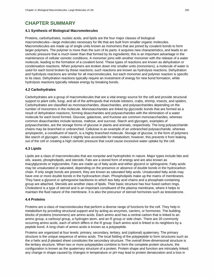
CHAPTER SUMMARY
4.1 Synthesis of Biological Macromolecules
Proteins, carbohydrates, nucleic acids, and lipids are the four major classes of biologicalmacromolecules—large molecules necessary for life that are built from smaller organic molecules.Macromolecules are made up of single units known as monomers that are joined by covalent bonds to formlarger polymers. The polymer is more than the sum of its parts: it acquires new characteristics, and leads to anosmotic pressure that is much lower than that formed by its ingredients; this is an important advantage in themaintenance of cellular osmotic conditions. A monomer joins with another monomer with the release of a watermolecule, leading to the formation of a covalent bond. These types of reactions are known as dehydration orcondensation reactions. When polymers are broken down into smaller units (monomers), a molecule of water isused for each bond broken by these reactions; such reactions are known as hydrolysis reactions. Dehydrationand hydrolysis reactions are similar for all macromolecules, but each monomer and polymer reaction is specificto its class. Dehydration reactions typically require an investment of energy for new bond formation, whilehydrolysis reactions typically release energy by breaking bonds.
4.2 Carbohydrates
Carbohydrates are a group of macromolecules that are a vital energy source for the cell and provide structuralsupport to plant cells, fungi, and all of the arthropods that include lobsters, crabs, shrimp, insects, and spiders.Carbohydrates are classified as monosaccharides, disaccharides, and polysaccharides depending on thenumber of monomers in the molecule. Monosaccharides are linked by glycosidic bonds that are formed as aresult of dehydration reactions, forming disaccharides and polysaccharides with the elimination of a watermolecule for each bond formed. Glucose, galactose, and fructose are common monosaccharides, whereascommon disaccharides include lactose, maltose, and sucrose. Starch and glycogen, examples ofpolysaccharides, are the storage forms of glucose in plants and animals, respectively. The long polysaccharidechains may be branched or unbranched. Cellulose is an example of an unbranched polysaccharide, whereasamylopectin, a constituent of starch, is a highly branched molecule. Storage of glucose, in the form of polymerslike starch of glycogen, makes it slightly less accessible for metabolism; however, this prevents it from leakingout of the cell or creating a high osmotic pressure that could cause excessive water uptake by the cell.
4.3 Lipids
Lipids are a class of macromolecules that are nonpolar and hydrophobic in nature. Major types include fats andoils, waxes, phospholipids, and steroids. Fats are a stored form of energy and are also known astriacylglycerols or triglycerides. Fats are made up of fatty acids and either glycerol or sphingosine. Fatty acidsmay be unsaturated or saturated, depending on the presence or absence of double bonds in the hydrocarbonchain. If only single bonds are present, they are known as saturated fatty acids. Unsaturated fatty acids mayhave one or more double bonds in the hydrocarbon chain. Phospholipids make up the matrix of membranes.They have a glycerol or sphingosine backbone to which two fatty acid chains and a phosphate-containinggroup are attached. Steroids are another class of lipids. Their basic structure has four fused carbon rings.Cholesterol is a type of steroid and is an important constituent of the plasma membrane, where it helps tomaintain the fluid nature of the membrane. It is also the precursor of steroid hormones such as testosterone.
4.4 Proteins
Proteins are a class of macromolecules that perform a diverse range of functions for the cell. They help inmetabolism by providing structural support and by acting as enzymes, carriers, or hormones. The buildingblocks of proteins (monomers) are amino acids. Each amino acid has a central carbon that is linked to anamino group, a carboxyl group, a hydrogen atom, and an R group or side chain. There are 20 commonlyoccurring amino acids, each of which differs in the R group. Each amino acid is linked to its neighbors by apeptide bond. A long chain of amino acids is known as a polypeptide.
Proteins are organized at four levels: primary, secondary, tertiary, and (optional) quaternary. The primarystructure is the unique sequence of amino acids. The local folding of the polypeptide to form structures such asthe α helix and β-pleated sheet constitutes the secondary structure. The overall three-dimensional structure isthe tertiary structure. When two or more polypeptides combine to form the complete protein structure, theconfiguration is known as the quaternary structure of a protein. Protein shape and function are intricately linked;any change in shape caused by changes in temperature or pH may lead to protein denaturation and a loss in
Chapter 4 | Biological Macromolecules: Large Molecules of Life 265
function.
4.5 Nucleic Acids
Nucleic acids are molecules made up of nucleotides that direct cellular activities such as cell division andprotein synthesis. Each nucleotide is made up of a pentose sugar, a nitrogenous base, and a phosphate group.There are two types of nucleic acids: DNA and RNA. DNA carries the genetic blueprint of the cell and is passedon from parents to offspring (in the form of chromosomes). It has a double-helical structure with the two strandsrunning in opposite directions, connected by hydrogen bonds, and complementary to each other. RNA is single-stranded and is made of a pentose sugar (ribose), a nitrogenous base, and a phosphate group. RNA isinvolved in protein synthesis and its regulation. Messenger RNA (mRNA) is copied from the DNA, is exportedfrom the nucleus to the cytoplasm, and contains information for the construction of proteins. Ribosomal RNA(rRNA) is a part of the ribosomes at the site of protein synthesis, whereas transfer RNA (tRNA) carries theamino acid to the site of protein synthesis. microRNA regulates the use of mRNA for protein synthesis.
ART CONNECTION QUESTIONS
1. Figure 4.5 What kind of sugars are these, aldoseor ketose?
2. Figure 4.23 Which categories of amino acid wouldyou expect to find on the surface of a soluble protein,and which would you expect to find in the interior?
What distribution of amino acids would you expect tofind in a protein embedded in a lipid bilayer?
3. Figure 4.33 A mutation occurs, and cytosine isreplaced with adenine. What impact do you think thiswill have on the DNA structure?
REVIEW QUESTIONS
4. Dehydration synthesis leads to formation of
a. monomersb. polymersc. water and polymersd. none of the above
5. During the breakdown of polymers, which of thefollowing reactions takes place?
a. hydrolysisb. dehydrationc. condensationd. covalent bond
6. An example of a monosaccharide is ________.
a. fructoseb. glucosec. galactosed. all of the above
7. Cellulose and starch are examples of:a. monosaccharidesb. disaccharidesc. lipidsd. polysaccharides
8. Plant cell walls contain which of the following inabundance?
a. starchb. cellulosec. glycogend. lactose
9. Lactose is a disaccharide formed by the formationof a ________ bond between glucose and ________.
a. glycosidic; lactoseb. glycosidic; galactosec. hydrogen; sucrosed. hydrogen; fructose
10. Saturated fats have all of the followingcharacteristics except:
a. they are solid at room temperatureb. they have single bonds within the carbon
chainc. they are usually obtained from animal
sourcesd. they tend to dissolve in water easily
11. Phospholipids are important components of________.
a. the plasma membrane of animal cellsb. the ring structure of steroidsc. the waxy covering on leavesd. the double bond in hydrocarbon chains
12. The monomers that make up proteins are called________.
a. nucleotidesb. disaccharidesc. amino acidsd. chaperones
13. The α helix and the β-pleated sheet are part ofwhich protein structure?
a. primaryb. secondaryc. tertiaryd. quaternary
266 Chapter 4 | Biological Macromolecules: Large Molecules of Life
This OpenStax book is available for free at https://legacy.cnx.org/content/col12104/1.1
14. A nucleotide of DNA may contain ________.
a. ribose, uracil, and a phosphate groupb. deoxyribose, uracil, and a phosphate groupc. deoxyribose, thymine, and a phosphate
groupd. ribose, thymine, and a phosphate group
15. The building blocks of nucleic acids are________.
a. sugarsb. nitrogenous basesc. peptidesd. nucleotides
CRITICAL THINKING QUESTIONS
16. Why are biological macromolecules consideredorganic?
17. What role do electrons play in dehydrationsynthesis and hydrolysis?
18. Describe the similarities and differences betweenglycogen and starch.
19. Why is it impossible for humans to digest foodthat contains cellulose?
20. Explain at least three functions that lipids serve inplants and/or animals.
21. Why have trans fats been banned from some
restaurants? How are they created?
22. Explain what happens if even one amino acid issubstituted for another in a polypeptide chain.Provide a specific example.
23. Describe the differences in the four proteinstructures.
24. What are the structural differences between RNAand DNA?
25. What are the four types of RNA and how do theyfunction?
Chapter 4 | Biological Macromolecules: Large Molecules of Life 267

268 Chapter 4 | Biological Macromolecules: Large Molecules of Life
This OpenStax book is available for free at https://legacy.cnx.org/content/col12104/1.1
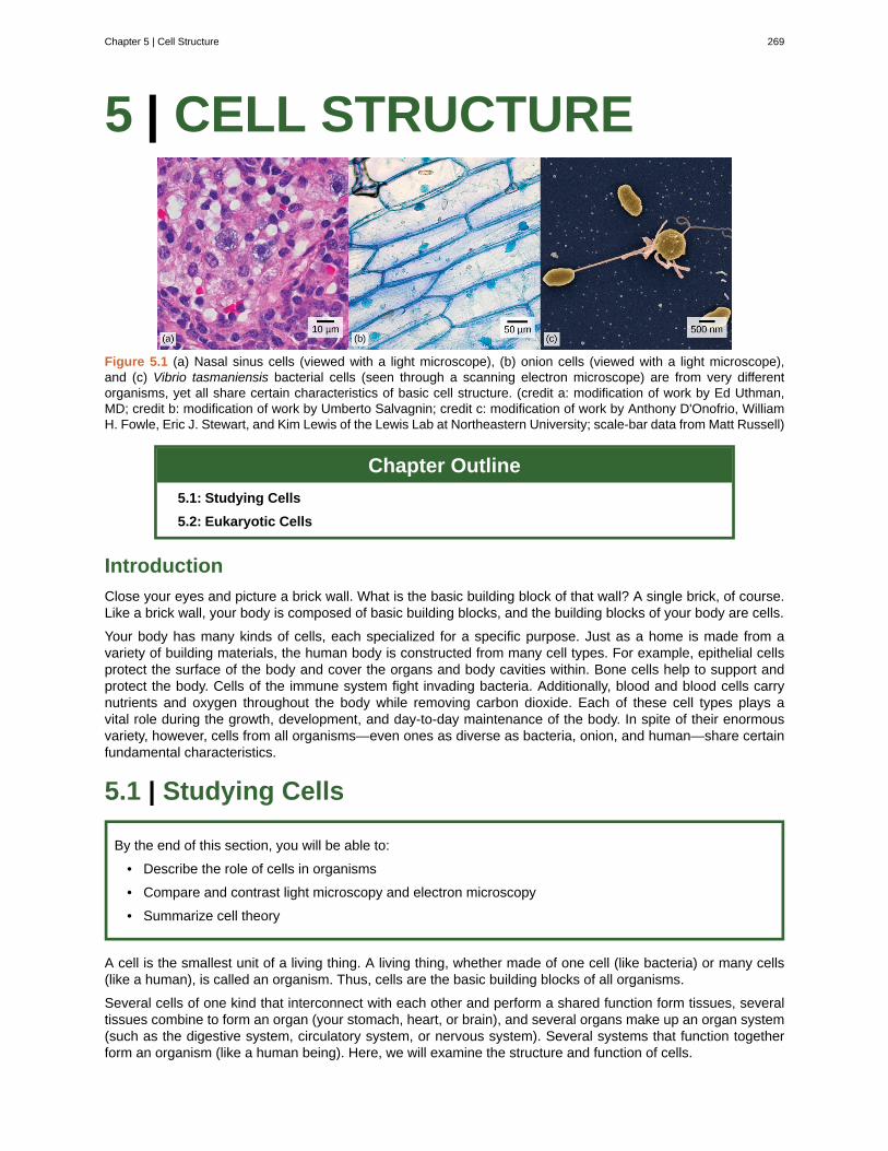
5 | CELL STRUCTURE
Figure 5.1 (a) Nasal sinus cells (viewed with a light microscope), (b) onion cells (viewed with a light microscope),and (c) Vibrio tasmaniensis bacterial cells (seen through a scanning electron microscope) are from very differentorganisms, yet all share certain characteristics of basic cell structure. (credit a: modification of work by Ed Uthman,MD; credit b: modification of work by Umberto Salvagnin; credit c: modification of work by Anthony D'Onofrio, WilliamH. Fowle, Eric J. Stewart, and Kim Lewis of the Lewis Lab at Northeastern University; scale-bar data from Matt Russell)
Chapter Outline
5.1: Studying Cells
5.2: Eukaryotic Cells
Introduction
Close your eyes and picture a brick wall. What is the basic building block of that wall? A single brick, of course.Like a brick wall, your body is composed of basic building blocks, and the building blocks of your body are cells.
Your body has many kinds of cells, each specialized for a specific purpose. Just as a home is made from avariety of building materials, the human body is constructed from many cell types. For example, epithelial cellsprotect the surface of the body and cover the organs and body cavities within. Bone cells help to support andprotect the body. Cells of the immune system fight invading bacteria. Additionally, blood and blood cells carrynutrients and oxygen throughout the body while removing carbon dioxide. Each of these cell types plays avital role during the growth, development, and day-to-day maintenance of the body. In spite of their enormousvariety, however, cells from all organisms—even ones as diverse as bacteria, onion, and human—share certainfundamental characteristics.
5.1 | Studying Cells
By the end of this section, you will be able to:
• Describe the role of cells in organisms
• Compare and contrast light microscopy and electron microscopy
• Summarize cell theory
A cell is the smallest unit of a living thing. A living thing, whether made of one cell (like bacteria) or many cells(like a human), is called an organism. Thus, cells are the basic building blocks of all organisms.
Several cells of one kind that interconnect with each other and perform a shared function form tissues, severaltissues combine to form an organ (your stomach, heart, or brain), and several organs make up an organ system(such as the digestive system, circulatory system, or nervous system). Several systems that function togetherform an organism (like a human being). Here, we will examine the structure and function of cells.
Chapter 5 | Cell Structure 269
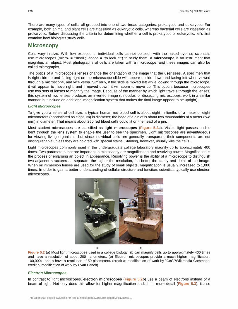
There are many types of cells, all grouped into one of two broad categories: prokaryotic and eukaryotic. Forexample, both animal and plant cells are classified as eukaryotic cells, whereas bacterial cells are classified asprokaryotic. Before discussing the criteria for determining whether a cell is prokaryotic or eukaryotic, let’s firstexamine how biologists study cells.
Microscopy
Cells vary in size. With few exceptions, individual cells cannot be seen with the naked eye, so scientistsuse microscopes (micro- = “small”; -scope = “to look at”) to study them. A microscope is an instrument thatmagnifies an object. Most photographs of cells are taken with a microscope, and these images can also becalled micrographs.
The optics of a microscope’s lenses change the orientation of the image that the user sees. A specimen thatis right-side up and facing right on the microscope slide will appear upside-down and facing left when viewedthrough a microscope, and vice versa. Similarly, if the slide is moved left while looking through the microscope,it will appear to move right, and if moved down, it will seem to move up. This occurs because microscopesuse two sets of lenses to magnify the image. Because of the manner by which light travels through the lenses,this system of two lenses produces an inverted image (binocular, or dissecting microscopes, work in a similarmanner, but include an additional magnification system that makes the final image appear to be upright).
Light Microscopes
To give you a sense of cell size, a typical human red blood cell is about eight millionths of a meter or eightmicrometers (abbreviated as eight μm) in diameter; the head of a pin of is about two thousandths of a meter (twomm) in diameter. That means about 250 red blood cells could fit on the head of a pin.
Most student microscopes are classified as light microscopes (Figure 5.2a). Visible light passes and isbent through the lens system to enable the user to see the specimen. Light microscopes are advantageousfor viewing living organisms, but since individual cells are generally transparent, their components are notdistinguishable unless they are colored with special stains. Staining, however, usually kills the cells.
Light microscopes commonly used in the undergraduate college laboratory magnify up to approximately 400times. Two parameters that are important in microscopy are magnification and resolving power. Magnification isthe process of enlarging an object in appearance. Resolving power is the ability of a microscope to distinguishtwo adjacent structures as separate: the higher the resolution, the better the clarity and detail of the image.When oil immersion lenses are used for the study of small objects, magnification is usually increased to 1,000times. In order to gain a better understanding of cellular structure and function, scientists typically use electronmicroscopes.
Figure 5.2 (a) Most light microscopes used in a college biology lab can magnify cells up to approximately 400 timesand have a resolution of about 200 nanometers. (b) Electron microscopes provide a much higher magnification,100,000x, and a have a resolution of 50 picometers. (credit a: modification of work by "GcG"/Wikimedia Commons;credit b: modification of work by Evan Bench)
Electron Microscopes
In contrast to light microscopes, electron microscopes (Figure 5.2b) use a beam of electrons instead of abeam of light. Not only does this allow for higher magnification and, thus, more detail (Figure 5.3), it also
270 Chapter 5 | Cell Structure
This OpenStax book is available for free at https://legacy.cnx.org/content/col12104/1.1

provides higher resolving power. The method used to prepare the specimen for viewing with an electronmicroscope kills the specimen. Electrons have short wavelengths (shorter than photons) that move best in avacuum, so living cells cannot be viewed with an electron microscope.
In a scanning electron microscope, a beam of electrons moves back and forth across a cell’s surface, creatingdetails of cell surface characteristics. In a transmission electron microscope, the electron beam penetratesthe cell and provides details of a cell’s internal structures. As you might imagine, electron microscopes aresignificantly more bulky and expensive than light microscopes.
(a) (b)Figure 5.3 (a) These Salmonella bacteria appear as tiny purple dots when viewed with a light microscope. (b) Thisscanning electron microscope micrograph shows Salmonella bacteria (in red) invading human cells (yellow). Eventhough subfigure (b) shows a different Salmonella specimen than subfigure (a), you can still observe the comparativeincrease in magnification and detail. (credit a: modification of work by CDC/Armed Forces Institute of Pathology,Charles N. Farmer, Rocky Mountain Laboratories; credit b: modification of work by NIAID, NIH; scale-bar data fromMatt Russell)
For another perspective on cell size, try the HowBig interactive at this site (http://openstaxcollege.org/l/cell_sizes) .
Cell Theory
The microscopes we use today are far more complex than those used in the 1600s by Antony van Leeuwenhoek,a Dutch shopkeeper who had great skill in crafting lenses. Despite the limitations of his now-ancient lenses,van Leeuwenhoek observed the movements of protista (a type of single-celled organism) and sperm, which hecollectively termed “animalcules.”
In a 1665 publication called Micrographia, experimental scientist Robert Hooke coined the term “cell” for the box-like structures he observed when viewing cork tissue through a lens. In the 1670s, van Leeuwenhoek discoveredbacteria and protozoa. Later advances in lenses, microscope construction, and staining techniques enabledother scientists to see some components inside cells.
By the late 1830s, botanist Matthias Schleiden and zoologist Theodor Schwann were studying tissues andproposed the unified cell theory, which states that all living things are composed of one or more cells, the cellis the basic unit of life, and new cells arise from existing cells. Rudolf Virchow later made important contributionsto this theory.
Chapter 5 | Cell Structure 271
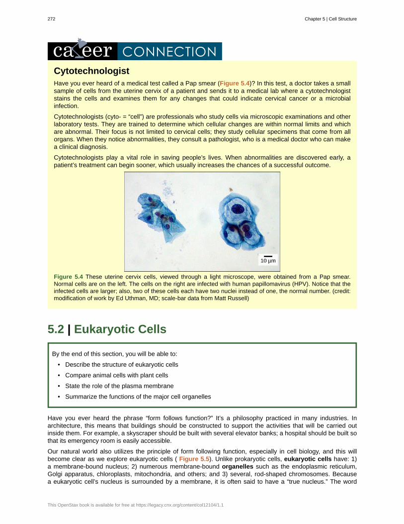
CytotechnologistHave you ever heard of a medical test called a Pap smear (Figure 5.4)? In this test, a doctor takes a smallsample of cells from the uterine cervix of a patient and sends it to a medical lab where a cytotechnologiststains the cells and examines them for any changes that could indicate cervical cancer or a microbialinfection.
Cytotechnologists (cyto- = “cell”) are professionals who study cells via microscopic examinations and otherlaboratory tests. They are trained to determine which cellular changes are within normal limits and whichare abnormal. Their focus is not limited to cervical cells; they study cellular specimens that come from allorgans. When they notice abnormalities, they consult a pathologist, who is a medical doctor who can makea clinical diagnosis.
Cytotechnologists play a vital role in saving people’s lives. When abnormalities are discovered early, apatient’s treatment can begin sooner, which usually increases the chances of a successful outcome.
Figure 5.4 These uterine cervix cells, viewed through a light microscope, were obtained from a Pap smear.Normal cells are on the left. The cells on the right are infected with human papillomavirus (HPV). Notice that theinfected cells are larger; also, two of these cells each have two nuclei instead of one, the normal number. (credit:modification of work by Ed Uthman, MD; scale-bar data from Matt Russell)
5.2 | Eukaryotic Cells
By the end of this section, you will be able to:
• Describe the structure of eukaryotic cells
• Compare animal cells with plant cells
• State the role of the plasma membrane
• Summarize the functions of the major cell organelles
Have you ever heard the phrase “form follows function?” It’s a philosophy practiced in many industries. Inarchitecture, this means that buildings should be constructed to support the activities that will be carried outinside them. For example, a skyscraper should be built with several elevator banks; a hospital should be built sothat its emergency room is easily accessible.
Our natural world also utilizes the principle of form following function, especially in cell biology, and this willbecome clear as we explore eukaryotic cells ( Figure 5.5). Unlike prokaryotic cells, eukaryotic cells have: 1)a membrane-bound nucleus; 2) numerous membrane-bound organelles such as the endoplasmic reticulum,Golgi apparatus, chloroplasts, mitochondria, and others; and 3) several, rod-shaped chromosomes. Becausea eukaryotic cell’s nucleus is surrounded by a membrane, it is often said to have a “true nucleus.” The word
272 Chapter 5 | Cell Structure
This OpenStax book is available for free at https://legacy.cnx.org/content/col12104/1.1

“organelle” means “little organ,” and, as already mentioned, organelles have specialized cellular functions, justas the organs of your body have specialized functions.
At this point, it should be clear to you that eukaryotic cells have a more complex structure than prokaryoticcells. Organelles allow different functions to be compartmentalized in different areas of the cell. Before turning toorganelles, let’s first examine two important components of the cell: the plasma membrane and the cytoplasm.
Chapter 5 | Cell Structure 273
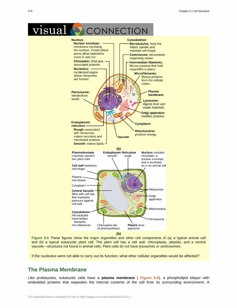
(a)
(b)Figure 5.5 These figures show the major organelles and other cell components of (a) a typical animal celland (b) a typical eukaryotic plant cell. The plant cell has a cell wall, chloroplasts, plastids, and a centralvacuole—structures not found in animal cells. Plant cells do not have lysosomes or centrosomes.
If the nucleolus were not able to carry out its function, what other cellular organelles would be affected?
The Plasma Membrane
Like prokaryotes, eukaryotic cells have a plasma membrane ( Figure 5.6), a phospholipid bilayer withembedded proteins that separates the internal contents of the cell from its surrounding environment. A
274 Chapter 5 | Cell Structure
This OpenStax book is available for free at https://legacy.cnx.org/content/col12104/1.1
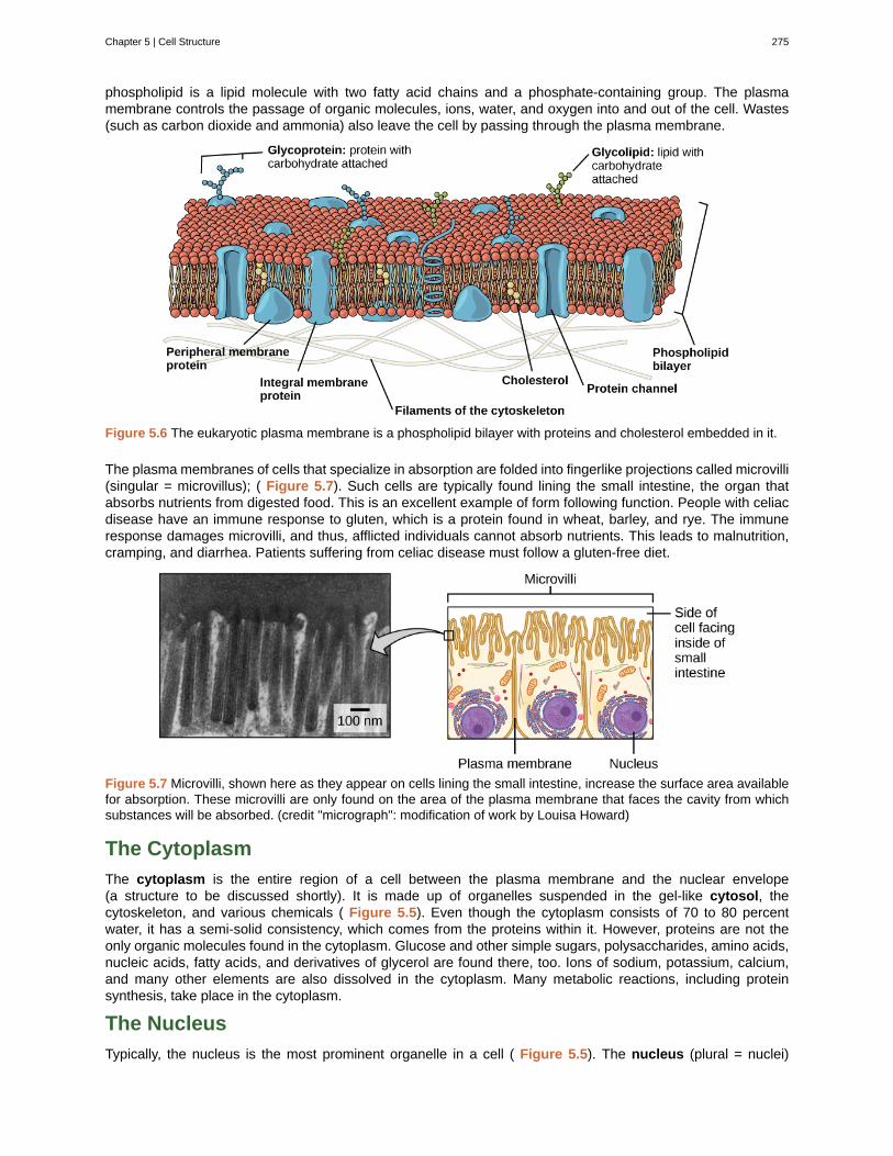
phospholipid is a lipid molecule with two fatty acid chains and a phosphate-containing group. The plasmamembrane controls the passage of organic molecules, ions, water, and oxygen into and out of the cell. Wastes(such as carbon dioxide and ammonia) also leave the cell by passing through the plasma membrane.
Figure 5.6 The eukaryotic plasma membrane is a phospholipid bilayer with proteins and cholesterol embedded in it.
The plasma membranes of cells that specialize in absorption are folded into fingerlike projections called microvilli(singular = microvillus); ( Figure 5.7). Such cells are typically found lining the small intestine, the organ thatabsorbs nutrients from digested food. This is an excellent example of form following function. People with celiacdisease have an immune response to gluten, which is a protein found in wheat, barley, and rye. The immuneresponse damages microvilli, and thus, afflicted individuals cannot absorb nutrients. This leads to malnutrition,cramping, and diarrhea. Patients suffering from celiac disease must follow a gluten-free diet.
Figure 5.7 Microvilli, shown here as they appear on cells lining the small intestine, increase the surface area availablefor absorption. These microvilli are only found on the area of the plasma membrane that faces the cavity from whichsubstances will be absorbed. (credit "micrograph": modification of work by Louisa Howard)
The Cytoplasm
The cytoplasm is the entire region of a cell between the plasma membrane and the nuclear envelope(a structure to be discussed shortly). It is made up of organelles suspended in the gel-like cytosol, thecytoskeleton, and various chemicals ( Figure 5.5). Even though the cytoplasm consists of 70 to 80 percentwater, it has a semi-solid consistency, which comes from the proteins within it. However, proteins are not theonly organic molecules found in the cytoplasm. Glucose and other simple sugars, polysaccharides, amino acids,nucleic acids, fatty acids, and derivatives of glycerol are found there, too. Ions of sodium, potassium, calcium,and many other elements are also dissolved in the cytoplasm. Many metabolic reactions, including proteinsynthesis, take place in the cytoplasm.
The Nucleus
Typically, the nucleus is the most prominent organelle in a cell ( Figure 5.5). The nucleus (plural = nuclei)
Chapter 5 | Cell Structure 275
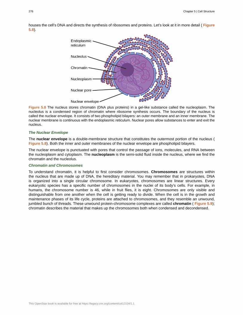
houses the cell’s DNA and directs the synthesis of ribosomes and proteins. Let’s look at it in more detail ( Figure5.8).
Figure 5.8 The nucleus stores chromatin (DNA plus proteins) in a gel-like substance called the nucleoplasm. Thenucleolus is a condensed region of chromatin where ribosome synthesis occurs. The boundary of the nucleus iscalled the nuclear envelope. It consists of two phospholipid bilayers: an outer membrane and an inner membrane. Thenuclear membrane is continuous with the endoplasmic reticulum. Nuclear pores allow substances to enter and exit thenucleus.
The Nuclear Envelope
The nuclear envelope is a double-membrane structure that constitutes the outermost portion of the nucleus (Figure 5.8). Both the inner and outer membranes of the nuclear envelope are phospholipid bilayers.
The nuclear envelope is punctuated with pores that control the passage of ions, molecules, and RNA betweenthe nucleoplasm and cytoplasm. The nucleoplasm is the semi-solid fluid inside the nucleus, where we find thechromatin and the nucleolus.
Chromatin and Chromosomes
To understand chromatin, it is helpful to first consider chromosomes. Chromosomes are structures withinthe nucleus that are made up of DNA, the hereditary material. You may remember that in prokaryotes, DNAis organized into a single circular chromosome. In eukaryotes, chromosomes are linear structures. Everyeukaryotic species has a specific number of chromosomes in the nuclei of its body’s cells. For example, inhumans, the chromosome number is 46, while in fruit flies, it is eight. Chromosomes are only visible anddistinguishable from one another when the cell is getting ready to divide. When the cell is in the growth andmaintenance phases of its life cycle, proteins are attached to chromosomes, and they resemble an unwound,jumbled bunch of threads. These unwound protein-chromosome complexes are called chromatin ( Figure 5.9);chromatin describes the material that makes up the chromosomes both when condensed and decondensed.
276 Chapter 5 | Cell Structure
This OpenStax book is available for free at https://legacy.cnx.org/content/col12104/1.1
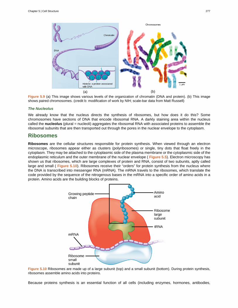
(a) (b)
Figure 5.9 (a) This image shows various levels of the organization of chromatin (DNA and protein). (b) This imageshows paired chromosomes. (credit b: modification of work by NIH; scale-bar data from Matt Russell)
The Nucleolus
We already know that the nucleus directs the synthesis of ribosomes, but how does it do this? Somechromosomes have sections of DNA that encode ribosomal RNA. A darkly staining area within the nucleuscalled the nucleolus (plural = nucleoli) aggregates the ribosomal RNA with associated proteins to assemble theribosomal subunits that are then transported out through the pores in the nuclear envelope to the cytoplasm.
Ribosomes
Ribosomes are the cellular structures responsible for protein synthesis. When viewed through an electronmicroscope, ribosomes appear either as clusters (polyribosomes) or single, tiny dots that float freely in thecytoplasm. They may be attached to the cytoplasmic side of the plasma membrane or the cytoplasmic side of theendoplasmic reticulum and the outer membrane of the nuclear envelope ( Figure 5.5). Electron microscopy hasshown us that ribosomes, which are large complexes of protein and RNA, consist of two subunits, aptly calledlarge and small ( Figure 5.10). Ribosomes receive their “orders” for protein synthesis from the nucleus wherethe DNA is transcribed into messenger RNA (mRNA). The mRNA travels to the ribosomes, which translate thecode provided by the sequence of the nitrogenous bases in the mRNA into a specific order of amino acids in aprotein. Amino acids are the building blocks of proteins.
Figure 5.10 Ribosomes are made up of a large subunit (top) and a small subunit (bottom). During protein synthesis,ribosomes assemble amino acids into proteins.
Because proteins synthesis is an essential function of all cells (including enzymes, hormones, antibodies,
Chapter 5 | Cell Structure 277
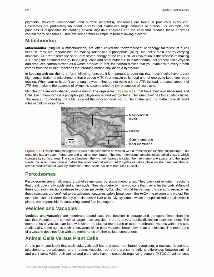
pigments, structural components, and surface receptors), ribosomes are found in practically every cell.Ribosomes are particularly abundant in cells that synthesize large amounts of protein. For example, thepancreas is responsible for creating several digestive enzymes and the cells that produce these enzymescontain many ribosomes. Thus, we see another example of form following function.
Mitochondria
Mitochondria (singular = mitochondrion) are often called the “powerhouses” or “energy factories” of a cellbecause they are responsible for making adenosine triphosphate (ATP), the cell’s main energy-carryingmolecule. ATP represents the short-term stored energy of the cell. Cellular respiration is the process of makingATP using the chemical energy found in glucose and other nutrients. In mitochondria, this process uses oxygenand produces carbon dioxide as a waste product. In fact, the carbon dioxide that you exhale with every breathcomes from the cellular reactions that produce carbon dioxide as a byproduct.
In keeping with our theme of form following function, it is important to point out that muscle cells have a veryhigh concentration of mitochondria that produce ATP. Your muscle cells need a lot of energy to keep your bodymoving. When your cells don’t get enough oxygen, they do not make a lot of ATP. Instead, the small amount ofATP they make in the absence of oxygen is accompanied by the production of lactic acid.
Mitochondria are oval-shaped, double membrane organelles ( Figure 5.11) that have their own ribosomes andDNA. Each membrane is a phospholipid bilayer embedded with proteins. The inner layer has folds called cristae.The area surrounded by the folds is called the mitochondrial matrix. The cristae and the matrix have differentroles in cellular respiration.
Figure 5.11 This electron micrograph shows a mitochondrion as viewed with a transmission electron microscope. Thisorganelle has an outer membrane and an inner membrane. The inner membrane contains folds, called cristae, whichincrease its surface area. The space between the two membranes is called the intermembrane space, and the spaceinside the inner membrane is called the mitochondrial matrix. ATP synthesis takes place on the inner membrane.(credit: modification of work by Matthew Britton; scale-bar data from Matt Russell)
Peroxisomes
Peroxisomes are small, round organelles enclosed by single membranes. They carry out oxidation reactionsthat break down fatty acids and amino acids. They also detoxify many poisons that may enter the body. (Many ofthese oxidation reactions release hydrogen peroxide, H2O2, which would be damaging to cells; however, whenthese reactions are confined to peroxisomes, enzymes safely break down the H2O2 into oxygen and water.) Forexample, alcohol is detoxified by peroxisomes in liver cells. Glyoxysomes, which are specialized peroxisomes inplants, are responsible for converting stored fats into sugars.
Vesicles and Vacuoles
Vesicles and vacuoles are membrane-bound sacs that function in storage and transport. Other than thefact that vacuoles are somewhat larger than vesicles, there is a very subtle distinction between them: Themembranes of vesicles can fuse with either the plasma membrane or other membrane systems within the cell.Additionally, some agents such as enzymes within plant vacuoles break down macromolecules. The membraneof a vacuole does not fuse with the membranes of other cellular components.
Animal Cells versus Plant Cells
At this point, you know that each eukaryotic cell has a plasma membrane, cytoplasm, a nucleus, ribosomes,mitochondria, peroxisomes, and in some, vacuoles, but there are some striking differences between animaland plant cells. While both animal and plant cells have microtubule organizing centers (MTOCs), animal cells
278 Chapter 5 | Cell Structure
This OpenStax book is available for free at https://legacy.cnx.org/content/col12104/1.1
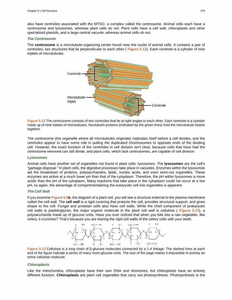
also have centrioles associated with the MTOC: a complex called the centrosome. Animal cells each have acentrosome and lysosomes, whereas plant cells do not. Plant cells have a cell wall, chloroplasts and otherspecialized plastids, and a large central vacuole, whereas animal cells do not.
The Centrosome
The centrosome is a microtubule-organizing center found near the nuclei of animal cells. It contains a pair ofcentrioles, two structures that lie perpendicular to each other ( Figure 5.12). Each centriole is a cylinder of ninetriplets of microtubules.
Figure 5.12 The centrosome consists of two centrioles that lie at right angles to each other. Each centriole is a cylindermade up of nine triplets of microtubules. Nontubulin proteins (indicated by the green lines) hold the microtubule tripletstogether.
The centrosome (the organelle where all microtubules originate) replicates itself before a cell divides, and thecentrioles appear to have some role in pulling the duplicated chromosomes to opposite ends of the dividingcell. However, the exact function of the centrioles in cell division isn’t clear, because cells that have had thecentrosome removed can still divide, and plant cells, which lack centrosomes, are capable of cell division.
Lysosomes
Animal cells have another set of organelles not found in plant cells: lysosomes. The lysosomes are the cell’s“garbage disposal.” In plant cells, the digestive processes take place in vacuoles. Enzymes within the lysosomesaid the breakdown of proteins, polysaccharides, lipids, nucleic acids, and even worn-out organelles. Theseenzymes are active at a much lower pH than that of the cytoplasm. Therefore, the pH within lysosomes is moreacidic than the pH of the cytoplasm. Many reactions that take place in the cytoplasm could not occur at a lowpH, so again, the advantage of compartmentalizing the eukaryotic cell into organelles is apparent.
The Cell Wall
If you examine Figure 5.5b, the diagram of a plant cell, you will see a structure external to the plasma membranecalled the cell wall. The cell wall is a rigid covering that protects the cell, provides structural support, and givesshape to the cell. Fungal and protistan cells also have cell walls. While the chief component of prokaryoticcell walls is peptidoglycan, the major organic molecule in the plant cell wall is cellulose ( Figure 5.13), apolysaccharide made up of glucose units. Have you ever noticed that when you bite into a raw vegetable, likecelery, it crunches? That’s because you are tearing the rigid cell walls of the celery cells with your teeth.
Figure 5.13 Cellulose is a long chain of β-glucose molecules connected by a 1-4 linkage. The dashed lines at eachend of the figure indicate a series of many more glucose units. The size of the page makes it impossible to portray anentire cellulose molecule.
Chloroplasts
Like the mitochondria, chloroplasts have their own DNA and ribosomes, but chloroplasts have an entirelydifferent function. Chloroplasts are plant cell organelles that carry out photosynthesis. Photosynthesis is the
Chapter 5 | Cell Structure 279
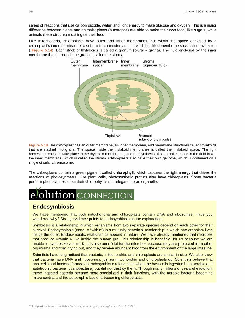
series of reactions that use carbon dioxide, water, and light energy to make glucose and oxygen. This is a majordifference between plants and animals; plants (autotrophs) are able to make their own food, like sugars, whileanimals (heterotrophs) must ingest their food.
Like mitochondria, chloroplasts have outer and inner membranes, but within the space enclosed by achloroplast’s inner membrane is a set of interconnected and stacked fluid-filled membrane sacs called thylakoids( Figure 5.14). Each stack of thylakoids is called a granum (plural = grana). The fluid enclosed by the innermembrane that surrounds the grana is called the stroma.
Figure 5.14 The chloroplast has an outer membrane, an inner membrane, and membrane structures called thylakoidsthat are stacked into grana. The space inside the thylakoid membranes is called the thylakoid space. The lightharvesting reactions take place in the thylakoid membranes, and the synthesis of sugar takes place in the fluid insidethe inner membrane, which is called the stroma. Chloroplasts also have their own genome, which is contained on asingle circular chromosome.
The chloroplasts contain a green pigment called chlorophyll, which captures the light energy that drives thereactions of photosynthesis. Like plant cells, photosynthetic protists also have chloroplasts. Some bacteriaperform photosynthesis, but their chlorophyll is not relegated to an organelle.
EndosymbiosisWe have mentioned that both mitochondria and chloroplasts contain DNA and ribosomes. Have youwondered why? Strong evidence points to endosymbiosis as the explanation.
Symbiosis is a relationship in which organisms from two separate species depend on each other for theirsurvival. Endosymbiosis (endo- = “within”) is a mutually beneficial relationship in which one organism livesinside the other. Endosymbiotic relationships abound in nature. We have already mentioned that microbesthat produce vitamin K live inside the human gut. This relationship is beneficial for us because we areunable to synthesize vitamin K. It is also beneficial for the microbes because they are protected from otherorganisms and from drying out, and they receive abundant food from the environment of the large intestine.
Scientists have long noticed that bacteria, mitochondria, and chloroplasts are similar in size. We also knowthat bacteria have DNA and ribosomes, just as mitochondria and chloroplasts do. Scientists believe thathost cells and bacteria formed an endosymbiotic relationship when the host cells ingested both aerobic andautotrophic bacteria (cyanobacteria) but did not destroy them. Through many millions of years of evolution,these ingested bacteria became more specialized in their functions, with the aerobic bacteria becomingmitochondria and the autotrophic bacteria becoming chloroplasts.
280 Chapter 5 | Cell Structure
This OpenStax book is available for free at https://legacy.cnx.org/content/col12104/1.1

The Central Vacuole
Previously, we mentioned vacuoles as essential components of plant cells. If you look at Figure 5.5b, you willsee that plant cells each have a large central vacuole that occupies most of the area of the cell. The centralvacuole plays a key role in regulating the cell’s concentration of water in changing environmental conditions.Have you ever noticed that if you forget to water a plant for a few days, it wilts? That’s because as the waterconcentration in the soil becomes lower than the water concentration in the plant, water moves out of the centralvacuoles and cytoplasm. As the central vacuole shrinks, it leaves the cell wall unsupported. This loss of supportto the cell walls of plant cells results in the wilted appearance of the plant.
The central vacuole also supports the expansion of the cell. When the central vacuole holds more water, the cellgets larger without having to invest a lot of energy in synthesizing new cytoplasm.
Chapter 5 | Cell Structure 281
cell theory
cell wall
central vacuole
centrosome
chlorophyll
chloroplast
chromatin
chromosome
cytoplasm
cytosol
electron microscope
eukaryotic cell
light microscope
lysosome
microscope
mitochondria
nuclear envelope
nucleolus
nucleoplasm
nucleus
organelle
peroxisome
plasma membrane
ribosome
unified cell theory
KEY TERMS
see unified cell theory
rigid cell covering made of various molecules that protects the cell, provides structural support, andgives shape to the cell
large plant cell organelle that regulates the cell’s storage compartment, holds water, and playsa significant role in cell growth as the site of macromolecule degradation
region in animal cells made of two centrioles
green pigment that captures the light energy that drives the light reactions of photosynthesis
plant cell organelle that carries out photosynthesis
protein-DNA complex that serves as the building material of chromosomes
structure within the nucleus that is made up of chromatin that contains DNA, the hereditarymaterial
entire region between the plasma membrane and the nuclear envelope, consisting of organellessuspended in the gel-like cytosol, the cytoskeleton, and various chemicals
gel-like material of the cytoplasm in which cell structures are suspended
an instrument that magnifies an object using a beam of electrons passed and bentthrough a lens system to visualize a specimen
cell that has a membrane-bound nucleus and several other membrane-bound compartments orsacs
an instrument that magnifies an object using a beam visible light passed and bent through alens system to visualize a specimen
organelle in an animal cell that functions as the cell’s digestive component; it breaks down proteins,polysaccharides, lipids, nucleic acids, and even worn-out organelles
an instrument that magnifies an object
(singular = mitochondrion) cellular organelles responsible for carrying out cellular respiration,resulting in the production of ATP, the cell’s main energy-carrying molecule
double-membrane structure that constitutes the outermost portion of the nucleus
darkly staining body within the nucleus that is responsible for assembling the subunits of theribosomes
semi-solid fluid inside the nucleus that contains the chromatin and nucleolus
cell organelle that houses the cell’s DNA and directs the synthesis of ribosomes and proteins
compartment or sac within a cell
small, round organelle that contains hydrogen peroxide, oxidizes fatty acids and amino acids, anddetoxifies many poisons
phospholipid bilayer with embedded (integral) or attached (peripheral) proteins, andseparates the internal content of the cell from its surrounding environment
cellular structure that carries out protein synthesis
a biological concept that states that all organisms are composed of one or more cells; the
282 Chapter 5 | Cell Structure
This OpenStax book is available for free at https://legacy.cnx.org/content/col12104/1.1
vacuole
vesicle
cell is the basic unit of life; and new cells arise from existing cells
membrane-bound sac, somewhat larger than a vesicle, which functions in cellular storage andtransport
small, membrane-bound sac that functions in cellular storage and transport; its membrane is capable offusing with the plasma membrane and the membranes of the endoplasmic reticulum and Golgi apparatus
CHAPTER SUMMARY
5.1 Studying Cells
A cell is the smallest unit of life. Most cells are so tiny that they cannot be seen with the naked eye. Therefore,scientists use microscopes to study cells. Electron microscopes provide higher magnification, higher resolution,and more detail than light microscopes. The unified cell theory states that all organisms are composed of oneor more cells, the cell is the basic unit of life, and new cells arise from existing cells.
5.2 Eukaryotic Cells
Like a prokaryotic cell, a eukaryotic cell has a plasma membrane, cytoplasm, and ribosomes, but a eukaryoticcell is typically larger than a prokaryotic cell, has a true nucleus (meaning its DNA is surrounded by amembrane), and has other membrane-bound organelles that allow for compartmentalization of functions. Theplasma membrane is a phospholipid bilayer embedded with proteins. The nucleus’s nucleolus is the site ofribosome assembly. Ribosomes are either found in the cytoplasm or attached to the cytoplasmic side of theplasma membrane or endoplasmic reticulum. They perform protein synthesis. Mitochondria participate incellular respiration; they are responsible for the majority of ATP produced in the cell. Peroxisomes hydrolyzefatty acids, amino acids, and some toxins. Vesicles and vacuoles are storage and transport compartments. Inplant cells, vacuoles also help break down macromolecules.
Animal cells also have a centrosome and lysosomes. The centrosome has two bodies perpendicular to eachother, the centrioles, and has an unknown purpose in cell division. Lysosomes are the digestive organelles ofanimal cells.
Plant cells and plant-like cells each have a cell wall, chloroplasts, and a central vacuole. The plant cell wall,whose primary component is cellulose, protects the cell, provides structural support, and gives shape to thecell. Photosynthesis takes place in chloroplasts. The central vacuole can expand without having to producemore cytoplasm.
ART CONNECTION QUESTIONS
1. Figure 5.5 If the nucleolus were not able to carryout its function, what other cellular organelles would
be affected?
REVIEW QUESTIONS
2. When viewing a specimen through a lightmicroscope, scientists use ________ to distinguishthe individual components of cells.
a. a beam of electronsb. radioactive isotopesc. special stainsd. high temperatures
3. The ________ is the basic unit of life.a. organismb. cellc. tissued. organ
4. Which of the following is surrounded by twophospholipid bilayers?
a. the ribosomesb. the vesiclesc. the cytoplasmd. the nucleoplasm
5. Peroxisomes got their name because hydrogenperoxide is:
a. used in their detoxification reactionsb. produced during their oxidation reactionsc. incorporated into their membranesd. a cofactor for the organelles’ enzymes
6. In plant cells, the function of the lysosomes iscarried out by __________.
Chapter 5 | Cell Structure 283
a. vacuolesb. peroxisomesc. ribosomesd. nuclei
7. Which of the following is found both in eukaryotic
and prokaryotic cells?a. nucleusb. mitochondrionc. vacuoled. ribosomes
CRITICAL THINKING QUESTIONS
8. In your everyday life, you have probably noticedthat certain instruments are ideal for certainsituations. For example, you would use a spoonrather than a fork to eat soup because a spoon isshaped for scooping, while soup would slip betweenthe tines of a fork. The use of ideal instruments alsoapplies in science. In what situation(s) would the useof a light microscope be ideal, and why?
9. In what situation(s) would the use of a scanningelectron microscope be ideal, and why?
10. In what situation(s) would a transmission electron
microscope be ideal, and why?
11. What are the advantages and disadvantages ofeach of these types of microscopes?
12. You already know that ribosomes are abundant inred blood cells. In what other cells of the body wouldyou find them in great abundance? Why?
13. What are the structural and functional similaritiesand differences between mitochondria andchloroplasts?
284 Chapter 5 | Cell Structure
This OpenStax book is available for free at https://legacy.cnx.org/content/col12104/1.1
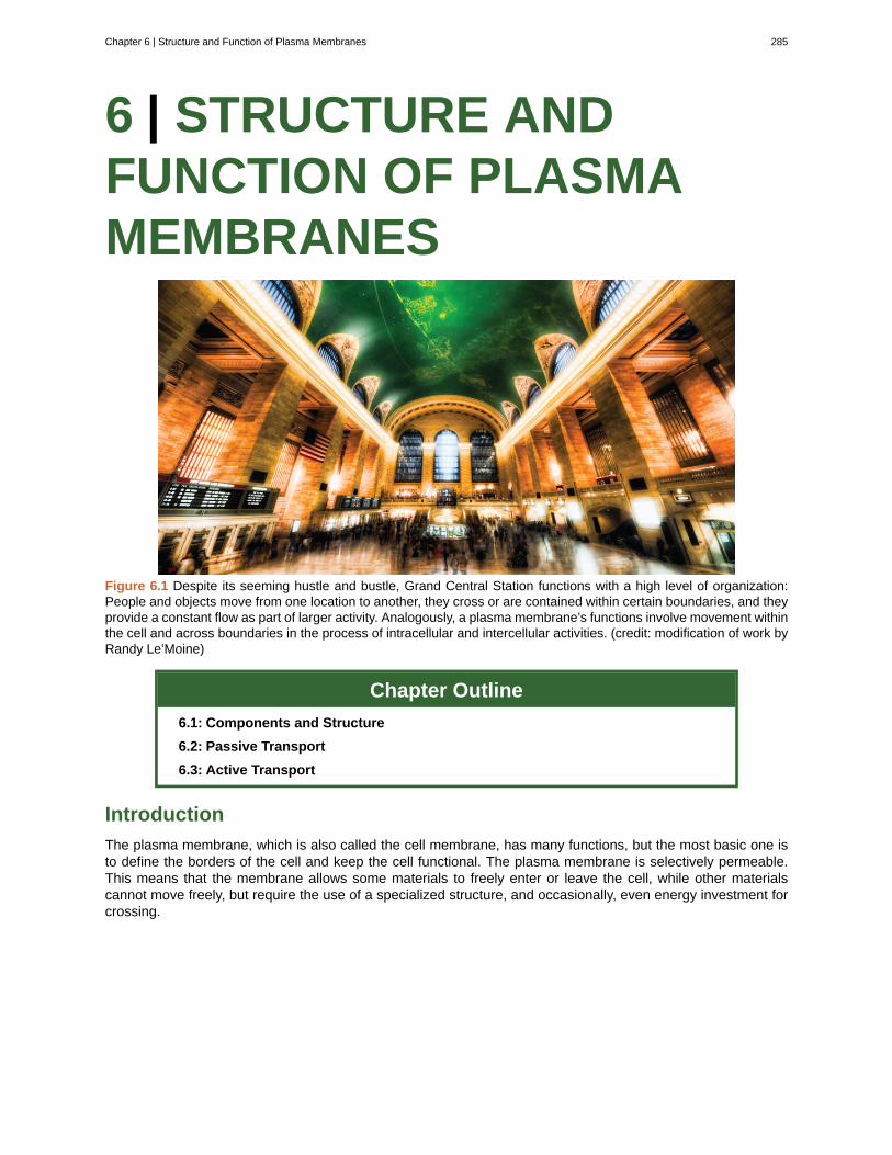
6 | STRUCTURE ANDFUNCTION OF PLASMAMEMBRANES
Figure 6.1 Despite its seeming hustle and bustle, Grand Central Station functions with a high level of organization:People and objects move from one location to another, they cross or are contained within certain boundaries, and theyprovide a constant flow as part of larger activity. Analogously, a plasma membrane’s functions involve movement withinthe cell and across boundaries in the process of intracellular and intercellular activities. (credit: modification of work byRandy Le’Moine)
Chapter Outline
6.1: Components and Structure
6.2: Passive Transport
6.3: Active Transport
Introduction
The plasma membrane, which is also called the cell membrane, has many functions, but the most basic one isto define the borders of the cell and keep the cell functional. The plasma membrane is selectively permeable.This means that the membrane allows some materials to freely enter or leave the cell, while other materialscannot move freely, but require the use of a specialized structure, and occasionally, even energy investment forcrossing.
Chapter 6 | Structure and Function of Plasma Membranes 285
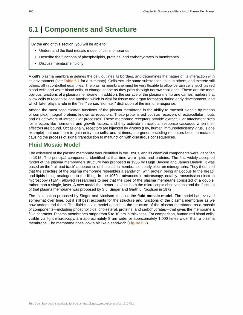
6.1 | Components and Structure
By the end of this section, you will be able to:
• Understand the fluid mosaic model of cell membranes
• Describe the functions of phospholipids, proteins, and carbohydrates in membranes
• Discuss membrane fluidity
A cell’s plasma membrane defines the cell, outlines its borders, and determines the nature of its interaction withits environment (see Table 6.1 for a summary). Cells exclude some substances, take in others, and excrete stillothers, all in controlled quantities. The plasma membrane must be very flexible to allow certain cells, such as redblood cells and white blood cells, to change shape as they pass through narrow capillaries. These are the moreobvious functions of a plasma membrane. In addition, the surface of the plasma membrane carries markers thatallow cells to recognize one another, which is vital for tissue and organ formation during early development, andwhich later plays a role in the “self” versus “non-self” distinction of the immune response.
Among the most sophisticated functions of the plasma membrane is the ability to transmit signals by meansof complex, integral proteins known as receptors. These proteins act both as receivers of extracellular inputsand as activators of intracellular processes. These membrane receptors provide extracellular attachment sitesfor effectors like hormones and growth factors, and they activate intracellular response cascades when theireffectors are bound. Occasionally, receptors are hijacked by viruses (HIV, human immunodeficiency virus, is oneexample) that use them to gain entry into cells, and at times, the genes encoding receptors become mutated,causing the process of signal transduction to malfunction with disastrous consequences.
Fluid Mosaic Model
The existence of the plasma membrane was identified in the 1890s, and its chemical components were identifiedin 1915. The principal components identified at that time were lipids and proteins. The first widely acceptedmodel of the plasma membrane’s structure was proposed in 1935 by Hugh Davson and James Danielli; it wasbased on the “railroad track” appearance of the plasma membrane in early electron micrographs. They theorizedthat the structure of the plasma membrane resembles a sandwich, with protein being analogous to the bread,and lipids being analogous to the filling. In the 1950s, advances in microscopy, notably transmission electronmicroscopy (TEM), allowed researchers to see that the core of the plasma membrane consisted of a double,rather than a single, layer. A new model that better explains both the microscopic observations and the functionof that plasma membrane was proposed by S.J. Singer and Garth L. Nicolson in 1972.
The explanation proposed by Singer and Nicolson is called the fluid mosaic model. The model has evolvedsomewhat over time, but it still best accounts for the structure and functions of the plasma membrane as wenow understand them. The fluid mosaic model describes the structure of the plasma membrane as a mosaicof components—including phospholipids, cholesterol, proteins, and carbohydrates—that gives the membrane afluid character. Plasma membranes range from 5 to 10 nm in thickness. For comparison, human red blood cells,visible via light microscopy, are approximately 8 µm wide, or approximately 1,000 times wider than a plasmamembrane. The membrane does look a bit like a sandwich (Figure 6.2).
286 Chapter 6 | Structure and Function of Plasma Membranes
This OpenStax book is available for free at https://legacy.cnx.org/content/col12104/1.1
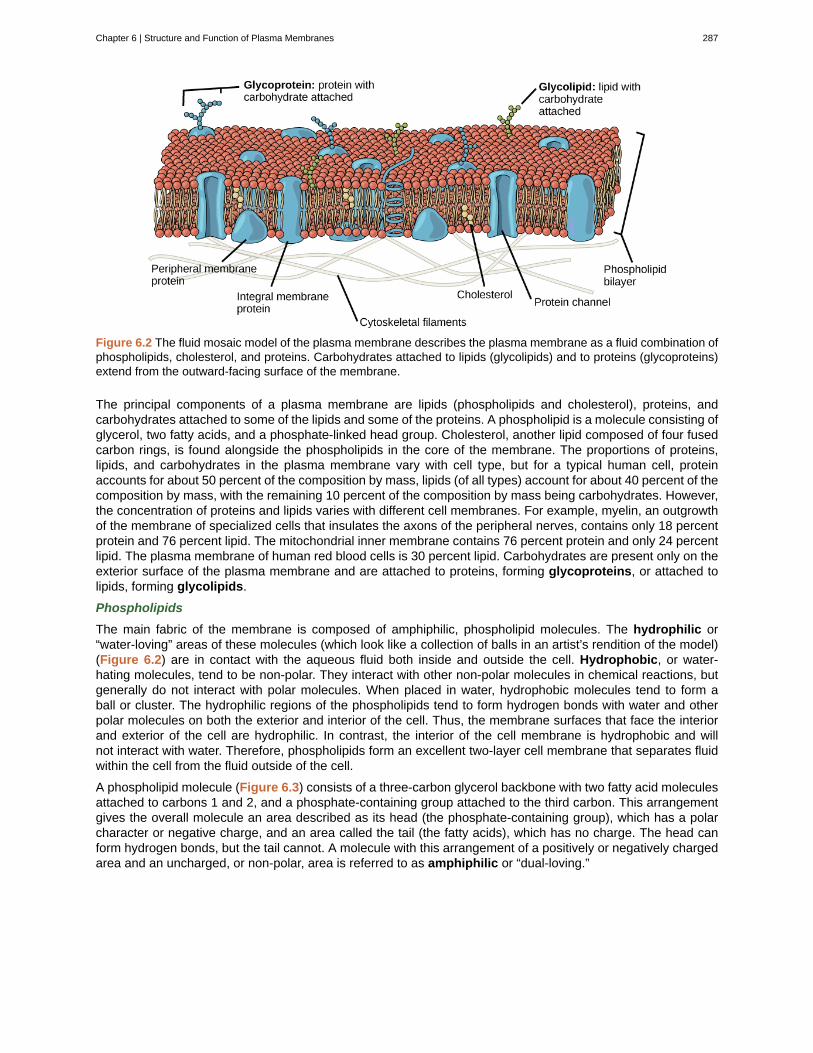
Figure 6.2 The fluid mosaic model of the plasma membrane describes the plasma membrane as a fluid combination ofphospholipids, cholesterol, and proteins. Carbohydrates attached to lipids (glycolipids) and to proteins (glycoproteins)extend from the outward-facing surface of the membrane.
The principal components of a plasma membrane are lipids (phospholipids and cholesterol), proteins, andcarbohydrates attached to some of the lipids and some of the proteins. A phospholipid is a molecule consisting ofglycerol, two fatty acids, and a phosphate-linked head group. Cholesterol, another lipid composed of four fusedcarbon rings, is found alongside the phospholipids in the core of the membrane. The proportions of proteins,lipids, and carbohydrates in the plasma membrane vary with cell type, but for a typical human cell, proteinaccounts for about 50 percent of the composition by mass, lipids (of all types) account for about 40 percent of thecomposition by mass, with the remaining 10 percent of the composition by mass being carbohydrates. However,the concentration of proteins and lipids varies with different cell membranes. For example, myelin, an outgrowthof the membrane of specialized cells that insulates the axons of the peripheral nerves, contains only 18 percentprotein and 76 percent lipid. The mitochondrial inner membrane contains 76 percent protein and only 24 percentlipid. The plasma membrane of human red blood cells is 30 percent lipid. Carbohydrates are present only on theexterior surface of the plasma membrane and are attached to proteins, forming glycoproteins, or attached tolipids, forming glycolipids.
Phospholipids
The main fabric of the membrane is composed of amphiphilic, phospholipid molecules. The hydrophilic or“water-loving” areas of these molecules (which look like a collection of balls in an artist’s rendition of the model)(Figure 6.2) are in contact with the aqueous fluid both inside and outside the cell. Hydrophobic, or water-hating molecules, tend to be non-polar. They interact with other non-polar molecules in chemical reactions, butgenerally do not interact with polar molecules. When placed in water, hydrophobic molecules tend to form aball or cluster. The hydrophilic regions of the phospholipids tend to form hydrogen bonds with water and otherpolar molecules on both the exterior and interior of the cell. Thus, the membrane surfaces that face the interiorand exterior of the cell are hydrophilic. In contrast, the interior of the cell membrane is hydrophobic and willnot interact with water. Therefore, phospholipids form an excellent two-layer cell membrane that separates fluidwithin the cell from the fluid outside of the cell.
A phospholipid molecule (Figure 6.3) consists of a three-carbon glycerol backbone with two fatty acid moleculesattached to carbons 1 and 2, and a phosphate-containing group attached to the third carbon. This arrangementgives the overall molecule an area described as its head (the phosphate-containing group), which has a polarcharacter or negative charge, and an area called the tail (the fatty acids), which has no charge. The head canform hydrogen bonds, but the tail cannot. A molecule with this arrangement of a positively or negatively chargedarea and an uncharged, or non-polar, area is referred to as amphiphilic or “dual-loving.”
Chapter 6 | Structure and Function of Plasma Membranes 287
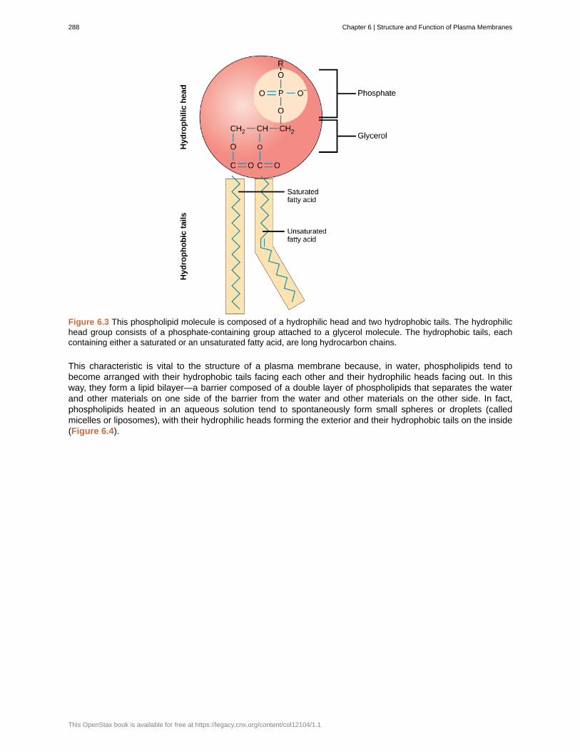
Figure 6.3 This phospholipid molecule is composed of a hydrophilic head and two hydrophobic tails. The hydrophilichead group consists of a phosphate-containing group attached to a glycerol molecule. The hydrophobic tails, eachcontaining either a saturated or an unsaturated fatty acid, are long hydrocarbon chains.
This characteristic is vital to the structure of a plasma membrane because, in water, phospholipids tend tobecome arranged with their hydrophobic tails facing each other and their hydrophilic heads facing out. In thisway, they form a lipid bilayer—a barrier composed of a double layer of phospholipids that separates the waterand other materials on one side of the barrier from the water and other materials on the other side. In fact,phospholipids heated in an aqueous solution tend to spontaneously form small spheres or droplets (calledmicelles or liposomes), with their hydrophilic heads forming the exterior and their hydrophobic tails on the inside(Figure 6.4).
288 Chapter 6 | Structure and Function of Plasma Membranes
This OpenStax book is available for free at https://legacy.cnx.org/content/col12104/1.1
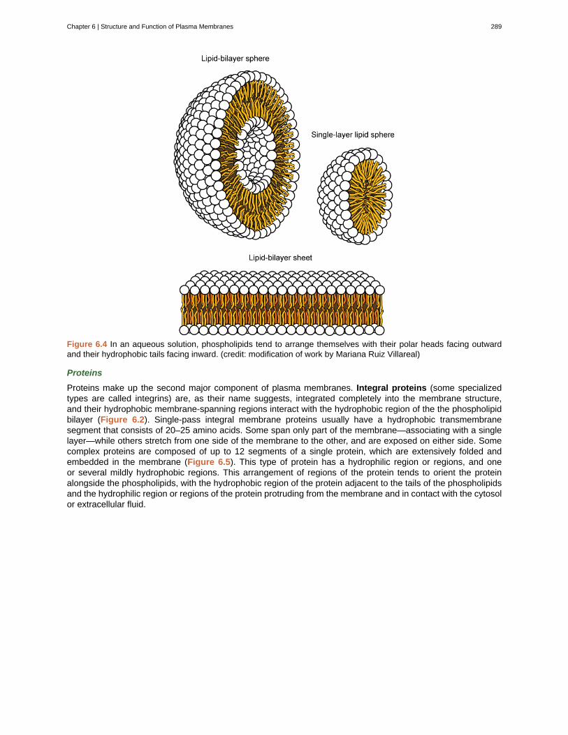
Figure 6.4 In an aqueous solution, phospholipids tend to arrange themselves with their polar heads facing outwardand their hydrophobic tails facing inward. (credit: modification of work by Mariana Ruiz Villareal)
Proteins
Proteins make up the second major component of plasma membranes. Integral proteins (some specializedtypes are called integrins) are, as their name suggests, integrated completely into the membrane structure,and their hydrophobic membrane-spanning regions interact with the hydrophobic region of the the phospholipidbilayer (Figure 6.2). Single-pass integral membrane proteins usually have a hydrophobic transmembranesegment that consists of 20–25 amino acids. Some span only part of the membrane—associating with a singlelayer—while others stretch from one side of the membrane to the other, and are exposed on either side. Somecomplex proteins are composed of up to 12 segments of a single protein, which are extensively folded andembedded in the membrane (Figure 6.5). This type of protein has a hydrophilic region or regions, and oneor several mildly hydrophobic regions. This arrangement of regions of the protein tends to orient the proteinalongside the phospholipids, with the hydrophobic region of the protein adjacent to the tails of the phospholipidsand the hydrophilic region or regions of the protein protruding from the membrane and in contact with the cytosolor extracellular fluid.
Chapter 6 | Structure and Function of Plasma Membranes 289
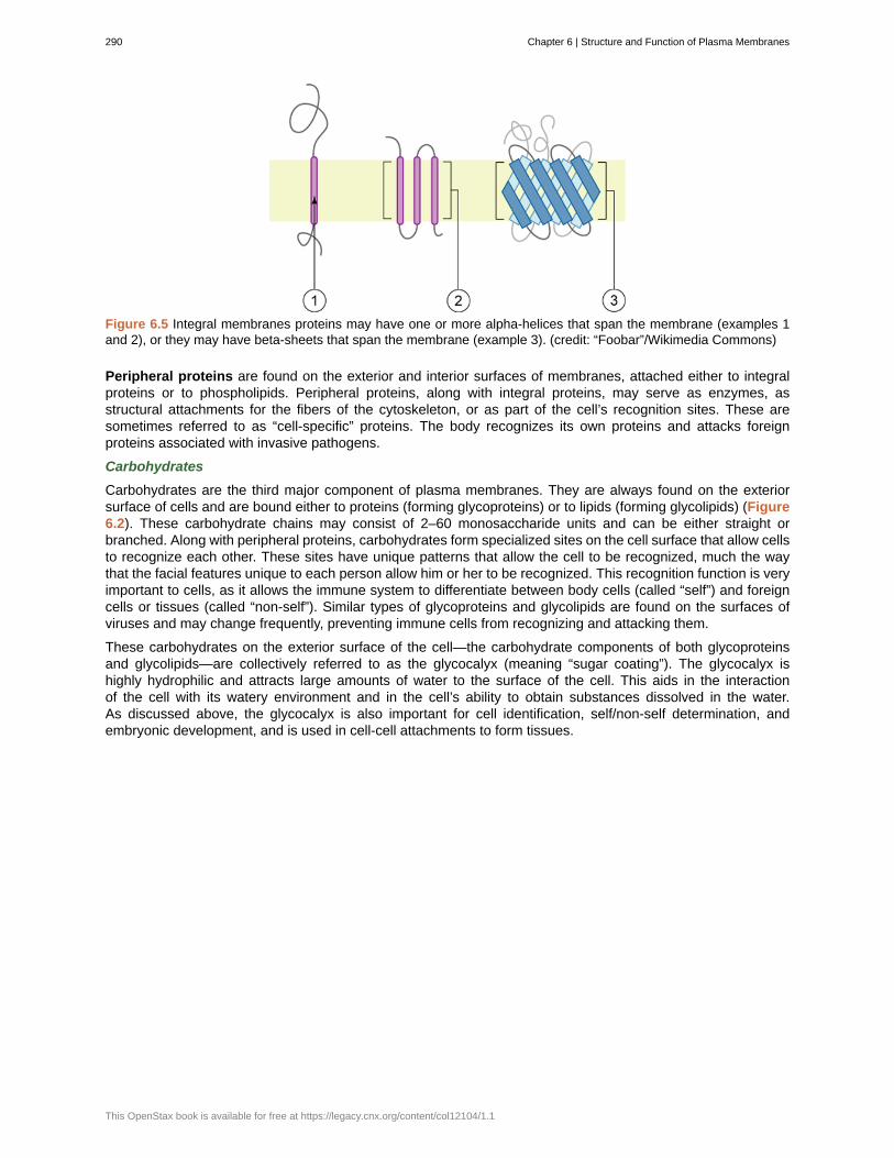
Figure 6.5 Integral membranes proteins may have one or more alpha-helices that span the membrane (examples 1and 2), or they may have beta-sheets that span the membrane (example 3). (credit: “Foobar”/Wikimedia Commons)
Peripheral proteins are found on the exterior and interior surfaces of membranes, attached either to integralproteins or to phospholipids. Peripheral proteins, along with integral proteins, may serve as enzymes, asstructural attachments for the fibers of the cytoskeleton, or as part of the cell’s recognition sites. These aresometimes referred to as “cell-specific” proteins. The body recognizes its own proteins and attacks foreignproteins associated with invasive pathogens.
Carbohydrates
Carbohydrates are the third major component of plasma membranes. They are always found on the exteriorsurface of cells and are bound either to proteins (forming glycoproteins) or to lipids (forming glycolipids) (Figure6.2). These carbohydrate chains may consist of 2–60 monosaccharide units and can be either straight orbranched. Along with peripheral proteins, carbohydrates form specialized sites on the cell surface that allow cellsto recognize each other. These sites have unique patterns that allow the cell to be recognized, much the waythat the facial features unique to each person allow him or her to be recognized. This recognition function is veryimportant to cells, as it allows the immune system to differentiate between body cells (called “self”) and foreigncells or tissues (called “non-self”). Similar types of glycoproteins and glycolipids are found on the surfaces ofviruses and may change frequently, preventing immune cells from recognizing and attacking them.
These carbohydrates on the exterior surface of the cell—the carbohydrate components of both glycoproteinsand glycolipids—are collectively referred to as the glycocalyx (meaning “sugar coating”). The glycocalyx ishighly hydrophilic and attracts large amounts of water to the surface of the cell. This aids in the interactionof the cell with its watery environment and in the cell’s ability to obtain substances dissolved in the water.As discussed above, the glycocalyx is also important for cell identification, self/non-self determination, andembryonic development, and is used in cell-cell attachments to form tissues.
290 Chapter 6 | Structure and Function of Plasma Membranes
This OpenStax book is available for free at https://legacy.cnx.org/content/col12104/1.1
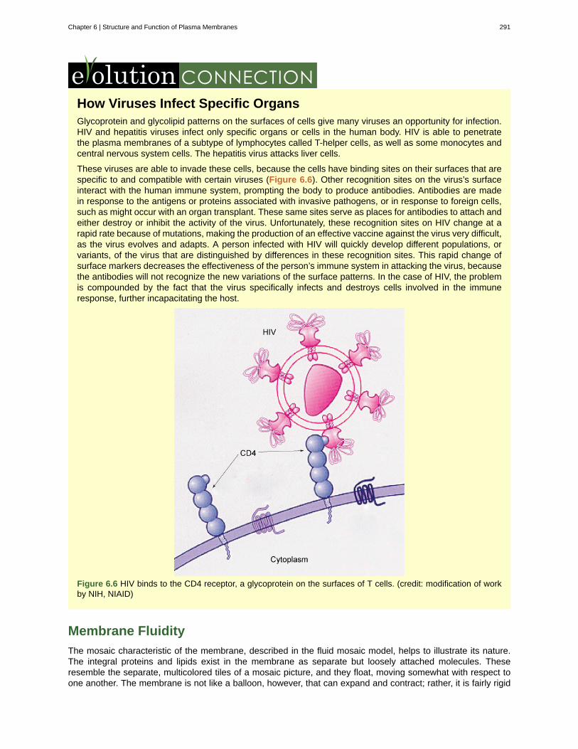
How Viruses Infect Specific OrgansGlycoprotein and glycolipid patterns on the surfaces of cells give many viruses an opportunity for infection.HIV and hepatitis viruses infect only specific organs or cells in the human body. HIV is able to penetratethe plasma membranes of a subtype of lymphocytes called T-helper cells, as well as some monocytes andcentral nervous system cells. The hepatitis virus attacks liver cells.
These viruses are able to invade these cells, because the cells have binding sites on their surfaces that arespecific to and compatible with certain viruses (Figure 6.6). Other recognition sites on the virus’s surfaceinteract with the human immune system, prompting the body to produce antibodies. Antibodies are madein response to the antigens or proteins associated with invasive pathogens, or in response to foreign cells,such as might occur with an organ transplant. These same sites serve as places for antibodies to attach andeither destroy or inhibit the activity of the virus. Unfortunately, these recognition sites on HIV change at arapid rate because of mutations, making the production of an effective vaccine against the virus very difficult,as the virus evolves and adapts. A person infected with HIV will quickly develop different populations, orvariants, of the virus that are distinguished by differences in these recognition sites. This rapid change ofsurface markers decreases the effectiveness of the person’s immune system in attacking the virus, becausethe antibodies will not recognize the new variations of the surface patterns. In the case of HIV, the problemis compounded by the fact that the virus specifically infects and destroys cells involved in the immuneresponse, further incapacitating the host.
Figure 6.6 HIV binds to the CD4 receptor, a glycoprotein on the surfaces of T cells. (credit: modification of workby NIH, NIAID)
Membrane Fluidity
The mosaic characteristic of the membrane, described in the fluid mosaic model, helps to illustrate its nature.The integral proteins and lipids exist in the membrane as separate but loosely attached molecules. Theseresemble the separate, multicolored tiles of a mosaic picture, and they float, moving somewhat with respect toone another. The membrane is not like a balloon, however, that can expand and contract; rather, it is fairly rigid
Chapter 6 | Structure and Function of Plasma Membranes 291
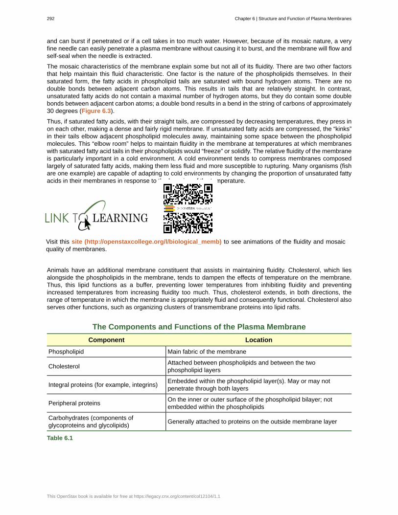
and can burst if penetrated or if a cell takes in too much water. However, because of its mosaic nature, a veryfine needle can easily penetrate a plasma membrane without causing it to burst, and the membrane will flow andself-seal when the needle is extracted.
The mosaic characteristics of the membrane explain some but not all of its fluidity. There are two other factorsthat help maintain this fluid characteristic. One factor is the nature of the phospholipids themselves. In theirsaturated form, the fatty acids in phospholipid tails are saturated with bound hydrogen atoms. There are nodouble bonds between adjacent carbon atoms. This results in tails that are relatively straight. In contrast,unsaturated fatty acids do not contain a maximal number of hydrogen atoms, but they do contain some doublebonds between adjacent carbon atoms; a double bond results in a bend in the string of carbons of approximately30 degrees (Figure 6.3).
Thus, if saturated fatty acids, with their straight tails, are compressed by decreasing temperatures, they press inon each other, making a dense and fairly rigid membrane. If unsaturated fatty acids are compressed, the “kinks”in their tails elbow adjacent phospholipid molecules away, maintaining some space between the phospholipidmolecules. This “elbow room” helps to maintain fluidity in the membrane at temperatures at which membraneswith saturated fatty acid tails in their phospholipids would “freeze” or solidify. The relative fluidity of the membraneis particularly important in a cold environment. A cold environment tends to compress membranes composedlargely of saturated fatty acids, making them less fluid and more susceptible to rupturing. Many organisms (fishare one example) are capable of adapting to cold environments by changing the proportion of unsaturated fattyacids in their membranes in response to the lowering of the temperature.
Visit this site (http://openstaxcollege.org/l/biological_memb) to see animations of the fluidity and mosaicquality of membranes.
Animals have an additional membrane constituent that assists in maintaining fluidity. Cholesterol, which liesalongside the phospholipids in the membrane, tends to dampen the effects of temperature on the membrane.Thus, this lipid functions as a buffer, preventing lower temperatures from inhibiting fluidity and preventingincreased temperatures from increasing fluidity too much. Thus, cholesterol extends, in both directions, therange of temperature in which the membrane is appropriately fluid and consequently functional. Cholesterol alsoserves other functions, such as organizing clusters of transmembrane proteins into lipid rafts.
The Components and Functions of the Plasma Membrane
Component Location
Phospholipid Main fabric of the membrane
CholesterolAttached between phospholipids and between the twophospholipid layers
Integral proteins (for example, integrins)Embedded within the phospholipid layer(s). May or may notpenetrate through both layers
Peripheral proteinsOn the inner or outer surface of the phospholipid bilayer; notembedded within the phospholipids
Carbohydrates (components ofglycoproteins and glycolipids)
Generally attached to proteins on the outside membrane layer
Table 6.1
292 Chapter 6 | Structure and Function of Plasma Membranes
This OpenStax book is available for free at https://legacy.cnx.org/content/col12104/1.1
ImmunologistThe variations in peripheral proteins and carbohydrates that affect a cell’s recognition sites are of primeinterest in immunology. These changes are taken into consideration in vaccine development. Manyinfectious diseases, such as smallpox, polio, diphtheria, and tetanus, were conquered by the use ofvaccines.
Immunologists are the physicians and scientists who research and develop vaccines, as well as treat andstudy allergies or other immune problems. Some immunologists study and treat autoimmune problems(diseases in which a person’s immune system attacks his or her own cells or tissues, such as lupus)and immunodeficiencies, whether acquired (such as acquired immunodeficiency syndrome, or AIDS) orhereditary (such as severe combined immunodeficiency, or SCID). Immunologists are called in to help treatorgan transplantation patients, who must have their immune systems suppressed so that their bodies willnot reject a transplanted organ. Some immunologists work to understand natural immunity and the effectsof a person’s environment on it. Others work on questions about how the immune system affects diseasessuch as cancer. In the past, the importance of having a healthy immune system in preventing cancer wasnot at all understood.
To work as an immunologist, a PhD or MD is required. In addition, immunologists undertake at least 2–3years of training in an accredited program and must pass an examination given by the American Board ofAllergy and Immunology. Immunologists must possess knowledge of the functions of the human body asthey relate to issues beyond immunization, and knowledge of pharmacology and medical technology, suchas medications, therapies, test materials, and surgical procedures.
6.2 | Passive Transport
By the end of this section, you will be able to:
• Explain why and how passive transport occurs
• Understand the processes of osmosis and diffusion
• Define tonicity and describe its relevance to passive transport
Plasma membranes must allow certain substances to enter and leave a cell, and prevent some harmful materialsfrom entering and some essential materials from leaving. In other words, plasma membranes are selectivelypermeable—they allow some substances to pass through, but not others. If they were to lose this selectivity,the cell would no longer be able to sustain itself, and it would be destroyed. Some cells require larger amountsof specific substances than do other cells; they must have a way of obtaining these materials from extracellularfluids. This may happen passively, as certain materials move back and forth, or the cell may have specialmechanisms that facilitate transport. Some materials are so important to a cell that it spends some of its energy,hydrolyzing adenosine triphosphate (ATP), to obtain these materials. Red blood cells use some of their energydoing just that. All cells spend the majority of their energy to maintain an imbalance of sodium and potassiumions between the interior and exterior of the cell.
The most direct forms of membrane transport are passive. Passive transport is a naturally occurringphenomenon and does not require the cell to exert any of its energy to accomplish the movement. In passivetransport, substances move from an area of higher concentration to an area of lower concentration. A physicalspace in which there is a range of concentrations of a single substance is said to have a concentrationgradient.
Selective Permeability
Plasma membranes are asymmetric: the interior of the membrane is not identical to the exterior of themembrane. In fact, there is a considerable difference between the array of phospholipids and proteins between
Chapter 6 | Structure and Function of Plasma Membranes 293
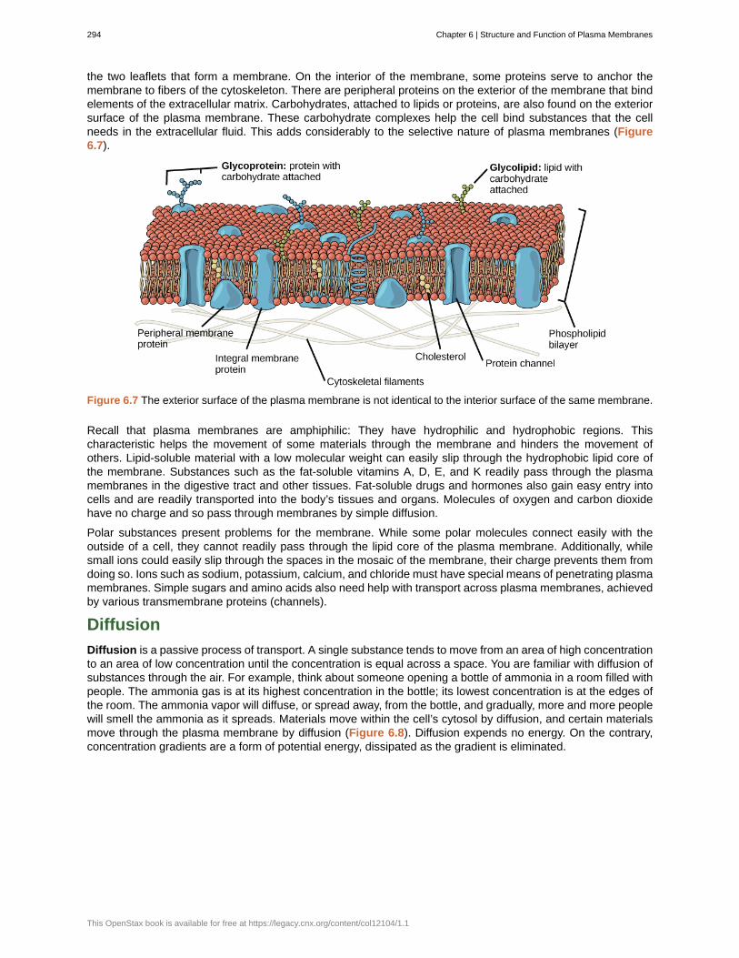
the two leaflets that form a membrane. On the interior of the membrane, some proteins serve to anchor themembrane to fibers of the cytoskeleton. There are peripheral proteins on the exterior of the membrane that bindelements of the extracellular matrix. Carbohydrates, attached to lipids or proteins, are also found on the exteriorsurface of the plasma membrane. These carbohydrate complexes help the cell bind substances that the cellneeds in the extracellular fluid. This adds considerably to the selective nature of plasma membranes (Figure6.7).
Figure 6.7 The exterior surface of the plasma membrane is not identical to the interior surface of the same membrane.
Recall that plasma membranes are amphiphilic: They have hydrophilic and hydrophobic regions. Thischaracteristic helps the movement of some materials through the membrane and hinders the movement ofothers. Lipid-soluble material with a low molecular weight can easily slip through the hydrophobic lipid core ofthe membrane. Substances such as the fat-soluble vitamins A, D, E, and K readily pass through the plasmamembranes in the digestive tract and other tissues. Fat-soluble drugs and hormones also gain easy entry intocells and are readily transported into the body’s tissues and organs. Molecules of oxygen and carbon dioxidehave no charge and so pass through membranes by simple diffusion.
Polar substances present problems for the membrane. While some polar molecules connect easily with theoutside of a cell, they cannot readily pass through the lipid core of the plasma membrane. Additionally, whilesmall ions could easily slip through the spaces in the mosaic of the membrane, their charge prevents them fromdoing so. Ions such as sodium, potassium, calcium, and chloride must have special means of penetrating plasmamembranes. Simple sugars and amino acids also need help with transport across plasma membranes, achievedby various transmembrane proteins (channels).
Diffusion
Diffusion is a passive process of transport. A single substance tends to move from an area of high concentrationto an area of low concentration until the concentration is equal across a space. You are familiar with diffusion ofsubstances through the air. For example, think about someone opening a bottle of ammonia in a room filled withpeople. The ammonia gas is at its highest concentration in the bottle; its lowest concentration is at the edges ofthe room. The ammonia vapor will diffuse, or spread away, from the bottle, and gradually, more and more peoplewill smell the ammonia as it spreads. Materials move within the cell’s cytosol by diffusion, and certain materialsmove through the plasma membrane by diffusion (Figure 6.8). Diffusion expends no energy. On the contrary,concentration gradients are a form of potential energy, dissipated as the gradient is eliminated.
294 Chapter 6 | Structure and Function of Plasma Membranes
This OpenStax book is available for free at https://legacy.cnx.org/content/col12104/1.1
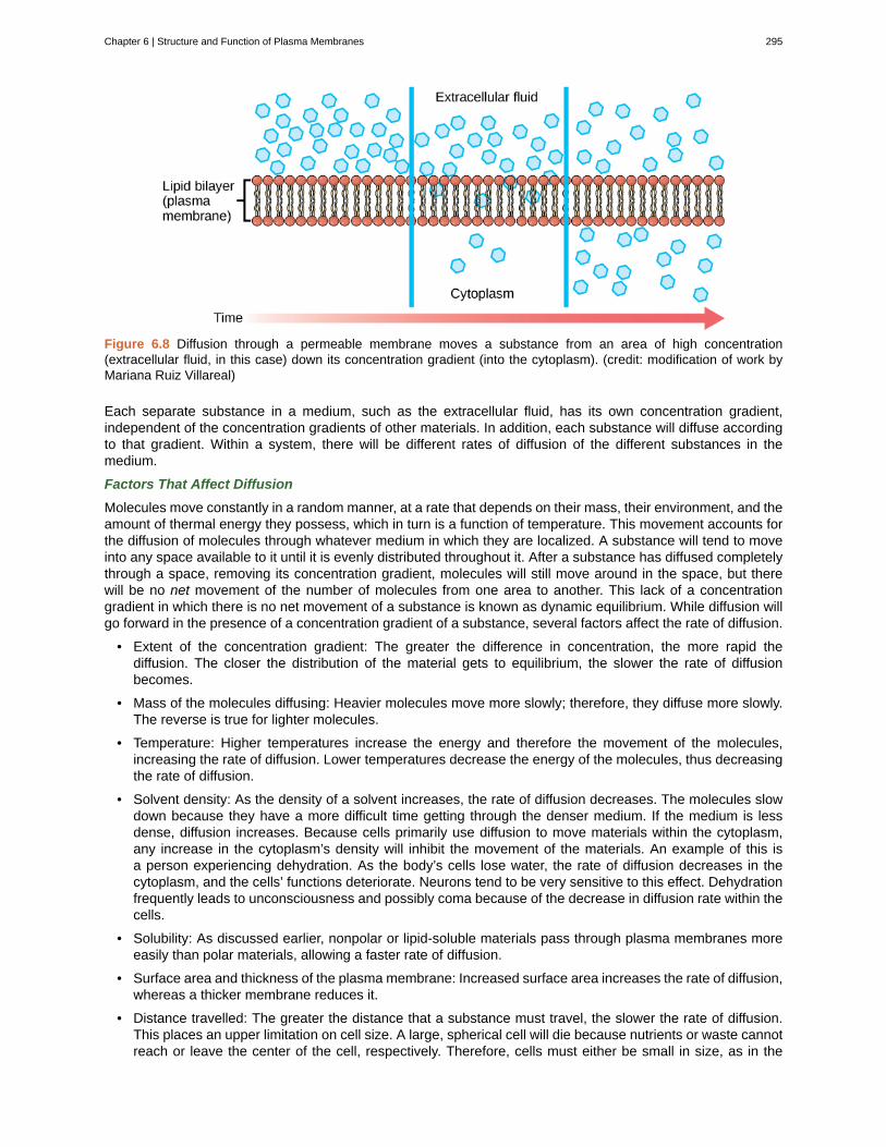
Figure 6.8 Diffusion through a permeable membrane moves a substance from an area of high concentration(extracellular fluid, in this case) down its concentration gradient (into the cytoplasm). (credit: modification of work byMariana Ruiz Villareal)
Each separate substance in a medium, such as the extracellular fluid, has its own concentration gradient,independent of the concentration gradients of other materials. In addition, each substance will diffuse accordingto that gradient. Within a system, there will be different rates of diffusion of the different substances in themedium.
Factors That Affect Diffusion
Molecules move constantly in a random manner, at a rate that depends on their mass, their environment, and theamount of thermal energy they possess, which in turn is a function of temperature. This movement accounts forthe diffusion of molecules through whatever medium in which they are localized. A substance will tend to moveinto any space available to it until it is evenly distributed throughout it. After a substance has diffused completelythrough a space, removing its concentration gradient, molecules will still move around in the space, but therewill be no net movement of the number of molecules from one area to another. This lack of a concentrationgradient in which there is no net movement of a substance is known as dynamic equilibrium. While diffusion willgo forward in the presence of a concentration gradient of a substance, several factors affect the rate of diffusion.
• Extent of the concentration gradient: The greater the difference in concentration, the more rapid thediffusion. The closer the distribution of the material gets to equilibrium, the slower the rate of diffusionbecomes.
• Mass of the molecules diffusing: Heavier molecules move more slowly; therefore, they diffuse more slowly.The reverse is true for lighter molecules.
• Temperature: Higher temperatures increase the energy and therefore the movement of the molecules,increasing the rate of diffusion. Lower temperatures decrease the energy of the molecules, thus decreasingthe rate of diffusion.
• Solvent density: As the density of a solvent increases, the rate of diffusion decreases. The molecules slowdown because they have a more difficult time getting through the denser medium. If the medium is lessdense, diffusion increases. Because cells primarily use diffusion to move materials within the cytoplasm,any increase in the cytoplasm’s density will inhibit the movement of the materials. An example of this isa person experiencing dehydration. As the body’s cells lose water, the rate of diffusion decreases in thecytoplasm, and the cells’ functions deteriorate. Neurons tend to be very sensitive to this effect. Dehydrationfrequently leads to unconsciousness and possibly coma because of the decrease in diffusion rate within thecells.
• Solubility: As discussed earlier, nonpolar or lipid-soluble materials pass through plasma membranes moreeasily than polar materials, allowing a faster rate of diffusion.
• Surface area and thickness of the plasma membrane: Increased surface area increases the rate of diffusion,whereas a thicker membrane reduces it.
• Distance travelled: The greater the distance that a substance must travel, the slower the rate of diffusion.This places an upper limitation on cell size. A large, spherical cell will die because nutrients or waste cannotreach or leave the center of the cell, respectively. Therefore, cells must either be small in size, as in the
Chapter 6 | Structure and Function of Plasma Membranes 295
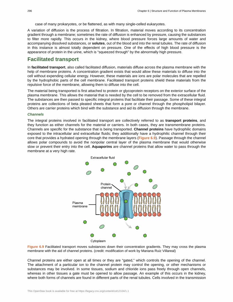
case of many prokaryotes, or be flattened, as with many single-celled eukaryotes.
A variation of diffusion is the process of filtration. In filtration, material moves according to its concentrationgradient through a membrane; sometimes the rate of diffusion is enhanced by pressure, causing the substancesto filter more rapidly. This occurs in the kidney, where blood pressure forces large amounts of water andaccompanying dissolved substances, or solutes, out of the blood and into the renal tubules. The rate of diffusionin this instance is almost totally dependent on pressure. One of the effects of high blood pressure is theappearance of protein in the urine, which is “squeezed through” by the abnormally high pressure.
Facilitated transport
In facilitated transport, also called facilitated diffusion, materials diffuse across the plasma membrane with thehelp of membrane proteins. A concentration gradient exists that would allow these materials to diffuse into thecell without expending cellular energy. However, these materials are ions are polar molecules that are repelledby the hydrophobic parts of the cell membrane. Facilitated transport proteins shield these materials from therepulsive force of the membrane, allowing them to diffuse into the cell.
The material being transported is first attached to protein or glycoprotein receptors on the exterior surface of theplasma membrane. This allows the material that is needed by the cell to be removed from the extracellular fluid.The substances are then passed to specific integral proteins that facilitate their passage. Some of these integralproteins are collections of beta pleated sheets that form a pore or channel through the phospholipid bilayer.Others are carrier proteins which bind with the substance and aid its diffusion through the membrane.
Channels
The integral proteins involved in facilitated transport are collectively referred to as transport proteins, andthey function as either channels for the material or carriers. In both cases, they are transmembrane proteins.Channels are specific for the substance that is being transported. Channel proteins have hydrophilic domainsexposed to the intracellular and extracellular fluids; they additionally have a hydrophilic channel through theircore that provides a hydrated opening through the membrane layers (Figure 6.9). Passage through the channelallows polar compounds to avoid the nonpolar central layer of the plasma membrane that would otherwiseslow or prevent their entry into the cell. Aquaporins are channel proteins that allow water to pass through themembrane at a very high rate.
Figure 6.9 Facilitated transport moves substances down their concentration gradients. They may cross the plasmamembrane with the aid of channel proteins. (credit: modification of work by Mariana Ruiz Villareal)
Channel proteins are either open at all times or they are “gated,” which controls the opening of the channel.The attachment of a particular ion to the channel protein may control the opening, or other mechanisms orsubstances may be involved. In some tissues, sodium and chloride ions pass freely through open channels,whereas in other tissues a gate must be opened to allow passage. An example of this occurs in the kidney,where both forms of channels are found in different parts of the renal tubules. Cells involved in the transmission
296 Chapter 6 | Structure and Function of Plasma Membranes
This OpenStax book is available for free at https://legacy.cnx.org/content/col12104/1.1

of electrical impulses, such as nerve and muscle cells, have gated channels for sodium, potassium, and calciumin their membranes. Opening and closing of these channels changes the relative concentrations on opposingsides of the membrane of these ions, resulting in the facilitation of electrical transmission along membranes (inthe case of nerve cells) or in muscle contraction (in the case of muscle cells).
Carrier Proteins
Another type of protein embedded in the plasma membrane is a carrier protein. This aptly named protein bindsa substance and, in doing so, triggers a change of its own shape, moving the bound molecule from the outside ofthe cell to its interior (Figure 6.10); depending on the gradient, the material may move in the opposite direction.Carrier proteins are typically specific for a single substance. This selectivity adds to the overall selectivity of theplasma membrane. The exact mechanism for the change of shape is poorly understood. Proteins can changeshape when their hydrogen bonds are affected, but this may not fully explain this mechanism. Each carrierprotein is specific to one substance, and there are a finite number of these proteins in any membrane. Thiscan cause problems in transporting enough of the material for the cell to function properly. When all of theproteins are bound to their ligands, they are saturated and the rate of transport is at its maximum. Increasing theconcentration gradient at this point will not result in an increased rate of transport.
Figure 6.10 Some substances are able to move down their concentration gradient across the plasma membrane withthe aid of carrier proteins. Carrier proteins change shape as they move molecules across the membrane. (credit:modification of work by Mariana Ruiz Villareal)
An example of this process occurs in the kidney. Glucose, water, salts, ions, and amino acids needed by thebody are filtered in one part of the kidney. This filtrate, which includes glucose, is then reabsorbed in another partof the kidney. Because there are only a finite number of carrier proteins for glucose, if more glucose is presentthan the proteins can handle, the excess is not transported and it is excreted from the body in the urine. In adiabetic individual, this is described as “spilling glucose into the urine.” A different group of carrier proteins calledglucose transport proteins, or GLUTs, are involved in transporting glucose and other hexose sugars throughplasma membranes within the body.
Channel and carrier proteins transport material at different rates. Channel proteins transport much more quicklythan do carrier proteins. Channel proteins facilitate diffusion at a rate of tens of millions of molecules per second,whereas carrier proteins work at a rate of a thousand to a million molecules per second.
Osmosis
Osmosis is the movement of water through a semipermeable membrane according to the concentrationgradient of water across the membrane, which is inversely proportional to the concentration of solutes. Whilediffusion transports material across membranes and within cells, osmosis transports only water across amembrane and the membrane limits the diffusion of solutes in the water. Not surprisingly, the aquaporins thatfacilitate water movement play a large role in osmosis, most prominently in red blood cells and the membranesof kidney tubules.
Mechanism
Osmosis is a special case of diffusion. Water, like other substances, moves from an area of high concentration
Chapter 6 | Structure and Function of Plasma Membranes 297
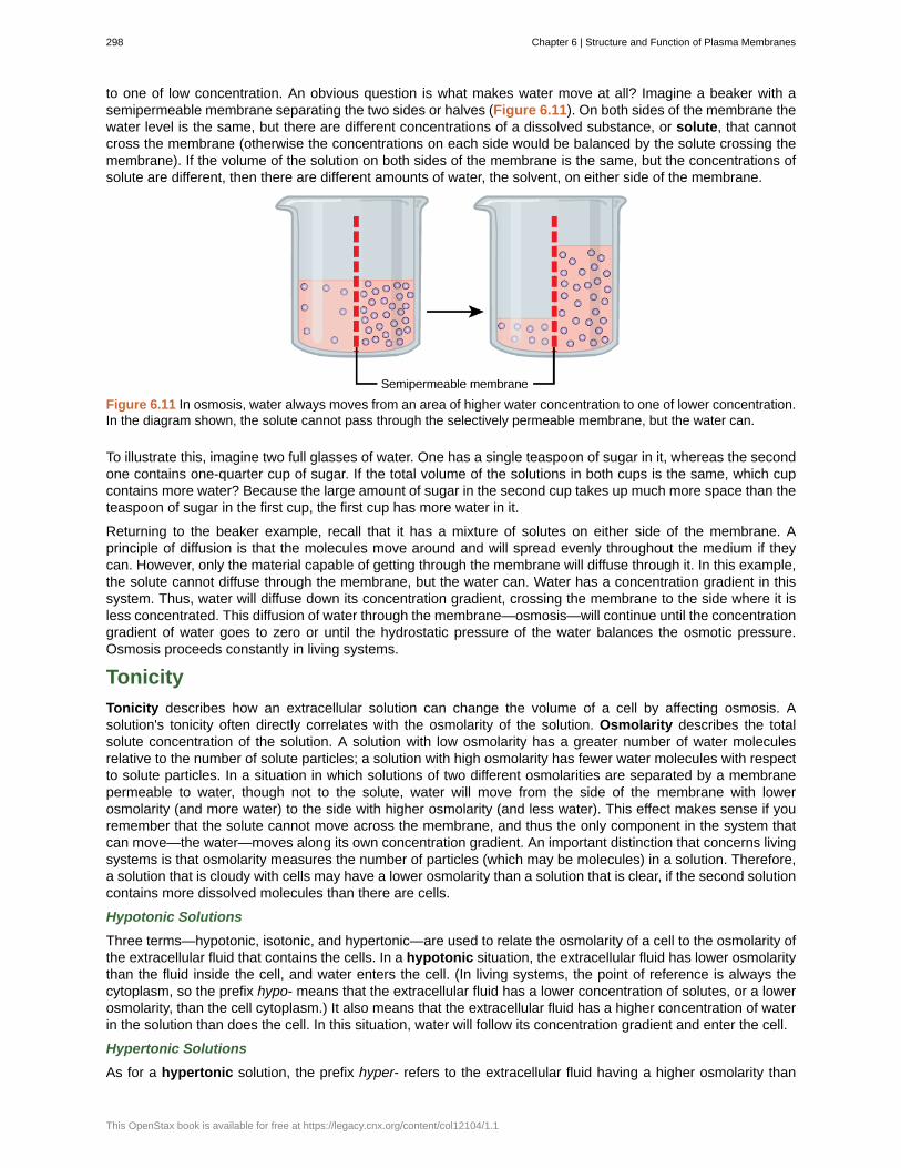
to one of low concentration. An obvious question is what makes water move at all? Imagine a beaker with asemipermeable membrane separating the two sides or halves (Figure 6.11). On both sides of the membrane thewater level is the same, but there are different concentrations of a dissolved substance, or solute, that cannotcross the membrane (otherwise the concentrations on each side would be balanced by the solute crossing themembrane). If the volume of the solution on both sides of the membrane is the same, but the concentrations ofsolute are different, then there are different amounts of water, the solvent, on either side of the membrane.
Figure 6.11 In osmosis, water always moves from an area of higher water concentration to one of lower concentration.In the diagram shown, the solute cannot pass through the selectively permeable membrane, but the water can.
To illustrate this, imagine two full glasses of water. One has a single teaspoon of sugar in it, whereas the secondone contains one-quarter cup of sugar. If the total volume of the solutions in both cups is the same, which cupcontains more water? Because the large amount of sugar in the second cup takes up much more space than theteaspoon of sugar in the first cup, the first cup has more water in it.
Returning to the beaker example, recall that it has a mixture of solutes on either side of the membrane. Aprinciple of diffusion is that the molecules move around and will spread evenly throughout the medium if theycan. However, only the material capable of getting through the membrane will diffuse through it. In this example,the solute cannot diffuse through the membrane, but the water can. Water has a concentration gradient in thissystem. Thus, water will diffuse down its concentration gradient, crossing the membrane to the side where it isless concentrated. This diffusion of water through the membrane—osmosis—will continue until the concentrationgradient of water goes to zero or until the hydrostatic pressure of the water balances the osmotic pressure.Osmosis proceeds constantly in living systems.
Tonicity
Tonicity describes how an extracellular solution can change the volume of a cell by affecting osmosis. Asolution's tonicity often directly correlates with the osmolarity of the solution. Osmolarity describes the totalsolute concentration of the solution. A solution with low osmolarity has a greater number of water moleculesrelative to the number of solute particles; a solution with high osmolarity has fewer water molecules with respectto solute particles. In a situation in which solutions of two different osmolarities are separated by a membranepermeable to water, though not to the solute, water will move from the side of the membrane with lowerosmolarity (and more water) to the side with higher osmolarity (and less water). This effect makes sense if youremember that the solute cannot move across the membrane, and thus the only component in the system thatcan move—the water—moves along its own concentration gradient. An important distinction that concerns livingsystems is that osmolarity measures the number of particles (which may be molecules) in a solution. Therefore,a solution that is cloudy with cells may have a lower osmolarity than a solution that is clear, if the second solutioncontains more dissolved molecules than there are cells.
Hypotonic Solutions
Three terms—hypotonic, isotonic, and hypertonic—are used to relate the osmolarity of a cell to the osmolarity ofthe extracellular fluid that contains the cells. In a hypotonic situation, the extracellular fluid has lower osmolaritythan the fluid inside the cell, and water enters the cell. (In living systems, the point of reference is always thecytoplasm, so the prefix hypo- means that the extracellular fluid has a lower concentration of solutes, or a lowerosmolarity, than the cell cytoplasm.) It also means that the extracellular fluid has a higher concentration of waterin the solution than does the cell. In this situation, water will follow its concentration gradient and enter the cell.
Hypertonic Solutions
As for a hypertonic solution, the prefix hyper- refers to the extracellular fluid having a higher osmolarity than
298 Chapter 6 | Structure and Function of Plasma Membranes
This OpenStax book is available for free at https://legacy.cnx.org/content/col12104/1.1
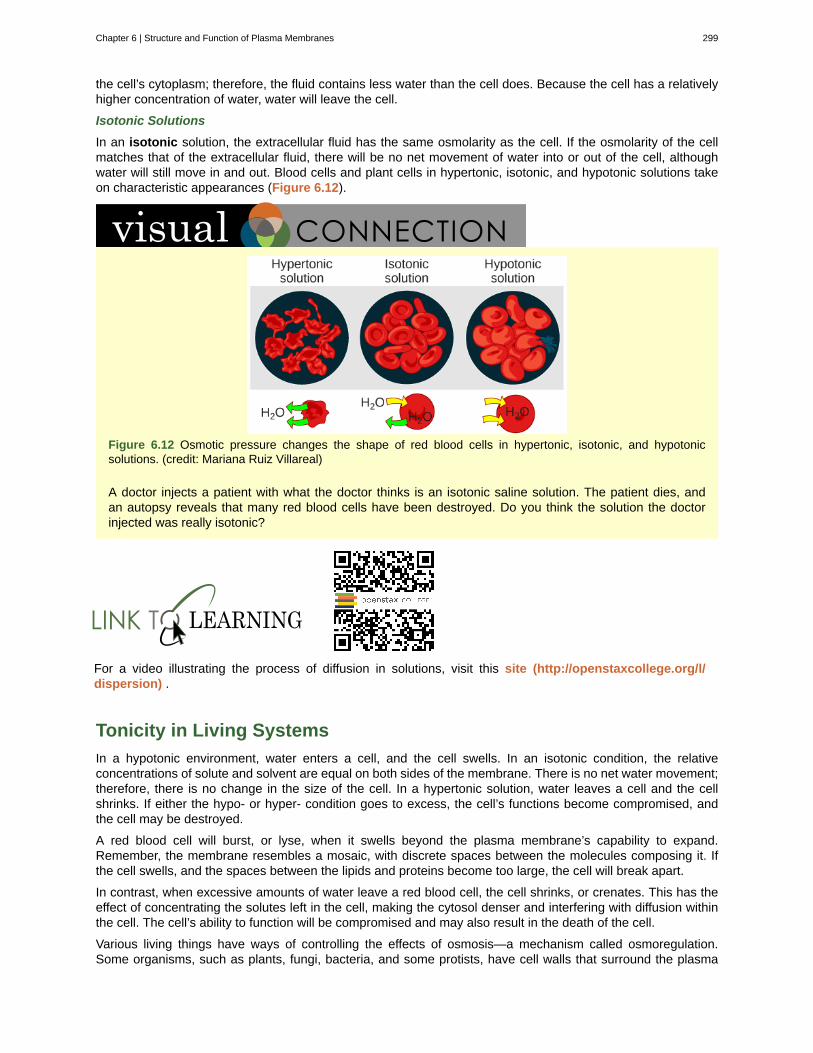
the cell’s cytoplasm; therefore, the fluid contains less water than the cell does. Because the cell has a relativelyhigher concentration of water, water will leave the cell.
Isotonic Solutions
In an isotonic solution, the extracellular fluid has the same osmolarity as the cell. If the osmolarity of the cellmatches that of the extracellular fluid, there will be no net movement of water into or out of the cell, althoughwater will still move in and out. Blood cells and plant cells in hypertonic, isotonic, and hypotonic solutions takeon characteristic appearances (Figure 6.12).
Figure 6.12 Osmotic pressure changes the shape of red blood cells in hypertonic, isotonic, and hypotonicsolutions. (credit: Mariana Ruiz Villareal)
A doctor injects a patient with what the doctor thinks is an isotonic saline solution. The patient dies, andan autopsy reveals that many red blood cells have been destroyed. Do you think the solution the doctorinjected was really isotonic?
For a video illustrating the process of diffusion in solutions, visit this site (http://openstaxcollege.org/l/dispersion) .
Tonicity in Living Systems
In a hypotonic environment, water enters a cell, and the cell swells. In an isotonic condition, the relativeconcentrations of solute and solvent are equal on both sides of the membrane. There is no net water movement;therefore, there is no change in the size of the cell. In a hypertonic solution, water leaves a cell and the cellshrinks. If either the hypo- or hyper- condition goes to excess, the cell’s functions become compromised, andthe cell may be destroyed.
A red blood cell will burst, or lyse, when it swells beyond the plasma membrane’s capability to expand.Remember, the membrane resembles a mosaic, with discrete spaces between the molecules composing it. Ifthe cell swells, and the spaces between the lipids and proteins become too large, the cell will break apart.
In contrast, when excessive amounts of water leave a red blood cell, the cell shrinks, or crenates. This has theeffect of concentrating the solutes left in the cell, making the cytosol denser and interfering with diffusion withinthe cell. The cell’s ability to function will be compromised and may also result in the death of the cell.
Various living things have ways of controlling the effects of osmosis—a mechanism called osmoregulation.Some organisms, such as plants, fungi, bacteria, and some protists, have cell walls that surround the plasma
Chapter 6 | Structure and Function of Plasma Membranes 299
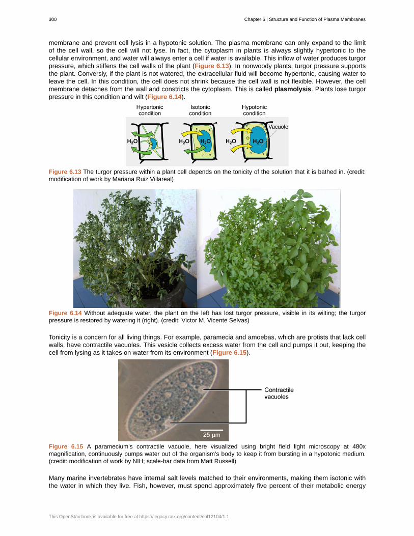
membrane and prevent cell lysis in a hypotonic solution. The plasma membrane can only expand to the limitof the cell wall, so the cell will not lyse. In fact, the cytoplasm in plants is always slightly hypertonic to thecellular environment, and water will always enter a cell if water is available. This inflow of water produces turgorpressure, which stiffens the cell walls of the plant (Figure 6.13). In nonwoody plants, turgor pressure supportsthe plant. Conversly, if the plant is not watered, the extracellular fluid will become hypertonic, causing water toleave the cell. In this condition, the cell does not shrink because the cell wall is not flexible. However, the cellmembrane detaches from the wall and constricts the cytoplasm. This is called plasmolysis. Plants lose turgorpressure in this condition and wilt (Figure 6.14).
Figure 6.13 The turgor pressure within a plant cell depends on the tonicity of the solution that it is bathed in. (credit:modification of work by Mariana Ruiz Villareal)
Figure 6.14 Without adequate water, the plant on the left has lost turgor pressure, visible in its wilting; the turgorpressure is restored by watering it (right). (credit: Victor M. Vicente Selvas)
Tonicity is a concern for all living things. For example, paramecia and amoebas, which are protists that lack cellwalls, have contractile vacuoles. This vesicle collects excess water from the cell and pumps it out, keeping thecell from lysing as it takes on water from its environment (Figure 6.15).
Figure 6.15 A paramecium’s contractile vacuole, here visualized using bright field light microscopy at 480xmagnification, continuously pumps water out of the organism’s body to keep it from bursting in a hypotonic medium.(credit: modification of work by NIH; scale-bar data from Matt Russell)
Many marine invertebrates have internal salt levels matched to their environments, making them isotonic withthe water in which they live. Fish, however, must spend approximately five percent of their metabolic energy
300 Chapter 6 | Structure and Function of Plasma Membranes
This OpenStax book is available for free at https://legacy.cnx.org/content/col12104/1.1
maintaining osmotic homeostasis. Freshwater fish live in an environment that is hypotonic to their cells. Thesefish actively take in salt through their gills and excrete diluted urine to rid themselves of excess water. Saltwaterfish live in the reverse environment, which is hypertonic to their cells, and they secrete salt through their gills andexcrete highly concentrated urine.
In vertebrates, the kidneys regulate the amount of water in the body. Osmoreceptors are specialized cells in thebrain that monitor the concentration of solutes in the blood. If the levels of solutes increase beyond a certainrange, a hormone is released that retards water loss through the kidney and dilutes the blood to safer levels.Animals also have high concentrations of albumin, which is produced by the liver, in their blood. This protein istoo large to pass easily through plasma membranes and is a major factor in controlling the osmotic pressuresapplied to tissues.
6.3 | Active Transport
By the end of this section, you will be able to:
• Understand how electrochemical gradients affect ions
• Distinguish between primary active transport and secondary active transport
Active transport mechanisms require the use of the cell’s energy, usually in the form of adenosine triphosphate(ATP). If a substance must move into the cell against its concentration gradient—that is, if the concentrationof the substance inside the cell is greater than its concentration in the extracellular fluid (and vice versa)—thecell must use energy to move the substance. Some active transport mechanisms move small-molecular weightmaterials, such as ions, through the membrane. Other mechanisms transport much larger molecules.
Electrochemical Gradient
We have discussed simple concentration gradients—differential concentrations of a substance across a spaceor a membrane—but in living systems, gradients are more complex. Because ions move into and out of cellsand because cells contain proteins that do not move across the membrane and are mostly negatively charged,there is also an electrical gradient, a difference of charge, across the plasma membrane. The interior of livingcells is electrically negative with respect to the extracellular fluid in which they are bathed, and at the same
time, cells have higher concentrations of potassium (K+) and lower concentrations of sodium (Na+) than does
the extracellular fluid. So in a living cell, the concentration gradient of Na+ tends to drive it into the cell, and
the electrical gradient of Na+ (a positive ion) also tends to drive it inward to the negatively charged interior.
The situation is more complex, however, for other elements such as potassium. The electrical gradient of K+, a
positive ion, also tends to drive it into the cell, but the concentration gradient of K+ tends to drive K+ out of thecell (Figure 6.16). The combined gradient of concentration and electrical charge that affects an ion is called itselectrochemical gradient.
Chapter 6 | Structure and Function of Plasma Membranes 301
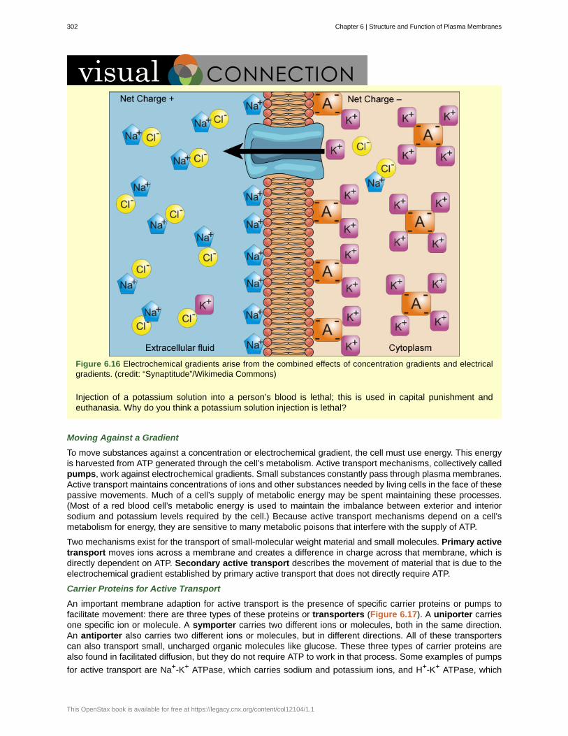
Figure 6.16 Electrochemical gradients arise from the combined effects of concentration gradients and electricalgradients. (credit: “Synaptitude”/Wikimedia Commons)
Injection of a potassium solution into a person’s blood is lethal; this is used in capital punishment andeuthanasia. Why do you think a potassium solution injection is lethal?
Moving Against a Gradient
To move substances against a concentration or electrochemical gradient, the cell must use energy. This energyis harvested from ATP generated through the cell’s metabolism. Active transport mechanisms, collectively calledpumps, work against electrochemical gradients. Small substances constantly pass through plasma membranes.Active transport maintains concentrations of ions and other substances needed by living cells in the face of thesepassive movements. Much of a cell’s supply of metabolic energy may be spent maintaining these processes.(Most of a red blood cell’s metabolic energy is used to maintain the imbalance between exterior and interiorsodium and potassium levels required by the cell.) Because active transport mechanisms depend on a cell’smetabolism for energy, they are sensitive to many metabolic poisons that interfere with the supply of ATP.
Two mechanisms exist for the transport of small-molecular weight material and small molecules. Primary activetransport moves ions across a membrane and creates a difference in charge across that membrane, which isdirectly dependent on ATP. Secondary active transport describes the movement of material that is due to theelectrochemical gradient established by primary active transport that does not directly require ATP.
Carrier Proteins for Active Transport
An important membrane adaption for active transport is the presence of specific carrier proteins or pumps tofacilitate movement: there are three types of these proteins or transporters (Figure 6.17). A uniporter carriesone specific ion or molecule. A symporter carries two different ions or molecules, both in the same direction.An antiporter also carries two different ions or molecules, but in different directions. All of these transporterscan also transport small, uncharged organic molecules like glucose. These three types of carrier proteins arealso found in facilitated diffusion, but they do not require ATP to work in that process. Some examples of pumps
for active transport are Na+-K+ ATPase, which carries sodium and potassium ions, and H+-K+ ATPase, which
302 Chapter 6 | Structure and Function of Plasma Membranes
This OpenStax book is available for free at https://legacy.cnx.org/content/col12104/1.1
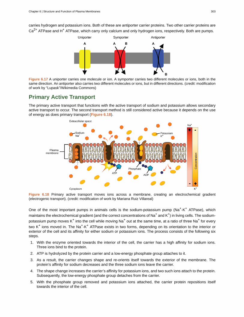
carries hydrogen and potassium ions. Both of these are antiporter carrier proteins. Two other carrier proteins are
Ca2+ ATPase and H+ ATPase, which carry only calcium and only hydrogen ions, respectively. Both are pumps.
Figure 6.17 A uniporter carries one molecule or ion. A symporter carries two different molecules or ions, both in thesame direction. An antiporter also carries two different molecules or ions, but in different directions. (credit: modificationof work by “Lupask”/Wikimedia Commons)
Primary Active Transport
The primary active transport that functions with the active transport of sodium and potassium allows secondaryactive transport to occur. The second transport method is still considered active because it depends on the useof energy as does primary transport (Figure 6.18).
Figure 6.18 Primary active transport moves ions across a membrane, creating an electrochemical gradient(electrogenic transport). (credit: modification of work by Mariana Ruiz Villareal)
One of the most important pumps in animals cells is the sodium-potassium pump (Na+-K+ ATPase), which
maintains the electrochemical gradient (and the correct concentrations of Na+ and K+) in living cells. The sodium-
potassium pump moves K+ into the cell while moving Na+ out at the same time, at a ratio of three Na+ for every
two K+ ions moved in. The Na+-K+ ATPase exists in two forms, depending on its orientation to the interior orexterior of the cell and its affinity for either sodium or potassium ions. The process consists of the following sixsteps.
1. With the enzyme oriented towards the interior of the cell, the carrier has a high affinity for sodium ions.Three ions bind to the protein.
2. ATP is hydrolyzed by the protein carrier and a low-energy phosphate group attaches to it.
3. As a result, the carrier changes shape and re-orients itself towards the exterior of the membrane. Theprotein’s affinity for sodium decreases and the three sodium ions leave the carrier.
4. The shape change increases the carrier’s affinity for potassium ions, and two such ions attach to the protein.Subsequently, the low-energy phosphate group detaches from the carrier.
5. With the phosphate group removed and potassium ions attached, the carrier protein repositions itselftowards the interior of the cell.
Chapter 6 | Structure and Function of Plasma Membranes 303
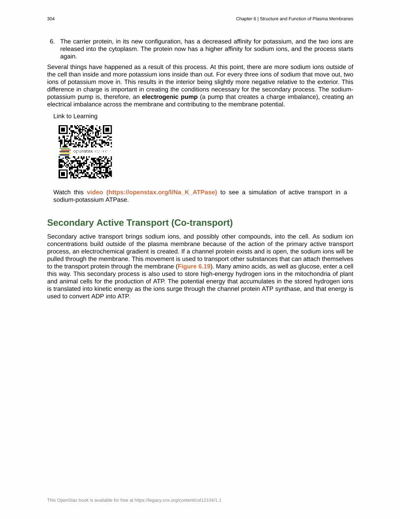
6. The carrier protein, in its new configuration, has a decreased affinity for potassium, and the two ions arereleased into the cytoplasm. The protein now has a higher affinity for sodium ions, and the process startsagain.
Several things have happened as a result of this process. At this point, there are more sodium ions outside ofthe cell than inside and more potassium ions inside than out. For every three ions of sodium that move out, twoions of potassium move in. This results in the interior being slightly more negative relative to the exterior. Thisdifference in charge is important in creating the conditions necessary for the secondary process. The sodium-potassium pump is, therefore, an electrogenic pump (a pump that creates a charge imbalance), creating anelectrical imbalance across the membrane and contributing to the membrane potential.
Link to Learning
Watch this video (https://openstax.org/l/Na_K_ATPase) to see a simulation of active transport in asodium-potassium ATPase.
Secondary Active Transport (Co-transport)
Secondary active transport brings sodium ions, and possibly other compounds, into the cell. As sodium ionconcentrations build outside of the plasma membrane because of the action of the primary active transportprocess, an electrochemical gradient is created. If a channel protein exists and is open, the sodium ions will bepulled through the membrane. This movement is used to transport other substances that can attach themselvesto the transport protein through the membrane (Figure 6.19). Many amino acids, as well as glucose, enter a cellthis way. This secondary process is also used to store high-energy hydrogen ions in the mitochondria of plantand animal cells for the production of ATP. The potential energy that accumulates in the stored hydrogen ionsis translated into kinetic energy as the ions surge through the channel protein ATP synthase, and that energy isused to convert ADP into ATP.
304 Chapter 6 | Structure and Function of Plasma Membranes
This OpenStax book is available for free at https://legacy.cnx.org/content/col12104/1.1
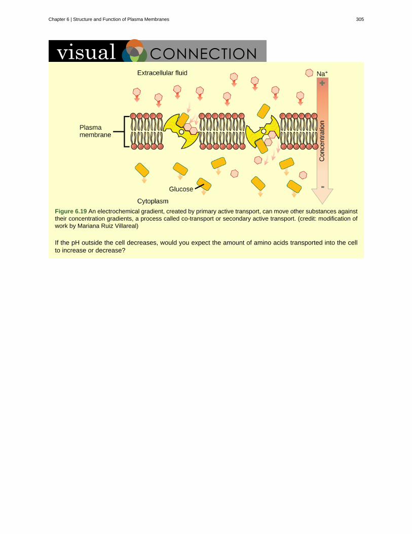
Figure 6.19 An electrochemical gradient, created by primary active transport, can move other substances againsttheir concentration gradients, a process called co-transport or secondary active transport. (credit: modification ofwork by Mariana Ruiz Villareal)
If the pH outside the cell decreases, would you expect the amount of amino acids transported into the cellto increase or decrease?
Chapter 6 | Structure and Function of Plasma Membranes 305
active transport
amphiphilic
antiporter
aquaporin
carrier protein
channel protein
concentration gradient
diffusion
electrochemical gradient
electrogenic pump
facilitated transport
fluid mosaic model
glycolipid
glycoprotein
hydrophilic
hydrophobic
hypertonic
hypotonic
integral protein
isotonic
osmolarity
osmosis
passive transport
peripheral protein
KEY TERMS
method of transporting material that requires energy
molecule possessing a polar or charged area and a nonpolar or uncharged area capable ofinteracting with both hydrophilic and hydrophobic environments
transporter that carries two ions or small molecules in different directions
channel protein that allows water through the membrane at a very high rate
membrane protein that moves a substance across the plasma membrane by changing its ownshape
membrane protein that allows a substance to pass through its hollow core across the plasmamembrane
area of high concentration adjacent to an area of low concentration
passive process of transport of low-molecular weight material according to its concentration gradient
gradient produced by the combined forces of an electrical gradient and a chemicalgradient
pump that creates a charge imbalance
process by which material moves down a concentration gradient (from high to lowconcentration) using integral membrane proteins
describes the structure of the plasma membrane as a mosaic of components includingphospholipids, cholesterol, proteins, glycoproteins, and glycolipids (sugar chains attached to proteins orlipids, respectively), resulting in a fluid character (fluidity)
combination of carbohydrates and lipids
combination of carbohydrates and proteins
molecule with the ability to bond with water; “water-loving”
molecule that does not have the ability to bond with water; “water-hating”
situation in which extracellular fluid has a higher osmolarity than the fluid inside the cell, resulting inwater moving out of the cell
situation in which extracellular fluid has a lower osmolarity than the fluid inside the cell, resulting inwater moving into the cell
protein integrated into the membrane structure that interacts extensively with the hydrocarbonchains of membrane lipids and often spans the membrane; these proteins can be removed only by thedisruption of the membrane by detergents
situation in which the extracellular fluid has the same osmolarity as the fluid inside the cell, resulting inno net movement of water into or out of the cell
total amount of substances dissolved in a specific amount of solution
transport of water through a semipermeable membrane according to the concentration gradient ofwater across the membrane that results from the presence of solute that cannot pass through themembrane
method of transporting material through a membrane that does not require energy
protein found at the surface of a plasma membrane either on its exterior or interior side;
306 Chapter 6 | Structure and Function of Plasma Membranes
This OpenStax book is available for free at https://legacy.cnx.org/content/col12104/1.1
plasmolysis
primary active transport
pump
secondary active transport
selectively permeable
solute
symporter
tonicity
transport protein
transporter
uniporter
these proteins can be removed (washed off of the membrane) by a high-salt wash
detaching of the cell membrane from the cell wall and constriction of the cell membrane when aplant cell is in a hypertonic solution
active transport that moves ions or small molecules across a membrane and maycreate a difference in charge across that membrane
active transport mechanism that works against electrochemical gradients
movement of material that is due to the electrochemical gradient established byprimary active transport
characteristic of a membrane that allows some substances through but not others
substance dissolved in a liquid to form a solution
transporter that carries two different ions or small molecules, both in the same direction
amount of solute in a solution
membrane protein that facilitates passage of a substance across a membrane by binding it
specific carrier proteins or pumps that facilitate movement
transporter that carries one specific ion or molecule
CHAPTER SUMMARY
6.1 Components and Structure
The modern understanding of the plasma membrane is referred to as the fluid mosaic model. The plasmamembrane is composed of a bilayer of phospholipids, with their hydrophobic, fatty acid tails in contact witheach other. The landscape of the membrane is studded with proteins, some of which span the membrane.Some of these proteins serve to transport materials into or out of the cell. Carbohydrates are attached to someof the proteins and lipids on the outward-facing surface of the membrane, forming complexes that function toidentify the cell to other cells. The fluid nature of the membrane is due to temperature, the configuration of thefatty acid tails (some kinked by double bonds), the presence of cholesterol embedded in the membrane, andthe mosaic nature of the proteins and protein-carbohydrate combinations, which are not firmly fixed in place.Plasma membranes enclose and define the borders of cells, but rather than being a static bag, they aredynamic and constantly in flux.
6.2 Passive Transport
The passive forms of transport, diffusion and osmosis, move materials of small molecular weight acrossmembranes. Substances diffuse from areas of high concentration to areas of lower concentration, and thisprocess continues until the substance is evenly distributed in a system. In solutions containing more than onesubstance, each type of molecule diffuses according to its own concentration gradient, independent of thediffusion of other substances. Many factors can affect the rate of diffusion, including concentration gradient,size of the particles that are diffusing, temperature of the system, and so on.
In living systems, diffusion of substances into and out of cells is mediated by the plasma membrane. Somematerials diffuse readily through the membrane, but others are hindered, and their passage is made possibleby specialized proteins, such as channels and transporters. The chemistry of living things occurs in aqueoussolutions, and balancing the concentrations of those solutions is an ongoing problem. In living systems,diffusion of some substances would be slow or difficult without membrane proteins that facilitate transport.
6.3 Active Transport
The combined gradient that affects an ion includes its concentration gradient and its electrical gradient. Apositive ion, for example, might tend to diffuse into a new area, down its concentration gradient, but if it isdiffusing into an area of net positive charge, its diffusion will be hampered by its electrical gradient. When
Chapter 6 | Structure and Function of Plasma Membranes 307

dealing with ions in aqueous solutions, a combination of the electrochemical and concentration gradients,rather than just the concentration gradient alone, must be considered. Living cells need certain substances thatexist inside the cell in concentrations greater than they exist in the extracellular space. Moving substances uptheir electrochemical gradients requires energy from the cell. Active transport uses energy stored in ATP to fuelthis transport. Active transport of small molecular-sized materials uses integral proteins in the cell membrane tomove the materials: These proteins are analogous to pumps. Some pumps, which carry out primary activetransport, couple directly with ATP to drive their action. In co-transport (or secondary active transport), energyfrom primary transport can be used to move another substance into the cell and up its concentration gradient.
ART CONNECTION QUESTIONS
1. Figure 6.12 A doctor injects a patient with whatthe doctor thinks is an isotonic saline solution. Thepatient dies, and an autopsy reveals that many redblood cells have been destroyed. Do you think thesolution the doctor injected was really isotonic?
2. Figure 6.16 Injection of a potassium solution into a
person’s blood is lethal; this is used in capitalpunishment and euthanasia. Why do you think apotassium solution injection is lethal?
3. Figure 6.19 If the pH outside the cell decreases,would you expect the amount of amino acidstransported into the cell to increase or decrease?
REVIEW QUESTIONS
4. Which plasma membrane component can beeither found on its surface or embedded in themembrane structure?
a. proteinb. cholesterolc. carbohydrated. phospholipid
5. Which characteristic of a phospholipid contributesto the fluidity of the membrane?
a. its headb. cholesterolc. a saturated fatty acid taild. double bonds in the fatty acid tail
6. What is the primary function of carbohydratesattached to the exterior of cell membranes?
a. identification of the cellb. flexibility of the membranec. strengthening the membraned. channels through membrane
7. Water moves via osmosis _________.a. throughout the cytoplasmb. from an area with a high concentration of
other solutes to a lower onec. from an area with a high concentration of
water to one of lower concentrationd. from an area with a low concentration of
water to one of higher concentration
8. The principal force driving movement in diffusion isthe __________.
a. temperatureb. particle sizec. concentration gradientd. membrane surface area
9. What problem is faced by organisms that live infresh water?
a. Their bodies tend to take in too much water.b. They have no way of controlling their
tonicity.c. Only salt water poses problems for animals
that live in it.d. Their bodies tend to lose too much water to
their environment.
10. Active transport must function continuouslybecause __________.
a. plasma membranes wear outb. not all membranes are amphiphilicc. facilitated transport opposes active transportd. diffusion is constantly moving solutes in
opposite directions
11. How does the sodium-potassium pump make theinterior of the cell negatively charged?
a. by expelling anionsb. by pulling in anionsc. by expelling more cations than are taken ind. by taking in and expelling an equal number
of cations
12. What is the combination of an electrical gradientand a concentration gradient called?
a. potential gradientb. electrical potentialc. concentration potentiald. electrochemical gradient
308 Chapter 6 | Structure and Function of Plasma Membranes
This OpenStax book is available for free at https://legacy.cnx.org/content/col12104/1.1

CRITICAL THINKING QUESTIONS
13. Why is it advantageous for the cell membrane tobe fluid in nature?
14. Why do phospholipids tend to spontaneouslyorient themselves into something resembling amembrane?
15. Discuss why the following affect the rate ofdiffusion: molecular size, temperature, solutiondensity, and the distance that must be traveled.
16. Why does water move through a membrane?
17. Both of the regular intravenous solutionsadministered in medicine, normal saline and lactatedRinger’s solution, are isotonic. Why is this important?
18. Where does the cell get energy for activetransport processes?
19. How does the sodium-potassium pump contributeto the net negative charge of the interior of the cell?
Chapter 6 | Structure and Function of Plasma Membranes 309

310 Chapter 6 | Structure and Function of Plasma Membranes
This OpenStax book is available for free at https://legacy.cnx.org/content/col12104/1.1
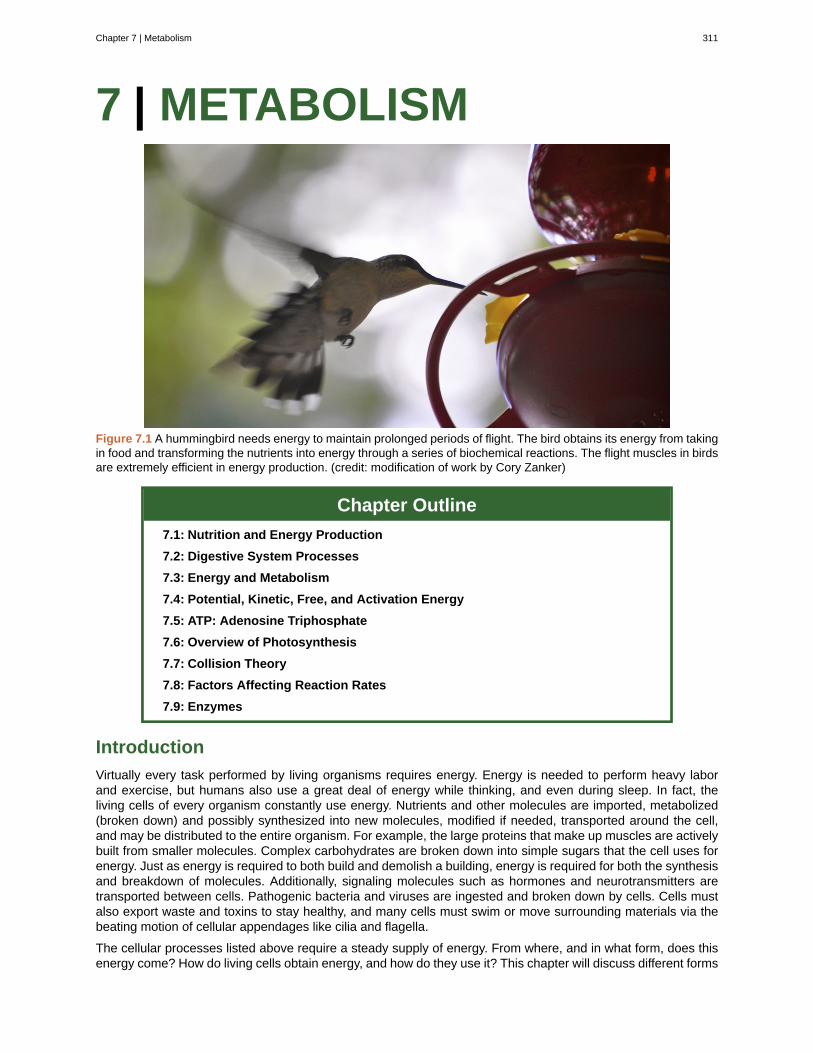
7 | METABOLISM
Figure 7.1 A hummingbird needs energy to maintain prolonged periods of flight. The bird obtains its energy from takingin food and transforming the nutrients into energy through a series of biochemical reactions. The flight muscles in birdsare extremely efficient in energy production. (credit: modification of work by Cory Zanker)
Chapter Outline
7.1: Nutrition and Energy Production
7.2: Digestive System Processes
7.3: Energy and Metabolism
7.4: Potential, Kinetic, Free, and Activation Energy
7.5: ATP: Adenosine Triphosphate
7.6: Overview of Photosynthesis
7.7: Collision Theory
7.8: Factors Affecting Reaction Rates
7.9: Enzymes
Introduction
Virtually every task performed by living organisms requires energy. Energy is needed to perform heavy laborand exercise, but humans also use a great deal of energy while thinking, and even during sleep. In fact, theliving cells of every organism constantly use energy. Nutrients and other molecules are imported, metabolized(broken down) and possibly synthesized into new molecules, modified if needed, transported around the cell,and may be distributed to the entire organism. For example, the large proteins that make up muscles are activelybuilt from smaller molecules. Complex carbohydrates are broken down into simple sugars that the cell uses forenergy. Just as energy is required to both build and demolish a building, energy is required for both the synthesisand breakdown of molecules. Additionally, signaling molecules such as hormones and neurotransmitters aretransported between cells. Pathogenic bacteria and viruses are ingested and broken down by cells. Cells mustalso export waste and toxins to stay healthy, and many cells must swim or move surrounding materials via thebeating motion of cellular appendages like cilia and flagella.
The cellular processes listed above require a steady supply of energy. From where, and in what form, does thisenergy come? How do living cells obtain energy, and how do they use it? This chapter will discuss different forms
Chapter 7 | Metabolism 311
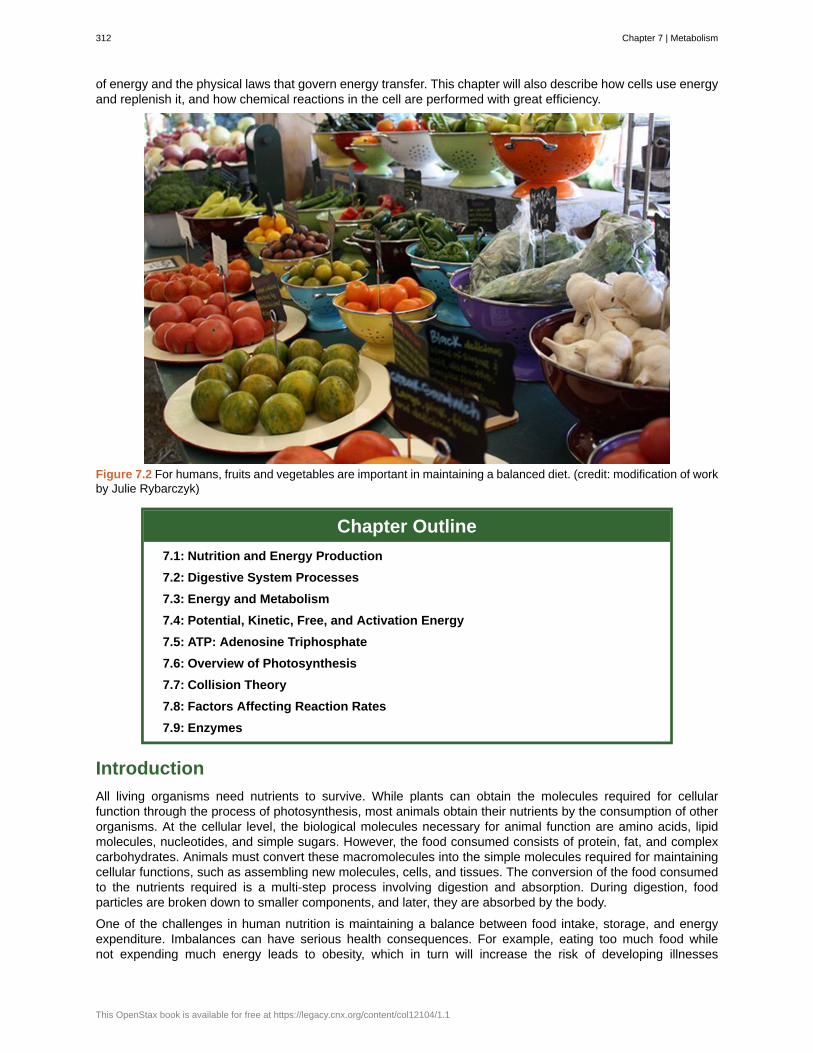
of energy and the physical laws that govern energy transfer. This chapter will also describe how cells use energyand replenish it, and how chemical reactions in the cell are performed with great efficiency.
Figure 7.2 For humans, fruits and vegetables are important in maintaining a balanced diet. (credit: modification of workby Julie Rybarczyk)
Chapter Outline
7.1: Nutrition and Energy Production
7.2: Digestive System Processes
7.3: Energy and Metabolism
7.4: Potential, Kinetic, Free, and Activation Energy
7.5: ATP: Adenosine Triphosphate
7.6: Overview of Photosynthesis
7.7: Collision Theory
7.8: Factors Affecting Reaction Rates
7.9: Enzymes
Introduction
All living organisms need nutrients to survive. While plants can obtain the molecules required for cellularfunction through the process of photosynthesis, most animals obtain their nutrients by the consumption of otherorganisms. At the cellular level, the biological molecules necessary for animal function are amino acids, lipidmolecules, nucleotides, and simple sugars. However, the food consumed consists of protein, fat, and complexcarbohydrates. Animals must convert these macromolecules into the simple molecules required for maintainingcellular functions, such as assembling new molecules, cells, and tissues. The conversion of the food consumedto the nutrients required is a multi-step process involving digestion and absorption. During digestion, foodparticles are broken down to smaller components, and later, they are absorbed by the body.
One of the challenges in human nutrition is maintaining a balance between food intake, storage, and energyexpenditure. Imbalances can have serious health consequences. For example, eating too much food whilenot expending much energy leads to obesity, which in turn will increase the risk of developing illnesses
312 Chapter 7 | Metabolism
This OpenStax book is available for free at https://legacy.cnx.org/content/col12104/1.1
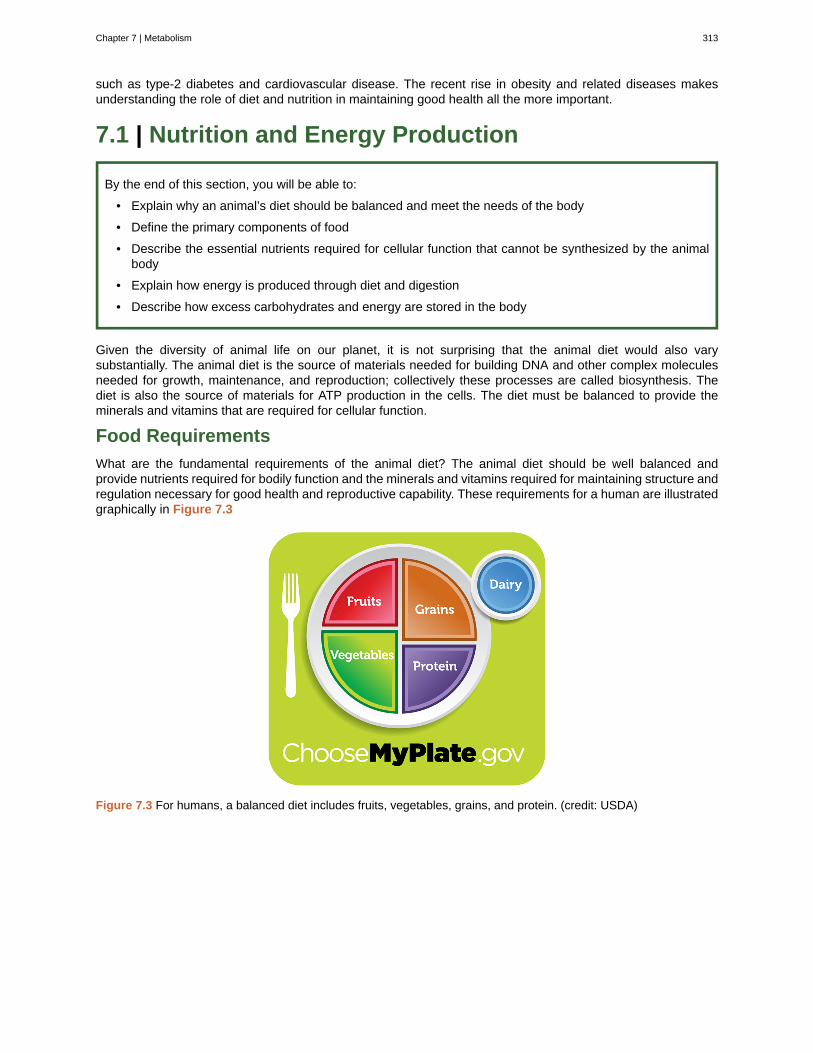
such as type-2 diabetes and cardiovascular disease. The recent rise in obesity and related diseases makesunderstanding the role of diet and nutrition in maintaining good health all the more important.
7.1 | Nutrition and Energy Production
By the end of this section, you will be able to:
• Explain why an animal’s diet should be balanced and meet the needs of the body
• Define the primary components of food
• Describe the essential nutrients required for cellular function that cannot be synthesized by the animalbody
• Explain how energy is produced through diet and digestion
• Describe how excess carbohydrates and energy are stored in the body
Given the diversity of animal life on our planet, it is not surprising that the animal diet would also varysubstantially. The animal diet is the source of materials needed for building DNA and other complex moleculesneeded for growth, maintenance, and reproduction; collectively these processes are called biosynthesis. Thediet is also the source of materials for ATP production in the cells. The diet must be balanced to provide theminerals and vitamins that are required for cellular function.
Food Requirements
What are the fundamental requirements of the animal diet? The animal diet should be well balanced andprovide nutrients required for bodily function and the minerals and vitamins required for maintaining structure andregulation necessary for good health and reproductive capability. These requirements for a human are illustratedgraphically in Figure 7.3
Figure 7.3 For humans, a balanced diet includes fruits, vegetables, grains, and protein. (credit: USDA)
Chapter 7 | Metabolism 313

The first step in ensuring that you are meeting the food requirements of your body is an awareness of thefood groups and the nutrients they provide. To learn more about each food group and the recommendeddaily amounts, explore this interactive site (http://openstaxcollege.org/l/food_groups) by the United StatesDepartment of Agriculture.
Let’s Move! CampaignObesity is a growing epidemic and the rate of obesity among children is rapidly rising in the United States.To combat childhood obesity and ensure that children get a healthy start in life, first lady Michelle Obamahas launched the Let’s Move! campaign. The goal of this campaign is to educate parents and caregiverson providing healthy nutrition and encouraging active lifestyles to future generations. This program aims toinvolve the entire community, including parents, teachers, and healthcare providers to ensure that childrenhave access to healthy foods—more fruits, vegetables, and whole grains—and consume fewer calories fromprocessed foods. Another goal is to ensure that children get physical activity. With the increase in televisionviewing and stationary pursuits such as video games, sedentary lifestyles have become the norm. Learnmore at www.letsmove.gov.
Organic Precursors
The organic molecules required for building cellular material and tissues must come from food. Carbohydrates orsugars are the primary source of organic carbons in the animal body. During digestion, digestible carbohydratesare ultimately broken down into glucose and used to provide energy through metabolic pathways. Complexcarbohydrates, including polysaccharides, can be broken down into glucose through biochemical modification;however, humans do not produce the enzyme cellulase and lack the ability to derive glucose from thepolysaccharide cellulose. In humans, these molecules provide the fiber required for moving waste through thelarge intestine and a healthy colon. The intestinal flora in the human gut are able to extract some nutrition fromthese plant fibers. The excess sugars in the body are converted into glycogen and stored in the liver and musclesfor later use. Glycogen stores are used to fuel prolonged exertions, such as long-distance running, and to provideenergy during food shortage. Excess glycogen can be converted to fats, which are stored in the lower layer ofthe skin of mammals for insulation and energy storage. Excess digestible carbohydrates are stored by mammalsin order to survive famine and aid in mobility.
Another important requirement is that of nitrogen. Protein catabolism provides a source of organic nitrogen.Amino acids are the building blocks of proteins and protein breakdown provides amino acids that are used forcellular function. The carbon and nitrogen derived from these become the building block for nucleotides, nucleicacids, proteins, cells, and tissues. Excess nitrogen must be excreted as it is toxic. Fats add flavor to food andpromote a sense of satiety or fullness. Fatty foods are also significant sources of energy because one gramof fat contains nine calories. Fats are required in the diet to aid the absorption of fat-soluble vitamins and theproduction of fat-soluble hormones.
Essential Nutrients
While the animal body can synthesize many of the molecules required for function from the organic precursors,there are some nutrients that need to be consumed from food. These nutrients are termed essential nutrients,meaning they must be eaten, and the body cannot produce them.
The omega-3 alpha-linolenic acid and the omega-6 linoleic acid are essential fatty acids needed to make somemembrane phospholipids. Vitamins are another class of essential organic molecules that are required in smallquantities for many enzymes to function and, for this reason, are considered to be co-enzymes. Absence or lowlevels of vitamins can have a dramatic effect on health, as outlined in Table 7.1 and Table 7.2. Both fat-soluble
314 Chapter 7 | Metabolism
This OpenStax book is available for free at https://legacy.cnx.org/content/col12104/1.1

and water-soluble vitamins must be obtained from food. Minerals, listed in Table 7.3, are inorganic essentialnutrients that must be obtained from food. Among their many functions, minerals help in structure and regulationand are considered co-factors. Certain amino acids also must be procured from food and cannot be synthesizedby the body. These amino acids are the “essential” amino acids. The human body can synthesize only 11 of the20 required amino acids; the rest must be obtained from food. The essential amino acids are listed in Table 7.4.
Water-soluble Essential Vitamins
Vitamin Function Deficiencies Can Lead To Sources
Vitamin B1(Thiamine)
Needed by the body to process lipids,proteins, and carbohydrates Coenzymeremoves CO2 from organic compounds
Muscle weakness, Beriberi: reducedheart function, CNS problems
Milk, meat,dried beans,whole grains
Vitamin B2(Riboflavin)
Takes an active role in metabolism,aiding in the conversion of food to energy(FAD and FMN)
Cracks or sores on the outer surfaceof the lips (cheliosis); inflammationand redness of the tongue; moist,scaly skin inflammation (seborrheicdermatitis)
Meat, eggs,enrichedgrains,vegetables
Vitamin B3(Niacin)
Used by the body to release energy fromcarbohydrates and to process alcohol;required for the synthesis of sexhormones; component of coenzyme
NAD+ and NADP+
Pellagra, which can result indermatitis, diarrhea, dementia, anddeath
Meat, eggs,grains, nuts,potatoes
Vitamin B5(Pantothenic
acid)
Assists in producing energy from foods(lipids, in particular); component ofcoenzyme A
Fatigue, poor coordination, retardedgrowth, numbness, tingling of handsand feet
Meat, wholegrains, milk,fruits,vegetables
Vitamin B6(Pyridoxine)
The principal vitamin for processingamino acids and lipids; also helpsconvert nutrients into energy
Irritability, depression, confusion,mouth sores or ulcers, anemia,muscular twitching
Meat, dairyproducts,whole grains,orange juice
Vitamin B7(Biotin)
Used in energy and amino acidmetabolism, fat synthesis, and fatbreakdown; helps the body use bloodsugar
Hair loss, dermatitis, depression,numbness and tingling in theextremities; neuromuscular disorders
Meat, eggs,legumes andothervegetables
Vitamin B9(Folic acid)
Assists the normal development of cells,especially during fetal development;helps metabolize nucleic and aminoacids
Deficiency during pregnancy isassociated with birth defects, such asneural tube defects and anemia
Leafy greenvegetables,whole wheat,fruits, nuts,legumes
Vitamin B12(Cobalamin)
Maintains healthy nervous system andassists with blood cell formation;coenzyme in nucleic acid metabolism
Anemia, neurological disorders,numbness, loss of balance
Meat, eggs,animalproducts
Vitamin C(Ascorbic
acid)
Helps maintain connective tissue: bone,cartilage, and dentin; boosts the immunesystem
Scurvy, which results in bleeding, hairand tooth loss; joint pain andswelling; delayed wound healing
Citrus fruits,broccoli,tomatoes, redsweet bellpeppers
Table 7.1
Chapter 7 | Metabolism 315
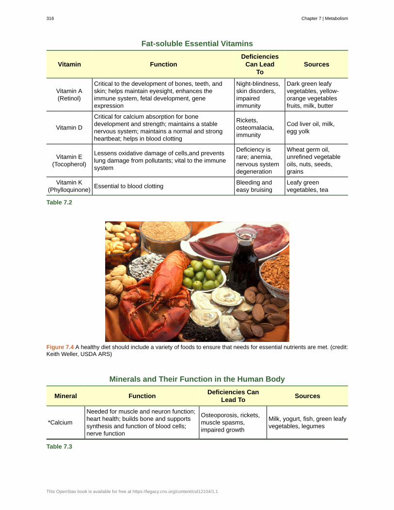
Fat-soluble Essential Vitamins
Vitamin FunctionDeficiencies
Can LeadTo
Sources
Vitamin A(Retinol)
Critical to the development of bones, teeth, andskin; helps maintain eyesight, enhances theimmune system, fetal development, geneexpression
Night-blindness,skin disorders,impairedimmunity
Dark green leafyvegetables, yellow-orange vegetablesfruits, milk, butter
Vitamin D
Critical for calcium absorption for bonedevelopment and strength; maintains a stablenervous system; maintains a normal and strongheartbeat; helps in blood clotting
Rickets,osteomalacia,immunity
Cod liver oil, milk,egg yolk
Vitamin E(Tocopherol)
Lessens oxidative damage of cells,and preventslung damage from pollutants; vital to the immunesystem
Deficiency israre; anemia,nervous systemdegeneration
Wheat germ oil,unrefined vegetableoils, nuts, seeds,grains
Vitamin K(Phylloquinone)
Essential to blood clottingBleeding andeasy bruising
Leafy greenvegetables, tea
Table 7.2
Figure 7.4 A healthy diet should include a variety of foods to ensure that needs for essential nutrients are met. (credit:Keith Weller, USDA ARS)
Minerals and Their Function in the Human Body
Mineral FunctionDeficiencies Can
Lead ToSources
*Calcium
Needed for muscle and neuron function;heart health; builds bone and supportssynthesis and function of blood cells;nerve function
Osteoporosis, rickets,muscle spasms,impaired growth
Milk, yogurt, fish, green leafyvegetables, legumes
Table 7.3
316 Chapter 7 | Metabolism
This OpenStax book is available for free at https://legacy.cnx.org/content/col12104/1.1
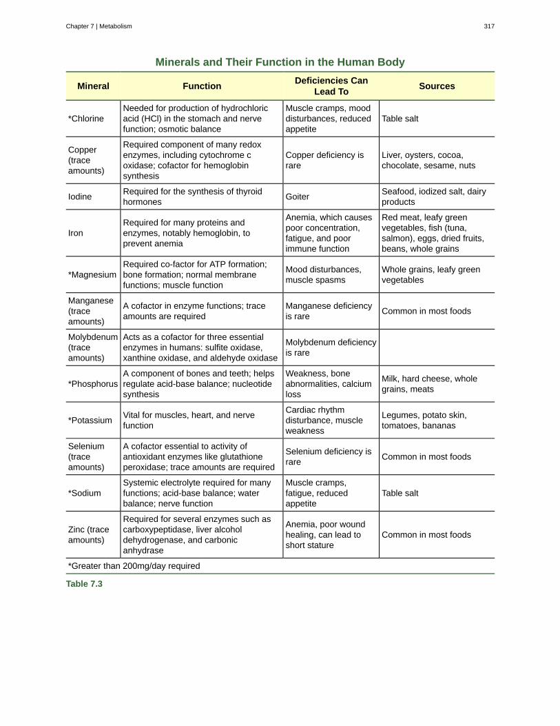
Minerals and Their Function in the Human Body
Mineral FunctionDeficiencies Can
Lead ToSources
*ChlorineNeeded for production of hydrochloricacid (HCl) in the stomach and nervefunction; osmotic balance
Muscle cramps, mooddisturbances, reducedappetite
Table salt
Copper(traceamounts)
Required component of many redoxenzymes, including cytochrome coxidase; cofactor for hemoglobinsynthesis
Copper deficiency israre
Liver, oysters, cocoa,chocolate, sesame, nuts
IodineRequired for the synthesis of thyroidhormones
GoiterSeafood, iodized salt, dairyproducts
IronRequired for many proteins andenzymes, notably hemoglobin, toprevent anemia
Anemia, which causespoor concentration,fatigue, and poorimmune function
Red meat, leafy greenvegetables, fish (tuna,salmon), eggs, dried fruits,beans, whole grains
*MagnesiumRequired co-factor for ATP formation;bone formation; normal membranefunctions; muscle function
Mood disturbances,muscle spasms
Whole grains, leafy greenvegetables
Manganese(traceamounts)
A cofactor in enzyme functions; traceamounts are required
Manganese deficiencyis rare
Common in most foods
Molybdenum(traceamounts)
Acts as a cofactor for three essentialenzymes in humans: sulfite oxidase,xanthine oxidase, and aldehyde oxidase
Molybdenum deficiencyis rare
*PhosphorusA component of bones and teeth; helpsregulate acid-base balance; nucleotidesynthesis
Weakness, boneabnormalities, calciumloss
Milk, hard cheese, wholegrains, meats
*PotassiumVital for muscles, heart, and nervefunction
Cardiac rhythmdisturbance, muscleweakness
Legumes, potato skin,tomatoes, bananas
Selenium(traceamounts)
A cofactor essential to activity ofantioxidant enzymes like glutathioneperoxidase; trace amounts are required
Selenium deficiency israre
Common in most foods
*SodiumSystemic electrolyte required for manyfunctions; acid-base balance; waterbalance; nerve function
Muscle cramps,fatigue, reducedappetite
Table salt
Zinc (traceamounts)
Required for several enzymes such ascarboxypeptidase, liver alcoholdehydrogenase, and carbonicanhydrase
Anemia, poor woundhealing, can lead toshort stature
Common in most foods
*Greater than 200mg/day required
Table 7.3
Chapter 7 | Metabolism 317
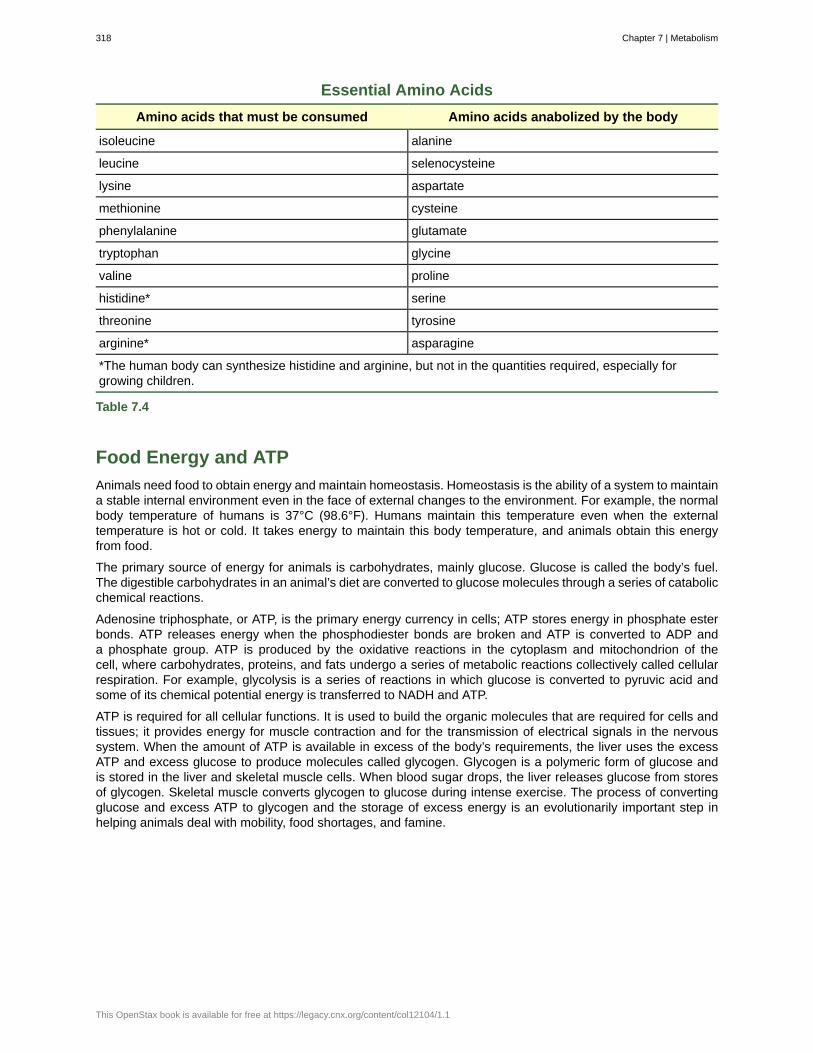
Essential Amino Acids
Amino acids that must be consumed Amino acids anabolized by the body
isoleucine alanine
leucine selenocysteine
lysine aspartate
methionine cysteine
phenylalanine glutamate
tryptophan glycine
valine proline
histidine* serine
threonine tyrosine
arginine* asparagine
*The human body can synthesize histidine and arginine, but not in the quantities required, especially forgrowing children.
Table 7.4
Food Energy and ATP
Animals need food to obtain energy and maintain homeostasis. Homeostasis is the ability of a system to maintaina stable internal environment even in the face of external changes to the environment. For example, the normalbody temperature of humans is 37°C (98.6°F). Humans maintain this temperature even when the externaltemperature is hot or cold. It takes energy to maintain this body temperature, and animals obtain this energyfrom food.
The primary source of energy for animals is carbohydrates, mainly glucose. Glucose is called the body’s fuel.The digestible carbohydrates in an animal’s diet are converted to glucose molecules through a series of catabolicchemical reactions.
Adenosine triphosphate, or ATP, is the primary energy currency in cells; ATP stores energy in phosphate esterbonds. ATP releases energy when the phosphodiester bonds are broken and ATP is converted to ADP anda phosphate group. ATP is produced by the oxidative reactions in the cytoplasm and mitochondrion of thecell, where carbohydrates, proteins, and fats undergo a series of metabolic reactions collectively called cellularrespiration. For example, glycolysis is a series of reactions in which glucose is converted to pyruvic acid andsome of its chemical potential energy is transferred to NADH and ATP.
ATP is required for all cellular functions. It is used to build the organic molecules that are required for cells andtissues; it provides energy for muscle contraction and for the transmission of electrical signals in the nervoussystem. When the amount of ATP is available in excess of the body’s requirements, the liver uses the excessATP and excess glucose to produce molecules called glycogen. Glycogen is a polymeric form of glucose andis stored in the liver and skeletal muscle cells. When blood sugar drops, the liver releases glucose from storesof glycogen. Skeletal muscle converts glycogen to glucose during intense exercise. The process of convertingglucose and excess ATP to glycogen and the storage of excess energy is an evolutionarily important step inhelping animals deal with mobility, food shortages, and famine.
318 Chapter 7 | Metabolism
This OpenStax book is available for free at https://legacy.cnx.org/content/col12104/1.1
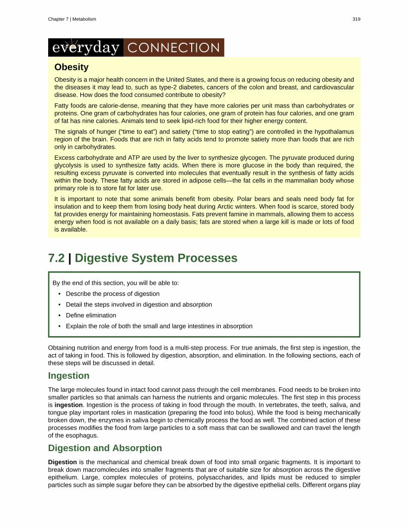
ObesityObesity is a major health concern in the United States, and there is a growing focus on reducing obesity andthe diseases it may lead to, such as type-2 diabetes, cancers of the colon and breast, and cardiovasculardisease. How does the food consumed contribute to obesity?
Fatty foods are calorie-dense, meaning that they have more calories per unit mass than carbohydrates orproteins. One gram of carbohydrates has four calories, one gram of protein has four calories, and one gramof fat has nine calories. Animals tend to seek lipid-rich food for their higher energy content.
The signals of hunger (“time to eat”) and satiety (“time to stop eating”) are controlled in the hypothalamusregion of the brain. Foods that are rich in fatty acids tend to promote satiety more than foods that are richonly in carbohydrates.
Excess carbohydrate and ATP are used by the liver to synthesize glycogen. The pyruvate produced duringglycolysis is used to synthesize fatty acids. When there is more glucose in the body than required, theresulting excess pyruvate is converted into molecules that eventually result in the synthesis of fatty acidswithin the body. These fatty acids are stored in adipose cells—the fat cells in the mammalian body whoseprimary role is to store fat for later use.
It is important to note that some animals benefit from obesity. Polar bears and seals need body fat forinsulation and to keep them from losing body heat during Arctic winters. When food is scarce, stored bodyfat provides energy for maintaining homeostasis. Fats prevent famine in mammals, allowing them to accessenergy when food is not available on a daily basis; fats are stored when a large kill is made or lots of foodis available.
7.2 | Digestive System Processes
By the end of this section, you will be able to:
• Describe the process of digestion
• Detail the steps involved in digestion and absorption
• Define elimination
• Explain the role of both the small and large intestines in absorption
Obtaining nutrition and energy from food is a multi-step process. For true animals, the first step is ingestion, theact of taking in food. This is followed by digestion, absorption, and elimination. In the following sections, each ofthese steps will be discussed in detail.
Ingestion
The large molecules found in intact food cannot pass through the cell membranes. Food needs to be broken intosmaller particles so that animals can harness the nutrients and organic molecules. The first step in this processis ingestion. Ingestion is the process of taking in food through the mouth. In vertebrates, the teeth, saliva, andtongue play important roles in mastication (preparing the food into bolus). While the food is being mechanicallybroken down, the enzymes in saliva begin to chemically process the food as well. The combined action of theseprocesses modifies the food from large particles to a soft mass that can be swallowed and can travel the lengthof the esophagus.
Digestion and Absorption
Digestion is the mechanical and chemical break down of food into small organic fragments. It is important tobreak down macromolecules into smaller fragments that are of suitable size for absorption across the digestiveepithelium. Large, complex molecules of proteins, polysaccharides, and lipids must be reduced to simplerparticles such as simple sugar before they can be absorbed by the digestive epithelial cells. Different organs play
Chapter 7 | Metabolism 319
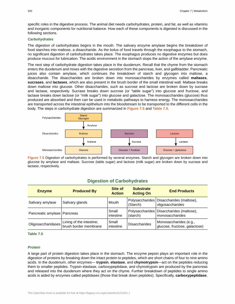
specific roles in the digestive process. The animal diet needs carbohydrates, protein, and fat, as well as vitaminsand inorganic components for nutritional balance. How each of these components is digested is discussed in thefollowing sections.
Carbohydrates
The digestion of carbohydrates begins in the mouth. The salivary enzyme amylase begins the breakdown offood starches into maltose, a disaccharide. As the bolus of food travels through the esophagus to the stomach,no significant digestion of carbohydrates takes place. The esophagus produces no digestive enzymes but doesproduce mucous for lubrication. The acidic environment in the stomach stops the action of the amylase enzyme.
The next step of carbohydrate digestion takes place in the duodenum. Recall that the chyme from the stomachenters the duodenum and mixes with the digestive secretion from the pancreas, liver, and gallbladder. Pancreaticjuices also contain amylase, which continues the breakdown of starch and glycogen into maltose, adisaccharide. The disaccharides are broken down into monosaccharides by enzymes called maltases,sucrases, and lactases, which are also present in the brush border of the small intestinal wall. Maltase breaksdown maltose into glucose. Other disaccharides, such as sucrose and lactose are broken down by sucraseand lactase, respectively. Sucrase breaks down sucrose (or “table sugar”) into glucose and fructose, andlactase breaks down lactose (or “milk sugar”) into glucose and galactose. The monosaccharides (glucose) thusproduced are absorbed and then can be used in metabolic pathways to harness energy. The monosaccharidesare transported across the intestinal epithelium into the bloodstream to be transported to the different cells in thebody. The steps in carbohydrate digestion are summarized in Figure 7.5 and Table 7.5.
Figure 7.5 Digestion of carbohydrates is performed by several enzymes. Starch and glycogen are broken down intoglucose by amylase and maltase. Sucrose (table sugar) and lactose (milk sugar) are broken down by sucrase andlactase, respectively.
Digestion of Carbohydrates
Enzyme Produced BySite ofAction
SubstrateActing On
End Products
Salivary amylase Salivary glands MouthPolysaccharides(Starch)
Disaccharides (maltose),oligosaccharides
Pancreatic amylase PancreasSmallintestine
Polysaccharides(starch)
Disaccharides (maltose),monosaccharides
OligosaccharidasesLining of the intestine;brush border membrane
Smallintestine
DisaccharidesMonosaccharides (e.g.,glucose, fructose, galactose)
Table 7.5
Protein
A large part of protein digestion takes place in the stomach. The enzyme pepsin plays an important role in thedigestion of proteins by breaking down the intact protein to peptides, which are short chains of four to nine aminoacids. In the duodenum, other enzymes— trypsin, elastase, and chymotrypsin—act on the peptides reducingthem to smaller peptides. Trypsin elastase, carboxypeptidase, and chymotrypsin are produced by the pancreasand released into the duodenum where they act on the chyme. Further breakdown of peptides to single aminoacids is aided by enzymes called peptidases (those that break down peptides). Specifically, carboxypeptidase,
320 Chapter 7 | Metabolism
This OpenStax book is available for free at https://legacy.cnx.org/content/col12104/1.1
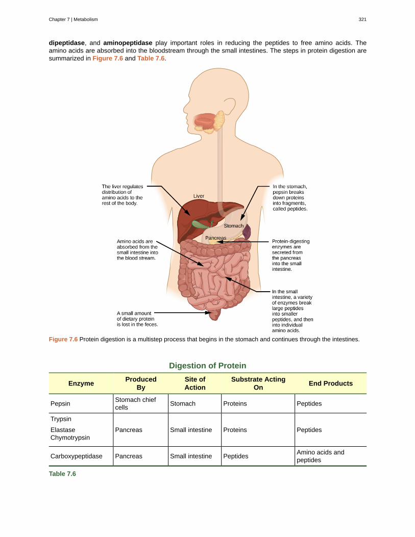
dipeptidase, and aminopeptidase play important roles in reducing the peptides to free amino acids. Theamino acids are absorbed into the bloodstream through the small intestines. The steps in protein digestion aresummarized in Figure 7.6 and Table 7.6.
Figure 7.6 Protein digestion is a multistep process that begins in the stomach and continues through the intestines.
Digestion of Protein
EnzymeProduced
BySite ofAction
Substrate ActingOn
End Products
PepsinStomach chiefcells
Stomach Proteins Peptides
Trypsin
ElastaseChymotrypsin
Pancreas Small intestine Proteins Peptides
Carboxypeptidase Pancreas Small intestine PeptidesAmino acids andpeptides
Table 7.6
Chapter 7 | Metabolism 321
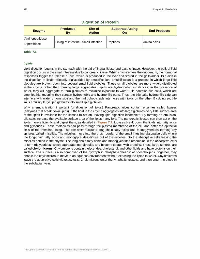
Digestion of Protein
EnzymeProduced
BySite ofAction
Substrate ActingOn
End Products
Aminopeptidase
DipeptidaseLining of intestine Small intestine Peptides Amino acids
Table 7.6
Lipids
Lipid digestion begins in the stomach with the aid of lingual lipase and gastric lipase. However, the bulk of lipiddigestion occurs in the small intestine due to pancreatic lipase. When chyme enters the duodenum, the hormonalresponses trigger the release of bile, which is produced in the liver and stored in the gallbladder. Bile aids inthe digestion of lipids, primarily triglycerides by emulsification. Emulsification is a process in which large lipidglobules are broken down into several small lipid globules. These small globules are more widely distributedin the chyme rather than forming large aggregates. Lipids are hydrophobic substances: in the presence ofwater, they will aggregate to form globules to minimize exposure to water. Bile contains bile salts, which areamphipathic, meaning they contain hydrophobic and hydrophilic parts. Thus, the bile salts hydrophilic side caninterface with water on one side and the hydrophobic side interfaces with lipids on the other. By doing so, bilesalts emulsify large lipid globules into small lipid globules.
Why is emulsification important for digestion of lipids? Pancreatic juices contain enzymes called lipases(enzymes that break down lipids). If the lipid in the chyme aggregates into large globules, very little surface areaof the lipids is available for the lipases to act on, leaving lipid digestion incomplete. By forming an emulsion,bile salts increase the available surface area of the lipids many fold. The pancreatic lipases can then act on thelipids more efficiently and digest them, as detailed in Figure 7.7. Lipases break down the lipids into fatty acidsand glycerides. These molecules can pass through the plasma membrane of the cell and enter the epithelialcells of the intestinal lining. The bile salts surround long-chain fatty acids and monoglycerides forming tinyspheres called micelles. The micelles move into the brush border of the small intestine absorptive cells wherethe long-chain fatty acids and monoglycerides diffuse out of the micelles into the absorptive cells leaving themicelles behind in the chyme. The long-chain fatty acids and monoglycerides recombine in the absorptive cellsto form triglycerides, which aggregate into globules and become coated with proteins. These large spheres arecalled chylomicrons. Chylomicrons contain triglycerides, cholesterol, and other lipids and have proteins on theirsurface. The surface is also composed of the hydrophilic phosphate "heads" of phospholipids. Together, theyenable the chylomicron to move in an aqueous environment without exposing the lipids to water. Chylomicronsleave the absorptive cells via exocytosis. Chylomicrons enter the lymphatic vessels, and then enter the blood inthe subclavian vein.
322 Chapter 7 | Metabolism
This OpenStax book is available for free at https://legacy.cnx.org/content/col12104/1.1
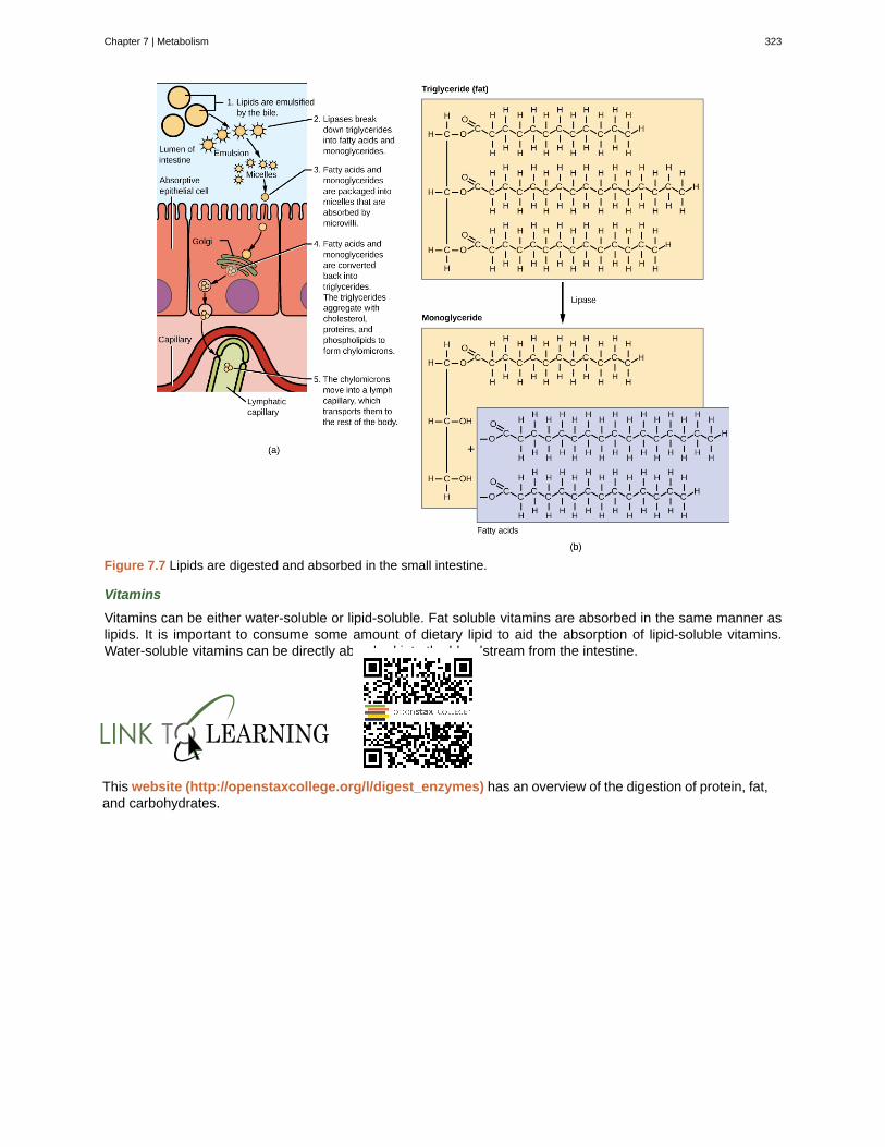
Figure 7.7 Lipids are digested and absorbed in the small intestine.
Vitamins
Vitamins can be either water-soluble or lipid-soluble. Fat soluble vitamins are absorbed in the same manner aslipids. It is important to consume some amount of dietary lipid to aid the absorption of lipid-soluble vitamins.Water-soluble vitamins can be directly absorbed into the bloodstream from the intestine.
This website (http://openstaxcollege.org/l/digest_enzymes) has an overview of the digestion of protein, fat,and carbohydrates.
Chapter 7 | Metabolism 323
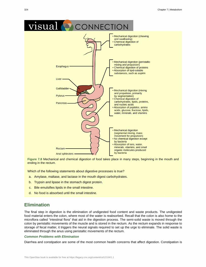
Figure 7.8 Mechanical and chemical digestion of food takes place in many steps, beginning in the mouth andending in the rectum.
Which of the following statements about digestive processes is true?
a. Amylase, maltase, and lactase in the mouth digest carbohydrates.
b. Trypsin and lipase in the stomach digest protein.
c. Bile emulsifies lipids in the small intestine.
d. No food is absorbed until the small intestine.
Elimination
The final step in digestion is the elimination of undigested food content and waste products. The undigestedfood material enters the colon, where most of the water is reabsorbed. Recall that the colon is also home to themicroflora called “intestinal flora” that aid in the digestion process. The semi-solid waste is moved through thecolon by peristaltic movements of the muscle and is stored in the rectum. As the rectum expands in response tostorage of fecal matter, it triggers the neural signals required to set up the urge to eliminate. The solid waste iseliminated through the anus using peristaltic movements of the rectum.
Common Problems with Elimination
Diarrhea and constipation are some of the most common health concerns that affect digestion. Constipation is
324 Chapter 7 | Metabolism
This OpenStax book is available for free at https://legacy.cnx.org/content/col12104/1.1
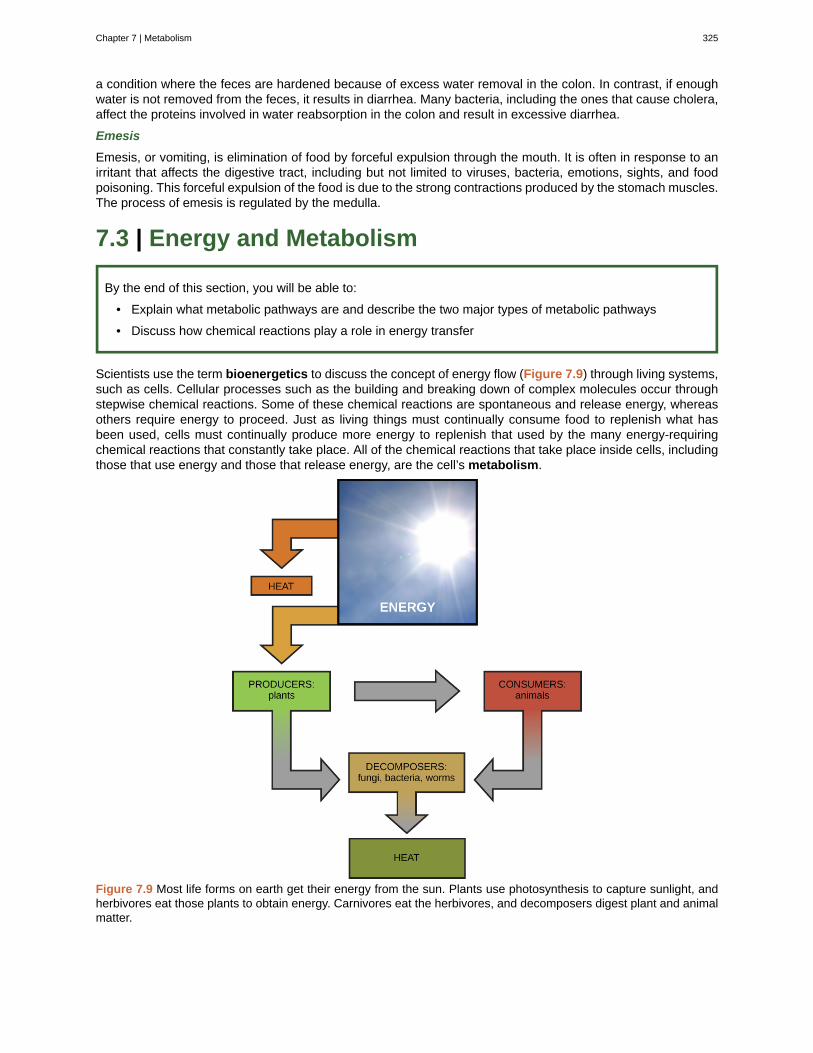
a condition where the feces are hardened because of excess water removal in the colon. In contrast, if enoughwater is not removed from the feces, it results in diarrhea. Many bacteria, including the ones that cause cholera,affect the proteins involved in water reabsorption in the colon and result in excessive diarrhea.
Emesis
Emesis, or vomiting, is elimination of food by forceful expulsion through the mouth. It is often in response to anirritant that affects the digestive tract, including but not limited to viruses, bacteria, emotions, sights, and foodpoisoning. This forceful expulsion of the food is due to the strong contractions produced by the stomach muscles.The process of emesis is regulated by the medulla.
7.3 | Energy and Metabolism
By the end of this section, you will be able to:
• Explain what metabolic pathways are and describe the two major types of metabolic pathways
• Discuss how chemical reactions play a role in energy transfer
Scientists use the term bioenergetics to discuss the concept of energy flow (Figure 7.9) through living systems,such as cells. Cellular processes such as the building and breaking down of complex molecules occur throughstepwise chemical reactions. Some of these chemical reactions are spontaneous and release energy, whereasothers require energy to proceed. Just as living things must continually consume food to replenish what hasbeen used, cells must continually produce more energy to replenish that used by the many energy-requiringchemical reactions that constantly take place. All of the chemical reactions that take place inside cells, includingthose that use energy and those that release energy, are the cell’s metabolism.
Figure 7.9 Most life forms on earth get their energy from the sun. Plants use photosynthesis to capture sunlight, andherbivores eat those plants to obtain energy. Carnivores eat the herbivores, and decomposers digest plant and animalmatter.
Chapter 7 | Metabolism 325
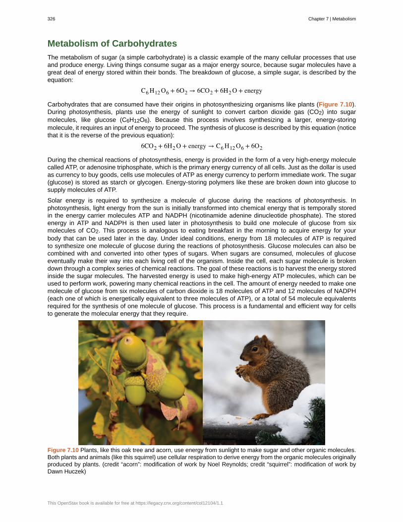
Metabolism of Carbohydrates
The metabolism of sugar (a simple carbohydrate) is a classic example of the many cellular processes that useand produce energy. Living things consume sugar as a major energy source, because sugar molecules have agreat deal of energy stored within their bonds. The breakdown of glucose, a simple sugar, is described by theequation:
C6 H12 O6 + 6O2 → 6CO2 + 6H2 O + energy
Carbohydrates that are consumed have their origins in photosynthesizing organisms like plants (Figure 7.10).During photosynthesis, plants use the energy of sunlight to convert carbon dioxide gas (CO2) into sugarmolecules, like glucose (C6H12O6). Because this process involves synthesizing a larger, energy-storingmolecule, it requires an input of energy to proceed. The synthesis of glucose is described by this equation (noticethat it is the reverse of the previous equation):
6CO2 + 6H2 O + energy → C6 H12 O6 + 6O2
During the chemical reactions of photosynthesis, energy is provided in the form of a very high-energy moleculecalled ATP, or adenosine triphosphate, which is the primary energy currency of all cells. Just as the dollar is usedas currency to buy goods, cells use molecules of ATP as energy currency to perform immediate work. The sugar(glucose) is stored as starch or glycogen. Energy-storing polymers like these are broken down into glucose tosupply molecules of ATP.
Solar energy is required to synthesize a molecule of glucose during the reactions of photosynthesis. Inphotosynthesis, light energy from the sun is initially transformed into chemical energy that is temporally storedin the energy carrier molecules ATP and NADPH (nicotinamide adenine dinucleotide phosphate). The storedenergy in ATP and NADPH is then used later in photosynthesis to build one molecule of glucose from sixmolecules of CO2. This process is analogous to eating breakfast in the morning to acquire energy for yourbody that can be used later in the day. Under ideal conditions, energy from 18 molecules of ATP is requiredto synthesize one molecule of glucose during the reactions of photosynthesis. Glucose molecules can also becombined with and converted into other types of sugars. When sugars are consumed, molecules of glucoseeventually make their way into each living cell of the organism. Inside the cell, each sugar molecule is brokendown through a complex series of chemical reactions. The goal of these reactions is to harvest the energy storedinside the sugar molecules. The harvested energy is used to make high-energy ATP molecules, which can beused to perform work, powering many chemical reactions in the cell. The amount of energy needed to make onemolecule of glucose from six molecules of carbon dioxide is 18 molecules of ATP and 12 molecules of NADPH(each one of which is energetically equivalent to three molecules of ATP), or a total of 54 molecule equivalentsrequired for the synthesis of one molecule of glucose. This process is a fundamental and efficient way for cellsto generate the molecular energy that they require.
Figure 7.10 Plants, like this oak tree and acorn, use energy from sunlight to make sugar and other organic molecules.Both plants and animals (like this squirrel) use cellular respiration to derive energy from the organic molecules originallyproduced by plants. (credit “acorn”: modification of work by Noel Reynolds; credit “squirrel”: modification of work byDawn Huczek)
326 Chapter 7 | Metabolism
This OpenStax book is available for free at https://legacy.cnx.org/content/col12104/1.1
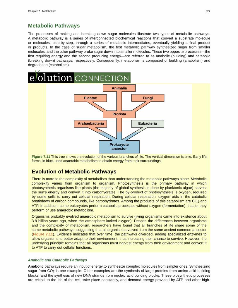
Metabolic Pathways
The processes of making and breaking down sugar molecules illustrate two types of metabolic pathways.A metabolic pathway is a series of interconnected biochemical reactions that convert a substrate moleculeor molecules, step-by-step, through a series of metabolic intermediates, eventually yielding a final productor products. In the case of sugar metabolism, the first metabolic pathway synthesized sugar from smallermolecules, and the other pathway broke sugar down into smaller molecules. These two opposite processes—thefirst requiring energy and the second producing energy—are referred to as anabolic (building) and catabolic(breaking down) pathways, respectively. Consequently, metabolism is composed of building (anabolism) anddegradation (catabolism).
Figure 7.11 This tree shows the evolution of the various branches of life. The vertical dimension is time. Early lifeforms, in blue, used anaerobic metabolism to obtain energy from their surroundings.
Evolution of Metabolic PathwaysThere is more to the complexity of metabolism than understanding the metabolic pathways alone. Metaboliccomplexity varies from organism to organism. Photosynthesis is the primary pathway in whichphotosynthetic organisms like plants (the majority of global synthesis is done by planktonic algae) harvestthe sun’s energy and convert it into carbohydrates. The by-product of photosynthesis is oxygen, requiredby some cells to carry out cellular respiration. During cellular respiration, oxygen aids in the catabolicbreakdown of carbon compounds, like carbohydrates. Among the products of this catabolism are CO2 andATP. In addition, some eukaryotes perform catabolic processes without oxygen (fermentation); that is, theyperform or use anaerobic metabolism.
Organisms probably evolved anaerobic metabolism to survive (living organisms came into existence about3.8 billion years ago, when the atmosphere lacked oxygen). Despite the differences between organismsand the complexity of metabolism, researchers have found that all branches of life share some of thesame metabolic pathways, suggesting that all organisms evolved from the same ancient common ancestor(Figure 7.11). Evidence indicates that over time, the pathways diverged, adding specialized enzymes toallow organisms to better adapt to their environment, thus increasing their chance to survive. However, theunderlying principle remains that all organisms must harvest energy from their environment and convert itto ATP to carry out cellular functions.
Anabolic and Catabolic Pathways
Anabolic pathways require an input of energy to synthesize complex molecules from simpler ones. Synthesizingsugar from CO2 is one example. Other examples are the synthesis of large proteins from amino acid buildingblocks, and the synthesis of new DNA strands from nucleic acid building blocks. These biosynthetic processesare critical to the life of the cell, take place constantly, and demand energy provided by ATP and other high-
Chapter 7 | Metabolism 327
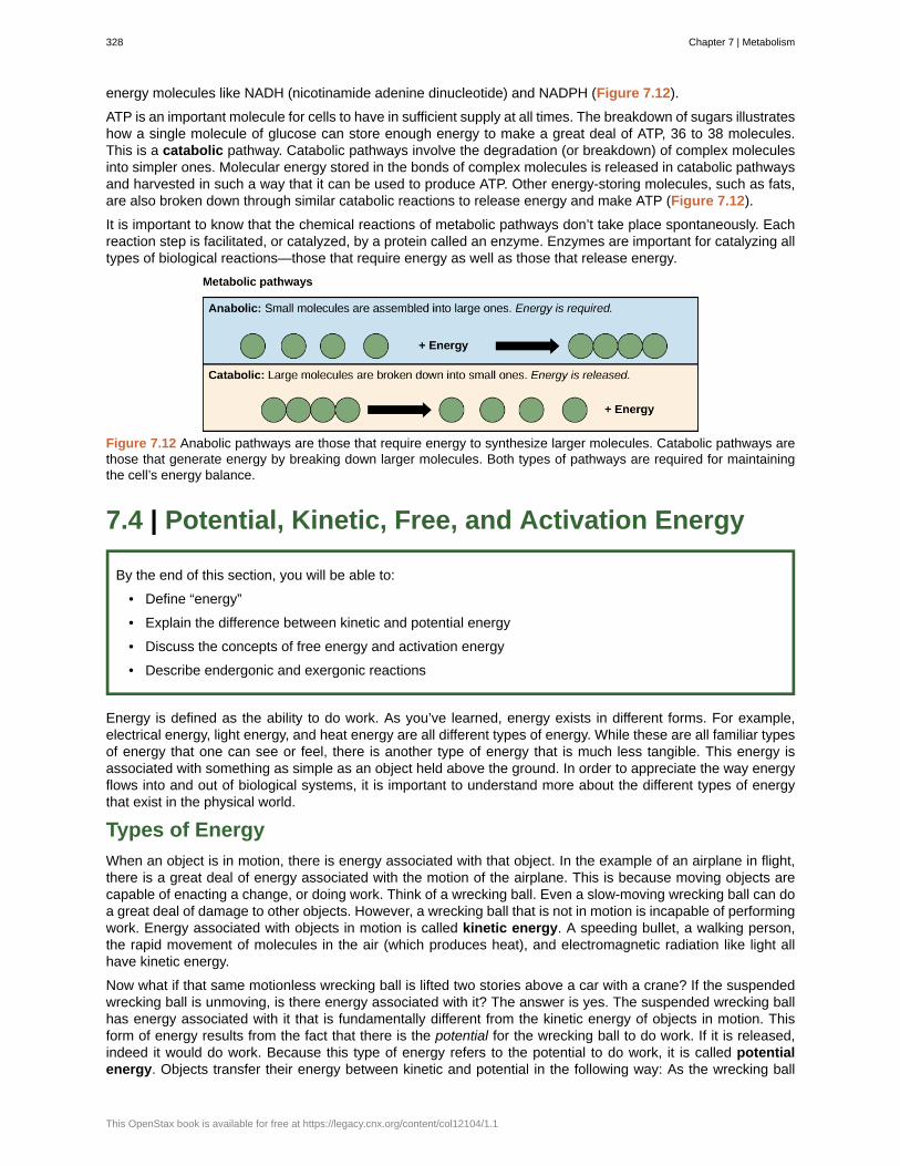
energy molecules like NADH (nicotinamide adenine dinucleotide) and NADPH (Figure 7.12).
ATP is an important molecule for cells to have in sufficient supply at all times. The breakdown of sugars illustrateshow a single molecule of glucose can store enough energy to make a great deal of ATP, 36 to 38 molecules.This is a catabolic pathway. Catabolic pathways involve the degradation (or breakdown) of complex moleculesinto simpler ones. Molecular energy stored in the bonds of complex molecules is released in catabolic pathwaysand harvested in such a way that it can be used to produce ATP. Other energy-storing molecules, such as fats,are also broken down through similar catabolic reactions to release energy and make ATP (Figure 7.12).
It is important to know that the chemical reactions of metabolic pathways don’t take place spontaneously. Eachreaction step is facilitated, or catalyzed, by a protein called an enzyme. Enzymes are important for catalyzing alltypes of biological reactions—those that require energy as well as those that release energy.
Figure 7.12 Anabolic pathways are those that require energy to synthesize larger molecules. Catabolic pathways arethose that generate energy by breaking down larger molecules. Both types of pathways are required for maintainingthe cell’s energy balance.
7.4 | Potential, Kinetic, Free, and Activation Energy
By the end of this section, you will be able to:
• Define “energy”
• Explain the difference between kinetic and potential energy
• Discuss the concepts of free energy and activation energy
• Describe endergonic and exergonic reactions
Energy is defined as the ability to do work. As you’ve learned, energy exists in different forms. For example,electrical energy, light energy, and heat energy are all different types of energy. While these are all familiar typesof energy that one can see or feel, there is another type of energy that is much less tangible. This energy isassociated with something as simple as an object held above the ground. In order to appreciate the way energyflows into and out of biological systems, it is important to understand more about the different types of energythat exist in the physical world.
Types of Energy
When an object is in motion, there is energy associated with that object. In the example of an airplane in flight,there is a great deal of energy associated with the motion of the airplane. This is because moving objects arecapable of enacting a change, or doing work. Think of a wrecking ball. Even a slow-moving wrecking ball can doa great deal of damage to other objects. However, a wrecking ball that is not in motion is incapable of performingwork. Energy associated with objects in motion is called kinetic energy. A speeding bullet, a walking person,the rapid movement of molecules in the air (which produces heat), and electromagnetic radiation like light allhave kinetic energy.
Now what if that same motionless wrecking ball is lifted two stories above a car with a crane? If the suspendedwrecking ball is unmoving, is there energy associated with it? The answer is yes. The suspended wrecking ballhas energy associated with it that is fundamentally different from the kinetic energy of objects in motion. Thisform of energy results from the fact that there is the potential for the wrecking ball to do work. If it is released,indeed it would do work. Because this type of energy refers to the potential to do work, it is called potentialenergy. Objects transfer their energy between kinetic and potential in the following way: As the wrecking ball
328 Chapter 7 | Metabolism
This OpenStax book is available for free at https://legacy.cnx.org/content/col12104/1.1

hangs motionless, it has 0 kinetic and 100 percent potential energy. Once it is released, its kinetic energy beginsto increase because it builds speed due to gravity. At the same time, as it nears the ground, it loses potentialenergy. Somewhere mid-fall it has 50 percent kinetic and 50 percent potential energy. Just before it hits theground, the ball has nearly lost its potential energy and has near-maximal kinetic energy. Other examples ofpotential energy include the energy of water held behind a dam (Figure 7.13), or a person about to skydive outof an airplane.
Figure 7.13 Water behind a dam has potential energy. Moving water, such as in a waterfall or a rapidly flowing river,has kinetic energy. (credit “dam”: modification of work by "Pascal"/Flickr; credit “waterfall”: modification of work byFrank Gualtieri)
Potential energy is not only associated with the location of matter (such as a child sitting on a tree branch), butalso with the structure of matter. A spring on the ground has potential energy if it is compressed; so does arubber band that is pulled taut. The very existence of living cells relies heavily on structural potential energy. Ona chemical level, the bonds that hold the atoms of molecules together have potential energy. Remember thatanabolic cellular pathways require energy to synthesize complex molecules from simpler ones, and catabolicpathways release energy when complex molecules are broken down. The fact that energy can be releasedby the breakdown of certain chemical bonds implies that those bonds have potential energy. In fact, there ispotential energy stored within the bonds of all the food molecules we eat, which is eventually harnessed foruse. This is because these bonds can release energy when broken. The type of potential energy that existswithin chemical bonds, and is released when those bonds are broken, is called chemical energy (Figure 7.14).Chemical energy is responsible for providing living cells with energy from food. The release of energy is broughtabout by breaking the molecular bonds within fuel molecules.
Chapter 7 | Metabolism 329
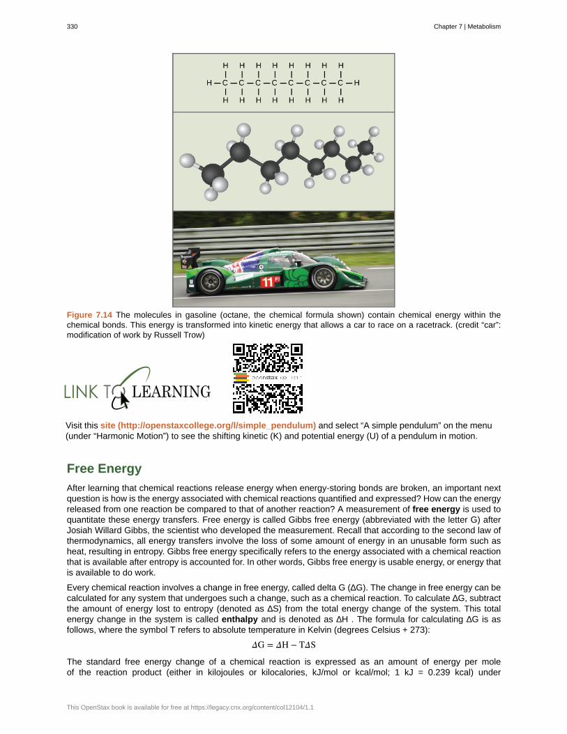
Figure 7.14 The molecules in gasoline (octane, the chemical formula shown) contain chemical energy within thechemical bonds. This energy is transformed into kinetic energy that allows a car to race on a racetrack. (credit “car”:modification of work by Russell Trow)
Visit this site (http://openstaxcollege.org/l/simple_pendulum) and select “A simple pendulum” on the menu(under “Harmonic Motion”) to see the shifting kinetic (K) and potential energy (U) of a pendulum in motion.
Free Energy
After learning that chemical reactions release energy when energy-storing bonds are broken, an important nextquestion is how is the energy associated with chemical reactions quantified and expressed? How can the energyreleased from one reaction be compared to that of another reaction? A measurement of free energy is used toquantitate these energy transfers. Free energy is called Gibbs free energy (abbreviated with the letter G) afterJosiah Willard Gibbs, the scientist who developed the measurement. Recall that according to the second law ofthermodynamics, all energy transfers involve the loss of some amount of energy in an unusable form such asheat, resulting in entropy. Gibbs free energy specifically refers to the energy associated with a chemical reactionthat is available after entropy is accounted for. In other words, Gibbs free energy is usable energy, or energy thatis available to do work.
Every chemical reaction involves a change in free energy, called delta G (∆G). The change in free energy can becalculated for any system that undergoes such a change, such as a chemical reaction. To calculate ∆G, subtractthe amount of energy lost to entropy (denoted as ∆S) from the total energy change of the system. This totalenergy change in the system is called enthalpy and is denoted as ∆H . The formula for calculating ∆G is asfollows, where the symbol T refers to absolute temperature in Kelvin (degrees Celsius + 273):
ΔG = ΔH − TΔS
The standard free energy change of a chemical reaction is expressed as an amount of energy per moleof the reaction product (either in kilojoules or kilocalories, kJ/mol or kcal/mol; 1 kJ = 0.239 kcal) under
330 Chapter 7 | Metabolism
This OpenStax book is available for free at https://legacy.cnx.org/content/col12104/1.1
standard pH, temperature, and pressure conditions. Standard pH, temperature, and pressure conditions aregenerally calculated at pH 7.0 in biological systems, 25 degrees Celsius, and 100 kilopascals (1 atm pressure),respectively. It is important to note that cellular conditions vary considerably from these standard conditions, andso standard calculated ∆G values for biological reactions will be different inside the cell.
Endergonic Reactions and Exergonic Reactions
If energy is released during a chemical reaction, then the resulting value from the above equation will be anegative number. In other words, reactions that release energy have a ∆G < 0. A negative ∆G also meansthat the products of the reaction have less free energy than the reactants, because they gave off some freeenergy during the reaction. Reactions that have a negative ∆G and consequently release free energy are calledexergonic reactions. Think: exergonic means energy is exiting the system. These reactions are also referred toas spontaneous reactions, because they can occur without the addition of energy into the system. Understandingwhich chemical reactions are spontaneous and release free energy is extremely useful for biologists, becausethese reactions can be harnessed to perform work inside the cell. An important distinction must be drawnbetween the term spontaneous and the idea of a chemical reaction that occurs immediately. Contrary to theeveryday use of the term, a spontaneous reaction is not one that suddenly or quickly occurs. The rusting of ironis an example of a spontaneous reaction that occurs slowly, little by little, over time.
If a chemical reaction requires an input of energy rather than releasing energy, then the ∆G for that reaction willbe a positive value. In this case, the products have more free energy than the reactants. Thus, the products ofthese reactions can be thought of as energy-storing molecules. These chemical reactions are called endergonicreactions, and they are non-spontaneous. An endergonic reaction will not take place on its own without theaddition of free energy.
Let’s revisit the example of the synthesis and breakdown of the food molecule, glucose. Remember that thebuilding of complex molecules, such as sugars, from simpler ones is an anabolic process and requires energy.Therefore, the chemical reactions involved in anabolic processes are endergonic reactions. On the other hand,the catabolic process of breaking sugar down into simpler molecules releases energy in a series of exergonicreactions. Like the example of rust above, the breakdown of sugar involves spontaneous reactions, but thesereactions don’t occur instantaneously. Figure 7.15 shows some other examples of endergonic and exergonicreactions. Later sections will provide more information about what else is required to make even spontaneousreactions happen more efficiently.
Chapter 7 | Metabolism 331
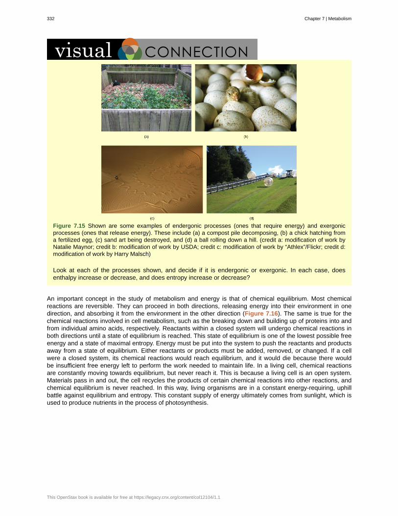
Figure 7.15 Shown are some examples of endergonic processes (ones that require energy) and exergonicprocesses (ones that release energy). These include (a) a compost pile decomposing, (b) a chick hatching froma fertilized egg, (c) sand art being destroyed, and (d) a ball rolling down a hill. (credit a: modification of work byNatalie Maynor; credit b: modification of work by USDA; credit c: modification of work by “Athlex”/Flickr; credit d:modification of work by Harry Malsch)
Look at each of the processes shown, and decide if it is endergonic or exergonic. In each case, doesenthalpy increase or decrease, and does entropy increase or decrease?
An important concept in the study of metabolism and energy is that of chemical equilibrium. Most chemicalreactions are reversible. They can proceed in both directions, releasing energy into their environment in onedirection, and absorbing it from the environment in the other direction (Figure 7.16). The same is true for thechemical reactions involved in cell metabolism, such as the breaking down and building up of proteins into andfrom individual amino acids, respectively. Reactants within a closed system will undergo chemical reactions inboth directions until a state of equilibrium is reached. This state of equilibrium is one of the lowest possible freeenergy and a state of maximal entropy. Energy must be put into the system to push the reactants and productsaway from a state of equilibrium. Either reactants or products must be added, removed, or changed. If a cellwere a closed system, its chemical reactions would reach equilibrium, and it would die because there wouldbe insufficient free energy left to perform the work needed to maintain life. In a living cell, chemical reactionsare constantly moving towards equilibrium, but never reach it. This is because a living cell is an open system.Materials pass in and out, the cell recycles the products of certain chemical reactions into other reactions, andchemical equilibrium is never reached. In this way, living organisms are in a constant energy-requiring, uphillbattle against equilibrium and entropy. This constant supply of energy ultimately comes from sunlight, which isused to produce nutrients in the process of photosynthesis.
332 Chapter 7 | Metabolism
This OpenStax book is available for free at https://legacy.cnx.org/content/col12104/1.1
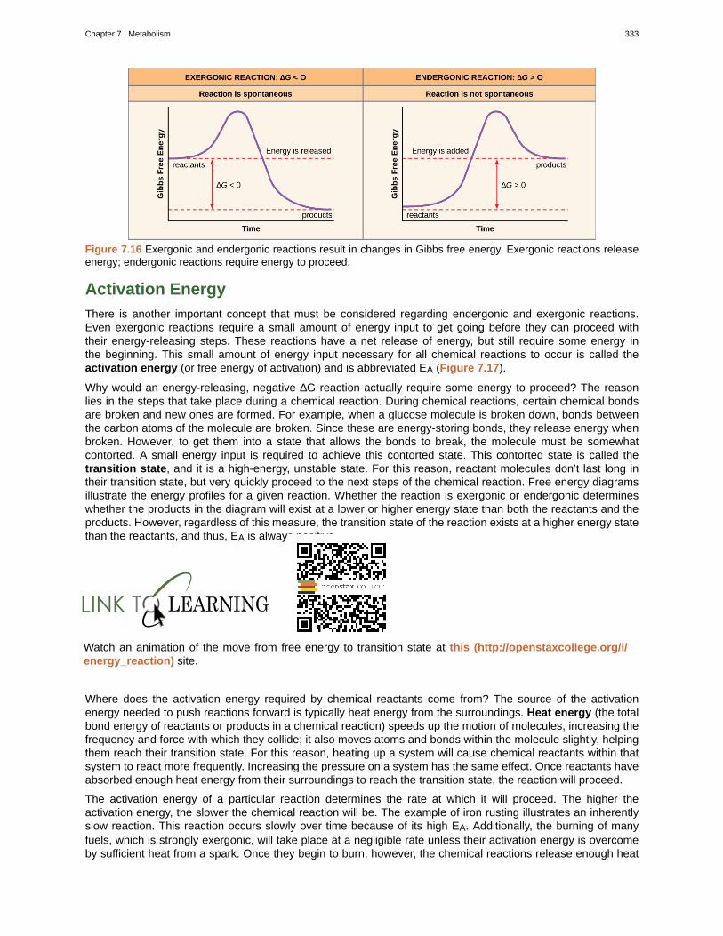
Figure 7.16 Exergonic and endergonic reactions result in changes in Gibbs free energy. Exergonic reactions releaseenergy; endergonic reactions require energy to proceed.
Activation Energy
There is another important concept that must be considered regarding endergonic and exergonic reactions.Even exergonic reactions require a small amount of energy input to get going before they can proceed withtheir energy-releasing steps. These reactions have a net release of energy, but still require some energy inthe beginning. This small amount of energy input necessary for all chemical reactions to occur is called theactivation energy (or free energy of activation) and is abbreviated EA (Figure 7.17).
Why would an energy-releasing, negative ∆G reaction actually require some energy to proceed? The reasonlies in the steps that take place during a chemical reaction. During chemical reactions, certain chemical bondsare broken and new ones are formed. For example, when a glucose molecule is broken down, bonds betweenthe carbon atoms of the molecule are broken. Since these are energy-storing bonds, they release energy whenbroken. However, to get them into a state that allows the bonds to break, the molecule must be somewhatcontorted. A small energy input is required to achieve this contorted state. This contorted state is called thetransition state, and it is a high-energy, unstable state. For this reason, reactant molecules don’t last long intheir transition state, but very quickly proceed to the next steps of the chemical reaction. Free energy diagramsillustrate the energy profiles for a given reaction. Whether the reaction is exergonic or endergonic determineswhether the products in the diagram will exist at a lower or higher energy state than both the reactants and theproducts. However, regardless of this measure, the transition state of the reaction exists at a higher energy statethan the reactants, and thus, EA is always positive.
Watch an animation of the move from free energy to transition state at this (http://openstaxcollege.org/l/energy_reaction) site.
Where does the activation energy required by chemical reactants come from? The source of the activationenergy needed to push reactions forward is typically heat energy from the surroundings. Heat energy (the totalbond energy of reactants or products in a chemical reaction) speeds up the motion of molecules, increasing thefrequency and force with which they collide; it also moves atoms and bonds within the molecule slightly, helpingthem reach their transition state. For this reason, heating up a system will cause chemical reactants within thatsystem to react more frequently. Increasing the pressure on a system has the same effect. Once reactants haveabsorbed enough heat energy from their surroundings to reach the transition state, the reaction will proceed.
The activation energy of a particular reaction determines the rate at which it will proceed. The higher theactivation energy, the slower the chemical reaction will be. The example of iron rusting illustrates an inherentlyslow reaction. This reaction occurs slowly over time because of its high EA. Additionally, the burning of manyfuels, which is strongly exergonic, will take place at a negligible rate unless their activation energy is overcomeby sufficient heat from a spark. Once they begin to burn, however, the chemical reactions release enough heat
Chapter 7 | Metabolism 333
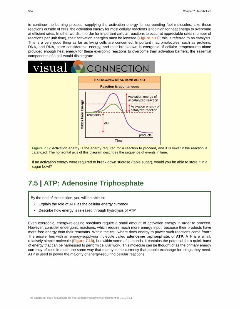
to continue the burning process, supplying the activation energy for surrounding fuel molecules. Like thesereactions outside of cells, the activation energy for most cellular reactions is too high for heat energy to overcomeat efficient rates. In other words, in order for important cellular reactions to occur at appreciable rates (number ofreactions per unit time), their activation energies must be lowered (Figure 7.17); this is referred to as catalysis.This is a very good thing as far as living cells are concerned. Important macromolecules, such as proteins,DNA, and RNA, store considerable energy, and their breakdown is exergonic. If cellular temperatures aloneprovided enough heat energy for these exergonic reactions to overcome their activation barriers, the essentialcomponents of a cell would disintegrate.
Figure 7.17 Activation energy is the energy required for a reaction to proceed, and it is lower if the reaction iscatalyzed. The horizontal axis of this diagram describes the sequence of events in time.
If no activation energy were required to break down sucrose (table sugar), would you be able to store it in asugar bowl?
7.5 | ATP: Adenosine Triphosphate
By the end of this section, you will be able to:
• Explain the role of ATP as the cellular energy currency
• Describe how energy is released through hydrolysis of ATP
Even exergonic, energy-releasing reactions require a small amount of activation energy in order to proceed.However, consider endergonic reactions, which require much more energy input, because their products havemore free energy than their reactants. Within the cell, where does energy to power such reactions come from?The answer lies with an energy-supplying molecule called adenosine triphosphate, or ATP. ATP is a small,relatively simple molecule (Figure 7.18), but within some of its bonds, it contains the potential for a quick burstof energy that can be harnessed to perform cellular work. This molecule can be thought of as the primary energycurrency of cells in much the same way that money is the currency that people exchange for things they need.ATP is used to power the majority of energy-requiring cellular reactions.
334 Chapter 7 | Metabolism
This OpenStax book is available for free at https://legacy.cnx.org/content/col12104/1.1
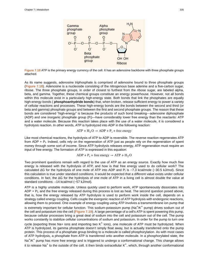
Figure 7.18 ATP is the primary energy currency of the cell. It has an adenosine backbone with three phosphate groupsattached.
As its name suggests, adenosine triphosphate is comprised of adenosine bound to three phosphate groups(Figure 7.18). Adenosine is a nucleoside consisting of the nitrogenous base adenine and a five-carbon sugar,ribose. The three phosphate groups, in order of closest to furthest from the ribose sugar, are labeled alpha,beta, and gamma. Together, these chemical groups constitute an energy powerhouse. However, not all bondswithin this molecule exist in a particularly high-energy state. Both bonds that link the phosphates are equallyhigh-energy bonds ( phosphoanhydride bonds) that, when broken, release sufficient energy to power a varietyof cellular reactions and processes. These high-energy bonds are the bonds between the second and third (orbeta and gamma) phosphate groups and between the first and second phosphate groups. The reason that thesebonds are considered “high-energy” is because the products of such bond breaking—adenosine diphosphate(ADP) and one inorganic phosphate group (Pi)—have considerably lower free energy than the reactants: ATPand a water molecule. Because this reaction takes place with the use of a water molecule, it is considered ahydrolysis reaction. In other words, ATP is hydrolyzed into ADP in the following reaction:
ATP + H2 O → ADP + Pi + free energy
Like most chemical reactions, the hydrolysis of ATP to ADP is reversible. The reverse reaction regenerates ATPfrom ADP + Pi. Indeed, cells rely on the regeneration of ATP just as people rely on the regeneration of spentmoney through some sort of income. Since ATP hydrolysis releases energy, ATP regeneration must require aninput of free energy. The formation of ATP is expressed in this equation:
ADP + Pi + free energy → ATP + H2 O
Two prominent questions remain with regard to the use of ATP as an energy source. Exactly how much freeenergy is released with the hydrolysis of ATP, and how is that free energy used to do cellular work? Thecalculated ∆G for the hydrolysis of one mole of ATP into ADP and Pi is −7.3 kcal/mole (−30.5 kJ/mol). Sincethis calculation is true under standard conditions, it would be expected that a different value exists under cellularconditions. In fact, the ∆G for the hydrolysis of one mole of ATP in a living cell is almost double the value atstandard conditions: –14 kcal/mol (−57 kJ/mol).
ATP is a highly unstable molecule. Unless quickly used to perform work, ATP spontaneously dissociates intoADP + Pi, and the free energy released during this process is lost as heat. The second question posed above,that is, how the energy released by ATP hydrolysis is used to perform work inside the cell, depends on astrategy called energy coupling. Cells couple the exergonic reaction of ATP hydrolysis with endergonic reactions,allowing them to proceed. One example of energy coupling using ATP involves a transmembrane ion pump that
is extremely important for cellular function. This sodium-potassium pump (Na+/K+ pump) drives sodium out ofthe cell and potassium into the cell (Figure 7.19). A large percentage of a cell’s ATP is spent powering this pump,because cellular processes bring a great deal of sodium into the cell and potassium out of the cell. The pumpworks constantly to stabilize cellular concentrations of sodium and potassium. In order for the pump to turn one
cycle (exporting three Na+ ions and importing two K+ ions), one molecule of ATP must be hydrolyzed. WhenATP is hydrolyzed, its gamma phosphate doesn’t simply float away, but is actually transferred onto the pumpprotein. This process of a phosphate group binding to a molecule is called phosphorylation. As with most casesof ATP hydrolysis, a phosphate from ATP is transferred onto another molecule. In a phosphorylated state, the
Na+/K+ pump has more free energy and is triggered to undergo a conformational change. This change allows
it to release Na+ to the outside of the cell. It then binds extracellular K+, which, through another conformational
Chapter 7 | Metabolism 335
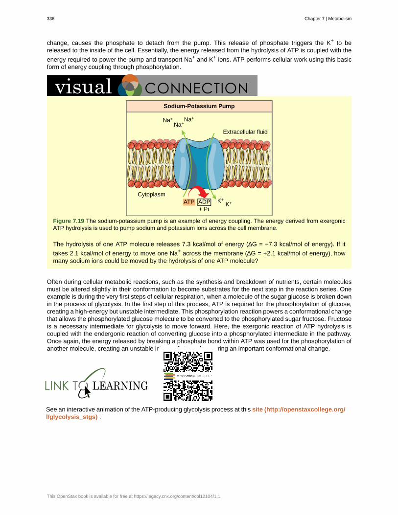
change, causes the phosphate to detach from the pump. This release of phosphate triggers the K+ to bereleased to the inside of the cell. Essentially, the energy released from the hydrolysis of ATP is coupled with the
energy required to power the pump and transport Na+ and K+ ions. ATP performs cellular work using this basicform of energy coupling through phosphorylation.
Figure 7.19 The sodium-potassium pump is an example of energy coupling. The energy derived from exergonicATP hydrolysis is used to pump sodium and potassium ions across the cell membrane.
The hydrolysis of one ATP molecule releases 7.3 kcal/mol of energy (∆G = −7.3 kcal/mol of energy). If it
takes 2.1 kcal/mol of energy to move one Na+ across the membrane (∆G = +2.1 kcal/mol of energy), howmany sodium ions could be moved by the hydrolysis of one ATP molecule?
Often during cellular metabolic reactions, such as the synthesis and breakdown of nutrients, certain moleculesmust be altered slightly in their conformation to become substrates for the next step in the reaction series. Oneexample is during the very first steps of cellular respiration, when a molecule of the sugar glucose is broken downin the process of glycolysis. In the first step of this process, ATP is required for the phosphorylation of glucose,creating a high-energy but unstable intermediate. This phosphorylation reaction powers a conformational changethat allows the phosphorylated glucose molecule to be converted to the phosphorylated sugar fructose. Fructoseis a necessary intermediate for glycolysis to move forward. Here, the exergonic reaction of ATP hydrolysis iscoupled with the endergonic reaction of converting glucose into a phosphorylated intermediate in the pathway.Once again, the energy released by breaking a phosphate bond within ATP was used for the phosphorylation ofanother molecule, creating an unstable intermediate and powering an important conformational change.
See an interactive animation of the ATP-producing glycolysis process at this site (http://openstaxcollege.org/l/glycolysis_stgs) .
336 Chapter 7 | Metabolism
This OpenStax book is available for free at https://legacy.cnx.org/content/col12104/1.1
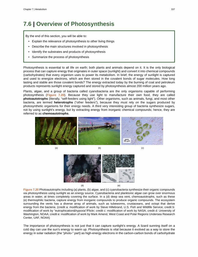
7.6 | Overview of Photosynthesis
By the end of this section, you will be able to:
• Explain the relevance of photosynthesis to other living things
• Describe the main structures involved in photosynthesis
• Identify the substrates and products of photosynthesis
• Summarize the process of photosynthesis
Photosynthesis is essential to all life on earth; both plants and animals depend on it. It is the only biologicalprocess that can capture energy that originates in outer space (sunlight) and convert it into chemical compounds(carbohydrates) that every organism uses to power its metabolism. In brief, the energy of sunlight is capturedand used to energize electrons, which are then stored in the covalent bonds of sugar molecules. How longlasting and stable are those covalent bonds? The energy extracted today by the burning of coal and petroleumproducts represents sunlight energy captured and stored by photosynthesis almost 200 million years ago.
Plants, algae, and a group of bacteria called cyanobacteria are the only organisms capable of performingphotosynthesis (Figure 7.20). Because they use light to manufacture their own food, they are calledphotoautotrophs (literally, “self-feeders using light”). Other organisms, such as animals, fungi, and most otherbacteria, are termed heterotrophs (“other feeders”), because they must rely on the sugars produced byphotosynthetic organisms for their energy needs. A third very interesting group of bacteria synthesize sugars,not by using sunlight’s energy, but by extracting energy from inorganic chemical compounds; hence, they arereferred to as chemoautotrophs.
Figure 7.20 Photoautotrophs including (a) plants, (b) algae, and (c) cyanobacteria synthesize their organic compoundsvia photosynthesis using sunlight as an energy source. Cyanobacteria and planktonic algae can grow over enormousareas in water, at times completely covering the surface. In a (d) deep sea vent, chemoautotrophs, such as these(e) thermophilic bacteria, capture energy from inorganic compounds to produce organic compounds. The ecosystemsurrounding the vents has a diverse array of animals, such as tubeworms, crustaceans, and octopi that deriveenergy from the bacteria. (credit a: modification of work by Steve Hillebrand, U.S. Fish and Wildlife Service; credit b:modification of work by "eutrophication&hypoxia"/Flickr; credit c: modification of work by NASA; credit d: University ofWashington, NOAA; credit e: modification of work by Mark Amend, West Coast and Polar Regions Undersea ResearchCenter, UAF, NOAA)
The importance of photosynthesis is not just that it can capture sunlight’s energy. A lizard sunning itself on acold day can use the sun’s energy to warm up. Photosynthesis is vital because it evolved as a way to store theenergy in solar radiation (the “photo-” part) as high-energy electrons in the carbon-carbon bonds of carbohydrate
Chapter 7 | Metabolism 337

molecules (the “-synthesis” part). Those carbohydrates are the energy source that heterotrophs use to powerthe synthesis of ATP via respiration. Therefore, photosynthesis powers 99 percent of Earth’s ecosystems. Whena top predator, such as a wolf, preys on a deer (Figure 7.21), the wolf is at the end of an energy path that wentfrom nuclear reactions on the surface of the sun, to light, to photosynthesis, to vegetation, to deer, and finally towolf.
Figure 7.21 The energy stored in carbohydrate molecules from photosynthesis passes through the food chain. Thepredator that eats these deer receives a portion of the energy that originated in the photosynthetic vegetation that thedeer consumed. (credit: modification of work by Steve VanRiper, U.S. Fish and Wildlife Service)
Main Structures and Summary of Photosynthesis
Photosynthesis is a multi-step process that requires sunlight, carbon dioxide (which is low in energy), andwater as substrates (Figure 7.22). After the process is complete, it releases oxygen and producesglyceraldehyde-3-phosphate (GA3P), simple carbohydrate molecules (which are high in energy) that cansubsequently be converted into glucose, sucrose, or any of dozens of other sugar molecules. These sugarmolecules contain energy and the energized carbon that all living things need to survive.
Figure 7.22 Photosynthesis uses solar energy, carbon dioxide, and water to produce energy-storing carbohydrates.Oxygen is generated as a waste product of photosynthesis.
The following is the chemical equation for photosynthesis (Figure 7.23):
338 Chapter 7 | Metabolism
This OpenStax book is available for free at https://legacy.cnx.org/content/col12104/1.1
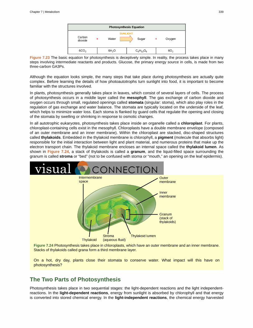
Figure 7.23 The basic equation for photosynthesis is deceptively simple. In reality, the process takes place in manysteps involving intermediate reactants and products. Glucose, the primary energy source in cells, is made from twothree-carbon GA3Ps.
Although the equation looks simple, the many steps that take place during photosynthesis are actually quitecomplex. Before learning the details of how photoautotrophs turn sunlight into food, it is important to becomefamiliar with the structures involved.
In plants, photosynthesis generally takes place in leaves, which consist of several layers of cells. The processof photosynthesis occurs in a middle layer called the mesophyll. The gas exchange of carbon dioxide andoxygen occurs through small, regulated openings called stomata (singular: stoma), which also play roles in theregulation of gas exchange and water balance. The stomata are typically located on the underside of the leaf,which helps to minimize water loss. Each stoma is flanked by guard cells that regulate the opening and closingof the stomata by swelling or shrinking in response to osmotic changes.
In all autotrophic eukaryotes, photosynthesis takes place inside an organelle called a chloroplast. For plants,chloroplast-containing cells exist in the mesophyll. Chloroplasts have a double membrane envelope (composedof an outer membrane and an inner membrane). Within the chloroplast are stacked, disc-shaped structurescalled thylakoids. Embedded in the thylakoid membrane is chlorophyll, a pigment (molecule that absorbs light)responsible for the initial interaction between light and plant material, and numerous proteins that make up theelectron transport chain. The thylakoid membrane encloses an internal space called the thylakoid lumen. Asshown in Figure 7.24, a stack of thylakoids is called a granum, and the liquid-filled space surrounding thegranum is called stroma or “bed” (not to be confused with stoma or “mouth,” an opening on the leaf epidermis).
Figure 7.24 Photosynthesis takes place in chloroplasts, which have an outer membrane and an inner membrane.Stacks of thylakoids called grana form a third membrane layer.
On a hot, dry day, plants close their stomata to conserve water. What impact will this have onphotosynthesis?
The Two Parts of Photosynthesis
Photosynthesis takes place in two sequential stages: the light-dependent reactions and the light independent-reactions. In the light-dependent reactions, energy from sunlight is absorbed by chlorophyll and that energyis converted into stored chemical energy. In the light-independent reactions, the chemical energy harvested
Chapter 7 | Metabolism 339
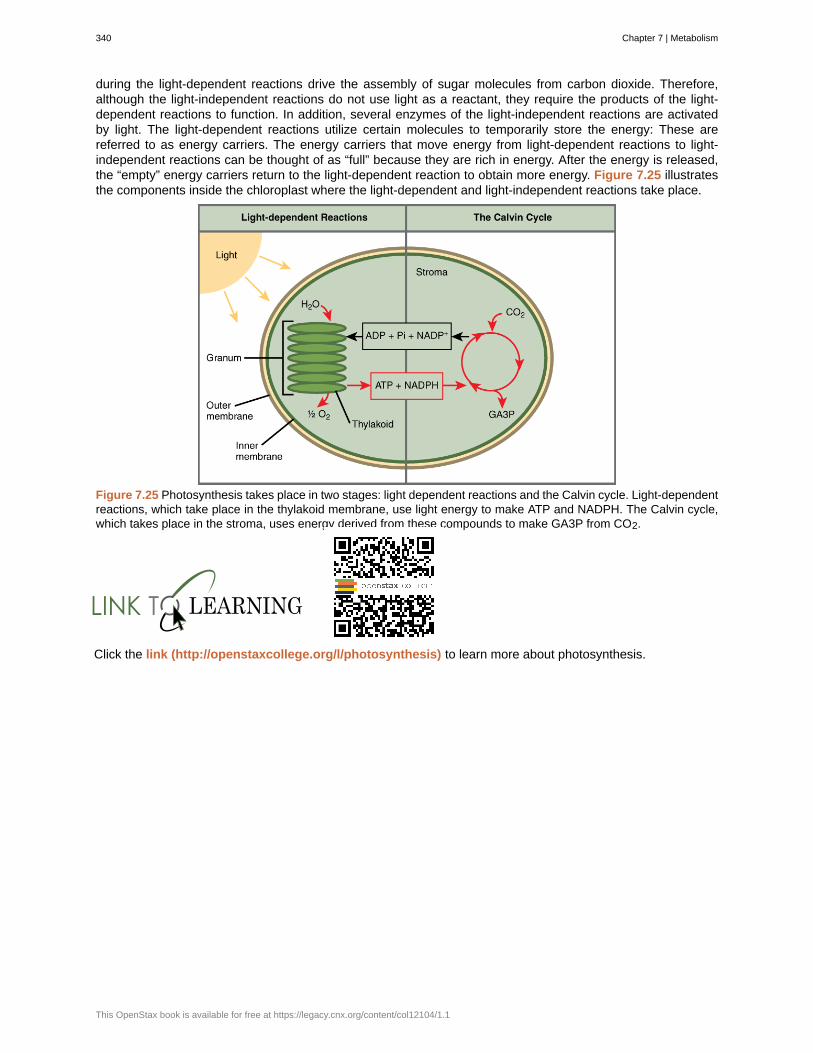
during the light-dependent reactions drive the assembly of sugar molecules from carbon dioxide. Therefore,although the light-independent reactions do not use light as a reactant, they require the products of the light-dependent reactions to function. In addition, several enzymes of the light-independent reactions are activatedby light. The light-dependent reactions utilize certain molecules to temporarily store the energy: These arereferred to as energy carriers. The energy carriers that move energy from light-dependent reactions to light-independent reactions can be thought of as “full” because they are rich in energy. After the energy is released,the “empty” energy carriers return to the light-dependent reaction to obtain more energy. Figure 7.25 illustratesthe components inside the chloroplast where the light-dependent and light-independent reactions take place.
Figure 7.25 Photosynthesis takes place in two stages: light dependent reactions and the Calvin cycle. Light-dependentreactions, which take place in the thylakoid membrane, use light energy to make ATP and NADPH. The Calvin cycle,which takes place in the stroma, uses energy derived from these compounds to make GA3P from CO2.
Click the link (http://openstaxcollege.org/l/photosynthesis) to learn more about photosynthesis.
340 Chapter 7 | Metabolism
This OpenStax book is available for free at https://legacy.cnx.org/content/col12104/1.1

Photosynthesis at the Grocery Store
Figure 7.26 Foods that humans consume originate from photosynthesis. (credit: Associação Brasileira deSupermercados)
Major grocery stores in the United States are organized into departments, such as dairy, meats, produce,bread, cereals, and so forth. Each aisle (Figure 7.26) contains hundreds, if not thousands, of differentproducts for customers to buy and consume.
Although there is a large variety, each item links back to photosynthesis. Meats and dairy link, because theanimals were fed plant-based foods. The breads, cereals, and pastas come largely from starchy grains,which are the seeds of photosynthesis-dependent plants. What about desserts and drinks? All of theseproducts contain sugar—sucrose is a plant product, a disaccharide, a carbohydrate molecule, which is builtdirectly from photosynthesis. Moreover, many items are less obviously derived from plants: For instance,paper goods are generally plant products, and many plastics (abundant as products and packaging) arederived from algae. Virtually every spice and flavoring in the spice aisle was produced by a plant as a leaf,root, bark, flower, fruit, or stem. Ultimately, photosynthesis connects to every meal and every food a personconsumes.
7.7 | Collision Theory
By the end of this section, you will be able to:
• Use the postulates of collision theory to explain the effects of physical state, temperature, andconcentration on reaction rates
• Define the concepts of activation energy and transition state
• Use the Arrhenius equation in calculations relating rate constants to temperature
We should not be surprised that atoms, molecules, or ions must collide before they can react with each other.Atoms must be close together to form chemical bonds. This simple premise is the basis for a very powerfultheory that explains many observations regarding chemical kinetics, including factors affecting reaction rates.
Collision theory is based on the following postulates:
1. The rate of a reaction is proportional to the rate of reactant collisions:
reaction rate ∝ # collisionstime
2. The reacting species must collide in an orientation that allows contact between the atoms that will becomebonded together in the product.
Chapter 7 | Metabolism 341
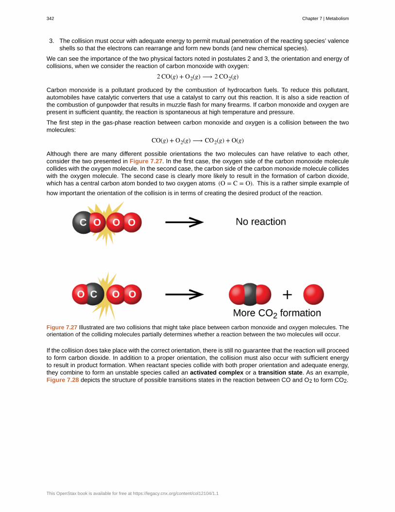
3. The collision must occur with adequate energy to permit mutual penetration of the reacting species’ valenceshells so that the electrons can rearrange and form new bonds (and new chemical species).
We can see the importance of the two physical factors noted in postulates 2 and 3, the orientation and energy ofcollisions, when we consider the reaction of carbon monoxide with oxygen:
2 CO(g) + O2(g) ⟶ 2 CO2(g)
Carbon monoxide is a pollutant produced by the combustion of hydrocarbon fuels. To reduce this pollutant,automobiles have catalytic converters that use a catalyst to carry out this reaction. It is also a side reaction ofthe combustion of gunpowder that results in muzzle flash for many firearms. If carbon monoxide and oxygen arepresent in sufficient quantity, the reaction is spontaneous at high temperature and pressure.
The first step in the gas-phase reaction between carbon monoxide and oxygen is a collision between the twomolecules:
CO(g) + O2(g) ⟶ CO2(g) + O(g)
Although there are many different possible orientations the two molecules can have relative to each other,consider the two presented in Figure 7.27. In the first case, the oxygen side of the carbon monoxide moleculecollides with the oxygen molecule. In the second case, the carbon side of the carbon monoxide molecule collideswith the oxygen molecule. The second case is clearly more likely to result in the formation of carbon dioxide,which has a central carbon atom bonded to two oxygen atoms (O = C = O). This is a rather simple example of
how important the orientation of the collision is in terms of creating the desired product of the reaction.
Figure 7.27 Illustrated are two collisions that might take place between carbon monoxide and oxygen molecules. Theorientation of the colliding molecules partially determines whether a reaction between the two molecules will occur.
If the collision does take place with the correct orientation, there is still no guarantee that the reaction will proceedto form carbon dioxide. In addition to a proper orientation, the collision must also occur with sufficient energyto result in product formation. When reactant species collide with both proper orientation and adequate energy,they combine to form an unstable species called an activated complex or a transition state. As an example,Figure 7.28 depicts the structure of possible transitions states in the reaction between CO and O2 to form CO2.
342 Chapter 7 | Metabolism
This OpenStax book is available for free at https://legacy.cnx.org/content/col12104/1.1
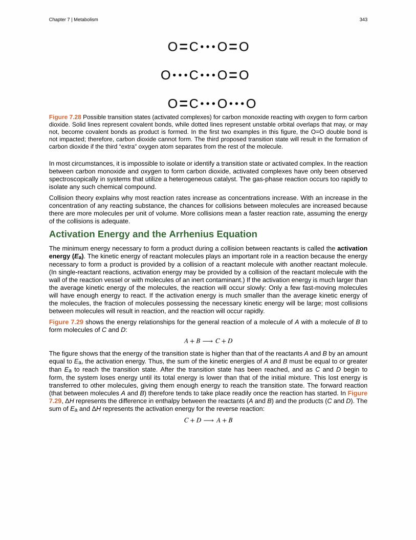
Figure 7.28 Possible transition states (activated complexes) for carbon monoxide reacting with oxygen to form carbondioxide. Solid lines represent covalent bonds, while dotted lines represent unstable orbital overlaps that may, or maynot, become covalent bonds as product is formed. In the first two examples in this figure, the O=O double bond isnot impacted; therefore, carbon dioxide cannot form. The third proposed transition state will result in the formation ofcarbon dioxide if the third “extra” oxygen atom separates from the rest of the molecule.
In most circumstances, it is impossible to isolate or identify a transition state or activated complex. In the reactionbetween carbon monoxide and oxygen to form carbon dioxide, activated complexes have only been observedspectroscopically in systems that utilize a heterogeneous catalyst. The gas-phase reaction occurs too rapidly toisolate any such chemical compound.
Collision theory explains why most reaction rates increase as concentrations increase. With an increase in theconcentration of any reacting substance, the chances for collisions between molecules are increased becausethere are more molecules per unit of volume. More collisions mean a faster reaction rate, assuming the energyof the collisions is adequate.
Activation Energy and the Arrhenius Equation
The minimum energy necessary to form a product during a collision between reactants is called the activationenergy (Ea). The kinetic energy of reactant molecules plays an important role in a reaction because the energynecessary to form a product is provided by a collision of a reactant molecule with another reactant molecule.(In single-reactant reactions, activation energy may be provided by a collision of the reactant molecule with thewall of the reaction vessel or with molecules of an inert contaminant.) If the activation energy is much larger thanthe average kinetic energy of the molecules, the reaction will occur slowly: Only a few fast-moving moleculeswill have enough energy to react. If the activation energy is much smaller than the average kinetic energy ofthe molecules, the fraction of molecules possessing the necessary kinetic energy will be large; most collisionsbetween molecules will result in reaction, and the reaction will occur rapidly.
Figure 7.29 shows the energy relationships for the general reaction of a molecule of A with a molecule of B toform molecules of C and D:
A + B ⟶ C + D
The figure shows that the energy of the transition state is higher than that of the reactants A and B by an amountequal to Ea, the activation energy. Thus, the sum of the kinetic energies of A and B must be equal to or greaterthan Ea to reach the transition state. After the transition state has been reached, and as C and D begin toform, the system loses energy until its total energy is lower than that of the initial mixture. This lost energy istransferred to other molecules, giving them enough energy to reach the transition state. The forward reaction(that between molecules A and B) therefore tends to take place readily once the reaction has started. In Figure7.29, ΔH represents the difference in enthalpy between the reactants (A and B) and the products (C and D). Thesum of Ea and ΔH represents the activation energy for the reverse reaction:
C + D ⟶ A + B
Chapter 7 | Metabolism 343
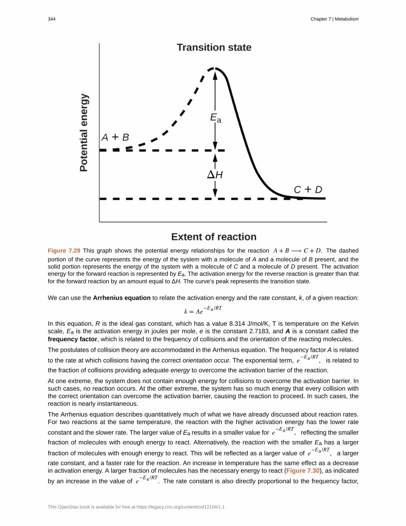
Figure 7.29 This graph shows the potential energy relationships for the reaction A + B ⟶ C + D. The dashed
portion of the curve represents the energy of the system with a molecule of A and a molecule of B present, and thesolid portion represents the energy of the system with a molecule of C and a molecule of D present. The activationenergy for the forward reaction is represented by Ea. The activation energy for the reverse reaction is greater than thatfor the forward reaction by an amount equal to ΔH. The curve’s peak represents the transition state.
We can use the Arrhenius equation to relate the activation energy and the rate constant, k, of a given reaction:
k = Ae−Ea /RT
In this equation, R is the ideal gas constant, which has a value 8.314 J/mol/K, T is temperature on the Kelvinscale, Ea is the activation energy in joules per mole, e is the constant 2.7183, and A is a constant called thefrequency factor, which is related to the frequency of collisions and the orientation of the reacting molecules.
The postulates of collision theory are accommodated in the Arrhenius equation. The frequency factor A is related
to the rate at which collisions having the correct orientation occur. The exponential term, e−Ea /RT, is related to
the fraction of collisions providing adequate energy to overcome the activation barrier of the reaction.
At one extreme, the system does not contain enough energy for collisions to overcome the activation barrier. Insuch cases, no reaction occurs. At the other extreme, the system has so much energy that every collision withthe correct orientation can overcome the activation barrier, causing the reaction to proceed. In such cases, thereaction is nearly instantaneous.
The Arrhenius equation describes quantitatively much of what we have already discussed about reaction rates.For two reactions at the same temperature, the reaction with the higher activation energy has the lower rate
constant and the slower rate. The larger value of Ea results in a smaller value for e−Ea /RT, reflecting the smaller
fraction of molecules with enough energy to react. Alternatively, the reaction with the smaller Ea has a larger
fraction of molecules with enough energy to react. This will be reflected as a larger value of e−Ea /RT, a larger
rate constant, and a faster rate for the reaction. An increase in temperature has the same effect as a decreasein activation energy. A larger fraction of molecules has the necessary energy to react (Figure 7.30), as indicated
by an increase in the value of e−Ea /RT. The rate constant is also directly proportional to the frequency factor,
344 Chapter 7 | Metabolism
This OpenStax book is available for free at https://legacy.cnx.org/content/col12104/1.1
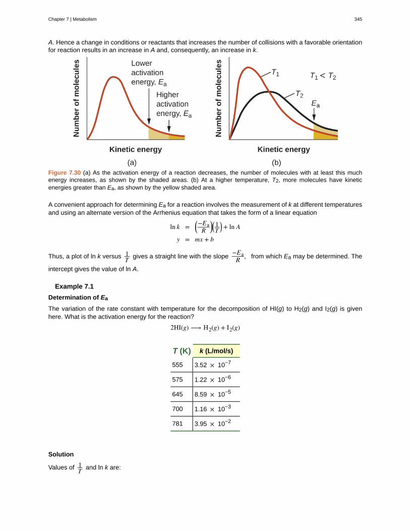
A. Hence a change in conditions or reactants that increases the number of collisions with a favorable orientationfor reaction results in an increase in A and, consequently, an increase in k.
Figure 7.30 (a) As the activation energy of a reaction decreases, the number of molecules with at least this muchenergy increases, as shown by the shaded areas. (b) At a higher temperature, T2, more molecules have kineticenergies greater than Ea, as shown by the yellow shaded area.
A convenient approach for determining Ea for a reaction involves the measurement of k at different temperaturesand using an alternate version of the Arrhenius equation that takes the form of a linear equation
ln k = ⎛⎝−EaR⎞⎠⎛⎝1T⎞⎠+ ln A
y = mx + b
Thus, a plot of ln k versus 1T gives a straight line with the slope
−EaR , from which Ea may be determined. The
intercept gives the value of ln A.
Example 7.1
Determination of Ea
The variation of the rate constant with temperature for the decomposition of HI(g) to H2(g) and I2(g) is givenhere. What is the activation energy for the reaction?
2HI(g) ⟶ H2(g) + I2(g)
T (K) k (L/mol/s)
555 3.52 × 10⁻⁷
575 1.22 × 10⁻⁶
645 8.59 × 10⁻⁵
700 1.16 × 10⁻³
781 3.95 × 10⁻²
Solution
Values of 1T and ln k are:
Chapter 7 | Metabolism 345
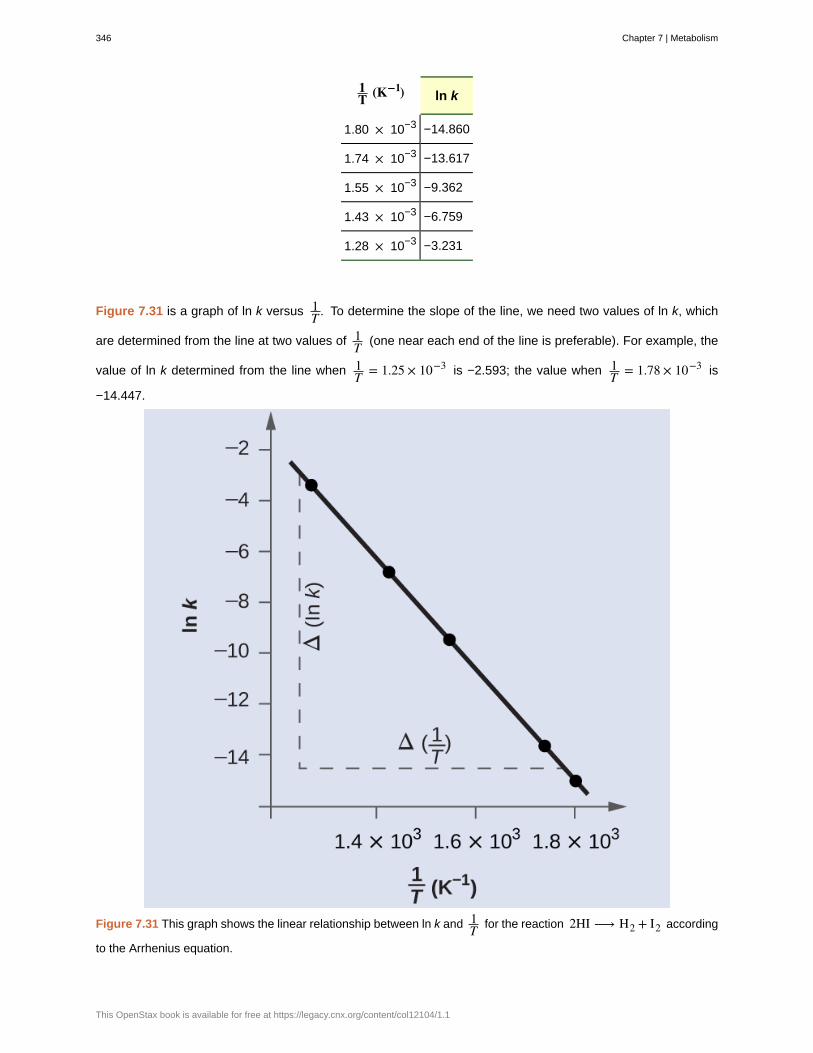
1T (K−1) ln k
1.80 × 10⁻³ −14.860
1.74 × 10⁻³ −13.617
1.55 × 10⁻³ −9.362
1.43 × 10⁻³ −6.759
1.28 × 10⁻³ −3.231
Figure 7.31 is a graph of ln k versus 1T . To determine the slope of the line, we need two values of ln k, which
are determined from the line at two values of 1T (one near each end of the line is preferable). For example, the
value of ln k determined from the line when 1T = 1.25 × 10⁻³ is −2.593; the value when 1
T = 1.78 × 10⁻³ is
−14.447.
Figure 7.31 This graph shows the linear relationship between ln k and 1T for the reaction 2HI ⟶ H2 + I2 according
to the Arrhenius equation.
346 Chapter 7 | Metabolism
This OpenStax book is available for free at https://legacy.cnx.org/content/col12104/1.1
The slope of this line is given by the following expression:
Slope = Δ(ln k)Δ⎛⎝1T⎞⎠
= (−14.447) − (−2.593)⎛⎝1.78 × 10⁻³ K−1⎞⎠ − ⎛⎝1.25 × 10⁻³ K−1⎞⎠
= −11.8540.53 × 10−3 K−1 = 2.2 × 10⁴ K
= − EaR
Thus:
Ea = −slope × R = − (−2.2 × 104 K × 8.314 J mol−1 K−1)
Ea = 1.8 × 10⁵ J mol
In many situations, it is possible to obtain a reasonable estimate of the activation energy without going throughthe entire process of constructing the Arrhenius plot. The Arrhenius equation:
ln k = ⎛⎝−EaR⎞⎠ ⎛⎝1T⎞⎠+ ln A
can be rearranged as shown to give:
Δ(ln k)Δ⎛⎝1T⎞⎠
= − EaR
or
ln k1k2
= EaR⎛⎝ 1T2
− 1T1
⎞⎠
This equation can be rearranged to give a one-step calculation to obtain an estimate for the activation energy:
Ea = −R⎛
⎝⎜⎜ln k2 − ln k1⎛⎝ 1T2⎞⎠ − ⎛⎝ 1
T1⎞⎠
⎞
⎠⎟⎟
Using the experimental data presented here, we can simply select two data entries. For this example, we selectthe first entry and the last entry:
T (K) k (L/mol/s)1T (K−1) ln k
555 3.52 × 10⁻⁷ 1.80 × 10⁻³ −14.860
781 3.95 × 10⁻² 1.28 × 10⁻³ −3.231
After calculating 1T and ln k, we can substitute into the equation:
Ea = −8.314 J mol−1 K−1⎛⎝ −3.231 − (−14.860)1.28 × 10⁻³ K−1 − 1.80 × 10⁻³ K−1
⎞⎠
and the result is Ea = 185,900 J/mol.
This method is very effective, especially when a limited number of temperature-dependent rate constants areavailable for the reaction of interest.
Check Your Learning
Chapter 7 | Metabolism 347

The rate constant for the rate of decomposition of N2O5 to NO and O2 in the gas phase is 1.66 L/mol/s at 650 Kand 7.39 L/mol/s at 700 K:
2N2 O5(g) ⟶ 4NO(g) + 3O2(g)
Assuming the kinetics of this reaction are consistent with the Arrhenius equation, calculate the activation energyfor this decomposition.
Answer:
113,000 J/mol
Key Equations
• k = Ae−Ea /RT
• ln k = ⎛⎝−EaR⎞⎠ ⎛⎝1T⎞⎠+ ln A
• ln k1k2
= EaR⎛⎝ 1T2
− 1T1
⎞⎠
Chemistry End of Chapter Exercises
Exercise 7.1
Chemical reactions occur when reactants collide. What are two factors that may prevent a collision fromproducing a chemical reaction?
SolutionThe reactants either may be moving too slowly to have enough kinetic energy to exceed the activation energyfor the reaction, or the orientation of the molecules when they collide may prevent the reaction from occurring.
Exercise 7.2
When every collision between reactants leads to a reaction, what determines the rate at which the reactionoccurs?
Exercise 7.3
What is the activation energy of a reaction, and how is this energy related to the activated complex of thereaction?
SolutionThe activation energy is the minimum amount of energy necessary to form the activated complex in a reaction.It is usually expressed as the energy necessary to form one mole of activated complex.
Exercise 7.4
Account for the relationship between the rate of a reaction and its activation energy.
Exercise 7.5
Describe how graphical methods can be used to determine the activation energy of a reaction from a series ofdata that includes the rate of reaction at varying temperatures.
Solution
After finding k at several different temperatures, a plot of ln k versus 1T , gives a straight line with the slope
−EaR
from which Ea may be determined.
Exercise 7.6
348 Chapter 7 | Metabolism
This OpenStax book is available for free at https://legacy.cnx.org/content/col12104/1.1
How does an increase in temperature affect rate of reaction? Explain this effect in terms of the collision theory ofthe reaction rate.
Exercise 7.7
The rate of a certain reaction doubles for every 10 °C rise in temperature.
(a) How much faster does the reaction proceed at 45 °C than at 25 °C?
(b) How much faster does the reaction proceed at 95 °C than at 25 °C?
Solution(a) 4-times faster (b) 128-times faster
Exercise 7.8
In an experiment, a sample of NaClO3 was 90% decomposed in 48 min. Approximately how long would thisdecomposition have taken if the sample had been heated 20 °C higher? (Hint: Assume the rate doubles for each10 °C rise in temperature.)
Exercise 7.9
The rate constant at 325 °C for the decomposition reaction C4 H8 ⟶ 2C2 H4 is 6.1 × 10⁻⁸ s−1, and the
activation energy is 261 kJ per mole of C4H8. Determine the frequency factor for the reaction.
Solution
3.9 × 10¹⁵ s−1
Exercise 7.10
The rate constant for the decomposition of acetaldehyde, CH3CHO, to methane, CH4, and carbon monoxide,
CO, in the gas phase is 1.1 × 10⁻² L/mol/s at 703 K and 4.95 L/mol/s at 865 K. Determine the activation energy
for this decomposition.
Exercise 7.11
An elevated level of the enzyme alkaline phosphatase (ALP) in human serum is an indication of possibleliver or bone disorder. The level of serum ALP is so low that it is very difficult to measure directly. However,ALP catalyzes a number of reactions, and its relative concentration can be determined by measuring the rateof one of these reactions under controlled conditions. One such reaction is the conversion of p-nitrophenylphosphate (PNPP) to p-nitrophenoxide ion (PNP) and phosphate ion. Control of temperature during the test isvery important; the rate of the reaction increases 1.47 times if the temperature changes from 30 °C to 37 °C.What is the activation energy for the ALP–catalyzed conversion of PNPP to PNP and phosphate?
Solution43.0 kJ/mol
Exercise 7.12
In terms of collision theory, to which of the following is the rate of a chemical reaction proportional?
(a) the change in free energy per second
(b) the change in temperature per second
(c) the number of collisions per second
(d) the number of product molecules
Exercise 7.13
Hydrogen iodide, HI, decomposes in the gas phase to produce hydrogen, H2, and iodine, I2. The value of therate constant, k, for the reaction was measured at several different temperatures and the data are shown here:
Chapter 7 | Metabolism 349
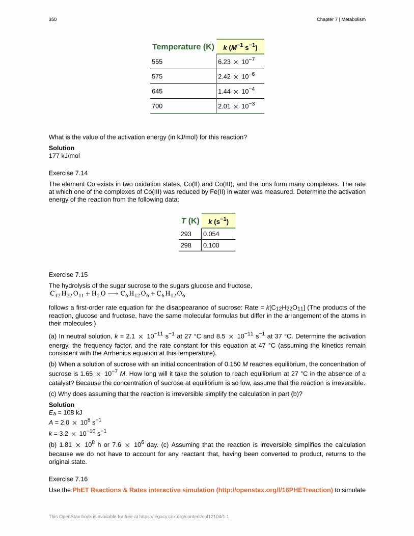
Temperature (K) k (M−1 s−1)
555 6.23 × 10⁻⁷
575 2.42 × 10⁻⁶
645 1.44 × 10⁻⁴
700 2.01 × 10⁻³
What is the value of the activation energy (in kJ/mol) for this reaction?
Solution177 kJ/mol
Exercise 7.14
The element Co exists in two oxidation states, Co(II) and Co(III), and the ions form many complexes. The rateat which one of the complexes of Co(III) was reduced by Fe(II) in water was measured. Determine the activationenergy of the reaction from the following data:
T (K) k (s−1)
293 0.054
298 0.100
Exercise 7.15
The hydrolysis of the sugar sucrose to the sugars glucose and fructose,C12 H22 O11 + H2 O ⟶ C6 H12 O6 + C6 H12 O6
follows a first-order rate equation for the disappearance of sucrose: Rate = k[C12H22O11] (The products of thereaction, glucose and fructose, have the same molecular formulas but differ in the arrangement of the atoms intheir molecules.)
(a) In neutral solution, k = 2.1 × 10⁻¹¹ s−1 at 27 °C and 8.5 × 10⁻¹¹ s−1 at 37 °C. Determine the activation
energy, the frequency factor, and the rate constant for this equation at 47 °C (assuming the kinetics remainconsistent with the Arrhenius equation at this temperature).
(b) When a solution of sucrose with an initial concentration of 0.150 M reaches equilibrium, the concentration of
sucrose is 1.65 × 10⁻⁷ M. How long will it take the solution to reach equilibrium at 27 °C in the absence of a
catalyst? Because the concentration of sucrose at equilibrium is so low, assume that the reaction is irreversible.
(c) Why does assuming that the reaction is irreversible simplify the calculation in part (b)?
SolutionEa = 108 kJ
A = 2.0 × 10⁸ s−1
k = 3.2 × 10⁻¹⁰ s−1
(b) 1.81 × 10⁸ h or 7.6 × 10⁶ day. (c) Assuming that the reaction is irreversible simplifies the calculation
because we do not have to account for any reactant that, having been converted to product, returns to theoriginal state.
Exercise 7.16
Use the PhET Reactions & Rates interactive simulation (http://openstax.org/l/16PHETreaction) to simulate
350 Chapter 7 | Metabolism
This OpenStax book is available for free at https://legacy.cnx.org/content/col12104/1.1
a system. On the “Single collision” tab of the simulation applet, enable the “Energy view” by clicking the “+” icon.Select the first A + BC ⟶ AB + C reaction (A is yellow, B is purple, and C is navy blue). Using the “straight
shot” default option, try launching the A atom with varying amounts of energy. What changes when the TotalEnergy line at launch is below the transition state of the Potential Energy line? Why? What happens when it isabove the transition state? Why?
Exercise 7.17
Use the PhET Reactions & Rates interactive simulation (http://openstax.org/l/16PHETreaction) to simulatea system. On the “Single collision” tab of the simulation applet, enable the “Energy view” by clicking the “+” icon.Select the first A + BC ⟶ AB + C reaction (A is yellow, B is purple, and C is navy blue). Using the “angled
shot” option, try launching the A atom with varying angles, but with more Total energy than the transition state.What happens when the A atom hits the BC molecule from different directions? Why?
SolutionThe A atom has enough energy to react with BC; however, the different angles at which it bounces off of BCwithout reacting indicate that the orientation of the molecule is an important part of the reaction kinetics anddetermines whether a reaction will occur.
7.8 | Factors Affecting Reaction Rates
By the end of this section, you will be able to:
• Describe the effects of chemical nature, physical state, temperature, concentration, and catalysis onreaction rates
The rates at which reactants are consumed and products are formed during chemical reactions vary greatly.We can identify five factors that affect the rates of chemical reactions: the chemical nature of the reactingsubstances, the state of subdivision (one large lump versus many small particles) of the reactants, thetemperature of the reactants, the concentration of the reactants, and the presence of a catalyst.
The Chemical Nature of the Reacting Substances
The rate of a reaction depends on the nature of the participating substances. Reactions that appear similar mayhave different rates under the same conditions, depending on the identity of the reactants. For example, whensmall pieces of the metals iron and sodium are exposed to air, the sodium reacts completely with air overnight,whereas the iron is barely affected. The active metals calcium and sodium both react with water to form hydrogengas and a base. Yet calcium reacts at a moderate rate, whereas sodium reacts so rapidly that the reaction isalmost explosive.
The State of Subdivision of the Reactants
Except for substances in the gaseous state or in solution, reactions occur at the boundary, or interface, betweentwo phases. Hence, the rate of a reaction between two phases depends to a great extent on the surface contactbetween them. A finely divided solid has more surface area available for reaction than does one large piece ofthe same substance. Thus a liquid will react more rapidly with a finely divided solid than with a large piece of thesame solid. For example, large pieces of iron react slowly with acids; finely divided iron reacts much more rapidly(Figure 7.32). Large pieces of wood smolder, smaller pieces burn rapidly, and saw dust burns explosively.
Chapter 7 | Metabolism 351
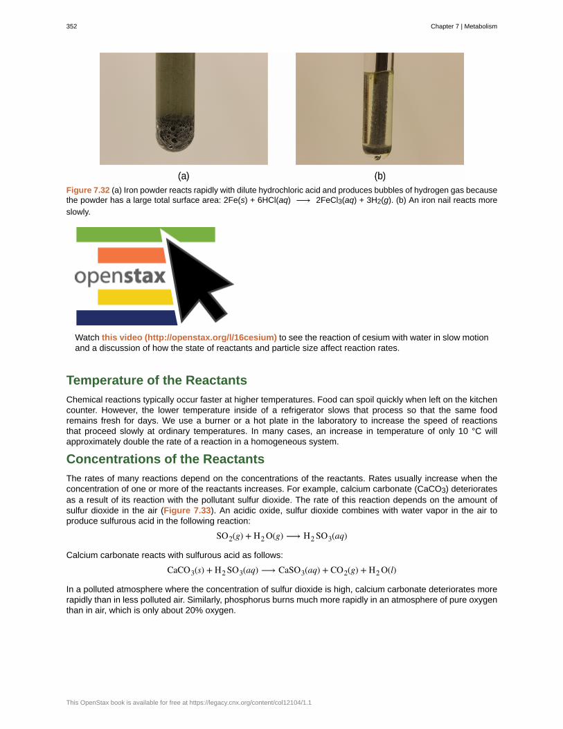
Figure 7.32 (a) Iron powder reacts rapidly with dilute hydrochloric acid and produces bubbles of hydrogen gas becausethe powder has a large total surface area: 2Fe(s) + 6HCl(aq) ⟶ 2FeCl3(aq) + 3H2(g). (b) An iron nail reacts more
slowly.
Watch this video (http://openstax.org/l/16cesium) to see the reaction of cesium with water in slow motionand a discussion of how the state of reactants and particle size affect reaction rates.
Temperature of the Reactants
Chemical reactions typically occur faster at higher temperatures. Food can spoil quickly when left on the kitchencounter. However, the lower temperature inside of a refrigerator slows that process so that the same foodremains fresh for days. We use a burner or a hot plate in the laboratory to increase the speed of reactionsthat proceed slowly at ordinary temperatures. In many cases, an increase in temperature of only 10 °C willapproximately double the rate of a reaction in a homogeneous system.
Concentrations of the Reactants
The rates of many reactions depend on the concentrations of the reactants. Rates usually increase when theconcentration of one or more of the reactants increases. For example, calcium carbonate (CaCO3) deterioratesas a result of its reaction with the pollutant sulfur dioxide. The rate of this reaction depends on the amount ofsulfur dioxide in the air (Figure 7.33). An acidic oxide, sulfur dioxide combines with water vapor in the air toproduce sulfurous acid in the following reaction:
SO2(g) + H2 O(g) ⟶ H2 SO3(aq)
Calcium carbonate reacts with sulfurous acid as follows:
CaCO3(s) + H2 SO3(aq) ⟶ CaSO3(aq) + CO2(g) + H2 O(l)
In a polluted atmosphere where the concentration of sulfur dioxide is high, calcium carbonate deteriorates morerapidly than in less polluted air. Similarly, phosphorus burns much more rapidly in an atmosphere of pure oxygenthan in air, which is only about 20% oxygen.
352 Chapter 7 | Metabolism
This OpenStax book is available for free at https://legacy.cnx.org/content/col12104/1.1

Figure 7.33 Statues made from carbonate compounds such as limestone and marble typically weather slowly overtime due to the actions of water, and thermal expansion and contraction. However, pollutants like sulfur dioxide canaccelerate weathering. As the concentration of air pollutants increases, deterioration of limestone occurs more rapidly.(credit: James P Fisher III)
Phosphorous burns rapidly in air, but it will burn even more rapidly if the concentration of oxygen is higher.Watch this video (http://openstax.org/l/16phosphor) to see an example.
The Presence of a Catalyst
Hydrogen peroxide solutions foam when poured onto an open wound because substances in the exposedtissues act as catalysts, increasing the rate of hydrogen peroxide’s decomposition. However, in the absence ofthese catalysts (for example, in the bottle in the medicine cabinet) complete decomposition can take months.A catalyst is a substance that increases the rate of a chemical reaction by lowering the activation energywithout itself being consumed by the reaction. Activation energy is the minimum amount of energy required fora chemical reaction to proceed in the forward direction. A catalyst increases the reaction rate by providing analternative pathway or mechanism for the reaction to follow (Figure 7.34). Catalysis will be discussed in greaterdetail later in this chapter as it relates to mechanisms of reactions.
Chapter 7 | Metabolism 353
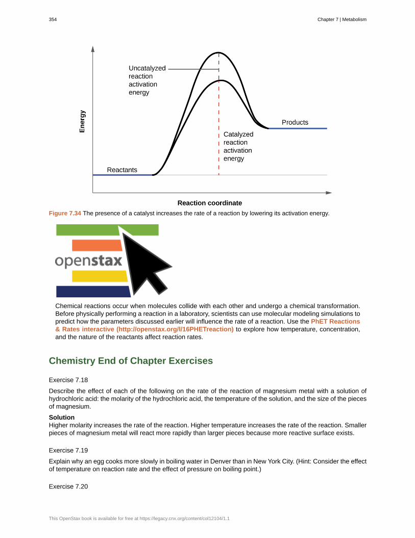
Figure 7.34 The presence of a catalyst increases the rate of a reaction by lowering its activation energy.
Chemical reactions occur when molecules collide with each other and undergo a chemical transformation.Before physically performing a reaction in a laboratory, scientists can use molecular modeling simulations topredict how the parameters discussed earlier will influence the rate of a reaction. Use the PhET Reactions& Rates interactive (http://openstax.org/l/16PHETreaction) to explore how temperature, concentration,and the nature of the reactants affect reaction rates.
Chemistry End of Chapter Exercises
Exercise 7.18
Describe the effect of each of the following on the rate of the reaction of magnesium metal with a solution ofhydrochloric acid: the molarity of the hydrochloric acid, the temperature of the solution, and the size of the piecesof magnesium.
SolutionHigher molarity increases the rate of the reaction. Higher temperature increases the rate of the reaction. Smallerpieces of magnesium metal will react more rapidly than larger pieces because more reactive surface exists.
Exercise 7.19
Explain why an egg cooks more slowly in boiling water in Denver than in New York City. (Hint: Consider the effectof temperature on reaction rate and the effect of pressure on boiling point.)
Exercise 7.20
354 Chapter 7 | Metabolism
This OpenStax book is available for free at https://legacy.cnx.org/content/col12104/1.1
Go to the PhET Reactions & Rates (http://openstax.org/l/16PHETreaction) interactive. Use the SingleCollision tab to represent how the collision between monatomic oxygen (O) and carbon monoxide (CO) resultsin the breaking of one bond and the formation of another. Pull back on the red plunger to release the atom andobserve the results. Then, click on “Reload Launcher” and change to “Angled shot” to see the difference.
(a) What happens when the angle of the collision is changed?
(b) Explain how this is relevant to rate of reaction.
Solution(a) Depending on the angle selected, the atom may take a long time to collide with the molecule and, when acollision does occur, it may not result in the breaking of the bond and the forming of the other. (b) Particles ofreactant must come into contact with each other before they can react.
Exercise 7.21
In the PhET Reactions & Rates (http://openstax.org/l/16PHETreaction) interactive, use the “Many Collisions”tab to observe how multiple atoms and molecules interact under varying conditions. Select a molecule to pumpinto the chamber. Set the initial temperature and select the current amounts of each reactant. Select “Showbonds” under Options. How is the rate of the reaction affected by concentration and temperature?
Exercise 7.22
In the PhET Reactions & Rates (http://openstax.org/l/16PHETreaction) interactive, on the Many Collisionstab, set up a simulation with 15 molecules of A and 10 molecules of BC. Select “Show Bonds” under Options.
(a) Leave the Initial Temperature at the default setting. Observe the reaction. Is the rate of reaction fast or slow?
(b) Click “Pause” and then “Reset All,” and then enter 15 molecules of A and 10 molecules of BC once again.Select “Show Bonds” under Options. This time, increase the initial temperature until, on the graph, the totalaverage energy line is completely above the potential energy curve. Describe what happens to the reaction.
Solution(a) very slow; (b) As the temperature is increased, the reaction proceeds at a faster rate. The amount of reactantsdecreases, and the amount of products increases. After a while, there is a roughly equal amount of BC, AB, andC in the mixture and a slight excess of A.
7.9 | Enzymes
By the end of this section, you will be able to:
• Describe the role of enzymes in metabolic pathways
• Explain how enzymes function as molecular catalysts
• Discuss enzyme regulation by various factors
A substance that helps a chemical reaction to occur is a catalyst, and the special molecules that catalyzebiochemical reactions are called enzymes. Almost all enzymes are proteins, made up of chains of aminoacids, and they perform the critical task of lowering the activation energies of chemical reactions inside thecell. Enzymes do this by binding to the reactant molecules, and holding them in such a way as to make thechemical bond-breaking and bond-forming processes take place more readily. It is important to remember thatenzymes don’t change the ∆G of a reaction. In other words, they don’t change whether a reaction is exergonic(spontaneous) or endergonic. This is because they don’t change the free energy of the reactants or products.They only reduce the activation energy required to reach the transition state (Figure 7.35).
Chapter 7 | Metabolism 355
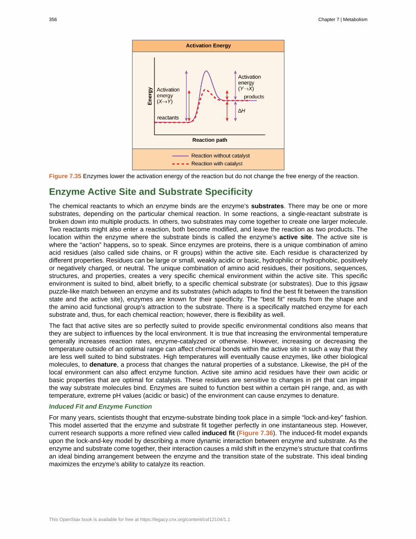
Figure 7.35 Enzymes lower the activation energy of the reaction but do not change the free energy of the reaction.
Enzyme Active Site and Substrate Specificity
The chemical reactants to which an enzyme binds are the enzyme’s substrates. There may be one or moresubstrates, depending on the particular chemical reaction. In some reactions, a single-reactant substrate isbroken down into multiple products. In others, two substrates may come together to create one larger molecule.Two reactants might also enter a reaction, both become modified, and leave the reaction as two products. Thelocation within the enzyme where the substrate binds is called the enzyme’s active site. The active site iswhere the “action” happens, so to speak. Since enzymes are proteins, there is a unique combination of aminoacid residues (also called side chains, or R groups) within the active site. Each residue is characterized bydifferent properties. Residues can be large or small, weakly acidic or basic, hydrophilic or hydrophobic, positivelyor negatively charged, or neutral. The unique combination of amino acid residues, their positions, sequences,structures, and properties, creates a very specific chemical environment within the active site. This specificenvironment is suited to bind, albeit briefly, to a specific chemical substrate (or substrates). Due to this jigsawpuzzle-like match between an enzyme and its substrates (which adapts to find the best fit between the transitionstate and the active site), enzymes are known for their specificity. The “best fit” results from the shape andthe amino acid functional group’s attraction to the substrate. There is a specifically matched enzyme for eachsubstrate and, thus, for each chemical reaction; however, there is flexibility as well.
The fact that active sites are so perfectly suited to provide specific environmental conditions also means thatthey are subject to influences by the local environment. It is true that increasing the environmental temperaturegenerally increases reaction rates, enzyme-catalyzed or otherwise. However, increasing or decreasing thetemperature outside of an optimal range can affect chemical bonds within the active site in such a way that theyare less well suited to bind substrates. High temperatures will eventually cause enzymes, like other biologicalmolecules, to denature, a process that changes the natural properties of a substance. Likewise, the pH of thelocal environment can also affect enzyme function. Active site amino acid residues have their own acidic orbasic properties that are optimal for catalysis. These residues are sensitive to changes in pH that can impairthe way substrate molecules bind. Enzymes are suited to function best within a certain pH range, and, as withtemperature, extreme pH values (acidic or basic) of the environment can cause enzymes to denature.
Induced Fit and Enzyme Function
For many years, scientists thought that enzyme-substrate binding took place in a simple “lock-and-key” fashion.This model asserted that the enzyme and substrate fit together perfectly in one instantaneous step. However,current research supports a more refined view called induced fit (Figure 7.36). The induced-fit model expandsupon the lock-and-key model by describing a more dynamic interaction between enzyme and substrate. As theenzyme and substrate come together, their interaction causes a mild shift in the enzyme’s structure that confirmsan ideal binding arrangement between the enzyme and the transition state of the substrate. This ideal bindingmaximizes the enzyme’s ability to catalyze its reaction.
356 Chapter 7 | Metabolism
This OpenStax book is available for free at https://legacy.cnx.org/content/col12104/1.1
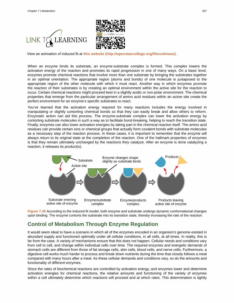
View an animation of induced fit at this website (http://openstaxcollege.org/l/hexokinase) .
When an enzyme binds its substrate, an enzyme-substrate complex is formed. This complex lowers theactivation energy of the reaction and promotes its rapid progression in one of many ways. On a basic level,enzymes promote chemical reactions that involve more than one substrate by bringing the substrates togetherin an optimal orientation. The appropriate region (atoms and bonds) of one molecule is juxtaposed to theappropriate region of the other molecule with which it must react. Another way in which enzymes promotethe reaction of their substrates is by creating an optimal environment within the active site for the reaction tooccur. Certain chemical reactions might proceed best in a slightly acidic or non-polar environment. The chemicalproperties that emerge from the particular arrangement of amino acid residues within an active site create theperfect environment for an enzyme’s specific substrates to react.
You’ve learned that the activation energy required for many reactions includes the energy involved inmanipulating or slightly contorting chemical bonds so that they can easily break and allow others to reform.Enzymatic action can aid this process. The enzyme-substrate complex can lower the activation energy bycontorting substrate molecules in such a way as to facilitate bond-breaking, helping to reach the transition state.Finally, enzymes can also lower activation energies by taking part in the chemical reaction itself. The amino acidresidues can provide certain ions or chemical groups that actually form covalent bonds with substrate moleculesas a necessary step of the reaction process. In these cases, it is important to remember that the enzyme willalways return to its original state at the completion of the reaction. One of the hallmark properties of enzymesis that they remain ultimately unchanged by the reactions they catalyze. After an enzyme is done catalyzing areaction, it releases its product(s).
Figure 7.36 According to the induced-fit model, both enzyme and substrate undergo dynamic conformational changesupon binding. The enzyme contorts the substrate into its transition state, thereby increasing the rate of the reaction.
Control of Metabolism Through Enzyme Regulation
It would seem ideal to have a scenario in which all of the enzymes encoded in an organism’s genome existed inabundant supply and functioned optimally under all cellular conditions, in all cells, at all times. In reality, this isfar from the case. A variety of mechanisms ensure that this does not happen. Cellular needs and conditions varyfrom cell to cell, and change within individual cells over time. The required enzymes and energetic demands ofstomach cells are different from those of fat storage cells, skin cells, blood cells, and nerve cells. Furthermore, adigestive cell works much harder to process and break down nutrients during the time that closely follows a mealcompared with many hours after a meal. As these cellular demands and conditions vary, so do the amounts andfunctionality of different enzymes.
Since the rates of biochemical reactions are controlled by activation energy, and enzymes lower and determineactivation energies for chemical reactions, the relative amounts and functioning of the variety of enzymeswithin a cell ultimately determine which reactions will proceed and at which rates. This determination is tightly
Chapter 7 | Metabolism 357
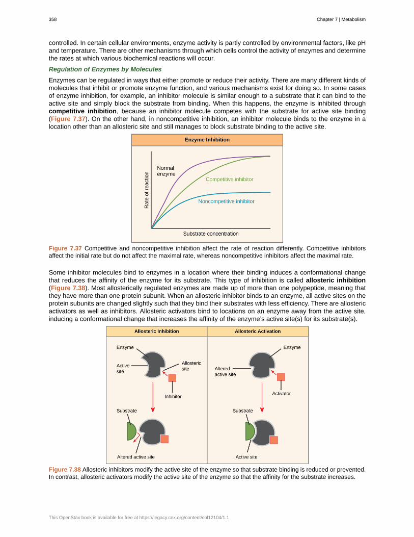
controlled. In certain cellular environments, enzyme activity is partly controlled by environmental factors, like pHand temperature. There are other mechanisms through which cells control the activity of enzymes and determinethe rates at which various biochemical reactions will occur.
Regulation of Enzymes by Molecules
Enzymes can be regulated in ways that either promote or reduce their activity. There are many different kinds ofmolecules that inhibit or promote enzyme function, and various mechanisms exist for doing so. In some casesof enzyme inhibition, for example, an inhibitor molecule is similar enough to a substrate that it can bind to theactive site and simply block the substrate from binding. When this happens, the enzyme is inhibited throughcompetitive inhibition, because an inhibitor molecule competes with the substrate for active site binding(Figure 7.37). On the other hand, in noncompetitive inhibition, an inhibitor molecule binds to the enzyme in alocation other than an allosteric site and still manages to block substrate binding to the active site.
Figure 7.37 Competitive and noncompetitive inhibition affect the rate of reaction differently. Competitive inhibitorsaffect the initial rate but do not affect the maximal rate, whereas noncompetitive inhibitors affect the maximal rate.
Some inhibitor molecules bind to enzymes in a location where their binding induces a conformational changethat reduces the affinity of the enzyme for its substrate. This type of inhibition is called allosteric inhibition(Figure 7.38). Most allosterically regulated enzymes are made up of more than one polypeptide, meaning thatthey have more than one protein subunit. When an allosteric inhibitor binds to an enzyme, all active sites on theprotein subunits are changed slightly such that they bind their substrates with less efficiency. There are allostericactivators as well as inhibitors. Allosteric activators bind to locations on an enzyme away from the active site,inducing a conformational change that increases the affinity of the enzyme’s active site(s) for its substrate(s).
Figure 7.38 Allosteric inhibitors modify the active site of the enzyme so that substrate binding is reduced or prevented.In contrast, allosteric activators modify the active site of the enzyme so that the affinity for the substrate increases.
358 Chapter 7 | Metabolism
This OpenStax book is available for free at https://legacy.cnx.org/content/col12104/1.1
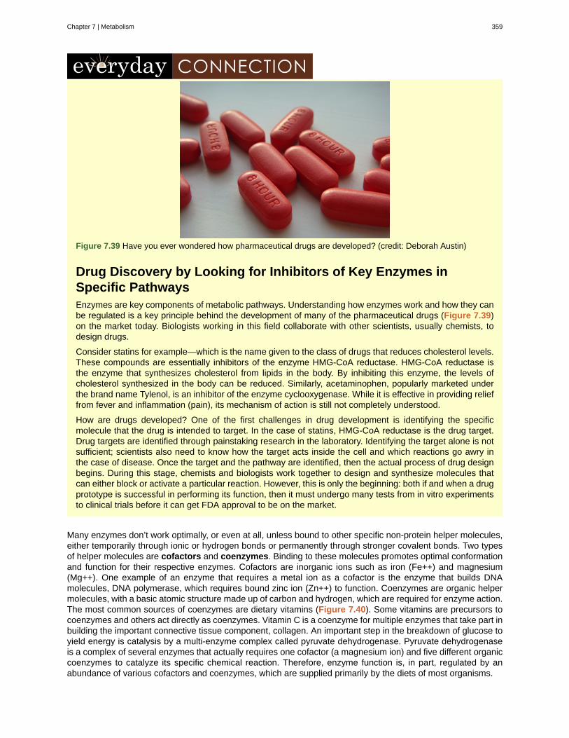
Figure 7.39 Have you ever wondered how pharmaceutical drugs are developed? (credit: Deborah Austin)
Drug Discovery by Looking for Inhibitors of Key Enzymes inSpecific PathwaysEnzymes are key components of metabolic pathways. Understanding how enzymes work and how they canbe regulated is a key principle behind the development of many of the pharmaceutical drugs (Figure 7.39)on the market today. Biologists working in this field collaborate with other scientists, usually chemists, todesign drugs.
Consider statins for example—which is the name given to the class of drugs that reduces cholesterol levels.These compounds are essentially inhibitors of the enzyme HMG-CoA reductase. HMG-CoA reductase isthe enzyme that synthesizes cholesterol from lipids in the body. By inhibiting this enzyme, the levels ofcholesterol synthesized in the body can be reduced. Similarly, acetaminophen, popularly marketed underthe brand name Tylenol, is an inhibitor of the enzyme cyclooxygenase. While it is effective in providing relieffrom fever and inflammation (pain), its mechanism of action is still not completely understood.
How are drugs developed? One of the first challenges in drug development is identifying the specificmolecule that the drug is intended to target. In the case of statins, HMG-CoA reductase is the drug target.Drug targets are identified through painstaking research in the laboratory. Identifying the target alone is notsufficient; scientists also need to know how the target acts inside the cell and which reactions go awry inthe case of disease. Once the target and the pathway are identified, then the actual process of drug designbegins. During this stage, chemists and biologists work together to design and synthesize molecules thatcan either block or activate a particular reaction. However, this is only the beginning: both if and when a drugprototype is successful in performing its function, then it must undergo many tests from in vitro experimentsto clinical trials before it can get FDA approval to be on the market.
Many enzymes don’t work optimally, or even at all, unless bound to other specific non-protein helper molecules,either temporarily through ionic or hydrogen bonds or permanently through stronger covalent bonds. Two typesof helper molecules are cofactors and coenzymes. Binding to these molecules promotes optimal conformationand function for their respective enzymes. Cofactors are inorganic ions such as iron (Fe++) and magnesium(Mg++). One example of an enzyme that requires a metal ion as a cofactor is the enzyme that builds DNAmolecules, DNA polymerase, which requires bound zinc ion (Zn++) to function. Coenzymes are organic helpermolecules, with a basic atomic structure made up of carbon and hydrogen, which are required for enzyme action.The most common sources of coenzymes are dietary vitamins (Figure 7.40). Some vitamins are precursors tocoenzymes and others act directly as coenzymes. Vitamin C is a coenzyme for multiple enzymes that take part inbuilding the important connective tissue component, collagen. An important step in the breakdown of glucose toyield energy is catalysis by a multi-enzyme complex called pyruvate dehydrogenase. Pyruvate dehydrogenaseis a complex of several enzymes that actually requires one cofactor (a magnesium ion) and five different organiccoenzymes to catalyze its specific chemical reaction. Therefore, enzyme function is, in part, regulated by anabundance of various cofactors and coenzymes, which are supplied primarily by the diets of most organisms.
Chapter 7 | Metabolism 359
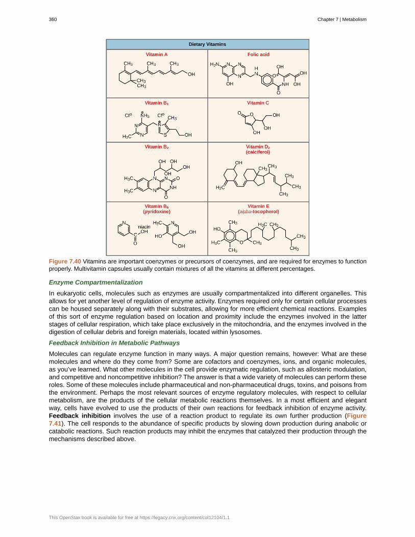
Figure 7.40 Vitamins are important coenzymes or precursors of coenzymes, and are required for enzymes to functionproperly. Multivitamin capsules usually contain mixtures of all the vitamins at different percentages.
Enzyme Compartmentalization
In eukaryotic cells, molecules such as enzymes are usually compartmentalized into different organelles. Thisallows for yet another level of regulation of enzyme activity. Enzymes required only for certain cellular processescan be housed separately along with their substrates, allowing for more efficient chemical reactions. Examplesof this sort of enzyme regulation based on location and proximity include the enzymes involved in the latterstages of cellular respiration, which take place exclusively in the mitochondria, and the enzymes involved in thedigestion of cellular debris and foreign materials, located within lysosomes.
Feedback Inhibition in Metabolic Pathways
Molecules can regulate enzyme function in many ways. A major question remains, however: What are thesemolecules and where do they come from? Some are cofactors and coenzymes, ions, and organic molecules,as you’ve learned. What other molecules in the cell provide enzymatic regulation, such as allosteric modulation,and competitive and noncompetitive inhibition? The answer is that a wide variety of molecules can perform theseroles. Some of these molecules include pharmaceutical and non-pharmaceutical drugs, toxins, and poisons fromthe environment. Perhaps the most relevant sources of enzyme regulatory molecules, with respect to cellularmetabolism, are the products of the cellular metabolic reactions themselves. In a most efficient and elegantway, cells have evolved to use the products of their own reactions for feedback inhibition of enzyme activity.Feedback inhibition involves the use of a reaction product to regulate its own further production (Figure7.41). The cell responds to the abundance of specific products by slowing down production during anabolic orcatabolic reactions. Such reaction products may inhibit the enzymes that catalyzed their production through themechanisms described above.
360 Chapter 7 | Metabolism
This OpenStax book is available for free at https://legacy.cnx.org/content/col12104/1.1
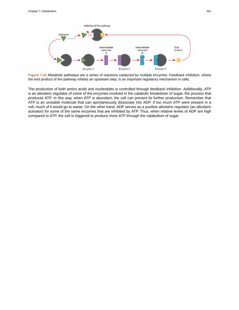
Figure 7.41 Metabolic pathways are a series of reactions catalyzed by multiple enzymes. Feedback inhibition, wherethe end product of the pathway inhibits an upstream step, is an important regulatory mechanism in cells.
The production of both amino acids and nucleotides is controlled through feedback inhibition. Additionally, ATPis an allosteric regulator of some of the enzymes involved in the catabolic breakdown of sugar, the process thatproduces ATP. In this way, when ATP is abundant, the cell can prevent its further production. Remember thatATP is an unstable molecule that can spontaneously dissociate into ADP. If too much ATP were present in acell, much of it would go to waste. On the other hand, ADP serves as a positive allosteric regulator (an allostericactivator) for some of the same enzymes that are inhibited by ATP. Thus, when relative levels of ADP are highcompared to ATP, the cell is triggered to produce more ATP through the catabolism of sugar.
Chapter 7 | Metabolism 361
activated complex
activation energy
activation energy (Ea)
active site
allosteric inhibition
aminopeptidase
anabolic
Arrhenius equation
ATP
bioenergetics
carboxypeptidase
catabolic
catalyst
chemical energy
chemoautotroph
chloroplast
chylomicron
chymotrypsin
coenzyme
cofactor
collision theory
competitive inhibition
denature
digestion
dipeptidase
KEY TERMS
(also, transition state) unstable combination of reactant species representing the highestenergy state of a reaction system
energy necessary for reactions to occur
energy necessary in order for a reaction to take place
specific region of the enzyme to which the substrate binds
inhibition by a binding event at a site different from the active site, which induces aconformational change and reduces the affinity of the enzyme for its substrate
protease that breaks down peptides to single amino acids; secreted by the brush border ofsmall intestine
(also, anabolism) pathways that require an input of energy to synthesize complex molecules fromsimpler ones
mathematical relationship between the rate constant and the activation energy of areaction
adenosine triphosphate, the cell’s energy currency
study of energy flowing through living systems
protease that breaks down peptides to single amino acids; secreted by the brush border ofthe small intestine
(also, catabolism) pathways in which complex molecules are broken down into simpler ones
substance that increases the rate of a reaction without itself being consumed by the reaction
potential energy in chemical bonds that is released when those bonds are broken
organism that can build organic molecules using energy derived from inorganic chemicalsinstead of sunlight
organelle in which photosynthesis takes place
small lipid globule
pancreatic protease
small organic molecule, such as a vitamin or its derivative, which is required to enhance the activityof an enzyme
inorganic ion, such as iron and magnesium ions, required for optimal regulation of enzyme activity
model that emphasizes the energy and orientation of molecular collisions to explain andpredict reaction kinetics
type of inhibition in which the inhibitor competes with the substrate molecule by bindingto the active site of the enzyme
process that changes the natural properties of a substance
mechanical and chemical break down of food into small organic fragments
protease that breaks down peptides to single amino acids; secreted by the brush border of smallintestine
362 Chapter 7 | Metabolism
This OpenStax book is available for free at https://legacy.cnx.org/content/col12104/1.1
elastase
endergonic
enthalpy
essential nutrient
exergonic
feedback inhibition
free energy
frequency factor (A)
granum
heat energy
heterotroph
induced fit
ingestion
kinetic energy
lactase
light-dependent reaction
light-independent reaction
maltase
mesophyll
metabolism
mineral
phosphoanhydride bond
photoautotroph
pigment
potential energy
stoma
stroma
substrate
pancreatic protease
describes chemical reactions that require energy input
total energy of a system
nutrient that cannot be synthesized by the body; it must be obtained from food
describes chemical reactions that release free energy
effect of a product of a reaction sequence to decrease its further production by inhibitingthe activity of the first enzyme in the pathway that produces it
Gibbs free energy is the usable energy, or energy that is available to do work.
proportionality constant in the Arrhenius equation, related to the relative number ofcollisions having an orientation capable of leading to product formation
stack of thylakoids located inside a chloroplast
total bond energy of reactants or products in a chemical reaction
organism that consumes organic substances or other organisms for food
dynamic fit between the enzyme and its substrate, in which both components modify their structuresto allow for ideal binding
act of taking in food
type of energy associated with objects or particles in motion
enzyme that breaks down lactose into glucose and galactose
first stage of photosynthesis where certain wavelengths of the visible light areabsorbed to form two energy-carrying molecules (ATP and NADPH)
second stage of photosynthesis, though which carbon dioxide is used to buildcarbohydrate molecules using energy from ATP and NADPH
enzyme that breaks down maltose into glucose
middle layer of chlorophyll-rich cells in a leaf
all the chemical reactions that take place inside cells, including anabolism and catabolism
inorganic, elemental molecule that carries out important roles in the body
bond that connects phosphates in an ATP molecule
organism capable of producing its own organic compounds from sunlight
molecule that is capable of absorbing certain wavelengths of light and reflecting others (whichaccounts for its color)
type of energy that has the potential to do work; stored energy
opening that regulates gas exchange and water evaporation between leaves and the environment,typically situated on the underside of leaves
fluid-filled space surrounding the grana inside a chloroplast where the light-independent reactions ofphotosynthesis take place
molecule on which the enzyme acts
Chapter 7 | Metabolism 363
sucrase
thylakoid
thylakoid lumen
transition state
trypsin
vitamin
enzyme that breaks down sucrose into glucose and fructose
disc-shaped, membrane-bound structure inside a chloroplast where the light-dependent reactions ofphotosynthesis take place; stacks of thylakoids are called grana
aqueous space bound by a thylakoid membrane where protons accumulate during light-drivenelectron transport
high-energy, unstable state (an intermediate form between the substrate and the product)occurring during a chemical reaction
pancreatic protease that breaks down protein
organic substance necessary in small amounts to sustain life
CHAPTER SUMMARY
7.2 Nutrition and Energy Production
Animal diet should be balanced and meet the needs of the body. Carbohydrates, proteins, and fats are theprimary components of food. Some essential nutrients are required for cellular function but cannot be producedby the animal body. These include vitamins, minerals, some fatty acids, and some amino acids. Food intake inmore than necessary amounts is stored as glycogen in the liver and muscle cells, and in fat cells. Excessadipose storage can lead to obesity and serious health problems. ATP is the energy currency of the cell and isobtained from the metabolic pathways. Excess carbohydrates and energy are stored as glycogen in the body.
7.3 Digestive System Processes
Digestion begins with ingestion, where the food is taken in the mouth. Digestion and absorption take place in aseries of steps with special enzymes playing important roles in digesting carbohydrates, proteins, and lipids.Elimination describes removal of undigested food contents and waste products from the body. While mostabsorption occurs in the small intestines, the large intestine is responsible for the final removal of water thatremains after the absorptive process of the small intestines. The cells that line the large intestine absorb somevitamins as well as any leftover salts and water. The large intestine (colon) is also where feces is formed.
7.4 Energy and Metabolism
Cells perform the functions of life through various chemical reactions. A cell’s metabolism refers to the chemicalreactions that take place within it. There are metabolic reactions that involve the breaking down of complexchemicals into simpler ones, such as the breakdown of large macromolecules. This process is referred to ascatabolism, and such reactions are associated with a release of energy. On the other end of the spectrum,anabolism refers to metabolic processes that build complex molecules out of simpler ones, such as thesynthesis of macromolecules. Anabolic processes require energy. Glucose synthesis and glucose breakdownare examples of anabolic and catabolic pathways, respectively.
7.5 Potential, Kinetic, Free, and Activation Energy
Energy comes in many different forms. Objects in motion do physical work, and kinetic energy is the energy ofobjects in motion. Objects that are not in motion may have the potential to do work, and thus, have potentialenergy. Molecules also have potential energy because the breaking of molecular bonds has the potential torelease energy. Living cells depend on the harvesting of potential energy from molecular bonds to performwork. Free energy is a measure of energy that is available to do work. The free energy of a system changesduring energy transfers such as chemical reactions, and this change is referred to as ∆G.
The ∆G of a reaction can be negative or positive, meaning that the reaction releases energy or consumesenergy, respectively. A reaction with a negative ∆G that gives off energy is called an exergonic reaction. Onewith a positive ∆G that requires energy input is called an endergonic reaction. Exergonic reactions are said tobe spontaneous, because their products have less energy than their reactants. The products of endergonicreactions have a higher energy state than the reactants, and so these are nonspontaneous reactions. However,all reactions (including spontaneous -∆G reactions) require an initial input of energy in order to reach thetransition state, at which they’ll proceed. This initial input of energy is called the activation energy.
364 Chapter 7 | Metabolism
This OpenStax book is available for free at https://legacy.cnx.org/content/col12104/1.1
7.6 ATP: Adenosine Triphosphate
ATP is the primary energy-supplying molecule for living cells. ATP is made up of a nucleotide, a five-carbonsugar, and three phosphate groups. The bonds that connect the phosphates (phosphoanhydride bonds) havehigh-energy content. The energy released from the hydrolysis of ATP into ADP + Pi is used to perform cellularwork. Cells use ATP to perform work by coupling the exergonic reaction of ATP hydrolysis with endergonicreactions. ATP donates its phosphate group to another molecule via a process known as phosphorylation. Thephosphorylated molecule is at a higher-energy state and is less stable than its unphosphorylated form, and thisadded energy from the addition of the phosphate allows the molecule to undergo its endergonic reaction.
7.7 Overview of Photosynthesis
The process of photosynthesis transformed life on Earth. By harnessing energy from the sun, photosynthesisevolved to allow living things access to enormous amounts of energy. Because of photosynthesis, living thingsgained access to sufficient energy that allowed them to build new structures and achieve the biodiversityevident today.
Only certain organisms, called photoautotrophs, can perform photosynthesis; they require the presence ofchlorophyll, a specialized pigment that absorbs certain portions of the visible spectrum and can capture energyfrom sunlight. Photosynthesis uses carbon dioxide and water to assemble carbohydrate molecules and releaseoxygen as a waste product into the atmosphere. Eukaryotic autotrophs, such as plants and algae, haveorganelles called chloroplasts in which photosynthesis takes place, and starch accumulates. In prokaryotes,such as cyanobacteria, the process is less localized and occurs within folded membranes, extensions of theplasma membrane, and in the cytoplasm.
7.8 Collision Theory
Chemical reactions require collisions between reactant species. These reactant collisions must be of properorientation and sufficient energy in order to result in product formation. Collision theory provides a simple buteffective explanation for the effect of many experimental parameters on reaction rates. The Arrhenius equationdescribes the relation between a reaction’s rate constant and its activation energy, temperature, anddependence on collision orientation.
7.9 Factors Affecting Reaction Rates
The rate of a chemical reaction is affected by several parameters. Reactions involving two phases proceedmore rapidly when there is greater surface area contact. If temperature or reactant concentration is increased,the rate of a given reaction generally increases as well. A catalyst can increase the rate of a reaction byproviding an alternative pathway that causes the activation energy of the reaction to decrease.
7.10 Enzymes
Enzymes are chemical catalysts that accelerate chemical reactions at physiological temperatures by loweringtheir activation energy. Enzymes are usually proteins consisting of one or more polypeptide chains. Enzymeshave an active site that provides a unique chemical environment, made up of certain amino acid R groups(residues). This unique environment is perfectly suited to convert particular chemical reactants for that enzyme,called substrates, into unstable intermediates called transition states. Enzymes and substrates are thought tobind with an induced fit, which means that enzymes undergo slight conformational adjustments upon substratecontact, leading to full, optimal binding. Enzymes bind to substrates and catalyze reactions in four differentways: bringing substrates together in an optimal orientation, compromising the bond structures of substrates sothat bonds can be more easily broken, providing optimal environmental conditions for a reaction to occur, orparticipating directly in their chemical reaction by forming transient covalent bonds with the substrates.
Enzyme action must be regulated so that in a given cell at a given time, the desired reactions are beingcatalyzed and the undesired reactions are not. Enzymes are regulated by cellular conditions, such astemperature and pH. They are also regulated through their location within a cell, sometimes beingcompartmentalized so that they can only catalyze reactions under certain circumstances. Inhibition andactivation of enzymes via other molecules are other important ways that enzymes are regulated. Inhibitors canact competitively, noncompetitively, or allosterically; noncompetitive inhibitors are usually allosteric. Activatorscan also enhance the function of enzymes allosterically. The most common method by which cells regulate theenzymes in metabolic pathways is through feedback inhibition. During feedback inhibition, the products of a
Chapter 7 | Metabolism 365
metabolic pathway serve as inhibitors (usually allosteric) of one or more of the enzymes (usually the firstcommitted enzyme of the pathway) involved in the pathway that produces them.
ART CONNECTION QUESTIONS
23. Figure 7.8 Which of the following statementsabout digestive processes is true?
a. Amylase, maltase and lactase in the mouthdigest carbohydrates.
b. Trypsin and lipase in the stomach digestprotein.
c. Bile emulsifies lipids in the small intestine.d. No food is absorbed until the small intestine.
24. Figure 7.15 Look at each of the processesshown, and decide if it is endergonic or exergonic. Ineach case, does enthalpy increase or decrease, anddoes entropy increase or decrease?
25. Figure 7.17 If no activation energy were required
to break down sucrose (table sugar), would you beable to store it in a sugar bowl?
26. Figure 7.19 The hydrolysis of one ATP moleculereleases 7.3 kcal/mol of energy (∆G = −7.3 kcal/molof energy). If it takes 2.1 kcal/mol of energy to move
one Na+ across the membrane (∆G = +2.1 kcal/molof energy), how many sodium ions could be movedby the hydrolysis of one ATP molecule?
27. Figure 7.24 On a hot, dry day, plants close theirstomata to conserve water. What impact will thishave on photosynthesis?
REVIEW QUESTIONS
28. Which of the following statements is not true?
a. Essential nutrients can be synthesized bythe body.
b. Vitamins are required in small quantities forbodily function.
c. Some amino acids can be synthesized bythe body, while others need to be obtainedfrom diet.
d. Vitamins come in two categories: fat-solubleand water-soluble.
29. Which of the following is a water-soluble vitamin?
a. vitamin Ab. vitamin Ec. vitamin Kd. vitamin C
30. What is the primary fuel for the body?a. carbohydratesb. lipidsc. proteind. glycogen
31. Excess glucose is stored as ________.a. fatb. glucagonc. glycogend. it is not stored in the body
32. Where does the majority of protein digestion takeplace?
a. stomachb. duodenumc. mouthd. jejunum
33. Lipases are enzymes that break down ________.
a. disaccharidesb. lipidsc. proteinsd. cellulose
34. Energy is stored long-term in the bonds of _____and used short-term to perform work from a(n) _____molecule.
a. ATP : glucoseb. an anabolic molecule : catabolic moleculec. glucose : ATPd. a catabolic molecule : anabolic molecule
35. DNA replication involves unwinding two strandsof parent DNA, copying each strand to synthesizecomplementary strands, and releasing the parent anddaughter DNA. Which of the following accuratelydescribes this process?
a. This is an anabolic processb. This is a catabolic processc. This is both anabolic and catabolicd. This is a metabolic process but is neither
anabolic nor catabolic
36. Consider a pendulum swinging. Which type(s) ofenergy is/are associated with the pendulum in thefollowing instances: i. the moment at which itcompletes one cycle, just before it begins to fall backtowards the other end, ii. the moment that it is in themiddle between the two ends, iii. just before itreaches the end of one cycle (just before instant i.).
366 Chapter 7 | Metabolism
This OpenStax book is available for free at https://legacy.cnx.org/content/col12104/1.1
a. i. potential and kinetic, ii. potential andkinetic, iii. kinetic
b. i. potential, ii. potential and kinetic, iii.potential and kinetic
c. i. potential, ii. kinetic, iii. potential and kineticd. i. potential and kinetic, ii. kinetic iii. kinetic
37. Which of the following comparisons or contrastsbetween endergonic and exergonic reactions isfalse?
a. Endergonic reactions have a positive ∆Gand exergonic reactions have a negative ∆G
b. Endergonic reactions consume energy andexergonic reactions release energy
c. Both endergonic and exergonic reactionsrequire a small amount of energy toovercome an activation barrier
d. Endergonic reactions take place slowly andexergonic reactions take place quickly
38. Which of the following is the best way to judgethe relative activation energies between two givenchemical reactions?
a. Compare the ∆G values between the tworeactions
b. Compare their reaction ratesc. Compare their ideal environmental
conditionsd. Compare the spontaneity between the two
reactions
39. The energy released by the hydrolysis of ATP is
a. primarily stored between the alpha and betaphosphates
b. equal to −57 kcal/molc. harnessed as heat energy by the cell to
perform workd. providing energy to coupled reactions
40. Which of the following molecules is likely to havethe most potential energy?
a. sucroseb. ATPc. glucosed. ADP
41. Which of the following components is not used byboth plants and cyanobacteria to carry outphotosynthesis?
a. chloroplastsb. chlorophyllc. carbon dioxided. water
42. What two main products result fromphotosynthesis?
a. oxygen and carbon dioxideb. chlorophyll and oxygenc. sugars/carbohydrates and oxygend. sugars/carbohydrates and carbon dioxide
43. In which compartment of the plant cell do thelight-independent reactions of photosynthesis takeplace?
a. thylakoidb. stromac. outer membraned. mesophyll
44. Which statement about thylakoids in eukaryotesis not correct?
a. Thylakoids are assembled into stacks.b. Thylakoids exist as a maze of folded
membranes.c. The space surrounding thylakoids is called
stroma.d. Thylakoids contain chlorophyll.
45. Which of the following is not true about enzymes:
a. They increase ∆G of reactionsb. They are usually made of amino acidsc. They lower the activation energy of
chemical reactionsd. Each one is specific to the particular
substrate(s) to which it binds
46. An allosteric inhibitor does which of the following?
a. Binds to an enzyme away from the activesite and changes the conformation of theactive site, increasing its affinity forsubstrate binding
b. Binds to the active site and blocks it frombinding substrate
c. Binds to an enzyme away from the activesite and changes the conformation of theactive site, decreasing its affinity for thesubstrate
d. Binds directly to the active site and mimicsthe substrate
47. Which of the following analogies best describethe induced-fit model of enzyme-substrate binding?
a. A hug between two peopleb. A key fitting into a lockc. A square peg fitting through the square hole
and a round peg fitting through the roundhole of a children’s toy
d. The fitting together of two jigsaw puzzlepieces.
CRITICAL THINKING QUESTIONS
48. What are essential nutrients?
49. What is the role of minerals in maintaining good
health?
50. Discuss why obesity is a growing epidemic.
Chapter 7 | Metabolism 367
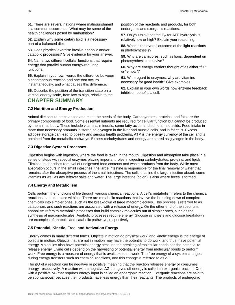
51. There are several nations where malnourishmentis a common occurrence. What may be some of thehealth challenges posed by malnutrition?
52. Explain why some dietary lipid is a necessarypart of a balanced diet.
53. Does physical exercise involve anabolic and/orcatabolic processes? Give evidence for your answer.
54. Name two different cellular functions that requireenergy that parallel human energy-requiringfunctions.
55. Explain in your own words the difference betweena spontaneous reaction and one that occursinstantaneously, and what causes this difference.
56. Describe the position of the transition state on avertical energy scale, from low to high, relative to the
position of the reactants and products, for bothendergonic and exergonic reactions.
57. Do you think that the EA for ATP hydrolysis isrelatively low or high? Explain your reasoning.
58. What is the overall outcome of the light reactionsin photosynthesis?
59. Why are carnivores, such as lions, dependent onphotosynthesis to survive?
60. Why are energy carriers thought of as either “full”or “empty”?
61. With regard to enzymes, why are vitaminsnecessary for good health? Give examples.
62. Explain in your own words how enzyme feedbackinhibition benefits a cell.
CHAPTER SUMMARY
7.2 Nutrition and Energy Production
Animal diet should be balanced and meet the needs of the body. Carbohydrates, proteins, and fats are theprimary components of food. Some essential nutrients are required for cellular function but cannot be producedby the animal body. These include vitamins, minerals, some fatty acids, and some amino acids. Food intake inmore than necessary amounts is stored as glycogen in the liver and muscle cells, and in fat cells. Excessadipose storage can lead to obesity and serious health problems. ATP is the energy currency of the cell and isobtained from the metabolic pathways. Excess carbohydrates and energy are stored as glycogen in the body.
7.3 Digestive System Processes
Digestion begins with ingestion, where the food is taken in the mouth. Digestion and absorption take place in aseries of steps with special enzymes playing important roles in digesting carbohydrates, proteins, and lipids.Elimination describes removal of undigested food contents and waste products from the body. While mostabsorption occurs in the small intestines, the large intestine is responsible for the final removal of water thatremains after the absorptive process of the small intestines. The cells that line the large intestine absorb somevitamins as well as any leftover salts and water. The large intestine (colon) is also where feces is formed.
7.4 Energy and Metabolism
Cells perform the functions of life through various chemical reactions. A cell’s metabolism refers to the chemicalreactions that take place within it. There are metabolic reactions that involve the breaking down of complexchemicals into simpler ones, such as the breakdown of large macromolecules. This process is referred to ascatabolism, and such reactions are associated with a release of energy. On the other end of the spectrum,anabolism refers to metabolic processes that build complex molecules out of simpler ones, such as thesynthesis of macromolecules. Anabolic processes require energy. Glucose synthesis and glucose breakdownare examples of anabolic and catabolic pathways, respectively.
7.5 Potential, Kinetic, Free, and Activation Energy
Energy comes in many different forms. Objects in motion do physical work, and kinetic energy is the energy ofobjects in motion. Objects that are not in motion may have the potential to do work, and thus, have potentialenergy. Molecules also have potential energy because the breaking of molecular bonds has the potential torelease energy. Living cells depend on the harvesting of potential energy from molecular bonds to performwork. Free energy is a measure of energy that is available to do work. The free energy of a system changesduring energy transfers such as chemical reactions, and this change is referred to as ∆G.
The ∆G of a reaction can be negative or positive, meaning that the reaction releases energy or consumesenergy, respectively. A reaction with a negative ∆G that gives off energy is called an exergonic reaction. Onewith a positive ∆G that requires energy input is called an endergonic reaction. Exergonic reactions are said tobe spontaneous, because their products have less energy than their reactants. The products of endergonic
368 Chapter 7 | Metabolism
This OpenStax book is available for free at https://legacy.cnx.org/content/col12104/1.1
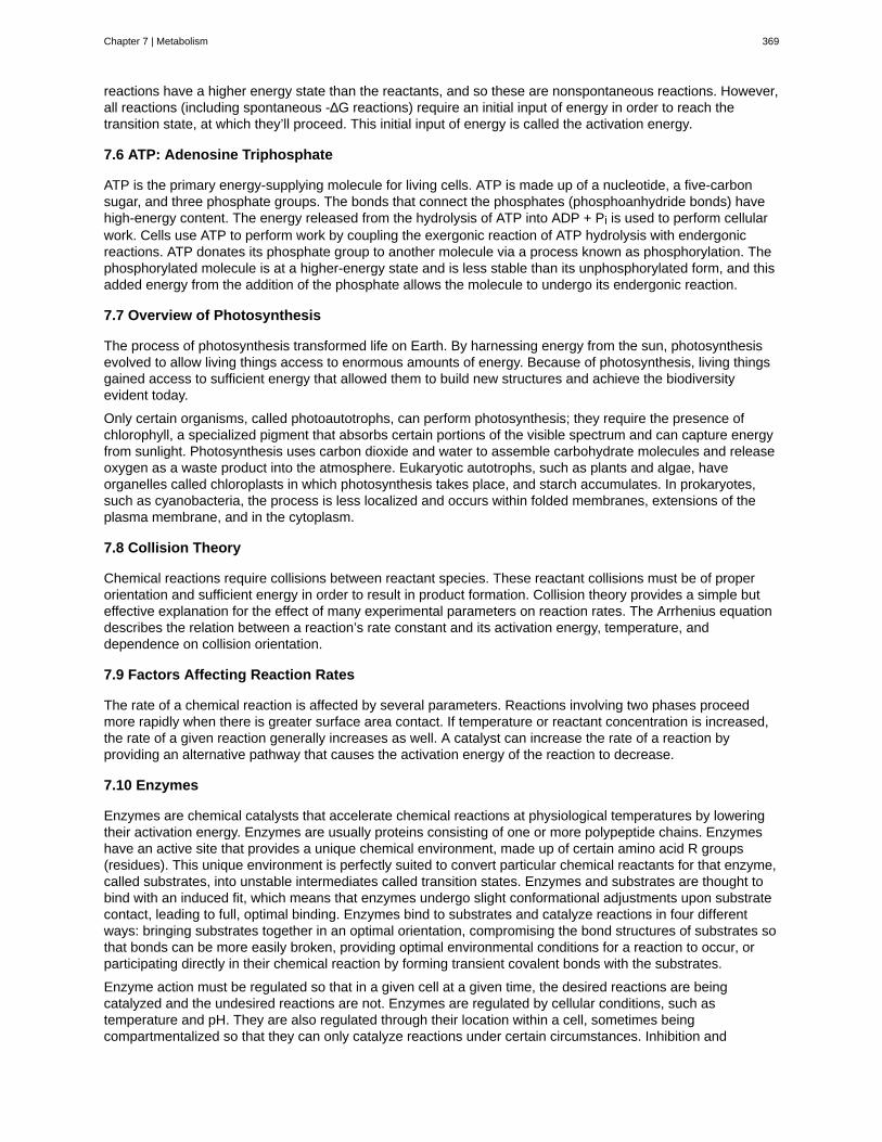
reactions have a higher energy state than the reactants, and so these are nonspontaneous reactions. However,all reactions (including spontaneous -∆G reactions) require an initial input of energy in order to reach thetransition state, at which they’ll proceed. This initial input of energy is called the activation energy.
7.6 ATP: Adenosine Triphosphate
ATP is the primary energy-supplying molecule for living cells. ATP is made up of a nucleotide, a five-carbonsugar, and three phosphate groups. The bonds that connect the phosphates (phosphoanhydride bonds) havehigh-energy content. The energy released from the hydrolysis of ATP into ADP + Pi is used to perform cellularwork. Cells use ATP to perform work by coupling the exergonic reaction of ATP hydrolysis with endergonicreactions. ATP donates its phosphate group to another molecule via a process known as phosphorylation. Thephosphorylated molecule is at a higher-energy state and is less stable than its unphosphorylated form, and thisadded energy from the addition of the phosphate allows the molecule to undergo its endergonic reaction.
7.7 Overview of Photosynthesis
The process of photosynthesis transformed life on Earth. By harnessing energy from the sun, photosynthesisevolved to allow living things access to enormous amounts of energy. Because of photosynthesis, living thingsgained access to sufficient energy that allowed them to build new structures and achieve the biodiversityevident today.
Only certain organisms, called photoautotrophs, can perform photosynthesis; they require the presence ofchlorophyll, a specialized pigment that absorbs certain portions of the visible spectrum and can capture energyfrom sunlight. Photosynthesis uses carbon dioxide and water to assemble carbohydrate molecules and releaseoxygen as a waste product into the atmosphere. Eukaryotic autotrophs, such as plants and algae, haveorganelles called chloroplasts in which photosynthesis takes place, and starch accumulates. In prokaryotes,such as cyanobacteria, the process is less localized and occurs within folded membranes, extensions of theplasma membrane, and in the cytoplasm.
7.8 Collision Theory
Chemical reactions require collisions between reactant species. These reactant collisions must be of properorientation and sufficient energy in order to result in product formation. Collision theory provides a simple buteffective explanation for the effect of many experimental parameters on reaction rates. The Arrhenius equationdescribes the relation between a reaction’s rate constant and its activation energy, temperature, anddependence on collision orientation.
7.9 Factors Affecting Reaction Rates
The rate of a chemical reaction is affected by several parameters. Reactions involving two phases proceedmore rapidly when there is greater surface area contact. If temperature or reactant concentration is increased,the rate of a given reaction generally increases as well. A catalyst can increase the rate of a reaction byproviding an alternative pathway that causes the activation energy of the reaction to decrease.
7.10 Enzymes
Enzymes are chemical catalysts that accelerate chemical reactions at physiological temperatures by loweringtheir activation energy. Enzymes are usually proteins consisting of one or more polypeptide chains. Enzymeshave an active site that provides a unique chemical environment, made up of certain amino acid R groups(residues). This unique environment is perfectly suited to convert particular chemical reactants for that enzyme,called substrates, into unstable intermediates called transition states. Enzymes and substrates are thought tobind with an induced fit, which means that enzymes undergo slight conformational adjustments upon substratecontact, leading to full, optimal binding. Enzymes bind to substrates and catalyze reactions in four differentways: bringing substrates together in an optimal orientation, compromising the bond structures of substrates sothat bonds can be more easily broken, providing optimal environmental conditions for a reaction to occur, orparticipating directly in their chemical reaction by forming transient covalent bonds with the substrates.
Enzyme action must be regulated so that in a given cell at a given time, the desired reactions are beingcatalyzed and the undesired reactions are not. Enzymes are regulated by cellular conditions, such astemperature and pH. They are also regulated through their location within a cell, sometimes beingcompartmentalized so that they can only catalyze reactions under certain circumstances. Inhibition and
Chapter 7 | Metabolism 369
activation of enzymes via other molecules are other important ways that enzymes are regulated. Inhibitors canact competitively, noncompetitively, or allosterically; noncompetitive inhibitors are usually allosteric. Activatorscan also enhance the function of enzymes allosterically. The most common method by which cells regulate theenzymes in metabolic pathways is through feedback inhibition. During feedback inhibition, the products of ametabolic pathway serve as inhibitors (usually allosteric) of one or more of the enzymes (usually the firstcommitted enzyme of the pathway) involved in the pathway that produces them.
ART CONNECTION QUESTIONS
63. Figure 7.8 Which of the following statementsabout digestive processes is true?
a. Amylase, maltase and lactase in the mouthdigest carbohydrates.
b. Trypsin and lipase in the stomach digestprotein.
c. Bile emulsifies lipids in the small intestine.d. No food is absorbed until the small intestine.
64. Figure 7.15 Look at each of the processesshown, and decide if it is endergonic or exergonic. Ineach case, does enthalpy increase or decrease, anddoes entropy increase or decrease?
65. Figure 7.17 If no activation energy were required
to break down sucrose (table sugar), would you beable to store it in a sugar bowl?
66. Figure 7.19 The hydrolysis of one ATP moleculereleases 7.3 kcal/mol of energy (∆G = −7.3 kcal/molof energy). If it takes 2.1 kcal/mol of energy to move
one Na+ across the membrane (∆G = +2.1 kcal/molof energy), how many sodium ions could be movedby the hydrolysis of one ATP molecule?
67. Figure 7.24 On a hot, dry day, plants close theirstomata to conserve water. What impact will thishave on photosynthesis?
REVIEW QUESTIONS
68. Which of the following statements is not true?
a. Essential nutrients can be synthesized bythe body.
b. Vitamins are required in small quantities forbodily function.
c. Some amino acids can be synthesized bythe body, while others need to be obtainedfrom diet.
d. Vitamins come in two categories: fat-solubleand water-soluble.
69. Which of the following is a water-soluble vitamin?
a. vitamin Ab. vitamin Ec. vitamin Kd. vitamin C
70. What is the primary fuel for the body?a. carbohydratesb. lipidsc. proteind. glycogen
71. Excess glucose is stored as ________.a. fatb. glucagonc. glycogend. it is not stored in the body
72. Where does the majority of protein digestion takeplace?
a. stomachb. duodenumc. mouthd. jejunum
73. Lipases are enzymes that break down ________.
a. disaccharidesb. lipidsc. proteinsd. cellulose
74. Energy is stored long-term in the bonds of _____and used short-term to perform work from a(n) _____molecule.
a. ATP : glucoseb. an anabolic molecule : catabolic moleculec. glucose : ATPd. a catabolic molecule : anabolic molecule
75. DNA replication involves unwinding two strandsof parent DNA, copying each strand to synthesizecomplementary strands, and releasing the parent anddaughter DNA. Which of the following accuratelydescribes this process?
a. This is an anabolic processb. This is a catabolic processc. This is both anabolic and catabolicd. This is a metabolic process but is neither
anabolic nor catabolic
76. Consider a pendulum swinging. Which type(s) ofenergy is/are associated with the pendulum in thefollowing instances: i. the moment at which itcompletes one cycle, just before it begins to fall back
370 Chapter 7 | Metabolism
This OpenStax book is available for free at https://legacy.cnx.org/content/col12104/1.1
towards the other end, ii. the moment that it is in themiddle between the two ends, iii. just before itreaches the end of one cycle (just before instant i.).
a. i. potential and kinetic, ii. potential andkinetic, iii. kinetic
b. i. potential, ii. potential and kinetic, iii.potential and kinetic
c. i. potential, ii. kinetic, iii. potential and kineticd. i. potential and kinetic, ii. kinetic iii. kinetic
77. Which of the following comparisons or contrastsbetween endergonic and exergonic reactions isfalse?
a. Endergonic reactions have a positive ∆Gand exergonic reactions have a negative ∆G
b. Endergonic reactions consume energy andexergonic reactions release energy
c. Both endergonic and exergonic reactionsrequire a small amount of energy toovercome an activation barrier
d. Endergonic reactions take place slowly andexergonic reactions take place quickly
78. Which of the following is the best way to judgethe relative activation energies between two givenchemical reactions?
a. Compare the ∆G values between the tworeactions
b. Compare their reaction ratesc. Compare their ideal environmental
conditionsd. Compare the spontaneity between the two
reactions
79. The energy released by the hydrolysis of ATP is
a. primarily stored between the alpha and betaphosphates
b. equal to −57 kcal/molc. harnessed as heat energy by the cell to
perform workd. providing energy to coupled reactions
80. Which of the following molecules is likely to havethe most potential energy?
a. sucroseb. ATPc. glucosed. ADP
81. Which of the following components is not used byboth plants and cyanobacteria to carry outphotosynthesis?
a. chloroplastsb. chlorophyllc. carbon dioxided. water
82. What two main products result fromphotosynthesis?
a. oxygen and carbon dioxideb. chlorophyll and oxygenc. sugars/carbohydrates and oxygend. sugars/carbohydrates and carbon dioxide
83. In which compartment of the plant cell do thelight-independent reactions of photosynthesis takeplace?
a. thylakoidb. stromac. outer membraned. mesophyll
84. Which statement about thylakoids in eukaryotesis not correct?
a. Thylakoids are assembled into stacks.b. Thylakoids exist as a maze of folded
membranes.c. The space surrounding thylakoids is called
stroma.d. Thylakoids contain chlorophyll.
85. Which of the following is not true about enzymes:
a. They increase ∆G of reactionsb. They are usually made of amino acidsc. They lower the activation energy of
chemical reactionsd. Each one is specific to the particular
substrate(s) to which it binds
86. An allosteric inhibitor does which of the following?
a. Binds to an enzyme away from the activesite and changes the conformation of theactive site, increasing its affinity forsubstrate binding
b. Binds to the active site and blocks it frombinding substrate
c. Binds to an enzyme away from the activesite and changes the conformation of theactive site, decreasing its affinity for thesubstrate
d. Binds directly to the active site and mimicsthe substrate
87. Which of the following analogies best describethe induced-fit model of enzyme-substrate binding?
a. A hug between two peopleb. A key fitting into a lockc. A square peg fitting through the square hole
and a round peg fitting through the roundhole of a children’s toy
d. The fitting together of two jigsaw puzzlepieces.
Chapter 7 | Metabolism 371
CRITICAL THINKING QUESTIONS
88. What are essential nutrients?
89. What is the role of minerals in maintaining goodhealth?
90. Discuss why obesity is a growing epidemic.
91. There are several nations where malnourishmentis a common occurrence. What may be some of thehealth challenges posed by malnutrition?
92. Explain why some dietary lipid is a necessarypart of a balanced diet.
93. Does physical exercise involve anabolic and/orcatabolic processes? Give evidence for your answer.
94. Name two different cellular functions that requireenergy that parallel human energy-requiringfunctions.
95. Explain in your own words the difference betweena spontaneous reaction and one that occursinstantaneously, and what causes this difference.
96. Describe the position of the transition state on avertical energy scale, from low to high, relative to theposition of the reactants and products, for bothendergonic and exergonic reactions.
97. Do you think that the EA for ATP hydrolysis isrelatively low or high? Explain your reasoning.
98. What is the overall outcome of the light reactionsin photosynthesis?
99. Why are carnivores, such as lions, dependent onphotosynthesis to survive?
100. Why are energy carriers thought of as either“full” or “empty”?
101. With regard to enzymes, why are vitaminsnecessary for good health? Give examples.
102. Explain in your own words how enzymefeedback inhibition benefits a cell.
372 Chapter 7 | Metabolism
This OpenStax book is available for free at https://legacy.cnx.org/content/col12104/1.1
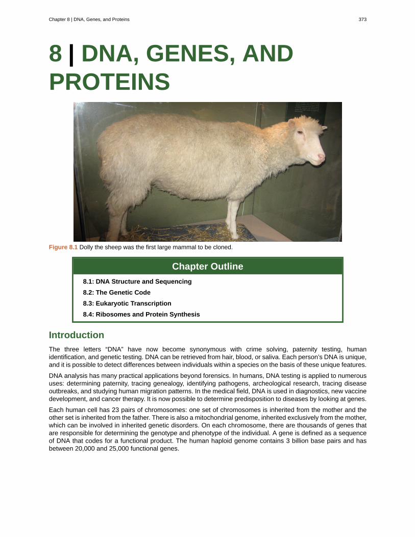
8 | DNA, GENES, ANDPROTEINS
Figure 8.1 Dolly the sheep was the first large mammal to be cloned.
Chapter Outline
8.1: DNA Structure and Sequencing
8.2: The Genetic Code
8.3: Eukaryotic Transcription
8.4: Ribosomes and Protein Synthesis
Introduction
The three letters “DNA” have now become synonymous with crime solving, paternity testing, humanidentification, and genetic testing. DNA can be retrieved from hair, blood, or saliva. Each person’s DNA is unique,and it is possible to detect differences between individuals within a species on the basis of these unique features.
DNA analysis has many practical applications beyond forensics. In humans, DNA testing is applied to numeroususes: determining paternity, tracing genealogy, identifying pathogens, archeological research, tracing diseaseoutbreaks, and studying human migration patterns. In the medical field, DNA is used in diagnostics, new vaccinedevelopment, and cancer therapy. It is now possible to determine predisposition to diseases by looking at genes.
Each human cell has 23 pairs of chromosomes: one set of chromosomes is inherited from the mother and theother set is inherited from the father. There is also a mitochondrial genome, inherited exclusively from the mother,which can be involved in inherited genetic disorders. On each chromosome, there are thousands of genes thatare responsible for determining the genotype and phenotype of the individual. A gene is defined as a sequenceof DNA that codes for a functional product. The human haploid genome contains 3 billion base pairs and hasbetween 20,000 and 25,000 functional genes.
Chapter 8 | DNA, Genes, and Proteins 373
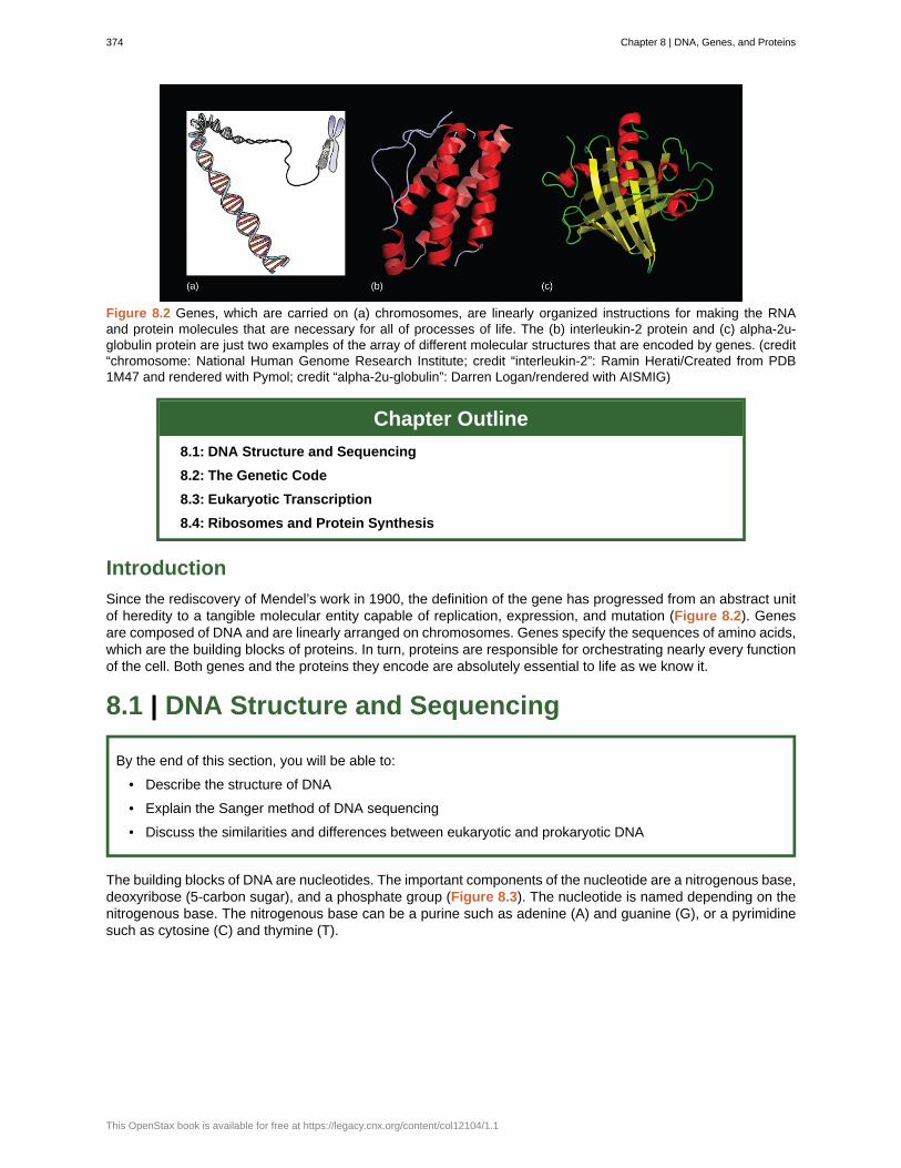
Figure 8.2 Genes, which are carried on (a) chromosomes, are linearly organized instructions for making the RNAand protein molecules that are necessary for all of processes of life. The (b) interleukin-2 protein and (c) alpha-2u-globulin protein are just two examples of the array of different molecular structures that are encoded by genes. (credit“chromosome: National Human Genome Research Institute; credit “interleukin-2”: Ramin Herati/Created from PDB1M47 and rendered with Pymol; credit “alpha-2u-globulin”: Darren Logan/rendered with AISMIG)
Chapter Outline
8.1: DNA Structure and Sequencing
8.2: The Genetic Code
8.3: Eukaryotic Transcription
8.4: Ribosomes and Protein Synthesis
Introduction
Since the rediscovery of Mendel’s work in 1900, the definition of the gene has progressed from an abstract unitof heredity to a tangible molecular entity capable of replication, expression, and mutation (Figure 8.2). Genesare composed of DNA and are linearly arranged on chromosomes. Genes specify the sequences of amino acids,which are the building blocks of proteins. In turn, proteins are responsible for orchestrating nearly every functionof the cell. Both genes and the proteins they encode are absolutely essential to life as we know it.
8.1 | DNA Structure and Sequencing
By the end of this section, you will be able to:
• Describe the structure of DNA
• Explain the Sanger method of DNA sequencing
• Discuss the similarities and differences between eukaryotic and prokaryotic DNA
The building blocks of DNA are nucleotides. The important components of the nucleotide are a nitrogenous base,deoxyribose (5-carbon sugar), and a phosphate group (Figure 8.3). The nucleotide is named depending on thenitrogenous base. The nitrogenous base can be a purine such as adenine (A) and guanine (G), or a pyrimidinesuch as cytosine (C) and thymine (T).
374 Chapter 8 | DNA, Genes, and Proteins
This OpenStax book is available for free at https://legacy.cnx.org/content/col12104/1.1
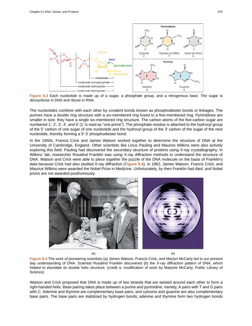
Figure 8.3 Each nucleotide is made up of a sugar, a phosphate group, and a nitrogenous base. The sugar isdeoxyribose in DNA and ribose in RNA.
The nucleotides combine with each other by covalent bonds known as phosphodiester bonds or linkages. Thepurines have a double ring structure with a six-membered ring fused to a five-membered ring. Pyrimidines aresmaller in size; they have a single six-membered ring structure. The carbon atoms of the five-carbon sugar arenumbered 1', 2', 3', 4', and 5' (1' is read as “one prime”). The phosphate residue is attached to the hydroxyl groupof the 5' carbon of one sugar of one nucleotide and the hydroxyl group of the 3' carbon of the sugar of the nextnucleotide, thereby forming a 5'-3' phosphodiester bond.
In the 1950s, Francis Crick and James Watson worked together to determine the structure of DNA at theUniversity of Cambridge, England. Other scientists like Linus Pauling and Maurice Wilkins were also activelyexploring this field. Pauling had discovered the secondary structure of proteins using X-ray crystallography. InWilkins’ lab, researcher Rosalind Franklin was using X-ray diffraction methods to understand the structure ofDNA. Watson and Crick were able to piece together the puzzle of the DNA molecule on the basis of Franklin'sdata because Crick had also studied X-ray diffraction (Figure 8.4). In 1962, James Watson, Francis Crick, andMaurice Wilkins were awarded the Nobel Prize in Medicine. Unfortunately, by then Franklin had died, and Nobelprizes are not awarded posthumously.
Figure 8.4 The work of pioneering scientists (a) James Watson, Francis Crick, and Maclyn McCarty led to our presentday understanding of DNA. Scientist Rosalind Franklin discovered (b) the X-ray diffraction pattern of DNA, whichhelped to elucidate its double helix structure. (credit a: modification of work by Marjorie McCarty, Public Library ofScience)
Watson and Crick proposed that DNA is made up of two strands that are twisted around each other to form aright-handed helix. Base pairing takes place between a purine and pyrimidine; namely, A pairs with T and G pairswith C. Adenine and thymine are complementary base pairs, and cytosine and guanine are also complementarybase pairs. The base pairs are stabilized by hydrogen bonds; adenine and thymine form two hydrogen bonds
Chapter 8 | DNA, Genes, and Proteins 375
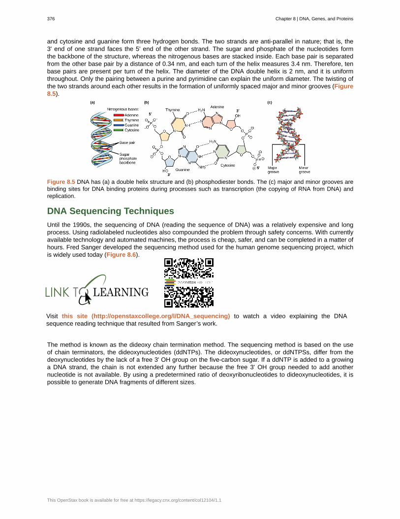
and cytosine and guanine form three hydrogen bonds. The two strands are anti-parallel in nature; that is, the3' end of one strand faces the 5' end of the other strand. The sugar and phosphate of the nucleotides formthe backbone of the structure, whereas the nitrogenous bases are stacked inside. Each base pair is separatedfrom the other base pair by a distance of 0.34 nm, and each turn of the helix measures 3.4 nm. Therefore, tenbase pairs are present per turn of the helix. The diameter of the DNA double helix is 2 nm, and it is uniformthroughout. Only the pairing between a purine and pyrimidine can explain the uniform diameter. The twisting ofthe two strands around each other results in the formation of uniformly spaced major and minor grooves (Figure8.5).
Figure 8.5 DNA has (a) a double helix structure and (b) phosphodiester bonds. The (c) major and minor grooves arebinding sites for DNA binding proteins during processes such as transcription (the copying of RNA from DNA) andreplication.
DNA Sequencing Techniques
Until the 1990s, the sequencing of DNA (reading the sequence of DNA) was a relatively expensive and longprocess. Using radiolabeled nucleotides also compounded the problem through safety concerns. With currentlyavailable technology and automated machines, the process is cheap, safer, and can be completed in a matter ofhours. Fred Sanger developed the sequencing method used for the human genome sequencing project, whichis widely used today (Figure 8.6).
Visit this site (http://openstaxcollege.org/l/DNA_sequencing) to watch a video explaining the DNAsequence reading technique that resulted from Sanger’s work.
The method is known as the dideoxy chain termination method. The sequencing method is based on the useof chain terminators, the dideoxynucleotides (ddNTPs). The dideoxynucleotides, or ddNTPSs, differ from thedeoxynucleotides by the lack of a free 3' OH group on the five-carbon sugar. If a ddNTP is added to a growinga DNA strand, the chain is not extended any further because the free 3' OH group needed to add anothernucleotide is not available. By using a predetermined ratio of deoxyribonucleotides to dideoxynucleotides, it ispossible to generate DNA fragments of different sizes.
376 Chapter 8 | DNA, Genes, and Proteins
This OpenStax book is available for free at https://legacy.cnx.org/content/col12104/1.1
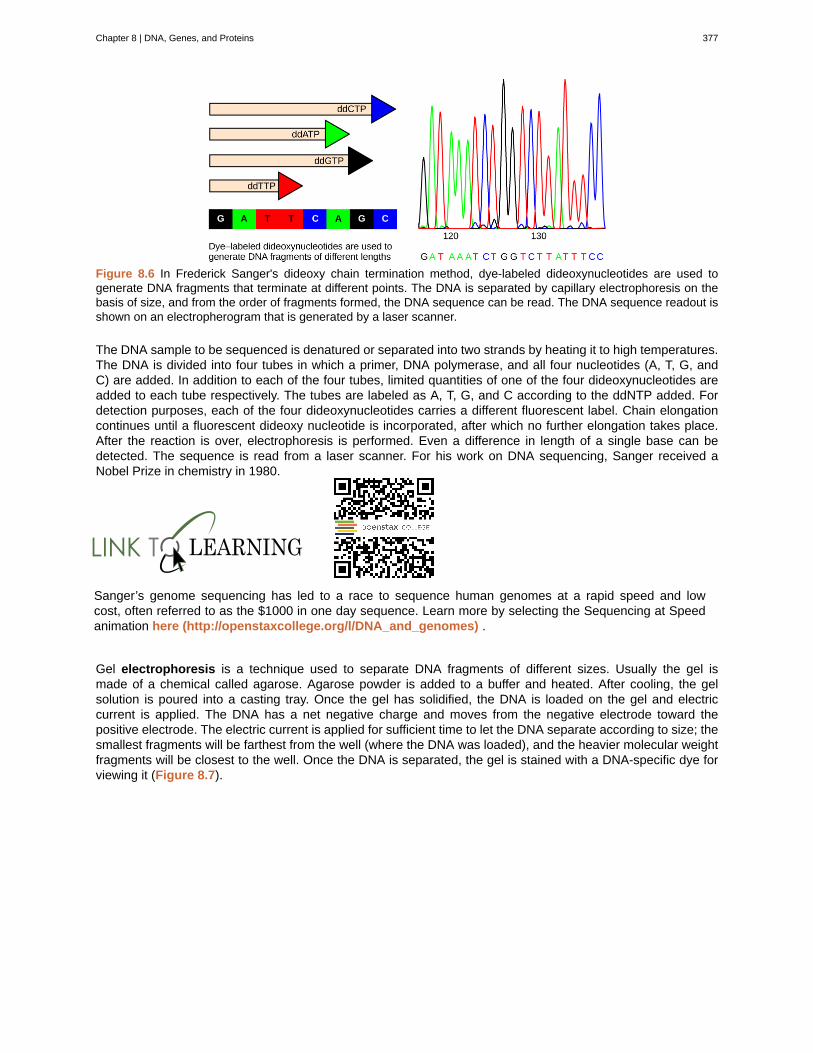
Figure 8.6 In Frederick Sanger's dideoxy chain termination method, dye-labeled dideoxynucleotides are used togenerate DNA fragments that terminate at different points. The DNA is separated by capillary electrophoresis on thebasis of size, and from the order of fragments formed, the DNA sequence can be read. The DNA sequence readout isshown on an electropherogram that is generated by a laser scanner.
The DNA sample to be sequenced is denatured or separated into two strands by heating it to high temperatures.The DNA is divided into four tubes in which a primer, DNA polymerase, and all four nucleotides (A, T, G, andC) are added. In addition to each of the four tubes, limited quantities of one of the four dideoxynucleotides areadded to each tube respectively. The tubes are labeled as A, T, G, and C according to the ddNTP added. Fordetection purposes, each of the four dideoxynucleotides carries a different fluorescent label. Chain elongationcontinues until a fluorescent dideoxy nucleotide is incorporated, after which no further elongation takes place.After the reaction is over, electrophoresis is performed. Even a difference in length of a single base can bedetected. The sequence is read from a laser scanner. For his work on DNA sequencing, Sanger received aNobel Prize in chemistry in 1980.
Sanger’s genome sequencing has led to a race to sequence human genomes at a rapid speed and lowcost, often referred to as the $1000 in one day sequence. Learn more by selecting the Sequencing at Speedanimation here (http://openstaxcollege.org/l/DNA_and_genomes) .
Gel electrophoresis is a technique used to separate DNA fragments of different sizes. Usually the gel ismade of a chemical called agarose. Agarose powder is added to a buffer and heated. After cooling, the gelsolution is poured into a casting tray. Once the gel has solidified, the DNA is loaded on the gel and electriccurrent is applied. The DNA has a net negative charge and moves from the negative electrode toward thepositive electrode. The electric current is applied for sufficient time to let the DNA separate according to size; thesmallest fragments will be farthest from the well (where the DNA was loaded), and the heavier molecular weightfragments will be closest to the well. Once the DNA is separated, the gel is stained with a DNA-specific dye forviewing it (Figure 8.7).
Chapter 8 | DNA, Genes, and Proteins 377
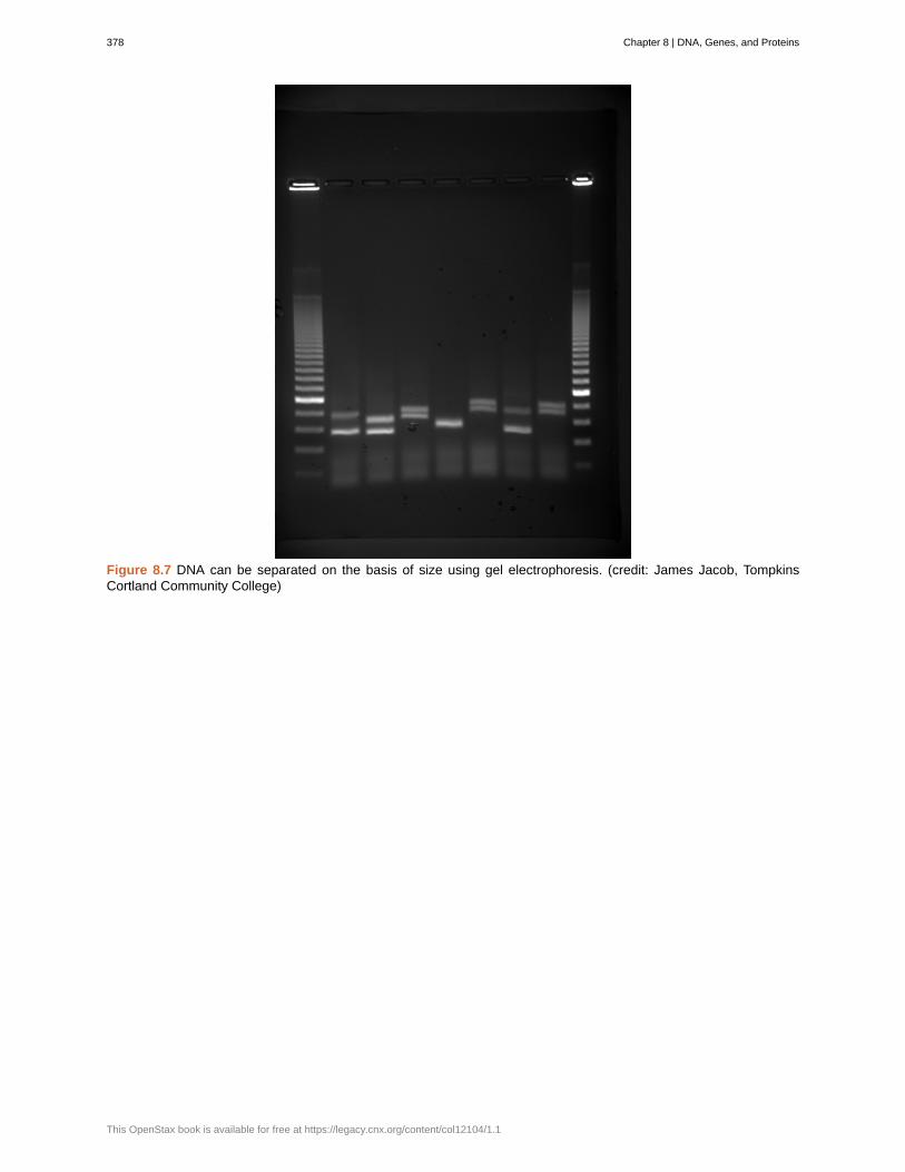
Figure 8.7 DNA can be separated on the basis of size using gel electrophoresis. (credit: James Jacob, TompkinsCortland Community College)
378 Chapter 8 | DNA, Genes, and Proteins
This OpenStax book is available for free at https://legacy.cnx.org/content/col12104/1.1
Neanderthal Genome: How Are We Related?The first draft sequence of the Neanderthal genome was recently published by Richard E. Green et al.
in 2010.[1]
Neanderthals are the closest ancestors of present-day humans. They were known to havelived in Europe and Western Asia before they disappeared from fossil records approximately 30,000 yearsago. Green’s team studied almost 40,000-year-old fossil remains that were selected from sites acrossthe world. Extremely sophisticated means of sample preparation and DNA sequencing were employedbecause of the fragile nature of the bones and heavy microbial contamination. In their study, the scientistswere able to sequence some four billion base pairs. The Neanderthal sequence was compared with thatof present-day humans from across the world. After comparing the sequences, the researchers foundthat the Neanderthal genome had 2 to 3 percent greater similarity to people living outside Africa thanto people in Africa. While current theories have suggested that all present-day humans can be traced toa small ancestral population in Africa, the data from the Neanderthal genome may contradict this view.Green and his colleagues also discovered DNA segments among people in Europe and Asia that aremore similar to Neanderthal sequences than to other contemporary human sequences. Another interestingobservation was that Neanderthals are as closely related to people from Papua New Guinea as to thosefrom China or France. This is surprising because Neanderthal fossil remains have been located onlyin Europe and West Asia. Most likely, genetic exchange took place between Neanderthals and modernhumans as modern humans emerged out of Africa, before the divergence of Europeans, East Asians, andPapua New Guineans.
Several genes seem to have undergone changes from Neanderthals during the evolution of present-day humans. These genes are involved in cranial structure, metabolism, skin morphology, and cognitivedevelopment. One of the genes that is of particular interest is RUNX2, which is different in modern dayhumans and Neanderthals. This gene is responsible for the prominent frontal bone, bell-shaped rib cage,and dental differences seen in Neanderthals. It is speculated that an evolutionary change in RUNX2 wasimportant in the origin of modern-day humans, and this affected the cranium and the upper body.
Watch Svante Pääbo’s talk (http://openstaxcollege.org/l/neanderthal) explaining the Neanderthal genomeresearch at the 2011 annual TED (Technology, Entertainment, Design) conference.
DNA Packaging in Cells
When comparing prokaryotic cells to eukaryotic cells, prokaryotes are much simpler than eukaryotes in many oftheir features (Figure 8.8). Most prokaryotes contain a single, circular chromosome that is found in an area ofthe cytoplasm called the nucleoid.
1. Richard E. Green et al., “A Draft Sequence of the Neandertal Genome,” Science 328 (2010): 710-22.
Chapter 8 | DNA, Genes, and Proteins 379
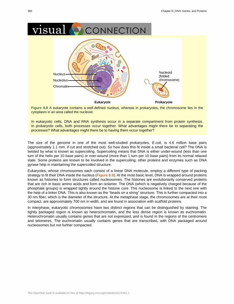
Figure 8.8 A eukaryote contains a well-defined nucleus, whereas in prokaryotes, the chromosome lies in thecytoplasm in an area called the nucleoid.
In eukaryotic cells, DNA and RNA synthesis occur in a separate compartment from protein synthesis.In prokaryotic cells, both processes occur together. What advantages might there be to separating theprocesses? What advantages might there be to having them occur together?
The size of the genome in one of the most well-studied prokaryotes, E.coli, is 4.6 million base pairs(approximately 1.1 mm, if cut and stretched out). So how does this fit inside a small bacterial cell? The DNA istwisted by what is known as supercoiling. Supercoiling means that DNA is either under-wound (less than oneturn of the helix per 10 base pairs) or over-wound (more than 1 turn per 10 base pairs) from its normal relaxedstate. Some proteins are known to be involved in the supercoiling; other proteins and enzymes such as DNAgyrase help in maintaining the supercoiled structure.
Eukaryotes, whose chromosomes each consist of a linear DNA molecule, employ a different type of packingstrategy to fit their DNA inside the nucleus (Figure 8.9). At the most basic level, DNA is wrapped around proteinsknown as histones to form structures called nucleosomes. The histones are evolutionarily conserved proteinsthat are rich in basic amino acids and form an octamer. The DNA (which is negatively charged because of thephosphate groups) is wrapped tightly around the histone core. This nucleosome is linked to the next one withthe help of a linker DNA. This is also known as the “beads on a string” structure. This is further compacted into a30 nm fiber, which is the diameter of the structure. At the metaphase stage, the chromosomes are at their mostcompact, are approximately 700 nm in width, and are found in association with scaffold proteins.
In interphase, eukaryotic chromosomes have two distinct regions that can be distinguished by staining. Thetightly packaged region is known as heterochromatin, and the less dense region is known as euchromatin.Heterochromatin usually contains genes that are not expressed, and is found in the regions of the centromereand telomeres. The euchromatin usually contains genes that are transcribed, with DNA packaged aroundnucleosomes but not further compacted.
380 Chapter 8 | DNA, Genes, and Proteins
This OpenStax book is available for free at https://legacy.cnx.org/content/col12104/1.1

Figure 8.9 These figures illustrate the compaction of the eukaryotic chromosome.
8.2 | The Genetic Code
By the end of this section, you will be able to:
• Explain the “central dogma” of protein synthesis
• Describe the genetic code and how the nucleotide sequence prescribes the amino acid and the proteinsequence
The cellular process of transcription generates messenger RNA (mRNA), a mobile molecular copy of one ormore genes with an alphabet of A, C, G, and uracil (U). Translation of the mRNA template converts nucleotide-based genetic information into a protein product. Protein sequences consist of 20 commonly occurring aminoacids; therefore, it can be said that the protein alphabet consists of 20 letters (Figure 8.10). Each amino acid isdefined by a three-nucleotide sequence called the triplet codon. Different amino acids have different chemistries(such as acidic versus basic, or polar and nonpolar) and different structural constraints. Variation in amino acidsequence gives rise to enormous variation in protein structure and function.
Chapter 8 | DNA, Genes, and Proteins 381

Figure 8.10 Structures of the 20 amino acids found in proteins are shown. Each amino acid is composed of an amino
group ( NH3+ ), a carboxyl group (COO-), and a side chain (blue). The side chain may be nonpolar, polar, or charged,
as well as large or small. It is the variety of amino acid side chains that gives rise to the incredible variation of proteinstructure and function.
The Central Dogma: DNA Encodes RNA; RNA Encodes Protein
The flow of genetic information in cells from DNA to mRNA to protein is described by the Central Dogma (Figure8.11), which states that genes specify the sequence of mRNAs, which in turn specify the sequence of proteins.The decoding of one molecule to another is performed by specific proteins and RNAs. Because the informationstored in DNA is so central to cellular function, it makes intuitive sense that the cell would make mRNA copiesof this information for protein synthesis, while keeping the DNA itself intact and protected. The copying of DNAto RNA is relatively straightforward, with one nucleotide being added to the mRNA strand for every nucleotideread in the DNA strand. The translation to protein is a bit more complex because three mRNA nucleotidescorrespond to one amino acid in the polypeptide sequence. However, the translation to protein is still systematicand colinear, such that nucleotides 1 to 3 correspond to amino acid 1, nucleotides 4 to 6 correspond to aminoacid 2, and so on.
382 Chapter 8 | DNA, Genes, and Proteins
This OpenStax book is available for free at https://legacy.cnx.org/content/col12104/1.1
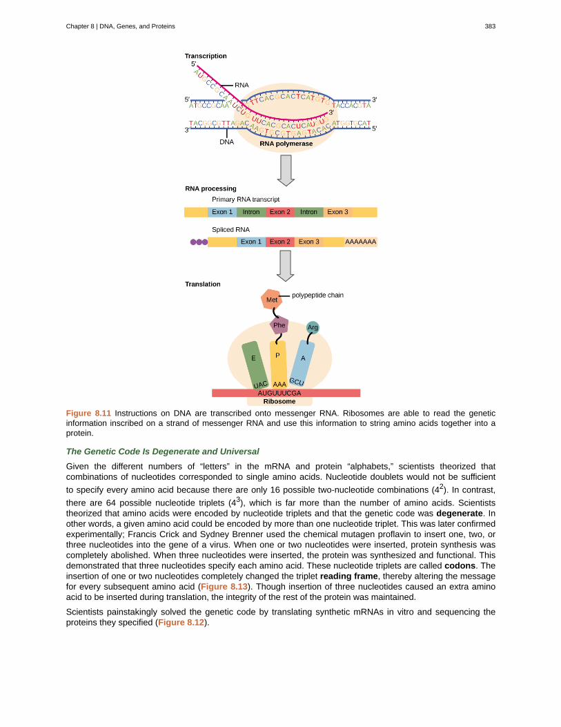
Figure 8.11 Instructions on DNA are transcribed onto messenger RNA. Ribosomes are able to read the geneticinformation inscribed on a strand of messenger RNA and use this information to string amino acids together into aprotein.
The Genetic Code Is Degenerate and Universal
Given the different numbers of “letters” in the mRNA and protein “alphabets,” scientists theorized thatcombinations of nucleotides corresponded to single amino acids. Nucleotide doublets would not be sufficient
to specify every amino acid because there are only 16 possible two-nucleotide combinations (42). In contrast,
there are 64 possible nucleotide triplets (43), which is far more than the number of amino acids. Scientiststheorized that amino acids were encoded by nucleotide triplets and that the genetic code was degenerate. Inother words, a given amino acid could be encoded by more than one nucleotide triplet. This was later confirmedexperimentally; Francis Crick and Sydney Brenner used the chemical mutagen proflavin to insert one, two, orthree nucleotides into the gene of a virus. When one or two nucleotides were inserted, protein synthesis wascompletely abolished. When three nucleotides were inserted, the protein was synthesized and functional. Thisdemonstrated that three nucleotides specify each amino acid. These nucleotide triplets are called codons. Theinsertion of one or two nucleotides completely changed the triplet reading frame, thereby altering the messagefor every subsequent amino acid (Figure 8.13). Though insertion of three nucleotides caused an extra aminoacid to be inserted during translation, the integrity of the rest of the protein was maintained.
Scientists painstakingly solved the genetic code by translating synthetic mRNAs in vitro and sequencing theproteins they specified (Figure 8.12).
Chapter 8 | DNA, Genes, and Proteins 383
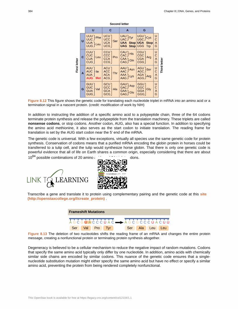
Figure 8.12 This figure shows the genetic code for translating each nucleotide triplet in mRNA into an amino acid or atermination signal in a nascent protein. (credit: modification of work by NIH)
In addition to instructing the addition of a specific amino acid to a polypeptide chain, three of the 64 codonsterminate protein synthesis and release the polypeptide from the translation machinery. These triplets are callednonsense codons, or stop codons. Another codon, AUG, also has a special function. In addition to specifyingthe amino acid methionine, it also serves as the start codon to initiate translation. The reading frame fortranslation is set by the AUG start codon near the 5' end of the mRNA.
The genetic code is universal. With a few exceptions, virtually all species use the same genetic code for proteinsynthesis. Conservation of codons means that a purified mRNA encoding the globin protein in horses could betransferred to a tulip cell, and the tulip would synthesize horse globin. That there is only one genetic code ispowerful evidence that all of life on Earth shares a common origin, especially considering that there are about
1084 possible combinations of 20 amino acids and 64 triplet codons.
Transcribe a gene and translate it to protein using complementary pairing and the genetic code at this site(http://openstaxcollege.org/l/create_protein) .
Figure 8.13 The deletion of two nucleotides shifts the reading frame of an mRNA and changes the entire proteinmessage, creating a nonfunctional protein or terminating protein synthesis altogether.
Degeneracy is believed to be a cellular mechanism to reduce the negative impact of random mutations. Codonsthat specify the same amino acid typically only differ by one nucleotide. In addition, amino acids with chemicallysimilar side chains are encoded by similar codons. This nuance of the genetic code ensures that a single-nucleotide substitution mutation might either specify the same amino acid but have no effect or specify a similaramino acid, preventing the protein from being rendered completely nonfunctional.
384 Chapter 8 | DNA, Genes, and Proteins
This OpenStax book is available for free at https://legacy.cnx.org/content/col12104/1.1
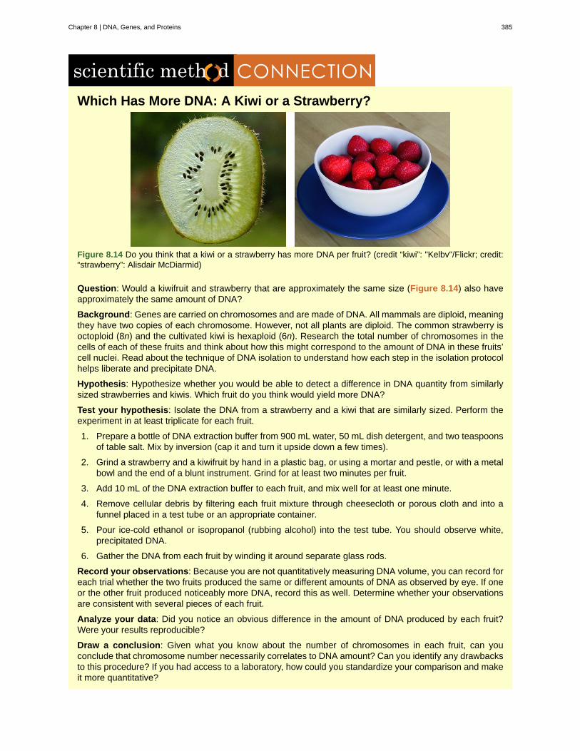
Which Has More DNA: A Kiwi or a Strawberry?
Figure 8.14 Do you think that a kiwi or a strawberry has more DNA per fruit? (credit “kiwi”: "Kelbv"/Flickr; credit:“strawberry”: Alisdair McDiarmid)
Question: Would a kiwifruit and strawberry that are approximately the same size (Figure 8.14) also haveapproximately the same amount of DNA?
Background: Genes are carried on chromosomes and are made of DNA. All mammals are diploid, meaningthey have two copies of each chromosome. However, not all plants are diploid. The common strawberry isoctoploid (8n) and the cultivated kiwi is hexaploid (6n). Research the total number of chromosomes in thecells of each of these fruits and think about how this might correspond to the amount of DNA in these fruits’cell nuclei. Read about the technique of DNA isolation to understand how each step in the isolation protocolhelps liberate and precipitate DNA.
Hypothesis: Hypothesize whether you would be able to detect a difference in DNA quantity from similarlysized strawberries and kiwis. Which fruit do you think would yield more DNA?
Test your hypothesis: Isolate the DNA from a strawberry and a kiwi that are similarly sized. Perform theexperiment in at least triplicate for each fruit.
1. Prepare a bottle of DNA extraction buffer from 900 mL water, 50 mL dish detergent, and two teaspoonsof table salt. Mix by inversion (cap it and turn it upside down a few times).
2. Grind a strawberry and a kiwifruit by hand in a plastic bag, or using a mortar and pestle, or with a metalbowl and the end of a blunt instrument. Grind for at least two minutes per fruit.
3. Add 10 mL of the DNA extraction buffer to each fruit, and mix well for at least one minute.
4. Remove cellular debris by filtering each fruit mixture through cheesecloth or porous cloth and into afunnel placed in a test tube or an appropriate container.
5. Pour ice-cold ethanol or isopropanol (rubbing alcohol) into the test tube. You should observe white,precipitated DNA.
6. Gather the DNA from each fruit by winding it around separate glass rods.
Record your observations: Because you are not quantitatively measuring DNA volume, you can record foreach trial whether the two fruits produced the same or different amounts of DNA as observed by eye. If oneor the other fruit produced noticeably more DNA, record this as well. Determine whether your observationsare consistent with several pieces of each fruit.
Analyze your data: Did you notice an obvious difference in the amount of DNA produced by each fruit?Were your results reproducible?
Draw a conclusion: Given what you know about the number of chromosomes in each fruit, can youconclude that chromosome number necessarily correlates to DNA amount? Can you identify any drawbacksto this procedure? If you had access to a laboratory, how could you standardize your comparison and makeit more quantitative?
Chapter 8 | DNA, Genes, and Proteins 385
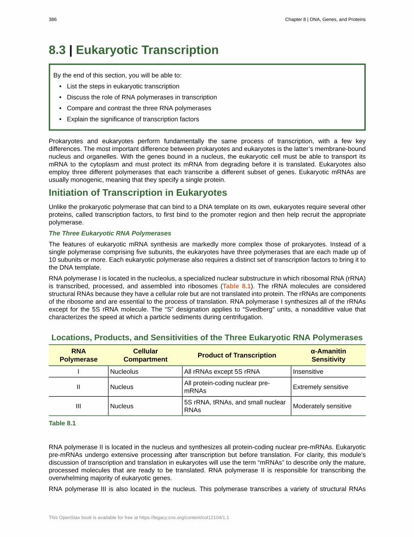
8.3 | Eukaryotic Transcription
By the end of this section, you will be able to:
• List the steps in eukaryotic transcription
• Discuss the role of RNA polymerases in transcription
• Compare and contrast the three RNA polymerases
• Explain the significance of transcription factors
Prokaryotes and eukaryotes perform fundamentally the same process of transcription, with a few keydifferences. The most important difference between prokaryotes and eukaryotes is the latter’s membrane-boundnucleus and organelles. With the genes bound in a nucleus, the eukaryotic cell must be able to transport itsmRNA to the cytoplasm and must protect its mRNA from degrading before it is translated. Eukaryotes alsoemploy three different polymerases that each transcribe a different subset of genes. Eukaryotic mRNAs areusually monogenic, meaning that they specify a single protein.
Initiation of Transcription in Eukaryotes
Unlike the prokaryotic polymerase that can bind to a DNA template on its own, eukaryotes require several otherproteins, called transcription factors, to first bind to the promoter region and then help recruit the appropriatepolymerase.
The Three Eukaryotic RNA Polymerases
The features of eukaryotic mRNA synthesis are markedly more complex those of prokaryotes. Instead of asingle polymerase comprising five subunits, the eukaryotes have three polymerases that are each made up of10 subunits or more. Each eukaryotic polymerase also requires a distinct set of transcription factors to bring it tothe DNA template.
RNA polymerase I is located in the nucleolus, a specialized nuclear substructure in which ribosomal RNA (rRNA)is transcribed, processed, and assembled into ribosomes (Table 8.1). The rRNA molecules are consideredstructural RNAs because they have a cellular role but are not translated into protein. The rRNAs are componentsof the ribosome and are essential to the process of translation. RNA polymerase I synthesizes all of the rRNAsexcept for the 5S rRNA molecule. The “S” designation applies to “Svedberg” units, a nonadditive value thatcharacterizes the speed at which a particle sediments during centrifugation.
Locations, Products, and Sensitivities of the Three Eukaryotic RNA Polymerases
RNAPolymerase
CellularCompartment
Product of Transcriptionα-AmanitinSensitivity
I Nucleolus All rRNAs except 5S rRNA Insensitive
II NucleusAll protein-coding nuclear pre-mRNAs
Extremely sensitive
III Nucleus5S rRNA, tRNAs, and small nuclearRNAs
Moderately sensitive
Table 8.1
RNA polymerase II is located in the nucleus and synthesizes all protein-coding nuclear pre-mRNAs. Eukaryoticpre-mRNAs undergo extensive processing after transcription but before translation. For clarity, this module’sdiscussion of transcription and translation in eukaryotes will use the term “mRNAs” to describe only the mature,processed molecules that are ready to be translated. RNA polymerase II is responsible for transcribing theoverwhelming majority of eukaryotic genes.
RNA polymerase III is also located in the nucleus. This polymerase transcribes a variety of structural RNAs
386 Chapter 8 | DNA, Genes, and Proteins
This OpenStax book is available for free at https://legacy.cnx.org/content/col12104/1.1
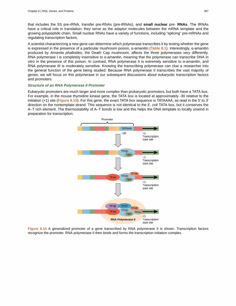
that includes the 5S pre-rRNA, transfer pre-RNAs (pre-tRNAs), and small nuclear pre- RNAs. The tRNAshave a critical role in translation; they serve as the adaptor molecules between the mRNA template and thegrowing polypeptide chain. Small nuclear RNAs have a variety of functions, including “splicing” pre-mRNAs andregulating transcription factors.
A scientist characterizing a new gene can determine which polymerase transcribes it by testing whether the geneis expressed in the presence of a particular mushroom poison, α-amanitin (Table 8.1). Interestingly, α-amanitinproduced by Amanita phalloides, the Death Cap mushroom, affects the three polymerases very differently.RNA polymerase I is completely insensitive to α-amanitin, meaning that the polymerase can transcribe DNA invitro in the presence of this poison. In contrast, RNA polymerase II is extremely sensitive to α-amanitin, andRNA polymerase III is moderately sensitive. Knowing the transcribing polymerase can clue a researcher intothe general function of the gene being studied. Because RNA polymerase II transcribes the vast majority ofgenes, we will focus on this polymerase in our subsequent discussions about eukaryotic transcription factorsand promoters.
Structure of an RNA Polymerase II Promoter
Eukaryotic promoters are much larger and more complex than prokaryotic promoters, but both have a TATA box.For example, in the mouse thymidine kinase gene, the TATA box is located at approximately -30 relative to theinitiation (+1) site (Figure 8.15). For this gene, the exact TATA box sequence is TATAAAA, as read in the 5' to 3'direction on the nontemplate strand. This sequence is not identical to the E. coli TATA box, but it conserves theA–T rich element. The thermostability of A–T bonds is low and this helps the DNA template to locally unwind inpreparation for transcription.
Figure 8.15 A generalized promoter of a gene transcribed by RNA polymerase II is shown. Transcription factorsrecognize the promoter. RNA polymerase II then binds and forms the transcription initiation complex.
Chapter 8 | DNA, Genes, and Proteins 387

Figure 8.16 Eukaryotic mRNA contains introns that must be spliced out. A 5' cap and 3' poly-A tail are also added.
A scientist splices a eukaryotic promoter in front of a bacterial gene and inserts the gene in a bacterialchromosome. Would you expect the bacteria to transcribe the gene?
The mouse genome includes one gene and two pseudogenes for cytoplasmic thymidine kinase. Pseudogenesare genes that have lost their protein-coding ability or are no longer expressed by the cell. These pseudogenesare copied from mRNA and incorporated into the chromosome. For example, the mouse thymidine kinasepromoter also has a conserved CAAT box (GGCCAATCT) at approximately -80. This sequence is essential andis involved in binding transcription factors. Further upstream of the TATA box, eukaryotic promoters may alsocontain one or more GC-rich boxes (GGCG) or octamer boxes (ATTTGCAT). These elements bind cellularfactors that increase the efficiency of transcription initiation and are often identified in more “active” genes thatare constantly being expressed by the cell.
Transcription Factors for RNA Polymerase II
The complexity of eukaryotic transcription does not end with the polymerases and promoters. An army of basaltranscription factors, enhancers, and silencers also help to regulate the frequency with which pre-mRNA issynthesized from a gene. Enhancers and silencers affect the efficiency of transcription but are not necessary fortranscription to proceed. Basal transcription factors are crucial in the formation of a preinitiation complex onthe DNA template that subsequently recruits RNA polymerase II for transcription initiation.
The names of the basal transcription factors begin with “TFII” (this is the transcription factor for RNA polymeraseII) and are specified with the letters A–J. The transcription factors systematically fall into place on the DNAtemplate, with each one further stabilizing the preinitiation complex and contributing to the recruitment of RNApolymerase II.
The processes of bringing RNA polymerases I and III to the DNA template involve slightly less complexcollections of transcription factors, but the general theme is the same. Eukaryotic transcription is a tightlyregulated process that requires a variety of proteins to interact with each other and with the DNA strand.Although the process of transcription in eukaryotes involves a greater metabolic investment than in prokaryotes,it ensures that the cell transcribes precisely the pre-mRNAs that it needs for protein synthesis.
388 Chapter 8 | DNA, Genes, and Proteins
This OpenStax book is available for free at https://legacy.cnx.org/content/col12104/1.1
The Evolution of PromotersThe evolution of genes may be a familiar concept. Mutations can occur in genes during DNA replication, andthe result may or may not be beneficial to the cell. By altering an enzyme, structural protein, or some otherfactor, the process of mutation can transform functions or physical features. However, eukaryotic promotersand other gene regulatory sequences may evolve as well. For instance, consider a gene that, over manygenerations, becomes more valuable to the cell. Maybe the gene encodes a structural protein that the cellneeds to synthesize in abundance for a certain function. If this is the case, it would be beneficial to the cellfor that gene’s promoter to recruit transcription factors more efficiently and increase gene expression.
Scientists examining the evolution of promoter sequences have reported varying results. In part, this isbecause it is difficult to infer exactly where a eukaryotic promoter begins and ends. Some promoters occurwithin genes; others are located very far upstream, or even downstream, of the genes they are regulating.However, when researchers limited their examination to human core promoter sequences that were definedexperimentally as sequences that bind the preinitiation complex, they found that promoters evolve evenfaster than protein-coding genes.
It is still unclear how promoter evolution might correspond to the evolution of humans or other higherorganisms. However, the evolution of a promoter to effectively make more or less of a given gene product
is an intriguing alternative to the evolution of the genes themselves.[2]
Promoter Structures for RNA Polymerases I and III
In eukaryotes, the conserved promoter elements differ for genes transcribed by RNA polymerases I, II, and III.RNA polymerase I transcribes genes that have two GC-rich promoter sequences in the -45 to +20 region. Thesesequences alone are sufficient for transcription initiation to occur, but promoters with additional sequences in theregion from -180 to -105 upstream of the initiation site will further enhance initiation. Genes that are transcribedby RNA polymerase III have upstream promoters or promoters that occur within the genes themselves.
Eukaryotic Elongation and Termination
Following the formation of the preinitiation complex, the polymerase is released from the other transcriptionfactors, and elongation is allowed to proceed as it does in prokaryotes with the polymerase synthesizing pre-mRNA in the 5' to 3' direction. As discussed previously, RNA polymerase II transcribes the major share ofeukaryotic genes, so this section will focus on how this polymerase accomplishes elongation and termination.
Although the enzymatic process of elongation is essentially the same in eukaryotes and prokaryotes, the DNAtemplate is more complex. When eukaryotic cells are not dividing, their genes exist as a diffuse mass ofDNA and proteins called chromatin. The DNA is tightly packaged around charged histone proteins at repeatedintervals. These DNA–histone complexes, collectively called nucleosomes, are regularly spaced and include 146nucleotides of DNA wound around eight histones like thread around a spool.
For polynucleotide synthesis to occur, the transcription machinery needs to move histones out of the wayevery time it encounters a nucleosome. This is accomplished by a special protein complex called FACT, whichstands for “facilitates chromatin transcription.” This complex pulls histones away from the DNA template as thepolymerase moves along it. Once the pre-mRNA is synthesized, the FACT complex replaces the histones torecreate the nucleosomes.
The termination of transcription is different for the different polymerases. Unlike in prokaryotes, elongation byRNA polymerase II in eukaryotes takes place 1,000–2,000 nucleotides beyond the end of the gene beingtranscribed. This pre-mRNA tail is subsequently removed by cleavage during mRNA processing. On the otherhand, RNA polymerases I and III require termination signals. Genes transcribed by RNA polymerase I contain aspecific 18-nucleotide sequence that is recognized by a termination protein. The process of termination in RNApolymerase III involves an mRNA hairpin similar to rho-independent termination of transcription in prokaryotes.
2. H Liang et al., “Fast evolution of core promoters in primate genomes,” Molecular Biology and Evolution 25 (2008): 1239–44.
Chapter 8 | DNA, Genes, and Proteins 389

8.4 | Ribosomes and Protein Synthesis
By the end of this section, you will be able to:
• Describe the different steps in protein synthesis
• Discuss the role of ribosomes in protein synthesis
The synthesis of proteins consumes more of a cell’s energy than any other metabolic process. In turn, proteinsaccount for more mass than any other component of living organisms (with the exception of water), and proteinsperform virtually every function of a cell. The process of translation, or protein synthesis, involves the decoding ofan mRNA message into a polypeptide product. Amino acids are covalently strung together by interlinking peptidebonds in lengths ranging from approximately 50 amino acid residues to more than 1,000. Each individual aminoacid has an amino group (NH2) and a carboxyl (COOH) group. Polypeptides are formed when the amino groupof one amino acid forms an amide (i.e., peptide) bond with the carboxyl group of another amino acid (Figure8.17). This reaction is catalyzed by ribosomes and generates one water molecule.
Figure 8.17 A peptide bond links the carboxyl end of one amino acid with the amino end of another, expelling onewater molecule. For simplicity in this image, only the functional groups involved in the peptide bond are shown. The Rand R' designations refer to the rest of each amino acid structure.
The Protein Synthesis Machinery
In addition to the mRNA template, many molecules and macromolecules contribute to the process of translation.The composition of each component may vary across species; for instance, ribosomes may consist of differentnumbers of rRNAs and polypeptides depending on the organism. However, the general structures and functionsof the protein synthesis machinery are comparable from bacteria to human cells. Translation requires the inputof an mRNA template, ribosomes, tRNAs, and various enzymatic factors.
Click through the steps of this PBS interactive (http://openstaxcollege.org/l/prokary_protein) to seeprotein synthesis in action.
Ribosomes
Even before an mRNA is translated, a cell must invest energy to build each of its ribosomes. In E. coli, thereare between 10,000 and 70,000 ribosomes present in each cell at any given time. A ribosome is a complexmacromolecule composed of structural and catalytic rRNAs, and many distinct polypeptides. In eukaryotes, thenucleolus is completely specialized for the synthesis and assembly of rRNAs.
390 Chapter 8 | DNA, Genes, and Proteins
This OpenStax book is available for free at https://legacy.cnx.org/content/col12104/1.1
Ribosomes exist in the cytoplasm in prokaryotes and in the cytoplasm and rough endoplasmic reticulum ineukaryotes. Mitochondria and chloroplasts also have their own ribosomes in the matrix and stroma, which lookmore similar to prokaryotic ribosomes (and have similar drug sensitivities) than the ribosomes just outside theirouter membranes in the cytoplasm. Ribosomes dissociate into large and small subunits when they are notsynthesizing proteins and reassociate during the initiation of translation. In E. coli, the small subunit is describedas 30S, and the large subunit is 50S, for a total of 70S (recall that Svedberg units are not additive). Mammalianribosomes have a small 40S subunit and a large 60S subunit, for a total of 80S. The small subunit is responsiblefor binding the mRNA template, whereas the large subunit sequentially binds tRNAs. Each mRNA molecule issimultaneously translated by many ribosomes, all synthesizing protein in the same direction: reading the mRNAfrom 5' to 3' and synthesizing the polypeptide from the N terminus to the C terminus. The complete mRNA/poly-ribosome structure is called a polysome.
tRNAs
The tRNAs are structural RNA molecules that were transcribed from genes by RNA polymerase III. Dependingon the species, 40 to 60 types of tRNAs exist in the cytoplasm. Serving as adaptors, specific tRNAs bind tosequences on the mRNA template and add the corresponding amino acid to the polypeptide chain. Therefore,tRNAs are the molecules that actually “translate” the language of RNA into the language of proteins.
Of the 64 possible mRNA codons—or triplet combinations of A, U, G, and C—three specify the termination ofprotein synthesis and 61 specify the addition of amino acids to the polypeptide chain. Of these 61, one codon(AUG) also encodes the initiation of translation. Each tRNA anticodon can base pair with one of the mRNAcodons and add an amino acid or terminate translation, according to the genetic code. For instance, if thesequence CUA occurred on an mRNA template in the proper reading frame, it would bind a tRNA expressingthe complementary sequence, GAU, which would be linked to the amino acid leucine.
As the adaptor molecules of translation, it is surprising that tRNAs can fit so much specificity into such a smallpackage. Consider that tRNAs need to interact with three factors: 1) they must be recognized by the correctaminoacyl synthetase (see below); 2) they must be recognized by ribosomes; and 3) they must bind to thecorrect sequence in mRNA.
Aminoacyl tRNA Synthetases
The process of pre-tRNA synthesis by RNA polymerase III only creates the RNA portion of the adaptor molecule.The corresponding amino acid must be added later, once the tRNA is processed and exported to the cytoplasm.Through the process of tRNA “charging,” each tRNA molecule is linked to its correct amino acid by a group ofenzymes called aminoacyl tRNA synthetases. At least one type of aminoacyl tRNA synthetase exists for eachof the 20 amino acids; the exact number of aminoacyl tRNA synthetases varies by species. These enzymes firstbind and hydrolyze ATP to catalyze a high-energy bond between an amino acid and adenosine monophosphate(AMP); a pyrophosphate molecule is expelled in this reaction. The activated amino acid is then transferred to thetRNA, and AMP is released.
The Mechanism of Protein Synthesis
As with mRNA synthesis, protein synthesis can be divided into three phases: initiation, elongation, andtermination. The process of translation is similar in prokaryotes and eukaryotes. Here we’ll explore howtranslation occurs in E. coli, a representative prokaryote, and specify any differences between prokaryotic andeukaryotic translation.
Initiation of Translation
Protein synthesis begins with the formation of an initiation complex. In E. coli, this complex involves thesmall 30S ribosome, the mRNA template, three initiation factors (IFs; IF-1, IF-2, and IF-3), and a special
initiator tRNA, called tRNAfMet . The initiator tRNA interacts with the start codon AUG (or rarely, GUG),
links to a formylated methionine called fMet, and can also bind IF-2. Formylated methionine is inserted by
fMet − tRNAfMet at the beginning of every polypeptide chain synthesized by E. coli, but it is usually clipped
off after translation is complete. When an in-frame AUG is encountered during translation elongation, a non-
formylated methionine is inserted by a regular Met-tRNAMet.
In E. coli mRNA, a sequence upstream of the first AUG codon, called the Shine-Dalgarno sequence(AGGAGG), interacts with the rRNA molecules that compose the ribosome. This interaction anchors the 30Sribosomal subunit at the correct location on the mRNA template. Guanosine triphosphate (GTP), which is apurine nucleotide triphosphate, acts as an energy source during translation—both at the start of elongation and
Chapter 8 | DNA, Genes, and Proteins 391

during the ribosome’s translocation.
In eukaryotes, a similar initiation complex forms, comprising mRNA, the 40S small ribosomal subunit, IFs, andnucleoside triphosphates (GTP and ATP). The charged initiator tRNA, called Met-tRNAi, does not bind fMet ineukaryotes, but is distinct from other Met-tRNAs in that it can bind IFs.
Instead of depositing at the Shine-Dalgarno sequence, the eukaryotic initiation complex recognizes the7-methylguanosine cap at the 5' end of the mRNA. A cap-binding protein (CBP) and several other IFs assist themovement of the ribosome to the 5' cap. Once at the cap, the initiation complex tracks along the mRNA in the 5'to 3' direction, searching for the AUG start codon. Many eukaryotic mRNAs are translated from the first AUG, butthis is not always the case. According to Kozak’s rules, the nucleotides around the AUG indicate whether it isthe correct start codon. Kozak’s rules state that the following consensus sequence must appear around the AUGof vertebrate genes: 5'-gccRccAUGG-3'. The R (for purine) indicates a site that can be either A or G, but cannotbe C or U. Essentially, the closer the sequence is to this consensus, the higher the efficiency of translation.
Once the appropriate AUG is identified, the other proteins and CBP dissociate, and the 60S subunit bindsto the complex of Met-tRNAi, mRNA, and the 40S subunit. This step completes the initiation of translation ineukaryotes.
Translation, Elongation, and Termination
In prokaryotes and eukaryotes, the basics of elongation are the same, so we will review elongation from theperspective of E. coli. The 50S ribosomal subunit of E. coli consists of three compartments: the A (aminoacyl)site binds incoming charged aminoacyl tRNAs. The P (peptidyl) site binds charged tRNAs carrying amino acidsthat have formed peptide bonds with the growing polypeptide chain but have not yet dissociated from theircorresponding tRNA. The E (exit) site releases dissociated tRNAs so that they can be recharged with free amino
acids. There is one exception to this assembly line of tRNAs: in E. coli, fMet − tRNAfMet is capable of entering
the P site directly without first entering the A site. Similarly, the eukaryotic Met-tRNAi, with help from otherproteins of the initiation complex, binds directly to the P site. In both cases, this creates an initiation complexwith a free A site ready to accept the tRNA corresponding to the first codon after the AUG.
During translation elongation, the mRNA template provides specificity. As the ribosome moves along the mRNA,each mRNA codon comes into register, and specific binding with the corresponding charged tRNA anticodon isensured. If mRNA were not present in the elongation complex, the ribosome would bind tRNAs nonspecifically.
Elongation proceeds with charged tRNAs entering the A site and then shifting to the P site followed by the Esite with each single-codon “step” of the ribosome. Ribosomal steps are induced by conformational changes thatadvance the ribosome by three bases in the 3' direction. The energy for each step of the ribosome is donatedby an elongation factor that hydrolyzes GTP. Peptide bonds form between the amino group of the amino acidattached to the A-site tRNA and the carboxyl group of the amino acid attached to the P-site tRNA. The formationof each peptide bond is catalyzed by peptidyl transferase, an RNA-based enzyme that is integrated into the50S ribosomal subunit. The energy for each peptide bond formation is derived from GTP hydrolysis, which iscatalyzed by a separate elongation factor. The amino acid bound to the P-site tRNA is also linked to the growingpolypeptide chain. As the ribosome steps across the mRNA, the former P-site tRNA enters the E site, detachesfrom the amino acid, and is expelled (Figure 8.18). Amazingly, the E. coli translation apparatus takes only 0.05seconds to add each amino acid, meaning that a 200-amino acid protein can be translated in just 10 seconds.
392 Chapter 8 | DNA, Genes, and Proteins
This OpenStax book is available for free at https://legacy.cnx.org/content/col12104/1.1
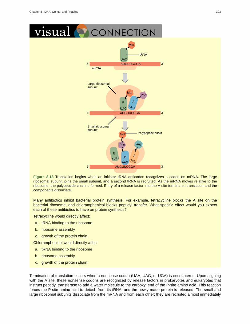
Figure 8.18 Translation begins when an initiator tRNA anticodon recognizes a codon on mRNA. The largeribosomal subunit joins the small subunit, and a second tRNA is recruited. As the mRNA moves relative to theribosome, the polypeptide chain is formed. Entry of a release factor into the A site terminates translation and thecomponents dissociate.
Many antibiotics inhibit bacterial protein synthesis. For example, tetracycline blocks the A site on thebacterial ribosome, and chloramphenicol blocks peptidyl transfer. What specific effect would you expecteach of these antibiotics to have on protein synthesis?
Tetracycline would directly affect:
a. tRNA binding to the ribosome
b. ribosome assembly
c. growth of the protein chain
Chloramphenicol would directly affect
a. tRNA binding to the ribosome
b. ribosome assembly
c. growth of the protein chain
Termination of translation occurs when a nonsense codon (UAA, UAG, or UGA) is encountered. Upon aligningwith the A site, these nonsense codons are recognized by release factors in prokaryotes and eukaryotes thatinstruct peptidyl transferase to add a water molecule to the carboxyl end of the P-site amino acid. This reactionforces the P-site amino acid to detach from its tRNA, and the newly made protein is released. The small andlarge ribosomal subunits dissociate from the mRNA and from each other; they are recruited almost immediately
Chapter 8 | DNA, Genes, and Proteins 393
into another translation initiation complex. After many ribosomes have completed translation, the mRNA isdegraded so the nucleotides can be reused in another transcription reaction.
Protein Folding, Modification, and Targeting
During and after translation, individual amino acids may be chemically modified, signal sequences may beappended, and the new protein “folds” into a distinct three-dimensional structure as a result of intramolecularinteractions. A signal sequence is a short tail of amino acids that directs a protein to a specific cellularcompartment. These sequences at the amino end or the carboxyl end of the protein can be thought of as theprotein’s “train ticket” to its ultimate destination. Other cellular factors recognize each signal sequence and helptransport the protein from the cytoplasm to its correct compartment. For instance, a specific sequence at theamino terminus will direct a protein to the mitochondria or chloroplasts (in plants). Once the protein reaches itscellular destination, the signal sequence is usually clipped off.
Many proteins fold spontaneously, but some proteins require helper molecules, called chaperones, to preventthem from aggregating during the complicated process of folding. Even if a protein is properly specified byits corresponding mRNA, it could take on a completely dysfunctional shape if abnormal temperature or pHconditions prevent it from folding correctly.
394 Chapter 8 | DNA, Genes, and Proteins
This OpenStax book is available for free at https://legacy.cnx.org/content/col12104/1.1
aminoacyl tRNA synthetase
CAAT box
Central Dogma
codon
colinear
degeneracy
electrophoresis
FACT
GC-rich box
initiator tRNA
Kozak’s rules
nonsense codon
Octamer box
peptidyl transferase
polysome
preinitiation complex
reading frame
Shine-Dalgarno sequence
signal sequence
small nuclear RNA
start codon
KEY TERMS
enzyme that “charges” tRNA molecules by catalyzing a bond between the tRNAand a corresponding amino acid
(GGCCAATCT) essential eukaryotic promoter sequence involved in binding transcription factors
states that genes specify the sequence of mRNAs, which in turn specify the sequence ofproteins
three consecutive nucleotides in mRNA that specify the insertion of an amino acid or the release of apolypeptide chain during translation
in terms of RNA and protein, three “units” of RNA (nucleotides) specify one “unit” of protein (aminoacid) in a consecutive fashion
(of the genetic code) describes that a given amino acid can be encoded by more than onenucleotide triplet; the code is degenerate, but not ambiguous
technique used to separate DNA fragments according to size
complex that “facilitates chromatin transcription” by disassembling nucleosomes ahead of a transcribingRNA polymerase II and reassembling them after the polymerase passes by
(GGCG) nonessential eukaryotic promoter sequence that binds cellular factors to increase theefficiency of transcription; may be present several times in a promoter
in prokaryotes, called tRNA fMet ; in eukaryotes, called tRNAi; a tRNA that interacts with a start
codon, binds directly to the ribosome P site, and links to a special methionine to begin a polypeptide chain
determines the correct initiation AUG in a eukaryotic mRNA; the following consensus sequencemust appear around the AUG: 5’-GCC(purine)CCAUGG-3’; the bolded bases are most important
one of the three mRNA codons that specifies termination of translation
(ATTTGCAT) nonessential eukaryotic promoter sequence that binds cellular factors to increasethe efficiency of transcription; may be present several times in a promoter
RNA-based enzyme that is integrated into the 50S ribosomal subunit and catalyzes theformation of peptide bonds
mRNA molecule simultaneously being translated by many ribosomes all going in the same direction
cluster of transcription factors and other proteins that recruit RNA polymerase II fortranscription of a DNA template
sequence of triplet codons in mRNA that specify a particular protein; a ribosome shift of one ortwo nucleotides in either direction completely abolishes synthesis of that protein
(AGGAGG); initiates prokaryotic translation by interacting with rRNA moleculescomprising the 30S ribosome
short tail of amino acids that directs a protein to a specific cellular compartment
molecules synthesized by RNA polymerase III that have a variety of functions, includingsplicing pre-mRNAs and regulating transcription factors
AUG (or rarely, GUG) on an mRNA from which translation begins; always specifies methionine
Chapter 8 | DNA, Genes, and Proteins 395
CHAPTER SUMMARY
8.1 DNA Structure and Sequencing
The currently accepted model of the double-helix structure of DNA was proposed by Watson and Crick. Someof the salient features are that the two strands that make up the double helix are complementary and anti-parallel in nature. Deoxyribose sugars and phosphates form the backbone of the structure, and the nitrogenousbases are stacked inside. The diameter of the double helix, 2 nm, is uniform throughout. A purine always pairswith a pyrimidine; A pairs with T, and G pairs with C. One turn of the helix has ten base pairs. During celldivision, each daughter cell receives a copy of the DNA by a process known as DNA replication. Prokaryotesare much simpler than eukaryotes in many of their features. Most prokaryotes contain a single, circularchromosome. In general, eukaryotic chromosomes contain a linear DNA molecule packaged into nucleosomes,and have two distinct regions that can be distinguished by staining, reflecting different states of packaging andcompaction.
8.3 The Genetic Code
The genetic code refers to the DNA alphabet (A, T, C, G), the RNA alphabet (A, U, C, G), and the polypeptidealphabet (20 amino acids). The Central Dogma describes the flow of genetic information in the cell from genesto mRNA to proteins. Genes are used to make mRNA by the process of transcription; mRNA is used tosynthesize proteins by the process of translation. The genetic code is degenerate because 64 triplet codons inmRNA specify only 20 amino acids and three nonsense codons. Almost every species on the planet uses thesame genetic code.
8.4 Eukaryotic Transcription
Transcription in eukaryotes involves one of three types of polymerases, depending on the gene beingtranscribed. RNA polymerase II transcribes all of the protein-coding genes, whereas RNA polymerase Itranscribes rRNA genes, and RNA polymerase III transcribes rRNA, tRNA, and small nuclear RNA genes. Theinitiation of transcription in eukaryotes involves the binding of several transcription factors to complex promotersequences that are usually located upstream of the gene being copied. The mRNA is synthesized in the 5' to 3'direction, and the FACT complex moves and reassembles nucleosomes as the polymerase passes by.Whereas RNA polymerases I and III terminate transcription by protein- or RNA hairpin-dependent methods,RNA polymerase II transcribes for 1,000 or more nucleotides beyond the gene template and cleaves theexcess during pre-mRNA processing.
8.5 Ribosomes and Protein Synthesis
The players in translation include the mRNA template, ribosomes, tRNAs, and various enzymatic factors. Thesmall ribosomal subunit forms on the mRNA template either at the Shine-Dalgarno sequence (prokaryotes) orthe 5' cap (eukaryotes). Translation begins at the initiating AUG on the mRNA, specifying methionine. Theformation of peptide bonds occurs between sequential amino acids specified by the mRNA template accordingto the genetic code. Charged tRNAs enter the ribosomal A site, and their amino acid bonds with the amino acidat the P site. The entire mRNA is translated in three-nucleotide “steps” of the ribosome. When a nonsensecodon is encountered, a release factor binds and dissociates the components and frees the new protein.Folding of the protein occurs during and after translation.
ART CONNECTION QUESTIONS
1. Figure 8.8 In eukaryotic cells, DNA and RNAsynthesis occur in a separate compartment fromprotein synthesis. In prokaryotic cells, both processesoccur together. What advantages might there be toseparating the processes? What advantages mightthere be to having them occur together?
2. Figure 8.16 A scientist splices a eukaryoticpromoter in front of a bacterial gene and inserts thegene in a bacterial chromosome. Would you expectthe bacteria to transcribe the gene?
3. Figure 8.18 Many antibiotics inhibit bacterialprotein synthesis. For example, tetracycline blocksthe A site on the bacterial ribosome, andchloramphenicol blocks peptidyl transfer. Whatspecific effect would you expect each of theseantibiotics to have on protein synthesis?
Tetracycline would directly affect:
396 Chapter 8 | DNA, Genes, and Proteins
This OpenStax book is available for free at https://legacy.cnx.org/content/col12104/1.1
a. tRNA binding to the ribosomeb. ribosome assemblyc. growth of the protein chain
Chloramphenicol would directly affect
a. tRNA binding to the ribosomeb. ribosome assemblyc. growth of the protein chain
REVIEW QUESTIONS
4. DNA double helix does not have which of thefollowing?
a. antiparallel configurationb. complementary base pairingc. major and minor groovesd. uracil
5. In eukaryotes, what is the DNA wrapped around?
a. single-stranded binding proteinsb. sliding clampc. polymerased. histones
6. The AUC and AUA codons in mRNA both specifyisoleucine. What feature of the genetic code explainsthis?
a. complementarityb. nonsense codonsc. universalityd. degeneracy
7. How many nucleotides are in 12 mRNA codons?
a. 12b. 24c. 36d. 48
8. Which feature of promoters can be found in bothprokaryotes and eukaryotes?
a. GC boxb. TATA boxc. octamer boxd. -10 and -35 sequences
9. What transcripts will be most affected by low levelsof α-amanitin?
a. 18S and 28S rRNAsb. pre-mRNAsc. 5S rRNAs and tRNAsd. other small nuclear RNAs
10. The RNA components of ribosomes aresynthesized in the ________.
a. cytoplasmb. nucleusc. nucleolusd. endoplasmic reticulum
11. In any given species, there are at least how manytypes of aminoacyl tRNA synthetases?
a. 20b. 40c. 100d. 200
CRITICAL THINKING QUESTIONS
12. Provide a brief summary of the Sangersequencing method.
13. Describe the structure and complementary basepairing of DNA.
14. Imagine if there were 200 commonly occurringamino acids instead of 20. Given what you knowabout the genetic code, what would be the shortestpossible codon length? Explain.
15. Discuss how degeneracy of the genetic codemakes cells more robust to mutations.
16. Transcribe and translate the following DNAsequence (nontemplate strand): 5'-ATGGCCGGTTATTAAGCA-3'
17. Explain how single nucleotide changes can havevastly different effects on protein function.
CHAPTER SUMMARY
8.1 DNA Structure and Sequencing
The currently accepted model of the double-helix structure of DNA was proposed by Watson and Crick. Someof the salient features are that the two strands that make up the double helix are complementary and anti-parallel in nature. Deoxyribose sugars and phosphates form the backbone of the structure, and the nitrogenousbases are stacked inside. The diameter of the double helix, 2 nm, is uniform throughout. A purine always pairswith a pyrimidine; A pairs with T, and G pairs with C. One turn of the helix has ten base pairs. During celldivision, each daughter cell receives a copy of the DNA by a process known as DNA replication. Prokaryotesare much simpler than eukaryotes in many of their features. Most prokaryotes contain a single, circularchromosome. In general, eukaryotic chromosomes contain a linear DNA molecule packaged into nucleosomes,and have two distinct regions that can be distinguished by staining, reflecting different states of packaging and
Chapter 8 | DNA, Genes, and Proteins 397
compaction.
8.3 The Genetic Code
The genetic code refers to the DNA alphabet (A, T, C, G), the RNA alphabet (A, U, C, G), and the polypeptidealphabet (20 amino acids). The Central Dogma describes the flow of genetic information in the cell from genesto mRNA to proteins. Genes are used to make mRNA by the process of transcription; mRNA is used tosynthesize proteins by the process of translation. The genetic code is degenerate because 64 triplet codons inmRNA specify only 20 amino acids and three nonsense codons. Almost every species on the planet uses thesame genetic code.
8.4 Eukaryotic Transcription
Transcription in eukaryotes involves one of three types of polymerases, depending on the gene beingtranscribed. RNA polymerase II transcribes all of the protein-coding genes, whereas RNA polymerase Itranscribes rRNA genes, and RNA polymerase III transcribes rRNA, tRNA, and small nuclear RNA genes. Theinitiation of transcription in eukaryotes involves the binding of several transcription factors to complex promotersequences that are usually located upstream of the gene being copied. The mRNA is synthesized in the 5' to 3'direction, and the FACT complex moves and reassembles nucleosomes as the polymerase passes by.Whereas RNA polymerases I and III terminate transcription by protein- or RNA hairpin-dependent methods,RNA polymerase II transcribes for 1,000 or more nucleotides beyond the gene template and cleaves theexcess during pre-mRNA processing.
8.5 Ribosomes and Protein Synthesis
The players in translation include the mRNA template, ribosomes, tRNAs, and various enzymatic factors. Thesmall ribosomal subunit forms on the mRNA template either at the Shine-Dalgarno sequence (prokaryotes) orthe 5' cap (eukaryotes). Translation begins at the initiating AUG on the mRNA, specifying methionine. Theformation of peptide bonds occurs between sequential amino acids specified by the mRNA template accordingto the genetic code. Charged tRNAs enter the ribosomal A site, and their amino acid bonds with the amino acidat the P site. The entire mRNA is translated in three-nucleotide “steps” of the ribosome. When a nonsensecodon is encountered, a release factor binds and dissociates the components and frees the new protein.Folding of the protein occurs during and after translation.
ART CONNECTION QUESTIONS
18. Figure 8.8 In eukaryotic cells, DNA and RNAsynthesis occur in a separate compartment fromprotein synthesis. In prokaryotic cells, both processesoccur together. What advantages might there be toseparating the processes? What advantages mightthere be to having them occur together?
19. Figure 8.16 A scientist splices a eukaryoticpromoter in front of a bacterial gene and inserts thegene in a bacterial chromosome. Would you expectthe bacteria to transcribe the gene?
20. Figure 8.18 Many antibiotics inhibit bacterialprotein synthesis. For example, tetracycline blocksthe A site on the bacterial ribosome, andchloramphenicol blocks peptidyl transfer. What
specific effect would you expect each of theseantibiotics to have on protein synthesis?
Tetracycline would directly affect:
a. tRNA binding to the ribosomeb. ribosome assemblyc. growth of the protein chain
Chloramphenicol would directly affect
a. tRNA binding to the ribosomeb. ribosome assemblyc. growth of the protein chain
REVIEW QUESTIONS
21. DNA double helix does not have which of thefollowing?
a. antiparallel configurationb. complementary base pairingc. major and minor groovesd. uracil
22. In eukaryotes, what is the DNA wrapped around?
398 Chapter 8 | DNA, Genes, and Proteins
This OpenStax book is available for free at https://legacy.cnx.org/content/col12104/1.1
a. single-stranded binding proteinsb. sliding clampc. polymerased. histones
23. The AUC and AUA codons in mRNA both specifyisoleucine. What feature of the genetic code explainsthis?
a. complementarityb. nonsense codonsc. universalityd. degeneracy
24. How many nucleotides are in 12 mRNA codons?
a. 12b. 24c. 36d. 48
25. Which feature of promoters can be found in bothprokaryotes and eukaryotes?
a. GC boxb. TATA boxc. octamer boxd. -10 and -35 sequences
26. What transcripts will be most affected by lowlevels of α-amanitin?
a. 18S and 28S rRNAsb. pre-mRNAsc. 5S rRNAs and tRNAsd. other small nuclear RNAs
27. The RNA components of ribosomes aresynthesized in the ________.
a. cytoplasmb. nucleusc. nucleolusd. endoplasmic reticulum
28. In any given species, there are at least how manytypes of aminoacyl tRNA synthetases?
a. 20b. 40c. 100d. 200
CRITICAL THINKING QUESTIONS
29. Provide a brief summary of the Sangersequencing method.
30. Describe the structure and complementary basepairing of DNA.
31. Imagine if there were 200 commonly occurringamino acids instead of 20. Given what you knowabout the genetic code, what would be the shortestpossible codon length? Explain.
32. Discuss how degeneracy of the genetic codemakes cells more robust to mutations.
33. Transcribe and translate the following DNAsequence (nontemplate strand): 5'-ATGGCCGGTTATTAAGCA-3'
34. Explain how single nucleotide changes can havevastly different effects on protein function.
Chapter 8 | DNA, Genes, and Proteins 399

400 Chapter 8 | DNA, Genes, and Proteins
This OpenStax book is available for free at https://legacy.cnx.org/content/col12104/1.1

APPENDIX A | THE PERIODIC TABLE
OF ELEMENTS
Figure A1
Appendix A 401

402 Appendix A
This OpenStax book is available for free at https://legacy.cnx.org/content/col12104/1.1
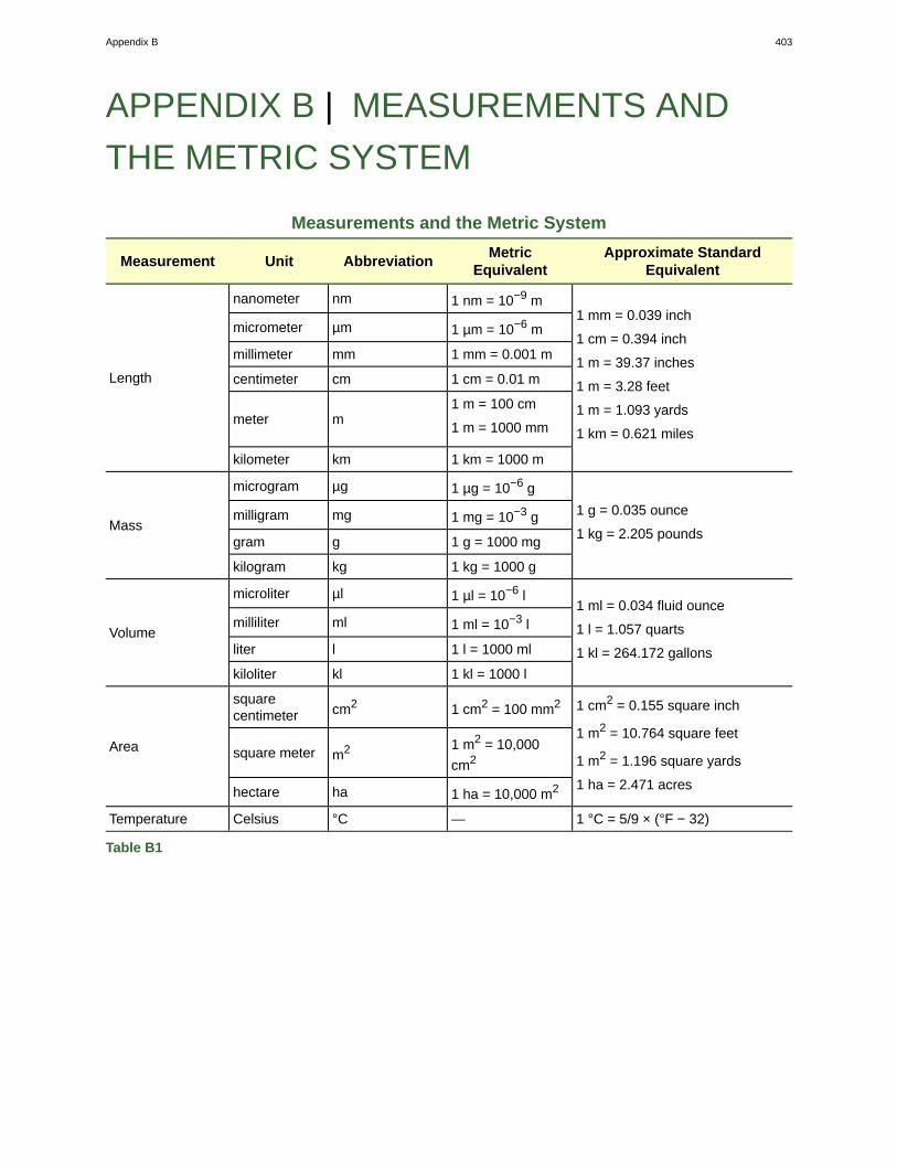
APPENDIX B | MEASUREMENTS AND
THE METRIC SYSTEM
Measurements and the Metric System
Measurement Unit AbbreviationMetric
EquivalentApproximate Standard
Equivalent
nanometer nm 1 nm = 10−9 m
micrometer µm 1 µm = 10−6 m
millimeter mm 1 mm = 0.001 m
centimeter cm 1 cm = 0.01 m
meter m1 m = 100 cm
1 m = 1000 mm
Length
kilometer km 1 km = 1000 m
1 mm = 0.039 inch
1 cm = 0.394 inch
1 m = 39.37 inches
1 m = 3.28 feet
1 m = 1.093 yards
1 km = 0.621 miles
microgram µg 1 µg = 10−6 g
milligram mg 1 mg = 10−3 g
gram g 1 g = 1000 mgMass
kilogram kg 1 kg = 1000 g
1 g = 0.035 ounce
1 kg = 2.205 pounds
microliter µl 1 µl = 10−6 l
milliliter ml 1 ml = 10−3 l
liter l 1 l = 1000 mlVolume
kiloliter kl 1 kl = 1000 l
1 ml = 0.034 fluid ounce
1 l = 1.057 quarts
1 kl = 264.172 gallons
squarecentimeter cm2 1 cm2 = 100 mm2
square meter m2 1 m2 = 10,000
cm2Area
hectare ha 1 ha = 10,000 m2
1 cm2 = 0.155 square inch
1 m2 = 10.764 square feet
1 m2 = 1.196 square yards
1 ha = 2.471 acres
Temperature Celsius °C — 1 °C = 5/9 × (°F − 32)
Table B1
Appendix B 403

404 Appendix B
This OpenStax book is available for free at https://legacy.cnx.org/content/col12104/1.1

ANSWER KEY
Chapter 1
27 Figure 1.15 Carbon-12 has six neutrons. Carbon-13 has seven neutrons. 29 A 31 C 33 Ionic bonds are createdbetween ions. The electrons are not shared between the atoms, but rather are associated more with one ion than theother. Ionic bonds are strong bonds, but are weaker than covalent bonds, meaning it takes less energy to break anionic bond compared with a covalent one.
Chapter 2
Chapter 3
Chapter 4
1 Figure 4.5 Glucose and galactose are aldoses. Fructose is a ketose. 3 Figure 4.33 Adenine is larger than cytosineand will not be able to base pair properly with the guanine on the opposing strand. This will cause the DNA tobulge. DNA repair enzymes may recognize the bulge and replace the incorrect nucleotide. 4 C 6 D 8 B 10 D 12C 14 C 16 Biological macromolecules are organic because they contain carbon. 18 Glycogen and starch arepolysaccharides. They are the storage form of glucose. Glycogen is stored in animals in the liver and in muscle cells,whereas starch is stored in the roots, seeds, and leaves of plants. Starch has two different forms, one unbranched(amylose) and one branched (amylopectin), whereas glycogen is a single type of a highly branched molecule. 20 Fatserves as a valuable way for animals to store energy. It can also provide insulation. Waxes can protect plant leavesand mammalian fur from getting wet. Phospholipids and steroids are important components of animal cell membranes,as well as plant, fungal, and bacterial membranes. 22 A change in gene sequence can lead to a different amino acidbeing added to a polypeptide chain instead of the normal one. This causes a change in protein structure and function.For example, in sickle cell anemia, the hemoglobin β chain has a single amino acid substitution—the amino acidglutamic acid in position six is substituted by valine. Because of this change, hemoglobin molecules form aggregates,and the disc-shaped red blood cells assume a crescent shape, which results in serious health problems. 24 DNA hasa double-helix structure. The sugar and the phosphate are on the outside of the helix and the nitrogenous bases arein the interior. The monomers of DNA are nucleotides containing deoxyribose, one of the four nitrogenous bases (A,T, G and C), and a phosphate group. RNA is usually single-stranded and is made of ribonucleotides that are linkedby phosphodiester linkages. A ribonucleotide contains ribose (the pentose sugar), one of the four nitrogenous bases(A,U, G, and C), and the phosphate group.
Chapter 5
1 Figure 5.5 Free ribosomes and rough endoplasmic reticulum (which contains ribosomes) would not be able toform. 2 C 4 D 6 A 8 A light microscope would be ideal when viewing a small living organism, especially whenthe cell has been stained to reveal details. 10 A transmission electron microscope would be ideal for viewing thecell’s internal structures, because many of the internal structures have membranes that are not visible by the lightmicroscope. 12 Ribosomes are abundant in muscle cells as well because muscle cells are constructed of the proteinsmade by the ribosomes.
Chapter 6
1 Figure 6.12 No, it must have been hypotonic as a hypotonic solution would cause water to enter the cells, thereby
making them burst. 3 Figure 6.19 A decrease in pH means an increase in positively charged H+ ions, and an increasein the electrical gradient across the membrane. The transport of amino acids into the cell will increase. 4 A 6 A 8C 10 D 12 D 13 The fluid characteristic of the cell membrane allows greater flexibility to the cell than it would ifthe membrane were rigid. It also allows the motion of membrane components, required for some types of membranetransport. 15 Heavy molecules move more slowly than lighter ones. It takes more energy in the medium to move themalong. Increasing or decreasing temperature increases or decreases the energy in the medium, affecting molecularmovement. The denser a solution is, the harder it is for molecules to move through it, causing diffusion to slow downdue to friction. Living cells require a steady supply of nutrients and a steady rate of waste removal. If the distancethese substances need to travel is too great, diffusion cannot move nutrients and waste materials efficiently to sustainlife. 17 Injection of isotonic solutions ensures that there will be no perturbation of the osmotic balance, and no water
taken from tissues or added to them from the blood. 19 The sodium-potassium pump forces out three (positive) Na+
Answer Key 405
ions for every two (positive) K+ ions it pumps in, thus the cell loses a positive charge at every cycle of the pump.
Chapter 7
23 Figure 7.8 C 25 Figure 7.17 No. We can store chemical energy because of the need to overcome the barrier toits breakdown. 27 Figure 7.24 Levels of carbon dioxide (a necessary photosynthetic substrate) will immediately fall.As a result, the rate of photosynthesis will be inhibited. 28 A 30 A 32 A 34 C 36 C 38 B 40 A 42 C 44 B 46C 48 Essential nutrients are those nutrients that must be obtained from the diet because they cannot be producedby the body. Vitamins and minerals are examples of essential nutrients. 50 In the United States, obesity, particularlychildhood obesity, is a growing concern. Some of the contributors to this situation include sedentary lifestyles andconsuming more processed foods and less fruits and vegetables. As a result, even young children who are obesecan face health concerns. 52 Lipids add flavor to food and promote a sense of satiety or fullness. Fatty foods aresources of high energy; one gram of lipid contains nine calories. Lipids are also required in the diet to aid the absorptionof lipid-soluble vitamins and for the production of lipid-soluble hormones. 54 Energy is required for cellular motion,through beating of cilia or flagella, as well as human motion, produced by muscle contraction. Cells also need energyto perform digestion, as humans require energy to digest food. 56 The transition state is always higher in energythan the reactants and the products of a reaction (therefore, above), regardless of whether the reaction is endergonicor exergonic. 58 The outcome of light reactions in photosynthesis is the conversion of solar energy into chemicalenergy that the chloroplasts can use to do work (mostly anabolic production of carbohydrates from carbon dioxide). 60The energy carriers that move from the light-dependent reaction to the light-independent one are “full” because theybring energy. After the energy is released, the “empty” energy carriers return to the light-dependent reaction to obtainmore energy. There is not much actual movement involved. Both ATP and NADPH are produced in the stroma where
they are also used and reconverted into ADP, Pi, and NADP+. 62 Feedback inhibition allows cells to control theamounts of metabolic products produced. If there is too much of a particular product relative to what the cell’s needs,feedback inhibition effectively causes the cell to decrease production of that particular product. In general, this reducesthe production of superfluous products and conserves energy, maximizing energy efficiency. 23 Figure 7.8 C 25Figure 7.17 No. We can store chemical energy because of the need to overcome the barrier to its breakdown. 27Figure 7.24 Levels of carbon dioxide (a necessary photosynthetic substrate) will immediately fall. As a result, the rateof photosynthesis will be inhibited. 28 A 30 A 32 A 34 C 36 C 38 B 40 A 42 C 44 B 46 C 48 Essential nutrientsare those nutrients that must be obtained from the diet because they cannot be produced by the body. Vitamins andminerals are examples of essential nutrients. 50 In the United States, obesity, particularly childhood obesity, is agrowing concern. Some of the contributors to this situation include sedentary lifestyles and consuming more processedfoods and less fruits and vegetables. As a result, even young children who are obese can face health concerns. 52Lipids add flavor to food and promote a sense of satiety or fullness. Fatty foods are sources of high energy; one gramof lipid contains nine calories. Lipids are also required in the diet to aid the absorption of lipid-soluble vitamins and forthe production of lipid-soluble hormones. 54 Energy is required for cellular motion, through beating of cilia or flagella,as well as human motion, produced by muscle contraction. Cells also need energy to perform digestion, as humansrequire energy to digest food. 56 The transition state is always higher in energy than the reactants and the products ofa reaction (therefore, above), regardless of whether the reaction is endergonic or exergonic. 58 The outcome of lightreactions in photosynthesis is the conversion of solar energy into chemical energy that the chloroplasts can use to dowork (mostly anabolic production of carbohydrates from carbon dioxide). 60 The energy carriers that move from thelight-dependent reaction to the light-independent one are “full” because they bring energy. After the energy is released,the “empty” energy carriers return to the light-dependent reaction to obtain more energy. There is not much actualmovement involved. Both ATP and NADPH are produced in the stroma where they are also used and reconverted into
ADP, Pi, and NADP+. 62 Feedback inhibition allows cells to control the amounts of metabolic products produced. Ifthere is too much of a particular product relative to what the cell’s needs, feedback inhibition effectively causes the cellto decrease production of that particular product. In general, this reduces the production of superfluous products andconserves energy, maximizing energy efficiency.
Chapter 8
1 Figure 8.7 Compartmentalization enables a eukaryotic cell to divide processes into discrete steps so it can buildmore complex protein and RNA products. But there is an advantage to having a single compartment as well: RNA andprotein synthesis occurs much more quickly in a prokaryotic cell. 3 Figure 8.18 Tetracycline: a; Chloramphenicol: c. 4D 6 D 8 B 10 C 12 The template DNA strand is mixed with a DNA polymerase, a primer, the 4 deoxynucleotides,and a limiting concentration of 4 dideoxynucleotides. DNA polymerase synthesizes a strand complementary to thetemplate. Incorporation of ddNTPs at different locations results in DNA fragments that have terminated at everypossible base in the template. These fragments are separated by gel electrophoresis and visualized by a laser detectorto determine the sequence of bases. 14 For 200 commonly occurring amino acids, codons consisting of four types of
nucleotides would have to be at least four nucleotides long, because 44 = 256. There would be much less degeneracyin this case. 16 The mRNA would be: 5'-AUGGCCGGUUAUUAAGCA-3'. The protein would be: MAGY. Even thoughthere are six codons, the fifth codon corresponds to a stop, so the sixth codon would not be translated. 1 Figure 8.7
406 Answer Key
This OpenStax book is available for free at https://legacy.cnx.org/content/col12104/1.1

Compartmentalization enables a eukaryotic cell to divide processes into discrete steps so it can build more complexprotein and RNA products. But there is an advantage to having a single compartment as well: RNA and proteinsynthesis occurs much more quickly in a prokaryotic cell. 3 Figure 8.18 Tetracycline: a; Chloramphenicol: c. 4 D 6D 8 B 10 C 12 The template DNA strand is mixed with a DNA polymerase, a primer, the 4 deoxynucleotides, and alimiting concentration of 4 dideoxynucleotides. DNA polymerase synthesizes a strand complementary to the template.Incorporation of ddNTPs at different locations results in DNA fragments that have terminated at every possible basein the template. These fragments are separated by gel electrophoresis and visualized by a laser detector to determinethe sequence of bases. 14 For 200 commonly occurring amino acids, codons consisting of four types of nucleotides
would have to be at least four nucleotides long, because 44 = 256. There would be much less degeneracy in thiscase. 16 The mRNA would be: 5'-AUGGCCGGUUAUUAAGCA-3'. The protein would be: MAGY. Even though thereare six codons, the fifth codon corresponds to a stop, so the sixth codon would not be translated.
Answer Key 407

408 Answer Key
This OpenStax book is available for free at https://legacy.cnx.org/content/col12104/1.1
INDEXSymbols
α-helix, 254β-pleated sheet, 254
A
A, 344acid, 119, 227acid ionization, 124, 227acidic, 132, 227actinide, 38actinides, 33activated complex, 342, 362activation energy, 333, 362activation energy (Ea), 343, 362active site, 356, 362Active transport, 301active transport, 306addition reaction, 161, 227adenosine triphosphate, 334adhesion, 118, 227alcohol, 227Alcohols, 177aldehyde, 227aldehydes, 185aliphatic hydrocarbon, 227aliphatic hydrocarbons, 140alkali metal, 38alkali metals, 33alkaline earth metal, 38alkaline earth metals, 33alkane, 227Alkanes, 146alkene, 227alkenes, 158alkyl group, 152, 227alkyne, 227alkynes, 162allosteric inhibition, 358, 362alpha-helix structure (α-helix),263amide, 227Amides, 201amine, 227Amines, 196amino acid, 263Amino acids, 250aminoacyl tRNA synthetase,395aminoacyl tRNA synthetases,391aminopeptidase, 321, 362amphiphilic, 287, 306
amphiprotic, 127, 227amphoteric, 127, 227Anabolic, 327anabolic, 362anion, 38Anions, 27antiporter, 302, 306aquaporin, 306Aquaporins, 296aromatic hydrocarbon, 227, 227aromatic hydrocarbons, 140,163Arrhenius, 123Arrhenius equation, 344, 362atom, 8, 18, 38, 38atomic mass, 19, 38atomic number, 19, 38ATP, 334, 362autoionization, 125, 227Autumn, 215
B
balanced chemical equation, 27,38base, 119, 227base ionization, 125, 227basic, 132, 227beta-pleated sheet (β-pleated),263bioenergetics, 325, 362biological macromolecule, 263biological macromolecules, 232bond length, 84, 227Boyle, 123Brønsted-Lowry acid, 123, 227Brønsted-Lowry base, 123, 227buffer, 227Buffers, 120
C
CAAT box, 388, 395calorie, 116, 227capillary action, 118, 228carbohydrate, 263Carbohydrates, 233carbonyl group, 185, 228carboxylic acid, 228carboxylic acids, 189carboxypeptidase, 320, 362carrier protein, 297, 306catabolic, 328, 362catalyst, 353, 362cation, 38Cations, 27cell theory, 282
cell wall, 279, 282Cellulose, 239cellulose, 263Celsius (°C), 46, 64Central Dogma, 382, 395central vacuole, 281, 282centrosome, 279, 282chalcogen, 38chalcogens, 33channel protein, 306Channel proteins, 296chaperone, 263chaperones, 257chemical bond, 38chemical bonds, 26chemical energy, 329, 362chemical reaction, 38Chemical reactions, 26chemical reactivity, 22, 38chemoautotroph, 362chemoautotrophs, 337chitin, 240, 263chlorophyll, 280, 282chloroplast, 282, 339, 362Chloroplasts, 279chromatin, 276, 282chromosome, 282Chromosomes, 276chylomicron, 362chylomicrons, 322chymotrypsin, 320, 362codon, 395codons, 383coenzyme, 362coenzymes, 359cofactor, 362cofactors, 359cohesion, 117, 228colinear, 382, 395Collision theory, 341collision theory, 362competitive inhibition, 358, 362compound, 38, 38compounds, 10, 27concentration gradient, 293, 306conjugate acid, 124, 228conjugate base, 123, 228covalent bond, 38, 228, 228covalent bonds, 28, 78, 83covalent compound, 228Cronin, 68
cubic centimeter (cm3 or cc), 64
cubic centimeter (cm3), 47
cubic meter (m3), 46, 64cytoplasm, 275, 282
Index 409
cytosol, 275, 282
D
Dalton, 9Davy, 123degeneracy, 395degenerate, 383dehydration synthesis, 232, 263denaturation, 249, 263denature, 356, 362density, 47, 64deoxyribonucleic acid (DNA),258, 263Diffusion, 294diffusion, 306Digestion, 319digestion, 362dimensional analysis, 52, 64dipeptidase, 321, 362dipole-dipole attraction, 216,228disaccharide, 263Disaccharides, 236dispersion force, 212, 228dissociation, 117, 228double bond, 94, 228
E
elastase, 320, 363electrochemical gradient, 301,306electrogenic pump, 304, 306electrolyte, 38electrolytes, 28electron, 38electron configuration, 25, 38electron microscope, 282electron microscopes, 270electron orbital, 38electron orbitals, 24electron transfer, 27, 38electronegativity, 29, 38, 85, 228Electrons, 19electrophoresis, 377, 395element, 38, 38elements, 10Elements, 18empirical formula, 66, 228Enantiomers, 142enantiomers, 228endergonic, 363endergonic reactions, 331enthalpy, 330, 363enzyme, 263Enzymes, 249
equilibrium, 27, 38essential nutrient, 363essential nutrients, 314ester, 228esters, 189ether, 228Ethers, 178eukaryotic cell, 282eukaryotic cells, 272evaporation, 116, 228exergonic, 363exergonic reactions, 331
F
facilitated transport, 296, 306FACT, 389, 395factor-label method, 52Fahrenheit, 55, 64Feedback inhibition, 360feedback inhibition, 363fluid mosaic model, 286, 306free energy, 330, 363free radical, 228free radicals, 100frequency factor, 344frequency factor (A), 363functional group, 157, 228, 228Functional groups, 142
G
gas, 6, 39GC-rich box, 395GC-rich boxes, 388geometric isomer, 228Geometric isomers, 140Glycogen, 239glycogen, 263glycolipid, 306glycolipids, 287glycoprotein, 306glycoproteins, 287glycosidic bond, 236, 263granum, 339, 363Greaney, 215group, 39groups, 32
H
halogen, 39halogens, 33Heat energy, 333heat energy, 363heat of vaporization, 116heat of vaporization of water,
228heterogeneous mixture, 11, 39heterotroph, 363heterotrophs, 337homogeneous mixture, 11, 39hormone, 263Hormones, 249Hu, 215hydrocarbon, 228Hydrocarbons, 139hydrogen bond, 30, 39hydrogen bonding, 217, 228hydrolysis, 263hydrolysis reactions, 233hydrophilic, 114, 229, 287, 306hydrophobic, 114, 229, 306Hydrophobic, 287hypertonic, 298, 306hypervalent molecule, 229hypervalent molecules, 101hypotonic, 298, 306
I
induced dipole, 212, 229induced fit, 356, 363inert gas, 39, 39inert gases, 24, 33ingestion, 319, 363initiator tRNA, 391, 395inner transition metal, 39inner transition metals, 33instantaneous dipole, 212, 229integral protein, 306Integral proteins, 289intermolecular force, 229intermolecular forces, 210International System of Units,43ion, 39ion-product constant for water(Kw), 125, 229ionic bond, 39, 229Ionic bonds, 28ionic bonds, 78ionic compound, 78, 229ions, 24irreversible, 27irreversible chemical reaction,39isomers, 69, 140, 229, 229isotonic, 299, 306isotope, 39Isotopes, 20IUPAC, 151
410 Index
This OpenStax book is available for free at https://legacy.cnx.org/content/col12104/1.1
K
kelvin (K), 46, 64ketone, 229ketones, 185kilogram (kg), 45, 64kinetic energy, 328, 363Kozak’s rules, 392, 395
L
lactase, 363lactases, 320lanthanide, 39lanthanides, 33law of conservation of matter, 8,39law of mass action, 27, 39length, 45, 64Lewis structure, 229Lewis structures, 93Lewis symbol, 92, 229light microscope, 282light microscopes, 270light-dependent reaction, 363light-dependent reactions, 339light-independent reaction, 363light-independent reactions, 339lipid, 263Lipids, 242liquid, 6, 39liter (L) , 47, 64litmus, 119litmus paper, 229London, 212London dispersion force, 212lone pair, 229lone pairs, 93lysosome, 282lysosomes, 279
M
main-group element, 39main-group elements, 33maltase, 363maltases, 320mass, 7, 39mass number, 19, 39Matter, 6, 18matter, 39, 39Mendeleev, 31mesophyll, 339, 363messenger RNA (mRNA), 258,263metabolism, 325, 363
metal, 39metalloid, 39metalloids, 33metals, 33meter (m), 45, 64Meyer, 31microscope, 270, 282milliliter (mL), 47, 64mineral, 363Minerals, 315Mitochondria, 278mitochondria, 282mixture, 11, 39molecular compound, 229molecular compounds, 81molecular formula, 65, 229molecular mass, 67molecule, 10, 39, 39Molecules, 22monatomic ion, 229monatomic ions, 77monomer, 263monomers, 232monosaccharide, 263Monosaccharides, 234
N
neutral, 131, 229neutron, 19, 39noble gas, 39, 40noble gases, 24, 33nonmetal, 40nonmetals, 33nonpolar covalent bond, 40Nonpolar covalent bonds, 29nonsense codon, 395nonsense codons, 384nuclear envelope, 276, 282nucleic acid, 263Nucleic acids, 258nucleolus, 277, 282nucleoplasm, 276, 282nucleotide, 263nucleotides, 258nucleus, 18, 40, 275, 282
O
Octamer box, 395octamer boxes, 388octet rule, 23, 40, 93, 229Omega, 245omega fat, 263orbital, 40orbitals, 23organelle, 282
organelles, 272organic compound, 229organic compounds, 146organic molecule, 229organic molecules, 138Osmolarity, 298osmolarity, 306Osmosis, 297osmosis, 306oxyanion, 229Oxyanions, 77
P
Passive transport, 293passive transport, 306Pauling, 86peptide bond, 251, 263peptidyl transferase, 392, 395period, 40periodic law, 32, 40periodic table, 22, 32, 40, 40periods, 32peripheral protein, 306Peripheral proteins, 290peroxisome, 282Peroxisomes, 278pH, 132, 229pH paper, 229pH scale, 119, 229phosphoanhydride bond, 363phosphoanhydride bonds, 335phosphodiester, 260, 264phospholipid, 264Phospholipids, 246photoautotroph, 363photoautotrophs, 337pigment, 339, 363plasma, 6, 40plasma membrane, 274, 282plasmolysis, 300, 307pnictogen, 40pnictogens, 33pOH, 132, 230polar covalent bond, 29, 40, 85,230polarizability, 213, 230polyatomic ion, 230polyatomic ions, 77polymer, 264polymers, 232polynucleotide, 258, 264polypeptide, 264polysaccharide, 238, 264polysome, 391, 395potential energy, 328, 363
Index 411
preinitiation complex, 388, 395Primary active transport, 302primary active transport, 307primary structure, 253, 264product, 40products, 26protein, 264Proteins, 249proton, 19, 40pump, 307pumps, 302pure covalent bond, 85, 230pure substance, 10, 40purine, 264purines, 259pyrimidine, 264pyrimidines, 259
Q
quaternary structure, 256, 264
R
radioisotope, 40radioisotopes, 20reactant, 40reactants, 26reading frame, 383, 395representative element, 40representative elements, 33reversible chemical reaction, 40Reversible reactions, 27ribonucleic acid (RNA), 258, 264Ribosomal RNA (rRNA), 261ribosomal RNA (rRNA), 264ribosome, 282Ribosomes, 277RNAs, 387
S
saturated fatty acid, 243, 264saturated hydrocarbon, 230saturated hydrocarbons, 146second (s), 46, 64Secondary active transport, 302secondary active transport, 307secondary structure, 254, 264selectively permeable, 293, 307series, 32, 40Shine-Dalgarno sequence, 391,395SI Units, 43SI units (International System ofUnits), 64signal sequence, 394, 395
single bond, 93, 230skeletal structure, 146, 230small nuclear , 387small nuclear RNA, 395Smalley, 99solid, 6, 40solute, 298, 307solutes, 296solution, 11solvent, 117, 230spatial isomers, 70, 230specific heat capacity, 116, 230sphere of hydration, 117, 230starch, 233, 264start codon, 391, 395steroid, 264steroids, 248stoma, 363stomata, 339stroma, 339, 363structural formula, 65, 230structural isomer, 230structural isomers, 70, 230Structural isomers, 140substituent, 230substituents, 151substituted hydrocarbon, 230substituted hydrocarbons, 142substitution reaction, 157, 230substrate, 363substrates, 356sucrase, 364sucrases, 320surface tension, 117, 230symporter, 302, 307
T
temperature, 55tertiary structure, 255, 264thylakoid, 364thylakoid lumen, 339, 364thylakoids, 339Tonicity, 298tonicity, 307trans fat, 244, 264transcription, 262, 264Transfer RNA (tRNA), 262transfer RNA (tRNA), 264transition metal, 40transition metals, 33transition state, 333, 342, 364translation, 262, 264transport protein, 307transport proteins, 296transporter, 307
transporters, 302triacylglycerol (also,triglyceride), 264triacylglycerols, 243triglycerides, 243triple bond, 94, 230trypsin, 320, 364
U
unified cell theory, 271, 282uniporter, 302, 307unit, 64unit conversion factor, 52, 64Units, 43unsaturated, 244unsaturated fatty acid, 264
V
vacuole, 283vacuoles, 278valence shell, 23, 40van der Waals force, 230van der Waals forces, 212van der Waals interaction, 40van der Waals interactions, 30vesicle, 283Vesicles, 278vitamin, 364Vitamins, 314Volume, 46volume, 64
W
Wax, 246wax, 264Weight, 7weight, 40
412 Index
This OpenStax book is available for free at https://legacy.cnx.org/content/col12104/1.1
ATTRIBUTIONS
Collection: General, Organic, and Biological Chemistry: A Cellular PerspectiveEdited by: Mike SpringerURL: https://legacy.cnx.org/content/col12104/1.1/Copyright: Mike SpringerLicense: http://creativecommons.org/licenses/by/4.0/Based on: Biology <http://legacy.cnx.org/content/col11448/1.10> arranged by OpenStax.
Module: IntroductionBy: OpenStaxURL: https://legacy.cnx.org/content/m44389/1.2/Copyright: Rice UniversityLicense: http://creativecommons.org/licenses/by/3.0/
Module: Phases and Classification of MatterBy: OpenStaxURL: https://legacy.cnx.org/content/m50986/1.5/Copyright: Rice UniversityLicense: http://creativecommons.org/licenses/by/4.0/
Module: Atoms, Isotopes, Ions, and Molecules: The Building BlocksBy: OpenStaxURL: https://legacy.cnx.org/content/m44390/1.17/Copyright: Rice UniversityLicense: http://creativecommons.org/licenses/by/4.0/
Module: The Periodic TableBy: OpenStaxURL: https://legacy.cnx.org/content/m51003/1.10/Copyright: Rice UniversityLicense: http://creativecommons.org/licenses/by/4.0/
Module: MeasurementsBy: OpenStaxURL: https://legacy.cnx.org/content/m50989/1.5/Copyright: Rice UniversityLicense: http://creativecommons.org/licenses/by/4.0/
Module: Mathematical Treatment of Measurement ResultsBy: OpenStaxURL: https://legacy.cnx.org/content/m50996/1.7/Copyright: Rice UniversityLicense: http://creativecommons.org/licenses/by/4.0/
Module: Chemical FormulasBy: OpenStaxURL: https://legacy.cnx.org/content/m51002/1.4/Copyright: Rice UniversityLicense: http://creativecommons.org/licenses/by/4.0/
Module: Molecular and Ionic CompoundsBy: OpenStaxURL: https://legacy.cnx.org/content/m51005/1.9/Copyright: Rice UniversityLicense: http://creativecommons.org/licenses/by/4.0/
Index 413

Module: Covalent BondingBy: OpenStaxURL: https://legacy.cnx.org/content/m51048/1.4/Copyright: Rice UniversityLicense: http://creativecommons.org/licenses/by/4.0/
Module: Lewis Symbols and StructuresBy: OpenStaxURL: https://legacy.cnx.org/content/m51049/1.10/Copyright: Rice UniversityLicense: http://creativecommons.org/licenses/by/4.0/
Module: WaterBy: OpenStaxURL: https://legacy.cnx.org/content/m44392/1.10/Copyright: Rice UniversityLicense: http://creativecommons.org/licenses/by/4.0/
Module: Brønsted-Lowry Acids and BasesBy: OpenStaxURL: https://legacy.cnx.org/content/m51116/1.11/Copyright: Rice UniversityLicense: http://creativecommons.org/licenses/by/4.0/
Module: pH and pOHBy: OpenStaxURL: https://legacy.cnx.org/content/m51117/1.5/Copyright: Rice UniversityLicense: http://creativecommons.org/licenses/by/4.0/
Module: CarbonBy: OpenStaxURL: https://legacy.cnx.org/content/m44393/1.6/Copyright: Rice UniversityLicense: http://creativecommons.org/licenses/by/4.0/
Module: HydrocarbonsBy: OpenStaxURL: https://legacy.cnx.org/content/m51191/1.9/Copyright: Rice UniversityLicense: http://creativecommons.org/licenses/by/4.0/
Module: Alcohols and EthersBy: OpenStaxURL: https://legacy.cnx.org/content/m51192/1.5/Copyright: Rice UniversityLicense: http://creativecommons.org/licenses/by/4.0/
Module: Aldehydes, Ketones, Carboxylic Acids, and EstersBy: OpenStaxURL: https://legacy.cnx.org/content/m51193/1.7/Copyright: Rice UniversityLicense: http://creativecommons.org/licenses/by/4.0/
Module: Amines and AmidesBy: OpenStaxURL: https://legacy.cnx.org/content/m51194/1.4/Copyright: Rice UniversityLicense: http://creativecommons.org/licenses/by/4.0/
414 Index
This OpenStax book is available for free at https://legacy.cnx.org/content/col12104/1.1

Module: Intermolecular ForcesBy: OpenStaxURL: https://legacy.cnx.org/content/m51077/1.4/Copyright: Rice UniversityLicense: http://creativecommons.org/licenses/by/4.0/
Module: IntroductionBy: OpenStaxURL: https://legacy.cnx.org/content/m44395/1.4/Copyright: Rice UniversityLicense: http://creativecommons.org/licenses/by/3.0/
Module: Synthesis of Biological MacromoleculesBy: OpenStaxURL: https://legacy.cnx.org/content/m44397/1.12/Copyright: Rice UniversityLicense: http://creativecommons.org/licenses/by/4.0/
Module: CarbohydratesBy: OpenStaxURL: https://legacy.cnx.org/content/m44400/1.6/Copyright: Rice UniversityLicense: http://creativecommons.org/licenses/by/4.0/
Module: LipidsBy: OpenStaxURL: https://legacy.cnx.org/content/m44401/1.8/Copyright: Rice UniversityLicense: http://creativecommons.org/licenses/by/4.0/
Module: ProteinsBy: OpenStaxURL: https://legacy.cnx.org/content/m44402/1.7/Copyright: Rice UniversityLicense: http://creativecommons.org/licenses/by/4.0/
Module: Nucleic AcidsBy: OpenStaxURL: https://legacy.cnx.org/content/m44403/1.8/Copyright: Rice UniversityLicense: http://creativecommons.org/licenses/by/4.0/
Module: IntroductionBy: OpenStaxURL: https://legacy.cnx.org/content/m44404/1.2/Copyright: Rice UniversityLicense: http://creativecommons.org/licenses/by/3.0/
Module: Studying CellsBy: OpenStaxURL: https://legacy.cnx.org/content/m44405/1.8/Copyright: Rice UniversityLicense: http://creativecommons.org/licenses/by/3.0/
Module: Eukaryotic CellsBy: OpenStaxURL: https://legacy.cnx.org/content/m44407/1.14/Copyright: Rice UniversityLicense: http://creativecommons.org/licenses/by/4.0/
Index 415

Module: IntroductionBy: OpenStaxURL: https://legacy.cnx.org/content/m44415/1.2/Copyright: Rice UniversityLicense: http://creativecommons.org/licenses/by/3.0/
Module: Components and StructureBy: OpenStaxURL: https://legacy.cnx.org/content/m44416/1.9/Copyright: Rice UniversityLicense: http://creativecommons.org/licenses/by/4.0/
Module: Passive TransportBy: OpenStaxURL: https://legacy.cnx.org/content/m44417/1.12/Copyright: Rice UniversityLicense: http://creativecommons.org/licenses/by/4.0/
Module: Active TransportBy: OpenStaxURL: https://legacy.cnx.org/content/m44418/1.6/Copyright: Rice UniversityLicense: http://creativecommons.org/licenses/by/4.0/
Module: IntroductionBy: OpenStaxURL: https://legacy.cnx.org/content/m44421/1.2/Copyright: Rice UniversityLicense: http://creativecommons.org/licenses/by/3.0/
Module: IntroductionBy: OpenStaxURL: https://legacy.cnx.org/content/m44734/1.2/Copyright: Rice UniversityLicense: http://creativecommons.org/licenses/by/3.0/
Module: Nutrition and Energy ProductionBy: OpenStaxURL: https://legacy.cnx.org/content/m44740/1.5/Copyright: Rice UniversityLicense: http://creativecommons.org/licenses/by/3.0/
Module: Digestive System ProcessesBy: OpenStaxURL: https://legacy.cnx.org/content/m44742/1.4/Copyright: Rice UniversityLicense: http://creativecommons.org/licenses/by/3.0/
Module: Energy and MetabolismBy: OpenStaxURL: https://legacy.cnx.org/content/m44422/1.8/Copyright: Rice UniversityLicense: http://creativecommons.org/licenses/by/3.0/
Module: Potential, Kinetic, Free, and Activation EnergyBy: OpenStaxURL: https://legacy.cnx.org/content/m44425/1.7/Copyright: Rice UniversityLicense: http://creativecommons.org/licenses/by/4.0/
416 Index
This OpenStax book is available for free at https://legacy.cnx.org/content/col12104/1.1

Module: ATP: Adenosine TriphosphateBy: OpenStaxURL: https://legacy.cnx.org/content/m44427/1.7/Copyright: Rice UniversityLicense: http://creativecommons.org/licenses/by/4.0/
Module: Overview of PhotosynthesisBy: OpenStaxURL: https://legacy.cnx.org/content/m44447/1.8/Copyright: Rice UniversityLicense: http://creativecommons.org/licenses/by/3.0/
Module: Collision TheoryBy: OpenStaxURL: https://legacy.cnx.org/content/m51102/1.5/Copyright: Rice UniversityLicense: http://creativecommons.org/licenses/by/4.0/
Module: Factors Affecting Reaction RatesBy: OpenStaxURL: https://legacy.cnx.org/content/m51098/1.3/Copyright: Rice UniversityLicense: http://creativecommons.org/licenses/by/4.0/
Module: EnzymesBy: OpenStaxURL: https://legacy.cnx.org/content/m44429/1.7/Copyright: Rice UniversityLicense: http://creativecommons.org/licenses/by/4.0/
Module: IntroductionBy: OpenStaxURL: https://legacy.cnx.org/content/m44484/1.2/Copyright: Rice UniversityLicense: http://creativecommons.org/licenses/by/3.0/
Module: DNA Structure and SequencingBy: OpenStaxURL: https://legacy.cnx.org/content/m44486/1.7/Copyright: Rice UniversityLicense: http://creativecommons.org/licenses/by/4.0/
Module: IntroductionBy: OpenStaxURL: https://legacy.cnx.org/content/m44518/1.3/Copyright: Rice UniversityLicense: http://creativecommons.org/licenses/by/3.0/
Module: The Genetic CodeBy: OpenStaxURL: https://legacy.cnx.org/content/m44522/1.9/Copyright: Rice UniversityLicense: http://creativecommons.org/licenses/by/4.0/
Module: Eukaryotic TranscriptionBy: OpenStaxURL: https://legacy.cnx.org/content/m44524/1.5/Copyright: Rice UniversityLicense: http://creativecommons.org/licenses/by/3.0/
Index 417

Module: Ribosomes and Protein SynthesisBy: OpenStaxURL: https://legacy.cnx.org/content/m44529/1.7/Copyright: Rice UniversityLicense: http://creativecommons.org/licenses/by/3.0/
Module: The Periodic Table of ElementsBy: OpenStaxURL: https://legacy.cnx.org/content/m45849/1.6/Copyright: Rice UniversityLicense: http://creativecommons.org/licenses/by/4.0/
Module: Measurements and the Metric SystemBy: Cnx BiologyURL: https://legacy.cnx.org/content/m60108/1.4/Copyright: Cnx BiologyLicense: http://creativecommons.org/licenses/by/4.0/
418 Index
This OpenStax book is available for free at https://legacy.cnx.org/content/col12104/1.1

ABOUT CONNEXIONSSince 1999, Connexions has been pioneering a global system where anyone can create course materials and makethem fully accessible and easily reusable free of charge. We are a Web-based authoring, teaching and learningenvironment open to anyone interested in education, including students, teachers, professors and lifelong learners. Weconnect ideas and facilitate educational communities. Connexions's modular, interactive courses are in use worldwideby universities, community colleges, K-12 schools, distance learners, and lifelong learners. Connexions materials arein many languages, including English, Spanish, Chinese, Japanese, Italian, Vietnamese, French, Portuguese, andThai.
Index 419