Gene dosage as a relevant mechanism contributing to the determination of ovarian function in Turner...
12
ORIGINAL ARTICLE Reproductive genetics Gene dosage as a relevant mechanism contributing to the determination of ovarian function in Turner syndrome Chiara Castronovo 1,† , Raffaella Rossetti 2,† , Daniela Rusconi 1 , Maria P. Recalcati 1 , Chiara Cacciatore 2,3 , Elena Beccaria 3 , Valeria Calcaterra 4 , Pietro Invernizzi 5 , Daniela Larizza 4 , Palma Finelli 1,6 , and Luca Persani 2,3, * 1 Medical Cytogenetics and Molecular Genetics Lab, IRCSS Istituto Auxologico Italiano, 20145 Milan, Italy 2 Dipartimento di Scienze Cliniche e di Comunita `, Universita ` degli Studi di Milano, 20122 Milan, Italy 3 Laboratory of Endocrine and Metabolic Research and Division of Endocrine and Metabolic Diseases, IRCSS Istituto Auxologico Italiano, 20145 Milan, Italy 4 Dipartimento di Pediatria, Ospedale San Matteo, Universita ` degli Studi di Pavia, 27100 Pavia, Italy 5 Center for Autoimmune Liver Diseases, Humanitas Clinical and Research Center, 20089 Rozzano (MI), Italy 6 Dipartimento di Biotecnologie Mediche e Medicina Traslazionale, Universita ` degli Studi di Milano, 20133 Milan, Italy *Correspondence address. Division of Endocrine and Metabolic Diseases, San Luca Hospital, Piazza Brescia 20 – 20149 Milano, Italy. Tel: +390261911-2738; Fax: 2777; E-mail: [email protected] Submitted on July 16, 2013; resubmitted on October 31, 2013; accepted on November 8, 2013 studyquestion: What is the burden of X chromosome mosaicism in the occurrence of spontaneous menarche (SM) in Turner syndrome (TS)? summary answer: SM was significantly associated with X chromosome mosaicism in the TS patients; a mosaicism with around 10% euploid cell line may predict spontaneous pubertal development when determined by molecular-cytogenetic techniques on uncultivated tissues. what is known already: Spontaneous puberty can be observed in a minority of patients with TS, more frequently, but not exclusively, in those with a high level of 46,XX/45,X mosaicism at standard karyotype. The genetic mechanisms contributing to ovarian function in TS patients are still not determined. However, submicroscopic X-linked and autosomal copy number variations (CNVs) have recently emerged as an import- ant genetic risk category for premature ovarian insufficiency and may be involved in modulating the TS ovarian phenotype. study design, size, duration: A group of 40 patients with a diagnosis of TS at conventional karyotyping participated in the study; 6 patients had SM and 34 patients had primary amenorrhoea (PA). All clinical data and the patients’ DNA samples were collected over the years at a single paediatric clinic. participants/materials, setting, methods: The patients’ samples were used to perform both genetic (Copy Number Assay) and molecular-cytogenetic (array-CGH and iFISH, interphase-FISH) analyses in order to evaluate the X chromosome mosaicism rate and to detect possible rare CNVs of genes with a known or predicted role in female fertility. main results and the role of chance: All TS patients showed variable percentages of the 46,XX lineage, but these percentages were higher in the SM group (P , 0.01). A mosaicism around 10% for the euploid cell line may predict spontaneous pubertal development when determined by molecular-cytogenetic techniques performed in uncultivated tissues. A few CNVs involving autosomal and X-linked ovary-related loci were identified by array-CGH analysis and confirmed by real-time quantitative PCR, including a BMP15 gene duplication at Xp11.22, a deletion interrupting the PAPPA gene at 9q33.1, and an intragenic duplication involving the PDE8A gene at 15q25.3. limitations, reasons for caution: This is a pilot study on a relatively small sample size and confirmation in larger TS cohorts may be required. The ovarian tissue could not be studied in any patients and in a subgroup of patients, the mosaicism was estimated in tissues of dif- ferent embryonic origin. wider implications of the findings: The combined determination of X chromosome mosaicism by molecular and molecular- cytogenetic techniques may become useful for the prediction of SM in TS. The detection of CNVs in both X-linked and autosomal ovary-related † The authors consider that the first two authors should be regarded as joint first authors. & The Author 2013. Published by Oxford University Press on behalf of the European Society of Human Reproduction and Embryology. This is an Open Access article distributed under the terms of the Creative Commons Attribution Non-Commercial License (http://creativecommons.org/licenses/by-nc/3.0/), which permits non-commercial re-use, distribution, and reproduction in any medium, provided the original work is properly cited. For commercial re-use, please contact [email protected] Human Reproduction, Vol.29, No.2 pp. 368 – 379, 2014 Advanced Access publication on December 8, 2013 doi:10.1093/humrep/det436
-
Upload
independent -
Category
Documents
-
view
2 -
download
0
Transcript of Gene dosage as a relevant mechanism contributing to the determination of ovarian function in Turner...

ORIGINAL ARTICLE Reproductive genetics
Gene dosage as a relevant mechanismcontributing to the determination ofovarian function in Turner syndromeChiara Castronovo1,†, Raffaella Rossetti2,†, Daniela Rusconi1,Maria P. Recalcati1, Chiara Cacciatore2,3, Elena Beccaria3,Valeria Calcaterra4, Pietro Invernizzi5, Daniela Larizza4,Palma Finelli1,6, and Luca Persani2,3,*1Medical Cytogenetics and Molecular Genetics Lab, IRCSS Istituto Auxologico Italiano, 20145 Milan, Italy 2Dipartimento di Scienze Cliniche e diComunita, Universita degli Studi di Milano, 20122 Milan, Italy 3Laboratory of Endocrine and Metabolic Research and Division of Endocrine andMetabolic Diseases, IRCSS Istituto Auxologico Italiano, 20145 Milan, Italy 4Dipartimento di Pediatria, Ospedale San Matteo, Universita degli Studidi Pavia, 27100 Pavia, Italy 5Center for Autoimmune Liver Diseases, Humanitas Clinical and Research Center, 20089 Rozzano (MI), Italy6Dipartimento di Biotecnologie Mediche e Medicina Traslazionale, Universita degli Studi di Milano, 20133 Milan, Italy
*Correspondence address. Division of Endocrine and Metabolic Diseases, San Luca Hospital, Piazza Brescia 20 – 20149 Milano, Italy.Tel: +390261911-2738; Fax: 2777; E-mail: [email protected]
Submitted on July 16, 2013; resubmitted on October 31, 2013; accepted on November 8, 2013
studyquestion: What is theburdenofXchromosomemosaicismintheoccurrenceof spontaneousmenarche(SM) inTurnersyndrome(TS)?
summary answer: SM was significantly associated with X chromosome mosaicism in the TS patients; a mosaicism with around 10%euploid cell line may predict spontaneous pubertal development when determined by molecular-cytogenetic techniques on uncultivated tissues.
what is known already: Spontaneous puberty can be observed in a minority of patients with TS, more frequently, but not exclusively,in those with a high level of 46,XX/45,X mosaicism at standard karyotype. The genetic mechanisms contributing to ovarian function in TS patientsare still not determined. However, submicroscopic X-linked and autosomal copy number variations (CNVs) have recently emerged as an import-ant genetic risk category for premature ovarian insufficiency and may be involved in modulating the TS ovarian phenotype.
study design, size, duration: A group of 40 patients with a diagnosis of TS at conventional karyotyping participated in the study; 6patients had SM and 34 patients had primary amenorrhoea (PA). All clinical data and the patients’ DNA samples were collected over the years at asingle paediatric clinic.
participants/materials, setting, methods: The patients’ samples were used to perform both genetic (Copy NumberAssay) and molecular-cytogenetic (array-CGH and iFISH, interphase-FISH) analyses in order to evaluate the X chromosome mosaicism rateand to detect possible rare CNVs of genes with a known or predicted role in female fertility.
main results and the role of chance: All TS patients showed variable percentages of the 46,XX lineage, but these percentageswere higher in the SM group (P , 0.01). A mosaicism around 10% for the euploid cell line may predict spontaneous pubertal development whendetermined by molecular-cytogenetic techniques performed in uncultivated tissues. A few CNVs involving autosomal and X-linked ovary-relatedloci were identified byarray-CGH analysis and confirmed by real-time quantitative PCR, including a BMP15 gene duplication at Xp11.22, a deletioninterrupting the PAPPA gene at 9q33.1, and an intragenic duplication involving the PDE8A gene at 15q25.3.
limitations, reasons for caution: This is a pilot study on a relatively small sample size and confirmation in larger TS cohorts maybe required. The ovarian tissue could not be studied in any patients and in a subgroup of patients, the mosaicism was estimated in tissues of dif-ferent embryonic origin.
wider implications of the findings: The combined determination of X chromosome mosaicism by molecular and molecular-cytogenetic techniques may become useful for the prediction of SM in TS. The detection of CNVs in both X-linked and autosomal ovary-related
† The authors consider that the first two authors should be regarded as joint first authors.
& The Author 2013. Published by Oxford University Press on behalf of the European Society of Human Reproduction and Embryology.This is an Open Access article distributed under the terms of the Creative Commons Attribution Non-Commercial License (http://creativecommons.org/licenses/by-nc/3.0/), which permitsnon-commercial re-use, distribution, and reproduction in any medium, provided the original work is properly cited. For commercial re-use, please contact [email protected]
Human Reproduction, Vol.29, No.2 pp. 368–379, 2014
Advanced Access publication on December 8, 2013 doi:10.1093/humrep/det436

genes further suggests gene dosage as a relevant mechanism contributing to the ovarian phenotype of TS patients. These CNVs maypinpoint novelcandidates relevant to female fertility and generate further insights into the mechanisms contributing to ovarian function.
study funding/competing interest(s): This study was funded by Telethon Foundation (grant no: GGP09126 to L.P.), theItalian Ministry of the University and Research (grant number: 2006065999 to P.F.) and a Ministry of Health grant ‘Ricerca Corrente’ toIRCCS Istituto Auxologico Italiano (grant number: 08C704-2006). The authors have no conflict of interest to declare.
Key words: Turner syndrome / ovary / BMP15 / X chromosome / copy number variations
IntroductionTurner syndrome (TS) represents one of the most common chromo-somal anomalies, with a prevalence of about 1:2500-1:3000 livefemale births (Sybert and McCauley, 2004). Affected individuals have avariable spectrum of physical and functional alterations likely due to awide variety of karyotype abnormalities, ranging from 45,X monosomy(50% of cases) to various forms of 45,X/46,XX mosaicism with orwithout structural defects of the X chromosome in the euploid cell line(Saenger, 1996; Sybert and McCauley, 2004). Among the clinical featuresof TS, short stature and ovarian defects are present in almost all cases.Ovarian dysfunction in TS women is likely caused by rapid oocyte-lossin the early stages of the meiotic prophase after the 18th week of fetallife, later resulting in ovarian dysgenesis and streak ovaries and partialor complete absence of pubertal development. Consequently, infertilityis one of the main problems for TS women (Reynaud et al., 2004). Studiesof the X chromosome breakpoints have revealed the existence of twodistinct loci on the Xq (Xq13-q21 and Xq23-q27) and one region onthe Xp22.1-p11.2 that are significantly associated with the ovarianphenotype (Toniolo, 2006; Persani et al., 2009). Different mechanismsto explain the variable phenotypic manifestations of TS include the hap-loinsufficiencyof X-linked genes that escape X chromosome inactivation,such as SHOX for short stature (Zinn and Ross, 1998; Sybert and McCau-ley, 2004), and meiotic-chromosome pairing errors (Ogata and Matsuo,1995). Nevertheless, spontaneous puberty has been observed in 15–20% of 45,X patients (Pasquino et al., 1997; Gracia Bouthelier et al.,2004; Martın Campagne and Roa Llamazares, 2008) and in 32% ofmosaic patients, inversely correlating with the severity of chromosomalanomalies at conventional karyotype (Pasquino et al., 1997). Consistent-ly, subjects with 45,X frequently have serum follicle-stimulating hormone(FSH) levels already in the post-menopausal range during infancy,whereas FSH levels are generally normal in patients of the same agewith mosaic Turner syndrome (Fechner et al., 2006; Aso et al., 2010).Furthermore, a biochemical parameter of ovarian reserve, the antimul-lerian hormone is generally low in infants with 45,X or structural abnor-malities of the second X, but similar to controls in mosaic Turner patients(Kallio et al., 2012).
The mechanism supporting pubertal development in a small subset ofpatients with 45,X is presently unexplained, but low percentage mosai-cism, undetected at standard karyotyping, might be a possible explan-ation. Additionally, submicroscopic copy number variations (CNVs)have recently emerged as an important genetic risk category for prema-ture ovarian insufficiency (POI), both in cases of primary and secondaryamenorrhoea. Indeed, in a few cohorts of 46,XX patients diagnosed withPOI and analysed by means of high throughput techniques, such as arraycomparative genomic hybridization (aCGH) and single-nucleotide poly-morphism (SNP) array, CNVs affecting several X-linked and autosomal
loci with a possible role in female fertility have been detected (Abouraet al., 2009; Dudding et al., 2010; Ledig et al., 2010; Quilter et al.,2010; Liao et al., 2011; McGuire et al., 2011). Very recently, the SNParray technology has been applied to genotype a large cohort of TSpatients, demonstrating the equivalence of this method to metaphasecytogenetics for the diagnosis of TS (Prakash et al., 2013). However,the potential involvement of CNVs in the modulation of the TS ovarianphenotype has not been investigated.
In this pilot study, we performed new generation genetic (CopyNumber Assay) and molecular-cytogenetic (aCGH and iFISH) investiga-tions in a cohort of TS patients in order to better clarify the synergisticburden of X chromosome mosaicism and rare X-linked and autosomalCNVs on the observed ovarian phenotypes of these patients.
Materials and Methods
Ethics statementThe Institutional Ethic Committees of the IRCCS San Matteo Hospital andIRCCS Istituto Auxologico Italiano approved the study. All participantsgave their written informed consent to the study and to the sampling oftheir tissues.
PatientsForty patients with TS were recruited at the Paediatric Department of theUniversity of Pavia, in San Matteo Hospital. All the patients had been diag-nosed with TS at birth or during the early infancy on the basis of the conven-tional karyotyping of at least 30 metaphases from cultured peripheral bloodlymphocytes.
The patients were classified according to the presence of the spontaneousmenarche (SM) and to the results of the conventional karyotyping (Table I).Six of the 40 patients (15%) experienced SM with FSH and LH values at me-narche within the normal range. The clinical data of SM patients are summar-ized in Table II (ovarian phenotype) and Supplementary data, Table SI(extra-ovarian phenotype). The mean age of the SM patients was 18.8 +6.7 years (median: 16.5 years). SM occurred at 11.9–14.4 years (mean:13.1+ 1.0 years;median: 13 years). One older patient with SM (SM1) under-went secondary amenorrhoea at 18.6 years and was then put on hormonereplacement therapy (Table II). The only other TS feature shown by SM1was short stature (Supplementary data, Table SI). The other 34 patients(.12 years of age; median 18.3 years) had hormone levels in the post-menopausal range on at least two independent determinations (FSH.30 U/l, data not shown) and were classified as primary amenorrhoea (PA).
Array-CGH analysisAll cases underwent aCGH analysis. Genomic DNA (gDNA) was extractedfrom the whole blood using the GenEluteTM Blood Genomic DNA kit(Sigma-Aldrich, St. Louis, MO, USA) in accordance with the manufacturer’sinstructions. Pooled DNA from the peripheral blood of 10 healthy donors
Gene dosage and ovarian defects in Turner syndrome 369

(Promega, Southampton, UK), sex-mismatched to the samples, was used as areference. The genome scan was performed using the Human Genome CGHMicroarray Kit 244 K (Agilent Technologies, Santa Clara, CA, USA) whichconsists of �236 000 60-mer oligonucleotide probes covering the entiregenome at an average spatial resolution of �30 kb. Briefly, 3 mg of DNAfrom the test and the normal reference samples were processed followingthe manufacturer’s protocol. Dye emission was captured by means of a dual-laser scanner. The images were extracted using the Agilent Feature Extrac-tion 9.1 software, and analysed using the DNA Analytics 4.0 software.
A log ratio plot between the test and reference gDNAwas assigned so thatany aberrations in the copy number of the test DNA ata particular locus wereidentified as deviations from a modal value of 0 by the ADM-2 algorithm.CNV classification was performed according to the Database of GenomicVariants (http://projects.tcag.ca/variation/): a CNV was defined as ‘rare’or ‘common’ if never or previously reported in control subjects, respectively.
The X chromosome mosaic condition was detected qualitatively evaluat-ing, through a mosaicism artificial scale, the vertical right shift of the log ratioprofile from the expected value of 0 (Ratio (R): X chromosome copy numberof test DNA/X chromosome copy number of reference DNA ¼ 1) towards+1 (R ¼ 2) or +1.58 (R ¼ 3), the latter due to the isochromosome Xqwhich involves the presence of three copies of the same chromosomeregion rather than two. The artificial mosaicism scale was created by
mixing the blood samples of one normal diploid male with one normaldiploid female, as previously described (Ballif et al., 2006). The DNA of theartificial mosaicism representing 75, 50, 30 and 10% of 46,XX cell line wasused in aCGH experiments, and the obtained log ratio profile shifts werecompared with the aCGH results of all TS patients by overlapping every TSX chromosome array profile to the artificial scale, thus giving an estimationof the patient mosaicism level (Supplementary data, Fig. S1). The mosaicismdetection limit of this technique was therefore set at 10%.
FISH analysisThe RPCI-11 BAC clone CTD-2004N6, covering the entire BMP15 gene atXp11.22, was selected according to the UCSC Genome Browser (http://genome.cse.ucsc.edu, hg19, February 2009) and provided by InvitrogenLtd (Paisley, UK). The probe was labelled by nick translation with Cy3(Amersham Biosciences, Chalfont St. Giles, UK) and its physical positionwas verified on a few female control metaphases derived from peripheralblood lymphocytes.
Multi-colour iFISH analysis was performed in a subset of 13 TS patients(6 SM + 7 PA). For each patient, 300 nuclei derived from PHA-stimulatedperipheral blood lymphocytes cultivated for 72 h were analysed by usingthe CEPX, CEPY, CEP18-Aneuvysion kit (Abbott-Vysis, Chicago, IL, USA),according to the manufacturer’s instructions. In addition, a few metaphaseswere analysed to check the structure of the second X chromosome in theeuploid cell line. Furthermore, in patient SM1 dual-colour FISH analysiswas performed by using the BAC probe specific for BMP15 in combinationwith the commercially available centromeric specific probe CEP6 (locusD6Z1) (Abbott-Vysis). In both cases, the combined hybridizations allowedthe G1 and G2 cells to be distinguished. In addition, in patient PA4 the pres-ence of a possible mosaic subtelomeric Xp deletion was verified by perform-ing multi-colour FISH analysis using the Vysis ToTelVysion Multi-Color FISHProbe Kit-Mixture 1, which includes the probes TelVysion 1p, TelVysion 1q,TelVysion Xp/Yp and CEP X (Abbott-Vysis). The FISH protocols of Lichteret al. (1990) and Lichter and Cremer (1992) were followed, with minormodifications.
For iFISH analysis on buccal mucosal cells, the subjects were asked to rinsetheir mouth twice with drinking water. The surface cells were then scrapedfrom the buccal mucosa of both right and left cheek of each individual byusing a cytobrush. The cells were removed from the cytobrush by agitationin ice-cold 70% ethanol. After the transport to the laboratory, the cell suspen-sion was centrifuged at 1200 rpm for 10 min and the supernatant was dis-carded. The pellet was resuspended in fresh 70% ethanol and dropped
........................................................................................
Table I Distribution of the cohort of Turner syndromepatients on the basis of conventional karyotype andpubertal development.
Conventional karyotype TS fullcohort
Completespontaneouspuberty
n (%) n (%)
X-monosomy 31 (77.5) 2 (33)
X-mosaicism with structuralabnormalities of the second X
3 (7.5) 0 (0)
X-mosaicism without structuralabnormalities of the second X
5 (12.5) 4 (67)
Structural abnormalities of thesecond X
1 (2.5) 0 (0)
Total 40 (100) 6 (100)
.............................................................................................................................................................................................
Table II Detailed hormone dosages at menarche and next follow-up in the six patients with spontaneous pubertaldevelopment.
Patient Age (years;months)
Standardkaryotype
Menarche(years; months)
Secondary amenorrhoea(years; months)
FSH(UI/l)
LH(UI/l)
E2(pg/ml)
FSH(UI/l)
LH(UI/l)
E2(pg/ml)
At menarche At last follow-up
SM1a 32; 8 45,X 14; 5 18; 2 7.4 1.4 14 132b 40b 16b
SM2 18; 3 45,X 12; 11 – 1.7 4.7 234 4.3 2.9 84.6
SM3 15; 9 45,X/46,XX 12; 4 – 4.9 3.2 76.3 5.8 2.7 114
SM4 19; 9 45,X/46,XX 14; 1 – 6.1 3.8 55.4 7.1 1.1 64.2
SM5 15; 2 45,X/46,XX 13; 1 – 4.4 4.8 61.1 3.2 6.1 80
SM6 15; 1 45,X/46,XX 11; 11 – 9.8 3.7 30 6 2.9 24.8
aAfter secondary amenorrhoea, SM1 was put on hormone replacement therapy.bBefore replacement therapy.E2, 17 b-estradiol; FSH, follicle-stimulating hormone; LH, luteinizing hormone.
370 Castronovo et al.

across a clean slide. The slides were fixed in fixative (1:1 methanol:acetic acid)for 10 min and then air dried. The slides were checked with a phase micro-scope for the presence of cells with adequate morphology. Multi-colouriFISH analysis was performed on 300 buccal cells as described above, andthe FISH protocol of Reddy and Mak (2001) was followed, with minormodifications.
Real-time quantitative PCRFor copy number quantification using real-time quantitative PCR, gDNAspreviously extracted from whole blood were analysed for 13 TS patients ofthe cohort (6 SM and 7 PA). gDNAs were also extracted from buccal epithe-lium of a subgroup of patients (SM1, SM2, SM3 and PA4) and from vaginal epi-thelium of SM1, using Oragene DNA Self-Collection Kit (DNA Genotek,Kanata, Ontario, Canada). As controls, gDNAs from 46,XX females, whodid not bear CNVs in the regions of interest, and from a 46,XY male wereobtained. gDNAs were then used as target templates for the TaqManCopy Number Assay (Life Technologies, Carlsbad, CA, USA), accordingto the manufacturer’s instructions. The target TaqMan Copy Numbermixes were chosen to detect: exon 8 (Hs01858886_cn) of the GYG2 gene(MIM *300198, at Xp22.3); exon 1 (Hs01299108_cn) and exon 2(HS00957878_cn) of the BMP15 gene (MIM *300247, at Xp11.22); exon10 (Hs05613398_cn) of the SMARCA1 gene (MIM *300012, at Xq26.1);exon 2 (Hs02373345_cn) and exon 7 (Hs01648393_cn) of the PAPPA gene(MIM *176385, at 9q33.1); intron 2 (Hs03902365) and exon 21(Hs03005502_cn) of the PDE8A gene (MIM *602972, at 15q25.3); andexon 9 (Hs02909158_cn) of the PRKX gene (MIM *300083, at Xp22.33).The RNase P probe targeting the Ribonuclease P RNA component H1gene (RPPH1, MIM *608513) on chromosome 14q11.2 was chosen as refer-ence probe. Reactions were run on a 7900HT Real-Time PCR instrument(Life Technologies), SDS Software v2.3, performing the Absolute Quantita-tion method. Four replicates for each sample were analysed to generate re-liable copy number calls. After amplification, data files containing the samplereplicate CT values for each reporter dyewere imported into the CopyCallersoftware analysis tool (Life Technologies) to calculate sample copy numbervalues by relative quantification, using the comparative CT (DDCT)method. The method measured the CT difference (DCT) between targetand reference sequences and then compared the DCT values of eachsample to the DCT of calibrator samples (normal 46,XX females), knownto have two copies of the target sequence. Each assay was repeated atleast twice.
Automated sequencingThe entire coding sequence and intron–exon junctions of the BMP15 gene(GenBank numbers AF082349.1 and AF082350.1) were analysed inpatient SM1 by denaturing high-performance liquid chromatography(DHPLC) of PCR-amplified fragments on the WAVE apparatus automatic in-strument (Transgenomics, Omaha, NE, USA), as previously described(Rossetti et al., 2009).
Statistical analysisFisher’s exact probability test was used to determine whether the prevalenceof mosaic karyotypes was significantly different between groups of patientswith SM or PA as well as whether the prevalence of the identified autosomal‘common’ CNVs was significantly different between TS patients with PA anda control group of 55 healthy women, who presented a normal spontaneouspubertal development and have successfully reproduced. A significance testassigns a P-value equal to or smaller than 5%.
Results
High-resolution a-CGH analysis detects rarecopy number variants in X-linked andautosomal genes relevant to ovarian functionThrough high-resolution aCGH analysis on 40 TS patients, one X-linkedand 16 autosomic heterozygous CNVs were identified (Table III).All ofthese CNVs affect genes with a known or a possible role in female fertility.Of the 17 CNVs, 3 were rare and involved the PAPPA, PDE8A and BMP15genes (Fig. 1A–C). In addition, in patient PA4, a rare mosaic deletion of atleast 2 Mb (chrX:2700316-4739185, hg19) at Xp22.33-p22.32 wasdetected (Fig. 1D), thus revealing the presence of a structural abnormalityof the second X chromosome not previously identified by conventionalcytogenetic analysis. In order to investigate a possible associationbetween the lackof pubertal development in TS patients and the identified14 autosomal ‘common’ CNVs, we performed a case–control genetic as-sociation study comparing the frequency of the common CNVs in the PAgroup (34 individuals) with that found in a group of 55 healthy women withnormal puberty. None of these CNVs were significantly enriched in PA TSin comparison with the control population (data not shown).
To clarify the structural characteristics of the BMP15 duplication inpatient SM1 (Fig. 1C), a specific FISH analysis was performed. A dupli-cated signal was observed in all the analysed cells (n ¼ 100), thus under-lying the presence of a tandem duplication (Fig. 2A). Interestingly, anadditional single signal was found in a few nuclei and metaphases (9%),due to the presence of an euploid cell line, which was characterizedby a ring(X) chromosome in addition to the conserved one (Fig. 2Aand B). A wild-type coding sequence of BMP15 was detected in SM1.
The presence of a terminal deletion in the euploid cell line of patientPA4 (Fig. 1D) was confirmed by multi-colour FISH analysis, using aprobe specific for the chromosome Xp subtelomeric region (Fig. 2Cand D), and by real-time quantitative PCR, using specific Taqmanprobes for the GYG2 and PRKX genes (Fig. 2E), which revealed onecopy of both genes compared with a 46,XX control. Similarly, wecould also confirm the rare duplication of part of the PDE8A gene inPA4 (Figs 1B and 2E), and the rare deletion interrupting the PAPPAgene in PA5 (Figs 1A and 2E).
The mosaicism level influences theoccurrence of SM in TSOn the basis of the conventional cytogenetic results, the frequency ofmosaicism in our cohort was significantly higher in the group of patientswith pubertal development than in those with PA (67 versus 12%, P ,
0.01) (Table I). The X chromosome mosaic condition was confirmedby the aCGH analysis on peripheral blood cells using the mosaicism arti-ficial scale (Supplementary data, Fig. S1, Table IV). We demonstratedthat all SM patients, including SM1 and SM2, earlier reported as 45,X,showed an appreciable shift of the X chromosome log ratio profile com-pared with only 4 out of 34 PA patients (Table IV). All SM patients exceptSM1 were found to be carriers of 45,X/46,XX mosaics≥10% (Table IV).Of note, in SM1 the profile shift was limited to the region Xp22.1–Xq26.2, thus clarifying the structure of the mosaic ring(X) chromosome,which was previously identified by iFISH analysis (Figs 1C, 2B and Supple-mentary data, Fig. S1, Table IV). Otherwise, the four cytogeneticallyascertained mosaic patients of the PA group showed mosaicism levels
Gene dosage and ovarian defects in Turner syndrome 371

.10%, but invariably contained abnormalities of the second X chromo-some (Table IV).
The mosaicism rates were then evaluated by multi-colour iFISH ana-lysis and real-time quantitative PCR on peripheral blood. These twoapproaches were performed on a subgroup of 13 patients, SM1-6 andPA1-7, including the only 4 patients with PA who were mosaics ataCGH analysis (PA4-7) and 3 representative cases with absent mosai-cism signals (Table IV). The iFISH analysis revealed a mosaic conditionin PHA-stimulated cultures of all SM and PA patients, based on the pres-ence of one or two signals specific for the X chromosome centromerica-sat DNA (locus DxZ1) (Fig. 3A), even if at a very low degree (≤1%)in the three 45,X patients PA with absent X chromosome shift ataCGH (Table IV). The same approach was carried out on the buccalmucosa of SM2, SM3 and PA4. The mosaicism rate found in peripheralblood was confirmed in buccal mucosa cells of SM2 and SM3, whereasa double amount of the euploid cell line was identified in PA4(Table IV, Fig. 3B).
A mean of two copies of GYG2, BMP15 and SMARCA1 genes, whichare located at chromosome Xp22.3, Xp11.22 and Xq25, respectively,were identified by real-time quantitative PCR in the only the threepatients with a mosaicism level ≥30% (SM4-6), thus establishing a sensi-tivity threshold of around 30% for the mosaicism detection by means ofthis technique (Table IV, Fig. 3C–F). As expected, two copies of BMP15were also detected in SM1 (Table IV, Fig. 3D and E). All the seven PApatients showed one copy of the three analysed genes, except for PA5and PA6 who presented with more than two copies of SMARCA1
because of a duplication of the only Xq arm in .30% of the cells(Table IV, Fig. 3C–F). For SM1-3 and PA4, this analysis could befurther extended to another tissue of different embryonic origin, suchas buccal epithelium or vaginal epithelium, confirming the same bloodcopy number profile for BMP15, GYG2 and SMARCA1, except for PA4who showed an increment only in BMP15 and SMARCA1 copynumbers (Table IV, Fig. 3C–F).
DiscussionIn the present work, we have analysed by different molecular andmolecular-cytogenetic approaches a series of 40 TS patients, 6 ofthem with a spontaneous pubertal development. The results of thispilot study suggest that a mosaicism ,10% for the euploid cell lineage,as measured by modern molecular-cytogenetic techniques on unculti-vated tissues, may predict the lack of future pubertal development.When confirmed on larger cohorts, this approach may become auseful complement to the previously proposed biochemical markers(Fechner et al., 2006; Aso et al., 2010; Kallio et al., 2012). The wholeof our data, including those collected on tissues of different embryologic-al origins (peripheral blood, buccal and vaginal mucosa) in a subgroup ofTS patients, are consistent with (i) the advanced hypothesis that all TSpatients bear some degree of mosaicism (Held et al., 1992; Ranke andSaenger, 2001) and (ii) the positive correlation between the TS mosaickaryotype and the probability of a spontaneous pubertal onset (Pasquinoet al., 1997). Indeed, mosaicism-like signals were detected by aCGH in
.............................................................................................................................................................................................
Table III Detailed list of the identified CNVs affecting genes potentially related to female fertility.
CNVa Physical positionb Chromosomal band Size (kb) Genes included in the CNVc Patient ID
Rare
Loss chr9:118971743-119187538 9q33.1 216 PAPPA, ASTN2 PA5
Gain chr15:85591615-85666309 15q25.3 75 PDE8Ad PA4
Gain chrX:50400649-50955142 Xp11.22 554 BMP15, SHROOM4 SM1
Common
Gain chr3:100347505-100419624 3q12.2 72 GPR128e SM3
Gain chr3:100380047-100419624 3q12.2 40 GPR128e PA7
Gain chr5:69711984-70314582 5q13.2 603 SERF1A, SMN2, NAIPe PA26
Gain chr5:69705562-70587018 5q13.2 881 SERF1A, SMN2, NAIP, GTF2H2 PA37
Loss chr5:69705562-70309855 5q13.2 604 SERF1A, SMN2, NAIP PA40
Loss chr6:266079-307998 6p25.3 42 DUSP SM3
Loss chr6:259528-293493 6p25.3 34 DUSP PA17
Loss chr6:283968-375949 6p25.3 92 DUSP PA31
Gain chr6:259528-317679 6p25.3 58 DUSPe PA33
Gain chr6:259528-293493 6p25.3 34 DUSPe PA34
Loss chr8:15952011-16015454 8p22 63 MSR1 PA41
Loss chr22:39359112-39385485 22q13.1 26 APOBEC3A, APOBEC3B SM5
Loss chr22:39359112-39385485 22q13.1 26 APOBEC3A, APOBEC3B PA18
Loss chr22:39359112-39385485 22q13.1 26 APOBEC3A, APOBEC3B PA38
aCopy number variations classification has been performed according to the Database of Genomic Variants (http://projects.tcag.ca/variation/).bAccording to the genome assembly hg19 (UCSC Genome Browser, release February 2009, http://genome.cse.ucsc.edu, hg19).cGenes likely to be implicated in female fertility are indicated in bold.dIntragenic duplication likely perturbing gene expression.ePartial gene duplications involving either 5’ or 3’ end must be further molecularly characterized to clarify the actual perturbation of gene expression.
372 Castronovo et al.

four PA patients, but the additional studies confirmed the presenceof mosaicisms with cells harbouring abnormalities of the second Xchromosome.
Therefore, aCGH represents a valuable alternative method for TSdiagnosis, as recently proposed for SNP array (Prakash et al., 2013).The aCGH could indeed detect structural abnormalities of the secondX in the euploid cell lines, which were not identified by standard karyo-type, and precisely maps the breakpoints, as seen in SM1 and PA4(Table IV, Fig. 1C and D). By avoiding the growth bias in favour of theeuploid cell line during in vitro culture, aCGH was able to detect the
presence of the euploid cell lineages at low mosaic levels (around 10%)in two SM patients, both previously karyotyped as 45,X (Tables I andIV). Of note, the use of combined molecular-cytogenetic approachesin different tissues would be of help also in unveiling cryptic 45,X/46,XY mosaics. Nevertheless, in contrast with the SNP array, a quanti-tative estimation of X chromosome mosaicism rate cannot be per-formed directly by analysing the aCGH profile shift and an ad hocmosaicism scale must be applied (Supplementary data, Fig. S1).
The patients SM1 and PA4 represent two cases apparently in contrastwith the ‘10% mosaicism threshold’. The autosomic or X-linked CNVs
Figure 1 Detection of copy number variations. High-resolution aCGH analysis 244 K Agilent identified in patient PA5 a deletion of at least 216 kb at 9q33.1(chr9:118971743-119187538, hg19), which interrupts the PAPPA gene coding region (A), in patient PA4 a duplication of at least 75 kb at 15q25.3(chr15:85591615-85666309, hg19), which includes part of the PDE8A gene coding region (B), and in patient SM1 a duplication of at least 554 kb atXp11.22 (chrX:50400649-50955142, hg19,), which contains the entire BMP15 gene (C). In addition, a mosaic deletion of at least 2 Mb atXp22.33-p22.32 (chrX:2700316-4739185, hg19), which includes the entire PRKX gene, was detected in patient PA4 (D). The most important genes involvedin the identified microrearrangements are highlighted in light blue.
Gene dosage and ovarian defects in Turner syndrome 373

Figure 2 Validation and structural characterization of the identified CNVs. (A) iFISH analysis performed on 100 nuclei from patient SM1 characterizedthe rearrangement involving BMP15 earlier identified by array-CGH analysis as a tandem duplication (thin arrow). In the 9% of the cells an additional smallersignal was identified (big arrow) which represents the low-level mosaic euploid cell line carrying a ring(X) chromosome as seen in the patient’s metaphases(B). The BAC probe CTD-2004N6, specific for BMP15, is shown in red and the CEP6 probe (locus D6Z1) is in green. (C and D) Multi-colour FISH analysison metaphases from patient PA4 identified both the 45,X (C) and the 46,XX (D) cell lineages, and confirmed the presence of a heterozygous Xp terminaldeletion in the euploid cell lineage (D), as demonstrated by the loss of the expected subtelomeric Xp signal (arrowhead) compared with the conserved Xchromosome (arrow). The TelVysion 1p probe is shown in green, the TelVysion 1q is in red, the TelVysion Xp/Yp is in yellow (red + green merged) andCEPX (locus DXZ1) is in blue. (E) Real-time quantitative PCR on gDNAs from patients PA4 and PA5. PA4’s copy number (CN) profile for both the PRKXand GYG2 genes, located at Xp22.32, showed only one copy of both genes (predicted CN ¼ 1n), and confirmed the heterozygous duplication of part of thePDE8A gene, located at 15q25.3, by comparing the regions within (dark orange bar, 3n) and outside the duplication (light orange bar, 2n). The CN profile ofpatient PN5 identified the heterozygous deletion interrupting the PAPPA gene, located at 9q33.1, by comparing the region not affected (light purple bar, 2n)with the deleted one (dark purple bar, 1n). All samples were also compared with 46,XX control (calibrator) gDNA. *The CN profile of the interestedregions has been previously tested by array-CGH analysis. Calculated mean CNs are shown on the left-axis, while predicted CNs are displayed on thetop of each bar. SD, standard deviation, is represented by the error bars (CN means of the calibrator have been calculated as 2n by the analysis softwareand therefore no SD values are shown).
374 Castronovo et al.

.................................................................................................... ..............................................
............................. ............................. ............................. ..............................................
..........................................................................................................................................................................................................................................................
Table IV Detailed molecular and molecular-cytogenetic results.
Patient quantitative real-time PCR aCGH iFISHb Conventionalkaryotypingc
BMP15 at Xp11.22 GYG2 at Xp22.33 SMARCA1 at Xq25 X chromosome profileshift, % of mosaicism
CEPX at Xp11.1-q11
pb bm vm pb bm vm pb bm vm pb pblc bm pblc
SM
SM1a 2n 2n 2n n n n n n n +, ,10% r(X)(p22.1q26.2) ss[91]/ds[9] NP* 45,X
SM2 n n NP* n n NP* n n NP* +, 10% ss[93.2]/ds[6.8] ss[90.7]/ds[9.3] 45,X
SM3 n n NP* n n NP* n n NP* +, 10% ss[82]/ds[18] ss[80]/ds[20] 45,X/46,XX
SM4 2n NP* NP* 2n NP* NP* 2n NP* NP* +, 30% ss[26]/ds[74] NP* 45,X/46,XX
SM5 2n NP* NP* 2n NP* NP* 2n NP* NP* +, 75% ss[19]/ds[81] NP* 45,X/46,XX
SM6 2n NP* NP* 2n NP* NP* 2n NP* NP* +, 75% ss[19]/ds[81] NP* 45,X/46,XX
PA
PA1 n NP* NP* n NP* NP* n NP* NP* Absent ss[99]/ds[1] NP* 45,X
PA2 n NP* NP* n NP* NP* n NP* NP* Absent ss[99.4]/ds[0.6] NP* 45,X
PA3 n NP* NP* n NP* NP* n NP* NP* Absent ss[99]/ds[1] NP* 45,X
PA4 n 2n NP* n n NP* n 2n NP* +, 10-30% del(X)(p22.33�p22.32) ss[63.7]/ds[36.3] ss[36]/ds[64] 45,X/46,XX
PA5 n NP* NP* n NP* NP* 3n NP* NP* 2 (Xp) + (Xq), % NA ss[15]/ds[85] NP* 45,X/46,X,i(Xq)
PA6 n NP* NP* n NP* NP* 3n NP* NP* 2 (Xp) + (Xq), % NA ss[23]/ds[77] NP* 45,X/46,X,i(Xq)
PA7 n NP* NP* n NP* NP* n NP* NP* 2 (Xp) + (Xq), % NA ss[85]/ds[15] NP 45,X/46,X,i(Xq)
PA8-PA40 NP NP NP NP NP NP NP NP NP Absent NP NP 45,X
aThe patient has a BMP15 wild-type sequence.bThe euploid cell line was generally estimated by FISH at a higher rate than the expected rate on the basis of the aCGH profile probably due to a selection bias in favour of the euploid cell line during cell culture.cAt least 30 metaphases were analysed.bm, buccal mucosa; ds, double signal; n, copy number; NA, not available due to the lack of an artificial mosaicism scale ad hoc; NP, not performed; NP*, not performed due to the lack of proper samples; PA, primary amenorrhoea; pb, peripheralblood; pblc, peripheral blood lymphocytes cultures; SM, spontaneous menarche; ss, single signal; vm, vaginal mucosa; +, present; 2, absent.
Gene
dosageand
ovariandefects
inT
urnersyndrom
e375

Figure 3 X chromosome mosaicism evaluation. In patient PA4, the euploid cell line mosaic condition was established by iFISH analysis both in peripheralblood lymphocyte culture (A) and in buccal mucosa (B) where a double amount of 46,XX cells was identified. The CEPX probe (locus DXZ1) is shown ingreen and the CEP18 (locus D18Z1) is in blue. (C–F) Mosaicism levels in SM patients (pink bars on the left) and PA patients (green-light blue bars on theright) were evaluated by real-time quantitative PCR in peripheral blood (pb), buccal (bm) and vaginal (vm) mucosa, depending on tissues availability. SM1,harbouring the BMP15 duplication, SM4, SM5 and SM6 all showed two copies of BMP15 (D and E). Two copies of GYG2 and SMARCA1 genes were identifiedin SM4, SM5 and SM6 (C and F) who have mosaicism levels ≥30% (sensitivity cut-off of the method), whereas SM1, SM2 and SM3 showed only one copy ofGYG2 and SMARCA1 (mosaicism level ,10%) (C and F). SM1’s copy number profile was confirmed also in bm and vm. PA1-7 showed one copy of BMP15,GYG2 and SMARCA1 genes (C and F), except for PA5 and PA6 who presented with more than two copies of SMARCA1 (3n) because of the isochromo-some(Xq) in .30% of the euploid cell line (F). In PA4’s bm, real-time quantitative PCR confirmed the increased copy number mosaic levels for BMP15and SMARCA1 (2n) and the mosaic deletion at Xp22.33-p22.32 including GYG2. All samples were also compared with 46,XX (calibrator) and 46,XYcontrol gDNAs. Calculated mean CNs are shown on the left-axis, while predicted CNs are displayed on the top of each bar. SD, standard deviation, isrepresented by the error bars (CN means of controls have been calculated 2n by the analysis software and therefore no SD values are shown).
376 Castronovo et al.
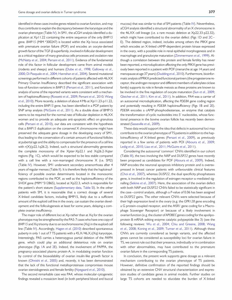
identified in these cases involve genes related to ovarian function, and maythus contribute to explain the discrepancy between the karyotype and theovarian phenotype (Table IV). In SM1, the aCGH analysis identified a du-plication at Xp11.22 containing the entire sequence of the only BMP15gene. BMP15 (MIM *300247) is located within the Xp locus associatedwith premature ovarian failure (POF) and encodes an oocyte-derivedgrowth factor of the TGF-b superfamily, involved in follicular developmentas a critical regulator of many granulosa cells processes and ovulation rate(McNatty et al., 2004; Persani et al., 2011). Evidence of the fundamentalrole of this factor in follicular development came from animal models(rodents and sheeps) and humans (Dube et al., 1998; Galloway et al.,2000; Di Pasquale et al., 2004; Hanrahan et al., 2004). Several mutationalscreenings performed in different cohorts of patients affected with 46,XXPrimary Ovarian Insufficiency described the significant association withloss-of-function variations in BMP15 (Persani et al., 2011), and functionalanalysis of some of the reported variants were consistent with a mechan-ism of haploinsufficiency (Rossetti et al., 2009; Persani et al., 2010; Wanget al., 2010). More recently, a deletion of about 4 Mb at Xp11.23-p11.22,including the entire BMP15 gene, has been identified in a POF patient bySNP array analysis (McGuire et al., 2011). As a double dose of BMP15seems to be required for the normal rate of follicular depletion in 46,XXwomen and to provide an adequate anti-apoptotic effect on granulosacells (Persani et al., 2010), our data are consistent with the hypothesisthat a BMP15 duplication on the conserved X chromosome might havepreserved the adequate gene dosage in the developing ovary of SM1,thus leading to the conservation of a certain amount of functional folliclesat pubertal age and the ability to compensate for the presence of a cell linewith r(X)(p22.1q26.2). Indeed, such a structural abnormality generatesthe complete monosomy of the Xpter-Xp22.1 and Xq26.2-Xqterregions (Fig. 1C), which would be expected to be less stable comparedwith a cell line with a non-rearranged chromosome X (i.e. SM2)(Table IV). However, SM1 underwent secondary amenorrhoea after 4years of regular menses (Table II). It is therefore likely that the haploinsuf-ficiency of possible ovarian determinants located in the monosomicregions contributed to the POF, as well as the haploinsufficiency of theSHOX gene (MIM *312865), located at Xp22.3, which is responsible forthe patient’s short stature (Supplementary data, Table SI). In the otherpatients with SM, it is reasonable that a correct dosage of severalX-linked candidate factors, including BMP15, likely due to a sufficientamount of the euploid cell line in the ovary, can sustain the ovarian devel-opment and the folliculogenesis at least for some years, delaying a com-plete ovarian insufficiency.
The major role of different loci at Xp rather than at Xq for the ovarianphenotype may be strengthened by the PA5-7cases who have one copy ofBMP15 and Xq trisomy due to an isochromosome (Xq) in the euploid cellline (Table IV). Accordingly, Hagen et al. (2010) described spontaneouspuberty in only 1 out of 7 TS patients with a 45,X/46,X,i(Xq) karyotype.Interestingly, PA5 carries a heterozygous partial deletion of the PAPPAgene, which could play an additional deleterious role on ovarianphenotype (Figs 1A and 2E). Indeed, the involvement of PAPPA, thepregnancy-associated plasma protein-A, in modulating ovarian functionby control of the bioavailability of ovarian insulin-like growth factor isknown (Ohnishi et al., 2005) and, recently, it has been demonstratedthat the lack of this functional metalloproteinase compromises mouseovarian steroidogenesis and female fertility (Nyegaard et al., 2010).
The second remarkable case was PA4, whose molecular-cytogeneticfindings revealed a mosaicism level (in both peripheral blood and buccal
mucosa) that was similar to that of SM patients (Table IV). Nevertheless,aCGH analysis identified a structural abnormality of an X chromosome inthe 46,XX cell lineage (i.e. a rare mosaic deletion at Xp22.33-p22.32),which might have contributed to the ovarian defect (Figs 1D and 2C–E). The deleted region, indeed, includes among others the PRKX genewhich encodes an X-linked cAMP-dependent protein kinase expressedin the ovary, with a possible role in renal epithelial morphogenesis and inmacrophage and granulocyte maturation (Zimmermann et al., 1999). Al-though a correlation between this protein and female fertility has neverbeen reported, a microduplication affecting the only PRKX gene has previ-ously been reported in a patient with POF (menarche at age 14 years andmenopause at age 37 years) (Dudding et al., 2010). Furthermore, bioinfor-matic analysisofPRKXpredicted functionalpartners (theprogesteronere-ceptor, the androgen receptor and different members of adenylate ciclasefamily) supports its role in female meiosis as these proteins are known tobe involved in the fine regulation of oocyte maturation (Sun et al., 2009;Gleicher et al., 2011; Kim et al., 2011). In addition, PA4 was a carrier ofan autosomal microduplication, affecting the PDE8A gene coding regionand potentially resulting in PDE8A haploinsufficiency (Figs 1B and 2E).PDE8A encodes a cAMP-phosphodiesterase, an enzyme that catalysisthe transformation of cyclic nucleotides into 5′ nucleotides, whose func-tional presence in the bovine ovarian follicle has recently been demon-strated (Sasseville et al., 2009).
These data would support the idea that defects in autosomal loci maycontribute to the ovarian phenotype of TS patients in addition to the hap-loinsufficiency of X-linked genes (Persani et al., 2009), as previouslyreported in a few series of patients with POI (Aboura et al., 2009;Ledig et al., 2010; Liao et al., 2011; McGuire et al., 2011).
Considering the autosomal ‘common’ CNVs identified in our cohort(Table III), the two involving the NAIP and DUSP22 genes have recentlybeen proposed as candidates for POF (Aboura et al., 2009). Indeed,NAIP encodes the neuronal apoptosis inhibitory protein and is overex-pressed in breast cancer patients with unfavourable clinical features(Choi et al., 2007), whereas DUSP22, the dual specificity phosphatasesgene, is involved in the regulation of estrogen receptor a-mediated sig-nalling (Sekine et al., 2007). Here, the association of the ovarian defectwith both NAIP and DUSP22 CNVs failed to be statistically significant inthe case–control analysis, although a P-value of 0.06 has been assignedto DUSP22 gains. The other relevant CNVs were selected because oftheir high expression level in the ovary (e.g. the GPR128 gene encodinga G protein-coupled receptor, and the MSR1 gene coding for a Macro-phage Scavenger Receptor) or because of a likely involvement inovarian function (e.g. the cluster of APOBEC genes coding for the apolipo-protein B mRNA-editing enzyme catalytic polypeptide-like 3) (see thefollowing reviews: Wu et al., 2004; Chiu and Greene, 2008; Wanget al., 2008; Koning et al., 2009; Turner et al., 2011). Although theseCNVs are currently considered as benign variants, and the affectedgenes cannot be considered as susceptibility loci for ovarian failure inTS, we cannot rule out that their presence, individually or in combinationwith other abnormalities, may have contributed to the prematureovarian failure in the corresponding TS patients.
In conclusion, the present work supports gene dosage as a relevantmechanism contributing to the ovarian phenotype of TS patients.However, definitive confirmation of the reported findings should beobtained by an extensive CNV structural characterization and expres-sion studies of candidate genes in animal models. Further studies onlarge TS cohorts are needed to elucidate the burden of X-linked
Gene dosage and ovarian defects in Turner syndrome 377

dosage-sensitive genes in spontaneous pubertal development. Theseshould evaluate the X chromosome mosaicism rates in tissues of differ-ent embryonic origin, including the ovary. The first duplication rearrange-ment of BMP15 in a 45,X TS patient with SM brings additional evidencefor a prominent determinant role of this Xp-linked gene on ovarian folli-culogenesis. The detection of CNVs in autosomal ovary-related genesmay generate further insights on the fundamental mechanisms contribut-ing to ovarian function.
Supplementary dataSupplementary data areavailable athttp://humrep.oxfordjournals.org/.
AcknowledgementsThe authors thank the patients and their families for their cooperation.
Authors’ rolesThe authors were responsible for the following aspects of the study.C.Cas.: study design, acquisition, analysis and interpretation of datafrom array-CGH and FISH analysis; manuscript draft preparation; R.R.:study design, acquisition, analysis and interpretation of data from real-time quantitative PCR, manuscript draft preparation; D.R.: acquisition,analysis and interpretation of data from array-CGH; M.P.R.: chromo-somal sample collection and preparation; C.Cac.: sample preparationand clinical data collection; E.B.: acquisition, analysis and interpretationof data from DHPLC analysis; V.C.: patient recruitment and clinicaldata collection; P.I.: study design and critical revision of the manuscript;D.L.: patient recruitment and critical revision of manuscript; P.F. and L.P.:study design and coordination, data interpretation and critical revision ofthe manuscript. All authors reviewed and approved the final version ofthe manuscript.
FundingThis work was partially supported by Telethon Foundation, Italy (grantnumber: GGP09126 to L.P.), the Italian Ministry of the University and Re-search (grant number: 2006065999 to P.F.) and a Ministry of Health grant“Ricerca Corrente” to Istituto Auxologico Italiano IRCCS (grantnumber:08C704-2006). The funders had no role in study design, data collectionand analysis, decision to publish, or preparation of the manuscript.Funding to pay the Open Access publication charges for this articlewas provided by the Foundation Telethon Italia.
Conflict of interestThe authors have none to declare.
ReferencesAboura A, Dupas C, Tachdjian G, Portnoı MF, Bourcigaux N, Dewailly D,
Frydman R, Fauser B, Ronci-Chaix N, Donadille B et al. Array comparativegenomic hybridization profiling analysis reveals deoxyribonucleic acid copynumber variations associated with premature ovarian failure. J ClinEndocrinol Metab 2009;9411:4540–4546.
Aso K, Koto S, Higuchi A, Ariyasu D, Izawa M, Miyamoto Igaki J, Hasegawa Y.Serum FSH level below 10 mIU/mL at twelve years old is an index ofspontaneous and cyclical menstruation in Turner syndrome. Endocr J2010;57:909–913.
Ballif BC, Rorem EA, Sundin K, Lincicum M, Gaskin S, Coppinger J,Kashork CD, Shaffer LG, Bejjani BA. Detection of low-level mosaicismby array CGH in routine diagnostic specimens. Am J Med Genet A 2006;140:2757–2767.
Chiu YL, Greene WC. The APOBEC3 cytidine deaminases: an innatedefensive network opposing exogenous retroviruses and endogenousretroelements. Annu Rev Immunol 2008;26:317–353.
Choi J, Hwang YK, Choi YJ, Yoo KE, Kim JH, Nam SJ, Yang JH, Lee SJ, Yoo KH,Sung KW et al. Neuronal apoptosis inhibitory protein is overexpressed inpatients with unfavourable prognostic factors in breast cancer. J KoreanMed Sci 2007;22(Suppl):S17–S23.
Di Pasquale E, Beck-Peccoz P, Persani L. Hypergonadotropic ovarian failureassociated with an inherited mutation of human bone morphogeneticprotein-15 (BMP15) gene. Am J Hum Genet 2004;75:106–111.
Dube JL, Wang P, Elvin J, Lyons KM, Celeste AJ, Matzuk MM. The bonemorphogenetic protein 15 gene is X-linked and expressed in oocytes.Mol Endocrinol 1998;12:1809–1817.
Dudding TE, Lawrence O, Winship I, Froyen G, Vandewalle J, Scott R,Shelling AN. Array comparative genomic hybridization for the detectionof submicroscopic copy number variations of the X chromosome inwomen with premature ovarian failure. Hum Reprod 2010;25:3159–3160.
Fechner PY, Davenport ML, Qualy RL, Ross JL, Gunther DF, Eugster EA,Huseman C, Zagar AJ, Quigley CA; Toddler Turner Study Group.Differences in follicle-stimulating hormone secretion between 45,Xmonosomy Turner syndrome and 45,X/46,XX mosaicism are evidentat an early age. J Clin Endocrinol Metab 2006;91:4896–4902.
Galloway SM, McNatty KP, Cambridge LM, Laitinen MP, Juengel JL,Jokiranta JL, McLaren RJ, Luiro K, Dodds KG, Montgomery GW et al.Mutations in an oocyte-derived growth factor gene (BMP15) causeincreased ovulation rate and infertility in a dosage-sensitive manner. NatGenet 2000;25:279–283.
Gleicher N, Weghofer A, Barad DH. The role of androgens in folliclematuration and ovulation induction: friend or foe of infertility treatment?Reprod Biol Endocrinol 2011;9:116.
Gracia Bouthelier R, Oliver Iguacel A, Gonzalez Casado I, Alcalde deAlvare A. Optimization of treatment in Turner’s syndrome. J PediatrEndocrinol Metab 2004;17:427–434.
Hagen CP, Main KM, Kjaergaard S, Juul A. FSH, LH, inhibin B and estradiollevels in Turner syndrome depend on age and karyotype: longitudinalstudy of 70 Turner girls with or without spontaneous puberty. HumReprod 2010;25:3134–3141.
Hanrahan JP, Gregan SM, Mulsant P, Mullen M, Davis GH, Powell R,Galloway SM. Mutations in the genes for oocyte-derived growth factorsGDF9 and BMP15 are associated with both increased ovulation rate andsterility in Cambridge and Belclare sheep (Ovis aries). Biol Reprod 2004;70:900–909.
Held KR, Kerber S, Kaminsky E, Singh S, Goetz P, SeemanovaE, Goedde HW.Mosaicism in 45,X Turner syndrome: does survival in early pregnancydepend on the presence of two sex chromosomes? Hum Genet 1992;88:288–294.
Kallio S, Aittomaki K, Piltonen T, Veijola R, Liakka A, Vaskivuo TE, Dunkel L,Tapanainen JS. Anti-Mullerian hormone as a predictor of follicular reservein ovarian insufficiency: special emphasis on FSH-resistant ovaries. HumReprod 2012;27:854–860.
Kim J, Bagchi IC, Bagchi MK. Control of ovulation in mice by proges-terone receptor-regulated gene networks. Mol Hum Reprod 2011;15:821–828.
378 Castronovo et al.

Koning FA, Newman EN, Kim EY, Kunstman KJ, Wolinsky SM, Malim MH.Defining APOBEC3 expression patterns in human tissues andhematopoietic cell subsets. J Virol 2009;83:9474–9485.
Ledig S, Ropke A, Wieacker P. Copy number variants in premature ovarianfailure and ovarian dysgenesis. Sex Dev 2010;4:225–232.
Liao C, Fu F, Yang X, Sun YM, Li DZ. Analysis of Chinese women with primaryovarian insufficiency by high resolution array-comparative genomichybridization. Chin Med J 2011;124:1739–1742.
Lichter P, Cremer T. Chromosome analysis by non-isotopic in situhybridization. In Rooney DE, Czepulkowski BH (eds). HumanCytogenetics. A Practical Approach. Oxford: IRL Press at OxfordUniversity Press, 1992, 157–192.
Lichter P, Tang CJ, Call K, Hermanson G, Evans GA, Housman D, Ward DC.High resolution mapping of human chromosome 11 by in situ hybridizationwith cosmid clones. Science 1990;247:64–69.
Martın Campagne E, Roa Llamazares C. Spontaneous puberty and menarchein a patient with Turner syndrome and 45X monosomy. An Pediatr 2008;70:200–202.
McGuire MM, Bowden W, Engel NJ, Ahn HW, Kovanci E, Rajkovic A.Genomic analysis using high-resolution single-nucleotide polymorphismarrays reveals novel microdeletions associated with premature ovarianfailure. Fertil Steril 2011;95:1595–1600.
McNatty KP, Moore LG, Hudson NL, Quirke LD, Lawrence SB, Reader K,Hanrahan JP, Smith P, Groome NP, Laitinen M et al. The oocyte and itsrole in regulating ovulation rate: a new paradigm in reproductive biology.Reproduction 2004;128:379–386.
Nyegaard M, Overgaard MT, Su YQ, Hamilton AE, Kwintkiewicz J, Hsieh M,Nayak NR, Conti M, Conover CA, Giudice LC. Lack of functionalpregnancy-associated plasma protein-A (PAPPA) compromises mouseovarian steroidogenesis and female fertility. Biol Reprod 2010;82:1129–1138.
Ogata T, Matsuo N. Turner syndrome and female sex chromosomeaberrations: deduction of the principal factors involved in thedevelopment of clinical features. Hum Genet 1995;95:607–629.
Ohnishi J, Ohnishi E, Shibuya H, Takahashi T. Functions for proteinases in theovulatory process. Biochim Biophys Acta 2005;1751:95–109.
Pasquino AM, Passeri F, Pucarelli I, Segni M, Municchi G. Spontaneouspubertal development in Turner’s syndrome. Italian Study Group forTurner’s Syndrome. J Clin Endocrinol Metab 1997;82:1810–1813.
Persani L, Rossetti R, Cacciatore C, Bonomi M. Primary ovarian insufficiency:X chromosome defects and autoimmunity. J Autoimmun 2009;33:35–41.
Persani L, Rossetti R, Cacciatore C. Genes involved in human prematureovarian failure. J Mol Endocrinol 2010;45:405.
Persani L, Rossetti R, Cacciatore C, Fabre S. Genetic defects of ovarianTGF-b-like factors and premature ovarian failure. J Endocrinol Invest2011;34:244–251.
Prakash S, Guo D, Maslen CL, Silberbach M, Milewicz D, Bondy CA; GenTACInvestigators. Single-nucleotide polymorphism array genotyping is
equivalent to metaphase cytogenetics for diagnosis of Turner syndrome.Genet Med. June 6 2013. doi:10.1038/gim.2013.77.
Quilter CR, Karcanias AC, Bagga MR, Duncan S, Murray A, Conway GS,Sargent CA, Affara NA. Analysis of X chromosome genomic DNAsequence copy number variation associated with premature ovarianfailure (POF). Hum Reprod 2010;258:2139–2150.
Ranke MB, Saenger P. Turner’s syndrome. Lancet 2001;358:309–314.Reddy KS, Mak L. Mosaic unbalanced structural abnormalities confirmed
using FISH on buccal mucosal cells. Ann Genet 2001;44:37–40.Reynaud K, Cortvrindt R, Verlinde F, De Schepper J, Bourgain C, Smitz J.
Number of ovarian follicles in human fetuses with the 45,X karyotype.Fertil Steril 2004;81:1112–1119.
Rossetti R, Di Pasquale E, Marozzi A, Bione S, Toniolo D, Grammatico P,Nelson LM, Beck-Peccoz P, Persani L. BMP15 mutations associated withprimary ovarian insufficiency cause a defective production of bioactiveprotein. Hum Mutat 2009;30:804–810.
Saenger P. Turner’s syndrome. N Engl J Med 1996;335:1749–1754.Sasseville M, Albuz FK, Cote N, Guillemette C, Gilchrist RB, Richard FJ.
Characterization of novel phosphodiesterases in the bovine ovarianfollicle. Biol Reprod 2009;81:415–425.
Sekine Y, Ikeda O, Hayakawa Y, Tsuji S, Imoto S, Aoki N, Sugiyama K,Matsuda T. DUSP22/LMW-DSP2 regulates estrogen receptor-alpha-mediated signaling through dephosphorylation of Ser-118. Oncogene2007;26:6038–6049.
Sun QY, Miao YL, Schatten H. Towards a new understanding on theregulation of mammalian oocyte meiosis resumption. Cell Cycle 2009;1:2741–2747.
Sybert VP, McCauley E. Turner’s syndrome. N Engl J Med 2004;351:1227–1238.
Toniolo D. X-linked premature ovarian failure: a complex disease. Curr OpinGenet Dev 2006;6:293–300.
Turner EC, Hughes J, Wilson H, Clay M, Mylonas KJ. Conditional ablation ofmacrophages disrupts ovarian vasculature. Reproduction 2011;141:821–831.
Wang C, Prossnitz ER, Roy SK. G protein-coupled receptor 30 expression isrequired for estrogen stimulation of primordial follicle formation in thehamster ovary. Endocrinology 2008;149:4452–4461.
Wang B, Wen Q, Ni F, Zhou S, Wang J, Cao Y, Ma X. Analyses of growthdifferentiation factor 9 (GDF9) and bone morphogenetic protein 15(BMP15) mutation in Chinese women with premature ovarian failure.Clin Endocrinol 2010;72:135–136.
Wu R, Van der Hoek KH, Ryan NK, Norman RJ, Robker RL. Macrophagecontributions to ovarian function. Hum Reprod Update 2004;10:119–133.
Zimmermann B, Chiorini JA, Ma Y, Kotin RM, Herberg FW. PrKX is a novelcatalytic subunit of the cAMP-dependent protein kinase regulated by theregulatory subunit type I. J Biol Chem 1999;274:5370–5378.
Zinn AR, Ross JL. Turner syndrome and haploinsufficiency. Curr Opin GenetDev 1998;8:322–327.
Gene dosage and ovarian defects in Turner syndrome 379