2011 Turner Szabo Maas Poyer Zoran
16
Regulation and Restoration of Motoneuronal Synaptic Transmission During Neuromuscular Regeneration in the Pulmonate Snail Helisoma trivolvis M. B. TURNER 1 , T. M. SZABO-MAAS 2,† J. C. POYER 3 , AND M. J. ZORAN 3, * 1 Department of Cellular Biology, University of Georgia, Athens, Georgia 30602; 2 Department of Biology, Brandeis University, Waltham, Massachusetts 02453; and 3 Department of Biology, Texas A&M University, College Station, Texas 77843 Abstract. Regeneration of motor systems involves rees- tablishment of central control networks, reinnervation of muscle targets by motoneurons, and reconnection of neuro- modulatory circuits. Still, how these processes are inte- grated as motor function is restored during regeneration remains ill defined. Here, we examined the mechanisms underlying motoneuronal regeneration of neuromuscular synapses related to feeding movements in the pulmonate snail Helisoma trivolvis. Neurons B19 and B110, although activated during different phases of the feeding pattern, innervate similar sets of muscles. However, the percentage of muscle fibers innervated, the efficacy of excitatory junc- tion potentials, and the strength of muscle contractions were different for each cell’s specific connections. After periph- eral nerve crush, a sequence of transient electrical and chemical connections formed centrally within the buccal ganglia. Neuromuscular synapse regeneration involved a three-phase process: the emergence of spontaneous synaptic transmission (P1), the acquisition of evoked potentials of weak efficacy (P2), and the establishment of functional reinnervation (P3). Differential synaptic efficacy at muscle contacts was recapitulated in cell culture. Differences in motoneuronal presynaptic properties (i.e., quantal content) were the basis of disparate neuromuscular synapse function, suggesting a role for retrograde target influences. We pro- pose a homeostatic model of molluscan motor system re- generation. This model has three restoration events: (1) transient central synaptogenesis during axonal outgrowth, (2) intermotoneuronal inhibitory synaptogenesis during ini- tial neuromuscular synapse formation, and (3) target-depen- dent regulation of neuromuscular junction formation. Introduction Regeneration is the process of repairing damaged tissues or replacing lost body parts after injury. The mechanisms that underlie regeneration vary considerably among animal groups and across a wide range of tissue and organ systems. For well over a century scientists have questioned why animals vary in their ability to regenerate (Morgan, 1901). Still, most animal groups, particularly invertebrates (Myo- hara et al., 1999; Martinez et al., 2005; Agata and Umesono, 2008), have the capacity to regenerate their damaged ner- vous systems, and much is understood about these processes (Purves and Lichtman, 1985; Moffett, 1996). Furthermore, invertebrate systems like leeches and molluscs provide ex- cellent frameworks for elucidation of the cellular mecha- nisms underlying the regeneration of specific neural con- nections (Ready and Nicholls, 1979; Allison and Benjamin, 1985; French and Muller, 1986; Haydon and Kater, 1988; Schacher et al., 1988; Carrow and Levitan, 1989; Liu and Nicholls, 1989; Snyder and Moffett, 1990; Nicholls et al., 1990; Syed et al., 1992; Ross et al, 1994; Zoran and Poyer, 1996; Matsuo et al., 2010). In fact, regenerated networks of neurons of the pond snails Lymnaea and Helisoma exhibit patterns of activity (i.e., fictive behaviors) in vitro that are Received 12 April 2010; accepted 5 May 2010. * To whom correspondence should be addressed. E-mail: zoran@mail. bio.tamu.edu † Current address: Department of Biology, Delaware State University, Dover, Delaware 19904. Abbreviations: Aj, anterior jugalis; AP-PSP, action potential-evoked postsynaptic potentials; DM, defined medium; EJP, excitatory junction potential; Lbn, laterobuccal nerve; mEJP, miniature excitatory junction potential; mPSP, miniature postsynaptic potential; PSP, postsynaptic po- tential; Slrt, supralateral radular tensor; Vbn, ventrobuccal nerve. Reference: Biol. Bull. 221: 110 –125. (August 2011) © 2011 Marine Biological Laboratory 110
Transcript of 2011 Turner Szabo Maas Poyer Zoran
Regulation and Restoration of Motoneuronal SynapticTransmission During Neuromuscular Regeneration in
the Pulmonate Snail Helisoma trivolvis
M. B. TURNER1, T. M. SZABO-MAAS2,† J. C. POYER3, AND M. J. ZORAN3,*1Department of Cellular Biology, University of Georgia, Athens, Georgia 30602; 2Department of
Biology, Brandeis University, Waltham, Massachusetts 02453; and 3Department of Biology, Texas A&MUniversity, College Station, Texas 77843
Abstract. Regeneration of motor systems involves rees-tablishment of central control networks, reinnervation ofmuscle targets by motoneurons, and reconnection of neuro-modulatory circuits. Still, how these processes are inte-grated as motor function is restored during regenerationremains ill defined. Here, we examined the mechanismsunderlying motoneuronal regeneration of neuromuscularsynapses related to feeding movements in the pulmonatesnail Helisoma trivolvis. Neurons B19 and B110, althoughactivated during different phases of the feeding pattern,innervate similar sets of muscles. However, the percentageof muscle fibers innervated, the efficacy of excitatory junc-tion potentials, and the strength of muscle contractions weredifferent for each cell’s specific connections. After periph-eral nerve crush, a sequence of transient electrical andchemical connections formed centrally within the buccalganglia. Neuromuscular synapse regeneration involved athree-phase process: the emergence of spontaneous synaptictransmission (P1), the acquisition of evoked potentials ofweak efficacy (P2), and the establishment of functionalreinnervation (P3). Differential synaptic efficacy at musclecontacts was recapitulated in cell culture. Differences inmotoneuronal presynaptic properties (i.e., quantal content)
were the basis of disparate neuromuscular synapse function,suggesting a role for retrograde target influences. We pro-pose a homeostatic model of molluscan motor system re-generation. This model has three restoration events: (1)transient central synaptogenesis during axonal outgrowth,(2) intermotoneuronal inhibitory synaptogenesis during ini-tial neuromuscular synapse formation, and (3) target-depen-dent regulation of neuromuscular junction formation.
Introduction
Regeneration is the process of repairing damaged tissuesor replacing lost body parts after injury. The mechanismsthat underlie regeneration vary considerably among animalgroups and across a wide range of tissue and organ systems.For well over a century scientists have questioned whyanimals vary in their ability to regenerate (Morgan, 1901).Still, most animal groups, particularly invertebrates (Myo-hara et al., 1999; Martinez et al., 2005; Agata and Umesono,2008), have the capacity to regenerate their damaged ner-vous systems, and much is understood about these processes(Purves and Lichtman, 1985; Moffett, 1996). Furthermore,invertebrate systems like leeches and molluscs provide ex-cellent frameworks for elucidation of the cellular mecha-nisms underlying the regeneration of specific neural con-nections (Ready and Nicholls, 1979; Allison and Benjamin,1985; French and Muller, 1986; Haydon and Kater, 1988;Schacher et al., 1988; Carrow and Levitan, 1989; Liu andNicholls, 1989; Snyder and Moffett, 1990; Nicholls et al.,1990; Syed et al., 1992; Ross et al, 1994; Zoran and Poyer,1996; Matsuo et al., 2010). In fact, regenerated networks ofneurons of the pond snails Lymnaea and Helisoma exhibitpatterns of activity (i.e., fictive behaviors) in vitro that are
Received 12 April 2010; accepted 5 May 2010.* To whom correspondence should be addressed. E-mail: zoran@mail.
bio.tamu.edu† Current address: Department of Biology, Delaware State University,
Dover, Delaware 19904.Abbreviations: Aj, anterior jugalis; AP-PSP, action potential-evoked
postsynaptic potentials; DM, defined medium; EJP, excitatory junctionpotential; Lbn, laterobuccal nerve; mEJP, miniature excitatory junctionpotential; mPSP, miniature postsynaptic potential; PSP, postsynaptic po-tential; Slrt, supralateral radular tensor; Vbn, ventrobuccal nerve.
Reference: Biol. Bull. 221: 110–125. (August 2011)© 2011 Marine Biological Laboratory
110
almost indistinguishable from those recorded in intact ani-mals (Syed et al., 1990, 1993). Thus, cell culture approachesare powerful tools for determining cellular mechanismsgoverning neural regeneration.
In Helisoma, motoneuronal regeneration following nervecrush begins with the growth of numerous processes fromthe axon that ultimately terminate on potential target cells,both appropriate and inappropriate, where synapse forma-tion takes place (Murphy and Kater, 1978; Bulloch andKater, 1982; Haydon et al., 1987). Much of the workconducted in this and other molluscan systems has focusedon motor systems, especially motoneurons controlling thebuccal feeding apparatus and radula (Murphy, 2001; Elliottand Susswein, 2002; Wentzell et al., 2009). Murphy andKater (1978) demonstrated that after axotomy, motoneuronswithin the buccal ganglia of Helisoma undergo extensiveregeneration and finally reinnervation of their normal targetorgans. Axonal processes of these neurons are capable ofdiscriminating between appropriate and inappropriate path-ways (Murphy and Kater, 1980a), regenerate central con-nections (Bulloch and Kater, 1982), and accurately reinner-vate their peripheral target organs (Murphy and Kater,1980b). How the reestablishment of central control net-works and restoration of peripheral neuromuscular connec-tions are integrated during motor system regeneration re-mains unclear.
Here, we have examined the mechanisms underlyingmotoneuronal regeneration of neuromuscular synapses.Helisoma buccal neurons B19 and B110, although activatedduring different feeding pattern phases, innervate largelythe same muscle groups. Motoneuron B19 possesses a syn-aptogenic nature characterized by the selective formation ofneuromuscular synapses (Zoran and Poyer, 1996); that is, itrequires specific muscle contact to upregulate its neurose-cretory function (Zoran et al., 1990; Poyer and Zoran,1996). In contrast, neuron B110 is promiscuous in its for-mation of chemical connections, exhibiting neurotransmis-sion within minutes of contact with a cholinoceptive post-synaptic target (Szabo et al., 2004). Once contacts areformed during regeneration, synapse formation results indifferential synaptic properties of the two motoneurons, andphysiological analyses suggest the involvement of retro-grade target influences on the presynaptic neuronal func-tion. We demonstrate here that ultimately these motoneu-rons innervate the same muscle groups; however, they do sowith varying degrees of synaptic coverage and with variablesynaptic efficacy.
Materials and Methods
Animals
Experiments were conducted on laboratory stocks of al-bino (red) pond snails, Helisoma trivolvis (also Planorbellatrivolvis Say, 1817), which were maintained in 20-gallon
aquaria at 26 °C. Aquaria were kept on a controlled photo-period of 12-hour light/12-hour dark. Animals were fedlettuce and/or trout chow daily.
Adult buccal ganglia preparations
Dissection methods have been described previously(Haydon and Zoran, 1991). Two neurons of the buccalganglia, which innervate radular tensor muscle groups(Kater, 1974; Zoran et al., 1989), were primarily used forthese studies: buccal neurons B19 and B110. Snails werede-shelled and pinned to a Sylgard-coated dissecting dishcontaining Helisoma saline (39.9 mmol l–1 NaCl, 1.7 mmoll–1 KCl, 4.1 mmol l–1 CaCl2, 1.5 mmol l–1 MgCl2, 10 mmoll–1 HEPES, pH 7.3). Ganglia were excised along with intactbuccal nerves and muscles. Buccal motoneurons B19 andB110 and their connections with the anterior jugalis (Aj)and supralateral radular tensor (Slrt) muscle groups werestudied. For neuron B110 recordings, the buccal gangliawere pinned with the rostral side and with the musculaturedissected to allow for access to both Aj and Slrt musclefibers. Neuron B19 is located caudally, therefore whenassessing connectivity in this neuron, the buccal gangliawere pinned with the caudal side exposed. Experiments onisolated buccal ganglia were performed in defined medium(DM). DM consisted of Leibowitz-15 (L-15, Formula No.82-5154EC, Gibco Laboratories) containing Helisoma salts(mmol l–1: 40.0 NaCl, 1.7 KCl, 4.1 CaCl2, 1.5 MgCl2, and10.0 HEPES at pH 7.5).
Regenerating buccal ganglia
Intact Helisoma were placed in a saturated menthol so-lution for 15–30 min. Suction was gently applied to theventral portion of the foot to extract the anterior region ofthe body from the shell. A dorsal incision was made, and thebuccal commissure or the laterobuccal nerves (Lbn) andventrobuccal nerves (Vbn) nerves associated with the gan-glia (bilateral) were crushed with fine forceps. The bodywall was sutured, and the animal was allowed from 1 day to5 weeks to recuperate. Ganglia were then removed from theanimals and synaptic connectivity was examined electro-physiologically. Sham operations were also performed inwhich incisions were made, but nerve trunks were notcrushed.
Two points must be noted regarding assessments of post-crush connectivity between neurons B19 and B110. First,for simultaneous electrophysiological recordings, one buc-cal ganglion was rotated 180° with respect to the otheraround the buccal commissure so that both a rostral andcaudal side were accessible. Although care was taken not todamage the commissure, some newly forming connectionsmight have been disrupted during this procedure. Second,membrane potential changes produced at distant sites ofsynapse formation in the commissure were recorded at the
111NEUROMUSCULAR SYNAPSE REGENERATION
cell bodies. Therefore, the magnitude of early, transientelectrical coupling in these in vivo studies was likely un-derestimated.
Neuronal cell culture
Buccal ganglia were isolated from the animal and treatedwith 0.2% trypsin (Sigma) for 20 min. Dissection methodsfor in vitro preparations have been described previously(Haydon and Zoran, 1991). Neurons B19 and B110 wereidentified and isolated into culture on bovine serum albumin(BSA)-coated 35-mm plastic dishes (Falcon 1008) contain-ing 2 ml of conditioned medium and incubated at 25 °C (inhigh humidity) for 2 days. Conditioned medium was pre-pared by incubating defined medium (DM) with sterileHelisoma central ring ganglia (2 ganglia per milliliter) for 3days. DM contained 50% Leibowitz-15 (Gibco) to whichL-glutamine (30 mg/100 ml), gentamicin (50 mg/ml), andinorganic salts were added to give final concentrations of 40mmol l–1 NaCl, 1.7 mmol l�1 KCl, 1.5 mmol l–1 MgCl2, 4.1mmol l–1 CaCl2, 10 mmol l–1 HEPES, pH 7.4.
Buccal muscle dissociation
The Slrt and Aj muscle groups were dissected from thebuccal mass and sterilized through three washes of Heli-soma antibiotic saline (sterile water containing Helisomasalts and 150 mg/ml gentamicin) and placed into a sterile,5-ml glass test tube pre-coated with BSA. Muscles weredissociated into individual fibers through incubation in DMcontaining 2 mg/ml collagenase/dispase (Boehringer-Mann-heim) at room temperature for 12 h in a 32 °C water bath.After incubation, DM was added to the test tube, and musclefibers were fully dissociated using gentle agitation andcentrifugation (1000 � g for 5 min). Fibers were thencollected and transferred to a BSA-coated plastic culturedish (Falcon 1008). Subsequently, the individual musclefibers were transferred into poly-L-lysine (PLL)-coated35-mm dishes and plated into culture on opposite sides of aneuronal cell body.
Electrophysiological techniques
Electrophysiological analyses were performed using in-tracellular recording techniques. Glass microelectrodes(borosilicate; FHC) were filled with 1.5 mol l–1 KCl, andpossessed tip resistances ranging from 10 to 20 M�. Cur-rent-clamp recordings of neuronal membrane potentialswere amplified using bridge-balanced electrometers (Get-ting Instruments or World Precision Instruments). Simulta-neous recordings were made from cell pairs or from neuron-muscle preparations. For in situ recordings, ganglia werebathed in high-calcium (41 mmol l–1 CaCl2) DM to reducefeeding motor-pattern activity and other synaptic inputs tomuscle fibers. Preparations were visualized with a dissect-
ing microscope (Olympus). Neuronal membrane potentialwas maintained with base current injection at approximately–70 mV, and muscle membrane potential was maintained atapproximately –50 mV. Electrical coupling was measuredby injection of constant-amplitude, hyperpolarizing currentpulses (0.5–1 nA pulse for 3 s) into one neuron, whilemembrane voltage changes in both the presynaptic (in-jected) neuron and its postsynaptic (noninjected) partnerwere monitored. In some experiments neurons were injectedwith the fluorescent dye Lucifer yellow (Sigma) to examineneuronal architecture and verify cell identity.
The neuron-muscle chemical connection, as well as thechemical connection from neuron B110 onto neuron B19,was studied by injection of depolarizing current into pre-synaptic neuron B110. Four to five trains of action poten-tials (1 per second) were evoked, and postsynaptic poten-tials (PSPs) were examined. The presence or absence ofPSPs and the location of the fiber from which the recordingwas made within each identified muscle group were used togenerate topographical synaptic maps. Electrophysiologicalrecordings were digitized by a PowerLab A/D data acqui-sition system linked to an Apple computer using Chartsoftware (AD Instruments, ver. 4.1.1).
For in vitro assessments of connectivity, isolated neuronswere incubated for 2 days in nonadhesive conditions andthen transferred between parallel grooves scored onto thebottom of PLL-coated 35-mm dishes (Falcon 3001). Thesegrooves restricted neuritic outgrowth to the area betweenthe etched parallel lines. Aj and Slrt muscle fibers were thenmanipulated into the culture dishes on opposite sides of theneuron and plated within 50–100 �m of the cell body. Afteran additional 3 days of co-culture, an ACh-sensitive assaycell was manipulated into contact with the neurites of theneurons. Assay cells were neurons cultured as spheres for 2days on BSA-coated dishes containing 2 ml of conditionedmedium. These spherical B19 somata are highly cholino-ceptive and function as ACh-sensors detecting neurotrans-mitter release from presynaptic neurites (Haydon andZoran, 1989, 1991). Neurite-assay contacts were established30 min to 1 h prior to the assessment of ACh release. Assaycells were positioned just proximal to sites of neurite-muscle contact.
Quantal analysis of synaptic transmission
The amplitudes of miniature postsynaptic potentials(mPSPs) were measured over a period of 2 min. The am-plitudes of the evoked PSPs were then measured during aconcomitant current injection of the presynaptic neuron toelicit trains of action potentials. The assay cell membranepotential was maintained with a base current injection at–80 mV. There were approximately 50–100 mPSPs and200–400 PSPs measured for each preparation. The ampli-tudes were measured from baseline to the peak of the
112 M. B. TURNER ET AL.

synaptic potential. To determine quantal size, a model cellpreparation was used, and the number of occurrences andtheir amplitudes of the mPSP were plotted. The amplitudeof the most frequent mPSP was used as an estimate ofquantal size.
Neuronal track tracing
Whole-mount preparations were pinned out on Sylgardblocks in Helisoma saline. Individual identified neuronswere injected via micropipettes containing a 3% Luciferyellow/fast green solution by using a picospritzer (GeneralValve). Following varying periods of diffusion (2–24 h),preparations were fixed and processed for imaging. In someexperiments, preparations were embedded, and cryosectionswere cut and mounted on glass slides. Preparations wereimaged using an Olympus inverted microscope and SimplePCI software (Hamamatsu, ver. 6.0) and imported intoAdobe Photoshop for post-capture processing.
Data analysis
Statistical analyses were performed using Statview soft-ware (Abacus Concepts, Inc., ver. 4.5). The Student’s t-testwas used for comparison between two populations. Foranalyzing multiple populations, an analysis of variance wasemployed, and comparisons were made using Fisher’spaired least significant difference test. Data is presented asa mean � SEM unless otherwise stated. Statistical signifi-cance was represented as P � 0.05.
Results
Connectivity of the Helisoma buccal neuromuscularsystem
Bilaterally symmetrical motor neurons, located in thepaired buccal ganglia in Helisoma, control the feeding mus-culature and associated structures: the radula, odontophore,salivary glands, and esophagus. Like other invertebrate mo-tor systems, the buccal musculature of Helisoma is inner-vated by both motor and modulatory neurons. For example,cerebral neuron 1 (C1) is a serotonergic neuron that projectsthrough the cerebral-buccal connective to innervate the buc-cal ganglion, but also projects via buccal nerves to thebuccal musculature (Fig. 1A). Stimulation of C1 elicitsrhythmic firing in buccal neurons and increases the strengthof contractions of radular muscles (Murphy, 2001; Fig. 1B).Several aspects of the regeneration of buccal motor neuronsare differentially regulated by serotonin (Murphy et al.,1985; McCobb et al., 1988; Murrain et al., 1990; Price andGoldberg, 1993). In the present studies of buccal neuromus-cular regeneration, we have limited our analysis to buccalretractor neuron B110, which fires during the S2 phase ofthe motor pattern, and hyper-retractor neuron B19, whichfires during the S3 phase (Fig. 1C). Bursts of action poten-
tials in these motor neurons drive summating excitatoryjunction potentials (EJPs) in specific sets of muscle fibers.For example, spikes in B19 evoke EJPs in muscle fibers ofthe supralateral radular tensor (Slrt) muscle (Fig. 1D). In-terestingly, although neurons B19 and B110 fire out ofphase during rhythmic motor outputs and are involved indifferent muscle movements, they possess axonal projec-tions that terminate onto similar sets of buccal muscles,which regulate the movements of the radula and odonto-phore.
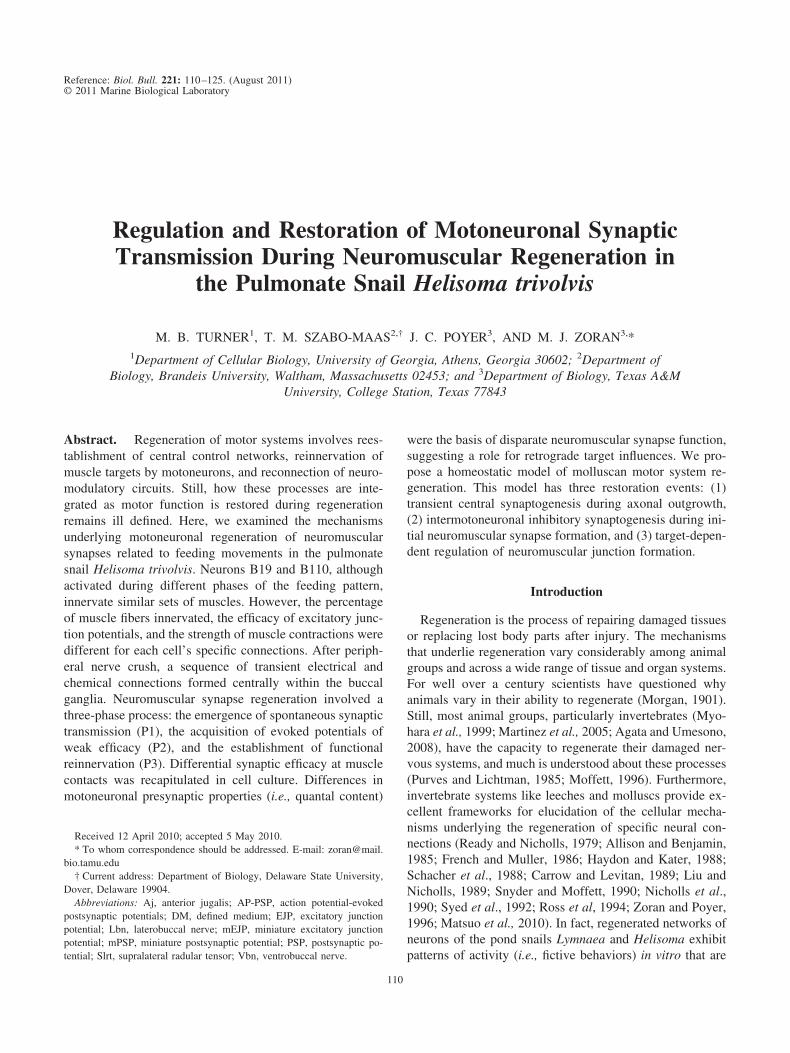
The axons of buccal motor neurons responsible for de-livering rhythmic excitation to neuromuscular synapses are,in part, located in the Vbn and Lbn. For example, neuronB19 possesses two initial axons extending from the cellbody, one that projects ipsilaterally (Fig. 2A) and onethat projects contralaterally. Both of these axons thenbifurcate, with one branch connecting to buccal musclesby way of the Vbn and the other by way of the Lbn. Atthe buccal muscle, the axon branches multiple times toterminate in a meandering chain of synaptic boutons (Fig.2B, C). In this way, each motoneuron (e.g., B19) is respon-sible for the excitation and contraction of hundreds ofindividual cells within the fiber bundles of specific musclegroups (e.g., the Slrt).
Neuromuscular connectivity of neurons in the Helisomamotor system
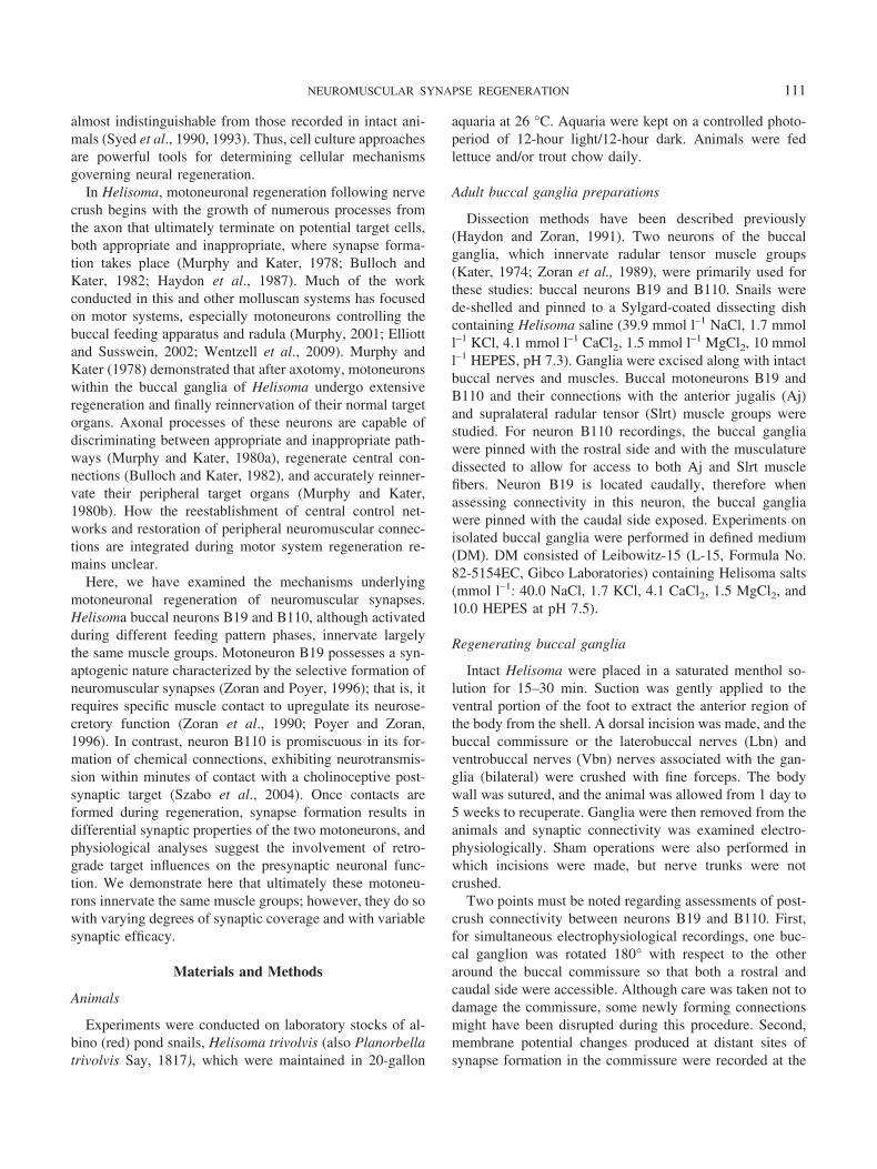
Kater (1974) originally described the peripheral connec-tivity of motoneuron B19 to muscles of the buccal mass. Asoriginally described by Kater, and as seen in Figures 1 and2, one of the muscle targets of B19 is the Slrt muscle group.However, a rigorous examination of innervation patterns,both ipsilateral and contralateral, for retractor and hyper-retractor motoneurons has not been performed. Therefore,we simultaneously recorded from identified motoneurons inthe buccal ganglia as well as from the potential muscletargets for each in the buccal mass, while stimulating burstsof action potentials in the motoneuron using intracellularcurrent injection. The major muscle targets investigatedare illustrated in Figure 3A. Stimulus-evoked EJPs weremeasured for each muscle fiber penetration, and the per-centage of muscle recordings with detectable EJPs wasdocumented. As expected, neuron B19 innervated the Slrtmuscles, and similar patterns of connectivity were pres-ent in both ipsilateral and contralateral muscle bundles(Fig. 3B; n � 3– 6 animals per muscle group). NeuronB19 also innervated the supramedial radular tensor(Smrt) and inframedial radular tensor (Imrt) musclegroups bilaterally, with essentially equal functional syn-aptic coverage (Fig. 3B; n � 3– 6 animals per musclegroup). Similar observations have been made in Aplysia,where buccal motoneurons innervate multiple musclegroups of the buccal mass (Jordan et al., 1993). Consid-
113NEUROMUSCULAR SYNAPSE REGENERATION

erable connectivity between neuron B19 and the Aj mus-cle of the buccal mass outer wall was detected on both sidesof the feeding structure. This was surprising because we hadnot observed obvious contractions of the Aj following B19stimulation. In fact, B19-evoked contractions of the Ajmuscle were almost undetectable, suggesting less effica-cious excitation-contraction coupling.
Peripheral connectivity of neuron B110 was also exam-ined, and several features of its innervation pattern differedfrom those of B19. Neuron B110 was connected to each ofthe muscle groups tested (the Imrt was not examined), butits synaptic coverage of the Slrt muscle group was lessextensive than its connectivity to the Aj muscle group (Fig.3C; n � 3–6 animals per muscle group). Furthermore, incontrast to the neuromuscular effectiveness of B19, neuronB110 elicited robust contractions in the Aj, but only weakresponses in the Slrt. In a rare case of bilateral asymmetryin innervation by these motoneurons, B110-evoked EJPswere detected in the ipsilateral, but not the contralateral,Smrt muscle group.
Synaptic connections between B19 and B110 form afternerve crush
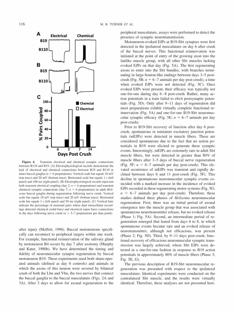
We used electrophysiological recordings identical tothose previously employed for assessment of adult mo-toneuronal connectivity to assess synaptic regeneration afteraxonal damage by nerve crush. We should point out thatneuron B19 is known to form electrical synapses both ipsi-and contralaterally with other motor neurons in the buccalganglia, and these connections have been well documented(Kater, 1974; Kaneko et al., 1978; Haydon and Kater, 1988;Szabo et al., 2004). Neurons B19 and B110 do not possesselectrical or chemical synaptic connectivity with each otherin adult ganglia (Fig. 4A; n � 6 preparations). In Helisoma,nerve crush and axonal regeneration result in the formationof many novel neuronal (central) connections, both electri-cal and chemical (Hadley et al., 1982). Following nervecrush and axotomy in the present studies, transient electricaland chemical connections form between motoneurons B19and B110 (Fig. 4B; n � 6 preparations). The timing of
Figure 1. Neural organization of the Helisoma buccal motor system. (A) The cell body of neuron C1 of thecerebral ganglion (CG) is visualized by injection with Lucifer yellow. The axon of C1 projects through thecerebrobuccal connective (CBC) and branches to innervate targets in the buccal ganglion (BG) and the buccalmass (BM), the latter through the buccal nerves (BN). Horizontal scale bar equals 180 �m. (B) Stimulation ofC1 activates rhythmic firing in buccal motor neurons and is associated with contractions of the radularmusculature (RM). Vertical scale bar equals 40 mV. Horizontal scale bar equals 5 s. (C) Activity of neurons B19and B110 is not in phase during serotonin-evoked rhythmic firing patterns. B110 functions during the S2 phase(i.e., fictive retraction of the radula), while B19 functions during the S3 phase (i.e., hyper-retraction of theradula). Vertical scale bar equals 30 mV. Horizontal scale bar equals 50 ms. (D) B19 action potentials activateone-for-one, summating EJPs in Slrt muscle fibers. Vertical scale bar equals 30 mV (top trace) and 20 mV(bottom trace). Horizontal scale bar equals 100 ms.
114 M. B. TURNER ET AL.
electrical and chemical synapse formation of these neuronswas staggered, such that electrical connections emergedwithin the first days after nerve crush and were largelyundetectable by day 7 post-crush (Fig. 4C). In contrast, thechemical connection between B110 and B19 did not reachpeak strength until transient electrical connectivity was di-minished. This chemical connection is unidirectional fromB110 (presynaptic neuron) to B19 (postsynaptic neuron)and is inhibitory (Szabo et al., 2004). Furthermore, thechemical connection persisted for weeks following nerveinjury (Fig. 4C).
It should be noted that the recording of this chemicalsynapse in Figure 4B was conducted with KCl electrodes,which reverse the polarity (i.e., direction of charge move-ment) of the underlying synaptic current. Thus, although therecord appears to indicate an excitatory connection, it is infact inhibitory (Szabo et al., 2004). Therefore, in responseto nerve injury, neurons B19 and B110 form two synapticconnections: first, a bidirectional electrical connection thatis strongest on day 2 post-crush and weakens by day 7;second, a unidirectional inhibitory connection from B110onto B19 that forms on day 6 post-crush and does notdiminish in strength for many weeks. We will see in the nextsection that the transition between central electrical andchemical synapse formation by motoneurons occurs at thetime that regenerating axons are first contacting the periph-eral buccal musculature.
Neuron B19 regenerates peripheral connectivityaccurately and rapidly
Like many other invertebrate animal groups, molluscsregenerate their nervous systems quickly and accurately
Figure 3. Neuromuscular connectivity of buccal motor neurons B19and B110. (A) The buccal muscles innervated by these motoneurons (MN)are indicated in this illustration of a dissected and flattened feeding mus-culature. Vbn, ventrobuccal nerve; Lbn, laterobuccal nerve; Col, collostyle;Imrt, inframedial radular tensor; Slrt, supralateral radular tensor; Rad,radula; Smrt, supramedial radular tensor; Ict, intracartilage tensor; Odt,odontophore; Aj, anterior jugalis. (B) B19 axons project both ipsilaterallyand contralaterally to innervate muscles on both sides of the buccal mass.Vertical bars equal the percentage of muscle fibers where intracellularrecordings detected B19-evoked EJPs. Solid bars indicate ipsilateral mus-cles and open bars indicate contralateral muscles. (C) B110 also innervatesmuscles on both sides of the buccal mass. Vertical bars equal the percent-age of muscle fibers where EJPs were detected. ND, no data collected. n �3–6 preparations per muscle group.
Figure 2. Neuromuscular structure of Helisoma buccal motoneurons.(A) Lucifer yellow injection of buccal neuron B19 illustrates the twoipsilateral axons that project to muscles of buccal mass, such as the Slrt.Two buccal nerves, the Lbn and Vbn, connect buccal motoneurons to someof the radular muscles. Horizontal scale bar equals 180 �m. (B) Luciferyellow-filled axons of B19 terminate in the Slrt in chains of synapticboutons (fluorescent puncta) that innervate many individual muscle fibers.Vertical scale bar equals 10 �m. (C) Frozen section of Slrt muscle fibersillustrating the dye-filled synaptic terminals of neuron B19 (arrows). Thearrowhead indicates a B19 axonal branch point. Vertical scale bar, 10 �m.
115NEUROMUSCULAR SYNAPSE REGENERATION
after injury (Moffett, 1996). Buccal motoneurons specifi-cally can reconnect to peripheral targets within one week.For example, functional reinnervation of the salivary glandby motoneuron B4 occurs by day 7 after axotomy (Murphyand Kater, 1980b). We have determined the timing andfidelity of neuromuscular synapse regeneration by buccalmotoneuron B19. These experiments used both sham-oper-ated animals (defined as day 0 controls) and animals inwhich the axons of this neuron were severed by bilateralcrush of both the Lbn and Vbn, the two nerves that connectthe buccal ganglia to the buccal musculature (Figs. 2A and3A). After 3 days to allow for axonal regeneration to the
peripheral musculature, assays were performed to detect thepresence of synaptic neurotransmission.
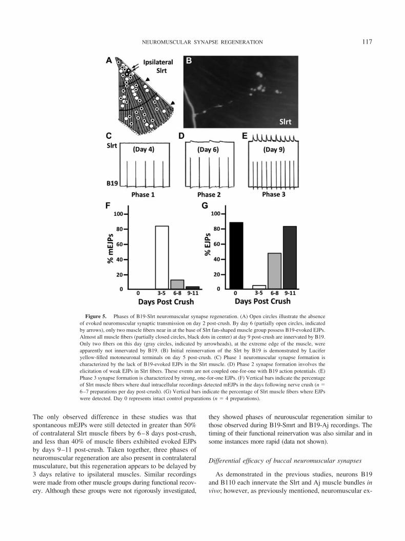
Motoneuron-evoked EJPs at B19-Slrt synapses were firstdetected in the ipsilateral musculature on day 6 after crushof the buccal nerves. This functional reinnervation wasinitiated at the point of entry of the growing axon into thefanlike muscle group, with all other Slrt muscles lackingevoked EJPs on that day (Fig. 5A). The first regeneratingaxons to enter into the Slrt bundles, with branches termi-nating in large bouton-like endings between days 3–5 post-crush (Fig. 5B; n � 6–7 animals per day post-crush), a timewhen evoked EJPs were not detected (Fig. 5C). Onceevoked EJPs were present, their efficacy was typically notone-for-one during day 6–8 post-crush. Rather, many ac-tion potentials in a train failed to elicit postsynaptic poten-tials (Fig. 5D). Only after 9–11 days of regeneration didmost preparations exhibit virtually complete functional re-innervation (Fig. 5A) and one-for-one B19-Slrt neuromus-cular synaptic efficacy (Fig. 5E; n � 6–7 animals per daypost-crush).
Prior to B19-Slrt recovery of function after day 6 post-crush, spontaneous or miniature excitatory junction poten-tials (mEJPs) were detected in muscle fibers. These areconsidered spontaneous due to the fact that no action po-tentials in B19 were elicited to generate these synapticevents. Interestingly, mEJPs are extremely rare in adult Slrtmuscle fibers, but were detected in greater than 80% ofmuscle fibers after 3–5 days of buccal nerve regeneration(Fig. 5F; n � 6–7 animals per day post-crush). This ele-vated occurrence of mEJPs was transient and rapidly de-clined between days 6 and 11 post-crush (Fig. 5F). Thisdecline in spontaneous neuromuscular synaptic events co-incided with a marked increase in the incidence of evokedEJPs recorded in these regenerating motor systems (Fig. 5G;n � 6–7 animals per day post-crush). Therefore, thesestudies defined three phases of Helisoma neuromuscularregeneration. First, there was an initial period of axonalemergence into the muscle group that was associated withspontaneous neurotransmitter release, but no evoked release(Phase 1; Fig. 5A). Second, an intermediate period of re-generation emerged that lasted from days 6 to 8, in whichspontaneous events became rare and an evoked release ofneurotransmitter, although not efficacious, was present(Phase 2; Fig. 5D). Third, by 9–11 days post-crush, func-tional recovery of efficacious neuromuscular synaptic trans-mission was largely achieved, where Slrt EJPs were de-tected in a one-for-one fashion in response to B19 actionpotentials in approximately 80% of muscle fibers (Phase 3;Fig. 5E, G).
The previous description of B19-Slrt neuromuscular re-generation was presented with respect to the ipsilateralmusculature. Identical experiments were conducted on thecontralateral Slrt muscle, and the results were virtuallyidentical. Therefore, these analyses are not presented here.
Figure 4. Transient electrical and chemical synaptic connectionsbetween B110 and B19. (A) Electrophysiological records demonstrate thelack of electrical and chemical connections between B19 and B110 inintact buccal ganglia (n � 6 preparations). Vertical scale bar equals 10 mV(top trace) and 20 mV (bottom trace). Horizontal scale bar equals 1 s (leftpanel) and 100 ms (right panel). (B) Electrophysiological records representboth transient electrical coupling (day 2; n � 6 preparations) and transientchemical synaptic connections (day 7; n � 6 preparations) in adult Heli-soma buccal ganglia during regeneration following nerve crush. Verticalscale bar equals 10 mV (top trace) and 20 mV (bottom trace). Horizontalscale bar equals 1 s (left panel) and 50 ms (right panel). (C) Vertical barsindicate the percentage of neuronal pairs where dual intracellular record-ings detected chemical (solid bars) and electrical (open bars) connectionsin the days following nerve crush (n � 5–7 preparations per data point).
116 M. B. TURNER ET AL.
The only observed difference in these studies was thatspontaneous mEJPs were still detected in greater than 50%of contralateral Slrt muscle fibers by 6–8 days post-crush,and less than 40% of muscle fibers exhibited evoked EJPsby days 9–11 post-crush. Taken together, three phases ofneuromuscular regeneration are also present in contralateralmusculature, but this regeneration appears to be delayed by3 days relative to ipsilateral muscles. Similar recordingswere made from other muscle groups during functional recov-ery. Although these groups were not rigorously investigated,
they showed phases of neurouscular regeneration similar tothose observed during B19-Smrt and B19-Aj recordings. Thetiming of their functional reinervation was also similar and insome instances more rapid (data not shown).
Differential efficacy of buccal neuromuscular synapses
As demonstrated in the previous studies, neurons B19and B110 each innervate the Slrt and Aj muscle bundles invivo; however, as previously mentioned, neuromuscular ex-
Figure 5. Phases of B19-Slrt neuromuscular synapse regeneration. (A) Open circles illustrate the absenceof evoked neuromuscular synaptic transmission on day 2 post-crush. By day 6 (partially open circles, indicatedby arrows), only two muscle fibers near in at the base of Slrt fan-shaped muscle group possess B19-evoked EJPs.Almost all muscle fibers (partially closed circles, black dots in center) at day 9 post-crush are innervated by B19.Only two fibers on this day (gray circles, indicated by arrowheads), at the extreme edge of the muscle, wereapparently not innervated by B19. (B) Initial reinnervation of the Slrt by B19 is demonstrated by Luciferyellow-filled motoneuronal terminals on day 5 post-crush. (C) Phase 1 neuromuscular synapse formation ischaracterized by the lack of B19-evoked EJPs in the Slrt muscle. (D) Phase 2 synapse formation involves theelicitation of weak EJPs in Slrt fibers. These events are not coupled one-for-one with B19 action potentials. (E)Phase 3 synapse formation is characterized by strong, one-for-one EJPs. (F) Vertical bars indicate the percentageof Slrt muscle fibers where dual intracellular recordings detected mEJPs in the days following nerve crush (n �6–7 preparations per day post-crush). (G) Vertical bars indicate the percentage of Slrt muscle fibers where EJPswere detected. Day 0 represents intact control preparations (n � 4 preparations).
117NEUROMUSCULAR SYNAPSE REGENERATION
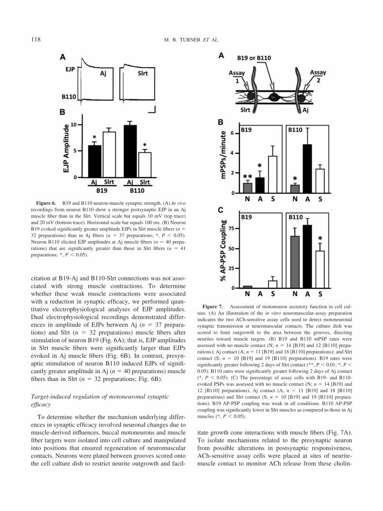
citation at B19-Aj and B110-Slrt connections was not asso-ciated with strong muscle contractions. To determinewhether these weak muscle contractions were associatedwith a reduction in synaptic efficacy, we performed quan-titative electrophysiological analyses of EJP amplitudes.Dual electrophysiological recordings demonstrated differ-ences in amplitude of EJPs between Aj (n � 37 prepara-tions) and Slrt (n � 32 preparations) muscle fibers afterstimulation of neuron B19 (Fig. 6A); that is, EJP amplitudesin Slrt muscle fibers were significantly larger than EJPsevoked in Aj muscle fibers (Fig. 6B). In contrast, presyn-aptic stimulation of neuron B110 induced EJPs of signifi-cantly greater amplitude in Aj (n � 40 preparations) musclefibers than in Slrt (n � 32 preparations; Fig. 6B).
Target-induced regulation of motoneuronal synapticefficacy
To determine whether the mechanism underlying differ-ences in synaptic efficacy involved neuronal changes due tomuscle-derived influences, buccal motoneurons and musclefiber targets were isolated into cell culture and manipulatedinto positions that ensured regeneration of neuromuscularcontacts. Neurons were plated between grooves scored ontothe cell culture dish to restrict neurite outgrowth and facil-
itate growth cone interactions with muscle fibers (Fig. 7A).To isolate mechanisms related to the presynaptic neuronfrom possible alterations in postsynaptic responsiveness,ACh-sensitive assay cells were placed at sites of neurite-muscle contact to monitor ACh release from these cholin-
Figure 6. B19 and B110 neuron-muscle synaptic strength. (A) In vivorecordings from neuron B110 show a stronger postsynaptic EJP in an Ajmuscle fiber than in the Slrt. Vertical scale bar equals 10 mV (top trace)and 20 mV (bottom trace). Horizontal scale bar equals 100 ms. (B) NeuronB19 evoked significantly greater amplitude EJPs in Slrt muscle fibers (n �32 preparations) than in Aj fibers (n � 37 preparations; *, P � 0.05).Neuron B110 elicited EJP amplitudes at Aj muscle fibers (n � 40 prepa-rations) that are significantly greater than those in Slrt fibers (n � 41preparations; *, P � 0.05).
Figure 7. Assessment of motoneuron secretory function in cell cul-ture. (A) An illustration of the in vitro neuromuscular-assay preparationindicates the two ACh-sensitive assay cells used to detect motoneuronalsynaptic transmission at neuromuscular contacts. The culture dish wasscored to limit outgrowth to the area between the grooves, directingneurites toward muscle targets. (B) B19 and B110 mPSP rates wereassessed with no muscle contact (N; n � 14 [B19] and 12 [B110] prepa-rations); Aj contact (A; n � 11 [B19] and 18 [B110] preparations); and Slrtcontact (S; n � 10 [B19] and 19 [B110] preparations). B19 rates weresignificantly greater following 2 days of Slrt contact (**, P � 0.01; *, P �0.05). B110 rates were significantly greater following 2 days of Aj contact(*, P � 0.05). (C) The percentage of assay cells with B19- and B110-evoked PSPs was assessed with no muscle contact (N; n � 14 [B19] and12 [B110] preparations), Aj contact (A; n � 11 [B19] and 18 [B110]preparations) and Slrt contact (S; n � 10 [B19] and 19 [B110] prepara-tions). B19 AP-PSP coupling was weak in all conditions. B110 AP-PSPcoupling was significantly lower in Slrt muscles as compared to those in Ajmuscles (*, P � 0.05).
118 M. B. TURNER ET AL.

ergic motoneurons. ACh-sensitive assay cells are well-de-fined tools for monitoring transmitter release from buccalneurons (Haydon and Zoran, 1989, 1991; Zoran et al.,1990). To assess the rate of spontaneous ACh release fromeach neuron, mPSPs were recorded. Neurites of both neu-rons B19 and B110 possessed higher mPSP rates followingcontact with Slrt (n � 10, B19 and 19, B110) and AJ (n �11, B19 and 18, B110) muscle fibers as compared to neu-rites of neurons alone (n � 14, B19 and 12, B110) in culture(Fig. 7B). However, neuron B19 exhibited a significantlyhigher mPSP rate at Slrt-neurite contacts than at Aj-con-tacted neurites (Fig. 7B). In contrast, neurites of B110contacting Aj muscle fibers possessed higher rates of mP-SPs than those of neurites contacting Slrt fibers (Fig. 7B).
Unlike neuron B19’s requirement for an appropriate tar-get for the regeneration of functional synapses (Zoran et al.,1996; Zoran and Poyer, 1999), neuron B110 is able to formchemical synapses when contacted by any cholinoceptivetarget (Fig. 7C). Action potential-evoked postsynaptic po-tentials (AP-PSPs) recorded with ACh-reporter cells weremonitored in dual muscle co-cultures. Although neuronB110 does not require contact with a muscle target toacquire ACh secretory capability, neurites of B110 contact-ing Slrt muscle fibers exhibited less efficacious release ofACh than those contacting Aj muscle fibers (Fig. 7C). Thisresult indicates a retrograde influence of the muscle fiber onpresynaptic mechanisms of ACh release in motor neuronB110. In contrast, B19 has minimal synaptic efficacy inthese cultures, with or without muscle contact. B19 AP-PSPcoupling was approximately 10% following interaction withSlrt muscle fibers only (Fig. 7C).
Retrograde regulation of presynaptic release properties
We hypothesized that differences in motoneuronal syn-aptic efficacy at neurites contacting different muscle targetswere mediated by changes in presynaptic properties, sincePSPs were recorded with assay cells characterized by con-sistent ACh-sensitivity (Haydon and Zoran, 1991). Addi-tionally, muscle target contact is known to alter actionpotential-dependent calcium accumulation in presynapticneurites of buccal neurons (Zoran et al., 1993; Funte andHaydon, 1993). Therefore, the probability of vesicular re-lease may be altered at neuritic sites of muscle targets in aneuron-specific fashion. To test this hypothesis in neuronB19 would be difficult because synaptic efficacy in theseneurons is very low in cell culture (Fig. 7C). Consequently,we analyzed the secretory properties of the more efficaciousneuron B110 during regeneration in cell culture. It should benoted that quantal analyses were conducted with assay cellsto again avoid differences in postsynaptic receptor densityor sensitivity to ACh.
Since the rate of miniature excitatory synaptic potentialsfor neuron B110 was elevated following contact with Aj
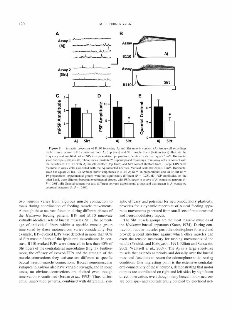
muscle fibers in culture, we tested whether quantal size wasalso affected by Aj muscle. Neuron B110 co-cultured withAj (n � 18 preparations) and Slrt (n � 19 preparations)muscle fiber contacts again showed elevated rates of mPSPsonly at sites of contact with Aj muscle fibers (Fig. 8A).However, mean mPSP amplitudes were not significantlydifferent between Aj and Slrt-contacted neurites (P � 0.25;Fig. 8C). Thus, even with a more robust frequency ofspontaneous release at Aj contacts, there was no change inthe amount of neurotransmitter released per vesicle, asindicated by the voltage change associated with one spon-taneous release event (quantal size).
Since quantal size was not different between Aj andSlrt-contacted neurites, we next assessed evoked PSP am-plitudes as a measure of the amount of neurotransmitter(i.e., level of exocytosis of synaptic vesicles) per actionpotential at neurite-muscle contact sites for neuron B110.To achieve this, trains of action potentials were stimulatedpresynaptically while simultaneously recording postsynap-tic responses in an assay cell contacting B110 neurites (Fig.8B). Neuron B110 exhibited efficacious release of ACh atsites of contact with both Slrt and Aj muscle fibers; how-ever, Aj-contacted neurites possessed significantly greaterPSP amplitudes than Slrt-contacted neurites (Fig. 8D). Sev-eral features of the PSPs elicited in ACh reporter cellssuggested enhanced abilities for synaptic transmission ofAj-contacted neurites. First, PSPs maintained consistentlyhigher amplitudes during high-frequency stimulation thandid Slrt-contacted neurites. Second, the slope of the risingphase of PSPs was consistently greater at Aj-contactedneurites. Finally, estimates of quantal content at Aj-con-tacted neurites, calculated as the sum of the mean PSPamplitude divided by the mean mPSP amplitude, were sig-nificantly different than those at Slrt-contacted processes(Fig. 8E). Taken together, these results support a mecha-nism of retrograde regulation of presynaptic release prop-erties by muscle targets during neuron B110 neuromuscularregeneration.
Discussion
A fundamental difference between vertebrate and inver-tebrate motor systems is the nature of their neuromuscularinnervation. Most invertebrate muscle fibers, includingthose of the Helisoma buccal musculature, receive synapticinputs from multiple motoneurons (for example, B19, B110,and many others) and modulatory neurons (for example,C1), such that significant motor plasticity can be generatedby neuromuscular synapses in the periphery (Heyer et al.,1973). Vertebrate skeletal muscles, on the other hand, typ-ically have motoneuronal input from a single spinal mo-toneuron, and most modulation occurs centrally. B19 andB110 are active during different phases of fictive Helisomafeeding behavior, and the phases of activation between these
119NEUROMUSCULAR SYNAPSE REGENERATION
two neurons varies from vigorous muscle contraction totonus during coordination of feeding muscle movements.Although these neurons function during different phases ofthe Helisoma feeding pattern, B19 and B110 innervatevirtually identical sets of buccal muscles. Still, the percent-age of individual fibers within a specific muscle groupinnervated by these motoneurons varies considerably. Forexample, B19-evoked EJPs were detected in more than 80%of Slrt muscle fibers of the ipsilateral musculature. In con-trast, B110-evoked EJPs were detected in less than 40% ofSlrt fibers of the contralateral musculature (Fig. 3). Further-more, the efficacy of evoked-EJPs and the strength of themuscle contractions they activate are different at specificbuccal neuron-muscle connections. Buccal neuromuscularsynapses in Aplysia also have variable strength, and in somecases, no obvious contractions are elicited even thoughinnervation is confirmed (Jordan et al., 1993). Thus, differ-ential innervation patterns, combined with differential syn-
aptic efficacy and potential for neuromodulatory plasticity,provides for a dynamic repertoire of buccal feeding appa-ratus movements generated from small sets of motoneuronaland neuromodulatory inputs.
The Slrt muscle groups are the most massive muscles ofthe Helisoma buccal apparatus (Kater, 1974). During con-traction, radular muscles push the odontophore forward andprovide a solid structure against which other muscles canexert the tension necessary for rasping movements of theradula (Yoshida and Kobayashi, 1991; Elliott and Susswein,2002; Wentzell et al., 2009). The Aj is a large sheet-likemuscle that extends anteriorly and dorsally over the buccalmass and functions to return the odontophore to its restingcondition. One interesting point is the extensive contralat-eral connectivity of these neurons, demonstrating that motoroutputs are coordinated on right and left sides by significantdirect innervation, even though many buccal motor neuronsare both ipsi- and contralaterally coupled by electrical net-
Figure 8. Synaptic properties of B110 following Aj and Slrt muscle contact. (A) Assay-cell recordingsmade from a neuron B110 contacting both Aj (top trace) and Slrt muscle fibers (bottom trace) illustrate thefrequency and amplitude of mPSPs in representative preparations. Vertical scale bar equals 5 mV. Horizontalscale bar equals 500 ms. (B) These traces illustrate 25 superimposed recordings from assay cells in contact withthe neurites of a B110 with Aj muscle contact (top trace) and Slrt contact (bottom trace). Large EJPs wererecorded in assay cells associated with the Aj-contacted neurites. Vertical scale bar equals 2 mV. Horizontalscale bar equals 20 ms. (C) Average mPSP amplitudes in B110-Aj (n � 18 preparations) and B110-Slrt (n �19 preparations) experimental groups were not significantly different (P � 0.25). (D) PSP amplitudes, on theother hand, were different between experimental groups, with PSPs larger in assays of Aj-contacted neurons (*,P � 0.01). (E) Quantal content was also different between experimental groups and was greater in Aj-contactedneuronal synapses (*, P � 0.04).
120 M. B. TURNER ET AL.
works. It has been suggested that the redundancy in Heli-soma bilateral coordination of motor outputs is due to bothelectrical coupling and bilateral axon projections, thus as-suring synchronous bilateral function (Bahls et al., 1980).
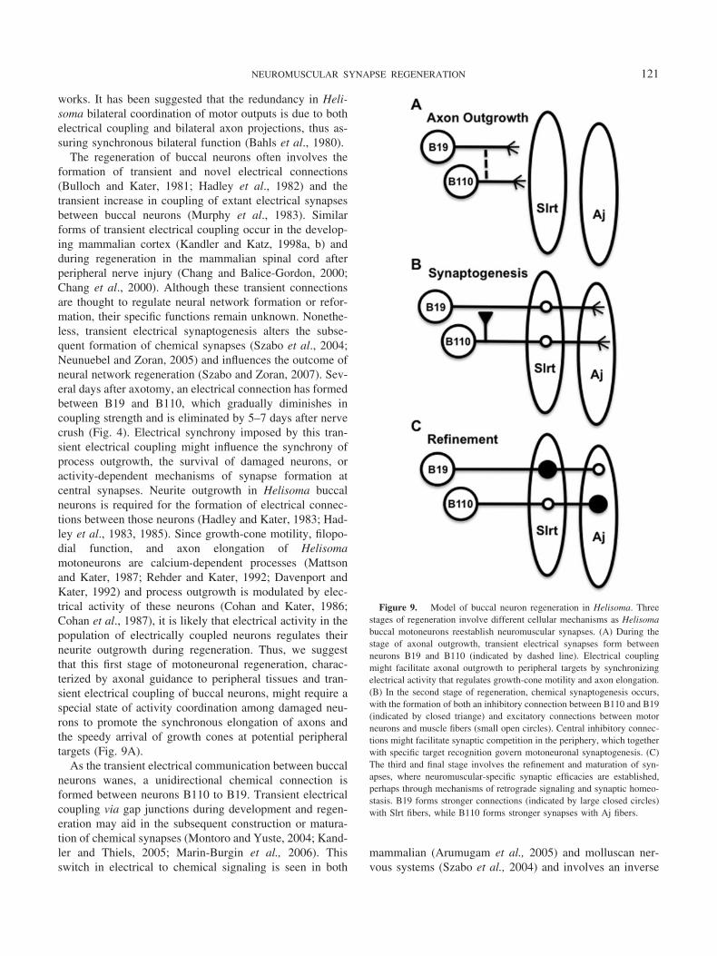
The regeneration of buccal neurons often involves theformation of transient and novel electrical connections(Bulloch and Kater, 1981; Hadley et al., 1982) and thetransient increase in coupling of extant electrical synapsesbetween buccal neurons (Murphy et al., 1983). Similarforms of transient electrical coupling occur in the develop-ing mammalian cortex (Kandler and Katz, 1998a, b) andduring regeneration in the mammalian spinal cord afterperipheral nerve injury (Chang and Balice-Gordon, 2000;Chang et al., 2000). Although these transient connectionsare thought to regulate neural network formation or refor-mation, their specific functions remain unknown. Nonethe-less, transient electrical synaptogenesis alters the subse-quent formation of chemical synapses (Szabo et al., 2004;Neunuebel and Zoran, 2005) and influences the outcome ofneural network regeneration (Szabo and Zoran, 2007). Sev-eral days after axotomy, an electrical connection has formedbetween B19 and B110, which gradually diminishes incoupling strength and is eliminated by 5–7 days after nervecrush (Fig. 4). Electrical synchrony imposed by this tran-sient electrical coupling might influence the synchrony ofprocess outgrowth, the survival of damaged neurons, oractivity-dependent mechanisms of synapse formation atcentral synapses. Neurite outgrowth in Helisoma buccalneurons is required for the formation of electrical connec-tions between those neurons (Hadley and Kater, 1983; Had-ley et al., 1983, 1985). Since growth-cone motility, filopo-dial function, and axon elongation of Helisomamotoneurons are calcium-dependent processes (Mattsonand Kater, 1987; Rehder and Kater, 1992; Davenport andKater, 1992) and process outgrowth is modulated by elec-trical activity of these neurons (Cohan and Kater, 1986;Cohan et al., 1987), it is likely that electrical activity in thepopulation of electrically coupled neurons regulates theirneurite outgrowth during regeneration. Thus, we suggestthat this first stage of motoneuronal regeneration, charac-terized by axonal guidance to peripheral tissues and tran-sient electrical coupling of buccal neurons, might require aspecial state of activity coordination among damaged neu-rons to promote the synchronous elongation of axons andthe speedy arrival of growth cones at potential peripheraltargets (Fig. 9A).
As the transient electrical communication between buccalneurons wanes, a unidirectional chemical connection isformed between neurons B110 to B19. Transient electricalcoupling via gap junctions during development and regen-eration may aid in the subsequent construction or matura-tion of chemical synapses (Montoro and Yuste, 2004; Kand-ler and Thiels, 2005; Marin-Burgin et al., 2006). Thisswitch in electrical to chemical signaling is seen in both
mammalian (Arumugam et al., 2005) and molluscan ner-vous systems (Szabo et al., 2004) and involves an inverse
Figure 9. Model of buccal neuron regeneration in Helisoma. Threestages of regeneration involve different cellular mechanisms as Helisomabuccal motoneurons reestablish neuromuscular synapses. (A) During thestage of axonal outgrowth, transient electrical synapses form betweenneurons B19 and B110 (indicated by dashed line). Electrical couplingmight facilitate axonal outgrowth to peripheral targets by synchronizingelectrical activity that regulates growth-cone motility and axon elongation.(B) In the second stage of regeneration, chemical synaptogenesis occurs,with the formation of both an inhibitory connection between B110 and B19(indicated by closed triange) and excitatory connections between motorneurons and muscle fibers (small open circles). Central inhibitory connec-tions might facilitate synaptic competition in the periphery, which togetherwith specific target recognition govern motoneuronal synaptogenesis. (C)The third and final stage involves the refinement and maturation of syn-apses, where neuromuscular-specific synaptic efficacies are established,perhaps through mechanisms of retrograde signaling and synaptic homeo-stasis. B19 forms stronger connections (indicated by large closed circles)with Slrt fibers, while B110 forms stronger synapses with Aj fibers.
121NEUROMUSCULAR SYNAPSE REGENERATION
relationship between chemical and electrical communica-tion, a homeostatic regulation of synapse formation early innetwork formation or regeneration. The chemical connec-tion formed between B110 and B19 is cholinergic andinhibitory (Szabo et al., 2004). Thus, when B110 is active,B19’s activity might be suppressed by this inhibitory con-nection, but only after 5–7 days of regeneration (Fig. 4).Some Helisoma buccal neurons, such as B19, are con-strained in their ability to regenerate synaptic connections,and the formation of a synapse on one target contact doesnot alter that neuron’s synaptic function at another target(Poyer and Zoran, 1996). This differential regulation ofsynaptic efficacy requires recognition of an appropriatepostsynaptic target (Zoran and Poyer, 1996). Therefore, thefact that neuron B19 does not also form a cholinergicchemical synapse onto B110 during regeneration is notsurprising. Since B110 is known to be more promiscuous inits synapse formation, a strategy of synaptogenesis reportedfor other Helisoma buccal neurons (Haydon and Zoran,1989), it possesses the capacity for rapid inhibitory synapseformation with other cholinoceptive neurons. The demon-stration here that regeneration of identified buccal neuronsin Helisoma involves dynamic plasticity of synaptic inter-actions is a further extension of our understanding thatneural regeneration in invertebrate animals, particularlygastropod molluscs, incorporates many conserved and basicmechanisms that govern adult neuronal plasticity (Moffett,2000). Furthermore, factors present in medium conditionedwith damaged neural tissues influence the kinds of synapsesthat regenerate between molluscan neurons in cell culture—for example, chemical versus electrical or excitatory versusinhibitory (Hamakawa et al., 1999; Szabo et al., 2004).
We consider this period of transient inhibitory synapseformation, which coincides with neuromuscular synapseregeneration, to be the second stage of motoneuronal regen-eration; that is, a stage of synaptogenesis following initialprocess outgrowth (Fig. 9B). The formation of a unidirec-tional inhibitory connection between B110 and B19 mightprovide an important central regulatory cue for peripheralneuromuscular regeneration. This inhibitory connectionforms at precisely the time (6–8 days post-crush) whensynaptic boutons and evoked EJPs appear in the peripheralmusculature, and it remains strong and sustained until day14 post-crush. Therefore, this transient inhibitory synapsewould provide a mechanism for the inhibition of motoneu-ronal competitors during neuromuscular synapse regenera-tion. For example, inhibition of B19 would assure that itsfiring activity is asynchronous with that of B110, suggestinga role for activity-dependent synaptic competition duringthe regeneration of the Helisoma buccal motor system.
The present studies describe the time course for regener-ation of B19 peripheral neuromuscular synapses. The firstevidence of the presence of motoneuronal transmitter re-lease onto buccal muscle is at 3–5 days after buccal nerve
crush. Spontaneous mEJPs are first detected and then in-crease over this time period such that they are recorded innearly 80% of muscle fibers tested. In contrast, B19-evokedEJPs are not detected at this time (Fig. 5). We call thisperiod of regeneration Phase 1 (P1) neuromuscular synapseformation. By days 6–8 post-crush, weak B19-evoked EJPsare detected in Slrt muscle fibers, and spontaneous neuro-muscular transmission is dramatically reduced (Fig. 5). Wenamed this period Phase 2 (P2) neuromuscular synapseformation. Finally, starting at about days 9–11 and extend-ing well into the second week after nerve injury, mostmuscle fibers that were innervated prior to injury becomereinnervated, as demonstrated by the presence of B19-evoked EJPs. In addition, synaptic efficacy is restored topre-injury levels and spontaneous mEJPs are virtually ab-sent. We call this period Phase 3 (P3) neuromuscular syn-apse formation. These phases of neuromuscular synapseformation are known to involve the elevation of restingcalcium, voltage-activated calcium influx, and neurotrans-mitter release following contact of growth cones with mus-cle targets (Zoran et al., 1993; Funte and Haydon, 1993;Zoran and Poyer, 1996). Similar developmental and cascad-ing events occur in vertebrate motoneurons after initialmuscle contact (Dai and Peng, 1993) and at fly neuromus-cular synapses (Littleton et al., 1993). Interestingly, action-potential-evoked neurotransmitter release is significantly el-evated following periods of sustained contact withappropriate Slrt muscle fibers, but not when mismatchedwith inappropriate muscle targets (Zoran and Poyer, 1996).This discrimination is likely based on target-specific recog-nition, as neurons are capable of distinguishing betweensynaptic targets through the recognition of cell surface cuesin many animal systems (Johansen et al., 1989; Chiba et al.,1993; Merz and Drapeau, 1994; Bate and Broadie, 1995;Zhu et al., 1995). Therefore, neuromuscular synapse regen-eration is an accurate mechanism, likely based on cell-cellsignaling processes that regulate the emergence of sponta-neous neuromuscular synaptic transmission (P1), the acqui-sition of intermittent evoked EJPs of weak efficacy (P2),and the formation of one-for-one, functional reinnervation(P3).
In our proposed model of buccal motoneuronal regener-ation, the third and final stage is one of synaptic refinement,whereby homeostatic mechanisms of synaptic plasticity es-tablish differential motoneuronal efficacy at muscles inner-vated by multiple neuronal inputs (Fig. 9C). Molluscansynapses regenerated in cell culture require ongoing andmultiple modifications that affect functional maturation(Sun and Schacher, 1996; Conrad et al., 1999). Cell culturestudies of isolated buccal neurons have established many ofthe cellular mechanisms governing the reformation ofchemical and electrical synaptic connections (Haydon andKater, 1988; Haydon and Zoran, 1989; Zoran and Poyer,1996). Neuron B19 when stimulated in intact neuromuscu-
122 M. B. TURNER ET AL.

lar preparations induces high-amplitude EJPs, associatedwith strong contractions of the Slrt muscle, and lower am-plitude EJPs and weaker contractions of the Aj muscle (Fig.6). The opposite synaptic relationships exist at B110 neu-romuscular synapses. This differential synaptic physiologyis recapitulated after neuronal synapse regeneration in vitro,where neuron B19 forms a stronger synaptic connection atSlrt rather than Aj contacts, and neuron B110 exhibits moreefficacious neurotransmitter release at contact sites with Ajmuscle fibers. These differences in motoneuronal presynap-tic properties include a 2-fold elevation in quantal content ofneurotransmitter release at B110 sites of Aj muscle contactas compared to sites of Slrt contact (Fig. 8). These resultssuggest a role for retrograde influences of the muscle targetson presynaptic motoneuronal secretory mechanisms. It haslong been understood that neuronal growth cones have theability to recognize molecular cues on specific muscle tar-gets with which they form synaptic connections (Chiba etal., 1995) and that these retrograde signals affect presynap-tic transmitter release (Davis and Goodman 1998; Frank etal., 2006). Retrograde modulation of transmitter releaseproperties occurs between isolated neurons and target cellsin culture (Haydon and Zoran, 1994; Fernandez-de-Migueland Drapeau, 1995). Our results on the maturation of buccalmotoneuronal synapses suggest a role for retrograde influ-ences of muscle targets on presynaptic motoneuronal secre-tory mechanisms, which ultimately shape the mature patternof functional buccal muscle innervation. Therefore, we con-clude that target-induced alterations in synaptic strength arean important final homeostatic stage in the regeneration ofmature neuromuscular synapses.
In summary, a three-stage model is proposed to explainour observations of Helisoma buccal motoneuron regener-ation. Transient electrical synaptogenesis might facilitateaxonal outgrowth to peripheral targets during the first stageof recovery of function. In the second stage, differentialmotoneuronal synaptogenic capabilities, mechanisms ofspecific target recognition, and transient inhibitory chemicalsynpatogenesis act together to regulate the initial stages ofneuromuscular synapse regeneration. The third and finalstage of maturation involves the establishment of differen-tial motoneuronal efficacies and employs mechanisms ofboth synaptic competition and synaptic homeostasis. Thefinal maturation stage might be especially important in theregeneration of invertebrate neuromuscular systems, wheremuscle targets are innervated by multiple neuronal inputs. Itremains to be seen how widespread the expression of tran-sient electrical connections and transient inhibitory chemi-cal connections is in regeneration of neural systems ingeneral. It is clear that these transiently regenerated connec-tions both affect future connectivity and are affected byexisting and previous synaptic connections (Bulloch et al.,1984; Szabo and Zoran, 2007). However, our suggestionthat one fundamental role of these transient connections is
to regulate the regeneration of peripheral neuromuscularsynapse requires further rigorous examination.
Acknowledgments
The authors thank J. Clay Gaspard for his research assis-tance with aspects of the electrophysiological analysis, andJarret Richardson for his comments on the manuscript. Thisresearch was supported by National Science Foundation(IBN-9421372) and National Institutes of Health (P01NS39546) grants to M. J. Z.
Literature Cited
Agata K., and Y. Umesono. 2008. Brain regeneration from pluripotentstem cells in planarian. Philos. Trans. R. Soc. Lond. B 363: 2071–2078.
Allison, P., and P. R. Benjamin. 1985. Anatomical studies of centralregeneration of an identified molluscan interneuron. Proc. R. Soc.Lond. B 226: 135–157.
Arumugam, H., X. Liu, P. J. Colombo, R. A. Corriveau, and A. B.Belousov. 2005. NMDA receptors regulate developmental gap junc-tion uncoupling via CREB signaling, Nat. Neurosci. 8: 1720–1726.
Bahls, F. H., S. B. Kater, and R. W. Joyner. 1980. Neuronal mecha-nisms for bilateral coordination of salivary gland activity in Helisoma.J. Neurobiol. 11: 365–739.
Bate, M., and K. Broadie. 1995. Wiring by fly: the neuromuscularsystem of the Drosophila embryo. Neuron 15: 513–525.
Bulloch, A. G., and S. B. Kater. 1981. Selection of a novel connectionby adult molluscan neurons. Science 212: 79–81.
Bulloch, A. G., and S. B. Kater. 1982. Neurite outgrowth and selectionof new electrical connections by adult Helisoma neurons. J. Neuro-physiol. 48: 569–583.
Bulloch, A. G., S. B. Kater, and H. R. Miller. 1984. Stability of newelectrical connections between adult Helisoma neurons is influenced bypreexisting neuronal interactions. J. Neurophysiol. 52: 1094–1105.
Carrow, G. M., and I. B. Levitan. 1989. Selective formation andmodulation of electrical synapses between cultured Aplysia neurons.J. Neurosci. 9: 3657–3664.
Chang, Q., and R. J. Balice-Gordon. 2000. Gap junctional communi-cation among developing and injured motor neurons. Brain Res. Rev.32: 242–249.
Chang, Q., A. Pereda, M. J. Pinter, and R. J. Balice-Gordon. 2000.Nerve injury induces gap junctional coupling among axotomized adultmotor neurons. J. Neurosci. 20: 674–684.
Chiba, A., H. Hing, S. Cash, and H. Keshishian. 1993. Growth conechoices of Drosophila motoneurons in response to muscle fiber mis-match. J. Neurosci. 13: 714–732.
Chiba, A., P. Snow, H. Keshishian, and Y. Hotta. 1995. Fasciclin IIIas a synaptic target recognition molecule in Drosophila. Nature 374:166–168.
Cohan, C. S., and S. B. Kater. 1986. Suppression of neurite elongationand growth cone motility by electrical activity. Science 232: 1623–1640.
Cohan, C. S., J. A. Connor, and S. B. Kater. 1987. Electrically andchemically mediated increases in intracellular calcium in neuronalgrowth cones. J. Neurosci. 7: 3588–3599.
Conrad, P., F. Wu, and S. Schacher. 1999. Changes in functionalglutamate receptors on a postsynaptic neuron accompany formationand maturation of an identified synapse. J. Neurobiol. 39: 237–248.
Dai, Z., and H. B. Peng. 1993. Elevation in presynaptic Ca2� levelaccompanying initial nerve-muscle contact in tissue culture. Neuron10: 827–837.
123NEUROMUSCULAR SYNAPSE REGENERATION
Davenport, R. W., and S. B. Kater. 1992. Local increases in intracel-lular calcium elicit local filopodial responses in Helisoma neuronalgrowth cones. Neuron 9: 405–416.
Davis, G. W., and C. S. Goodman. 1998. Synapse-specific control ofsynaptic efficacy at the terminals of a single neuron. Nature 392:82–86.
Elliott, J. H., and A. J. Susswein. 2002. Comparative neuroethology offeeding control in mollusks. J. Exp. Biol. 205: 877–896.
Fernandez-de-Miguel, F., and P. Drapeau. 1995. Synapse formationand function: insights from identified leech neurons in culture. J. Neu-robiol. 27: 367–379.
Frank, C. A., M. J. Kennedy, C. P. Goold, K. W. Marek, and G. W.Davis. 2006. Mechanisms underlying the rapid induction and sus-tained expression of synaptic homeostasis. Neuron 52: 663–677.
French, K. A., and K. J. Muller. 1986. Regeneration of a distinctive setof axosomatic contacts in the leech central nervous system. J. Neurosci.6: 318–324.
Funte, L. R., and P. G. Haydon. 1993. Synaptic target contact en-hances presynaptic calcium influx by activating cAMP-dependent pro-tein kinase during synaptogenesis. Neuron 10: 1069–1078.
Hadley, R. D., and S. B. Kater. 1983. Competence to form electricalconnections is restricted to growing neurites in the snail, Helisoma.J. Neurosci. 3: 924–932.
Hadley, R. D., R. G. Wong, S. B. Kater, D. L. Barker, and A. G.Bulloch. 1982. Formation of novel central and peripheral connec-tions between molluscan central neurons in organ cultured ganglia.J. Neurobiol. 13: 217–230.
Hadley, R. D., S. B. Kater, and C. S. Cohan. 1983. Electrical synapseformation depends on interaction of mutually growing neurites. Science221: 466–468.
Hadley, R. D., D. A. Bodnar, and S. B. Kater. 1985. Formation ofelectrical synapses between isolated, cultured Helisoma neurons re-quires mutual neurite elongation. J. Neurosci. 5: 3145–3153.
Hamakawa, T., M. A. Woodin, M. C. Bjorgum, S. D. Painter, M.Takasaki, K. Lukowiak, G. T. Nagle, and N. I. Syed. 1999. Ex-citatory synaptogenesis between identified Lymnaea neurons requiresextrinsic trophic factors and is mediated by receptor tyrosine kinases.J. Neurosci. 19: 9306–9312.
Haydon, P. G., and S. B. Kater. 1988. The differential regulation offormation of chemical and electrical connections in Helisoma. J. Neu-robiol. 19: 636–655.
Haydon, P. G., and M. J. Zoran. 1989. Formation and modulation ofchemical connections: evoked acetylcholine release from growth conesand neurites of specific identified neurons. Neuron 2: 1483–1490.
Haydon, P. G., and M. J. Zoran. 1991. Chemical synapses in cellculture. Pp. 57–71 in Cellular Neurobiology: A Practical Approach,J. Chad and H. Wheal, eds. Oxford University Press, New York.
Haydon, P. G., and M. J. Zoran. 1994. Retrograde regulation ofpresynaptic development during synaptogenesis. J. Neurobiol. 25:694–706.
Haydon, P. G., D. P. McCobb, and S. B. Kater. 1987. The regulationof neurite outgrowth, growth cone motility, and electrical synaptogen-esis by serotonin. J. Neurobiol. 18: 197–215.
Heyer, C. B., S. B. Kater, and U. L. Karllson. 1973. Neuromuscularsystems in molluscs. Am. Zool. 13: 247–270.
Johansen, J., M. E. Halpern, and H. Keshishian. 1989. Axonalguidance and the development of muscle fiber-specific innervation inDrosophila embryos. J. Neurosci. 9: 4318–4332.
Jordan, R., K. P. Cohen, and M. D. Kirk. 1993. Control of intrinsicbuccal muscles by motoneurons B11, B15, and B16 in Aplysia cali-fornica. J. Exp. Zool. 265: 496–506.
Kandler, K., and L. C. Katz. 1998a. Coordination of neuronal activityin developing visual cortex by gap junction-mediated biochemicalcommunication. J. Neurosci. 18: 1419–1427.
Kandler, K., and L. C. Katz. 1998b. Relationship between dye cou-pling and spontaneous activity in developing ferret visual cortex. Dev.Neurosci. 20: 59–64.
Kandler, K., and E. Thiels. 2005. Flipping the switch from electrical tochemical communication. Nat. Neurosci. 8: 1633–1634.
Kaneko, C. R., M. Merickel, and S. B. Kater. 1978. Centrally pro-grammed feeding in Helisoma: identification and characteristics of anelectrically coupled premotor neuron network. Brain Res. 146: 1–21.
Kater, S. B. 1974. Feeding in Helisoma trivolvis: the morphological andphysiological bases of a fixed action pattern. Am. Zool. 14: 1017–1036.
Littleton, J. T., H. J. Bellen, and M. S. Perin. 1993. Expression ofsynaptotagmin in Drosophila reveals transport and localization ofsynaptic vesicles to the synapse. Development 118: 1077–1088.
Liu, Y., and J. Nicholls. 1989. Steps in the development of chemicaland electrical synapses by pairs of identified leech neurons in culture.Proc. R. Soc. Lond. B Biol. Sci. 236: 253–268.
Marin-Burgin, A., F. J. Eisenhart, W. B. Kristan, Jr., and K. A.French. 2006. Embryonic electrical connections appear to pre-figurea behavioral circuit in the leech CNS. J. Comp. Physiol. A Neuroethol.Sens. Neural. Behav. Physiol. 192: 123–133.
Martinez, V. G., G. J. Menger III, and M. J. Zoran. 2005. Regener-ation and asexual reproduction share common molecular changes:upregulation of a neural glycoepitope during morphallaxis in Lum-briculus. Mech. Dev. 122: 721–732.
Matsuo, R., S. Kobayashi, J. Murakami, and E. Ito. 2010. Sponta-neous recovery of the injured higher olfactory center in the terrestrialslug Limax. PLoS One 5: e9054.
Mattson, M. P., and S. B. Kater. 1987. Calcium regulation of neuriteelongation and growth cone motility. J. Neurosci. 7: 4034–4043.
McCobb, D. P., P. G. Haydon, and S. B. Kater. 1988. Dopamine andserotonin inhibition of neurite elongation of different identified neu-rons. J. Neurosci. Res. 19: 19–26.
Merz, D. C., and P. Drapeau. 1994. Segmental specificity of neuronalrecognition during synapse formation between identified leech neurons.J. Neurosci. 14: 4125–4129.
Moffett, S. B. 1996. Nervous System Regeneration in the Invertebrates.Springer-Verlag, Berlin.
Moffett, S. B. 2000. Regeneration as an application of gastropod neuralplasticity. Microsc. Res. Tech. 49: 579–588.
Montoro, R. J., and R. Yuste. 2004. Gap junctions in developingneocortex: a review. Brain Res. Brain Res. Rev. 47: 216–226.
Morgan, T. H. 1901. Regeneration. MacMillan, New York.Murphy, A. D. 2001. The neuronal basis of feeding in the snail,
Helisoma, with comparisons to selected gastropods. Prog. Neurobiol.63: 383–408.
Murphy, A. D., and S. B. Kater. 1978. Specific reinnervation of atarget organ by a pair of identified molluscan neurons. Brain Res. 156:322–328.
Murphy, A. D., and S. B. Kater. 1980a. Differential discrimination ofappropriate pathways by regenerating identified neurons in Helisoma.J. Comp. Neurol. 190: 395–403.
Murphy, A. D., and S. B. Kater. 1980b. Sprouting and functionalregeneration of an identified neuron in Helisoma. Brain Res. 186:251–272.
Murphy, A. D., R. D. Hadley, and S. B. Kater. 1983. Axotomy-induced parallel increases in electrical and dye coupling betweenidentified neurons of Helisoma. J. Neurosci. 3: 1422–1429.
Murphy, A. D., D. L. Barker, J. F. Loring, and S. B. Kater. 1985.Sprouting and functional regeneration of an identified serotonergicneuron following axotomy. J. Neurobiol. 16: 137–151.
Murrain, M., A. D. Murphy, L. R. Mills, and S. B. Kater. 1990.Neuron-specific modulation by serotonin of regenerative outgrowthand intracellular calcium within the CNS of Helisoma trivolvis. J. Neu-robiol. 21: 611–618.
124 M. B. TURNER ET AL.
Myohara, M., C. Yoshida-Noro, F. Kobari, and S. Tochitani. 1999.Fragmenting oligochaete Echytraeus japonensis: a new material forregeneration study. Dev. Growth Differ. 41: 549–555.
Neunuebel, J. P., and M. J. Zoran. 2005. Electrical synapse formationdisrupts calcium-dependent exocytosis, but not vesicle mobilization.Synapse 56: 154–165.
Nicholls, J. G., Y. Liu, B. W. Payton, and D. P. Kuffler. 1990. Thespecificity of synapse formation by identified leech neurones in culture.J. Exp. Biol. 153: 141–154.
Poyer, J. C., and M. J. Zoran. 1996. Activity-dependent induction offunctional secretory properties at cultured neuromuscular synapses ofHelisoma. J. Neurophysiol. 76: 2635–2643.
Price, C. J., and J. I. Goldberg. 1993. Serotonin activation of a cyclicAMP-dependent sodium current in an identified neuron from Helisomatrivolvis. J. Neurosci. 13: 4979–4987.
Purves, D., and J. W. Lichtman. 1985. Principles of Neural Develop-ment. Sinauer Associates, Sunderland, MA.
Ready, D. F., and J. Nicholls. 1979. Identified neurones isolated fromleech CNS make selective connections in culture. Nature 281: 67–69.
Rehder, V., and S. B. Kater. 1992. Regulation of neuronal growth conefilopodia by intracellular calcium. J. Neurosci. 12: 3175–3186.
Ross, T. L., C. K. Govind, and M. D. Kirk. 1994. Neuromuscularregeneration by buccal motoneuron B15 after peripheral nerve crush inAplysia californica. J. Neurophysiol. 72: 1897–1910.
Schacher, S., V. F. Castellucci, and E. R. Kandel. 1988. cAMP evokeslong-term facilitation in Aplysia sensory neurons that requires newprotein synthesis. Science 240: 1667–1669.
Snyder, K. A., and S. B. Moffett. 1990. Locomotion in the pulmonatesnail Melampus—II. Recovery after pedal ganglion excision. Comp.Biochem. Physiol. A Physiol. 96: 407–414.
Sun, Z.-Y., and S. Schacher. 1996. Development of short-term het-erosynaptic facilitation at Aplysia sensorimotor synapses in vitro isaccompanied by changes in the functional expression of presynapticserotonin receptors. J. Neurophysiol. 76: 2250–2261.
Syed, N. I., A. G. M. Bulloch, and K. Lukowiak. 1990. In vitroreconstruction of the respiratory central pattern generator of the mol-lusk Lymnaea. Science 250: 282–285.
Syed, N. I., A. G. M. Bulloch, and K. Lukowiak. 1992. The respiratory
central pattern generator (CPG) of Lymnaea reconstructed in vitro. ActaBiol. Hung. 43: 409–419.
Syed, N. I., I. Roger, R. L. Ridgway, L. G. Bauce, K. Lukowiak, andA. G. Bulloch. 1993. Identification, characterisation and in vitroreconstruction of an interneuronal network of the snail Helisoma triv-olvis. J. Exp. Biol. 174: 19–44.
Szabo, T. M., and M. J. Zoran. 2007. Transient electrical couplingregulates formation of neuronal networks. Brain Res. 1129: 63–71.
Szabo, T. M., D. S. Faber, and M. J. Zoran. 2004. Transient electricalcoupling delays the onset of chemical neurotransmission at developingsynapses. J. Neurosci. 24: 112–120.
Wentzell, M. M., C. Martı́nez-Rubio, M. W. Miller, and A. D. Murphy.2009. Comparative neurobiology of feeding in the opisthobranch seaslug, Aplysia, and the pulmonate snail, Helisoma: evolutionary consid-erations. Brain Behav. Evol. 74: 219–230.
Yoshida, M., and M. Kobayashi. 1991. Neural control of the buccalmuscle movement in the African giant snail Achatina fulica. J. Exp.Biol. 155: 415–433.
Zhu, H., F. Wu, and S. Schacher. 1995. Changes in expression anddistribution of Aplysia cell adhesion molecules can influence synapseformation and elimination in vitro. J. Neurosci. 15: 4173–4183.
Zoran, M. J., and J. Poyer. 1996. Cellular mechanisms governingsynapse formation: lessons from identified neurons in culture. Inver-tebr. Neurosci. 2: 1–8.
Zoran, M. J., and J. C. Poyer. 1999. Invertebrate neurons: Helisomagiant neurons. Pp. 643–650 in The Neuron in Tissue Culture, L. W.Haynes, ed. John Wiley, New York.
Zoran, M. J., P. G. Haydon, and P. J. Matthews. 1989. Aminergic andpeptidergic modulation of motor function at an identified neuromuscu-lar junction in Helisoma. J. Exp. Biol. 142: 225–243.
Zoran, M. J., R. T. Doyle, and P. G. Haydon. 1990. Target-dependentinduction of secretory capabilities in an identified motoneuron duringsynaptogenesis. Dev. Biol. 138: 202–213.
Zoran, M. J., L. R. Funte, S. B. Kater, and P. G. Haydon. 1993.Neuron-muscle contact changes the presynaptic resting calcium set-point. Dev. Biol. 158: 163–171.
Zoran, M. J., B. A. Metts, and J. C. Poyer. 1996. Specific musclecontacts induce increased transmitter release and neuritic arborizationin motoneuronal cultures. Dev. Biol. 179: 212–222.
125NEUROMUSCULAR SYNAPSE REGENERATION


![[Bryan S. Turner] Religion and Modern Society Cit(Book ZZ org)](https://static.fdokumen.com/doc/165x107/631a7fedd43f4e1763047967/bryan-s-turner-religion-and-modern-society-citbook-zz-org.jpg)